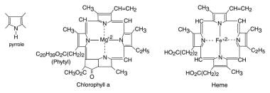
Tetrapyrroles
Bile Pigments
Urobilin
Biliverdine
Phycobilins
Porphyrins
Protoporphyrins
Phycocyanin
Chlorophyll
Aminolevulinic Acid
Heme
Organization of genes for tetrapyrrole biosynthesis in gram--positive bacteria. (1/160)
Clusters of genes encoding enzymes for tetrapyrrole biosynthesis were cloned from Bacillus sphaericus, Bacillus stearothermophilus, Brevibacillus brevis and Paenibacillus macerans. The sequences of all hemX genes found, and of a 6.3 kbp hem gene cluster from P. macerans, were determined. The structure of the hem gene clusters was compared to that of other Gram-positive bacteria. The Bacillus and Brevibacillus species have a conserved organization of the genes hemAXCDBL, required for biosynthesis of uroporphyrinogen III (UroIII) from glutamyl-tRNA. In P. macerans, the hem genes for UroIII synthesis are also closely linked but their organization is different: there is no hemX gene and the gene cluster also contains genes, cysG8 and cysG(A)-hemD, encoding the enzymes required for synthesis of sirohaem from UroIII. Bacillus subtilis contains genes for three proteins, NasF, YInD and YInF, with sequence similarity to Escherichia coli CysG, which is a multi-functional protein catalysing sirohaem synthesis from UroIII. It is shown that YInF is required for sirohaem synthesis and probably catalyses the precorrin-2 to sirohaem conversion. YInD probably catalyses precorrin-2 synthesis from UroIII and NasF seems to be specific for nitrite reduction. (+info)Crystal structure of allophycocyanin from red algae Porphyra yezoensis at 2.2-A resolution. (2/160)
The crystal structure of allophycocyanin from red algae Porphyra yezoensis (APC-PY) at 2.2-A resolution has been determined by the molecular replacement method. The crystal belongs to space group R32 with cell parameters a = b = 105.3 A, c = 189.4 A, alpha = beta = 90 degrees, gamma = 120 degrees. After several cycles of refinement using program X-PLOR and model building based on the electron density map, the crystallographic R-factor converged to 19.3% (R-free factor is 26.9%) in the range of 10.0 to 2.2 A. The r.m.s. deviations of bond length and angles are 0.015 A and 2.9 degrees, respectively. In the crystal, two APC-PY trimers associate face to face into a hexamer. The assembly of two trimers within the hexamer is similar to that of C-phycocyanin (C-PC) and R-phycoerythrin (R-PE) hexamers, but the assembly tightness of the two trimers to the hexamer is not so high as that in C-PC and R-PE hexamers. The chromophore-protein interactions and possible pathway of energy transfer were discussed. Phycocyanobilin 1alpha84 of APC-PY forms 5 hydrogen bonds with 3 residues in subunit 2beta of another monomer. In R-PE and C-PC, chromophore 1alpha84 only forms 1 hydrogen bond with 2beta77 residue in subunit 2beta. This result may support and explain great spectrum difference exists between APC trimer and monomer. (+info)Introduction of a new branchpoint in tetrapyrrole biosynthesis in Escherichia coli by co-expression of genes encoding the chlorophyll-specific enzymes magnesium chelatase and magnesium protoporphyrin methyltransferase. (3/160)
The genes encoding the three Mg chelatase subunits, ChlH, ChlI and ChlD, from the cyanobacterium Synechocystis PCC6803 were all cloned in the same pET9a-based Escherichia coli expression plasmid, forming an artificial chlH-I-D operon under the control of the strong T7 promoter. When a soluble extract from IPTG-induced E. coli cells containing the pET9a-ChlHID plasmid was assayed for Mg chelatase activity in vitro, a high activity was obtained, suggesting that all three subunits are present in a soluble and active form. The chlM gene of Synechocystis PCC6803 was also cloned in a pET-based E. coli expression vector. Soluble extract from an E. coli strain expressing chlM converted Mg-protoporphyrin IX to Mg-protoporphyrin monomethyl ester, demonstrating that chlM encodes the Mg-protoporphyrin methyltransferase of Synechocystis. Co-expression of the chlM gene together with the chlH-I-D construct yielded soluble protein extracts which converted protoporphyrin IX to Mg-protoporphyrin IX monomethyl ester without detectable accumulation of the Mg-protoporphyrin IX intermediate. Thus, active Mg chelatase and Mg-protoporphyrin IX methyltransferase can be coupled in E. coli extracts. Purified ChlI, -D and -H subunits in combination with purified ChlM protein were subsequently used to demonstrate in vitro that a molar ratio of ChlM to ChlH of 1 to 1 results in conversion of protoporphyrin IX to Mg-protoporphyrin monomethyl ester without significant accumulation of Mg-protoporphyrin. (+info)Swimming marine Synechococcus strains with widely different photosynthetic pigment ratios form a monophyletic group. (4/160)
Unicellular marine cyanobacteria are ubiquitous in both coastal and oligotrophic regimes. The contribution of these organisms to primary production and nutrient cycling is substantial on a global scale. Natural populations of marine Synechococcus strains include multiple genetic lineages, but the link, if any, between unique phenotypic traits and specific genetic groups is still not understood. We studied the genetic diversity (as determined by the DNA-dependent RNA polymerase rpoC1 gene sequence) of a set of marine Synechococcus isolates that are able to swim. Our results show that these isolates form a monophyletic group. This finding represents the first example of correspondence between a physiological trait and a phylogenetic group in marine Synechococcus. In contrast, the phycourobilin (PUB)/phycoerythrobilin (PEB) pigment ratios of members of the motile clade varied considerably. An isolate obtained from the California Current (strain CC9703) displayed a pigment signature identical to that of nonmotile strain WH7803, which is considered a model for low-PUB/PEB-ratio strains, whereas several motile strains had higher PUB/PEB ratios than strain WH8103, which is considered a model for high-PUB/PEB-ratio strains. These findings indicate that the PUB/PEB pigment ratio is not a useful characteristic for defining phylogenetic groups of marine Synechococcus strains. (+info)TspO of rhodobacter sphaeroides. A structural and functional model for the mammalian peripheral benzodiazepine receptor. (5/160)
The function and specific structural aspects of the tryptophan-rich sensory protein (TspO) of Rhodobacter sphaeroides 2.4.1 were studied using site-directed mutagenesis involving some 17 different amino acids. The choice of these amino acids changes was dictated from an analysis of the TspO family of proteins derived from the data bases. These studies demonstrated the importance of several highly conserved tryptophan residues in the sensory transduction pathway involving TspO through the proposed binding of an intermediate(s) in the tetrapyrrole biosynthesis pathway. These studies also revealed that the substitution of one or several of the amino acid residues dramatically affected, either directly or indirectly, the levels of TspO in the membranes of R. sphaeroides. Mounting evidence is presented suggesting that TspO normally forms a dimer within the bacterial outer membrane, and the dimer form of TspO may be the active form for TspO function. Because our earlier studies provided us with a functional framework within which to view these amino acid substitutions, we are able to suggest a preliminary model for TspO structure-function. Not only do these studies tell us more about TspO, but they also shed light on the TspO homologue, the drug-binding component of the mitochondrial peripheral benzodiazepine receptor. Mounting evidence draws numerous parallelism between these proteins and supports the significance of using TspO as a model for the structure and function of the mitochondrial protein. (+info)Novel activity of a phycobiliprotein lyase: both the attachment of phycocyanobilin and the isomerization to phycoviolobilin are catalyzed by the proteins PecE and PecF encoded by the phycoerythrocyanin operon. (6/160)
The structure of phycoviolobilin, the photoactive chromophore of alpha-phycoerythrocyanin, is incompatible with a chromophore ligation to the apoprotein via SH-addition (cysteine) to a Delta3, 3(1)-double bond of the phycobilin. The two putative phycoerythrocyanin lyase genes of Mastigocladus laminosus, pecE and pecF, were overexpressed in Escherichia coli. Their action has been studied on the addition reaction of phycocyanobilin to apo-alpha-phycoerythrocyanin (PecA). In the absence of the components of alpha-PEC-phycoviolobilin lyase PecE and PecF, or in the presence of only one of them, phycocyanobilin binds covalently to PecA forming a fluorescent chromoprotein with a red-shifted absorption (lambda(max)=641 nm) and low photoactivity (<10%). In the presence of both PecE and PecF, a chromoprotein forms which by its absorption (lambda(max)=565 nm) and high photoreversible photochromism (100% type I) has been identified as integral alpha-phycoerythrocyanin. We conclude that PecE and PecF jointly catalyze not only the addition of phycocyanobilin to PecA, but also its isomerization to the native phycoviolobilin chromophore. (+info)Arabidopsis phytochromes C and E have different spectral characteristics from those of phytochromes A and B. (7/160)
The red/far-red light absorbing phytochromes play a major role as sensor proteins in photomorphogenesis of plants. In Arabidopsis the phytochromes belong to a small gene family of five members, phytochrome A (phyA) to E (phyE). Knowledge of the dynamic properties of the phytochrome molecules is the basis of phytochrome signal transduction research. Beside photoconversion and destruction, dark reversion is a molecular property of some phytochromes. A possible role of dark reversion is the termination of signal transduction. Since Arabidopsis is a model plant for biological and genetic research, we focussed on spectroscopic characterization of Arabidopsis phytochromes, expressed in yeast. For the first time, we were able to determine the relative absorption maxima and minima for a phytochrome C (phyC) as 661/725 nm and for a phyE as 670/724 nm. The spectral characteristics of phyC and E are strictly different from those of phyA and B. Furthermore, we show that both phyC and phyE apoprotein chromophore adducts undergo a strong dark reversion. Difference spectra, monitored with phycocyanobilin and phytochromobilin as the apoprotein's chromophore, and in vivo dark reversion of the Arabidopsis phytochrome apoprotein phycocyanobilin adducts are discussed with respect to their physiological function. (+info)Role of magnesium chelatase activity in the early steps of the tetrapyrrole biosynthetic pathway. (8/160)
Magnesium-protoporphyrin IX chelatase (Mg-chelatase) is located at the branchpoint of tetrapyrrole biosynthesis, at which point protoporphyrin IX is distributed for the synthesis of chlorophyll and heme. We investigated the regulatory contribution of Mg-chelatase to the flow of metabolites. In plants, the enzyme complex consists of three subunits, designated CHL D, CHL I, and CHL H. Transgenic tobacco (Nicotiana tabacum) plants expressing antisense RNA for the Mg-chelatase subunit CHL H were analyzed to elucidate further the role of Mg-chelatase in the distribution of protoporphyrin IX into the branched tetrapyrrolic pathway. The transgenic plants displayed a reduced growth rate and chlorophyll deficiency. Both phenotypical properties were correlated with lower Mg-chelatase activity. Unexpectedly, less protoporphyrin IX and heme accumulated, and a decrease in 5-aminolevulinate (ALA)-synthesizing capacity and ALA dehydratase activity paralleled the progressive reduction in Mg-chelatase activity in the transformants compared with control plants. The reduced activities of the early enzymatic steps corresponded with lower levels of transcripts encoding glutamyl-tRNA reductase and ALA-dehydratase. The decreased expression and activities of early enzymes in the pathway could be explained by a feedback-controlled mechanism in response to lower Mg-chelatase activity. We discuss intercompartmental signaling that synchronizes the activities of the first steps in tetrapyrrolic metabolism with the late steps for the synthesis of end products. (+info)Tetrapyrroles are a class of organic compounds that contain four pyrrole rings joined together in a macrocyclic structure. They are important in biology because they form the core structure of many essential cofactors and prosthetic groups in proteins, including heme, chlorophyll, and cobalamin (vitamin B12).
Heme is a tetrapyrrole that contains iron and is a crucial component of hemoglobin, the protein responsible for oxygen transport in red blood cells. Chlorophyll is another tetrapyrrole that contains magnesium and plays a vital role in photosynthesis, the process by which plants convert light energy into chemical energy. Cobalamin contains cobalt and is essential for DNA synthesis, fatty acid metabolism, and neurotransmitter synthesis.
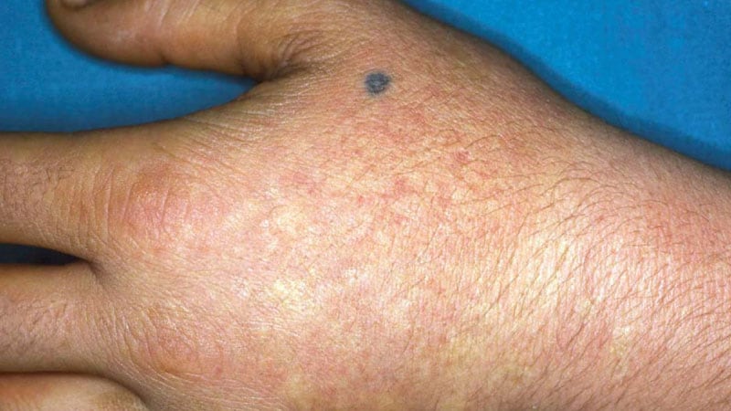
Abnormalities in tetrapyrrole biosynthesis can lead to various diseases, such as porphyrias, which are characterized by the accumulation of toxic intermediates in the heme biosynthetic pathway.
Bile pigments are the yellow-brown colored end products of hemoglobin breakdown in the liver. Hemoglobin is a protein found in red blood cells that carries oxygen throughout the body. When these cells are broken down, heme (the non-protein part of hemoglobin) is converted into biliverdin, which is then converted into bilirubin. Bilirubin is further metabolized and excreted by the liver as a component of bile, a digestive fluid that helps break down fats in the small intestine.
Under normal conditions, the liver effectively removes and excretes bilirubin from the body through the bile ducts into the small intestine. However, when there is an overproduction of bilirubin or a problem with its elimination, it can accumulate in the blood, leading to jaundice (yellowing of the skin and eyes) and other symptoms associated with liver dysfunction.
In summary, bile pigments are the waste products formed during the breakdown of hemoglobin, primarily consisting of bilirubin, which is eliminated from the body via the liver and bile ducts.
Urobilin is a pigment produced in the liver as a byproduct of the breakdown of bilirubin, which is a waste product resulting from the breakdown of hemoglobin in red blood cells. Some urobilin is excreted through the bile into the intestines, where it can be converted by bacteria into stercobilin, another pigment responsible for the brown color of feces. A portion of the urobilin produced in the liver is reabsorbed into the bloodstream and eventually excreted through the urine, giving it a yellow color. Therefore, urobilin can be detected in both urine and feces.
Biliverdine is a greenish pigment that is a byproduct of the breakdown of heme, which is a component of hemoglobin in red blood cells. It is formed when bilirubin, another byproduct of heme degradation, is reduced in the liver. Biliverdine is then converted back to bilirubin and excreted from the body as part of bile.
Elevated levels of biliverdine in the blood can indicate liver dysfunction or other medical conditions that affect the breakdown of heme. It may also be present in high concentrations in certain types of hemolytic anemia, where there is excessive destruction of red blood cells and subsequent release of large amounts of heme into the circulation.
Phycobilins are linear tetrapyrrole chromophores found in cyanobacteria, red algae, and glaucophytes. They are the light-harvesting pigments associated with phycobiliproteins in the phycobilisome complex, which is a type of antenna system used to capture light for photosynthesis. The main types of phycobilins are phycocyanobilin, phycoerythrobilin, and allophycocyanobilin. These pigments absorb light in the blue-green to red region of the electromagnetic spectrum and transfer the energy to chlorophyll a for use in photosynthesis. Phycobilins are also used as fluorescent labels in various biochemical and medical research applications.
Porphyrins are complex organic compounds that contain four pyrrole rings joined together by methine bridges (=CH-). They play a crucial role in the biochemistry of many organisms, as they form the core structure of various heme proteins and other metalloproteins. Some examples of these proteins include hemoglobin, myoglobin, cytochromes, and catalases, which are involved in essential processes such as oxygen transport, electron transfer, and oxidative metabolism.
In the human body, porphyrins are synthesized through a series of enzymatic reactions known as the heme biosynthesis pathway. Disruptions in this pathway can lead to an accumulation of porphyrins or their precursors, resulting in various medical conditions called porphyrias. These disorders can manifest as neurological symptoms, skin lesions, and gastrointestinal issues, depending on the specific type of porphyria and the site of enzyme deficiency.
It is important to note that while porphyrins are essential for life, their accumulation in excessive amounts or at inappropriate locations can result in pathological conditions. Therefore, understanding the regulation and function of porphyrin metabolism is crucial for diagnosing and managing porphyrias and other related disorders.
Protoporphyrins are organic compounds that are the immediate precursors to heme in the porphyrin synthesis pathway. They are composed of a porphyrin ring, which is a large, complex ring made up of four pyrrole rings joined together, with an acetate and a propionate side chain at each pyrrole. Protoporphyrins are commonly found in nature and are important components of many biological systems, including hemoglobin, the protein in red blood cells that carries oxygen throughout the body.
There are several different types of protoporphyrins, including protoporphyrin IX, which is the most common form found in humans and other animals. Protoporphyrins can be measured in the blood or other tissues as a way to diagnose or monitor certain medical conditions, such as lead poisoning or porphyrias, which are rare genetic disorders that affect the production of heme. Elevated levels of protoporphyrins in the blood or tissues can indicate the presence of these conditions and may require further evaluation and treatment.
Phycocyanin is a pigment-protein complex found in cyanobacteria and some types of algae, such as Spirulina. It belongs to the family of phycobiliproteins and plays a crucial role in the light-harvesting process during photosynthesis. Phycocyanin absorbs light in the orange and red regions of the visible spectrum and transfers the energy to chlorophyll for use in photosynthesis. It has been studied for its potential health benefits, including antioxidant, anti-inflammatory, and neuroprotective properties. However, more research is needed to fully understand its effects and potential therapeutic uses.
Chlorophyll is a green pigment found in the chloroplasts of photosynthetic plants, algae, and some bacteria. It plays an essential role in light-dependent reactions of photosynthesis by absorbing light energy, primarily from the blue and red parts of the electromagnetic spectrum, and converting it into chemical energy to fuel the synthesis of carbohydrates from carbon dioxide and water. The structure of chlorophyll includes a porphyrin ring, which binds a central magnesium ion, and a long phytol tail. There are several types of chlorophyll, including chlorophyll a and chlorophyll b, which have distinct absorption spectra and slightly different structures. Chlorophyll is crucial for the process of photosynthesis, enabling the conversion of sunlight into chemical energy and the release of oxygen as a byproduct.
"Pyrroles" is not a medical term in and of itself, but "pyrrole" is an organic compound that contains one nitrogen atom and four carbon atoms in a ring structure. In the context of human health, "pyrroles" often refers to a group of compounds called pyrrol derivatives or pyrrole metabolites.
In clinical settings, "pyrroles" is sometimes used to refer to a urinary metabolite called "pyrrole-protein conjugate," which contains a pyrrole ring and is excreted in the urine. Elevated levels of this compound have been associated with certain psychiatric and behavioral disorders, such as schizophrenia and mood disorders. However, the relationship between pyrroles and these conditions is not well understood, and more research is needed to establish a clear medical definition or diagnostic criteria for "pyrrole disorder" or "pyroluria."
Bilirubin is a yellowish pigment that is produced by the liver when it breaks down old red blood cells. It is a normal byproduct of hemoglobin metabolism and is usually conjugated (made water-soluble) in the liver before being excreted through the bile into the digestive system. Elevated levels of bilirubin can cause jaundice, a yellowing of the skin and eyes. Increased bilirubin levels may indicate liver disease or other medical conditions such as gallstones or hemolysis. It is also measured to assess liver function and to help diagnose various liver disorders.
Aminolevulinic acid (ALA) is a naturally occurring compound in the human body and is a key precursor in the biosynthesis of heme, which is a component of hemoglobin in red blood cells. It is also used as a photosensitizer in dermatology for the treatment of certain types of skin conditions such as actinic keratosis and basal cell carcinoma.
In medical terms, ALA is classified as an α-keto acid and a porphyrin precursor. It is synthesized in the mitochondria from glycine and succinyl-CoA in a reaction catalyzed by the enzyme aminolevulinic acid synthase. After its synthesis, ALA is transported to the cytosol where it undergoes further metabolism to form porphyrins, which are then used for heme biosynthesis in the mitochondria.
In dermatology, topical application of ALA followed by exposure to a specific wavelength of light can lead to the production of reactive oxygen species that destroy abnormal cells in the skin while leaving healthy cells unharmed. This makes it an effective treatment for precancerous and cancerous lesions on the skin.
It is important to note that ALA can cause photosensitivity, which means that patients who have undergone ALA-based treatments should avoid exposure to sunlight or other sources of bright light for a period of time after the treatment to prevent adverse reactions.
Aldehyde oxidoreductases are a class of enzymes that catalyze the oxidation of aldehydes to carboxylic acids using NAD+ or FAD as cofactors. They play a crucial role in the detoxification of aldehydes generated from various metabolic processes, such as lipid peroxidation and alcohol metabolism. These enzymes are widely distributed in nature and have been identified in bacteria, yeast, plants, and animals.
The oxidation reaction catalyzed by aldehyde oxidoreductases involves the transfer of electrons from the aldehyde substrate to the cofactor, resulting in the formation of a carboxylic acid and reduced NAD+ or FAD. The enzymes are classified into several families based on their sequence similarity and cofactor specificity.
One of the most well-known members of this family is alcohol dehydrogenase (ADH), which catalyzes the oxidation of alcohols to aldehydes or ketones as part of the alcohol metabolism pathway. Another important member is aldehyde dehydrogenase (ALDH), which further oxidizes the aldehydes generated by ADH to carboxylic acids, thereby preventing the accumulation of toxic aldehydes in the body.
Deficiencies in ALDH enzymes have been linked to several human diseases, including alcoholism and certain types of cancer. Therefore, understanding the structure and function of aldehyde oxidoreductases is essential for developing new therapeutic strategies to treat these conditions.
Heme is not a medical term per se, but it is a term used in the field of medicine and biology. Heme is a prosthetic group found in hemoproteins, which are proteins that contain a heme iron complex. This complex plays a crucial role in various biological processes, including oxygen transport (in hemoglobin), electron transfer (in cytochromes), and chemical catalysis (in peroxidases and catalases).
The heme group consists of an organic component called a porphyrin ring, which binds to a central iron atom. The iron atom can bind or release electrons, making it essential for redox reactions in the body. Heme is also vital for the formation of hemoglobin and myoglobin, proteins responsible for oxygen transport and storage in the blood and muscles, respectively.
In summary, heme is a complex organic-inorganic structure that plays a critical role in several biological processes, particularly in electron transfer and oxygen transport.
Molecular structure, in the context of biochemistry and molecular biology, refers to the arrangement and organization of atoms and chemical bonds within a molecule. It describes the three-dimensional layout of the constituent elements, including their spatial relationships, bond lengths, and angles. Understanding molecular structure is crucial for elucidating the functions and reactivities of biological macromolecules such as proteins, nucleic acids, lipids, and carbohydrates. Various experimental techniques, like X-ray crystallography, nuclear magnetic resonance (NMR) spectroscopy, and cryo-electron microscopy (cryo-EM), are employed to determine molecular structures at atomic resolution, providing valuable insights into their biological roles and potential therapeutic targets.