Micronucleus, Germline
Macronucleus
RNA, Protozoan
Tetrahymenina
Cilia
RNA, Catalytic
RNA, Ribosomal
Base Sequence
Molecular Sequence Data
Nucleic Acid Conformation
Cell Nucleus
Conjugation, Genetic
Introns
DNA, Ribosomal
Ciliophora
RNA
Histones
Amino Acid Sequence
Guanosine
Telomerase
Organoids
Dyneins
Tubulin
RNA Splicing
Microscopy, Electron
Mutation
Transformation, Genetic
DNA
Cloning, Molecular
Microtubules
Peptide Elongation Factor 1
Paramecium
Specificity from steric restrictions in the guanosine binding pocket of a group I ribozyme. (1/768)
The 3' splice site of group I introns is defined, in part, by base pairs between the intron core and residues just upstream of the splice site, referred to as P9.0. We have studied the specificity imparted by P9.0 using the well-characterized L-21 Scal ribozyme from Tetrahymena by adding residues to the 5' end of the guanosine (G) that functions as a nucleophile in the oligonucleotide cleavage reaction: CCCUCUA5 (S) + NNG <--> CCCUCU + NNGA5. UCG, predicted to form two base pairs in P9.0, reacts with a (kcat/KM) value approximately 10-fold greater than G, consistent with previous results. Altering the bases that form P9.0 in both the trinucleotide G analog and the ribozyme affects the specificity in the manner predicted for base-pairing. Strikingly, oligonucleotides incapable of forming P9.0 react approximately 10-fold more slowly than G, for which the mispaired residues are simply absent. The observed specificity is consistent with a model in which the P9.0 site is sterically restricted such that an energetic penalty, not present for G, must be overcome by G analogs with 5' extensions. Shortening S to include only one residue 3' of the cleavage site (CCCUCUA) eliminates this penalty and uniformly enhances the reactions of matched and mismatched oligonucleotides relative to guanosine. These results suggest that the 3' portion of S occupies the P9.0 site, sterically interfering with binding of G analogs with 5' extensions. Similar steric effects may more generally allow structured RNAs to avoid formation of incorrect contacts, thereby helping to avoid kinetic traps during folding and enhancing cooperative formation of the correct structure. (+info)A hydrogen-bonding triad stabilizes the chemical transition state of a group I ribozyme. (2/768)
BACKGROUND: The group I intron is an RNA enzyme capable of efficiently catalyzing phosphoryl-transfer reactions. Functional groups that stabilize the chemical transition state of the cleavage reaction have been identified, but they are all located within either the 5'-exon (P1) helix or the guanosine cofactor, which are the substrates of the reaction. Functional groups within the ribozyme active site are also expected to assist in transition-state stabilization, and their role must be explored to understand the chemical basis of group I intron catalysis. RESULTS: Using nucleotide analog interference mapping and site-specific functional group substitution experiments, we demonstrate that the 2'-OH at A207, a highly conserved nucleotide in the ribozyme active site, specifically stabilizes the chemical transition state by approximately 2 kcal mol-1. The A207 2'-OH only makes its contribution when the U(-1) 2'-OH immediately adjacent to the scissile phosphate is present, suggesting that the 2'-OHs of A207 and U(-1) interact during the chemical step. CONCLUSIONS: These data support a model in which the 3'-oxyanion leaving group of the transesterification reaction is stabilized by a hydrogen-bonding triad consisting of the 2'-OH groups of U(-1) and A207 and the exocyclic amine of G22. Because all three nucleotides occur within highly conserved non-canonical base pairings, this stabilization mechanism is likely to occur throughout group I introns. Although this mechanism utilizes functional groups distinctive of RNA enzymes, it is analogous to the transition states of some protein enzymes that perform similar phosphoryl-transfer reactions. (+info)Trans-activation of the Tetrahymena group I intron ribozyme via a non-native RNA-RNA interaction. (3/768)
The peripheral P2.1 domain of the Tetrahymena group I intron ribozyme has been shown to be non-essential for splicing. We found, however, that separately prepared P2.1 RNA efficiently accelerates the 3' splice-site-specific hydrolysis reaction of a mutant ribozyme lacking both P2.1 and its upstream region in trans. We report here the unusual properties of this trans-activation. Compensatory mutational analysis revealed that non-native long-range base-pairings between the loop region of P2.1 RNA and L5c region of the mutant ribozyme are needed for the activation in spite of the fact that P2.1 forms base-pairings with P9.1 in the Tetrahymena ribozyme. The trans -activation depends on the non-native RNA-RNA interaction together with the higher order structure of P2.1 RNA. This activation is unique among the known trans-activations that utilize native tertiary interactions or RNA chaperons. (+info)A conserved motif in group IC3 introns is a new class of GNRA receptor. (4/768)
Terminal tetraloops consisting of GNRA sequences are often found in biologically active large RNAs. The loops appear to contribute towards the organization of higher order RNA structures by forming specific tertiary interactions with their receptors. Group IC3 introns which possess a GAAA loop in the L2 region often have a phylogenetically conserved motif in their P8 domains. In this report, we show that this conserved motif stands as a new class of receptor that distinguishes the sequences of GNRA loops less stringently than previously known receptors. The motif can functionally substitute an 11 nt motif receptor in the Tetrahymena ribozyme. Its structural and functional similarity to one class of synthetic receptors obtained from in vitro selection is observed. (+info)The conjusome: a novel structure in Tetrahymena found only during sexual reorganization. (5/768)
A unique structure, the conjusome, has been identified and initially characterized in Tetrahymena thermophila. The conjusome appears only during a specific phase of conjugation. Immunofluorescence microscopy reveals that the conjusome is strongly labeled by antibodies to the protein Pdd1p. Pdd1p is a chromodomain protein and participates in the formation of chromatin-containing structures in developing macronuclear anlagen. Recent studies suggest that Pdd1p is physically associated with the elimination of specific germ-line sequences from developing macronuclei (anlagen) and may play a role in heterochromatin assembly. The conjusome contains Pdd1p, but it is devoid of any detectable DNA. The conjusome appears before DNA elimination begins in the developing anlagen and after Pdd1p is found in the parental macronucleus. Transmission electron microscopic observations reveal that the conjusome is not a membrane-bounded structure. The conjusome ranges in size from about 1 microm to sizes approaching 7 microm, depending on its maturity. It is composed of a coarse reticulum of a fibrous, electron dense material, interspersed with apparent background cytoplasm. Our initial characterization does suggest a number of possible functions for what may be a new, transient organelle. (+info)Tetrahymena telomerase ribonucleoprotein RNA-protein interactions. (6/768)
Telomerase is an enzyme that is essential for the replication and maintenance of chromosomal termini. It is a ribonucleoprotein consisting of a catalytic subunit, one or more associated proteins, and an integral RNA subunit that serves as a template for the synthesisof telomeric repeats. We identified a Tetrahymena telomerase RNA-protein complex by an electrophoretic mobility shift assay, using telomerase partially purified from whole cell extracts and radiolabeled, in vitro transcribed wild-type Tetrahymena telomerase RNA. Complex formation was specific as unlabeled Tetra-hymena telomerase RNA, but not Escherichia coli ribo-somal RNAs, competitively inhibited complex formation. Binding required concentrations of MgCl2of at least 10 mM and occurred over a wide range of potassium glutamate concentrations (20-220 mM). The RNA-protein complex was optimally reconstituted with a 30 degrees C preincubation for +info)A vector-based method for drawing RNA secondary structure. (7/768)
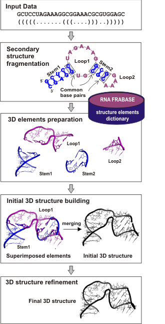
MOTIVATION: To produce a polygonal display of RNA secondary structure with minimal overlap and distortion of structural elements, with minimal search for positioning them, and with minimal user intervention. RESULTS: A new algorithm for automatically drawing RNA secondary structure has been developed. The algorithm represents the direction and space for a structural element using vector and vector space. Two heuristics are used. The first heuristic is concerned with ordering structural elements to be positioned and the second with positioning them in space. The algorithm and a graphical user interface have been implemented in a working program called VizQFolder on IBM PC compatibles. Experimental results demonstrate that VizQFolder is capable of automatically generating nearly overlap-free polygonal displays for long RNA molecules. The only distortion performed to avoid overlap is the rotation of helices, leading to efficient generation of a polygonal display without sacrificing its readability. VizQFolder is not coupled to a specific prediction program of RNA secondary structure, and thus can be used for visualizing secondary structure models obtained by any means. AVAILABILITY: The executable code of VizQFolder is available at http://automation.inha.ac.kr/khan. It can also be obtained from the authors upon request. (+info)Magnesium-dependent folding of self-splicing RNA: exploring the link between cooperativity, thermodynamics, and kinetics. (8/768)
Folding of the Tetrahymena self-splicing RNA into its active conformation involves a set of discrete intermediate states. The Mg2+-dependent equilibrium transition from the intermediates to the native structure is more cooperative than the formation of the intermediates from the unfolded states. We show that the degree of cooperativity is linked to the free energy of each transition and that the rate of the slow transition from the intermediates to the native state decreases exponentially with increasing Mg2+ concentration. Monovalent salts, which stabilize the folded RNA nonspecifically, induce states that fold in less than 30 s after Mg2+ is added to the RNA. A simple model is proposed that predicts the folding kinetics from the Mg2+-dependent change in the relative stabilities of the intermediate and native states. (+info)Tetrahymena is not a medical term itself, but it is a genus of unicellular organisms known as ciliates. They are commonly found in freshwater environments and can be studied in the field of biology and microbiology. Some species of Tetrahymena have been used in scientific research, including studies on genetics, cell division, and protein function. It is not a term that would typically be used in a medical context.
Tetrahymena pyriformis is not a medical term, but rather it's a species of ciliated protozoan that is commonly used in biological research. Here's a scientific definition:
Tetrahymena pyriformis is a free-living, freshwater ciliate protozoan species with a pear-shaped (pyriform) morphology. It belongs to the genus Tetrahymena and the family Euplotidae in the phylum Ciliophora. This microorganism is widely used as a model organism in various research fields, including cell biology, genetics, and molecular biology. Its relatively large size (50-60 µm), rapid growth rate, and ease of culturing make it an ideal subject for experimental studies. Tetrahymena pyriformis has complex cellular structures, such as macronuclei and micronuclei, which are involved in its reproduction and genetic inheritance. Additionally, this species is known for its ability to undergo rapid evolutionary changes, making it a valuable tool for studying evolution and adaptation.
Tetrahymena thermophila is not a medical term, but rather it refers to a species of ciliated protozoan that is commonly used in scientific research, including biomedical research. Here's a brief biological definition:
Tetrahymena thermophila is a free-living, freshwater ciliate protozoan found in various aquatic environments. It has a complex cell structure with two types of nuclei (a macronucleus and a micronucleus) and numerous cilia for movement. This organism is known for its ability to reproduce both sexually and asexually, making it a valuable model for studying genetic processes. Its genome has been fully sequenced, and it is widely used in research fields such as molecular biology, cell biology, and genetics due to its ease of cultivation and manipulation.
While not directly related to medical terminology, Tetrahymena thermophila has contributed significantly to our understanding of various biological processes with potential implications for medical research, including gene regulation, protein function, and DNA repair mechanisms.
A micronucleus is a small extranuclear body that can be formed when chromosome fragments or whole chromosomes fail to incorporate into the main nucleus during cell division. A germline micronucleus specifically refers to this occurrence in the cells that give rise to gametes, or reproductive cells (such as sperm or egg cells). Germline micronuclei are of particular interest in genetic toxicology and genetics research because they can indicate genetic damage or mutations, which may have implications for the health of future generations.
A macronucleus is a large, polyploid nucleus found in certain protozoa and some algal cells. It is responsible for the majority of the cell's vegetative functions, such as gene expression and protein synthesis, and it typically contains multiple copies of the genetic material. In contrast to the micronucleus, which is a smaller, diploid nucleus that is involved in the sexual reproduction of the cell, the macronucleus does not participate in the reproductive process.
In ciliates, such as Paramecium and Tetrahymena, the macronucleus is derived from the micronucleus during a process called differentiation. The micronucleus undergoes a series of divisions and develops into a multinucleated structure, which then fragments to form multiple macronuclei. These macronuclei are retained in the vegetative cells and are essential for their survival and function.
It is important to note that not all protozoa or algal cells have both a macronucleus and a micronucleus. Some species only have a single nucleus, while others may have multiple nuclei of different types. The presence and function of these various types of nuclei can vary significantly between different groups of organisms.
I'd be happy to help! However, I believe there might be a slight confusion in your question. RNA stands for Ribonucleic Acid, which is a type of nucleic acid involved in various biological roles in the coding, decoding, regulation, and expression of genes. On the other hand, protozoan refers to a unicellular organism from the kingdom Protista, which includes a wide variety of simple eukaryotic organisms such as amoebas, paramecia, and plasmodium (the malaria-causing parasite).
There isn't a specific medical definition for "RNA, protozoan" since RNA is a molecule present in all living cells, including human cells, and protozoans are a group of organisms. However, I can tell you that RNA plays crucial roles in protozoan biology, such as acting as a messenger between DNA and ribosomes during protein synthesis or regulating gene expression.
If you have any further questions or need more specific information about RNA in protozoans, please let me know!
I apologize for any confusion, but "Tetrahymenina" is not a medical term or concept. It is a taxonomic category in biology, specifically a suborder of ciliated protozoans, which are single-celled organisms. These organisms are often found in various aquatic environments and can move around using tiny hair-like structures called cilia.
If you have any questions related to medical terminology or concepts, I would be happy to help clarify those for you.
There doesn't seem to be a specific medical definition for "DNA, protozoan" as it is simply a reference to the DNA found in protozoa. Protozoa are single-celled eukaryotic organisms that can be found in various environments such as soil, water, and the digestive tracts of animals.
Protozoan DNA refers to the genetic material present in these organisms. It is composed of nucleic acids, including deoxyribonucleic acid (DNA) and ribonucleic acid (RNA), which contain the instructions for the development, growth, and reproduction of the protozoan.
The DNA in protozoa, like in other organisms, is made up of two strands of nucleotides that coil together to form a double helix. The four nucleotide bases that make up protozoan DNA are adenine (A), thymine (T), guanine (G), and cytosine (C). These bases pair with each other to form the rungs of the DNA ladder, with A always pairing with T and G always pairing with C.
The genetic information stored in protozoan DNA is encoded in the sequence of these nucleotide bases. This information is used to synthesize proteins, which are essential for the structure and function of the organism's cells. Protozoan DNA also contains other types of genetic material, such as regulatory sequences that control gene expression and repetitive elements with no known function.
Understanding the DNA of protozoa is important for studying their biology, evolution, and pathogenicity. It can help researchers develop new treatments for protozoan diseases and gain insights into the fundamental principles of genetics and cellular function.
Cilia are tiny, hair-like structures that protrude from the surface of many types of cells in the body. They are composed of a core bundle of microtubules surrounded by a protein matrix and are covered with a membrane. Cilia are involved in various cellular functions, including movement of fluid or mucus across the cell surface, detection of external stimuli, and regulation of signaling pathways.
There are two types of cilia: motile and non-motile. Motile cilia are able to move in a coordinated manner to propel fluids or particles across a surface, such as those found in the respiratory tract and reproductive organs. Non-motile cilia, also known as primary cilia, are present on most cells in the body and serve as sensory organelles that detect chemical and mechanical signals from the environment.
Defects in cilia structure or function can lead to a variety of diseases, collectively known as ciliopathies. These conditions can affect multiple organs and systems in the body, including the brain, kidneys, liver, and eyes. Examples of ciliopathies include polycystic kidney disease, Bardet-Biedl syndrome, and Meckel-Gruber syndrome.
A catalytic RNA, often referred to as a ribozyme, is a type of RNA molecule that has the ability to act as an enzyme and catalyze chemical reactions. These RNA molecules contain specific sequences and structures that allow them to bind to other molecules and accelerate chemical reactions without being consumed in the process.
Ribozymes play important roles in various biological processes, such as RNA splicing, translation regulation, and gene expression. One of the most well-known ribozymes is the self-splicing intron found in certain RNA molecules, which can excise itself from the host RNA and then ligase the flanking exons together.
The discovery of catalytic RNAs challenged the central dogma of molecular biology, which held that proteins were solely responsible for carrying out biological catalysis. The finding that RNA could also function as an enzyme opened up new avenues of research and expanded our understanding of the complexity and versatility of biological systems.
Genes in protozoa refer to the hereditary units of these single-celled organisms that carry genetic information necessary for their growth, development, and reproduction. These genes are made up of DNA (deoxyribonucleic acid) molecules, which contain sequences of nucleotide bases that code for specific proteins or RNA molecules. Protozoan genes are responsible for various functions, such as metabolism, response to environmental stimuli, and reproduction.
It is important to note that the study of protozoan genes has contributed significantly to our understanding of genetics and evolution, particularly in areas such as molecular biology, cell biology, and genomics. However, there is still much to be learned about the genetic diversity and complexity of these organisms, which continue to be an active area of research.
I'm sorry for any confusion, but "Protozoan Proteins" is not a specific medical or scientific term. Protozoa are single-celled eukaryotic organisms, and proteins are large biological molecules consisting of one or more chains of amino acid residues. Therefore, "Protozoan Proteins" generally refers to the various types of proteins found in protozoa.
However, if you're looking for information about proteins specific to certain protozoan parasites with medical relevance (such as Plasmodium falciparum, which causes malaria), I would be happy to help! Please provide more context or specify the particular protozoan of interest.
Ribosomal RNA (rRNA) is a type of RNA molecule that is a key component of ribosomes, which are the cellular structures where protein synthesis occurs in cells. In ribosomes, rRNA plays a crucial role in the process of translation, where genetic information from messenger RNA (mRNA) is translated into proteins.
Ribosomal RNA is synthesized in the nucleus and then transported to the cytoplasm, where it assembles with ribosomal proteins to form ribosomes. Within the ribosome, rRNA provides a structural framework for the assembly of the ribosome and also plays an active role in catalyzing the formation of peptide bonds between amino acids during protein synthesis.
There are several different types of rRNA molecules, including 5S, 5.8S, 18S, and 28S rRNA, which vary in size and function. These rRNA molecules are highly conserved across different species, indicating their essential role in protein synthesis and cellular function.
A base sequence in the context of molecular biology refers to the specific order of nucleotides in a DNA or RNA molecule. In DNA, these nucleotides are adenine (A), guanine (G), cytosine (C), and thymine (T). In RNA, uracil (U) takes the place of thymine. The base sequence contains genetic information that is transcribed into RNA and ultimately translated into proteins. It is the exact order of these bases that determines the genetic code and thus the function of the DNA or RNA molecule.
Molecular sequence data refers to the specific arrangement of molecules, most commonly nucleotides in DNA or RNA, or amino acids in proteins, that make up a biological macromolecule. This data is generated through laboratory techniques such as sequencing, and provides information about the exact order of the constituent molecules. This data is crucial in various fields of biology, including genetics, evolution, and molecular biology, allowing for comparisons between different organisms, identification of genetic variations, and studies of gene function and regulation.
Nucleic acid conformation refers to the three-dimensional structure that nucleic acids (DNA and RNA) adopt as a result of the bonding patterns between the atoms within the molecule. The primary structure of nucleic acids is determined by the sequence of nucleotides, while the conformation is influenced by factors such as the sugar-phosphate backbone, base stacking, and hydrogen bonding.
Two common conformations of DNA are the B-form and the A-form. The B-form is a right-handed helix with a diameter of about 20 Å and a pitch of 34 Å, while the A-form has a smaller diameter (about 18 Å) and a shorter pitch (about 25 Å). RNA typically adopts an A-form conformation.
The conformation of nucleic acids can have significant implications for their function, as it can affect their ability to interact with other molecules such as proteins or drugs. Understanding the conformational properties of nucleic acids is therefore an important area of research in molecular biology and medicine.
The cell nucleus is a membrane-bound organelle found in the eukaryotic cells (cells with a true nucleus). It contains most of the cell's genetic material, organized as DNA molecules in complex with proteins, RNA molecules, and histones to form chromosomes.
The primary function of the cell nucleus is to regulate and control the activities of the cell, including growth, metabolism, protein synthesis, and reproduction. It also plays a crucial role in the process of mitosis (cell division) by separating and protecting the genetic material during this process. The nuclear membrane, or nuclear envelope, surrounding the nucleus is composed of two lipid bilayers with numerous pores that allow for the selective transport of molecules between the nucleoplasm (nucleus interior) and the cytoplasm (cell exterior).
The cell nucleus is a vital structure in eukaryotic cells, and its dysfunction can lead to various diseases, including cancer and genetic disorders.
Genetic conjugation is a type of genetic transfer that occurs between bacterial cells. It involves the process of one bacterium (the donor) transferring a piece of its DNA to another bacterium (the recipient) through direct contact or via a bridge-like connection called a pilus. This transferred DNA may contain genes that provide the recipient cell with new traits, such as antibiotic resistance or virulence factors, which can make the bacteria more harmful or difficult to treat. Genetic conjugation is an important mechanism for the spread of antibiotic resistance and other traits among bacterial populations.
Introns are non-coding sequences of DNA that are present within the genes of eukaryotic organisms, including plants, animals, and humans. Introns are removed during the process of RNA splicing, in which the initial RNA transcript is cut and reconnected to form a mature, functional RNA molecule.
After the intron sequences are removed, the remaining coding sequences, known as exons, are joined together to create a continuous stretch of genetic information that can be translated into a protein or used to produce non-coding RNAs with specific functions. The removal of introns allows for greater flexibility in gene expression and regulation, enabling the generation of multiple proteins from a single gene through alternative splicing.
In summary, introns are non-coding DNA sequences within genes that are removed during RNA processing to create functional RNA molecules or proteins.
Ribosomal DNA (rDNA) refers to the specific regions of DNA in a cell that contain the genes for ribosomal RNA (rRNA). Ribosomes are complex structures composed of proteins and rRNA, which play a crucial role in protein synthesis by translating messenger RNA (mRNA) into proteins.
In humans, there are four types of rRNA molecules: 18S, 5.8S, 28S, and 5S. These rRNAs are encoded by multiple copies of rDNA genes that are organized in clusters on specific chromosomes. In humans, the majority of rDNA genes are located on the short arms of acrocentric chromosomes 13, 14, 15, 21, and 22.
Each cluster of rDNA genes contains both transcribed and non-transcribed spacer regions. The transcribed regions contain the genes for the four types of rRNA, while the non-transcribed spacers contain regulatory elements that control the transcription of the rRNA genes.
The number of rDNA copies varies between species and even within individuals of the same species. The copy number can also change during development and in response to environmental factors. Variations in rDNA copy number have been associated with various diseases, including cancer and neurological disorders.
Ciliophora is a phylum in the taxonomic classification system that consists of unicellular organisms commonly known as ciliates. These are characterized by the presence of hair-like structures called cilia, which are attached to the cell surface and beat in a coordinated manner to facilitate movement and feeding. Ciliophora includes a diverse group of organisms, many of which are found in aquatic environments. Examples of ciliates include Paramecium, Tetrahymena, and Vorticella.
RNA (Ribonucleic Acid) is a single-stranded, linear polymer of ribonucleotides. It is a nucleic acid present in the cells of all living organisms and some viruses. RNAs play crucial roles in various biological processes such as protein synthesis, gene regulation, and cellular signaling. There are several types of RNA including messenger RNA (mRNA), ribosomal RNA (rRNA), transfer RNA (tRNA), small nuclear RNA (snRNA), microRNA (miRNA), and long non-coding RNA (lncRNA). These RNAs differ in their structure, function, and location within the cell.
Histones are highly alkaline proteins found in the chromatin of eukaryotic cells. They are rich in basic amino acid residues, such as arginine and lysine, which give them their positive charge. Histones play a crucial role in packaging DNA into a more compact structure within the nucleus by forming a complex with it called a nucleosome. Each nucleosome contains about 146 base pairs of DNA wrapped around an octamer of eight histone proteins (two each of H2A, H2B, H3, and H4). The N-terminal tails of these histones are subject to various post-translational modifications, such as methylation, acetylation, and phosphorylation, which can influence chromatin structure and gene expression. Histone variants also exist, which can contribute to the regulation of specific genes and other nuclear processes.
Ciliophora is a group of protozoan organisms that are characterized by the presence of hair-like structures called cilia. Some species of Ciliophora can cause infections in humans, known as ciliophoriasis or ciliate infections. These infections typically occur in individuals with weakened immune systems, such as those with HIV/AIDS, cancer, or who are taking immunosuppressive drugs.
The most common way that Ciliophora infect humans is through the ingestion of contaminated food or water. Once inside the body, the ciliates can cause a range of symptoms depending on the species and the location of the infection. For example, infections in the gastrointestinal tract can cause abdominal pain, diarrhea, and vomiting, while lung infections can lead to coughing, wheezing, and difficulty breathing.
Treatment for Ciliophora infections typically involves the use of antiprotozoal medications, such as metronidazole or tinidazole. In severe cases, hospitalization may be necessary to manage symptoms and prevent complications. Preventing ciliophoriasis involves practicing good hygiene, avoiding contaminated food and water, and taking steps to boost the immune system in individuals who are at high risk of infection.
Paromomycin is an antiprotozoal medication, which belongs to the class of aminoglycoside antibiotics. It is primarily used to treat various intestinal infectious diseases caused by protozoa, such as amebiasis (an infection caused by Entamoeba histolytica) and giardiasis (an infection caused by Giardia lamblia). Paromomycin works by inhibiting the protein synthesis in the parasites, leading to their death. It is not typically used to treat bacterial infections in humans, as other aminoglycosides are.
It's important to note that paromomycin has limited systemic absorption and is primarily active within the gastrointestinal tract when taken orally. This makes it a valuable option for treating intestinal parasitic infections without causing significant harm to the beneficial bacteria in the gut or systemically affecting other organs.
Paromomycin is also used in veterinary medicine to treat various protozoal infections in animals, including leishmaniasis in dogs. The medication is available in different forms, such as tablets, capsules, and powder for oral suspension. As with any medication, paromomycin should be taken under the supervision of a healthcare professional, and its use may be subject to specific dosage, frequency, and duration guidelines.
Cell nucleus division, also known as nuclear division, is the process by which the genetic material within the cell nucleus, referred to as chromosomes, is separated into two equal sets in preparation for cell division. This process results in the formation of two daughter nuclei, each with a complete set of chromosomes.
There are two types of nuclear division: mitosis and meiosis.
Mitosis is the type of nuclear division that occurs in somatic cells (cells other than sex cells) during growth, repair, and maintenance of tissues. It results in the formation of two genetically identical daughter nuclei. The process of mitosis can be divided into several stages: prophase, prometaphase, metaphase, anaphase, and telophase.
Meiosis, on the other hand, is the type of nuclear division that occurs in sex cells (sperm and egg cells) during sexual reproduction. It results in the formation of four genetically unique daughter nuclei, each with half the number of chromosomes as the parent cell. Meiosis consists of two consecutive divisions: meiosis I and meiosis II.
Both types of nuclear division are essential for the growth, development, and reproduction of living organisms.
An amino acid sequence is the specific order of amino acids in a protein or peptide molecule, formed by the linking of the amino group (-NH2) of one amino acid to the carboxyl group (-COOH) of another amino acid through a peptide bond. The sequence is determined by the genetic code and is unique to each type of protein or peptide. It plays a crucial role in determining the three-dimensional structure and function of proteins.
Guanosine is a nucleoside that consists of a guanine base linked to a ribose sugar molecule through a beta-N9-glycosidic bond. It plays a crucial role in various biological processes, such as serving as a building block for DNA and RNA during replication and transcription. Guanosine triphosphate (GTP) and guanosine diphosphate (GDP) are important energy carriers and signaling molecules involved in intracellular regulation. Additionally, guanosine has been studied for its potential role as a neuroprotective agent and possible contribution to cell-to-cell communication.
Telomerase is an enzyme that adds repetitive DNA sequences (telomeres) to the ends of chromosomes, which are lost during each cell division due to the incomplete replication of the ends of linear chromosomes. Telomerase is not actively present in most somatic cells, but it is highly expressed in germ cells and stem cells, allowing them to divide indefinitely. However, in many types of cancer cells, telomerase is abnormally activated, which leads to the maintenance or lengthening of telomeres, contributing to their unlimited replicative potential and tumorigenesis.
Organoids are 3D tissue cultures grown from stem cells that mimic the structure and function of specific organs. They are used in research to study development, disease, and potential treatments. The term "organoid" refers to the fact that these cultures can organize themselves into structures that resemble rudimentary organs, with differentiated cell types arranged in a pattern similar to their counterparts in the body. Organoids can be derived from various sources, including embryonic stem cells, induced pluripotent stem cells (iPSCs), or adult stem cells, and they provide a valuable tool for studying complex biological processes in a controlled laboratory setting.
Dyneins are a type of motor protein that play an essential role in the movement of cellular components and structures within eukaryotic cells. They are responsible for generating force and motion along microtubules, which are critical components of the cell's cytoskeleton. Dyneins are involved in various cellular processes, including intracellular transport, organelle positioning, and cell division.
There are several types of dyneins, but the two main categories are cytoplasmic dyneins and axonemal dyneins. Cytoplasmic dyneins are responsible for moving various cargoes, such as vesicles, organelles, and mRNA complexes, toward the minus-end of microtubules, which is usually located near the cell center. Axonemal dyneins, on the other hand, are found in cilia and flagella and are responsible for their movement by sliding adjacent microtubules past each other.
Dyneins consist of multiple subunits, including heavy chains, intermediate chains, light-intermediate chains, and light chains. The heavy chains contain the motor domain that binds to microtubules and hydrolyzes ATP to generate force. Dysfunction in dynein proteins has been linked to various human diseases, such as neurodevelopmental disorders, ciliopathies, and cancer.
Tubulin is a type of protein that forms microtubules, which are hollow cylindrical structures involved in the cell's cytoskeleton. These structures play important roles in various cellular processes, including maintaining cell shape, cell division, and intracellular transport. There are two main types of tubulin proteins: alpha-tubulin and beta-tubulin. They polymerize to form heterodimers, which then assemble into microtubules. The assembly and disassembly of microtubules are dynamic processes that are regulated by various factors, including GTP hydrolysis, motor proteins, and microtubule-associated proteins (MAPs). Tubulin is an essential component of the eukaryotic cell and has been a target for anti-cancer drugs such as taxanes and vinca alkaloids.
RNA splicing is a post-transcriptional modification process in which the non-coding sequences (introns) are removed and the coding sequences (exons) are joined together in a messenger RNA (mRNA) molecule. This results in a continuous mRNA sequence that can be translated into a single protein. Alternative splicing, where different combinations of exons are included or excluded, allows for the creation of multiple proteins from a single gene.
Electron microscopy (EM) is a type of microscopy that uses a beam of electrons to create an image of the sample being examined, resulting in much higher magnification and resolution than light microscopy. There are several types of electron microscopy, including transmission electron microscopy (TEM), scanning electron microscopy (SEM), and reflection electron microscopy (REM).
In TEM, a beam of electrons is transmitted through a thin slice of the sample, and the electrons that pass through the sample are focused to form an image. This technique can provide detailed information about the internal structure of cells, viruses, and other biological specimens, as well as the composition and structure of materials at the atomic level.
In SEM, a beam of electrons is scanned across the surface of the sample, and the electrons that are scattered back from the surface are detected to create an image. This technique can provide information about the topography and composition of surfaces, as well as the structure of materials at the microscopic level.
REM is a variation of SEM in which the beam of electrons is reflected off the surface of the sample, rather than scattered back from it. This technique can provide information about the surface chemistry and composition of materials.
Electron microscopy has a wide range of applications in biology, medicine, and materials science, including the study of cellular structure and function, disease diagnosis, and the development of new materials and technologies.
A mutation is a permanent change in the DNA sequence of an organism's genome. Mutations can occur spontaneously or be caused by environmental factors such as exposure to radiation, chemicals, or viruses. They may have various effects on the organism, ranging from benign to harmful, depending on where they occur and whether they alter the function of essential proteins. In some cases, mutations can increase an individual's susceptibility to certain diseases or disorders, while in others, they may confer a survival advantage. Mutations are the driving force behind evolution, as they introduce new genetic variability into populations, which can then be acted upon by natural selection.
Genetic transformation is the process by which an organism's genetic material is altered or modified, typically through the introduction of foreign DNA. This can be achieved through various techniques such as:
* Gene transfer using vectors like plasmids, phages, or artificial chromosomes
* Direct uptake of naked DNA using methods like electroporation or chemically-mediated transfection
* Use of genome editing tools like CRISPR-Cas9 to introduce precise changes into the organism's genome.
The introduced DNA may come from another individual of the same species (cisgenic), from a different species (transgenic), or even be synthetically designed. The goal of genetic transformation is often to introduce new traits, functions, or characteristics that do not exist naturally in the organism, or to correct genetic defects.
This technique has broad applications in various fields, including molecular biology, biotechnology, and medical research, where it can be used to study gene function, develop genetically modified organisms (GMOs), create cell lines for drug screening, and even potentially treat genetic diseases through gene therapy.
Deoxyribonucleic acid (DNA) is the genetic material present in the cells of organisms where it is responsible for the storage and transmission of hereditary information. DNA is a long molecule that consists of two strands coiled together to form a double helix. Each strand is made up of a series of four nucleotide bases - adenine (A), guanine (G), cytosine (C), and thymine (T) - that are linked together by phosphate and sugar groups. The sequence of these bases along the length of the molecule encodes genetic information, with A always pairing with T and C always pairing with G. This base-pairing allows for the replication and transcription of DNA, which are essential processes in the functioning and reproduction of all living organisms.
Molecular cloning is a laboratory technique used to create multiple copies of a specific DNA sequence. This process involves several steps:
1. Isolation: The first step in molecular cloning is to isolate the DNA sequence of interest from the rest of the genomic DNA. This can be done using various methods such as PCR (polymerase chain reaction), restriction enzymes, or hybridization.
2. Vector construction: Once the DNA sequence of interest has been isolated, it must be inserted into a vector, which is a small circular DNA molecule that can replicate independently in a host cell. Common vectors used in molecular cloning include plasmids and phages.
3. Transformation: The constructed vector is then introduced into a host cell, usually a bacterial or yeast cell, through a process called transformation. This can be done using various methods such as electroporation or chemical transformation.
4. Selection: After transformation, the host cells are grown in selective media that allow only those cells containing the vector to grow. This ensures that the DNA sequence of interest has been successfully cloned into the vector.
5. Amplification: Once the host cells have been selected, they can be grown in large quantities to amplify the number of copies of the cloned DNA sequence.
Molecular cloning is a powerful tool in molecular biology and has numerous applications, including the production of recombinant proteins, gene therapy, functional analysis of genes, and genetic engineering.
Microtubules are hollow, cylindrical structures composed of tubulin proteins in the cytoskeleton of eukaryotic cells. They play crucial roles in various cellular processes such as maintaining cell shape, intracellular transport, and cell division (mitosis and meiosis). Microtubules are dynamic, undergoing continuous assembly and disassembly, which allows them to rapidly reorganize in response to cellular needs. They also form part of important cellular structures like centrioles, basal bodies, and cilia/flagella.
Peptide Elongation Factor 1 (PEF1) is not a commonly used medical term, but it is a term used in biochemistry and molecular biology. Here's the definition:
Peptide Elongation Factor 1 (also known as EF-Tu in prokaryotes or EFT1A/EFT1B in eukaryotes) is a protein involved in the elongation phase of protein synthesis, specifically during translation. It plays a crucial role in delivering aminoacyl-tRNAs to the ribosome, enabling the addition of new amino acids to the growing polypeptide chain.
In eukaryotic cells, EF1A and EF1B (also known as EF-Ts) form a complex that helps facilitate the binding of aminoacyl-tRNAs to the ribosome. In prokaryotic cells, EF-Tu forms a complex with GTP and aminoacyl-tRNA, which then binds to the ribosome. Once bound, GTP is hydrolyzed to GDP, causing a conformational change that releases the aminoacyl-tRNA into the acceptor site of the ribosome, allowing for peptide bond formation. The EF-Tu/GDP complex then dissociates from the ribosome and is recycled by another protein called EF-G (EF-G in prokaryotes or EFL1 in eukaryotes).
Therefore, Peptide Elongation Factor 1 plays a critical role in ensuring that the correct amino acids are added to the growing peptide chain during protein synthesis.
I'm sorry for any confusion, but "Paramecium" is not a medical term. It is a genus of unicellular organisms commonly found in freshwater environments. Paramecia are classified as ciliates due to the presence of hair-like structures called cilia on their surface. They use these cilia for locomotion and feeding. If you have any questions about biology or another topic, I'd be happy to try to help!
In the context of medicine and pharmacology, "kinetics" refers to the study of how a drug moves throughout the body, including its absorption, distribution, metabolism, and excretion (often abbreviated as ADME). This field is called "pharmacokinetics."
1. Absorption: This is the process of a drug moving from its site of administration into the bloodstream. Factors such as the route of administration (e.g., oral, intravenous, etc.), formulation, and individual physiological differences can affect absorption.
2. Distribution: Once a drug is in the bloodstream, it gets distributed throughout the body to various tissues and organs. This process is influenced by factors like blood flow, protein binding, and lipid solubility of the drug.
3. Metabolism: Drugs are often chemically modified in the body, typically in the liver, through processes known as metabolism. These changes can lead to the formation of active or inactive metabolites, which may then be further distributed, excreted, or undergo additional metabolic transformations.
4. Excretion: This is the process by which drugs and their metabolites are eliminated from the body, primarily through the kidneys (urine) and the liver (bile).
Understanding the kinetics of a drug is crucial for determining its optimal dosing regimen, potential interactions with other medications or foods, and any necessary adjustments for special populations like pediatric or geriatric patients, or those with impaired renal or hepatic function.
 Tetrahymena - Wikipedia
 1QSR: Crystal Structure Of Tetrahymena Gcn5 With Bound Acetyl- Coenzyme A
 Profilin Functions in Cytokinesis, Nuclear Positioning, and Stomatogenesis in Tetrahymena thermophila
 Swimming rate - Tetrahymena thermophila - BNID 111429
 Microtubule-membrane interactions in cilia. I. Isolation and characterization of ciliary membranes from Tetrahymena pyriformis
 An Investigation into the Molecular Targets Mediating Cannabidiol Action in the Free-living Ciliate Tetrahymena pyriformis -...
 Purification of Tetrahymena Telomerase and Cloning of Genes Encoding the Two Protein Components of the Enzyme
 REGULATION OF MICROTUBULES IN TETRAHYMENA | Citedby Results | Journal of Cell Biology | Rockefeller University Press
 Tails of Tetrahymena - Wikidata
 Isotype: -kappa-light-chain, MIgG2a, RIgG1, Positive Tested Species Reactivity: Rat, Tetrahymena
 Comparative transcriptome analysis uncovers roles of hydrogen sulfide for alleviating cadmium toxicity in Tetrahymena...
 TGD | Tetrahymena Genome Database Wiki
 Tetrahymena Stock Center - Stock ID SD01360
 Tetrahymena vorax | Illinois State Academy of Science - since 1907
  Update for our Microbial Eukaryotes website users | Broad Institute
 Dopplereffekt / Objekt - Hypnagogia(12inch) - CLONE.NL
 "Kinetodesmal Fibers in Tetrahymena thermophila" by Katherine Jones and Andrew Lee
 Cryo-Electron Microscope | UC Davis
 Respiration in Tetrahymena Is Different Than in Other Organisms - Step Water Rentals
 "The Effect of a Second Stress on Growth of Tetrahymena Pyriformis" by Janice M. Granum