Synovial Membrane
Synovitis
Arthritis, Rheumatoid
Synovial Fluid
Osteoarthritis
Rheumatoid Nodule
Temporomandibular Joint
Membranes
Membrane Lipids
Cell Membrane
Arthritis
Intracellular Membranes
Membrane Potentials
Arthritis-Encephalitis Virus, Caprine
Membranes, Artificial
Arthritis, Reactive
Gold Sodium Thiomalate
Cartilage, Articular
Erythrocyte Membrane
Membrane Fluidity
Chondromatosis, Synovial
Arthritis, Experimental
Joints
Immunoenzyme Techniques
Joint Capsule
Microscopy, Electron
Cell Membrane Permeability
Lentivirus Infections
Wrist Joint
Arthritis, Infectious
Basement Membrane
Cells, Cultured
Goats
Fibroblasts
Immunohistochemistry
Spondylitis, Ankylosing
Macrophages
Temporomandibular Joint Dysfunction Syndrome
Arthritis, Psoriatic
Osteoarthritis, Knee
Matrix Metalloproteinase 13
RNA, Messenger
Visna-maedi virus
Membrane Transport Proteins
Fluorescent Antibody Technique
Interleukin 1 Receptor Antagonist Protein
T-Lymphocytes
Rabbits
Gadolinium DTPA
Interleukin-1
Sialoglycoproteins
Lipid Bilayers
Cartilage
Antigens, Differentiation, Myelomonocytic
Tumor Necrosis Factor-alpha
Mitochondrial Membranes
Joint Diseases
Matrix Metalloproteinase 1
Protein Transport
Cytokines
Synaptic Membranes
Reverse Transcriptase Polymerase Chain Reaction
Culture Techniques
Antigens, CD
Base Sequence
Liposomes
Gene Expression
Magnetic Resonance Imaging
Protein Binding
Calcium
Models, Biological
HLA-DR Antigens
Cell Fractionation
Image Processing, Computer-Assisted
Collagen
B-Lymphocytes
Protein Structure, Tertiary
Carrier Proteins
Extraembryonic Membranes
Lymphocytes
Hydrogen-Ion Concentration
Detergents
Escherichia coli
In Situ Hybridization
Electrophoresis, Polyacrylamide Gel
Endoplasmic Reticulum
Clone Cells
Interleukin-6
Phosphatidylcholines
Golgi Apparatus
Mutation
Biological Markers
Microscopy, Fluorescence
Freeze Fracturing
Polymerase Chain Reaction
Molecular Sequence Data
Recombinant Fusion Proteins
Monocytes
Cell Membrane Structures
Potassium
Inflammation
Temperature
Subcellular Fractions
Mitochondria
Amino Acid Sequence
Biological Transport, Active
Endocytosis
Enzyme-Linked Immunosorbent Assay
Microscopy, Confocal
Disease Models, Animal
Cytoplasm
Erythrocytes
Models, Molecular
Binding Sites
Membrane Potential, Mitochondrial
Purple Membrane
Ion Channels
Sodium
Protein Conformation
Prevention of collagen-induced arthritis by gene delivery of soluble p75 tumour necrosis factor receptor. (1/3207)
Collagen type II-induced arthritis (CIA) in DBA/1 mice can be passively transferred to SCID mice with spleen B- and T-lymphocytes. In the present study, we show that infection ex vivo of splenocytes from arthritic DBA/1 mice with a retroviral vector, containing cDNA for the soluble form of human p75 receptor of tumour necrosis factor (TNF-R) before transfer, prevents the development of arthritis, bone erosion and joint inflammation in the SCID recipients. Assessment of IgG subclass levels and studies of synovial histology suggest that down-regulating the effector functions of T helper-type 1 (Th1) cells may, at least in part, explain the inhibition of arthritis in the SCID recipients. In contrast, the transfer of splenocytes infected with mouse TNF-alpha gene construct resulted in exacerbated arthritis and enhancement of IgG2a antibody levels. Intriguingly, infection of splenocytes from arthritic DBA/1 mice with a construct for mouse IL-10 had no modulating effect on the transfer of arthritis. The data suggest that manipulation of the immune system with cytokines, or cytokine inhibitors using gene transfer protocols can be an effective approach to ameliorate arthritis. (+info)Overexpression of human homologs of the bacterial DnaJ chaperone in the synovial tissue of patients with rheumatoid arthritis. (2/3207)
OBJECTIVE: To study the expression of the chaperone family of J proteins in the synovial tissue of patients with rheumatoid arthritis (RA) or osteoarthritis. METHODS: Rabbit antibodies specific for a synthetic peptide (pHSJ1: EAYEVLSDKHKREIYD), representing the most conserved part of all J domains thus far identified--among them the Drosophila tumor suppressor Tid56--were used in immunohistochemical analyses of frozen sections of synovial tissue and immunoblotting of protein extracts of adherent synovial cells. IgG specific for Tid56 was also used. RESULTS: Both antisera predominantly and intensely stained synovial lining cells from RA patients; other cells did not stain or stained only faintly. In immunoblots, anti-pHSJ1 specifically detected several bands with molecular weights of >74 kd (type I), 57-64 kd (type II), 41-48 kd (type III), and < or =36 kd (type IV). The strongest band detected in RA adherent synovial cells was the type II band, whereas in a B cell line, a type I band was prominent. CONCLUSION: Several potentially new members of the J family are described. The type II band represents the human homolog of the Drosophila Tid56 protein and is strongly expressed in RA synovial tissue. (+info)Establishment and characterization of nurse cell-like stromal cell lines from synovial tissues of patients with rheumatoid arthritis. (3/3207)
OBJECTIVE: To investigate the features of synovial stromal cells established from patients with rheumatoid arthritis (RA), and to define these cells as nurse cells. METHODS: Synovial nurse-like stromal cell lines (RA-SNCs) were established from patients with RA. These cell lines were examined for morphology, pseudoemperipolesis activity, cell surface markers, and cytokine production. The interaction between these RA-SNCs and a synovial tissue B cell clone was also examined. RESULTS: RA-SNCs had nurse cell activity. They spontaneously produced interleukin-6 (IL-6), IL-8, granulocyte colony-stimulating factor, and granulocyte-macrophage colony-stimulating factor. Furthermore, they produced IL-1beta and tumor necrosis factor alpha and expressed higher levels of the other cytokines after coculture with the B cell clone. Proliferation and Ig production by the B cell clone were dependent on direct contact with RA-SNCs. CONCLUSION: These results indicate that the RA-SNCs were nurse cells. The findings suggest that RA-SNCs may play an important role in the pathogenesis of RA by producing large amounts of cytokines and maintaining infiltrating lymphocytes. (+info)Serum response elements activate and cAMP responsive elements inhibit expression of transcription factor Egr-1 in synovial fibroblasts of rheumatoid arthritis patients. (4/3207)
Analyzing the induction kinetics and promoter elements regulating the expression of the transcription factor Egr-1, we found elevated levels of Egr-1-encoding mRNA in synovial fibroblasts of rheumatoid arthritis (RA) patients when compared to controls. By contrast, synovial lymphocytes and macrophages do not show an elevated Egr-1 transcription. Therefore, the overexpression of Egr-1 may serve as a diagnostic marker to characterize synovial fibroblasts of RA patients. To study the regulatory mechanisms controlling Egr-1 expression we analyzed the function of transcription factor binding sites located in the Egr-1 promoter. Individual transcription factor binding sites within the Egr-1 promoter were specifically mutated and Egr-1 promoter activity was tested using reporter gene constructs. Our experiments demonstrate that serum response elements are the main positive regulators and binding to a cAMP responsive element represents the major negative regulator for Egr-1 expression in synovial fibroblasts. In addition, we functionally defined a new element, which was not yet described in the human Egr-1 promoter and which serves as a second negative regulatory element for Egr-1 expression. Therefore increased serum response factor activity or failure of Egr-1 repressing signals may account for Egr-1 overexpression in RA synovial fibroblasts. (+info)Inhibition of IL-6 and IL-8 induction from cultured rheumatoid synovial fibroblasts by treatment with aurothioglucose. (5/3207)
Gold compounds have long been used in the treatment of rheumatoid arthritis (RA). However, their actions in RA have not been clarified. In this study, we examined the effect of one of the monovalent gold compounds, aurothioglucose (AuTG), on the IL-1-induced production of IL-6, IL-8 and granulocyte macrophage colony stimulating factor (GM-CSF) from rheumatoid synovial fibroblasts (RSF) isolated from three RA patients. IL-6 and IL-8 induction but not GM-CSF induction was inhibited in most of the RSF after pretreatment with AuTG. Since gene expression of these cytokines is known to be under the control of a common transcription factor, NF-kappaB, the effect of AuTG on the cellular localization of NF-kappaB (p65 subunit) and on NF-kappaB-DNA binding was examined. Although AuTG treatment did not prevent NF-kappaB nuclear translocation, AuTG blocked the DNA-binding activity of NF-kappaB when examined in vitro. Morphologically, both metal-specific cell staining using p-dimethylaminobenzylidene rhodamine and transmission electron microscopic examinations demonstrated the accumulation of metal gold in the cytoplama and some organella (mitochondria and lysosomes) of the AuTG-treated RSF. These results indicate that one of the anti-rheumatic actions of AuTG might be through its inhibitory action on NF-kappaB. (+info)Nuclear factor-kappa B activity in T cells from patients with rheumatic diseases: a preliminary report. (6/3207)
OBJECTIVE: The NF-kappa B/Rel family of transcription factors regulates the expression of many genes involved in the immune or inflammatory response at the transcriptional level. The aim of this study was to determine whether distinctive patterns of NF-kappa B activation are seen in different forms of joint disease. METHODS: The DNA binding activity of these nucleoproteins was examined in purified synovial and peripheral T cells from patients with various chronic rheumatic diseases (12: four with rheumatoid arthritis; five with spondyloarthropathies; and three with osteoarthritis). RESULTS: Electrophoretic mobility shift assays disclosed two specific complexes bound to a NF-kappa B specific 32P-labelled oligonucleotide in nucleoproteins extracted from purified T cells isolated from synovial fluid and peripheral blood of patients with rheumatoid arthritis. The complexes consisted of p50/p50 homodimers and p50/p65 heterodimers. Increased NF-kappa B binding to DNA in synovial T cells was observed relative to peripheral T cells. In non-rheumatoid arthritis, binding of NF-kappa B in synovial T cells was exclusively mediated by p50/p50 homodimers. CONCLUSION: Overall, the results suggest that NF-kappa B may play a central part in the activation of infiltrating T cells in chronic rheumatoid arthritis. The activation of this nuclear factor is qualitatively different in rheumatoid synovial T cells to that in other forms of non-rheumatoid arthritis (for example, osteoarthritis, spondyloarthropathies). (+info)Down regulation by iron of prostaglandin E2 production by human synovial fibroblasts. (7/3207)
OBJECTIVE: To examine the effect of iron on the prostaglandin (PG) E2 production by human synovial fibroblasts in vitro. METHODS: Human synovial fibroblasts were isolated from synovial tissue of rheumatoid arthritis (RA) and osteoarthritis (OA) patients and cultured in medium. Synovial fibroblasts were stimulated by human recombinant interleukin (IL) 1 beta (0.1-10 ng/ml) with or without ferric citrate (Fe-citrate, 0.01-1 mM). The amount of PGE2 in the culture medium was measured by an enzyme linked immunosorbent assay. RESULTS: The production of PGE2 by the synovial fibroblasts was increased by stimulation with IL1 beta at all concentrations tested. Fe-citrate but not sodium citrate (Na-citrate) down regulated the production of PGE2 by the synovial fibroblasts, both with and without stimulation by IL1 beta. Fe-citrate inhibited the spontaneous PGE2 production by the cells in a dose dependent manner, and a maximum inhibition by Fe-citrate was observed at the concentration of 0.1 mM with IL1 beta stimulation. The down regulation by iron was reversed by the co-addition of desferrioxamine (100 micrograms/ml), an iron chelator. CONCLUSION: Iron down regulates the PGE2 production by synovial fibroblasts in vitro. (+info)Plasma cell development in synovial germinal centers in patients with rheumatoid and reactive arthritis. (8/3207)
Plasma cells are found surrounding the inflammatory infiltrates of macrophages, T, and B cells in the synovial tissue of patients with rheumatoid and reactive arthritis. This characteristic arrangement suggests that in the synovial tissue CD20+ B cells differentiate into plasma cells. To examine clonal relationships, we have used micromanipulation to separately isolate CD20+ B cells and plasma cells from single infiltrates. DNA was extracted, and from both populations the VH/VL gene repertoires was determined. The data show that in the inflamed synovial tissue activated B cells are clonally expanded. During proliferation in the network of follicular dendritic cells, V gene variants are generated by the hypermutation mechanism. Surprisingly, we do not find identical rearrangements between CD20+ B cells and plasma cells. Nevertheless, the finding of clonally related plasma cells within single infiltrates suggests that these cells underwent terminal differentiation in the synovial tissue. These results indicate that B cell differentiation in the synovial tissue is a dynamic process. Whereas CD20+ B cells may turnover rapidly, plasma cells may well be long lived and thus accumulate in the synovial tissue. The analysis of individual B cells recovered from synovial tissue opens a new way to determine the specificity of those cells that take part in the local immune reaction. This will provide new insights into the pathogenesis of chronic inflammatory diseases like rheumatoid or reactive arthritis. (+info)The synovial membrane, also known as the synovium, is the soft tissue that lines the inner surface of the capsule of a synovial joint, which is a type of joint that allows for smooth movement between bones. This membrane secretes synovial fluid, a viscous substance that lubricates and nourishes the cartilage and helps to reduce friction within the joint during movement.
The synovial membrane has a highly specialized structure, consisting of two layers: the intima and the subintima. The intima is a thin layer of cells that are in direct contact with the synovial fluid, while the subintima is a more fibrous layer that contains blood vessels and nerves.
The main function of the synovial membrane is to produce and regulate the production of synovial fluid, as well as to provide nutrients to the articular cartilage. It also plays a role in the immune response within the joint, helping to protect against infection and inflammation. However, abnormalities in the synovial membrane can lead to conditions such as rheumatoid arthritis, where the membrane becomes inflamed and produces excess synovial fluid, leading to pain, swelling, and joint damage.
Synovitis is a medical condition characterized by inflammation of the synovial membrane, which is the soft tissue that lines the inner surface of joint capsules and tendon sheaths. The synovial membrane produces synovial fluid, which lubricates the joint and allows for smooth movement.
Inflammation of the synovial membrane can cause it to thicken, redden, and become painful and swollen. This can lead to stiffness, limited mobility, and discomfort in the affected joint or tendon sheath. Synovitis may occur as a result of injury, overuse, infection, or autoimmune diseases such as rheumatoid arthritis.
If left untreated, synovitis can cause irreversible damage to the joint and surrounding tissues, including cartilage loss and bone erosion. Treatment typically involves a combination of medications, physical therapy, and lifestyle modifications to reduce inflammation and manage pain.
Rheumatoid arthritis (RA) is a systemic autoimmune disease that primarily affects the joints. It is characterized by persistent inflammation, synovial hyperplasia, and subsequent damage to the articular cartilage and bone. The immune system mistakenly attacks the body's own tissues, specifically targeting the synovial membrane lining the joint capsule. This results in swelling, pain, warmth, and stiffness in affected joints, often most severely in the hands and feet.
RA can also have extra-articular manifestations, affecting other organs such as the lungs, heart, skin, eyes, and blood vessels. The exact cause of RA remains unknown, but it is believed to involve a complex interplay between genetic susceptibility and environmental triggers. Early diagnosis and treatment are crucial in managing rheumatoid arthritis to prevent joint damage, disability, and systemic complications.
Synovial fluid is a viscous, clear, and straw-colored fluid found in the cavities of synovial joints, bursae, and tendon sheaths. It is produced by the synovial membrane, which lines the inner surface of the capsule surrounding these structures.
The primary function of synovial fluid is to reduce friction between articulating surfaces, providing lubrication for smooth and painless movement. It also acts as a shock absorber, protecting the joints from external forces during physical activities. Synovial fluid contains nutrients that nourish the articular cartilage, hyaluronic acid, which provides its viscoelastic properties, and lubricin, a protein responsible for boundary lubrication.
Abnormalities in synovial fluid composition or volume can indicate joint-related disorders, such as osteoarthritis, rheumatoid arthritis, gout, infection, or trauma. Analysis of synovial fluid is often used diagnostically to determine the underlying cause of joint pain, inflammation, or dysfunction.
Osteoarthritis (OA) is a type of joint disease that is characterized by the breakdown and eventual loss of cartilage - the tissue that cushions the ends of bones where they meet in the joints. This breakdown can cause the bones to rub against each other, causing pain, stiffness, and loss of mobility. OA can occur in any joint, but it most commonly affects the hands, knees, hips, and spine. It is often associated with aging and can be caused or worsened by obesity, injury, or overuse.
The medical definition of osteoarthritis is: "a degenerative, non-inflammatory joint disease characterized by the loss of articular cartilage, bone remodeling, and the formation of osteophytes (bone spurs). It is often associated with pain, stiffness, and decreased range of motion in the affected joint."
A Rheumatoid nodule is defined as a type of non-suppurative inflammatory lesion that occurs in the subcutaneous tissue, commonly associated with rheumatoid arthritis (RA). These nodules are firm, round to oval shaped, and usually range from 0.5 to 5 cm in size. They are typically found over bony prominences such as the elbow, heel, or fingers, but can occur in various locations throughout the body.
Histologically, rheumatoid nodules are characterized by a central area of fibrinoid necrosis surrounded by palisading histiocytes and fibroblasts, with an outer layer of chronic inflammatory cells, including lymphocytes and plasma cells. Rheumatoid nodules can be asymptomatic or cause pain and discomfort, depending on their size and location. They are more common in patients with severe RA and are associated with a poorer prognosis.
The temporomandibular joint (TMJ) is the articulation between the mandible (lower jaw) and the temporal bone of the skull. It's a complex joint that involves the movement of two bones, several muscles, and various ligaments. The TMJ allows for movements like rotation and translation, enabling us to open and close our mouth, chew, speak, and yawn. Dysfunction in this joint can lead to temporomandibular joint disorders (TMD), which can cause pain, discomfort, and limited jaw movement.
In medical terms, membranes refer to thin layers of tissue that cover or line various structures in the body. They are composed of connective tissue and epithelial cells, and they can be found lining the outer surface of the body, internal organs, blood vessels, and nerves. There are several types of membranes in the human body, including:
1. Serous Membranes: These membranes line the inside of body cavities and cover the organs contained within them. They produce a lubricating fluid that reduces friction between the organ and the cavity wall. Examples include the pleura (lungs), pericardium (heart), and peritoneum (abdominal cavity).
2. Mucous Membranes: These membranes line the respiratory, gastrointestinal, and genitourinary tracts, as well as the inner surface of the eyelids and the nasal passages. They produce mucus to trap particles, bacteria, and other substances, which helps protect the body from infection.
3. Synovial Membranes: These membranes line the joint cavities and produce synovial fluid, which lubricates the joints and allows for smooth movement.
4. Meninges: These are three layers of membranes that cover and protect the brain and spinal cord. They include the dura mater (outermost layer), arachnoid mater (middle layer), and pia mater (innermost layer).
5. Amniotic Membrane: This is a thin, transparent membrane that surrounds and protects the fetus during pregnancy. It produces amniotic fluid, which provides a cushion for the developing baby and helps regulate its temperature.
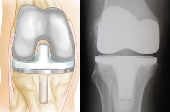
The knee joint, also known as the tibiofemoral joint, is the largest and one of the most complex joints in the human body. It is a synovial joint that connects the thighbone (femur) to the shinbone (tibia). The patella (kneecap), which is a sesamoid bone, is located in front of the knee joint and helps in the extension of the leg.
The knee joint is made up of three articulations: the femorotibial joint between the femur and tibia, the femoropatellar joint between the femur and patella, and the tibiofibular joint between the tibia and fibula. These articulations are surrounded by a fibrous capsule that encloses the synovial membrane, which secretes synovial fluid to lubricate the joint.
The knee joint is stabilized by several ligaments, including the medial and lateral collateral ligaments, which provide stability to the sides of the joint, and the anterior and posterior cruciate ligaments, which prevent excessive forward and backward movement of the tibia relative to the femur. The menisci, which are C-shaped fibrocartilaginous structures located between the femoral condyles and tibial plateaus, also help to stabilize the joint by absorbing shock and distributing weight evenly across the articular surfaces.
The knee joint allows for flexion, extension, and a small amount of rotation, making it essential for activities such as walking, running, jumping, and sitting.
Membrane lipids are the main component of biological membranes, forming a lipid bilayer in which various cellular processes take place. These lipids include phospholipids, glycolipids, and cholesterol. Phospholipids are the most abundant type, consisting of a hydrophilic head (containing a phosphate group) and two hydrophobic tails (composed of fatty acid chains). Glycolipids contain a sugar group attached to the lipid molecule. Cholesterol helps regulate membrane fluidity and permeability. Together, these lipids create a selectively permeable barrier that separates cells from their environment and organelles within cells.
A cell membrane, also known as the plasma membrane, is a thin semi-permeable phospholipid bilayer that surrounds all cells in animals, plants, and microorganisms. It functions as a barrier to control the movement of substances in and out of the cell, allowing necessary molecules such as nutrients, oxygen, and signaling molecules to enter while keeping out harmful substances and waste products. The cell membrane is composed mainly of phospholipids, which have hydrophilic (water-loving) heads and hydrophobic (water-fearing) tails. This unique structure allows the membrane to be flexible and fluid, yet selectively permeable. Additionally, various proteins are embedded in the membrane that serve as channels, pumps, receptors, and enzymes, contributing to the cell's overall functionality and communication with its environment.
Arthritis is a medical condition characterized by inflammation in one or more joints, leading to symptoms such as pain, stiffness, swelling, and reduced range of motion. There are many different types of arthritis, including osteoarthritis, rheumatoid arthritis, psoriatic arthritis, gout, and lupus, among others.
Osteoarthritis is the most common form of arthritis and is caused by wear and tear on the joints over time. Rheumatoid arthritis, on the other hand, is an autoimmune disorder in which the body's immune system mistakenly attacks the joint lining, causing inflammation and damage.
Arthritis can affect people of all ages, including children, although it is more common in older adults. Treatment for arthritis may include medications to manage pain and reduce inflammation, physical therapy, exercise, and in some cases, surgery.
Intracellular membranes refer to the membrane structures that exist within a eukaryotic cell (excluding bacteria and archaea, which are prokaryotic and do not have intracellular membranes). These membranes compartmentalize the cell, creating distinct organelles or functional regions with specific roles in various cellular processes.
Major types of intracellular membranes include:
1. Nuclear membrane (nuclear envelope): A double-membraned structure that surrounds and protects the genetic material within the nucleus. It consists of an outer and inner membrane, perforated by nuclear pores that regulate the transport of molecules between the nucleus and cytoplasm.
2. Endoplasmic reticulum (ER): An extensive network of interconnected tubules and sacs that serve as a major site for protein folding, modification, and lipid synthesis. The ER has two types: rough ER (with ribosomes on its surface) and smooth ER (without ribosomes).
3. Golgi apparatus/Golgi complex: A series of stacked membrane-bound compartments that process, sort, and modify proteins and lipids before they are transported to their final destinations within the cell or secreted out of the cell.
4. Lysosomes: Membrane-bound organelles containing hydrolytic enzymes for breaking down various biomolecules (proteins, carbohydrates, lipids, and nucleic acids) in the process called autophagy or from outside the cell via endocytosis.
5. Peroxisomes: Single-membrane organelles involved in various metabolic processes, such as fatty acid oxidation and detoxification of harmful substances like hydrogen peroxide.
6. Vacuoles: Membrane-bound compartments that store and transport various molecules, including nutrients, waste products, and enzymes. Plant cells have a large central vacuole for maintaining turgor pressure and storing metabolites.
7. Mitochondria: Double-membraned organelles responsible for generating energy (ATP) through oxidative phosphorylation and other metabolic processes, such as the citric acid cycle and fatty acid synthesis.
8. Chloroplasts: Double-membraned organelles found in plant cells that convert light energy into chemical energy during photosynthesis, producing oxygen and organic compounds (glucose) from carbon dioxide and water.
9. Endoplasmic reticulum (ER): A network of interconnected membrane-bound tubules involved in protein folding, modification, and transport; it is divided into two types: rough ER (with ribosomes on the surface) and smooth ER (without ribosomes).
10. Nucleus: Double-membraned organelle containing genetic material (DNA) and associated proteins involved in replication, transcription, RNA processing, and DNA repair. The nuclear membrane separates the nucleoplasm from the cytoplasm and contains nuclear pores for transporting molecules between the two compartments.
Membrane potential is the electrical potential difference across a cell membrane, typically for excitable cells such as nerve and muscle cells. It is the difference in electric charge between the inside and outside of a cell, created by the selective permeability of the cell membrane to different ions. The resting membrane potential of a typical animal cell is around -70 mV, with the interior being negative relative to the exterior. This potential is generated and maintained by the active transport of ions across the membrane, primarily through the action of the sodium-potassium pump. Membrane potentials play a crucial role in many physiological processes, including the transmission of nerve impulses and the contraction of muscle cells.
I apologize, but there is no medical definition for "Arthritis-Encephalitis Virus, Caprine." The term "Caprine" refers to something related to or derived from goats. Arthritis-Encephalitis Viruses (AEV) are a group of viruses that can affect various animal species and cause joint inflammation (arthritis) and brain inflammation (encephalitis). However, I couldn't find any specific virus named "Arthritis-Encephalitis Virus, Caprine" in the medical literature.
There are several viruses that can affect goats and cause arthritis and encephalitis, such as CAEV (Caprine Arthritis-Encephalitis Virus) or PPRV (Peste des Petits Ruminants Virus). If you have any specific concerns about a particular virus affecting goats, please provide more context so I can give you a more accurate and helpful response.
Artificial membranes are synthetic or man-made materials that possess properties similar to natural biological membranes, such as selective permeability and barrier functions. These membranes can be designed to control the movement of molecules, ions, or cells across them, making them useful in various medical and biotechnological applications.
Examples of artificial membranes include:
1. Dialysis membranes: Used in hemodialysis for patients with renal failure, these semi-permeable membranes filter waste products and excess fluids from the blood while retaining essential proteins and cells.
2. Hemofiltration membranes: Utilized in extracorporeal circuits to remove larger molecules, such as cytokines or inflammatory mediators, from the blood during critical illnesses or sepsis.
3. Drug delivery systems: Artificial membranes can be used to encapsulate drugs, allowing for controlled release and targeted drug delivery in specific tissues or cells.
4. Tissue engineering: Synthetic membranes serve as scaffolds for cell growth and tissue regeneration, guiding the formation of new functional tissues.
5. Biosensors: Artificial membranes can be integrated into biosensing devices to selectively detect and quantify biomolecules, such as proteins or nucleic acids, in diagnostic applications.
6. Microfluidics: Artificial membranes are used in microfluidic systems for lab-on-a-chip applications, enabling the manipulation and analysis of small volumes of fluids for various medical and biological purposes.
Reactive arthritis is a form of inflammatory arthritis that occurs in response to an infection in another part of the body, such as the genitals, urinary tract, or gastrointestinal tract. It is also known as Reiter's syndrome. The symptoms of reactive arthritis include joint pain and swelling, typically affecting the knees, ankles, and feet; inflammation of the eyes, skin, and mucous membranes; and urethritis or cervicitis. It is more common in men than women and usually develops within 1-4 weeks after a bacterial infection. The diagnosis is made based on the symptoms, medical history, physical examination, and laboratory tests. Treatment typically includes antibiotics to eliminate the underlying infection and medications to manage the symptoms of arthritis.
Gold sodium thiomalate is a disease-modifying antirheumatic drug (DMARD) that contains gold, which can help reduce pain, swelling, and stiffness in joints caused by rheumatoid arthritis. It works by possibly inhibiting certain enzymes involved in inflammation and modulating the immune system's response to reduce tissue damage.
This medication is given as an intramuscular injection and requires medical supervision due to its potential side effects, including kidney and liver problems, skin rashes, mouth sores, and changes in blood cell counts. Regular monitoring of blood and urine tests is necessary during treatment with gold sodium thiomalate.
It's important to note that the use of this medication has declined over time due to the availability of newer and more effective treatments for rheumatoid arthritis, as well as its potential side effects.
Articular cartilage is the smooth, white tissue that covers the ends of bones where they come together to form joints. It provides a cushion between bones and allows for smooth movement by reducing friction. Articular cartilage also absorbs shock and distributes loads evenly across the joint, protecting the bones from damage. It is avascular, meaning it does not have its own blood supply, and relies on the surrounding synovial fluid for nutrients. Over time, articular cartilage can wear down or become damaged due to injury or disease, leading to conditions such as osteoarthritis.
Arthroscopy is a minimally invasive surgical procedure where an orthopedic surgeon uses an arthroscope (a thin tube with a light and camera on the end) to diagnose and treat problems inside a joint. The surgeon makes a small incision, inserts the arthroscope into the joint, and then uses the attached camera to view the inside of the joint on a monitor. They can then insert other small instruments through additional incisions to repair or remove damaged tissue.
Arthroscopy is most commonly used for joints such as the knee, shoulder, hip, ankle, and wrist. It offers several advantages over traditional open surgery, including smaller incisions, less pain and bleeding, faster recovery time, and reduced risk of infection. The procedure can be used to diagnose and treat a wide range of conditions, including torn ligaments or cartilage, inflamed synovial tissue, loose bone or cartilage fragments, and joint damage caused by arthritis.
An erythrocyte, also known as a red blood cell, is a type of cell that circulates in the blood and is responsible for transporting oxygen throughout the body. The erythrocyte membrane refers to the thin, flexible barrier that surrounds the erythrocyte and helps to maintain its shape and stability.
The erythrocyte membrane is composed of a lipid bilayer, which contains various proteins and carbohydrates. These components help to regulate the movement of molecules into and out of the erythrocyte, as well as provide structural support and protection for the cell.
The main lipids found in the erythrocyte membrane are phospholipids and cholesterol, which are arranged in a bilayer structure with the hydrophilic (water-loving) heads facing outward and the hydrophobic (water-fearing) tails facing inward. This arrangement helps to maintain the integrity of the membrane and prevent the leakage of cellular components.
The proteins found in the erythrocyte membrane include integral proteins, which span the entire width of the membrane, and peripheral proteins, which are attached to the inner or outer surface of the membrane. These proteins play a variety of roles, such as transporting molecules across the membrane, maintaining the shape of the erythrocyte, and interacting with other cells and proteins in the body.
The carbohydrates found in the erythrocyte membrane are attached to the outer surface of the membrane and help to identify the cell as part of the body's own immune system. They also play a role in cell-cell recognition and adhesion.
Overall, the erythrocyte membrane is a complex and dynamic structure that plays a critical role in maintaining the function and integrity of red blood cells.
Membrane fluidity, in the context of cell biology, refers to the ability of the phospholipid bilayer that makes up the cell membrane to change its structure and organization in response to various factors. The membrane is not a static structure but rather a dynamic one, with its lipids constantly moving and changing position.
Membrane fluidity is determined by the fatty acid composition of the phospholipids that make up the bilayer. Lipids with unsaturated fatty acids have kinks in their hydrocarbon chains, which prevent them from packing closely together and increase membrane fluidity. In contrast, lipids with saturated fatty acids can pack closely together, reducing membrane fluidity.
Membrane fluidity is important for various cellular processes, including the movement of proteins within the membrane, the fusion of vesicles with the membrane during exocytosis and endocytosis, and the ability of the membrane to respond to changes in temperature and other environmental factors. Abnormalities in membrane fluidity have been linked to various diseases, including cancer, neurological disorders, and infectious diseases.
Synovial chondromatosis is a rare condition that affects the synovial membrane, which is the lining of joints, bursae (fluid-filled sacs that cushion bones), and tendon sheaths. In this condition, nodules made up of cartilage form in the synovial membrane. These nodules can detach from the synovial membrane and float freely in the synovial fluid, which lubricates the joint. If they become numerous, they can cause joint pain, stiffness, and decreased range of motion. In some cases, the loose bodies may also cause locking or catching sensations in the joint. Surgery is typically required to remove the cartilaginous nodules and relieve symptoms. If left untreated, synovial chondromatosis can lead to osteoarthritis and other joint problems.
Experimental arthritis refers to the induction of joint inflammation in animal models for the purpose of studying the disease process and testing potential treatments. This is typically achieved through the use of various methods such as injecting certain chemicals or proteins into the joints, genetically modifying animals to develop arthritis-like symptoms, or immunizing animals to induce an autoimmune response against their own joint tissues. These models are crucial for advancing our understanding of the underlying mechanisms of arthritis and for developing new therapies to treat this debilitating disease.
Intra-articular injections refer to the administration of medication directly into a joint space. This route of administration is used for treating various joint conditions such as inflammation, pain, and arthritis. Commonly injected medications include corticosteroids, local anesthetics, and viscosupplementation agents. The procedure is usually performed using imaging guidance, like ultrasound or fluoroscopy, to ensure accurate placement of the medication within the joint.
A joint is the location at which two or more bones make contact. They are constructed to allow movement and provide support and stability to the body during motion. Joints can be classified in several ways, including structure, function, and the type of tissue that forms them. The three main types of joints based on structure are fibrous (or fixed), cartilaginous, and synovial (or diarthrosis). Fibrous joints do not have a cavity and have limited movement, while cartilaginous joints allow for some movement and are connected by cartilage. Synovial joints, the most common and most movable type, have a space between the articular surfaces containing synovial fluid, which reduces friction and wear. Examples of synovial joints include hinge, pivot, ball-and-socket, saddle, and condyloid joints.
Immunoenzyme techniques are a group of laboratory methods used in immunology and clinical chemistry that combine the specificity of antibody-antigen reactions with the sensitivity and amplification capabilities of enzyme reactions. These techniques are primarily used for the detection, quantitation, or identification of various analytes (such as proteins, hormones, drugs, viruses, or bacteria) in biological samples.
In immunoenzyme techniques, an enzyme is linked to an antibody or antigen, creating a conjugate. This conjugate then interacts with the target analyte in the sample, forming an immune complex. The presence and amount of this immune complex can be visualized or measured by detecting the enzymatic activity associated with it.
There are several types of immunoenzyme techniques, including:
1. Enzyme-linked Immunosorbent Assay (ELISA): A widely used method for detecting and quantifying various analytes in a sample. In ELISA, an enzyme is attached to either the capture antibody or the detection antibody. After the immune complex formation, a substrate is added that reacts with the enzyme, producing a colored product that can be measured spectrophotometrically.
2. Immunoblotting (Western blot): A method used for detecting specific proteins in a complex mixture, such as a protein extract from cells or tissues. In this technique, proteins are separated by gel electrophoresis and transferred to a membrane, where they are probed with an enzyme-conjugated antibody directed against the target protein.
3. Immunohistochemistry (IHC): A method used for detecting specific antigens in tissue sections or cells. In IHC, an enzyme-conjugated primary or secondary antibody is applied to the sample, and the presence of the antigen is visualized using a chromogenic substrate that produces a colored product at the site of the antigen-antibody interaction.
4. Immunofluorescence (IF): A method used for detecting specific antigens in cells or tissues by employing fluorophore-conjugated antibodies. The presence of the antigen is visualized using a fluorescence microscope.
5. Enzyme-linked immunosorbent assay (ELISA): A method used for detecting and quantifying specific antigens or antibodies in liquid samples, such as serum or culture supernatants. In ELISA, an enzyme-conjugated detection antibody is added after the immune complex formation, and a substrate is added that reacts with the enzyme to produce a colored product that can be measured spectrophotometrically.
These techniques are widely used in research and diagnostic laboratories for various applications, including protein characterization, disease diagnosis, and monitoring treatment responses.
A joint capsule is the fibrous sac that encloses a synovial joint, which is a type of joint characterized by the presence of a cavity filled with synovial fluid. The joint capsule provides stability and strength to the joint, while also allowing for a range of motion. It consists of two layers: an outer fibrous layer and an inner synovial membrane. The fibrous layer is made up of dense connective tissue that helps to stabilize the joint, while the synovial membrane produces synovial fluid, which lubricates the joint and reduces friction during movement.
Electron microscopy (EM) is a type of microscopy that uses a beam of electrons to create an image of the sample being examined, resulting in much higher magnification and resolution than light microscopy. There are several types of electron microscopy, including transmission electron microscopy (TEM), scanning electron microscopy (SEM), and reflection electron microscopy (REM).
In TEM, a beam of electrons is transmitted through a thin slice of the sample, and the electrons that pass through the sample are focused to form an image. This technique can provide detailed information about the internal structure of cells, viruses, and other biological specimens, as well as the composition and structure of materials at the atomic level.
In SEM, a beam of electrons is scanned across the surface of the sample, and the electrons that are scattered back from the surface are detected to create an image. This technique can provide information about the topography and composition of surfaces, as well as the structure of materials at the microscopic level.
REM is a variation of SEM in which the beam of electrons is reflected off the surface of the sample, rather than scattered back from it. This technique can provide information about the surface chemistry and composition of materials.
Electron microscopy has a wide range of applications in biology, medicine, and materials science, including the study of cellular structure and function, disease diagnosis, and the development of new materials and technologies.
Cell membrane permeability refers to the ability of various substances, such as molecules and ions, to pass through the cell membrane. The cell membrane, also known as the plasma membrane, is a thin, flexible barrier that surrounds all cells, controlling what enters and leaves the cell. Its primary function is to protect the cell's internal environment and maintain homeostasis.
The permeability of the cell membrane depends on its structure, which consists of a phospholipid bilayer interspersed with proteins. The hydrophilic (water-loving) heads of the phospholipids face outward, while the hydrophobic (water-fearing) tails face inward, creating a barrier that is generally impermeable to large, polar, or charged molecules.
However, specific proteins within the membrane, called channels and transporters, allow certain substances to cross the membrane. Channels are protein structures that span the membrane and provide a pore for ions or small uncharged molecules to pass through. Transporters, on the other hand, are proteins that bind to specific molecules and facilitate their movement across the membrane, often using energy in the form of ATP.
The permeability of the cell membrane can be influenced by various factors, such as temperature, pH, and the presence of certain chemicals or drugs. Changes in permeability can have significant consequences for the cell's function and survival, as they can disrupt ion balances, nutrient uptake, waste removal, and signal transduction.
Lentivirus infections refer to the infectious disease caused by lentiviruses, a genus of retroviruses. These viruses are characterized by their ability to cause persistent and long-term infections, often leading to chronic diseases. They primarily target cells of the immune system, such as T-cells and macrophages, and can cause significant immunosuppression.
Lentiviruses have a slow replication cycle and can remain dormant in the host for extended periods. This makes them particularly effective at evading the host's immune response and can result in progressive damage to infected tissues over time.
One of the most well-known lentiviruses is the human immunodeficiency virus (HIV), which causes acquired immunodeficiency syndrome (AIDS). HIV infects and destroys CD4+ T-cells, leading to a weakened immune system and increased susceptibility to opportunistic infections.
Other examples of lentiviruses include simian immunodeficiency virus (SIV), feline immunodeficiency virus (FIV), and equine infectious anemia virus (EIAV). While these viruses primarily infect non-human animals, they are closely related to HIV and serve as important models for studying lentivirus infections and developing potential therapies.
The wrist joint, also known as the radiocarpal joint, is a condyloid joint that connects the distal end of the radius bone in the forearm to the proximal row of carpal bones in the hand (scaphoid, lunate, and triquetral bones). It allows for flexion, extension, radial deviation, and ulnar deviation movements of the hand. The wrist joint is surrounded by a capsule and reinforced by several ligaments that provide stability and strength to the joint.
Infectious arthritis, also known as septic arthritis, is a type of joint inflammation that is caused by a bacterial or fungal infection. The infection can enter the joint through the bloodstream or directly into the synovial fluid of the joint, often as a result of a traumatic injury, surgery, or an underlying condition such as diabetes or a weakened immune system.
The most common symptoms of infectious arthritis include sudden onset of severe pain and swelling in the affected joint, fever, chills, and difficulty moving the joint. If left untreated, infectious arthritis can lead to serious complications such as joint damage or destruction, sepsis, and even death. Treatment typically involves antibiotics or antifungal medications to eliminate the infection, along with rest, immobilization, and sometimes surgery to drain the infected synovial fluid.
It is important to seek medical attention promptly if you experience symptoms of infectious arthritis, as early diagnosis and treatment can help prevent long-term complications and improve outcomes.
The basement membrane is a thin, specialized layer of extracellular matrix that provides structural support and separates epithelial cells (which line the outer surfaces of organs and blood vessels) from connective tissue. It is composed of two main layers: the basal lamina, which is produced by the epithelial cells, and the reticular lamina, which is produced by the connective tissue. The basement membrane plays important roles in cell adhesion, migration, differentiation, and survival.
The basal lamina is composed mainly of type IV collagen, laminins, nidogens, and proteoglycans, while the reticular lamina contains type III collagen, fibronectin, and other matrix proteins. The basement membrane also contains a variety of growth factors and cytokines that can influence cell behavior.
Defects in the composition or organization of the basement membrane can lead to various diseases, including kidney disease, eye disease, and skin blistering disorders.
"Cells, cultured" is a medical term that refers to cells that have been removed from an organism and grown in controlled laboratory conditions outside of the body. This process is called cell culture and it allows scientists to study cells in a more controlled and accessible environment than they would have inside the body. Cultured cells can be derived from a variety of sources, including tissues, organs, or fluids from humans, animals, or cell lines that have been previously established in the laboratory.
Cell culture involves several steps, including isolation of the cells from the tissue, purification and characterization of the cells, and maintenance of the cells in appropriate growth conditions. The cells are typically grown in specialized media that contain nutrients, growth factors, and other components necessary for their survival and proliferation. Cultured cells can be used for a variety of purposes, including basic research, drug development and testing, and production of biological products such as vaccines and gene therapies.
It is important to note that cultured cells may behave differently than they do in the body, and results obtained from cell culture studies may not always translate directly to human physiology or disease. Therefore, it is essential to validate findings from cell culture experiments using additional models and ultimately in clinical trials involving human subjects.
I'm sorry for any confusion, but "goats" is not a term commonly used in medical definitions. It is a common noun referring to the domesticated animal species Capra aegagrus hircus. If you have any questions about a specific medical condition or term, please provide that and I would be happy to help.
Fibroblasts are specialized cells that play a critical role in the body's immune response and wound healing process. They are responsible for producing and maintaining the extracellular matrix (ECM), which is the non-cellular component present within all tissues and organs, providing structural support and biochemical signals for surrounding cells.
Fibroblasts produce various ECM proteins such as collagens, elastin, fibronectin, and laminins, forming a complex network of fibers that give tissues their strength and flexibility. They also help in the regulation of tissue homeostasis by controlling the turnover of ECM components through the process of remodeling.
In response to injury or infection, fibroblasts become activated and start to proliferate rapidly, migrating towards the site of damage. Here, they participate in the inflammatory response, releasing cytokines and chemokines that attract immune cells to the area. Additionally, they deposit new ECM components to help repair the damaged tissue and restore its functionality.
Dysregulation of fibroblast activity has been implicated in several pathological conditions, including fibrosis (excessive scarring), cancer (where they can contribute to tumor growth and progression), and autoimmune diseases (such as rheumatoid arthritis).
Immunohistochemistry (IHC) is a technique used in pathology and laboratory medicine to identify specific proteins or antigens in tissue sections. It combines the principles of immunology and histology to detect the presence and location of these target molecules within cells and tissues. This technique utilizes antibodies that are specific to the protein or antigen of interest, which are then tagged with a detection system such as a chromogen or fluorophore. The stained tissue sections can be examined under a microscope, allowing for the visualization and analysis of the distribution and expression patterns of the target molecule in the context of the tissue architecture. Immunohistochemistry is widely used in diagnostic pathology to help identify various diseases, including cancer, infectious diseases, and immune-mediated disorders.
Ankylosing spondylitis is a type of inflammatory arthritis that primarily affects the spine, although other joints can also be involved. It causes swelling in the spinal joints (vertebrae) that can lead to stiffness and pain. Over time, some of these joints may grow together, causing new bone formation and resulting in a rigid spine. This fusion of the spine is called ankylosis.
The condition typically begins in the sacroiliac joints, where the spine connects to the pelvis. From there, it can spread up the spine and potentially involve other areas of the body such as the eyes, heart, lungs, and gastrointestinal system.
Ankylosing spondylitis has a strong genetic link, with most people carrying the HLA-B27 gene. However, not everyone with this gene will develop the condition. It primarily affects males more often than females and tends to start in early adulthood.
Treatment usually involves a combination of medication, physical therapy, and exercise to help manage pain, maintain mobility, and prevent deformity. In severe cases, surgery may be considered.
Macrophages are a type of white blood cell that are an essential part of the immune system. They are large, specialized cells that engulf and destroy foreign substances, such as bacteria, viruses, parasites, and fungi, as well as damaged or dead cells. Macrophages are found throughout the body, including in the bloodstream, lymph nodes, spleen, liver, lungs, and connective tissues. They play a critical role in inflammation, immune response, and tissue repair and remodeling.
Macrophages originate from monocytes, which are a type of white blood cell produced in the bone marrow. When monocytes enter the tissues, they differentiate into macrophages, which have a larger size and more specialized functions than monocytes. Macrophages can change their shape and move through tissues to reach sites of infection or injury. They also produce cytokines, chemokines, and other signaling molecules that help coordinate the immune response and recruit other immune cells to the site of infection or injury.
Macrophages have a variety of surface receptors that allow them to recognize and respond to different types of foreign substances and signals from other cells. They can engulf and digest foreign particles, bacteria, and viruses through a process called phagocytosis. Macrophages also play a role in presenting antigens to T cells, which are another type of immune cell that helps coordinate the immune response.
Overall, macrophages are crucial for maintaining tissue homeostasis, defending against infection, and promoting wound healing and tissue repair. Dysregulation of macrophage function has been implicated in a variety of diseases, including cancer, autoimmune disorders, and chronic inflammatory conditions.
Temporomandibular Joint Dysfunction Syndrome, often abbreviated as TMJD or TMD, is a group of conditions that cause pain and dysfunction in the temporomandibular joint (TMJ) - the joint that connects the jawbone to the skull. Here's a more detailed medical definition:
Temporomandibular Joint Dysfunction Syndrome is a complex disorder characterized by pain, clicking, popping, or grating sounds in the TMJ; limited movement or locking of the jaw; and/or painful chewing movements. The condition may be caused by a variety of factors, including muscle tension, joint inflammation, structural problems with the joint itself, or injury to the head, neck, or jaw.
Symptoms of TMJD can include:
- Pain or tenderness in the face, jaw joint area, neck, and/or shoulders
- Limited ability to open the mouth wide
- Jaw locking, making it difficult to close or open the mouth
- Clicking, popping, or grating sounds in the TMJ when opening or closing the mouth
- A significant change in the way the upper and lower teeth fit together
- Headaches, earaches, dizziness, and hearing problems
Treatment for TMJD can vary depending on the severity of the condition and its underlying cause. It may include self-care practices such as eating soft foods, avoiding extreme jaw movements, and practicing relaxation techniques; physical therapy; medication to reduce pain and inflammation; dental treatments such as mouthguards or bite adjustments; and, in rare cases, surgery.
Psoriatic arthritis is a form of inflammatory arthritis that occurs in some people with psoriasis, a skin condition characterized by scaly, red, and itchy patches. The Arthritis Foundation defines psoriatic arthritis as "a chronic disease characterized by swelling, pain, and stiffness in and around the joints. It usually affects the fingers and toes but can also affect the lower back, knees, ankles, and spine."
Psoriatic arthritis can cause a variety of symptoms, including:
* Joint pain, swelling, and stiffness
* Swollen fingers or toes (dactylitis)
* Tenderness, pain, and swelling where tendons and ligaments attach to bones (enthesitis)
* Changes in nail growth, such as pitting, ridging, or separation from the nail bed
* Fatigue and weakness
* Reduced range of motion and mobility
The exact cause of psoriatic arthritis is not fully understood, but it is believed to involve a combination of genetic, environmental, and immune system factors. Treatment typically involves a combination of medications, lifestyle changes, and physical therapy to manage symptoms and prevent joint damage.
Osteoarthritis (OA) of the knee is a degenerative joint disease that affects the articular cartilage and subchondral bone in the knee joint. It is characterized by the breakdown and eventual loss of the smooth, cushioning cartilage that covers the ends of bones and allows for easy movement within joints. As the cartilage wears away, the bones rub against each other, causing pain, stiffness, and limited mobility. Osteoarthritis of the knee can also lead to the formation of bone spurs (osteophytes) and cysts in the joint. This condition is most commonly found in older adults, but it can also occur in younger people as a result of injury or overuse. Risk factors include obesity, family history, previous joint injuries, and repetitive stress on the knee joint. Treatment options typically include pain management, physical therapy, and in some cases, surgery.
Medical Definition:
Matrix Metalloproteinase 13 (MMP-13), also known as collagenase 3, is an enzyme belonging to the family of Matrix Metalloproteinases. These enzymes are involved in the degradation of extracellular matrix components, playing crucial roles in various physiological and pathological processes such as tissue remodeling, wound healing, and cancer progression.
MMP-13 has a specific affinity for cleaving type II collagen, one of the major structural proteins found in articular cartilage. It is also capable of degrading other extracellular matrix components like proteoglycans, elastin, and gelatin. This enzyme is primarily produced by chondrocytes, synovial fibroblasts, and osteoblasts.
Increased expression and activity of MMP-13 have been implicated in the pathogenesis of several diseases, most notably osteoarthritis (OA) and cancer. In OA, overexpression of MMP-13 leads to excessive degradation of articular cartilage, contributing to joint damage and degeneration. In cancer, MMP-13 facilitates tumor cell invasion and metastasis by breaking down the surrounding extracellular matrix.
Regulation of MMP-13 activity is essential for maintaining tissue homeostasis and preventing disease progression. Various therapeutic strategies aiming to inhibit MMP-13 activity are being explored as potential treatments for osteoarthritis and cancer.
Monoclonal antibodies are a type of antibody that are identical because they are produced by a single clone of cells. They are laboratory-produced molecules that act like human antibodies in the immune system. They can be designed to attach to specific proteins found on the surface of cancer cells, making them useful for targeting and treating cancer. Monoclonal antibodies can also be used as a therapy for other diseases, such as autoimmune disorders and inflammatory conditions.
Monoclonal antibodies are produced by fusing a single type of immune cell, called a B cell, with a tumor cell to create a hybrid cell, or hybridoma. This hybrid cell is then able to replicate indefinitely, producing a large number of identical copies of the original antibody. These antibodies can be further modified and engineered to enhance their ability to bind to specific targets, increase their stability, and improve their effectiveness as therapeutic agents.
Monoclonal antibodies have several mechanisms of action in cancer therapy. They can directly kill cancer cells by binding to them and triggering an immune response. They can also block the signals that promote cancer growth and survival. Additionally, monoclonal antibodies can be used to deliver drugs or radiation directly to cancer cells, increasing the effectiveness of these treatments while minimizing their side effects on healthy tissues.
Monoclonal antibodies have become an important tool in modern medicine, with several approved for use in cancer therapy and other diseases. They are continuing to be studied and developed as a promising approach to treating a wide range of medical conditions.
Messenger RNA (mRNA) is a type of RNA (ribonucleic acid) that carries genetic information copied from DNA in the form of a series of three-base code "words," each of which specifies a particular amino acid. This information is used by the cell's machinery to construct proteins, a process known as translation. After being transcribed from DNA, mRNA travels out of the nucleus to the ribosomes in the cytoplasm where protein synthesis occurs. Once the protein has been synthesized, the mRNA may be degraded and recycled. Post-transcriptional modifications can also occur to mRNA, such as alternative splicing and addition of a 5' cap and a poly(A) tail, which can affect its stability, localization, and translation efficiency.
Visna-maedi virus (VMV) is an retrovirus that belongs to the genus Lentivirus, which is part of the family Retroviridae. This virus is the causative agent of a slowly progressive, fatal disease in sheep known as maedi-visna. The term "visna" refers to a inflammatory disease of the central nervous system (CNS) and "maedi" refers to a progressive interstitial pneumonia.
The Visna-Maedi virus is closely related to the human immunodeficiency virus (HIV), which causes AIDS, as well as to other lentiviruses that affect animals such as caprine arthritis encephalitis virus (CAEV) and equine infectious anemia virus (EIAV).
Visna-maedi virus primarily targets the immune system cells, specifically monocytes/macrophages, leading to a weakened immune response in infected animals. This makes them more susceptible to other infections and diseases. The virus is transmitted through the respiratory route and infection can occur through inhalation of infectious aerosols or by ingestion of contaminated milk or colostrum from infected ewes.
There is no effective treatment or vaccine available for Visna-maedi virus infection, and control measures are focused on identifying and isolating infected animals to prevent the spread of the disease within sheep flocks.
Membrane transport proteins are specialized biological molecules, specifically integral membrane proteins, that facilitate the movement of various substances across the lipid bilayer of cell membranes. They are responsible for the selective and regulated transport of ions, sugars, amino acids, nucleotides, and other molecules into and out of cells, as well as within different cellular compartments. These proteins can be categorized into two main types: channels and carriers (or pumps). Channels provide a passive transport mechanism, allowing ions or small molecules to move down their electrochemical gradient, while carriers actively transport substances against their concentration gradient, requiring energy usually in the form of ATP. Membrane transport proteins play a crucial role in maintaining cell homeostasis, signaling processes, and many other physiological functions.
The Fluorescent Antibody Technique (FAT) is a type of immunofluorescence assay used in laboratory medicine and pathology for the detection and localization of specific antigens or antibodies in tissues, cells, or microorganisms. In this technique, a fluorescein-labeled antibody is used to selectively bind to the target antigen or antibody, forming an immune complex. When excited by light of a specific wavelength, the fluorescein label emits light at a longer wavelength, typically visualized as green fluorescence under a fluorescence microscope.
The FAT is widely used in diagnostic microbiology for the identification and characterization of various bacteria, viruses, fungi, and parasites. It has also been applied in the diagnosis of autoimmune diseases and certain cancers by detecting specific antibodies or antigens in patient samples. The main advantage of FAT is its high sensitivity and specificity, allowing for accurate detection and differentiation of various pathogens and disease markers. However, it requires specialized equipment and trained personnel to perform and interpret the results.
Interleukin-1 Receptor Antagonist Protein (IL-1Ra) is a naturally occurring protein that acts as a competitive inhibitor of the interleukin-1 (IL-1) receptor. IL-1 is a pro-inflammatory cytokine involved in various physiological processes, including the immune response and inflammation. The binding of IL-1 to its receptor triggers a signaling cascade that leads to the activation of inflammatory genes and cellular responses.
IL-1Ra shares structural similarities with IL-1 but does not initiate the downstream signaling pathway. Instead, it binds to the same receptor site as IL-1, preventing IL-1 from interacting with its receptor and thus inhibiting the inflammatory response.
Increased levels of IL-1Ra have been found in various inflammatory conditions, such as rheumatoid arthritis, inflammatory bowel disease, and sepsis, where it acts to counterbalance the pro-inflammatory effects of IL-1. Recombinant IL-1Ra (Anakinra) is used clinically as a therapeutic agent for the treatment of rheumatoid arthritis and other inflammatory diseases.
T-lymphocytes, also known as T-cells, are a type of white blood cell that plays a key role in the adaptive immune system's response to infection. They are produced in the bone marrow and mature in the thymus gland. There are several different types of T-cells, including CD4+ helper T-cells, CD8+ cytotoxic T-cells, and regulatory T-cells (Tregs).
CD4+ helper T-cells assist in activating other immune cells, such as B-lymphocytes and macrophages. They also produce cytokines, which are signaling molecules that help coordinate the immune response. CD8+ cytotoxic T-cells directly kill infected cells by releasing toxic substances. Regulatory T-cells help maintain immune tolerance and prevent autoimmune diseases by suppressing the activity of other immune cells.
T-lymphocytes are important in the immune response to viral infections, cancer, and other diseases. Dysfunction or depletion of T-cells can lead to immunodeficiency and increased susceptibility to infections. On the other hand, an overactive T-cell response can contribute to autoimmune diseases and chronic inflammation.
Collagenases are a group of enzymes that have the ability to break down collagen, which is a structural protein found in connective tissues such as tendons, ligaments, and skin. Collagen is an important component of the extracellular matrix, providing strength and support to tissues throughout the body.
Collagenases are produced by various organisms, including bacteria, animals, and humans. In humans, collagenases play a crucial role in normal tissue remodeling and repair processes, such as wound healing and bone resorption. However, excessive or uncontrolled activity of collagenases can contribute to the development of various diseases, including arthritis, periodontitis, and cancer metastasis.
Bacterial collagenases are often used in research and medical applications for their ability to digest collagen quickly and efficiently. For example, they may be used to study the structure and function of collagen or to isolate cells from tissues. However, the clinical use of bacterial collagenases is limited due to concerns about their potential to cause tissue damage and inflammation.
Overall, collagenases are important enzymes that play a critical role in maintaining the health and integrity of connective tissues throughout the body.
I believe there may be some confusion in your question. "Rabbits" is a common name used to refer to the Lagomorpha species, particularly members of the family Leporidae. They are small mammals known for their long ears, strong legs, and quick reproduction.
However, if you're referring to "rabbits" in a medical context, there is a term called "rabbit syndrome," which is a rare movement disorder characterized by repetitive, involuntary movements of the fingers, resembling those of a rabbit chewing. It is also known as "finger-chewing chorea." This condition is usually associated with certain medications, particularly antipsychotics, and typically resolves when the medication is stopped or adjusted.
Membrane glycoproteins are proteins that contain oligosaccharide chains (glycans) covalently attached to their polypeptide backbone. They are integral components of biological membranes, spanning the lipid bilayer and playing crucial roles in various cellular processes.
The glycosylation of these proteins occurs in the endoplasmic reticulum (ER) and Golgi apparatus during protein folding and trafficking. The attached glycans can vary in structure, length, and composition, which contributes to the diversity of membrane glycoproteins.
Membrane glycoproteins can be classified into two main types based on their orientation within the lipid bilayer:
1. Type I (N-linked): These glycoproteins have a single transmembrane domain and an extracellular N-terminus, where the oligosaccharides are predominantly attached via asparagine residues (Asn-X-Ser/Thr sequon).
2. Type II (C-linked): These glycoproteins possess two transmembrane domains and an intracellular C-terminus, with the oligosaccharides linked to tryptophan residues via a mannose moiety.
Membrane glycoproteins are involved in various cellular functions, such as:
* Cell adhesion and recognition
* Receptor-mediated signal transduction
* Enzymatic catalysis
* Transport of molecules across membranes
* Cell-cell communication
* Immunological responses
Some examples of membrane glycoproteins include cell surface receptors (e.g., growth factor receptors, cytokine receptors), adhesion molecules (e.g., integrins, cadherins), and transporters (e.g., ion channels, ABC transporters).
Gadolinium DTPA (Diethylenetriaminepentaacetic acid) is a type of gadolinium-based contrast agent (GBCA) used in medical imaging, particularly magnetic resonance imaging (MRI) and magnetic resonance angiography (MRA). It functions as a paramagnetic substance that enhances the visibility of internal body structures during these imaging techniques.
The compound Gadolinium DTPA is formed when gadolinium ions are bound to diethylenetriaminepentaacetic acid, a chelating agent. This binding helps to make the gadolinium ion safer for use in medical imaging by reducing its toxicity and improving its stability in the body.
Gadolinium DTPA is eliminated from the body primarily through the kidneys, making it important to monitor renal function before administering this contrast agent. In some cases, Gadolinium DTPA may cause adverse reactions, including allergic-like responses and nephrogenic systemic fibrosis (NSF) in patients with impaired kidney function.
Interleukin-1 (IL-1) is a type of cytokine, which are proteins that play a crucial role in cell signaling. Specifically, IL-1 is a pro-inflammatory cytokine that is involved in the regulation of immune and inflammatory responses in the body. It is produced by various cells, including monocytes, macrophages, and dendritic cells, in response to infection or injury.
IL-1 exists in two forms, IL-1α and IL-1β, which have similar biological activities but are encoded by different genes. Both forms of IL-1 bind to the same receptor, IL-1R, and activate intracellular signaling pathways that lead to the production of other cytokines, chemokines, and inflammatory mediators.
IL-1 has a wide range of biological effects, including fever induction, activation of immune cells, regulation of hematopoiesis (the formation of blood cells), and modulation of bone metabolism. Dysregulation of IL-1 production or activity has been implicated in various inflammatory diseases, such as rheumatoid arthritis, gout, and inflammatory bowel disease. Therefore, IL-1 is an important target for the development of therapies aimed at modulating the immune response and reducing inflammation.
Sialglycoproteins are a type of glycoprotein that have sialic acid as the terminal sugar in their oligosaccharide chains. These complex molecules are abundant on the surface of many cell types and play important roles in various biological processes, including cell recognition, cell-cell interactions, and protection against proteolytic degradation.
The presence of sialic acid on the outermost part of these glycoproteins makes them negatively charged, which can affect their interaction with other molecules such as lectins, antibodies, and enzymes. Sialglycoproteins are also involved in the regulation of various physiological functions, including blood coagulation, inflammation, and immune response.
Abnormalities in sialglycoprotein expression or structure have been implicated in several diseases, such as cancer, autoimmune disorders, and neurodegenerative conditions. Therefore, understanding the biology of sialoglycoproteins is important for developing new diagnostic and therapeutic strategies for these diseases.
A lipid bilayer is a thin membrane made up of two layers of lipid molecules, primarily phospholipids. The hydrophilic (water-loving) heads of the lipids face outwards, coming into contact with watery environments on both sides, while the hydrophobic (water-fearing) tails point inward, away from the aqueous surroundings. This unique structure allows lipid bilayers to form a stable barrier that controls the movement of molecules and ions in and out of cells and organelles, thus playing a crucial role in maintaining cellular compartmentalization and homeostasis.
Cartilage is a type of connective tissue that is found throughout the body in various forms. It is made up of specialized cells called chondrocytes, which are embedded in a firm, flexible matrix composed of collagen fibers and proteoglycans. This unique structure gives cartilage its characteristic properties of being both strong and flexible.
There are three main types of cartilage in the human body: hyaline cartilage, elastic cartilage, and fibrocartilage.
1. Hyaline cartilage is the most common type and is found in areas such as the articular surfaces of bones (where they meet to form joints), the nose, trachea, and larynx. It has a smooth, glassy appearance and provides a smooth, lubricated surface for joint movement.
2. Elastic cartilage contains more elastin fibers than hyaline cartilage, which gives it greater flexibility and resilience. It is found in structures such as the external ear and parts of the larynx and epiglottis.
3. Fibrocartilage has a higher proportion of collagen fibers and fewer chondrocytes than hyaline or elastic cartilage. It is found in areas that require high tensile strength, such as the intervertebral discs, menisci (found in joints like the knee), and the pubic symphysis.
Cartilage plays a crucial role in supporting and protecting various structures within the body, allowing for smooth movement and providing a cushion between bones to absorb shock and prevent wear and tear. However, cartilage has limited capacity for self-repair and regeneration, making damage or degeneration of cartilage tissue a significant concern in conditions such as osteoarthritis.
Antigens are substances (usually proteins) on the surface of cells, or viruses, bacteria, and other microorganisms, that can stimulate an immune response.
Differentiation in the context of myelomonocytic cells refers to the process by which these cells mature and develop into specific types of immune cells, such as monocytes, macrophages, and neutrophils.
Myelomonocytic cells are a type of white blood cell that originate from stem cells in the bone marrow. They give rise to two main types of immune cells: monocytes and granulocytes (which include neutrophils, eosinophils, and basophils).
Therefore, 'Antigens, Differentiation, Myelomonocytic' refers to the study or examination of how antigens affect the differentiation process of myelomonocytic cells into specific types of immune cells. This is an important area of research in immunology and hematology as it relates to understanding how the body responds to infections, inflammation, and cancer.
Tumor Necrosis Factor-alpha (TNF-α) is a cytokine, a type of small signaling protein involved in immune response and inflammation. It is primarily produced by activated macrophages, although other cell types such as T-cells, natural killer cells, and mast cells can also produce it.
TNF-α plays a crucial role in the body's defense against infection and tissue injury by mediating inflammatory responses, activating immune cells, and inducing apoptosis (programmed cell death) in certain types of cells. It does this by binding to its receptors, TNFR1 and TNFR2, which are found on the surface of many cell types.
In addition to its role in the immune response, TNF-α has been implicated in the pathogenesis of several diseases, including autoimmune disorders such as rheumatoid arthritis, inflammatory bowel disease, and psoriasis, as well as cancer, where it can promote tumor growth and metastasis.
Therapeutic agents that target TNF-α, such as infliximab, adalimumab, and etanercept, have been developed to treat these conditions. However, these drugs can also increase the risk of infections and other side effects, so their use must be carefully monitored.
Mitochondrial membranes refer to the double-layered structure that surrounds the mitochondrion, an organelle found in the cells of most eukaryotes. The outer mitochondrial membrane is a smooth, porous membrane that allows small molecules and ions to pass through freely, while the inner mitochondrial membrane is highly folded and selectively permeable, controlling the movement of larger molecules and maintaining the electrochemical gradient necessary for ATP synthesis. The space between the two membranes is called the intermembrane space, and the space within the inner membrane is called the matrix. Together, these membranes play a crucial role in energy production, metabolism, and cellular homeostasis.
Joint diseases is a broad term that refers to various conditions affecting the joints, including but not limited to:
1. Osteoarthritis (OA): A degenerative joint disease characterized by the breakdown of cartilage and underlying bone, leading to pain, stiffness, and potential loss of function.
2. Rheumatoid Arthritis (RA): An autoimmune disorder causing inflammation in the synovial membrane lining the joints, resulting in swelling, pain, and joint damage if left untreated.
3. Infectious Arthritis: Joint inflammation caused by bacterial, viral, or fungal infections that spread through the bloodstream or directly enter the joint space.
4. Gout: A type of arthritis resulting from the buildup of uric acid crystals in the joints, typically affecting the big toe and characterized by sudden attacks of severe pain, redness, and swelling.
5. Psoriatic Arthritis (PsA): An inflammatory joint disease associated with psoriasis, causing symptoms such as pain, stiffness, and swelling in the joints and surrounding tissues.
6. Juvenile Idiopathic Arthritis (JIA): A group of chronic arthritis conditions affecting children, characterized by joint inflammation, pain, and stiffness.
7. Ankylosing Spondylitis: A form of arthritis primarily affecting the spine, causing inflammation, pain, and potential fusion of spinal vertebrae.
8. Bursitis: Inflammation of the fluid-filled sacs (bursae) that cushion joints, leading to pain and swelling.
9. Tendinitis: Inflammation or degeneration of tendons, which connect muscles to bones, often resulting in pain and stiffness near joints.
These conditions can impact the function and mobility of affected joints, causing discomfort and limiting daily activities. Proper diagnosis and treatment are essential for managing joint diseases and preserving joint health.
Medical Definition of Matrix Metalloproteinase 1 (MMP-1):
Matrix metalloproteinase 1, also known as collagenase-1 or fibroblast collagenase, is a member of the matrix metalloproteinase family of enzymes. These enzymes are involved in degrading and remodeling extracellular matrix components, such as collagens, gelatins, and other proteins. MMP-1 specifically targets interstitial collagens (types I, II, III, VII, and X) and plays a crucial role in tissue repair, wound healing, and pathological processes like tumor invasion and metastasis. It is secreted as an inactive proenzyme and requires activation before it can carry out its proteolytic functions. MMP-1 activity is regulated at various levels, including transcription, activation, and inhibition by endogenous tissue inhibitors of metalloproteinases (TIMPs). Dysregulation of MMP-1 has been implicated in several diseases, such as arthritis, cancer, and fibrosis.
Protein transport, in the context of cellular biology, refers to the process by which proteins are actively moved from one location to another within or between cells. This is a crucial mechanism for maintaining proper cell function and regulation.
Intracellular protein transport involves the movement of proteins within a single cell. Proteins can be transported across membranes (such as the nuclear envelope, endoplasmic reticulum, Golgi apparatus, or plasma membrane) via specialized transport systems like vesicles and transport channels.
Intercellular protein transport refers to the movement of proteins from one cell to another, often facilitated by exocytosis (release of proteins in vesicles) and endocytosis (uptake of extracellular substances via membrane-bound vesicles). This is essential for communication between cells, immune response, and other physiological processes.
It's important to note that any disruption in protein transport can lead to various diseases, including neurological disorders, cancer, and metabolic conditions.
Cytokines are a broad and diverse category of small signaling proteins that are secreted by various cells, including immune cells, in response to different stimuli. They play crucial roles in regulating the immune response, inflammation, hematopoiesis, and cellular communication.
Cytokines mediate their effects by binding to specific receptors on the surface of target cells, which triggers intracellular signaling pathways that ultimately result in changes in gene expression, cell behavior, and function. Some key functions of cytokines include:
1. Regulating the activation, differentiation, and proliferation of immune cells such as T cells, B cells, natural killer (NK) cells, and macrophages.
2. Coordinating the inflammatory response by recruiting immune cells to sites of infection or tissue damage and modulating their effector functions.
3. Regulating hematopoiesis, the process of blood cell formation in the bone marrow, by controlling the proliferation, differentiation, and survival of hematopoietic stem and progenitor cells.
4. Modulating the development and function of the nervous system, including neuroinflammation, neuroprotection, and neuroregeneration.
Cytokines can be classified into several categories based on their structure, function, or cellular origin. Some common types of cytokines include interleukins (ILs), interferons (IFNs), tumor necrosis factors (TNFs), chemokines, colony-stimulating factors (CSFs), and transforming growth factors (TGFs). Dysregulation of cytokine production and signaling has been implicated in various pathological conditions, such as autoimmune diseases, chronic inflammation, cancer, and neurodegenerative disorders.
Synaptic membranes, also known as presynaptic and postsynaptic membranes, are specialized structures in neurons where synaptic transmission occurs. The presynaptic membrane is the portion of the neuron's membrane where neurotransmitters are released into the synaptic cleft, a small gap between two neurons. The postsynaptic membrane, on the other hand, is the portion of the neighboring neuron's membrane that contains receptors for the neurotransmitters released by the presynaptic neuron. Together, these structures facilitate the transmission of electrical signals from one neuron to another through the release and binding of chemical messengers.
In the field of medicine, "time factors" refer to the duration of symptoms or time elapsed since the onset of a medical condition, which can have significant implications for diagnosis and treatment. Understanding time factors is crucial in determining the progression of a disease, evaluating the effectiveness of treatments, and making critical decisions regarding patient care.
For example, in stroke management, "time is brain," meaning that rapid intervention within a specific time frame (usually within 4.5 hours) is essential to administering tissue plasminogen activator (tPA), a clot-busting drug that can minimize brain damage and improve patient outcomes. Similarly, in trauma care, the "golden hour" concept emphasizes the importance of providing definitive care within the first 60 minutes after injury to increase survival rates and reduce morbidity.
Time factors also play a role in monitoring the progression of chronic conditions like diabetes or heart disease, where regular follow-ups and assessments help determine appropriate treatment adjustments and prevent complications. In infectious diseases, time factors are crucial for initiating antibiotic therapy and identifying potential outbreaks to control their spread.
Overall, "time factors" encompass the significance of recognizing and acting promptly in various medical scenarios to optimize patient outcomes and provide effective care.
Antirheumatic agents are a class of drugs used to treat rheumatoid arthritis, other inflammatory types of arthritis, and related conditions. These medications work by reducing inflammation in the body, relieving symptoms such as pain, swelling, and stiffness in the joints. They can also help slow down or prevent joint damage and disability caused by the disease.
There are several types of antirheumatic agents, including:
1. Nonsteroidal anti-inflammatory drugs (NSAIDs): These medications, such as ibuprofen and naproxen, reduce inflammation and relieve pain. They are often used to treat mild to moderate symptoms of arthritis.
2. Corticosteroids: These powerful anti-inflammatory drugs, such as prednisone and cortisone, can quickly reduce inflammation and suppress the immune system. They are usually used for short-term relief of severe symptoms or in combination with other antirheumatic agents.
3. Disease-modifying antirheumatic drugs (DMARDs): These medications, such as methotrexate and hydroxychloroquine, work by slowing down the progression of rheumatoid arthritis and preventing joint damage. They can take several weeks or months to become fully effective.
4. Biologic response modifiers (biologics): These are a newer class of DMARDs that target specific molecules involved in the immune response. They include drugs such as adalimumab, etanercept, and infliximab. Biologics are usually used in combination with other antirheumatic agents for patients who have not responded to traditional DMARD therapy.
5. Janus kinase (JAK) inhibitors: These medications, such as tofacitinib and baricitinib, work by blocking the action of enzymes called JAKs that are involved in the immune response. They are used to treat moderate to severe rheumatoid arthritis and can be used in combination with other antirheumatic agents.
It is important to note that antirheumatic agents can have significant side effects and should only be prescribed by a healthcare provider who is experienced in the management of rheumatoid arthritis. Regular monitoring and follow-up are essential to ensure safe and effective treatment.
Reverse Transcriptase Polymerase Chain Reaction (RT-PCR) is a laboratory technique used in molecular biology to amplify and detect specific DNA sequences. This technique is particularly useful for the detection and quantification of RNA viruses, as well as for the analysis of gene expression.
The process involves two main steps: reverse transcription and polymerase chain reaction (PCR). In the first step, reverse transcriptase enzyme is used to convert RNA into complementary DNA (cDNA) by reading the template provided by the RNA molecule. This cDNA then serves as a template for the PCR amplification step.
In the second step, the PCR reaction uses two primers that flank the target DNA sequence and a thermostable polymerase enzyme to repeatedly copy the targeted cDNA sequence. The reaction mixture is heated and cooled in cycles, allowing the primers to anneal to the template, and the polymerase to extend the new strand. This results in exponential amplification of the target DNA sequence, making it possible to detect even small amounts of RNA or cDNA.
RT-PCR is a sensitive and specific technique that has many applications in medical research and diagnostics, including the detection of viruses such as HIV, hepatitis C virus, and SARS-CoV-2 (the virus that causes COVID-19). It can also be used to study gene expression, identify genetic mutations, and diagnose genetic disorders.
Culture techniques are methods used in microbiology to grow and multiply microorganisms, such as bacteria, fungi, or viruses, in a controlled laboratory environment. These techniques allow for the isolation, identification, and study of specific microorganisms, which is essential for diagnostic purposes, research, and development of medical treatments.
The most common culture technique involves inoculating a sterile growth medium with a sample suspected to contain microorganisms. The growth medium can be solid or liquid and contains nutrients that support the growth of the microorganisms. Common solid growth media include agar plates, while liquid growth media are used for broth cultures.
Once inoculated, the growth medium is incubated at a temperature that favors the growth of the microorganisms being studied. During incubation, the microorganisms multiply and form visible colonies on the solid growth medium or turbid growth in the liquid growth medium. The size, shape, color, and other characteristics of the colonies can provide important clues about the identity of the microorganism.
Other culture techniques include selective and differential media, which are designed to inhibit the growth of certain types of microorganisms while promoting the growth of others, allowing for the isolation and identification of specific pathogens. Enrichment cultures involve adding specific nutrients or factors to a sample to promote the growth of a particular type of microorganism.
Overall, culture techniques are essential tools in microbiology and play a critical role in medical diagnostics, research, and public health.
CD (cluster of differentiation) antigens are cell-surface proteins that are expressed on leukocytes (white blood cells) and can be used to identify and distinguish different subsets of these cells. They are important markers in the field of immunology and hematology, and are commonly used to diagnose and monitor various diseases, including cancer, autoimmune disorders, and infectious diseases.
CD antigens are designated by numbers, such as CD4, CD8, CD19, etc., which refer to specific proteins found on the surface of different types of leukocytes. For example, CD4 is a protein found on the surface of helper T cells, while CD8 is found on cytotoxic T cells.
CD antigens can be used as targets for immunotherapy, such as monoclonal antibody therapy, in which antibodies are designed to bind to specific CD antigens and trigger an immune response against cancer cells or infected cells. They can also be used as markers to monitor the effectiveness of treatments and to detect minimal residual disease (MRD) after treatment.
It's important to note that not all CD antigens are exclusive to leukocytes, some can be found on other cell types as well, and their expression can vary depending on the activation state or differentiation stage of the cells.
A base sequence in the context of molecular biology refers to the specific order of nucleotides in a DNA or RNA molecule. In DNA, these nucleotides are adenine (A), guanine (G), cytosine (C), and thymine (T). In RNA, uracil (U) takes the place of thymine. The base sequence contains genetic information that is transcribed into RNA and ultimately translated into proteins. It is the exact order of these bases that determines the genetic code and thus the function of the DNA or RNA molecule.
A cell line is a culture of cells that are grown in a laboratory for use in research. These cells are usually taken from a single cell or group of cells, and they are able to divide and grow continuously in the lab. Cell lines can come from many different sources, including animals, plants, and humans. They are often used in scientific research to study cellular processes, disease mechanisms, and to test new drugs or treatments. Some common types of human cell lines include HeLa cells (which come from a cancer patient named Henrietta Lacks), HEK293 cells (which come from embryonic kidney cells), and HUVEC cells (which come from umbilical vein endothelial cells). It is important to note that cell lines are not the same as primary cells, which are cells that are taken directly from a living organism and have not been grown in the lab.
Liposomes are artificially prepared, small, spherical vesicles composed of one or more lipid bilayers that enclose an aqueous compartment. They can encapsulate both hydrophilic and hydrophobic drugs, making them useful for drug delivery applications in the medical field. The lipid bilayer structure of liposomes is similar to that of biological membranes, which allows them to merge with and deliver their contents into cells. This property makes liposomes a valuable tool in delivering drugs directly to targeted sites within the body, improving drug efficacy while minimizing side effects.
Chondrocytes are the specialized cells that produce and maintain the extracellular matrix of cartilage tissue. They are responsible for synthesizing and secreting the collagen fibers, proteoglycans, and other components that give cartilage its unique properties, such as elasticity, resiliency, and resistance to compression. Chondrocytes are located within lacunae, or small cavities, in the cartilage matrix, and they receive nutrients and oxygen through diffusion from the surrounding tissue fluid. They are capable of adapting to changes in mechanical stress by modulating the production and organization of the extracellular matrix, which allows cartilage to withstand various loads and maintain its structural integrity. Chondrocytes play a crucial role in the development, maintenance, and repair of cartilaginous tissues throughout the body, including articular cartilage, costal cartilage, and growth plate cartilage.
Gene expression is the process by which the information encoded in a gene is used to synthesize a functional gene product, such as a protein or RNA molecule. This process involves several steps: transcription, RNA processing, and translation. During transcription, the genetic information in DNA is copied into a complementary RNA molecule, known as messenger RNA (mRNA). The mRNA then undergoes RNA processing, which includes adding a cap and tail to the mRNA and splicing out non-coding regions called introns. The resulting mature mRNA is then translated into a protein on ribosomes in the cytoplasm through the process of translation.
The regulation of gene expression is a complex and highly controlled process that allows cells to respond to changes in their environment, such as growth factors, hormones, and stress signals. This regulation can occur at various stages of gene expression, including transcriptional activation or repression, RNA processing, mRNA stability, and translation. Dysregulation of gene expression has been implicated in many diseases, including cancer, genetic disorders, and neurological conditions.
Medical Definition:
Magnetic Resonance Imaging (MRI) is a non-invasive diagnostic imaging technique that uses a strong magnetic field and radio waves to create detailed cross-sectional or three-dimensional images of the internal structures of the body. The patient lies within a large, cylindrical magnet, and the scanner detects changes in the direction of the magnetic field caused by protons in the body. These changes are then converted into detailed images that help medical professionals to diagnose and monitor various medical conditions, such as tumors, injuries, or diseases affecting the brain, spinal cord, heart, blood vessels, joints, and other internal organs. MRI does not use radiation like computed tomography (CT) scans.
Protein binding, in the context of medical and biological sciences, refers to the interaction between a protein and another molecule (known as the ligand) that results in a stable complex. This process is often reversible and can be influenced by various factors such as pH, temperature, and concentration of the involved molecules.
In clinical chemistry, protein binding is particularly important when it comes to drugs, as many of them bind to proteins (especially albumin) in the bloodstream. The degree of protein binding can affect a drug's distribution, metabolism, and excretion, which in turn influence its therapeutic effectiveness and potential side effects.
Protein-bound drugs may be less available for interaction with their target tissues, as only the unbound or "free" fraction of the drug is active. Therefore, understanding protein binding can help optimize dosing regimens and minimize adverse reactions.
Calcium is an essential mineral that is vital for various physiological processes in the human body. The medical definition of calcium is as follows:
Calcium (Ca2+) is a crucial cation and the most abundant mineral in the human body, with approximately 99% of it found in bones and teeth. It plays a vital role in maintaining structural integrity, nerve impulse transmission, muscle contraction, hormonal secretion, blood coagulation, and enzyme activation.
Calcium homeostasis is tightly regulated through the interplay of several hormones, including parathyroid hormone (PTH), calcitonin, and vitamin D. Dietary calcium intake, absorption, and excretion are also critical factors in maintaining optimal calcium levels in the body.
Hypocalcemia refers to low serum calcium levels, while hypercalcemia indicates high serum calcium levels. Both conditions can have detrimental effects on various organ systems and require medical intervention to correct.
Biological models, also known as physiological models or organismal models, are simplified representations of biological systems, processes, or mechanisms that are used to understand and explain the underlying principles and relationships. These models can be theoretical (conceptual or mathematical) or physical (such as anatomical models, cell cultures, or animal models). They are widely used in biomedical research to study various phenomena, including disease pathophysiology, drug action, and therapeutic interventions.
Examples of biological models include:
1. Mathematical models: These use mathematical equations and formulas to describe complex biological systems or processes, such as population dynamics, metabolic pathways, or gene regulation networks. They can help predict the behavior of these systems under different conditions and test hypotheses about their underlying mechanisms.
2. Cell cultures: These are collections of cells grown in a controlled environment, typically in a laboratory dish or flask. They can be used to study cellular processes, such as signal transduction, gene expression, or metabolism, and to test the effects of drugs or other treatments on these processes.
3. Animal models: These are living organisms, usually vertebrates like mice, rats, or non-human primates, that are used to study various aspects of human biology and disease. They can provide valuable insights into the pathophysiology of diseases, the mechanisms of drug action, and the safety and efficacy of new therapies.
4. Anatomical models: These are physical representations of biological structures or systems, such as plastic models of organs or tissues, that can be used for educational purposes or to plan surgical procedures. They can also serve as a basis for developing more sophisticated models, such as computer simulations or 3D-printed replicas.
Overall, biological models play a crucial role in advancing our understanding of biology and medicine, helping to identify new targets for therapeutic intervention, develop novel drugs and treatments, and improve human health.
HLA-DR antigens are a type of human leukocyte antigen (HLA) class II molecule that plays a crucial role in the immune system. They are found on the surface of antigen-presenting cells, such as dendritic cells, macrophages, and B lymphocytes. HLA-DR molecules present peptide antigens to CD4+ T cells, also known as helper T cells, thereby initiating an immune response.
HLA-DR antigens are highly polymorphic, meaning that there are many different variants of these molecules in the human population. This diversity allows for a wide range of potential peptide antigens to be presented and recognized by the immune system. HLA-DR antigens are encoded by genes located on chromosome 6 in the major histocompatibility complex (MHC) region.
In transplantation, HLA-DR compatibility between donor and recipient is an important factor in determining the success of the transplant. Incompatibility can lead to a heightened immune response against the transplanted organ or tissue, resulting in rejection. Additionally, certain HLA-DR types have been associated with increased susceptibility to autoimmune diseases, such as rheumatoid arthritis and multiple sclerosis.
Cell fractionation is a laboratory technique used to separate different cellular components or organelles based on their size, density, and other physical properties. This process involves breaking open the cell (usually through homogenization), and then separating the various components using various methods such as centrifugation, filtration, and ultracentrifugation.
The resulting fractions can include the cytoplasm, mitochondria, nuclei, endoplasmic reticulum, Golgi apparatus, lysosomes, peroxisomes, and other organelles. Each fraction can then be analyzed separately to study the biochemical and functional properties of the individual components.
Cell fractionation is a valuable tool in cell biology research, allowing scientists to study the structure, function, and interactions of various cellular components in a more detailed and precise manner.
Computer-assisted image processing is a medical term that refers to the use of computer systems and specialized software to improve, analyze, and interpret medical images obtained through various imaging techniques such as X-ray, CT (computed tomography), MRI (magnetic resonance imaging), ultrasound, and others.
The process typically involves several steps, including image acquisition, enhancement, segmentation, restoration, and analysis. Image processing algorithms can be used to enhance the quality of medical images by adjusting contrast, brightness, and sharpness, as well as removing noise and artifacts that may interfere with accurate diagnosis. Segmentation techniques can be used to isolate specific regions or structures of interest within an image, allowing for more detailed analysis.
Computer-assisted image processing has numerous applications in medical imaging, including detection and characterization of lesions, tumors, and other abnormalities; assessment of organ function and morphology; and guidance of interventional procedures such as biopsies and surgeries. By automating and standardizing image analysis tasks, computer-assisted image processing can help to improve diagnostic accuracy, efficiency, and consistency, while reducing the potential for human error.
Collagen is the most abundant protein in the human body, and it is a major component of connective tissues such as tendons, ligaments, skin, and bones. Collagen provides structure and strength to these tissues and helps them to withstand stretching and tension. It is made up of long chains of amino acids, primarily glycine, proline, and hydroxyproline, which are arranged in a triple helix structure. There are at least 16 different types of collagen found in the body, each with slightly different structures and functions. Collagen is important for maintaining the integrity and health of tissues throughout the body, and it has been studied for its potential therapeutic uses in various medical conditions.
B-lymphocytes, also known as B-cells, are a type of white blood cell that plays a key role in the immune system's response to infection. They are responsible for producing antibodies, which are proteins that help to neutralize or destroy pathogens such as bacteria and viruses.
When a B-lymphocyte encounters a pathogen, it becomes activated and begins to divide and differentiate into plasma cells, which produce and secrete large amounts of antibodies specific to the antigens on the surface of the pathogen. These antibodies bind to the pathogen, marking it for destruction by other immune cells such as neutrophils and macrophages.
B-lymphocytes also have a role in presenting antigens to T-lymphocytes, another type of white blood cell involved in the immune response. This helps to stimulate the activation and proliferation of T-lymphocytes, which can then go on to destroy infected cells or help to coordinate the overall immune response.
Overall, B-lymphocytes are an essential part of the adaptive immune system, providing long-lasting immunity to previously encountered pathogens and helping to protect against future infections.
Tertiary protein structure refers to the three-dimensional arrangement of all the elements (polypeptide chains) of a single protein molecule. It is the highest level of structural organization and results from interactions between various side chains (R groups) of the amino acids that make up the protein. These interactions, which include hydrogen bonds, ionic bonds, van der Waals forces, and disulfide bridges, give the protein its unique shape and stability, which in turn determines its function. The tertiary structure of a protein can be stabilized by various factors such as temperature, pH, and the presence of certain ions. Any changes in these factors can lead to denaturation, where the protein loses its tertiary structure and thus its function.
Carrier proteins, also known as transport proteins, are a type of protein that facilitates the movement of molecules across cell membranes. They are responsible for the selective and active transport of ions, sugars, amino acids, and other molecules from one side of the membrane to the other, against their concentration gradient. This process requires energy, usually in the form of ATP (adenosine triphosphate).
Carrier proteins have a specific binding site for the molecule they transport, and undergo conformational changes upon binding, which allows them to move the molecule across the membrane. Once the molecule has been transported, the carrier protein returns to its original conformation, ready to bind and transport another molecule.
Carrier proteins play a crucial role in maintaining the balance of ions and other molecules inside and outside of cells, and are essential for many physiological processes, including nerve impulse transmission, muscle contraction, and nutrient uptake.
Extraembryonic membranes are specialized structures that form around the developing embryo in utero and provide vital support and protection during fetal development. There are three main extraembryonic membranes: the amnion, the chorion, and the allantois.
The amnion is the innermost membrane that surrounds the embryo itself, forming a fluid-filled sac known as the amniotic cavity. This sac provides a protective cushion for the developing embryo and helps to regulate its temperature and moisture levels.
The chorion is the outermost of the extraembryonic membranes, and it forms the boundary between the developing fetus and the mother's uterine wall. The chorion contains blood vessels that exchange nutrients and waste products with the mother's circulation, allowing for the growth and development of the fetus.
The allantois is a small membranous sac that arises from the developing fetal gut and eventually becomes part of the umbilical cord. It serves as a reservoir for fetal urine and helps to exchange waste products between the fetal and maternal circulations.
Together, these extraembryonic membranes play a critical role in supporting fetal development and ensuring a healthy pregnancy.
Lymphocytes are a type of white blood cell that is an essential part of the immune system. They are responsible for recognizing and responding to potentially harmful substances such as viruses, bacteria, and other foreign invaders. There are two main types of lymphocytes: B-lymphocytes (B-cells) and T-lymphocytes (T-cells).
B-lymphocytes produce antibodies, which are proteins that help to neutralize or destroy foreign substances. When a B-cell encounters a foreign substance, it becomes activated and begins to divide and differentiate into plasma cells, which produce and secrete large amounts of antibodies. These antibodies bind to the foreign substance, marking it for destruction by other immune cells.
T-lymphocytes, on the other hand, are involved in cell-mediated immunity. They directly attack and destroy infected cells or cancerous cells. T-cells can also help to regulate the immune response by producing chemical signals that activate or inhibit other immune cells.
Lymphocytes are produced in the bone marrow and mature in either the bone marrow (B-cells) or the thymus gland (T-cells). They circulate throughout the body in the blood and lymphatic system, where they can be found in high concentrations in lymph nodes, the spleen, and other lymphoid organs.
Abnormalities in the number or function of lymphocytes can lead to a variety of immune-related disorders, including immunodeficiency diseases, autoimmune disorders, and cancer.
Hydrogen-ion concentration, also known as pH, is a measure of the acidity or basicity of a solution. It is defined as the negative logarithm (to the base 10) of the hydrogen ion activity in a solution. The standard unit of measurement is the pH unit. A pH of 7 is neutral, less than 7 is acidic, and greater than 7 is basic.
In medical terms, hydrogen-ion concentration is important for maintaining homeostasis within the body. For example, in the stomach, a high hydrogen-ion concentration (low pH) is necessary for the digestion of food. However, in other parts of the body such as blood, a high hydrogen-ion concentration can be harmful and lead to acidosis. Conversely, a low hydrogen-ion concentration (high pH) in the blood can lead to alkalosis. Both acidosis and alkalosis can have serious consequences on various organ systems if not corrected.
Detergents are cleaning agents that are often used to remove dirt, grease, and stains from various surfaces. They contain one or more surfactants, which are compounds that lower the surface tension between two substances, such as water and oil, allowing them to mix more easily. This makes it possible for detergents to lift and suspend dirt particles in water so they can be rinsed away.
Detergents may also contain other ingredients, such as builders, which help to enhance the cleaning power of the surfactants by softening hard water or removing mineral deposits. Some detergents may also include fragrances, colorants, and other additives to improve their appearance or performance.
In a medical context, detergents are sometimes used as disinfectants or antiseptics, as they can help to kill bacteria, viruses, and other microorganisms on surfaces. However, it is important to note that not all detergents are effective against all types of microorganisms, and some may even be toxic or harmful if used improperly.
It is always important to follow the manufacturer's instructions when using any cleaning product, including detergents, to ensure that they are used safely and effectively.
'Escherichia coli' (E. coli) is a type of gram-negative, facultatively anaerobic, rod-shaped bacterium that commonly inhabits the intestinal tract of humans and warm-blooded animals. It is a member of the family Enterobacteriaceae and one of the most well-studied prokaryotic model organisms in molecular biology.
While most E. coli strains are harmless and even beneficial to their hosts, some serotypes can cause various forms of gastrointestinal and extraintestinal illnesses in humans and animals. These pathogenic strains possess virulence factors that enable them to colonize and damage host tissues, leading to diseases such as diarrhea, urinary tract infections, pneumonia, and sepsis.
E. coli is a versatile organism with remarkable genetic diversity, which allows it to adapt to various environmental niches. It can be found in water, soil, food, and various man-made environments, making it an essential indicator of fecal contamination and a common cause of foodborne illnesses. The study of E. coli has contributed significantly to our understanding of fundamental biological processes, including DNA replication, gene regulation, and protein synthesis.
In situ hybridization (ISH) is a molecular biology technique used to detect and localize specific nucleic acid sequences, such as DNA or RNA, within cells or tissues. This technique involves the use of a labeled probe that is complementary to the target nucleic acid sequence. The probe can be labeled with various types of markers, including radioisotopes, fluorescent dyes, or enzymes.
During the ISH procedure, the labeled probe is hybridized to the target nucleic acid sequence in situ, meaning that the hybridization occurs within the intact cells or tissues. After washing away unbound probe, the location of the labeled probe can be visualized using various methods depending on the type of label used.
In situ hybridization has a wide range of applications in both research and diagnostic settings, including the detection of gene expression patterns, identification of viral infections, and diagnosis of genetic disorders.
Electrophoresis, polyacrylamide gel (EPG) is a laboratory technique used to separate and analyze complex mixtures of proteins or nucleic acids (DNA or RNA) based on their size and electrical charge. This technique utilizes a matrix made of cross-linked polyacrylamide, a type of gel, which provides a stable and uniform environment for the separation of molecules.
In this process:
1. The polyacrylamide gel is prepared by mixing acrylamide monomers with a cross-linking agent (bis-acrylamide) and a catalyst (ammonium persulfate) in the presence of a buffer solution.
2. The gel is then poured into a mold and allowed to polymerize, forming a solid matrix with uniform pore sizes that depend on the concentration of acrylamide used. Higher concentrations result in smaller pores, providing better resolution for separating smaller molecules.
3. Once the gel has set, it is placed in an electrophoresis apparatus containing a buffer solution. Samples containing the mixture of proteins or nucleic acids are loaded into wells on the top of the gel.
4. An electric field is applied across the gel, causing the negatively charged molecules to migrate towards the positive electrode (anode) while positively charged molecules move toward the negative electrode (cathode). The rate of migration depends on the size, charge, and shape of the molecules.
5. Smaller molecules move faster through the gel matrix and will migrate farther from the origin compared to larger molecules, resulting in separation based on size. Proteins and nucleic acids can be selectively stained after electrophoresis to visualize the separated bands.
EPG is widely used in various research fields, including molecular biology, genetics, proteomics, and forensic science, for applications such as protein characterization, DNA fragment analysis, cloning, mutation detection, and quality control of nucleic acid or protein samples.
The endoplasmic reticulum (ER) is a network of interconnected tubules and sacs that are present in the cytoplasm of eukaryotic cells. It is a continuous membranous organelle that plays a crucial role in the synthesis, folding, modification, and transport of proteins and lipids.
The ER has two main types: rough endoplasmic reticulum (RER) and smooth endoplasmic reticulum (SER). RER is covered with ribosomes, which give it a rough appearance, and is responsible for protein synthesis. On the other hand, SER lacks ribosomes and is involved in lipid synthesis, drug detoxification, calcium homeostasis, and steroid hormone production.
In summary, the endoplasmic reticulum is a vital organelle that functions in various cellular processes, including protein and lipid metabolism, calcium regulation, and detoxification.
A clone is a group of cells that are genetically identical to each other because they are derived from a common ancestor cell through processes such as mitosis or asexual reproduction. Therefore, the term "clone cells" refers to a population of cells that are genetic copies of a single parent cell.
In the context of laboratory research, cells can be cloned by isolating a single cell and allowing it to divide in culture, creating a population of genetically identical cells. This is useful for studying the behavior and characteristics of individual cell types, as well as for generating large quantities of cells for use in experiments.
It's important to note that while clone cells are genetically identical, they may still exhibit differences in their phenotype (physical traits) due to epigenetic factors or environmental influences.
Interleukin-6 (IL-6) is a cytokine, a type of protein that plays a crucial role in communication between cells, especially in the immune system. It is produced by various cells including T-cells, B-cells, fibroblasts, and endothelial cells in response to infection, injury, or inflammation.
IL-6 has diverse effects on different cell types. In the immune system, it stimulates the growth and differentiation of B-cells into plasma cells that produce antibodies. It also promotes the activation and survival of T-cells. Moreover, IL-6 plays a role in fever induction by acting on the hypothalamus to raise body temperature during an immune response.
In addition to its functions in the immune system, IL-6 has been implicated in various physiological processes such as hematopoiesis (the formation of blood cells), bone metabolism, and neural development. However, abnormal levels of IL-6 have also been associated with several diseases, including autoimmune disorders, chronic inflammation, and cancer.
Phosphatidylcholines (PtdCho) are a type of phospholipids that are essential components of cell membranes in living organisms. They are composed of a hydrophilic head group, which contains a choline moiety, and two hydrophobic fatty acid chains. Phosphatidylcholines are crucial for maintaining the structural integrity and function of cell membranes, and they also serve as important precursors for the synthesis of signaling molecules such as acetylcholine. They can be found in various tissues and biological fluids, including blood, and are abundant in foods such as soybeans, eggs, and meat. Phosphatidylcholines have been studied for their potential health benefits, including their role in maintaining healthy lipid metabolism and reducing the risk of cardiovascular disease.
The Golgi apparatus, also known as the Golgi complex or simply the Golgi, is a membrane-bound organelle found in the cytoplasm of most eukaryotic cells. It plays a crucial role in the processing, sorting, and packaging of proteins and lipids for transport to their final destinations within the cell or for secretion outside the cell.
The Golgi apparatus consists of a series of flattened, disc-shaped sacs called cisternae, which are stacked together in a parallel arrangement. These stacks are often interconnected by tubular structures called tubules or vesicles. The Golgi apparatus has two main faces: the cis face, which is closest to the endoplasmic reticulum (ER) and receives proteins and lipids directly from the ER; and the trans face, which is responsible for sorting and dispatching these molecules to their final destinations.
The Golgi apparatus performs several essential functions in the cell:
1. Protein processing: After proteins are synthesized in the ER, they are transported to the cis face of the Golgi apparatus, where they undergo various post-translational modifications, such as glycosylation (the addition of sugar molecules) and sulfation. These modifications help determine the protein's final structure, function, and targeting.
2. Lipid modification: The Golgi apparatus also modifies lipids by adding or removing different functional groups, which can influence their properties and localization within the cell.
3. Protein sorting and packaging: Once proteins and lipids have been processed, they are sorted and packaged into vesicles at the trans face of the Golgi apparatus. These vesicles then transport their cargo to various destinations, such as lysosomes, plasma membrane, or extracellular space.
4. Intracellular transport: The Golgi apparatus serves as a central hub for intracellular trafficking, coordinating the movement of vesicles and other transport carriers between different organelles and cellular compartments.
5. Cell-cell communication: Some proteins that are processed and packaged in the Golgi apparatus are destined for secretion, playing crucial roles in cell-cell communication and maintaining tissue homeostasis.
In summary, the Golgi apparatus is a vital organelle involved in various cellular processes, including post-translational modification, sorting, packaging, and intracellular transport of proteins and lipids. Its proper functioning is essential for maintaining cellular homeostasis and overall organismal health.
A mutation is a permanent change in the DNA sequence of an organism's genome. Mutations can occur spontaneously or be caused by environmental factors such as exposure to radiation, chemicals, or viruses. They may have various effects on the organism, ranging from benign to harmful, depending on where they occur and whether they alter the function of essential proteins. In some cases, mutations can increase an individual's susceptibility to certain diseases or disorders, while in others, they may confer a survival advantage. Mutations are the driving force behind evolution, as they introduce new genetic variability into populations, which can then be acted upon by natural selection.
A biological marker, often referred to as a biomarker, is a measurable indicator that reflects the presence or severity of a disease state, or a response to a therapeutic intervention. Biomarkers can be found in various materials such as blood, tissues, or bodily fluids, and they can take many forms, including molecular, histologic, radiographic, or physiological measurements.
In the context of medical research and clinical practice, biomarkers are used for a variety of purposes, such as:
1. Diagnosis: Biomarkers can help diagnose a disease by indicating the presence or absence of a particular condition. For example, prostate-specific antigen (PSA) is a biomarker used to detect prostate cancer.
2. Monitoring: Biomarkers can be used to monitor the progression or regression of a disease over time. For instance, hemoglobin A1c (HbA1c) levels are monitored in diabetes patients to assess long-term blood glucose control.
3. Predicting: Biomarkers can help predict the likelihood of developing a particular disease or the risk of a negative outcome. For example, the presence of certain genetic mutations can indicate an increased risk for breast cancer.
4. Response to treatment: Biomarkers can be used to evaluate the effectiveness of a specific treatment by measuring changes in the biomarker levels before and after the intervention. This is particularly useful in personalized medicine, where treatments are tailored to individual patients based on their unique biomarker profiles.
It's important to note that for a biomarker to be considered clinically valid and useful, it must undergo rigorous validation through well-designed studies, including demonstrating sensitivity, specificity, reproducibility, and clinical relevance.
Bacterial proteins are a type of protein that are produced by bacteria as part of their structural or functional components. These proteins can be involved in various cellular processes, such as metabolism, DNA replication, transcription, and translation. They can also play a role in bacterial pathogenesis, helping the bacteria to evade the host's immune system, acquire nutrients, and multiply within the host.
Bacterial proteins can be classified into different categories based on their function, such as:
1. Enzymes: Proteins that catalyze chemical reactions in the bacterial cell.
2. Structural proteins: Proteins that provide structural support and maintain the shape of the bacterial cell.
3. Signaling proteins: Proteins that help bacteria to communicate with each other and coordinate their behavior.
4. Transport proteins: Proteins that facilitate the movement of molecules across the bacterial cell membrane.
5. Toxins: Proteins that are produced by pathogenic bacteria to damage host cells and promote infection.
6. Surface proteins: Proteins that are located on the surface of the bacterial cell and interact with the environment or host cells.
Understanding the structure and function of bacterial proteins is important for developing new antibiotics, vaccines, and other therapeutic strategies to combat bacterial infections.
Fluorescence microscopy is a type of microscopy that uses fluorescent dyes or proteins to highlight and visualize specific components within a sample. In this technique, the sample is illuminated with high-energy light, typically ultraviolet (UV) or blue light, which excites the fluorescent molecules causing them to emit lower-energy, longer-wavelength light, usually visible light in the form of various colors. This emitted light is then collected by the microscope and detected to produce an image.
Fluorescence microscopy has several advantages over traditional brightfield microscopy, including the ability to visualize specific structures or molecules within a complex sample, increased sensitivity, and the potential for quantitative analysis. It is widely used in various fields of biology and medicine, such as cell biology, neuroscience, and pathology, to study the structure, function, and interactions of cells and proteins.
There are several types of fluorescence microscopy techniques, including widefield fluorescence microscopy, confocal microscopy, two-photon microscopy, and total internal reflection fluorescence (TIRF) microscopy, each with its own strengths and limitations. These techniques can provide valuable insights into the behavior of cells and proteins in health and disease.
Freeze fracturing is not a medical term itself, but it is a technique used in the field of electron microscopy, which is a type of imaging commonly used in scientific research and medical fields to visualize structures at a very small scale, such as cells and cellular components.
In freeze fracturing, a sample is rapidly frozen to preserve its structure and then fractured or split along a plane of weakness, often along the membrane of a cell. The freshly exposed surface is then shadowed with a thin layer of metal, such as platinum or gold, to create a replica of the surface. This replica can then be examined using an electron microscope to reveal details about the structure and organization of the sample at the molecular level.
Freeze fracturing is particularly useful for studying membrane structures, such as lipid bilayers and protein complexes, because it allows researchers to visualize these structures in their native state, without the need for staining or other chemical treatments that can alter or damage the samples.
Polymerase Chain Reaction (PCR) is a laboratory technique used to amplify specific regions of DNA. It enables the production of thousands to millions of copies of a particular DNA sequence in a rapid and efficient manner, making it an essential tool in various fields such as molecular biology, medical diagnostics, forensic science, and research.
The PCR process involves repeated cycles of heating and cooling to separate the DNA strands, allow primers (short sequences of single-stranded DNA) to attach to the target regions, and extend these primers using an enzyme called Taq polymerase, resulting in the exponential amplification of the desired DNA segment.
In a medical context, PCR is often used for detecting and quantifying specific pathogens (viruses, bacteria, fungi, or parasites) in clinical samples, identifying genetic mutations or polymorphisms associated with diseases, monitoring disease progression, and evaluating treatment effectiveness.
Molecular sequence data refers to the specific arrangement of molecules, most commonly nucleotides in DNA or RNA, or amino acids in proteins, that make up a biological macromolecule. This data is generated through laboratory techniques such as sequencing, and provides information about the exact order of the constituent molecules. This data is crucial in various fields of biology, including genetics, evolution, and molecular biology, allowing for comparisons between different organisms, identification of genetic variations, and studies of gene function and regulation.
Molecular weight, also known as molecular mass, is the mass of a molecule. It is expressed in units of atomic mass units (amu) or daltons (Da). Molecular weight is calculated by adding up the atomic weights of each atom in a molecule. It is a useful property in chemistry and biology, as it can be used to determine the concentration of a substance in a solution, or to calculate the amount of a substance that will react with another in a chemical reaction.
Recombinant fusion proteins are artificially created biomolecules that combine the functional domains or properties of two or more different proteins into a single protein entity. They are generated through recombinant DNA technology, where the genes encoding the desired protein domains are linked together and expressed as a single, chimeric gene in a host organism, such as bacteria, yeast, or mammalian cells.
The resulting fusion protein retains the functional properties of its individual constituent proteins, allowing for novel applications in research, diagnostics, and therapeutics. For instance, recombinant fusion proteins can be designed to enhance protein stability, solubility, or immunogenicity, making them valuable tools for studying protein-protein interactions, developing targeted therapies, or generating vaccines against infectious diseases or cancer.
Examples of recombinant fusion proteins include:
1. Etaglunatide (ABT-523): A soluble Fc fusion protein that combines the heavy chain fragment crystallizable region (Fc) of an immunoglobulin with the extracellular domain of the human interleukin-6 receptor (IL-6R). This fusion protein functions as a decoy receptor, neutralizing IL-6 and its downstream signaling pathways in rheumatoid arthritis.
2. Etanercept (Enbrel): A soluble TNF receptor p75 Fc fusion protein that binds to tumor necrosis factor-alpha (TNF-α) and inhibits its proinflammatory activity, making it a valuable therapeutic option for treating autoimmune diseases like rheumatoid arthritis, ankylosing spondylitis, and psoriasis.
3. Abatacept (Orencia): A fusion protein consisting of the extracellular domain of cytotoxic T-lymphocyte antigen 4 (CTLA-4) linked to the Fc region of an immunoglobulin, which downregulates T-cell activation and proliferation in autoimmune diseases like rheumatoid arthritis.
4. Belimumab (Benlysta): A monoclonal antibody that targets B-lymphocyte stimulator (BLyS) protein, preventing its interaction with the B-cell surface receptor and inhibiting B-cell activation in systemic lupus erythematosus (SLE).
5. Romiplostim (Nplate): A fusion protein consisting of a thrombopoietin receptor agonist peptide linked to an immunoglobulin Fc region, which stimulates platelet production in patients with chronic immune thrombocytopenia (ITP).
6. Darbepoetin alfa (Aranesp): A hyperglycosylated erythropoiesis-stimulating protein that functions as a longer-acting form of recombinant human erythropoietin, used to treat anemia in patients with chronic kidney disease or cancer.
7. Palivizumab (Synagis): A monoclonal antibody directed against the F protein of respiratory syncytial virus (RSV), which prevents RSV infection and is administered prophylactically to high-risk infants during the RSV season.
8. Ranibizumab (Lucentis): A recombinant humanized monoclonal antibody fragment that binds and inhibits vascular endothelial growth factor A (VEGF-A), used in the treatment of age-related macular degeneration, diabetic retinopathy, and other ocular disorders.
9. Cetuximab (Erbitux): A chimeric monoclonal antibody that binds to epidermal growth factor receptor (EGFR), used in the treatment of colorectal cancer and head and neck squamous cell carcinoma.
10. Adalimumab (Humira): A fully humanized monoclonal antibody that targets tumor necrosis factor-alpha (TNF-α), used in the treatment of various inflammatory diseases, including rheumatoid arthritis, psoriasis, and Crohn's disease.
11. Bevacizumab (Avastin): A recombinant humanized monoclonal antibody that binds to VEGF-A, used in the treatment of various cancers, including colorectal, lung, breast, and kidney cancer.
12. Trastuzumab (Herceptin): A humanized monoclonal antibody that targets HER2/neu receptor, used in the treatment of breast cancer.
13. Rituximab (Rituxan): A chimeric monoclonal antibody that binds to CD20 antigen on B cells, used in the treatment of non-Hodgkin's lymphoma and rheumatoid arthritis.
14. Palivizumab (Synagis): A humanized monoclonal antibody that binds to the F protein of respiratory syncytial virus, used in the prevention of respiratory syncytial virus infection in high-risk infants.
15. Infliximab (Remicade): A chimeric monoclonal antibody that targets TNF-α, used in the treatment of various inflammatory diseases, including Crohn's disease, ulcerative colitis, rheumatoid arthritis, and ankylosing spondylitis.
16. Natalizumab (Tysabri): A humanized monoclonal antibody that binds to α4β1 integrin, used in the treatment of multiple sclerosis and Crohn's disease.
17. Adalimumab (Humira): A fully human monoclonal antibody that targets TNF-α, used in the treatment of various inflammatory diseases, including rheumatoid arthritis, psoriatic arthritis, ankylosing spondylitis, Crohn's disease, and ulcerative colitis.
18. Golimumab (Simponi): A fully human monoclonal antibody that targets TNF-α, used in the treatment of rheumatoid arthritis, psoriatic arthritis, ankylosing spondylitis, and ulcerative colitis.
19. Certolizumab pegol (Cimzia): A PEGylated Fab' fragment of a humanized monoclonal antibody that targets TNF-α, used in the treatment of rheumatoid arthritis, psoriatic arthritis, ankylosing spondylitis, and Crohn's disease.
20. Ustekinumab (Stelara): A fully human monoclonal antibody that targets IL-12 and IL-23, used in the treatment of psoriasis, psoriatic arthritis, and Crohn's disease.
21. Secukinumab (Cosentyx): A fully human monoclonal antibody that targets IL-17A, used in the treatment of psoriasis, psoriatic arthritis, and ankylosing spondylitis.
22. Ixekizumab (Taltz): A fully human monoclonal antibody that targets IL-17A, used in the treatment of psoriasis and psoriatic arthritis.
23. Brodalumab (Siliq): A fully human monoclonal antibody that targets IL-17 receptor A, used in the treatment of psoriasis.
24. Sarilumab (Kevzara): A fully human monoclonal antibody that targets the IL-6 receptor, used in the treatment of rheumatoid arthritis.
25. Tocilizumab (Actemra): A humanized monoclonal antibody that targets the IL-6 receptor, used in the treatment of rheumatoid arthritis, systemic juvenile idiopathic arthritis, polyarticular juvenile idiopathic arthritis, giant cell arteritis, and chimeric antigen receptor T-cell-induced cytokine release syndrome.
26. Siltuximab (Sylvant): A chimeric monoclonal antibody that targets IL-6, used in the treatment of multicentric Castleman disease.
27. Satralizumab (Enspryng): A humanized monoclonal antibody that targets IL-6 receptor alpha, used in the treatment of neuromyelitis optica spectrum disorder.
28. Sirukumab (Plivensia): A human monoclonal antibody that targets IL-6, used in the treatment
Monocytes are a type of white blood cell that are part of the immune system. They are large cells with a round or oval shape and a nucleus that is typically indented or horseshoe-shaped. Monocytes are produced in the bone marrow and then circulate in the bloodstream, where they can differentiate into other types of immune cells such as macrophages and dendritic cells.
Monocytes play an important role in the body's defense against infection and tissue damage. They are able to engulf and digest foreign particles, microorganisms, and dead or damaged cells, which helps to clear them from the body. Monocytes also produce cytokines, which are signaling molecules that help to coordinate the immune response.
Elevated levels of monocytes in the bloodstream can be a sign of an ongoing infection, inflammation, or other medical conditions such as cancer or autoimmune disorders.
A cell membrane, also known as the plasma membrane, is a thin, flexible barrier that surrounds every cell in an organism. It is composed of two layers of phospholipid molecules, which have both hydrophilic (water-attracting) and hydrophobic (water-repelling) properties. This unique structure allows the cell membrane to selectively control the movement of materials into and out of the cell.
The cell membrane is composed of several different types of molecules, including proteins, carbohydrates, and lipids. These molecules are organized into various structures that perform specific functions:
1. Phospholipid bilayer: The main component of the cell membrane is a double layer of phospholipid molecules. Each phospholipid molecule has a hydrophilic head and two hydrophobic tails. The heads face outwards, towards the watery environment inside and outside the cell, while the tails face inwards, creating a hydrophobic barrier that is difficult for most polar molecules to cross.
2. Integral proteins: These proteins are embedded within the phospholipid bilayer and can span all or part of the membrane. They play various roles, such as serving as channels or pumps for the transport of molecules across the membrane, acting as receptors for hormones and other signaling molecules, and providing structural support to the membrane.
3. Peripheral proteins: These proteins are associated with the outer or inner surface of the cell membrane but do not span its entire thickness. They can perform various functions, such as participating in cell-cell recognition, anchoring the cytoskeleton to the membrane, and acting as enzymes that catalyze chemical reactions.
4. Glycolipids: These are lipid molecules with a carbohydrate group attached to them. They are found on the outer surface of the cell membrane and play a role in cell-cell recognition and adhesion.
5. Glycoproteins: These are proteins with carbohydrate groups attached to them. Like glycolipids, they are found on the outer surface of the cell membrane and contribute to cell-cell recognition and adhesion.
6. Membrane microdomains (rafts): These are small, highly organized regions of the cell membrane that contain a high concentration of cholesterol and sphingolipids. They provide a platform for various cellular processes, such as signal transduction, membrane trafficking, and protein sorting.
7. Membrane asymmetry: The inner and outer leaflets of the cell membrane have different lipid compositions. For example, phosphatidylserine is primarily located in the inner leaflet, while sphingomyelin and glycosphingolipids are enriched in the outer leaflet. This asymmetry plays a role in various cellular processes, such as blood clotting and apoptosis (programmed cell death).
The complex structure of the cell membrane allows it to perform its many functions, including maintaining cell shape, providing a barrier between the inside and outside of the cell, regulating the movement of molecules across the membrane, and participating in various signaling pathways.
Potassium is a essential mineral and an important electrolyte that is widely distributed in the human body. The majority of potassium in the body (approximately 98%) is found within cells, with the remaining 2% present in blood serum and other bodily fluids. Potassium plays a crucial role in various physiological processes, including:
1. Regulation of fluid balance and maintenance of normal blood pressure through its effects on vascular tone and sodium excretion.
2. Facilitation of nerve impulse transmission and muscle contraction by participating in the generation and propagation of action potentials.
3. Protein synthesis, enzyme activation, and glycogen metabolism.
4. Regulation of acid-base balance through its role in buffering systems.
The normal serum potassium concentration ranges from 3.5 to 5.0 mEq/L (milliequivalents per liter) or mmol/L (millimoles per liter). Potassium levels outside this range can have significant clinical consequences, with both hypokalemia (low potassium levels) and hyperkalemia (high potassium levels) potentially leading to serious complications such as cardiac arrhythmias, muscle weakness, and respiratory failure.
Potassium is primarily obtained through the diet, with rich sources including fruits (e.g., bananas, oranges, and apricots), vegetables (e.g., leafy greens, potatoes, and tomatoes), legumes, nuts, dairy products, and meat. In cases of deficiency or increased needs, potassium supplements may be recommended under the guidance of a healthcare professional.
Microvilli are small, finger-like projections that line the apical surface (the side facing the lumen) of many types of cells, including epithelial and absorptive cells. They serve to increase the surface area of the cell membrane, which in turn enhances the cell's ability to absorb nutrients, transport ions, and secrete molecules.
Microvilli are typically found in high density and are arranged in a brush-like border called the "brush border." They contain a core of actin filaments that provide structural support and allow for their movement and flexibility. The membrane surrounding microvilli contains various transporters, channels, and enzymes that facilitate specific functions related to absorption and secretion.
In summary, microvilli are specialized structures on the surface of cells that enhance their ability to interact with their environment by increasing the surface area for transport and secretory processes.
Inflammation is a complex biological response of tissues to harmful stimuli, such as pathogens, damaged cells, or irritants. It is characterized by the following signs: rubor (redness), tumor (swelling), calor (heat), dolor (pain), and functio laesa (loss of function). The process involves the activation of the immune system, recruitment of white blood cells, and release of inflammatory mediators, which contribute to the elimination of the injurious stimuli and initiation of the healing process. However, uncontrolled or chronic inflammation can also lead to tissue damage and diseases.
Temperature, in a medical context, is a measure of the degree of hotness or coldness of a body or environment. It is usually measured using a thermometer and reported in degrees Celsius (°C), degrees Fahrenheit (°F), or kelvin (K). In the human body, normal core temperature ranges from about 36.5-37.5°C (97.7-99.5°F) when measured rectally, and can vary slightly depending on factors such as time of day, physical activity, and menstrual cycle. Elevated body temperature is a common sign of infection or inflammation, while abnormally low body temperature can indicate hypothermia or other medical conditions.
Subcellular fractions refer to the separation and collection of specific parts or components of a cell, including organelles, membranes, and other structures, through various laboratory techniques such as centrifugation and ultracentrifugation. These fractions can be used in further biochemical and molecular analyses to study the structure, function, and interactions of individual cellular components. Examples of subcellular fractions include nuclear extracts, mitochondrial fractions, microsomal fractions (membrane vesicles), and cytosolic fractions (cytoplasmic extracts).
Mitochondria are specialized structures located inside cells that convert the energy from food into ATP (adenosine triphosphate), which is the primary form of energy used by cells. They are often referred to as the "powerhouses" of the cell because they generate most of the cell's supply of chemical energy. Mitochondria are also involved in various other cellular processes, such as signaling, differentiation, and apoptosis (programmed cell death).
Mitochondria have their own DNA, known as mitochondrial DNA (mtDNA), which is inherited maternally. This means that mtDNA is passed down from the mother to her offspring through the egg cells. Mitochondrial dysfunction has been linked to a variety of diseases and conditions, including neurodegenerative disorders, diabetes, and aging.
An amino acid sequence is the specific order of amino acids in a protein or peptide molecule, formed by the linking of the amino group (-NH2) of one amino acid to the carboxyl group (-COOH) of another amino acid through a peptide bond. The sequence is determined by the genetic code and is unique to each type of protein or peptide. It plays a crucial role in determining the three-dimensional structure and function of proteins.
Biological transport, active is the process by which cells use energy to move materials across their membranes from an area of lower concentration to an area of higher concentration. This type of transport is facilitated by specialized proteins called transporters or pumps that are located in the cell membrane. These proteins undergo conformational changes to physically carry the molecules through the lipid bilayer of the membrane, often against their concentration gradient.
Active transport requires energy because it works against the natural tendency of molecules to move from an area of higher concentration to an area of lower concentration, a process known as diffusion. Cells obtain this energy in the form of ATP (adenosine triphosphate), which is produced through cellular respiration.
Examples of active transport include the uptake of glucose and amino acids into cells, as well as the secretion of hormones and neurotransmitters. The sodium-potassium pump, which helps maintain resting membrane potential in nerve and muscle cells, is a classic example of an active transporter.
Endocytosis is the process by which cells absorb substances from their external environment by engulfing them in membrane-bound structures, resulting in the formation of intracellular vesicles. This mechanism allows cells to take up large molecules, such as proteins and lipids, as well as small particles, like bacteria and viruses. There are two main types of endocytosis: phagocytosis (cell eating) and pinocytosis (cell drinking). Phagocytosis involves the engulfment of solid particles, while pinocytosis deals with the uptake of fluids and dissolved substances. Other specialized forms of endocytosis include receptor-mediated endocytosis and caveolae-mediated endocytosis, which allow for the specific internalization of molecules through the interaction with cell surface receptors.
An Enzyme-Linked Immunosorbent Assay (ELISA) is a type of analytical biochemistry assay used to detect and quantify the presence of a substance, typically a protein or peptide, in a liquid sample. It takes its name from the enzyme-linked antibodies used in the assay.
In an ELISA, the sample is added to a well containing a surface that has been treated to capture the target substance. If the target substance is present in the sample, it will bind to the surface. Next, an enzyme-linked antibody specific to the target substance is added. This antibody will bind to the captured target substance if it is present. After washing away any unbound material, a substrate for the enzyme is added. If the enzyme is present due to its linkage to the antibody, it will catalyze a reaction that produces a detectable signal, such as a color change or fluorescence. The intensity of this signal is proportional to the amount of target substance present in the sample, allowing for quantification.
ELISAs are widely used in research and clinical settings to detect and measure various substances, including hormones, viruses, and bacteria. They offer high sensitivity, specificity, and reproducibility, making them a reliable choice for many applications.
Contrast media are substances that are administered to a patient in order to improve the visibility of internal body structures or processes in medical imaging techniques such as X-rays, CT scans, MRI scans, and ultrasounds. These media can be introduced into the body through various routes, including oral, rectal, or intravenous administration.
Contrast media work by altering the appearance of bodily structures in imaging studies. For example, when a patient undergoes an X-ray examination, contrast media can be used to highlight specific organs, tissues, or blood vessels, making them more visible on the resulting images. In CT and MRI scans, contrast media can help to enhance the differences between normal and abnormal tissues, allowing for more accurate diagnosis and treatment planning.
There are several types of contrast media available, each with its own specific properties and uses. Some common examples include barium sulfate, which is used as a contrast medium in X-ray studies of the gastrointestinal tract, and iodinated contrast media, which are commonly used in CT scans to highlight blood vessels and other structures.
While contrast media are generally considered safe, they can sometimes cause adverse reactions, ranging from mild symptoms such as nausea or hives to more serious complications such as anaphylaxis or kidney damage. As a result, it is important for healthcare providers to carefully evaluate each patient's medical history and individual risk factors before administering contrast media.
Confocal microscopy is a powerful imaging technique used in medical and biological research to obtain high-resolution, contrast-rich images of thick samples. This super-resolution technology provides detailed visualization of cellular structures and processes at various depths within a specimen.
In confocal microscopy, a laser beam focused through a pinhole illuminates a small spot within the sample. The emitted fluorescence or reflected light from this spot is then collected by a detector, passing through a second pinhole that ensures only light from the focal plane reaches the detector. This process eliminates out-of-focus light, resulting in sharp images with improved contrast compared to conventional widefield microscopy.
By scanning the laser beam across the sample in a raster pattern and collecting fluorescence at each point, confocal microscopy generates optical sections of the specimen. These sections can be combined to create three-dimensional reconstructions, allowing researchers to study cellular architecture and interactions within complex tissues.
Confocal microscopy has numerous applications in medical research, including studying protein localization, tracking intracellular dynamics, analyzing cell morphology, and investigating disease mechanisms at the cellular level. Additionally, it is widely used in clinical settings for diagnostic purposes, such as analyzing skin lesions or detecting pathogens in patient samples.
Animal disease models are specialized animals, typically rodents such as mice or rats, that have been genetically engineered or exposed to certain conditions to develop symptoms and physiological changes similar to those seen in human diseases. These models are used in medical research to study the pathophysiology of diseases, identify potential therapeutic targets, test drug efficacy and safety, and understand disease mechanisms.
The genetic modifications can include knockout or knock-in mutations, transgenic expression of specific genes, or RNA interference techniques. The animals may also be exposed to environmental factors such as chemicals, radiation, or infectious agents to induce the disease state.
Examples of animal disease models include:
1. Mouse models of cancer: Genetically engineered mice that develop various types of tumors, allowing researchers to study cancer initiation, progression, and metastasis.
2. Alzheimer's disease models: Transgenic mice expressing mutant human genes associated with Alzheimer's disease, which exhibit amyloid plaque formation and cognitive decline.
3. Diabetes models: Obese and diabetic mouse strains like the NOD (non-obese diabetic) or db/db mice, used to study the development of type 1 and type 2 diabetes, respectively.
4. Cardiovascular disease models: Atherosclerosis-prone mice, such as ApoE-deficient or LDLR-deficient mice, that develop plaque buildup in their arteries when fed a high-fat diet.
5. Inflammatory bowel disease models: Mice with genetic mutations affecting intestinal barrier function and immune response, such as IL-10 knockout or SAMP1/YitFc mice, which develop colitis.
Animal disease models are essential tools in preclinical research, but it is important to recognize their limitations. Differences between species can affect the translatability of results from animal studies to human patients. Therefore, researchers must carefully consider the choice of model and interpret findings cautiously when applying them to human diseases.
Cytoplasm is the material within a eukaryotic cell (a cell with a true nucleus) that lies between the nuclear membrane and the cell membrane. It is composed of an aqueous solution called cytosol, in which various organelles such as mitochondria, ribosomes, endoplasmic reticulum, Golgi apparatus, lysosomes, and vacuoles are suspended. Cytoplasm also contains a variety of dissolved nutrients, metabolites, ions, and enzymes that are involved in various cellular processes such as metabolism, signaling, and transport. It is where most of the cell's metabolic activities take place, and it plays a crucial role in maintaining the structure and function of the cell.
Erythrocytes, also known as red blood cells (RBCs), are the most common type of blood cell in circulating blood in mammals. They are responsible for transporting oxygen from the lungs to the body's tissues and carbon dioxide from the tissues to the lungs.
Erythrocytes are formed in the bone marrow and have a biconcave shape, which allows them to fold and bend easily as they pass through narrow blood vessels. They do not have a nucleus or mitochondria, which makes them more flexible but also limits their ability to reproduce or repair themselves.
In humans, erythrocytes are typically disc-shaped and measure about 7 micrometers in diameter. They contain the protein hemoglobin, which binds to oxygen and gives blood its red color. The lifespan of an erythrocyte is approximately 120 days, after which it is broken down in the liver and spleen.
Abnormalities in erythrocyte count or function can lead to various medical conditions, such as anemia, polycythemia, and sickle cell disease.
Molecular models are three-dimensional representations of molecular structures that are used in the field of molecular biology and chemistry to visualize and understand the spatial arrangement of atoms and bonds within a molecule. These models can be physical or computer-generated and allow researchers to study the shape, size, and behavior of molecules, which is crucial for understanding their function and interactions with other molecules.
Physical molecular models are often made up of balls (representing atoms) connected by rods or sticks (representing bonds). These models can be constructed manually using materials such as plastic or wooden balls and rods, or they can be created using 3D printing technology.
Computer-generated molecular models, on the other hand, are created using specialized software that allows researchers to visualize and manipulate molecular structures in three dimensions. These models can be used to simulate molecular interactions, predict molecular behavior, and design new drugs or chemicals with specific properties. Overall, molecular models play a critical role in advancing our understanding of molecular structures and their functions.
In the context of medical and biological sciences, a "binding site" refers to a specific location on a protein, molecule, or cell where another molecule can attach or bind. This binding interaction can lead to various functional changes in the original protein or molecule. The other molecule that binds to the binding site is often referred to as a ligand, which can be a small molecule, ion, or even another protein.
The binding between a ligand and its target binding site can be specific and selective, meaning that only certain ligands can bind to particular binding sites with high affinity. This specificity plays a crucial role in various biological processes, such as signal transduction, enzyme catalysis, or drug action.
In the case of drug development, understanding the location and properties of binding sites on target proteins is essential for designing drugs that can selectively bind to these sites and modulate protein function. This knowledge can help create more effective and safer therapeutic options for various diseases.
Mitochondrial membrane potential is the electric potential difference (voltage) across the inner mitochondrial membrane. It is negative inside the mitochondria and positive outside. This electrical gradient is established by the active transport of hydrogen ions (protons) out of the mitochondrial matrix and into the intermembrane space by complexes in the electron transport chain during oxidative phosphorylation. The energy stored in this electrochemical gradient is used to generate ATP, which is the main source of energy for cellular metabolism.
The term "Purple Membrane" is used in the context of medical research and biochemistry to refer to a specific structure within certain types of cells. It's not a commonly used term in general clinical medicine, but it does have significance in specific areas of study.
In a medical definition, Purple Membrane refers to a specialized portion of the cell membrane found in certain halobacteria (salt-loving bacteria). This membrane is called "purple" because it contains a light-absorbing pigment-protein complex called bacteriorhodopsin, which appears purple. Bacteriorhodopsin plays a crucial role in energy production for the cell by converting light energy into chemical energy through a process called chemiosmosis.
It's important to note that this term is highly specialized and not something that would typically come up in routine medical practice or patient care.
Ion channels are specialized transmembrane proteins that form hydrophilic pores or gaps in the lipid bilayer of cell membranes. They regulate the movement of ions (such as sodium, potassium, calcium, and chloride) across the cell membrane by allowing these charged particles to pass through selectively in response to various stimuli, including voltage changes, ligand binding, mechanical stress, or temperature changes. This ion movement is essential for many physiological processes, including electrical signaling, neurotransmission, muscle contraction, and maintenance of resting membrane potential. Ion channels can be categorized based on their activation mechanisms, ion selectivity, and structural features. Dysfunction of ion channels can lead to various diseases, making them important targets for drug development.
Sodium is an essential mineral and electrolyte that is necessary for human health. In a medical context, sodium is often discussed in terms of its concentration in the blood, as measured by serum sodium levels. The normal range for serum sodium is typically between 135 and 145 milliequivalents per liter (mEq/L).
Sodium plays a number of important roles in the body, including:
* Regulating fluid balance: Sodium helps to regulate the amount of water in and around your cells, which is important for maintaining normal blood pressure and preventing dehydration.
* Facilitating nerve impulse transmission: Sodium is involved in the generation and transmission of electrical signals in the nervous system, which is necessary for proper muscle function and coordination.
* Assisting with muscle contraction: Sodium helps to regulate muscle contractions by interacting with other minerals such as calcium and potassium.
Low sodium levels (hyponatremia) can cause symptoms such as confusion, seizures, and coma, while high sodium levels (hypernatremia) can lead to symptoms such as weakness, muscle cramps, and seizures. Both conditions require medical treatment to correct.
Protein conformation refers to the specific three-dimensional shape that a protein molecule assumes due to the spatial arrangement of its constituent amino acid residues and their associated chemical groups. This complex structure is determined by several factors, including covalent bonds (disulfide bridges), hydrogen bonds, van der Waals forces, and ionic bonds, which help stabilize the protein's unique conformation.
Protein conformations can be broadly classified into two categories: primary, secondary, tertiary, and quaternary structures. The primary structure represents the linear sequence of amino acids in a polypeptide chain. The secondary structure arises from local interactions between adjacent amino acid residues, leading to the formation of recurring motifs such as α-helices and β-sheets. Tertiary structure refers to the overall three-dimensional folding pattern of a single polypeptide chain, while quaternary structure describes the spatial arrangement of multiple folded polypeptide chains (subunits) that interact to form a functional protein complex.
Understanding protein conformation is crucial for elucidating protein function, as the specific three-dimensional shape of a protein directly influences its ability to interact with other molecules, such as ligands, nucleic acids, or other proteins. Any alterations in protein conformation due to genetic mutations, environmental factors, or chemical modifications can lead to loss of function, misfolding, aggregation, and disease states like neurodegenerative disorders and cancer.
Solubility is a fundamental concept in pharmaceutical sciences and medicine, which refers to the maximum amount of a substance (solute) that can be dissolved in a given quantity of solvent (usually water) at a specific temperature and pressure. Solubility is typically expressed as mass of solute per volume or mass of solvent (e.g., grams per liter, milligrams per milliliter). The process of dissolving a solute in a solvent results in a homogeneous solution where the solute particles are dispersed uniformly throughout the solvent.
Understanding the solubility of drugs is crucial for their formulation, administration, and therapeutic effectiveness. Drugs with low solubility may not dissolve sufficiently to produce the desired pharmacological effect, while those with high solubility might lead to rapid absorption and short duration of action. Therefore, optimizing drug solubility through various techniques like particle size reduction, salt formation, or solubilization is an essential aspect of drug development and delivery.
 Synovial membrane
Synovial sheath
Intercarpal joints
Sacrococcygeal symphysis
Shoulder problem
Shoulder
Rheumatoid arthritis
Haaniella saussurei
Elbow
Radiate ligament of head of rib
Temporomandibular joint
Synovitis
Articularis genus muscle
Synovial osteochondromatosis
List of -ectomies
Tendon sheath
Middle genicular artery
Carpometacarpal joint
Bursitis
Ganglion cyst
Tissue membrane
Posterior sternoclavicular ligament
Veit Krenn
Intermetacarpal joints
Cuboideonavicular joint
Cuneonavicular joint
Intercuneiform joints
DEFB103A
Anterior sternoclavicular ligament
Interleukin 20
Great tarsal synovial membrane
Articular capsule of the knee joint
METRNL
Synovial fluid
Synovial membrane - Wikipedia
 Citations: Zerumbone`s Effect on Major Histocompatibility Complex Type II Cells in Synovial Membrane of OsteoArthritic Joint
Synovial Membranes of Wrist Hand, and Fingers. | anatomy of the carpal joints
 Single-cell RNA-seq of rheumatoid arthritis synovial tissue using low-cost microfluidic instrumentation
 Methotrexate concentrations in synovial membrane and trabecular and cortical bone in rheumatoid arthritis patients<...
 Histological Appearance of the Synovial Membrane after Treatment of Knee Osteoarthritis with Polyacrylamide Gel Injections: A...
 Hemophilia B (Factor IX Deficiency): Practice Essentials, Background, Pathophysiology
 Two monoclonal antibodies generated against human hsp60 show reactivity with synovial membranes of patients with juvenile...
 What are the anatomy of the synovial membrane and the importance of synovial fluid in such a joint? | College Nursing Papers
 A New Understanding Of Osteoarthritis: Synovial Inflammation Inhibits Cartilage Repair (Part 1)
 Nutrients | Free Full-Text | Dietary Interventions with Polyphenols in Osteoarthritis: A Systematic Review Directed from the...
 Tophus in Gout: Tophi Removal, Treatment, and More
 Biblio | Page 2 | Linus Pauling Institute | Oregon State University
 Bursitis vs. arthritis: How to tell the difference
Mycoplasma leachii sp. nov. in Calves, China - Volume 17, Number 9-September 2011 - Emerging Infectious Diseases journal - CDC
 Streptococcus dysgalactiae equisimilis Infection in Pigs - Generalized Conditions - Merck Veterinary Manual
 Core Data, 1989
 Science
 Magnetic resonance imaging, radiography, and scintigraphy of the finger joints: one year follow up of patients with early...
Ankylosing spondylitis: Treatment, effects on the body, and more
 Exploring the Application of Boger's Approach in Clinical Practice - An Observational Case Series Study - Navnit Vachhani
 Tranexamic Acid 500mg/5ml Solution for Injection - Summary of Product Characteristics (SmPC) - (emc)
 Patellofemoral Instability - Symptoms, Causes, Treatment & Exercises
Hemophilia B (Factor IX Deficiency): Practice Essentials, Background, Pathophysiology
 Bassett Collection Large Image - Lane Medical Library, Stanford University Medical Center
 A-Z Medical Glossary - Terms Beginning With 'M'
 Acne Conglobata - What is it? - ProgressiveHealth.com
 Odontoid Pannus (Rheumatoid Pannus Arthritis, O. Synovial Cyst)
Comparison of rheumatoid articular adipose and synovial tissue reactivity to proinflammatory stimuli: contribution to...
Fluid36
- It makes direct contact with the fibrous membrane on the outside surface and with the synovial fluid lubricant on the inside surface. (wikipedia.org)
- In contact with the synovial fluid at the tissue surface are many rounded macrophage-like synovial cells (type A) and also type B cells, which are also known as fibroblast-like synoviocytes (FLS). (wikipedia.org)
- Type A cells maintain the synovial fluid by removing wear-and-tear debris. (wikipedia.org)
- As for the FLS, they produce hyaluronan, as well as other extracellular components in the synovial fluid. (wikipedia.org)
- The inner layer (in contact with synovial fluid), or intima, consists of a sheet of cells thinner than a piece of paper. (wikipedia.org)
- This membrane, together with the cells of the intima, provides something like an inner tube, sealing the synovial fluid from the surrounding tissue (effectively stopping the joints from being squeezed dry when subject to impact, such as running). (wikipedia.org)
- Some areas of cartilage have to obtain nutrients indirectly and may do so either from diffusion through cartilage or possibly by 'stirring' of synovial fluid. (wikipedia.org)
- The synovial fluid can be thought of as a specialized fluid form of synovial extracellular matrix rather than a secretion in the usual sense. (wikipedia.org)
- The fluid is transudative in nature which facilitates continuous exchange of oxygen, carbon dioxide and metabolites between blood and synovial fluid. (wikipedia.org)
- What are the anatomy of the synovial membrane and the importance of synovial fluid in such a joint? (collegenursingpapers.com)
- Through a series of experiments, they discovered that inflammation in the synovial fluid may be the proximate cause. (forbes.com)
- The synovial membrane encapsulates the joint, creating a fluid-filled sac. (forbes.com)
- A hypothesis formed: if the membrane can successfully initiate the regeneration process, perhaps there are unknown factors in the synovial fluid that may interfere with the healing process. (forbes.com)
- first collected synovial fluid from osteoarthritis patients, and then separated the cells from the fluid. (forbes.com)
- Beta-2m is present on the surface of most nucleated cells and in most biologic fluids, including urine and synovial fluid. (medscape.com)
- Nearly all diarthroidal joints were enlarged and contained yellow-gray turbid synovial fluid and large yellow fibrin clots. (cdc.gov)
- and turbid synovial fluid. (merckvetmanual.com)
- A membrane that surrounds a synovial joint, producing synovial fluid and keeping it in place. (jeopardylabs.com)
- Bursa: A small fluid-filled sac made of white fibrous tissue and lined with synovial membrane. (disabled-world.com)
- The synovial fluid concentration of interleukin-1 receptor antagonist (assessed by use of mouse anti-interleukin-1 receptor antagonist antibody) was increased following treatment with ACS. (avma.org)
- Lameness, response to joint flexion, synovial effusion, gross and histologic joint assessments, and serum and synovial fluid biochemical data were compared between treatment groups to identify effects of treatment. (avma.org)
- This membrane releases a fluid that lubricates the cartilage and reduces friction. (aaos.org)
- This inner membrane nourishes the cartilage and produces "joint juice", the synovial fluid, which reduces the frictional movement at the joints, protecting them from wear and tear. (audioboo.fm)
- At the same time, the synovial fluid isolates the joint surfaces from each other, forming a slippery surface. (audioboo.fm)
- At the same time, the matrix is rich in glycosaminoglycans, hyaluronic acid and chondroitin sulfate - structural components of synovial fluid.Hyaluronic acid is found in many of the tissues of the human body such as cartilage, bones, glassy body, heart valves, skin and synovial fluid. (audioboo.fm)
- It stretches the skin and ligaments, maintains the elasticity of the cartilage and lubricates our joints, being the essential component of synovial or joint fluid. (audioboo.fm)
- Patients should have fungal and mycobacterial cultures taken of synovial fluid or synovial tissue, as well as routine studies. (merckmanuals.com)
- Chemokines are abundant in RA synovial fluid, while OA synovial fluid also reveals the presence of chondrocytes, synovial cells and other cells capable of both expressing and responding to chemokines (2-4). (researchsquare.com)
- Rheumatoid arthritis, which causes inflammation of the synovial membrane and results in excessive synovial fluid, can lead to pain and stiffness. (researchandmarkets.com)
- A synovial joint is one that has a clear fluid inside of it called synovial fluid. (hog.org)
- By keeping the bone ends from rubbing against each other, the synovial fluid protects the bones from wear and tear. (hog.org)
- The synovial membrane contains many small blood vessels and makes the synovial fluid. (hog.org)
- This membrane produces a slippery fluid. (yourhealthmagazine.net)
- After entering the joint, the bacteria colonize the synovial membrane, produce an acute inflammatory response, and reach the synovial fluid. (bvsalud.org)
- These cartilage surfaces are lubricated with the help of synovial fluid, which allows for them to glide smoothly against each other without friction to facilitate joint mobility and distribute joint load evenly. (lu.se)
- When the synovial membrane becomes inflamed, it starts to produce more synovial fluid which causes the joint to swell and hurt. (lu.se)
Cartilage5
- gross pathologic and histologic examinations of cartilage and synovial membrane samples were performed at necropsy. (avma.org)
- although not significant, the ACS-treated joints also appeared to have less gross cartilage fibrillation and synovial membrane hemorrhage. (avma.org)
- Osteoarthritis (OA) is a chronic degenerative joint disease characterized by progressive cartilage loss, subchondral bone remodeling, and synovial inflammation. (frontiersin.org)
- In chronic infectious arthritis, the synovial membrane can proliferate and can erode articular cartilage and subchondral bone. (merckmanuals.com)
- The byproducts of inflammation can rapidly destroy the synovial membrane and collagen matrix, thus inhibiting cartilage synthesis. (bvsalud.org)
Joints11
- The synovial membrane (also known as the synovial stratum, synovium or stratum synoviale) is a specialized connective tissue that lines the inner surface of capsules of synovial joints and tendon sheath. (wikipedia.org)
- anatomy of the carpal joints - Synovial Membranes of Wrist Hand, and Fingers. (cloudaccess.net)
- OBJECTIVES To evaluate synovial membrane hypertrophy, tenosynovitis, and erosion development of the 2nd to 5th metacarpophalangeal (MCP) and proximal interphalangeal (PIP) joints by magnetic resonance imaging in a group of patients with rheumatoid arthritis (RA) or suspected RA followed up for one year. (bmj.com)
- AS causes inflammation of the tissue lining the inside of the joints, called the synovial membrane. (medicalnewstoday.com)
- Walking, dancing, running or simply rocking our legs, sitting in an armchair, we move every day, and our mobility is ensured by our joints.As we know from biology lessons, each joint is made up of the articular faces of the bones, covered with cartilaginous tissue, the joint capsule of connective tissue and the synovial membrane that lines the inner capsule and secretes synovium (synovial fluid). (audioboo.fm)
- This study details how we found that CXCL1 promoted interleukin (IL)-6 expression in RA and OA synovial fibroblasts (SFs), worsening the inflammatory status in the joints of both diseases. (researchsquare.com)
- Intervertebral joints connect vertebras and these joints are lined by lubricating synovial membranes. (hafnerchiropractic.com)
- Certain kinds of joints are called synovial joints. (hog.org)
- In a person with hemophilia, the synovial joints are the ones damaged most often. (hog.org)
- Weak joints will have more bleeding into the synovial capsule than strong ones. (hog.org)
- swelling and irritation) of the synovial (pronounced: suh- no -vee-ul) membrane, which lines the joints (like the knees or knuckles). (kidshealth.org)
Tissue8
- To address this, we developed a 3D-printed, low-cost droplet microfluidic control instrument and deploy it in a clinical environment to perform single-cell transcriptome profiling of disaggregated synovial tissue from five rheumatoid arthritis patients. (nih.gov)
- This membrane of granulated tissue forms most often as a result of rheumatoid arthritis, in which the condition is called a rheumatoid pannus . (upmc.com)
- This tissue will spread from the synovial membrane, causing considerable destruction as it invades the subchondral bone. (upmc.com)
- Objectives (1) To compare spontaneous and stimuli-induced adipocytokine secretion by articular adipose tissue (AAT) and synovial membrane (SM) explants obtained from patients with rheumatoid arthritis (RA). (bmj.com)
- 1 In RA, numerous cytokines play a fundamental role in inflammation and joint destruction and are thought to originate primarily from synoviocytes and leucocytes that massively infiltrate synovial tissue. (bmj.com)
- He directs the synovial tissue bank based at the Flinders Medical Centre in Adelaide (the only such facility in Australia) and has expertise in synovial biopsy analyses. (edu.au)
- The human primary synovial fibroblasts (SFs) were obtained from human OA and RA tissue samples. (researchsquare.com)
- Both diseases are characterized by the extravasation of leukocytes from the vascular endothelium into the synovial tissue, a process that involves numerous chemokines and their receptors acting as synovial chemotactic mediators (5). (researchsquare.com)
Inflammation2
- Synovitis is inflammation of the synovial membrane that lines a joint capsule. (learninginfo.org)
- In the extremities, chronic inflammation and fibrosis of the synovial membrane and surfaces and periarticular soft tissues occur. (msdmanuals.com)
Becomes inflamed1
- This lining (synovial membrane) becomes inflamed and swollen. (mayoclinichealthsystem.org)
Serum and synovial1
- Results of clinical, radiographic, nuclear scintigraphic, and computed tomographic examinations, and serum and synovial concentrations of biochemical markers of bone metabolism were compared between groups. (avma.org)
Tendons2
- Synovial membranes surrounding the extrinsic finger flexor tendons are compressed during forceful exertions with the wrist flexed or extended. (cdc.gov)
- Knees are well-oiled biomechanical marvels with bones, tendons, ligaments and membranes packed beautifully into your legs. (giantmicrobes.com)
Bones1
- Synovial sac of the wrist-joint Synovial sac of the carpus Synovial sac, occasionally separate, for the fourth and fifth metacarpal bones. (cloudaccess.net)
Periarticular tissues2
- Fibrosis and multiple focal abscessation of periarticular tissues and hypertrophy of synovial villi also occur. (merckvetmanual.com)
- The infection resides in synovial or periarticular tissues and is usually bacterial-in younger adults. (merckmanuals.com)
Cells1
- Cells developing into the synovial membrane of a joint. (diagnose-me.com)
Reactivity1
- Two monoclonal antibodies generated against human hsp60 show reactivity with synovial membranes of patients with juvenile chronic arthritis. (silverchair.com)
Hypertrophy2
- The following MRI variables were assessed at baseline and one year: synovial membrane hypertrophy score, number of erosions, and tenosynovitis score. (bmj.com)
- MR synovial membrane hypertrophy and scintigraphy scores did not parallel the changes seen over time in clinically assessed swollen and tender joint counts. (bmj.com)
Tendon1
- In natural disease in cattle, endothelial hyperplasia and perivascular neutrophil infiltrates in venules and capillaries in synovial membranes, tendon sheaths, muscles fascia and skin (2). (cdc.gov)
Synovitis1
- This problem with the synovial lining is called synovitis. (hog.org)
Biopsy1
- Ultrasonography-guided synovial biopsy of the second metacarpophalangeal joint of the right hand in a patient with rheumatoid arthritis of the hands. (medscape.com)
Surfaces1
- The remaining surfaces of the knee are covered in the synovial membrane, a thin lining that releases a lubricant to reduce friction. (giantmicrobes.com)
Capsule6
- Rheumatoid arthritis - In rheumatoid arthritis, the body's immune system attacks the lining of the joint capsule, a tough membrane that encloses all the joint parts. (mayoclinichealthsystem.org)
- The joint capsule is covered from the inside with a special substance called the synovial membrane. (audioboo.fm)
- Synovial capsule - a tough, elastic casing which surrounds and protects the entire joint. (hog.org)
- The synovial membrane lines the inside of the synovial capsule. (hog.org)
- A synovial joint is surrounded by a tough capsule that protects and supports it. (yourhealthmagazine.net)
- The joint capsule is lined with a synovial membrane. (yourhealthmagazine.net)
Arthritis3
- Furthermore, both LK1 and LK2 showed a raised level of staining in light microscopy immunohistochemistry of synovial membranes in patients with juvenile chronic arthritis. (silverchair.com)
- His areas of interest are synovial membrane pathology, in health and disease, particularly early rheumatoid arthritis. (edu.au)
- In rheumatoid arthritis, the synovial membrane that covers the knee joint begins to swell. (aaos.org)
Knee joint2
- A Synovial plica is a fold of the synovial membrane in the knee joint. (sportsinjuryclinic.net)
- The knee joint is surrounded by a thin lining called the synovial membrane. (aaos.org)
Substances1
- Cuprophan and cellulose acetate membranes previously used in conventional HD have small pores and cannot clear substances with molecular weights higher than 200 daltons. (medscape.com)
Symptoms1
- A similar but rarely diagnosed condition is an odontoid synovial cyst , which has similar symptoms and treatment. (upmc.com)
Infection1
- This is an infection of the membranes covering the brain and spinal cord. (medlineplus.gov)
Patients1
- Beta-2m amyloidosis has also been described in patients receiving long-term CAPD, despite the permeability characteristics of the peritoneal membrane. (medscape.com)