Subclavian Vein
Catheterization, Central Venous
Jugular Veins
Axillary Vein
Thoracic Outlet Syndrome
Upper Extremity Deep Vein Thrombosis
Phlebography
Clavicle
Ribs
Brachiocephalic Veins
Femoral Vein
Iothalamate Meglumine
Pacemaker, Artificial
Catheters, Indwelling
Vena Cava, Superior
Decompression, Surgical
Catheterization
Equipment Failure
Pneumothorax
Electrodes, Implanted
Ultrasonography, Doppler, Pulsed
Arteriovenous Shunt, Surgical
Pulmonary Veins
Subclavian Steal Syndrome
Iatrogenic Disease
Superior Vena Cava Syndrome
Right atrial bypass grafting for central venous obstruction associated with dialysis access: another treatment option. (1/217)
PURPOSE: Central venous obstruction is a common problem in patients with chronic renal failure who undergo maintenance hemodialysis. We studied the use of right atrial bypass grafting in nine cases of central venous obstruction associated with upper extremity venous hypertension. To better understand the options for managing this condition, we discuss the roles of surgery and percutaneous transluminal angioplasty with stent placement. METHODS: All patients had previously undergone placement of bilateral temporary subclavian vein dialysis catheters. Severe arm swelling, graft thrombosis, or graft malfunction developed because of central venous stenosis or obstruction in the absence of alternative access sites. A large-diameter (10 to 16 mm) externally reinforced polytetrafluoroethylene (GoreTex) graft was used to bypass the obstructed vein and was anastomosed to the right atrial appendage. This technique was used to bypass six lesions in the subclavian vein, two lesions at the innominate vein/superior vena caval junction, and one lesion in the distal axillary vein. RESULTS: All patients except one had significant resolution of symptoms without operative mortality. Bypass grafts remained patent, allowing the arteriovenous grafts to provide functional access for 1.5 to 52 months (mean, 15.4 months) after surgery. CONCLUSION: Because no mortality directly resulted from the procedure and the morbidity rate was acceptable, this bypass grafting technique was adequate in maintaining the dialysis access needed by these patients. Because of the magnitude of the procedure, we recommend it only for the occasional patient in whom all other access sites are exhausted and in whom percutaneous dilation and/or stenting has failed. (+info)Totally implantable central venous access ports for high-dose chemotherapy administration and autologous stem cell transplantation: analysis of overall and septic complications in 68 cases using a single type of device. (2/217)
Sixty-eight patients suffering from breast cancer, ovarian cancer, lymphoma or multiple myeloma were treated with high-dose chemotherapy and autologous stem cell transplantation. They underwent placement of a central venous port via the subclavian vein for delivery of chemotherapy and reinfusion of stem cells. All patients were followed prospectively for device-related and overall complications, comprising a total of 18,213 days in situ (median: 267 days, range: 90-480). One patient experienced a pneumothorax (1.4%) spontaneously resolved, as an acute toxicity. Two patients (2.8%, 0.1 episodes/1000 days of use) were forced to have the port removed due to infection, caused by Streptococcus mitis in one case, while the causative agent was not identified by laboratory tests in the second. The other 66 patients completed the therapeutic programme, including peripheral stem cell reinfusions and supportive care, such as i.v. antibiotics, antiemetics or fluid administration and blood sample collection, without additional complications. In conclusion, the use of totally implantable central venous access ports has resulted in good long-term access to central veins, in spite of the severe neutropenia and increased septic risk of this category of oncology patients. (+info)Central venous injuries of the subclavian-jugular and innominate-caval confluences. (3/217)
Injuries to the central venous system can result from penetrating trauma or iatrogenic causes. Injuries to major venous confluences can be particularly problematic, because the clavicle and sternum seriously limit exposure of the injury site. We report our institution's experience with central venous injuries of the subclavian-jugular and innominate-caval venous confluences. Significant injuries of the subclavian-jugular venous confluence frequently result from penetrating trauma, while injuries to the innominate-caval confluence are usually catheter-related. Median sternotomy provides adequate exposure of the innominate-caval confluence, while exposure of the subclavian-jugular venous confluence requires extension of the median sternotomy incision into the neck and resection of the clavicle. The literature is reviewed. (+info)Management of penetrating cervicomediastinal venous trauma. (4/217)
OBJECTIVES: to evaluate the results of management of penetrating cervicomediastinal venous trauma. DESIGN: retrospective study. Materials forty-nine consecutive patients with cervical and thoracic venous injuries treated at a tertiary hospital between 1991 and 1997. Method patients identified from a computerised database and data extracted from case records. RESULTS: forty-five patients were male and the mean age was 25.3 years. Forty injuries were due to stabs and 9 to gunshots. 22 patients were shocked, 25 actively bleeding and 31 were anaemic. Veins injured were internal jugular in 25, subclavian in 15, brachiocephalic in 6, and superior vena cava in 3. Injured veins were ligated in 25 cases and repaired by lateral suture in 22. No complex repairs were performed. There were 8 perioperative deaths and 5 cases of transient postoperative oedema. Venous ligation was not associated with increased risk of postoperative oedema. CONCLUSIONS: ligation is an acceptable form of treatment of cervicomediastinal venous injuries in the presence of haemodynamic instability, or where complex methods of repair would otherwise be necessary. (+info)Exchange of pacing or defibrillator leads following laser sheath extraction of non-functional leads in patients with ipsilateral obstructed venous access. (5/217)
Occlusion of the subclavian or brachiocephalic vein in pacemaker or defibrillator patients prohibits ipsilateral implantation of new leads with standard techniques in the event of lead malfunction. Three patients are presented in whom laser sheath extraction of a non-functional lead was performed in order to recanalise the occluded vein and to secure a route for implantation of new leads. This technique avoids abandoning a useful subpectoral site for pacing or defibrillator therapy. The laser sheath does not affect normally functioning leads at the same site. (+info)Surgical intervention is not required for all patients with subclavian vein thrombosis. (6/217)
PURPOSE: The role of thoracic outlet decompression in the treatment of primary axillary-subclavian vein thrombosis remains controversial. The timing and indications for surgery are not well defined, and thoracic outlet procedures may be associated with infrequent, but significant, morbidity. We examined the outcomes of patients treated with or without surgery after the results of initial thrombolytic therapy and a short period of outpatient anticoagulation. METHODS: Patients suspected of having a primary deep venous thrombosis underwent an urgent color-flow venous duplex ultrasound scan, followed by a venogram and catheter-directed thrombolysis. They were then converted from heparin to outpatient warfarin. Patients who remained asymptomatic received anticoagulants for 3 months. Patients who, at 4 weeks, had persistent symptoms of venous hypertension and positional obstruction of the subclavian vein, venous collaterals, or both demonstrated by means of venogram underwent thoracic outlet decompression and postoperative anticoagulation for 1 month. RESULTS: Twenty-two patients were treated between June 1996 and June 1999. Of the 18 patients who received catheter-directed thrombolysis, complete patency was achieved in eight patients (44%), and partial patency was achieved in the remaining 10 patients (56%). Nine of 22 patients (41%) did not require surgery, and the remaining 13 patients underwent thoracic outlet decompression through a supraclavicular approach with scalenectomy, first-rib resection, and venolysis. Recurrent thrombosis developed in only one patient during the immediate period of anticoagulation. Eleven of 13 patients (85%) treated with surgery and eight of nine patients (89%) treated without surgery sustained durable relief of their symptoms and a return to their baseline level of physical activity. All patients who underwent surgery maintained their venous patency on follow-up duplex scanning imaging. CONCLUSION: Not all patients with primary axillary-subclavian vein thrombosis require surgical intervention. A period of observation while patients are receiving oral anticoagulation for at least 1 month allows the selection of patients who will do well with nonoperative therapy. Patients with persistent symptoms and venous obstruction should be offered thoracic outlet decompression. Chronic anticoagulation is not required in these patients. (+info)Claviculectomy for subclavian venous repair: long-term functional results. (7/217)
OBJECTIVES: The purpose of this study was to determine the long-term functional results after medial claviculectomy and venous patch angioplasty or bypass grafting using internal jugular vein after incomplete thrombolysis of effort thrombosis of the subclavian vein. METHODS: The records of 11 patients with effort thrombosis who were treated over the past 9 years were reviewed. Patients have been followed up between 3 and 9 years at 6-month intervals with duplex imaging and contrast venography when indicated and have had an orthopedic evaluation of their shoulder function. RESULTS: All reconstructed veins are patent, and only one patient complains of any arm swelling after prolonged usage. This patient is one of three with postphlebitic changes at the site of repair and has similar findings in her basilic vein. All patients have returned to their prethrombosis vocation without limitation. Four of the 11 patients have jobs requiring heavy physical labor. No patient describes any limitations of shoulder function, but one man who works as a diesel mechanic complains of shoulder aching with overuse with repetitive pulling. Three patients describe upper extremity paresthesias when lying on the operated side. Two patients (one man and one woman) are bothered by the large scar and indentation at the site of the incision. Every patient considers the overall result completely successful from a functional standpoint. CONCLUSIONS: Early subclavian venous repair performed through a medial claviculectomy is a durable operation with excellent long-term functional results. Half of the patients noted minor but significant symptoms, but all are uniformly able to return to normal function. (+info)Identification of microemboli during haemodialysis using Doppler ultrasound. (8/217)
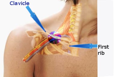
BACKGROUND: Doppler ultrasound methods were used during haemodialysis sessions for the detection of microemboli and determination of their origin. METHODS: A 2-MHz ultrasound probe (Multidop X(4) DWL((TM))) was used to assess the number of microembolic signals (MES) in the subclavian vein downstream from the arteriovenous fistula before the dialysis session and over two periods of 15 min at the beginning and end of haemodialysis sessions in 25 patients without previous cardiovascular disease. A similar probe was used during in vitro studies to detect MES at different sites in the dialysis machine (before and downstream from the blood pump, and before and downstream from the air trap). RESULTS: No MES were detected during in vivo studies before haemodialysis sessions. MES were registered in all patients (100%) at the beginning and end of the haemodialysis procedure at an average of 12.7+/-9 and 16. 7+/-11.5 signals/min respectively. The average intensity of MES was 19.2+/-5.0 dB and 19.4+/-3.9 dB respectively. No MES were detected on the arterial line during in vitro studies. In contrast, 19+/-6 MES/min were detected after the blood pump, 13+/-4.2 before the air trap, and 16.5+/-5.5 thereafter. CONCLUSIONS: In all patients, MES were recorded during haemodialysis sessions in the drainage vein from arteriovenous fistulae. The results of in vitro studies indicate that MES are formed by the blood pump of the haemodialysis machine. The intensity of the MES suggests that they correspond to synthetic particles or microbubbles, which are not detected by the air trap. The final destination of these microbubbles will be assessed in further studies. (+info)The subclavian vein is a large venous structure that carries deoxygenated blood from the upper limb and part of the thorax back to the heart. It forms when the axillary vein passes through the narrow space between the first rib and the clavicle (collarbone), becoming the subclavian vein.
On the left side, the subclavian vein joins with the internal jugular vein to form the brachiocephalic vein, while on the right side, the subclavian vein directly merges with the internal jugular vein to create the brachiocephalic vein. These brachiocephalic veins then unite to form the superior vena cava, which drains blood into the right atrium of the heart.
The subclavian vein is an essential structure for venous access in various medical procedures and interventions, such as placing central venous catheters or performing blood tests.
Central venous catheterization is a medical procedure in which a flexible tube called a catheter is inserted into a large vein in the body, usually in the neck (internal jugular vein), chest (subclavian vein), or groin (femoral vein). The catheter is threaded through the vein until it reaches a central location, such as the superior vena cava or the right atrium of the heart.
Central venous catheterization may be performed for several reasons, including:
1. To administer medications, fluids, or nutritional support directly into the bloodstream.
2. To monitor central venous pressure (CVP), which can help assess a patient's volume status and cardiac function.
3. To draw blood samples for laboratory tests.
4. To deliver chemotherapy drugs or other medications that may be harmful to peripheral veins.
5. To provide access for hemodialysis or other long-term therapies.
The procedure requires careful attention to sterile technique to minimize the risk of infection, and it is usually performed under local anesthesia with sedation or general anesthesia. Complications of central venous catheterization may include bleeding, infection, pneumothorax (collapsed lung), arterial puncture, and catheter-related bloodstream infections (CRBSI).
The jugular veins are a pair of large, superficial veins that carry blood from the head and neck to the heart. They are located in the neck and are easily visible when looking at the side of a person's neck. The external jugular vein runs along the surface of the muscles in the neck, while the internal jugular vein runs within the carotid sheath along with the carotid artery and the vagus nerve.
The jugular veins are important in clinical examinations because they can provide information about a person's cardiovascular function and intracranial pressure. For example, distention of the jugular veins may indicate heart failure or increased intracranial pressure, while decreased venous pulsations may suggest a low blood pressure or shock.
It is important to note that medical conditions such as deep vein thrombosis (DVT) can also affect the jugular veins and can lead to serious complications if not treated promptly.
The axillary vein is a large vein that runs through the axilla or armpit region. It is formed by the union of the brachial vein and the basilic vein at the lower border of the teres major muscle. The axillary vein carries deoxygenated blood from the upper limb, chest wall, and breast towards the heart. As it moves proximally, it becomes continuous with the subclavian vein to form the brachiocephalic vein. It is accompanied by the axillary artery and forms part of the important neurovascular bundle in the axilla.
Thoracic outlet syndrome (TOS) is a group of disorders that occur when the blood vessels or nerves in the thoracic outlet, the space between the collarbone (clavicle) and the first rib, become compressed. This compression can cause pain, numbness, and weakness in the neck, shoulder, arm, and hand.
There are three types of TOS:
1. Neurogenic TOS: This is the most common type and occurs when the nerves (brachial plexus) that pass through the thoracic outlet become compressed, causing symptoms such as pain, numbness, tingling, and weakness in the arm and hand.
2. Venous TOS: This type occurs when the veins that pass through the thoracic outlet become compressed, leading to swelling, pain, and discoloration of the arm.
3. Arterial TOS: This is the least common type and occurs when the arteries that pass through the thoracic outlet become compressed, causing decreased blood flow to the arm, which can result in pain, numbness, and coldness in the arm and hand.
TOS can be caused by a variety of factors, including an extra rib (cervical rib), muscle tightness or spasm, poor posture, repetitive motions, trauma, or tumors. Treatment for TOS may include physical therapy, pain management, and in some cases, surgery.
Upper extremity deep vein thrombosis (UEDVT) is a medical condition that refers to the formation of a blood clot (thrombus) in the deep veins located in the arm or shoulder. This condition can occur due to various reasons, including trauma, surgery, cancer, certain medications, and underlying medical conditions that increase the risk of blood clotting.
The deep veins are larger vessels that run through the body's muscles and are surrounded by fascia, a connective tissue. UEDVT can cause partial or complete blockage of blood flow in the affected vein, leading to swelling, pain, redness, warmth, and decreased function in the arm or hand. In some cases, the clot can break off and travel to the lungs, causing a potentially life-threatening condition called pulmonary embolism (PE).
Diagnosis of UEDVT typically involves a physical exam, medical history, and imaging tests such as ultrasound, CT scan, or MRI. Treatment may include anticoagulant medications to prevent the clot from growing or breaking off, thrombolytic therapy to dissolve the clot, or surgical intervention in severe cases. Compression stockings or other devices may also be used to help improve blood flow and reduce swelling.
Phlebography is a medical imaging technique used to visualize and assess the veins, particularly in the legs. It involves the injection of a contrast agent into the veins, followed by X-ray imaging to capture the flow of the contrast material through the veins. This allows doctors to identify any abnormalities such as blood clots, blockages, or malformations in the venous system.
There are different types of phlebography, including ascending phlebography (where the contrast agent is injected into a foot vein and travels up the leg) and descending phlebography (where the contrast agent is injected into a vein in the groin or neck and travels down the leg).
Phlebography is an invasive procedure that requires careful preparation and monitoring, and it is typically performed by radiologists or vascular specialists. It has largely been replaced by non-invasive imaging techniques such as ultrasound and CT angiography in many clinical settings.
Veins are blood vessels that carry deoxygenated blood from the tissues back to the heart. They have a lower pressure than arteries and contain valves to prevent the backflow of blood. Veins have a thin, flexible wall with a larger lumen compared to arteries, allowing them to accommodate more blood volume. The color of veins is often blue or green due to the absorption characteristics of light and the reduced oxygen content in the blood they carry.
The clavicle, also known as the collarbone, is a long, slender bone that lies horizontally between the breastbone (sternum) and the shoulder blade (scapula). It is part of the shoulder girdle and plays a crucial role in supporting the upper limb. The clavicle has two ends: the medial end, which articulates with the sternum, and the lateral end, which articulates with the acromion process of the scapula. It is a common site of fracture due to its superficial location and susceptibility to direct trauma.
In medical terms, ribs are the long, curved bones that make up the ribcage in the human body. They articulate with the thoracic vertebrae posteriorly and connect to the sternum anteriorly via costal cartilages. There are 12 pairs of ribs in total, and they play a crucial role in protecting the lungs and heart, allowing room for expansion and contraction during breathing. Ribs also provide attachment points for various muscles involved in respiration and posture.
The brachiocephalic veins, also known as the innominate veins, are large veins in the human body. They are formed by the union of the subclavian vein and the internal jugular vein on each side of the body. The resulting vein then carries blood from the upper limbs, head, and neck to the superior vena cava, which is the large vein that returns blood to the heart.
Here's a more detailed medical definition:
The brachiocephalic veins are paired venous structures that result from the union of the subclavian vein and the internal jugular vein on each side of the body. These veins are located in the superior mediastinum, near the base of the neck, and are typically about 2 to 3 centimeters in length. The brachiocephalic veins receive blood from several sources, including the upper extremities, head, neck, and thoracic wall. They then transport this blood to the superior vena cava, which is a large vein that returns blood to the right atrium of the heart.
It's worth noting that the brachiocephalic veins are subject to various pathological conditions, including thrombosis (blood clots), stenosis (narrowing), and compression by nearby structures such as the first rib or the scalene muscles. These conditions can lead to a variety of symptoms, including swelling, pain, and difficulty breathing.
The femoral vein is the large vein that runs through the thigh and carries oxygen-depleted blood from the lower limbs back to the heart. It is located in the femoral triangle, along with the femoral artery and nerve. The femoral vein begins at the knee as the popliteal vein, which then joins with the deep vein of the thigh to form the femoral vein. As it moves up the leg, it is joined by several other veins, including the great saphenous vein, before it becomes the external iliac vein at the inguinal ligament in the groin.
Iothalamate Meglumine is not a medical condition, but rather a diagnostic contrast agent used in various imaging studies such as computed tomography (CT) scans and magnetic resonance imaging (MRI) exams. Iothalamate Meglumine is a type of radiocontrast medium that contains iodine atoms which help to enhance the visibility of internal structures during these imaging tests.
The medical definition of Iothalamate Meglumine is:
A radiocontrast agent used in diagnostic imaging, specifically in CT scans and MR urography exams. It contains iodine atoms that help to improve the contrast and visibility of internal structures such as the urinary tract. Iothalamate Meglumine is typically administered intravenously or instilled directly into the bladder.
It's important to note that while Iothalamate Meglumine is generally considered safe, it can cause allergic reactions or kidney damage in some individuals, particularly those with pre-existing kidney disease or diabetes. Therefore, it's essential to inform your healthcare provider of any medical conditions or allergies before undergoing an imaging exam that involves the use of this contrast agent.
Venous thrombosis is a medical condition characterized by the formation of a blood clot (thrombus) in the deep veins, often in the legs (deep vein thrombosis or DVT), but it can also occur in other parts of the body such as the arms, pelvis, or lungs (pulmonary embolism).
The formation of a venous thrombus can be caused by various factors, including injury to the blood vessel wall, changes in blood flow, and alterations in the composition of the blood. These factors can lead to the activation of clotting factors and platelets, which can result in the formation of a clot that blocks the vein.
Symptoms of venous thrombosis may include swelling, pain, warmth, and redness in the affected area. In some cases, the clot can dislodge and travel to other parts of the body, causing potentially life-threatening complications such as pulmonary embolism.
Risk factors for venous thrombosis include advanced age, obesity, smoking, pregnancy, use of hormonal contraceptives or hormone replacement therapy, cancer, recent surgery or trauma, prolonged immobility, and a history of previous venous thromboembolism. Treatment typically involves the use of anticoagulant medications to prevent further clotting and dissolve existing clots.
An artificial pacemaker is a medical device that uses electrical impulses to regulate the beating of the heart. It is typically used when the heart's natural pacemaker, the sinoatrial node, is not functioning properly and the heart rate is too slow or irregular. The pacemaker consists of a small generator that contains a battery and electronic circuits, which are connected to one or more electrodes that are placed in the heart.
The generator sends electrical signals through the electrodes to stimulate the heart muscle and cause it to contract, thereby maintaining a regular heart rhythm. Artificial pacemakers can be programmed to deliver electrical impulses at a specific rate or in response to the body's needs. They are typically implanted in the chest during a surgical procedure and can last for many years before needing to be replaced.
Artificial pacemakers are an effective treatment for various types of bradycardia, which is a heart rhythm disorder characterized by a slow heart rate. Pacemakers can significantly improve symptoms associated with bradycardia, such as fatigue, dizziness, shortness of breath, and fainting spells.
The saphenous vein is a term used in anatomical description to refer to the great or small saphenous veins, which are superficial veins located in the lower extremities of the human body.
The great saphenous vein (GSV) is the longest vein in the body and originates from the medial aspect of the foot, ascending along the medial side of the leg and thigh, and drains into the femoral vein at the saphenofemoral junction, located in the upper third of the thigh.
The small saphenous vein (SSV) is a shorter vein that originates from the lateral aspect of the foot, ascends along the posterior calf, and drains into the popliteal vein at the saphenopopliteal junction, located in the popliteal fossa.
These veins are often used as conduits for coronary artery bypass grafting (CABG) surgery due to their consistent anatomy and length.
Hemothorax is a medical condition characterized by the presence of blood in the pleural space, which is the area between the lungs and the chest wall. This accumulation of blood can occur due to various reasons such as trauma, rupture of a blood vessel, or complications from lung or heart surgery.
The buildup of blood in the pleural space can cause the affected lung to collapse, leading to symptoms such as shortness of breath, chest pain, and cough. In severe cases, hemothorax can be life-threatening if not promptly diagnosed and treated. Treatment options may include chest tube drainage, blood transfusion, or surgery, depending on the severity and underlying cause of the condition.
Indwelling catheters, also known as Foley catheters, are medical devices that are inserted into the bladder to drain urine. They have a small balloon at the tip that is inflated with water once the catheter is in the correct position in the bladder, allowing it to remain in place and continuously drain urine. Indwelling catheters are typically used for patients who are unable to empty their bladders on their own, such as those who are bedridden or have nerve damage that affects bladder function. They are also used during and after certain surgical procedures. Prolonged use of indwelling catheters can increase the risk of urinary tract infections and other complications.
The superior vena cava is a large vein that carries deoxygenated blood from the upper half of the body to the right atrium of the heart. It is formed by the union of the left and right brachiocephalic veins (also known as the internal jugular and subclavian veins) near the base of the neck. The superior vena cava runs posteriorly to the sternum and enters the upper right portion of the right atrium, just posterior to the opening of the inferior vena cava. It plays a crucial role in the circulatory system by allowing blood returning from the head, neck, upper limbs, and thorax to bypass the liver before entering the heart.
The portal vein is the large venous trunk that carries blood from the gastrointestinal tract, spleen, pancreas, and gallbladder to the liver. It is formed by the union of the superior mesenteric vein (draining the small intestine and a portion of the large intestine) and the splenic vein (draining the spleen and pancreas). The portal vein then divides into right and left branches within the liver, where the blood flows through the sinusoids and gets enriched with oxygen and nutrients before being drained by the hepatic veins into the inferior vena cava. This unique arrangement allows the liver to process and detoxify the absorbed nutrients, remove waste products, and regulate metabolic homeostasis.
Surgical decompression is a medical procedure that involves relieving pressure on a nerve or tissue by creating additional space. This is typically accomplished through the removal of a portion of bone or other tissue that is causing the compression. The goal of surgical decompression is to alleviate symptoms such as pain, numbness, tingling, or weakness caused by the compression.
In the context of spinal disorders, surgical decompression is often used to treat conditions such as herniated discs, spinal stenosis, or bone spurs that are compressing nerves in the spine. The specific procedure used may vary depending on the location and severity of the compression, but common techniques include laminectomy, discectomy, and foraminotomy.
It's important to note that surgical decompression is a significant medical intervention that carries risks such as infection, bleeding, and injury to surrounding tissues. As with any surgery, it should be considered as a last resort after other conservative treatments have been tried and found to be ineffective. A thorough evaluation by a qualified medical professional is necessary to determine whether surgical decompression is appropriate in a given case.
Hydrothorax is a medical term that refers to the abnormal accumulation of serous fluid in the pleural space, which is the potential space between the lungs and the chest wall. This condition often results from various underlying pathological processes such as liver cirrhosis, heart failure, or kidney disease, where there is an imbalance in the body's fluid regulation leading to the accumulation of fluid in the pleural cavity. The presence of hydrothorax can cause respiratory distress and other symptoms related to lung function impairment.
Varicose veins are defined as enlarged, swollen, and twisting veins often appearing blue or dark purple, which usually occur in the legs. They are caused by weakened valves and vein walls that can't effectively push blood back toward the heart. This results in a buildup of blood, causing the veins to bulge and become varicose.
The condition is generally harmless but may cause symptoms like aching, burning, muscle cramp, or a feeling of heaviness in the legs. In some cases, varicose veins can lead to more serious problems, such as skin ulcers, blood clots, or chronic venous insufficiency. Treatment options include lifestyle changes, compression stockings, and medical procedures like sclerotherapy, laser surgery, or endovenous ablation.
Catheterization is a medical procedure in which a catheter (a flexible tube) is inserted into the body to treat various medical conditions or for diagnostic purposes. The specific definition can vary depending on the area of medicine and the particular procedure being discussed. Here are some common types of catheterization:
1. Urinary catheterization: This involves inserting a catheter through the urethra into the bladder to drain urine. It is often performed to manage urinary retention, monitor urine output in critically ill patients, or assist with surgical procedures.
2. Cardiac catheterization: A procedure where a catheter is inserted into a blood vessel, usually in the groin or arm, and guided to the heart. This allows for various diagnostic tests and treatments, such as measuring pressures within the heart chambers, assessing blood flow, or performing angioplasty and stenting of narrowed coronary arteries.
3. Central venous catheterization: A catheter is inserted into a large vein, typically in the neck, chest, or groin, to administer medications, fluids, or nutrition, or to monitor central venous pressure.
4. Peritoneal dialysis catheterization: A catheter is placed into the abdominal cavity for individuals undergoing peritoneal dialysis, a type of kidney replacement therapy.
5. Neurological catheterization: In some cases, a catheter may be inserted into the cerebrospinal fluid space (lumbar puncture) or the brain's ventricular system (ventriculostomy) to diagnose or treat various neurological conditions.
These are just a few examples of catheterization procedures in medicine. The specific definition and purpose will depend on the medical context and the particular organ or body system involved.
Equipment failure is a term used in the medical field to describe the malfunction or breakdown of medical equipment, devices, or systems that are essential for patient care. This can include simple devices like syringes and thermometers, as well as complex machines such as ventilators, infusion pumps, and imaging equipment.
Equipment failure can have serious consequences for patients, including delayed or inappropriate treatment, injury, or even death. It is therefore essential that medical equipment is properly maintained, tested, and repaired to ensure its safe and effective operation.
There are many potential causes of equipment failure, including:
* Wear and tear from frequent use
* Inadequate cleaning or disinfection
* Improper handling or storage
* Power supply issues
* Software glitches or bugs
* Mechanical failures or defects
* Human error or misuse
To prevent equipment failure, healthcare facilities should have established policies and procedures for the acquisition, maintenance, and disposal of medical equipment. Staff should be trained in the proper use and handling of equipment, and regular inspections and testing should be performed to identify and address any potential issues before they lead to failure.
Pneumothorax is a medical condition that refers to the presence of air in the pleural space, which is the potential space between the lungs and the chest wall. This collection of air can result in a partial or complete collapse of the lung. The symptoms of pneumothorax may include sudden chest pain, shortness of breath, cough, and rapid heartbeat.
The two main types of pneumothorax are spontaneous pneumothorax, which occurs without any apparent cause or underlying lung disease, and secondary pneumothorax, which is caused by an underlying lung condition such as chronic obstructive pulmonary disease (COPD), asthma, or lung cancer.
Treatment for pneumothorax may include observation, oxygen therapy, needle aspiration, or chest tube insertion to remove the excess air from the pleural space and allow the lung to re-expand. In severe cases, surgery may be required to prevent recurrence.
Implanted electrodes are medical devices that are surgically placed inside the body to interface directly with nerves, neurons, or other electrically excitable tissue for various therapeutic purposes. These electrodes can be used to stimulate or record electrical activity from specific areas of the body, depending on their design and application.
There are several types of implanted electrodes, including:
1. Deep Brain Stimulation (DBS) electrodes: These are placed deep within the brain to treat movement disorders such as Parkinson's disease, essential tremor, and dystonia. DBS electrodes deliver electrical impulses that modulate abnormal neural activity in targeted brain regions.
2. Spinal Cord Stimulation (SCS) electrodes: These are implanted along the spinal cord to treat chronic pain syndromes. SCS electrodes emit low-level electrical pulses that interfere with pain signals traveling to the brain, providing relief for patients.
3. Cochlear Implant electrodes: These are surgically inserted into the cochlea of the inner ear to restore hearing in individuals with severe to profound hearing loss. The electrodes stimulate the auditory nerve directly, bypassing damaged hair cells within the cochlea.
4. Retinal Implant electrodes: These are implanted in the retina to treat certain forms of blindness caused by degenerative eye diseases like retinitis pigmentosa. The electrodes convert visual information from a camera into electrical signals, which stimulate remaining retinal cells and transmit the information to the brain via the optic nerve.
5. Sacral Nerve Stimulation (SNS) electrodes: These are placed near the sacral nerves in the lower back to treat urinary or fecal incontinence and overactive bladder syndrome. SNS electrodes deliver electrical impulses that regulate the function of the affected muscles and nerves.
6. Vagus Nerve Stimulation (VNS) electrodes: These are wrapped around the vagus nerve in the neck to treat epilepsy and depression. VNS electrodes provide intermittent electrical stimulation to the vagus nerve, which has connections to various regions of the brain involved in these conditions.
Overall, implanted electrodes serve as a crucial component in many neuromodulation therapies, offering an effective treatment option for numerous neurological and sensory disorders.
Ultrasonography, Doppler, Pulsed is a type of diagnostic ultrasound technique that uses the Doppler effect to measure blood flow in the body. In this technique, short bursts of ultrasound are emitted and then listened for as they bounce back off moving red blood cells. By analyzing the frequency shift of the returning sound waves, the velocity and direction of blood flow can be determined. This information is particularly useful in evaluating conditions such as deep vein thrombosis, carotid artery stenosis, and fetal heart abnormalities. Pulsed Doppler ultrasonography provides more detailed information about blood flow than traditional color Doppler imaging, making it a valuable tool for diagnosing and monitoring various medical conditions.
An arteriovenous shunt is a surgically created connection between an artery and a vein. This procedure is typically performed to reroute blood flow or to provide vascular access for various medical treatments. In a surgical setting, the creation of an arteriovenous shunt involves connecting an artery directly to a vein, bypassing the capillary network in between.
There are different types of arteriovenous shunts used for specific medical purposes:
1. Arteriovenous Fistula (AVF): This is a surgical connection created between an artery and a vein, usually in the arm or leg. The procedure involves dissecting both the artery and vein, then suturing them directly together. Over time, the increased blood flow to the vein causes it to dilate and thicken, making it suitable for repeated needle punctures during hemodialysis treatments for patients with kidney failure.
2. Arteriovenous Graft (AVG): An arteriovenous graft is a synthetic tube used to connect an artery and a vein when a direct AVF cannot be created due to insufficient vessel size or poor quality. The graft can be made of various materials, such as polytetrafluoroethylene (PTFE) or Dacron. Grafts are more prone to infection and clotting compared to native AVFs but remain an essential option for patients requiring hemodialysis access.
3. Central Venous Catheter (CVC): A central venous catheter is a flexible tube inserted into a large vein, often in the neck or groin, and advanced towards the heart. CVCs can be used as temporary arteriovenous shunts for patients who require immediate hemodialysis access but do not have time to wait for an AVF or AVG to mature. However, they are associated with higher risks of infection and thrombosis compared to native AVFs and AVGs.
In summary, a surgical arteriovenous shunt is a connection between an artery and a vein established through a medical procedure. The primary purpose of these shunts is to provide vascular access for hemodialysis in patients with end-stage renal disease or to serve as temporary access when native AVFs or AVGs are not feasible.
Pulmonary veins are blood vessels that carry oxygenated blood from the lungs to the left atrium of the heart. There are four pulmonary veins in total, two from each lung, and they are the only veins in the body that carry oxygen-rich blood. The oxygenated blood from the pulmonary veins is then pumped by the left ventricle to the rest of the body through the aorta. Any blockage or damage to the pulmonary veins can lead to various cardiopulmonary conditions, such as pulmonary hypertension and congestive heart failure.
Prosthesis implantation is a surgical procedure where an artificial device or component, known as a prosthesis, is placed inside the body to replace a missing or damaged body part. The prosthesis can be made from various materials such as metal, plastic, or ceramic and is designed to perform the same function as the original body part.
The implantation procedure involves making an incision in the skin to create a pocket where the prosthesis will be placed. The prosthesis is then carefully positioned and secured in place using screws, cement, or other fixation methods. In some cases, tissue from the patient's own body may be used to help anchor the prosthesis.
Once the prosthesis is in place, the incision is closed with sutures or staples, and the area is bandaged. The patient will typically need to undergo rehabilitation and physical therapy to learn how to use the new prosthesis and regain mobility and strength.
Prosthesis implantation is commonly performed for a variety of reasons, including joint replacement due to arthritis or injury, dental implants to replace missing teeth, and breast reconstruction after mastectomy. The specific procedure and recovery time will depend on the type and location of the prosthesis being implanted.
Subclavian Steal Syndrome is a medical condition that occurs when there is a narrowing or blockage (stenosis) in the subclavian artery, usually at or near its origin from the aorta. This stenosis causes reduced blood flow to the ipsilateral upper extremity. The decreased blood supply to the arm leads to reversal of flow in the vertebral artery, which normally supplies blood to the brain and neck structures. As a result, the brain may receive insufficient blood flow, causing symptoms such as dizziness, lightheadedness, syncope (fainting), or transient ischemic attacks (TIAs or "mini-strokes").
The syndrome is called 'subclavian steal' because the vertebral artery essentially "steals" blood from the circle of Willis (the network of arteries at the base of the brain) to compensate for the reduced flow in the subclavian artery. The condition most commonly affects the left subclavian artery, but it can also occur on the right side or both sides.
Subclavian Steal Syndrome is typically diagnosed through a combination of physical examination, medical history, and imaging tests such as Doppler ultrasound, CT angiography (CTA), or magnetic resonance angiography (MRA). Treatment options include surgical bypass, endovascular stenting, or medication to manage symptoms and reduce the risk of stroke.
Iatrogenic disease refers to any condition or illness that is caused, directly or indirectly, by medical treatment or intervention. This can include adverse reactions to medications, infections acquired during hospitalization, complications from surgical procedures, or injuries caused by medical equipment. It's important to note that iatrogenic diseases are unintended and often preventable with proper care and precautions.
Superior Vena Cava Syndrome (SVCS) is a medical condition characterized by the obstruction of the superior vena cava (SVC), which is the large vein that carries blood from the upper body to the heart. This obstruction can be caused by cancerous tumors, thrombosis (blood clots), or other compressive factors.
The obstruction results in the impaired flow of blood from the head, neck, arms, and upper chest, leading to a variety of symptoms such as swelling of the face, neck, and upper extremities; shortness of breath; cough; chest pain; and distended veins visible on the skin surface. In severe cases, SVCS can cause life-threatening complications like cerebral edema (swelling of the brain) or pulmonary edema (fluid accumulation in the lungs).
Immediate medical attention is required for individuals with suspected SVCS to prevent further complications and to manage the underlying cause. Treatment options may include chemotherapy, radiation therapy, anticoagulation therapy, or surgery, depending on the etiology of the obstruction.
Vascular surgical procedures are operations that are performed to treat conditions and diseases related to the vascular system, which includes the arteries, veins, and capillaries. These procedures can be invasive or minimally invasive and are often used to treat conditions such as peripheral artery disease, carotid artery stenosis, aortic aneurysms, and venous insufficiency.
Some examples of vascular surgical procedures include:
* Endarterectomy: a procedure to remove plaque buildup from the inside of an artery
* Bypass surgery: creating a new path for blood to flow around a blocked or narrowed artery
* Angioplasty and stenting: using a balloon to open a narrowed artery and placing a stent to keep it open
* Aneurysm repair: surgically repairing an aneurysm, a weakened area in the wall of an artery that has bulged out and filled with blood
* Embolectomy: removing a blood clot from a blood vessel
* Thrombectomy: removing a blood clot from a vein
These procedures are typically performed by vascular surgeons, who are trained in the diagnosis and treatment of vascular diseases.
Thrombosis is the formation of a blood clot (thrombus) inside a blood vessel, obstructing the flow of blood through the circulatory system. When a clot forms in an artery, it can cut off the supply of oxygen and nutrients to the tissues served by that artery, leading to damage or tissue death. If a thrombus forms in the heart, it can cause a heart attack. If a thrombus breaks off and travels through the bloodstream, it can lodge in a smaller vessel, causing blockage and potentially leading to damage in the organ that the vessel supplies. This is known as an embolism.
Thrombosis can occur due to various factors such as injury to the blood vessel wall, abnormalities in blood flow, or changes in the composition of the blood. Certain medical conditions, medications, and lifestyle factors can increase the risk of thrombosis. Treatment typically involves anticoagulant or thrombolytic therapy to dissolve or prevent further growth of the clot, as well as addressing any underlying causes.
Pathological constriction refers to an abnormal narrowing or tightening of a body passage or organ, which can interfere with the normal flow of blood, air, or other substances through the area. This constriction can occur due to various reasons such as inflammation, scarring, or abnormal growths, and can affect different parts of the body, including blood vessels, airways, intestines, and ureters. Pathological constriction can lead to a range of symptoms and complications depending on its location and severity, and may require medical intervention to correct.
 Subclavian vein
Venous translucence
Costoclavicular ligament
Thoracic outlet syndrome
Chest pain in children
Central venous catheter
Subclavian artery
Leopold von Schrötter
Deep vein thrombosis
Jugular vein
Subclavian nerve
Paget-Schroetter disease
Lymphatic vessel
Execution of Clayton Lockett
Right lymphatic duct
Fatty acid
Subclavian triangle
Head and neck anatomy
Lymphatic system
Pulmonary artery catheter
Dorsal scapular vein
Lymph heart
Lymph sacs
Axillary vein
Angioplasty
May-Thurner syndrome
Physiology of decompression
Francesco Durante (surgeon)
Trendelenburg position
Clavipectoral triangle
Axillary vein6
- medical citation needed] Each subclavian vein is a continuation of the axillary vein and runs from the outer border of the first rib to the medial border of anterior scalene muscle. (wikipedia.org)
- subclavian The vein is a continuation of the axillary vein from the outer edge of the first rib, with a total length of about 3 ~4 cm and a width of about 1~2 cm. walking. (articles01.com)
- Superior Thoracic Artery commonly runs inferomedially anterior to axillary vein. (brainscape.com)
- STA runs inferomedially posterior to axillary vein. (brainscape.com)
- I had issues with ports--but it was from the use of dissolvable stitches that coused abcess twice, the Interventional Radiologist was too egotistical to beleive it was due to the stitches--the third placement was rough and he went into the axillary vein instead of subclavian, but did use glue to close so no more abscess, just a huge blood clot from neck to elbow! (cancer.org)
- The continuation of the axillary vein which follows the subclavian artery and then joins the internal jugular vein to form the brachiocephalic vein. (nih.gov)
Catheterization8
- Subclavian vein catheterization may cause various complications. (medscape.com)
- [ 8 , 9 ] Ultrasound-assisted location of the vein has been reported to have no effect on the rate of complications or failures of SCV catheterization in previous reports. (medscape.com)
- The thoracic duct rises out of the chest, arches over the left subclavian artery and enters the junction of left internal jugular and subclavian veins where it is subject to injury during left sided catheterization. (vesalius.com)
- Using ultrasonographic guidance has been shown to reduce procedural complications of subclavian vein catheterization but is not yet widely recommended or practiced. (msdmanuals.com)
- Peripheral Vein Catheterization A number of procedures are used to gain vascular access. (msdmanuals.com)
- Naina, Harris V K. / Central venous catheterization - Subclavian vein . (elsevierpure.com)
- Central vein catheterization is also referred to as central line placement. (medscape.com)
- 5. [Contralateral hemothorax: a late complication of subclavian vein catheterization]. (nih.gov)
Thrombosis11
- Paget-Schroetter disease includes the thrombosis of the subclavian veins, in this case usually caused by exercise-induced strains. (wikipedia.org)
- Bosch FTM, Nisio MD, Büller HR, van Es N. Diagnostic and Therapeutic Management of Upper Extremity Deep Vein Thrombosis. (medscape.com)
- Singh O, Juneja D. Upper extremity deep vein thrombosis: An intensivist's perspective. (medscape.com)
- Management Strategy for Patients With Chronic Subclavian Vein Thrombosis. (medscape.com)
- Young baseball pitcher who developed subclavian vein thrombosis with significant swelling of dominant right arm. (medscape.com)
- Risk of malposition was higher following right subclavian vein cannulation as compared to right IJV cannulation (9.1% vs 1.4%).There were no cases of vascular perforation, local venous thrombosis or embolism in association with the malpositioned catheter tips. (ispub.com)
- 20. Elevated levels of D-dimer and fragment 1+2 upon central venous catheter insertion and factor V Leiden predict subclavian vein thrombosis. (nih.gov)
- Arising from the posterior region of this mass was a tubular structure with FDG avidity and internal thrombosis that appeared to form a collateral with the right brachiocephalic vein (Figures 3(a) and 3(b) ). (hindawi.com)
- There were no other risk factors for portal vein thrombosis or for thromboembolic disease. (nih.gov)
- The case is reported to draw attention to portal vein thrombosis as an extremely rare complication of OCS. (nih.gov)
- A female patient is diagnosed with deep-vein thrombosis. (nclexnursing.com)
Posterior6
- Thus, the subclavian vein lies anterior to the anterior scalene while the subclavian artery lies posterior to the anterior scalene (and anterior to the middle scalene). (wikipedia.org)
- Because of the AP slope of the superior aperture, the arteries are more cephalad as well as more posterior than the veins. (vesalius.com)
- The brachial plexus nerves approach and encircle the axillary artery laterally and are relatively protected by their posterior position near the first rib where subclavian puncture is performed. (vesalius.com)
- the pleural roof is medial and posterior, and the posterior wall of the subclavian vein is only 5 mm away from the pleura. (articles01.com)
- and the posterior lymph sacs appear near the junctions of the primitive iliac veins and the posterior cardinal veins. (slideshare.net)
- The retromandibular joins with the posterior auricular vein to create the external jugular vein. (healthline.com)
Cannulation of the subclavian vein2
- The present data suggested that ultrasound-guided cannulation of the subclavian vein in critical care patients is superior to the landmark method and should be the method of choice in these patients. (medscape.com)
- Percutaneous cannulation of the subclavian vein uses anatomic landmarks to guide venipuncture and a Seldinger technique to thread a central venous catheter through the subclavian vein and into the superior vena cava. (msdmanuals.com)
Follows the subclavian artery1
- The subclavian vein follows the subclavian artery and is separated from the subclavian artery by the insertion of anterior scalene. (wikipedia.org)
External jugu8
- We are reporting a case of a 91-year-old male with a primary malignancy of the right parotid gland with radiographic thrombus extension within the right external jugular vein. (hindawi.com)
- Here we report a case of a parotid malignancy with direct intravenous thrombus extending down the right external jugular vein to the level of the subclavian vein documented with computed tomography (CT) and positron emission tomography (PET) and review the literature on this rare radiographic finding. (hindawi.com)
- There was invasion of the right external jugular vein with thrombus within the vein extending to the level of the right subclavian vein (Figures 2(a) and 2(b) ). (hindawi.com)
- The main vessels are the external jugular vein and the interior jugular vein. (onteenstoday.com)
- Four jugular veins can be identified in humans: right internal jugular vein, left internal jugular vein, right external jugular vein, and left external jugular vein. (onteenstoday.com)
- Each set consists of one subclavian vein, one internal jugular vein, one external jugular vein, and connectors. (anatomywarehouse.com)
- How superficial is the external jugular vein? (freezingblue.com)
- Why would someone have a prominent external jugular vein? (freezingblue.com)
Femoral veins2
- When filter design allows placement through the jugular or femoral veins, the filter is specially packaged to ensure that it is deployed in the correct orientation. (medscape.com)
- Traditionally, it was assumed that PICC lines have a lower rate of catheter-related bloodstream infection than conventional central venous catheters (CVCs) placed in the internal jugular, subclavian, or femoral veins. (the-hospitalist.org)
Anatomy3
- Anatomy for subclavian approach. (medscape.com)
- Image source: LWW Anatomy Essentials The SCV tube wall is attached to the proper cervical fascia, the periosteum of the first rib, the scalene anterior muscle, and the subclavian fascia sheath, and its position is constant, and it is not easy to move. (articles01.com)
- The Anatomy of the Internal Jugular Vein. (onteenstoday.com)
Jugular49
- The thoracic duct drains into the left subclavian vein, near its junction with the left internal jugular vein. (wikipedia.org)
- The right lymphatic duct drains its lymph into the junction of the right internal jugular vein, and the right subclavian vein. (wikipedia.org)
- It is less commonly used than other approaches, such as the right internal jugular vein, due to the risk of pneumothorax, haemothorax, and puncture of the accompanying subclavian artery. (wikipedia.org)
- [ 1-5 ] Mechanical complications occur more frequently through the subclavian vein (SCV) route if compared with the internal jugular vein (IJV) and to the femoral vein routes. (medscape.com)
- Central venous stenosis after subclavian versus internal jugular dialysis catheter insertion (CITES) in adults in need of a temporary central dialysis catheter: study protocol for a two-arm, parallel-group, non-inferiority randomised controlled trial. (medscape.com)
- The subclavian and internal jugular veins unite inside the anterior rim of the superior thoracic aperture, forming right and left brachiocephalic veins behind the manubrium. (vesalius.com)
- How To Do Internal Jugular Vein Cannulation, Ultrasound-Guided Ultrasound-guided cannulation of the internal jugular vein uses real-time (dynamic) ultrasound to guide venipuncture and a guidewire (Seldinger technique) to thread a central venous catheter. (msdmanuals.com)
- Whereas ultrasonographic (US) guidance has proved to be a useful adjunct for internal jugular vein cannulation , its use for subclavian routes has not been as commonly studied. (medscape.com)
- Deep vein puncture is one of the basic skills necessary for physicians, and most of them are internal jugular, subclavian, and femoral vein puncture. (articles01.com)
- The inner and lower edge of the sternoclavicular is aligned with the suprasternal fossa and slowly advanced, and the bevel of the needle tip faces the direction of the heart, so as to avoid entering the internal jugular vein when the guide wire is delivered. (articles01.com)
- The two jugular lymph sacs were proposed to develop in the junction of the sub-clavian and anterior cardinal veins by endothelial budding from the anterior cardinal veins. (slideshare.net)
- Malposition of central venous catheter is one such complication which can occur.1 This is a case report on a misdirected central venous catheter into opposite internal jugular vein (IJV) following subclavian vein cannulation. (ispub.com)
- Malposition of central venous catheter (CVC) is a (not unusual) known complication.1 There are many case reports on malposition of CVC into ipsilateral internal jugular vein(IJV) following subclavian vein cannulation and is regarded as a common complication.2 However the misplacement of CVC into contralateral IJV is extremely rare. (ispub.com)
- The left common carotid artery and left internal jugular vein have been resected. (stanford.edu)
- The innovative product is a hyper-realistic soft tissue training system for teaching blind or ultrasound-guided vascular access at the Internal Jugular Vein or Subclavian Vein with the Subclavian or Supraclavicular. (simulab.com)
- Why is it called the jugular vein? (onteenstoday.com)
- The internal jugular vein collects blood from the brain, the outside of the face and the neck. (onteenstoday.com)
- What side of the neck is the jugular vein on? (onteenstoday.com)
- Internal and external jugular veins run along the right and left sides of your neck. (onteenstoday.com)
- Do you have 2 jugular veins? (onteenstoday.com)
- There is a pair of internal jugular veins (right and left) and a pair of external jugular veins. (onteenstoday.com)
- Jugular vein, any of several veins of the neck that drain blood from the brain, face, and neck, returning it to the heart via the superior vena cava. (onteenstoday.com)
- What happens if the jugular vein is blocked? (onteenstoday.com)
- When it comes to your head and neck, the internal jugular vein is the drainage system. (onteenstoday.com)
- Can a jugular vein be removed? (onteenstoday.com)
- Internal Jugular Vein: The internal jugular veins are large veins on either side of the neck that help to drain blood from the brain and head back to the heart. (onteenstoday.com)
- One jugular vein can be removed without causing any symptoms. (onteenstoday.com)
- How do you know if your jugular vein is blocked? (onteenstoday.com)
- Why is the jugular vein so important? (onteenstoday.com)
- The internal jugular vein is a major blood vessel that drains blood from important body organs and parts, such as the brain, face, and neck. (onteenstoday.com)
- What is the difference between carotid artery and jugular vein? (onteenstoday.com)
- The common carotid artery (CCA) and the internal jugular vein (IJV) run side-by-side in the neck, one pair on the left and one on the right. (onteenstoday.com)
- Is the jugular vein the biggest vein in the body? (onteenstoday.com)
- The internal jugular vein is the largest vein in the neck that serves as the main source of blood flow down from the head. (onteenstoday.com)
- Can you live without a jugular vein? (onteenstoday.com)
- Removal of one jugular vein usually causes minimal or no problems. (onteenstoday.com)
- the internal jugular veins join with the subclavian veins to form the brachiocephalic veins, which join to form the superior vena cava. (onteenstoday.com)
- What causes enlarged jugular vein? (onteenstoday.com)
- Where does the blood flow from the jugular vein? (onteenstoday.com)
- The internal jugular vein is formed by the anastomosis of blood from the sigmoid sinus of the dura mater and the common facial vein. (onteenstoday.com)
- What are the names of the four jugular veins? (onteenstoday.com)
- 1. What is Jugular Vein 2. (onteenstoday.com)
- Is the internal jugular vein used for cannulation? (onteenstoday.com)
- The internal jugular vein is used for cannulation since it is large, superficial in location, and usually does not vary in its course along the neck. (onteenstoday.com)
- What does it mean when jugular vein pressure is low? (onteenstoday.com)
- Ultrasound and venography confirmed extensive veno-occlusive disease involving the iliac veins, the right femoral vein, the lower inferior vena cava, the left internal jugular vein, and both subclavian veins. (bmj.com)
- A transjugular liver biopsy needle was passed through a vascular sheath in the right internal jugular vein and advanced into the right atrium by pushing through the SVC obstruction. (bmj.com)
- Post deployment venogram via the right internal jugular vein shows SVC stent satisfactorily placed with good flow into the right atrium. (bmj.com)
- It is recommended to use other sites such as the subclavian or internal jugular veins for central venous access in adult patients. (proprofs.com)
Catheter8
- Continue to thread the catheter into the vein to the desired length. (medscape.com)
- Placement of a central vein catheter is a common procedure, and house staff require substantial training and supervision to become facile with this technique. (medscape.com)
- A physician should have a thorough foreknowledge of the procedure and its complications before placing a central vein catheter. (medscape.com)
- 1. [Late rupture of central catheter implanted through subclavian route]. (nih.gov)
- 3. [The "pinch off" syndrome: a complication of implantable catheter systems in the subclavian vein]. (nih.gov)
- Vascular surgeons use medical treatments, minimally-invasive catheter procedures, and surgery to make sure your arteries and veins stay healthy. (utah.edu)
- The tube (called a central venous catheter) must be inserted into a large vein, such as the subclavian vein, which is located under the collarbone. (msdmanuals.com)
- The transjugular needle was removed and the SVC obstruction was dilated using an 8 mm diameter balloon catheter passed over the guide wire from the left femoral vein. (bmj.com)
Puncture3
- Subclavian vein puncture always fails? (articles01.com)
- How to effectively improve the success rate of deep vein puncture and reduce various complications is the focus of clinicians. (articles01.com)
- Subclavian vein (SCV) puncture is often used in the following situations: Those who need a large amount of fluid or blood transfusion in a short period of time and peripheral venipuncture is difficult , Central venous pressure measurement, temporary pacing electrode implantation, permanent pacemaker implantation, etc. (articles01.com)
Artery and vein1
- Additionally, the subclavian artery and vein may also be compromised. (erikdalton.com)
Arteries and veins2
Placement4
- Insert the guide wire through the needle into the vein with the J-tip directed caudally to improve successful placement into the subclavian vein. (medscape.com)
- An abnormal cardiac stress test prompted a coronary angiography, which revealed severe coronary artery disease.The patient underwent coronary artery bypass and in situ replacement of the infected subclavian artery pseudoaneurysm with a superficial femoral vein, along with placement of a pectoralis major muscle flap to cover the vein graft. (nih.gov)
- The subclavian vein may be less preferred for stiff catheters (because of difficulty achieving the sharp turn into the superior van cava) or large-bore hemodialysis catheters (which can cause venous stenosis that renders the ipsilateral arm unsuitable for arteriovenous shunt placement). (msdmanuals.com)
- If the infrarenal segment of the inferior vena cava is too short for a filter placement, the filter should be placed above the renal veins. (medscape.com)
Infraclavicular approach2
- It offers several advantages over the infraclavicular approach to the subclavian vein . (medscape.com)
- Under aseptic precautions, the right subclavian vein was successfully cannulated with 7 French triple lumen CVC ( B Braun) by infraclavicular approach using Seldinger's technique. (ispub.com)
Cardiac1
- Advance the wire until it is mostly in the vein or until ectopy is seen on the cardiac monitor. (medscape.com)
Neck7
- Figure shows area where subclavian vein is obstructed in neck region. (medscape.com)
- Prepare the neck as well, in case the subclavian approach fails and another approach must be attempted. (medscape.com)
- Tumor thrombus within neck veins is an uncommon event and in the head and neck it has most often been reported in association with thyroid malignancies [ 1 - 6 ]. (hindawi.com)
- Upon presentation at our institution, physical examination demonstrated significant enlargement in the size of the mass with no evidence of swelling, palpable veins, or tenderness in the neck. (hindawi.com)
- What is the main vein in your neck called? (onteenstoday.com)
- Anatomically, there are two of these veins that lie along each side of the neck. (onteenstoday.com)
- There are many other veins in the neck and the blood can flow back through them. (onteenstoday.com)
Superficial femo1
- We present the first case of in situ replacement of an infected subclavian artery using superficial femoral vein and the fourth reported case of an infected arterial pseudoaneurysm caused by pseudomonas pseudomallei. (nih.gov)
Insertion2
- We compared the ultrasound-guided subclavian vein cannulation (200 patients) vs. the landmark method (201 patients) using an infraclavicular needle insertion point in all cases. (medscape.com)
- [ 9 ] At the insertion site, the subclavian vein is closer to the skin, and the right-side approach offers a straighter path into the subclavian vein. (medscape.com)
Ultrasound2
- Cite this: Real-time Ultrasound-guided Subclavian Vein Cannulation versus the Landmark Method in Critical Care Patients - Medscape - Jul 01, 2011. (medscape.com)
- Forty-eight articles (11,004 patients) met the inclusion criteria and examined ventilation/perfusion (V/Q) lung scanning, computed tomography (CT) angiography, leg vein ultrasound (U/S), echocardiography, magnetic resonance (MR) angiography, and the D-dimer test. (the-hospitalist.org)
Drains1
- That vein drains into the subclavian vein. (healthline.com)
Anterior3
- Vein is usually compressed by first rib, clavicle, and serratus anterior muscle. (medscape.com)
- The anterior scalene muscles attach to the first ribs between the subclavian veins and arteries. (vesalius.com)
- The remaining lymph sacs originate from the mesonephric vein and those in the dorsomedial edge of the Wolffian bodies in the junction of the subclavian and anterior cardinal veins. (slideshare.net)
Brachial1
- Especially at risk should have been the brachial nerves to the left arm, perhaps the subclavian vein and/or artery, a portion of the second rib or the top of the scapula (shoulder blade). (truewestmagazine.com)
Vasculature2
- D) Connection of the lymphatic system with the blood vasculature at the subclavian veins. (slideshare.net)
- Veins of the right arm down to the elbow region representative of healthy vasculature. (bdclabs.com)
Large vein1
- The subclavian vein is a paired large vein, one on either side of the body, that is responsible for draining blood from the upper extremities, allowing this blood to return to the heart. (wikipedia.org)
Thoracic duct1
- The left subclavian vein plays a key role in the absorption of lipids, by allowing products that have been carried by lymph in the thoracic duct to enter the bloodstream. (wikipedia.org)
Computed tomography2
- Computed tomography and arteriography revealed a 6.5-cm pseudoaneurysm of the proximal right subclavian artery. (nih.gov)
- The case is described of a 38 year old woman who developed severe right upper quadrant abdominal pain that was associated with partial portal vein occlusion, as confirmed by flow Doppler ultrasonography and dynamic contrast computed tomography. (nih.gov)
Angioplasty2
- Chapter 65 - Axillosubclavian Vein Thrombectomy, Thrombolysis, and Angioplasty", Endovascular Surgery (Fourth Edition), Philadelphia: W.B. Saunders, pp. 679-686, doi:10.1016/b978-1-4160-6208-0.10065-5, ISBN 978-1-4160-6208-0, retrieved 2020-11-20 "What is the Subclavian Vein? (wikipedia.org)
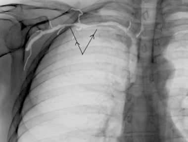
- Subclavian vein prior to angioplasty. (medscape.com)
Right atrium2
- this facilitates smooth caudal progression of the guide wire down the vein toward the right atrium. (medscape.com)
- A guide wire was passed through the lumen of the needle and pulled from the right atrium with a vascular snare out through a left femoral vein sheath. (bmj.com)
Varicose veins3
Occlusion1
- Bilateral simultaneous arm venogram showing occlusion of both subclavian veins, with numerous collaterals, and upper thorax. (bmj.com)
Obstruction1
- Venogram in patient with subclavian vein obstruction. (medscape.com)
Syndrome1
- The subclavian vein may be blocked during thoracic outlet syndrome. (wikipedia.org)
Peripheral1
- Central venous cannulation was considered as the peripheral veins appeared collapsed and were inaccessible. (ispub.com)
Superior4
- The heads of the clavicles extend posteriorly into the superior aperture, displacing the veins posteriorly from the sternum. (vesalius.com)
- They bring blood from your head to the superior vena cava, which is the largest vein in the upper body. (onteenstoday.com)
- Síndrome neurovascular asociado con compresión del PLEXO BRAQUIAL, ARTERIA SUBCLAVIA y la VENA SUBCLAVIA en su salida torácica superior. (bvsalud.org)
- and SUBCLAVIAN VEIN at the superior thoracic outlet. (bvsalud.org)
Needle5
- Once under the clavicle, the needle should be advanced toward the suprasternal notch until the vein is entered. (medscape.com)
- If the vein is difficult to locate, remove the introducer needle, flush it clean of clots, and try again. (medscape.com)
- thus, errant needle punctures (eg, of the subclavian artery or pleura) are less likely. (msdmanuals.com)
- Press the needle tip down slightly with the thumb to make it pass through the subclavian space. (articles01.com)
- The needle should not be inserted too deep, from the skin to the subclavian vein, 4-7 cm for adults and 1-3 cm for children. (articles01.com)
Vessel1
- develops and applies programs either a native vessel (e.g. saphenous vein) or a designed to improve the health of the people of synthetic material. (nih.gov)
Left4
- Consequently, the left subclavian vein plays a key role in the absorption of these fats and lipids. (wikipedia.org)
- We report a case of malposition of CVC tip into left IJV following right subclavian vein cannulation. (ispub.com)
- The first image below demonstrates an IVC of normal size without a thrombus, but there is a circumaortic left renal vein and an inflow defect at the origin of the right renal vein. (medscape.com)
- left subclavian vein. (cdc.gov)
Tributaries1
- The venæ cavæ and azygos veins, with their tributaries. (wikipedia.org)
Vessels2
- Hippocrates first described vessels containing "white blood" around 400 B.C. Gasparo Aselli re-identified lymphatic vessels in the 1600's, noting the presence of lipid-filled "milky veins" in the gut of a "well-fed" dog (Aselli, 1627). (slideshare.net)
- Toxin then enters lymph vessels that drain to the subclavian vein to enter the liver via the portal circulation. (nih.gov)