Strongyloidiasis
Strongyloides stercoralis
The control of morph development in the parasitic nematode Strongyloides ratti. (1/26)
The parasitic nematode Strongyloides ratti has a complex life cycle. The progeny of the parasitic females can develop into three distinct morphs, namely directly developing infective third-stage larvae (iL3s), free-living adult males and free-living adult females. We have analysed of the effect of host immune status (an intra-host factor), environmental temperature (an extra-host factor) and their interaction on the proportion of larvae that develop into these three morphs. The results are consistent with the developmental decision of larvae being controlled by at least two discrete developmental switches. One is a sex-determination event that is affected by host immune status and the other is a switch between alternative female morphs that is affected by both host immune status and environmental temperature. These findings clarify the basis of the life cycle of S. ratti and demonstrate how such complex life cycles can result from a combination of simple developmental switches. (+info)Functional consequences of genetic diversity in Strongyloides ratti infections. (2/26)
Parasitic nematodes show levels of genetic diversity comparable to other taxa, but the functional consequences of this are not understood. Thus, a large body of theoretical work highlights the potential consequences of parasite genetic diversity for the epidemiology of parasite infections and its possible implications for the evolution of host and parasite populations. However, few relevant empirical data are available from parasites in general and none from parasitic nematodes in particular. Here, we test two hypotheses. First, that different parasitic nematode genotypes vary in life-history traits, such as survivorship and fecundity, which may cause variation in infection dynamics. Second, that different parasitic nematode genotypes interact within the host (either directly or via the host immune system) to increase the mean reproductive output of mixed-genotype infections compared with single-genotype infections. We test these hypotheses in laboratory infections using genetically homogeneous lines of Strongyloides ratti. We find that nematode genotypes do vary in their survivorship and fecundity and, consequently, in their dynamics of infection. However, we find little evidence of interactions between genotypes within hosts under a variety of trickle- and single-infected infection regimes. (+info)Western blotting using Strongyloides ratti antigen for the detection of IgG antibodies as confirmatory test in human strongyloidiasis. (3/26)
The present study was conducted to evaluate the frequency of antigenic components recognized by serum IgG antibodies in Western blotting (WB) using a Strongyloides ratti larval extract for the diagnosis of human strongyloidiasis. In addition, the WB results were compared to the enzyme-linked immunosorbent assay (ELISA) and the indirect immunofluorescence antibody test (IFAT) results. Serum samples of 180 individuals were analyzed (80 with strongyloidiasis, 60 with other intestinal parasitoses, and 40 healthy individuals). S. ratti was obtained from fecal culture of experimentally infected Rattus rattus. For IFAT, S. ratti larvae were used as antigen and S. ratti larval antigenic extracts were employed in WB and ELISA. Eleven S. ratti antigenic components were predominantly recognized by IgG antibodies in sera of patients with strongyloidiasis. There was a positive concordance for the three tests in 87.5% of the cases of strongyloidiasis. The negative concordance in the three tests was 94% and 97.5%, in patients with other intestinal parasitoses and healthy individuals, respectively. In cases of positive ELISA and negative IFAT results, diagnosis could be confirmed by WB. ELISA, IFAT, and WB using S. ratti antigens showed a high rate of sensitivity and specificity. In conclusion, WB using S. ratti larval extract was able to recognize 11 immunodominant antigenic components, showing to be a useful tool to define the diagnosis in cases of equivocal serology. (+info)Heterologous antigen extract in ELISA for the detection of human IgE anti-Strongyloides stercoralis. (4/26)
Strongyloides ratti larval extract was used for the standardization of ELISA to detect genus-specific IgE in human strongyloidiasis. Forty serum samples from monoinfected patients shedding S. stercoralis larvae (Group I), 40 from patients with other intestinal parasites (Group II), and 40 from copronegative healthy subjects (Group III) were analyzed. Genus-specific IgE levels (ELISA Index: EI) were significantly higher in the group I (EI = 1.43) than groups II (EI = 0.70) and III (EI = 0.71), showing positivity rates of 55%, 2.5% and 0%, respectively. Similarly, sera from copropositive patients had significantly higher levels of total IgE (866 IU/mL) as compared to those from group II (302 IU/mL) and III (143 IU/mL). A significant positive correlation was found between levels of Strongyloides specific-IgE and total IgE in sera from patients with strongyloidiasis. In conclusion, S. ratti heterologous extract showed to be a useful tool for detecting genus-specific IgE by ELISA, contributing for a better characterization of the immune response profile in human strongyloidiasis. (+info)Strongyloides ratti antigenic components recognized by IgE antibodies in immunoblotting as an additional tool for improving the immunodiagnosis in human strongyloidiasis. (5/26)
IgE antibody response in human strongyloidiasis was evaluated by enzyme-linked immunosorbent assay (ELISA) and immunoblotting (IB) using Strongyloides ratti saline extract as heterologous antigen. A total of 50 serum samples of patients who were shedding S. stercoralis larvae in feces (group I, copropositive), 38 of patients with other intestinal parasites (group II), and 38 of subjects with negative results in three parasitologic assays (group III, copronegative) were analyzed. Levels of IgE anti-Strongyloides expressed in ELISA Index (EI) were significantly higher in patients of group I (1.32) than in group II (0.51) and group III (0.81), with positivity rates of 54%, 0%, and 10.5%, respectively. Fifteen S. ratti antigenic components were recognized in IB-IgE by sera of group I, with frequency ranging from 8% to 46%. In group II, only two antigenic bands (101, 81 kDa) were detected in a frequency of 10% and no reactivity was found in group III. Sera with EI values > 1.5 recognized five from 13 specific antigenic bands (70, 63, 61, 44, 7 kDa). It can be concluded that these five antigenic components recognized by IB-IgE using S. ratti antigen might be employed as an additional tool for improving the immunodiagnosis in human strongyloidiasis. (+info)A microarray analysis of gene expression in the free-living stages of the parasitic nematode Strongyloides ratti. (6/26)
BACKGROUND: The nematode Strongyloides ratti has two adult phases in its lifecycle: one obligate, female and parasitic and one facultative, dioecious and free-living. The molecular control of the development of this free-living generation remains to be elucidated. RESULTS: We have constructed an S. ratti cDNA microarray and used it to interrogate changes in gene expression during the free-living phase of the S. ratti life-cycle. We have found very extensive differences in gene expression between first-stage larvae (L1) passed in faeces and infective L3s preparing to infect hosts. In L1 stages there was comparatively greater expression of genes involved in growth. We have also compared gene expression in L2 stages destined to develop directly into infective L3s with those destined to develop indirectly into free-living adults. This revealed relatively small differences in gene expression. We find little evidence for the conservation of transcription profiles between S. ratti and S. stercoralis or C. elegans. CONCLUSION: This is the first multi-gene study of gene expression in S. ratti. This has shown that robust data can be generated, with consistent measures of expression within computationally determined clusters and contigs. We find inconsistencies between EST representation data and microarray hybridization data in the identification of genes with stage-specific expression and highly expressed genes. Many of the genes whose expression is significantly different between L1 and iL3s stages are unknown beyond alignments to predicted genes. This highlights the forthcoming challenge in actually determining the role of these genes in the life of S. ratti. (+info)Experimental evolution of parasite life-history traits in Strongyloides ratti (Nematoda). (7/26)
Evolutionary ecology predicts that parasite life-history traits, including a parasite's survivorship and fecundity within a host, will evolve in response to selection and that their evolution will be constrained by trade-offs between traits. Here, we test these predictions using a nematode parasite of rats, Strongyloides ratti, as a model. We performed a selection experiment by passage of parasite progeny from either early in an infection ('fast' lines) or late in an infection ('slow' lines). We found that parasite fecundity responded to selection but that parasite survivorship did not. We found a trade-off mediated via conspecific density-dependent constraints; namely, that fast lines exhibit higher density-independent fecundity than slow lines, but fast lines suffered greater reduction in fecundity in the presence of density-dependent constraints than slow lines. We also found that slow lines both stimulate a higher level of IgG1, which is a marker for a Th2-type immune response, and show less of a reduction in fecundity in response to IgG1 levels than for fast lines. Our results confirm the general prediction that parasite life-history traits can evolve in response to selection and indicate that such evolutionary responses may have significant implications for the epidemiology of infectious disease. (+info)The immune response during a Strongyloides ratti infection of rats. (8/26)
A range of immune parameters was measured during a primary infection of Strongyloides ratti in its natural rat host. The immune parameters measured were interleukin-4 (IL-4) and interferon-gamma from both the spleen and mesenteric lymph node (MLN) cells; parasite-specific immunoglobulin G(1)(IgG(1)), IgG(2a) and IgG(2b) in serum and in intestinal tissue; parasite-specific IgG and total IgE in serum; parasite-specific and total IgA in intestinal tissue and rat mast cell protease II in intestinal tissue. Parasite-specific IgG(1), IgG(2a) and total IgE in serum and parasite-specific IgA and rat mast cell protease II in intestinal tissue all occurred at significantly greater concentrations in infected animals, compared with non-infected animals. Similarly, the production of IL-4 by MLN cells stimulated with parasitic female antigen or concanavalin A occurred at significantly greater concentrations in infected animals, compared with non-infected animals. In all, this suggests that there is a T-helper 2-type immune response during a primary S. ratti infection. These data also show the temporal changes in these components of the host immune response during a primary S. ratti infection. (+info)"Strongyloides ratti" is a species of parasitic roundworm that infects the intestines of laboratory rats. The adult female worm lives in the mucosa of the small intestine, where it lays eggs that hatch into larvae. These larvae can either mature into adults within the host's intestine or be passed in the feces and then develop into infective larvae on the outside. The infective larvae can penetrate the skin of a new host, enter the bloodstream, and migrate to the lungs, from where they are coughed up and swallowed, returning to the intestine to mature. This complex life cycle is known as "heterogonic" or "discontinuous."
Infection with Strongyloides ratti can cause symptoms such as diarrhea, weight loss, and intestinal bleeding in rats. In immunocompromised individuals, the parasite can also infect humans and cause a similar disease called "strongyloidiasis," which can be asymptomatic or lead to severe complications if left untreated.
It's worth noting that Strongyloides ratti is not a human pathogen and it's mainly used as a laboratory model for studying the biology of Strongyloides stercoralis, a closely related species that infects humans.
Strongyloidiasis is a tropical and subtropical parasitic disease caused by the nematode (roundworm) Strongyloides stercoralis. The infection occurs when the larvae of this parasite penetrate the skin, usually of the feet, and are carried through the bloodstream to the lungs. Here they mature, are coughed up and swallowed, and then mature in the small intestine where they lay eggs. These hatch into larvae that can either pass out with the feces or penetrate the skin of the anal area and restart the cycle.
The disease is often asymptomatic but can cause a range of symptoms including gastrointestinal (diarrhea, abdominal pain) and pulmonary (cough, wheezing) symptoms. Disseminated strongyloidiasis, where the larvae spread throughout the body, can occur in immunocompromised individuals and can be life-threatening.
Treatment is with anti-parasitic drugs such as ivermectin or thiabendazole. Prevention involves avoiding skin contact with contaminated soil and good hygiene practices.
Strongyloides is a type of parasitic roundworm that can infect humans and other animals. The most common species to infect humans is Strongyloides stercoralis. These tiny worms can cause a condition known as strongyloidiasis, which can lead to symptoms such as abdominal pain, diarrhea, and skin rashes.
The life cycle of Strongyloides is unique among parasitic roundworms because it can complete its entire life cycle within a single host, without needing to exit the body and infect a new host. This is known as "autoinfection" and it allows the worm to persist in the human body for many years, even in the absence of new infections.
Strongyloides infection typically occurs when larvae (immature worms) penetrate the skin, often through contaminated soil. The larvae then travel through the bloodstream to the lungs, where they mature and are coughed up and swallowed, allowing them to reach the intestines and mature into adults. Female adult worms can lay eggs that hatch into larvae, which can either be excreted in feces or undergo autoinfection by penetrating the intestinal wall and entering the bloodstream again.
While many people with Strongyloides infection do not experience any symptoms, severe infections can lead to complications such as chronic diarrhea, malnutrition, and bacterial bloodstream infections. In immunocompromised individuals, Strongyloides infection can be life-threatening due to the rapid multiplication of larvae in the body, a condition known as "hyperinfection."
"Strongyloides stercoralis" is a species of parasitic roundworm that can infect humans and other animals. The adult female worms live in the small intestine, where they lay eggs that hatch into larvae. These larvae can then either mature into adult worms within the host's intestine or be passed out of the body in feces. If the larvae in the feces come into contact with suitable moist soil, they can mature into infective larvae that can penetrate the skin of a new host and cause infection.
In humans, "Strongyloides stercoralis" infection can cause a range of symptoms, including abdominal pain, diarrhea, bloating, and weight loss. In some cases, the infection can become chronic and lead to serious complications, such as disseminated disease or gram-negative sepsis, particularly in individuals with weakened immune systems.
The diagnosis of "Strongyloides stercoralis" infection typically involves the detection of larvae in the stool or other bodily fluids, although serological tests and PCR assays are also available. Treatment usually involves the use of anti-parasitic drugs, such as ivermectin or albendazole, to kill the worms and prevent the progression of the infection.
Antibodies are proteins produced by the immune system in response to the presence of a foreign substance, known as an antigen. They are capable of recognizing and binding to specific antigens, neutralizing or marking them for destruction by other immune cells.
Helminths are parasitic worms that can infect humans and animals. They include roundworms, tapeworms, and flukes, among others. Helminth infections can cause a range of symptoms, depending on the type of worm and the location of the infection.
Antibodies to helminths are produced by the immune system in response to an infection with one of these parasitic worms. These antibodies can be detected in the blood and serve as evidence of a current or past infection. They may also play a role in protecting against future infections with the same type of worm.
There are several different classes of antibodies, including IgA, IgD, IgE, IgG, and IgM. Antibodies to helminths are typically of the IgE class, which are associated with allergic reactions and the defense against parasites. IgE antibodies can bind to mast cells and basophils, triggering the release of histamine and other inflammatory mediators that help to protect against the worm.
In addition to IgE, other classes of antibodies may also be produced in response to a helminth infection. For example, IgG antibodies may be produced later in the course of the infection and can provide long-term immunity to reinfection. IgA antibodies may also be produced and can help to prevent the attachment and entry of the worm into the body.
Overall, the production of antibodies to helminths is an important part of the immune response to these parasitic worms. However, in some cases, the presence of these antibodies may also be associated with allergic reactions or other immunological disorders.
 Assessment of FDA-approved drugs against Strongyloides ratti in vitro and in vivo to identify potentially active drugs against...
 Corydaline - Wikipedia
Nematode.net v4.0
miRBase entry: str-mir-8387
 Strongyloidiasis: Background, Pathophysiology, Etiology
 Scientific Program | Biocuration 2017 | Stanford Medicine
 Toxocara Canis Diagnosis Elisa Kit | Quadratech Diagnostics
GH19 2 Eukaryota Acacia koa AFY08283.1 ncbi
Rapid DNA barcoding analysis of large datasets using the composition vector method | BMC Bioinformatics | Full Text
Transgenesis in parasitic helminths: a brief history and prospects for the future | Parasites & Vectors | Full Text
Ratti
 SPELL - Nematode
 Consensus statement on placebo effects in sports and exercise: the need for conceptual clarity, methodological rigour, and the...
 Browse by Research Team - MDC Repository
Prof. Dr. Marc Hübner - Mikrobiologie Bonn
Heat shock proteins and parasitic diseases: Part 1: Helminths Abaza SM - Parasitol United J
 Strongyloides Serology, Blood - Canterbury Health Laboratories
 Imunidade
Tren npp cycle, steroidi anabolizzanti tipi | Dr. Pep Mondragón
 Comparative characterization of two galectins excreted-secreted from intestine-dwelling parasitic versus free-living females of...
Strongyloidiasis: Background, Pathophysiology, Etiology
Evaluation of emodepside in laboratory models of human intestinal nematode and schistosome infections - edoc
Strongyloides stercoralis seroprevalence in Vietnam - Centre for Tropical Medicine and Global Health
 Search
 Legit Hacks | Exploits, Updated, Fake Lag - SuaCultura
 Gene: 21ur-12944 (WBGene00168018) - Summary - Caenorhabditis elegans - Ensembl Genomes 57
 WormBase ParaSite: Caenorhabditis elegans - Genomic Context - Gene: col-138 (WBGene00000711)
WormBase ParaSite: Schmidtea mediterranea - Genomic Context - Gene: tph (h1SMcG0000075)
 Milenia Biotec | Simple rapid test development
miRNEST 2.0, an integrative microRNA resource :: Browse
Stercoralis5
- Infections with Strongyloides stercoralis belong to the most neglected helminth diseases, and research and development (R&D) efforts on novel drugs are inadequate. (biomedcentral.com)
- Strongyloidiasis, caused by infections with the soil-transmitted helminth Strongyloides stercoralis and occasionally Strongyloides fuelleborni , is a highly neglected tropical disease (NTD) in tropical and subtropical settings. (biomedcentral.com)
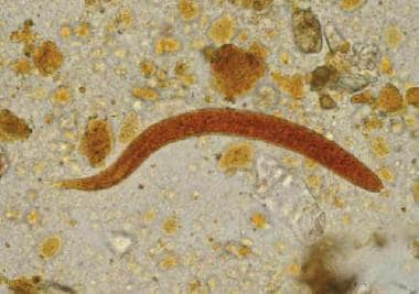
- Rhabditiform larva of Strongyloides stercoralis in stool specimen (wet mount stained with iodine). (medscape.com)
- The life cycle of Strongyloides stercoralis is complex and unique among the intestinal nematodes . (medscape.com)
- SUMMARYStrongyloidiasis is a neglected tropical disease caused by the roundworm Strongyloides stercoralis affecting 30-100 million people worldwide. (ox.ac.uk)
Larvae3
- A commercially available library containing 1600 FDA-approved drugs was tested in vitro against Strongyloides ratti larvae (L3) at 100 µM. (biomedcentral.com)
- Corydaline is nematocidal against S. ratti and S. venezuelensis third instar larvae with 50% paralysis (PC50) values of 18 and 30 μM, respectively. (wikipedia.org)
- Corrigendum: Eosinophils and Neutrophils Eliminate Migrating Strongyloides ratti Larvae at the Site of Infection in the Context of Extracellular DNA Trap Formation. (microbiology-bonn.de)
Vitro1
- In vitro viability assays were performed over a time course of 72 hours for Trichuris muris, Necator americanus, Ancylostoma ceylanicum, Heligmosomoides polygyrus, Strongyloides ratti, Schistosoma mansoni and Schistosoma haematobium. (unibas.ch)
Infection1
- To assist in confirming the serological evidence of current or recent Strongyloides infection, it is recommended that faecal examinations for larval recovery be made. (chl.co.nz)
Activity1
- The aim of the present study was to evaluate the activity of 1600 Food and Drug Administration (FDA)-approved drugs against S. ratti in an attempt to identify an alternative drug against Strongyloides spp. (biomedcentral.com)
Parasitic nematode3
- We have studied aging in an unusual nematode, Strongyloides ratti, to gain insight into the nature of these mechanisms, in this first detailed examination of aging in a parasitic nematode. (bris.ac.uk)
- Strongyloides ratti is a common parasitic nematode of wild rats and we have investigated its population genetics using single-worm, whole-genome sequencing. (bvsalud.org)
- These patterns of parasitic nematode population genetics have not been found before and may also apply to Strongyloides spp. (bvsalud.org)
Toxocara1
- The nematodes most frequently studied in murine hosts in Australasia are Heligmosomoides bakeri (previously known as Heligmosomoides polygyrus or Nematospiroides dubius), Strongyloides ratti, Nippostrongylus brasiliensis and Toxocara canis. (rafinhibitors.com)
Venezuelensis2
- Corydaline is nematocidal against S. ratti and S. venezuelensis third instar larvae with 50% paralysis (PC50) values of 18 and 30 μM, respectively. (wikipedia.org)
- either because of quarantine restrictions (Trichinella spiralis) or because of limited local experience and opportunity (Trichuris muris, Strongyloides venezuelensis, Angiostrongylus cantonensis, Litomosoides sigmodontis). (rafinhibitors.com)
Species1
- Rarer human-infecting species of Strongyloides are the zoonotic S. fuelleborni ( fülleborni ) subsp. (cdc.gov)