Streptococcus
Streptococcus pyogenes
Streptococcus mutans
Streptococcus pneumoniae
Streptococcus suis
Streptococcus bovis
Streptococcus equi
Streptococcus oralis
Streptococcus sobrinus
Salmonella Phages
Streptococcus thermophilus
Streptococcus gordonii
Pseudomonas Phages
Lysogeny
RNA Phages
Serotyping
Molecular Sequence Data
Enterococcus faecalis
Peptide Library
Mouth
Bacillus Phages
Streptolysins
Microbial Sensitivity Tests
Streptococcus intermedius
Escherichia coli
Bacteriolysis
Dental Plaque
Erythromycin
Bacteriophage Typing
Base Sequence
Bacterial Adhesion
Siphoviridae
Amino Acid Sequence
Lactococcus lactis
Myoviridae
Drug Resistance, Microbial
Saliva
Bacteriophage lambda
Culture Media
Sequence Analysis, DNA
Bacterial Capsules
Pharynx
Penicillins
Drug Resistance, Bacterial
Dental Caries
Streptococcus constellatus
Glucosyltransferases
Mutation
Dextranase
Virulence
Actinomyces
Gene Expression Regulation, Bacterial
Nasopharynx
Inovirus
Transformation, Bacterial
Adhesins, Bacterial
Streptococcus milleri Group
Macrolides
Species Specificity
Bacterial Typing Techniques
Bacteria
Endocarditis, Bacterial
Meningitis, Pneumococcal
N-Acetylmuramoyl-L-alanine Amidase
Plasmids
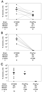
Complete genomic sequence of the lytic bacteriophage DT1 of Streptococcus thermophilus. (1/117)
Streptococcus thermophilus lytic bacteriophage DT1, isolated from a mozzarella whey, was characterized at the microbiological and molecular levels. Phage DT1 had an isometric head of 60 nm and a noncontractile tail of 260 x 8 nm, two major structural proteins of 26 and 32 kDa, and a linear double-stranded DNA genome with cohesive ends at its extremities. The host range of phage DT1 was limited to 5 of the 21 S. thermophilus strains tested. Using S. thermophilus SMQ-301 as a host, phage DT1 had a burst size of 276 +/- 36 and a latent period of 25 min. The genome of phage DT1 contained 34,820 bp with a GC content of 39.1%. Forty-six open reading frames (ORFs) of more than 40 codons were found and putative functions were assigned to 20 ORFs, mostly in the late region of phage DT1. Comparative genomic analysis of DT1 with the completely sequenced S. thermophilus temperate phage O1205 revealed two large homologous regions interspersed by two heterologous segments. The homologous regions consisted of the early replication genes, the late morphogenesis genes, and the lysis cassette. The divergent segments contained the DNA packaging machinery, the major structural proteins, and remnants of a lysogeny module. (+info)Structural and functional analysis of pCI65st, a 6.5 kb plasmid from Streptococcus thermophilus NDI-6. (2/117)
The 6.5 kb cryptic plasmid pCI65st from Streptococcus thermophilus NDI-6, a strain isolated from the Indian fermented milk dahi, was subcloned and sequenced. Five putative ORFs were identified. ORF1 could encode a 315 aa polypeptide almost identical to the RepA protein of previously sequenced S. thermophilus plasmids, indicating that pCI65st is one of the pC194 group of small gram-positive rolling-circle plasmids. ORFs 2 and 4 were virtually identical and could specify proteins of approximately 150 aa with significant similarity to the small heat-shock proteins described from a variety of gram-positive bacteria. ORF3 could encode a 415 aa protein similar to enolase, an enzyme involved in glycolysis and gluconeogenesis. ORF5 could encode a 412 aa protein which had high similarity to the HsdS (specificity) proteins of type I restriction-modification systems. Variants of strain NDI-6 which lacked pCI65st were readily isolated after subculture of the parent strain at 32 degrees C. The plasmid-bearing parent culture was significantly more resistant to a temperature shift from 42 degrees C to 62 degrees C than its plasmid-free variant and expressed proteins which corresponded with the predicted translation products from ORF2 and ORF4. In addition, plasmid-free mutants were lysed in broth by bacteriophages to which the parent culture was resistant. (+info)The genetic relationship between virulent and temperate Streptococcus thermophilus bacteriophages: whole genome comparison of cos-site phages Sfi19 and Sfi21. (3/117)
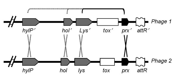
The virulent cos-site Streptococcus thermophilus bacteriophage Sfi19 has a 37,392-bp-long genome consisting of 44 open reading frames all encoded on the same DNA strand. The genome of the temperate cos-site S. thermophilus phage Sfi21 is 3.3 kb longer (40,740 bp, 53 orfs). Both genomes are very similarly organized and differed mainly by gene deletion and DNA rearrangement events in the lysogeny module; gene replacement, duplication, and deletion events in the DNA replication module, and numerous point mutations. The level of point mutations varied from <1% (lysis and DNA replication modules) to >15% (DNA packaging and head morphogenesis modules). A dotplot analysis showed nearly a straight line over the left 25 kb of their genomes. Over the right genome half, a more variable dotplot pattern was observed. The entire lysogeny module from Sfi21 comprising 12 genes was replaced by 7 orfs in Sfi19, six showed similarity with genes from temperate pac-site S. thermophilus phages. None of the genes implicated in the establishment of the lysogenic state (integrase, superinfection immunity, repressor) or remnants of it were conserved in Sfi19, while a Cro-like repressor was detected. Downstream of the highly conserved DNA replication module 11 and 13 orfs were found in Sfi19 and phiSfi21, respectively: Two orfs from Sfi21 were replaced by a different gene and a duplication of the phage origin of replication in Sfi19; a further orf was only found in Sfi21. All other orfs from this region, which included a second putative phage repressor, were closely related between both phages. Two noncoding regions of Sfi19 showed sequence similarity to pST1, a small cryptic plasmid of S. thermophilus. (+info)Comparative sequence analysis of the DNA packaging, head, and tail morphogenesis modules in the temperate cos-site Streptococcus thermophilus bacteriophage Sfi21. (4/117)
The temperate Streptococcus thermophilus bacteriophage Sfi21 possesses 15-nucleotide-long cohesive ends with a 3' overhang that reconstitutes a cos-site with twofold hyphenated rotational symmetry. Over the DNA packaging, head and tail morphogenesis modules, the Sfi21 sequence predicts a gene map that is strikingly similar to that of lambdoid coliphages in the absence of any sequence similarity. A nearly one to one gene correlation was found with the phage lambda genes Nu1 to H, except for gene B-to-E complex, where the Sfi21 map resembled that of coliphage HK97. The similarity between Sfi21 and HK97 was striking: both major head proteins showed an N-terminal coiled-coil structure, the mature major head proteins started at amino acid positions 105 and 104, respectively, and both major head genes were preceded by genes encoding a possible protease and portal protein. The purported Sfi21 protease is the first viral member of the ClpP protease family. The prediction of Sfi21 gene functions by reference to the gene map of intensively investigated coliphages was experimentally confirmed for the major head and tail gene. Phage Sfi21 shows nucleotide sequence similarity with Lactococcus phage BK5-T and a lactococcal prophage and amino acid sequence similarity with the Lactobacillus phage A2 and the Staphylococcus phage PVL. PVL is a missing link that connects the portal proteins from Sfi21 and HK97 with respect to sequence similarity. These observations and database searches, which demonstrate sequence similarity between proteins of phage from gram-positive bacteria, proteobacteria, and Archaea, constrain models of phage evolution. (+info)The autolysin-encoding gene (lytA) of Streptococcus pneumoniae displays restricted allelic variation despite localized recombination events with genes of pneumococcal bacteriophage encoding cell wall lytic enzymes. (5/117)
The lytA-encoded autolysin (N-acetylmuramoyl-L-alanine amidase) of Streptococcus pneumoniae is believed to play an important role in the pathogenesis of pneumococcal infection and has been identified as a putative vaccine target. Allelic diversity of lytA in an extensive collection of clinical isolates was assessed by restriction fragment length polymorphism and confirmatory sequencing studies. Genetic diversity within lytA is limited, especially compared to the high levels of diversity seen in other pneumococcal virulence factor genes, although small blocks generating mosaic structure were identified. Sequence comparisons with genes encoding cell wall lytic enzymes of pneumococcal bacteriophage suggest that localized recombination events have occurred between host lytA and these bacteriophage genes. These results confirm earlier suggestions that recombination between DNA encoding bacteriophage autolytic enzymes and chromosomally encoded lytA might be important in the evolution of lytA. The implications of these findings for understanding the evolution of lytA and the potential utility of LytA as a vaccine target are discussed. (+info)TPW22, a lactococcal temperate phage with a site-specific integrase closely related to Streptococcus thermophilus phage integrases. (6/117)
The temperate phage TPW22, induced from Lactococcus lactis subsp. cremoris W22, and the evolutionarily interesting integrase of this phage were characterized. Phage TPW22 was propagated lytically on L. lactis subsp. cremoris 3107, which could also be lysogenized by site-specific integration. The attachment site (attP), 5'-TAAGGCGACGGTCG-3', of phage TPW22 was present on a 7.5-kb EcoRI fragment, a 3.4-kb EcoRI-HindIII fragment of which was sequenced. Sequence information revealed the presence of an integrase gene (int). The deduced amino acid sequence showed 42 and 28% identity with integrases of streptococcal and lactococcal phages, respectively. The identities with these integrase-encoding genes were 52 and 45%, respectively, at the nucleotide level. This could indicate horizontal gene transfer. A stable integration vector containing attP and int was constructed, and integration in L. lactis subsp. cremoris MG1363 was obtained. The existence of an exchangeable lactococcal phage integration module was suggested. The proposed module covers the phage attachment site, the integrase gene, and surrounding factor-independent terminator structures. The phages phiLC3, TP901-1, and TPW22 all have different versions of this module. Phylogenetically, the TPW22 Int links the phiLC3 lactococcal integrase with known Streptococcus thermophilus integrases. (+info)Widespread distribution of a group I intron and its three deletion derivatives in the lysin gene of Streptococcus thermophilus bacteriophages. (7/117)
Of 62 Streptococcus thermophilus bacteriophages isolated from various ecological settings, half contain a lysin gene interrupted by a group IA2 intron. Phage mRNA splicing was demonstrated. Five phages possess a variant form of the intron resulting from three distinct deletion events located in the intron-harbored open reading frame (orf 253). The predicted orf 253 gene sequence showed a significantly lower GC content than the surrounding intron and lysin gene sequences, and the predicted protein shared a motif with endonucleases found in phages from both gram-positive and gram-negative bacteria. A comparison of the phage lysin genes revealed a clear division between intron-containing and intron-free alleles, leading to the establishment of a 14-bp consensus sequence associated with intron possession. The conserved intron was not found elsewhere in the phage or S. thermophilus bacterial genomes. Folding of the intron RNA revealed secondary structure elements shared with other phage introns: first, a 38-bp insertion between regions P3 and P4 that can be folded into two stem-loop structures (shared with introns from Bacillus phage SPO1 and relatives); second, a conserved P7.2 region (shared with all phage introns); third, the location of the stop codon from orf 253 in the P8 stem (shared with coliphage T4 and Bacillus phage SPO1 introns); fourth, orf 253, which has sequence similarity with the H-N-H motif of putative endonuclease genes found in introns from Lactococcus, Lactobacillus, and Bacillus phages. (+info)The Streptococcus thermophilus autolytic phenotype results from a leaky prophage. (8/117)
Streptococcus thermophilus autolytic strains are characterized by a typical bell-shaped growth curve when grown under appropriate conditions. The cellular mechanisms involved in the triggering of lysis and the bacteriolytic activities of these strains were investigated in this study. Lactose depletion and organic solvents (ethanol, methanol, and chloroform) were shown to trigger a premature and immediate lysis of M17 exponentially growing cells. These factors and compounds are suspected to act by altering the cell envelope properties, causing either the permeabilization (organic solvents) or the depolarization (lactose depletion) of the cytoplasmic membrane. The autolytic character was shown to be associated with lysogeny. Phage particles, most of which were defective, were observed in the culture supernatants after both mitomycin C-induced and spontaneous lysis. By renaturing sodium dodecyl sulfate-polyacrylamide gel electrophoresis, a bacteriolytic activity was detected at 31 kDa exclusively in the autolytic strains. This enzyme was detected during both growth and spontaneous lysis with the same intensity. We have shown that it was prophage encoded and homologous to the endolysin Lyt51 of the streptococcal temperate bacteriophage phi01205 (M. Sheehan, E. Stanley, G. F. Fitzgerald, and D. van Sinderen, Appl. Environ. Microbiol. 65:569-577, 1999). It appears from our results that the autolytic properties are conferred to the S. thermophilus strains by a leaky prophage but do not result from massive prophage induction. More specifically, we propose that phagic genes are constitutively expressed in almost all the cells at a low and nonlethal level and that lysis is controlled and achieved by the prophage-encoded lysis proteins. (+info)I'm sorry for any confusion, but "Streptococcus Phages" is not a standard medical term. The term "Streptococcus" refers to a genus of gram-positive bacteria that can cause various infections in humans, such as strep throat and skin infections. A "phage," on the other hand, is a virus that infects and replicates within bacteria.
Therefore, if you are referring to bacteriophages that infect Streptococcus bacteria, then the term would be "Streptococcus phages" or more specifically, the name of the particular phage species that infect Streptococcus bacteria (e.g., Streptococcus phage C1, Streptococcus phage Φ29). However, it's important to note that there are many different types of bacteriophages that can infect various strains of Streptococcus bacteria, so the specific phage would need to be identified.
Streptococcus is a genus of Gram-positive, spherical bacteria that typically form pairs or chains when clustered together. These bacteria are facultative anaerobes, meaning they can grow in the presence or absence of oxygen. They are non-motile and do not produce spores.
Streptococcus species are commonly found on the skin and mucous membranes of humans and animals. Some strains are part of the normal flora of the body, while others can cause a variety of infections, ranging from mild skin infections to severe and life-threatening diseases such as sepsis, meningitis, and toxic shock syndrome.
The pathogenicity of Streptococcus species depends on various virulence factors, including the production of enzymes and toxins that damage tissues and evade the host's immune response. One of the most well-known Streptococcus species is Streptococcus pyogenes, also known as group A streptococcus (GAS), which is responsible for a wide range of clinical manifestations, including pharyngitis (strep throat), impetigo, cellulitis, necrotizing fasciitis, and rheumatic fever.
It's important to note that the classification of Streptococcus species has evolved over time, with many former members now classified as different genera within the family Streptococcaceae. The current classification system is based on a combination of phenotypic characteristics (such as hemolysis patterns and sugar fermentation) and genotypic methods (such as 16S rRNA sequencing and multilocus sequence typing).
Streptococcus pyogenes is a Gram-positive, beta-hemolytic streptococcus bacterium that causes various suppurative (pus-forming) and nonsuppurative infections in humans. It is also known as group A Streptococcus (GAS) due to its ability to produce the M protein, which confers type-specific antigenicity and allows for serological classification into more than 200 distinct Lancefield groups.
S. pyogenes is responsible for a wide range of clinical manifestations, including pharyngitis (strep throat), impetigo, cellulitis, erysipelas, scarlet fever, rheumatic fever, and acute poststreptococcal glomerulonephritis. In rare cases, it can lead to invasive diseases such as necrotizing fasciitis (flesh-eating disease) and streptococcal toxic shock syndrome (STSS).
The bacterium is typically transmitted through respiratory droplets or direct contact with infected skin lesions. Effective prevention strategies include good hygiene practices, such as frequent handwashing and avoiding sharing personal items, as well as prompt recognition and treatment of infections to prevent spread.
Streptococcus mutans is a gram-positive, facultatively anaerobic, beta-hemolytic species of bacteria that's part of the normal microbiota of the oral cavity in humans. It's one of the primary etiological agents associated with dental caries, or tooth decay, due to its ability to produce large amounts of acid as a byproduct of sugar metabolism, which can lead to demineralization of tooth enamel and dentin. The bacterium can also adhere to tooth surfaces and form biofilms, further contributing to the development of dental caries.
Streptococcus pneumoniae, also known as the pneumococcus, is a gram-positive, alpha-hemolytic bacterium frequently found in the upper respiratory tract of healthy individuals. It is a leading cause of community-acquired pneumonia and can also cause other infectious diseases such as otitis media (ear infection), sinusitis, meningitis, and bacteremia (bloodstream infection). The bacteria are encapsulated, and there are over 90 serotypes based on variations in the capsular polysaccharide. Some serotypes are more virulent or invasive than others, and the polysaccharide composition is crucial for vaccine development. S. pneumoniae infection can be treated with antibiotics, but the emergence of drug-resistant strains has become a significant global health concern.
Streptococcus agalactiae, also known as Group B Streptococcus (GBS), is a type of bacteria that commonly colonizes the gastrointestinal and genitourinary tracts of humans. It is Gram-positive, facultatively anaerobic, and forms chains when viewed under the microscope.
While S. agalactiae can be carried asymptomatically by many adults, it can cause serious infections in newborns, pregnant women, elderly individuals, and people with weakened immune systems. In newborns, GBS can lead to sepsis, pneumonia, and meningitis, which can result in long-term health complications or even be fatal if left untreated.
Pregnant women are often screened for GBS colonization during the third trimester of pregnancy, and those who test positive may receive intrapartum antibiotics to reduce the risk of transmission to their newborns during delivery.
Streptococcal infections are a type of infection caused by group A Streptococcus bacteria (Streptococcus pyogenes). These bacteria can cause a variety of illnesses, ranging from mild skin infections to serious and potentially life-threatening conditions such as sepsis, pneumonia, and necrotizing fasciitis (flesh-eating disease).
Some common types of streptococcal infections include:
* Streptococcal pharyngitis (strep throat) - an infection of the throat and tonsils that can cause sore throat, fever, and swollen lymph nodes.
* Impetigo - a highly contagious skin infection that causes sores or blisters on the skin.
* Cellulitis - a bacterial infection of the deeper layers of the skin and underlying tissue that can cause redness, swelling, pain, and warmth in the affected area.
* Scarlet fever - a streptococcal infection that causes a bright red rash on the body, high fever, and sore throat.
* Necrotizing fasciitis - a rare but serious bacterial infection that can cause tissue death and destruction of the muscles and fascia (the tissue that covers the muscles).
Treatment for streptococcal infections typically involves antibiotics to kill the bacteria causing the infection. It is important to seek medical attention if you suspect a streptococcal infection, as prompt treatment can help prevent serious complications.
Streptococcus suis is a Gram-positive, beta-hemolytic streptococcus that is a significant pathogen in pig populations worldwide. It can cause a variety of clinical manifestations in pigs, including meningitis, arthritis, endocarditis, and septicemia. Transmission to humans can occur through contact with infected pigs or contaminated pork products, resulting in diseases such as meningitis, sepsis, endocarditis, and arthritis. There are 35 serotypes of S. suis, but only a few (including serotypes 1, 2, 4, 5, 9, 14, 16, 21, 24, and 31) are commonly associated with disease in pigs and humans.
Bacteriophages, often simply called phages, are viruses that infect and replicate within bacteria. They consist of a protein coat, called the capsid, that encases the genetic material, which can be either DNA or RNA. Bacteriophages are highly specific, meaning they only infect certain types of bacteria, and they reproduce by hijacking the bacterial cell's machinery to produce more viruses.
Once a phage infects a bacterium, it can either replicate its genetic material and create new phages (lytic cycle), or integrate its genetic material into the bacterial chromosome and replicate along with the bacterium (lysogenic cycle). In the lytic cycle, the newly formed phages are released by lysing, or breaking open, the bacterial cell.
Bacteriophages play a crucial role in shaping microbial communities and have been studied as potential alternatives to antibiotics for treating bacterial infections.
Streptococcus bovis is a type of bacteria that is part of the Streptococcus genus. It is a gram-positive, facultatively anaerobic coccus (spherical) bacterium that is commonly found in the gastrointestinal tracts of animals, including cattle, and can also be found in the human gastrointestinal tract, particularly in the colon.
There are several subspecies of Streptococcus bovis, including S. bovis biotype I (also known as Streptococcus gallolyticus), S. bovis biotype II/2, and S. bovis biotype II/1. Some strains of these bacteria have been associated with human diseases, such as endocarditis, bacteremia, and abscesses in various organs. Additionally, there is evidence to suggest that S. bovis biotype I may be associated with an increased risk of colorectal cancer.
It's important to note that Streptococcus bovis is not a common cause of infection in healthy individuals, but it can cause serious infections in people with underlying medical conditions, such as valvular heart disease or a weakened immune system.
Streptococcus mitis is a species of gram-positive, beta-hemolytic streptococci that are part of the viridans group streptococci (VGS). It is a normal commensal of the human oral cavity, upper respiratory tract, and gastrointestinal tract. However, it can occasionally cause invasive infections such as bacteremia, endocarditis, and meningitis, particularly in immunocompromised individuals or those with underlying medical conditions. S. mitis is also known to be a significant contributor to dental caries. It is often misidentified as Streptococcus sanguinis due to their similar phenotypic characteristics. Accurate identification of this organism is important because of its potential to cause invasive disease and its resistance to some antibiotics.
Streptococcus equi is a gram-positive, beta-hemolytic bacterium that belongs to the Lancefield group C. It is a significant pathogen in horses, causing a respiratory disease known as "strangles." The bacterium can spread through direct contact with infected horses or contaminated objects and can lead to severe complications such as purpura hemorrhagica and bastard strangles.
While Streptococcus equi is not typically associated with human infections, there have been rare cases of zoonotic transmission from horses to humans, causing respiratory tract infections, endocarditis, and soft tissue infections. However, it is essential to note that this bacterium is primarily a pathogen of horses and not a common cause of infection in humans.
Streptococcus oralis is a type of gram-positive, facultatively anaerobic coccus (round-shaped bacterium) that belongs to the viridans group of streptococci. It is commonly found in the human oral cavity, particularly on the surface of the teeth and gums.
S. oralis is generally considered to be a commensal organism, meaning that it can exist harmlessly in the mouth without causing any negative effects. However, under certain circumstances, such as when the immune system is weakened or when there is damage to the oral tissues, S. oralis can cause infections. These infections may include dental caries (cavities), periodontal disease, and endocarditis (inflammation of the inner lining of the heart).
Like other streptococci, S. oralis is able to form biofilms, which are complex communities of bacteria that adhere to surfaces and can be difficult to remove. This ability to form biofilms may contribute to its ability to cause infections.
It's important to note that while S. oralis is a normal part of the oral microbiome, good oral hygiene practices such as brushing and flossing regularly can help prevent an overgrowth of this bacterium and reduce the risk of infection.
Streptococcus sobrinus is a gram-positive, facultatively anaerobic coccus that belongs to the viridans group of streptococci. It's a type of bacteria commonly found in the oral cavity and is one of the primary causative agents of dental caries (tooth decay) along with Streptococcus mutans.
S. sobrinus has the ability to metabolize sugars and produce acid as a byproduct, which can lower the pH of the oral environment and contribute to tooth demineralization and cavity formation. This organism is often found in higher numbers in individuals with a high risk of caries and is associated with a more severe form of the disease.
It's important to note that while S. sobrinus is a significant contributor to dental caries, good oral hygiene practices, such as regular brushing and flossing, limiting sugar intake, and receiving professional dental care can help prevent the negative effects of this bacteria on oral health.
Salmonella phages are viruses that infect and replicate within bacteria of the genus Salmonella. These phages, also known as bacteriophages or simply phages, are composed of a protein capsid that encases the genetic material, which can be either DNA or RNA. They specifically target Salmonella bacteria, using the bacteria's resources to replicate and produce new phage particles. This process often leads to the lysis (breaking open) of the bacterial cell, resulting in the release of newly formed phages.
Salmonella phages have been studied as potential alternatives to antibiotics for controlling Salmonella infections, particularly in food production settings. They offer the advantage of being highly specific to their target bacteria, reducing the risk of disrupting beneficial microbiota. However, further research is needed to fully understand their safety and efficacy before they can be widely used as therapeutic or prophylactic agents.
Bacterial proteins are a type of protein that are produced by bacteria as part of their structural or functional components. These proteins can be involved in various cellular processes, such as metabolism, DNA replication, transcription, and translation. They can also play a role in bacterial pathogenesis, helping the bacteria to evade the host's immune system, acquire nutrients, and multiply within the host.
Bacterial proteins can be classified into different categories based on their function, such as:
1. Enzymes: Proteins that catalyze chemical reactions in the bacterial cell.
2. Structural proteins: Proteins that provide structural support and maintain the shape of the bacterial cell.
3. Signaling proteins: Proteins that help bacteria to communicate with each other and coordinate their behavior.
4. Transport proteins: Proteins that facilitate the movement of molecules across the bacterial cell membrane.
5. Toxins: Proteins that are produced by pathogenic bacteria to damage host cells and promote infection.
6. Surface proteins: Proteins that are located on the surface of the bacterial cell and interact with the environment or host cells.
Understanding the structure and function of bacterial proteins is important for developing new antibiotics, vaccines, and other therapeutic strategies to combat bacterial infections.
Streptococcus thermophilus is a gram-positive, facultatively anaerobic, non-motile, non-spore forming bacterium that belongs to the Streptococcaceae family. It is a species of streptococcus that is mesophilic, meaning it grows best at moderate temperatures, typically between 30-45°C. S. thermophilus is commonly found in milk and dairy products and is one of the starter cultures used in the production of yogurt and other fermented dairy products. It is also used as a probiotic due to its potential health benefits, such as improving lactose intolerance and enhancing the immune system. S. thermophilus is not considered pathogenic and does not normally cause infections in humans.
Pneumococcal infections are illnesses caused by the bacterium Streptococcus pneumoniae, also known as pneumococcus. This bacterium can infect different parts of the body, including the lungs (pneumonia), blood (bacteremia or sepsis), and the covering of the brain and spinal cord (meningitis). Pneumococcal infections can also cause ear infections and sinus infections. The bacteria spread through close contact with an infected person, who may spread the bacteria by coughing or sneezing. People with weakened immune systems, children under 2 years of age, adults over 65, and those with certain medical conditions are at increased risk for developing pneumococcal infections.
Streptococcus gordonii is a species of gram-positive, non-spore forming, facultatively anaerobic bacteria that belongs to the viridans group of streptococci. It is part of the normal flora in the oral cavity and is commonly found on the teeth and mucous membranes.
S. gordonii is a commensal organism, meaning it usually exists harmoniously with its human host without causing harm. However, under certain circumstances, such as when the immune system is compromised or there is damage to the oral tissues, S. gordonii can cause infections. It has been implicated in dental caries (cavities), endocarditis (inflammation of the inner lining of the heart), and other invasive infections.
Like other streptococci, S. gordonii is a coccus-shaped bacterium that tends to occur in pairs or chains. It is catalase-negative, which means it does not produce the enzyme catalase, and it ferments various sugars to produce acid as a byproduct. These characteristics help distinguish S. gordonii from other types of bacteria.
It's important to note that maintaining good oral hygiene practices, such as brushing and flossing regularly, can help prevent the overgrowth of S. gordonii and reduce the risk of dental caries and other infections.
Pseudomonas phages are viruses that infect and replicate within bacteria of the genus Pseudomonas. These phages are important in the study of Pseudomonas species, which include several significant human pathogens such as P. aeruginosa. Phages can be used for therapeutic purposes to treat bacterial infections, including those caused by Pseudomonas. Additionally, they are also useful tools in molecular biology and genetic research.
It's worth noting that while "Pseudomonas phages" refers specifically to phages that infect Pseudomonas bacteria, the term "phage" on its own is used to describe any virus that infects and replicates within a bacterial host.
Lysogeny is a process in the life cycle of certain viruses, known as bacteriophages or phages, which can infect bacteria. In lysogeny, the viral DNA integrates into the chromosome of the host bacterium and replicates along with it, remaining dormant and not producing any new virus particles. This state is called lysogeny or the lysogenic cycle.
The integrated viral DNA is known as a prophage. The bacterial cell that contains a prophage is called a lysogen. The lysogen can continue to grow and divide normally, passing the prophage onto its daughter cells during reproduction. This dormant state can last for many generations of the host bacterium.
However, under certain conditions such as DNA damage or exposure to UV radiation, the prophage can be induced to excise itself from the bacterial chromosome and enter the lytic cycle. In the lytic cycle, the viral DNA replicates rapidly, producing many new virus particles, which eventually leads to the lysis (breaking open) of the host cell and the release of the newly formed virions.
Lysogeny is an important mechanism for the spread and survival of bacteriophages in bacterial populations. It also plays a role in horizontal gene transfer between bacteria, as genes carried by prophages can be transferred to other bacteria during transduction.
RNA phages are a type of bacteriophage, which is a virus that infects bacteria. Unlike most other bacteriophages, RNA phages have an RNA genome instead of a DNA genome. These viruses infect and replicate within bacteria that have an RNA genome or those that can incorporate RNA into their replication cycle.
RNA phages are relatively simple in structure, consisting of an icosahedral capsid (protein shell) containing the single-stranded RNA genome. The genome may be either positive-sense (+) or negative-sense (-), depending on whether it can serve directly as messenger RNA (mRNA) for translation or if it must first be transcribed into a complementary RNA strand before translation.
Examples of well-known RNA phages include the MS2, Qβ, and φ6 phages. These viruses have been extensively studied as model systems to understand fundamental principles of RNA biology, virus replication strategies, and host-pathogen interactions. They also have potential applications in biotechnology, such as in the development of RNA-based vaccines and gene therapy vectors.
Serotyping is a laboratory technique used to classify microorganisms, such as bacteria and viruses, based on the specific antigens or proteins present on their surface. It involves treating the microorganism with different types of antibodies and observing which ones bind to its surface. Each distinct set of antigens corresponds to a specific serotype, allowing for precise identification and characterization of the microorganism. This technique is particularly useful in epidemiology, vaccine development, and infection control.
Molecular sequence data refers to the specific arrangement of molecules, most commonly nucleotides in DNA or RNA, or amino acids in proteins, that make up a biological macromolecule. This data is generated through laboratory techniques such as sequencing, and provides information about the exact order of the constituent molecules. This data is crucial in various fields of biology, including genetics, evolution, and molecular biology, allowing for comparisons between different organisms, identification of genetic variations, and studies of gene function and regulation.
Enterococcus faecalis is a species of gram-positive, facultatively anaerobic bacteria that are part of the normal gut microbiota in humans and animals. It is a type of enterococci that can cause a variety of infections, including urinary tract infections, bacteremia, endocarditis, and meningitis, particularly in hospitalized patients or those with compromised immune systems.
E. faecalis is known for its ability to survive in a wide range of environments and resist various antibiotics, making it difficult to treat infections caused by this organism. It can also form biofilms, which further increase its resistance to antimicrobial agents and host immune responses. Accurate identification and appropriate treatment of E. faecalis infections are essential to prevent complications and ensure positive patient outcomes.
Bacterial DNA refers to the genetic material found in bacteria. It is composed of a double-stranded helix containing four nucleotide bases - adenine (A), thymine (T), guanine (G), and cytosine (C) - that are linked together by phosphodiester bonds. The sequence of these bases in the DNA molecule carries the genetic information necessary for the growth, development, and reproduction of bacteria.
Bacterial DNA is circular in most bacterial species, although some have linear chromosomes. In addition to the main chromosome, many bacteria also contain small circular pieces of DNA called plasmids that can carry additional genes and provide resistance to antibiotics or other environmental stressors.
Unlike eukaryotic cells, which have their DNA enclosed within a nucleus, bacterial DNA is present in the cytoplasm of the cell, where it is in direct contact with the cell's metabolic machinery. This allows for rapid gene expression and regulation in response to changing environmental conditions.
Anti-bacterial agents, also known as antibiotics, are a type of medication used to treat infections caused by bacteria. These agents work by either killing the bacteria or inhibiting their growth and reproduction. There are several different classes of anti-bacterial agents, including penicillins, cephalosporins, fluoroquinolones, macrolides, and tetracyclines, among others. Each class of antibiotic has a specific mechanism of action and is used to treat certain types of bacterial infections. It's important to note that anti-bacterial agents are not effective against viral infections, such as the common cold or flu. Misuse and overuse of antibiotics can lead to antibiotic resistance, which is a significant global health concern.
A peptide library is a collection of a large number of peptides, which are short chains of amino acids. Each peptide in the library is typically composed of a defined length and sequence, and may contain a variety of different amino acids. Peptide libraries can be synthesized using automated techniques and are often used in scientific research to identify potential ligands (molecules that bind to specific targets) or to study the interactions between peptides and other molecules.
In a peptide library, each peptide is usually attached to a solid support, such as a resin bead, and the entire library can be created using split-and-pool synthesis techniques. This allows for the rapid and efficient synthesis of a large number of unique peptides, which can then be screened for specific activities or properties.
Peptide libraries are used in various fields such as drug discovery, proteomics, and molecular biology to identify potential therapeutic targets, understand protein-protein interactions, and develop new diagnostic tools.
In medical terms, the mouth is officially referred to as the oral cavity. It is the first part of the digestive tract and includes several structures: the lips, vestibule (the space enclosed by the lips and teeth), teeth, gingiva (gums), hard and soft palate, tongue, floor of the mouth, and salivary glands. The mouth is responsible for several functions including speaking, swallowing, breathing, and eating, as it is the initial point of ingestion where food is broken down through mechanical and chemical processes, beginning the digestive process.
Bacillus phages are viruses that infect and replicate within bacteria of the genus Bacillus. These phages, also known as bacteriophages or simply phages, are a type of virus that is specifically adapted to infect and multiply within bacteria. They use the bacterial cell's machinery to produce new copies of themselves, often resulting in the lysis (breakdown) of the bacterial cell. Bacillus phages are widely studied for their potential applications in biotechnology, medicine, and basic research.
Coliphages are viruses that infect and replicate within certain species of bacteria that belong to the coliform group, particularly Escherichia coli (E. coli). These viruses are commonly found in water and soil environments and are frequently used as indicators of fecal contamination in water quality testing. Coliphages are not harmful to humans or animals, but their presence in water can suggest the potential presence of pathogenic bacteria or other microorganisms that may pose a health risk. There are two main types of coliphages: F-specific RNA coliphages and somatic (or non-F specific) DNA coliphages.
Streptolysins are exotoxins produced by certain strains of Streptococcus bacteria, primarily Group A Streptococcus (GAS). These toxins are classified into two types: streptolysin O (SLO) and streptolysin S (SLS).
1. Streptolysin O (SLO): It is a protein exotoxin that exhibits oxygen-labile hemolytic activity, meaning it can lyse or destroy red blood cells in the presence of oxygen. SLO is capable of entering host cells and causing various cellular damages, including inhibition of phagocytosis, modulation of immune responses, and induction of apoptosis (programmed cell death).
2. Streptolysin S (SLS): It is a non-protein, oxygen-stable hemolysin that can also lyse red blood cells but does so independently of oxygen presence. SLS is more heat-resistant than SLO and has a stronger ability to penetrate host cell membranes.
Both streptolysins contribute to the virulence of Streptococcus pyogenes, which can cause various clinical infections such as pharyngitis (strep throat), impetigo, scarlet fever, and invasive diseases like necrotizing fasciitis and toxic shock syndrome.
The detection of streptolysin O antibodies (ASO titer) is often used as a diagnostic marker for past or recent GAS infections, particularly in cases of rheumatic fever, where elevated ASO titers indicate ongoing or previous streptococcal infection.
Microbial sensitivity tests, also known as antibiotic susceptibility tests (ASTs) or bacterial susceptibility tests, are laboratory procedures used to determine the effectiveness of various antimicrobial agents against specific microorganisms isolated from a patient's infection. These tests help healthcare providers identify which antibiotics will be most effective in treating an infection and which ones should be avoided due to resistance. The results of these tests can guide appropriate antibiotic therapy, minimize the potential for antibiotic resistance, improve clinical outcomes, and reduce unnecessary side effects or toxicity from ineffective antimicrobials.
There are several methods for performing microbial sensitivity tests, including:
1. Disk diffusion method (Kirby-Bauer test): A standardized paper disk containing a predetermined amount of an antibiotic is placed on an agar plate that has been inoculated with the isolated microorganism. After incubation, the zone of inhibition around the disk is measured to determine the susceptibility or resistance of the organism to that particular antibiotic.
2. Broth dilution method: A series of tubes or wells containing decreasing concentrations of an antimicrobial agent are inoculated with a standardized microbial suspension. After incubation, the minimum inhibitory concentration (MIC) is determined by observing the lowest concentration of the antibiotic that prevents visible growth of the organism.
3. Automated systems: These use sophisticated technology to perform both disk diffusion and broth dilution methods automatically, providing rapid and accurate results for a wide range of microorganisms and antimicrobial agents.
The interpretation of microbial sensitivity test results should be done cautiously, considering factors such as the site of infection, pharmacokinetics and pharmacodynamics of the antibiotic, potential toxicity, and local resistance patterns. Regular monitoring of susceptibility patterns and ongoing antimicrobial stewardship programs are essential to ensure optimal use of these tests and to minimize the development of antibiotic resistance.
Streptococcus intermedius is a type of Gram-positive coccus bacterium that is part of the Streptococcus anginosus group, also known as the Streptococcus milleri group. These bacteria are normal inhabitants of the mouth, upper respiratory tract, and gastrointestinal tract in humans. However, they can cause opportunistic infections in various parts of the body, such as the brain, lungs, liver, and heart valves, particularly in individuals with compromised immune systems.
S. intermedius infections can range from mild to severe and include abscesses, endocarditis, meningitis, and sepsis. Proper identification of this bacterium is essential for appropriate antibiotic therapy and management of associated infections.
'Escherichia coli' (E. coli) is a type of gram-negative, facultatively anaerobic, rod-shaped bacterium that commonly inhabits the intestinal tract of humans and warm-blooded animals. It is a member of the family Enterobacteriaceae and one of the most well-studied prokaryotic model organisms in molecular biology.
While most E. coli strains are harmless and even beneficial to their hosts, some serotypes can cause various forms of gastrointestinal and extraintestinal illnesses in humans and animals. These pathogenic strains possess virulence factors that enable them to colonize and damage host tissues, leading to diseases such as diarrhea, urinary tract infections, pneumonia, and sepsis.
E. coli is a versatile organism with remarkable genetic diversity, which allows it to adapt to various environmental niches. It can be found in water, soil, food, and various man-made environments, making it an essential indicator of fecal contamination and a common cause of foodborne illnesses. The study of E. coli has contributed significantly to our understanding of fundamental biological processes, including DNA replication, gene regulation, and protein synthesis.
Pharyngitis is the medical term for inflammation of the pharynx, which is the back portion of the throat. This condition is often characterized by symptoms such as sore throat, difficulty swallowing, and scratchiness in the throat. Pharyngitis can be caused by a variety of factors, including viral infections (such as the common cold), bacterial infections (such as strep throat), and irritants (such as smoke or chemical fumes). Treatment for pharyngitis depends on the underlying cause of the condition, but may include medications to relieve symptoms or antibiotics to treat a bacterial infection.
Bacteriolysis is the breaking down or destruction of bacterial cells. This process can occur naturally or as a result of medical treatment, such as when antibiotics target and destroy bacteria by disrupting their cell walls. The term "bacteriolysis" specifically refers to the breakdown of the bacterial cell membrane, which can lead to the release of the contents of the bacterial cell and ultimately result in the death of the organism.
Dental plaque is a biofilm or mass of bacteria that accumulates on the surface of the teeth, restorative materials, and prosthetic devices such as dentures. It is initiated when bacterial colonizers attach to the smooth surfaces of teeth through van der Waals forces and specific molecular adhesion mechanisms.
The microorganisms within the dental plaque produce extracellular polysaccharides that help to stabilize and strengthen the biofilm, making it resistant to removal by simple brushing or rinsing. Over time, if not regularly removed through oral hygiene practices such as brushing and flossing, dental plaque can mineralize and harden into tartar or calculus.
The bacteria in dental plaque can cause tooth decay (dental caries) by metabolizing sugars and producing acid that demineralizes the tooth enamel. Additionally, certain types of bacteria in dental plaque can cause periodontal disease, an inflammation of the gums that can lead to tissue damage and bone loss around the teeth. Regular professional dental cleanings and good oral hygiene practices are essential for preventing the buildup of dental plaque and maintaining good oral health.
Erythromycin is a type of antibiotic known as a macrolide, which is used to treat various types of bacterial infections. It works by inhibiting the bacteria's ability to produce proteins, which are necessary for the bacteria to survive and multiply. Erythromycin is often used to treat respiratory tract infections, skin infections, and sexually transmitted diseases. It may also be used to prevent endocarditis (inflammation of the lining of the heart) in people at risk of this condition.
Erythromycin is generally considered safe for most people, but it can cause side effects such as nausea, vomiting, and diarrhea. It may also interact with other medications, so it's important to tell your doctor about all the drugs you are taking before starting erythromycin.
Like all antibiotics, erythromycin should only be used to treat bacterial infections, as it is not effective against viral infections such as the common cold or flu. Overuse of antibiotics can lead to antibiotic resistance, which makes it harder to treat infections in the future.
Bacteriophage typing is a laboratory method used to identify and differentiate bacterial strains based on their susceptibility to specific bacteriophages, which are viruses that infect and replicate within bacteria. In this technique, a standard set of bacteriophages with known host ranges are allowed to infect and form plaques on a lawn of bacterial cells grown on a solid medium, such as agar. The pattern and number of plaques formed are then used to identify the specific bacteriophage types that are able to infect the bacterial strain, providing a unique "fingerprint" or profile that can be used for typing and differentiating different bacterial strains.
Bacteriophage typing is particularly useful in epidemiological studies, as it can help track the spread of specific bacterial clones within a population, monitor antibiotic resistance patterns, and provide insights into the evolution and ecology of bacterial pathogens. It has been widely used in the study of various bacterial species, including Staphylococcus aureus, Salmonella enterica, and Mycobacterium tuberculosis, among others.
A bacterial gene is a segment of DNA (or RNA in some viruses) that contains the genetic information necessary for the synthesis of a functional bacterial protein or RNA molecule. These genes are responsible for encoding various characteristics and functions of bacteria such as metabolism, reproduction, and resistance to antibiotics. They can be transmitted between bacteria through horizontal gene transfer mechanisms like conjugation, transformation, and transduction. Bacterial genes are often organized into operons, which are clusters of genes that are transcribed together as a single mRNA molecule.
It's important to note that the term "bacterial gene" is used to describe genetic elements found in bacteria, but not all genetic elements in bacteria are considered genes. For example, some DNA sequences may not encode functional products and are therefore not considered genes. Additionally, some bacterial genes may be plasmid-borne or phage-borne, rather than being located on the bacterial chromosome.
Bacterial antigens are substances found on the surface or produced by bacteria that can stimulate an immune response in a host organism. These antigens can be proteins, polysaccharides, teichoic acids, lipopolysaccharides, or other molecules that are recognized as foreign by the host's immune system.
When a bacterial antigen is encountered by the host's immune system, it triggers a series of responses aimed at eliminating the bacteria and preventing infection. The host's immune system recognizes the antigen as foreign through the use of specialized receptors called pattern recognition receptors (PRRs), which are found on various immune cells such as macrophages, dendritic cells, and neutrophils.
Once a bacterial antigen is recognized by the host's immune system, it can stimulate both the innate and adaptive immune responses. The innate immune response involves the activation of inflammatory pathways, the recruitment of immune cells to the site of infection, and the production of antimicrobial peptides.
The adaptive immune response, on the other hand, involves the activation of T cells and B cells, which are specific to the bacterial antigen. These cells can recognize and remember the antigen, allowing for a more rapid and effective response upon subsequent exposures.
Bacterial antigens are important in the development of vaccines, as they can be used to stimulate an immune response without causing disease. By identifying specific bacterial antigens that are associated with virulence or pathogenicity, researchers can develop vaccines that target these antigens and provide protection against infection.
Pneumonia, pneumococcal is a type of pneumonia caused by the bacterium Streptococcus pneumoniae (also known as pneumococcus). This bacteria can colonize the upper respiratory tract and occasionally invade the lower respiratory tract, causing infection.
Pneumococcal pneumonia can affect people of any age but is most common in young children, older adults, and those with weakened immune systems. The symptoms of pneumococcal pneumonia include fever, chills, cough, chest pain, shortness of breath, and rapid breathing. In severe cases, it can lead to complications such as bacteremia (bacterial infection in the blood), meningitis (inflammation of the membranes surrounding the brain and spinal cord), and respiratory failure.
Pneumococcal pneumonia can be prevented through vaccination with the pneumococcal conjugate vaccine (PCV) or the pneumococcal polysaccharide vaccine (PPSV). These vaccines protect against the most common strains of Streptococcus pneumoniae that cause invasive disease. It is also important to practice good hygiene, such as covering the mouth and nose when coughing or sneezing, and washing hands frequently, to prevent the spread of pneumococcal bacteria.
A base sequence in the context of molecular biology refers to the specific order of nucleotides in a DNA or RNA molecule. In DNA, these nucleotides are adenine (A), guanine (G), cytosine (C), and thymine (T). In RNA, uracil (U) takes the place of thymine. The base sequence contains genetic information that is transcribed into RNA and ultimately translated into proteins. It is the exact order of these bases that determines the genetic code and thus the function of the DNA or RNA molecule.
Penicillin resistance is the ability of certain bacteria to withstand the antibacterial effects of penicillin, a type of antibiotic. This occurs when these bacteria have developed mechanisms that prevent penicillin from binding to and inhibiting the function of their cell wall biosynthesis proteins, particularly the enzyme transpeptidase.
One common mechanism of penicillin resistance is the production of beta-lactamases, enzymes that can hydrolyze and inactivate the beta-lactam ring structure present in penicillin and other related antibiotics. Another mechanism involves alterations in the bacterial cell wall that prevent penicillin from binding to its target proteins.
Penicillin resistance is a significant concern in clinical settings, as it can limit treatment options for bacterial infections and may necessitate the use of more potent or toxic antibiotics. It is important to note that misuse or overuse of antibiotics can contribute to the development and spread of antibiotic-resistant bacteria, including those resistant to penicillin.
Bacterial adhesion is the initial and crucial step in the process of bacterial colonization, where bacteria attach themselves to a surface or tissue. This process involves specific interactions between bacterial adhesins (proteins, fimbriae, or pili) and host receptors (glycoproteins, glycolipids, or extracellular matrix components). The attachment can be either reversible or irreversible, depending on the strength of interaction. Bacterial adhesion is a significant factor in initiating biofilm formation, which can lead to various infectious diseases and medical device-associated infections.
Siphoviridae is a family of tailed bacteriophages, which are viruses that infect and replicate within bacteria. The members of this family are characterized by their long, non-contractile tails, which are typically around 100-1000 nanometers in length. The tail fibers at the end of the tail are used to recognize and attach to specific receptors on the surface of bacterial cells.
The Siphoviridae family includes many well-known bacteriophages, such as the lambda phage that infects Escherichia coli bacteria. The genetic material of Siphoviridae viruses is double-stranded DNA, which is packaged inside an icosahedral capsid (the protein shell of the virus).
It's worth noting that Siphoviridae is one of the five families in the order Caudovirales, which includes all tailed bacteriophages. The other four families are Myoviridae, Podoviridae, Herelleviridae, and Ackermannviridae.
Streptococcal vaccines are immunizations designed to protect against infections caused by Streptococcus bacteria. These vaccines contain antigens, which are substances that trigger an immune response and help the body recognize and fight off specific types of Streptococcus bacteria. There are several different types of streptococcal vaccines available or in development, including:
1. Pneumococcal conjugate vaccine (PCV): This vaccine protects against Streptococcus pneumoniae, a type of bacteria that can cause pneumonia, meningitis, and other serious infections. PCV is recommended for all children under 2 years old, as well as older children and adults with certain medical conditions.
2. Pneumococcal polysaccharide vaccine (PPSV): This vaccine also protects against Streptococcus pneumoniae, but it is recommended for adults 65 and older, as well as younger people with certain medical conditions.
3. Streptococcus pyogenes vaccine: This vaccine is being developed to protect against Group A Streptococcus (GAS), which can cause a variety of infections, including strep throat, skin infections, and serious diseases like rheumatic fever and toxic shock syndrome. There are several different GAS vaccine candidates in various stages of development.
4. Streptococcus agalactiae vaccine: This vaccine is being developed to protect against Group B Streptococcus (GBS), which can cause serious infections in newborns, pregnant women, and older adults with certain medical conditions. There are several different GBS vaccine candidates in various stages of development.
Overall, streptococcal vaccines play an important role in preventing bacterial infections and reducing the burden of disease caused by Streptococcus bacteria.
An amino acid sequence is the specific order of amino acids in a protein or peptide molecule, formed by the linking of the amino group (-NH2) of one amino acid to the carboxyl group (-COOH) of another amino acid through a peptide bond. The sequence is determined by the genetic code and is unique to each type of protein or peptide. It plays a crucial role in determining the three-dimensional structure and function of proteins.
"Lactococcus lactis" is a species of gram-positive, facultatively anaerobic bacteria that are commonly found in nature, particularly in environments involving plants and dairy products. It is a catalase-negative, non-spore forming coccus that typically occurs in pairs or short chains.
"Lactococcus lactis" has significant industrial importance as it plays a crucial role in the production of fermented foods such as cheese and buttermilk. The bacterium converts lactose into lactic acid, which contributes to the sour taste and preservative qualities of these products.
In addition to its use in food production, "Lactococcus lactis" has been explored for its potential therapeutic applications. It can be used as a vector for delivering therapeutic proteins or vaccines to the gastrointestinal tract due to its ability to survive and colonize there.
It's worth noting that "Lactococcus lactis" is generally considered safe for human consumption, and it's one of the most commonly used probiotics in food and supplements.
Bacterial polysaccharides are complex carbohydrates that consist of long chains of sugar molecules (monosaccharides) linked together by glycosidic bonds. They are produced and used by bacteria for various purposes such as:
1. Structural components: Bacterial polysaccharides, such as peptidoglycan and lipopolysaccharide (LPS), play a crucial role in maintaining the structural integrity of bacterial cells. Peptidoglycan is a major component of the bacterial cell wall, while LPS forms the outer layer of the outer membrane in gram-negative bacteria.
2. Nutrient storage: Some bacteria synthesize and store polysaccharides as an energy reserve, similar to how plants store starch. These polysaccharides can be broken down and utilized by the bacterium when needed.
3. Virulence factors: Bacterial polysaccharides can also function as virulence factors, contributing to the pathogenesis of bacterial infections. For example, certain bacteria produce capsular polysaccharides (CPS) that surround and protect the bacterial cells from host immune defenses, allowing them to evade phagocytosis and persist within the host.
4. Adhesins: Some polysaccharides act as adhesins, facilitating the attachment of bacteria to surfaces or host cells. This is important for biofilm formation, which helps bacteria resist environmental stresses and antibiotic treatments.
5. Antigenic properties: Bacterial polysaccharides can be highly antigenic, eliciting an immune response in the host. The antigenicity of these molecules can vary between different bacterial species or even strains within a species, making them useful as targets for vaccines and diagnostic tests.
In summary, bacterial polysaccharides are complex carbohydrates that serve various functions in bacteria, including structural support, nutrient storage, virulence factor production, adhesion, and antigenicity.
Myoviridae is a family of bacteriophages, which are viruses that infect and replicate within bacteria. Here is the medical definition of Myoviridae:
Myoviridae is a family of tailed bacteriophages characterized by a contractile sheath surrounding the tail structure. The members of this family have a double-stranded DNA (dsDNA) genome, which is relatively large, ranging from 40 to over 200 kilobases in size. Myoviridae viruses typically infect Gram-negative bacteria and are known to cause lysis of the host cell upon replication. The family includes many well-known bacteriophages such as T4, T5, and λ phages, which have been extensively studied for their biological properties and potential applications in molecular biology and medicine.
It's worth noting that while Myoviridae viruses can be useful tools in scientific research, they are not used in clinical practice as therapeutic agents. However, there is ongoing research into the use of bacteriophages, including those from the family Myoviridae, for the treatment of bacterial infections that are resistant to antibiotics.
Microbial drug resistance is a significant medical issue that refers to the ability of microorganisms (such as bacteria, viruses, fungi, or parasites) to withstand or survive exposure to drugs or medications designed to kill them or limit their growth. This phenomenon has become a major global health concern, particularly in the context of bacterial infections, where it is also known as antibiotic resistance.
Drug resistance arises due to genetic changes in microorganisms that enable them to modify or bypass the effects of antimicrobial agents. These genetic alterations can be caused by mutations or the acquisition of resistance genes through horizontal gene transfer. The resistant microbes then replicate and multiply, forming populations that are increasingly difficult to eradicate with conventional treatments.
The consequences of drug-resistant infections include increased morbidity, mortality, healthcare costs, and the potential for widespread outbreaks. Factors contributing to the emergence and spread of microbial drug resistance include the overuse or misuse of antimicrobials, poor infection control practices, and inadequate surveillance systems.
To address this challenge, it is crucial to promote prudent antibiotic use, strengthen infection prevention and control measures, develop new antimicrobial agents, and invest in research to better understand the mechanisms underlying drug resistance.
Saliva is a complex mixture of primarily water, but also electrolytes, enzymes, antibacterial compounds, and various other substances. It is produced by the salivary glands located in the mouth. Saliva plays an essential role in maintaining oral health by moistening the mouth, helping to digest food, and protecting the teeth from decay by neutralizing acids produced by bacteria.
The medical definition of saliva can be stated as:
"A clear, watery, slightly alkaline fluid secreted by the salivary glands, consisting mainly of water, with small amounts of electrolytes, enzymes (such as amylase), mucus, and antibacterial compounds. Saliva aids in digestion, lubrication of oral tissues, and provides an oral barrier against microorganisms."
Bacteriophage lambda, often simply referred to as phage lambda, is a type of virus that infects the bacterium Escherichia coli (E. coli). It is a double-stranded DNA virus that integrates its genetic material into the bacterial chromosome as a prophage when it infects the host cell. This allows the phage to replicate along with the bacterium until certain conditions trigger the lytic cycle, during which new virions are produced and released by lysing, or breaking open, the host cell.
Phage lambda is widely studied in molecular biology due to its well-characterized life cycle and genetic structure. It has been instrumental in understanding various fundamental biological processes such as gene regulation, DNA recombination, and lysis-lysogeny decision.
Culture media is a substance that is used to support the growth of microorganisms or cells in an artificial environment, such as a petri dish or test tube. It typically contains nutrients and other factors that are necessary for the growth and survival of the organisms being cultured. There are many different types of culture media, each with its own specific formulation and intended use. Some common examples include blood agar, which is used to culture bacteria; Sabouraud dextrose agar, which is used to culture fungi; and Eagle's minimum essential medium, which is used to culture animal cells.
DNA Sequence Analysis is the systematic determination of the order of nucleotides in a DNA molecule. It is a critical component of modern molecular biology, genetics, and genetic engineering. The process involves determining the exact order of the four nucleotide bases - adenine (A), guanine (G), cytosine (C), and thymine (T) - in a DNA molecule or fragment. This information is used in various applications such as identifying gene mutations, studying evolutionary relationships, developing molecular markers for breeding, and diagnosing genetic diseases.
The process of DNA Sequence Analysis typically involves several steps, including DNA extraction, PCR amplification (if necessary), purification, sequencing reaction, and electrophoresis. The resulting data is then analyzed using specialized software to determine the exact sequence of nucleotides.
In recent years, high-throughput DNA sequencing technologies have revolutionized the field of genomics, enabling the rapid and cost-effective sequencing of entire genomes. This has led to an explosion of genomic data and new insights into the genetic basis of many diseases and traits.
Bacterial capsules are slimy, gel-like layers that surround many types of bacteria. They are made up of polysaccharides, proteins, or lipopolysaccharides and are synthesized by the bacterial cell. These capsules play a crucial role in the virulence and pathogenicity of bacteria as they help the bacteria to evade the host's immune system and promote their survival and colonization within the host. The presence of a capsule can also contribute to the bacteria's resistance to desiccation, phagocytosis, and antibiotics.
The chemical composition and structure of bacterial capsules vary among different species of bacteria, which is one factor that contributes to their serological specificity and allows for their identification and classification using methods such as the Quellung reaction or immunofluorescence microscopy.
The pharynx is a part of the digestive and respiratory systems that serves as a conduit for food and air. It is a musculo-membranous tube extending from the base of the skull to the level of the sixth cervical vertebra where it becomes continuous with the esophagus.
The pharynx has three regions: the nasopharynx, oropharynx, and laryngopharynx. The nasopharynx is the uppermost region, which lies above the soft palate and is connected to the nasal cavity. The oropharynx is the middle region, which includes the area between the soft palate and the hyoid bone, including the tonsils and base of the tongue. The laryngopharynx is the lowest region, which lies below the hyoid bone and connects to the larynx.
The primary function of the pharynx is to convey food from the oral cavity to the esophagus during swallowing and to allow air to pass from the nasal cavity to the larynx during breathing. It also plays a role in speech, taste, and immune defense.
Penicillins are a group of antibiotics derived from the Penicillium fungus. They are widely used to treat various bacterial infections due to their bactericidal activity, which means they kill bacteria by interfering with the synthesis of their cell walls. The first penicillin, benzylpenicillin (also known as penicillin G), was discovered in 1928 by Sir Alexander Fleming. Since then, numerous semi-synthetic penicillins have been developed to expand the spectrum of activity and stability against bacterial enzymes that can inactivate these drugs.
Penicillins are classified into several groups based on their chemical structure and spectrum of activity:
1. Natural Penicillins (e.g., benzylpenicillin, phenoxymethylpenicillin): These have a narrow spectrum of activity, mainly targeting Gram-positive bacteria such as streptococci and staphylococci. However, they are susceptible to degradation by beta-lactamase enzymes produced by some bacteria.
2. Penicillinase-resistant Penicillins (e.g., methicillin, oxacillin, nafcillin): These penicillins resist degradation by certain bacterial beta-lactamases and are primarily used to treat infections caused by staphylococci, including methicillin-susceptible Staphylococcus aureus (MSSA).
3. Aminopenicillins (e.g., ampicillin, amoxicillin): These penicillins have an extended spectrum of activity compared to natural penicillins, including some Gram-negative bacteria such as Escherichia coli and Haemophilus influenzae. However, they are still susceptible to degradation by many beta-lactamases.
4. Antipseudomonal Penicillins (e.g., carbenicillin, ticarcillin): These penicillins have activity against Pseudomonas aeruginosa and other Gram-negative bacteria with increased resistance to other antibiotics. They are often combined with beta-lactamase inhibitors such as clavulanate or tazobactam to protect them from degradation.
5. Extended-spectrum Penicillins (e.g., piperacillin): These penicillins have a broad spectrum of activity, including many Gram-positive and Gram-negative bacteria. They are often combined with beta-lactamase inhibitors to protect them from degradation.
Penicillins are generally well-tolerated antibiotics; however, they can cause allergic reactions in some individuals, ranging from mild skin rashes to life-threatening anaphylaxis. Cross-reactivity between different penicillin classes and other beta-lactam antibiotics (e.g., cephalosporins) is possible but varies depending on the specific drugs involved.
Bacterial drug resistance is a type of antimicrobial resistance that occurs when bacteria evolve the ability to survive and reproduce in the presence of drugs (such as antibiotics) that would normally kill them or inhibit their growth. This can happen due to various mechanisms, including genetic mutations or the acquisition of resistance genes from other bacteria.
As a result, bacterial infections may become more difficult to treat, requiring higher doses of medication, alternative drugs, or longer treatment courses. In some cases, drug-resistant infections can lead to serious health complications, increased healthcare costs, and higher mortality rates.
Examples of bacterial drug resistance include methicillin-resistant Staphylococcus aureus (MRSA), vancomycin-resistant Enterococci (VRE), and multidrug-resistant tuberculosis (MDR-TB). Preventing the spread of bacterial drug resistance is crucial for maintaining effective treatments for infectious diseases.
Dental caries, also known as tooth decay or cavities, refers to the damage or breakdown of the hard tissues of the teeth (enamel, dentin, and cementum) due to the activity of acid-producing bacteria. These bacteria ferment sugars from food and drinks, producing acids that dissolve and weaken the tooth structure, leading to cavities.
The process of dental caries development involves several stages:
1. Demineralization: The acidic environment created by bacterial activity causes minerals (calcium and phosphate) to be lost from the tooth surface, making it weaker and more susceptible to decay.
2. Formation of a white spot lesion: As demineralization progresses, a chalky white area appears on the tooth surface, indicating early caries development.
3. Cavity formation: If left untreated, the demineralization process continues, leading to the breakdown and loss of tooth structure, resulting in a cavity or hole in the tooth.
4. Infection and pulp involvement: As the decay progresses deeper into the tooth, it can reach the dental pulp (the soft tissue containing nerves and blood vessels), causing infection, inflammation, and potentially leading to toothache, abscess, or even tooth loss.
Preventing dental caries involves maintaining good oral hygiene, reducing sugar intake, using fluoride toothpaste and mouthwash, and having regular dental check-ups and cleanings. Early detection and treatment of dental caries can help prevent further progression and more severe complications.
Streptococcus constellatus is a type of Gram-positive coccus bacteria that belongs to the Streptococcus anginosus group, also known as the "streptococci of uncertain taxonomic position" or S. milleri group. These bacteria are part of the normal flora in the human mouth, upper respiratory tract, and gastrointestinal tract. However, they can cause opportunistic infections when they enter other parts of the body, particularly in individuals with weakened immune systems.
S. constellatus has been associated with a variety of infections, including abscesses, endocarditis, meningitis, septicemia, and dental and respiratory tract infections. It is important to note that the clinical significance of S. constellatus can vary, as it may sometimes be found as a commensal organism or as part of a polymicrobial infection. Proper identification and antimicrobial susceptibility testing are crucial for appropriate treatment.
Glucosyltransferases (GTs) are a group of enzymes that catalyze the transfer of a glucose molecule from an activated donor to an acceptor molecule, resulting in the formation of a glycosidic bond. These enzymes play crucial roles in various biological processes, including the biosynthesis of complex carbohydrates, cell wall synthesis, and protein glycosylation. In some cases, GTs can also contribute to bacterial pathogenesis by facilitating the attachment of bacteria to host tissues through the formation of glucans, which are polymers of glucose molecules.
GTs can be classified into several families based on their sequence similarities and catalytic mechanisms. The donor substrates for GTs are typically activated sugars such as UDP-glucose, TDP-glucose, or GDP-glucose, which serve as the source of the glucose moiety that is transferred to the acceptor molecule. The acceptor can be a wide range of molecules, including other sugars, proteins, lipids, or small molecules.
In the context of human health and disease, GTs have been implicated in various pathological conditions, such as cancer, inflammation, and microbial infections. For example, some GTs can modify proteins on the surface of cancer cells, leading to increased cell proliferation, migration, and invasion. Additionally, GTs can contribute to bacterial resistance to antibiotics by modifying the structure of bacterial cell walls or by producing biofilms that protect bacteria from host immune responses and antimicrobial agents.
Overall, Glucosyltransferases are essential enzymes involved in various biological processes, and their dysregulation has been associated with several human diseases. Therefore, understanding the structure, function, and regulation of GTs is crucial for developing novel therapeutic strategies to target these enzymes and treat related pathological conditions.
A mutation is a permanent change in the DNA sequence of an organism's genome. Mutations can occur spontaneously or be caused by environmental factors such as exposure to radiation, chemicals, or viruses. They may have various effects on the organism, ranging from benign to harmful, depending on where they occur and whether they alter the function of essential proteins. In some cases, mutations can increase an individual's susceptibility to certain diseases or disorders, while in others, they may confer a survival advantage. Mutations are the driving force behind evolution, as they introduce new genetic variability into populations, which can then be acted upon by natural selection.
Dextranase is an enzyme that breaks down dextran, a type of complex sugar (polysaccharide) consisting of many glucose molecules linked together in a chain. Dextran is produced by certain bacteria and can be found in some foods, as well as in the body during infections or after surgery. Dextranase is used medically to help prevent or treat complications associated with dextran, such as blockages in blood vessels caused by the accumulation of dextran molecules. It may also be used in research and industry for various purposes, including the production of clarified fruit juices and wine.
Virulence, in the context of medicine and microbiology, refers to the degree or severity of damage or harm that a pathogen (like a bacterium, virus, fungus, or parasite) can cause to its host. It is often associated with the ability of the pathogen to invade and damage host tissues, evade or suppress the host's immune response, replicate within the host, and spread between hosts.
Virulence factors are the specific components or mechanisms that contribute to a pathogen's virulence, such as toxins, enzymes, adhesins, and capsules. These factors enable the pathogen to establish an infection, cause tissue damage, and facilitate its transmission between hosts. The overall virulence of a pathogen can be influenced by various factors, including host susceptibility, environmental conditions, and the specific strain or species of the pathogen.
Actinomyces is a genus of gram-positive, rod-shaped bacteria that are normal inhabitants of the human mouth, colon, and urogenital tract. Under certain conditions, such as poor oral hygiene or tissue trauma, these bacteria can cause infections known as actinomycosis. These infections often involve the formation of abscesses or granulomas and can affect various tissues, including the lungs, mouth, and female reproductive organs. Actinomyces species are also known to form complex communities called biofilms, which can contribute to their ability to cause infection.
Gene expression regulation in bacteria refers to the complex cellular processes that control the production of proteins from specific genes. This regulation allows bacteria to adapt to changing environmental conditions and ensure the appropriate amount of protein is produced at the right time.
Bacteria have a variety of mechanisms for regulating gene expression, including:
1. Operon structure: Many bacterial genes are organized into operons, which are clusters of genes that are transcribed together as a single mRNA molecule. The expression of these genes can be coordinately regulated by controlling the transcription of the entire operon.
2. Promoter regulation: Transcription is initiated at promoter regions upstream of the gene or operon. Bacteria have regulatory proteins called sigma factors that bind to the promoter and recruit RNA polymerase, the enzyme responsible for transcribing DNA into RNA. The binding of sigma factors can be influenced by environmental signals, allowing for regulation of transcription.
3. Attenuation: Some operons have regulatory regions called attenuators that control transcription termination. These regions contain hairpin structures that can form in the mRNA and cause transcription to stop prematurely. The formation of these hairpins is influenced by the concentration of specific metabolites, allowing for regulation of gene expression based on the availability of those metabolites.
4. Riboswitches: Some bacterial mRNAs contain regulatory elements called riboswitches that bind small molecules directly. When a small molecule binds to the riboswitch, it changes conformation and affects transcription or translation of the associated gene.
5. CRISPR-Cas systems: Bacteria use CRISPR-Cas systems for adaptive immunity against viruses and plasmids. These systems incorporate short sequences from foreign DNA into their own genome, which can then be used to recognize and cleave similar sequences in invading genetic elements.
Overall, gene expression regulation in bacteria is a complex process that allows them to respond quickly and efficiently to changing environmental conditions. Understanding these regulatory mechanisms can provide insights into bacterial physiology and help inform strategies for controlling bacterial growth and behavior.
Viral DNA refers to the genetic material present in viruses that consist of DNA as their core component. Deoxyribonucleic acid (DNA) is one of the two types of nucleic acids that are responsible for storing and transmitting genetic information in living organisms. Viruses are infectious agents much smaller than bacteria that can only replicate inside the cells of other organisms, called hosts.
Viral DNA can be double-stranded (dsDNA) or single-stranded (ssDNA), depending on the type of virus. Double-stranded DNA viruses have a genome made up of two complementary strands of DNA, while single-stranded DNA viruses contain only one strand of DNA.
Examples of dsDNA viruses include Adenoviruses, Herpesviruses, and Poxviruses, while ssDNA viruses include Parvoviruses and Circoviruses. Viral DNA plays a crucial role in the replication cycle of the virus, encoding for various proteins necessary for its multiplication and survival within the host cell.
The nasopharynx is the uppermost part of the pharynx (throat), which is located behind the nose. It is a muscular cavity that serves as a passageway for air and food. The nasopharynx extends from the base of the skull to the lower border of the soft palate, where it continues as the oropharynx. Its primary function is to allow air to flow into the respiratory system through the nostrils while also facilitating the drainage of mucus from the nose into the throat. The nasopharynx contains several important structures, including the adenoids and the opening of the Eustachian tubes, which connect the middle ear to the back of the nasopharynx.
Bacteriological techniques refer to the various methods and procedures used in the laboratory for the cultivation, identification, and study of bacteria. These techniques are essential in fields such as medicine, biotechnology, and research. Here are some common bacteriological techniques:
1. **Sterilization**: This is a process that eliminates or kills all forms of life, including bacteria, viruses, fungi, and spores. Common sterilization methods include autoclaving (using steam under pressure), dry heat (in an oven), chemical sterilants, and radiation.
2. **Aseptic Technique**: This refers to practices used to prevent contamination of sterile materials or environments with microorganisms. It includes the use of sterile equipment, gloves, and lab coats, as well as techniques such as flaming, alcohol swabbing, and using aseptic transfer devices.
3. **Media Preparation**: This involves the preparation of nutrient-rich substances that support bacterial growth. There are various types of media, including solid (agar), liquid (broth), and semi-solid (e.g., stab agar). The choice of medium depends on the type of bacteria being cultured and the purpose of the investigation.
4. **Inoculation**: This is the process of introducing a bacterial culture into a medium. It can be done using a loop, swab, or needle. The inoculum should be taken from a pure culture to avoid contamination.
5. **Incubation**: After inoculation, the bacteria are allowed to grow under controlled conditions of temperature, humidity, and atmospheric composition. This process is called incubation.
6. **Staining and Microscopy**: Bacteria are too small to be seen with the naked eye. Therefore, they need to be stained and observed under a microscope. Gram staining is a common method used to differentiate between two major groups of bacteria based on their cell wall composition.
7. **Biochemical Tests**: These are tests used to identify specific bacterial species based on their biochemical characteristics, such as their ability to ferment certain sugars, produce particular enzymes, or resist certain antibiotics.
8. **Molecular Techniques**: Advanced techniques like PCR and DNA sequencing can provide more precise identification of bacteria. They can also be used for genetic analysis and epidemiological studies.
Remember, handling microorganisms requires careful attention to biosafety procedures to prevent accidental infection or environmental contamination.
I'm sorry for any confusion, but "Inovirus" is not a recognized term in current medical or scientific nomenclature. It seems there might be some mistake, as it is not listed in any major virology or medical databases. Inoviruses are actually a group of filamentous bacteriophages (viruses that infect bacteria) with a unique structure and replication strategy. If you have any more context or details about where you encountered this term, I'd be happy to help further!
Bacterial transformation is a natural process by which exogenous DNA is taken up and incorporated into the genome of a bacterial cell. This process was first discovered in 1928 by Frederick Griffith, who observed that dead virulent bacteria could transfer genetic material to live avirulent bacteria, thereby conferring new properties such as virulence to the recipient cells.
The uptake of DNA by bacterial cells typically occurs through a process called "competence," which can be either naturally induced under certain environmental conditions or artificially induced in the laboratory using various methods. Once inside the cell, the exogenous DNA may undergo recombination with the host genome, resulting in the acquisition of new genes or the alteration of existing ones.
Bacterial transformation has important implications for both basic research and biotechnology. It is a powerful tool for studying gene function and for engineering bacteria with novel properties, such as the ability to produce valuable proteins or degrade environmental pollutants. However, it also poses potential risks in the context of genetic engineering and biocontainment, as transformed bacteria may be able to transfer their newly acquired genes to other organisms in the environment.
Bacterial antibodies are a type of antibodies produced by the immune system in response to an infection caused by bacteria. These antibodies are proteins that recognize and bind to specific antigens on the surface of the bacterial cells, marking them for destruction by other immune cells. Bacterial antibodies can be classified into several types based on their structure and function, including IgG, IgM, IgA, and IgE. They play a crucial role in the body's defense against bacterial infections and provide immunity to future infections with the same bacteria.
Bacterial adhesins are proteins or structures on the surface of bacterial cells that allow them to attach to other cells or surfaces. This ability to adhere to host tissues is an important first step in the process of bacterial infection and colonization. Adhesins can recognize and bind to specific receptors on host cells, such as proteins or sugars, enabling the bacteria to establish a close relationship with the host and evade immune responses.
There are several types of bacterial adhesins, including fimbriae, pili, and non-fimbrial adhesins. Fimbriae and pili are thin, hair-like structures that extend from the bacterial surface and can bind to a variety of host cell receptors. Non-fimbrial adhesins are proteins that are directly embedded in the bacterial cell wall and can also mediate attachment to host cells.
Bacterial adhesins play a crucial role in the pathogenesis of many bacterial infections, including urinary tract infections, respiratory tract infections, and gastrointestinal infections. Understanding the mechanisms of bacterial adhesion is important for developing new strategies to prevent and treat bacterial infections.
Streptococcus milleri Group (SMG) is not a single species, but a group of closely related streptococcal species that are often difficult to distinguish from each other using conventional laboratory methods. The group includes Streptococcus anginosus, Streptococcus intermedius, and Streptococcus constellatus. These bacteria are part of the normal flora in the human mouth, upper respiratory tract, and gastrointestinal system. However, they can cause a variety of infectious diseases, particularly in immunocompromised individuals or when they invade deep tissues or sterile sites. Infections caused by SMG can range from mild to severe, including abscesses, endocarditis, and sepsis. Due to the complexity of identifying these organisms to the species level, they are often reported together as the Streptococcus milleri Group.
Macrolides are a class of antibiotics derived from natural products obtained from various species of Streptomyces bacteria. They have a large ring structure consisting of 12, 14, or 15 atoms, to which one or more sugar molecules are attached. Macrolides inhibit bacterial protein synthesis by binding to the 50S ribosomal subunit, thereby preventing peptide bond formation. Common examples of macrolides include erythromycin, azithromycin, and clarithromycin. They are primarily used to treat respiratory, skin, and soft tissue infections caused by susceptible gram-positive and gram-negative bacteria.
Species specificity is a term used in the field of biology, including medicine, to refer to the characteristic of a biological entity (such as a virus, bacterium, or other microorganism) that allows it to interact exclusively or preferentially with a particular species. This means that the biological entity has a strong affinity for, or is only able to infect, a specific host species.
For example, HIV is specifically adapted to infect human cells and does not typically infect other animal species. Similarly, some bacterial toxins are species-specific and can only affect certain types of animals or humans. This concept is important in understanding the transmission dynamics and host range of various pathogens, as well as in developing targeted therapies and vaccines.
Bacterial typing techniques are methods used to identify and differentiate bacterial strains or isolates based on their unique characteristics. These techniques are essential in epidemiological studies, infection control, and research to understand the transmission dynamics, virulence, and antibiotic resistance patterns of bacterial pathogens.
There are various bacterial typing techniques available, including:
1. **Bacteriophage Typing:** This method involves using bacteriophages (viruses that infect bacteria) to identify specific bacterial strains based on their susceptibility or resistance to particular phages.
2. **Serotyping:** It is a technique that differentiates bacterial strains based on the antigenic properties of their cell surface components, such as capsules, flagella, and somatic (O) and flagellar (H) antigens.
3. **Biochemical Testing:** This method uses biochemical reactions to identify specific metabolic pathways or enzymes present in bacterial strains, which can be used for differentiation. Commonly used tests include the catalase test, oxidase test, and various sugar fermentation tests.
4. **Molecular Typing Techniques:** These methods use genetic markers to identify and differentiate bacterial strains at the DNA level. Examples of molecular typing techniques include:
* **Pulsed-Field Gel Electrophoresis (PFGE):** This method uses restriction enzymes to digest bacterial DNA, followed by electrophoresis in an agarose gel under pulsed electrical fields. The resulting banding patterns are analyzed and compared to identify related strains.
* **Multilocus Sequence Typing (MLST):** It involves sequencing specific housekeeping genes to generate unique sequence types that can be used for strain identification and phylogenetic analysis.
* **Whole Genome Sequencing (WGS):** This method sequences the entire genome of a bacterial strain, providing the most detailed information on genetic variation and relatedness between strains. WGS data can be analyzed using various bioinformatics tools to identify single nucleotide polymorphisms (SNPs), gene deletions or insertions, and other genetic changes that can be used for strain differentiation.
These molecular typing techniques provide higher resolution than traditional methods, allowing for more accurate identification and comparison of bacterial strains. They are particularly useful in epidemiological investigations to track the spread of pathogens and identify outbreaks.
Bacteria are single-celled microorganisms that are among the earliest known life forms on Earth. They are typically characterized as having a cell wall and no membrane-bound organelles. The majority of bacteria have a prokaryotic organization, meaning they lack a nucleus and other membrane-bound organelles.
Bacteria exist in diverse environments and can be found in every habitat on Earth, including soil, water, and the bodies of plants and animals. Some bacteria are beneficial to their hosts, while others can cause disease. Beneficial bacteria play important roles in processes such as digestion, nitrogen fixation, and biogeochemical cycling.
Bacteria reproduce asexually through binary fission or budding, and some species can also exchange genetic material through conjugation. They have a wide range of metabolic capabilities, with many using organic compounds as their source of energy, while others are capable of photosynthesis or chemosynthesis.
Bacteria are highly adaptable and can evolve rapidly in response to environmental changes. This has led to the development of antibiotic resistance in some species, which poses a significant public health challenge. Understanding the biology and behavior of bacteria is essential for developing strategies to prevent and treat bacterial infections and diseases.
Bacterial endocarditis is a medical condition characterized by the inflammation and infection of the inner layer of the heart, known as the endocardium. This infection typically occurs when bacteria enter the bloodstream and attach themselves to damaged or abnormal heart valves or other parts of the endocardium. The bacteria can then multiply and cause the formation of vegetations, which are clusters of infected tissue that can further damage the heart valves and lead to serious complications such as heart failure, stroke, or even death if left untreated.
Bacterial endocarditis is a relatively uncommon but potentially life-threatening condition that requires prompt medical attention. Risk factors for developing bacterial endocarditis include pre-existing heart conditions such as congenital heart defects, artificial heart valves, previous history of endocarditis, or other conditions that damage the heart valves. Intravenous drug use is also a significant risk factor for this condition.
Symptoms of bacterial endocarditis may include fever, chills, fatigue, muscle and joint pain, shortness of breath, chest pain, and a new or changing heart murmur. Diagnosis typically involves a combination of medical history, physical examination, blood cultures, and imaging tests such as echocardiography. Treatment usually involves several weeks of intravenous antibiotics to eradicate the infection, and in some cases, surgical intervention may be necessary to repair or replace damaged heart valves.
Pneumococcal meningitis is a specific type of bacterial meningitis, which is an inflammation of the membranes covering the brain and spinal cord (meninges). It is caused by the bacterium Streptococcus pneumoniae, also known as pneumococcus. This bacterium is commonly found in the upper respiratory tract and middle ear fluid of healthy individuals. However, under certain circumstances, it can invade the bloodstream and reach the meninges, leading to meningitis.
Pneumococcal meningitis is a serious and potentially life-threatening condition that requires immediate medical attention. Symptoms may include sudden onset of fever, severe headache, stiff neck, nausea, vomiting, confusion, and sensitivity to light (photophobia). In some cases, it can also lead to complications such as hearing loss, brain damage, or even death if not treated promptly and effectively.
Treatment typically involves the use of antibiotics that are effective against pneumococcus, such as ceftriaxone or vancomycin. In some cases, corticosteroids may also be used to reduce inflammation and prevent complications. Prevention measures include vaccination with the pneumococcal conjugate vaccine (PCV13) or the pneumococcal polysaccharide vaccine (PPSV23), which can help protect against pneumococcal infections, including meningitis.
N-Acetylmuramoyl-L-alanine Amidase (also known as NAM Amidase or MurNAc-LAA Amidase) is an enzyme that plays a crucial role in the bacterial cell wall metabolism. It is responsible for cleaving the amide bond between N-acetylmuramic acid (NAM) and L-alanine (L-Ala) in the peptidoglycan, which is a major component of the bacterial cell wall.
The enzyme's systematic name is N-acetylmuramoyl-L-alanine amidase, but it can also be referred to as:
* N-acetylmuramic acid lyase
* Peptidoglycan N-acetylmuramoylhydrolase
* N-acetylmuramoyl-L-alanine glycohydrolase
* N-acetylmuramoyl-L-alanine amidohydrolase
N-Acetylmuramoyl-L-alanine Amidase is an essential enzyme for bacterial cell division and morphogenesis, as it facilitates the separation of daughter cells by cleaving peptidoglycan crosslinks. This enzyme has been studied extensively due to its potential as a target for developing new antibiotics that can selectively inhibit bacterial cell wall biosynthesis without affecting human cells.
A plasmid is a small, circular, double-stranded DNA molecule that is separate from the chromosomal DNA of a bacterium or other organism. Plasmids are typically not essential for the survival of the organism, but they can confer beneficial traits such as antibiotic resistance or the ability to degrade certain types of pollutants.
Plasmids are capable of replicating independently of the chromosomal DNA and can be transferred between bacteria through a process called conjugation. They often contain genes that provide resistance to antibiotics, heavy metals, and other environmental stressors. Plasmids have also been engineered for use in molecular biology as cloning vectors, allowing scientists to replicate and manipulate specific DNA sequences.
Plasmids are important tools in genetic engineering and biotechnology because they can be easily manipulated and transferred between organisms. They have been used to produce vaccines, diagnostic tests, and genetically modified organisms (GMOs) for various applications, including agriculture, medicine, and industry.
Gram-positive bacteria are a type of bacteria that stain dark purple or blue when subjected to the Gram staining method, which is a common technique used in microbiology to classify and identify different types of bacteria based on their structural differences. This staining method was developed by Hans Christian Gram in 1884.
The key characteristic that distinguishes Gram-positive bacteria from other types, such as Gram-negative bacteria, is the presence of a thick layer of peptidoglycan in their cell walls, which retains the crystal violet stain used in the Gram staining process. Additionally, Gram-positive bacteria lack an outer membrane found in Gram-negative bacteria.
Examples of Gram-positive bacteria include Staphylococcus aureus, Streptococcus pyogenes, and Bacillus subtilis. Some Gram-positive bacteria can cause various human diseases, while others are beneficial or harmless.
Molecular cloning is a laboratory technique used to create multiple copies of a specific DNA sequence. This process involves several steps:
1. Isolation: The first step in molecular cloning is to isolate the DNA sequence of interest from the rest of the genomic DNA. This can be done using various methods such as PCR (polymerase chain reaction), restriction enzymes, or hybridization.
2. Vector construction: Once the DNA sequence of interest has been isolated, it must be inserted into a vector, which is a small circular DNA molecule that can replicate independently in a host cell. Common vectors used in molecular cloning include plasmids and phages.
3. Transformation: The constructed vector is then introduced into a host cell, usually a bacterial or yeast cell, through a process called transformation. This can be done using various methods such as electroporation or chemical transformation.
4. Selection: After transformation, the host cells are grown in selective media that allow only those cells containing the vector to grow. This ensures that the DNA sequence of interest has been successfully cloned into the vector.
5. Amplification: Once the host cells have been selected, they can be grown in large quantities to amplify the number of copies of the cloned DNA sequence.
Molecular cloning is a powerful tool in molecular biology and has numerous applications, including the production of recombinant proteins, gene therapy, functional analysis of genes, and genetic engineering.
 Scarlet fever