Spectroscopy, Electron Energy-Loss
Microscopy, Energy-Filtering Transmission Electron
Electron Probe Microanalysis
Electrons
Energy Metabolism
Radiation Dosage
Magnetic Resonance Spectroscopy
Microscopy, Electron
Numerical Analysis, Computer-Assisted
Particulate and microbial contamination in in-use admixed intravenous infusions. (1/19)
We compared particulate and microbial contamination in residual solutions of peripheral intravenous admixtures after the termination of drip infusion between intravenous fluids admixed with glass ampoule drugs and those admixed with pre-filled syringe drugs. The mean number of particles>or=1.3 microm in diameter per 1 ml of residual solution was 758.4 for fluids (n=60) admixed with potassium chloride in a glass ampoule (20 ml volume), 158.6 for fluids (n=63) admixed with potassium chloride in a pre-filled syringe (20 ml volume), 736.5 for fluids (n=66) admixed with sodium chloride in a glass ampoule (20 ml volume), 179.2 for fluids (n=15) admixed with sodium chloride in a pre-filled syringe (20 ml volume), 1884.5 in fluids (n=30) admixed with dobutamine hydrochloride in 3 glass ampoules (5 ml volume), and 178.9 (n=10) in diluted dobutamine hydrochloride in pre-filled syringes (50 ml volume: For these samples alone, particulate and microbial contamination were evaluated in sealed products.) Thus, for potassium chloride or sodium chloride for injection, the number of particles>or=1.3 microm in diameter in the residual intravenous solution was significantly higher for fluids admixed with glass ampoule drugs than for those admixed with pre-filled syringe drugs (p<0.0001). For dobutamine hydrochloride for injection, the number of particles>or=1.3 microm in diameter in the residual intravenous solution was estimated to be higher for fluids admixed with its glass ampoule drug than for those admixed with its pre-filled syringe drug. Observation of the residual solutions of fluids admixed with potassium chloride, sodium chloride, or dobutamine hydrochloride in glass ampoules using an electron microscope with an X-ray analyzer showed glass fragments in each residual solution. Therefore, for the prevention of glass particle contamination in peripheral intravenous admixtures, the use of pre-filled syringe drugs may a useful method. No microbial contamination was observed in any of the residual solutions of 5 types of admixture. (+info)Comparative chemical study of MTA and Portland cements. (2/19)
Portland cement has been analyzed and compared to mineral trioxide aggregate (MTA) because of their chemical similarity. The possibility of using this material as a less expensive alternative to MTA in dental practice should be considered. In view of this, the present study compared the components of a Portland cement (Votoran) to two commercial brands of MTA (Pro-Root and MTA-Angelus). Twelve specimens of each material were fabricated and examined by scanning electron microscopy (SEM) with energy dispersive spectroscopy (EDS) to obtain their percentage of chemical elements. The means of the chemical elements found in each material was compared by descriptive statistics. Bismuth was present only in MTA cements to provide radiopacity. In conclusion, the tested cements have similar components, which supports, as far as composition is concerned, the possible clinical use of Portland as an option to MTA. (+info)Butyl isocyanide as a probe of the activation mechanism of soluble guanylate cyclase. Investigating the role of non-heme nitric oxide. (3/19)
Nitric oxide (NO) is a physiologically relevant activator of the hemoprotein soluble guanylate cyclase (sGC). In the presence of NO, sGC is activated several hundredfold above the basal level by a mechanism that remains to be elucidated. The heme ligand n-butyl isocyanide (BIC) was used to probe the mechanism of NO activation of sGC. Electronic absorption spectroscopy was used to show that BIC binds to the sGC heme, forming a 6-coordinate complex with an absorbance maximum at 429 nm. BIC activates sGC 2-5-fold, and synergizes with the allosteric activator YC-1, to activate the enzyme 15-25-fold. YC-1 activates the sGC-BIC complex, and leads to an increase in both the V(max) and K(m). BIC was also used to probe the mechanism of NO activation. The activity of the sGC-BIC complex increases 15-fold in the presence of NO, without displacing BIC at the heme, which is consistent with previous reports that proposed the involvement of a non-heme NO binding site in the activation process. (+info)Ultrastructural evidence of dermal gadolinium deposits in a patient with nephrogenic systemic fibrosis and end-stage renal disease. (4/19)
(+info)Dynamics of chemical bonding mapped by energy-resolved 4D electron microscopy. (5/19)
(+info)Iron sources used by the nonpathogenic lactic acid bacterium Lactobacillus sakei as revealed by electron energy loss spectroscopy and secondary-ion mass spectrometry. (6/19)
(+info)Absolute cross sections for electronic excitations of cytosine by low energy electron impact. (7/19)
(+info)EELS characterization of radiolytic products in frozen samples. (8/19)
(+info)Electron Energy-Loss Spectroscopy (EELS) is a technique used in electron microscopy to analyze the properties of materials at the atomic level. In EELS, a beam of electrons is fired at a sample and the energies of the electrons that are scattered by the sample are measured. Some of the electrons will lose energy as they interact with the sample, and the amount of energy lost can provide information about the elements present in the sample and their electronic structure.
In particular, EELS can be used to determine the oxidation state of elements, the presence of chemical bonds, and the electronic structure of materials. This makes it a powerful tool for materials science, chemistry, and physics research, as well as for applications in fields such as nanotechnology and catalysis.
EELS is often combined with other electron microscopy techniques, such as transmission electron microscopy (TEM) or scanning transmission electron microscopy (STEM), to provide detailed structural and chemical information about materials at the nanoscale.
Energy-Filtering Transmission Electron Microscopy (EF-TEM) is a type of electron microscopy that uses an energy filter to select electrons that have lost specific amounts of energy when passing through a sample. This technique allows for the identification and analysis of individual chemical elements within the sample, providing detailed information about its composition and structure at the atomic level.
In EF-TEM, a transmission electron microscope (TEM) is used to produce a beam of electrons that passes through a thin sample. The transmitted electrons carry information about the sample's structure and composition, including any energy lost due to interactions with the sample's atoms. An energy filter is then used to select electrons that have lost specific amounts of energy, which can be attributed to specific chemical elements in the sample.
The resulting images and spectra provide detailed information about the distribution and identity of chemical elements within the sample, making EF-TEM a powerful tool for materials science, biology, and other fields where atomic-level analysis is critical.
Electron Probe Microanalysis (EPMA) is a technique used in materials science and geology to analyze the chemical composition of materials at very small scales, typically on the order of microns or less. In this technique, a focused beam of electrons is directed at a sample, causing the emission of X-rays that are characteristic of the elements present in the sample. By analyzing the energy and intensity of these X-rays, researchers can determine the concentration of different elements in the sample with high precision and accuracy.
EPMA is typically performed using a specialized instrument called an electron probe microanalyzer (EPMA), which consists of an electron column for generating and focusing the electron beam, an X-ray spectrometer for analyzing the emitted X-rays, and a stage for positioning and manipulating the sample. The technique is widely used in fields such as mineralogy, geochemistry, metallurgy, and materials science to study the composition and structure of minerals, alloys, semiconductors, and other materials.
One of the key advantages of EPMA is its ability to analyze the chemical composition of small regions within a sample, even in cases where there are spatial variations in composition or where the sample is heterogeneous. This makes it an ideal technique for studying the distribution and behavior of trace elements in minerals, the microstructure of alloys and other materials, and the composition of individual grains or phases within a polyphase material. Additionally, EPMA can be used to analyze both conductive and non-conductive samples, making it a versatile tool for a wide range of applications.
An electron is a subatomic particle, symbol e-, with a negative electric charge. Electrons are fundamental components of atoms and are responsible for the chemical bonding between atoms to form molecules. They are located in an atom's electron cloud, which is the outermost region of an atom and contains negatively charged electrons that surround the positively charged nucleus.
Electrons have a mass that is much smaller than that of protons or neutrons, making them virtually weightless on the atomic scale. They are also known to exhibit both particle-like and wave-like properties, which is a fundamental concept in quantum mechanics. Electrons play a crucial role in various physical phenomena, such as electricity, magnetism, and chemical reactions.
Energy metabolism is the process by which living organisms produce and consume energy to maintain life. It involves a series of chemical reactions that convert nutrients from food, such as carbohydrates, fats, and proteins, into energy in the form of adenosine triphosphate (ATP).
The process of energy metabolism can be divided into two main categories: catabolism and anabolism. Catabolism is the breakdown of nutrients to release energy, while anabolism is the synthesis of complex molecules from simpler ones using energy.
There are three main stages of energy metabolism: glycolysis, the citric acid cycle (also known as the Krebs cycle), and oxidative phosphorylation. Glycolysis occurs in the cytoplasm of the cell and involves the breakdown of glucose into pyruvate, producing a small amount of ATP and nicotinamide adenine dinucleotide (NADH). The citric acid cycle takes place in the mitochondria and involves the further breakdown of pyruvate to produce more ATP, NADH, and carbon dioxide. Oxidative phosphorylation is the final stage of energy metabolism and occurs in the inner mitochondrial membrane. It involves the transfer of electrons from NADH and other electron carriers to oxygen, which generates a proton gradient across the membrane. This gradient drives the synthesis of ATP, producing the majority of the cell's energy.
Overall, energy metabolism is a complex and essential process that allows organisms to grow, reproduce, and maintain their bodily functions. Disruptions in energy metabolism can lead to various diseases, including diabetes, obesity, and neurodegenerative disorders.
Radiation dosage, in the context of medical physics, refers to the amount of radiation energy that is absorbed by a material or tissue, usually measured in units of Gray (Gy), where 1 Gy equals an absorption of 1 Joule of radiation energy per kilogram of matter. In the clinical setting, radiation dosage is used to plan and assess the amount of radiation delivered to a patient during treatments such as radiotherapy. It's important to note that the biological impact of radiation also depends on other factors, including the type and energy level of the radiation, as well as the sensitivity of the irradiated tissues or organs.
Magnetic Resonance Spectroscopy (MRS) is a non-invasive diagnostic technique that provides information about the biochemical composition of tissues, including their metabolic state. It is often used in conjunction with Magnetic Resonance Imaging (MRI) to analyze various metabolites within body tissues, such as the brain, heart, liver, and muscles.
During MRS, a strong magnetic field, radio waves, and a computer are used to produce detailed images and data about the concentration of specific metabolites in the targeted tissue or organ. This technique can help detect abnormalities related to energy metabolism, neurotransmitter levels, pH balance, and other biochemical processes, which can be useful for diagnosing and monitoring various medical conditions, including cancer, neurological disorders, and metabolic diseases.
There are different types of MRS, such as Proton (^1^H) MRS, Phosphorus-31 (^31^P) MRS, and Carbon-13 (^13^C) MRS, each focusing on specific elements or metabolites within the body. The choice of MRS technique depends on the clinical question being addressed and the type of information needed for diagnosis or monitoring purposes.
Electron microscopy (EM) is a type of microscopy that uses a beam of electrons to create an image of the sample being examined, resulting in much higher magnification and resolution than light microscopy. There are several types of electron microscopy, including transmission electron microscopy (TEM), scanning electron microscopy (SEM), and reflection electron microscopy (REM).
In TEM, a beam of electrons is transmitted through a thin slice of the sample, and the electrons that pass through the sample are focused to form an image. This technique can provide detailed information about the internal structure of cells, viruses, and other biological specimens, as well as the composition and structure of materials at the atomic level.
In SEM, a beam of electrons is scanned across the surface of the sample, and the electrons that are scattered back from the surface are detected to create an image. This technique can provide information about the topography and composition of surfaces, as well as the structure of materials at the microscopic level.
REM is a variation of SEM in which the beam of electrons is reflected off the surface of the sample, rather than scattered back from it. This technique can provide information about the surface chemistry and composition of materials.
Electron microscopy has a wide range of applications in biology, medicine, and materials science, including the study of cellular structure and function, disease diagnosis, and the development of new materials and technologies.
"Energy intake" is a medical term that refers to the amount of energy or calories consumed through food and drink. It is an important concept in the study of nutrition, metabolism, and energy balance, and is often used in research and clinical settings to assess an individual's dietary habits and health status.
Energy intake is typically measured in kilocalories (kcal) or joules (J), with one kcal equivalent to approximately 4.184 J. The recommended daily energy intake varies depending on factors such as age, sex, weight, height, physical activity level, and overall health status.
It's important to note that excessive energy intake, particularly when combined with a sedentary lifestyle, can lead to weight gain and an increased risk of chronic diseases such as obesity, type 2 diabetes, and cardiovascular disease. On the other hand, inadequate energy intake can lead to malnutrition, decreased immune function, and other health problems. Therefore, it's essential to maintain a balanced energy intake that meets individual nutritional needs while promoting overall health and well-being.
I'm sorry for any confusion, but "Numerical Analysis, Computer-Assisted" is not a commonly used medical term or concept. Numerical analysis is a branch of mathematics dealing with the approximation of problems by numerical values and the use of algorithms to solve these problems. It can be used in various fields, including medicine, for example, in modeling biological systems or analyzing medical data. However, "computer-assisted" generally refers to the use of computers to aid in a task, which is not exclusive to numerical analysis.
If you have any questions related to medicine or healthcare, I would be happy to try and help answer them!
The Electron Transport Chain (ETC) is a series of complexes in the inner mitochondrial membrane that are involved in the process of cellular respiration. It is the final pathway for electrons derived from the oxidation of nutrients such as glucose, fatty acids, and amino acids to be transferred to molecular oxygen. This transfer of electrons drives the generation of a proton gradient across the inner mitochondrial membrane, which is then used by ATP synthase to produce ATP, the main energy currency of the cell.
The electron transport chain consists of four complexes (I-IV) and two mobile electron carriers (ubiquinone and cytochrome c). Electrons from NADH and FADH2 are transferred to Complex I and Complex II respectively, which then pass them along to ubiquinone. Ubiquinone then transfers the electrons to Complex III, which passes them on to cytochrome c. Finally, cytochrome c transfers the electrons to Complex IV, where they combine with oxygen and protons to form water.
The transfer of electrons through the ETC is accompanied by the pumping of protons from the mitochondrial matrix to the intermembrane space, creating a proton gradient. The flow of protons back across the inner membrane through ATP synthase drives the synthesis of ATP from ADP and inorganic phosphate.
Overall, the electron transport chain is a crucial process for generating energy in the form of ATP in the cell, and it plays a key role in many metabolic pathways.
 Electron energy loss spectroscopy
Spin-polarized electron energy loss spectroscopy
High resolution electron energy loss spectroscopy
Electron spectroscopy
Bragg's law
Electron diffraction
Scanning transmission electron microscopy
Transmission electron microscopy
Sodium iodide
Lithium iodide
Caesium
Caesium fluoride
Kramers-Kronig relations
YAMBO code
Metal L-edge
Liquid-Phase Electron Microscopy
Annick Loiseau
Marijan Šunjić (physicist)
Spectroscopy
Harald Ibach
Ronald Fuchs
Lisa M. Porter
Photon-Induced Near-field Electron Microscopy
Magnetic lens
Pines' demon
Surface phonon
Peter Schattschneider
Aerographite
Electron tomography
Carbon nanotubes in interconnects
Electron energy loss spectroscopy - Wikipedia
 ELECTRON ENERGY LOSS SPECTROSCOPY AND ITS APPLICATION TO BIOLOGY. - Nokia Bell Labs
![2103.04632] Electronic band structure of narrow-bandgap p-n nanojunctions in
heterostructured nanowires measured by electron...](data:image/png;base64,iVBORw0KGgoAAAANSUhEUgAAABAAAAAQCAMAAAAoLQ9TAAAA21BMVEUA/wD//wAA4wAH9AcAqgD58Qfu1hXGxgBV/1WOjgDBuQdVVQAN6A0c/xwUpBQbmRtxqgAAAADYyA7j4wBbdByq8pmMnGsNdw2MqXxUgDKbs3pagUqi0JG3z5a4mXVNUisAxgB3kVZUYzJ4nwcNIg2Mgko3cCc3jCexuweknAcNPg1mjw67qw5KOg62nhWwkBxsnAeCcg7GvJSgkhz05A6OZiOO/47j5cFpYjdggRXpyRyrgyMUTxSFflMq6Cqb3Iq+3b5/fTyNq1vpzrdpb0hpi0gqThmprHcH1wf+jw/KAAAAsElEQVQYlT2PhxKCMAyG05YCAgoK7r037r237/9EdmHuv+byXZL+AQDTARVxk7A3nclGwMzlGSkUSw5gTcNAypVqDSDh1htNAyHDa7U7RHR2e0hEfyBr8BCKCQ1ljVl/UucaYQE01p2iXLqmALX4CosqgA3L9hHyx+FEjsCUbbTtGaWBMjxfiF+XKzmx3mwTu1DfH4Lj6cyMwuV6A2X9/niyQ8nrHR33/bg8EQx/AvADZfIMcuFrkSgAAAAASUVORK5CYII=) 2103.04632] Electronic band structure of narrow-bandgap p-n nanojunctions in
heterostructured nanowires measured by electron...
2103.04632] Electronic band structure of narrow-bandgap p-n nanojunctions in
heterostructured nanowires measured by electron...
Electron Energy Loss Spectroscopy (EELS) - Crozier Research Group
 Electron Energy Loss Spectroscopy Microscopy Hand Free Pdf Books
 Electron Energy Loss Spectroscopy - HyperSpy 0.8.6+dev documentation
 Dislocations in diamond: Electron energy-loss spectroscopy - ePrints - Newcastle University
Low-dimensional oxides - High Resolution Energy Electron Loss Spectroscopy - INSP
 Optical properties of silicon and germanium determined by high-precision analysis of reflection electron energy loss...
Stem microanalysis by transmission electron energy loss spectroscopy in crystals<...
VUV optical-absorption and electron-energy-loss spectroscopy of formamide<...
Frontiers of Ultrahigh Energy Resolution Electron Energy Loss Spectroscopy - Canadian Centre for Electron Microscopy
 Journal of Electron Spectroscopy and Related Phenomena - Materials Today
 Electron energy loss spectroscopy analysis of the interaction of Cr and V with MWCNTs. | EELS.info
 In Situ Electron Energy Loss Spectroscopy Study of Metallic Co and Co Oxides - Regalbuto Research Group
High-order plasmonic modes on aluminum nanoantennas unveiled by electron energy loss spectroscopy - Université de technologie...
 Diffraction paradox: An unusually broad diffraction background marks high quality graphene (Journal Article) | DOE PAGES
 Capabilities of a New Compact SEM / STEM Electron Detector for Energy Resolved Scanning Imaging, Reflection Electron Energy...
 Plasmon Spectroscopy and Imaging of Individual Gold Nanodecahedra: A Combined Optical Microscopy, Cathodoluminescence, and...
 Determination of the hydrogen content in diamond-like carbon and polymeric thin films by reflection electron energy loss...
 Cécile Hébert - People - EPFL
 Program Details | The Graduate School
 American Mineralogist Volume 84 Issue 5-6
 Microresonators for inductive-detection electron paramagnetic resonance spectroscopy of volume-limited samples | NIST
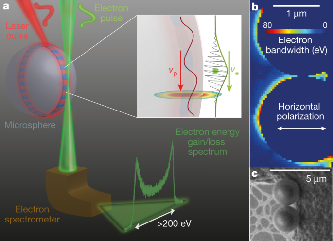
 Controlling free electrons with optical whispering-gallery modes | Nature
Grant Detail
 Publications 2017 - Department of Information Technology - Uppsala University
 STEM Microscope | Talos F200i TEM | Thermo Fisher Scientific - US
 JYX - Electronic, structural and chemical properties of gold clusters on ultra-thin oxide films
Microscopy27
- Electron energy loss spectroscopy (EELS) is a form of electron microscopy in which a material is exposed to a beam of electrons with a known, narrow range of kinetic energies. (wikipedia.org)
- Scanning Tunneling Microscopy 3 ( )exp( 1.025 ) 2 ( ) ( )exp 2 V E Z M E Z I Vbias S EF ∝ Bias S F − ⋅ ⎡ − ∝ − ρ φ φ ρ, (1) Where M Is The Mass Of Electron And ħ Is The Planck's Constant. (unpad.ac.id)
- World-class scientists have been invited to present their work, discuss the use of sub 5 meV EELS in the context of advanced electron microscopy characterization, and potentially set the future paths of this new arena of EELS. (mcmaster.ca)
- The presented scanning transmission electron microscopy (STEM) and electron energy-loss spectroscopy (EELS) results show the strong reaction of Cr and V with the graphitic walls of MWCNTs. (eels.info)
- Leading the Center for Electron Microscopy, I see my task as beeing threefold: -- guarantee a high level Electron Microscopy at EPFL by providing research in new techniques of Electron Microscopy. (epfl.ch)
- The fields currently covered are: - Computational Electron Microscopy with the JEMS programm (P. Stadelmann) - Angular resolved Electron Energy Loss Spectrometry (C. Hébert) - Three dimensional imaging and Chemical Analysis in a FIB. (epfl.ch)
- She obtained her Ingeneer degree (physics) and her PhD degree (design of a new energy filter for transmission electron microscopy, under the direction of Prof. B. Jouffrey) at the Ecole Centrale in Paris. (epfl.ch)
- Article: The Canadian Centre for Electron Microscopy: a national facility for ultrahigh resolution electron microscopy Journal: International Journal of Nanotechnology (IJNT) 2008 Vol.5 No.9/10/11/12 pp.1082 - 1093 Abstract: The Canadian Centre for Electron Microscopy is described and the facilities available to Canadian researchers and international collaborators are presented. (inderscience.com)
- The facility offers aberration-corrected transmission electron microscopy instrumentation, conventional transmission electron microscopes, scanning electron microscopes and a suite of sample preparation facilities. (inderscience.com)
- The Canadian Centre for Electron Microscopy is described and the facilities available to Canadian researchers and international collaborators are presented. (inderscience.com)
- aberration-corrected electron microscopy. (inderscience.com)
- The natural matching of free electrons to these quintessential optical modes could enable the application of integrated photonics technology in electron microscopy, with broad implications for attosecond structuring, probing quantum emitters and possible electron-light entanglement. (nature.com)
- Carbone, F., Kwon, O.-H. & Zewail, A. H. Dynamics of chemical bonding mapped by energy-resolved 4D electron microscopy. (nature.com)
- By means of scanning electron microscopy (SEM), we study the porous and/or granular structures of different anode materials. (uni-ulm.de)
- With high-resolution transmission electron microscopy (HRTEM), the atomic structure of the battery materials can be studied. (uni-ulm.de)
- Electron Microscopy (EM) provides insight at all scales and modalities of Materials Science research: from discovering new materials and understanding their physical and chemical properties to innovating advanced processes and conducting product quality control. (dectris.com)
- Easy: our detectors integrate straightforwardly into any modern data pipeline or electron microscopy suite. (dectris.com)
- The team tackled a multi-fold challenge in developing the electrochemically functional cell - making the cell cycle like a regular battery while ensuring it was small enough to fit into the millimeter-sized sample space of the TEM column,'' said co-author and senior scientist Yimei Zhu, who leads the Electron Microscopy and Nanostructure Group in Brookhaven's Condensed Matter Physics and Materials Science (CMPMS) Division. (materialstoday.com)
- It combines ultra-high resolution field emission Scanning Electron Microscopy (SEM) and precise Focused Ion Beam (FIB) etch and deposition. (epfl.ch)
- The plasmon wave size can be measured using various techniques, such as electron energy loss spectroscopy (EELS) or scanning electron microscopy (SEM). (physicsforums.com)
- Transmission electron microscopy (TEM) is the only technique able to characterize the nature of buried interfaces in these engineered van der Waals crystals and hence to provide insights into their optical, electronic and mechanical properties. (mrs.org)
- Intriguing phenomena arising at the atomic scale have been found in functional complex oxide materials in recent years due to significant progress in technical and methodological development in scanning transmission electron microscopy (STEM). (mrs.org)
- Surface analytical techniques including X-ray photoelectron spectroscopy (XPS), energy dispersive spectroscopy (EDS), and scanning electron microscopy (SEM) have been used in the characterization process. (epa.gov)
- ENERGY-FILTERED TRANSMISSION ELECTRON MICROSCOPY is a type of electron energy loss spectroscopy carried out in electron microscopes specially outfitted to analyze the spectrum of electron energy loss. (bvsalud.org)
- Soft X-ray scanning transmission microscopy (STXM) is a powerful tool for nanoscale materials analysis, with significant advantages over analytical electron microscopies for studies of radiation sensitive materials, and for in situ and operando studies. (lu.se)
- Regarding the methods, microhardness 2,5 , scanning electron and polarized light microscopy 7,14 , micro energy-dispersive x-ray fluorescence spectrometry (μEDXRF) 11 , Fourier transform-Raman (FTRaman) spectroscopy10 and atomic absorption spectrometer (AAS) 10 have been used to determine the adverse effects resulting from bleaching techniques. (bvsalud.org)
- Methods: Asbestos was identified using scanning electron microscopy and energy dispersive spectroscopy. (bvsalud.org)
EELS24
- EELS is spoken of as being complementary to energy-dispersive x-ray spectroscopy (variously called EDX, EDS, XEDS, etc.), which is another common spectroscopy technique available on many electron microscopes. (wikipedia.org)
- EELS tends to work best at relatively low atomic numbers, where the excitation edges tend to be sharp, well-defined, and at experimentally accessible energy losses (the signal being very weak beyond about 3 keV energy loss). (wikipedia.org)
- The difference is mainly due to the difference in energy resolution between the two techniques (~1 eV or better for EELS, perhaps a few tens of eV for EDX). (wikipedia.org)
- There are several basic flavors of EELS, primarily classified by the geometry and by the kinetic energy of the incident electrons (typically measured in kiloelectron-volts, or keV). (wikipedia.org)
- Probably the most common today is transmission EELS, in which the kinetic energies are typically 100 to 300 keV and the incident electrons pass entirely through the material sample. (wikipedia.org)
- Other flavors include reflection EELS (including reflection high-energy electron energy-loss spectroscopy (RHEELS)), typically at 10 to 30 keV, and aloof EELS (sometimes called near-field EELS), in which the electron beam does not in fact strike the sample but instead interacts with it via the long-ranged Coulomb interaction. (wikipedia.org)
- Aloof EELS is particularly sensitive to surface properties but is limited to very small energy losses such as those associated with surface plasmons or direct interband transitions. (wikipedia.org)
- EELS, the study of energy-loss due to inelastic scattering of electrons can in principle be used to measure atomic composition, chemical bonding, valence and conduction band electronic properties and surface properties. (asu.edu)
- can estimate the thickness from a low-loss EELS spectrum using the Log-Ratio method. (hyperspy.org)
- can be used to calculate the integral of the zero loss peak (elastic intensity) from EELS low-loss spectra containing the zero loss peak using the (rudimentary) threshold method. (hyperspy.org)
- can be used to calculate separation point between elastic and inelastic scattering on EELS low-loss spectra. (hyperspy.org)
- method inplements the Kramers-Kronig FFT method as in [Egerton2011] to estimate the complex dielectric funtion from a low-loss EELS spectrum. (hyperspy.org)
- HyperSpy makes it really easy to quantify EELS core-loss spectra by curve fitting as it is shown in the next example of quantification of a boron nitride EELS spectrum from the The EELS Data Base . (hyperspy.org)
- A FREE TWO-DAY WORKSHOP on the current state and innovations in ultrahigh energy resolution electron energy loss spectroscopy (EELS). (mcmaster.ca)
- The talks are in topics ranging from phonon mapping in real and reciprocal spaces, localized and delocalized phonon scattering in bulk and nanoscale structural features, vibrational spectroscopy of organic and non-organic complex materials, momentum resolved EELS, spectroscopy of mid-infrared and optical excitations and their coupling (phonons, excitons, plasmons), electronic transitions in novel materials at room and low temperatures with high spatial resolution, etc. (mcmaster.ca)
- Electron energy-loss spectroscopy (EELS) offers the possibility to study the chemical composition and properties. (uni-ulm.de)
- Fast, sensitive, and radiation-hard, DECTRIS ELA® electron-counting detector offers unmatched performance in EELS and 4D STEM. (dectris.com)
- Electron energy loss spectroscopy (EELS) measurements were made in a Carl Zeiss LEO-922 TEM equipped with an Omega filter. (springer.com)
- Cruz, A.F. (2004) Element Storage in Spores of Gigaspora margarita Becker & Hall Measured by Electron Energy Loss Spectroscopy (EELS). (scirp.org)
- This electrochemical cell allowed the team to conduct electron energy-loss spectroscopy (EELS) during battery charge and discharge. (materialstoday.com)
- In EELS, the change in energy of electrons after they have interacted with a sample is measured to reveal information about the sample's local chemical states. (materialstoday.com)
- To measure the EELS signals from the lithium, a very thin sample is needed, beyond what is normally required for the transparency of probing electrons in TEMs. (materialstoday.com)
- Electron energy loss spectroscopy (EELS) in the transmission electron microscope (TEM) was used to compare the iron oxidation state at the surface and interior of gamma-Fe2O3 nanoparticle s produced by the combustion process under fuel conditions leading to low and high soot concentrations. (cdc.gov)
- A professor at McMaster since 1979, his group has studied inner shell electron energy loss (EELS) spectroscopy of gases and surfaces, using home built instruments. (lu.se)
High Resolution E2
- The HREELS spectroscopy (High Resolution Electron Energy Loss Spectroscopy) consists in analysing both in energy and direction electron back reflected by a surface irradiated by a beam of mono-kinetic electrons the energy of which is around a few eV with a resolution of a few meV. (upmc.fr)
- Vibrational signature of the graphene nanoribbon edge structure from high-resolution electron energy-loss spectroscopy. (mpg.de)
Spectra6
- This can be useful e.g. to align core-loss spectra acquired quasi-simultaneously. (hyperspy.org)
- We interpret these results using ab initio calculations, where we model low-loss and core-excitation EEL spectra acquired on various dislocation cores in diamond and compare them with bulk spectra. (ncl.ac.uk)
- Applying this model in our reverse Monte Carlo method, we determined, with high-precision, electron-energy loss functions of silicon and germanium based on the theoretical analysis of the high-energy resolution reflected electron energy loss spectroscopy (REELS) spectra, measured at 3, 4, and 5 keV incident electron energies. (mtak.hu)
- Experimental microdiffraction patterns and energy loss spectra collected at symmetry points within a single unit cell of a barium alumina specimen are used, together with the results of dynamical calculations, to discuss the interpretation and usefulness of filtered microdiffraction patterns and lattice images. (elsevierpure.com)
- 250-110 nm), along with electron-energy-loss (EEL) spectra at (i) high incident electron energies and low scattering angles and (ii) near-threshold incident energies and large scattering angles. (hw.ac.uk)
- Figure3: Valence electron energy loss spectra (VEELS) of electrochemically (a) and chemically lithiated (b) Li1+xNi0.5Mn1.5O4 (red) compared to pristine LiNi1.5Mn0.5O4 (black). (uni-ulm.de)
Beam11
- With some care, and looking at a wide range of energy losses, one can determine the types of atoms, and the numbers of atoms of each type, being struck by the beam. (wikipedia.org)
- However, it still represents a challenge for narrow-bandgap semiconductors, since an electron beam with low energy spread is required. (arxiv.org)
- The electron beam extracted from a field emission source in a microscope column, with a known narrow range of kinetic energies, scatters due to interaction with the specimen material and can be used to form images and perform spectroscopy alike. (asu.edu)
- Here we couple a free-electron beam to a travelling-wave resonant cavity mode. (nature.com)
- The nanostructures were grown in porous silicon substrates in situ within the TEM by the electron beam-induced deposition method. (springer.com)
- However, an advantage that amorphous carbon (a-C) nanotips have over carbon nanotubes is that when a-C nanotips are synthesized using the electron beam-induced deposition method with a transmission electron microscope (TEM-EBID) their growth process can be followed in real-time and the nanostructures can be grown at preferred positions by controlling the electron beam [ 11 ]. (springer.com)
- Minimum deposited line width : 20 nm achievable with electrons and 50 nm achievable with ions beam (Pt). (epfl.ch)
- Gas chemistry solution for Ion or Electron beam deposition of Platinum-containing material. (epfl.ch)
- SESAME (Synchrotron-light for Experimental Science and Application in the Middle East) is Mid-dle East's first major international research centre, it is third generation light source located in Allan-Jordan using 2.5GeV, 400mA electron beam. (lu.se)
- To reduce electron losses and decrease radiation levels around the syn-chrotron, optimizations of the electron beam during injection into the storage ring, ramping and standard operation were carried out. (lu.se)
- Interaction of a high-intensity optical laser beam with a solid target can generate `hot' electrons, which generate radiation hazards (mainly bremsstrahlung photons and neutrons) from interaction of hot electrons with target and the surrounding materials. (lu.se)
Soft X-ray Spectrosco2
- All kinds of problems can arise that negatively impact how the battery functions," said co-author Adrian Hunt, a scientist at the In situ and Operando Soft X-ray Spectroscopy (IOS) beamline at NSLS-II, where part of the research was conducted. (bnl.gov)
- In 1980 he started synchrotron experiments, initially hard X-ray spectroscopy of materials at Cornell (USA), then soft X-ray spectroscopy of gases at LURE (France) and SRC (USA). (lu.se)
Characterization3
- The materials properties addressed include characterization of ground and excited state properties as well as time resolved electron dynamics. (materialstoday.com)
- Its standard X-Twin pole piece gap-giving the highest flexibility in applications-combined with a reproducibly performing electron column opens opportunities for high-resolution 2D and 3D characterization, in situ dynamic observations, and diffraction applications. (thermofisher.com)
- Advances in electron microscopic characterization technology have greatly improved the ability to quantify real microstructures found in Nd-Fe-B magnets. (hindawi.com)
Graphene3
- Magnetization By Partial Dehydrogenation Of The Sheet (Neek-Amal AndPeeters,2011)werereported.Experimentally,adsorption Of Hydrogen On Graphene Was Indeed Observed To Result In A Gap Opening In The Electron States (Elias Et Al. (unpad.ac.id)
- A likely mechanism relates to the the spatial confinement of the graphene electrons, within a single layer. (osti.gov)
- Doped graphene characterized via Raman spectroscopy and magneto-transport measurements. (mpg.de)
Excitations3
- The rest of the electrons lose a finite amount of energy during their interaction with core-shell and valence electrons and while facilitating mechanisms such as vibrational excitations, inter and intra-band transitions, guided light modes, and Ćerenkov radiation i.e. they interact inelastically with the material. (asu.edu)
- low energy electronic excitations, in particular due to the bandgap states. (upmc.fr)
- The theory of energy-filtered microdifraction patterns from localized core-loss excitations in crystal is outlined and the characteristic features of these discussed. (elsevierpure.com)
REELS1
- Capabilities of a New Compact SEM / STEM Electron Detector for Energy Resolved Scanning Imaging, Reflection Electron Energy Loss Spectroscopy (REELS) and Elastic Peak Electron Spectroscopy (EPES). (bvsalud.org)
Atoms5
- As expected, a majority of the fast traveling electrons interact elastically with the material i.e. they lose negligible energy in their dynamic interaction with atoms that make up the material. (asu.edu)
- The method relies on quantification of the intensity of elastic peak stemming from the backscattering of electrons with the hydrogen atoms present in the samples as measured by reflection electron energy loss spectroscopy. (us-csic.es)
- Low current primary electron beams of ∼ 1500 eV are used to minimize hydrogen desorption by electron bombardment and to provide enough energy separation between the elastic signals coming from hydrogen and other atoms (mainly C and O atoms) from the thin film materials. (us-csic.es)
- E ectively, a cluster can be seen as a harmonic potential to which the 6s valence electrons of gold atoms and the charge transferred from the substrate are con ned. (jyu.fi)
- Some of the electrons passing through the specimen will lose energy when they ionize inner shell electrons of the atoms in the specimen. (bvsalud.org)
Diffraction5
- It is only seen by electron diffraction but not with x-ray or He scattering. (osti.gov)
- The short-range order of individual fractal-like amorphous carbon nanotips was investigated by means of energy-filtered electron diffraction in a transmission electron microscope (TEM). (springer.com)
- In addition, we also present information of the short-range order of our nanostructures, using the radial distribution function (RDF) obtained by electron diffraction patterns. (springer.com)
- Energy-filtered electron diffraction studies were done in a Phillips CM-200 TEM at 200 kV using a Gatan imaging filter. (springer.com)
- The energy selecting window used to filter the electron diffraction pattern was 10 eV and was centered at the zero loss peak of the electron energy loss spectrum. (springer.com)
Aberration-corrected electron1
- With the high spatial resolution of an aberration-corrected electron microscope, we can detect changes in the local chemistry of the material, which can give us a fundamental understanding of the scientific processes that manifest at the atomic scale in the material. (asu.edu)
Measurements3
- A five-axis manipulator allows setting sample orientation/position and to cool it down (100 K). Measurements of bulk insulating surfaces are possible thanks to adefocussed ancillary electron gun that allows compensating from charge effects. (upmc.fr)
- Planar microresonators are fabricated in the NIST Nanofab, tested in our laboratory, and incorporated into a commercial 9 GHz or NIST-built 34 GHz electron paramagnetic resonance (EPR) spectrometer for measurements of thin films and other volume-limited samples. (nist.gov)
- In addition, electrochemical impedance spectroscopy (EIS), fourier-transform infrared spectroscopy (FT-IR), weight loss/gain measurements, and coating/steel adhesion-strength studies have been performed to evaluate performance of sample coupons and paint films in aqueous acid environments. (epa.gov)
Inner-shell electron1
- This is approximately the amount of energy needed to remove an inner-shell electron from a carbon atom, which can be taken as evidence that there is a significant amount of carbon present in the sample. (wikipedia.org)
Dielectric6
- The refractive index n, the extinction coefficient k, and the complex dielectric function (epsilon = epsilon(1) + i epsilon(2)) were calculated from the obtained energy loss function in a wide energy loss range of 0-200 eV. (mtak.hu)
- Compared to cavity resonators, power losses from resistive, dielectric, and radiative losses (Figure 1a) are greater in open resonant structures such as loop-gap resonators and microstrip-based structures. (nist.gov)
- The enhanced interaction with the optical whispering-gallery modes of dielectric microresonators induces a strong phase modulation on co-propagating electrons, which leads to a spectral broadening of 700 electronvolts, corresponding to the absorption and emission of hundreds of photons. (nature.com)
- Demonstration of electron acceleration in a laser-driven dielectric microstructure. (nature.com)
- Breuer, J. & Hommelhoff, P. Laser-based acceleration of nonrelativistic electrons at a dielectric structure. (nature.com)
- The plasmon wave size is affected by several factors, including the material properties (such as the dielectric constant and electron density), the shape and size of the particle, and the wavelength of the incident light. (physicsforums.com)
Free electrons2
- Fig. 3: Spectral and temporal properties of the interaction between free electrons and WGMs. (nature.com)
- The collective charge of the free electrons in a conductor is very large indeed, so it seems to me that only the slightest movement on a sub atomic scale would be required to create the y-component of the E-field. (physicsforums.com)
Spectrometry1
- The MC of the enamel was determined before and after bleaching using Fourier transform (FT-Raman) spectroscopy and micro energy-dispersive x-ray fluorescence spectrometry (μEDXRF). (bvsalud.org)
Raman3
- Raman spectroscopy of holey nanographene C216. (mpg.de)
- We investigated the suitability of the graphitic carbon (GC) content of diesel particulate matter (DPM), measured using Raman spectroscopy, as a surrogate measure of elemental carbon (EC) determined by thermal optical analysis. (cdc.gov)
- The FT-Raman and AAS analyses detected MC reduction and Ca loss after HP bleaching. (bvsalud.org)
Transmission5
- Usually this occurs in a transmission electron microscope (TEM), although some dedicated systems exist which enable extreme resolution in terms of energy and momentum transfer at the expense of spatial resolution. (wikipedia.org)
- Valence electron energy-loss spectroscopy (VEELS) in the transmission electron microscope (TEM) provides the possibility of measuring this property of semiconductors with high spatial resolution. (arxiv.org)
- The Thermo Scientific Talos F200i (S)TEM is a 20-200 kV field emission (scanning) transmission electron microscope uniquely designed for performance and productivity across a wide range of Materials Science samples and applications. (thermofisher.com)
- In this study, the scientists were able to track the migration of lithium ions in LTO nanoparticles in real time by designing an electrochemical cell to operate inside a transmission electron microscope (TEM). (materialstoday.com)
- Surface oxidation state of combustion-synthesized gamma-Fe2O3 nanoparticle s determined by electron energy loss spectroscopy in the transmission electron microscope. (cdc.gov)
Photons2
- Kfir, O. Entanglements of electrons and cavity photons in the strong-coupling regime. (nature.com)
- By looking at the electrons coming out of the sample we can study the surface, and by looking at the photons coming out the sample we can study the bulk. (bnl.gov)
Microscope1
- As a research assistant at the Vienna University of Technology, she was one of the main participant to the CHIRALTEM project dealing with the measurement of magnetic circular dichroism in the electron microscope. (epfl.ch)
Excitation3
- In the optical- and high-energy EEL data, the excitation energies of the historical amide absorption bands (W, R1, V1, R2 and Q) are in agreement with expectation. (hw.ac.uk)
- By mapping the near-field interaction with ultrashort electron pulses in space and time, we trace the lifetime of the the microresonator following a femtosecond excitation and observe the spectral response of the cavity. (nature.com)
- His research focus is inner shell excitation spectroscopies and spectromicroscopies. (lu.se)
Incident1
- The energy lost by incident electrons during various inelastic interactions with the material are characteristic of the chemical composition and bonding arrangements in the material and can be used for spectroscopic studies. (asu.edu)
Ionizing radiation1
- Papers using photoemission and other techniques, in which synchrotron radiation, Free Electron Lasers, laboratory lasers or other sources of ionizing radiation, combined with electron velocity analysis are especially welcome. (materialstoday.com)
Zero loss peak2
- can be used to estimate the position of the zero-loss peak. (hyperspy.org)
- Simultaneously record the Zero-Loss Peak (ZLP) and the core-loss features that are present in the spectrum with our radiation-hard, high-dynamic-range electron detectors. (dectris.com)
Deposition1
- For Example, Cooks And Colleagues Used Fe(CO) 5 · To Compare The Energy Deposition Of Collision-induced Dissociation And Surface-induced Dis-sociation [16]. (unpad.ac.id)
Elastically1
- Finally, this leads to large spread in their wave vector which is transferred by electron-electron interactions to the elastically scattered electrons to generate the BSC. (osti.gov)
Detector2
- A side-entry retractable Energy Dispersive X-ray Spectroscopy (EDS) detector can be added to the configuration to enable chemical analysis. (thermofisher.com)
- With its continuous readout and noise-free acquisition, DECTRIS QUADRO® detector brings microED and electron imaging techniques to a new level. (dectris.com)
Microscopic1
- Nowadays, the new nanoanalytical electron microscopic techniques with atomic resolution allow the creation of precise microstructural models suitable for the numerical micromagnetic calculation of the demagnetization curve including the coercive field value. (hindawi.com)
Situ2
- Zhao Y., Feltes T.E., Regalbuto J.R., Meyer R.J., and Klie R.F., In Situ Electron Energy Loss Spectroscopy Study of Metallic Co and Co Oxides , J. Appl. (regalbutocatalusc.net)
- New techniques for measuring isotope ratios using secondary ion mass spectroscopy (SIMS) with the ion microprobe open up new opportunities for in situ analyses of individual grains and fluid inclusions (Hervig, Chapter 16). (minsocam.org)
Interaction2
- This Electron-phonon (e-ph) Interaction Is One Of The Most Important Candidates To Theoretically Ex-plain NDC[15, 16, 17]. (unpad.ac.id)
- Electron energy loss spectroscopy analysis of the interaction of Cr and V with MWCNTs. (eels.info)
STEM2
- The form of energy-filtered characteristic loss STEM lattice images is discussed, and the effects of partial localization on these images described. (elsevierpure.com)
- Collect 4D STEM data of the highest possible quality with our precise and reliable electron-counting detectors. (dectris.com)
Spectrometer1
- The amount of energy loss can be measured via an electron spectrometer and interpreted in terms of what caused the energy loss. (wikipedia.org)
Atomic4
- The interpretation of microdiffraction patterns from crystals formed with a coherent electron probe which is "smaller" than the crystal unit cell is discussed, with particular reference to the problem of extracting atomic number information. (elsevierpure.com)
- The journal encourages contributions in the general area of atomic, molecular, ionic, liquid and solid state spectroscopy carried out using electron impact, synchrotron radiation (including free electron lasers) and short wavelength lasers. (materialstoday.com)
- Magnetic resonance spectroscopies based on inductive detection are powerful and versatile techniques that can provide atomic-level structural and functional information about a wide range of samples under broadly variable conditions. (nist.gov)
- The atomic percentages of Ca and P were evaluated by Energy Dispersive X-ray Spectroscopy (EDS). (bvsalud.org)
Plasmons2
- In summary, the conversation discusses plasmons, which are waves formed by electron density fluctuations on a metallic surface. (physicsforums.com)
- Hello, Plasmons are waves formed by electron density fluctuations on a metallic surface. (physicsforums.com)
Spectrum2
- The simulations show the absence of deep gap states for the more stable partial dislocations but there are characteristic changes to the low-loss EEL spectrum in the 6-12 eV region. (ncl.ac.uk)
- Analysis of the energy loss spectrum reveals the elemental composition of a specimen. (bvsalud.org)
Techniques3
- Electron energy-loss (EEL) spectroscopy performed near dislocation cores is one of the few experimental techniques that can yield valuable information about the electronic levels associated with dislocations. (ncl.ac.uk)
- Lithium ions are light, making them elusive to traditional electron- or x-ray-based probing techniques - especially when the ions are shuffling rapidly within active materials, such as LTO nanoparticles in an operating battery electrode. (materialstoday.com)
- These techniques allow for the visualization and measurement of the electron density and wavelength of the plasmon wave. (physicsforums.com)
Ions2
- The intercalation battery compounds allow that lithium ions compensate for the charge through the electrolyte when electrons power the electrical consumer. (uni-ulm.de)
- A team led by scientists at the US Department of Energy (DOE)'s Brookhaven National Laboratory and Lawrence Berkeley National Laboratory has captured in real time how lithium ions move in lithium titanate (LTO), a fast-charging battery electrode material made of lithium, titanium and oxygen. (materialstoday.com)
Spatial1
- The technique is able to take advantage of modern aberration-corrected probe forming systems to attain spatial resolutions down to ~0.1 nm, while with a monochromated electron source and/or careful deconvolution the energy resolution can be 0.1 eV or better. (wikipedia.org)
Peak1
- But theoretically (I suppose no-one has tried to actually do this kind of measurement yet), just how tall would these electron waves be, if one were to measure their peak amplitude (height from surface) vs. average electron operator in a non-excited state occupying a similar metallic surface. (physicsforums.com)
Magnetic2
- A study with 31 P (phosphorus) nuclear magnetic resonance (NMR) spectroscopy showed an impairment of high-energy phosphate metabolism, which explains why these patients are unable, rather than unwilling, to exercise. (medscape.com)
- In vivo mitochondrial function [maximal ATP synthesis rate (ATPmax), ATPflux/O 2 (P/O)] was determined by 31 P-magnetic resonance spectroscopy and optical spectroscopy, and body composition was determined by dual-energy X-ray absorptiometry. (medscape.com)
Resonance1
- for example, inductive-detection electron paramagnetic resonance (EPR) spectroscopy currently requires sample volumes on the order of tens of microliters. (nist.gov)
Analysis1
- A comparison is also made with data provided by infrared spectroscopy analysis of the same samples. (us-csic.es)
Radiative2
Synchrotron1
- They teamed up with scientists at the National Synchrotron Light Source II (NSLS-II) and used facilities at the Center for Functional Nanomaterials (CFN)-two U.S. Department of Energy (DOE) Office of Science User Facilities at DOE's Brookhaven National Laboratory-to study chemical changes in the battery over time. (bnl.gov)
Specimen2
Microstructure1
- The increasing demand of high-performance rare earth permanent magnets with a high coercive field and an energy density product value suitable for large scale applications in wind turbines and electrically powered automotive devices led to the development of heavy rare earth lean/rare earth-free Nd-Fe-B based magnets and to the optimization of the complex multiphase microstructure of the magnets [ 1 ]. (hindawi.com)
Theoretical1
- theoretical treatments of the photoemission, X-ray emission, Auger, energy loss and Penning ionization processes. (materialstoday.com)
Wavelength1
- A plasmon wave size refers to the physical size or wavelength of a plasmon wave, which is a type of collective oscillation of electrons in a material. (physicsforums.com)
Spectral1
- Fig. 2: Electron spectral broadening induced by WGMs. (nature.com)
Detection3
- Accurate: this technology offers single-electron counting and noise-free detection. (dectris.com)
- Acquire high-quality microED data fast using direct electron detection, combined with the high dynamic range and radiation hardness of DECTRIS' electron detectors. (dectris.com)
- Detection: in-lens secondary electrons (SE) and back scattered electrons (BSE). (epfl.ch)
Inverse1
- Figure 1: (a) Schematic of the planar inverse anapole resonator and the types of power losses occurring from planar microresonators. (nist.gov)
Experimental2
- In this study, we present experimental observations of low-loss EEL spectroscopy acquired on grain boundary dislocations in a CVD diamond film. (ncl.ac.uk)
- Se llevó a cabo un estudio experimental in vitro en una muestra de 18 premolares humanos, los cuales fueron tratados con una de las tres pastas dentales evaluadas y una fluorada. (bvsalud.org)
Probes1
- 10 eV) or electrons as probes or detected particles in the investigation. (materialstoday.com)