Sinus Thrombosis, Intracranial
Sagittal Sinus Thrombosis
Cavernous Sinus Thrombosis
Lateral Sinus Thrombosis
Cranial Sinuses
Intracranial Thrombosis
Cavernous Sinus
Dura Mater
Intracranial Hypertension
Cerebral Angiography
Phlebography
Mastoiditis
Superior Sagittal Sinus
Maxillary Sinus
Tomography, X-Ray Computed
Pseudotumor Cerebri
Papilledema
Paranasal Sinuses
Magnetic Resonance Angiography
Transverse Sinuses
Coronary Thrombosis
Intracranial Embolism and Thrombosis
Sphenoid Sinusitis
Carotid Sinus
Headache
Magnetic Resonance Imaging
Frontal Sinus
Heparin
Paranasal Sinus Diseases
Streptococcus constellatus
Putaminal Hemorrhage
Thrombectomy
Sphenoid Sinus
Cerebral Hemorrhage
Carotid Artery Thrombosis
Coronary Sinus
Battered Child Syndrome
Thrombolytic Therapy
Blood Coagulation Disorders
Subarachnoid Space
Sick Sinus Syndrome
Follow-Up Studies
Ethmoid Sinus
Cerebral Infarction
Treatment Outcome
Angiography, Digital Subtraction
Urokinase-Type Plasminogen Activator
Contraceptives, Oral
Seizures
Intracranial Pressure
Retrospective Studies
Fatal Outcome
Risk Factors
Tachycardia, Sinus
Randomized, placebo-controlled trial of anticoagulant treatment with low-molecular-weight heparin for cerebral sinus thrombosis. (1/193)
BACKGROUND AND PURPOSE: Treatment of cerebral sinus thrombosis with heparin is controversial. We conducted a double-blind, placebo-controlled multicenter trial to examine whether anticoagulant treatment improves outcome in patients with sinus thrombosis. METHODS: Patients were randomized between body weight-adjusted subcutaneous nadroparin (180 anti-factor Xa units/kg per 24 hours) and matching placebo for 3 weeks (double-blind part of trial), followed by 3 months of oral anticoagulants for patients allocated nadroparin (open part). Patients with cerebral hemorrhage caused by sinus thrombosis were also included. RESULTS: Sixty patients were enrolled, and none were lost to follow-up. In 1 patient the diagnosis proved wrong after randomization. After 3 weeks, 6 of 30 patients (20%) in the nadroparin group and 7 of 29 patients (24%) in the placebo group had a poor outcome, defined as death or Barthel Index score of <15 (risk difference, -4%; 95% CI, -25 to 17%; NS). After 12 weeks, 4 of 30 patients (13%) in the nadroparin group and 6 of 29 (21%) in the placebo group had a poor outcome, defined as death or Oxford Handicap Score of >/=3 (risk difference, -7%; 95% CI, -26% to 12%; NS). There were no new symptomatic cerebral hemorrhages. One patient in the nadroparin group had a major gastrointestinal hemorrhage, and 1 patient in the placebo group died from clinically suspected pulmonary embolism. CONCLUSIONS: Patients with cerebral sinus thrombosis treated with anticoagulants (low-molecular-weight heparin followed by oral anticoagulation) had a favorable outcome more often than controls, but the difference was not statistically significant. Anticoagulation proved to be safe, even in patients with cerebral hemorrhage. (+info)Helical CT angiography: dynamic cerebrovascular imaging in children. (2/193)
BACKGROUND AND PURPOSE: The purpose of this study was to assess the feasibility of helical CT cerebrovascular imaging (CTCVI) in children and to make initial comparisons with MR angiography and digital subtraction angiography (DSA). METHODS: Twenty-six patients, ages 3 days to 17 years, were examined with CTCVI. Patients were scanned with 1-mm collimation and 2:1 pitch 30 seconds after the initiation of a hand injection of 2 mL/kg nonionic contrast material (320 mg/dL iodine) with a maximum dose that did not exceed 80 mL (minimum volume, 5 mL in a 2.5-kg infant). Reconstructions were done using maximum intensity projection and integral rendering algorithms. Four patients had CTCVI, MR angiography, and DSA (42 vessels studied) and nine patients had CTCVI and DSA (136 vessels studied). Scores of 1 (not present) to 3 (present in continuity to the first bifurcation) were assigned independently by two radiologists to 32 vessels in each correlated case for each available technique. RESULTS: There were no technical failures. CTCVI depicted 18 thrombosed dural sinuses, three vascular malformations, one intracranial aneurysm, and four tumors. Ninety-five percent of the vessels seen with DSA were also seen with CTCVI. CTCVI identified all vessels seen on MR angiography. CONCLUSION: Helical CTCVI is an effective technique for assessing the intracranial circulation in children. In this initial comparison, CTCVI showed more vascular detail than MR angiography, and had fewer technical limitations. (+info)Rapid thrombectomy of superior sagittal sinus and transverse sinus thrombosis with a rheolytic catheter device. (3/193)
Thrombosis of the dural venous sinuses is a potentially lethal condition that remains a diagnostic dilemma. Clinical outcome is typically dependent on the timeliness of diagnosis and definitive treatment. We report a case of successful rapid thrombectomy of extensive thrombus within the superior sagittal and transverse sinuses using a rheolytic catheter device. This appears to be a promising treatment option, particularly in those patients who do not respond to other, more established, forms of therapy. (+info)Application of a rheolytic thrombectomy device in the treatment of dural sinus thrombosis: a new technique. (4/193)
We present a novel application of a transvascular rheolytic thrombectomy system in the treatment of symptomatic dural sinus thrombosis in a 54-year-old woman with somnolence and left-sided weakness. The diagnosis of bilateral transverse and superior sagittal sinus thrombosis was made and the patient was treated with anticoagulant therapy. After an initial period of improvement, she became comatose and hemiplegic 8 days after presentation. After excluding intracerebral hemorrhage by MR imaging, we performed angiography and transfemoral venous thrombolysis with a hydrodynamic thrombectomy catheter, followed by intrasinus urokinase thrombolytic therapy over the course of 2 days. This technique resulted in dramatic sinus thrombolysis and near total neurologic recovery. Six months after treatment, the patient showed mild cognitive impairment and no focal neurologic deficit. Our preliminary experience suggests that this technique may play a significant role in the endovascular treatment of this potentially devastating disease. (+info)Variant arteriovenous fistula of the superior sagittal sinus--case report. (5/193)
A 57-year-old male presented with a rare variant of dural arteriovenous fistula, located in the wall of an unobstructed superior sagittal sinus. Drainage occurred through a cortical vein no longer connected to its parent sinus, which filled up a cluster of transmedullary running veins, one of which was the presumed site of hemorrhage. Arterial blood was supplied via the external carotid artery branches. This type of fistula seriously increases the risk of hemorrhage in the patient and therefore requires complete obliteration. Attempts to embolize the fistula failed. The draining vein was isolated and coagulated resulting in permanent occlusion of the fistula. The fistula probably developed through a process of thrombophlebitis and revascularization via arterioles of the vein rather than previous occlusion of the sinus. (+info)Removal of a thrombus from the sigmoid and transverse sinuses with a rheolytic thrombectomy catheter. (6/193)
A rheolytic thrombectomy catheter was used to remove thrombus without thrombolytics from the sigmoid and transverse sinuses of a 34-year-old woman. Using small, high-flow fluid jets and Venturi-effect suction, this catheter allowed mechanical removal of thrombus. This technique may obviate the need for thrombolytic agents and the risks associated with their use. (+info)Extrinsic cerebral venous sinus obstruction resulting in intracranial hypertension. (7/193)
We report the case of a 70-year-old man reporting with headache and visual disturbances who was being treated for prostate cancer. Investigations showed him to have intracranial hypertension caused by venous sinus obstruction. Patients with metastatic disease and raised intracranial pressure in the absence of focal signs should be considered as possible cases of venous outflow obstruction. (+info)Cerebral venous sinus thrombosis. (8/193)
Cerebral venous sinus thrombosis is a challenging condition because of its variability of clinical symptoms and signs. It is very often unrecognised at initial presentation. All age groups can be affected. Large sinuses such as the superior sagittal sinus are most frequently involved. Extensive collateral circulation within the cerebral venous system allows for a significant degree of compensation in the early stages of thrombus formation. Systemic inflammatory diseases and inherited as well as acquired coagulation disorders are frequent causes, although in up to 30% of cases no underlying cause can be identified. The oral contraceptive pill appears to be an important additional risk factor. The spectrum of clinical presentations ranges from headache with papilloedema to focal deficit, seizures and coma. Magnetic resonance imaging with venography is the investigation of choice; computed tomography alone will miss a significant number of cases. It has now been conclusively shown that intravenous heparin is the first-line treatment for cerebral venous sinus thrombosis because of its efficacy, safety and feasability. Local thrombolysis may be indicated in cases of deterioration, despite adequate heparinisation. This should be followed by oral anticoagulation for 3-6 months. The prognosis of cerebral venous sinus thrombosis is generally favourable. A high index of clinical suspicion is needed to diagnose this uncommon condition so that appropriate treatment can be initiated. (+info)Intracranial sinus thrombosis is a medical condition characterized by the formation of a blood clot (thrombus) within the intracranial venous sinuses, which are responsible for draining blood from the brain. The condition can lead to various neurological symptoms and complications, such as increased intracranial pressure, headaches, seizures, visual disturbances, and altered consciousness. Intracranial sinus thrombosis may result from various factors, including hypercoagulable states, infections, trauma, and malignancies. Immediate medical attention is necessary for proper diagnosis and treatment to prevent potential long-term neurological damage or even death.
Sagittal sinus thrombosis is a medical condition that refers to the formation of a blood clot (thrombus) in the sagittal sinus, which is a venous structure located in the brain. The sagittal sinus runs along the midline of the brain and receives blood from the superficial veins of the brain.
Sagittal sinus thrombosis can occur as a result of various conditions, such as head trauma, infection, cancer, or certain medical disorders that cause hypercoagulability (an increased tendency to form blood clots). The formation of a blood clot in the sagittal sinus can obstruct the flow of blood from the brain, leading to symptoms such as headache, seizures, altered consciousness, and focal neurological deficits.
Diagnosis of sagittal sinus thrombosis typically involves imaging studies such as computed tomography (CT) or magnetic resonance imaging (MRI) scans, which can show the presence of a blood clot in the sagittal sinus. Treatment may involve administering anticoagulant medications to prevent further growth of the blood clot and reduce the risk of complications such as pulmonary embolism or cerebral infarction. In some cases, surgical intervention may be necessary to remove the blood clot or alleviate pressure on the brain.
Cavernous sinus thrombosis is a medical condition that refers to the formation of a blood clot (thrombus) in the cavernous sinuses, which are located near the base of the brain and are important for draining blood from the face and brain. This condition can occur as a complication of an infection in the facial area or sinuses, or it can be associated with other medical conditions such as cancer or trauma.
Symptoms of cavernous sinus thrombosis may include headache, fever, eye pain, swelling or bulging of the eyes, double vision, and decreased vision. If left untreated, this condition can lead to serious complications such as meningitis, brain abscess, or even death. Treatment typically involves administering antibiotics to treat any underlying infection and anticoagulants to prevent further clot formation. In some cases, surgery may be necessary to remove the clot.
Lateral sinus thrombosis, also known as sigmoid sinus thrombosis, is a medical condition characterized by the formation of a blood clot (thrombus) in the lateral or sigmoid sinus, which are venous structures located in the skull that help drain blood from the brain.
The lateral sinuses are situated near the mastoid process of the temporal bone and can become thrombosed due to various reasons such as infection (often associated with ear or mastoid infections), trauma, tumors, or other underlying medical conditions that increase the risk of blood clot formation.
Symptoms of lateral sinus thrombosis may include headache, fever, neck stiffness, altered mental status, and signs of increased intracranial pressure such as papilledema (swelling of the optic nerve disc). Diagnosis is typically made with the help of imaging studies like CT or MRI scans, and treatment often involves anticoagulation therapy to prevent clot expansion and potential complications. In some cases, surgical intervention may be necessary to remove the clot or manage any underlying conditions.
Cranial sinuses are a part of the venous system in the human head. They are air-filled spaces located within the skull and are named according to their location. The cranial sinuses include:
1. Superior sagittal sinus: It runs along the top of the brain, inside the skull, and drains blood from the scalp and the veins of the brain.
2. Inferior sagittal sinus: It runs along the bottom of the brain and drains into the straight sinus.
3. Straight sinus: It is located at the back of the brain and receives blood from the inferior sagittal sinus and great cerebral vein.
4. Occipital sinuses: They are located at the back of the head and drain blood from the scalp and skull.
5. Cavernous sinuses: They are located on each side of the brain, near the temple, and receive blood from the eye and surrounding areas.
6. Sphenoparietal sinus: It is a small sinus that drains blood from the front part of the brain into the cavernous sinus.
7. Petrosquamosal sinuses: They are located near the ear and drain blood from the scalp and skull.
The cranial sinuses play an essential role in draining blood from the brain and protecting it from injury.
Thrombosis is the formation of a blood clot (thrombus) inside a blood vessel, obstructing the flow of blood through the circulatory system. When a clot forms in an artery, it can cut off the supply of oxygen and nutrients to the tissues served by that artery, leading to damage or tissue death. If a thrombus forms in the heart, it can cause a heart attack. If a thrombus breaks off and travels through the bloodstream, it can lodge in a smaller vessel, causing blockage and potentially leading to damage in the organ that the vessel supplies. This is known as an embolism.
Thrombosis can occur due to various factors such as injury to the blood vessel wall, abnormalities in blood flow, or changes in the composition of the blood. Certain medical conditions, medications, and lifestyle factors can increase the risk of thrombosis. Treatment typically involves anticoagulant or thrombolytic therapy to dissolve or prevent further growth of the clot, as well as addressing any underlying causes.
Cerebral veins are the blood vessels that carry deoxygenated blood from the brain to the dural venous sinuses, which are located between the layers of tissue covering the brain. The largest cerebral vein is the superior sagittal sinus, which runs along the top of the brain. Other major cerebral veins include the straight sinus, transverse sinus, sigmoid sinus, and cavernous sinus. These veins receive blood from smaller veins called venules that drain the surface and deep structures of the brain. The cerebral veins play an important role in maintaining normal circulation and pressure within the brain.
Venous thrombosis is a medical condition characterized by the formation of a blood clot (thrombus) in the deep veins, often in the legs (deep vein thrombosis or DVT), but it can also occur in other parts of the body such as the arms, pelvis, or lungs (pulmonary embolism).
The formation of a venous thrombus can be caused by various factors, including injury to the blood vessel wall, changes in blood flow, and alterations in the composition of the blood. These factors can lead to the activation of clotting factors and platelets, which can result in the formation of a clot that blocks the vein.
Symptoms of venous thrombosis may include swelling, pain, warmth, and redness in the affected area. In some cases, the clot can dislodge and travel to other parts of the body, causing potentially life-threatening complications such as pulmonary embolism.
Risk factors for venous thrombosis include advanced age, obesity, smoking, pregnancy, use of hormonal contraceptives or hormone replacement therapy, cancer, recent surgery or trauma, prolonged immobility, and a history of previous venous thromboembolism. Treatment typically involves the use of anticoagulant medications to prevent further clotting and dissolve existing clots.
Intracranial thrombosis refers to the formation of a blood clot (thrombus) within the intracranial vessels, which supply blood to the brain. This condition can occur in any of the cerebral arteries or veins and can lead to serious complications such as ischemic stroke, transient ischemic attack (TIA), or venous sinus thrombosis.
The formation of an intracranial thrombus can be caused by various factors, including atherosclerosis, cardiac embolism, vasculitis, sickle cell disease, hypercoagulable states, and head trauma. Symptoms may vary depending on the location and extent of the thrombosis but often include sudden onset of headache, weakness or numbness in the face or limbs, difficulty speaking or understanding speech, vision changes, and loss of balance or coordination.
Diagnosis of intracranial thrombosis typically involves imaging studies such as computed tomography (CT) angiography, magnetic resonance angiography (MRA), or digital subtraction angiography (DSA). Treatment options may include anticoagulation therapy, thrombolysis, endovascular intervention, or surgical intervention, depending on the underlying cause and severity of the condition.
The cavernous sinus is a venous structure located in the middle cranial fossa, which is a depression in the skull that houses several important nerves and blood vessels. The cavernous sinus is situated on either side of the sphenoid bone, near the base of the skull, and it contains several important structures:
* The internal carotid artery, which supplies oxygenated blood to the brain
* The abducens nerve (cranial nerve VI), which controls lateral movement of the eye
* The oculomotor nerve (cranial nerve III), which controls most of the muscles that move the eye
* The trochlear nerve (cranial nerve IV), which controls one of the muscles that moves the eye
* The ophthalmic and maxillary divisions of the trigeminal nerve (cranial nerve V), which transmit sensory information from the face and head
The cavernous sinus is an important structure because it serves as a conduit for several critical nerves and blood vessels. However, it is also vulnerable to various pathological conditions such as thrombosis (blood clots), infection, tumors, or aneurysms, which can lead to serious neurological deficits or even death.
Dura Mater is the thickest and outermost of the three membranes (meninges) that cover the brain and spinal cord. It provides protection and support to these delicate structures. The other two layers are called the Arachnoid Mater and the Pia Mater, which are thinner and more delicate than the Dura Mater. Together, these three layers form a protective barrier around the central nervous system.
Intracranial hypertension is a medical condition characterized by an increased pressure within the skull (intracranial space) that contains the brain, cerebrospinal fluid (CSF), and blood. Normally, the pressure inside the skull is carefully regulated to maintain a balance between the formation and absorption of CSF. However, when the production of CSF exceeds its absorption or when there is an obstruction in the flow of CSF, the pressure inside the skull can rise, leading to intracranial hypertension.
The symptoms of intracranial hypertension may include severe headaches, nausea, vomiting, visual disturbances such as blurred vision or double vision, and papilledema (swelling of the optic nerve disc). In some cases, intracranial hypertension can lead to serious complications such as vision loss, brain herniation, and even death if left untreated.
Intracranial hypertension can be idiopathic, meaning that there is no identifiable cause, or secondary to other underlying medical conditions such as brain tumors, meningitis, hydrocephalus, or certain medications. The diagnosis of intracranial hypertension typically involves a combination of clinical evaluation, imaging studies (such as MRI or CT scans), and lumbar puncture to measure the pressure inside the skull and assess the CSF composition. Treatment options may include medications to reduce CSF production, surgery to relieve pressure on the brain, or shunting procedures to drain excess CSF from the intracranial space.
Cerebral angiography is a medical procedure that involves taking X-ray images of the blood vessels in the brain after injecting a contrast dye into them. This procedure helps doctors to diagnose and treat various conditions affecting the blood vessels in the brain, such as aneurysms, arteriovenous malformations, and stenosis (narrowing of the blood vessels).
During the procedure, a catheter is inserted into an artery in the leg and threaded through the body to the blood vessels in the neck or brain. The contrast dye is then injected through the catheter, and X-ray images are taken to visualize the blood flow through the brain's blood vessels.
Cerebral angiography provides detailed images of the blood vessels in the brain, allowing doctors to identify any abnormalities or blockages that may be causing symptoms or increasing the risk of stroke. Based on the results of the cerebral angiography, doctors can develop a treatment plan to address these issues and prevent further complications.
Phlebography is a medical imaging technique used to visualize and assess the veins, particularly in the legs. It involves the injection of a contrast agent into the veins, followed by X-ray imaging to capture the flow of the contrast material through the veins. This allows doctors to identify any abnormalities such as blood clots, blockages, or malformations in the venous system.
There are different types of phlebography, including ascending phlebography (where the contrast agent is injected into a foot vein and travels up the leg) and descending phlebography (where the contrast agent is injected into a vein in the groin or neck and travels down the leg).
Phlebography is an invasive procedure that requires careful preparation and monitoring, and it is typically performed by radiologists or vascular specialists. It has largely been replaced by non-invasive imaging techniques such as ultrasound and CT angiography in many clinical settings.
Mastoiditis is a medical condition characterized by an infection and inflammation of the mastoid process, which is the bony prominence located behind the ear. The mastoid process contains air cells that are connected to the middle ear, and an infection in the middle ear (otitis media) can spread to the mastoid process, resulting in mastoiditis.
The symptoms of mastoiditis may include:
* Pain and tenderness behind the ear
* Swelling or redness of the skin behind the ear
* Ear drainage or discharge
* Fever and headache
* Hearing loss or difficulty hearing
Mastoiditis is a serious condition that requires prompt medical attention. Treatment typically involves antibiotics to eliminate the infection, as well as possible surgical intervention if the infection does not respond to medication or if it has caused significant damage to the mastoid process. If left untreated, mastoiditis can lead to complications such as meningitis, brain abscess, or even death.
The Superior Sagittal Sinus is a medical term that refers to a venous sinus (a channel for blood flow) located in the superior part (highest portion) of the sagittal suture, which is the line along the top of the skull where the two parietal bones join in the middle. It runs from front to back, starting at the frontal bone and ending at the occipital bone, and it receives blood from veins that drain the cerebral hemispheres (the right and left halves of the brain).
The Superior Sagittal Sinus is an important structure in the circulatory system of the brain as it plays a critical role in draining venous blood from the cranial cavity. It also contains valveless venous channels that allow for the flow of cerebrospinal fluid (CSF) between the intracranial and extracranial compartments.
It is worth noting that any damage to this structure, such as through trauma or infection, can lead to serious neurological complications, including increased intracranial pressure, seizures, and even death.
The maxillary sinuses, also known as the antrums of Highmore, are the largest of the four pairs of paranasal sinuses located in the maxilla bones. They are air-filled cavities that surround the nasolacrimal duct and are situated superior to the upper teeth and lateral to the nasal cavity. Each maxillary sinus is lined with a mucous membrane, which helps to warm, humidify, and filter the air we breathe. Inflammation or infection of the maxillary sinuses can result in conditions such as sinusitis, leading to symptoms like facial pain, headaches, and nasal congestion.
X-ray computed tomography (CT or CAT scan) is a medical imaging method that uses computer-processed combinations of many X-ray images taken from different angles to produce cross-sectional (tomographic) images (virtual "slices") of the body. These cross-sectional images can then be used to display detailed internal views of organs, bones, and soft tissues in the body.
The term "computed tomography" is used instead of "CT scan" or "CAT scan" because the machines take a series of X-ray measurements from different angles around the body and then use a computer to process these data to create detailed images of internal structures within the body.
CT scanning is a noninvasive, painless medical test that helps physicians diagnose and treat medical conditions. CT imaging provides detailed information about many types of tissue including lung, bone, soft tissue and blood vessels. CT examinations can be performed on every part of the body for a variety of reasons including diagnosis, surgical planning, and monitoring of therapeutic responses.
In computed tomography (CT), an X-ray source and detector rotate around the patient, measuring the X-ray attenuation at many different angles. A computer uses this data to construct a cross-sectional image by the process of reconstruction. This technique is called "tomography". The term "computed" refers to the use of a computer to reconstruct the images.
CT has become an important tool in medical imaging and diagnosis, allowing radiologists and other physicians to view detailed internal images of the body. It can help identify many different medical conditions including cancer, heart disease, lung nodules, liver tumors, and internal injuries from trauma. CT is also commonly used for guiding biopsies and other minimally invasive procedures.
In summary, X-ray computed tomography (CT or CAT scan) is a medical imaging technique that uses computer-processed combinations of many X-ray images taken from different angles to produce cross-sectional images of the body. It provides detailed internal views of organs, bones, and soft tissues in the body, allowing physicians to diagnose and treat medical conditions.
Pseudotumor cerebri, also known as idiopathic intracranial hypertension, is a condition characterized by increased pressure around the brain without any identifiable cause such as a tumor or other space-occupying lesion. The symptoms mimic those of a brain mass, hence the term "pseudotumor."
The primary manifestation of this condition is headaches, often accompanied by vision changes like blurry vision, double vision, or temporary loss of vision, and pulsatile tinnitus (a rhythmic whooshing sound in the ears). Other symptoms can include neck pain, nausea, vomiting, and papilledema (swelling of the optic nerve disc). If left untreated, pseudotumor cerebri can lead to permanent vision loss.
The exact cause of pseudotumor cerebri remains unknown, but it has been associated with certain factors such as obesity, rapid weight gain, female gender (particularly during reproductive years), sleep apnea, and the use of certain medications like tetracyclines, vitamin A derivatives, and steroid withdrawal. Diagnosis typically involves a series of tests including neurological examination, imaging studies (such as MRI or CT scan), and lumbar puncture to measure cerebrospinal fluid pressure. Treatment usually focuses on lowering intracranial pressure through medications, weight loss, and sometimes surgical interventions like optic nerve sheath fenestration or shunting procedures.
Papilledema is a medical term that refers to swelling of the optic nerve head, also known as the disc, which is the point where the optic nerve enters the back of the eye (the retina). This swelling can be caused by increased pressure within the skull, such as from brain tumors, meningitis, or idiopathic intracranial hypertension. Papilledema is usually detected through a routine eye examination and may be accompanied by symptoms such as headaches, visual disturbances, and nausea. If left untreated, papilledema can lead to permanent vision loss.
Paranasal sinuses are air-filled cavities in the skull that surround the nasal cavity. There are four pairs of paranasal sinuses, including the maxillary, frontal, ethmoid, and sphenoid sinuses. These sinuses help to warm, humidify, and filter the air we breathe. They also contribute to our voice resonance and provide a slight cushioning effect for the skull. The openings of the paranasal sinuses lead directly into the nasal cavity, allowing mucus produced in the sinuses to drain into the nose. Infections or inflammation of the paranasal sinuses can result in conditions such as sinusitis.
Anticoagulants are a class of medications that work to prevent the formation of blood clots in the body. They do this by inhibiting the coagulation cascade, which is a series of chemical reactions that lead to the formation of a clot. Anticoagulants can be given orally, intravenously, or subcutaneously, depending on the specific drug and the individual patient's needs.
There are several different types of anticoagulants, including:
1. Heparin: This is a naturally occurring anticoagulant that is often used in hospitalized patients who require immediate anticoagulation. It works by activating an enzyme called antithrombin III, which inhibits the formation of clots.
2. Low molecular weight heparin (LMWH): LMWH is a form of heparin that has been broken down into smaller molecules. It has a longer half-life than standard heparin and can be given once or twice daily by subcutaneous injection.
3. Direct oral anticoagulants (DOACs): These are newer oral anticoagulants that work by directly inhibiting specific clotting factors in the coagulation cascade. Examples include apixaban, rivaroxaban, and dabigatran.
4. Vitamin K antagonists: These are older oral anticoagulants that work by inhibiting the action of vitamin K, which is necessary for the formation of clotting factors. Warfarin is an example of a vitamin K antagonist.
Anticoagulants are used to prevent and treat a variety of conditions, including deep vein thrombosis (DVT), pulmonary embolism (PE), atrial fibrillation, and prosthetic heart valve thrombosis. It is important to note that anticoagulants can increase the risk of bleeding, so they must be used with caution and regular monitoring of blood clotting times may be required.
Magnetic Resonance Angiography (MRA) is a non-invasive medical imaging technique that uses magnetic fields and radio waves to create detailed images of the blood vessels or arteries within the body. It is a type of Magnetic Resonance Imaging (MRI) that focuses specifically on the circulatory system.
MRA can be used to diagnose and evaluate various conditions related to the blood vessels, such as aneurysms, stenosis (narrowing of the vessel), or the presence of plaques or tumors. It can also be used to plan for surgeries or other treatments related to the vascular system. The procedure does not use radiation and is generally considered safe, although people with certain implants like pacemakers may not be able to have an MRA due to safety concerns.
The transverse sinuses are a pair of venous channels located within the skull. They are part of the intracranial venous system and are responsible for draining blood from the brain. The transverse sinuses run horizontally along the upper portion of the inner skull, starting at the occipital bone (at the back of the head) and extending to the temporal bones (on the sides of the head).
These sinuses receive blood from the superior sagittal sinus, straight sinus, and the occipital sinus. After passing through the transverse sinuses, the blood is then drained into the sigmoid sinuses, which in turn drain into the internal jugular veins. The transverse sinuses are an essential component of the cerebral venous system, ensuring proper blood flow and drainage from the brain.
Coronary thrombosis is a medical condition that refers to the formation of a blood clot (thrombus) inside a coronary artery, which supplies oxygenated blood to the heart muscle. The development of a thrombus can partially or completely obstruct blood flow, leading to insufficient oxygen supply to the heart muscle. This can cause chest pain (angina) or a heart attack (myocardial infarction), depending on the severity and duration of the blockage.
Coronary thrombosis often results from the rupture of an atherosclerotic plaque, a buildup of cholesterol, fat, calcium, and other substances in the inner lining (endothelium) of the coronary artery. The ruptured plaque exposes the underlying tissue to the bloodstream, triggering the coagulation cascade and resulting in the formation of a thrombus.
Immediate medical attention is crucial for managing coronary thrombosis, as timely treatment can help restore blood flow, prevent further damage to the heart muscle, and reduce the risk of complications such as heart failure or life-threatening arrhythmias. Treatment options may include medications, such as antiplatelet agents, anticoagulants, and thrombolytic drugs, or interventional procedures like angioplasty and stenting to open the blocked artery. In some cases, surgical intervention, such as coronary artery bypass grafting (CABG), may be necessary.
The Sinus of Valsalva are three pouch-like dilations or outpouchings located at the upper part (root) of the aorta, just above the aortic valve. They are named after Antonio Maria Valsalva, an Italian anatomist and physician. These sinuses are divided into three parts:
1. Right Sinus of Valsalva: It is located to the right of the ascending aorta and usually gives rise to the right coronary artery.
2. Left Sinus of Valsalva: It is situated to the left of the ascending aorta and typically gives rise to the left coronary artery.
3. Non-coronary Sinus of Valsalva: This sinus is located in between the right and left coronary sinuses, and it does not give rise to any coronary arteries.
These sinuses play a crucial role during the cardiac cycle, particularly during ventricular contraction (systole). The pressure difference between the aorta and the ventricles causes the aortic valve cusps to be pushed into these sinuses, preventing the backflow of blood from the aorta into the ventricles.
Anatomical variations in the size and shape of the Sinuses of Valsalva can occur, and certain conditions like congenital heart diseases (e.g., aortic valve stenosis or bicuspid aortic valve) may affect their structure and function. Additionally, aneurysms or ruptures of the sinuses can lead to severe complications, such as cardiac tamponade, endocarditis, or stroke.
The mastoid is a term used in anatomy and refers to the bony prominence located at the base of the skull, posterior to the ear. More specifically, it's part of the temporal bone, one of the bones that forms the side and base of the skull. The mastoid process provides attachment for various muscles involved in chewing and moving the head.
In a medical context, "mastoid" can also refer to conditions or procedures related to this area. For example, mastoiditis is an infection of the mastoid process, while a mastoidectomy is a surgical procedure that involves removing part or all of the mastoid process.
1. Intracranial Embolism: This is a medical condition that occurs when a blood clot or other particle (embolus) formed elsewhere in the body, travels through the bloodstream and lodges itself in the intracranial blood vessels, blocking the flow of blood to a part of the brain. This can lead to various neurological symptoms such as weakness, numbness, speech difficulties, or even loss of consciousness, depending on the severity and location of the blockage.
2. Intracranial Thrombosis: This is a medical condition that occurs when a blood clot (thrombus) forms within the intracranial blood vessels. The clot can partially or completely obstruct the flow of blood, leading to various symptoms such as headache, confusion, seizures, or neurological deficits, depending on the severity and location of the thrombosis. Intracranial thrombosis can occur due to various factors including atherosclerosis, hypertension, diabetes, and other medical conditions that increase the risk of blood clot formation.
Sphenoid sinusitis is a medical condition characterized by the inflammation or infection of the sphenoid sinuses, which are air-filled cavities located in the sphenoid bone at the center of the skull base, behind the eyes. These sinuses are relatively small and difficult to access, making infections less common than in other sinuses. However, when sphenoid sinusitis does occur, it can cause various symptoms such as headaches, facial pain, nasal congestion, fever, and vision problems. Sphenoid sinusitis may result from bacterial or fungal infections, allergies, or autoimmune disorders. Diagnosis typically involves a combination of clinical evaluation, imaging studies like CT scans, and sometimes endoscopic examination. Treatment options include antibiotics for bacterial infections, antifungal medications for fungal infections, nasal sprays, decongestants, pain relievers, and, in severe or recurrent cases, surgical intervention.
The carotid sinus is a small, dilated area located at the bifurcation (or fork) of the common carotid artery into the internal and external carotid arteries. It is a baroreceptor region, which means it contains specialized sensory nerve endings that can detect changes in blood pressure. When the blood pressure increases, the walls of the carotid sinus stretch, activating these nerve endings and sending signals to the brain. The brain then responds by reducing the heart rate and relaxing the blood vessels, which helps to lower the blood pressure back to normal.
The carotid sinus is an important part of the body's autonomic nervous system, which regulates various involuntary functions such as heart rate, blood pressure, and digestion. It plays a crucial role in maintaining cardiovascular homeostasis and preventing excessive increases in blood pressure that could potentially damage vital organs.
A headache is defined as pain or discomfort in the head, scalp, or neck. It can be a symptom of various underlying conditions such as stress, sinus congestion, migraine, or more serious issues like meningitis or concussion. Headaches can vary in intensity, ranging from mild to severe, and may be accompanied by other symptoms such as nausea, vomiting, or sensitivity to light and sound. There are over 150 different types of headaches, including tension headaches, cluster headaches, and sinus headaches, each with their own specific characteristics and causes.
Medical Definition:
Magnetic Resonance Imaging (MRI) is a non-invasive diagnostic imaging technique that uses a strong magnetic field and radio waves to create detailed cross-sectional or three-dimensional images of the internal structures of the body. The patient lies within a large, cylindrical magnet, and the scanner detects changes in the direction of the magnetic field caused by protons in the body. These changes are then converted into detailed images that help medical professionals to diagnose and monitor various medical conditions, such as tumors, injuries, or diseases affecting the brain, spinal cord, heart, blood vessels, joints, and other internal organs. MRI does not use radiation like computed tomography (CT) scans.
A frontal sinus is a paired, air-filled paranasal sinus located in the frontal bone of the skull, above the eyes and behind the forehead. It is one of the four pairs of sinuses found in the human head. The frontal sinuses are lined with mucous membrane and are interconnected with the nasal cavity through small openings called ostia. They help to warm, humidify, and filter the air we breathe, and contribute to the resonance of our voice. Variations in size, shape, and asymmetry of frontal sinuses are common among individuals.
Heparin is defined as a highly sulfated glycosaminoglycan (a type of polysaccharide) that is widely present in many tissues, but is most commonly derived from the mucosal tissues of mammalian lungs or intestinal mucosa. It is an anticoagulant that acts as an inhibitor of several enzymes involved in the blood coagulation cascade, primarily by activating antithrombin III which then neutralizes thrombin and other clotting factors.
Heparin is used medically to prevent and treat thromboembolic disorders such as deep vein thrombosis, pulmonary embolism, and certain types of heart attacks. It can also be used during hemodialysis, cardiac bypass surgery, and other medical procedures to prevent the formation of blood clots.
It's important to note that while heparin is a powerful anticoagulant, it does not have any fibrinolytic activity, meaning it cannot dissolve existing blood clots. Instead, it prevents new clots from forming and stops existing clots from growing larger.
Paranasal sinus diseases refer to a group of medical conditions that affect the paranasal sinuses, which are air-filled cavities located within the skull near the nasal cavity. These sinuses include the maxillary, frontal, ethmoid, and sphenoid sinuses.
Paranasal sinus diseases can be caused by a variety of factors, including viral, bacterial, or fungal infections, allergies, structural abnormalities, or autoimmune disorders. Some common paranasal sinus diseases include:
1. Sinusitis: Inflammation or infection of the sinuses, which can cause symptoms such as nasal congestion, thick nasal discharge, facial pain or pressure, and reduced sense of smell.
2. Nasal polyps: Soft, benign growths that develop in the lining of the nasal passages or sinuses, which can obstruct airflow and cause difficulty breathing through the nose.
3. Sinonasal tumors: Abnormal growths that can be benign or malignant, which can cause symptoms such as nasal congestion, facial pain, and bleeding from the nose.
4. Sinus cysts: Fluid-filled sacs that form in the sinuses, which can cause symptoms similar to those of sinusitis.
5. Fungal sinusitis: Infection of the sinuses with fungi, which can cause symptoms such as nasal congestion, facial pain, and thick, discolored mucus.
Treatment for paranasal sinus diseases depends on the underlying cause and severity of the condition. Treatment options may include medications, such as antibiotics, antihistamines, or corticosteroids, as well as surgical intervention in more severe cases.
Streptococcus constellatus is a type of Gram-positive coccus bacteria that belongs to the Streptococcus anginosus group, also known as the "streptococci of uncertain taxonomic position" or S. milleri group. These bacteria are part of the normal flora in the human mouth, upper respiratory tract, and gastrointestinal tract. However, they can cause opportunistic infections when they enter other parts of the body, particularly in individuals with weakened immune systems.
S. constellatus has been associated with a variety of infections, including abscesses, endocarditis, meningitis, septicemia, and dental and respiratory tract infections. It is important to note that the clinical significance of S. constellatus can vary, as it may sometimes be found as a commensal organism or as part of a polymicrobial infection. Proper identification and antimicrobial susceptibility testing are crucial for appropriate treatment.
A putaminal hemorrhage is a type of intracranial hemorrhage, which is defined as bleeding within the brain. Specifically, it refers to bleeding that occurs in the putamen, which is a region located deep within the forebrain and is part of the basal ganglia.
Putaminal hemorrhages are often caused by hypertension (high blood pressure) or rupture of small aneurysms (weakened areas in the walls of blood vessels). Symptoms can vary depending on the severity and location of the bleed, but may include sudden onset of headache, altered consciousness, weakness or paralysis on one side of the body, difficulty speaking or understanding speech, and visual disturbances.
Diagnosis is typically made using imaging studies such as computed tomography (CT) or magnetic resonance imaging (MRI). Treatment may involve supportive care, medications to control blood pressure and prevent seizures, and surgical intervention in some cases. The prognosis for putaminal hemorrhage depends on various factors, including the patient's age, overall health status, and the severity of the bleed.
A thrombectomy is a medical procedure that involves the removal of a blood clot (thrombus) from a blood vessel. This is typically performed to restore blood flow in cases where the clot is causing significant blockage, which can lead to serious complications such as tissue damage or organ dysfunction.
During a thrombectomy, a surgeon makes an incision and accesses the affected blood vessel, often with the help of imaging guidance. Specialized tools are then used to extract the clot, after which the blood vessel is usually repaired. Thrombectomies can be performed on various blood vessels throughout the body, including those in the brain, heart, lungs, and limbs.
This procedure may be recommended for patients with deep vein thrombosis (DVT), pulmonary embolism (PE), or certain types of stroke, depending on the specific circumstances and the patient's overall health. It is generally considered when anticoagulation therapy or clot-dissolving medications are not sufficient or appropriate to treat the blood clot.
The sphenoid sinuses are air-filled spaces located within the sphenoid bone, which is one of the bones that make up the skull base. These sinuses are located deep inside the skull, behind the eyes and nasal cavity. They are paired and separated by a thin bony septum, and each one opens into the corresponding nasal cavity through a small opening called the sphenoethmoidal recess. The sphenoid sinuses vary greatly in size and shape between individuals. They develop during childhood and continue to grow until early adulthood. The function of the sphenoid sinuses, like other paranasal sinuses, is not entirely clear, but they may contribute to reducing the weight of the skull, resonating voice during speech, and insulating the brain from trauma.
Eye manifestations refer to any changes or abnormalities in the eye that can be observed or detected. These manifestations can be related to various medical conditions, diseases, or disorders affecting the eye or other parts of the body. They can include structural changes, such as swelling or bulging of the eye, as well as functional changes, such as impaired vision or sensitivity to light. Examples of eye manifestations include cataracts, glaucoma, diabetic retinopathy, macular degeneration, and uveitis.
A cerebral hemorrhage, also known as an intracranial hemorrhage or intracerebral hemorrhage, is a type of stroke that results from bleeding within the brain tissue. It occurs when a weakened blood vessel bursts and causes localized bleeding in the brain. This bleeding can increase pressure in the skull, damage nearby brain cells, and release toxic substances that further harm brain tissues.
Cerebral hemorrhages are often caused by chronic conditions like hypertension (high blood pressure) or cerebral amyloid angiopathy, which weakens the walls of blood vessels over time. Other potential causes include trauma, aneurysms, arteriovenous malformations, illicit drug use, and brain tumors. Symptoms may include sudden headache, weakness, numbness, difficulty speaking or understanding speech, vision problems, loss of balance, and altered level of consciousness. Immediate medical attention is required to diagnose and manage cerebral hemorrhage through imaging techniques, supportive care, and possible surgical interventions.
Orbital diseases refer to a group of medical conditions that affect the orbit, which is the bony cavity in the skull that contains the eye, muscles, nerves, fat, and blood vessels. These diseases can cause various symptoms such as eyelid swelling, protrusion or displacement of the eyeball, double vision, pain, and limited extraocular muscle movement.
Orbital diseases can be broadly classified into inflammatory, infectious, neoplastic (benign or malignant), vascular, traumatic, and congenital categories. Some examples of orbital diseases include:
* Orbital cellulitis: a bacterial or fungal infection that causes swelling and inflammation in the orbit
* Graves' disease: an autoimmune disorder that affects the thyroid gland and can cause protrusion of the eyeballs (exophthalmos)
* Orbital tumors: benign or malignant growths that develop in the orbit, such as optic nerve gliomas, lacrimal gland tumors, and lymphomas
* Carotid-cavernous fistulas: abnormal connections between the carotid artery and cavernous sinus, leading to pulsatile proptosis and other symptoms
* Orbital fractures: breaks in the bones surrounding the orbit, often caused by trauma
* Congenital anomalies: structural abnormalities present at birth, such as craniofacial syndromes or dermoid cysts.
Proper diagnosis and management of orbital diseases require a multidisciplinary approach involving ophthalmologists, neurologists, radiologists, and other specialists.
Carotid artery thrombosis is a medical condition characterized by the formation of a blood clot (thrombus) inside the carotid artery, which is one of the major blood vessels that supplies oxygenated blood to the head and neck. This condition can lead to serious complications such as a stroke or transient ischemic attack (TIA), also known as a "mini-stroke," if the clot dislodges and travels to the brain, blocking the flow of blood and oxygen.
Carotid artery thrombosis can result from various factors, including atherosclerosis (the buildup of fats, cholesterol, and other substances in the artery walls), hypertension (high blood pressure), diabetes, smoking, and genetic predisposition. Symptoms may include neck pain or stiffness, weakness or numbness in the face or limbs, difficulty speaking or understanding speech, vision problems, and sudden severe headaches. Diagnosis typically involves imaging tests such as ultrasound, CT angiography, or MRI angiography. Treatment options may include anticoagulant or antiplatelet medications, endovascular procedures to remove the clot, or surgery to clean out the artery (carotid endarterectomy).
The coronary sinus is a large vein that receives blood from the heart's muscle tissue. It is located on the posterior side of the heart and is a part of the cardiovascular system. The coronary sinus collects oxygen-depleted blood from the myocardium (the heart muscle) and drains it into the right atrium, where it will then be pumped to the lungs for oxygenation.
The coronary sinus is an essential structure in medical procedures such as cardiac catheterization and electrophysiological studies. It is also a common site for the implantation of pacemakers and other cardiac devices.
Fibrinolytic agents are medications that dissolve or break down blood clots by activating plasminogen, which is converted into plasmin. Plasmin is a proteolytic enzyme that degrades fibrin, the structural protein in blood clots. Fibrinolytic agents are used medically to treat conditions such as acute ischemic stroke, deep vein thrombosis, pulmonary embolism, and myocardial infarction (heart attack) by restoring blood flow in occluded vessels. Examples of fibrinolytic agents include alteplase, reteplase, and tenecteplase. It is important to note that these medications carry a risk of bleeding complications and should be administered with caution.
Battered Child Syndrome is a medical condition in which a child has been physically abused and harmed, often over a period of time. It is also known as Non-accidental Injury (NAI) or Inflicted Traumatic Injury. The syndrome is characterized by a pattern of injuries, including bruises, fractures, burns, and internal injuries, which are not consistent with the history provided by the caregiver.
The symptoms of Battered Child Syndrome may include:
1. Unexplained or inconsistent explanations for injuries
2. Multiple injuries in various stages of healing
3. Injuries to different parts of the body, such as the ears, mouth, and genitals
4. Frequent visits to the emergency department or doctor's office for treatment of injuries
5. Delayed seeking of medical attention for serious injuries
6. Behavioral changes, such as fearfulness, regression, or aggression
7. Developmental delays or learning difficulties
8. Failure to thrive (poor growth and weight gain)
The diagnosis of Battered Child Syndrome is made by a healthcare professional based on the history, physical examination, and any diagnostic tests that may be necessary. The syndrome is a serious form of child abuse that requires immediate intervention and protection for the child. Treatment typically involves medical care for injuries, counseling and support for the child and family, and reporting the abuse to child protective services or law enforcement agencies.
Thrombolytic therapy, also known as thrombolysis, is a medical treatment that uses medications called thrombolytics or fibrinolytics to dissolve or break down blood clots (thrombi) in blood vessels. These clots can obstruct the flow of blood to vital organs such as the heart, lungs, or brain, leading to serious conditions like myocardial infarction (heart attack), pulmonary embolism, or ischemic stroke.
The goal of thrombolytic therapy is to restore blood flow as quickly and efficiently as possible to prevent further damage to the affected organ and potentially save lives. Commonly used thrombolytic drugs include alteplase (tPA), reteplase, and tenecteplase. It's essential to administer these medications as soon as possible after the onset of symptoms for optimal treatment outcomes. However, there are risks associated with thrombolytic therapy, such as an increased chance of bleeding complications, which must be carefully weighed against its benefits in each individual case.
Blood coagulation disorders, also known as bleeding disorders or clotting disorders, refer to a group of medical conditions that affect the body's ability to form blood clots properly. Normally, when a blood vessel is injured, the body's coagulation system works to form a clot to stop the bleeding and promote healing.
In blood coagulation disorders, there can be either an increased tendency to bleed due to problems with the formation of clots (hemorrhagic disorder), or an increased tendency for clots to form inappropriately even without injury, leading to blockages in the blood vessels (thrombotic disorder).
Examples of hemorrhagic disorders include:
1. Hemophilia - a genetic disorder that affects the ability to form clots due to deficiencies in clotting factors VIII or IX.
2. Von Willebrand disease - another genetic disorder caused by a deficiency or abnormality of the von Willebrand factor, which helps platelets stick together to form a clot.
3. Liver diseases - can lead to decreased production of coagulation factors, increasing the risk of bleeding.
4. Disseminated intravascular coagulation (DIC) - a serious condition where clotting and bleeding occur simultaneously due to widespread activation of the coagulation system.
Examples of thrombotic disorders include:
1. Factor V Leiden mutation - a genetic disorder that increases the risk of inappropriate blood clot formation.
2. Antithrombin III deficiency - a genetic disorder that impairs the body's ability to break down clots, increasing the risk of thrombosis.
3. Protein C or S deficiencies - genetic disorders that lead to an increased risk of thrombosis due to impaired regulation of the coagulation system.
4. Antiphospholipid syndrome (APS) - an autoimmune disorder where the body produces antibodies against its own clotting factors, increasing the risk of thrombosis.
Treatment for blood coagulation disorders depends on the specific diagnosis and may include medications to manage bleeding or prevent clots, as well as lifestyle changes and monitoring to reduce the risk of complications.
The subarachnoid space is the area between the arachnoid mater and pia mater, which are two of the three membranes covering the brain and spinal cord (the third one being the dura mater). This space is filled with cerebrospinal fluid (CSF), which provides protection and cushioning to the central nervous system. The subarachnoid space also contains blood vessels that supply the brain and spinal cord with oxygen and nutrients. It's important to note that subarachnoid hemorrhage, a type of stroke, can occur when there is bleeding into this space.
Sick Sinus Syndrome (SSS) is a term used to describe a group of abnormal heart rhythm disturbances that originates in the sinoatrial node (the natural pacemaker of the heart). This syndrome is characterized by impaired functioning of the sinoatrial node, resulting in various abnormalities such as sinus bradycardia (abnormally slow heart rate), sinus arrest (complete cessation of sinus node activity), and/or sinoatrial exit block (failure of the electrical impulse to leave the sinus node and spread to the atria).
People with SSS may experience symptoms such as palpitations, dizziness, fatigue, shortness of breath, or syncope (fainting) due to inadequate blood supply to the brain caused by slow heart rate. The diagnosis of SSS is typically made based on the patient's symptoms and the results of an electrocardiogram (ECG), Holter monitoring, or event recorder that shows evidence of abnormal sinus node function. Treatment options for SSS may include lifestyle modifications, medications, or implantation of a pacemaker to regulate the heart rate.
Follow-up studies are a type of longitudinal research that involve repeated observations or measurements of the same variables over a period of time, in order to understand their long-term effects or outcomes. In medical context, follow-up studies are often used to evaluate the safety and efficacy of medical treatments, interventions, or procedures.
In a typical follow-up study, a group of individuals (called a cohort) who have received a particular treatment or intervention are identified and then followed over time through periodic assessments or data collection. The data collected may include information on clinical outcomes, adverse events, changes in symptoms or functional status, and other relevant measures.
The results of follow-up studies can provide important insights into the long-term benefits and risks of medical interventions, as well as help to identify factors that may influence treatment effectiveness or patient outcomes. However, it is important to note that follow-up studies can be subject to various biases and limitations, such as loss to follow-up, recall bias, and changes in clinical practice over time, which must be carefully considered when interpreting the results.
The ethmoid sinuses are a pair of air-filled spaces located in the ethmoid bone, which is a part of the skull that forms the upper portion of the nasal cavity and the inner eye socket. These sinuses are divided into anterior and posterior groups and are present in adults, but not at birth. They continue to grow and develop until early adulthood.
The ethmoid sinuses are lined with mucous membrane, which helps to warm, humidify, and filter the air we breathe. They are surrounded by a network of blood vessels and nerves, making them susceptible to inflammation and infection. Inflammation of the ethmoid sinuses can lead to conditions such as sinusitis, which can cause symptoms such as nasal congestion, headache, and facial pain.
Cerebral infarction, also known as a "stroke" or "brain attack," is the sudden death of brain cells caused by the interruption of their blood supply. It is most commonly caused by a blockage in one of the blood vessels supplying the brain (an ischemic stroke), but can also result from a hemorrhage in or around the brain (a hemorrhagic stroke).
Ischemic strokes occur when a blood clot or other particle blocks a cerebral artery, cutting off blood flow to a part of the brain. The lack of oxygen and nutrients causes nearby brain cells to die. Hemorrhagic strokes occur when a weakened blood vessel ruptures, causing bleeding within or around the brain. This bleeding can put pressure on surrounding brain tissues, leading to cell death.
Symptoms of cerebral infarction depend on the location and extent of the affected brain tissue but may include sudden weakness or numbness in the face, arm, or leg; difficulty speaking or understanding speech; vision problems; loss of balance or coordination; and severe headache with no known cause. Immediate medical attention is crucial for proper diagnosis and treatment to minimize potential long-term damage or disability.
Thrombophlebitis is a medical condition characterized by the inflammation and clotting of blood in a vein, usually in the legs. The term thrombophlebitis comes from two words: "thrombo" which means blood clot, and "phlebitis" which refers to inflammation of the vein.
The condition can occur in superficial or deep veins. Superficial thrombophlebitis affects the veins just below the skin's surface, while deep vein thrombophlebitis (DVT) occurs in the deeper veins. DVT is a more serious condition as it can lead to complications such as pulmonary embolism if the blood clot breaks off and travels to the lungs.
Symptoms of thrombophlebitis may include redness, warmth, pain, swelling, or discomfort in the affected area. In some cases, there may be visible surface veins that are hard, tender, or ropy to touch. If left untreated, thrombophlebitis can lead to chronic venous insufficiency and other long-term complications. Treatment typically involves medications such as anticoagulants, antiplatelet agents, or thrombolytics, along with compression stockings and other supportive measures.
Treatment outcome is a term used to describe the result or effect of medical treatment on a patient's health status. It can be measured in various ways, such as through symptoms improvement, disease remission, reduced disability, improved quality of life, or survival rates. The treatment outcome helps healthcare providers evaluate the effectiveness of a particular treatment plan and make informed decisions about future care. It is also used in clinical research to compare the efficacy of different treatments and improve patient care.
Digital subtraction angiography (DSA) is a medical imaging technique used to visualize the blood vessels and blood flow within the body. It combines the use of X-ray technology with digital image processing to produce detailed images of the vascular system.
In DSA, a contrast agent is injected into the patient's bloodstream through a catheter, which is typically inserted into an artery in the leg and guided to the area of interest using fluoroscopy. As the contrast agent flows through the blood vessels, X-ray images are taken at multiple time points.
The digital subtraction process involves taking a baseline image without contrast and then subtracting it from subsequent images taken with contrast. This allows for the removal of background structures and noise, resulting in clearer images of the blood vessels. DSA can be used to diagnose and evaluate various vascular conditions, such as aneurysms, stenosis, and tumors, and can also guide interventional procedures such as angioplasty and stenting.
Urokinase-type plasminogen activator (uPA) is a serine protease enzyme that plays a crucial role in the degradation of the extracellular matrix and cell migration. It catalyzes the conversion of plasminogen to plasmin, which then breaks down various proteins in the extracellular matrix, leading to tissue remodeling and repair.
uPA is synthesized as a single-chain molecule, pro-uPA, which is activated by cleavage into two chains, forming the mature and active enzyme. uPA binds to its specific receptor, uPAR, on the cell surface, where it exerts its proteolytic activity.
Abnormal regulation of uPA and uPAR has been implicated in various pathological conditions, including cancer, where they contribute to tumor invasion and metastasis. Therefore, uPA is a potential target for therapeutic intervention in cancer and other diseases associated with excessive extracellular matrix degradation.
Oral contraceptives, also known as "birth control pills," are medications taken by mouth to prevent pregnancy. They contain synthetic hormones that mimic the effects of natural hormones estrogen and progesterone in a woman's body, thereby preventing ovulation, fertilization, or implantation of a fertilized egg in the uterus.
There are two main types of oral contraceptives: combined pills, which contain both estrogen and progestin, and mini-pills, which contain only progestin. Combined pills work by preventing ovulation, thickening cervical mucus to make it harder for sperm to reach the egg, and thinning the lining of the uterus to make it less likely for a fertilized egg to implant. Mini-pills work mainly by thickening cervical mucus and changing the lining of the uterus.
Oral contraceptives are highly effective when used correctly, but they do not protect against sexually transmitted infections (STIs). It is important to use them consistently and as directed by a healthcare provider. Side effects may include nausea, breast tenderness, headaches, mood changes, and irregular menstrual bleeding. In rare cases, oral contraceptives may increase the risk of serious health problems such as blood clots, stroke, or liver tumors. However, for most women, the benefits of using oral contraceptives outweigh the risks.
A seizure is an uncontrolled, abnormal firing of neurons (brain cells) that can cause various symptoms such as convulsions, loss of consciousness, altered awareness, or changes in behavior. Seizures can be caused by a variety of factors including epilepsy, brain injury, infection, toxic substances, or genetic disorders. They can also occur without any identifiable cause, known as idiopathic seizures. Seizures are a medical emergency and require immediate attention.
Intracranial pressure (ICP) is the pressure inside the skull and is typically measured in millimeters of mercury (mmHg). It's the measurement of the pressure exerted by the cerebrospinal fluid (CSF), blood, and brain tissue within the confined space of the skull.
Normal ICP ranges from 5 to 15 mmHg in adults when lying down. Intracranial pressure may increase due to various reasons such as bleeding in the brain, swelling of the brain, increased production or decreased absorption of CSF, and brain tumors. Elevated ICP is a serious medical emergency that can lead to brain damage or even death if not promptly treated. Symptoms of high ICP may include severe headache, vomiting, altered consciousness, and visual changes.
Paranasal sinus neoplasms refer to abnormal growths or tumors that develop within the paranasal sinuses, which are air-filled cavities located inside the skull near the nasal cavity. These tumors can be benign (noncancerous) or malignant (cancerous), and they can arise from various types of tissue within the sinuses, such as the lining of the sinuses (mucosa), bone, or other soft tissues.
Paranasal sinus neoplasms can cause a variety of symptoms, including nasal congestion, nosebleeds, facial pain or numbness, and visual disturbances. The diagnosis of these tumors typically involves a combination of imaging studies (such as CT or MRI scans) and biopsy to determine the type and extent of the tumor. Treatment options may include surgery, radiation therapy, chemotherapy, or a combination of these approaches, depending on the specific type and stage of the neoplasm.
Retrospective studies, also known as retrospective research or looking back studies, are a type of observational study that examines data from the past to draw conclusions about possible causal relationships between risk factors and outcomes. In these studies, researchers analyze existing records, medical charts, or previously collected data to test a hypothesis or answer a specific research question.
Retrospective studies can be useful for generating hypotheses and identifying trends, but they have limitations compared to prospective studies, which follow participants forward in time from exposure to outcome. Retrospective studies are subject to biases such as recall bias, selection bias, and information bias, which can affect the validity of the results. Therefore, retrospective studies should be interpreted with caution and used primarily to generate hypotheses for further testing in prospective studies.
A fatal outcome is a term used in medical context to describe a situation where a disease, injury, or illness results in the death of an individual. It is the most severe and unfortunate possible outcome of any medical condition, and is often used as a measure of the severity and prognosis of various diseases and injuries. In clinical trials and research, fatal outcome may be used as an endpoint to evaluate the effectiveness and safety of different treatments or interventions.
Medical Definition:
"Risk factors" are any attribute, characteristic or exposure of an individual that increases the likelihood of developing a disease or injury. They can be divided into modifiable and non-modifiable risk factors. Modifiable risk factors are those that can be changed through lifestyle choices or medical treatment, while non-modifiable risk factors are inherent traits such as age, gender, or genetic predisposition. Examples of modifiable risk factors include smoking, alcohol consumption, physical inactivity, and unhealthy diet, while non-modifiable risk factors include age, sex, and family history. It is important to note that having a risk factor does not guarantee that a person will develop the disease, but rather indicates an increased susceptibility.
Sinus tachycardia is a type of rapid heart rate, characterized by an abnormally fast sinus rhythm, with a rate greater than 100 beats per minute in adults. The sinoatrial node (SA node), which is the natural pacemaker of the heart, generates these impulses regularly and at an increased rate.
Sinus tachycardia is usually a physiological response to various stimuli or conditions, such as physical exertion, strong emotions, fever, anxiety, pain, or certain medications. It can also be caused by hormonal imbalances, anemia, hyperthyroidism, or other medical disorders.
In most cases, sinus tachycardia is not harmful and resolves once the underlying cause is addressed. However, if it occurs persistently or is associated with symptoms like palpitations, shortness of breath, dizziness, or chest discomfort, further evaluation by a healthcare professional is recommended to rule out any underlying heart conditions or other medical issues.
Prospective studies, also known as longitudinal studies, are a type of cohort study in which data is collected forward in time, following a group of individuals who share a common characteristic or exposure over a period of time. The researchers clearly define the study population and exposure of interest at the beginning of the study and follow up with the participants to determine the outcomes that develop over time. This type of study design allows for the investigation of causal relationships between exposures and outcomes, as well as the identification of risk factors and the estimation of disease incidence rates. Prospective studies are particularly useful in epidemiology and medical research when studying diseases with long latency periods or rare outcomes.
 Intracranial Sinus Thrombosis (Cranial Sinus Thromboses): Symptoms, Diagnosis and Treatment - Symptoma
 Cerebral venous sinus thrombosis - Wikipedia
 Acute Headache - StatPearls - NCBI Bookshelf
 Anticoagulation for the treatment of septic cerebral venous sinus thrombosis in the setting of pediatric sinogenic and otogenic...
 Sinus thrombophlebitis; infammatory diseases of the venous sinuses of the dura mater., by Alfred Braun | The Online Books Page
 Cancer and risk of cerebral venous thrombosis: a case-control study
 Otogenic Lateral Sinus Thrombosis: Background, History of the Procedure, Problem
 Increased intracranial pressure: MedlinePlus Medical Encyclopedia
 Optic Nerve Decompression Surgery - Medical Clinical Policy Bulletins | Aetna
 Mingwei Chen - NeL.edu
 CDC Library: COVID-19 Science Update: 04/23/2021
 "Chapter 18. Cerebrovascular Accidents" by Mark W....
 Peter Lindvall
 Lyme Disease Presenting with Multiple Cranial Nerve Deficits: Report of a Case
 Frontiers | Venous stroke-a stroke subtype that should not be ignored
 Languages: English / Copyright: Public domain / Publication Year: 1898 - Digital Collections - National Library of Medicine...
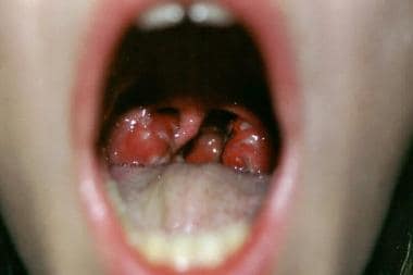
 Children with complications from acute sinusits may experience significant illness from their infection
 What Caused Stroke in Patient Already 'Recovered' From COVID-19? | MedPage Today
 Perioperative Neurosurgery, 3rd Floor VUH | Vanderbilt Nursing
 Headache Secondary to Epidural Abscess
 Global Neurovascular Devices Market To Be At Forefront By 2019-2028 | Abbott Laboratories, Terumo Corporation, Penumbra -...
 Periorbital Cellulitis - Kingston Hospital
 Biomedicines | Free Full-Text | Robert's Intragastric Alcohol-Induced Gastric Lesion Model as an Escalated General Peripheral...
 Abeer Farrag, MD | Cleveland Clinic
 These highlights do not include all the information needed to use DROSPIRENONE/ETHINYL ESTRADIOL/LEVOMEFOLATE CALCIUM TABLETS...