Shiga Toxin
Shiga Toxin 2
Shiga Toxin 1
Shiga Toxins
Shiga-Toxigenic Escherichia coli
Shigella dysenteriae
Escherichia coli O157
Hemolytic-Uremic Syndrome
Enterohemorrhagic Escherichia coli
Bacterial Toxins
Cholera Toxin
T-2 Toxin
Cytotoxins
Trihexosylceramides
Globosides
Ricin
Prophages
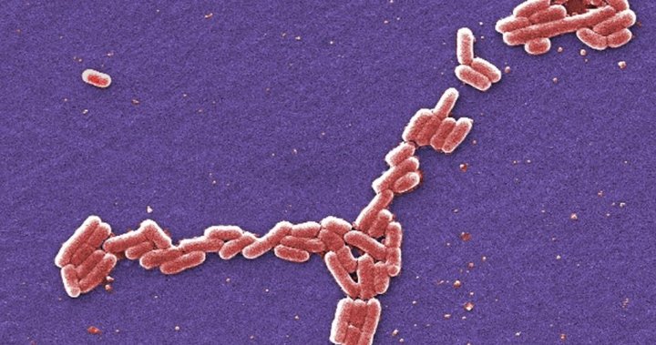
Escherichia coli
Vero Cells
Ribosome Inactivating Proteins
Tetanus Toxin
Antitoxins
Feces
Adhesins, Bacterial
Virulence Factors
Diarrhea
Escherichia coli Vaccines
Serotyping
Botulinum Toxins, Type A
Marine Toxins
Dysentery, Bacillary
Cercopithecus aethiops
Virulence
Enterotoxins
Shigella
Molecular Sequence Data
Butyric Acid
Toxoids
Hemolysin Proteins
Edema Disease of Swine
Polymerase Chain Reaction
Cattle Diseases
Food Microbiology
Golgi Apparatus
Cattle
Glycolipids
HeLa Cells
RNA, Ribosomal, 28S
Adenylate Cyclase Toxin
Foodborne Diseases
P Blood-Group System
trans-Golgi Network
Enteropathogenic Escherichia coli
Lethal Dose 50
Glycosphingolipids
Endosomes
Amino Acid Sequence
Lysogeny
Disease Outbreaks
Transcytosis
Rabbits
Electrophoresis, Gel, Pulsed-Field
Protein Synthesis Inhibitors
Enzyme-Linked Immunosorbent Assay
Brefeldin A
Protein Subunits
Receptors, Cell Surface
Protein Transport
Intestines
Neutralization Tests
Base Sequence
Siphoviridae
Immunochromatography
Biological Transport
Sequence Analysis, DNA
Endocytosis
Intestinal Mucosa
Serum Amyloid P-Component
Cell Survival
Meat
O Antigens
Butyrates
Scorpion Venoms
Germany
Clostridium difficile
Pinocytosis
Antigens, Tumor-Associated, Carbohydrate
Tosylphenylalanyl Chloromethyl Ketone
Bacterial Adhesion
Ileum
Neutral Glycosphingolipids
Clathrin
Responses of human intestinal microvascular endothelial cells to Shiga toxins 1 and 2 and pathogenesis of hemorrhagic colitis. (1/438)
Endothelial damage is characteristic of infection with Shiga toxin (Stx)-producing Escherichia coli (STEC). Because Stx-mediated endothelial cell damage at the site of infection may lead to the characteristic hemorrhagic colitis of STEC infection, we compared the effects of Stx1 and Stx2 on primary and transformed human intestinal microvascular endothelial cells (HIMEC) to those on macrovascular endothelial cells from human saphenous vein (HSVEC). Adhesion molecule, interleukin-8 (IL-8), and Stx receptor expression, the effects of cytokine activation and Stx toxins on these responses, and Stx1 and Stx2 binding kinetics and bioactivity were measured. Adhesion molecule and IL-8 expression increased in activated HIMEC, but these responses were blunted in the presence of toxin, especially in the presence of Stx1. In contrast to HSVEC, unstimulated HIMEC constitutively expressed Stx receptor at high levels, bound large amounts of toxin, were highly sensitive to toxin, and were not further sensitized by cytokines. Although the binding capacities of HIMEC for Stx1 and Stx2 were comparable, the binding affinity of Stx1 to HIMEC was 50-fold greater than that of Stx2. Nonetheless, Stx2 was more toxic to HIMEC than an equivalent amount of Stx1. The decreased binding affinity and increased toxicity for HIMEC of Stx2 compared to those of Stx1 may be relevant to the preponderance of Stx2-producing STEC involved in the pathogenesis of hemorrhagic colitis and its systemic complications. The differences between primary and transformed HIMEC in these responses were negligible. We conclude that transformed HIMEC lines could represent a simple physiologically relevant model to study the role of Stx in the pathogenesis of hemorrhagic colitis. (+info)Sequence of Shiga toxin 2 phage 933W from Escherichia coli O157:H7: Shiga toxin as a phage late-gene product. (2/438)
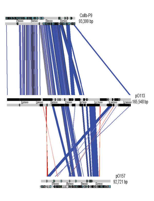
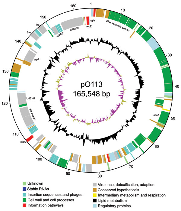
Lysogenic bacteriophages are major vehicles for the transfer of genetic information between bacteria, including pathogenicity and/or virulence determinants. In the enteric pathogen Escherichia coli O157:H7, which causes hemorrhagic colitis and hemolytic-uremic syndrome, Shiga toxins 1 and 2 (Stx1 and Stx2) are phage encoded. The sequence and analysis of the Stx2 phage 933W is presented here. We find evidence that the toxin genes are part of a late-phage transcript, suggesting that toxin production may be coupled with, if not dependent upon, phage release during lytic growth. Another phage gene, stk, encodes a product resembling eukaryotic serine/threonine protein kinases. Based on its position in the sequence, Stk may be produced by the prophage in the lysogenic state, and, like the YpkA protein of Yersinia species, it may interfere with the signal transduction pathway of the mammalian host. Three novel tRNA genes present in the phage genome may serve to increase the availability of rare tRNA species associated with efficient expression of pathogenicity determinants: both the Shiga toxin and serine/threonine kinase genes contain rare isoleucine and arginine codons. 933W also has homology to lom, encoding a member of a family of outer membrane proteins associated with virulence by conferring the ability to survive in macrophages, and bor, implicated in serum resistance. (+info)In vivo expression and immunoadjuvancy of a mutant of heat-labile enterotoxin of Escherichia coli in vaccine and vector strains of Vibrio cholerae. (3/438)
Vibrio cholerae secretes cholera toxin (CT) and the closely related heat-labile enterotoxin (LT) of Escherichia coli, the latter when expressed in V. cholerae. Both toxins are also potent immunoadjuvants. Mutant LT molecules that retain immunoadjuvant properties while possessing markedly diminished enterotoxic activities when expressed by E. coli have been developed. One such mutant LT molecule has the substitution of a glycine residue for arginine-192 [LT(R192G)]. Live attenuated strains of V. cholerae that have been used both as V. cholerae vaccines and as vectors for inducing mucosal and systemic immune responses directed against expressed heterologous antigens have been developed. In order to ascertain whether LT(R192G) can act as an immunoadjuvant when expressed in vivo by V. cholerae, we introduced a plasmid (pCS95) expressing this molecule into three vaccine strains of V. cholerae, Peru2, ETR3, and JRB14; the latter two strains contain genes encoding different heterologous antigens in the chromosome of the vaccine vectors. We found that LT(R192G) was expressed from pCS95 in vitro by both E. coli and V. cholerae strains but that LT(R192G) was detectable in the supernatant fraction of V. cholerae cultures only. In order to assess potential immunoadjuvanticity, groups of germfree mice were inoculated with the three V. cholerae vaccine strains alone and compared to groups inoculated with the V. cholerae vaccine strains supplemented with purified CT as an oral immunoadjuvant or V. cholerae vaccine strains expressing LT(R192G) from pCS95. We found that mice continued to pass stool containing V. cholerae strains with pCS95 for at least 4 days after oral inoculation, the last day evaluated. We found that inoculation with V. cholerae vaccine strains containing pCS95 resulted in anti-LT(R192G) immune responses, confirming in vivo expression. We were unable to detect immune responses directed against the heterologous antigens expressed at low levels in any group of animals, including animals that received purified CT as an immunoadjuvant. We were, however, able to measure increased vibriocidal immune responses against vaccine strains in animals that received V. cholerae vaccine strains expressing LT(R192G) from pCS95 compared to the responses in animals that received V. cholerae vaccine strains alone. These results demonstrate that mutant LT molecules can be expressed in vivo by attenuated vaccine strains of V. cholerae and that such expression can result in an immunoadjuvant effect. (+info)Emergence of fosfomycin-resistant isolates of Shiga-like toxin-producing Escherichia coli O26. (4/438)
We evaluated the susceptibilities of 129 Shiga-like toxin-producing Escherichia coli (STEC) isolates to various antibiotics. The numbers of isolates for which MICs were high (> or = 128 micrograms/ml) were as follows: 5 for fosfomycin, 14 for ampicillin, 1 for cefaclor, 6 for kanamycin, 22 for tetracycline, and 2 for doxycycline. For two isolates of STEC O26 MICs of fosfomycin were high (1,024 and 512 micrograms/ml, respectively). Conjugation experiments and glutathione S-transferase assays suggested that the fosfomycin resistance in these isolates was determined not by a plasmid but chromosomally. The amount of active intracellular fosfomycin in these STEC isolates was 100- to 200-fold less than that in E. coli C600 harboring pREFTT47B408 in the presence of either L-alpha-glycerophosphate or glucose-6-phosphate. Cloning, sequencing, and Northern blot analysis demonstrated that the transcriptional level of the murA gene encoding UDP-N-acetylglucosamine enolpyruvoyl transferase in these isolates was greater than that in E. coli C600. Our results suggest that the fosfomycin resistance in these STEC isolates is due to concurrent effects of alteration of the glpT and/or uhp transport systems and of the enhanced transcription of the murA gene. (+info)Export of virulence genes and Shiga toxin by membrane vesicles of Escherichia coli O157:H7. (5/438)
Membrane vesicles released by Escherichia coli O157:H7 into culture medium were purified and analyzed for protein and DNA content. Electron micrographs revealed vesicles that are spherical, range in size from 20 to 100 nm, and have a complete bilayer. Analysis of vesicle protein by sodium dodecyl sulfate-polyacrylamide gel electrophoresis demonstrates vesicles that contain many proteins with molecular sizes similar to outer membrane proteins and a number of cellular proteins. Immunoblot (Western) analysis of vesicles suggests the presence of cell antigens. Treatment of vesicles with exogenous DNase hydrolyzed surface-associated DNA; PCR demonstrated that vesicles contain DNA encoding the virulence genes eae, stx1 and stx2, and uidA, which encodes for beta-galactosidase. Immunoblot analysis of intact and lysed, proteinase K-treated vesicles demonstrate that Shiga toxins 1 and 2 are contained within vesicles. These results suggest that vesicles contain toxic material and transfer experiments demonstrate that vesicles can deliver genetic material to other gram-negative organisms. (+info)Genes for tRNA(Arg) located in the upstream region of the Shiga toxin II operon in enterohemorrhagic Escherichia coli O157:H7. (6/438)
We found two genes for tRNA(Arg) in the region upstream of genes for Shiga-like toxin type II (SLT-II) in Escherichia coli O157:H7. The two encoded forms of tRNA(Arg) recognize rare codons in E. coli K12 but these rare codons occur in the toxin genes at high frequency. (+info)Characterization of the baboon responses to Shiga-like toxin: descriptive study of a new primate model of toxic responses to Stx-1. (7/438)
The baboon response to intravenous infusion of Shiga toxin 1 (Stx-1) varied from acute renal failure, proteinuria, hyperkalemia, and melena with minimal perturbation of host inflammatory and hemostatic systems (high-dose group, 2.0 microg/kg; n = 5) to renal failure with hematuria, proteinuria, thrombocytopenia, schistocytosis, anemia, and melena (low-dose group, 0.05 to 0.2 microg/kg; n = 8). Both groups exhibited renal shutdown and died in 57 hours or less. Both groups produced urine that was positive for tumor necrosis factor and interleukin-6 although neither of these cytokines was detectable (+info)Molecular typing of Escherichia coli O157:H7 (H-) isolates from cattle in Japan. (8/438)
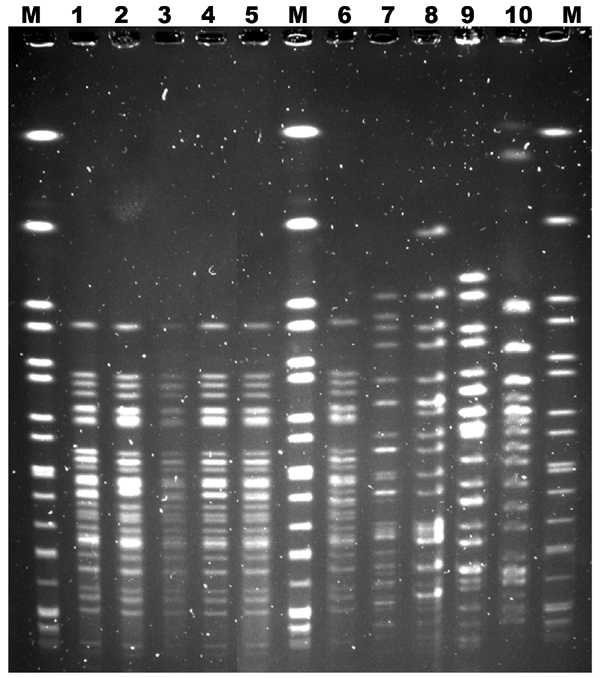
A total of 77 Escherichia coli O157:H7 (H-) isolates from cattle in Japan were investigated by molecular biological methods. Most of these isolates (43 isolates) possessed the stx-2 gene, but not stx1. Fifteen bacteriophage types and 50 pulsed-field gel electrophoresis (PFGE) profiles were observed. One isolate was indistinguishable from the human outbreak strain by these methods. This indicates that cattle must be considered as a possible source of human E. coli O157:H7 infection in Japan. (+info)Shiga toxins are a type of protein toxin produced by certain strains of bacteria, including some types of Escherichia coli (E. coli) and Shigella dysenteriae. These toxins get their name from Kiyoshi Shiga, the scientist who discovered them in 1897.
Shiga toxins are potent cytotoxins that can cause damage to cells by inhibiting protein synthesis. They consist of two main components: an enzymatically active A subunit and several B subunits that bind to specific receptors on the surface of target cells, facilitating the entry of the A subunit into the cell.
Once inside the cell, the A subunit cleaves a crucial component of the protein synthesis machinery called ribosome, leading to cell death or dysfunction. Shiga toxins can cause severe illnesses such as hemorrhagic colitis and hemolytic uremic syndrome (HUS), which can be life-threatening in some cases.
It's worth noting that Shiga toxin-producing E. coli (STEC) infections are often foodborne, and they can cause a range of symptoms from mild diarrhea to severe abdominal cramps, bloody diarrhea, and kidney failure. Prevention measures include proper food handling, cooking meat thoroughly, washing fruits and vegetables, and practicing good hygiene.
Shiga toxin 2 (Stx2) is a protein toxin produced by certain strains of the bacterium Escherichia coli (E. coli), specifically those that belong to serotype O157:H7 and some other Shiga toxin-producing E. coli (STEC) or enterohemorrhagic E. coli (EHEC).
Stx2 is named after Dr. Kiyoshi Shiga, who first discovered the related Shiga toxin in 1898. It is a powerful cytotoxin that can cause damage to cells lining the intestines and other organs. The toxin inhibits protein synthesis in the cells by removing an adenine residue from the 28S rRNA of the 60S ribosomal subunit, leading to cell death.
Exposure to Stx2 can occur through ingestion of contaminated food or water, or direct contact with infected animals or their feces. In severe cases, it can lead to hemorrhagic colitis, which is characterized by bloody diarrhea and abdominal cramps, and hemolytic uremic syndrome (HUS), a serious complication that can cause kidney failure, anemia, and neurological problems.
It's important to note that Stx2 has two major subtypes, Stx2a and Stx2b, which differ in their biological activities and clinical significance. Stx2a is considered more potent than Stx2b and is associated with a higher risk of developing HUS.
Shiga toxin 1 (Stx1) is a protein toxin produced by certain strains of the bacterium Escherichia coli (E. coli), specifically those that belong to serotype O157:H7 and some other Shiga toxin-producing E. coli (STEC) or enterohemorrhagic E. coli (EHEC).
Shiga toxins are named after Kiyoshi Shiga, who discovered the first strain of E. coli that produces this toxin in 1897. These toxins inhibit protein synthesis in eukaryotic cells and cause damage to the endothelial cells lining blood vessels, which can lead to various clinical manifestations such as hemorrhagic colitis (bloody diarrhea) and hemolytic uremic syndrome (HUS), a severe complication that can result in kidney failure.
Shiga toxin 1 is composed of two subunits, A and B. The B subunit binds to specific glycolipid receptors on the surface of target cells, facilitating the uptake of the toxin into the cell. Once inside the cell, the A subunit inhibits protein synthesis by removing an adenine residue from a specific region of the 28S rRNA molecule in the ribosome, thereby preventing peptide bond formation and leading to cell death.
Shiga toxin 1 is highly toxic and can cause significant morbidity and mortality, particularly in children, the elderly, and immunocompromised individuals. Antibiotics are generally not recommended for the treatment of Shiga toxin-producing E. coli infections because they may increase the risk of developing HUS by inducing bacterial lysis and releasing more toxins into the circulation. Supportive care, hydration, and close monitoring are essential for managing these infections.
Shiga toxins are a type of protein toxin produced by certain strains of bacteria, including some types of Escherichia coli (E. coli) and Shigella dysenteriae. These toxins get their name from Dr. Kiyoshi Shiga, who first discovered them in the late 19th century.
Shiga toxins are classified into two main types: Shiga toxin 1 (Stx1) and Shiga toxin 2 (Stx2). Both types of toxins are similar in structure and function, but they differ in their potency and genetic makeup. Shiga toxins inhibit protein synthesis in cells by removing an adenine residue from a specific region of the 28S rRNA molecule in the ribosome, which ultimately leads to cell death.
These toxins can cause severe damage to the lining of the intestines and are associated with hemorrhagic colitis, a potentially life-threatening condition characterized by bloody diarrhea, abdominal cramps, and fever. In some cases, Shiga toxins can also enter the bloodstream and cause systemic complications such as hemolytic uremic syndrome (HUS), which is characterized by kidney failure, anemia, and thrombocytopenia.
Exposure to Shiga toxins typically occurs through ingestion of contaminated food or water, or through direct contact with infected individuals or animals. Preventive measures include good hygiene practices, such as thorough handwashing, cooking meats thoroughly, and avoiding unpasteurized dairy products and untreated water.
Shiga-toxigenic Escherichia coli (STEC) are strains of the bacterium E. coli that produce one or both of two potent toxins called Shiga toxin or Shiga-like toxin. These toxins are named after Shigella dysenteriae type 1, from which the STEC Shiga toxin was originally isolated. The Shiga toxins cause severe damage to the lining of intestines and can lead to a range of symptoms such as diarrhea (often bloody), stomach cramps, vomiting, and fever. In severe cases, it can progress to hemolytic uremic syndrome (HUS), a serious complication that can cause kidney failure, brain damage, and even death, particularly in young children, the elderly, and immunocompromised individuals.
STEC is often found in the intestines of healthy animals, especially ruminants like cattle, goats, and sheep, and can be transmitted to humans through contaminated food or water, or direct contact with infected animals or their feces. Common sources of STEC include undercooked ground beef, raw milk, contaminated vegetables, and unpasteurized dairy products. It's important to note that not all strains of E. coli are Shiga-toxigenic, and only a small percentage of STEC infections result in severe illness or HUS.
"Shigella dysenteriae" is a specific species of bacteria that can cause severe forms of dysentery, a type of diarrheal disease. The infection caused by this bacterium is known as shigellosis. Shigella dysenteriae is highly infectious and can be transmitted through direct contact with an infected person or through contaminated food or water.
The bacteria produce toxins that can cause inflammation and damage to the lining of the intestine, leading to symptoms such as diarrhea (often containing blood and mucus), abdominal cramps, fever, and tenesmus (the urgent need to have a bowel movement). In severe cases, shigellosis can lead to complications such as dehydration, seizures, and hemolytic-uremic syndrome (HUS), a serious condition that can cause kidney failure.
Shigella dysenteriae is a public health concern, particularly in areas with poor sanitation and hygiene practices. Prevention measures include good hand hygiene, safe food handling practices, and access to clean water. Treatment typically involves antibiotics, fluids, and electrolyte replacement to manage symptoms and prevent complications.
Escherichia coli (E. coli) O157 is a serotype of the bacterium E. coli that is associated with foodborne illness. This strain is pathogenic and produces Shiga toxins, which can cause severe damage to the lining of the small intestine and potentially lead to hemorrhagic diarrhea and kidney failure. E. coli O157 is often transmitted through contaminated food, particularly undercooked ground beef, as well as raw or unpasteurized dairy products, fruits, and vegetables. It can also be spread through contact with infected individuals or animals, especially in settings like farms, petting zoos, and swimming pools. Proper food handling, cooking, and hygiene practices are crucial to preventing E. coli O157 infections.
Hemolytic-Uremic Syndrome (HUS) is a serious condition that affects the blood and kidneys. It is characterized by three major features: the breakdown of red blood cells (hemolysis), the abnormal clotting of small blood vessels (microthrombosis), and acute kidney failure.
The breakdown of red blood cells leads to the release of hemoglobin into the bloodstream, which can cause anemia. The microthrombi can obstruct the flow of blood in the kidneys' filtering system (glomeruli), leading to damaged kidney function and potentially acute kidney failure.
HUS is often caused by a bacterial infection, most commonly Escherichia coli (E. coli) that produces Shiga toxins. This form of HUS is known as STEC-HUS or Stx-HUS. Other causes include infections with other bacteria, viruses, medications, pregnancy complications, and certain medical conditions such as autoimmune diseases.
Symptoms of HUS may include fever, fatigue, decreased urine output, blood in the stool, swelling in the face, hands, or feet, and irritability or confusion. Treatment typically involves supportive care, including dialysis for kidney failure, transfusions to replace lost red blood cells, and managing high blood pressure. In severe cases, a kidney transplant may be necessary.
Escherichia coli (E. coli) infections refer to illnesses caused by the bacterium E. coli, which can cause a range of symptoms depending on the specific strain and site of infection. The majority of E. coli strains are harmless and live in the intestines of healthy humans and animals. However, some strains, particularly those that produce Shiga toxins, can cause severe illness.
E. coli infections can occur through various routes, including contaminated food or water, person-to-person contact, or direct contact with animals or their environments. Common symptoms of E. coli infections include diarrhea (often bloody), abdominal cramps, nausea, and vomiting. In severe cases, complications such as hemolytic uremic syndrome (HUS) can occur, which may lead to kidney failure and other long-term health problems.
Preventing E. coli infections involves practicing good hygiene, cooking meats thoroughly, avoiding cross-contamination of food during preparation, washing fruits and vegetables before eating, and avoiding unpasteurized dairy products and juices. Prompt medical attention is necessary if symptoms of an E. coli infection are suspected to prevent potential complications.
Enterohemorrhagic Escherichia coli (EHEC) are a type of Shiga toxin-producing E. coli (STEC). They are characterized by their ability to cause hemorrhagic diarrhea and the presence of a virulence factor known as Shiga toxin or Verocytotoxin. The most well-known serotype of EHEC is O157:H7, but there are other non-O157 serotypes that can also cause human illness.
EHEC infection typically occurs through the consumption of contaminated food or water, or direct contact with infected animals or their environment. Once ingested, EHEC colonize the intestines and produce Shiga toxins, which can damage the lining of the intestine and cause bloody diarrhea. In severe cases, Shiga toxins can also enter the bloodstream and cause hemolytic uremic syndrome (HUS), a serious complication that can lead to kidney failure and other long-term health problems.
Preventing EHEC infection involves practicing good food safety habits, such as washing hands thoroughly before preparing or eating food, cooking meats to the recommended internal temperature, avoiding unpasteurized dairy products and juices, and washing fruits and vegetables thoroughly before eating. It is also important to handle and store food properly to prevent cross-contamination with EHEC bacteria.
Bacterial toxins are poisonous substances produced and released by bacteria. They can cause damage to the host organism's cells and tissues, leading to illness or disease. Bacterial toxins can be classified into two main types: exotoxins and endotoxins.
Exotoxins are proteins secreted by bacterial cells that can cause harm to the host. They often target specific cellular components or pathways, leading to tissue damage and inflammation. Some examples of exotoxins include botulinum toxin produced by Clostridium botulinum, which causes botulism; diphtheria toxin produced by Corynebacterium diphtheriae, which causes diphtheria; and tetanus toxin produced by Clostridium tetani, which causes tetanus.
Endotoxins, on the other hand, are components of the bacterial cell wall that are released when the bacteria die or divide. They consist of lipopolysaccharides (LPS) and can cause a generalized inflammatory response in the host. Endotoxins can be found in gram-negative bacteria such as Escherichia coli and Pseudomonas aeruginosa.
Bacterial toxins can cause a wide range of symptoms depending on the type of toxin, the dose, and the site of infection. They can lead to serious illnesses or even death if left untreated. Vaccines and antibiotics are often used to prevent or treat bacterial infections and reduce the risk of severe complications from bacterial toxins.
Cholera toxin is a protein toxin produced by the bacterium Vibrio cholerae, which causes the infectious disease cholera. The toxin is composed of two subunits, A and B, and its primary mechanism of action is to alter the normal function of cells in the small intestine.
The B subunit of the toxin binds to ganglioside receptors on the surface of intestinal epithelial cells, allowing the A subunit to enter the cell. Once inside, the A subunit activates a signaling pathway that results in the excessive secretion of chloride ions and water into the intestinal lumen, leading to profuse, watery diarrhea, dehydration, and other symptoms associated with cholera.
Cholera toxin is also used as a research tool in molecular biology and immunology due to its ability to modulate cell signaling pathways. It has been used to study the mechanisms of signal transduction, protein trafficking, and immune responses.
T-2 toxin is a type B trichothecene mycotoxin, which is a secondary metabolite produced by certain Fusarium species of fungi. It is a low molecular weight sesquiterpene epoxide that is chemically stable and has a high toxicity profile. T-2 toxin can contaminate crops in the field or during storage, and it is often found in grains such as corn, wheat, barley, and oats.
T-2 toxin has a variety of adverse health effects, including nausea, vomiting, diarrhea, abdominal pain, immune suppression, skin irritation, and neurotoxicity. It is also known to have teratogenic and embryotoxic effects in animals, and it is considered a potential human carcinogen by some agencies.
Exposure to T-2 toxin can occur through ingestion, inhalation, or skin contact. Ingestion is the most common route of exposure, particularly in areas where contaminated grains are used as a food source. Inhalation exposure can occur during agricultural activities such as harvesting and processing contaminated crops. Skin contact with T-2 toxin can cause irritation and inflammation.
Prevention of T-2 toxin exposure involves good agricultural practices, including crop rotation, use of resistant varieties, and proper storage conditions. Monitoring of T-2 toxin levels in food and feed is also important to ensure that exposure limits are not exceeded.
Cytotoxins are substances that are toxic to cells. They can cause damage and death to cells by disrupting their membranes, interfering with their metabolism, or triggering programmed cell death (apoptosis). Cytotoxins can be produced by various organisms such as bacteria, fungi, plants, and animals, and they can also be synthesized artificially.
In medicine, cytotoxic drugs are used to treat cancer because they selectively target and kill rapidly dividing cells, including cancer cells. Examples of cytotoxic drugs include chemotherapy agents such as doxorubicin, cyclophosphamide, and methotrexate. However, these drugs can also damage normal cells, leading to side effects such as nausea, hair loss, and immune suppression.
It's important to note that cytotoxins are not the same as toxins, which are poisonous substances produced by living organisms that can cause harm to other organisms. While all cytotoxins are toxic to cells, not all toxins are cytotoxic. Some toxins may have systemic effects on organs or tissues rather than directly killing cells.
Trihexosylceramides are a type of glycosphingolipids, which are complex lipids found in animal tissues. They consist of a ceramide molecule (a sphingosine and fatty acid) with three hexose sugars attached to it in a specific sequence, typically glucose-galactose-galactose.
Trihexosylceramides are further classified into two types based on the type of ceramide they contain: lactosylceramide (Gal-Glc-Cer) and isoglobotrihexosylceramide (GalNAcβ1-4Galβ1-4Glc-Cer).
These lipids are important components of the cell membrane and play a role in various biological processes, including cell recognition, signal transduction, and cell adhesion. Abnormal accumulation of trihexosylceramides has been implicated in certain diseases, such as Gaucher disease and Tay-Sachs disease, which are caused by deficiencies in enzymes involved in their breakdown.
Globosides are a type of glycosphingolipids, which are molecules that consist of a lipid and a carbohydrate. They are found in animal tissues, especially in the nervous system. The term "globoside" refers to a specific structure of these molecules, where the carbohydrate portion consists of a complex chain of sugars, including galactose, N-acetylgalactosamine, and glucose. Globosides play important roles in cell recognition and interaction, and abnormalities in their metabolism have been associated with certain diseases, such as paroxysmal nocturnal hemoglobinuria (PNH).
Ricin is defined as a highly toxic protein that is derived from the seeds of the castor oil plant (Ricinus communis). It can be produced as a white, powdery substance or a mistable aerosol. Ricin works by getting inside cells and preventing them from making the proteins they need. Without protein, cells die. Eventually, this can cause organ failure and death.
It is not easily inhaled or absorbed through the skin, but if ingested or injected, it can be lethal in very small amounts. There is no antidote for ricin poisoning - treatment consists of supportive care. Ricin has been used as a bioterrorism agent in the past and continues to be a concern due to its relative ease of production and potential high toxicity.
A prophage is a bacteriophage (a virus that infects bacteria) genome that is integrated into the chromosome of a bacterium and replicates along with it. The phage genome remains dormant within the bacterial host until an environmental trigger, such as stress or damage to the host cell, induces the prophage to excise itself from the bacterial chromosome and enter a lytic cycle, during which new virions are produced and released by lysing the host cell. This process is known as lysogeny.
Prophages can play important roles in the biology of their bacterial hosts, such as contributing to genetic diversity through horizontal gene transfer, modulating bacterial virulence, and providing resistance to superinfection by other phages. However, they can also have detrimental effects on the host, such as causing lysis or altering bacterial phenotypes in ways that are disadvantageous for survival.
It's worth noting that not all bacteriophages form prophages; some exist exclusively as extrachromosomal elements, while others can integrate into the host genome but do not necessarily become dormant or replicate with the host cell.
'Escherichia coli' (E. coli) is a type of gram-negative, facultatively anaerobic, rod-shaped bacterium that commonly inhabits the intestinal tract of humans and warm-blooded animals. It is a member of the family Enterobacteriaceae and one of the most well-studied prokaryotic model organisms in molecular biology.
While most E. coli strains are harmless and even beneficial to their hosts, some serotypes can cause various forms of gastrointestinal and extraintestinal illnesses in humans and animals. These pathogenic strains possess virulence factors that enable them to colonize and damage host tissues, leading to diseases such as diarrhea, urinary tract infections, pneumonia, and sepsis.
E. coli is a versatile organism with remarkable genetic diversity, which allows it to adapt to various environmental niches. It can be found in water, soil, food, and various man-made environments, making it an essential indicator of fecal contamination and a common cause of foodborne illnesses. The study of E. coli has contributed significantly to our understanding of fundamental biological processes, including DNA replication, gene regulation, and protein synthesis.
Vero cells are a line of cultured kidney epithelial cells that were isolated from an African green monkey (Cercopithecus aethiops) in the 1960s. They are named after the location where they were initially developed, the Vervet Research Institute in Japan.
Vero cells have the ability to divide indefinitely under certain laboratory conditions and are often used in scientific research, including virology, as a host cell for viruses to replicate. This allows researchers to study the characteristics of various viruses, such as their growth patterns and interactions with host cells. Vero cells are also used in the production of some vaccines, including those for rabies, polio, and Japanese encephalitis.
It is important to note that while Vero cells have been widely used in research and vaccine production, they can still have variations between different cell lines due to factors like passage number or culture conditions. Therefore, it's essential to specify the exact source and condition of Vero cells when reporting experimental results.
Ribosome-inactivating proteins (RIPs) are a type of protein that can inhibit the function of ribosomes, which are the cellular structures responsible for protein synthesis. Ribosomes are made up of two subunits, and RIPs work by depurinating a specific adenine residue in the sarcin-ricin loop of the large rRNA subunit, leading to the inhibition of protein synthesis and ultimately, cell death.
RIPs can be found in various organisms, including plants, bacteria, and fungi. Some RIPs have N-glycosidase activity, while others have both N-glycosidase and RNA N-hydroxylase activities. Based on their structure and mechanism of action, RIPs are classified into two types: type 1 and type 2.
Type 1 RIPs consist of a single polypeptide chain with N-glycosidase activity, while type 2 RIPs consist of two chains - an A chain with N-glycosidase activity and a B chain that acts as a lectin, facilitating the entry of the A chain into the cell.
RIPs have been studied for their potential use in cancer therapy due to their ability to inhibit protein synthesis in cancer cells. However, their toxicity to normal cells limits their therapeutic use. Therefore, researchers are exploring ways to modify RIPs to increase their specificity towards cancer cells while minimizing their toxicity to normal cells.
Tetanus toxin, also known as tetanospasmin, is a potent neurotoxin produced by the bacterium Clostridium tetani. This toxin binds to nerve endings and is transported to the nervous system's inhibitory neurons, where it blocks the release of inhibitory neurotransmitters, particularly glycine and GABA (gamma-aminobutyric acid). As a result, it causes uncontrolled muscle contractions or spasms, which are the hallmark symptoms of tetanus disease.
The toxin has two main components: an N-terminal portion called the light chain, which is the enzymatically active part that inhibits neurotransmitter release, and a C-terminal portion called the heavy chain, which facilitates the toxin's entry into neurons. The heavy chain also contains a binding domain that allows the toxin to recognize specific receptors on nerve cells.
Tetanus toxin is one of the most potent toxins known, with an estimated human lethal dose of just 2.5-3 nanograms per kilogram of body weight when introduced into the bloodstream. Fortunately, tetanus can be prevented through vaccination with the tetanus toxoid, which is part of the standard diphtheria-tetanus-pertussis (DTaP or Tdap) immunization series for children and adolescents and the tetanus-diphtheria (Td) booster for adults.
Antitoxins are substances, typically antibodies, that neutralize toxins produced by bacteria or other harmful organisms. They work by binding to the toxin molecules and rendering them inactive, preventing them from causing harm to the body. Antitoxins can be produced naturally by the immune system during an infection, or they can be administered artificially through immunization or passive immunotherapy. In a medical context, antitoxins are often used as a treatment for certain types of bacterial infections, such as diphtheria and botulism, to help counteract the effects of the toxins produced by the bacteria.
Feces are the solid or semisolid remains of food that could not be digested or absorbed in the small intestine, along with bacteria and other waste products. After being stored in the colon, feces are eliminated from the body through the rectum and anus during defecation. Feces can vary in color, consistency, and odor depending on a person's diet, health status, and other factors.
'Escherichia coli (E. coli) proteins' refer to the various types of proteins that are produced and expressed by the bacterium Escherichia coli. These proteins play a critical role in the growth, development, and survival of the organism. They are involved in various cellular processes such as metabolism, DNA replication, transcription, translation, repair, and regulation.
E. coli is a gram-negative, facultative anaerobe that is commonly found in the intestines of warm-blooded organisms. It is widely used as a model organism in scientific research due to its well-studied genetics, rapid growth, and ability to be easily manipulated in the laboratory. As a result, many E. coli proteins have been identified, characterized, and studied in great detail.
Some examples of E. coli proteins include enzymes involved in carbohydrate metabolism such as lactase, sucrase, and maltose; proteins involved in DNA replication such as the polymerases, single-stranded binding proteins, and helicases; proteins involved in transcription such as RNA polymerase and sigma factors; proteins involved in translation such as ribosomal proteins, tRNAs, and aminoacyl-tRNA synthetases; and regulatory proteins such as global regulators, two-component systems, and transcription factors.
Understanding the structure, function, and regulation of E. coli proteins is essential for understanding the basic biology of this important organism, as well as for developing new strategies for combating bacterial infections and improving industrial processes involving bacteria.
Bacterial adhesins are proteins or structures on the surface of bacterial cells that allow them to attach to other cells or surfaces. This ability to adhere to host tissues is an important first step in the process of bacterial infection and colonization. Adhesins can recognize and bind to specific receptors on host cells, such as proteins or sugars, enabling the bacteria to establish a close relationship with the host and evade immune responses.
There are several types of bacterial adhesins, including fimbriae, pili, and non-fimbrial adhesins. Fimbriae and pili are thin, hair-like structures that extend from the bacterial surface and can bind to a variety of host cell receptors. Non-fimbrial adhesins are proteins that are directly embedded in the bacterial cell wall and can also mediate attachment to host cells.
Bacterial adhesins play a crucial role in the pathogenesis of many bacterial infections, including urinary tract infections, respiratory tract infections, and gastrointestinal infections. Understanding the mechanisms of bacterial adhesion is important for developing new strategies to prevent and treat bacterial infections.
Virulence factors are characteristics or components of a microorganism, such as bacteria, viruses, fungi, or parasites, that contribute to its ability to cause damage or disease in a host organism. These factors can include various structures, enzymes, or toxins that allow the pathogen to evade the host's immune system, attach to and invade host tissues, obtain nutrients from the host, or damage host cells directly.
Examples of virulence factors in bacteria include:
1. Endotoxins: lipopolysaccharides found in the outer membrane of Gram-negative bacteria that can trigger a strong immune response and inflammation.
2. Exotoxins: proteins secreted by some bacteria that have toxic effects on host cells, such as botulinum toxin produced by Clostridium botulinum or diphtheria toxin produced by Corynebacterium diphtheriae.
3. Adhesins: structures that help the bacterium attach to host tissues, such as fimbriae or pili in Escherichia coli.
4. Capsules: thick layers of polysaccharides or proteins that surround some bacteria and protect them from the host's immune system, like those found in Streptococcus pneumoniae or Klebsiella pneumoniae.
5. Invasins: proteins that enable bacteria to invade and enter host cells, such as internalins in Listeria monocytogenes.
6. Enzymes: proteins that help bacteria obtain nutrients from the host by breaking down various molecules, like hemolysins that lyse red blood cells to release iron or hyaluronidases that degrade connective tissue.
Understanding virulence factors is crucial for developing effective strategies to prevent and treat infectious diseases caused by these microorganisms.
Diarrhea is a condition in which an individual experiences loose, watery stools frequently, often exceeding three times a day. It can be acute, lasting for several days, or chronic, persisting for weeks or even months. Diarrhea can result from various factors, including viral, bacterial, or parasitic infections, food intolerances, medications, and underlying medical conditions such as inflammatory bowel disease or irritable bowel syndrome. Dehydration is a potential complication of diarrhea, particularly in severe cases or in vulnerable populations like young children and the elderly.
Escherichia coli (E. coli) vaccines are designed to protect against infections caused by various strains of the E. coli bacterium. These vaccines typically contain inactivated or attenuated (weakened) forms of the bacteria, which stimulate an immune response when introduced into the body. The immune system learns to recognize and fight off the specific strain of E. coli used in the vaccine, providing protection against future infections with that strain.
There are several types of E. coli vaccines available or in development, including:
1. Shiga toxin-producing E. coli (STEC) vaccines: These vaccines protect against STEC strains, such as O157:H7 and non-O157 STECs, which can cause severe illness, including hemorrhagic colitis and hemolytic uremic syndrome (HUS).
2. Enterotoxigenic E. coli (ETEC) vaccines: These vaccines target ETEC strains that are a common cause of traveler's diarrhea in people visiting areas with poor sanitation.
3. Enteropathogenic E. coli (EPEC) vaccines: EPEC strains can cause persistent diarrhea, especially in young children in developing countries. Vaccines against these strains are still in the research and development stage.
4. Extraintestinal pathogenic E. coli (ExPEC) vaccines: These vaccines aim to protect against ExPEC strains that can cause urinary tract infections, sepsis, and meningitis.
It is important to note that different E. coli vaccines are designed for specific purposes and may not provide cross-protection against other strains or types of E. coli infections.
Serotyping is a laboratory technique used to classify microorganisms, such as bacteria and viruses, based on the specific antigens or proteins present on their surface. It involves treating the microorganism with different types of antibodies and observing which ones bind to its surface. Each distinct set of antigens corresponds to a specific serotype, allowing for precise identification and characterization of the microorganism. This technique is particularly useful in epidemiology, vaccine development, and infection control.
Botulinum toxins type A are neurotoxins produced by the bacterium Clostridium botulinum and related species. These toxins act by blocking the release of acetylcholine at the neuromuscular junction, leading to muscle paralysis. Botulinum toxin type A is used in medical treatments for various conditions characterized by muscle spasticity or excessive muscle activity, such as cervical dystonia, blepharospasm, strabismus, and chronic migraine. It is also used cosmetically to reduce the appearance of wrinkles by temporarily paralyzing the muscles that cause them. The commercial forms of botulinum toxin type A include Botox, Dysport, and Xeomin.
Marine toxins are toxic compounds that are produced by certain marine organisms, including algae, bacteria, and various marine animals such as shellfish, jellyfish, and snails. These toxins can cause a range of illnesses and symptoms in humans who consume contaminated seafood or come into direct contact with the toxin-producing organisms. Some of the most well-known marine toxins include:
1. Saxitoxin: Produced by certain types of algae, saxitoxin can cause paralytic shellfish poisoning (PSP) in humans who consume contaminated shellfish. Symptoms of PSP include tingling and numbness of the lips, tongue, and fingers, followed by muscle weakness, paralysis, and in severe cases, respiratory failure.
2. Domoic acid: Produced by certain types of algae, domoic acid can cause amnesic shellfish poisoning (ASP) in humans who consume contaminated shellfish. Symptoms of ASP include nausea, vomiting, diarrhea, abdominal cramps, headache, and memory loss.
3. Okadaic acid: Produced by certain types of algae, okadaic acid can cause diarrhetic shellfish poisoning (DSP) in humans who consume contaminated shellfish. Symptoms of DSP include nausea, vomiting, diarrhea, abdominal cramps, and fever.
4. Ciguatoxin: Produced by certain types of dinoflagellates, ciguatoxin can cause ciguatera fish poisoning (CFP) in humans who consume contaminated fish. Symptoms of CFP include nausea, vomiting, diarrhea, abdominal pain, and neurological symptoms such as tingling and numbness of the lips, tongue, and fingers, as well as reversal of hot and cold sensations.
5. Tetrodotoxin: Found in certain types of pufferfish, tetrodotoxin can cause a severe form of food poisoning known as pufferfish poisoning or fugu poisoning. Symptoms of tetrodotoxin poisoning include numbness of the lips and tongue, difficulty speaking, muscle weakness, paralysis, and respiratory failure.
Prevention measures for these types of seafood poisoning include avoiding consumption of fish and shellfish that are known to be associated with these toxins, as well as cooking and preparing seafood properly before eating it. Additionally, monitoring programs have been established in many countries to monitor the levels of these toxins in seafood and issue warnings when necessary.
Bacillary dysentery is a type of dysentery caused by the bacterium Shigella. It is characterized by the inflammation of the intestines, particularly the colon, resulting in diarrhea that may contain blood and mucus. The infection is typically spread through contaminated food or water, or close contact with an infected person. Symptoms usually appear within 1-4 days after exposure and can include abdominal cramps, fever, nausea, vomiting, and tenesmus (the strong, frequent urge to have a bowel movement). In severe cases, bacillary dysentery can lead to dehydration, electrolyte imbalance, and other complications. Treatment typically involves antibiotics to kill the bacteria, as well as fluid replacement to prevent dehydration.
'Cercopithecus aethiops' is the scientific name for the monkey species more commonly known as the green monkey. It belongs to the family Cercopithecidae and is native to western Africa. The green monkey is omnivorous, with a diet that includes fruits, nuts, seeds, insects, and small vertebrates. They are known for their distinctive greenish-brown fur and long tail. Green monkeys are also important animal models in biomedical research due to their susceptibility to certain diseases, such as SIV (simian immunodeficiency virus), which is closely related to HIV.
Virulence, in the context of medicine and microbiology, refers to the degree or severity of damage or harm that a pathogen (like a bacterium, virus, fungus, or parasite) can cause to its host. It is often associated with the ability of the pathogen to invade and damage host tissues, evade or suppress the host's immune response, replicate within the host, and spread between hosts.
Virulence factors are the specific components or mechanisms that contribute to a pathogen's virulence, such as toxins, enzymes, adhesins, and capsules. These factors enable the pathogen to establish an infection, cause tissue damage, and facilitate its transmission between hosts. The overall virulence of a pathogen can be influenced by various factors, including host susceptibility, environmental conditions, and the specific strain or species of the pathogen.
Enterotoxins are types of toxic substances that are produced by certain microorganisms, such as bacteria. These toxins are specifically designed to target and affect the cells in the intestines, leading to symptoms such as diarrhea, vomiting, and abdominal cramps. One well-known example of an enterotoxin is the toxin produced by Staphylococcus aureus bacteria, which can cause food poisoning. Another example is the cholera toxin produced by Vibrio cholerae, which can cause severe diarrhea and dehydration. Enterotoxins work by interfering with the normal functioning of intestinal cells, leading to fluid accumulation in the intestines and subsequent symptoms.
Shigella is a genus of Gram-negative, facultatively anaerobic, rod-shaped bacteria that are primarily responsible for causing shigellosis, also known as bacillary dysentery. These pathogens are highly infectious and can cause severe gastrointestinal illness in humans through the consumption of contaminated food or water, or direct contact with an infected person's feces.
There are four main species of Shigella: S. dysenteriae, S. flexneri, S. boydii, and S. sonnei. Each species has distinct serotypes that differ in their epidemiology, clinical presentation, and antibiotic susceptibility patterns. The severity of shigellosis can range from mild diarrhea to severe dysentery with abdominal cramps, fever, and tenesmus (the strong, frequent urge to defecate). In some cases, Shigella infections may lead to complications such as bacteremia, seizures, or hemolytic uremic syndrome.
Preventive measures include maintaining good personal hygiene, proper food handling and preparation, access to clean water, and adequate sanitation facilities. Antibiotic treatment is generally recommended for severe cases of shigellosis, but the emergence of antibiotic-resistant strains has become a growing concern in recent years.
Molecular sequence data refers to the specific arrangement of molecules, most commonly nucleotides in DNA or RNA, or amino acids in proteins, that make up a biological macromolecule. This data is generated through laboratory techniques such as sequencing, and provides information about the exact order of the constituent molecules. This data is crucial in various fields of biology, including genetics, evolution, and molecular biology, allowing for comparisons between different organisms, identification of genetic variations, and studies of gene function and regulation.
Bacteriophages, often simply called phages, are viruses that infect and replicate within bacteria. They consist of a protein coat, called the capsid, that encases the genetic material, which can be either DNA or RNA. Bacteriophages are highly specific, meaning they only infect certain types of bacteria, and they reproduce by hijacking the bacterial cell's machinery to produce more viruses.
Once a phage infects a bacterium, it can either replicate its genetic material and create new phages (lytic cycle), or integrate its genetic material into the bacterial chromosome and replicate along with the bacterium (lysogenic cycle). In the lytic cycle, the newly formed phages are released by lysing, or breaking open, the bacterial cell.
Bacteriophages play a crucial role in shaping microbial communities and have been studied as potential alternatives to antibiotics for treating bacterial infections.
Butyric acid is a type of short-chain fatty acid that is naturally produced in the human body through the fermentation of dietary fiber in the colon. Its chemical formula is C4H8O2. It has a distinctive, rancid odor and is used in the production of perfumes, flavorings, and certain types of plasticizers. In addition to its natural occurrence in the human body, butyric acid is also found in some foods such as butter, parmesan cheese, and fermented foods like sauerkraut. It has been studied for its potential health benefits, including its role in gut health, immune function, and cancer prevention.
Toxoids are inactivated bacterial toxins that have lost their toxicity but retain their antigenicity. They are often used in vaccines to stimulate an immune response and provide protection against certain diseases without causing the harmful effects associated with the active toxin. The process of converting a toxin into a toxoid is called detoxication, which is typically achieved through chemical or heat treatment.
One example of a toxoid-based vaccine is the diphtheria and tetanus toxoids (DT) or diphtheria, tetanus, and pertussis toxoids (DTaP or TdaP) vaccines. These vaccines contain inactivated forms of the diphtheria and tetanus toxins, as well as inactivated pertussis toxin in the case of DTaP or TdaP vaccines. By exposing the immune system to these toxoids, the body learns to recognize and mount a response against the actual toxins produced by the bacteria, thereby providing immunity and protection against the diseases they cause.
Hemolysins are a type of protein toxin produced by certain bacteria, fungi, and plants that have the ability to damage and destroy red blood cells (erythrocytes), leading to their lysis or hemolysis. This results in the release of hemoglobin into the surrounding environment. Hemolysins can be classified into two main categories:
1. Exotoxins: These are secreted by bacteria and directly damage host cells. They can be further divided into two types:
* Membrane attack complex/perforin-like proteins (MACPF): These hemolysins create pores in the membrane of red blood cells, disrupting their integrity and causing lysis. Examples include alpha-hemolysin from Staphylococcus aureus and streptolysin O from Streptococcus pyogenes.
* Enzymatic hemolysins: These hemolysins are enzymes that degrade specific components of the red blood cell membrane, ultimately leading to lysis. An example is streptolysin S from Streptococcus pyogenes, which is a thiol-activated, oxygen-labile hemolysin.
2. Endotoxins: These are part of the outer membrane of Gram-negative bacteria and can cause indirect hemolysis by activating the complement system or by stimulating the release of inflammatory mediators from host cells.
Hemolysins play a significant role in bacterial pathogenesis, contributing to tissue damage, impaired immune responses, and disease progression.
Edema disease of swine, also known as porcine edema disease, is a condition that primarily affects young pigs between 2 weeks and 5 months of age. It is characterized by the sudden onset of neurological symptoms and fluid accumulation in various tissues, particularly in the brain and skin around the neck and shoulders.
The cause of edema disease is a bacterial toxin called Shiga-like toxin IIe (Stx2e) produced by certain strains of Escherichia coli (E. coli) bacteria. These bacteria colonize the pig's small intestine and produce the toxin, which then enters the bloodstream and damages the endothelial cells that line the blood vessels. This damage leads to increased permeability of the blood vessels, allowing fluid to leak out into surrounding tissues and causing edema (swelling).
The neurological symptoms of edema disease are thought to be caused by the direct toxic effects of Stx2e on nerve cells in the brainstem. The exact mechanism is not fully understood, but it is believed that the toxin disrupts the normal functioning of these nerve cells, leading to symptoms such as muscle weakness, tremors, and difficulty breathing.
Treatment of edema disease typically involves supportive care, such as fluid therapy and antibiotics to control the E. coli infection. Prevention measures include vaccination against E. coli strains that produce Stx2e and maintaining good hygiene practices in pig farming operations.
Coliphages are viruses that infect and replicate within certain species of bacteria that belong to the coliform group, particularly Escherichia coli (E. coli). These viruses are commonly found in water and soil environments and are frequently used as indicators of fecal contamination in water quality testing. Coliphages are not harmful to humans or animals, but their presence in water can suggest the potential presence of pathogenic bacteria or other microorganisms that may pose a health risk. There are two main types of coliphages: F-specific RNA coliphages and somatic (or non-F specific) DNA coliphages.
Polymerase Chain Reaction (PCR) is a laboratory technique used to amplify specific regions of DNA. It enables the production of thousands to millions of copies of a particular DNA sequence in a rapid and efficient manner, making it an essential tool in various fields such as molecular biology, medical diagnostics, forensic science, and research.
The PCR process involves repeated cycles of heating and cooling to separate the DNA strands, allow primers (short sequences of single-stranded DNA) to attach to the target regions, and extend these primers using an enzyme called Taq polymerase, resulting in the exponential amplification of the desired DNA segment.
In a medical context, PCR is often used for detecting and quantifying specific pathogens (viruses, bacteria, fungi, or parasites) in clinical samples, identifying genetic mutations or polymorphisms associated with diseases, monitoring disease progression, and evaluating treatment effectiveness.
Bacterial antibodies are a type of antibodies produced by the immune system in response to an infection caused by bacteria. These antibodies are proteins that recognize and bind to specific antigens on the surface of the bacterial cells, marking them for destruction by other immune cells. Bacterial antibodies can be classified into several types based on their structure and function, including IgG, IgM, IgA, and IgE. They play a crucial role in the body's defense against bacterial infections and provide immunity to future infections with the same bacteria.
Cattle diseases are a range of health conditions that affect cattle, which include but are not limited to:
1. Bovine Respiratory Disease (BRD): Also known as "shipping fever," BRD is a common respiratory illness in feedlot cattle that can be caused by several viruses and bacteria.
2. Bovine Viral Diarrhea (BVD): A viral disease that can cause a variety of symptoms, including diarrhea, fever, and reproductive issues.
3. Johne's Disease: A chronic wasting disease caused by the bacterium Mycobacterium avium subspecies paratuberculosis. It primarily affects the intestines and can cause severe diarrhea and weight loss.
4. Digital Dermatitis: Also known as "hairy heel warts," this is a highly contagious skin disease that affects the feet of cattle, causing lameness and decreased productivity.
5. Infectious Bovine Keratoconjunctivitis (IBK): Also known as "pinkeye," IBK is a common and contagious eye infection in cattle that can cause blindness if left untreated.
6. Salmonella: A group of bacteria that can cause severe gastrointestinal illness in cattle, including diarrhea, dehydration, and septicemia.
7. Leptospirosis: A bacterial disease that can cause a wide range of symptoms in cattle, including abortion, stillbirths, and kidney damage.
8. Blackleg: A highly fatal bacterial disease that causes rapid death in young cattle. It is caused by Clostridium chauvoei and vaccination is recommended for prevention.
9. Anthrax: A serious infectious disease caused by the bacterium Bacillus anthracis. Cattle can become infected by ingesting spores found in contaminated soil, feed or water.
10. Foot-and-Mouth Disease (FMD): A highly contagious viral disease that affects cloven-hooved animals, including cattle. It is characterized by fever and blisters on the feet, mouth, and teats. FMD is not a threat to human health but can have serious economic consequences for the livestock industry.
It's important to note that many of these diseases can be prevented or controlled through good management practices, such as vaccination, biosecurity measures, and proper nutrition. Regular veterinary care and monitoring are also crucial for early detection and treatment of any potential health issues in your herd.
Food microbiology is the study of the microorganisms that are present in food, including bacteria, viruses, fungi, and parasites. This field examines how these microbes interact with food, how they affect its safety and quality, and how they can be controlled during food production, processing, storage, and preparation. Food microbiology also involves the development of methods for detecting and identifying pathogenic microorganisms in food, as well as studying the mechanisms of foodborne illnesses and developing strategies to prevent them. Additionally, it includes research on the beneficial microbes found in certain fermented foods and their potential applications in improving food quality and safety.
The Golgi apparatus, also known as the Golgi complex or simply the Golgi, is a membrane-bound organelle found in the cytoplasm of most eukaryotic cells. It plays a crucial role in the processing, sorting, and packaging of proteins and lipids for transport to their final destinations within the cell or for secretion outside the cell.
The Golgi apparatus consists of a series of flattened, disc-shaped sacs called cisternae, which are stacked together in a parallel arrangement. These stacks are often interconnected by tubular structures called tubules or vesicles. The Golgi apparatus has two main faces: the cis face, which is closest to the endoplasmic reticulum (ER) and receives proteins and lipids directly from the ER; and the trans face, which is responsible for sorting and dispatching these molecules to their final destinations.
The Golgi apparatus performs several essential functions in the cell:
1. Protein processing: After proteins are synthesized in the ER, they are transported to the cis face of the Golgi apparatus, where they undergo various post-translational modifications, such as glycosylation (the addition of sugar molecules) and sulfation. These modifications help determine the protein's final structure, function, and targeting.
2. Lipid modification: The Golgi apparatus also modifies lipids by adding or removing different functional groups, which can influence their properties and localization within the cell.
3. Protein sorting and packaging: Once proteins and lipids have been processed, they are sorted and packaged into vesicles at the trans face of the Golgi apparatus. These vesicles then transport their cargo to various destinations, such as lysosomes, plasma membrane, or extracellular space.
4. Intracellular transport: The Golgi apparatus serves as a central hub for intracellular trafficking, coordinating the movement of vesicles and other transport carriers between different organelles and cellular compartments.
5. Cell-cell communication: Some proteins that are processed and packaged in the Golgi apparatus are destined for secretion, playing crucial roles in cell-cell communication and maintaining tissue homeostasis.
In summary, the Golgi apparatus is a vital organelle involved in various cellular processes, including post-translational modification, sorting, packaging, and intracellular transport of proteins and lipids. Its proper functioning is essential for maintaining cellular homeostasis and overall organismal health.
"Cattle" is a term used in the agricultural and veterinary fields to refer to domesticated animals of the genus *Bos*, primarily *Bos taurus* (European cattle) and *Bos indicus* (Zebu). These animals are often raised for meat, milk, leather, and labor. They are also known as bovines or cows (for females), bulls (intact males), and steers/bullocks (castrated males). However, in a strict medical definition, "cattle" does not apply to humans or other animals.
Glycolipids are a type of lipid (fat) molecule that contain one or more sugar molecules attached to them. They are important components of cell membranes, where they play a role in cell recognition and signaling. Glycolipids are also found on the surface of some viruses and bacteria, where they can be recognized by the immune system as foreign invaders.
There are several different types of glycolipids, including cerebrosides, gangliosides, and globosides. These molecules differ in the number and type of sugar molecules they contain, as well as the structure of their lipid tails. Glycolipids are synthesized in the endoplasmic reticulum and Golgi apparatus of cells, and they are transported to the cell membrane through vesicles.
Abnormalities in glycolipid metabolism or structure have been implicated in a number of diseases, including certain types of cancer, neurological disorders, and autoimmune diseases. For example, mutations in genes involved in the synthesis of glycolipids can lead to conditions such as Tay-Sachs disease and Gaucher's disease, which are characterized by the accumulation of abnormal glycolipids in cells.
HeLa cells are a type of immortalized cell line used in scientific research. They are derived from a cancer that developed in the cervical tissue of Henrietta Lacks, an African-American woman, in 1951. After her death, cells taken from her tumor were found to be capable of continuous division and growth in a laboratory setting, making them an invaluable resource for medical research.
HeLa cells have been used in a wide range of scientific studies, including research on cancer, viruses, genetics, and drug development. They were the first human cell line to be successfully cloned and are able to grow rapidly in culture, doubling their population every 20-24 hours. This has made them an essential tool for many areas of biomedical research.
It is important to note that while HeLa cells have been instrumental in numerous scientific breakthroughs, the story of their origin raises ethical questions about informed consent and the use of human tissue in research.
Clathrin Heavy Chains are the major structural components of clathrin coated vesicles, which are involved in intracellular trafficking and transport of proteins and lipids between different cellular compartments. These chains combine with light chains to form triskelions, a three-legged structure that polymerizes to form a cage-like lattice surrounding the vesicle membrane during the process of vesicle formation. The heavy chains are large polypeptides with a molecular weight of approximately 190 kDa and are subject to post-translational modifications such as phosphorylation, which can regulate their function in clathrin-mediated endocytosis.
28S ribosomal RNA (rRNA) is a component of the large subunit of the eukaryotic ribosome, which is the site of protein synthesis in the cell. The ribosome is composed of two subunits, one large and one small, that come together around an mRNA molecule to translate it into a protein.
The 28S rRNA is a type of rRNA that is found in the large subunit of the eukaryotic ribosome, along with the 5S and 5.8S rRNAs. Together, these rRNAs make up the structural framework of the ribosome and play a crucial role in the process of translation.
The 28S rRNA is synthesized in the nucleolus as a precursor RNA (pre-rRNA) that undergoes several processing steps, including cleavage and modification, to produce the mature 28S rRNA molecule. The length of the 28S rRNA varies between species, but it is typically around 4700-5000 nucleotides long in humans.
Abnormalities in the structure or function of the 28S rRNA can lead to defects in protein synthesis and have been implicated in various diseases, including cancer and neurological disorders.
Bacterial DNA refers to the genetic material found in bacteria. It is composed of a double-stranded helix containing four nucleotide bases - adenine (A), thymine (T), guanine (G), and cytosine (C) - that are linked together by phosphodiester bonds. The sequence of these bases in the DNA molecule carries the genetic information necessary for the growth, development, and reproduction of bacteria.
Bacterial DNA is circular in most bacterial species, although some have linear chromosomes. In addition to the main chromosome, many bacteria also contain small circular pieces of DNA called plasmids that can carry additional genes and provide resistance to antibiotics or other environmental stressors.
Unlike eukaryotic cells, which have their DNA enclosed within a nucleus, bacterial DNA is present in the cytoplasm of the cell, where it is in direct contact with the cell's metabolic machinery. This allows for rapid gene expression and regulation in response to changing environmental conditions.
Adenylate cyclase toxin is a type of exotoxin produced by certain bacteria, including Bordetella pertussis (the causative agent of whooping cough) and Vibrio cholerae. This toxin functions by entering host cells and catalyzing the conversion of adenosine triphosphate (ATP) to cyclic adenosine monophosphate (cAMP), leading to increased intracellular cAMP levels.
The elevated cAMP levels can disrupt various cellular processes, such as signal transduction and ion transport, resulting in a range of physiological effects that contribute to the pathogenesis of the bacterial infection. For example, in the case of Bordetella pertussis, adenylate cyclase toxin impairs the function of immune cells, allowing the bacteria to evade host defenses and establish a successful infection.
In summary, adenylate cyclase toxin is a virulence factor produced by certain pathogenic bacteria that increases intracellular cAMP levels in host cells, leading to disrupted cellular processes and contributing to bacterial pathogenesis.
Foodborne diseases, also known as foodborne illnesses or food poisoning, are defined as disorders caused by the consumption of contaminated foods or beverages, which contain harmful bacteria, parasites, viruses, toxins, or chemicals. These agents can cause a range of symptoms, including nausea, vomiting, diarrhea, abdominal cramps, fever, and dehydration. The severity of the illness can vary from mild discomfort to severe life-threatening conditions, depending on the type of infectious agent and the individual's immune system and overall health status. Common examples of foodborne diseases include Salmonella, Escherichia coli (E. coli), Listeria, Staphylococcus aureus, and Norovirus infections. Proper food handling, preparation, storage, and cooking can help prevent the occurrence of foodborne diseases.
The P blood group system is one of the rarest blood group systems in humans, with only a few antigens discovered so far. The main antigens in this system are P1 and P, which can be either present or absent on red blood cells (RBCs). The presence or absence of these antigens determines an individual's P blood group type.
The P1 antigen is a carbohydrate structure found on the surface of RBCs in individuals with the P1 phenotype, while those with the p phenotype lack this antigen. The P antigen is a protein found on the surface of RBCs in both P1 and p individuals.
Individuals with the P1 phenotype can develop antibodies against the P antigen if they are exposed to RBCs that lack the P1 antigen, such as those from a person with the p phenotype. Similarly, individuals with the p phenotype can develop antibodies against the P1 antigen if they are exposed to RBCs that have the P1 antigen.
Transfusion reactions can occur if an individual receives blood from a donor with a different P blood group type, leading to the destruction of RBCs and potentially life-threatening complications. Therefore, it is essential to determine an individual's P blood group type before transfusing blood or performing other medical procedures that involve RBCs.
Overall, the P blood group system is a complex and relatively rare system that requires careful consideration in medical settings to ensure safe and effective treatment.
The trans-Golgi network (TGN) is a structure in the cell's endomembrane system that is involved in the sorting and distribution of proteins and lipids to their final destinations within the cell or for secretion. It is a part of the Golgi apparatus, which consists of a series of flattened, membrane-bound sacs called cisternae. The TGN is located at the trans face (or "exit" side) of the Golgi complex and is the final stop for proteins that have been modified as they pass through the Golgi stacks.
At the TGN, proteins are sorted into different transport vesicles based on their specific targeting signals. These vesicles then bud off from the TGN and move to their respective destinations, such as endosomes, lysosomes, the plasma membrane, or secretory vesicles for exocytosis. The TGN also plays a role in the modification of lipids and the formation of primary lysosomes.
In summary, the trans-Golgi network is a crucial sorting and distribution center within the cell that ensures proteins and lipids reach their correct destinations to maintain proper cellular function.
Enteropathogenic Escherichia coli (EPEC) are a type of bacteria that can cause diarrheal illness in humans, particularly in children under the age of 2. These bacteria colonize and infect the small intestine, causing inflammation and damage to the intestinal lining. This results in a variety of symptoms, including watery diarrhea, abdominal cramps, vomiting, and fever.
EPEC are characterized by their ability to form attaching and effacing (A/E) lesions on intestinal cells. These lesions cause the cells to reorganize and form a structure called a pedestal, which helps the bacteria attach to the cell surface and evade the host's immune system. EPEC also produce toxins that can damage the intestinal lining and contribute to the development of diarrhea.
EPEC are transmitted through contaminated food and water, as well as person-to-person contact. They are a common cause of traveler's diarrhea and have been associated with outbreaks in child care centers and other settings where people are in close proximity to each other. Prevention measures include good hygiene practices, such as handwashing and proper food handling and preparation, as well as avoiding contaminated food and water sources.
A trisaccharide is a type of carbohydrate molecule composed of three monosaccharide units joined together by glycosidic bonds. Monosaccharides are simple sugars, such as glucose, fructose, and galactose, which serve as the building blocks of more complex carbohydrates.
In a trisaccharide, two monosaccharides are linked through a glycosidic bond to form a disaccharide, and then another monosaccharide is attached to the disaccharide via another glycosidic bond. The formation of these bonds involves the loss of a water molecule (dehydration synthesis) between the hemiacetal or hemiketal group of one monosaccharide and the hydroxyl group of another.
Examples of trisaccharides include raffinose (glucose + fructose + galactose), maltotriose (glucose + glucose + glucose), and melezitose (glucose + fructose + glucose). Trisaccharides can be found naturally in various foods, such as honey, sugar beets, and some fruits and vegetables. They play a role in energy metabolism, serving as an energy source for the body upon digestion into monosaccharides, which are then absorbed into the bloodstream and transported to cells for energy production or storage.
A bacterial gene is a segment of DNA (or RNA in some viruses) that contains the genetic information necessary for the synthesis of a functional bacterial protein or RNA molecule. These genes are responsible for encoding various characteristics and functions of bacteria such as metabolism, reproduction, and resistance to antibiotics. They can be transmitted between bacteria through horizontal gene transfer mechanisms like conjugation, transformation, and transduction. Bacterial genes are often organized into operons, which are clusters of genes that are transcribed together as a single mRNA molecule.
It's important to note that the term "bacterial gene" is used to describe genetic elements found in bacteria, but not all genetic elements in bacteria are considered genes. For example, some DNA sequences may not encode functional products and are therefore not considered genes. Additionally, some bacterial genes may be plasmid-borne or phage-borne, rather than being located on the bacterial chromosome.
Medical Definition:
Lethal Dose 50 (LD50) is a standard measurement in toxicology that refers to the estimated amount or dose of a substance, which if ingested, injected, inhaled, or absorbed through the skin by either human or animal, would cause death in 50% of the test population. It is expressed as the mass of a substance per unit of body weight (mg/kg, μg/kg, etc.). LD50 values are often used to compare the toxicity of different substances and help determine safe dosage levels.
Glycosphingolipids are a type of complex lipid molecule found in animal cell membranes, particularly in the outer leaflet of the plasma membrane. They consist of a hydrophobic ceramide backbone, which is composed of sphingosine and fatty acids, linked to one or more hydrophilic sugar residues, such as glucose or galactose.
Glycosphingolipids can be further classified into two main groups: neutral glycosphingolipids (which include cerebrosides and gangliosides) and acidic glycosphingolipids (which are primarily gangliosides). Glycosphingolipids play important roles in various cellular processes, including cell recognition, signal transduction, and cell adhesion.
Abnormalities in the metabolism or structure of glycosphingolipids have been implicated in several diseases, such as lysosomal storage disorders (e.g., Gaucher's disease, Fabry's disease) and certain types of cancer (e.g., ganglioside-expressing neuroblastoma).
Endosomes are membrane-bound compartments within eukaryotic cells that play a critical role in intracellular trafficking and sorting of various cargoes, including proteins and lipids. They are formed by the invagination of the plasma membrane during endocytosis, resulting in the internalization of extracellular material and cell surface receptors.
Endosomes can be classified into early endosomes, late endosomes, and recycling endosomes based on their morphology, molecular markers, and functional properties. Early endosomes are the initial sorting stations for internalized cargoes, where they undergo sorting and processing before being directed to their final destinations. Late endosomes are more acidic compartments that mature from early endosomes and are responsible for the transport of cargoes to lysosomes for degradation.
Recycling endosomes, on the other hand, are involved in the recycling of internalized cargoes back to the plasma membrane or to other cellular compartments. Endosomal sorting and trafficking are regulated by a complex network of molecular interactions involving various proteins, lipids, and intracellular signaling pathways.
Defects in endosomal function have been implicated in various human diseases, including neurodegenerative disorders, developmental abnormalities, and cancer. Therefore, understanding the mechanisms underlying endosomal trafficking and sorting is of great importance for developing therapeutic strategies to treat these conditions.
An amino acid sequence is the specific order of amino acids in a protein or peptide molecule, formed by the linking of the amino group (-NH2) of one amino acid to the carboxyl group (-COOH) of another amino acid through a peptide bond. The sequence is determined by the genetic code and is unique to each type of protein or peptide. It plays a crucial role in determining the three-dimensional structure and function of proteins.
Lysogeny is a process in the life cycle of certain viruses, known as bacteriophages or phages, which can infect bacteria. In lysogeny, the viral DNA integrates into the chromosome of the host bacterium and replicates along with it, remaining dormant and not producing any new virus particles. This state is called lysogeny or the lysogenic cycle.
The integrated viral DNA is known as a prophage. The bacterial cell that contains a prophage is called a lysogen. The lysogen can continue to grow and divide normally, passing the prophage onto its daughter cells during reproduction. This dormant state can last for many generations of the host bacterium.
However, under certain conditions such as DNA damage or exposure to UV radiation, the prophage can be induced to excise itself from the bacterial chromosome and enter the lytic cycle. In the lytic cycle, the viral DNA replicates rapidly, producing many new virus particles, which eventually leads to the lysis (breaking open) of the host cell and the release of the newly formed virions.
Lysogeny is an important mechanism for the spread and survival of bacteriophages in bacterial populations. It also plays a role in horizontal gene transfer between bacteria, as genes carried by prophages can be transferred to other bacteria during transduction.
A disease outbreak is defined as the occurrence of cases of a disease in excess of what would normally be expected in a given time and place. It may affect a small and localized group or a large number of people spread over a wide area, even internationally. An outbreak may be caused by a new agent, a change in the agent's virulence or host susceptibility, or an increase in the size or density of the host population.
Outbreaks can have significant public health and economic impacts, and require prompt investigation and control measures to prevent further spread of the disease. The investigation typically involves identifying the source of the outbreak, determining the mode of transmission, and implementing measures to interrupt the chain of infection. This may include vaccination, isolation or quarantine, and education of the public about the risks and prevention strategies.
Examples of disease outbreaks include foodborne illnesses linked to contaminated food or water, respiratory infections spread through coughing and sneezing, and mosquito-borne diseases such as Zika virus and West Nile virus. Outbreaks can also occur in healthcare settings, such as hospitals and nursing homes, where vulnerable populations may be at increased risk of infection.
Transcytosis is a cellular process in which substances, such as proteins and lipids, are transported across the cell membrane from one side to the other. This process involves the internalization of the substance into the cell through endocytosis, followed by the formation of vesicles containing the substance. These vesicles then traffic through the cytoplasm and fuse with the opposite side of the cell membrane, releasing the substance outside the cell.
In the context of the brain, transcytosis is a crucial mechanism that allows large molecules, such as antibodies and nanoparticles, to cross the blood-brain barrier (BBB) and enter the central nervous system (CNS). The BBB is a highly selective barrier that restricts the movement of substances between the bloodstream and the CNS. Transcytosis provides a way for certain substances to bypass this barrier and reach their targets in the brain.
Transcytosis can occur via two main pathways: receptor-mediated transcytosis (RMT) and adsorptive-mediated transcytosis (AMT). RMT involves the specific binding of a substance to a receptor on the cell surface, which triggers its internalization into the cell. AMT, on the other hand, relies on the electrostatic interaction between a positively charged substance and the negatively charged cell membrane, leading to its internalization.
Understanding transcytosis is essential for developing targeted drug delivery systems that can effectively transport therapeutic agents across biological barriers, including the BBB, to treat various neurological disorders.
I believe there may be some confusion in your question. "Rabbits" is a common name used to refer to the Lagomorpha species, particularly members of the family Leporidae. They are small mammals known for their long ears, strong legs, and quick reproduction.
However, if you're referring to "rabbits" in a medical context, there is a term called "rabbit syndrome," which is a rare movement disorder characterized by repetitive, involuntary movements of the fingers, resembling those of a rabbit chewing. It is also known as "finger-chewing chorea." This condition is usually associated with certain medications, particularly antipsychotics, and typically resolves when the medication is stopped or adjusted.
A cell line is a culture of cells that are grown in a laboratory for use in research. These cells are usually taken from a single cell or group of cells, and they are able to divide and grow continuously in the lab. Cell lines can come from many different sources, including animals, plants, and humans. They are often used in scientific research to study cellular processes, disease mechanisms, and to test new drugs or treatments. Some common types of human cell lines include HeLa cells (which come from a cancer patient named Henrietta Lacks), HEK293 cells (which come from embryonic kidney cells), and HUVEC cells (which come from umbilical vein endothelial cells). It is important to note that cell lines are not the same as primary cells, which are cells that are taken directly from a living organism and have not been grown in the lab.
Pulsed-field gel electrophoresis (PFGE) is a type of electrophoresis technique used in molecular biology to separate DNA molecules based on their size and conformation. In this method, the electric field is applied in varying directions, which allows for the separation of large DNA fragments that are difficult to separate using traditional gel electrophoresis methods.
The DNA sample is prepared by embedding it in a semi-solid matrix, such as agarose or polyacrylamide, and then subjected to an electric field that periodically changes direction. This causes the DNA molecules to reorient themselves in response to the changing electric field, which results in the separation of the DNA fragments based on their size and shape.
PFGE is a powerful tool for molecular biology research and has many applications, including the identification and characterization of bacterial pathogens, the analysis of genomic DNA, and the study of gene organization and regulation. It is also used in forensic science to analyze DNA evidence in criminal investigations.
Protein synthesis inhibitors are a class of medications or chemical substances that interfere with the process of protein synthesis in cells. Protein synthesis is the biological process by which cells create proteins, essential components for the structure, function, and regulation of tissues and organs. This process involves two main stages: transcription and translation.
Translation is the stage where the genetic information encoded in messenger RNA (mRNA) is translated into a specific sequence of amino acids, resulting in a protein molecule. Protein synthesis inhibitors work by targeting various components of the translation machinery, such as ribosomes, transfer RNAs (tRNAs), or translation factors, thereby preventing or disrupting the formation of new proteins.
These inhibitors have clinical applications in treating various conditions, including bacterial and viral infections, cancer, and autoimmune disorders. Some examples of protein synthesis inhibitors include:
1. Antibiotics: Certain antibiotics, like tetracyclines, macrolides, aminoglycosides, and chloramphenicol, target bacterial ribosomes and inhibit their ability to synthesize proteins, thereby killing or inhibiting the growth of bacteria.
2. Antiviral drugs: Protein synthesis inhibitors are used to treat viral infections by targeting various stages of the viral replication cycle, including protein synthesis. For example, ribavirin is an antiviral drug that can inhibit viral RNA-dependent RNA polymerase and mRNA capping, which are essential for viral protein synthesis.
3. Cancer therapeutics: Some chemotherapeutic agents target rapidly dividing cancer cells by interfering with their protein synthesis machinery. For instance, puromycin is an aminonucleoside antibiotic that can be incorporated into elongating polypeptide chains during translation, causing premature termination and inhibiting overall protein synthesis in cancer cells.
4. Immunosuppressive drugs: Protein synthesis inhibitors are also used as immunosuppressants to treat autoimmune disorders and prevent organ rejection after transplantation. For example, tacrolimus and cyclosporine bind to and inhibit the activity of calcineurin, a protein phosphatase that plays a crucial role in T-cell activation and cytokine production.
In summary, protein synthesis inhibitors are valuable tools for treating various diseases, including bacterial and viral infections, cancer, and autoimmune disorders. By targeting the protein synthesis machinery of pathogens or abnormal cells, these drugs can selectively inhibit their growth and proliferation while minimizing harm to normal cells.
An Enzyme-Linked Immunosorbent Assay (ELISA) is a type of analytical biochemistry assay used to detect and quantify the presence of a substance, typically a protein or peptide, in a liquid sample. It takes its name from the enzyme-linked antibodies used in the assay.
In an ELISA, the sample is added to a well containing a surface that has been treated to capture the target substance. If the target substance is present in the sample, it will bind to the surface. Next, an enzyme-linked antibody specific to the target substance is added. This antibody will bind to the captured target substance if it is present. After washing away any unbound material, a substrate for the enzyme is added. If the enzyme is present due to its linkage to the antibody, it will catalyze a reaction that produces a detectable signal, such as a color change or fluorescence. The intensity of this signal is proportional to the amount of target substance present in the sample, allowing for quantification.
ELISAs are widely used in research and clinical settings to detect and measure various substances, including hormones, viruses, and bacteria. They offer high sensitivity, specificity, and reproducibility, making them a reliable choice for many applications.
Brefeldin A is a fungal metabolite that inhibits protein transport from the endoplasmic reticulum to the Golgi apparatus. It disrupts the organization of the Golgi complex and causes the redistribution of its proteins to the endoplasmic reticulum. Brefeldin A is used in research to study various cellular processes, including vesicular transport, protein trafficking, and signal transduction pathways. In medicine, it has been studied as a potential anticancer agent due to its ability to induce apoptosis (programmed cell death) in certain types of cancer cells. However, its clinical use is not yet approved.
A protein subunit refers to a distinct and independently folding polypeptide chain that makes up a larger protein complex. Proteins are often composed of multiple subunits, which can be identical or different, that come together to form the functional unit of the protein. These subunits can interact with each other through non-covalent interactions such as hydrogen bonds, ionic bonds, and van der Waals forces, as well as covalent bonds like disulfide bridges. The arrangement and interaction of these subunits contribute to the overall structure and function of the protein.
Cell surface receptors, also known as membrane receptors, are proteins located on the cell membrane that bind to specific molecules outside the cell, known as ligands. These receptors play a crucial role in signal transduction, which is the process of converting an extracellular signal into an intracellular response.
Cell surface receptors can be classified into several categories based on their structure and mechanism of action, including:
1. Ion channel receptors: These receptors contain a pore that opens to allow ions to flow across the cell membrane when they bind to their ligands. This ion flux can directly activate or inhibit various cellular processes.
2. G protein-coupled receptors (GPCRs): These receptors consist of seven transmembrane domains and are associated with heterotrimeric G proteins that modulate intracellular signaling pathways upon ligand binding.
3. Enzyme-linked receptors: These receptors possess an intrinsic enzymatic activity or are linked to an enzyme, which becomes activated when the receptor binds to its ligand. This activation can lead to the initiation of various signaling cascades within the cell.
4. Receptor tyrosine kinases (RTKs): These receptors contain intracellular tyrosine kinase domains that become activated upon ligand binding, leading to the phosphorylation and activation of downstream signaling molecules.
5. Integrins: These receptors are transmembrane proteins that mediate cell-cell or cell-matrix interactions by binding to extracellular matrix proteins or counter-receptors on adjacent cells. They play essential roles in cell adhesion, migration, and survival.
Cell surface receptors are involved in various physiological processes, including neurotransmission, hormone signaling, immune response, and cell growth and differentiation. Dysregulation of these receptors can contribute to the development of numerous diseases, such as cancer, diabetes, and neurological disorders.
Protein transport, in the context of cellular biology, refers to the process by which proteins are actively moved from one location to another within or between cells. This is a crucial mechanism for maintaining proper cell function and regulation.
Intracellular protein transport involves the movement of proteins within a single cell. Proteins can be transported across membranes (such as the nuclear envelope, endoplasmic reticulum, Golgi apparatus, or plasma membrane) via specialized transport systems like vesicles and transport channels.
Intercellular protein transport refers to the movement of proteins from one cell to another, often facilitated by exocytosis (release of proteins in vesicles) and endocytosis (uptake of extracellular substances via membrane-bound vesicles). This is essential for communication between cells, immune response, and other physiological processes.
It's important to note that any disruption in protein transport can lead to various diseases, including neurological disorders, cancer, and metabolic conditions.
The intestines, also known as the bowel, are a part of the digestive system that extends from the stomach to the anus. They are responsible for the further breakdown and absorption of nutrients from food, as well as the elimination of waste products. The intestines can be divided into two main sections: the small intestine and the large intestine.
The small intestine is a long, coiled tube that measures about 20 feet in length and is lined with tiny finger-like projections called villi, which increase its surface area and enhance nutrient absorption. The small intestine is where most of the digestion and absorption of nutrients takes place.
The large intestine, also known as the colon, is a wider tube that measures about 5 feet in length and is responsible for absorbing water and electrolytes from digested food, forming stool, and eliminating waste products from the body. The large intestine includes several regions, including the cecum, colon, rectum, and anus.
Together, the intestines play a critical role in maintaining overall health and well-being by ensuring that the body receives the nutrients it needs to function properly.
Neutralization tests are a type of laboratory assay used in microbiology and immunology to measure the ability of a substance, such as an antibody or antitoxin, to neutralize the activity of a toxin or infectious agent. In these tests, the substance to be tested is mixed with a known quantity of the toxin or infectious agent, and the mixture is then incubated under controlled conditions. After incubation, the mixture is tested for residual toxicity or infectivity using a variety of methods, such as cell culture assays, animal models, or biochemical assays.
The neutralization titer is then calculated based on the highest dilution of the test substance that completely neutralizes the toxin or infectious agent. Neutralization tests are commonly used in the diagnosis and evaluation of immune responses to vaccines, as well as in the detection and quantification of toxins and other harmful substances.
Examples of neutralization tests include the serum neutralization test for measles antibodies, the plaque reduction neutralization test (PRNT) for dengue virus antibodies, and the cytotoxicity neutralization assay for botulinum neurotoxins.
A base sequence in the context of molecular biology refers to the specific order of nucleotides in a DNA or RNA molecule. In DNA, these nucleotides are adenine (A), guanine (G), cytosine (C), and thymine (T). In RNA, uracil (U) takes the place of thymine. The base sequence contains genetic information that is transcribed into RNA and ultimately translated into proteins. It is the exact order of these bases that determines the genetic code and thus the function of the DNA or RNA molecule.
Siphoviridae is a family of tailed bacteriophages, which are viruses that infect and replicate within bacteria. The members of this family are characterized by their long, non-contractile tails, which are typically around 100-1000 nanometers in length. The tail fibers at the end of the tail are used to recognize and attach to specific receptors on the surface of bacterial cells.
The Siphoviridae family includes many well-known bacteriophages, such as the lambda phage that infects Escherichia coli bacteria. The genetic material of Siphoviridae viruses is double-stranded DNA, which is packaged inside an icosahedral capsid (the protein shell of the virus).
It's worth noting that Siphoviridae is one of the five families in the order Caudovirales, which includes all tailed bacteriophages. The other four families are Myoviridae, Podoviridae, Herelleviridae, and Ackermannviridae.
Immunochromatography is a rapid and qualitative diagnostic assay that involves the use of immunological reagents, such as antibodies or antigens, to detect the presence or absence of a specific analyte in a sample. It is a type of chromatographic technique that utilizes the properties of antigen-antibody interactions for the detection and quantification of various analytes, including proteins, hormones, drugs, and infectious agents.
The assay typically involves the application of a sample to a porous membrane strip that contains immobilized antibodies or antigens at specific locations. As the sample migrates along the membrane by capillary action, it interacts with these reagents, leading to the formation of visible bands or lines that indicate the presence or absence of the target analyte.
One common type of immunochromatography is lateral flow assay (LFA), which is widely used in point-of-care testing for various applications, such as pregnancy tests, drug screening, and infectious disease diagnosis. LFAs are simple to use, do not require specialized equipment or technical expertise, and provide rapid results within a few minutes.
Overall, immunochromatography is a valuable tool in clinical diagnostics, providing a fast and reliable method for the detection of various analytes in a wide range of samples.
Biological transport refers to the movement of molecules, ions, or solutes across biological membranes or through cells in living organisms. This process is essential for maintaining homeostasis, regulating cellular functions, and enabling communication between cells. There are two main types of biological transport: passive transport and active transport.
Passive transport does not require the input of energy and includes:
1. Diffusion: The random movement of molecules from an area of high concentration to an area of low concentration until equilibrium is reached.
2. Osmosis: The diffusion of solvent molecules (usually water) across a semi-permeable membrane from an area of lower solute concentration to an area of higher solute concentration.
3. Facilitated diffusion: The assisted passage of polar or charged substances through protein channels or carriers in the cell membrane, which increases the rate of diffusion without consuming energy.
Active transport requires the input of energy (in the form of ATP) and includes:
1. Primary active transport: The direct use of ATP to move molecules against their concentration gradient, often driven by specific transport proteins called pumps.
2. Secondary active transport: The coupling of the movement of one substance down its electrochemical gradient with the uphill transport of another substance, mediated by a shared transport protein. This process is also known as co-transport or counter-transport.
DNA Sequence Analysis is the systematic determination of the order of nucleotides in a DNA molecule. It is a critical component of modern molecular biology, genetics, and genetic engineering. The process involves determining the exact order of the four nucleotide bases - adenine (A), guanine (G), cytosine (C), and thymine (T) - in a DNA molecule or fragment. This information is used in various applications such as identifying gene mutations, studying evolutionary relationships, developing molecular markers for breeding, and diagnosing genetic diseases.
The process of DNA Sequence Analysis typically involves several steps, including DNA extraction, PCR amplification (if necessary), purification, sequencing reaction, and electrophoresis. The resulting data is then analyzed using specialized software to determine the exact sequence of nucleotides.
In recent years, high-throughput DNA sequencing technologies have revolutionized the field of genomics, enabling the rapid and cost-effective sequencing of entire genomes. This has led to an explosion of genomic data and new insights into the genetic basis of many diseases and traits.
Endocytosis is the process by which cells absorb substances from their external environment by engulfing them in membrane-bound structures, resulting in the formation of intracellular vesicles. This mechanism allows cells to take up large molecules, such as proteins and lipids, as well as small particles, like bacteria and viruses. There are two main types of endocytosis: phagocytosis (cell eating) and pinocytosis (cell drinking). Phagocytosis involves the engulfment of solid particles, while pinocytosis deals with the uptake of fluids and dissolved substances. Other specialized forms of endocytosis include receptor-mediated endocytosis and caveolae-mediated endocytosis, which allow for the specific internalization of molecules through the interaction with cell surface receptors.
The intestinal mucosa is the innermost layer of the intestines, which comes into direct contact with digested food and microbes. It is a specialized epithelial tissue that plays crucial roles in nutrient absorption, barrier function, and immune defense. The intestinal mucosa is composed of several cell types, including absorptive enterocytes, mucus-secreting goblet cells, hormone-producing enteroendocrine cells, and immune cells such as lymphocytes and macrophages.
The surface of the intestinal mucosa is covered by a single layer of epithelial cells, which are joined together by tight junctions to form a protective barrier against harmful substances and microorganisms. This barrier also allows for the selective absorption of nutrients into the bloodstream. The intestinal mucosa also contains numerous lymphoid follicles, known as Peyer's patches, which are involved in immune surveillance and defense against pathogens.
In addition to its role in absorption and immunity, the intestinal mucosa is also capable of producing hormones that regulate digestion and metabolism. Dysfunction of the intestinal mucosa can lead to various gastrointestinal disorders, such as inflammatory bowel disease, celiac disease, and food allergies.
Serum Amyloid P-component (SAP) is a protein that is normally present in the blood and other bodily fluids. It is a part of the larger family of pentraxin proteins, which are involved in the innate immune response, meaning they provide immediate defense against foreign invaders without needing to adapt over time. SAP plays a role in inflammation, immune complex clearance, and complement activation.
In the context of amyloidosis, SAP binds to misfolded proteins called amyloid fibrils, which can deposit in various tissues and organs, leading to their dysfunction and failure. The accumulation of these amyloid fibrils with SAP is a hallmark of systemic amyloidosis.
It's important to note that while SAP plays a role in the pathogenesis of amyloidosis, it is not directly responsible for causing the disease. Instead, its presence can serve as a useful marker for diagnosing and monitoring the progression of amyloidosis.
Cell survival refers to the ability of a cell to continue living and functioning normally, despite being exposed to potentially harmful conditions or treatments. This can include exposure to toxins, radiation, chemotherapeutic drugs, or other stressors that can damage cells or interfere with their normal processes.
In scientific research, measures of cell survival are often used to evaluate the effectiveness of various therapies or treatments. For example, researchers may expose cells to a particular drug or treatment and then measure the percentage of cells that survive to assess its potential therapeutic value. Similarly, in toxicology studies, measures of cell survival can help to determine the safety of various chemicals or substances.
It's important to note that cell survival is not the same as cell proliferation, which refers to the ability of cells to divide and multiply. While some treatments may promote cell survival, they may also inhibit cell proliferation, making them useful for treating diseases such as cancer. Conversely, other treatments may be designed to specifically target and kill cancer cells, even if it means sacrificing some healthy cells in the process.
In a medical context, "meat" generally refers to the flesh of animals that is consumed as food. This includes muscle tissue, as well as fat and other tissues that are often found in meat products. However, it's worth noting that some people may have dietary restrictions or medical conditions that prevent them from consuming meat, so it's always important to consider individual preferences and needs when discussing food options.
It's also worth noting that the consumption of meat can have both positive and negative health effects. On the one hand, meat is a good source of protein, iron, vitamin B12, and other essential nutrients. On the other hand, consuming large amounts of red and processed meats has been linked to an increased risk of heart disease, stroke, and certain types of cancer. Therefore, it's generally recommended to consume meat in moderation as part of a balanced diet.
"O antigens" are a type of antigen found on the lipopolysaccharide (LPS) component of the outer membrane of Gram-negative bacteria. The "O" in O antigens stands for "outer" membrane. These antigens are composed of complex carbohydrates and can vary between different strains of the same species of bacteria, which is why they are also referred to as the bacterial "O" somatic antigens.
The O antigens play a crucial role in the virulence and pathogenesis of many Gram-negative bacteria, as they help the bacteria evade the host's immune system by changing the structure of the O antigen, making it difficult for the host to mount an effective immune response against the bacterial infection.
The identification and classification of O antigens are important in epidemiology, clinical microbiology, and vaccine development, as they can be used to differentiate between different strains of bacteria and to develop vaccines that provide protection against specific bacterial infections.
Butyrates are a type of fatty acid, specifically called short-chain fatty acids (SCFAs), that are produced in the gut through the fermentation of dietary fiber by gut bacteria. The name "butyrate" comes from the Latin word for butter, "butyrum," as butyrate was first isolated from butter.
Butyrates have several important functions in the body. They serve as a primary energy source for colonic cells and play a role in maintaining the health and integrity of the intestinal lining. Additionally, butyrates have been shown to have anti-inflammatory effects, regulate gene expression, and may even help prevent certain types of cancer.
In medical contexts, butyrate supplements are sometimes used to treat conditions such as ulcerative colitis, a type of inflammatory bowel disease (IBD), due to their anti-inflammatory properties and ability to promote gut health. However, more research is needed to fully understand the potential therapeutic uses of butyrates and their long-term effects on human health.
Scorpion venoms are complex mixtures of neurotoxins, enzymes, and other bioactive molecules that are produced by the venom glands of scorpions. These venoms are primarily used for prey immobilization and defense. The neurotoxins found in scorpion venoms can cause a variety of symptoms in humans, including pain, swelling, numbness, and in severe cases, respiratory failure and death.
Scorpion venoms are being studied for their potential medical applications, such as in the development of new pain medications and insecticides. Additionally, some components of scorpion venom have been found to have antimicrobial properties and may be useful in the development of new antibiotics.
I'm sorry for any confusion, but "Germany" is not a medical term or concept. It is the name of a country in central Europe. If you have any questions related to medical topics, I would be happy to try and help answer those for you!
'Clostridium difficile' (also known as 'C. difficile' or 'C. diff') is a type of Gram-positive, spore-forming bacterium that can be found in the environment, including in soil, water, and human and animal feces. It is a common cause of healthcare-associated infections, particularly in individuals who have recently received antibiotics or have other underlying health conditions that weaken their immune system.
C. difficile produces toxins that can cause a range of symptoms, from mild diarrhea to severe colitis (inflammation of the colon) and potentially life-threatening complications such as sepsis and toxic megacolon. The most common toxins produced by C. difficile are called TcdA and TcdB, which damage the lining of the intestine and cause inflammation.
C. difficile infections (CDIs) can be difficult to treat, particularly in severe cases or in patients who have recurrent infections. Treatment typically involves discontinuing any unnecessary antibiotics, if possible, and administering specific antibiotics that are effective against C. difficile, such as metronidazole, vancomycin, or fidaxomicin. In some cases, fecal microbiota transplantation (FMT) may be recommended as a last resort for patients with recurrent or severe CDIs who have not responded to other treatments.
Preventing the spread of C. difficile is critical in healthcare settings, and includes measures such as hand hygiene, contact precautions, environmental cleaning, and antibiotic stewardship programs that promote the appropriate use of antibiotics.
Pinocytosis is a type of cellular process involving the ingestion and absorption of extracellular fluid and dissolved substances into a cell. It is a form of endocytosis, where the cell membrane surrounds and engulfs the extracellular fluid to form a vesicle containing the fluid and its contents within the cell cytoplasm.
In pinocytosis, the cell membrane invaginates and forms small vesicles (pinocytotic vesicles) that contain extracellular fluid and dissolved substances. These vesicles then detach from the cell membrane and move into the cytoplasm, where they fuse with endosomes or lysosomes to break down and digest the contents of the vesicle.
Pinocytosis is a non-selective process that allows cells to take up small amounts of extracellular fluid and dissolved substances from their environment. It plays an important role in various physiological processes, including nutrient uptake, cell signaling, and the regulation of extracellular matrix composition.
Tumor-associated carbohydrate antigens (TACAs) are a type of tumor antigen that are expressed on the surface of cancer cells. These antigens are abnormal forms of carbohydrates, also known as glycans, which are attached to proteins and lipids on the cell surface.
TACAs are often overexpressed or expressed in a different form on cancer cells compared to normal cells. This makes them attractive targets for cancer immunotherapy because they can be recognized by the immune system as foreign and elicit an immune response. Some examples of TACAs include gangliosides, fucosylated glycans, and sialylated glycans.
Tumor-associated carbohydrate antigens have been studied as potential targets for cancer vaccines, antibody therapies, and other immunotherapeutic approaches. However, their use as targets for cancer therapy is still in the early stages of research and development.
Tosylphenylalanyl Chloromethyl Ketone (TPCK) is not a medical term per se, but it is a chemical compound that has been used in medical research. Here's the definition of this compound:
Tosylphenylalanyl Chloromethyl Ketone is a synthetic chemical compound with the formula C14H12ClNO3S. It is a white crystalline powder that is soluble in organic solvents and has a molecular weight of 307.75 g/mol.
TPCK is an irreversible inhibitor of serine proteases, which are enzymes that cut other proteins at specific amino acid sequences. TPCK works by reacting with the active site of these enzymes and forming a covalent bond, thereby blocking their activity. It has been used in research to study the role of serine proteases in various biological processes, including inflammation, blood coagulation, and cancer.
It is important to note that TPCK is highly toxic and should be handled with appropriate safety precautions, including the use of personal protective equipment (PPE) such as gloves and lab coats, and proper disposal in accordance with local regulations.
Mycotoxins are toxic secondary metabolites produced by certain types of fungi (molds) that can contaminate food and feed crops, both during growth and storage. These toxins can cause a variety of adverse health effects in humans and animals, ranging from acute poisoning to long-term chronic exposure, which may lead to immune suppression, cancer, and other diseases. Mycotoxin-producing fungi mainly belong to the genera Aspergillus, Penicillium, Fusarium, and Alternaria. Common mycotoxins include aflatoxins, ochratoxins, fumonisins, zearalenone, patulin, and citrinin. The presence of mycotoxins in food and feed is a significant public health concern and requires stringent monitoring and control measures to ensure safety.
Bacterial adhesion is the initial and crucial step in the process of bacterial colonization, where bacteria attach themselves to a surface or tissue. This process involves specific interactions between bacterial adhesins (proteins, fimbriae, or pili) and host receptors (glycoproteins, glycolipids, or extracellular matrix components). The attachment can be either reversible or irreversible, depending on the strength of interaction. Bacterial adhesion is a significant factor in initiating biofilm formation, which can lead to various infectious diseases and medical device-associated infections.
The ileum is the third and final segment of the small intestine, located between the jejunum and the cecum (the beginning of the large intestine). It plays a crucial role in nutrient absorption, particularly for vitamin B12 and bile salts. The ileum is characterized by its thin, lined walls and the presence of Peyer's patches, which are part of the immune system and help surveil for pathogens.
Neutral glycosphingolipids (NGSLs) are a type of glycosphingolipid, which are lipids that contain a ceramide backbone with one or more sugar residues attached. NGSLs are characterized by the absence of charged groups in their carbohydrate moiety. They consist of a core structure of ceramide, to which one or more sugars such as glucose or galactose are attached.
NGSLs can be further classified into two main categories: cerebrosides and globosides. Cerebrosides contain a single sugar residue (monosaccharide) attached to the ceramide backbone, while globosides contain more complex oligosaccharide chains. NGSLs are important components of cell membranes and play a role in various biological processes, including cell recognition, signal transduction, and cell adhesion.
Abnormal accumulation of NGSLs can lead to various genetic disorders known as sphingolipidoses, such as Gaucher's disease, Fabry's disease, and Krabbe's disease. These conditions are characterized by the buildup of lipids in various organs and tissues, leading to progressive damage and dysfunction.
Bacterial proteins are a type of protein that are produced by bacteria as part of their structural or functional components. These proteins can be involved in various cellular processes, such as metabolism, DNA replication, transcription, and translation. They can also play a role in bacterial pathogenesis, helping the bacteria to evade the host's immune system, acquire nutrients, and multiply within the host.
Bacterial proteins can be classified into different categories based on their function, such as:
1. Enzymes: Proteins that catalyze chemical reactions in the bacterial cell.
2. Structural proteins: Proteins that provide structural support and maintain the shape of the bacterial cell.
3. Signaling proteins: Proteins that help bacteria to communicate with each other and coordinate their behavior.
4. Transport proteins: Proteins that facilitate the movement of molecules across the bacterial cell membrane.
5. Toxins: Proteins that are produced by pathogenic bacteria to damage host cells and promote infection.
6. Surface proteins: Proteins that are located on the surface of the bacterial cell and interact with the environment or host cells.
Understanding the structure and function of bacterial proteins is important for developing new antibiotics, vaccines, and other therapeutic strategies to combat bacterial infections.
Clathrin is a type of protein that plays a crucial role in the formation of coated vesicles within cells. These vesicles are responsible for transporting materials between different cellular compartments, such as from the plasma membrane to the endoplasmic reticulum or Golgi apparatus. Clathrin molecules form a lattice-like structure that curves around the vesicle, providing stability and shape to the coated vesicle. This process is known as clathrin-mediated endocytosis.
The formation of clathrin-coated vesicles begins with the recruitment of clathrin proteins to specific sites on the membrane, where they assemble into a polygonal lattice structure. As more clathrin molecules join the assembly, the lattice curves and eventually pinches off from the membrane, forming a closed vesicle. The clathrin coat then disassembles, releasing the vesicle to continue with its intracellular transport mission.
Disruptions in clathrin-mediated endocytosis can lead to various cellular dysfunctions and diseases, including neurodegenerative disorders and certain types of cancer.
 Shiga toxin - Wikipedia
 Shiga Toxin-producing E. coli and Leafy Greens | DFWED | CDC
 Antimicrobial activity of Lactiplantibacillus plantarum against shiga toxin-producing Escherichia coli
 Some facts about the Shiga toxin-producing E. coli outbreak at Calgary daycares
 API study on the accuracy of non-O157:H7 Shiga toxin-producing E. coli testing 09/23 by Andy Moreno | Education
 Cluster of Shiga-toxin producing E. coli infections - King County, Washington
 An optical biosensor for detection of pathogen biomarkers from Shiga toxin-producing Escherichia coli in ground beef samples | ...
 PRIME PubMed | Behavior of enteroaggregative Escherichia coli, non-O157-shiga toxin-producing E. coli, enteroinvasive E. coli,...
 Frontiers | Abundance and Expression of Shiga Toxin Genes in Escherichia coli at the Recto-Anal Junction Relates to Host Immune...
 Shiga Toxins | Products | List Labs
 Ottawa County Department of Public Health Warns Community About Increasing Cases of Shiga-toxin producing (STEC) E. coli...
 Shiga toxin-producing Escherichia coli (STEC) isolated from fecal samples of African dromedary camels - Zurich Open...
 Some facts about the Shiga toxin-producing E. coli outbreak at Calgary daycares
 HUS from Shiga-toxin E. coli sucks, in kids and old people and anyone | barfblog
 National Enteric Disease Surveillance : Shiga toxin-producing Escherichia coli (STEC) annual report, 2011
 Detection of Shiga Toxin producing Escherchia coli (STEC) in Food from Campden BRI
 New insights on Shiga and ricin toxins, and glycosylation - Boston Children's Answers
![Shiga Toxin-Producing Escherichia coli O104:H4 - [scite report]](data:image/png;base64,iVBORw0KGgoAAAANSUhEUgAAABAAAAAQCAYAAAAf8/9hAAABjklEQVQ4jaXTv0uVURgH8M/xvuC9YRZhRb8UIuJSU9AWglh/gEFb4dDQKKlDILmFWz9cm0JscIikqaisqUGXwqGwMEyHWm5R4H3R7n0b7nsQpXuH+sKBw3me7/c8z/c8J4gYyyDgBC7iPLrz6Be8xGN8QmYiiIRILuEKhnESBdtRwxLuYhpVE0HIyUXcxGi+b4UUt3ELaVt+OIgRmaI66i0FihjJOZK83CGZ0uFOzh7lZ8r8GuubTUVKGMLrBAMy5e69PLjEvhK725lZZPw5taypSBkDCfplCqcOcOYQ4y/4XmWjRghoLlBAf4IegXdfebXMWB+VdSbftLw9oieaqJjw7CNXH7FcYfgc+3e1rAANE1dQPtjBaC+fKxzbw+I3fm2Ik9IMKwnmBBfmVxUuz9B3nB8pT95zpJMbvbQnW4wQePiWp0tqgrkEsxisc3phjYXVreSOLqq/G11keSttgc06Mh8Es3ESr+Gexvtux988CKq4jvvRxCnc0RjTnck7V5rnTsXwf36miH/8zn8AVbt9k//B4GQAAAAASUVORK5CYII=) Shiga Toxin-Producing Escherichia coli O104:H4 - [scite report]
Shiga Toxin-Producing Escherichia coli O104:H4 - [scite report] Adelaide Research & Scholarship: Therapeutic strategies for Shiga toxin-producing Escherichia coli infections
 Shiga Toxin | Profiles RNS