Serum Amyloid P-Component
Serum Amyloid A Protein
Amyloid
Amyloidosis
Methylgalactosides
C-Reactive Protein
Acute-Phase Reaction
Prealbumin
Iodine Radioisotopes
Protein Binding
Binding, Competitive
Mice, Transgenic
Turpentine
Mice, Inbred C57BL
Amino Acid Sequence
Molecular Sequence Data
Amyloid beta-Peptides
Amyloid Neuropathies, Familial
Acute-Phase Proteins
Rheumatology
Lions
Midline Thalamic Nuclei
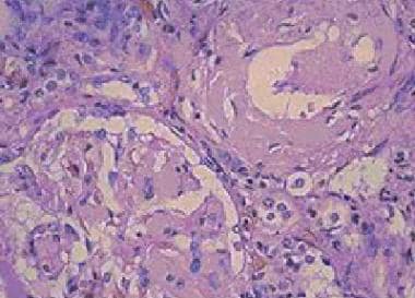
Impaired lysosomal processing of beta2-microglobulin by infiltrating macrophages in dialysis amyloidosis. (1/418)
BACKGROUND: Macrophages may participate in amyloid fibril formation by processing the protein precursor. Although this theory seems to apply for amyloidosis, in which proteolytic cleavage is a prerequisite for amyloid fibril formation, it has not been demonstrated for beta2-microglobulin (beta2m) amyloidosis. We aimed to establish the role played by macrophages in beta2m amyloidosis. METHODS: We used a double immunogold electron microscopy technique, including mouse antihuman CD68, rabbit antihuman beta2m, amyloid P component, and lysosome-associated membrane protein (LAMP-1) antibodies. Differential density labeling studies of beta2m and amyloid P component were performed extra- and intracellularly to assess protein processing by macrophages. RESULTS: The cells surrounding amyloid fibrils were found to be mostly CD68 positive, suggesting that they were of monocyte-macrophage lineage. Intracellular accumulation of amyloid fibrils was also observed; these fibrils were constantly surrounded by LAMP-1-linked gold particles, demonstrating that intracellular beta2m was almost exclusively lysosomal. The rough-surface endoplasmic reticulum was not labeled by beta2m antibody, suggesting that there was no active synthesis of beta2m by the cells. As a marker of endocytosis, protruded cytoplasmic processes in close relation with the intracellular accumulations of beta2m amyloid fibrils were observed. No difference in density labeling (extracellular vs. intracellular) was observed for beta2m, whereas intracellular P component labeling was significantly decreased. CONCLUSIONS: All of these data are strongly suggestive of phagocytosis and not synthesis of amyloid fibrils by macrophages. Further, they demonstrate an impaired lysosomal processing specific for beta2m, as other compounds of the amyloid fibrils (P component) are significantly cleared. (+info)Transthyretin Leu12Pro is associated with systemic, neuropathic and leptomeningeal amyloidosis. (2/418)
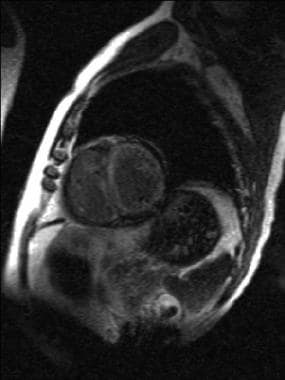
We report a middle-aged woman with a novel transthyretin (TTR) variant, Leu12Pro. She had extensive amyloid deposition in the leptomeninges and liver as well as the involvement of the heart and peripheral nervous system which characterizes familial amyloid polyneuropathy caused by variant TTR. Clinical features attributed to her leptomeningeal amyloid included radiculopathy, central hypoventilation, recurrent subarachnoid haemorrhage, depression, seizures and periods of decreased consciousness. MRI showed a marked enhancement throughout her meninges and ependyma, and TTR amyloid deposition was confirmed by meningeal biopsy. The simultaneous presence of extensive visceral amyloid and clinically significant deposits affecting both the peripheral and central nervous system extends the spectrum of amyloid-related disease associated with TTR mutations. The unusual association of severe peripheral neuropathy with symptoms of leptomeningeal amyloid indicates that leptomeningeal amyloidosis should be considered part of the syndrome of TTR-related familial amyloid polyneuropathy. (+info)Lipopolysaccharide (LPS)-binding synthetic peptides derived from serum amyloid P component neutralize LPS. (3/418)
Lipopolysaccharide (LPS) is the major mediator of gram-negative septic shock. Molecules that bind LPS and neutralize its toxic effects could have important clinical applications. We showed that serum amyloid P component (SAP) neutralizes LPS. A SAP-derived peptide, consisting of amino acids 27 to 39, inhibited LPS-mediated effects in the presence of human blood. In this study, we used a pepscan of overlapping 15-mer peptides and distinguished two additional LPS-binding regions within the SAP molecule, identified in the regions spanning amino acids 61 to 75 and 186 to 200. The corresponding SAP-derived peptides, pep61-75 and pep186-200, inhibited the binding of fluorescein isothiocyanate-labeled LPS to monocytes as efficiently as a bactericidal/permeability-increasing protein (BPI)-derived 15-mer peptide comprising amino acids 85 to 99. The same SAP-derived peptides very potently inhibited LPS-induced priming of phagocytes in human blood. Also, SAP-derived pep186-200 caused a prolonged survival of actinomycin D-sensitized mice treated with LPS to induce septic shock, indicating a potential use of this peptide in the defense against serious gram-negative sepsis in humans. (+info)Clinical, radiological and serum amyloid P component scintigraphic features of beta2-microglobulin amyloidosis associated with continuous ambulatory peritoneal dialysis. (4/418)
BACKGROUND: Beta2-Microglobulin (beta2M) amyloidosis occurs in patients with end-stage renal failure (ESRF) who undergo long-term continuous ambulatory peritoneal dialysis (CAPD), but its prevalence in patients treated exclusively by CAPD is unknown. In addition, its features may differ from those of haemodialysis-associated beta2M amyloidosis because CAPD is more biocompatible. METHODS: We performed serum amyloid P component (SAP) scintigraphy, a specific technique for imaging amyloid deposits, in 13 consecutive patients with ESRF who had been dialysed for >5 years, at least 80% of the time by CAPD. Clinical and radiological features of beta2M amyloidosis were sought and compared with the results of SAP scintigraphy. RESULTS: SAP scans showed articular amyloid deposits in seven patients, all of whom had evidence of carpal tunnel syndrome and four of whom had arthralgia characteristic of dialysis amyloidosis. Typical radiographic bone cysts were present in only one case who had been dialysed for >17 years. The remaining six patients had no clinical, radiological or scintigraphic evidence of beta2M amyloidosis. CONCLUSIONS: The prevalence of beta2M amyloidosis in this study was comparable with that in reported haemodialysis populations. Many of the amyloid deposits demonstrated by SAP scintigraphy were not associated with symptoms, but larger and longer term studies are required to determine whether CAPD favourably influences their clinical expression. (+info)Nurr1, an orphan nuclear receptor, is a transcriptional activator of endogenous tyrosine hydroxylase in neural progenitor cells derived from the adult brain. (5/418)
Adult rat-derived hippocampal progenitor cells express many of the molecules implicated in midbrain dopaminergic determination, including FGF receptors 1, 2 and 3, the sonic hedgehog receptor components Smo and Ptc, and the region-specific transcription factors Ptx3 and Nurr1. Here we use undifferentiated progenitors to probe the events leading to the dopaminergic phenotype and find that the influences of Nurr1 can be temporally and mechanistically uncoupled from the patterning influences of sonic hedgehog and FGF-8 or the more generic process of neuronal differentiation itself. In gain-of-function experiments, Nurr1 is able to activate transcription of the tyrosine hydroxylase gene by binding a response element within a region of the tyrosine hydroxylase promoter necessary for midbrain-specific expression. This activation is mediated through a retinoid X receptor independent mechanism and occurs in all precursors, regardless of differentiation status. Overexpression of Nurr1 does not affect proliferation or stimulate neuronal differentiation and has no influence on the expression of other dopaminergic markers. This uncoupling of tyrosine hydroxylase expression from other dopaminergic markers suggests that the midbrain dopaminergic identity is dictated by a combination of pan-dopaminergic (e.g., Shh/FGF-8) and region-specific (Nurr1) mechanisms. (+info)Hereditary renal amyloidosis associated with variant lysozyme in a large English family. (6/418)
BACKGROUND: Two kindreds with hereditary systemic amyloidosis caused by the first two mutations to be described in the human lysozyme gene were discovered recently and study of the variant lysozyme has been powerfully informative about mechanisms of amyloid fibrillogenesis. However, the clinical manifestations in these families, additional members of which have lately been identified, have not previously been reported in detail. METHODS: The proband presented with proteinuria aged 50 and a family history of amyloidosis, and underwent renal biopsy, whole-body serum amyloid P component (SAP) scintigraphy, and sequencing of the lysozyme gene. Her family history and the phenotype of hereditary lysozyme amyloidosis were thoroughly documented and compared with the presentation and natural history of all other known patients with this condition. RESULTS: The proband belonged to an extended English family other members of which were known to have hereditary lysozyme amyloidosis. Those with amyloid in previous generations presented with renal involvement, frequently developed complications due to gastrointestinal amyloid, and died before age 60. All amyloid deposits were composed of lysozyme and complete concordance was established between amyloid and heterozygosity for a point mutation in the lysozyme gene, encoding the previously reported Asp67His substitution in the mature protein. CONCLUSION: The phenotype, reported for the first time in this extended kindred, contrasts with that of an apparently unrelated family carrying the same mutation who presented with spontaneous hepatic haemorrhage and rupture, and with the manifestations in a family with the lysozyme Ile56Thr variant who presented with dermal petechiae before proceeding to fatal visceral amyloidosis. A remarkably wide spectrum of disease can be caused by the same amyloid fibril protein, although renal involvement predominates in all cases except those dying of hepatic rupture. (+info)Expression and production of the long pentraxin PTX3 in rheumatoid arthritis (RA). (7/418)
PTX3 is a secreted molecule which consists of a C-terminal domain similar to classical pentraxins (e.g. C-reactive protein (CRP)) and of an unrelated N-terminal domain. Unlike the classical pentraxins, the long pentraxin PTX3 is expressed in response to IL-1beta and tumour necrosis factor-alpha (TNF-alpha), but not to IL-6, in various cell types. The present study was designed to investigate the expression of PTX3 in RA. Dissociated RA and osteoarthritis (OA) type B synoviocytes were cultured in the presence and in the absence of inflammatory cytokines. PTX3 mRNA expression in synoviocytes was evaluated by Northern analysis. PTX3 protein levels in synovial cell cultures and synovial fluid were estimated by ELISA, and PTX3 distribution in synovial tissues by immunohistochemical techniques. OA synoviocytes were induced to express high levels of PTX3 mRNA by TNF-alpha, but not by other cytokines including IL-1beta and IL-6. RA synoviocytes, unlike OA synoviocytes, constitutively expressed high levels of PTX3 in the absence of deliberate stimulation. The constitutive expression of PTX3 in RA synoviocytes was not modified by anti-TNF-alpha antibodies, IL-1 receptor antagonist or a combination of the two agents. In contrast, interferon-gamma and transforming growth factor-beta inhibited PTX3 constitutive expression in RA synoviocytes. The joint fluid from RA patients contained higher levels of immunoreactive PTX3 than controls and the synovial tissue contained endothelial cells and synoviocytes positive for PTX3 by immunohistochemistry. In conclusion, PTX3 may play a role in inflammatory circuits of RA, and its relevance as a marker of disease activity deserves further study. (+info)Calumenin interacts with serum amyloid P component. (8/418)
We recently reported the identification of human calumenin, a novel Ca(2+) binding, transformation-sensitive and secreted protein [Vorum et al. (1998) Biochim. Biophys. Acta 1386, 121-131; Vorum et al. (1999) Exp. Cell Res. 248, 473-481] belonging to the family of multiple EF-hand proteins of the secretory pathway that include reticulocalbin, ERC-55, Cab45 and crocalbin. In order to further investigate the extracellular functions of calumenin we immobilized the recombinant protein to a column. After application of a placental tissue extract we were able to elute one protein that interacts with calumenin in the presence of Ca(2+). Amino acid sequencing identified this protein as serum amyloid P component (SAP). Furthermore, we verified and characterized the calumenin-SAP interaction by the surface plasmon resonance technique. The findings indicate that calumenin may participate in the immunological defense system and could be involved in the pathological process of amyloidosis that leads to formation of amyloid deposits seen in different types of tissues. (+info)Serum Amyloid P-component (SAP) is a protein that is normally present in the blood and other bodily fluids. It is a part of the larger family of pentraxin proteins, which are involved in the innate immune response, meaning they provide immediate defense against foreign invaders without needing to adapt over time. SAP plays a role in inflammation, immune complex clearance, and complement activation.
In the context of amyloidosis, SAP binds to misfolded proteins called amyloid fibrils, which can deposit in various tissues and organs, leading to their dysfunction and failure. The accumulation of these amyloid fibrils with SAP is a hallmark of systemic amyloidosis.
It's important to note that while SAP plays a role in the pathogenesis of amyloidosis, it is not directly responsible for causing the disease. Instead, its presence can serve as a useful marker for diagnosing and monitoring the progression of amyloidosis.
Serum Amyloid A (SAA) protein is an acute phase protein produced primarily in the liver, although it can also be produced by other cells in response to inflammation. It is a member of the apolipoprotein family and is found in high-density lipoproteins (HDL) in the blood. SAA protein levels increase rapidly during the acute phase response to infection, trauma, or tissue damage, making it a useful biomarker for inflammation.
In addition to its role as an acute phase protein, SAA has been implicated in several disease processes, including atherosclerosis and amyloidosis. In amyloidosis, SAA can form insoluble fibrils that deposit in various tissues, leading to organ dysfunction. There are four subtypes of SAA in humans (SAA1, SAA2, SAA3, and SAA4), with SAA1 and SAA2 being the most responsive to inflammatory stimuli.
Amyloid is a term used in medicine to describe abnormally folded protein deposits that can accumulate in various tissues and organs of the body. These misfolded proteins can form aggregates known as amyloid fibrils, which have a characteristic beta-pleated sheet structure. Amyloid deposits can be composed of different types of proteins, depending on the specific disease associated with the deposit.
In some cases, amyloid deposits can cause damage to organs and tissues, leading to various clinical symptoms. Some examples of diseases associated with amyloidosis include Alzheimer's disease (where amyloid-beta protein accumulates in the brain), systemic amyloidosis (where amyloid fibrils deposit in various organs such as the heart, kidneys, and liver), and type 2 diabetes (where amyloid deposits form in the pancreas).
It's important to note that not all amyloid deposits are harmful or associated with disease. However, when they do cause problems, treatment typically involves managing the underlying condition that is leading to the abnormal protein accumulation.
Amyloidosis is a medical condition characterized by the abnormal accumulation of insoluble proteins called amyloid in various tissues and organs throughout the body. These misfolded protein deposits can disrupt the normal function of affected organs, leading to a range of symptoms depending on the location and extent of the amyloid deposition.
There are different types of amyloidosis, classified based on the specific proteins involved:
1. Primary (AL) Amyloidosis: This is the most common form, accounting for around 80% of cases. It results from the overproduction and misfolding of immunoglobulin light chains, typically by clonal plasma cells in the bone marrow. The amyloid deposits can affect various organs, including the heart, kidneys, liver, and nervous system.
2. Secondary (AA) Amyloidosis: This form is associated with chronic inflammatory diseases, such as rheumatoid arthritis, tuberculosis, or familial Mediterranean fever. The amyloid fibrils are composed of serum amyloid A protein (SAA), an acute-phase reactant produced during the inflammatory response. The kidneys are commonly affected in this type of amyloidosis.
3. Hereditary or Familial Amyloidosis: These forms are caused by genetic mutations that result in the production of abnormal proteins prone to misfolding and amyloid formation. Examples include transthyretin (TTR) amyloidosis, fibrinogen amyloidosis, and apolipoprotein AI amyloidosis. These forms can affect various organs, including the heart, nerves, and kidneys.
4. Dialysis-Related Amyloidosis: This form is seen in patients undergoing long-term dialysis for chronic kidney disease. The amyloid fibrils are composed of beta-2 microglobulin, a protein that accumulates due to impaired clearance during dialysis. The joints and bones are commonly affected in this type of amyloidosis.
The diagnosis of amyloidosis typically involves a combination of clinical evaluation, imaging studies, and tissue biopsy with the demonstration of amyloid deposition using special stains (e.g., Congo red). Treatment depends on the specific type and extent of organ involvement and may include supportive care, medications to target the underlying cause (e.g., chemotherapy, immunomodulatory agents), and organ transplantation in some cases.
I'm sorry for any confusion, but "Methylgalactosides" is not a recognized term in medical or biological nomenclature. It appears that the term may be a combination of "methyl," a common chemical modification, and "galactosides," which are compounds containing galactose, a type of sugar. However, without more context, it's difficult to provide an accurate definition or medical relevance.
If you meant a different term or need information on a related topic, please provide more details so I can give a more helpful response.
C-reactive protein (CRP) is a protein produced by the liver in response to inflammation or infection in the body. It is named after its ability to bind to the C-polysaccharide of pneumococcus, a type of bacteria. CRP levels can be measured with a simple blood test and are often used as a marker of inflammation or infection. Elevated CRP levels may indicate a variety of conditions, including infections, tissue damage, and chronic diseases such as rheumatoid arthritis and cancer. However, it is important to note that CRP is not specific to any particular condition, so additional tests are usually needed to make a definitive diagnosis.
The acute-phase reaction is a complex series of physiological responses that occur in response to tissue injury, infection, or stress. It is characterized by the release of pro-inflammatory cytokines such as interleukin-1 (IL-1), IL-6, and tumor necrosis factor-alpha (TNF-α) from activated immune cells, including macrophages and neutrophils.
These cytokines trigger a range of systemic effects, including fever, increased heart rate and respiratory rate, decreased appetite, and changes in white blood cell count. They also stimulate the production of acute-phase proteins (APPs) by the liver, such as C-reactive protein (CRP), fibrinogen, and serum amyloid A.
The acute-phase reaction is an important part of the body's immune response to injury or infection, helping to promote healing and fight off pathogens. However, excessive or prolonged activation of the acute-phase reaction can contribute to the development of chronic inflammatory conditions and diseases such as rheumatoid arthritis, atherosclerosis, and cancer.
Prealbumin, also known as transthyretin, is a protein produced primarily in the liver and circulates in the blood. It plays a role in transporting thyroid hormones and vitamin A throughout the body. Prealbumin levels are often used as an indicator of nutritional status and liver function. Low prealbumin levels may suggest malnutrition or inflammation, while increased levels can be seen in certain conditions like hyperthyroidism. It is important to note that prealbumin levels should be interpreted in conjunction with other clinical findings and laboratory tests for a more accurate assessment of a patient's health status.
Iodine radioisotopes are radioactive isotopes of the element iodine, which decays and emits radiation in the form of gamma rays. Some commonly used iodine radioisotopes include I-123, I-125, I-131. These radioisotopes have various medical applications such as in diagnostic imaging, therapy for thyroid disorders, and cancer treatment.
For example, I-131 is commonly used to treat hyperthyroidism and differentiated thyroid cancer due to its ability to destroy thyroid tissue. On the other hand, I-123 is often used in nuclear medicine scans of the thyroid gland because it emits gamma rays that can be detected by a gamma camera, allowing for detailed images of the gland's structure and function.
It is important to note that handling and administering radioisotopes require specialized training and safety precautions due to their radiation-emitting properties.
Protein binding, in the context of medical and biological sciences, refers to the interaction between a protein and another molecule (known as the ligand) that results in a stable complex. This process is often reversible and can be influenced by various factors such as pH, temperature, and concentration of the involved molecules.
In clinical chemistry, protein binding is particularly important when it comes to drugs, as many of them bind to proteins (especially albumin) in the bloodstream. The degree of protein binding can affect a drug's distribution, metabolism, and excretion, which in turn influence its therapeutic effectiveness and potential side effects.
Protein-bound drugs may be less available for interaction with their target tissues, as only the unbound or "free" fraction of the drug is active. Therefore, understanding protein binding can help optimize dosing regimens and minimize adverse reactions.
"Competitive binding" is a term used in pharmacology and biochemistry to describe the behavior of two or more molecules (ligands) competing for the same binding site on a target protein or receptor. In this context, "binding" refers to the physical interaction between a ligand and its target.
When a ligand binds to a receptor, it can alter the receptor's function, either activating or inhibiting it. If multiple ligands compete for the same binding site, they will compete to bind to the receptor. The ability of each ligand to bind to the receptor is influenced by its affinity for the receptor, which is a measure of how strongly and specifically the ligand binds to the receptor.
In competitive binding, if one ligand is present in high concentrations, it can prevent other ligands with lower affinity from binding to the receptor. This is because the higher-affinity ligand will have a greater probability of occupying the binding site and blocking access to the other ligands. The competition between ligands can be described mathematically using equations such as the Langmuir isotherm, which describes the relationship between the concentration of ligand and the fraction of receptors that are occupied by the ligand.
Competitive binding is an important concept in drug development, as it can be used to predict how different drugs will interact with their targets and how they may affect each other's activity. By understanding the competitive binding properties of a drug, researchers can optimize its dosage and delivery to maximize its therapeutic effect while minimizing unwanted side effects.
Transgenic mice are genetically modified rodents that have incorporated foreign DNA (exogenous DNA) into their own genome. This is typically done through the use of recombinant DNA technology, where a specific gene or genetic sequence of interest is isolated and then introduced into the mouse embryo. The resulting transgenic mice can then express the protein encoded by the foreign gene, allowing researchers to study its function in a living organism.
The process of creating transgenic mice usually involves microinjecting the exogenous DNA into the pronucleus of a fertilized egg, which is then implanted into a surrogate mother. The offspring that result from this procedure are screened for the presence of the foreign DNA, and those that carry the desired genetic modification are used to establish a transgenic mouse line.
Transgenic mice have been widely used in biomedical research to model human diseases, study gene function, and test new therapies. They provide a valuable tool for understanding complex biological processes and developing new treatments for a variety of medical conditions.
Turpentine, also known as oil of turpentine, is not a medical term itself but a substance that has been used in some traditional medical preparations. It is a volatile essential oil obtained by the distillation of resin from live trees, mainly pines.
Medically, it has been used as a counterirritant and rubefacient (a substance that causes redness of the skin and increases blood flow) in liniments and plasters. However, its use in modern medicine is not very common due to potential toxicity and irritation. It's important to note that turpentine should not be ingested or used topically without proper medical supervision.
C57BL/6 (C57 Black 6) is an inbred strain of laboratory mouse that is widely used in biomedical research. The term "inbred" refers to a strain of animals where matings have been carried out between siblings or other closely related individuals for many generations, resulting in a population that is highly homozygous at most genetic loci.
The C57BL/6 strain was established in 1920 by crossing a female mouse from the dilute brown (DBA) strain with a male mouse from the black strain. The resulting offspring were then interbred for many generations to create the inbred C57BL/6 strain.
C57BL/6 mice are known for their robust health, longevity, and ease of handling, making them a popular choice for researchers. They have been used in a wide range of biomedical research areas, including studies of cancer, immunology, neuroscience, cardiovascular disease, and metabolism.
One of the most notable features of the C57BL/6 strain is its sensitivity to certain genetic modifications, such as the introduction of mutations that lead to obesity or impaired glucose tolerance. This has made it a valuable tool for studying the genetic basis of complex diseases and traits.
Overall, the C57BL/6 inbred mouse strain is an important model organism in biomedical research, providing a valuable resource for understanding the genetic and molecular mechanisms underlying human health and disease.
An amino acid sequence is the specific order of amino acids in a protein or peptide molecule, formed by the linking of the amino group (-NH2) of one amino acid to the carboxyl group (-COOH) of another amino acid through a peptide bond. The sequence is determined by the genetic code and is unique to each type of protein or peptide. It plays a crucial role in determining the three-dimensional structure and function of proteins.
Molecular sequence data refers to the specific arrangement of molecules, most commonly nucleotides in DNA or RNA, or amino acids in proteins, that make up a biological macromolecule. This data is generated through laboratory techniques such as sequencing, and provides information about the exact order of the constituent molecules. This data is crucial in various fields of biology, including genetics, evolution, and molecular biology, allowing for comparisons between different organisms, identification of genetic variations, and studies of gene function and regulation.
Amyloid beta-peptides (Aβ) are small protein fragments that are crucially involved in the pathogenesis of Alzheimer's disease. They are derived from a larger transmembrane protein called the amyloid precursor protein (APP) through a series of proteolytic cleavage events.
The two primary forms of Aβ peptides are Aβ40 and Aβ42, which differ in length by two amino acids. While both forms can be harmful, Aβ42 is more prone to aggregation and is considered to be the more pathogenic form. These peptides have the tendency to misfold and accumulate into oligomers, fibrils, and eventually insoluble plaques that deposit in various areas of the brain, most notably the cerebral cortex and hippocampus.
The accumulation of Aβ peptides is believed to initiate a cascade of events leading to neuroinflammation, oxidative stress, synaptic dysfunction, and neuronal death, which are all hallmarks of Alzheimer's disease. Although the exact role of Aβ in the onset and progression of Alzheimer's is still under investigation, it is widely accepted that they play a central part in the development of this debilitating neurodegenerative disorder.
Familial amyloid neuropathies are a group of inherited disorders characterized by the accumulation of abnormal deposits of amyloid proteins in various tissues and organs of the body. These abnormal deposits can cause damage to nerves, leading to a peripheral neuropathy that affects sensation, movement, and organ function.
There are several types of familial amyloid neuropathies, each caused by different genetic mutations. The most common type is known as transthyretin-related hereditary amyloidosis (TTR-HA), which is caused by mutations in the TTR gene. Other types include apolipoprotein A1-related hereditary amyloidosis (APOA1-HA) and gelsolin-related amyloidosis (AGel-HA).
Symptoms of familial amyloid neuropathies can vary depending on the type and severity of the disorder. Common symptoms include:
* Numbness, tingling, or pain in the hands and feet
* Weakness or loss of muscle strength in the legs and arms
* Autonomic nervous system dysfunction, leading to problems with digestion, heart rate, blood pressure, and temperature regulation
* Carpal tunnel syndrome
* Eye abnormalities, such as vitreous opacities or retinal deposits
* Kidney disease
Familial amyloid neuropathies are typically inherited in an autosomal dominant manner, meaning that a child has a 50% chance of inheriting the mutated gene from an affected parent. Diagnosis is usually made through genetic testing and confirmation of the presence of amyloid deposits in tissue samples.
Treatment for familial amyloid neuropathies typically involves managing symptoms and slowing the progression of the disease. This may include medications to control pain, physical therapy to maintain muscle strength and mobility, and devices such as braces or wheelchairs to assist with mobility. In some cases, liver transplantation may be recommended to remove the source of the mutated transthyretin protein.
Acute-phase proteins (APPs) are a group of plasma proteins whose concentrations change in response to various inflammatory conditions, such as infection, trauma, or tissue damage. They play crucial roles in the body's defense mechanisms and help mediate the innate immune response during the acute phase of an injury or illness.
There are several types of APPs, including:
1. C-reactive protein (CRP): Produced by the liver, CRP is one of the most sensitive markers of inflammation and increases rapidly in response to various stimuli, such as bacterial infections or tissue damage.
2. Serum amyloid A (SAA): Another liver-derived protein, SAA is involved in lipid metabolism and immune regulation. Its concentration rises quickly during the acute phase of inflammation.
3. Fibrinogen: A coagulation factor produced by the liver, fibrinogen plays a vital role in blood clotting and wound healing. Its levels increase during inflammation.
4. Haptoglobin: This protein binds free hemoglobin released from red blood cells, preventing oxidative damage to tissues. Its concentration rises during the acute phase of inflammation.
5. Alpha-1 antitrypsin (AAT): A protease inhibitor produced by the liver, AAT helps regulate the activity of enzymes involved in tissue breakdown and repair. Its levels increase during inflammation to protect tissues from excessive proteolysis.
6. Ceruloplasmin: This copper-containing protein is involved in iron metabolism and antioxidant defense. Its concentration rises during the acute phase of inflammation.
7. Ferritin: A protein responsible for storing iron, ferritin levels increase during inflammation as part of the body's response to infection or tissue damage.
These proteins have diagnostic and prognostic value in various clinical settings, such as monitoring disease activity, assessing treatment responses, and predicting outcomes in patients with infectious, autoimmune, or inflammatory conditions.
Rheumatology is a subspecialty of internal medicine that deals with the diagnosis and management of more than 200 diseases affecting the joints, muscles, and bones. These diseases are often complex, chronic, and systemic, meaning they can affect the whole body. Some common rheumatic diseases include rheumatoid arthritis, osteoarthritis, lupus, gout, osteoporosis, and various forms of vasculitis and connective tissue disorders.
Rheumatologists are medical doctors who have completed additional training in this field, becoming experts in the non-surgical treatment of musculoskeletal diseases. They use a combination of physical examination, patient history, laboratory testing, and imaging to diagnose and manage these conditions. Treatment may involve medications, lifestyle changes, physical therapy, or a combination of these approaches.
I'm sorry for any confusion, but "Lions" is not a term typically used in medical definitions. Lions are large, carnivorous mammals that are part of the Felidae family, specifically Panthera leo. If you have any questions about lions or if there's another term you meant to ask about, please provide more context so I can give you a more accurate response.
Optical illusions are visual phenomena that occur when the brain perceives an image or scene differently from the actual physical properties of that image or scene. They often result from the brain's attempt to interpret and make sense of ambiguous, contradictory, or incomplete information provided by the eyes. This can lead to visually perceived images that are different from the objective reality. Optical illusions can be categorized into different types such as literal illusions, physiological illusions, and cognitive illusions, based on the nature of the illusion and the underlying cause.
The midline thalamic nuclei are a group of nuclei located in the thalamus, which is a part of the diencephalon in the brain. The thalamus serves as a relay station for sensory and motor signals to the cerebral cortex. The midline thalamic nuclei are situated in the most medial portion of the thalamus, along the midline. They include several distinct nuclei, such as the paraventricular nucleus, the reuniens nucleus, the rhomboid nucleus, and the central medial nucleus. These nuclei have complex connections with various brain regions, including the hypothalamus, the hippocampus, and the prefrontal cortex. They are involved in a variety of functions, such as memory, emotion, and sleep regulation.
"Phleum" is the genus name for a group of plants commonly known as Timothy-grass or Cat's-tail. It is a type of grass that is widely used in agriculture and gardening. I believe you might be looking for a medical term related to "phleum," so let me clarify:
In medical terminology, the term "phleum" is not commonly used. However, if you are referring to "phlebothrombosis," it is a term that could be relevant. Phlebothrombosis refers to the formation of a blood clot (thrombus) within a vein, which can occur due to various medical conditions or situations, such as immobility, surgery, or certain diseases. The term "phlebo-" means vein, and "-thrombosis" refers to the formation of a thrombus or blood clot.
If this is not the term you were looking for, please provide more context or clarify your question so I can give you a more accurate answer.
A "periodical" in the context of medicine typically refers to a type of publication that is issued regularly, such as on a monthly or quarterly basis. These publications include peer-reviewed journals, magazines, and newsletters that focus on medical research, education, and practice. They may contain original research articles, review articles, case reports, editorials, letters to the editor, and other types of content related to medical science and clinical practice.
As a "Topic," periodicals in medicine encompass various aspects such as their role in disseminating new knowledge, their impact on clinical decision-making, their quality control measures, and their ethical considerations. Medical periodicals serve as a crucial resource for healthcare professionals, researchers, students, and other stakeholders to stay updated on the latest developments in their field and to share their findings with others.
The Journal Impact Factor (JIF) is a measure of the frequency with which the "average article" in a journal has been cited in a particular year. It is calculated by dividing the number of current year citations to the source items published in that journal during the previous two years. For example, if a journal has an Impact Factor of 3 in 2020, that means articles published in 2018 and 2019 were cited 3 times on average in 2020. It is used to gauge the importance or rank of a journal by comparing the times it's articles are cited relative to other journals in the field. However, it has been criticized for various limitations such as being manipulated by editors and not reflecting the quality of individual articles.
Amyloidosis30
- Amyloid and amyloidosis: proceedings of the Third International Symposium on Amyloidosis, Póvoa de Varzim, Portugal, 23-28 September 1979. (wikipedia.org)
- Serum amyloid P component prevents proteolysis of the amyloid fibrils of Alzheimer disease and systemic amyloidosis" (PDF). (wikipedia.org)
- Immunochemical studies on the nature of the serum component (SAA) related to secondary amyloidosis. (nih.gov)
- Pharmacodynamic evaluation and safety assessment of treatment with antibodies to serum amyloid P component in patients with cardiac amyloidosis: an open-label Phase 2 study and an adjunctive immuno-PET imaging study. (ox.ac.uk)
- BACKGROUND: In a Phase I study treatment with the serum amyloid P component (SAP) depleter miridesap followed by monoclonal antibody to SAP (dezamizumab) showed removal of amyloid from liver, spleen and kidney in patients with systemic amyloidosis. (ox.ac.uk)
- CONCLUSIONS: Unlike previous observations of visceral amyloid reduction, there was no appreciable evidence of amyloid removal in patients with cardiac amyloidosis in this Phase 2 trial, potentially related to limited cardiac uptake of dezamizumab as demonstrated in the immuno-PET study. (ox.ac.uk)
- Amyloidosis results from the accumulation of pathogenic amyloids-most of which are aggregates of misfolded proteins-in a variety of tissues. (medscape.com)
- Amyloidosis is a clinical disorder caused by extracellular and/or intracellular deposition of insoluble abnormal amyloid fibrils that alter the normal function of tissues. (medscape.com)
- Only 10% of amyloidosis deposits consist of components such as glycosaminoglycans (GAGs), apolipoprotein-E (apoE), and serum amyloid P-component (SAP), while nearly 90% of the deposits consist of amyloid fibrils that are formed by the aggregation of misfolded proteins. (medscape.com)
- The modern era of amyloidosis classification began in the late 1960s with the development of methods to solubilize amyloid fibrils. (medscape.com)
- Approximately 90% of the deposits consist of amyloid fibrils formed by the aggregation of misfolded proteins, and 10% of the deposits consist of glycosaminoglycans, apolipoprotein E, and serum amyloid P. Amyloidosis is found as a background change in some strains, such as CD-1 mice. (nih.gov)
- Pulmonary amyloidosis is a spontaneous lesion in aging rodents and may be localized to the lung, or it may be a component of systemic amyloidosis involving spleen, kidney, liver, and other organs. (nih.gov)
- 5. Serum amyloid P component: a predictor of clinical beta 2-microglobulin amyloidosis. (nih.gov)
- 8. Plasma kinetics of 125I-labelled amyloid P component in beta 2M amyloidosis: a possible approach to quantitate disease activity. (nih.gov)
- Laboratory studies, such as a complete blood cell count, serum chemistry profile, and liver function tests, often were part of a general workup in several case reports of patients with nodular localized cutaneous amyloidosis. (medscape.com)
- The amyloid in nodular localized cutaneous amyloidosis is located in the reticular dermis and subcutaneous fat, and this finding clearly differentiates nodular localized cutaneous amyloidosis from other forms of amyloidosis. (medscape.com)
- In nodular amyloidosis, amyloid is not limited to the papillary dermis but is present in the entire dermis and may extend to subcutaneous fat. (medscape.com)
- When eosinophilic amyloid material is exposed to potassium permanganate prior to staining with Congo red, the amyloid retains its congophilia, similar to systemic amyloidosis but in contradistinction to secondary amyloidosis. (medscape.com)
- Before the discovery that the major fibril component in these patients was an immunoglobulin fragment, patients with light chain-type amyloidosis were described as having primary amyloidosis (primary in the sense of idiopathic) or, when the burden of monoclonal plasma cells was large, myeloma-associated amyloidosis. (medscape.com)
- OBJECTIVES: To investigate equilibrium contrast-enhanced CT (EQ-CT) measurement of extracellular volume fraction (ECV) in patients with systemic amyloid light-chain (AL) amyloidosis, testing the hypothesis that ECV becomes elevated in the liver and spleen and ECV correlates with other estimates of organ amyloid burden. (ucl.ac.uk)
- CONCLUSION: In patients with systemic AL amyloidosis, EQ-CT can demonstrate increased spleen and liver ECV, which is associated with amyloid disease burden. (ucl.ac.uk)
- The old nomenclature of "primary" and "secondary" is no longer used, although AA amyloidosis, involving formation of fibrils from serum amyloid A (SAA) protein, is truly secondary to chronic inflammatory or infectious states, as SAA is an acute phase reactant. (cancertherapyadvisor.com)
- The diagnosis of amyloidosis rests upon three legs: proving there are amyloid fibrils in tissues, determining the protein precursor forming the fibrils, and assessing organ involvement. (cancertherapyadvisor.com)
- Wild-type and variant forms of transthyretin (TTR), a normal plasma protein, are amyloidogenic and can be deposited in the tissues as amyloid fibrils causing acquired and hereditary systemic TTR amyloidosis, a debilitating and usually fatal disease. (port.ac.uk)
- Reduction in the abundance of amyloid fibril precursor proteins arrests amyloid deposition and halts disease progression in all forms of amyloidosis including TTR type. (port.ac.uk)
- Maury CP, Wegelius O. Clinical value of serum amyloid A and C-reactive protein measurements in secondary amyloidosis. (ijbm.org)
- Amyloid fibrils are organized within an antiparallel conformation with a ?pleated sheet structure.1C3 It is suggested that amyloid and amyloidosis ought to be classified by the fibrillar proteins forming the amyloid deposits. (biopaqc.com)
- Table 1 ?Main protein types causing amyloidosis with the emphasis on cardiovascular system involvement Nomenclature of amyloid fibril proteins. (biopaqc.com)
- Husby G, Stenstad T, Magnus JH, Interaction between circulating amyloid fibril protein precursors and extracellular tissue matrix components in the pathogenesis of systemic amyloidosis. (biopaqc.com)
- Kawahara E, Shiroo M, Nakanishi I, The role of fibronectin in the development of experimental amyloidosis: evidence of immunohistochemical codistribution and binding house with serum amyloid protein A. Am J Pathol 1989;134:1305C14. (biopaqc.com)
Protein15
- This association is utilised in the routine clinical diagnostic technique of SAP scintigraphy whereby radio-labelled protein is injected into patients to locate areas of amyloid deposition. (wikipedia.org)
- Amyloid fibrils are protein polymers comprising identical monomer units (homopolymers). (medscape.com)
- All types of amyloid consist of one major fibrillar protein that defines the type of amyloid. (medscape.com)
- Polymorphisms that slightly vary native peptides or inflammatory processes set the stage for abnormal protein folding and amyloid fibril deposition. (medscape.com)
- Normal serum protein electrophoresis and urine protein electrophoresis studies help to exclude multiple myeloma, which can also cause amyloid deposits made up of immunoglobulin light chains. (medscape.com)
- Amyloid deposits in the skin also contain small amounts of a plasma-derived, nonfibrillar, amyloid-P protein. (medscape.com)
- C-reactive protein,mannose-binding lectin, and serum amyloid P component activate complement and act as opsonins. (msdmanuals.com)
- Within the V region families, certain amino acid residues occurring at particular positions in the L-chain sequence render those chains more amyloidogenic, with a combination of such residues increasing the chances of a particular L-chain protein being associated with tissue amyloid deposition. (medscape.com)
- The binding of the encoded protein to proteins in the pathological amyloid cross-beta fold suggests its possible role as a chaperone. (nih.gov)
- If it is familial (AF), the precursor protein can be an inherited mutant serum protein such as transthyretin, abbreviated ATTR, or proteins such as lysozyme, fibrinogen, gelsolin, or apolipoproteins. (cancertherapyadvisor.com)
- It interacts also with anticoagulant protein S and with serum amyloid P component. (cusabio.com)
- Human serum amyloid A (SAA) protein: a prominent acute-phase reactant for clinical prac-tice. (ijbm.org)
- The major acute phase reactants: C-reactive protein, serum amyloid P component and serum amyloid A protein. (ijbm.org)
- Researchers observing neurons found substantially less accumulation of beta-amyloid, neurofibrillary tangle, tau protein, and other "neuronal garbage" associated with Alzheimer's when those neurons were exposed to "physiologic quantities" of either estrogen or testosterone (depending on whether the neuron was from a woman or a man). (anh-usa.org)
- The researchers measured levels of C-reactive protein , serum amyloid A , interleukin-6 , leukocyte and neutrophil in 439 women. (fightaging.org)
Deposition11
- Deposition of amyloid can be focal, multifocal, or diffuse. (nih.gov)
- Since an association between amyloid deposition in the lung and xenobiotic administration has not been reported, amyloid deposition is generally considered a background finding. (nih.gov)
- There is perivascular and peribronchiolar amyloid deposition (arrows). (nih.gov)
- There is deposition of amyloid in the vascular wall (arrows). (nih.gov)
- Amyloid deposition may be particularly prominent in walls of small blood vessels and surrounding individual lipocytes (see the images). (medscape.com)
- However, in some patients, the physicochemical characteristics of the immunoglobulin L chain or L-chain fragment lead to its deposition as amyloid. (medscape.com)
- Clonal plasma cell proliferative diseases in which the V l 6 gene is expressed are always associated with amyloid deposition. (medscape.com)
- Another structural feature that appears to predispose to L chain - type amyloid deposition is enzymatic glycosylation of the L chain. (medscape.com)
- Mann-Whitney U test was used to test for a difference between patients with amyloid deposition (SAP grade 1-3) and those without (SAP grade 0). (ucl.ac.uk)
- these proteins is essential to the forming of amyloid, its sites of deposition, and the scientific symptoms. (biopaqc.com)
- Lyon AW, Narindrasorasak S, Young ID, Co-deposition of basement membrane components during the induction of murine splenic AA amyloid. (biopaqc.com)
Proteins3
- [ 5 ] In humans, about 23 different unrelated proteins are known to form amyloid fibrils in vivo. (medscape.com)
- Serum amyloid A and alpha-1 acid glycoprotein are transport proteins, and fibrinogen is a coagulation factor. (msdmanuals.com)
- In addition to cases of monoclonal gammopathy in which the secreted clonal immunoglobulin remains in solution and those in which secreted clonal immunoglobulin forms amyloid deposits, a third group consists of cases in which the monoclonal proteins accumulate in various organs, but the deposits do not form fibrils. (medscape.com)
APCS1
- APCS, a secreted glycoprotein found in serum and urine, interacts with DNA and histones and may scavenge nuclear material released from damaged circulating cells. (nih.gov)
Fibrils2
- All amyloid fibrils appear the same by light and electron microscopy. (cancertherapyadvisor.com)
- While routine immunohistochemistry or immunofluorescence can identify the origin of the fibrils, immunostaining of amyloid is fraught with a high rate of false positive and false negative testing. (cancertherapyadvisor.com)
Complement6
- The complement system, a major component of the innate immune system, is becoming increasingly recognised as a key participant in physiology and disease. (biomedcentral.com)
- Complement component 1q (C1q) is the recognition molecule of the classical pathway of the complement system that can bind to an array of closely spaced antigen-bound immunoglobulin G (IgG) and IgM antibodies. (explorationpub.com)
- Complement component 1q (C1q) is the first recognition subcomponent of the complement (C) classical pathway [ 1 ]. (explorationpub.com)
- In addition, several potential biomarkers-such as fibrinogen, fibronectin, complement C1r subcomponent and serum amyloid P-component-were shown to have a diagnostic feature presenting an area under the curve (AUC) of almost 1. (thehealthcoach1.com)
- It is suggested to perform IF-P in cases with a predominant membranous pattern when IF-F shows negative or weak staining for immunoglobulins and complement components. (ajkdblog.org)
- Preclinical studies show that LPS-activated microglia enable the conversion of trophic to A1 astrocytes by releasing several cytokines, including tumor necrosis factor alpha (TNF-alpha), interlukin-1 alpha (IL-1 alpha), and complement component C1q. (frontiersin.org)
Extracellular1
- During the course of a viral infection, host cells release exosomes and other extracellular vesicles carrying viral and host components that can modulate the immune response. (thehealthcoach1.com)
Scintigraphy2
- Screening for amyloid within organs can be accomplished using scintigraphy with radioiodinated serum amyloid P component (ie, SAP scanning). (medscape.com)
- Patients also underwent serum amyloid P (SAP) component scintigraphy with grading of liver and spleen involvement. (ucl.ac.uk)
Eosinophilic2
- Amorphous eosinophilic interstitial amyloid observed on a renal biopsy. (medscape.com)
- Acellular, eosinophilic material (amyloid) is present in the vessel wall (arrow). (nih.gov)
Antibodies4
- Immunotherapeutic clearance of systemic amyloid deposits by antibodies to serum amyloid P component. (sens.org)
- The Are There Igm Monoclonal Antibodies reagent is RUO (Research Use Only) to test human serum or cell culture lab samples. (chipgrade.com)
- Description: A competitive ELISA for quantitative measurement of Human Platelet antibodies IgG in samples from blood, plasma, serum, cell culture supernatant and other biological fluids. (chipgrade.com)
- Another option is the application of monoclonal antibodies against human serum amyloid P component. (worldneurologyonline.com)
Antibody1
- All of his Immunology and Virus, Epstein-Barr virus, Nivolumab, Antibody and Serum amyloid A investigations are sub-components of the entire Immunology study. (research.com)
Glycoprotein found1
- Amyloid P component is a small, non-fibrillar glycoprotein found in normal serum and in all amyloid deposits. (nih.gov)
Reactants1
- Recently, we analyzed serum biomarkers by using samples from the Gulu outbreak and identified associations between cytokines/chemokines, acute-phase reactants, makers of coagulopathy, and markers of endothelial function and patient death, hemorrhage, and viremia ( 10 ). (cdc.gov)
19991
- Amyloid 1999;6:63C6. (biopaqc.com)
Plasma4
- Plasma cells, which most likely produce the amyloid, occur within an adjacent and intermingled inflammatory infiltrate. (medscape.com)
- This ELISA kit applies to the in vitro quantitative determination of Rat Aβ1-40 concentrations in Serum, plasma and other biological fluids. (caslab.com)
- The aim of this study was to investigate the effect of combined exercise on resting levels of pentraxin3 (PTX3) and serum amyloid A (SAA) in the plasma of men with type-2 diabetes. (ac.ir)
- human serum, human plasma, and human whole blood samples. (rapidtestskits.com)
Nomenclature1
- 2007. A primer of amyloid nomenclature. (nih.gov)
Clinical3
- The SAP-amyloid association has also been identified as a possible drug target for anti-amyloid therapy, with the recent development and first stage clinical trials of a compound called CPHPC (R-1-[6-[R-2-carboxy-pyrrolidin-1-yl]-6-oxohexanoyl] pyrrolidine-2-carboxylic acid), a small molecule able to strip AP from deposits by reducing levels of circulating SAP. (wikipedia.org)
- Various descriptive classification systems were proposed based on the organ distribution of amyloid deposits and clinical findings. (medscape.com)
- Yamada T. Serum amyloid A (SAA): a concise review of biology, assay methods and clinical usefulness. (ijbm.org)
20021
- Amyloid 2002;9:197C200. (biopaqc.com)
Isolation2
Spleen1
- ECV increased with increasing amyloid burden, showing positive correlation with SAP grade in both the liver (r = 0.758) and spleen (r = 0.867). (ucl.ac.uk)
Tissue2
- This transmission electron micrograph of amyloid deposited in the tissue shows loosely interwoven straight filaments. (medscape.com)
- They may also help components of the tissue bind to inorganic substances, such as calcium in bone . (wikidoc.org)
19831
- The 15-year Princeton men's study determined that men who had higher serum free testosterone in 1983 had less risk of Alzheimer's disease in 1998. (anh-usa.org)
Characteristic1
- Characteristic staining for serum amyloid P within the deposits has been shown. (ajkdblog.org)
Specimen1
- Congo red staining of a cardiac biopsy specimen containing amyloid, viewed under polarized light. (medscape.com)
Alzheimer's6
- BDNF levels are diminished in the brain and serum of Alzheimer's patients. (zrtlab.com)
- Alzheimer's patients treated for ten weeks with lithium showed a significant increase in their BDNF serum levels. (zrtlab.com)
- BDNF also contributes to degradation of amyloid-β, a component of the amyloid plaques found in the brains of Alzheimer's patients. (zrtlab.com)
- Higher serum estrogen levels in women in their 60s are directly correlated with lower incidence of Alzheimer's in those same women decades later. (anh-usa.org)
- Once again, the reverse was also true: Lower serum free testosterone corresponded with higher risk of Alzheimer's. (anh-usa.org)
- 4) The model of Alzheimer's disease on which the drug targets (e.g., amyloid-β peptide) have been based may be an inaccurate or incomplete model of the disease. (omicsonline.org)
Congo2
- The bright pink homogeneous-appearing material seen is amyloid stained with Congo red. (medscape.com)
- A distinguishing feature of amyloid in the skin is an affinity to take up Congo red stain. (medscape.com)
Peptide1
- Functional amyloids play a beneficial role in a variety of physiologic processes (eg, long-term memory formation, gradual release of stored peptide hormones). (medscape.com)
Neurons1
- Apoptosis is usually induced by -amyloid in cultured nervous system neurons. (biopaqc.com)
Inflammation2
- Strain dependence of serum amyloid P-component (SAP) levels and response to inflammation. (aai.org)
- They also found that exercise alone, without a dietary weight loss component, had little effect on inflammation markers. (fightaging.org)
Mice1
- Systemic senile amyloid in senescence-accelerated mice. (nih.gov)
Immunochemical1
- 17. [Amyloid deposits in hemodialysis patients: immunochemical analysis]. (nih.gov)
Secondary1
- Where has secondary amyloid gone? (ijbm.org)
Lung2
Patients7
- Screening the sera of 228 normal individuals and of 297 patients with a variety of illnesses, we found that SAA is a component of all human sera, including cord blood (mean 94 plus or minus 57 ng/ml). (nih.gov)
- Troponin: Elevated levels of troponin T have been seen in patients with familial amyloid polyneuropathy (FAP), systolic dysfunction, and left ventricular (LV) hypertrophy (LVH). (medscape.com)
- Amyloid in biopsies of the gastrointestinal tract-a retrospective observational study on 542 patients. (sens.org)
- We used a series of multiplex assays to measure the concentrations of 55 serum analytes in specimens from patients from that outbreak to identify biomarkers specific to pediatric disease. (cdc.gov)
- In this study, we used a series of multiplex assays to measure the concentrations of 55 serum analytes in specimens from patients from the Gulu outbreak to identify biomarkers that had age-specific associations with survival, hemorrhagic manifestations, or both. (cdc.gov)
- 11. [Amyloid arthropathy in hemodialysis patients]. (nih.gov)
- In NOMID patients, at a median SC dose of 3 mg/kg once daily and a median treatment time of 3.5 years, the median (range) steady-state serum exposure of anakinra was Cmax 3628 (655-8511) ng/mL (n=16) and C24h 203 (53-1979) ng/mL (n=16). (clustermed.info)
Kinetics1
- The kinetics and magnitude of the synergistic activation of the serum amyloid A promoter by IL-1 beta and IL-6 is determined by the order of cytokine addition. (ijbm.org)
Molecules1
- Our previous demonstration that circulating serum amyloid P component (SAP) is efficiently depleted by administration of a specific small molecule ligand compound, that non-covalently crosslinks pairs of SAP molecules, suggested that TTR may be also amenable to this approach. (port.ac.uk)
Human4
- Using the radioactively-labeled alkaline-degraded acid-soluble fraction of amyloid ([ 125I ]DAA), we developed a radioimmunoassay for the previously described amyloid-related component of the human serum (SAA). (nih.gov)
- Immortalized human meibomian gland epithelial cells, which were recently generated in our laboratory [ 2 ], were cultured in Keratinocyte Serum-Free Medium [KSFM] supplemented with 50 μg/ml bovine pituitary extract (BPE), 5 ng/ml epidermal growth factor (EGF), and 100 U penicillin-streptomycin (Invitrogen, Carlsbad, CA). Cells were incubated in a humidified, 37 °C chamber under 5% CO 2 /95% air. (molvis.org)
- Honors Project Entitled 'The Co-Crystallization of Human Serum Amyloid P Component (SAP) with Hairpin and Self-complementary Oligonucleotides. (exeter.ac.uk)
- Lipopolysaccharide signaling induces serum amyloid A (SAA) synthesis in human hepatocytes in vitro. (ijbm.org)
Inflammatory1
- Not only are the omega-3 fatty acids in fish anti-inflammatory, but they're also essential components of the membranes of every brain cell we have. (anh-usa.org)
Levels2
- The concentration of this component increases significantly with the aging process, reaching very high levels in the eighth and nine decades. (nih.gov)
- For instance, low serum vitamin D levels were linked to increased occurrence of high burdens viral diseases such as hepatitis, influenza, Covid-19, and AIDS. (mdpi.com)
Methods1
- These methods permitted chemical amyloid studies. (medscape.com)