Leydig Cell Tumor
Leydig Cells
Sertoli Cell Tumor
Testicular Neoplasms
Sertoli Cells
Sertoli-Leydig Cell Tumor
Testis
Seminoma
Diethylstilbestrol
Testosterone
Granulosa Cell Tumor
Giant Cell Tumor of Bone
Dog Diseases
Aromatase
Giant Cell Tumors
Neoplasms, Germ Cell and Embryonal
Neoplasms, Experimental
Granular Cell Tumor
Seminiferous Tubules
Immunohistochemistry
Spermatogenesis
Germinoma
Chorionic Gonadotropin
Receptors, LH
Luteinizing Hormone
Seminiferous Epithelium
Mucinous cystadenoma coexisting with stromal tumor with minor sex-cord elements of the ovary: a case report. (1/22)
Mucinous neoplasms occur rarely in association with cystic teratoma, Sertoli-Leydig cell tumor, granulosa cell tumor or carcinoid tumor. Several cases of an ovarian stromal tumor with minor sex-cord elements have been reported in the literatures. However, there has been no report about an ovarian mucinous neoplasm coexisting with a stromal tumor with sex-cord elements yet. We report a case of an ovarian neoplasm composed of both mucinous cystadenoma and stromal tumor with minor sex-cord elements in a 58-yr-old female. The ovary including the mass measured 5 cm in size. On section, it revealed an unilocular cyst (4.5 cm in diameter) filled with mucinous fluid. There was a round, yellow, solid nodule, 1.5 cm in diameter within the wall. Microscopically, the cyst was lined by a single layer of endocervical mucinous epithelium and the nodule was composed of spindle cells showing an intersecting and whorled arrangement. There were cell nests showing polygonal shape with abundant cytoplasm among the spindle cells. They showed immunoreactivity for inhibin and did not have any connection with the adjacent mucinous epithelium. Therefore, we interpret the mucinous cystadenoma as having arisen de novo. (+info)Effects of ketoconazole on progesterone and cAMP production in MA-10 mouse Leydig tumor cells. (2/22)
The effects of ketoconazole (KCZ) on secretion of progesterone and cAMP in Leydig cells were investigated in vitro. MA-10 mouse Leydig tumor cells were used to conduct the experiments. KCZ significantly inhibited the progesterone production from MA-10 cells in a dose dependent fashion between 2 and 20 microM among 1, 2 and 3 h of incubation. There was a statistically significant difference in luteinizing hormone (LH) stimulated progesterone production inhibited by 2 and 20 microm KCZ treatment compared to the control. The effect of KCZ on progesterone biosynthesis in MA-10 cells was mediated by cAMP, since KCZ suppressed basal and LH stimulated cAMP production and content within the same dose range. The stimulatory effects of forskolin and sodium fluoride on the adenylate cyclase system were also inhibited by KCZ. Moreover, dibutyryl cAMP blocked the inhibitory effect on steroidogenesis of KCZ in MA-10 cells. These data indicated that KCZ induced the inhibition of a catalytic component of adenylate cyclase holoenzyme in MA-10 mouse Leydig tumor cells. (+info)Galectin-1: biphasic growth regulation of Leydig tumor cells. (3/22)
Galectin-1 (Gal-1) is a widely expressed beta-galactoside-binding protein that exerts pleiotropic biological functions. To gain insight into the potential role of Gal-1 as a novel modulator of Leydig cells, we investigated its effect on the growth and death of MA-10 tumor Leydig cells. In this study, we identified cytoplasmic Gal-1 expression in these tumor cells by cytofluorometry. DNA fragmentation, caspase-3, -8, and -9 activation, loss of mitochondrial membrane potential (DeltaPsim), cytochrome c (Cyt c) release, and FasL expression suggested that relatively high concentrations of exogenously added recombinant Gal-1 (rGal-1) induced apoptosis by the mitochondrial and death receptor pathways. These pathways were independently activated, as the presence of the inhibitor of caspase-8 or -9 only partially prevented Gal-1-effect. On the contrary, low concentrations of Gal-1 significantly promoted cell proliferation, without inducing cell death. Importantly, the presence of the disaccharide lactose prevented Gal-1 effects, suggesting the involvement of the carbohydrate recognition domain (CRD). This study provides strong evidence that Gal-1 is a novel biphasic regulator of Leydig tumor cell number, suggesting a novel role for Gal-1 in the reproductive physiopathology. (+info)Morphological approach to tumours of the testis and paratestis. (4/22)
Most neoplastic scrotal masses ultimately prove to be germ cell tumours and are recognisable with routine haematoxylin and eosin-stained sections. The differential diagnosis may be focused, even before reviewing histological sections, by knowledge of patient age, medical history, tumour site (testicular vs paratesticular) and gross findings. Some cases may prove to be diagnostically challenging, including rare tumours, a common tumour with an unusual pattern, a metastatic tumour, or a neoplasm with features that mimic another tumour. Several morphological patterns are seen with some frequency and these generate recurring sets of differential diagnostic considerations. These common patterns include testicular tumours with a predominant diffuse arrangement of cells with pale to clear cytoplasm, tumours with a glandular/tubular pattern, tumours with a microcystic pattern and tumours composed of oxyphilic cells. Intratubular proliferations of atypical cells, paratesticular glandular and/or papillary tumours, or tumours with spindle cell morphology can also be challenging to diagnose correctly. In some problematic cases, immunohistochemical staining may be useful to resolve these differential diagnoses. (+info)Cervical embryonal rhabdomyosarcoma and ovarian Sertoli-Leydig cell tumour: a more than coincidental association of two rare neoplasms? (5/22)
A case in which an embryonal rhabdomyosarcoma of the cervix and an ovarian Sertoli-Leydig cell tumour of intermediate differentiation occurred in a 13-year-old girl is described. Although initially considered as a chance association, a review of the literature showed the co-occurrence of these two uncommon neoplasms in three previous cases. The reason for this association, which is thought to be more than coincidental, is not known, although an underlying genetic abnormality is a possibility. The ovarian tumour in this case was characterised by the presence of foci of cells with extremely pleomorphic nuclei, which initially raised the possibility of metastatic rhabdomyosarcoma. These were interpreted as foci of bizarre nuclei within the Sertoli-Leydig cell tumour. (+info)Imaging of gynecological disease (2): clinical and ultrasound characteristics of Sertoli cell tumors, Sertoli-Leydig cell tumors and Leydig cell tumors. (6/22)
OBJECTIVE: To describe the clinical history and ultrasound findings in women with ovarian Sertoli cell, Sertoli-Leydig cell and Leydig cell tumors. METHODS: Women with a histological diagnosis of Sertoli cell tumor, Sertoli-Leydig cell tumor or Leydig cell tumor who had undergone preoperative ultrasound examination were identified from the databases of each of three participating ultrasound centers. The tumors were characterized on the basis of ultrasound images, ultrasound reports and research protocols (when applicable) using the terms and definitions published by the International Ovarian Tumor Analysis (IOTA) group. In addition, all images were reviewed and described using pattern recognition. RESULTS: Of 22 patients identified, 15 had Sertoli-Leydig cell tumors, two had Sertoli cell tumors and five had Leydig cell tumors. Four patients were postmenopausal, one 48-year-old woman had undergone hysterectomy, 16 were of fertile age and one was a 4-year-old girl. Most patients (82%, 18/22) had endocrine symptoms, the most common being bleeding disturbance (64%, 14/22) and hirsutism (32%, 7/22). Twenty-two (96%) of 23 tumors (one woman had bilateral tumors) contained a solid component; 16 (70%) were purely solid. Pattern recognition showed that the Leydig cell tumors were small solid tumors (four of five had a largest diameter of 1-3 cm) and the two Sertoli cell tumors were somewhat larger solid tumors (4 cm and 7 cm); the Sertoli-Leydig cell tumors were either small (3-4 cm) or medium-sized (6-7 cm) solid tumors, or multilocular solid tumors of any size (3-18 cm) with purely solid areas mixed with areas of innumerable closely packed small cyst locules. CONCLUSIONS: On the basis of endocrine symptoms, the woman's age and ultrasound findings, it should be possible to suggest a correct preoperative diagnosis of Sertoli cell, Sertoli-Leydig cell or Leydig cell tumors in many cases. (+info)Sertoli-Leydig cell tumor of the testis in a Sprague-Dawley rat. (7/22)
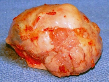
A rare intratubular gonadal stromal tumor was present in the testis of a 7-wk-old male Sprague-Dawley rat. The tumor comprised an intratubular mixture of 2 types of tumor cells with intercellular junctions: the predominant tumor cells were consistent with a Sertoli cell origin, and cells comprising the minor population were situated on basolateral side of the tubuli, consistent with a Leydig cell origin. The neoplastic Sertoli cells had large pleomorphic nuclei and clear cytoplasm with many tubulovesicular cristae and free ribosomes, whereas the neoplastic Leydig cells showed relatively small pleomorphic nuclei, dark cytoplasm with rich smooth endoplasmic reticulum, numerous mitochondria, and lipid droplets. Occasionally, a few transitional type neoplastic cells were observed. The presence of a thick or multilayered basement membrane was confirmed except in tumor-infiltrative lesions. The present case was considered to be a testicular mixed tubular Sertoli-Leydig cell tumor in a Sprague-Dawley rat. (+info)Bilateral Sertoli-Leydig cell tumor of the ovary: a rare case report. (8/22)
Sertoli leydig cell tumors also known as arrhenoblastoma, are a rare member of the sex cord-stromal tumor group of ovarian and testicular cancers, comprising less than 1% of all ovarian tumors, which occur in young adults and are almost always unilateral. We hereby report a case of a 17-year-old female presenting with a short history of irregular menses and an abdominal lump, which was histologically proven to be a bilateral sertoli leydig cell tumor of the ovary, an exceptionally rare entity in itself. (+info)A Leydig cell tumor is a rare type of sex cord-stromal tumor that arises from the Leydig cells (interstitial cells) of the testis in males or ovarian tissue in females. These cells are responsible for producing androgens, particularly testosterone.
Leydig cell tumors can occur at any age but are most common in middle-aged to older men. In women, they are extremely rare and usually found in postmenopausal women. Most Leydig cell tumors are benign (noncancerous), but about 10% can be malignant (cancerous) and have the potential to spread to other parts of the body.
Symptoms of a Leydig cell tumor may include:
* A painless testicular or ovarian mass
* Gynecomastia (enlargement of breast tissue in men) due to increased estrogen production
* Early puberty in children
* Decreased libido and erectile dysfunction in men
* Irregular menstrual cycles in women
Diagnosis is usually made through imaging tests such as ultrasound, CT scan, or MRI, followed by a biopsy to confirm the presence of a Leydig cell tumor. Treatment typically involves surgical removal of the tumor, and additional therapies such as radiation therapy or chemotherapy may be recommended for malignant tumors. Regular follow-up is necessary to monitor for recurrence.
Leydig cells, also known as interstitial cells of Leydig or interstitial cell-stroma, are cells in the testes that produce and release testosterone and other androgens into the bloodstream. They are located in the seminiferous tubules of the testis, near the blood vessels, and are named after Franz Leydig, the German physiologist who discovered them in 1850.
Leydig cells contain cholesterol esters, which serve as precursors for the synthesis of testosterone. They respond to luteinizing hormone (LH) released by the anterior pituitary gland, which stimulates the production and release of testosterone. Testosterone is essential for the development and maintenance of male secondary sexual characteristics, such as facial hair, deep voice, and muscle mass. It also plays a role in sperm production and bone density.
In addition to their endocrine function, Leydig cells have been shown to have non-hormonal functions, including phagocytosis, antigen presentation, and immune regulation. However, these functions are not as well understood as their hormonal roles.
A Sertoli cell tumor is a rare type of sex-cord stromal tumor that develops in the testicles or, more rarely, in the ovaries. These tumors arise from the Sertoli cells, which are specialized cells within the testicle that help to nurture and protect the developing sperm cells. In the ovary, Sertoli cell tumors are thought to arise from similar cells that are part of the supporting tissue in the ovary.
Sertoli cell tumors can occur in people of any age but are most commonly found in middle-aged adults. They are usually slow-growing and may not cause any symptoms, especially if they are small. However, larger tumors or those that have spread (metastasized) may cause various symptoms depending on their location and size.
Symptoms of a Sertoli cell tumor can include:
* A painless lump or swelling in the testicle or ovary
* Abdominal pain or discomfort
* Bloating or a feeling of fullness in the abdomen
* Changes in bowel habits or urinary frequency
* Pain during sexual intercourse (in women)
* Hormonal imbalances, such as gynecomastia (breast development) in men or menstrual irregularities in women.
Diagnosis of a Sertoli cell tumor typically involves a combination of imaging tests, such as ultrasound, CT scan, or MRI, and blood tests to check for elevated levels of certain hormones that may be produced by the tumor. A biopsy may also be performed to confirm the diagnosis and determine the tumor's grade and stage.
Treatment for Sertoli cell tumors typically involves surgical removal of the tumor, along with any affected lymph nodes or other tissues. Additional treatments, such as radiation therapy or chemotherapy, may be recommended in cases where the tumor has spread or is at a higher risk of recurrence. Regular follow-up care is also important to monitor for any signs of recurrence or new tumors.
Testicular neoplasms are abnormal growths or tumors in the testicle that can be benign (non-cancerous) or malignant (cancerous). They are a type of genitourinary cancer, which affects the reproductive and urinary systems. Testicular neoplasms can occur in men of any age but are most commonly found in young adults between the ages of 15 and 40.
Testicular neoplasms can be classified into two main categories: germ cell tumors and non-germ cell tumors. Germ cell tumors, which arise from the cells that give rise to sperm, are further divided into seminomas and non-seminomas. Seminomas are typically slow-growing and have a good prognosis, while non-seminomas tend to grow more quickly and can spread to other parts of the body.
Non-germ cell tumors are less common than germ cell tumors and include Leydig cell tumors, Sertoli cell tumors, and lymphomas. These tumors can have a variety of clinical behaviors, ranging from benign to malignant.
Testicular neoplasms often present as a painless mass or swelling in the testicle. Other symptoms may include a feeling of heaviness or discomfort in the scrotum, a dull ache in the lower abdomen or groin, and breast enlargement (gynecomastia).
Diagnosis typically involves a physical examination, imaging studies such as ultrasound or CT scan, and blood tests to detect tumor markers. Treatment options depend on the type and stage of the neoplasm but may include surgery, radiation therapy, chemotherapy, or a combination of these modalities. Regular self-examinations of the testicles are recommended for early detection and improved outcomes.
Sertoli cells, also known as sustentacular cells or nurse cells, are specialized cells in the seminiferous tubules of the testis in mammals. They play a crucial role in supporting and nurturing the development of sperm cells (spermatogenesis). Sertoli cells create a microenvironment within the seminiferous tubules that facilitates the differentiation, maturation, and survival of germ cells.
These cells have several essential functions:
1. Blood-testis barrier formation: Sertoli cells form tight junctions with each other, creating a physical barrier called the blood-testis barrier, which separates the seminiferous tubules into basal and adluminal compartments. This barrier protects the developing sperm cells from the immune system and provides an isolated environment for their maturation.
2. Nutrition and support: Sertoli cells provide essential nutrients and growth factors to germ cells, ensuring their proper development and survival. They also engulf and digest residual bodies, which are byproducts of spermatid differentiation.
3. Phagocytosis: Sertoli cells have phagocytic properties, allowing them to remove debris and dead cells within the seminiferous tubules.
4. Hormone metabolism: Sertoli cells express receptors for various hormones, such as follicle-stimulating hormone (FSH), testosterone, and estradiol. They play a role in regulating hormonal signaling within the testis by metabolizing these hormones or producing inhibins, which modulate FSH secretion from the pituitary gland.
5. Regulation of spermatogenesis: Sertoli cells produce and secrete various proteins and growth factors that influence germ cell development and proliferation. They also control the release of mature sperm cells into the epididymis through a process called spermiation.
A Sertoli-Leydig cell tumor is a rare type of sex cord-stromal tumor that develops in the ovaries. These tumors arise from the cells that produce hormones and help to form and maintain the ovarian tissue. Sertoli-Leydig cell tumors can occur in people of any age but are most commonly found in women between the ages of 20 and 40.
These tumors can be functional, meaning they produce hormones, or nonfunctional. Functional Sertoli-Leydig cell tumors may cause symptoms related to the production of male hormones (androgens), such as excess facial hair, a deepened voice, and irregular menstrual periods. Nonfunctional tumors typically do not cause any specific symptoms and are often found during routine pelvic examinations or imaging studies performed for other reasons.
Sertoli-Leydig cell tumors are usually slow-growing and can vary in size. Most of these tumors are benign (not cancerous), but some can be malignant (cancerous) and may spread to other parts of the body. Treatment typically involves surgical removal of the tumor, and additional therapies such as chemotherapy or radiation therapy may be recommended depending on the stage and grade of the tumor. Regular follow-up care is essential to monitor for any recurrence of the tumor.
The testis, also known as the testicle, is a male reproductive organ that is part of the endocrine system. It is located in the scrotum, outside of the abdominal cavity. The main function of the testis is to produce sperm and testosterone, the primary male sex hormone.
The testis is composed of many tiny tubules called seminiferous tubules, where sperm are produced. These tubules are surrounded by a network of blood vessels, nerves, and supportive tissues. The sperm then travel through a series of ducts to the epididymis, where they mature and become capable of fertilization.
Testosterone is produced in the Leydig cells, which are located in the interstitial tissue between the seminiferous tubules. Testosterone plays a crucial role in the development and maintenance of male secondary sexual characteristics, such as facial hair, deep voice, and muscle mass. It also supports sperm production and sexual function.
Abnormalities in testicular function can lead to infertility, hormonal imbalances, and other health problems. Regular self-examinations and medical check-ups are recommended for early detection and treatment of any potential issues.
Seminoma is a type of germ cell tumor that develops in the testicle. It is a malignant tumor, meaning it can spread to other parts of the body if left untreated. Seminomas are typically slow-growing and tend to remain localized to the testicle for a longer period compared to other types of testicular cancer. They usually occur in men between the ages of 25 and 45 but can develop at any age.
Seminomas can be classified into two main subtypes: classical seminoma and spermatocytic seminoma. Classical seminoma is more common and typically responds well to treatment, while spermatocytic seminoma is rarer and tends to have a better prognosis with a lower risk of spreading.
Seminomas are usually treated with surgery to remove the affected testicle (orchiectomy), followed by radiation therapy or chemotherapy to kill any remaining cancer cells. The prognosis for seminoma is generally good, especially when caught and treated early. Regular self-examinations of the testicles can help detect any lumps or abnormalities that may indicate the presence of a seminoma or other type of testicular cancer.
Diethylstilbestrol (DES) is a synthetic form of the hormone estrogen that was prescribed to pregnant women from the 1940s until the early 1970s to prevent miscarriage, premature labor, and other complications of pregnancy. However, it was later discovered that DES could cause serious health problems in both the mothers who took it and their offspring.
DES is a non-selective estrogen agonist, meaning that it binds to and activates both estrogen receptors (ERα and ERβ) in the body. It has a higher binding affinity for ERα than for ERβ, which can lead to disruptions in normal hormonal signaling pathways.
In addition to its use as a pregnancy aid, DES has also been used in the treatment of prostate cancer, breast cancer, and other conditions associated with hormonal imbalances. However, due to its potential health risks, including an increased risk of certain cancers, DES is no longer widely used in clinical practice.
Some of the known health effects of DES exposure include:
* In women who were exposed to DES in utero (i.e., their mothers took DES during pregnancy):
+ A rare form of vaginal or cervical cancer called clear cell adenocarcinoma
+ Abnormalities of the reproductive system, such as structural changes in the cervix and vagina, and an increased risk of infertility, ectopic pregnancy, and preterm delivery
+ An increased risk of breast cancer later in life
* In men who were exposed to DES in utero:
+ Undescended testicles
+ Abnormalities of the penis and scrotum
+ A higher risk of testicular cancer
* In both men and women who were exposed to DES in utero or who took DES themselves:
+ An increased risk of certain types of breast cancer
+ A possible increased risk of cardiovascular disease, including high blood pressure and stroke.
It is important for individuals who have been exposed to DES to inform their healthcare providers of this fact, as it may have implications for their medical care and monitoring.
Testosterone is a steroid hormone that belongs to androsten class of hormones. It is primarily secreted by the Leydig cells in the testes of males and, to a lesser extent, by the ovaries and adrenal glands in females. Testosterone is the main male sex hormone and anabolic steroid. It plays a key role in the development of masculine characteristics, such as body hair and muscle mass, and contributes to bone density, fat distribution, red cell production, and sex drive. In females, testosterone contributes to sexual desire and bone health. Testosterone is synthesized from cholesterol and its production is regulated by luteinizing hormone (LH) and follicle-stimulating hormone (FSH).
A Granulosa Cell Tumor is a type of sex cord-stromal tumor, which are uncommon neoplasms that arise from the supporting cells of the ovary or testis. These tumors account for approximately 5% of all ovarian tumors and can occur at any age, but they are most commonly found in perimenopausal and postmenopausal women.
Granulosa cell tumors originate from the granulosa cells, which are normally responsible for producing estrogen and supporting the development of the egg within the ovarian follicle. These tumors can be functional, meaning they produce hormones, or nonfunctional. Functional granulosa cell tumors often secrete estrogen, leading to symptoms such as irregular menstrual periods, postmenopausal bleeding, and, in rare cases, the development of male characteristics (virilization) due to androgen production.
Granulosa cell tumors are typically slow-growing and can vary in size. They are often diagnosed at an early stage because they cause symptoms related to hormonal imbalances or, less commonly, due to abdominal pain or distention caused by the growing mass. The diagnosis is usually confirmed through imaging studies (such as ultrasound, CT, or MRI) and a biopsy or surgical removal of the tumor, followed by histopathological examination.
Treatment for granulosa cell tumors typically involves surgery to remove the tumor and, in some cases, adjacent organs if there is evidence of spread. The role of chemotherapy and radiation therapy is less clear, but they may be used in certain situations, such as advanced-stage disease or high-risk features. Regular follow-up with imaging studies and tumor marker measurements (such as inhibin) is essential due to the risk of recurrence, even many years after initial treatment.
A Giant Cell Tumor (GCT) of bone is a relatively uncommon, locally aggressive tumor that can sometimes become malignant. It is characterized by the presence of multinucleated giant cells which are distributed throughout the tumor tissue. These giant cells are thought to be derived from osteoclasts, which are specialized cells responsible for bone resorption.
GCTs typically affect adults in their 20s and 30s, with a slight female predominance. The most common sites of involvement include the long bones near the knee (distal femur and proximal tibia), as well as the distal radius, sacrum, and spine.
The tumor usually presents as pain and swelling in the affected area, sometimes accompanied by restricted mobility or pathological fractures due to bone weakening. The diagnosis is typically made based on imaging studies (such as X-rays, CT scans, or MRI) and confirmed through a biopsy.
Treatment options for GCTs of bone may include intralesional curettage with or without the use of adjuvant therapies (like phenol, liquid nitrogen, or cement), radiation therapy, or surgical resection. In some cases, systemic treatments like denosumab, a monoclonal antibody targeting RANKL, may be used to control the growth and spread of the tumor. Regular follow-ups are essential to monitor for potential recurrence, which can occur in up to 50% of cases within five years after treatment.
There is no medical definition for "dog diseases" as it is too broad a term. However, dogs can suffer from various health conditions and illnesses that are specific to their species or similar to those found in humans. Some common categories of dog diseases include:
1. Infectious Diseases: These are caused by viruses, bacteria, fungi, or parasites. Examples include distemper, parvovirus, kennel cough, Lyme disease, and heartworms.
2. Hereditary/Genetic Disorders: Some dogs may inherit certain genetic disorders from their parents. Examples include hip dysplasia, elbow dysplasia, progressive retinal atrophy (PRA), and degenerative myelopathy.
3. Age-Related Diseases: As dogs age, they become more susceptible to various health issues. Common age-related diseases in dogs include arthritis, dental disease, cancer, and cognitive dysfunction syndrome (CDS).
4. Nutritional Disorders: Malnutrition or improper feeding can lead to various health problems in dogs. Examples include obesity, malnutrition, and vitamin deficiencies.
5. Environmental Diseases: These are caused by exposure to environmental factors such as toxins, allergens, or extreme temperatures. Examples include heatstroke, frostbite, and toxicities from ingesting harmful substances.
6. Neurological Disorders: Dogs can suffer from various neurological conditions that affect their nervous system. Examples include epilepsy, intervertebral disc disease (IVDD), and vestibular disease.
7. Behavioral Disorders: Some dogs may develop behavioral issues due to various factors such as anxiety, fear, or aggression. Examples include separation anxiety, noise phobias, and resource guarding.
It's important to note that regular veterinary care, proper nutrition, exercise, and preventative measures can help reduce the risk of many dog diseases.
Aromatase is a enzyme that belongs to the cytochrome P450 superfamily, and it is responsible for converting androgens into estrogens through a process called aromatization. This enzyme plays a crucial role in the steroid hormone biosynthesis pathway, particularly in females where it is primarily expressed in adipose tissue, ovaries, brain, and breast tissue.
Aromatase inhibitors are used as a treatment for estrogen receptor-positive breast cancer in postmenopausal women, as they work by blocking the activity of aromatase and reducing the levels of circulating estrogens in the body.
Giant cell tumors (GCTs) are a type of benign or rarely malignant bone tumor that is characterized by the presence of multinucleated giant cells. These tumors typically affect adults between the ages of 20 and 40, and they can occur in any bone, but they most commonly involve the long bones near the knee joint.
GCTs are composed of three types of cells: mononuclear stromal cells, which produce the matrix of the tumor; multinucleated osteoclast-like giant cells, which resemble the bone-resorbing cells found in normal bone; and macrophages, which are part of the body's immune system.
The mononuclear stromal cells produce a variety of growth factors that stimulate the formation and activity of the osteoclast-like giant cells, leading to localized bone destruction. The tumor may cause pain, swelling, and limited mobility in the affected area.
While GCTs are typically benign, they can be aggressive and locally destructive, with a tendency to recur after surgical removal. In some cases, GCTs may undergo malignant transformation, leading to the development of sarcomas. Treatment options for GCTs include curettage (scraping out) of the tumor, followed by bone grafting or the use of a cement spacer to fill the defect, and/or adjuvant therapy with radiation or chemotherapy.
Neoplasms, germ cell and embryonal are types of tumors that originate from the abnormal growth of cells. Here's a brief medical definition for each:
1. Neoplasms: Neoplasms refer to abnormal tissue growths or masses, which can be benign (non-cancerous) or malignant (cancerous). They result from uncontrolled cell division and may invade surrounding tissues or spread to other parts of the body through a process called metastasis.
2. Germ Cell Tumors: These are rare tumors that develop from the germ cells, which give rise to sperm and eggs in the reproductive organs (ovaries and testes). They can be benign or malignant and may occur in both children and adults. Germ cell tumors can also arise outside of the reproductive organs, a condition known as extragonadal germ cell tumors.
3. Embryonal Tumors: These are a type of malignant neoplasm that primarily affects infants and young children. They develop from embryonic cells, which are immature cells present during fetal development. Embryonal tumors can occur in various organs, including the brain (medulloblastomas), nervous system (primitive neuroectodermal tumors or PNETs), and other areas like the kidneys and liver.
It is essential to note that these conditions require professional medical evaluation and treatment by healthcare professionals with expertise in oncology and related fields.
Experimental neoplasms refer to abnormal growths or tumors that are induced and studied in a controlled laboratory setting, typically in animals or cell cultures. These studies are conducted to understand the fundamental mechanisms of cancer development, progression, and potential treatment strategies. By manipulating various factors such as genetic mutations, environmental exposures, and pharmacological interventions, researchers can gain valuable insights into the complex processes underlying neoplasm formation and identify novel targets for cancer therapy. It is important to note that experimental neoplasms may not always accurately represent human cancers, and further research is needed to translate these findings into clinically relevant applications.
A Granular Cell Tumor (GCT) is a rare, usually benign neoplasm that can occur in various parts of the body. These tumors are typically composed of large polygonal cells with abundant eosinophilic granular cytoplasm, which contain numerous mitochondria. They often involve the skin and subcutaneous tissues, but they can also arise in the oral cavity, gastrointestinal tract, respiratory system, and other visceral organs.
Granular Cell Tumors are thought to originate from Schwann cells, which are nerve sheath cells, although their exact origin is still a matter of debate. They usually present as solitary, slow-growing nodules or masses that are often painless, but they can become symptomatic if they involve sensitive areas or if they undergo malignant transformation, which occurs in about 1-2% of cases.
The diagnosis of Granular Cell Tumors is usually made based on histopathological examination of a biopsy specimen. Immunohistochemical staining can be used to confirm the Schwann cell origin of these tumors, as they typically express S-100 protein and other markers of neural differentiation.
Treatment options for Granular Cell Tumors depend on their location, size, and behavior. Solitary, benign tumors can often be excised surgically with a wide margin to reduce the risk of recurrence. However, malignant tumors or those that cannot be completely removed may require more aggressive treatment, such as radiation therapy or chemotherapy. Regular follow-up is recommended to monitor for recurrence or metastasis.
Seminiferous tubules are the long, convoluted tubes within the testicles that are responsible for producing sperm in males. They are lined with specialized epithelial cells called Sertoli cells, which provide structural support and nourishment to developing sperm cells. The seminiferous tubules also contain germ cells, which divide and differentiate into spermatozoa (sperm) through the process of spermatogenesis.
The seminiferous tubules are surrounded by a thin layer of smooth muscle called the tunica albuginea, which helps to maintain the structure and integrity of the testicle. The tubules are connected to the rete testis, a network of channels that transport sperm to the epididymis for further maturation and storage before ejaculation.
Damage or dysfunction of the seminiferous tubules can lead to male infertility, as well as other reproductive health issues.
Immunohistochemistry (IHC) is a technique used in pathology and laboratory medicine to identify specific proteins or antigens in tissue sections. It combines the principles of immunology and histology to detect the presence and location of these target molecules within cells and tissues. This technique utilizes antibodies that are specific to the protein or antigen of interest, which are then tagged with a detection system such as a chromogen or fluorophore. The stained tissue sections can be examined under a microscope, allowing for the visualization and analysis of the distribution and expression patterns of the target molecule in the context of the tissue architecture. Immunohistochemistry is widely used in diagnostic pathology to help identify various diseases, including cancer, infectious diseases, and immune-mediated disorders.
Spermatogenesis is the process by which sperm cells, or spermatozoa, are produced in male organisms. It occurs in the seminiferous tubules of the testes and involves several stages:
1. Spermatocytogenesis: This is the initial stage where diploid spermatogonial stem cells divide mitotically to produce more spermatogonia, some of which will differentiate into primary spermatocytes.
2. Meiosis: The primary spermatocytes undergo meiotic division to form haploid secondary spermatocytes, which then divide again to form haploid spermatids. This process results in the reduction of chromosome number from 46 (diploid) to 23 (haploid).
3. Spermiogenesis: The spermatids differentiate into spermatozoa, undergoing morphological changes such as the formation of a head and tail. During this stage, most of the cytoplasm is discarded, resulting in highly compacted and streamlined sperm cells.
4. Spermation: The final stage where mature sperm are released from the seminiferous tubules into the epididymis for further maturation and storage.
The entire process takes approximately 72-74 days in humans, with continuous production throughout adulthood.
A germinoma is a type of tumor that develops in the brain or the spine, primarily in the pituitary gland or pineal gland. It is a rare form of primary central nervous system (CNS) cancer and is classified as a type of germ cell tumor. These tumors arise from cells that normally develop into sperm or eggs, which can migrate to unusual locations during embryonic development.
Germinomas are highly sensitive to radiation therapy and chemotherapy, making them generally treatable and curable with appropriate medical intervention. Symptoms of a germinoma may include headaches, nausea, vomiting, visual disturbances, hormonal imbalances, and neurological deficits, depending on the location and size of the tumor. Diagnosis typically involves imaging studies like MRI or CT scans, followed by a biopsy to confirm the presence of malignant cells.
Chorionic Gonadotropin (hCG) is a hormone that is produced during pregnancy. It is produced by the placenta after implantation of the fertilized egg in the uterus. The main function of hCG is to prevent the disintegration of the corpus luteum, which is a temporary endocrine structure that forms in the ovary after ovulation and produces progesterone during early pregnancy. Progesterone is essential for maintaining the lining of the uterus and supporting the pregnancy.
hCG can be detected in the blood or urine as early as 10 days after conception, and its levels continue to rise throughout the first trimester of pregnancy. In addition to its role in maintaining pregnancy, hCG is also used as a clinical marker for pregnancy and to monitor certain medical conditions such as gestational trophoblastic diseases.
3-Hydroxysteroid dehydrogenases (3-HSDs) are a group of enzymes that play a crucial role in steroid hormone biosynthesis. These enzymes catalyze the conversion of 3-beta-hydroxy steroids to 3-keto steroids, which is an essential step in the production of various steroid hormones, including progesterone, cortisol, aldosterone, and sex hormones such as testosterone and estradiol.
There are several isoforms of 3-HSDs that are expressed in different tissues and have distinct substrate specificities. For instance, 3-HSD type I is primarily found in the ovary and adrenal gland, where it catalyzes the conversion of pregnenolone to progesterone and 17-hydroxyprogesterone to 17-hydroxycortisol. On the other hand, 3-HSD type II is mainly expressed in the testes, adrenal gland, and placenta, where it catalyzes the conversion of dehydroepiandrosterone (DHEA) to androstenedione and androstenedione to testosterone.
Defects in 3-HSDs can lead to various genetic disorders that affect steroid hormone production and metabolism, resulting in a range of clinical manifestations such as adrenal insufficiency, ambiguous genitalia, and sexual development disorders.
Luteinizing Hormone (LH) receptors are specialized protein structures found on the surface of certain cells in the body. They play a crucial role in the endocrine system by binding to specific hormones, such as Luteinizing Hormone, and triggering a series of intracellular events that ultimately lead to changes in cell function.
In particular, LH receptors are found on the cells of the ovaries and testes. In females, when LH binds to its receptor in the ovary, it stimulates ovulation and the development of the corpus luteum, which produces progesterone. In males, LH (also known as Interstitial Cell-Stimulating Hormone in this context) binding to its receptor on testicular Leydig cells triggers the production of testosterone.
Therefore, LH receptors are essential for reproductive processes and the maintenance of secondary sexual characteristics.
Luteinizing Hormone (LH) is a glycoprotein hormone, which is primarily produced and released by the anterior pituitary gland. In women, a surge of LH triggers ovulation, the release of an egg from the ovaries during the menstrual cycle. During pregnancy, LH stimulates the corpus luteum to produce progesterone. In men, LH stimulates the testes to produce testosterone. It plays a crucial role in sexual development, reproduction, and maintaining the reproductive system.
A mesylate is a salt formed when mesylic acid (methanesulfonic acid) reacts with a base. In the context of pharmaceuticals, many drugs are available in mesylate form as it can be more soluble and bioavailable than other forms. Mesylates are commonly used to improve the absorption and effectiveness of medications.
For example, a drug called atenolol (a beta blocker used to treat high blood pressure) is often formulated as atenolol mesylate because the mesylate form is more soluble in water than the free base form, making it easier for the body to absorb and utilize the medication.
It's important to note that mesylates are not a specific medical condition or disease, but rather a type of pharmaceutical preparation.
The seminiferous epithelium is a specialized type of epithelial tissue that lines the seminiferous tubules within the testes. It is composed of various cell types, including germ cells in different stages of development (spermatogonia, primary and secondary spermatocytes, spermatids) and supportive cells called Sertoli cells.
The primary function of the seminiferous epithelium is to support sperm production (spermatogenesis). The Sertoli cells provide structural support and nourishment to the developing germ cells, helping them to differentiate into mature spermatozoa (sperm). This process involves a series of complex cellular events, including mitosis, meiosis, and spermiogenesis.
In addition to its role in sperm production, the seminiferous epithelium also plays a crucial part in maintaining the blood-testis barrier, which separates the testicular environment from the systemic circulation. This barrier helps protect developing germ cells from potential immune attacks and maintains an optimal microenvironment for spermatogenesis.
 Sertoli-Leydig cell tumour