Seminiferous Epithelium
Sertoli Cells
Spermatogenesis
Seminiferous Tubules
Blood-Testis Barrier
Testis
Spermatids
Epithelium
Spermatogonia
Spermatocytes
Spermatozoa
Hydrazines
Indazoles
Contraceptive Agents, Male
Adherens Junctions
Intercellular Junctions
Tight Junctions
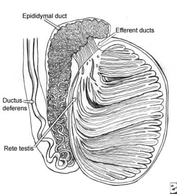
Epididymis
Leydig Cells
Testosterone
Felidae
Rats, Sprague-Dawley
Infertility, Male
Integrin alpha6beta1
Vitamin A Deficiency
Lepidium
Acrosome
Orchitis
Immunohistochemistry
Colubridae
Transforming Growth Factor beta3
Follicle Stimulating Hormone
Microscopy, Electron
Cryptorchidism
Occludin
Epithelial Cells
Epithelium, Corneal
Junctional Adhesion Molecules
Microscopy, Immunoelectron
Meiosis
Diethylhexyl Phthalate
Hypophysectomy
RNA, Messenger
Cell Communication
Fertility
Histocytochemistry
Genitalia, Male
Rats, Inbred Strains
Gene Expression
Zonula Occludens-1 Protein
Vitamin A
Rats, Wistar
Actins
Microscopy, Fluorescence
Identification of a nuclear localization signal in activin/inhibin betaA subunit; intranuclear betaA in rat spermatogenic cells. (1/321)
Activin is a dimeric glycoprotein hormone that was initially characterized by its ability to stimulate pituitary FSH secretion and was subsequently recognized as a growth factor with diverse biological functions in a large variety of tissues. In the testis, activin has been implicated in the auto/paracrine regulation of spermatogenesis through its cognate cell membrane receptors on Sertoli and germ cells. In this study we provide evidence for intranuclear activin/inhibin betaA subunit and show its distribution in the rat seminiferous epithelium. We have shown by transient expression in HeLa cells of beta-galactosidase fusion proteins that the betaA subunit precursor contains a functional nuclear localization signal within the lysine-rich sequence corresponding to amino acids 231-244. In all stages of the rat seminiferous epithelial cycle, an intense immunohistochemical staining of nuclear betaA was demonstrated in intermediate or type B spermatogonia or primary spermatocytes in their initial stages of the first meiotic prophase, as well as in pachytene spermatocytes and elongating spermatids primarily in stages IX-XII. In some pachytene spermatocytes, the pattern of betaA immunoreactivity was consistent with the characteristic distribution of pachytene chromosomes. In the nuclei of round spermatids, betaA immunoreactivity was less intense, and in late spermatids it was localized in the residual cytoplasm, suggesting disposal of betaA before spermatozoal maturation. Immunoblot analysis of a protein extract from isolated testicular nuclei revealed a nuclear betaA species with a molecular mass of approximately 24 kDa, which is more than 1.5 times that of the mature activin betaA subunit present in activin dimers. These results suggest that activin/inhibin betaA may elicit its biological functions through two parallel signal transduction pathways, one involving the dimeric molecule and cell surface receptors and the other an alternately processed betaA sequence acting directly within the nucleus. According to our immunohistochemical data, betaA may play a significant role in the regulation of nuclear functions during meiosis and spermiogenesis. (+info)Spermatid translocation in the rat seminiferous epithelium: coupling membrane trafficking machinery to a junction plaque. (2/321)
In this study, we demonstrate that specialized junction plaques that occur between Sertoli cells and spermatids in the rat testis support microtubule translocation in vitro. During spermatogenesis, Sertoli cells are attached to spermatids by specialized adhesion junctions termed ectoplasmic specializations (ESs). These structures consist of regions of the plasma membrane adherent to the spermatid head, a submembrane layer of tightly packed actin filaments, and an attached cistern of endoplasmic reticulum. It has been proposed that motor proteins on the endoplasmic reticulum interact with adjacent microtubules to translocate the junction plaques, and hence the attached spermatids, within the epithelium. If this hypothesis is true, then isolated junctions should support microtubule transport. To verify this prediction, we have mechanically isolated rat spermatids, together with their attached ESs, and tested them for their ability to transport microtubules in vitro. Most assays were done in the presence of 2 mg/ml testicular cytosol and at room temperature. ESs attached to spermatids supported microtubule translocation. In some cases in which motility events were detected, microtubules moved smoothly over the junction site. In others, the movement was slow but progressive, saltatory and "inch-worm-like." No motility was detected in the absence of exogenous ATP or in the presence of apyrase (an enzyme that catalyses the breakdown of ATP). Our results are consistent with the microtubule-based motility hypothesis of spermatid translocation. (+info)Rat testis motor proteins associated with spermatid translocation (dynein) and spermatid flagella (kinesin-II). (3/321)
In this study, we report sites in the seminiferous epithelium of the rat testis that are immunoreactive with antibodies to the intermediate chain of cytoplasmic dynein and kinesin II. The study was done to determine whether or not microtubule-dependent motor proteins are present in Sertoli cell regions involved with spermatid translocation. Sections and epithelial fragments of perfusion-fixed rat testis were probed with an antibody (clone 74.1) to the intermediate chain of cytoplasmic dynein (IC74) and to kinesin-II. Labeling with the antibody to cytoplasmic dynein was dramatically evident in Sertoli cell regions surrounding apical crypts containing attached spermatids and known to contain unique intercellular attachment plaques. The antibody to kinesin II reacted only with spermatid tails. The levels of cytoplasmic dynein visible on immunoblots of supernatants collected from spermatid/junction complexes treated with an actin-severing enzyme (gelsolin) were greater than those of controls, indicating that at least some of the dynein may have been associated with Sertoli cell junction plaques attached to spermatids. Results are consistent with the conclusion that an isoform of cytoplasmic dynein may be responsible for the apical translocation of elongate spermatids that occurs before sperm release. Also, this is the first report of kinesin-II in mammalian spermatid tails. (+info)Plectin is concentrated at intercellular junctions and at the nuclear surface in morphologically differentiated rat Sertoli cells. (4/321)
Intermediate filaments in Sertoli cells have a well-defined pattern of distribution. They form a basally situated perinuclear network from which filaments extend peripherally to adhesion plaques at the plasma membrane and to sites of codistribution with other major elements of the cytoskeleton, particularly with microtubules. Although the general pattern of intermediate filament distribution is known, the molecular components involved with linking the filaments to organelles and attachment plaques in these cells have not been identified. One candidate for such a linking element is plectin. In this study we test for the presence of, and determine the distribution of, plectin in Sertoli cells of the rat testis. Fixed frozen sections and fixed epithelial fragments of rat testis were probed for plectin and vimentin using antibodies. Tissue was evaluated using standard fluorescence microscopy and confocal microscopy. Plectin in Sertoli cells was concentrated in a narrow zone surrounding the nucleus, and at focal sites, presumably desmosome-like plaques, at interfaces with adjacent cells. Plectin was also concentrated at sites where intermediate filament bundles project into specialized actin-filament containing plaques at sites of attachment to elongate spermatids. Plectin in Sertoli cells is concentrated at the nuclear surface and in junction plaques associated with the plasma membrane. The pattern of distribution is consistent with plectin being involved with linking intermediate filaments centrally (basally) to the nucleus and peripherally to intercellular attachment sites. (+info)Postnatal differentiation of the ductus deferens, tail of the epididymis, and distal body of the epididymis in goats occurs independently of rete testis fluid. (5/321)
Observations from extratesticular rete-ligated, mature goats indicated that epithelial morphology in the tail of the epididymis can be maintained without any input from testicular fluid (Goyal et al., Acta Anat., 1994;150: 127-135). Hence, the objective of this study was to determine whether the tail of the epididymis and/or other regions of the male excurrent ducts can differentiate prior to the appearance of lumen in the seminiferous tubules, which is an indicator for the onset of seminiferous tubular fluid secretion. Based on age and scrotal circumference (SC), 20 male goats were divided into four groups of five animals each: 1-4 weeks (SC, 6.5-7.5 cm), 7-10 weeks (SC, 8.5-11.0 cm), 12-15 weeks (SC, 11.0-14.0 cm), and 15-25 weeks (SC, 16.0-19.0 cm). Tissues were collected from the testis, six regions of the epididymis (proximal, middle and distal head; proximal and distal body; and tail), and the ductus deferens, and were processed for light and electron microscopic examination. Changes in epithelial height and cytological features associated with absorption (microvilli, pinocytotic and coated vesicles) and protein secretion (RER, Golgi body) were used as markers for differentiation. Differentiation of all of these features was comparable to that observed in the 15-25-week-old animals in the ductus deferens by > or = 1 week, in the tail of the epididymis by > or = 7 weeks, in the distal body of the epididymis by > or = 12 weeks, and in the proximal body of the epididymis and all three regions of the head of the epididymis by > or = 15 weeks. Seminiferous tubules developed lumens between 12 and 15 weeks. In conclusion, epithelial differentiation in the ductus deferens, tail of the epididymis, and distal body of the epididymis follows a time-dependent, spatial, ascending order and is achieved before lumen formation in the seminiferous tubules. Conversely, epithelial differentiation in all three regions of the head and the proximal body of the epididymis occurs simultaneously and after lumen formation in the seminiferous tubules. (+info)The cycle duration of the seminiferous epithelium remains unaltered during GnRH antagonist-induced testicular involution in rats and monkeys. (6/321)
Although the gonadotropic control of the spermatogenic process is well established, the endocrine regulation of the timing and kinetics of germ cell development has received little attention. We found previously that the administration of a GnRH antagonist (ANT) over a period of 25 days could retard spermatid development and slightly prolong cycle length in intact adult cynomolgus monkeys (Macaca fascicularis). The aim of the present study was to investigate the effects of extended exposure to ANT on the duration of the cycle of the seminiferous epithelium in the monkey. Additionally, the duration of spermatogenesis was studied in the ANT-exposed rat model. In experiment 1, monkeys were given either saline or ANT (n=6/group) and on day 30 all animals received a single injection of 5-bromodeoxyuridine (BrdU) to label S-phase germ cells. Testicular biopsies were taken on days 39, 43, 47 and 51 (end of treatment) for BrdU localization and flow cytometric analysis. ANT treatment suppressed hormone levels, reduced testis size by >70% and severely impaired germ cell production. Despite these alterations, cycle duration remained unchanged at all time-points compared with controls (10.12+/-0.15 days vs 10.16+/- 0.44 days). In experiment 2, adult male Sprague-Dawley rats (n=15/group) received either vehicle (VEH) or ANT for 14 days and received BrdU injection on day 2. Cycle duration was found to be shorter in the ANT-treated group (12.45+/-0.09 days) than in the control group (12.75+/-0.08, P<0.05). As spermatogenic cycle length in this control group was longer than that of our historical controls (range: 12.37-12.53 days), experiment 2 was repeated (n=10/group). In experiment 3, cycle duration was 12.51+/-0.02 for VEH and 12.46+/-0.05 for the ANT-treated group (P>0.05) in both species. We concluded that the duration of the cycle of the seminiferous epithelium in monkeys and rats is independent of gonadotropins but is rather regulated by the spermatogenic tissue itself. (+info)Differential expression of gap-junction gene connexin 31 in seminiferous epithelium of rat testes. (7/321)
Spermatogenesis, a tightly regulated developmental process of male germ cells in testis, is associated with temporal and spatial expression of certain gap-junction connexins. Our findings by RT-PCR indicate that the Cx31 gene is expressed in testis tissue of adult and postnatal rats. During the postnatal spermatogenic process, the Cx31-specific signal became detectable at 15 dpp and onward by in situ hybridization, and apparently localized in the basal compartment of seminiferous epithelium where active spermatogonia and early primary spermatocytes reside. No signal was found in the luminal region. In adult testes, spermatids of elongation phase were also Cx31 positive. Immunohistochemical analysis with mouse anti-Cx31 antibody gave a similar staining pattern, providing further evidence that the gap-junction protein is abundant in the basal seminiferous epithelium, in accordance with the cellular distribution of Cx31 mRNA. These results represent the first demonstration of Cx31 expression at both transcriptional and protein levels in the seminiferous epithelium of rat testes. Thus, Cx31 may play a role in cell-cell communication during spermatogenesis. (+info)Activation of a UBC4-dependent pathway of ubiquitin conjugation during postnatal development of the rat testis. (8/321)
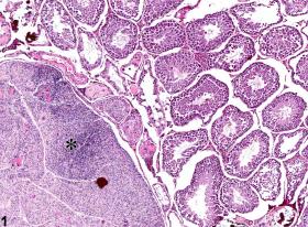
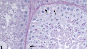
During spermatogenesis, germ cells undergo mitotic and meiotic divisions to form haploid round spermatids which mature to functional elongated spermatozoa. During this process there occurs remodeling of cell structure and loss of most of the cytoplasm and a large fraction of cellular proteins. To evaluate the role of the ubiquitin proteolytic system in this protein loss, we measured levels of ubiquitinated proteins and rates of ubiquitin conjugation in extracts of testes from rats of different ages. Endogenous ubiquitin-protein conjugates increased till day 30 and then reached a plateau. In parallel, there was a progressive increase in the rate of conjugation of ubiquitin to proteins in testis extracts from these animals. To test the importance of two major ubiquitin conjugating enzyme families in the conjugation, immunoprecipitation of UBC2 or UBC4 from 10- and 30-day-old testis extracts was carried out and the remaining conjugation activity in supernatants was assayed. Depletion of either enzyme family resulted in decreased conjugation. However, most of the conjugation activity and, more importantly, the increased conjugation during development were UBC4-dependent. Immunocytochemistry demonstrated a marked increase in expression of UBC4 in spermatids, consistent with the UBC4-dependent activation of conjugation seen in vitro. In situ hybridization studies evaluated the contribution of various UBC4 isoforms to this induction. UBC4-1 mRNA was expressed in most cells. UBC4-2 mRNA was restricted to germ cells with high levels of expression in round and elongated spermatids. UBC4-testis had previously been shown to be expressed only in spermatids. Our data suggest that induction of various UBC4 isoforms activates overall conjugation and plays an important role in the cellular remodeling and protein loss occurring during spermatogenesis. (+info)The seminiferous epithelium is a specialized type of epithelial tissue that lines the seminiferous tubules within the testes. It is composed of various cell types, including germ cells in different stages of development (spermatogonia, primary and secondary spermatocytes, spermatids) and supportive cells called Sertoli cells.
The primary function of the seminiferous epithelium is to support sperm production (spermatogenesis). The Sertoli cells provide structural support and nourishment to the developing germ cells, helping them to differentiate into mature spermatozoa (sperm). This process involves a series of complex cellular events, including mitosis, meiosis, and spermiogenesis.
In addition to its role in sperm production, the seminiferous epithelium also plays a crucial part in maintaining the blood-testis barrier, which separates the testicular environment from the systemic circulation. This barrier helps protect developing germ cells from potential immune attacks and maintains an optimal microenvironment for spermatogenesis.
Sertoli cells, also known as sustentacular cells or nurse cells, are specialized cells in the seminiferous tubules of the testis in mammals. They play a crucial role in supporting and nurturing the development of sperm cells (spermatogenesis). Sertoli cells create a microenvironment within the seminiferous tubules that facilitates the differentiation, maturation, and survival of germ cells.
These cells have several essential functions:
1. Blood-testis barrier formation: Sertoli cells form tight junctions with each other, creating a physical barrier called the blood-testis barrier, which separates the seminiferous tubules into basal and adluminal compartments. This barrier protects the developing sperm cells from the immune system and provides an isolated environment for their maturation.
2. Nutrition and support: Sertoli cells provide essential nutrients and growth factors to germ cells, ensuring their proper development and survival. They also engulf and digest residual bodies, which are byproducts of spermatid differentiation.
3. Phagocytosis: Sertoli cells have phagocytic properties, allowing them to remove debris and dead cells within the seminiferous tubules.
4. Hormone metabolism: Sertoli cells express receptors for various hormones, such as follicle-stimulating hormone (FSH), testosterone, and estradiol. They play a role in regulating hormonal signaling within the testis by metabolizing these hormones or producing inhibins, which modulate FSH secretion from the pituitary gland.
5. Regulation of spermatogenesis: Sertoli cells produce and secrete various proteins and growth factors that influence germ cell development and proliferation. They also control the release of mature sperm cells into the epididymis through a process called spermiation.
Spermatogenesis is the process by which sperm cells, or spermatozoa, are produced in male organisms. It occurs in the seminiferous tubules of the testes and involves several stages:
1. Spermatocytogenesis: This is the initial stage where diploid spermatogonial stem cells divide mitotically to produce more spermatogonia, some of which will differentiate into primary spermatocytes.
2. Meiosis: The primary spermatocytes undergo meiotic division to form haploid secondary spermatocytes, which then divide again to form haploid spermatids. This process results in the reduction of chromosome number from 46 (diploid) to 23 (haploid).
3. Spermiogenesis: The spermatids differentiate into spermatozoa, undergoing morphological changes such as the formation of a head and tail. During this stage, most of the cytoplasm is discarded, resulting in highly compacted and streamlined sperm cells.
4. Spermation: The final stage where mature sperm are released from the seminiferous tubules into the epididymis for further maturation and storage.
The entire process takes approximately 72-74 days in humans, with continuous production throughout adulthood.
Seminiferous tubules are the long, convoluted tubes within the testicles that are responsible for producing sperm in males. They are lined with specialized epithelial cells called Sertoli cells, which provide structural support and nourishment to developing sperm cells. The seminiferous tubules also contain germ cells, which divide and differentiate into spermatozoa (sperm) through the process of spermatogenesis.
The seminiferous tubules are surrounded by a thin layer of smooth muscle called the tunica albuginea, which helps to maintain the structure and integrity of the testicle. The tubules are connected to the rete testis, a network of channels that transport sperm to the epididymis for further maturation and storage before ejaculation.
Damage or dysfunction of the seminiferous tubules can lead to male infertility, as well as other reproductive health issues.
The Blood-Testis Barrier (BTB) is a unique structural and functional feature of the seminiferous epithelium in the testes, which forms a tight junction between adjacent Sertoli cells in the semi-niferous tubules. This barrier selectively restricts the passage of molecules, including potentially harmful substances and immune cells, from the systemic circulation into the adluminal compartment of the seminiferous epithelium where spermatogenesis occurs. This helps to maintain a immunologically privileged microenvironment that is essential for the survival and maturation of developing sperm cells, preventing an immune response against them. The BTB also regulates the movement of molecules required for spermatogenesis, such as nutrients, hormones, and signaling molecules, from the basal compartment to the adluminal compartment.
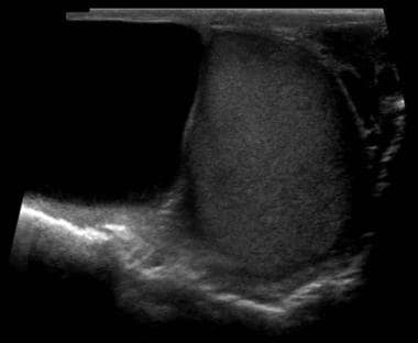
The testis, also known as the testicle, is a male reproductive organ that is part of the endocrine system. It is located in the scrotum, outside of the abdominal cavity. The main function of the testis is to produce sperm and testosterone, the primary male sex hormone.
The testis is composed of many tiny tubules called seminiferous tubules, where sperm are produced. These tubules are surrounded by a network of blood vessels, nerves, and supportive tissues. The sperm then travel through a series of ducts to the epididymis, where they mature and become capable of fertilization.
Testosterone is produced in the Leydig cells, which are located in the interstitial tissue between the seminiferous tubules. Testosterone plays a crucial role in the development and maintenance of male secondary sexual characteristics, such as facial hair, deep voice, and muscle mass. It also supports sperm production and sexual function.
Abnormalities in testicular function can lead to infertility, hormonal imbalances, and other health problems. Regular self-examinations and medical check-ups are recommended for early detection and treatment of any potential issues.
Spermatids are immature sperm cells that are produced during the process of spermatogenesis in the male testes. They are the product of the final stage of meiosis, where a diploid spermatocyte divides into four haploid spermatids. Each spermatid then undergoes a series of changes, including the development of a tail for motility and the condensation of its nucleus to form a head containing the genetic material. Once this process is complete, the spermatids are considered mature spermatozoa and are capable of fertilizing an egg.
Epithelium is the tissue that covers the outer surface of the body, lines the internal cavities and organs, and forms various glands. It is composed of one or more layers of tightly packed cells that have a uniform shape and size, and rest on a basement membrane. Epithelial tissues are avascular, meaning they do not contain blood vessels, and are supplied with nutrients by diffusion from the underlying connective tissue.
Epithelial cells perform a variety of functions, including protection, secretion, absorption, excretion, and sensation. They can be classified based on their shape and the number of cell layers they contain. The main types of epithelium are:
1. Squamous epithelium: composed of flat, scalelike cells that fit together like tiles on a roof. It forms the lining of blood vessels, air sacs in the lungs, and the outermost layer of the skin.
2. Cuboidal epithelium: composed of cube-shaped cells with equal height and width. It is found in glands, tubules, and ducts.
3. Columnar epithelium: composed of tall, rectangular cells that are taller than they are wide. It lines the respiratory, digestive, and reproductive tracts.
4. Pseudostratified epithelium: appears stratified or layered but is actually made up of a single layer of cells that vary in height. The nuclei of these cells appear at different levels, giving the tissue a stratified appearance. It lines the respiratory and reproductive tracts.
5. Transitional epithelium: composed of several layers of cells that can stretch and change shape to accommodate changes in volume. It is found in the urinary bladder and ureters.
Epithelial tissue provides a barrier between the internal and external environments, protecting the body from physical, chemical, and biological damage. It also plays a crucial role in maintaining homeostasis by regulating the exchange of substances between the body and its environment.
Spermatogonia are a type of diploid germ cells found in the seminiferous tubules of the testis. They are the stem cells responsible for sperm production (spermatogenesis) in males. There are two types of spermatogonia: A-dark (Ad) and A-pale (Ap). The Ad spermatogonia function as reserve stem cells, while the Ap spermatogonia serve as the progenitor cells that divide to produce type B spermatogonia. Type B spermatogonia then differentiate into primary spermatocytes, which undergo meiosis to form haploid spermatozoa.
Spermatocytes are a type of cell that is involved in the process of spermatogenesis, which is the formation of sperm in the testes. Specifically, spermatocytes are the cells that undergo meiosis, a special type of cell division that results in the production of four haploid daughter cells, each containing half the number of chromosomes as the parent cell.
There are two types of spermatocytes: primary and secondary. Primary spermatocytes are diploid cells that contain 46 chromosomes (23 pairs). During meiosis I, these cells undergo a process called crossing over, in which genetic material is exchanged between homologous chromosomes. After crossing over, the primary spermatocytes divide into two secondary spermatocytes, each containing 23 chromosomes (but still with 23 pairs).
Secondary spermatocytes then undergo meiosis II, which results in the formation of four haploid spermatids. Each spermatid contains 23 single chromosomes and will eventually develop into a mature sperm cell through a process called spermiogenesis.
It's worth noting that spermatocytes are only found in males, as they are specific to the male reproductive system.
Spermatozoa are the male reproductive cells, or gametes, that are produced in the testes. They are microscopic, flagellated (tail-equipped) cells that are highly specialized for fertilization. A spermatozoon consists of a head, neck, and tail. The head contains the genetic material within the nucleus, covered by a cap-like structure called the acrosome which contains enzymes to help the sperm penetrate the female's egg (ovum). The long, thin tail propels the sperm forward through fluid, such as semen, enabling its journey towards the egg for fertilization.
Hydrazines are not a medical term, but rather a class of organic compounds containing the functional group N-NH2. They are used in various industrial and chemical applications, including the production of polymers, pharmaceuticals, and agrochemicals. However, some hydrazines have been studied for their potential therapeutic uses, such as in the treatment of cancer and cardiovascular diseases. Exposure to high levels of hydrazines can be toxic and may cause damage to the liver, kidneys, and central nervous system. Therefore, medical professionals should be aware of the potential health hazards associated with hydrazine exposure.
Indazoles are not a medical term, but a chemical classification. They refer to a class of heterocyclic organic compounds that contain a indazole moiety, which is a benzene ring fused with a diazole ring. Indazoles have no specific medical relevance, but certain derivatives of indazoles have been developed and used as drugs in medicine, particularly in the treatment of cancer and cardiovascular diseases. For example, Tadalafil (Cialis), a medication used to treat erectile dysfunction and benign prostatic hyperplasia, is a selective inhibitor of cGMP-specific phosphodiesterase type 5 and has an indazole structure.
Contraceptive agents for males are substances or methods that are used to prevent pregnancy by reducing the likelihood of fertilization. These can include:
1. Barrier methods: Condoms, diaphragms, and spermicides create a physical barrier that prevents sperm from reaching the egg.
2. Hormonal methods: Testosterone and progestin hormone therapies can decrease sperm production and reduce fertility.
3. Intrauterine devices (IUDs) for men: These are still in the experimental stage, but they involve placing a device in the male reproductive tract to prevent sperm from reaching the female reproductive system.
4. Withdrawal method: This involves the man withdrawing his penis from the vagina before ejaculation, although this is not a highly reliable form of contraception.
5. Fertility awareness methods: These involve tracking the woman's menstrual cycle and avoiding sexual intercourse during her fertile period.
6. Sterilization: Vasectomy is a surgical procedure that blocks or cuts the vas deferens, preventing sperm from leaving the body. It is a permanent form of contraception for men.
It's important to note that no contraceptive method is 100% effective, and individuals should consult with their healthcare provider to determine which option is best for them based on their personal needs, lifestyle, and medical history.
Adherens junctions are specialized types of cell-cell contacts that play a crucial role in maintaining the integrity and stability of tissues. They are composed of transmembrane cadherin proteins, which connect to the actin cytoskeleton inside the cell through intracellular adaptor proteins such as catenins.
The cadherins on opposing cells interact with each other to form adhesive bonds that help to anchor the cells together and regulate various cellular processes, including cell growth, differentiation, and migration. Adherens junctions are essential for many physiological processes, such as embryonic development, wound healing, and tissue homeostasis, and their dysfunction has been implicated in a variety of diseases, including cancer and degenerative disorders.
Intercellular junctions are specialized areas of contact between two or more adjacent cells in multicellular organisms. They play crucial roles in maintaining tissue structure and function by regulating the movement of ions, molecules, and even larger cellular structures from one cell to another. There are several types of intercellular junctions, including:
1. Tight Junctions (Zonulae Occludentes): These are the most apical structures in epithelial and endothelial cells, forming a virtually impermeable barrier to prevent the paracellular passage of solutes and water between the cells. They create a tight seal by connecting the transmembrane proteins of adjacent cells, such as occludin and claudins.
2. Adherens Junctions: These are located just below the tight junctions and help maintain cell-to-cell adhesion and tissue integrity. Adherens junctions consist of cadherin proteins that form homophilic interactions with cadherins on adjacent cells, as well as intracellular adaptor proteins like catenins, which connect to the actin cytoskeleton.
3. Desmosomes: These are another type of cell-to-cell adhesion structure, primarily found in tissues that experience mechanical stress, such as the skin and heart. Desmosomes consist of cadherin proteins (desmocadherins) that interact with each other and connect to intermediate filaments (keratin in epithelial cells) via plakoglobin and desmoplakin.
4. Gap Junctions: These are specialized channels that directly connect the cytoplasm of adjacent cells, allowing for the exchange of small molecules, ions, and second messengers. Gap junctions consist of connexin proteins that form hexameric structures called connexons in the plasma membrane of each cell. When two connexons align, they create a continuous pore or channel between the cells.
In summary, intercellular junctions are essential for maintaining tissue structure and function by regulating paracellular transport, cell-to-cell adhesion, and intercellular communication.
Tight junctions, also known as zonula occludens, are specialized types of intercellular junctions that occur in epithelial and endothelial cells. They are located near the apical side of the lateral membranes of adjacent cells, where they form a continuous belt-like structure that seals off the space between the cells.
Tight junctions are composed of several proteins, including occludin, claudins, and junctional adhesion molecules (JAMs), which interact to form a network of strands that create a tight barrier. This barrier regulates the paracellular permeability of ions, solutes, and water, preventing their uncontrolled movement across the epithelial or endothelial layer.
Tight junctions also play an important role in maintaining cell polarity by preventing the mixing of apical and basolateral membrane components. Additionally, they are involved in various signaling pathways that regulate cell proliferation, differentiation, and survival.
The epididymis is a tightly coiled tube located on the upper and posterior portion of the testicle that serves as the site for sperm maturation and storage. It is an essential component of the male reproductive system. The epididymis can be divided into three parts: the head (where newly produced sperm enter from the testicle), the body, and the tail (where mature sperm exit and are stored). Any abnormalities or inflammation in the epididymis may lead to discomfort, pain, or infertility.
Leydig cells, also known as interstitial cells of Leydig or interstitial cell-stroma, are cells in the testes that produce and release testosterone and other androgens into the bloodstream. They are located in the seminiferous tubules of the testis, near the blood vessels, and are named after Franz Leydig, the German physiologist who discovered them in 1850.
Leydig cells contain cholesterol esters, which serve as precursors for the synthesis of testosterone. They respond to luteinizing hormone (LH) released by the anterior pituitary gland, which stimulates the production and release of testosterone. Testosterone is essential for the development and maintenance of male secondary sexual characteristics, such as facial hair, deep voice, and muscle mass. It also plays a role in sperm production and bone density.
In addition to their endocrine function, Leydig cells have been shown to have non-hormonal functions, including phagocytosis, antigen presentation, and immune regulation. However, these functions are not as well understood as their hormonal roles.
Testosterone is a steroid hormone that belongs to androsten class of hormones. It is primarily secreted by the Leydig cells in the testes of males and, to a lesser extent, by the ovaries and adrenal glands in females. Testosterone is the main male sex hormone and anabolic steroid. It plays a key role in the development of masculine characteristics, such as body hair and muscle mass, and contributes to bone density, fat distribution, red cell production, and sex drive. In females, testosterone contributes to sexual desire and bone health. Testosterone is synthesized from cholesterol and its production is regulated by luteinizing hormone (LH) and follicle-stimulating hormone (FSH).
Sexual maturation is the process of physical development during puberty that leads to the ability to reproduce. This process involves the development of primary and secondary sexual characteristics, changes in hormone levels, and the acquisition of reproductive capabilities. In females, this includes the onset of menstruation and the development of breasts and hips. In males, this includes the deepening of the voice, growth of facial hair, and the production of sperm. Achieving sexual maturation is an important milestone in human development and typically occurs during adolescence.
Sperm count, also known as sperm concentration, is the number of sperm present in a given volume of semen. The World Health Organization (WHO) previously defined a normal sperm count as at least 20 million sperm per milliliter of semen. However, more recent studies suggest that fertility may be affected even when sperm counts are slightly lower than this threshold. It's important to note that sperm count is just one factor among many that can influence male fertility. Other factors, such as sperm motility (the ability of sperm to move properly) and morphology (the shape of the sperm), also play crucial roles in successful conception.
Germ cells are the reproductive cells, also known as sex cells, that combine to form offspring in sexual reproduction. In females, germ cells are called ova or egg cells, and in males, they are called spermatozoa or sperm cells. These cells are unique because they carry half the genetic material necessary for creating new life. They are produced through a process called meiosis, which reduces their chromosome number by half, ensuring that when two germ cells combine during fertilization, the normal diploid number of chromosomes is restored.
Felidae is the biological family that includes all extant (living) members of the cat group, also known as felids. This family consists of big cats such as lions, tigers, and leopards, as well as small cats like domestic cats, cheetahs, and pumas. Felidae is part of the order Carnivora and is characterized by specialized adaptations for hunting and stalking prey, including retractile claws, sharp teeth, and flexible bodies. The family has a worldwide distribution, with species found in various habitats across all continents except Antarctica.
Organ size refers to the volume or physical measurement of an organ in the body of an individual. It can be described in terms of length, width, and height or by using specialized techniques such as imaging studies (like CT scans or MRIs) to determine the volume. The size of an organ can vary depending on factors such as age, sex, body size, and overall health status. Changes in organ size may indicate various medical conditions, including growths, inflammation, or atrophy.
Sprague-Dawley rats are a strain of albino laboratory rats that are widely used in scientific research. They were first developed by researchers H.H. Sprague and R.C. Dawley in the early 20th century, and have since become one of the most commonly used rat strains in biomedical research due to their relatively large size, ease of handling, and consistent genetic background.
Sprague-Dawley rats are outbred, which means that they are genetically diverse and do not suffer from the same limitations as inbred strains, which can have reduced fertility and increased susceptibility to certain diseases. They are also characterized by their docile nature and low levels of aggression, making them easier to handle and study than some other rat strains.
These rats are used in a wide variety of research areas, including toxicology, pharmacology, nutrition, cancer, and behavioral studies. Because they are genetically diverse, Sprague-Dawley rats can be used to model a range of human diseases and conditions, making them an important tool in the development of new drugs and therapies.
Male infertility is a condition characterized by the inability to cause pregnancy in a fertile female. It is typically defined as the failure to achieve a pregnancy after 12 months or more of regular unprotected sexual intercourse.
The causes of male infertility can be varied and include issues with sperm production, such as low sperm count or poor sperm quality, problems with sperm delivery, such as obstructions in the reproductive tract, or hormonal imbalances that affect sperm production. Other factors that may contribute to male infertility include genetic disorders, environmental exposures, lifestyle choices, and certain medical conditions or treatments.
It is important to note that male infertility can often be treated or managed with medical interventions, such as medication, surgery, or assisted reproductive technologies (ART). A healthcare provider can help diagnose the underlying cause of male infertility and recommend appropriate treatment options.
Integrin α6β1, also known as CD49f/CD29, is a heterodimeric transmembrane receptor protein composed of α6 and β1 subunits. It is widely expressed in various tissues, including epithelial cells, endothelial cells, fibroblasts, and hematopoietic cells. Integrin α6β1 plays a crucial role in cell-matrix adhesion, particularly to the laminin component of the extracellular matrix (ECM). This receptor is involved in various biological processes such as cell migration, proliferation, differentiation, and survival. Additionally, integrin α6β1 has been implicated in tumor progression, metastasis, and drug resistance in certain cancers.
Vitamin A deficiency (VAD) is a condition that occurs when there is a lack of vitamin A in the diet. This essential fat-soluble vitamin plays crucial roles in vision, growth, cell division, reproduction, and immune system regulation.
In its severe form, VAD leads to xerophthalmia, which includes night blindness (nyctalopia) and keratomalacia - a sight-threatening condition characterized by dryness of the conjunctiva and cornea, with eventual ulceration and perforation. Other symptoms of VAD may include Bitot's spots (foamy, triangular, white spots on the conjunctiva), follicular hyperkeratosis (goose bump-like bumps on the skin), and increased susceptibility to infections due to impaired immune function.
Vitamin A deficiency is most prevalent in developing countries where diets are often low in animal source foods and high in plant-based foods with low bioavailability of vitamin A. It primarily affects children aged 6 months to 5 years, pregnant women, and lactating mothers. Prevention strategies include dietary diversification, food fortification, and supplementation programs.
I'm sorry for any confusion, but "Lepidium" is not a medical term. It is the genus name of a group of plants that includes garden cress, peppergrass, and other similar herbs. These plants belong to the Brassicaceae family, also known as the mustard family. They have some nutritional and potential medicinal uses, but they are not commonly used in modern medical contexts. If you have any questions related to medical terminology or health concerns, I'd be happy to try to help with those instead!
The acrosome is a specialized structure located on the anterior part of the sperm head in many species of animals, including humans. It contains enzymes that help the sperm penetrate the outer covering of the egg (zona pellucida) during fertilization. The acrosome reaction is the process by which the acrosome releases its enzymes, allowing the sperm to digest a path through the zona pellucida and reach the egg plasma membrane for fusion and fertilization.
The acrosome is formed during spermatogenesis, the process of sperm production in the testis, from the Golgi apparatus, a cellular organelle involved in protein trafficking and modification. The acrosome contains hydrolytic enzymes such as hyaluronidase, acrosin, and proteases that are activated during the acrosome reaction to facilitate sperm-egg fusion.
Abnormalities in acrosome formation or function can lead to infertility in males.
Orchitis is a medical condition characterized by inflammation of one or both testicles, usually caused by an infection. The most common cause of orchitis is a bacterial infection that spreads from the epididymis, resulting in a condition known as epididymo-orchitis. However, viral infections such as mumps can also lead to orchitis. Symptoms may include sudden and severe pain in the testicle(s), swelling, warmth, redness of the overlying skin, nausea, vomiting, and fever. Treatment typically involves antibiotics for bacterial infections and supportive care for symptom relief. If left untreated, orchitis can lead to complications such as infertility or testicular atrophy.
Immunohistochemistry (IHC) is a technique used in pathology and laboratory medicine to identify specific proteins or antigens in tissue sections. It combines the principles of immunology and histology to detect the presence and location of these target molecules within cells and tissues. This technique utilizes antibodies that are specific to the protein or antigen of interest, which are then tagged with a detection system such as a chromogen or fluorophore. The stained tissue sections can be examined under a microscope, allowing for the visualization and analysis of the distribution and expression patterns of the target molecule in the context of the tissue architecture. Immunohistochemistry is widely used in diagnostic pathology to help identify various diseases, including cancer, infectious diseases, and immune-mediated disorders.
Testicular diseases refer to a range of conditions that affect the testicles, the male reproductive organs located in the scrotum. These diseases can affect either one or both testicles and may cause pain, swelling, or impact fertility. Here are some examples of testicular diseases:
1. Testicular cancer: A malignant tumor that develops in the testicle. It is a relatively rare cancer but is highly treatable if detected early.
2. Testicular torsion: A surgical emergency that occurs when the spermatic cord, which supplies blood to the testicle, becomes twisted, cutting off the blood flow.
3. Epididymitis: An infection or inflammation of the epididymis, a coiled tube that stores and carries sperm from the testicle.
4. Orchitis: An infection or inflammation of the testicle itself. It can occur on its own or as a complication of mumps.
5. Hydrocele: A fluid-filled sac that forms around the testicle, causing swelling.
6. Varicocele: Enlarged veins in the scrotum that can cause pain and affect fertility.
7. Inguinal hernia: A condition where a portion of the intestine or fat protrudes through a weakened area in the abdominal wall, often appearing as a bulge in the groin or scrotum.
8. Testicular trauma: Injury to the testicle, which can result from accidents, sports injuries, or other causes.
9. Undescended testicles: A condition where one or both testicles fail to descend from the abdomen into the scrotum before birth.
It is essential for men to perform regular self-examinations to check for any unusual lumps, swelling, or pain in the testicles and seek medical attention if they notice any changes.
Colubridae is a family of snakes that includes a large majority of the world's snake species. It is a diverse group, with members ranging from relatively small and harmless species to large and potentially dangerous ones. Some colubrids have evolved specialized adaptations for specific hunting strategies or defense mechanisms.
Colubridae species are found worldwide, except in Antarctica, and they inhabit various environments such as forests, grasslands, deserts, and wetlands. Many colubrids are constrictors, meaning they kill their prey by wrapping their bodies around it and squeezing until the prey can no longer breathe.
It is worth noting that some colubrid species were previously classified under other families such as Natricidae or Dipsadidae, but recent genetic studies have led to a reclassification of these snakes into Colubridae.
Some examples of colubrids include rat snakes, gopher snakes, racers, whip snakes, and tree snakes. The family also includes some well-known species like the king cobra (Ophiophagus hannah) and the black mamba (Dendroaspis polylepis), which are among the longest and most venomous snakes in the world. However, it is important to note that not all colubrids are venomous, and those that are typically pose little threat to humans due to their mild venom or shy nature.
Transforming Growth Factor-beta 3 (TGF-β3) is a type of cytokine, specifically a growth factor that belongs to the TGF-β family. It plays crucial roles in regulating various cellular processes such as proliferation, differentiation, apoptosis, and extracellular matrix production.
TGF-β3 has been identified to have significant functions during embryonic development and tissue repair. In particular, it is known to be involved in the regulation of wound healing and scar formation. TGF-β3 can influence the behavior of various cell types, including fibroblasts, epithelial cells, and immune cells.
In some cases, TGF-β3 has been investigated for its potential therapeutic use in reducing fibrosis and promoting tissue regeneration. However, more research is needed to fully understand its mechanisms and potential clinical applications.
Follicle-Stimulating Hormone (FSH) is a glycoprotein hormone secreted and released by the anterior pituitary gland. In females, it promotes the growth and development of ovarian follicles in the ovary, which ultimately leads to the maturation and release of an egg (ovulation). In males, FSH stimulates the testes to produce sperm. It works in conjunction with luteinizing hormone (LH) to regulate reproductive processes. The secretion of FSH is controlled by the hypothalamic-pituitary-gonadal axis and its release is influenced by the levels of gonadotropin-releasing hormone (GnRH), estrogen, inhibin, and androgens.
Electron microscopy (EM) is a type of microscopy that uses a beam of electrons to create an image of the sample being examined, resulting in much higher magnification and resolution than light microscopy. There are several types of electron microscopy, including transmission electron microscopy (TEM), scanning electron microscopy (SEM), and reflection electron microscopy (REM).
In TEM, a beam of electrons is transmitted through a thin slice of the sample, and the electrons that pass through the sample are focused to form an image. This technique can provide detailed information about the internal structure of cells, viruses, and other biological specimens, as well as the composition and structure of materials at the atomic level.
In SEM, a beam of electrons is scanned across the surface of the sample, and the electrons that are scattered back from the surface are detected to create an image. This technique can provide information about the topography and composition of surfaces, as well as the structure of materials at the microscopic level.
REM is a variation of SEM in which the beam of electrons is reflected off the surface of the sample, rather than scattered back from it. This technique can provide information about the surface chemistry and composition of materials.
Electron microscopy has a wide range of applications in biology, medicine, and materials science, including the study of cellular structure and function, disease diagnosis, and the development of new materials and technologies.
Cryptorchidism is a medical condition in which one or both of a male infant's testicles fail to descend from the abdomen into the scrotum before birth or within the first year of life. Normally, the testicles descend from the abdomen into the scrotum during fetal development in the second trimester. If the testicles do not descend on their own, medical intervention may be necessary to correct the condition.
Cryptorchidism is a common birth defect, affecting about 3-5% of full-term and 30% of preterm male infants. In most cases, the testicle will descend on its own within the first six months of life. If it does not, treatment may be necessary to prevent complications such as infertility, testicular cancer, and inguinal hernia.
Treatment for cryptorchidism typically involves surgery to bring the testicle down into the scrotum. This procedure is called orchiopexy and is usually performed before the age of 2. In some cases, hormonal therapy may be used as an alternative to surgery. However, this approach has limited success and is generally only recommended in certain situations.
Overall, cryptorchidism is a treatable condition that can help prevent future health problems if addressed early on. Regular check-ups with a pediatrician or healthcare provider can help ensure timely diagnosis and treatment of this condition.
Occludin is a protein that is a component of tight junctions, which are structures that form a barrier between adjacent cells in epithelial and endothelial tissues. Tight junctions help to regulate the movement of molecules between cells and play a crucial role in maintaining the integrity of these tissues.
Occludin is composed of four transmembrane domains, two extracellular loops, and intracellular N- and C-termini. The extracellular loops interact with other tight junction proteins to form the intercellular seal, while the intracellular domains interact with various signaling molecules and cytoskeletal components to regulate the assembly and disassembly of tight junctions.
Mutations in the gene that encodes occludin have been associated with various human diseases, including inflammatory bowel disease, liver cirrhosis, and skin disorders. Additionally, changes in occludin expression and localization have been implicated in the development of cancer and neurological disorders.
Epithelial cells are types of cells that cover the outer surfaces of the body, line the inner surfaces of organs and glands, and form the lining of blood vessels and body cavities. They provide a protective barrier against the external environment, regulate the movement of materials between the internal and external environments, and are involved in the sense of touch, temperature, and pain. Epithelial cells can be squamous (flat and thin), cuboidal (square-shaped and of equal height), or columnar (tall and narrow) in shape and are classified based on their location and function.
The corneal epithelium is the outermost layer of the cornea, which is the clear, dome-shaped surface at the front of the eye. It is a stratified squamous epithelium, consisting of several layers of flat, scale-like cells that are tightly packed together. The corneal epithelium serves as a barrier to protect the eye from microorganisms, dust, and other foreign particles. It also provides a smooth surface for the refraction of light, contributes to the maintenance of corneal transparency, and plays a role in the eye's sensitivity to touch and pain. The corneal epithelium is constantly being renewed through the process of cell division and shedding, with new cells produced by stem cells located at the limbus, the border between the cornea and the conjunctiva.
Junctional Adhesion Molecules (JAMs) are a group of proteins that play crucial roles in cell-cell adhesion, formation and maintenance of tight junctions, and regulation of trafficking of various molecules across the epithelial and endothelial barriers. They belong to the immunoglobulin superfamily and are typically composed of a single transmembrane domain, an extracellular domain with variable numbers of immunoglobulin-like motifs, and a cytoplasmic tail that interacts with intracellular signaling molecules.
JAMs are involved in various cellular processes, such as leukocyte migration, angiogenesis, and maintenance of epithelial polarity. Dysregulation of JAMs has been implicated in several pathological conditions, including inflammatory bowel disease, cancer, and viral infections.
Some examples of Junctional Adhesion Molecules include JAM-A, JAM-B, JAM-C, JAM-4, and coxsackievirus and adenovirus receptor (CAR). These proteins are differentially expressed in various tissues and cells, and they have distinct functions and binding partners.
Immunoelectron microscopy (IEM) is a specialized type of electron microscopy that combines the principles of immunochemistry and electron microscopy to detect and localize specific antigens within cells or tissues at the ultrastructural level. This technique allows for the visualization and identification of specific proteins, viruses, or other antigenic structures with a high degree of resolution and specificity.
In IEM, samples are first fixed, embedded, and sectioned to prepare them for electron microscopy. The sections are then treated with specific antibodies that have been labeled with electron-dense markers, such as gold particles or ferritin. These labeled antibodies bind to the target antigens in the sample, allowing for their visualization under an electron microscope.
There are several different methods of IEM, including pre-embedding and post-embedding techniques. Pre-embedding involves labeling the antigens before embedding the sample in resin, while post-embedding involves labeling the antigens after embedding. Post-embedding techniques are generally more commonly used because they allow for better preservation of ultrastructure and higher resolution.
IEM is a valuable tool in many areas of research, including virology, bacteriology, immunology, and cell biology. It can be used to study the structure and function of viruses, bacteria, and other microorganisms, as well as the distribution and localization of specific proteins and antigens within cells and tissues.
Meiosis is a type of cell division that results in the formation of four daughter cells, each with half the number of chromosomes as the parent cell. It is a key process in sexual reproduction, where it generates gametes or sex cells (sperm and eggs).
The process of meiosis involves one round of DNA replication followed by two successive nuclear divisions, meiosis I and meiosis II. In meiosis I, homologous chromosomes pair, form chiasma and exchange genetic material through crossing over, then separate from each other. In meiosis II, sister chromatids separate, leading to the formation of four haploid cells. This process ensures genetic diversity in offspring by shuffling and recombining genetic information during the formation of gametes.
Diethylhexyl Phthalate (DEHP) is a type of phthalate compound that is commonly used as a plasticizer, a substance added to plastics to make them more flexible and durable. DEHP is a colorless, oily liquid with an odor similar to oil or benzene. It is soluble in organic solvents but not in water.
DEHP is used primarily in the production of polyvinyl chloride (PVC) plastics, such as flexible tubing, hoses, and medical devices like blood bags and intravenous (IV) lines. DEHP can leach out of these products over time, particularly when they are subjected to heat or other stressors, leading to potential human exposure.
Exposure to DEHP has been linked to a variety of health effects, including reproductive toxicity, developmental and neurological problems, and an increased risk of cancer. As a result, the use of DEHP in certain applications has been restricted or banned in some countries. The medical community is also moving towards using alternative plasticizers that are considered safer for human health.
Hypophysectomy is a surgical procedure that involves the removal or partial removal of the pituitary gland, also known as the hypophysis. The pituitary gland is a small endocrine gland located at the base of the brain, just above the nasal cavity, and is responsible for producing and secreting several important hormones that regulate various bodily functions.
Hypophysectomy may be performed for therapeutic or diagnostic purposes. In some cases, it may be used to treat pituitary tumors or other conditions that affect the function of the pituitary gland. It may also be performed as a research procedure in animal models to study the effects of pituitary hormone deficiency on various physiological processes.
The surgical approach for hypophysectomy may vary depending on the specific indication and the patient's individual anatomy. In general, however, the procedure involves making an incision in the skull and exposing the pituitary gland through a small opening in the bone. The gland is then carefully dissected and removed or partially removed as necessary.
Potential complications of hypophysectomy include damage to surrounding structures such as the optic nerves, which can lead to vision loss, and cerebrospinal fluid leaks. Additionally, removal of the pituitary gland can result in hormonal imbalances that may require long-term management with hormone replacement therapy.
Messenger RNA (mRNA) is a type of RNA (ribonucleic acid) that carries genetic information copied from DNA in the form of a series of three-base code "words," each of which specifies a particular amino acid. This information is used by the cell's machinery to construct proteins, a process known as translation. After being transcribed from DNA, mRNA travels out of the nucleus to the ribosomes in the cytoplasm where protein synthesis occurs. Once the protein has been synthesized, the mRNA may be degraded and recycled. Post-transcriptional modifications can also occur to mRNA, such as alternative splicing and addition of a 5' cap and a poly(A) tail, which can affect its stability, localization, and translation efficiency.
Cell communication, also known as cell signaling, is the process by which cells exchange and transmit signals between each other and their environment. This complex system allows cells to coordinate their functions and maintain tissue homeostasis. Cell communication can occur through various mechanisms including:
1. Autocrine signaling: When a cell releases a signal that binds to receptors on the same cell, leading to changes in its behavior or function.
2. Paracrine signaling: When a cell releases a signal that binds to receptors on nearby cells, influencing their behavior or function.
3. Endocrine signaling: When a cell releases a hormone into the bloodstream, which then travels to distant target cells and binds to specific receptors, triggering a response.
4. Synaptic signaling: In neurons, communication occurs through the release of neurotransmitters that cross the synapse and bind to receptors on the postsynaptic cell, transmitting electrical or chemical signals.
5. Contact-dependent signaling: When cells physically interact with each other, allowing for the direct exchange of signals and information.
Cell communication is essential for various physiological processes such as growth, development, differentiation, metabolism, immune response, and tissue repair. Dysregulation in cell communication can contribute to diseases, including cancer, diabetes, and neurological disorders.
Fertility is the natural ability to conceive or to cause conception of offspring. In humans, it is the capacity of a woman and a man to reproduce through sexual reproduction. For women, fertility usually takes place during their reproductive years, which is from adolescence until menopause. A woman's fertility depends on various factors including her age, overall health, and the health of her reproductive system.
For men, fertility can be affected by a variety of factors such as age, genetics, general health, sexual function, and environmental factors that may affect sperm production or quality. Factors that can negatively impact male fertility include exposure to certain chemicals, radiation, smoking, alcohol consumption, drug use, and sexually transmitted infections (STIs).
Infertility is a common medical condition affecting about 10-15% of couples trying to conceive. Infertility can be primary or secondary. Primary infertility refers to the inability to conceive after one year of unprotected sexual intercourse, while secondary infertility refers to the inability to conceive following a previous pregnancy.
Infertility can be treated with various medical and surgical interventions depending on the underlying cause. These may include medications to stimulate ovulation, intrauterine insemination (IUI), in vitro fertilization (IVF), or surgery to correct anatomical abnormalities.
Histochemistry is the branch of pathology that deals with the microscopic localization of cellular or tissue components using specific chemical reactions. It involves the application of chemical techniques to identify and locate specific biomolecules within tissues, cells, and subcellular structures. This is achieved through the use of various staining methods that react with specific antigens or enzymes in the sample, allowing for their visualization under a microscope. Histochemistry is widely used in diagnostic pathology to identify different types of tissues, cells, and structures, as well as in research to study cellular and molecular processes in health and disease.
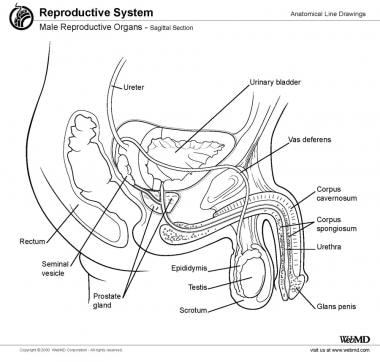
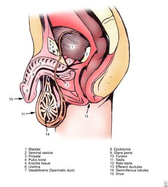
"Male genitalia" refers to the reproductive and sexual organs that are typically present in male individuals. These structures include:
1. Testes: A pair of oval-shaped glands located in the scrotum that produce sperm and testosterone.
2. Epididymis: A long, coiled tube that lies on the surface of each testicle where sperm matures and is stored.
3. Vas deferens: A pair of muscular tubes that transport sperm from the epididymis to the urethra.
4. Seminal vesicles: Glands that produce a fluid that mixes with sperm to create semen.
5. Prostate gland: A small gland that surrounds the urethra and produces a fluid that also mixes with sperm to create semen.
6. Bulbourethral glands (Cowper's glands): Two pea-sized glands that produce a lubricating fluid that is released into the urethra during sexual arousal.
7. Urethra: A tube that runs through the penis and carries urine from the bladder out of the body, as well as semen during ejaculation.
8. Penis: The external organ that serves as both a reproductive and excretory organ, expelling both semen and urine.
"Inbred strains of rats" are genetically identical rodents that have been produced through many generations of brother-sister mating. This results in a high degree of homozygosity, where the genes at any particular locus in the genome are identical in all members of the strain.
Inbred strains of rats are widely used in biomedical research because they provide a consistent and reproducible genetic background for studying various biological phenomena, including the effects of drugs, environmental factors, and genetic mutations on health and disease. Additionally, inbred strains can be used to create genetically modified models of human diseases by introducing specific mutations into their genomes.
Some commonly used inbred strains of rats include the Wistar Kyoto (WKY), Sprague-Dawley (SD), and Fischer 344 (F344) rat strains. Each strain has its own unique genetic characteristics, making them suitable for different types of research.
Gene expression is the process by which the information encoded in a gene is used to synthesize a functional gene product, such as a protein or RNA molecule. This process involves several steps: transcription, RNA processing, and translation. During transcription, the genetic information in DNA is copied into a complementary RNA molecule, known as messenger RNA (mRNA). The mRNA then undergoes RNA processing, which includes adding a cap and tail to the mRNA and splicing out non-coding regions called introns. The resulting mature mRNA is then translated into a protein on ribosomes in the cytoplasm through the process of translation.
The regulation of gene expression is a complex and highly controlled process that allows cells to respond to changes in their environment, such as growth factors, hormones, and stress signals. This regulation can occur at various stages of gene expression, including transcriptional activation or repression, RNA processing, mRNA stability, and translation. Dysregulation of gene expression has been implicated in many diseases, including cancer, genetic disorders, and neurological conditions.
Zonula Occludens-1 (ZO-1) protein is a tight junction (TJ) protein, which belongs to the membrane-associated guanylate kinase (MAGUK) family. It plays a crucial role in the formation and maintenance of tight junctions, which are complex structures that form a barrier between neighboring cells in epithelial and endothelial tissues.
Tight junctions are composed of several proteins, including transmembrane proteins and cytoplasmic plaque proteins. ZO-1 is one of the major cytoplasmic plaque proteins that interact with both transmembrane proteins (such as occludin and claudins) and other cytoskeletal proteins to form a network of protein interactions that maintain the integrity of tight junctions.
ZO-1 has multiple domains, including PDZ domains, SH3 domains, and a guanylate kinase-like domain, which allow it to interact with various binding partners. It is involved in regulating paracellular permeability, cell polarity, and signal transduction pathways that control cell proliferation, differentiation, and survival.
Mutations or dysfunction of ZO-1 protein have been implicated in several human diseases, including inflammatory bowel disease, cancer, and neurological disorders.
Medical Definition of Vitamin A:
Vitamin A is a fat-soluble vitamin that is essential for normal vision, immune function, and cell growth. It is also an antioxidant that helps protect the body's cells from damage caused by free radicals. Vitamin A can be found in two main forms: preformed vitamin A, which is found in animal products such as dairy, fish, and meat, particularly liver; and provitamin A carotenoids, which are found in plant-based foods such as fruits, vegetables, and vegetable oils.
The most active form of vitamin A is retinoic acid, which plays a critical role in the development and maintenance of the heart, lungs, kidneys, and other organs. Vitamin A deficiency can lead to night blindness, dry skin, and increased susceptibility to infections. Chronic vitamin A toxicity can cause nausea, dizziness, headaches, coma, and even death.
"Wistar rats" are a strain of albino rats that are widely used in laboratory research. They were developed at the Wistar Institute in Philadelphia, USA, and were first introduced in 1906. Wistar rats are outbred, which means that they are genetically diverse and do not have a fixed set of genetic characteristics like inbred strains.
Wistar rats are commonly used as animal models in biomedical research because of their size, ease of handling, and relatively low cost. They are used in a wide range of research areas, including toxicology, pharmacology, nutrition, cancer, cardiovascular disease, and behavioral studies. Wistar rats are also used in safety testing of drugs, medical devices, and other products.
Wistar rats are typically larger than many other rat strains, with males weighing between 500-700 grams and females weighing between 250-350 grams. They have a lifespan of approximately 2-3 years. Wistar rats are also known for their docile and friendly nature, making them easy to handle and work with in the laboratory setting.
Actin is a type of protein that forms part of the contractile apparatus in muscle cells, and is also found in various other cell types. It is a globular protein that polymerizes to form long filaments, which are important for many cellular processes such as cell division, cell motility, and the maintenance of cell shape. In muscle cells, actin filaments interact with another type of protein called myosin to enable muscle contraction. Actins can be further divided into different subtypes, including alpha-actin, beta-actin, and gamma-actin, which have distinct functions and expression patterns in the body.
Fluorescence microscopy is a type of microscopy that uses fluorescent dyes or proteins to highlight and visualize specific components within a sample. In this technique, the sample is illuminated with high-energy light, typically ultraviolet (UV) or blue light, which excites the fluorescent molecules causing them to emit lower-energy, longer-wavelength light, usually visible light in the form of various colors. This emitted light is then collected by the microscope and detected to produce an image.
Fluorescence microscopy has several advantages over traditional brightfield microscopy, including the ability to visualize specific structures or molecules within a complex sample, increased sensitivity, and the potential for quantitative analysis. It is widely used in various fields of biology and medicine, such as cell biology, neuroscience, and pathology, to study the structure, function, and interactions of cells and proteins.
There are several types of fluorescence microscopy techniques, including widefield fluorescence microscopy, confocal microscopy, two-photon microscopy, and total internal reflection fluorescence (TIRF) microscopy, each with its own strengths and limitations. These techniques can provide valuable insights into the behavior of cells and proteins in health and disease.
Cell adhesion refers to the binding of cells to extracellular matrices or to other cells, a process that is fundamental to the development, function, and maintenance of multicellular organisms. Cell adhesion is mediated by various cell surface receptors, such as integrins, cadherins, and immunoglobulin-like cell adhesion molecules (Ig-CAMs), which interact with specific ligands in the extracellular environment. These interactions lead to the formation of specialized junctions, such as tight junctions, adherens junctions, and desmosomes, that help to maintain tissue architecture and regulate various cellular processes, including proliferation, differentiation, migration, and survival. Disruptions in cell adhesion can contribute to a variety of diseases, including cancer, inflammation, and degenerative disorders.
 Stem-cell niche
Prostate evolution in monotreme mammals
Capybara
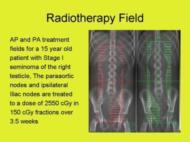
Seminoma
Spermatogenesis
Charles Philippe Leblond
Meiosis
Gonocyte
Male infertility
Germinal epithelium (male)
Spermatogenesis arrest
Testicular immunology
Cell junction
Ectoplasmic specialisation
Male contraceptive
Adjudin
Spermatogonial stem cell
Estrogen receptor alpha
Spermatocyte
Nimetazepam
Testicle
Neutering
Geminin coiled-coil domain-containing protein 1
List of MeSH codes (A10)
List of MeSH codes (A05)
FNA Mapping
Simple cuboidal epithelium
Seminiferous tubule
Gap junction
SCNN1A
 Localization of protamine 1 mRNA in different stages of the cycle of the rat seminiferous epithelium. | Journal of Cell Biology...
 Acrosomal marker SP-10 (gene name Acrv1) for staging of the cycle of seminiferous epithelium in the stallion<...
Stem-cell niche - Wikipedia
 PDF) Endocrine Active UV Filters: Developmental Toxicity and Exposure Through Breast Milk
Immunodistribution of RFamide-related peptide-3 (RFRP-3) through the seminiferous epithelium routine in a leave rodent...
Background Methotrexate (MTX) offers been proven to have an effect on the testes adversely, the seminiferous epithelium...
 JCI -
The key role of vitamin A in spermatogenesis
JCI -
The key role of vitamin A in spermatogenesis
 Pathology Outlines - Torsion
 High fat diet-induced obesity prolongs critical stages of the spermatogenic cycle in a Ldlr−/−.Leiden mouse model | Scientific...
 Taking Stock
 Animals | Free Full-Text | The Effect of Dietary Supplementation of Vitamin E, Selenium, Zinc, Folic Acid, and N-3...
 Spermatocele: Practice Essentials, History of the Procedure, Problem
 The actin filament network associated to Sertoli cell ectoplasmic specializations
 PRKRA Localizes to Nuage Structures and the Ectoplasmic Specialization and Tubulobulbar Complexes in Rat and Mouse Testis
 HumanKine® recombinant human GDNF protein | Proteintech
 These highlights do not include all the information needed to use ROSUVASTATIN TABLETS safely and effectively. See full...
 Phytoestrogens: an example of endocrine disruptor | IVIS
DailyMed - ROSUVASTATIN CALCIUM tablet, film coated
 Goji: MedlinePlus Supplements
 SciELO - Brazil - Scrotal thermographic profile and seminal characteristics of Mangalarga Marchador stallions bred in the...
 WikiGenes - Oligospermia
WikiGenes - Gardenal - 5-ethyl-5-phenyl-1,3-diazinane-2,4,6-trione
 Volume 100 Issue 1 | Biology of Reproduction
 Search - NeL.edu
 Japanese B Encephalitis | Iowa State University
 CABYR isoforms expressed in late steps of spermiogenesis bind with AKAPs and ropporin in mouse sperm fibrous sheath |...
 Cialis Tablets (Lilly ICOS), Drug Reference Encyclopedia
Tubules10
- Northern blot analyses of RNAs isolated from microdissected pools of seminiferous tubules show high P1 protamine mRNA concentrations during stages VIIc-XIV-III of the cycle and lower levels during stages IV-VIIb. (rupress.org)
- A ) The mammalian testis is composed of seminiferous tubules intertwined so that the "start" and "end" of these tubules are both connected to the rete testis. (jci.org)
- Immotile sperm flow from the lumen of the seminiferous tubules into the epididymis via the rete testis. (jci.org)
- B ) Histological cross section through an adult mouse testis depicting seminiferous tubules, the peritubular myoid cells, and the interstitium (space between tubules). (jci.org)
- We induced high fat diet (HFD) obesity in low-density lipoprotein receptor knock-out Leiden (Ldlr −/− .Leiden) mice, and studied the morphological structure of the testes and histologically examined the proportion of Sertoli cells, spermatocytes and spermatids in the seminiferous tubules. (nature.com)
- (b) The leydig cells of the seminiferous tubules secrete the male sex hormone called testosterone. (saralstudy.com)
- The production of sperms in the testes takes place in a highly coiled structure called the seminiferous tubules. (saralstudy.com)
- There are large polygonal cells known as interstitial cells or leydig cells just adjacent to seminiferous tubules. (saralstudy.com)
- It takes place in seminiferous tubules present inside the testes. (saralstudy.com)
- It is the process when mature spermatozoa are released from the sertoli cells into the lumen of seminiferous tubules. (saralstudy.com)
Germinal epithelium2
- The main alterations include decreased germinal epithelium in tubule, and the loss of a continuous proliferation and differentiation gamete layer. (nel.edu)
- Each seminiferous tubule is lined by germinal epithelium. (saralstudy.com)
Tubule4
- Germ cell movement within the epithelium is vital because germ cells, if induced to release into the tubule lumen prematurely, will be unable to fertilize the ovum. (proteinlounge.com)
- Describe the structure of a seminiferous tubule. (saralstudy.com)
- The low BW roosters had lower testes weight (TW), smaller seminiferous tubule diameter (STD) and lower comb and cloaca scores than the medium and high BW roosters (P≤0.05). (bvsalud.org)
- There was no statistical difference between the BW categories in FS and volumetric proportion of seminiferous tubule (ST) and interstitial tissue (IT). (bvsalud.org)
Sertoli3
- Labelling was found near the basal lamina, surrounding early spermatocytes (presumably in leptotene-zygotene) or at one of two levels in the seminiferous epithelium: (1) around deep infoldings of the Sertoli cell cytoplasm, in tubular stages before spermiation, and (2) in the superficial part of the seminiferous epithelium, in tubular stages after or during spermiation. (techscience.com)
- In the testis, Sertoli cells serve as supporting cells and reside as a basal epithelial lining within the seminiferous epithelium. (biomedcentral.com)
- On the other hand, if germ cells are forced to remain attached to the seminiferous epithelium for a period of time longer than necessary to complete their development, they will degenerate and eventually be phagocytosed by Sertoli cells and for this, proper regulation of germ cell migration in the seminiferous epithelium and at blood-testes barrier (abbreviated as BTB) is essential (Ref.1). (proteinlounge.com)
Testes1
- Animal studies have shown that the development of the male reproductive system, especially the seminiferous epithelium of the testes, may be disrupted by in utero exposure to high doses of di-n-butyl phthalate during the critical period for reproductive development. (cdc.gov)
Morphological2
- Short-term treatment with flutamide applied to adult rats exerts its primary effect on the basal ES, coexisting junctional complexes and their constituent proteins Cx43 and ZO-1, without any apparent morphological alterations in the seminiferous epithelium. (biomedcentral.com)
- Pancreatic ductal adenocarcinoma evolves from a progressive cascade of cellular, morphological and architectural changes from normal ductal epithelium through pre-neoplastic lesions termed PanIN (Pancreatic Intraepithelial Neoplasia). (proteinlounge.com)
Degenerative changes1
- In mature boars there is excessive fluid in the tunica vaginalis, orchitis, epididymitis, and degenerative changes in seminiferous epithelium. (iastate.edu)
Testis1
- In conclusion, significant interrelations among the β-actin network, the junctional complexes of the blood-testis barrier and the ectoplasmic specializations were detected at different stages of the seminiferous cycle. (techscience.com)
Cycle6
- Localization of protamine 1 mRNA in different stages of the cycle of the rat seminiferous epithelium. (rupress.org)
- A mouse protamine 1 cDNA probe was used to study P1 protamine gene expression during the cycle of the seminiferous epithelium in the rat. (rupress.org)
- The SP-10 antibody has been shown to be useful for staging the seminiferous cycle in the mouse and human. (illinois.edu)
- The objectives of the present study were to investigate the presence of SP-10 within the horse acrosome using an anti-mouse SP-10 antibody, to classify spermatids based on the shape of the acrosome, and then to use that information to assign stages of the cycle of the seminiferous epithelium. (illinois.edu)
- These changes over time and distance are designated the cycle of the seminiferous epithelium and the spermatogenic wave, respectively. (jci.org)
- In this Review, we briefly outline the roles that follicle-stimulating hormone (FSH) and testosterone play in regulating spermatogenesis and describe our current understanding of how vitamin A regulates germ cell differentiation and how it may lead to the generation of both the cycle of the seminiferous epithelium and the spermatogenic wave. (jci.org)
Sperm1
- The aim of this study was to examine ovine sperm cryoresistance during the rutting season (RS) and its association with sperm head area and seminiferous epithelium proliferation. (uni-bonn.de)
Ectoplasmic2
- Ectoplasmic specializations were found at two different levels of the seminiferous epithelium. (techscience.com)
- Particular emphasis was placed on the basal ectoplasmic specialization (ES) in the seminiferous epithelium and expression of gap junction protein, connexin 43 (Cx43). (biomedcentral.com)
Germ1
- Seminiferous epithelium of both groups of rats displayed normal histology without any loss of germ cells. (biomedcentral.com)
Morphology1
- Previous methods based on morphology alone without the aid of an antibody marker showed 8 stages in the horse seminiferous epithelium. (illinois.edu)
Germ cells4
- A disruption in the physical association between Sertoli cells and germ cells and premature loss of germ cells from the seminiferous epithelium has been widely described after phthalate treatment. (nih.gov)
- The understanding of spermatogenesis needs detailed informations about the organization of the germinal epithelium, the structure and function of different types of germ cells, endocrine and paracrine cells and mechanisms, intratesticular and extratesticular regulation of spermatogenesis. (biomedcentral.com)
- The germinal epithelium (Fig. 2A ) consists of cells that include different developmental stages of germ cells, namely spermatogonia, primary and secondary spermatocytes and spermatids. (biomedcentral.com)
- The seminiferous epithelium is composed of Sertoli cells (SCs) and germ cells (GCs). (biomedcentral.com)
Germinal4
- 900 (B) Sertoli cells divide the germinal epithelium in a basal and adluminal compartment. (biomedcentral.com)
- large, columnar cells that extend the full thickness of the germinal epithelium. (histologyguide.com)
- Many dividing cells are seen in the germinal epithelium. (histologyguide.com)
- arise from spermatogonia and cross from the basal epithelial to luminal compartment of the germinal epithelium. (histologyguide.com)
Epithelial2
- The second is that the expression level of Jagged1 oscillates in the seminiferous epithelial cycle. (cyberleninka.org)
- These results suggest that cAMP signaling may cause the periodicity of Jagged1 expression in the seminiferous epithelial cycle, and controlling Jagged1 expression by adding TNF-α or cAMP may contribute to normal spermatogenesis in vitro. (cyberleninka.org)
Adluminal compartment1
- It consists of adherens junctions (AJs), tight junctions (TJs) and gap junctions (GJs) and divides the seminiferous epithelium into a basal and an adluminal compartment [ 10 ]. (biomedcentral.com)
Sertoli Cells1
- Spermiation involves the final remodeling and release of sperm from the Sertoli cells of the seminiferous epithelium, prior to their passage to the epididymis. (monash.edu)
Spermatogenic epithelium1
- As the colocalization of Fas-L and Fas- R correlates with the germ cell types that die in response to androgen withdrawal, the potential exists for apoptosis in the rat spermatogenic epithelium to be regulated by the Fas pathway. (manchester.ac.uk)
Thickness1
- Epididymal epithelium and fibromascular thickness were also improved in extract-treated rats compared to control or CP groups. (researchgate.net)
Junctional1
- Since junctional proteins within the seminiferous epithelium are involved in cell organization, differentiation and proliferation, they represent interesting candidates for investigations on intercellular adhesion and communication in context with neoplastic progression. (biomedcentral.com)
Tubular1
- The same tissue section used for measuring tubular diameters was used to measure the seminiferous epithelium height. (unina.it)
Connective tissue1
- Again, numerous lymphocytes (Lym) are in the underlying connective tissue, and heaps of have migrated into the epithelium in the nonkeratinized area. (gov.ph)
Decrease1
- Between the two diamonds within the rectangular areas proven is epithelium of the decrease a part of the anal canal. (gov.ph)