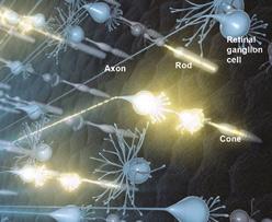
Retinal Ganglion Cells
Retina
Optic Nerve
Optic Nerve Injuries
Glaucoma
Ganglia
Rod Opsins
Visual Pathways
Superior Colliculi
Transcription Factor Brn-3B
Amacrine Cells
Cell Count
Axotomy
Transcription Factor Brn-3A
Optic Chiasm
Ganglia, Spinal
Axonal Transport
Photic Stimulation
Ocular Hypertension
Optic Nerve Diseases
Ganglia, Sensory
Ganglia, Sympathetic
Ganglia, Autonomic
Dendrites
Vision, Ocular
Action Potentials
Retinal Degeneration
Basal Ganglia
Ambystoma
Optic Atrophy, Autosomal Dominant
Cell Survival
Geniculate Bodies
Retrograde Degeneration
Transcription Factor Brn-3
Visual Fields
Trigeminal Ganglion
Ganglia, Parasympathetic
Cats
Retinal Diseases
Fluorescent Antibody Technique, Indirect
Neurons
Transcription Factor Brn-3C
Retinal Cone Photoreceptor Cells
Retinal Bipolar Cells
Cell Death
Reflex, Pupillary
Photoreceptor Cells, Vertebrate
Rats, Inbred BN
Adaptation, Ocular
Mice, Inbred C57BL
Retinal Neurons
Albinism
Antigens, Thy-1
Rats, Long-Evans
Horseradish Peroxidase
Neuroprotective Agents
Disease Models, Animal
Nerve Tissue Proteins
Dark Adaptation
Evoked Potentials, Visual
Rats, Sprague-Dawley
Nerve Fibers
Vitreous Body
Brain-Derived Neurotrophic Factor
Immunohistochemistry
Eye
Photoreceptor Cells
Spiral Ganglion
Nerve Degeneration
Models, Neurological
Chick Embryo
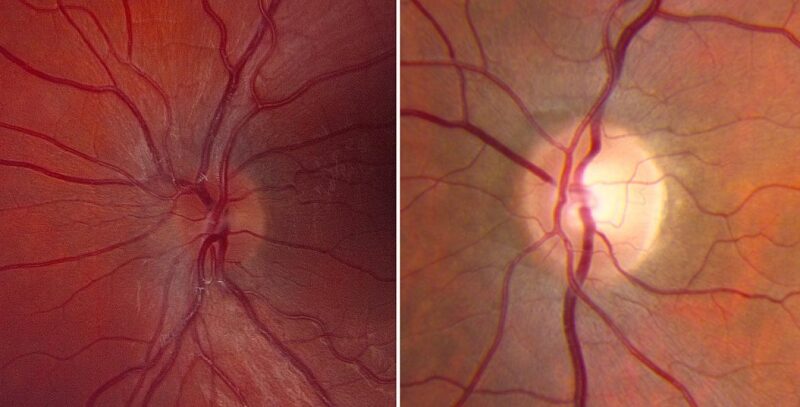
Optic Disk
Aminobutyrates
Urodela
Eye Proteins
Neurites
Synaptic Transmission
Synapses
Macaca fascicularis
Nerve Growth Factors
Cytoprotection
Neurofilament Proteins
N-Methylaspartate
Fluorescent Dyes
Carbocyanines
Electrophysiology
Cells, Cultured
Intravitreal Injections
In Situ Hybridization
Growth Cones
Stellate Ganglion
Patch-Clamp Techniques
Nodose Ganglion
Glycine Agents
In Situ Nick-End Labeling
Glutamic Acid
Optic Atrophy, Hereditary, Leber
Gene Expression Regulation, Developmental
Microscopy, Confocal
Neuroglia
Apoptosis
Strychnine
Mice, Transgenic
Retinal Rod Photoreceptor Cells
Visual Field Tests
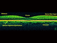
Tomography, Optical Coherence
Contrast Sensitivity
Tonometry, Ocular
Ciliary Neurotrophic Factor
Optic Neuritis
Receptor, EphB1
Green Fluorescent Proteins
Ferrets
Glaucoma, Open-Angle
Blotting, Western
Optic Atrophy
Zebrafish
Membrane Potentials
RNA, Messenger
Rats, Wistar
GAP-43 Protein
Optic Neuropathy, Ischemic
Microelectrodes
Mice, Knockout
Nissl Bodies
Neural Inhibition
Retinal Horizontal Cells
Glial Fibrillary Acidic Protein
Ephrin-A5
Microscopy, Fluorescence
Mice, Inbred DBA
Tetrodotoxin
Fovea Centralis
Ganglia, Invertebrate
gamma-Synuclein
Visual Prosthesis
Choline O-Acetyltransferase
Cryoultramicrotomy
Picrotoxin
Memantine
Cell Differentiation
Ephrin-A2
Autonomic Fibers, Preganglionic
Geniculate Ganglion
Tectum Mesencephali
Rabbits
Reverse Transcriptase Polymerase Chain Reaction
Color Perception
Electrical Synapses
Receptor, EphA3
Macaca
Organ Culture Techniques
Ciliary Body
Excitatory Amino Acid Antagonists
Visual Acuity
Immunoenzyme Techniques
Receptor, trkB
Fasciculation
Signal Transduction
Isoquinolines
Basal Ganglia Diseases
Postsynaptic Potential Summation
Low Tension Glaucoma
Color
Receptors, GABA
Nonlinear Dynamics
Ocular Physiological Phenomena
Pentazocine
Quinoxalines
Basic Helix-Loop-Helix Transcription Factors
Neuropil
Vision Disorders
Zebrafish Proteins
Receptors, N-Methyl-D-Aspartate
Calcium
Correlated firing in rabbit retinal ganglion cells. (1/3596)
A ganglion cell's receptive field is defined as that region on the retinal surface in which a light stimulus will produce a response. While neighboring ganglion cells may respond to the same stimulus in a region where their receptive fields overlap, it generally has been assumed that each cell makes an independent decision about whether to fire. Recent recordings from cat and salamander retina using multiple electrodes have challenged this view of independent firing by showing that neighboring ganglion cells have an increased tendency to fire together within +/-5 ms. However, there is still uncertainty about which types of ganglion cells fire together, the mechanisms that produce coordinated spikes, and the overall function of coordinated firing. To address these issues, the responses of up to 80 rabbit retinal ganglion cells were recorded simultaneously using a multielectrode array. Of the 11 classes of rabbit ganglion cells previously identified, coordinated firing was observed in five. Plots of the spike train cross-correlation function suggested that coordinated firing occurred through two mechanisms. In the first mechanism, a spike in an interneuron diverged to produce simultaneous spikes in two ganglion cells. This mechanism predominated in four of the five classes including the ON brisk transient cells. In the second mechanism, ganglion cells appeared to activate each other reciprocally. This was the predominant pattern of correlated firing in OFF brisk transient cells. By comparing the receptive field profiles of ON and OFF brisk transient cells, a peripheral extension of the OFF brisk transient cell receptive field was identified that might be produced by lateral spike spread. Thus an individual OFF brisk transient cell can respond both to a light stimulus directed at the center of its receptive field and to stimuli that activate neighboring OFF brisk transient cells through their receptive field centers. (+info)CNTF, not other trophic factors, promotes axonal regeneration of axotomized retinal ganglion cells in adult hamsters. (2/3596)
PURPOSE: To investigate the in vivo effects of trophic factors on the axonal regeneration of axotomized retinal ganglion cells in adult hamsters. METHODS: The left optic nerve was transected intracranially or intraorbitally, and a peripheral nerve graft was apposed or sutured to the axotomized optic nerve to enhance regeneration. Trophic factors were applied intravitreally every 5 days. Animals were allowed to survive for 3 or 4 weeks. Regenerating retinal ganglion cells (RGCs) were labeled by applying the dye Fluoro-Gold to the distal end of the peripheral nerve graft 3 days before the animals were killed. RESULTS: Intravitreal application of ciliary neurotrophic factor substantially enhanced the regeneration of damaged axons into a sciatic nerve graft in both experimental conditions (intracranial and intraorbital optic nerve transections) but did not increase the survival of distally axotomized RGCs. Basic fibroblast growth factor and neurotrophins such as nerve growth factor, brain-derived neurotrophic factor, neurotrophin-3, and neurotrophin-4/5 failed to enhance axonal regeneration of distally axotomized RGCs. CONCLUSIONS: Neurons of the adult central nervous system can regenerate in response to trophic supply after injury, and ciliary neurotrophic factor is at least one of the trophic factors that can promote axonal regeneration of axotomized RGCs. (+info)Pilocarpine toxicity in retinal ganglion cells. (3/3596)
PURPOSE: Muscarinic agents reduce intraocular pressure by enhancing aqueous outflow, probably by stimulating ciliary muscle contraction. However, pilocarpine is a well characterized neurotoxin and is widely used to generate animal seizure models. It was therefore investigated whether pilocarpine was also toxic to retinal ganglion cells. METHODS: Dissociated whole retinal preparations were prepared from postnatal day 16 to 19 rats. Retinal ganglion cells had been previously back-labeled with a fluorescent tracer. Retinal cells were incubated with pilocarpine, lithium, and inositol derivatives, and viability of the retrogradely labeled retinal ganglion cells was assayed after 24 hours. RESULTS: Pilocarpine was toxic to retinal ganglion cells in a dose-dependent fashion. This toxicity was potentiated by lithium and blocked by epi- and myo-inositol. CONCLUSIONS: Pilocarpine is toxic to retinal ganglion cells in a mixed culture assay. This toxicity appears to depend on the inositol pathway and is similar to its mode of action in other neurons. However, 0.4 mM pilocarpine (the lowest concentration that did not affect ganglion cell survival) is roughly 1000-fold higher than the vitreal concentration and 20-fold higher than the scleral concentration that can be obtained with topical administration of 2% pilocarpine in the rabbit eye. (+info)Retinal neurogenesis: the formation of the initial central patch of postmitotic cells. (4/3596)
We have investigated the relationship between the birthdate and the onset of differentiation of neurons in the embryonic zebrafish neural retina. Birthdates were established by a single injection of bromodeoxyuridine into embryos of closely spaced ages. Differentiation was revealed in the same embryos with a neuron-specific antibody, zn12. The first bromodeoxyuridine-negative (postmitotic) cells occupied the ganglion cell layer of ventronasal retina, where they formed a small cluster of 10 cells or less that included the first zn12-positive cells (neurons). New cells were recruited to both populations (bromodeoxyuridine-negative and zn12-positive) along the same front, similar to the unfolding of a fan, to produce a circular central patch of hundreds of cells in the ganglion cell layer about 9 h later. Thus the formation of this central patch, previously considered as the start of retinal neurogenesis, was actually a secondary event, with a developmental history of its own. The first neurons outside the ganglion cell layer also appeared in ventronasal retina, indicating that the ventronasal region was the site of initiation of all retinal neurogenesis. Within a column (a small cluster of neuroepithelial cells), postmitotic cells appeared first in the ganglion cell layer, then the inner nuclear layer, and then the outer nuclear layer, so cell birthday and cell fate were correlated within a column. The terminal mitoses occurred in three bursts separated by two 10-h intervals during which proliferation continued without terminal mitoses. (+info)Modulation of glycine receptors in retinal ganglion cells by zinc. (5/3596)
Effects of zinc, an endogenous neuromodulator in the central nervous system, on glycine receptors (GlyRs) in retinal ganglion cells were investigated by using the whole-cell voltage-clamp technique. Zn2+ at low concentration (<2 microM) potentiated the glycine-induced chloride current and at higher concentration (>10 microM) suppressed it. This biphasic regulatory action of zinc acted selectively on the fast component of the glycine-induced current mediated by the strychnine-sensitive GlyRs, but not on the slow component mediated by the 5,7-dichlorokynurenic acid-sensitive GlyRs. Dose-response studies showed that 1 microM Zn2+ increased the maximum glycine response (I approximately) and shifted the EC50 to the left, suggesting that Zn2+ at low concentrations acts as an allosteric activator of the strychnine-sensitive GlyRs. Zn2+ at a concentration of 100 microM did not alter I approximately and shifted the EC50 to the right, indicating that Zn2+ at high concentrations acts as a competitive inhibitor of the GlyRs. Physiological functions of zinc modulation of GlyRs in retinal ganglion cells are discussed. (+info)Action potentials in the dendrites of retinal ganglion cells. (6/3596)
The somas and dendrites of intact retinal ganglion cells were exposed by enzymatic removal of the overlying endfeet of the Muller glia. Simultaneous whole cell patch recordings were made from a ganglion cell's dendrite and the cell's soma. When a dendrite was stimulated with depolarizing current, impulses often propagated to the soma, where they appeared as a mixture of small depolarizations and action potentials. When the soma was stimulated, action potentials always propagated back through the dendrite. The site of initiation of action potentials, as judged by their timing, could be shifted between soma and dendrite by changing the site of stimulation. Applying QX-314 to the soma could eliminate somatic action potentials while leaving dendritic impulses intact. The absolute amplitudes of the dendritic action potentials varied somewhat at different distances from the soma, and it is not clear whether these variations are real or technical. Nonetheless, the qualitative experiments clearly suggest that the dendrites of retinal ganglion cells generate regenerative Na+ action potentials, at least in response to large direct depolarizations. (+info)Immunohistological studies of metabotropic glutamate receptor subtype 6-deficient mice show no abnormality of retinal cell organization and ganglion cell maturation. (7/3596)
Immature retinal ganglion cells (RGCs) initially show a multistratified dendritic pattern, and, during the postnatal period, these dendrites gradually monostratify into ON and OFF sublaminae. The selective agonist of group III metabotropic glutamate receptors (mGluR), L-2-amino-4-phosphonobutyrate (L-AP-4), hyperpolarizes ON bipolar cells and reduces glutamate release. On the basis of L-AP-4-evoked inhibitory effects on ON-OFF segregation of developing RGCs, it has been hypothesized that glutamate-mediated synaptic activity is crucial for formation of the ON-OFF network. Gene-targeted ablation of mGluR6 specifically expressed in ON bipolar cells blocks normal ON responses but has been predicted to enhance glutamate release from ON bipolar cells. The mGluR6 knock-out mouse therefore provides a unique opportunity to investigate whether glutamate release and ON responses are important factors in the development of ON-OFF segregation. The combination of several different morphological analyses indicates that ON bipolar cells, as well as several distinct amacrine cells, in mGluR6 knock-out mice are normally distributed and correctly extend their terminals to defined retinal laminae. Importantly, both alpha and delta RGCs in adult mGluR6 knock-out mice are found monostratified into cell type-specific layers. Furthermore, no difference between wild-type and mGluR6 knock-out mice is observed in the maturation and dendritic stratification of developing RGCs. Hence, despite a deficit in normal ON responses, mGluR6 deficiency causes no abnormality in the retinal cellular organization nor in the stratifications of both ON bipolar cells and developing and mature RGCs. Based on these findings, we discuss several possible mechanisms that may underlie ON-OFF segregation of RGCs. (+info)Differential effects of apamin- and charybdotoxin-sensitive K+ conductances on spontaneous discharge patterns of developing retinal ganglion cells. (8/3596)
The spontaneous discharge patterns of developing retinal ganglion cells are thought to play a crucial role in the refinement of early retinofugal projections. To investigate the contributions of intrinsic membrane properties to the spontaneous activity of developing ganglion cells, we assessed the effects of blocking large and small calcium-activated potassium conductances on the temporal pattern of such discharges by means of patch-clamp recordings from the intact retina of developing ferrets. Application of apamin and charybdotoxin (CTX), which selectively block the small and large calcium-activated potassium channels, respectively, resulted in significant changes in spontaneous firings. In cells recorded from the oldest animals [postnatal day 30 (P30)-P45], which manifested relatively sustained discharge patterns, application of either blocker induced bursting activity. With CTX the bursts were highly periodic, short in duration, and of high frequency. In contrast, with apamin the interburst intervals were longer, less regular, and lower in overall spike frequency. These differences between the effects of the two blockers on spontaneous activity were documented by spectral analysis of discharge patterns. Filling cells from which recordings were made with Lucifer yellow revealed that these effects were obtained in all three morphological classes of cells: alpha, beta, and gamma. These findings provide the first evidence that apamin- and CTX-sensitive K+ conductances can have differential effects on the spontaneous discharge patterns of retinal ganglion cells. Remarkably, the bursts of activity obtained after apamin application in more mature neurons appeared very similar to the spontaneous bursting patterns observed in developing neurons. These findings suggest that the maturation of calcium-activated potassium channels, particularly the apamin-sensitive conductance, may contribute to the changes in spontaneous firings exhibited by retinal ganglion cells during the course of normal development. (+info)Retinal Ganglion Cells (RGCs) are a type of neuron located in the innermost layer of the retina, the light-sensitive tissue at the back of the eye. These cells receive visual information from photoreceptors (rods and cones) via intermediate cells called bipolar cells. RGCs then send this visual information through their long axons to form the optic nerve, which transmits the signals to the brain for processing and interpretation as vision.
There are several types of RGCs, each with distinct morphological and functional characteristics. Some RGCs are specialized in detecting specific features of the visual scene, such as motion, contrast, color, or brightness. The diversity of RGCs allows for a rich and complex representation of the visual world in the brain.
Damage to RGCs can lead to various visual impairments, including loss of vision, reduced visual acuity, and altered visual fields. Conditions associated with RGC damage or degeneration include glaucoma, optic neuritis, ischemic optic neuropathy, and some inherited retinal diseases.
The retina is the innermost, light-sensitive layer of tissue in the eye of many vertebrates and some cephalopods. It receives light that has been focused by the cornea and lens, converts it into neural signals, and sends these to the brain via the optic nerve. The retina contains several types of photoreceptor cells including rods (which handle vision in low light) and cones (which are active in bright light and are capable of color vision).
In medical terms, any pathological changes or diseases affecting the retinal structure and function can lead to visual impairment or blindness. Examples include age-related macular degeneration, diabetic retinopathy, retinal detachment, and retinitis pigmentosa among others.
The optic nerve, also known as the second cranial nerve, is the nerve that transmits visual information from the retina to the brain. It is composed of approximately one million nerve fibers that carry signals related to vision, such as light intensity and color, from the eye's photoreceptor cells (rods and cones) to the visual cortex in the brain. The optic nerve is responsible for carrying this visual information so that it can be processed and interpreted by the brain, allowing us to see and perceive our surroundings. Damage to the optic nerve can result in vision loss or impairment.
Optic nerve injuries refer to damages or trauma inflicted on the optic nerve, which is a crucial component of the visual system. The optic nerve transmits visual information from the retina to the brain, enabling us to see. Injuries to the optic nerve can result in various visual impairments, including partial or complete vision loss, decreased visual acuity, changes in color perception, and reduced field of view.
These injuries may occur due to several reasons, such as:
1. Direct trauma to the eye or head
2. Increased pressure inside the eye (glaucoma)
3. Optic neuritis, an inflammation of the optic nerve
4. Ischemia, or insufficient blood supply to the optic nerve
5. Compression from tumors or other space-occupying lesions
6. Intrinsic degenerative conditions affecting the optic nerve
7. Toxic exposure to certain chemicals or medications
Optic nerve injuries are diagnosed through a comprehensive eye examination, including visual acuity testing, slit-lamp examination, dilated fundus exam, and additional diagnostic tests like optical coherence tomography (OCT) and visual field testing. Treatment options vary depending on the cause and severity of the injury but may include medications, surgery, or vision rehabilitation.
An axon is a long, slender extension of a neuron (a type of nerve cell) that conducts electrical impulses (nerve impulses) away from the cell body to target cells, such as other neurons or muscle cells. Axons can vary in length from a few micrometers to over a meter long and are typically surrounded by a myelin sheath, which helps to insulate and protect the axon and allows for faster transmission of nerve impulses.
Axons play a critical role in the functioning of the nervous system, as they provide the means by which neurons communicate with one another and with other cells in the body. Damage to axons can result in serious neurological problems, such as those seen in spinal cord injuries or neurodegenerative diseases like multiple sclerosis.
Glaucoma is a group of eye conditions that damage the optic nerve, often caused by an abnormally high pressure in the eye (intraocular pressure). This damage can lead to permanent vision loss or even blindness if left untreated. The most common type is open-angle glaucoma, which has no warning signs and progresses slowly. Angle-closure glaucoma, on the other hand, can cause sudden eye pain, redness, nausea, and vomiting, as well as rapid vision loss. Other less common types of glaucoma also exist. While there is no cure for glaucoma, early detection and treatment can help slow or prevent further vision loss.
A ganglion is a cluster of neuron cell bodies in the peripheral nervous system. Ganglia are typically associated with nerves and serve as sites for sensory processing, integration, and relay of information between the periphery and the central nervous system (CNS). The two main types of ganglia are sensory ganglia, which contain pseudounipolar neurons that transmit sensory information to the CNS, and autonomic ganglia, which contain multipolar neurons that control involuntary physiological functions.
Examples of sensory ganglia include dorsal root ganglia (DRG), which are associated with spinal nerves, and cranial nerve ganglia, such as the trigeminal ganglion. Autonomic ganglia can be further divided into sympathetic and parasympathetic ganglia, which regulate different aspects of the autonomic nervous system.
It's worth noting that in anatomy, "ganglion" refers to a group of nerve cell bodies, while in clinical contexts, "ganglion" is often used to describe a specific type of cystic structure that forms near joints or tendons, typically in the wrist or foot. These ganglia are not related to the peripheral nervous system's ganglia but rather are fluid-filled sacs that may cause discomfort or pain due to their size or location.
Rhodopsin, also known as visual purple, is a light-sensitive protein found in the rods of the eye's retina. It is a type of opsin, a class of proteins that are activated by light and play a crucial role in vision. Rhodopsin is composed of two parts: an apoprotein called opsin and a chromophore called 11-cis-retinal. When light hits the retina, it changes the shape of the 11-cis-retinal, which in turn activates the rhodopsin protein. This activation triggers a series of chemical reactions that ultimately lead to the transmission of a visual signal to the brain. Rhodopsin is highly sensitive to light and allows for vision in low-light conditions.
Visual pathways, also known as the visual system or the optic pathway, refer to the series of specialized neurons in the nervous system that transmit visual information from the eyes to the brain. This complex network includes the retina, optic nerve, optic chiasma, optic tract, lateral geniculate nucleus, pulvinar, and the primary and secondary visual cortices located in the occipital lobe of the brain.
The process begins when light enters the eye and strikes the photoreceptor cells (rods and cones) in the retina, converting the light energy into electrical signals. These signals are then transmitted to bipolar cells and subsequently to ganglion cells, whose axons form the optic nerve. The fibers from each eye's nasal hemiretina cross at the optic chiasma, while those from the temporal hemiretina continue without crossing. This results in the formation of the optic tract, which carries visual information from both eyes to the opposite side of the brain.
The majority of fibers in the optic tract synapse with neurons in the lateral geniculate nucleus (LGN), a part of the thalamus. The LGN sends this information to the primary visual cortex, also known as V1 or Brodmann area 17, located in the occipital lobe. Here, simple features like lines and edges are initially processed. Further processing occurs in secondary (V2) and tertiary (V3-V5) visual cortices, where more complex features such as shape, motion, and depth are analyzed. Ultimately, this information is integrated to form our perception of the visual world.
The superior colliculi are a pair of prominent eminences located on the dorsal surface of the midbrain, forming part of the tectum or roof of the midbrain. They play a crucial role in the integration and coordination of visual, auditory, and somatosensory information for the purpose of directing spatial attention and ocular movements. Essentially, they are involved in the reflexive orienting of the head and eyes towards novel or significant stimuli in the environment.
In a more detailed medical definition, the superior colliculi are two rounded, convex mounds of gray matter that are situated on the roof of the midbrain, specifically at the level of the rostral mesencephalic tegmentum. Each superior colliculus has a stratified laminated structure, consisting of several layers that process different types of sensory information and control specific motor outputs.
The superficial layers of the superior colliculi primarily receive and process visual input from the retina, lateral geniculate nucleus, and other visual areas in the brain. These layers are responsible for generating spatial maps of the visual field, which allow for the localization and identification of visual stimuli.
The intermediate and deep layers of the superior colliculi receive and process auditory and somatosensory information from various sources, including the inferior colliculus, medial geniculate nucleus, and ventral posterior nucleus of the thalamus. These layers are involved in the localization and identification of auditory and tactile stimuli, as well as the coordination of head and eye movements towards these stimuli.
The superior colliculi also contain a population of neurons called "motor command neurons" that directly control the muscles responsible for orienting the eyes, head, and body towards novel or significant sensory events. These motor command neurons are activated in response to specific patterns of activity in the sensory layers of the superior colliculus, allowing for the rapid and automatic orientation of attention and gaze towards salient stimuli.
In summary, the superior colliculi are a pair of structures located on the dorsal surface of the midbrain that play a critical role in the integration and coordination of visual, auditory, and somatosensory information for the purpose of orienting attention and gaze towards salient stimuli. They contain sensory layers that generate spatial maps of the environment, as well as motor command neurons that directly control the muscles responsible for orienting the eyes, head, and body.
Transcription Factor Brn-3B, also known as POU4F2, is a member of the POU-domain transcription factor family that plays crucial roles in the development and function of the nervous system. This protein contains a specific DNA-binding domain called the POU-domain, which recognizes and binds to specific DNA sequences, thereby regulating the expression of target genes.
Brn-3B is predominantly expressed in the developing and mature sensory neurons of the peripheral and central nervous systems. It has been implicated in several critical processes, such as neurogenesis, differentiation, survival, and maintenance of these neuronal populations. Additionally, Brn-3B has been associated with various neuropathological conditions, including neurodegenerative diseases and cancer, highlighting its importance in the proper functioning of the nervous system.
In summary, Transcription Factor Brn-3B is a DNA-binding protein that regulates gene expression in neurons, contributing to their development, maintenance, and function.
Amacrine cells are a type of neuron found in the inner nuclear layer of the retina, a light-sensitive tissue located at the back of the eye. These interneurons derive their name from the Greek word "amakrin," meaning "short-tailed," due to their short or absent axons.
Amacrine cells play a crucial role in processing and transmitting visual information within the retina. They receive input from bipolar cells, another type of retinal neuron, and synapse onto ganglion cells, which transmit visual signals to the brain via the optic nerve.
There are more than 30 different types of amacrine cells identified based on their morphology, neurotransmitter expression, and synaptic connections. These diverse cells contribute to various retinal functions, such as motion detection, contrast enhancement, direction selectivity, and spatial and temporal processing of visual signals.
Some amacrine cells release the neurotransmitter gamma-aminobutyric acid (GABA), which inhibits the activity of target neurons, while others use excitatory neurotransmitters like acetylcholine or glutamate. The intricate interplay between these various types of amacrine cells and other retinal neurons enables the retina to perform complex computations on visual information before it is relayed to the brain.
"Cell count" is a medical term that refers to the process of determining the number of cells present in a given volume or sample of fluid or tissue. This can be done through various laboratory methods, such as counting individual cells under a microscope using a specialized grid called a hemocytometer, or using automated cell counters that use light scattering and electrical impedance techniques to count and classify different types of cells.
Cell counts are used in a variety of medical contexts, including hematology (the study of blood and blood-forming tissues), microbiology (the study of microscopic organisms), and pathology (the study of diseases and their causes). For example, a complete blood count (CBC) is a routine laboratory test that includes a white blood cell (WBC) count, red blood cell (RBC) count, hemoglobin level, hematocrit value, and platelet count. Abnormal cell counts can indicate the presence of various medical conditions, such as infections, anemia, or leukemia.
Stilbamidines are a class of chemical compounds that are primarily used as veterinary medicines, specifically as parasiticides for the treatment and prevention of ectoparasites such as ticks and lice in livestock animals. Stilbamidines belong to the family of chemicals known as formamidines, which are known to have insecticidal and acaricidal properties.
The most common stilbamidine compound is chlorphentermine, which has been used as an appetite suppressant in human medicine. However, its use as a weight loss drug was discontinued due to its addictive properties and potential for serious side effects.
It's important to note that Stilbamidines are not approved for use in humans and should only be used under the supervision of a veterinarian for the intended purpose of treating and preventing ectoparasites in animals.
Axotomy is a medical term that refers to the surgical cutting or severing of an axon, which is the long, slender projection of a neuron (nerve cell) that conducts electrical impulses away from the cell body and toward other cells. Axons are a critical component of the nervous system, allowing for communication between different parts of the body.
Axotomy is often used in research settings to study the effects of axonal injury on neuronal function and regeneration. This procedure can provide valuable insights into the mechanisms underlying neurodegenerative disorders and potential therapies for nerve injuries. However, it is important to note that axotomy can also have significant consequences for the affected neuron, including changes in gene expression, metabolism, and overall survival.
A nerve crush injury is a type of peripheral nerve injury that occurs when there is excessive pressure or compression applied to a nerve, causing it to become damaged or dysfunctional. This can happen due to various reasons such as trauma from accidents, surgical errors, or prolonged pressure on the nerve from tight casts, clothing, or positions.
The compression disrupts the normal functioning of the nerve, leading to symptoms such as numbness, tingling, weakness, or pain in the affected area. In severe cases, a nerve crush injury can cause permanent damage to the nerve, leading to long-term disability or loss of function. Treatment for nerve crush injuries typically involves relieving the pressure on the nerve, providing supportive care, and in some cases, surgical intervention may be necessary to repair the damaged nerve.
Transcription Factor Brn-3A, also known as POU Class 4 Homeobox 1 (POU4F1), is a protein involved in the regulation of gene transcription. It belongs to the class IV subfamily of POU domain transcription factors, which are characterized by a highly conserved DNA-binding domain called the POU domain.
Brn-3A plays crucial roles in the development and function of the nervous system, particularly in the differentiation and survival of neurons. It regulates the expression of various target genes involved in neural functions such as neurotransmission, synaptic plasticity, and nerve regeneration. Brn-3A has been implicated in several neurological disorders, including neurodegenerative diseases and neuropathic pain.
The optic chiasm is a structure in the brain where the optic nerves from each eye meet and cross. This allows for the integration of visual information from both eyes into the brain's visual cortex, creating a single, combined image of the visual world. The optic chiasm plays an important role in the processing of visual information and helps to facilitate depth perception and other complex visual tasks. Damage to the optic chiasm can result in various visual field deficits, such as bitemporal hemianopsia, where there is a loss of vision in the outer halves (temporal fields) of both eyes' visual fields.
Intraocular pressure (IOP) is the fluid pressure within the eye, specifically within the anterior chamber, which is the space between the cornea and the iris. It is measured in millimeters of mercury (mmHg). The aqueous humor, a clear fluid that fills the anterior chamber, is constantly produced and drained, maintaining a balance that determines the IOP. Normal IOP ranges from 10-21 mmHg, with average values around 15-16 mmHg. Elevated IOP is a key risk factor for glaucoma, a group of eye conditions that can lead to optic nerve damage and vision loss if not treated promptly and effectively. Regular monitoring of IOP is essential in diagnosing and managing glaucoma and other ocular health issues.
Spinal ganglia, also known as dorsal root ganglia, are clusters of nerve cell bodies located in the peripheral nervous system. They are situated along the length of the spinal cord and are responsible for transmitting sensory information from the body to the brain. Each spinal ganglion contains numerous neurons, or nerve cells, with long processes called axons that extend into the periphery and innervate various tissues and organs. The cell bodies within the spinal ganglia receive sensory input from these axons and transmit this information to the central nervous system via the dorsal roots of the spinal nerves. This allows the brain to interpret and respond to a wide range of sensory stimuli, including touch, temperature, pain, and proprioception (the sense of the position and movement of one's body).
Axonal transport is the controlled movement of materials and organelles within axons, which are the nerve fibers of neurons (nerve cells). This intracellular transport system is essential for maintaining the structural and functional integrity of axons, particularly in neurons with long axonal processes. There are two types of axonal transport: anterograde transport, which moves materials from the cell body toward the synaptic terminals, and retrograde transport, which transports materials from the synaptic terminals back to the cell body. Anterograde transport is typically slower than retrograde transport and can be divided into fast and slow components based on velocity. Fast anterograde transport moves vesicles containing neurotransmitters and their receptors, as well as mitochondria and other organelles, at speeds of up to 400 mm/day. Slow anterograde transport moves cytoskeletal elements, proteins, and RNA at speeds of 1-10 mm/day. Retrograde transport is primarily responsible for recycling membrane components, removing damaged organelles, and transmitting signals from the axon terminal to the cell body. Dysfunctions in axonal transport have been implicated in various neurodegenerative disorders, such as Alzheimer's disease, Parkinson's disease, and amyotrophic lateral sclerosis (ALS).
Photic stimulation is a medical term that refers to the exposure of the eyes to light, specifically repetitive pulses of light, which is used as a method in various research and clinical settings. In neuroscience, it's often used in studies related to vision, circadian rhythms, and brain function.
In a clinical context, photic stimulation is sometimes used in the diagnosis of certain medical conditions such as seizure disorders (like epilepsy). By observing the response of the brain to this light stimulus, doctors can gain valuable insights into the functioning of the brain and the presence of any neurological disorders.
However, it's important to note that photic stimulation should be conducted under the supervision of a trained healthcare professional, as improper use can potentially trigger seizures in individuals who are susceptible to them.
Ocular hypertension is a medical condition characterized by elevated pressure within the eye (intraocular pressure or IOP), which is higher than normal but not necessarily high enough to cause any visible damage to the optic nerve or visual field loss. It serves as a significant risk factor for developing glaucoma, a sight-threatening disease.
The normal range of intraocular pressure is typically between 10-21 mmHg (millimeters of mercury). Ocular hypertension is often defined as an IOP consistently above 21 mmHg, although some studies suggest that even pressures between 22-30 mmHg may not cause damage in all individuals. Regular monitoring and follow-up with an ophthalmologist are essential for people diagnosed with ocular hypertension to ensure early detection and management of any potential glaucomatous changes. Treatment options include medications, laser therapy, or surgery to lower the IOP and reduce the risk of glaucoma onset.
Light signal transduction is a biological process that refers to the way in which cells convert light signals into chemical or electrical responses. This process typically involves several components, including a light-sensitive receptor (such as a photopigment), a signaling molecule (like a G-protein or calcium ion), and an effector protein that triggers a downstream response.
In the visual system, for example, light enters the eye and activates photoreceptor cells in the retina. These cells contain a light-sensitive pigment called rhodopsin, which undergoes a chemical change when struck by a photon of light. This change triggers a cascade of signaling events that ultimately lead to the transmission of visual information to the brain.
Light signal transduction is also involved in other biological processes, such as the regulation of circadian rhythms and the synthesis of vitamin D. In these cases, specialized cells contain light-sensitive receptors that allow them to detect changes in ambient light levels and adjust their physiology accordingly.
Overall, light signal transduction is a critical mechanism by which organisms are able to sense and respond to their environment.
Optic nerve diseases refer to a group of conditions that affect the optic nerve, which transmits visual information from the eye to the brain. These diseases can cause various symptoms such as vision loss, decreased visual acuity, changes in color vision, and visual field defects. Examples of optic nerve diseases include optic neuritis (inflammation of the optic nerve), glaucoma (damage to the optic nerve due to high eye pressure), optic nerve damage from trauma or injury, ischemic optic neuropathy (lack of blood flow to the optic nerve), and optic nerve tumors. Treatment for optic nerve diseases varies depending on the specific condition and may include medications, surgery, or lifestyle changes.
Sensory ganglia are clusters of nerve cell bodies located outside the central nervous system (the brain and spinal cord). They are primarily associated with sensory neurons, which are responsible for transmitting sensory information from various parts of the body to the central nervous system.
In humans, there are two main types of sensory ganglia: dorsal root ganglia and cranial nerve ganglia. Dorsal root ganglia are located along the spinal cord and contain the cell bodies of sensory neurons that innervate the skin, muscles, joints, and other tissues of the body. These neurons transmit information about touch, temperature, pain, and proprioception (the sense of the position and movement of the body).
Cranial nerve ganglia are associated with the cranial nerves, which are responsible for transmitting sensory information from the head and neck to the brain. For example, the trigeminal ganglion is a cranial nerve ganglion that contains the cell bodies of neurons that transmit sensory information from the face, mouth, and other structures of the head.
Overall, sensory ganglia play a critical role in our ability to perceive and interact with the world around us by transmitting important sensory information to the brain for processing.
Sympathetic ganglia are part of the autonomic nervous system, which controls involuntary bodily functions. These ganglia are clusters of nerve cell bodies located outside the central nervous system, along the spinal cord. They serve as a relay station for signals sent from the central nervous system to the organs and glands. The sympathetic ganglia are responsible for the "fight or flight" response, releasing neurotransmitters such as norepinephrine that prepare the body for action in response to stress or danger.
Autonomic ganglia are collections of neurons located outside the central nervous system (CNS) that are a part of the autonomic nervous system (ANS). The ANS is responsible for controlling various involuntary physiological functions such as heart rate, digestion, respiratory rate, pupillary response, urination, and sexual arousal.
Autonomic ganglia receive inputs from preganglionic neurons, whose cell bodies are located in the CNS, and send outputs to effector organs through postganglionic neurons. The autonomic ganglia can be divided into two main subsystems: the sympathetic and parasympathetic systems.
Sympathetic ganglia are typically located close to the spinal cord and receive inputs from preganglionic neurons whose cell bodies are located in the thoracic and lumbar regions of the spinal cord. The postganglionic neurons of the sympathetic system release noradrenaline (also known as norepinephrine) as their primary neurotransmitter, which acts on effector organs to produce a range of responses such as increasing heart rate and blood pressure, dilating pupils, and promoting glucose mobilization.
Parasympathetic ganglia are typically located closer to the target organs and receive inputs from preganglionic neurons whose cell bodies are located in the brainstem and sacral regions of the spinal cord. The postganglionic neurons of the parasympathetic system release acetylcholine as their primary neurotransmitter, which acts on effector organs to produce a range of responses such as decreasing heart rate and blood pressure, constricting pupils, and promoting digestion and urination.
Overall, autonomic ganglia play a critical role in regulating various physiological functions that are essential for maintaining homeostasis in the body.
Electroretinography (ERG) is a medical test used to evaluate the functioning of the retina, which is the light-sensitive tissue located at the back of the eye. The test measures the electrical responses of the retina to light stimulation.
During the procedure, a special contact lens or electrode is placed on the surface of the eye to record the electrical activity generated by the retina's light-sensitive cells (rods and cones) and other cells in the retina. The test typically involves presenting different levels of flashes of light to the eye while the electrical responses are recorded.
The resulting ERG waveform provides information about the overall health and function of the retina, including the condition of the photoreceptors, the integrity of the inner retinal layers, and the health of the retinal ganglion cells. This test is often used to diagnose and monitor various retinal disorders, such as retinitis pigmentosa, macular degeneration, and diabetic retinopathy.
Dendrites are the branched projections of a neuron that receive and process signals from other neurons. They are typically short and highly branching, increasing the surface area for receiving incoming signals. Dendrites are covered in small protrusions called dendritic spines, which can form connections with the axon terminals of other neurons through chemical synapses. The structure and function of dendrites play a critical role in the integration and processing of information in the nervous system.
Ocular vision refers to the ability to process and interpret visual information that is received by the eyes. This includes the ability to see clearly and make sense of the shapes, colors, and movements of objects in the environment. The ocular system, which includes the eye and related structures such as the optic nerve and visual cortex of the brain, works together to enable vision.
There are several components of ocular vision, including:
* Visual acuity: the clarity or sharpness of vision
* Field of vision: the extent of the visual world that is visible at any given moment
* Color vision: the ability to distinguish different colors
* Depth perception: the ability to judge the distance of objects in three-dimensional space
* Contrast sensitivity: the ability to distinguish an object from its background based on differences in contrast
Disorders of ocular vision can include refractive errors such as nearsightedness or farsightedness, as well as more serious conditions such as cataracts, glaucoma, and macular degeneration. These conditions can affect one or more aspects of ocular vision and may require medical treatment to prevent further vision loss.
An action potential is a brief electrical signal that travels along the membrane of a nerve cell (neuron) or muscle cell. It is initiated by a rapid, localized change in the permeability of the cell membrane to specific ions, such as sodium and potassium, resulting in a rapid influx of sodium ions and a subsequent efflux of potassium ions. This ion movement causes a brief reversal of the electrical potential across the membrane, which is known as depolarization. The action potential then propagates along the cell membrane as a wave, allowing the electrical signal to be transmitted over long distances within the body. Action potentials play a crucial role in the communication and functioning of the nervous system and muscle tissue.
Retinal degeneration is a broad term that refers to the progressive loss of photoreceptor cells (rods and cones) in the retina, which are responsible for converting light into electrical signals that are sent to the brain. This process can lead to vision loss or blindness. There are many different types of retinal degeneration, including age-related macular degeneration, retinitis pigmentosa, and Stargardt's disease, among others. These conditions can have varying causes, such as genetic mutations, environmental factors, or a combination of both. Treatment options vary depending on the specific type and progression of the condition.
Nerve regeneration is the process of regrowth and restoration of functional nerve connections following damage or injury to the nervous system. This complex process involves various cellular and molecular events, such as the activation of support cells called glia, the sprouting of surviving nerve fibers (axons), and the reformation of neural circuits. The goal of nerve regeneration is to enable the restoration of normal sensory, motor, and autonomic functions impaired due to nerve damage or injury.
The basal ganglia are a group of interconnected nuclei, or clusters of neurons, located in the base of the brain. They play a crucial role in regulating motor function, cognition, and emotion. The main components of the basal ganglia include the striatum (made up of the caudate nucleus, putamen, and ventral striatum), globus pallidus (divided into external and internal segments), subthalamic nucleus, and substantia nigra (with its pars compacta and pars reticulata).
The basal ganglia receive input from various regions of the cerebral cortex and other brain areas. They process this information and send output back to the thalamus and cortex, helping to modulate and coordinate movement. The basal ganglia also contribute to higher cognitive functions such as learning, decision-making, and habit formation. Dysfunction in the basal ganglia can lead to neurological disorders like Parkinson's disease, Huntington's disease, and dystonia.
"Ambystoma" is a genus of salamanders, also known as the mole salamanders. These amphibians are characterized by their fossorial (burrowing) habits and typically have four limbs, a tail, and moist skin. They are found primarily in North America, with a few species in Asia and Europe. Some well-known members of this genus include the axolotl (A. mexicanum), which is famous for its ability to regenerate lost body parts, and the spotted salamander (A. maculatum). The name "Ambystoma" comes from the Greek words "amblys," meaning blunt, and "stoma," meaning mouth, in reference to the wide, blunt snout of these animals.
Autosomal dominant optic atrophy (ADOA) is a genetic disorder that affects the optic nerve, which transmits visual information from the eye to the brain. The term "optic atrophy" refers to degeneration or damage to the optic nerve. In ADOA, this condition is inherited in an autosomal dominant manner, meaning that only one copy of the mutated gene, located on one of the autosomal chromosomes (not a sex chromosome), needs to be present for the individual to develop the disorder.
The most common form of ADOA is caused by mutations in the OPA1 gene, which provides instructions for making a protein involved in the maintenance of mitochondria, the energy-producing structures in cells. The exact role of this protein in optic nerve function is not fully understood, but it is thought to play a critical role in maintaining the health and function of retinal ganglion cells, which are the neurons that make up the optic nerve.
In ADOA, mutations in the OPA1 gene lead to progressive degeneration of retinal ganglion cells and their axons (nerve fibers) within the optic nerve. This results in decreased visual acuity, color vision deficits, and a characteristic visual field defect called centrocecal scotoma, which is an area of blindness near the center of the visual field. The onset and severity of these symptoms can vary widely among individuals with ADOA.
It's important to note that medical definitions may contain complex terminology. In simpler terms, autosomal dominant optic atrophy (ADOA) is a genetic condition affecting the optic nerve, leading to decreased visual acuity and other vision problems due to degeneration of retinal ganglion cells. The disorder is inherited in an autosomal dominant manner, meaning only one copy of the mutated gene is needed for the individual to develop ADOA.
Cell survival refers to the ability of a cell to continue living and functioning normally, despite being exposed to potentially harmful conditions or treatments. This can include exposure to toxins, radiation, chemotherapeutic drugs, or other stressors that can damage cells or interfere with their normal processes.
In scientific research, measures of cell survival are often used to evaluate the effectiveness of various therapies or treatments. For example, researchers may expose cells to a particular drug or treatment and then measure the percentage of cells that survive to assess its potential therapeutic value. Similarly, in toxicology studies, measures of cell survival can help to determine the safety of various chemicals or substances.
It's important to note that cell survival is not the same as cell proliferation, which refers to the ability of cells to divide and multiply. While some treatments may promote cell survival, they may also inhibit cell proliferation, making them useful for treating diseases such as cancer. Conversely, other treatments may be designed to specifically target and kill cancer cells, even if it means sacrificing some healthy cells in the process.
The geniculate bodies are part of the auditory pathway in the brainstem. They are two small, rounded eminences located on the lateral side of the upper pons, near the junction with the midbrain. The geniculate bodies are divided into an anterior and a posterior portion, known as the anterior and posterior geniculate bodies, respectively.
The anterior geniculate body receives inputs from the contralateral cochlear nucleus via the trapezoid body, and it is involved in the processing of sound localization. The posterior geniculate body receives inputs from the inferior colliculus via the lateral lemniscus and is involved in the processing of auditory information for conscious perception.
Overall, the geniculate bodies play a critical role in the processing and transmission of auditory information to higher brain areas for further analysis and interpretation.
Retrograde degeneration is a medical term that refers to the process of degeneration or damage in neurons (nerve cells) that occurs backward from the site of injury or disease along the axon, which is the part of the neuron that transmits electrical signals to other neurons. This can lead to functional loss and may eventually result in the death of the neuron. Retrograde degeneration is often seen in neurodegenerative disorders such as Amyotrophic Lateral Sclerosis (ALS) and Alzheimer's disease, as well as in spinal cord injuries.
Transcription Factor Brn-3, also known as POU Class 4 Homeobox 1 (POU4F1), is a member of the POU family of transcription factors that play crucial roles in the development and function of the nervous system. The Brn-3 proteins are characterized by a highly conserved DNA-binding domain called the POU domain, which specifically recognizes and binds to the octamer motif (ATGCAAAT) in the regulatory regions of target genes.
Brn-3 is primarily expressed in neuronal cells, where it regulates the expression of various genes involved in neuronal differentiation, survival, and function. It has been implicated in several processes, including the development and maintenance of sensory ganglia, the regulation of neurotransmitter gene expression, and the promotion of neuronal survival during development and in response to injury.
Mutations in the Brn-3 gene have been associated with various neurological disorders, such as deafness, peripheral neuropathy, and optic nerve degeneration. Therefore, understanding the function and regulation of Brn-3 is essential for developing therapies for these conditions.
In the context of medical terminology, "light" doesn't have a specific or standardized definition on its own. However, it can be used in various medical terms and phrases. For example, it could refer to:
1. Visible light: The range of electromagnetic radiation that can be detected by the human eye, typically between wavelengths of 400-700 nanometers. This is relevant in fields such as ophthalmology and optometry.
2. Therapeutic use of light: In some therapies, light is used to treat certain conditions. An example is phototherapy, which uses various wavelengths of ultraviolet (UV) or visible light for conditions like newborn jaundice, skin disorders, or seasonal affective disorder.
3. Light anesthesia: A state of reduced consciousness in which the patient remains responsive to verbal commands and physical stimulation. This is different from general anesthesia where the patient is completely unconscious.
4. Pain relief using light: Certain devices like transcutaneous electrical nerve stimulation (TENS) units have a 'light' setting, indicating lower intensity or frequency of electrical impulses used for pain management.
Without more context, it's hard to provide a precise medical definition of 'light'.
Visual fields refer to the total area in which objects can be seen while keeping the eyes focused on a central point. It is the entire area that can be observed using peripheral (side) vision while the eye gazes at a fixed point. A visual field test is used to detect blind spots or gaps (scotomas) in a person's vision, which could indicate various medical conditions such as glaucoma, retinal damage, optic nerve disease, brain tumors, or strokes. The test measures both the central and peripheral vision and maps the entire area that can be seen when focusing on a single point.
I'm sorry for any confusion, but "Goldfish" is not a term used in medical definitions. Goldfish are small domesticated fish that are often kept as pets. They belong to the family Cyprinidae and the genus Carassius. The most common species of goldfish is Carassius auratus. If you have any questions about goldfish or their care, I might be able to help with some general information, but for specific medical concerns, it would be best to consult a veterinarian.
The trigeminal ganglion, also known as the semilunar or Gasserian ganglion, is a sensory ganglion (a cluster of nerve cell bodies) located near the base of the skull. It is a part of the trigeminal nerve (the fifth cranial nerve), which is responsible for sensation in the face and motor functions such as biting and chewing.
The trigeminal ganglion contains the cell bodies of sensory neurons that carry information from three major branches of the trigeminal nerve: the ophthalmic, maxillary, and mandibular divisions. These divisions provide sensation to different areas of the face, head, and oral cavity, including the skin, mucous membranes, muscles, and teeth.
Damage to the trigeminal ganglion or its nerve branches can result in various sensory disturbances, such as pain, numbness, or tingling in the affected areas. Conditions like trigeminal neuralgia, a disorder characterized by intense, stabbing facial pain, may involve the trigeminal ganglion and its associated nerves.
Parasympathetic ganglia are collections of neurons located outside the central nervous system (CNS) that serve as relay stations for parasympathetic nerve impulses. The parasympathetic nervous system is one of the two subdivisions of the autonomic nervous system, which controls involuntary physiological responses.
The parasympathetic ganglia receive preganglionic fibers from the brainstem and sacral regions of the spinal cord. After synapsing in these ganglia, postganglionic fibers innervate target organs such as the heart, glands, and smooth muscles. The primary function of the parasympathetic nervous system is to promote rest, digestion, and energy conservation.
Parasympathetic ganglia are typically located close to or within the target organs they innervate. Examples include:
1. Ciliary ganglion: Innervates the ciliary muscle and iris sphincter in the eye, controlling accommodation and pupil constriction.
2. Pterygopalatine (sphenopalatine) ganglion: Supplies the lacrimal gland, mucous membranes of the nasal cavity, and palate, regulating tear production and nasal secretions.
3. Otic ganglion: Innervates the parotid gland, controlling salivary secretion.
4. Submandibular ganglion: Supplies the submandibular and sublingual salivary glands, regulating salivation.
5. Sacral parasympathetic ganglia: Located in the sacrum, they innervate the distal colon, rectum, and genitourinary organs, controlling defecation, urination, and sexual arousal.
These parasympathetic ganglia play crucial roles in maintaining homeostasis by regulating various bodily functions during rest and relaxation.
"Cat" is a common name that refers to various species of small carnivorous mammals that belong to the family Felidae. The domestic cat, also known as Felis catus or Felis silvestris catus, is a popular pet and companion animal. It is a subspecies of the wildcat, which is found in Europe, Africa, and Asia.
Domestic cats are often kept as pets because of their companionship, playful behavior, and ability to hunt vermin. They are also valued for their ability to provide emotional support and therapy to people. Cats are obligate carnivores, which means that they require a diet that consists mainly of meat to meet their nutritional needs.
Cats are known for their agility, sharp senses, and predatory instincts. They have retractable claws, which they use for hunting and self-defense. Cats also have a keen sense of smell, hearing, and vision, which allow them to detect prey and navigate their environment.
In medical terms, cats can be hosts to various parasites and diseases that can affect humans and other animals. Some common feline diseases include rabies, feline leukemia virus (FeLV), feline immunodeficiency virus (FIV), and toxoplasmosis. It is important for cat owners to keep their pets healthy and up-to-date on vaccinations and preventative treatments to protect both the cats and their human companions.
Retinal diseases refer to a group of conditions that affect the retina, which is the light-sensitive tissue located at the back of the eye. The retina is responsible for converting light into electrical signals that are sent to the brain and interpreted as visual images. Retinal diseases can cause vision loss or even blindness, depending on their severity and location in the retina.
Some common retinal diseases include:
1. Age-related macular degeneration (AMD): A progressive disease that affects the central part of the retina called the macula, causing blurred or distorted vision.
2. Diabetic retinopathy: A complication of diabetes that can damage the blood vessels in the retina, leading to vision loss.
3. Retinal detachment: A serious condition where the retina becomes separated from its underlying tissue, requiring immediate medical attention.
4. Macular edema: Swelling or thickening of the macula due to fluid accumulation, which can cause blurred vision.
5. Retinitis pigmentosa: A group of inherited eye disorders that affect the retina's ability to respond to light, causing progressive vision loss.
6. Macular hole: A small break in the macula that can cause distorted or blurry vision.
7. Retinal vein occlusion: Blockage of the retinal veins that can lead to bleeding, swelling, and potential vision loss.
Treatment for retinal diseases varies depending on the specific condition and its severity. Some treatments include medication, laser therapy, surgery, or a combination of these options. Regular eye exams are essential for early detection and treatment of retinal diseases.
The Fluorescent Antibody Technique (FAT), Indirect is a type of immunofluorescence assay used to detect the presence of specific antigens in a sample. In this method, the sample is first incubated with a primary antibody that binds to the target antigen. After washing to remove unbound primary antibodies, a secondary fluorescently labeled antibody is added, which recognizes and binds to the primary antibody. This indirect labeling approach allows for amplification of the signal, making it more sensitive than direct methods. The sample is then examined under a fluorescence microscope to visualize the location and amount of antigen based on the emitted light from the fluorescent secondary antibody. It's commonly used in diagnostic laboratories for detection of various bacteria, viruses, and other antigens in clinical specimens.
Neurons, also known as nerve cells or neurocytes, are specialized cells that constitute the basic unit of the nervous system. They are responsible for receiving, processing, and transmitting information and signals within the body. Neurons have three main parts: the dendrites, the cell body (soma), and the axon. The dendrites receive signals from other neurons or sensory receptors, while the axon transmits these signals to other neurons, muscles, or glands. The junction between two neurons is called a synapse, where neurotransmitters are released to transmit the signal across the gap (synaptic cleft) to the next neuron. Neurons vary in size, shape, and structure depending on their function and location within the nervous system.
Transcription Factor Brn-3C, also known as POU4F3, is a protein involved in the regulation of gene expression. It belongs to the class IV POU domain transcription factor family and plays crucial roles in the development, maintenance, and function of inner ear hair cells, which are essential for hearing. Mutations in the Brn-3C gene have been associated with deafness disorders in humans. The protein works by binding to specific DNA sequences in the promoter regions of target genes and controlling their transcription into messenger RNA (mRNA). This process is critical for various cellular functions, including cell growth, differentiation, and survival.
Retinal cone photoreceptor cells are specialized neurons located in the retina of the eye, responsible for visual phototransduction and color vision. They are one of the two types of photoreceptors, with the other being rods, which are more sensitive to low light levels. Cones are primarily responsible for high-acuity, color vision during daylight or bright-light conditions.
There are three types of cone cells, each containing different photopigments that absorb light at distinct wavelengths: short (S), medium (M), and long (L) wavelengths, which correspond to blue, green, and red light, respectively. The combination of signals from these three types of cones allows the human visual system to perceive a wide range of colors and discriminate between them. Cones are densely packed in the central region of the retina, known as the fovea, which provides the highest visual acuity.
Retinal bipolar cells are a type of neuron located in the inner nuclear layer of the retina, an light-sensitive tissue that lines the interior of the eye. These cells play a crucial role in the visual system by transmitting visual signals from photoreceptors (rods and cones) to ganglion cells, which then relay this information to the brain via the optic nerve.
Bipolar cells have two processes or "arms" that connect to either photoreceptors or ganglion cells: one process receives input from photoreceptors and the other transmits output to ganglion cells. They are called "bipolar" because of this dual connection. These cells can be classified into different types based on their morphology, neurotransmitter usage, and synaptic connections with photoreceptors and ganglion cells.
There are two primary types of retinal bipolar cells: rod bipolar cells and cone bipolar cells. Rod bipolar cells mainly transmit signals from rod photoreceptors, which are responsible for low-light vision, while cone bipolar cells connect to cone photoreceptors that handle color vision and high visual acuity in bright light conditions.
Retinal bipolar cells help process and encode visual information based on contrast, spatial patterns, and temporal changes in light intensity. Their output contributes significantly to the formation of visual perceptions such as brightness, contrast, and motion detection. Dysfunction or damage to retinal bipolar cells can lead to various visual impairments and diseases, including some forms of vision loss.
Cell death is the process by which cells cease to function and eventually die. There are several ways that cells can die, but the two most well-known and well-studied forms of cell death are apoptosis and necrosis.
Apoptosis is a programmed form of cell death that occurs as a normal and necessary process in the development and maintenance of healthy tissues. During apoptosis, the cell's DNA is broken down into small fragments, the cell shrinks, and the membrane around the cell becomes fragmented, allowing the cell to be easily removed by phagocytic cells without causing an inflammatory response.
Necrosis, on the other hand, is a form of cell death that occurs as a result of acute tissue injury or overwhelming stress. During necrosis, the cell's membrane becomes damaged and the contents of the cell are released into the surrounding tissue, causing an inflammatory response.
There are also other forms of cell death, such as autophagy, which is a process by which cells break down their own organelles and proteins to recycle nutrients and maintain energy homeostasis, and pyroptosis, which is a form of programmed cell death that occurs in response to infection and involves the activation of inflammatory caspases.
Cell death is an important process in many physiological and pathological processes, including development, tissue homeostasis, and disease. Dysregulation of cell death can contribute to the development of various diseases, including cancer, neurodegenerative disorders, and autoimmune diseases.
A pupillary reflex is a type of reflex that involves the constriction or dilation of the pupils in response to changes in light or near vision. It is mediated by the optic and oculomotor nerves. The pupillary reflex helps regulate the amount of light that enters the eye, improving visual acuity and protecting the retina from excessive light exposure.
In a clinical setting, the pupillary reflex is often assessed as part of a neurological examination. A normal pupillary reflex consists of both direct and consensual responses. The direct response occurs when light is shone into one eye and the pupil of that same eye constricts. The consensual response occurs when light is shone into one eye, causing the pupil of the other eye to also constrict.
Abnormalities in the pupillary reflex can indicate various neurological conditions, such as brainstem injuries or diseases affecting the optic or oculomotor nerves.
Photoreceptor cells in vertebrates are specialized types of neurons located in the retina of the eye that are responsible for converting light stimuli into electrical signals. These cells are primarily responsible for the initial process of vision and have two main types: rods and cones.
Rods are more numerous and are responsible for low-light vision or scotopic vision, enabling us to see in dimly lit conditions. They do not contribute to color vision but provide information about the shape and movement of objects.
Cones, on the other hand, are less numerous and are responsible for color vision and high-acuity vision or photopic vision. There are three types of cones, each sensitive to different wavelengths of light: short (S), medium (M), and long (L) wavelengths, which correspond to blue, green, and red, respectively. The combination of signals from these three types of cones allows us to perceive a wide range of colors.
Both rods and cones contain photopigments that consist of a protein called opsin and a light-sensitive chromophore called retinal. When light hits the photopigment, it triggers a series of chemical reactions that ultimately lead to the generation of an electrical signal that is transmitted to the brain via the optic nerve. This process enables us to see and perceive our visual world.
"Rats, Inbred BN" are a strain of laboratory rats (Rattus norvegicus) that have been inbred for many generations to maintain a high level of genetic consistency and uniformity within the strain. The "BN" designation refers to the place where they were first developed, Bratislava, Czechoslovakia (now Slovakia).
These rats are often used in biomedical research because their genetic homogeneity makes them useful for studying the effects of specific genes or environmental factors on health and disease. They have been widely used as a model organism to study various physiological and pathophysiological processes, including hypertension, kidney function, immunology, and neuroscience.
Inbred BN rats are known for their low renin-angiotensin system activity, which makes them a useful model for studying hypertension and related disorders. They also have a unique sensitivity to dietary protein, making them a valuable tool for studying the relationship between diet and kidney function.
Overall, Inbred BN rats are an important tool in biomedical research, providing researchers with a consistent and well-characterized model organism for studying various aspects of human health and disease.
Ocular adaptation is the ability of the eye to adjust and accommodate to changes in visual input and lighting conditions. This process allows the eye to maintain a clear and focused image over a range of different environments and light levels. There are several types of ocular adaptation, including:
1. Light Adaptation: This refers to the eye's ability to adjust to different levels of illumination. When moving from a dark environment to a bright one, the pupils constrict to let in less light, and the sensitivity of the retina decreases. Conversely, when moving from a bright environment to a dark one, the pupils dilate to let in more light, and the sensitivity of the retina increases.
2. Dark Adaptation: This is the process by which the eye adjusts to low light conditions. It involves the dilation of the pupils and an increase in the sensitivity of the rods (specialised cells in the retina that are responsible for vision in low light conditions). Dark adaptation can take several minutes to occur fully.
3. Color Adaptation: This refers to the eye's ability to adjust to changes in the color temperature of light sources. For example, when moving from a room lit by incandescent light to one lit by fluorescent light, the eye may need to adjust its perception of colors to maintain accurate color vision.
4. Accommodation: This is the process by which the eye changes focus from distant to near objects. The lens of the eye changes shape to bend the light rays entering the eye and bring them into sharp focus on the retina.
Overall, ocular adaptation is an essential function that allows us to see clearly and accurately in a wide range of environments and lighting conditions.
C57BL/6 (C57 Black 6) is an inbred strain of laboratory mouse that is widely used in biomedical research. The term "inbred" refers to a strain of animals where matings have been carried out between siblings or other closely related individuals for many generations, resulting in a population that is highly homozygous at most genetic loci.
The C57BL/6 strain was established in 1920 by crossing a female mouse from the dilute brown (DBA) strain with a male mouse from the black strain. The resulting offspring were then interbred for many generations to create the inbred C57BL/6 strain.
C57BL/6 mice are known for their robust health, longevity, and ease of handling, making them a popular choice for researchers. They have been used in a wide range of biomedical research areas, including studies of cancer, immunology, neuroscience, cardiovascular disease, and metabolism.
One of the most notable features of the C57BL/6 strain is its sensitivity to certain genetic modifications, such as the introduction of mutations that lead to obesity or impaired glucose tolerance. This has made it a valuable tool for studying the genetic basis of complex diseases and traits.
Overall, the C57BL/6 inbred mouse strain is an important model organism in biomedical research, providing a valuable resource for understanding the genetic and molecular mechanisms underlying human health and disease.
Retinal neurons are the specialized nerve cells located in the retina, which is the light-sensitive tissue that lines the inner surface of the eye. The retina converts incoming light into electrical signals, which are then transmitted to the brain and interpreted as visual images. There are several types of retinal neurons, including:
1. Photoreceptors (rods and cones): These are the primary sensory cells that convert light into electrical signals. Rods are responsible for low-light vision, while cones are responsible for color vision and fine detail.
2. Bipolar cells: These neurons receive input from photoreceptors and transmit signals to ganglion cells. They can be either ON or OFF bipolar cells, depending on whether they respond to an increase or decrease in light intensity.
3. Ganglion cells: These are the output neurons of the retina that send visual information to the brain via the optic nerve. There are several types of ganglion cells, including parasol, midget, and small bistratified cells, which have different functions in processing visual information.
4. Horizontal cells: These interneurons connect photoreceptors to each other and help regulate the sensitivity of the retina to light.
5. Amacrine cells: These interneurons connect bipolar cells to ganglion cells and play a role in modulating the signals that are transmitted to the brain.
Overall, retinal neurons work together to process visual information and transmit it to the brain for further analysis and interpretation.
Albinism is a group of genetic disorders that result in little or no production of melanin, the pigment responsible for coloring skin, hair, and eyes. It is caused by mutations in genes involved in the production of melanin. There are several types of albinism, including oculocutaneous albinism (OCA) and ocular albinism (OA). OCA affects the skin, hair, and eyes, while OA primarily affects the eyes.
People with albinism typically have very pale skin, white or light-colored hair, and light-colored eyes. They may also have vision problems, such as sensitivity to light (photophobia), rapid eye movements (nystagmus), and decreased visual acuity. The severity of these symptoms can vary depending on the type and extent of albinism.
Albinism is inherited in an autosomal recessive manner, which means that an individual must inherit two copies of the mutated gene, one from each parent, in order to have the condition. If both parents are carriers of a mutated gene for albinism, they have a 25% chance with each pregnancy of having a child with albinism.
There is no cure for albinism, but individuals with the condition can take steps to protect their skin and eyes from the sun and use visual aids to help with vision problems. It is important for people with albinism to undergo regular eye examinations and to use sun protection, such as sunscreen, hats, and sunglasses, to prevent skin damage and skin cancer.
Thy-1, also known as Thy-1 antigen or CD90, is a glycosylphosphatidylinositol (GPI)-anchored protein found on the surface of various cells in the body. It was first discovered as a cell surface antigen on thymocytes, hence the name Thy-1.
Thy-1 is a member of the immunoglobulin superfamily and is widely expressed in different tissues, including the brain, where it is found on the surface of neurons and glial cells. In the immune system, Thy-1 is expressed on the surface of T lymphocytes, natural killer (NK) cells, and some subsets of dendritic cells.
The function of Thy-1 is not fully understood, but it has been implicated in various biological processes, including cell adhesion, signal transduction, and regulation of immune responses. Thy-1 has also been shown to play a role in the development and maintenance of the nervous system, as well as in the pathogenesis of certain neurological disorders.
As an antigen, Thy-1 can be recognized by specific antibodies, which can be used in various research and clinical applications, such as immunohistochemistry, flow cytometry, and cell sorting.
"Long-Evans" is a strain of laboratory rats commonly used in scientific research. They are named after their developers, the scientists Long and Evans. This strain is albino, with a brownish-black hood over their eyes and ears, and they have an agouti (salt-and-pepper) color on their backs. They are often used as a model organism due to their size, ease of handling, and genetic similarity to humans. However, I couldn't find any specific medical definition related to "Long-Evans rats" as they are not a medical condition or disease.
Horseradish peroxidase (HRP) is not a medical term, but a type of enzyme that is derived from the horseradish plant. In biological terms, HRP is defined as a heme-containing enzyme isolated from the roots of the horseradish plant (Armoracia rusticana). It is widely used in molecular biology and diagnostic applications due to its ability to catalyze various oxidative reactions, particularly in immunological techniques such as Western blotting and ELISA.
HRP catalyzes the conversion of hydrogen peroxide into water and oxygen, while simultaneously converting a variety of substrates into colored or fluorescent products that can be easily detected. This enzymatic activity makes HRP a valuable tool in detecting and quantifying specific biomolecules, such as proteins and nucleic acids, in biological samples.
Neuroprotective agents are substances that protect neurons or nerve cells from damage, degeneration, or death caused by various factors such as trauma, inflammation, oxidative stress, or excitotoxicity. These agents work through different mechanisms, including reducing the production of free radicals, inhibiting the release of glutamate (a neurotransmitter that can cause cell damage in high concentrations), promoting the growth and survival of neurons, and preventing apoptosis (programmed cell death). Neuroprotective agents have been studied for their potential to treat various neurological disorders, including stroke, traumatic brain injury, Parkinson's disease, Alzheimer's disease, and multiple sclerosis. However, more research is needed to fully understand their mechanisms of action and to develop effective therapies.
Animal disease models are specialized animals, typically rodents such as mice or rats, that have been genetically engineered or exposed to certain conditions to develop symptoms and physiological changes similar to those seen in human diseases. These models are used in medical research to study the pathophysiology of diseases, identify potential therapeutic targets, test drug efficacy and safety, and understand disease mechanisms.
The genetic modifications can include knockout or knock-in mutations, transgenic expression of specific genes, or RNA interference techniques. The animals may also be exposed to environmental factors such as chemicals, radiation, or infectious agents to induce the disease state.
Examples of animal disease models include:
1. Mouse models of cancer: Genetically engineered mice that develop various types of tumors, allowing researchers to study cancer initiation, progression, and metastasis.
2. Alzheimer's disease models: Transgenic mice expressing mutant human genes associated with Alzheimer's disease, which exhibit amyloid plaque formation and cognitive decline.
3. Diabetes models: Obese and diabetic mouse strains like the NOD (non-obese diabetic) or db/db mice, used to study the development of type 1 and type 2 diabetes, respectively.
4. Cardiovascular disease models: Atherosclerosis-prone mice, such as ApoE-deficient or LDLR-deficient mice, that develop plaque buildup in their arteries when fed a high-fat diet.
5. Inflammatory bowel disease models: Mice with genetic mutations affecting intestinal barrier function and immune response, such as IL-10 knockout or SAMP1/YitFc mice, which develop colitis.
Animal disease models are essential tools in preclinical research, but it is important to recognize their limitations. Differences between species can affect the translatability of results from animal studies to human patients. Therefore, researchers must carefully consider the choice of model and interpret findings cautiously when applying them to human diseases.
Nerve tissue proteins are specialized proteins found in the nervous system that provide structural and functional support to nerve cells, also known as neurons. These proteins include:
1. Neurofilaments: These are type IV intermediate filaments that provide structural support to neurons and help maintain their shape and size. They are composed of three subunits - NFL (light), NFM (medium), and NFH (heavy).
2. Neuronal Cytoskeletal Proteins: These include tubulins, actins, and spectrins that provide structural support to the neuronal cytoskeleton and help maintain its integrity.
3. Neurotransmitter Receptors: These are specialized proteins located on the postsynaptic membrane of neurons that bind neurotransmitters released by presynaptic neurons, triggering a response in the target cell.
4. Ion Channels: These are transmembrane proteins that regulate the flow of ions across the neuronal membrane and play a crucial role in generating and transmitting electrical signals in neurons.
5. Signaling Proteins: These include enzymes, receptors, and adaptor proteins that mediate intracellular signaling pathways involved in neuronal development, differentiation, survival, and death.
6. Adhesion Proteins: These are cell surface proteins that mediate cell-cell and cell-matrix interactions, playing a crucial role in the formation and maintenance of neural circuits.
7. Extracellular Matrix Proteins: These include proteoglycans, laminins, and collagens that provide structural support to nerve tissue and regulate neuronal migration, differentiation, and survival.
Dark adaptation is the process by which the eyes adjust to low levels of light. This process allows the eyes to become more sensitive to light and see better in the dark. It involves the dilation of the pupils, as well as chemical changes in the rods and cones (photoreceptor cells) of the retina. These changes allow the eye to detect even small amounts of light and improve visual acuity in low-light conditions. Dark adaptation typically takes several minutes to occur fully, but can be faster or slower depending on various factors such as age, prior exposure to light, and certain medical conditions. It is an important process for maintaining good vision in a variety of lighting conditions.
Evoked potentials, visual, also known as visually evoked potentials (VEPs), are electrical responses recorded from the brain following the presentation of a visual stimulus. These responses are typically measured using electroencephalography (EEG) and can provide information about the functioning of the visual pathways in the brain.
There are several types of VEPs, including pattern-reversal VEPs and flash VEPs. Pattern-reversal VEPs are elicited by presenting alternating checkerboard patterns, while flash VEPs are elicited by flashing a light. The responses are typically analyzed in terms of their latency (the time it takes for the response to occur) and amplitude (the size of the response).
VEPs are often used in clinical settings to help diagnose and monitor conditions that affect the visual system, such as multiple sclerosis, optic neuritis, and brainstem tumors. They can also be used in research to study the neural mechanisms underlying visual perception.
Sprague-Dawley rats are a strain of albino laboratory rats that are widely used in scientific research. They were first developed by researchers H.H. Sprague and R.C. Dawley in the early 20th century, and have since become one of the most commonly used rat strains in biomedical research due to their relatively large size, ease of handling, and consistent genetic background.
Sprague-Dawley rats are outbred, which means that they are genetically diverse and do not suffer from the same limitations as inbred strains, which can have reduced fertility and increased susceptibility to certain diseases. They are also characterized by their docile nature and low levels of aggression, making them easier to handle and study than some other rat strains.
These rats are used in a wide variety of research areas, including toxicology, pharmacology, nutrition, cancer, and behavioral studies. Because they are genetically diverse, Sprague-Dawley rats can be used to model a range of human diseases and conditions, making them an important tool in the development of new drugs and therapies.
Nerve fibers are specialized structures that constitute the long, slender processes (axons) of neurons (nerve cells). They are responsible for conducting electrical impulses, known as action potentials, away from the cell body and transmitting them to other neurons or effector organs such as muscles and glands. Nerve fibers are often surrounded by supportive cells called glial cells and are grouped together to form nerve bundles or nerves. These fibers can be myelinated (covered with a fatty insulating sheath called myelin) or unmyelinated, which influences the speed of impulse transmission.
The vitreous body, also known simply as the vitreous, is the clear, gel-like substance that fills the space between the lens and the retina in the eye. It is composed mainly of water, but also contains collagen fibers, hyaluronic acid, and other proteins. The vitreous helps to maintain the shape of the eye and provides a transparent medium for light to pass through to reach the retina. With age, the vitreous can become more liquefied and may eventually separate from the retina, leading to symptoms such as floaters or flashes of light.
"Newborn animals" refers to the very young offspring of animals that have recently been born. In medical terminology, newborns are often referred to as "neonates," and they are classified as such from birth until about 28 days of age. During this time period, newborn animals are particularly vulnerable and require close monitoring and care to ensure their survival and healthy development.
The specific needs of newborn animals can vary widely depending on the species, but generally, they require warmth, nutrition, hydration, and protection from harm. In many cases, newborns are unable to regulate their own body temperature or feed themselves, so they rely heavily on their mothers for care and support.
In medical settings, newborn animals may be examined and treated by veterinarians to ensure that they are healthy and receiving the care they need. This can include providing medical interventions such as feeding tubes, antibiotics, or other treatments as needed to address any health issues that arise. Overall, the care and support of newborn animals is an important aspect of animal medicine and conservation efforts.
Brain-Derived Neurotrophic Factor (BDNF) is a type of protein called a neurotrophin, which is involved in the growth and maintenance of neurons (nerve cells) in the brain. BDNFA is encoded by the BDNF gene and is widely expressed throughout the central nervous system. It plays an essential role in supporting the survival of existing neurons, encouraging the growth and differentiation of new neurons and synapses, and contributing to neuroplasticity - the ability of the brain to change and adapt as a result of experience. Low levels of BDNF have been associated with several neurological disorders, including depression, Alzheimer's disease, and Huntington's disease.
Immunohistochemistry (IHC) is a technique used in pathology and laboratory medicine to identify specific proteins or antigens in tissue sections. It combines the principles of immunology and histology to detect the presence and location of these target molecules within cells and tissues. This technique utilizes antibodies that are specific to the protein or antigen of interest, which are then tagged with a detection system such as a chromogen or fluorophore. The stained tissue sections can be examined under a microscope, allowing for the visualization and analysis of the distribution and expression patterns of the target molecule in the context of the tissue architecture. Immunohistochemistry is widely used in diagnostic pathology to help identify various diseases, including cancer, infectious diseases, and immune-mediated disorders.
The eye is the organ of sight, primarily responsible for detecting and focusing on visual stimuli. It is a complex structure composed of various parts that work together to enable vision. Here are some of the main components of the eye:
1. Cornea: The clear front part of the eye that refracts light entering the eye and protects the eye from harmful particles and microorganisms.
2. Iris: The colored part of the eye that controls the amount of light reaching the retina by adjusting the size of the pupil.
3. Pupil: The opening in the center of the iris that allows light to enter the eye.
4. Lens: A biconvex structure located behind the iris that further refracts light and focuses it onto the retina.
5. Retina: A layer of light-sensitive cells (rods and cones) at the back of the eye that convert light into electrical signals, which are then transmitted to the brain via the optic nerve.
6. Optic Nerve: The nerve that carries visual information from the retina to the brain.
7. Vitreous: A clear, gel-like substance that fills the space between the lens and the retina, providing structural support to the eye.
8. Conjunctiva: A thin, transparent membrane that covers the front of the eye and the inner surface of the eyelids.
9. Extraocular Muscles: Six muscles that control the movement of the eye, allowing for proper alignment and focus.
The eye is a remarkable organ that allows us to perceive and interact with our surroundings. Various medical specialties, such as ophthalmology and optometry, are dedicated to the diagnosis, treatment, and management of various eye conditions and diseases.
Photoreceptor cells are specialized neurons in the retina of the eye that convert light into electrical signals. These cells consist of two types: rods and cones. Rods are responsible for vision at low light levels and provide black-and-white, peripheral, and motion sensitivity. Cones are active at higher light levels and are capable of color discrimination and fine detail vision. Both types of photoreceptor cells contain light-sensitive pigments that undergo chemical changes when exposed to light, triggering a series of electrical signals that ultimately reach the brain and contribute to visual perception.
The spiral ganglion is a structure located in the inner ear, specifically within the cochlea. It consists of nerve cell bodies that form the sensory component of the auditory nervous system. The spiral ganglion's neurons are bipolar and have peripheral processes that form synapses with hair cells in the organ of Corti, which is responsible for converting sound vibrations into electrical signals.
The central processes of these neurons then coalesce to form the cochlear nerve, which transmits these electrical signals to the brainstem and ultimately to the auditory cortex for processing and interpretation as sound. Damage to the spiral ganglion or its associated neural structures can lead to hearing loss or deafness.
Nerve degeneration, also known as neurodegeneration, is the progressive loss of structure and function of neurons, which can lead to cognitive decline, motor impairment, and various other symptoms. This process occurs due to a variety of factors, including genetics, environmental influences, and aging. It is a key feature in several neurological disorders such as Alzheimer's disease, Parkinson's disease, Huntington's disease, and multiple sclerosis. The degeneration can affect any part of the nervous system, leading to different symptoms depending on the location and extent of the damage.
Neurological models are simplified representations or simulations of various aspects of the nervous system, including its structure, function, and processes. These models can be theoretical, computational, or physical and are used to understand, explain, and predict neurological phenomena. They may focus on specific neurological diseases, disorders, or functions, such as memory, learning, or movement. The goal of these models is to provide insights into the complex workings of the nervous system that cannot be easily observed or understood through direct examination alone.
A chick embryo refers to the developing organism that arises from a fertilized chicken egg. It is often used as a model system in biological research, particularly during the stages of development when many of its organs and systems are forming and can be easily observed and manipulated. The study of chick embryos has contributed significantly to our understanding of various aspects of developmental biology, including gastrulation, neurulation, organogenesis, and pattern formation. Researchers may use various techniques to observe and manipulate the chick embryo, such as surgical alterations, cell labeling, and exposure to drugs or other agents.
The optic disk, also known as the optic nerve head, is the point where the optic nerve fibers exit the eye and transmit visual information to the brain. It appears as a pale, circular area in the back of the eye, near the center of the retina. The optic disk has no photoreceptor cells (rods and cones), so it is insensitive to light. It is an important structure to observe during eye examinations because changes in its appearance can indicate various ocular diseases or conditions, such as glaucoma, optic neuritis, or papilledema.
Aminobutyrates are compounds that contain an amino group (-NH2) and a butyric acid group (-CH2-CH2-CH2-COOH). The most common aminobutyrate is gamma-aminobutyric acid (GABA), which is a major inhibitory neurotransmitter in the central nervous system. GABA plays a crucial role in regulating brain excitability and is involved in various physiological processes, including sleep, memory, and anxiety regulation. Abnormalities in GABAergic neurotransmission have been implicated in several neurological and psychiatric disorders, such as epilepsy, anxiety disorders, and chronic pain. Other aminobutyrates may also have important biological functions, but their roles are less well understood than that of GABA.
Urodela is not a medical term, but a taxonomic category in the field of biology. It refers to a group of amphibians commonly known as newts and salamanders. These creatures are characterized by their slender bodies, moist skin, and four legs. They undergo a process of metamorphosis during their development, transitioning from an aquatic larval stage to a terrestrial adult stage.
While not a medical term itself, understanding the biology and ecology of Urodela can be relevant in fields such as environmental health and toxicology, where these animals may serve as indicators of ecosystem health or potential subjects for studying the effects of pollutants on living organisms.
Eye proteins, also known as ocular proteins, are specific proteins that are found within the eye and play crucial roles in maintaining proper eye function and health. These proteins can be found in various parts of the eye, including the cornea, iris, lens, retina, and other structures. They perform a wide range of functions, such as:
1. Structural support: Proteins like collagen and elastin provide strength and flexibility to the eye's tissues, enabling them to maintain their shape and withstand mechanical stress.
2. Light absorption and transmission: Proteins like opsins and crystallins are involved in capturing and transmitting light signals within the eye, which is essential for vision.
3. Protection against damage: Some eye proteins, such as antioxidant enzymes and heat shock proteins, help protect the eye from oxidative stress, UV radiation, and other environmental factors that can cause damage.
4. Regulation of eye growth and development: Various growth factors and signaling molecules, which are protein-based, contribute to the proper growth, differentiation, and maintenance of eye tissues during embryonic development and throughout adulthood.
5. Immune defense: Proteins involved in the immune response, such as complement components and immunoglobulins, help protect the eye from infection and inflammation.
6. Maintenance of transparency: Crystallin proteins in the lens maintain its transparency, allowing light to pass through unobstructed for clear vision.
7. Neuroprotection: Certain eye proteins, like brain-derived neurotrophic factor (BDNF), support the survival and function of neurons within the retina, helping to preserve vision.
Dysfunction or damage to these eye proteins can contribute to various eye disorders and diseases, such as cataracts, age-related macular degeneration, glaucoma, diabetic retinopathy, and others.
Neurites are extensions of a neuron (a type of cell in the nervous system) that can be either an axon or a dendrite. An axon is a thin, cable-like extension that carries signals away from the cell body, while a dendrite is a branching extension that receives signals from other neurons. Neurites play a crucial role in the communication between neurons and the formation of neural networks. They are involved in the transmission of electrical and chemical signals, as well as in the growth and development of the nervous system.
Synaptic transmission is the process by which a neuron communicates with another cell, such as another neuron or a muscle cell, across a junction called a synapse. It involves the release of neurotransmitters from the presynaptic terminal of the neuron, which then cross the synaptic cleft and bind to receptors on the postsynaptic cell, leading to changes in the electrical or chemical properties of the target cell. This process is critical for the transmission of signals within the nervous system and for controlling various physiological functions in the body.
A synapse is a structure in the nervous system that allows for the transmission of signals from one neuron (nerve cell) to another. It is the point where the axon terminal of one neuron meets the dendrite or cell body of another, and it is here that neurotransmitters are released and received. The synapse includes both the presynaptic and postsynaptic elements, as well as the cleft between them.
At the presynaptic side, an action potential travels down the axon and triggers the release of neurotransmitters into the synaptic cleft through exocytosis. These neurotransmitters then bind to receptors on the postsynaptic side, which can either excite or inhibit the receiving neuron. The strength of the signal between two neurons is determined by the number and efficiency of these synapses.
Synapses play a crucial role in the functioning of the nervous system, allowing for the integration and processing of information from various sources. They are also dynamic structures that can undergo changes in response to experience or injury, which has important implications for learning, memory, and recovery from neurological disorders.
"Macaca fascicularis" is the scientific name for the crab-eating macaque, also known as the long-tailed macaque. It's a species of monkey that is native to Southeast Asia. They are called "crab-eating" macaques because they are known to eat crabs and other crustaceans. These monkeys are omnivorous and their diet also includes fruits, seeds, insects, and occasionally smaller vertebrates.
Crab-eating macaques are highly adaptable and can be found in a wide range of habitats, including forests, grasslands, and wetlands. They are also known to live in close proximity to human settlements and are often considered pests due to their tendency to raid crops and steal food from humans.
These monkeys are social animals and live in large groups called troops. They have a complex social structure with a clear hierarchy and dominant males. Crab-eating macaques are also known for their intelligence and problem-solving abilities.
In medical research, crab-eating macaques are often used as animal models due to their close genetic relationship to humans. They are used in studies related to infectious diseases, neuroscience, and reproductive biology, among others.
Nerve Growth Factors (NGFs) are a family of proteins that play an essential role in the growth, maintenance, and survival of certain neurons (nerve cells). They were first discovered by Rita Levi-Montalcini and Stanley Cohen in 1956. NGF is particularly crucial for the development and function of the peripheral nervous system, which connects the central nervous system to various organs and tissues throughout the body.
NGF supports the differentiation and survival of sympathetic and sensory neurons during embryonic development. In adults, NGF continues to regulate the maintenance and repair of these neurons, contributing to neuroplasticity – the brain's ability to adapt and change over time. Additionally, NGF has been implicated in pain transmission and modulation, as well as inflammatory responses.
Abnormal levels or dysfunctional NGF signaling have been associated with various medical conditions, including neurodegenerative diseases (e.g., Alzheimer's and Parkinson's), chronic pain disorders, and certain cancers (e.g., small cell lung cancer). Therefore, understanding the role of NGF in physiological and pathological processes may provide valuable insights into developing novel therapeutic strategies for these conditions.
Cytoprotection refers to the protection of cells, particularly from harmful agents or damaging conditions. This can be achieved through various mechanisms, such as:
1. Activation of cellular defense pathways that help cells resist damage.
2. Inhibition of oxidative stress and inflammation, which can cause cellular damage.
3. Enhancement of cell repair processes, enabling cells to recover from damage more effectively.
4. Prevention of apoptosis (programmed cell death) or promotion of cell survival signals.
In the medical context, cytoprotective agents are often used to protect tissues and organs from injury due to various factors like chemotherapy, radiation therapy, ischemia-reperfusion injury, or inflammation. These agents can include antioxidants, anti-inflammatory drugs, growth factors, and other compounds that help maintain cellular integrity and function.
Neurofilament proteins (NFs) are type IV intermediate filament proteins that are specific to neurons. They are the major structural components of the neuronal cytoskeleton and play crucial roles in maintaining the structural integrity, stability, and diameter of axons. Neurofilaments are composed of three subunits: light (NFL), medium (NFM), and heavy (NFH) neurofilament proteins, which differ in their molecular weights. These subunits assemble into heteropolymers to form the neurofilament core, while the C-terminal tails of NFH and NFM extend outward from the core, interacting with other cellular components and participating in various neuronal functions. Increased levels of neurofilament proteins, particularly NFL, in cerebrospinal fluid (CSF) and blood are considered biomarkers for axonal damage and neurodegeneration in several neurological disorders, such as Alzheimer's disease, Parkinson's disease, amyotrophic lateral sclerosis (ALS), and multiple sclerosis (MS).
N-Methyl-D-Aspartate (NMDA) is not a medication but a type of receptor, specifically a glutamate receptor, found in the post-synaptic membrane in the central nervous system. Glutamate is a major excitatory neurotransmitter in the brain. NMDA receptors are involved in various functions such as synaptic plasticity, learning, and memory. They also play a role in certain neurological disorders like epilepsy, neurodegenerative diseases, and chronic pain.
NMDA receptors are named after N-Methyl-D-Aspartate, a synthetic analog of the amino acid aspartic acid, which is a selective agonist for this type of receptor. An agonist is a substance that binds to a receptor and causes a response similar to that of the natural ligand (in this case, glutamate).
Fluorescent dyes are substances that emit light upon excitation by absorbing light of a shorter wavelength. In a medical context, these dyes are often used in various diagnostic tests and procedures to highlight or mark certain structures or substances within the body. For example, fluorescent dyes may be used in imaging techniques such as fluorescence microscopy or fluorescence angiography to help visualize cells, tissues, or blood vessels. These dyes can also be used in flow cytometry to identify and sort specific types of cells. The choice of fluorescent dye depends on the specific application and the desired properties, such as excitation and emission spectra, quantum yield, and photostability.
An injection is a medical procedure in which a medication, vaccine, or other substance is introduced into the body using a needle and syringe. The substance can be delivered into various parts of the body, including into a vein (intravenous), muscle (intramuscular), under the skin (subcutaneous), or into the spinal canal (intrathecal or spinal).
Injections are commonly used to administer medications that cannot be taken orally, have poor oral bioavailability, need to reach the site of action quickly, or require direct delivery to a specific organ or tissue. They can also be used for diagnostic purposes, such as drawing blood samples (venipuncture) or injecting contrast agents for imaging studies.
Proper technique and sterile conditions are essential when administering injections to prevent infection, pain, and other complications. The choice of injection site depends on the type and volume of the substance being administered, as well as the patient's age, health status, and personal preferences.
Carbocyanines are a class of organic compounds that contain a polymethine chain, which is a type of carbon-based structure with alternating single and double bonds, and one or more cyanine groups. A cyanine group is a functional group consisting of a nitrogen atom connected to two carbon atoms by double bonds, with the remaining valences on the carbon atoms being satisfied by other groups.
Carbocyanines are known for their strong absorption and fluorescence properties in the visible and near-infrared regions of the electromagnetic spectrum. These properties make them useful as dyes and fluorescent labels in various applications, including biomedical research, clinical diagnostics, and material science.
In medicine, carbocyanines are sometimes used as fluorescent contrast agents for imaging purposes. They can be injected into the body and accumulate in certain tissues or organs, where they emit light when excited by a specific wavelength of light. This allows doctors to visualize the distribution of the agent and potentially detect abnormalities such as tumors or inflammation.
It is important to note that while carbocyanines have potential medical applications, they are not themselves medications or drugs. They are tools used in various medical procedures and research.
Electrophysiology is a branch of medicine that deals with the electrical activities of the body, particularly the heart. In a medical context, electrophysiology studies (EPS) are performed to assess abnormal heart rhythms (arrhythmias) and to evaluate the effectiveness of certain treatments, such as medication or pacemakers.
During an EPS, electrode catheters are inserted into the heart through blood vessels in the groin or neck. These catheters can record the electrical activity of the heart and stimulate it to help identify the source of the arrhythmia. The information gathered during the study can help doctors determine the best course of treatment for each patient.
In addition to cardiac electrophysiology, there are also other subspecialties within electrophysiology, such as neuromuscular electrophysiology, which deals with the electrical activity of the nervous system and muscles.
"Cells, cultured" is a medical term that refers to cells that have been removed from an organism and grown in controlled laboratory conditions outside of the body. This process is called cell culture and it allows scientists to study cells in a more controlled and accessible environment than they would have inside the body. Cultured cells can be derived from a variety of sources, including tissues, organs, or fluids from humans, animals, or cell lines that have been previously established in the laboratory.
Cell culture involves several steps, including isolation of the cells from the tissue, purification and characterization of the cells, and maintenance of the cells in appropriate growth conditions. The cells are typically grown in specialized media that contain nutrients, growth factors, and other components necessary for their survival and proliferation. Cultured cells can be used for a variety of purposes, including basic research, drug development and testing, and production of biological products such as vaccines and gene therapies.
It is important to note that cultured cells may behave differently than they do in the body, and results obtained from cell culture studies may not always translate directly to human physiology or disease. Therefore, it is essential to validate findings from cell culture experiments using additional models and ultimately in clinical trials involving human subjects.
Retinal vessels refer to the blood vessels that are located in the retina, which is the light-sensitive tissue that lines the inner surface of the eye. The retina contains two types of blood vessels: arteries and veins.
The central retinal artery supplies oxygenated blood to the inner layers of the retina, while the central retinal vein drains deoxygenated blood from the retina. These vessels can be visualized during a routine eye examination using an ophthalmoscope, which allows healthcare professionals to assess their health and any potential abnormalities.
Retinal vessels are essential for maintaining the health and function of the retina, and any damage or changes to these vessels can affect vision and lead to various eye conditions such as diabetic retinopathy, retinal vein occlusion, and hypertensive retinopathy.
In the field of medicine, "time factors" refer to the duration of symptoms or time elapsed since the onset of a medical condition, which can have significant implications for diagnosis and treatment. Understanding time factors is crucial in determining the progression of a disease, evaluating the effectiveness of treatments, and making critical decisions regarding patient care.
For example, in stroke management, "time is brain," meaning that rapid intervention within a specific time frame (usually within 4.5 hours) is essential to administering tissue plasminogen activator (tPA), a clot-busting drug that can minimize brain damage and improve patient outcomes. Similarly, in trauma care, the "golden hour" concept emphasizes the importance of providing definitive care within the first 60 minutes after injury to increase survival rates and reduce morbidity.
Time factors also play a role in monitoring the progression of chronic conditions like diabetes or heart disease, where regular follow-ups and assessments help determine appropriate treatment adjustments and prevent complications. In infectious diseases, time factors are crucial for initiating antibiotic therapy and identifying potential outbreaks to control their spread.
Overall, "time factors" encompass the significance of recognizing and acting promptly in various medical scenarios to optimize patient outcomes and provide effective care.
An intravitreal injection is a medical procedure in which medication is delivered directly into the vitreous cavity of the eye, which is the clear, gel-like substance that fills the space between the lens and the retina. This type of injection is typically used to treat various eye conditions such as age-related macular degeneration, diabetic retinopathy, retinal vein occlusion, and uveitis. The medication administered in intravitreal injections can help to reduce inflammation, inhibit the growth of new blood vessels, or prevent the formation of abnormal blood vessels in the eye.
Intravitreal injections are usually performed in an outpatient setting, and the procedure typically takes only a few minutes. Before the injection, the eye is numbed with anesthetic drops to minimize discomfort. The medication is then injected into the vitreous cavity using a small needle. After the injection, patients may experience some mild discomfort or a scratchy sensation in the eye, but this usually resolves within a few hours.
While intravitreal injections are generally safe, there are some potential risks and complications associated with the procedure, including infection, bleeding, retinal detachment, and increased intraocular pressure. Patients who undergo intravitreal injections should be closely monitored by their eye care provider to ensure that any complications are promptly identified and treated.
In situ hybridization (ISH) is a molecular biology technique used to detect and localize specific nucleic acid sequences, such as DNA or RNA, within cells or tissues. This technique involves the use of a labeled probe that is complementary to the target nucleic acid sequence. The probe can be labeled with various types of markers, including radioisotopes, fluorescent dyes, or enzymes.
During the ISH procedure, the labeled probe is hybridized to the target nucleic acid sequence in situ, meaning that the hybridization occurs within the intact cells or tissues. After washing away unbound probe, the location of the labeled probe can be visualized using various methods depending on the type of label used.
In situ hybridization has a wide range of applications in both research and diagnostic settings, including the detection of gene expression patterns, identification of viral infections, and diagnosis of genetic disorders.
Growth cones are specialized structures found at the tips of growing neurites (axons and dendrites) during the development and regeneration of the nervous system. They were first described by Santiago Ramón y Cajal in the late 19th century. Growth cones play a crucial role in the process of neurogenesis, guiding the extension and pathfinding of axons to their appropriate targets through a dynamic interplay with environmental cues. These cues include various guidance molecules, such as netrins, semaphorins, ephrins, and slits, which bind to receptors on the growth cone membrane and trigger intracellular signaling cascades that ultimately determine the direction of axonal outgrowth.
Morphologically, a growth cone consists of three main parts: the central domain (or "C-domain"), the peripheral domain (or "P-domain"), and the transition zone connecting them. The C-domain contains microtubules and neurofilaments, which provide structural support and transport materials to the growing neurite. The P-domain is rich in actin filaments and contains numerous membrane protrusions called filopodia and lamellipodia, which explore the environment for guidance cues and facilitate motility.
The dynamic behavior of growth cones allows them to navigate complex environments, make decisions at choice points, and ultimately form precise neural circuits during development. Understanding the mechanisms that regulate growth cone function is essential for developing strategies to promote neural repair and regeneration in various neurological disorders and injuries.
The Stellate Ganglion is a part of the sympathetic nervous system. It's a collection of nerve cells (a ganglion) located in the neck, more specifically at the level of the sixth and seventh cervical vertebrae. The stellate ganglion is formed by the fusion of the inferior cervical ganglion and the first thoracic ganglion.
This ganglion plays a crucial role in the body's "fight or flight" response, providing sympathetic innervation to the head, neck, upper extremities, and heart. It's responsible for various functions including regulation of blood flow, sweat gland activity, and contributing to the sensory innervation of the head and neck.
Stellate ganglion block is a medical procedure used to diagnose or treat certain conditions like pain disorders, by injecting local anesthetic near the stellate ganglion to numb the area and interrupt nerve signals.
Patch-clamp techniques are a group of electrophysiological methods used to study ion channels and other electrical properties of cells. These techniques were developed by Erwin Neher and Bert Sakmann, who were awarded the Nobel Prize in Physiology or Medicine in 1991 for their work. The basic principle of patch-clamp techniques involves creating a high resistance seal between a glass micropipette and the cell membrane, allowing for the measurement of current flowing through individual ion channels or groups of channels.
There are several different configurations of patch-clamp techniques, including:
1. Cell-attached configuration: In this configuration, the micropipette is attached to the outer surface of the cell membrane, and the current flowing across a single ion channel can be measured. This configuration allows for the study of the properties of individual channels in their native environment.
2. Whole-cell configuration: Here, the micropipette breaks through the cell membrane, creating a low resistance electrical connection between the pipette and the inside of the cell. This configuration allows for the measurement of the total current flowing across all ion channels in the cell membrane.
3. Inside-out configuration: In this configuration, the micropipette is pulled away from the cell after establishing a seal, resulting in the exposure of the inner surface of the cell membrane to the solution in the pipette. This configuration allows for the study of the properties of ion channels in isolation from other cellular components.
4. Outside-out configuration: Here, the micropipette is pulled away from the cell after establishing a seal, resulting in the exposure of the outer surface of the cell membrane to the solution in the pipette. This configuration allows for the study of the properties of ion channels in their native environment, but with the ability to control the composition of the extracellular solution.
Patch-clamp techniques have been instrumental in advancing our understanding of ion channel function and have contributed to numerous breakthroughs in neuroscience, pharmacology, and physiology.
The nodose ganglion is a part of the human autonomic nervous system. It is a collection of nerve cell bodies that are located in the upper neck, near the junction of the skull and the first vertebra (C1). The nodose ganglion is a component of the vagus nerve (cranial nerve X), which is a mixed nerve that carries both sensory and motor fibers.
The sensory fibers in the vagus nerve provide information about the state of the internal organs to the brain, including information about the heart, lungs, and digestive system. The cell bodies of these sensory fibers are located in the nodose ganglion.
The nodose ganglion contains neurons that have cell bodies with long processes called dendrites that extend into the mucous membranes of the respiratory and digestive tracts. These dendrites detect various stimuli, such as mechanical deformation (e.g., stretch), chemical changes (e.g., pH, osmolarity), and temperature changes in the internal environment. The information detected by these dendrites is then transmitted to the brain via the sensory fibers of the vagus nerve.
In summary, the nodose ganglion is a collection of nerve cell bodies that are part of the vagus nerve and provide sensory innervation to the internal organs in the thorax and abdomen.
I'm sorry for any confusion, but "Glycine Agents" is not a widely recognized or established term in medical or scientific communities. Glycine is an amino acid that serves as a neurotransmitter and has various other functions in the body. However, I'm not aware of any specific agents or medications that are referred to as "Glycine Agents."
If you have more context or information about where you encountered this term, I might be able to provide a more accurate and helpful response.
In situ nick-end labeling (ISEL, also known as TUNEL) is a technique used in pathology and molecular biology to detect DNA fragmentation, which is a characteristic of apoptotic cells (cells undergoing programmed cell death). The method involves labeling the 3'-hydroxyl termini of double or single stranded DNA breaks in situ (within tissue sections or individual cells) using modified nucleotides that are coupled to a detectable marker, such as a fluorophore or an enzyme. This technique allows for the direct visualization and quantification of apoptotic cells within complex tissues or cell populations.
Excitatory amino acid agonists are substances that bind to and activate excitatory amino acid receptors, leading to an increase in the excitation or activation of neurons. The most common excitatory amino acids in the central nervous system are glutamate and aspartate.
Agonists of excitatory amino acid receptors can be divided into two main categories: ionotropic and metabotropic. Ionotropic receptors, such as N-methyl-D-aspartate (NMDA), α-amino-3-hydroxy-5-methyl-4-isoxazolepropionic acid (AMPA), and kainite receptors, are ligand-gated ion channels that directly mediate fast excitatory synaptic transmission. Metabotropic receptors, on the other hand, are G protein-coupled receptors that modulate synaptic activity through second messenger systems.
Excitatory amino acid agonists have been implicated in various physiological and pathophysiological processes, including learning and memory, neurodevelopment, and neurodegenerative disorders such as stroke, epilepsy, and Alzheimer's disease. They are also used in research to study the functions of excitatory amino acid receptors and their roles in neuronal signaling. However, due to their potential neurotoxic effects, the therapeutic use of excitatory amino acid agonists is limited.
Glutamic acid is an alpha-amino acid, which is one of the 20 standard amino acids in the genetic code. The systematic name for this amino acid is (2S)-2-Aminopentanedioic acid. Its chemical formula is HO2CCH(NH2)CH2CH2CO2H.
Glutamic acid is a crucial excitatory neurotransmitter in the human brain, and it plays an essential role in learning and memory. It's also involved in the metabolism of sugars and amino acids, the synthesis of proteins, and the removal of waste nitrogen from the body.
Glutamic acid can be found in various foods such as meat, fish, beans, eggs, dairy products, and vegetables. In the human body, glutamic acid can be converted into gamma-aminobutyric acid (GABA), another important neurotransmitter that has a calming effect on the nervous system.
Denervation is a medical term that refers to the loss or removal of nerve supply to an organ or body part. This can occur as a result of surgical intervention, injury, or disease processes that damage the nerves leading to the affected area. The consequences of denervation depend on the specific organ or tissue involved, but generally, it can lead to changes in function, sensation, and muscle tone. For example, denervation of a skeletal muscle can cause weakness, atrophy, and altered reflexes. Similarly, denervation of an organ such as the heart can lead to abnormalities in heart rate and rhythm. In some cases, denervation may be intentional, such as during surgical procedures aimed at treating chronic pain or spasticity.
Hereditary Optic Atrophy, Leber type (LOA) is a mitochondrial DNA-associated inherited condition that primarily affects the optic nerve and leads to vision loss. It is characterized by the degeneration of retinal ganglion cells and their axons, which make up the optic nerve. This results in bilateral, painless, and progressive visual deterioration, typically beginning in young adulthood (14-35 years).
Leber's hereditary optic atrophy is caused by mutations in the mitochondrial DNA (mtDNA) gene MT-ND4 or MT-ND6. The condition follows a maternal pattern of inheritance, meaning that it is passed down through the mother's lineage.
The onset of LOA usually occurs in one eye first, followed by the second eye within weeks to months. Central vision is initially affected, leading to blurriness and loss of visual acuity. Color vision may also be impaired. The progression of the condition generally stabilizes after a few months, but complete recovery of vision is unlikely.
Currently, there is no cure for Leber's hereditary optic atrophy. Treatment focuses on managing symptoms and providing visual rehabilitation to help affected individuals adapt to their visual impairment.
Developmental gene expression regulation refers to the processes that control the activation or repression of specific genes during embryonic and fetal development. These regulatory mechanisms ensure that genes are expressed at the right time, in the right cells, and at appropriate levels to guide proper growth, differentiation, and morphogenesis of an organism.
Developmental gene expression regulation is a complex and dynamic process involving various molecular players, such as transcription factors, chromatin modifiers, non-coding RNAs, and signaling molecules. These regulators can interact with cis-regulatory elements, like enhancers and promoters, to fine-tune the spatiotemporal patterns of gene expression during development.
Dysregulation of developmental gene expression can lead to various congenital disorders and developmental abnormalities. Therefore, understanding the principles and mechanisms governing developmental gene expression regulation is crucial for uncovering the etiology of developmental diseases and devising potential therapeutic strategies.
Confocal microscopy is a powerful imaging technique used in medical and biological research to obtain high-resolution, contrast-rich images of thick samples. This super-resolution technology provides detailed visualization of cellular structures and processes at various depths within a specimen.
In confocal microscopy, a laser beam focused through a pinhole illuminates a small spot within the sample. The emitted fluorescence or reflected light from this spot is then collected by a detector, passing through a second pinhole that ensures only light from the focal plane reaches the detector. This process eliminates out-of-focus light, resulting in sharp images with improved contrast compared to conventional widefield microscopy.
By scanning the laser beam across the sample in a raster pattern and collecting fluorescence at each point, confocal microscopy generates optical sections of the specimen. These sections can be combined to create three-dimensional reconstructions, allowing researchers to study cellular architecture and interactions within complex tissues.
Confocal microscopy has numerous applications in medical research, including studying protein localization, tracking intracellular dynamics, analyzing cell morphology, and investigating disease mechanisms at the cellular level. Additionally, it is widely used in clinical settings for diagnostic purposes, such as analyzing skin lesions or detecting pathogens in patient samples.
Sensory thresholds are the minimum levels of stimulation that are required to produce a sensation in an individual, as determined through psychophysical testing. These tests measure the point at which a person can just barely detect the presence of a stimulus, such as a sound, light, touch, or smell.
There are two types of sensory thresholds: absolute and difference. Absolute threshold is the minimum level of intensity required to detect a stimulus 50% of the time. Difference threshold, also known as just noticeable difference (JND), is the smallest change in intensity that can be detected between two stimuli.
Sensory thresholds can vary between individuals and are influenced by factors such as age, attention, motivation, and expectations. They are often used in clinical settings to assess sensory function and diagnose conditions such as hearing or vision loss.
Neuroglia, also known as glial cells or simply glia, are non-neuronal cells that provide support and protection for neurons in the nervous system. They maintain homeostasis, form myelin sheaths around nerve fibers, and provide structural support. They also play a role in the immune response of the central nervous system. Some types of neuroglia include astrocytes, oligodendrocytes, microglia, and ependymal cells.
Apoptosis is a programmed and controlled cell death process that occurs in multicellular organisms. It is a natural process that helps maintain tissue homeostasis by eliminating damaged, infected, or unwanted cells. During apoptosis, the cell undergoes a series of morphological changes, including cell shrinkage, chromatin condensation, and fragmentation into membrane-bound vesicles called apoptotic bodies. These bodies are then recognized and engulfed by neighboring cells or phagocytic cells, preventing an inflammatory response. Apoptosis is regulated by a complex network of intracellular signaling pathways that involve proteins such as caspases, Bcl-2 family members, and inhibitors of apoptosis (IAPs).
Strychnine is a highly toxic, colorless, bitter-tasting crystalline alkaloid that is derived from the seeds of the Strychnos nux-vomica tree, native to India and Southeast Asia. It is primarily used in the manufacture of pesticides and rodenticides due to its high toxicity to insects and mammals.
Medically, strychnine has been used in the past as a stimulant and a treatment for various conditions such as asthma, heart failure, and neurological disorders. However, its use in modern medicine is extremely rare due to its narrow therapeutic index and high toxicity.
Strychnine works by blocking inhibitory neurotransmitters in the central nervous system, leading to increased muscle contractions, stiffness, and convulsions. Ingestion of even small amounts can cause severe symptoms such as muscle spasms, rigidity, seizures, and respiratory failure, which can be fatal if left untreated.
It is important to note that strychnine has no legitimate medical use in humans and its possession and use are highly regulated due to its high toxicity and potential for abuse.
Transgenic mice are genetically modified rodents that have incorporated foreign DNA (exogenous DNA) into their own genome. This is typically done through the use of recombinant DNA technology, where a specific gene or genetic sequence of interest is isolated and then introduced into the mouse embryo. The resulting transgenic mice can then express the protein encoded by the foreign gene, allowing researchers to study its function in a living organism.
The process of creating transgenic mice usually involves microinjecting the exogenous DNA into the pronucleus of a fertilized egg, which is then implanted into a surrogate mother. The offspring that result from this procedure are screened for the presence of the foreign DNA, and those that carry the desired genetic modification are used to establish a transgenic mouse line.
Transgenic mice have been widely used in biomedical research to model human diseases, study gene function, and test new therapies. They provide a valuable tool for understanding complex biological processes and developing new treatments for a variety of medical conditions.
Retinal rod photoreceptor cells are specialized neurons in the retina of the eye that are primarily responsible for vision in low light conditions. They contain a light-sensitive pigment called rhodopsin, which undergoes a chemical change when struck by a single photon of light. This triggers a cascade of biochemical reactions that ultimately leads to the generation of electrical signals, which are then transmitted to the brain via the optic nerve.
Rod cells do not provide color vision or fine detail, but they allow us to detect motion and see in dim light. They are more sensitive to light than cone cells, which are responsible for color vision and detailed sight in bright light conditions. Rod cells are concentrated at the outer edges of the retina, forming a crescent-shaped region called the peripheral retina, with fewer rod cells located in the central region of the retina known as the fovea.
A visual field test is a method used to measure an individual's entire scope of vision, which includes what can be seen straight ahead and in peripheral (or side) vision. During the test, the person being tested is asked to focus on a central point while gradually identifying the appearance of objects moving into their peripheral vision. The visual field test helps detect blind spots (scotomas) or gaps in the visual field, which can be caused by various conditions such as glaucoma, brain injury, optic nerve damage, or retinal disorders. It's an essential tool for diagnosing and monitoring eye-related diseases and conditions.
Optical coherence tomography (OCT) is a non-invasive imaging technique that uses low-coherence light to capture high-resolution cross-sectional images of biological tissues, particularly the retina and other ocular structures. OCT works by measuring the echo time delay of light scattered back from different depths within the tissue, creating a detailed map of the tissue's structure. This technique is widely used in ophthalmology to diagnose and monitor various eye conditions such as macular degeneration, diabetic retinopathy, and glaucoma.
Contrast sensitivity is a measure of the ability to distinguish between an object and its background based on differences in contrast, rather than differences in luminance. Contrast refers to the difference in light intensity between an object and its immediate surroundings. Contrast sensitivity is typically measured using specially designed charts that have patterns of parallel lines with varying widths and contrast levels.
In clinical settings, contrast sensitivity is often assessed as part of a comprehensive visual examination. Poor contrast sensitivity can affect a person's ability to perform tasks such as reading, driving, or distinguishing objects from their background, especially in low-light conditions. Reduced contrast sensitivity is a common symptom of various eye conditions, including cataracts, glaucoma, and age-related macular degeneration.
Ocular tonometry is a medical test used to measure the pressure inside the eye, also known as intraocular pressure (IOP). This test is an essential part of diagnosing and monitoring glaucoma, a group of eye conditions that can cause vision loss and blindness due to damage to the optic nerve from high IOP.
The most common method of ocular tonometry involves using a tonometer device that gently touches the front surface of the eye (cornea) with a small probe or prism. The device measures the amount of force required to flatten the cornea slightly, which correlates with the pressure inside the eye. Other methods of ocular tonometry include applanation tonometry, which uses a small amount of fluorescein dye and a blue light to measure the IOP, and rebound tonometry, which uses a lightweight probe that briefly touches the cornea and then bounces back to determine the IOP.
Regular ocular tonometry is important for detecting glaucoma early and preventing vision loss. It is typically performed during routine eye exams and may be recommended more frequently for individuals at higher risk of developing glaucoma, such as those with a family history of the condition or certain medical conditions like diabetes.
Ciliary Neurotrophic Factor (CNTF) is a protein that belongs to the neurotrophin family and plays a crucial role in the survival, development, and maintenance of certain neurons in the nervous system. It was initially identified as a factor that supports the survival of ciliary ganglion neurons, hence its name.
CNTF has a broad range of effects on various types of neurons, including motor neurons, sensory neurons, and autonomic neurons. It promotes the differentiation and survival of these cells during embryonic development and helps maintain their function in adulthood. CNTF also exhibits neuroprotective properties, protecting neurons from various forms of injury and degeneration.
In addition to its role in the nervous system, CNTF has been implicated in the regulation of immune responses and energy metabolism. It is primarily produced by glial cells, such as astrocytes and microglia, in response to inflammation or injury. The receptors for CNTF are found on various cell types, including neurons, muscle cells, and immune cells.
Overall, CNTF is an essential protein that plays a critical role in the development, maintenance, and protection of the nervous system. Its functions have attracted significant interest in the context of neurodegenerative diseases and potential therapeutic applications.
Optic neuritis is a medical condition characterized by inflammation and damage to the optic nerve, which transmits visual information from the eye to the brain. This condition can result in various symptoms such as vision loss, pain with eye movement, color vision disturbances, and pupillary abnormalities. Optic neuritis may occur in isolation or be associated with other underlying medical conditions, including multiple sclerosis, neuromyelitis optica, and autoimmune disorders. The diagnosis typically involves a comprehensive eye examination, including visual acuity testing, dilated funduscopic examination, and possibly imaging studies like MRI to evaluate the optic nerve and brain. Treatment options may include corticosteroids or other immunomodulatory therapies to reduce inflammation and prevent further damage to the optic nerve.
EphB1 is a type of receptor tyrosine kinase (RTK) that belongs to the Eph family of receptors. It is a single-pass transmembrane protein that contains an extracellular domain with a binding site for its ligand, ephrin-Bs, and an intracellular domain with tyrosine kinase activity.
EphB1 receptors are primarily expressed in the nervous system, where they play important roles in various developmental processes, including axon guidance, neuronal migration, and synaptic plasticity. They also have been implicated in tumorigenesis and cancer progression, as well as in the regulation of immune responses.
The binding of ephrin-Bs to EphB1 receptors triggers a variety of intracellular signaling pathways that can lead to both forward and reverse signaling. Forward signaling occurs when the activated EphB1 receptor phosphorylates downstream effector proteins, leading to changes in cell behavior such as repulsion or adhesion. Reverse signaling occurs when ephrin-Bs, which are also transmembrane proteins, activate their own intracellular signaling pathways upon binding to EphB1 receptors.
Overall, the EphB1 receptor is a crucial component of the Eph/ephrin signaling system that plays important roles in various biological processes and has potential implications for disease treatment and diagnosis.
Green Fluorescent Protein (GFP) is not a medical term per se, but a scientific term used in the field of molecular biology. GFP is a protein that exhibits bright green fluorescence when exposed to light, particularly blue or ultraviolet light. It was originally discovered in the jellyfish Aequorea victoria.
In medical and biological research, scientists often use recombinant DNA technology to introduce the gene for GFP into other organisms, including bacteria, plants, and animals, including humans. This allows them to track the expression and localization of specific genes or proteins of interest in living cells, tissues, or even whole organisms.
The ability to visualize specific cellular structures or processes in real-time has proven invaluable for a wide range of research areas, from studying the development and function of organs and organ systems to understanding the mechanisms of diseases and the effects of therapeutic interventions.
A ferret is a domesticated mammal that belongs to the weasel family, Mustelidae. The scientific name for the common ferret is Mustela putorius furo. Ferrets are native to Europe and have been kept as pets for thousands of years due to their playful and curious nature. They are small animals, typically measuring between 13-20 inches in length, including their tail, and weighing between 1.5-4 pounds.
Ferrets have a slender body with short legs, a long neck, and a pointed snout. They have a thick coat of fur that can vary in color from white to black, with many different patterns in between. Ferrets are known for their high level of activity and intelligence, and they require regular exercise and mental stimulation to stay healthy and happy.
Ferrets are obligate carnivores, which means that they require a diet that is high in protein and low in carbohydrates. They have a unique digestive system that allows them to absorb nutrients efficiently from their food, but it also means that they are prone to certain health problems if they do not receive proper nutrition.
Ferrets are social animals and typically live in groups. They communicate with each other using a variety of vocalizations, including barks, chirps, and purrs. Ferrets can be trained to use a litter box and can learn to perform simple tricks. With proper care and attention, ferrets can make loving and entertaining pets.
Open-angle glaucoma is a chronic, progressive type of glaucoma characterized by the gradual loss of optic nerve fibers and resulting in visual field defects. It is called "open-angle" because the angle where the iris meets the cornea (trabecular meshwork) appears to be normal and open on examination. The exact cause of this condition is not fully understood, but it is associated with increased resistance to the outflow of aqueous humor within the trabecular meshwork, leading to an increase in intraocular pressure (IOP). This elevated IOP can cause damage to the optic nerve and result in vision loss.
The onset of open-angle glaucoma is often asymptomatic, making regular comprehensive eye examinations crucial for early detection and management. Treatment typically involves lowering IOP using medications, laser therapy, or surgery to prevent further optic nerve damage and preserve vision.
Western blotting is a laboratory technique used in molecular biology to detect and quantify specific proteins in a mixture of many different proteins. This technique is commonly used to confirm the expression of a protein of interest, determine its size, and investigate its post-translational modifications. The name "Western" blotting distinguishes this technique from Southern blotting (for DNA) and Northern blotting (for RNA).
The Western blotting procedure involves several steps:
1. Protein extraction: The sample containing the proteins of interest is first extracted, often by breaking open cells or tissues and using a buffer to extract the proteins.
2. Separation of proteins by electrophoresis: The extracted proteins are then separated based on their size by loading them onto a polyacrylamide gel and running an electric current through the gel (a process called sodium dodecyl sulfate-polyacrylamide gel electrophoresis or SDS-PAGE). This separates the proteins according to their molecular weight, with smaller proteins migrating faster than larger ones.
3. Transfer of proteins to a membrane: After separation, the proteins are transferred from the gel onto a nitrocellulose or polyvinylidene fluoride (PVDF) membrane using an electric current in a process called blotting. This creates a replica of the protein pattern on the gel but now immobilized on the membrane for further analysis.
4. Blocking: The membrane is then blocked with a blocking agent, such as non-fat dry milk or bovine serum albumin (BSA), to prevent non-specific binding of antibodies in subsequent steps.
5. Primary antibody incubation: A primary antibody that specifically recognizes the protein of interest is added and allowed to bind to its target protein on the membrane. This step may be performed at room temperature or 4°C overnight, depending on the antibody's properties.
6. Washing: The membrane is washed with a buffer to remove unbound primary antibodies.
7. Secondary antibody incubation: A secondary antibody that recognizes the primary antibody (often coupled to an enzyme or fluorophore) is added and allowed to bind to the primary antibody. This step may involve using a horseradish peroxidase (HRP)-conjugated or alkaline phosphatase (AP)-conjugated secondary antibody, depending on the detection method used later.
8. Washing: The membrane is washed again to remove unbound secondary antibodies.
9. Detection: A detection reagent is added to visualize the protein of interest by detecting the signal generated from the enzyme-conjugated or fluorophore-conjugated secondary antibody. This can be done using chemiluminescent, colorimetric, or fluorescent methods.
10. Analysis: The resulting image is analyzed to determine the presence and quantity of the protein of interest in the sample.
Western blotting is a powerful technique for identifying and quantifying specific proteins within complex mixtures. It can be used to study protein expression, post-translational modifications, protein-protein interactions, and more. However, it requires careful optimization and validation to ensure accurate and reproducible results.
Optic atrophy is a medical term that refers to the degeneration and shrinkage (atrophy) of the optic nerve, which transmits visual information from the eye to the brain. This condition can result in various vision abnormalities, including loss of visual acuity, color vision deficiencies, and peripheral vision loss.
Optic atrophy can occur due to a variety of causes, such as:
* Traumatic injuries to the eye or optic nerve
* Glaucoma
* Optic neuritis (inflammation of the optic nerve)
* Ischemic optic neuropathy (reduced blood flow to the optic nerve)
* Compression or swelling of the optic nerve
* Hereditary or congenital conditions affecting the optic nerve
* Toxins and certain medications that can damage the optic nerve.
The diagnosis of optic atrophy typically involves a comprehensive eye examination, including visual acuity testing, refraction assessment, slit-lamp examination, and dilated funduscopic examination to evaluate the health of the optic nerve. In some cases, additional diagnostic tests such as visual field testing, optical coherence tomography (OCT), or magnetic resonance imaging (MRI) may be necessary to confirm the diagnosis and determine the underlying cause.
There is no specific treatment for optic atrophy, but addressing the underlying cause can help prevent further damage to the optic nerve. In some cases, vision rehabilitation may be recommended to help patients adapt to their visual impairment.
A zebrafish is a freshwater fish species belonging to the family Cyprinidae and the genus Danio. Its name is derived from its distinctive striped pattern that resembles a zebra's. Zebrafish are often used as model organisms in scientific research, particularly in developmental biology, genetics, and toxicology studies. They have a high fecundity rate, transparent embryos, and a rapid development process, making them an ideal choice for researchers. However, it is important to note that providing a medical definition for zebrafish may not be entirely accurate or relevant since they are primarily used in biological research rather than clinical medicine.
Membrane potential is the electrical potential difference across a cell membrane, typically for excitable cells such as nerve and muscle cells. It is the difference in electric charge between the inside and outside of a cell, created by the selective permeability of the cell membrane to different ions. The resting membrane potential of a typical animal cell is around -70 mV, with the interior being negative relative to the exterior. This potential is generated and maintained by the active transport of ions across the membrane, primarily through the action of the sodium-potassium pump. Membrane potentials play a crucial role in many physiological processes, including the transmission of nerve impulses and the contraction of muscle cells.
Messenger RNA (mRNA) is a type of RNA (ribonucleic acid) that carries genetic information copied from DNA in the form of a series of three-base code "words," each of which specifies a particular amino acid. This information is used by the cell's machinery to construct proteins, a process known as translation. After being transcribed from DNA, mRNA travels out of the nucleus to the ribosomes in the cytoplasm where protein synthesis occurs. Once the protein has been synthesized, the mRNA may be degraded and recycled. Post-transcriptional modifications can also occur to mRNA, such as alternative splicing and addition of a 5' cap and a poly(A) tail, which can affect its stability, localization, and translation efficiency.
"Wistar rats" are a strain of albino rats that are widely used in laboratory research. They were developed at the Wistar Institute in Philadelphia, USA, and were first introduced in 1906. Wistar rats are outbred, which means that they are genetically diverse and do not have a fixed set of genetic characteristics like inbred strains.
Wistar rats are commonly used as animal models in biomedical research because of their size, ease of handling, and relatively low cost. They are used in a wide range of research areas, including toxicology, pharmacology, nutrition, cancer, cardiovascular disease, and behavioral studies. Wistar rats are also used in safety testing of drugs, medical devices, and other products.
Wistar rats are typically larger than many other rat strains, with males weighing between 500-700 grams and females weighing between 250-350 grams. They have a lifespan of approximately 2-3 years. Wistar rats are also known for their docile and friendly nature, making them easy to handle and work with in the laboratory setting.
GAP-43 protein, also known as growth-associated protein 43 or B-50, is a neuronal protein that is highly expressed during development and axonal regeneration. It is involved in the regulation of synaptic plasticity, nerve impulse transmission, and neurite outgrowth. GAP-43 is localized to the growth cones of growing axons and is thought to play a role in the guidance and navigation of axonal growth during development and regeneration. It is a member of the calcium/calmodulin-dependent protein kinase substrate family and undergoes phosphorylation by several protein kinases, including PKC (protein kinase C), which regulates its function. GAP-43 has been implicated in various neurological disorders, such as Alzheimer's disease, Parkinson's disease, and schizophrenia.
Electric stimulation, also known as electrical nerve stimulation or neuromuscular electrical stimulation, is a therapeutic treatment that uses low-voltage electrical currents to stimulate nerves and muscles. It is often used to help manage pain, promote healing, and improve muscle strength and mobility. The electrical impulses can be delivered through electrodes placed on the skin or directly implanted into the body.
In a medical context, electric stimulation may be used for various purposes such as:
1. Pain management: Electric stimulation can help to block pain signals from reaching the brain and promote the release of endorphins, which are natural painkillers produced by the body.
2. Muscle rehabilitation: Electric stimulation can help to strengthen muscles that have become weak due to injury, illness, or surgery. It can also help to prevent muscle atrophy and improve range of motion.
3. Wound healing: Electric stimulation can promote tissue growth and help to speed up the healing process in wounds, ulcers, and other types of injuries.
4. Urinary incontinence: Electric stimulation can be used to strengthen the muscles that control urination and reduce symptoms of urinary incontinence.
5. Migraine prevention: Electric stimulation can be used as a preventive treatment for migraines by applying electrical impulses to specific nerves in the head and neck.
It is important to note that electric stimulation should only be administered under the guidance of a qualified healthcare professional, as improper use can cause harm or discomfort.
Ischemic optic neuropathy (ION) is a medical condition that refers to the damage or death of the optic nerve due to insufficient blood supply. The optic nerve is responsible for transmitting visual information from the eye to the brain.
In ION, the blood vessels that supply the optic nerve become blocked or narrowed, leading to decreased blood flow and oxygen delivery to the nerve fibers. This results in inflammation, swelling, and ultimately, damage to the optic nerve. The damage can cause sudden, painless vision loss, often noticed upon waking up in the morning.
There are two types of ION: anterior ischemic optic neuropathy (AION) and posterior ischemic optic neuropathy (PION). AION affects the front part of the optic nerve, while PION affects the back part of the nerve. AION is further classified into arteritic and non-arteritic types, depending on whether it is caused by giant cell arteritis or not.
Risk factors for ION include age (most commonly occurring in people over 50), hypertension, diabetes, smoking, sleep apnea, and other cardiovascular diseases. Treatment options depend on the type and cause of ION and may include controlling underlying medical conditions, administering corticosteroids, or undergoing surgical procedures to improve blood flow.
A microelectrode is a small electrode with dimensions ranging from several micrometers to a few tens of micrometers in diameter. They are used in various biomedical applications, such as neurophysiological studies, neuromodulation, and brain-computer interfaces. In these applications, microelectrodes serve to record electrical activity from individual or small groups of neurons or deliver electrical stimuli to specific neural structures with high spatial resolution.
Microelectrodes can be fabricated using various materials, including metals (e.g., tungsten, stainless steel, platinum), metal alloys, carbon fibers, and semiconductor materials like silicon. The design of microelectrodes may vary depending on the specific application, with some common types being sharpened metal wires, glass-insulated metal microwires, and silicon-based probes with multiple recording sites.
The development and use of microelectrodes have significantly contributed to our understanding of neural function in health and disease, enabling researchers and clinicians to investigate the underlying mechanisms of neurological disorders and develop novel therapies for conditions such as Parkinson's disease, epilepsy, and hearing loss.
A "knockout" mouse is a genetically engineered mouse in which one or more genes have been deleted or "knocked out" using molecular biology techniques. This allows researchers to study the function of specific genes and their role in various biological processes, as well as potential associations with human diseases. The mice are generated by introducing targeted DNA modifications into embryonic stem cells, which are then used to create a live animal. Knockout mice have been widely used in biomedical research to investigate gene function, disease mechanisms, and potential therapeutic targets.
The superior cervical ganglion is a part of the autonomic nervous system, specifically the sympathetic division. It is a collection of nerve cell bodies (ganglion) that are located in the neck region (cervical) and is formed by the fusion of several smaller ganglia.
This ganglion is responsible for providing innervation to various structures in the head and neck, including the eyes, scalp, face muscles, meninges (membranes surrounding the brain and spinal cord), and certain glands such as the salivary and sweat glands. It does this through the postganglionic fibers that branch off from the ganglion and synapse with target organs or tissues.
The superior cervical ganglion is an essential component of the autonomic nervous system, which controls involuntary physiological functions such as heart rate, blood pressure, digestion, and respiration.
Nissl bodies, also known as Nissl substance or chromatophilic substance, are granular structures present in the cytoplasm of neurons. They are composed of rough endoplasmic reticulum and ribosomes, which are involved in protein synthesis. These bodies were first described by Franz Nissl in the late 19th century and are often used as a marker for neural degeneration in various neurological conditions. They stain deeply with basic dyes such as methylene blue or cresyl violet, making them visible under a microscope.
A pupil, in medical terms, refers to the circular opening in the center of the iris (the colored part of the eye) that allows light to enter and reach the retina. The size of the pupil can change involuntarily in response to light intensity and emotional state, as well as voluntarily through certain eye exercises or with the use of eye drops. Pupillary reactions are important in clinical examinations as they can provide valuable information about the nervous system's functioning, particularly the brainstem and cranial nerves II and III.
Neural inhibition is a process in the nervous system that decreases or prevents the activity of neurons (nerve cells) in order to regulate and control communication within the nervous system. It is a fundamental mechanism that allows for the balance of excitation and inhibition necessary for normal neural function. Inhibitory neurotransmitters, such as GABA (gamma-aminobutyric acid) and glycine, are released from the presynaptic neuron and bind to receptors on the postsynaptic neuron, reducing its likelihood of firing an action potential. This results in a decrease in neural activity and can have various effects depending on the specific neurons and brain regions involved. Neural inhibition is crucial for many functions including motor control, sensory processing, attention, memory, and emotional regulation.
Retinal horizontal cells are a type of neuron located in the outer retina of the eye, specifically in the inner nuclear layer. These cells receive input from photoreceptors (rods and cones) and provide feedback to them through their extensive lateral connections, forming a neural network that helps in processing visual information.
Horizontal cells have dendrites that branch out and connect with multiple photoreceptor cells. They respond to light by hyperpolarizing, which means they become less excitable when exposed to light. This response is the opposite of photoreceptors, which depolarize (become more excitable) in response to light.
The primary function of retinal horizontal cells is to mediate lateral inhibition, a process that helps sharpen the contrast between adjacent areas of the visual scene. By comparing the signals from neighboring photoreceptors, horizontal cells can enhance the differences in light intensity and help create a more detailed and precise image. This information is then sent to bipolar cells, which relay it further to ganglion cells and ultimately to the brain for visual perception.
Glial Fibrillary Acidic Protein (GFAP) is a type of intermediate filament protein that is primarily found in astrocytes, which are a type of star-shaped glial cells in the central nervous system (CNS). These proteins play an essential role in maintaining the structural integrity and stability of astrocytes. They also participate in various cellular processes such as responding to injury, providing support to neurons, and regulating the extracellular environment.
GFAP is often used as a marker for astrocytic activation or reactivity, which can occur in response to CNS injuries, neuroinflammation, or neurodegenerative diseases. Elevated GFAP levels in cerebrospinal fluid (CSF) or blood can indicate astrocyte damage or dysfunction and are associated with several neurological conditions, including traumatic brain injury, stroke, multiple sclerosis, Alzheimer's disease, and Alexander's disease.
Ephrin-A5 is a type of protein that belongs to the ephrin family. Ephrins are membrane-bound proteins that interact with Eph receptors, which are tyrosine kinase receptors found on the surface of cells. The interaction between ephrins and Eph receptors plays a crucial role in the development and function of the nervous system, including axon guidance, cell migration, and synaptic plasticity.
Ephrin-A5 is specifically classified as a glycosylphosphatidylinositol (GPI)-anchored protein, which means it is attached to the outer layer of the cell membrane through a GPI anchor. It is primarily expressed in various tissues, including the brain, heart, and lungs.
In the nervous system, Ephrin-A5 and its receptor, EphA4, are involved in repulsive guidance cues that help to establish proper neuronal connections during development. Dysregulation of this interaction has been implicated in several neurological disorders, such as spinal cord injuries, Alzheimer's disease, and schizophrenia.
Fluorescence microscopy is a type of microscopy that uses fluorescent dyes or proteins to highlight and visualize specific components within a sample. In this technique, the sample is illuminated with high-energy light, typically ultraviolet (UV) or blue light, which excites the fluorescent molecules causing them to emit lower-energy, longer-wavelength light, usually visible light in the form of various colors. This emitted light is then collected by the microscope and detected to produce an image.
Fluorescence microscopy has several advantages over traditional brightfield microscopy, including the ability to visualize specific structures or molecules within a complex sample, increased sensitivity, and the potential for quantitative analysis. It is widely used in various fields of biology and medicine, such as cell biology, neuroscience, and pathology, to study the structure, function, and interactions of cells and proteins.
There are several types of fluorescence microscopy techniques, including widefield fluorescence microscopy, confocal microscopy, two-photon microscopy, and total internal reflection fluorescence (TIRF) microscopy, each with its own strengths and limitations. These techniques can provide valuable insights into the behavior of cells and proteins in health and disease.
Cyprinidae is a family of fish that includes carps, minnows, and barbs. It is the largest family of freshwater fish, with over 2,400 species found worldwide, particularly in Asia and Europe. These fish are characterized by their lack of teeth on the roof of their mouths and have a single dorsal fin. Some members of this family are economically important as food fish or for aquarium trade.
Neural pathways, also known as nerve tracts or fasciculi, refer to the highly organized and specialized routes through which nerve impulses travel within the nervous system. These pathways are formed by groups of neurons (nerve cells) that are connected in a series, creating a continuous communication network for electrical signals to transmit information between different regions of the brain, spinal cord, and peripheral nerves.
Neural pathways can be classified into two main types: sensory (afferent) and motor (efferent). Sensory neural pathways carry sensory information from various receptors in the body (such as those for touch, temperature, pain, and vision) to the brain for processing. Motor neural pathways, on the other hand, transmit signals from the brain to the muscles and glands, controlling movements and other effector functions.
The formation of these neural pathways is crucial for normal nervous system function, as it enables efficient communication between different parts of the body and allows for complex behaviors, cognitive processes, and adaptive responses to internal and external stimuli.
'DBA' is an abbreviation for 'Database of Genotypes and Phenotypes,' but in the context of "Inbred DBA mice," it refers to a specific strain of laboratory mice that have been inbred for many generations. The DBA strain is one of the oldest inbred strains, and it was established in 1909 by C.C. Little at the Bussey Institute of Harvard University.
The "Inbred DBA" mice are genetically identical mice that have been produced by brother-sister matings for more than 20 generations. This extensive inbreeding results in a homozygous population, where all members of the strain have the same genetic makeup. The DBA strain is further divided into several sub-strains, including DBA/1, DBA/2, and DBA/J, among others.
DBA mice are known for their black coat color, which can fade to gray with age, and they exhibit a range of phenotypic traits that make them useful for research purposes. For example, DBA mice have a high incidence of retinal degeneration, making them a valuable model for studying eye diseases. They also show differences in behavior, immune response, and susceptibility to various diseases compared to other inbred strains.
In summary, "Inbred DBA" mice are a specific strain of laboratory mice that have been inbred for many generations, resulting in a genetically identical population with distinct phenotypic traits. They are widely used in biomedical research to study various diseases and biological processes.
Tetrodotoxin (TTX) is a potent neurotoxin that is primarily found in certain species of pufferfish, blue-ringed octopuses, and other marine animals. It blocks voltage-gated sodium channels in nerve cell membranes, leading to muscle paralysis and potentially respiratory failure. TTX has no known antidote, and medical treatment focuses on supportive care for symptoms. Exposure can occur through ingestion, inhalation, or skin absorption, depending on the route of toxicity.
The fovea centralis, also known as the macula lutea, is a small pit or depression located in the center of the retina, an light-sensitive tissue at the back of the eye. It is responsible for sharp, detailed vision (central vision) and color perception. The fovea contains only cones, the photoreceptor cells that are responsible for color vision and high visual acuity. It has a higher concentration of cones than any other area in the retina, allowing it to provide the greatest detail and color discrimination. The center of the fovea is called the foveola, which contains the highest density of cones and is avascular, meaning it lacks blood vessels to avoid interfering with the light passing through to the photoreceptor cells.
In invertebrate biology, ganglia are clusters of neurons that function as a centralized nervous system. They can be considered as the equivalent to a vertebrate's spinal cord and brain. Ganglia serve to process sensory information, coordinate motor functions, and integrate various neural activities within an invertebrate organism.
Invertebrate ganglia are typically found in animals such as arthropods (insects, crustaceans), annelids (earthworms), mollusks (snails, squids), and cnidarians (jellyfish). The structure of the ganglia varies among different invertebrate groups.
For example, in arthropods, the central nervous system consists of a pair of connected ganglia called the supraesophageal ganglion or brain, and the subesophageal ganglion, located near the esophagus. The ventral nerve cord runs along the length of the body, containing pairs of ganglia that control specific regions of the body.
In mollusks, the central nervous system is composed of several ganglia, which can be fused or dispersed, depending on the species. In cephalopods (such as squids and octopuses), the brain is highly developed and consists of several lobes that perform various functions, including learning and memory.
Overall, invertebrate ganglia are essential components of the nervous system that allow these animals to respond to environmental stimuli, move, and interact with their surroundings.
Gamma-synuclein is a protein that belongs to the synuclein family, which also includes alpha-synuclein and beta-synuclein. These proteins are abundantly expressed in the brain and are involved in various cellular processes such as vesicle trafficking, neurotransmitter release, and maintaining the structure of neurons.
Gamma-synuclein is primarily found in the central nervous system and is expressed at lower levels compared to alpha-synuclein. It has been identified as a component of Lewy bodies, which are intracellular inclusions found in the brains of patients with Parkinson's disease and other synucleinopathies. However, its precise role in these diseases remains unclear.
Like other synucleins, gamma-synuclein can form aggregates under certain conditions, and this property has been suggested to contribute to its pathological role in neurodegenerative disorders. Additionally, gamma-synuclein has been implicated in cancer, where it promotes tumor growth and metastasis.
Overall, further research is needed to fully understand the physiological and pathological functions of gamma-synuclein.
A visual prosthesis, also known as a retinal implant or bionic eye, is a medical device that aims to restore some functional vision in individuals who have severe visual impairment or blindness due to certain eye conditions such as retinitis pigmentosa or age-related macular degeneration.
The prosthesis works by electrically stimulating the remaining viable nerve cells in the retina, which then transmit the signals to the brain via the optic nerve. The device typically consists of a camera that captures visual information, a processor that converts the images into electrical signals, and an electrode array that is implanted onto the surface of the retina.
The electrical stimulation of the retinal cells creates patterns of light in the individual's visual field, allowing them to perceive shapes, edges, and movements. While the level of visual acuity achieved with current visual prostheses is still limited, they can significantly improve the quality of life for some individuals by enabling them to perform tasks such as recognizing objects, navigating their environment, and identifying facial expressions.
Afferent neurons, also known as sensory neurons, are a type of nerve cell that conducts impulses or signals from peripheral receptors towards the central nervous system (CNS), which includes the brain and spinal cord. These neurons are responsible for transmitting sensory information such as touch, temperature, pain, sound, and light to the CNS for processing and interpretation. Afferent neurons have specialized receptor endings that detect changes in the environment and convert them into electrical signals, which are then transmitted to the CNS via synapses with other neurons. Once the signals reach the CNS, they are processed and integrated with other information to produce a response or reaction to the stimulus.
Choline O-Acetyltransferase (COAT, ChAT) is an enzyme that plays a crucial role in the synthesis of the neurotransmitter acetylcholine. It catalyzes the transfer of an acetyl group from acetyl CoA to choline, resulting in the formation of acetylcholine. Acetylcholine is a vital neurotransmitter involved in various physiological processes such as memory, cognition, and muscle contraction. COAT is primarily located in cholinergic neurons, which are nerve cells that use acetylcholine to transmit signals to other neurons or muscles. Inhibition of ChAT can lead to a decrease in acetylcholine levels and may contribute to neurological disorders such as Alzheimer's disease and myasthenia gravis.
Cryoultramicrotomy is a specialized microscopy technique used in the field of pathology and biology. It involves cutting extremely thin sections (typically less than 100 nanometers thick) of biological samples that have been frozen and hardened at very low temperatures, often using liquid nitrogen or helium.
The process begins by embedding the sample in a suitable medium, such as a cryoprotectant or a low-temperature wax, to prevent ice crystal formation during freezing. The embedded sample is then mounted on a specimen holder and cooled to a temperature below its glass transition point, typically around -150°C to -196°C.
Once the sample is frozen and hardened, it is cut using an ultramicrotome, a precision instrument that uses a diamond knife to slice the sample into thin sections. These sections are then collected on a grid or other support and can be stained with various dyes or stains to enhance contrast and visualization under an electron microscope.
Cryoultramicrotomy is particularly useful for studying the ultrastructure of biological samples, such as cells, tissues, and organelles, that may be sensitive to heat or chemical fixation methods commonly used in traditional histology techniques. It allows researchers to visualize details at the molecular level, providing valuable insights into cellular processes and disease mechanisms.
Picrotoxin is a toxic, white, crystalline compound that is derived from the seeds of the Asian plant Anamirta cocculus (also known as Colchicum luteum or C. autummale). It is composed of two stereoisomers, picrotin and strychnine, in a 1:2 ratio.
Medically, picrotoxin has been used as an antidote for barbiturate overdose and as a stimulant to the respiratory center in cases of respiratory depression caused by various drugs or conditions. However, its use is limited due to its narrow therapeutic index and potential for causing seizures and other adverse effects.
Picrotoxin works as a non-competitive antagonist at GABA (gamma-aminobutyric acid) receptors in the central nervous system, blocking the inhibitory effects of GABA and increasing neuronal excitability. This property also makes it a convulsant agent and explains its use as a research tool to study seizure mechanisms and as an insecticide.
It is important to note that picrotoxin should only be used under medical supervision, and its handling requires appropriate precautions due to its high toxicity.
Memantine is an antagonist of the N-methyl-D-aspartate (NMDA) receptor, which is a type of glutamate receptor found in nerve cells. It is primarily used to treat moderate to severe Alzheimer's disease, as it can help slow down cognitive decline and improve symptoms such as memory loss, confusion, and problems with thinking and reasoning. Memantine works by blocking the excessive activation of NMDA receptors, which can contribute to the damage and death of nerve cells in the brain associated with Alzheimer's disease. It is available in oral formulations, including tablets, capsules, and oral solution.
Cell differentiation is the process by which a less specialized cell, or stem cell, becomes a more specialized cell type with specific functions and structures. This process involves changes in gene expression, which are regulated by various intracellular signaling pathways and transcription factors. Differentiation results in the development of distinct cell types that make up tissues and organs in multicellular organisms. It is a crucial aspect of embryonic development, tissue repair, and maintenance of homeostasis in the body.
Ephrin-A2 is a type of protein that belongs to the ephrin family. It is a membrane-bound ligand for Eph receptors, which are tyrosine kinase receptors located on the cell surface. Ephrin-A2 and Eph receptors play critical roles in various biological processes, including axon guidance, tissue boundary formation, and tumorigenesis.
Ephrin-A2 is encoded by the EFNB2 gene and is expressed on the cell membrane as a glycosylphosphatidylinositol (GPI)-anchored protein. It can interact with several Eph receptors, including EphA3, EphA4, EphA5, and EphA7, leading to bidirectional signaling that regulates cell-cell interactions and communication.
In the nervous system, ephrin-A2 and its receptors are essential for the development and maintenance of neural circuits. They help to establish precise connections between neurons by mediating repulsive interactions that guide axon growth and fasciculation. Additionally, ephrin-A2 has been implicated in various pathological conditions, such as cancer, where it can contribute to tumor progression and metastasis.
Preganglionic autonomic fibers are the nerve fibers that originate from neurons located in the brainstem and spinal cord, and synapse with postganglionic neurons in autonomic ganglia. These preganglionic fibers release acetylcholine as a neurotransmitter to activate the postganglionic neurons, which then innervate effector organs such as smooth muscle, cardiac muscle, and glands.
The autonomic nervous system is divided into two main subdivisions: the sympathetic and parasympathetic systems. The preganglionic fibers of the sympathetic nervous system originate from the lateral horn of the spinal cord from levels T1 to L2/L3, while those of the parasympathetic nervous system originate from cranial nerves III, VII, IX, and X, as well as sacral segments S2 to S4.
Preganglionic fibers are generally longer than postganglionic fibers, and their cell bodies are located in the central nervous system. They are responsible for transmitting signals from the CNS to the peripheral autonomic ganglia, where they synapse with postganglionic neurons that innervate target organs.
The geniculate ganglion is a sensory ganglion (a cluster of nerve cell bodies) located in the facial nerve (cranial nerve VII). It is responsible for the special sense of taste for the anterior two-thirds of the tongue and the sensation of skin over the external ear and parts of the face. The term "geniculate" means "knee-shaped," which describes the appearance of this part of the facial nerve.
Cell size refers to the volume or spatial dimensions of a cell, which can vary widely depending on the type and function of the cell. In general, eukaryotic cells (cells with a true nucleus) tend to be larger than prokaryotic cells (cells without a true nucleus). The size of a cell is determined by various factors such as genetic makeup, the cell's role in the organism, and its environment.
The study of cell size and its relationship to cell function is an active area of research in biology, with implications for our understanding of cellular processes, evolution, and disease. For example, changes in cell size have been linked to various pathological conditions, including cancer and neurodegenerative disorders. Therefore, measuring and analyzing cell size can provide valuable insights into the health and function of cells and tissues.
The "tectum mesencephali" is a term used in anatomy to refer to the roof or dorsal portion of the midbrain, which is a part of the brainstem. It plays a crucial role in visual and auditory processing, as well as motor coordination. The tectum mesencephali contains several important structures, including the superior colliculi and the inferior colliculi, which are involved in the reflexive responses to visual and auditory stimuli, respectively. Additionally, the tectum mesencephali is connected to various other regions of the brain, allowing for the integration of sensory information and the coordination of motor responses.
I believe there may be some confusion in your question. "Rabbits" is a common name used to refer to the Lagomorpha species, particularly members of the family Leporidae. They are small mammals known for their long ears, strong legs, and quick reproduction.
However, if you're referring to "rabbits" in a medical context, there is a term called "rabbit syndrome," which is a rare movement disorder characterized by repetitive, involuntary movements of the fingers, resembling those of a rabbit chewing. It is also known as "finger-chewing chorea." This condition is usually associated with certain medications, particularly antipsychotics, and typically resolves when the medication is stopped or adjusted.
Reverse Transcriptase Polymerase Chain Reaction (RT-PCR) is a laboratory technique used in molecular biology to amplify and detect specific DNA sequences. This technique is particularly useful for the detection and quantification of RNA viruses, as well as for the analysis of gene expression.
The process involves two main steps: reverse transcription and polymerase chain reaction (PCR). In the first step, reverse transcriptase enzyme is used to convert RNA into complementary DNA (cDNA) by reading the template provided by the RNA molecule. This cDNA then serves as a template for the PCR amplification step.
In the second step, the PCR reaction uses two primers that flank the target DNA sequence and a thermostable polymerase enzyme to repeatedly copy the targeted cDNA sequence. The reaction mixture is heated and cooled in cycles, allowing the primers to anneal to the template, and the polymerase to extend the new strand. This results in exponential amplification of the target DNA sequence, making it possible to detect even small amounts of RNA or cDNA.
RT-PCR is a sensitive and specific technique that has many applications in medical research and diagnostics, including the detection of viruses such as HIV, hepatitis C virus, and SARS-CoV-2 (the virus that causes COVID-19). It can also be used to study gene expression, identify genetic mutations, and diagnose genetic disorders.
Color perception refers to the ability to detect, recognize, and differentiate various colors and color patterns in the visual field. This complex process involves the functioning of both the eyes and the brain.
The eye's retina contains two types of photoreceptor cells called rods and cones. Rods are more sensitive to light and dark changes and help us see in low-light conditions, but they do not contribute much to color vision. Cones, on the other hand, are responsible for color perception and function best in well-lit conditions.
There are three types of cone cells, each sensitive to a particular range of wavelengths corresponding to blue, green, and red colors. The combination of signals from these three types of cones allows us to perceive a wide spectrum of colors.
The brain then interprets these signals and translates them into the perception of different colors and hues. It is important to note that color perception can be influenced by various factors, including cultural background, personal experiences, and even language. Some individuals may also have deficiencies in color perception due to genetic or acquired conditions, such as color blindness or cataracts.
Electrical synapses, also known as gap junctions, are specialized types of connections between neurons that allow for the direct and rapid transmission of electrical signals from one cell to another. Unlike chemical synapses, which use neurotransmitters to transmit signals, electrical synapses contain channels called connexons that directly connect the cytoplasm of two adjacent cells. These channels are composed of proteins called connexins, which form a gap junction channel spanning the narrow gap between the pre- and postsynaptic membranes.
Electrical synapses allow for the rapid and synchronous transmission of action potentials between neurons, making them important for coordinating activity in neural circuits that require precise timing. They are also capable of bidirectional communication, allowing signals to be transmitted in both directions between connected cells. Additionally, electrical synapses can contribute to the generation and maintenance of synchronized oscillations in neural networks, which have been implicated in various cognitive processes such as attention, memory, and sensory processing.
Overall, electrical synapses play a crucial role in the functioning of the nervous system, particularly in situations where rapid and precise communication between neurons is necessary.
EphA3 is a type of receptor tyrosine kinase (RTK) that belongs to the Eph family of receptors. It is a transmembrane protein involved in cell-cell communication and signal transduction. The EphA3 receptor specifically binds to ephrin-A5, its ligand, leading to various intracellular signaling events that regulate cell behavior, including cell migration, adhesion, and differentiation.
EphA3 is widely expressed in various tissues, including the nervous system, hematopoietic cells, and epithelial cells. In the nervous system, EphA3 plays a crucial role in axon guidance and neuronal positioning during development. In hematopoietic cells, it has been implicated in the regulation of immune cell function and the development of certain types of leukemia.
Mutations or aberrant expression of EphA3 have been associated with several diseases, including cancer, making it a potential target for therapeutic intervention.
I'm sorry for any confusion, but "Macaca" is not a medical term. It is the name of a genus that includes several species of monkeys, commonly known as macaques. These primates are often used in biomedical research due to their similarities with humans in terms of genetics and physiology. If you have any questions related to medicine or health, I would be happy to try to help answer them.
Organ culture techniques refer to the methods used to maintain or grow intact organs or pieces of organs under controlled conditions in vitro, while preserving their structural and functional characteristics. These techniques are widely used in biomedical research to study organ physiology, pathophysiology, drug development, and toxicity testing.
Organ culture can be performed using a variety of methods, including:
1. Static organ culture: In this method, the organs or tissue pieces are placed on a porous support in a culture dish and maintained in a nutrient-rich medium. The medium is replaced periodically to ensure adequate nutrition and removal of waste products.
2. Perfusion organ culture: This method involves perfusing the organ with nutrient-rich media, allowing for better distribution of nutrients and oxygen throughout the tissue. This technique is particularly useful for studying larger organs such as the liver or kidney.
3. Microfluidic organ culture: In this approach, microfluidic devices are used to create a controlled microenvironment for organ cultures. These devices allow for precise control over the flow of nutrients and waste products, as well as the application of mechanical forces.
Organ culture techniques can be used to study various aspects of organ function, including metabolism, secretion, and response to drugs or toxins. Additionally, these methods can be used to generate three-dimensional tissue models that better recapitulate the structure and function of intact organs compared to traditional two-dimensional cell cultures.
The ciliary body is a part of the eye's internal structure that is located between the choroid and the iris. It is composed of muscle tissue and is responsible for adjusting the shape of the lens through a process called accommodation, which allows the eye to focus on objects at varying distances. Additionally, the ciliary body produces aqueous humor, the clear fluid that fills the anterior chamber of the eye and helps to nourish the eye's internal structures. The ciliary body is also responsible for maintaining the shape and position of the lens within the eye.
Excitatory amino acid antagonists are a class of drugs that block the action of excitatory neurotransmitters, particularly glutamate and aspartate, in the brain. These drugs work by binding to and blocking the receptors for these neurotransmitters, thereby reducing their ability to stimulate neurons and produce an excitatory response.
Excitatory amino acid antagonists have been studied for their potential therapeutic benefits in a variety of neurological conditions, including stroke, epilepsy, traumatic brain injury, and neurodegenerative disorders such as Alzheimer's disease and Parkinson's disease. However, their use is limited by the fact that blocking excitatory neurotransmission can also have negative effects on cognitive function and memory.
There are several types of excitatory amino acid receptors, including N-methyl-D-aspartate (NMDA), alpha-amino-3-hydroxy-5-methyl-4-isoxazolepropionic acid (AMPA), and kainite receptors. Different excitatory amino acid antagonists may target one or more of these receptor subtypes, depending on their specific mechanism of action.
Examples of excitatory amino acid antagonists include ketamine, memantine, and dextromethorphan. These drugs have been used in clinical practice for various indications, such as anesthesia, sedation, and treatment of neurological disorders. However, their use must be carefully monitored due to potential side effects and risks associated with blocking excitatory neurotransmission.
A "mutant strain of mice" in a medical context refers to genetically engineered mice that have specific genetic mutations introduced into their DNA. These mutations can be designed to mimic certain human diseases or conditions, allowing researchers to study the underlying biological mechanisms and test potential therapies in a controlled laboratory setting.
Mutant strains of mice are created through various techniques, including embryonic stem cell manipulation, gene editing technologies such as CRISPR-Cas9, and radiation-induced mutagenesis. These methods allow scientists to introduce specific genetic changes into the mouse genome, resulting in mice that exhibit altered physiological or behavioral traits.
These strains of mice are widely used in biomedical research because their short lifespan, small size, and high reproductive rate make them an ideal model organism for studying human diseases. Additionally, the mouse genome has been well-characterized, and many genetic tools and resources are available to researchers working with these animals.
Examples of mutant strains of mice include those that carry mutations in genes associated with cancer, neurodegenerative disorders, metabolic diseases, and immunological conditions. These mice provide valuable insights into the pathophysiology of human diseases and help advance our understanding of potential therapeutic interventions.
Visual acuity is a measure of the sharpness or clarity of vision. It is usually tested by reading an eye chart from a specific distance, such as 20 feet (6 meters). The standard eye chart used for this purpose is called the Snellen chart, which contains rows of letters that decrease in size as you read down the chart.
Visual acuity is typically expressed as a fraction, with the numerator representing the testing distance and the denominator indicating the smallest line of type that can be read clearly. For example, if a person can read the line on the eye chart that corresponds to a visual acuity of 20/20, it means they have normal vision at 20 feet. If their visual acuity is 20/40, it means they must be as close as 20 feet to see what someone with normal vision can see at 40 feet.
It's important to note that visual acuity is just one aspect of overall vision and does not necessarily reflect other important factors such as peripheral vision, depth perception, color vision, or contrast sensitivity.
Immunoenzyme techniques are a group of laboratory methods used in immunology and clinical chemistry that combine the specificity of antibody-antigen reactions with the sensitivity and amplification capabilities of enzyme reactions. These techniques are primarily used for the detection, quantitation, or identification of various analytes (such as proteins, hormones, drugs, viruses, or bacteria) in biological samples.
In immunoenzyme techniques, an enzyme is linked to an antibody or antigen, creating a conjugate. This conjugate then interacts with the target analyte in the sample, forming an immune complex. The presence and amount of this immune complex can be visualized or measured by detecting the enzymatic activity associated with it.
There are several types of immunoenzyme techniques, including:
1. Enzyme-linked Immunosorbent Assay (ELISA): A widely used method for detecting and quantifying various analytes in a sample. In ELISA, an enzyme is attached to either the capture antibody or the detection antibody. After the immune complex formation, a substrate is added that reacts with the enzyme, producing a colored product that can be measured spectrophotometrically.
2. Immunoblotting (Western blot): A method used for detecting specific proteins in a complex mixture, such as a protein extract from cells or tissues. In this technique, proteins are separated by gel electrophoresis and transferred to a membrane, where they are probed with an enzyme-conjugated antibody directed against the target protein.
3. Immunohistochemistry (IHC): A method used for detecting specific antigens in tissue sections or cells. In IHC, an enzyme-conjugated primary or secondary antibody is applied to the sample, and the presence of the antigen is visualized using a chromogenic substrate that produces a colored product at the site of the antigen-antibody interaction.
4. Immunofluorescence (IF): A method used for detecting specific antigens in cells or tissues by employing fluorophore-conjugated antibodies. The presence of the antigen is visualized using a fluorescence microscope.
5. Enzyme-linked immunosorbent assay (ELISA): A method used for detecting and quantifying specific antigens or antibodies in liquid samples, such as serum or culture supernatants. In ELISA, an enzyme-conjugated detection antibody is added after the immune complex formation, and a substrate is added that reacts with the enzyme to produce a colored product that can be measured spectrophotometrically.
These techniques are widely used in research and diagnostic laboratories for various applications, including protein characterization, disease diagnosis, and monitoring treatment responses.
Ophthalmoscopy is a medical examination technique used by healthcare professionals to observe the interior structures of the eye, including the retina, optic disc, and vitreous humor. This procedure typically involves using an ophthalmoscope, a handheld device that consists of a light and magnifying lenses. The healthcare provider looks through the ophthalmoscope and directly observes the internal structures of the eye by illuminating them.
There are several types of ophthalmoscopy, including direct ophthalmoscopy, indirect ophthalmoscopy, and slit-lamp biomicroscopy. Each type has its own advantages and disadvantages, and they may be used in different situations depending on the specific clinical situation and the information needed.
Ophthalmoscopy is an important diagnostic tool for detecting and monitoring a wide range of eye conditions, including diabetic retinopathy, glaucoma, age-related macular degeneration, and other retinal disorders. It can also provide valuable information about the overall health of the individual, as changes in the appearance of the retina or optic nerve may indicate the presence of systemic diseases such as hypertension or diabetes.
TrkB (Tropomyosin receptor kinase B) is a type of receptor tyrosine kinase that binds to and is activated by the neurotrophin called brain-derived neurotrophic factor (BDNF). TrkB receptors are widely expressed in the nervous system, including the brain and spinal cord.
The binding of BDNF to TrkB receptors leads to the activation of several intracellular signaling pathways that play important roles in neuronal survival, differentiation, synaptic plasticity, and neurotransmission. Dysregulation of TrkB signaling has been implicated in various neurological disorders, including depression, anxiety, and neurodegenerative diseases such as Alzheimer's disease and Parkinson's disease.
Therefore, targeting TrkB receptors and their signaling pathways has emerged as a potential therapeutic strategy for the treatment of these conditions.
A fasciculation is an involuntary muscle contraction and relaxation that occurs randomly and spontaneously, causing a visible twitching of the muscle. Fasciculations can occur in any skeletal muscle of the body and are often described as feeling like a "mini-charley horse." They are generally harmless and can occur in people without any underlying neurological conditions. However, they can also be a symptom of certain neuromuscular disorders, such as amyotrophic lateral sclerosis (ALS) or motor neuron disease. In these cases, fasciculations are often accompanied by other symptoms, such as muscle weakness, atrophy, and cramping. If you are experiencing persistent or frequent fasciculations, it is important to consult with a healthcare professional for further evaluation and diagnosis.
Signal transduction is the process by which a cell converts an extracellular signal, such as a hormone or neurotransmitter, into an intracellular response. This involves a series of molecular events that transmit the signal from the cell surface to the interior of the cell, ultimately resulting in changes in gene expression, protein activity, or metabolism.
The process typically begins with the binding of the extracellular signal to a receptor located on the cell membrane. This binding event activates the receptor, which then triggers a cascade of intracellular signaling molecules, such as second messengers, protein kinases, and ion channels. These molecules amplify and propagate the signal, ultimately leading to the activation or inhibition of specific cellular responses.
Signal transduction pathways are highly regulated and can be modulated by various factors, including other signaling molecules, post-translational modifications, and feedback mechanisms. Dysregulation of these pathways has been implicated in a variety of diseases, including cancer, diabetes, and neurological disorders.
Isoquinolines are not a medical term per se, but a chemical classification. They refer to a class of organic compounds that consist of a benzene ring fused to a piperidine ring. This structure is similar to that of quinoline, but with the nitrogen atom located at a different position in the ring.
Isoquinolines have various biological activities and can be found in some natural products, including certain alkaloids. Some isoquinoline derivatives have been developed as drugs for the treatment of various conditions, such as cardiovascular diseases, neurological disorders, and cancer. However, specific medical definitions related to isoquinolines typically refer to the use or effects of these specific drugs rather than the broader class of compounds.
Adenosine A3 receptor agonists are a type of pharmaceutical compound that bind to and activate the adenosine A3 receptor, which is a type of G-protein coupled receptor found in various tissues throughout the body. Activation of the A3 receptor has been shown to have anti-inflammatory and analgesic effects, making it a target for the development of drugs to treat conditions such as rheumatoid arthritis, inflammatory bowel disease, and chronic pain. Examples of adenosine A3 receptor agonists include IB-MECA, Cl-IB-MECA, and MRS1523.
Basal ganglia diseases are a group of neurological disorders that affect the function of the basal ganglia, which are clusters of nerve cells located deep within the brain. The basal ganglia play a crucial role in controlling movement and coordination. When they are damaged or degenerate, it can result in various motor symptoms such as tremors, rigidity, bradykinesia (slowness of movement), and difficulty with balance and walking.
Some examples of basal ganglia diseases include:
1. Parkinson's disease - a progressive disorder that affects movement due to the death of dopamine-producing cells in the basal ganglia.
2. Huntington's disease - an inherited neurodegenerative disorder that causes uncontrolled movements, emotional problems, and cognitive decline.
3. Dystonia - a movement disorder characterized by sustained or intermittent muscle contractions that cause twisting and repetitive movements or abnormal postures.
4. Wilson's disease - a rare genetic disorder that causes excessive copper accumulation in the liver and brain, leading to neurological and psychiatric symptoms.
5. Progressive supranuclear palsy (PSP) - a rare brain disorder that affects movement, gait, and balance, as well as speech and swallowing.
6. Corticobasal degeneration (CBD) - a rare neurological disorder characterized by progressive loss of nerve cells in the cerebral cortex and basal ganglia, leading to stiffness, rigidity, and difficulty with movement and coordination.
Treatment for basal ganglia diseases varies depending on the specific diagnosis and symptoms but may include medication, surgery, physical therapy, or a combination of these approaches.
Postsynaptic potential (PSP) summation is the process that occurs when multiple postsynaptic potentials are added together at a neuronal synapse, leading to a cumulative effect on the membrane potential of the postsynaptic cell. This phenomenon can be either excitatory or inhibitory and plays a crucial role in determining whether or not an action potential will be generated in the postsynaptic neuron.
There are two types of PSP summation:
1. Temporal Summation: This occurs when multiple PSPs are evoked at the same synapse within a short period, resulting in their summation and influencing the overall effect on the postsynaptic cell's membrane potential. When these PSPs are of the same polarity (either excitatory or inhibitory), they can either facilitate or depress the generation of an action potential.
2. Spatial Summation: This type of summation occurs when multiple PSPs are evoked at different synapses on the same postsynaptic neuron, and their effects combine to alter the membrane potential. The combined effect depends on the location, number, and strength of the activated synapses.
In summary, postsynaptic potential summation is a fundamental mechanism in neural computation that enables the integration of information from multiple presynaptic neurons onto a single postsynaptic neuron, ultimately influencing its excitability and contributing to the complex processing of signals within the nervous system.
Low tension glaucoma, also known as normal tension glaucoma, is a type of glaucoma characterized by optic nerve damage and visual field loss in the absence of consistently elevated intraocular pressure (IOP). In this form of glaucoma, the IOP typically remains within the statistically normal range, which is generally defined as below 21 mmHg. However, some individuals may have an IOP that is considered "low tension" for their specific optic nerve susceptibility.
The exact cause of low tension glaucoma remains unclear, but it is thought to involve factors such as impaired blood flow to the optic nerve, genetic predisposition, and sensitivity to minor fluctuations in IOP. People with low tension glaucoma may require close monitoring and management, including regular IOP checks, visual field testing, and sometimes the use of medications or surgical interventions to reduce the risk of further optic nerve damage and vision loss.
In the context of medical terminology, 'color' is not defined specifically with a unique meaning. Instead, it generally refers to the characteristic or appearance of something, particularly in relation to the color that a person may observe visually. For instance, doctors may describe the color of a patient's skin, eyes, hair, or bodily fluids to help diagnose medical conditions or monitor their progression.
For example, jaundice is a yellowing of the skin and whites of the eyes that can indicate liver problems, while cyanosis refers to a bluish discoloration of the skin and mucous membranes due to insufficient oxygen in the blood. Similarly, doctors may describe the color of stool or urine to help diagnose digestive or kidney issues.
Therefore, 'color' is not a medical term with a specific definition but rather a general term used to describe various visual characteristics of the body and bodily fluids that can provide important diagnostic clues for healthcare professionals.
GABA (gamma-aminobutyric acid) receptors are a type of neurotransmitter receptor found in the central nervous system. They are responsible for mediating the inhibitory effects of the neurotransmitter GABA, which is the primary inhibitory neurotransmitter in the mammalian brain.
GABA receptors can be classified into two main types: GABA-A and GABA-B receptors. GABA-A receptors are ligand-gated ion channels, which means that when GABA binds to them, it opens a channel that allows chloride ions to flow into the neuron, resulting in hyperpolarization of the membrane and decreased excitability. GABA-B receptors, on the other hand, are G protein-coupled receptors that activate inhibitory G proteins, which in turn reduce the activity of calcium channels and increase the activity of potassium channels, leading to hyperpolarization of the membrane and decreased excitability.
GABA receptors play a crucial role in regulating neuronal excitability and are involved in various physiological processes such as sleep, anxiety, muscle relaxation, and seizure control. Dysfunction of GABA receptors has been implicated in several neurological and psychiatric disorders, including epilepsy, anxiety disorders, and insomnia.
"Nonlinear dynamics is a branch of mathematics and physics that deals with the study of systems that exhibit nonlinear behavior, where the output is not directly proportional to the input. In the context of medicine, nonlinear dynamics can be used to model complex biological systems such as the human cardiovascular system or the brain, where the interactions between different components can lead to emergent properties and behaviors that are difficult to predict using traditional linear methods. Nonlinear dynamic models can help to understand the underlying mechanisms of these systems, make predictions about their behavior, and develop interventions to improve health outcomes."
"Ocular physiological phenomena" is not a standardized medical term with a specific definition. However, I can provide some examples of ocular physiological phenomena, which refer to various normal functions and processes that occur in the eye. Here are a few examples:
1. Accommodation: The ability of the eye to change optical power to maintain a clear image or focus on an object as its distance varies. This is primarily achieved by changing the curvature of the lens through the action of the ciliary muscles.
2. Pupillary reflex: The automatic adjustment of the pupil's size in response to changes in light intensity. In bright light, the pupil constricts (miosis), while in dim light, it dilates (mydriasis). This reflex helps regulate the amount of light that enters the eye.
3. Tear production: The continuous secretion of tears by the lacrimal glands to keep the eyes moist and protected from dust, microorganisms, and other foreign particles.
4. Extraocular muscle function: The coordinated movement of the six extraocular muscles that control eyeball rotation and enable various gaze directions.
5. Color vision: The ability to perceive and distinguish different colors based on the sensitivity of photoreceptor cells (cones) in the retina to specific wavelengths of light.
6. Dark adaptation: The process by which the eyes adjust to low-light conditions, improving visual sensitivity primarily through changes in the rod photoreceptors' sensitivity and pupil dilation.
7. Light adaptation: The ability of the eye to adjust to different levels of illumination, mainly through alterations in pupil size and photoreceptor cell response.
These are just a few examples of ocular physiological phenomena. There are many more processes and functions that occur within the eye, contributing to our visual perception and overall eye health.
Pentazocine is a synthetic opioid analgesic, chemically unrelated to other opiates or opioids. It acts as an agonist at the kappa-opioid receptor and as an antagonist at the mu-opioid receptor, which means it can produce pain relief but block the effects of full agonists such as heroin or morphine. Pentazocine is used for the management of moderate to severe pain and is available in oral, intramuscular, and intravenous formulations. Common side effects include dizziness, lightheadedness, sedation, nausea, and vomiting.
I am not aware of a medical definition for the term "darkness." In general, darkness refers to the absence of light. It is not a term that is commonly used in the medical field, and it does not have a specific clinical meaning. If you have a question about a specific medical term or concept, I would be happy to try to help you understand it.
Quinoxalines are not a medical term, but rather an organic chemical compound. They are a class of heterocyclic aromatic compounds made up of a benzene ring fused to a pyrazine ring. Quinoxalines have no specific medical relevance, but some of their derivatives have been synthesized and used in medicinal chemistry as antibacterial, antifungal, and antiviral agents. They are also used in the production of dyes and pigments.
Basic Helix-Loop-Helix (bHLH) transcription factors are a type of proteins that regulate gene expression through binding to specific DNA sequences. They play crucial roles in various biological processes, including cell growth, differentiation, and apoptosis. The bHLH domain is composed of two amphipathic α-helices separated by a loop region. This structure allows the formation of homodimers or heterodimers, which then bind to the E-box DNA motif (5'-CANNTG-3') to regulate transcription.
The bHLH family can be further divided into several subfamilies based on their sequence similarities and functional characteristics. Some members of this family are involved in the development and function of the nervous system, while others play critical roles in the development of muscle and bone. Dysregulation of bHLH transcription factors has been implicated in various human diseases, including cancer and neurodevelopmental disorders.
Neuropil refers to the complex network of interwoven nerve cell processes (dendrites, axons, and their synaptic connections) in the central nervous system that forms the basis for information processing and transmission. It is the part of the brain or spinal cord where the neuronal cell bodies are not present, and it mainly consists of unmyelinated axons, dendrites, and synapses. Neuropil plays a crucial role in neural communication and is often the site of various neurochemical interactions.
Vision disorders refer to a wide range of conditions that affect the visual system and result in various symptoms, such as blurry vision, double vision, distorted vision, impaired depth perception, and difficulty with visual tracking or focusing. These disorders can be categorized into several types, including:
1. Refractive errors: These occur when the shape of the eye prevents light from focusing directly on the retina, resulting in blurry vision. Examples include myopia (nearsightedness), hyperopia (farsightedness), astigmatism, and presbyopia (age-related loss of near vision).
2. Strabismus: Also known as crossed eyes or walleye, strabismus is a misalignment of the eyes where they point in different directions, which can lead to double vision or loss of depth perception.
3. Amblyopia: Often called lazy eye, amblyopia is a condition where one eye has reduced vision due to lack of proper visual development during childhood. It may be caused by strabismus, refractive errors, or other factors that interfere with normal visual development.
4. Accommodative disorders: These involve problems with the focusing ability of the eyes, such as convergence insufficiency (difficulty focusing on close objects) and accommodative dysfunction (inability to maintain clear vision at different distances).
5. Binocular vision disorders: These affect how the eyes work together as a team, leading to issues like poor depth perception, eye strain, and headaches. Examples include convergence insufficiency, divergence excess, and suppression.
6. Ocular motility disorders: These involve problems with eye movement, such as nystagmus (involuntary eye movements), strabismus, or restricted extraocular muscle function.
7. Visual processing disorders: These affect the brain's ability to interpret and make sense of visual information, even when the eyes themselves are healthy. Symptoms may include difficulty with reading, recognizing shapes and objects, and understanding spatial relationships.
8. Low vision: This term refers to significant visual impairment that cannot be fully corrected with glasses, contact lenses, medication, or surgery. It includes conditions like macular degeneration, diabetic retinopathy, glaucoma, and cataracts.
9. Blindness: Complete loss of sight in both eyes, which can be caused by various factors such as injury, disease, or genetic conditions.
Zebrafish proteins refer to the diverse range of protein molecules that are produced by the organism Danio rerio, commonly known as the zebrafish. These proteins play crucial roles in various biological processes such as growth, development, reproduction, and response to environmental stimuli. They are involved in cellular functions like enzymatic reactions, signal transduction, structural support, and regulation of gene expression.
Zebrafish is a popular model organism in biomedical research due to its genetic similarity with humans, rapid development, and transparent embryos that allow for easy observation of biological processes. As a result, the study of zebrafish proteins has contributed significantly to our understanding of protein function, structure, and interaction in both zebrafish and human systems.
Some examples of zebrafish proteins include:
* Transcription factors that regulate gene expression during development
* Enzymes involved in metabolic pathways
* Structural proteins that provide support to cells and tissues
* Receptors and signaling molecules that mediate communication between cells
* Heat shock proteins that assist in protein folding and protect against stress
The analysis of zebrafish proteins can be performed using various techniques, including biochemical assays, mass spectrometry, protein crystallography, and computational modeling. These methods help researchers to identify, characterize, and understand the functions of individual proteins and their interactions within complex networks.
N-Methyl-D-Aspartate (NMDA) receptors are a type of ionotropic glutamate receptor, which are found in the membranes of excitatory neurons in the central nervous system. They play a crucial role in synaptic plasticity, learning, and memory processes. NMDA receptors are ligand-gated channels that are permeable to calcium ions (Ca2+) and other cations.
NMDA receptors are composed of four subunits, which can be a combination of NR1, NR2A-D, and NR3A-B subunits. The binding of the neurotransmitter glutamate to the NR2 subunit and glycine to the NR1 subunit leads to the opening of the ion channel and the influx of Ca2+ ions.
NMDA receptors have a unique property in that they require both agonist binding and membrane depolarization for full activation, making them sensitive to changes in the electrical activity of the neuron. This property allows NMDA receptors to act as coincidence detectors, playing a critical role in synaptic plasticity and learning.
Abnormal functioning of NMDA receptors has been implicated in various neurological disorders, including Alzheimer's disease, Parkinson's disease, epilepsy, and chronic pain. Therefore, NMDA receptors are a common target for drug development in the treatment of these conditions.
Calcium is an essential mineral that is vital for various physiological processes in the human body. The medical definition of calcium is as follows:
Calcium (Ca2+) is a crucial cation and the most abundant mineral in the human body, with approximately 99% of it found in bones and teeth. It plays a vital role in maintaining structural integrity, nerve impulse transmission, muscle contraction, hormonal secretion, blood coagulation, and enzyme activation.
Calcium homeostasis is tightly regulated through the interplay of several hormones, including parathyroid hormone (PTH), calcitonin, and vitamin D. Dietary calcium intake, absorption, and excretion are also critical factors in maintaining optimal calcium levels in the body.
Hypocalcemia refers to low serum calcium levels, while hypercalcemia indicates high serum calcium levels. Both conditions can have detrimental effects on various organ systems and require medical intervention to correct.
Visual perception refers to the ability to interpret and organize information that comes from our eyes to recognize and understand what we are seeing. It involves several cognitive processes such as pattern recognition, size estimation, movement detection, and depth perception. Visual perception allows us to identify objects, navigate through space, and interact with our environment. Deficits in visual perception can lead to learning difficulties and disabilities.
A dependovirus, also known as a dependent adenovirus or satellite adenovirus, is a type of virus that requires the presence of another virus, specifically an adenovirus, to replicate. Dependoviruses are small, non-enveloped viruses with a double-stranded DNA genome. They cannot complete their replication cycle without the help of an adenovirus, which provides necessary functions for the dependovirus to replicate.
Dependoviruses are clinically significant because they can cause disease in humans, particularly in individuals with weakened immune systems. In some cases, dependoviruses may also affect the severity and outcome of adenovirus infections. However, it is important to note that not all adenovirus infections are associated with dependovirus co-infections.
Glycine is a simple amino acid that plays a crucial role in the body. According to the medical definition, glycine is an essential component for the synthesis of proteins, peptides, and other biologically important compounds. It is also involved in various metabolic processes, such as the production of creatine, which supports muscle function, and the regulation of neurotransmitters, affecting nerve impulse transmission and brain function. Glycine can be found as a free form in the body and is also present in many dietary proteins.
Gamma-Aminobutyric Acid (GABA) is a major inhibitory neurotransmitter in the mammalian central nervous system. It plays a crucial role in regulating neuronal excitability and preventing excessive neuronal firing, which helps to maintain neural homeostasis and reduce the risk of seizures. GABA functions by binding to specific receptors (GABA-A, GABA-B, and GABA-C) on the postsynaptic membrane, leading to hyperpolarization of the neuronal membrane and reduced neurotransmitter release from presynaptic terminals.
In addition to its role in the central nervous system, GABA has also been identified as a neurotransmitter in the peripheral nervous system, where it is involved in regulating various physiological processes such as muscle relaxation, hormone secretion, and immune function.
GABA can be synthesized in neurons from glutamate, an excitatory neurotransmitter, through the action of the enzyme glutamic acid decarboxylase (GAD). Once synthesized, GABA is stored in synaptic vesicles and released into the synapse upon neuronal activation. After release, GABA can be taken up by surrounding glial cells or degraded by the enzyme GABA transaminase (GABA-T) into succinic semialdehyde, which is further metabolized to form succinate and enter the Krebs cycle for energy production.
Dysregulation of GABAergic neurotransmission has been implicated in various neurological and psychiatric disorders, including epilepsy, anxiety, depression, and sleep disturbances. Therefore, modulating GABAergic signaling through pharmacological interventions or other therapeutic approaches may offer potential benefits for the treatment of these conditions.
GABA (gamma-aminobutyric acid) antagonists are substances that block the action of GABA, which is the primary inhibitory neurotransmitter in the central nervous system. GABA plays a crucial role in regulating neuronal excitability and reducing the transmission of nerve impulses.
GABA antagonists work by binding to the GABA receptors without activating them, thereby preventing the normal function of GABA and increasing neuronal activity. These agents can cause excitation of the nervous system, leading to various effects depending on the specific type of GABA receptor they target.
GABA antagonists are used in medical treatments for certain conditions, such as sleep disorders, depression, and cognitive enhancement. However, they can also have adverse effects, including anxiety, agitation, seizures, and even neurotoxicity at high doses. Examples of GABA antagonists include picrotoxin, bicuculline, and flumazenil.
I'm sorry for any confusion, but "turtles" is not a medical term. It refers to the group of reptiles that have a shell and include various species such as tortoises and terrapins. If you have any medical concerns or questions, I would be happy to try to help with those!
Aging is a complex, progressive and inevitable process of bodily changes over time, characterized by the accumulation of cellular damage and degenerative changes that eventually lead to increased vulnerability to disease and death. It involves various biological, genetic, environmental, and lifestyle factors that contribute to the decline in physical and mental functions. The medical field studies aging through the discipline of gerontology, which aims to understand the underlying mechanisms of aging and develop interventions to promote healthy aging and extend the human healthspan.
Ephrin-B2 is a type of protein that belongs to the ephrin family and is primarily involved in the development and function of the nervous system. It is a membrane-bound ligand for Eph receptor tyrosine kinases, and their interactions play crucial roles in cell-cell communication during embryogenesis and adult tissue homeostasis.
Ephrin-B2 is specifically a glycosylphosphatidylinositol (GPI)-anchored protein that is expressed on the cell membrane of various cell types, including endothelial cells, neurons, and some immune cells. Its interactions with Eph receptors, which are transmembrane proteins, lead to bidirectional signaling across the contacting cell membranes. This process regulates various aspects of cell behavior, such as adhesion, migration, repulsion, and proliferation.
In the context of the cardiovascular system, ephrin-B2 is essential for the development and maintenance of blood vessels. It is involved in the formation of arterial-venous boundaries, vascular branching, and remodeling. Mutations or dysregulation of ephrin-B2 have been implicated in various diseases, including cancer, where it can contribute to tumor angiogenesis and metastasis.
'Staining and labeling' are techniques commonly used in pathology, histology, cytology, and molecular biology to highlight or identify specific components or structures within tissues, cells, or molecules. These methods enable researchers and medical professionals to visualize and analyze the distribution, localization, and interaction of biological entities, contributing to a better understanding of diseases, cellular processes, and potential therapeutic targets.
Medical definitions for 'staining' and 'labeling' are as follows:
1. Staining: A process that involves applying dyes or stains to tissues, cells, or molecules to enhance their contrast and reveal specific structures or components. Stains can be categorized into basic stains (which highlight acidic structures) and acidic stains (which highlight basic structures). Common staining techniques include Hematoxylin and Eosin (H&E), which differentiates cell nuclei from the surrounding cytoplasm and extracellular matrix; special stains, such as PAS (Periodic Acid-Schiff) for carbohydrates or Masson's trichrome for collagen fibers; and immunostains, which use antibodies to target specific proteins.
2. Labeling: A process that involves attaching a detectable marker or tag to a molecule of interest, allowing its identification, quantification, or tracking within a biological system. Labels can be direct, where the marker is directly conjugated to the targeting molecule, or indirect, where an intermediate linker molecule is used to attach the label to the target. Common labeling techniques include fluorescent labels (such as FITC, TRITC, or Alexa Fluor), enzymatic labels (such as horseradish peroxidase or alkaline phosphatase), and radioactive labels (such as ³²P or ¹⁴C). Labeling is often used in conjunction with staining techniques to enhance the specificity and sensitivity of detection.
Together, staining and labeling provide valuable tools for medical research, diagnostics, and therapeutic development, offering insights into cellular and molecular processes that underlie health and disease.
Calbindin 2 is a calcium-binding protein that belongs to the calbindin family and is found in various tissues, including the brain and intestines. It has a molecular weight of approximately 28 kDa and plays a crucial role in regulating intracellular calcium levels, neurotransmitter release, and protecting neurons from excitotoxicity. Calbindin 2 is also known as calbindin D-28k or calbindin-D9k, depending on the species and its molecular weight. It has multiple isoforms generated by alternative splicing and is involved in various physiological processes, including muscle contraction, hormone secretion, and cell proliferation. In the nervous system, calbindin 2 is expressed in specific populations of neurons and glial cells, where it functions as a neuroprotective agent and modulates synaptic plasticity.
A dose-response relationship in the context of drugs refers to the changes in the effects or symptoms that occur as the dose of a drug is increased or decreased. Generally, as the dose of a drug is increased, the severity or intensity of its effects also increases. Conversely, as the dose is decreased, the effects of the drug become less severe or may disappear altogether.
The dose-response relationship is an important concept in pharmacology and toxicology because it helps to establish the safe and effective dosage range for a drug. By understanding how changes in the dose of a drug affect its therapeutic and adverse effects, healthcare providers can optimize treatment plans for their patients while minimizing the risk of harm.
The dose-response relationship is typically depicted as a curve that shows the relationship between the dose of a drug and its effect. The shape of the curve may vary depending on the drug and the specific effect being measured. Some drugs may have a steep dose-response curve, meaning that small changes in the dose can result in large differences in the effect. Other drugs may have a more gradual dose-response curve, where larger changes in the dose are needed to produce significant effects.
In addition to helping establish safe and effective dosages, the dose-response relationship is also used to evaluate the potential therapeutic benefits and risks of new drugs during clinical trials. By systematically testing different doses of a drug in controlled studies, researchers can identify the optimal dosage range for the drug and assess its safety and efficacy.
Opsins are a type of protein that are sensitive to light and play a crucial role in vision. They are found in the photoreceptor cells of the retina, which are the specialized cells in the eye that detect light. Opsins are activated by light, which triggers a series of chemical reactions that ultimately result in the transmission of a signal to the brain, allowing us to see.
There are several different types of opsins, including rhodopsin and the cone pigments, which are found in the rods and cones of the retina, respectively. Rhodopsin is responsible for dim-light vision, while the cone pigments are involved in color vision and bright-light vision.
Opsins belong to a larger family of proteins called G protein-coupled receptors (GPCRs), which are involved in many different physiological processes in the body. In addition to their role in vision, opsins have also been found to be involved in other light-dependent processes, such as the regulation of circadian rhythms and the entrainment of the biological clock.
Semaphorin-3A is a protein that belongs to the larger family of semaphorins, which are signaling molecules involved in various biological processes including axon guidance during neural development. Specifically, Semaphorin-3A is known as a chemorepellent, meaning it repels growing nerve cells (neurons) and regulates their migration, growth, and pathfinding. It plays crucial roles in the formation of the nervous system by controlling the navigation and fasciculation (the clustering together) of axons during development. Additionally, Semaphorin-3A has been implicated in immune responses and cancer progression, acting as a tumor suppressor or promoter depending on the context.
A circadian rhythm is a roughly 24-hour biological cycle that regulates various physiological and behavioral processes in living organisms. It is driven by the body's internal clock, which is primarily located in the suprachiasmatic nucleus (SCN) of the hypothalamus in the brain.
The circadian rhythm controls many aspects of human physiology, including sleep-wake cycles, hormone secretion, body temperature, and metabolism. It helps to synchronize these processes with the external environment, particularly the day-night cycle caused by the rotation of the Earth.
Disruptions to the circadian rhythm can have negative effects on health, leading to conditions such as insomnia, sleep disorders, depression, bipolar disorder, and even increased risk of chronic diseases like cancer, diabetes, and cardiovascular disease. Factors that can disrupt the circadian rhythm include shift work, jet lag, irregular sleep schedules, and exposure to artificial light at night.
Electroporation is a medical procedure that involves the use of electrical fields to create temporary pores or openings in the cell membrane, allowing for the efficient uptake of molecules, drugs, or genetic material into the cell. This technique can be used for various purposes, including delivering genes in gene therapy, introducing drugs for cancer treatment, or transforming cells in laboratory research. The electrical pulses are carefully controlled to ensure that they are strong enough to create pores in the membrane without causing permanent damage to the cell. After the electrical field is removed, the pores typically close and the cell membrane returns to its normal state.
The visual cortex is the part of the brain that processes visual information. It is located in the occipital lobe, which is at the back of the brain. The visual cortex is responsible for receiving and interpreting signals from the retina, which are then transmitted through the optic nerve and optic tract.
The visual cortex contains several areas that are involved in different aspects of visual processing, such as identifying shapes, colors, and movements. These areas work together to help us recognize and understand what we see. Damage to the visual cortex can result in various visual impairments, such as blindness or difficulty with visual perception.
Neural conduction is the process by which electrical signals, known as action potentials, are transmitted along the axon of a neuron (nerve cell) to transmit information between different parts of the nervous system. This electrical impulse is generated by the movement of ions across the neuronal membrane, and it propagates down the length of the axon until it reaches the synapse, where it can then stimulate the release of neurotransmitters to communicate with other neurons or target cells. The speed of neural conduction can vary depending on factors such as the diameter of the axon, the presence of myelin sheaths (which act as insulation and allow for faster conduction), and the temperature of the environment.
Sigma receptors are a type of cell surface receptor that were initially thought to be opioid receptors but later found to have a distinct pharmacology. They are a heterogeneous group of proteins that are widely distributed in the brain and other tissues, where they play a role in various physiological functions such as neurotransmission, signal transduction, and modulation of ion channels.
Sigma receptors can be divided into two subtypes: sigma-1 and sigma-2. Sigma-1 receptors are ligand-regulated chaperone proteins that are localized in the endoplasmic reticulum (ER) and mitochondria-associated ER membranes, where they modulate calcium signaling, protein folding, and stress responses. Sigma-2 receptors, on the other hand, are still poorly characterized and their endogenous ligands and physiological functions remain elusive.
Sigma receptors can be activated by a variety of drugs, including certain antidepressants, neuroleptics, psychostimulants, and hallucinogens, as well as some natural compounds such as steroids and phenolamines. The activation of sigma receptors has been implicated in various neurological and psychiatric disorders, such as schizophrenia, depression, anxiety, addiction, pain, and neurodegeneration, although their exact role and therapeutic potential are still under investigation.
I must clarify that the term "Guinea Pigs" is not typically used in medical definitions. However, in colloquial or informal language, it may refer to people who are used as the first to try out a new medical treatment or drug. This is known as being a "test subject" or "in a clinical trial."
In the field of scientific research, particularly in studies involving animals, guinea pigs are small rodents that are often used as experimental subjects due to their size, cost-effectiveness, and ease of handling. They are not actually pigs from Guinea, despite their name's origins being unclear. However, they do not exactly fit the description of being used in human medical experiments.
"Macaca mulatta" is the scientific name for the Rhesus macaque, a species of monkey that is native to South, Central, and Southeast Asia. They are often used in biomedical research due to their genetic similarity to humans.
GTP (Guanosine Triphosphate) Phosphohydrolases are a group of enzymes that catalyze the hydrolysis of GTP to GDP (Guanosine Diphosphate) and inorganic phosphate. This reaction plays a crucial role in regulating various cellular processes, including signal transduction pathways, protein synthesis, and vesicle trafficking.
The human genome encodes several different types of GTP Phosphohydrolases, such as GTPase-activating proteins (GAPs), GTPase effectors, and G protein-coupled receptors (GPCRs). These enzymes share a common mechanism of action, in which they utilize the energy released from GTP hydrolysis to drive conformational changes that enable them to interact with downstream effector molecules and modulate their activity.
Dysregulation of GTP Phosphohydrolases has been implicated in various human diseases, including cancer, neurodegenerative disorders, and infectious diseases. Therefore, understanding the structure, function, and regulation of these enzymes is essential for developing novel therapeutic strategies to target these conditions.
Embryonic and fetal development is the process of growth and development that occurs from fertilization of the egg (conception) to birth. The terms "embryo" and "fetus" are used to describe different stages of this development:
* Embryonic development: This stage begins at fertilization and continues until the end of the 8th week of pregnancy. During this time, the fertilized egg (zygote) divides and forms a blastocyst, which implants in the uterus and begins to develop into a complex structure called an embryo. The embryo consists of three layers of cells that will eventually form all of the organs and tissues of the body. During this stage, the basic structures of the body, including the nervous system, heart, and gastrointestinal tract, begin to form.
* Fetal development: This stage begins at the end of the 8th week of pregnancy and continues until birth. During this time, the embryo is called a fetus, and it grows and develops rapidly. The organs and tissues that were formed during the embryonic stage continue to mature and become more complex. The fetus also begins to move and kick, and it can hear and respond to sounds from outside the womb.
Overall, embryonic and fetal development is a complex and highly regulated process that involves the coordinated growth and differentiation of cells and tissues. It is a critical period of development that lays the foundation for the health and well-being of the individual throughout their life.
Kainic acid is not a medical term per se, but it is a compound that has been widely used in scientific research, particularly in neuroscience. It is a type of excitatory amino acid that acts as an agonist at certain types of receptors in the brain, specifically the AMPA and kainate receptors.
Kainic acid is often used in research to study the effects of excitotoxicity, which is a process that occurs when nerve cells are exposed to excessive amounts of glutamate or other excitatory neurotransmitters, leading to cell damage or death. Kainic acid can induce seizures and other neurological symptoms in animals, making it a valuable tool for studying epilepsy and related disorders.
While kainic acid itself is not a medical treatment or diagnosis, understanding its effects on the brain has contributed to our knowledge of neurological diseases and potential targets for therapy.
S100 calcium binding protein G, also known as calgranulin A or S100A8, is a member of the S100 family of proteins. These proteins are characterized by their ability to bind calcium ions and play a role in intracellular signaling and regulation of various cellular processes.
S100 calcium binding protein G forms a heterodimer with S100 calcium binding protein B (S100A9) and is involved in the inflammatory response, immune function, and tumor growth and progression. The S100A8/A9 heterocomplex has been shown to play a role in neutrophil activation and recruitment, as well as the regulation of cytokine production and cell proliferation.
Elevated levels of S100 calcium binding protein G have been found in various inflammatory conditions, such as rheumatoid arthritis, Crohn's disease, and psoriasis, as well as in several types of cancer, including breast, lung, and colon cancer. Therefore, it has been suggested that S100 calcium binding protein G may be a useful biomarker for the diagnosis and prognosis of these conditions.
Reaction time, in the context of medicine and physiology, refers to the time period between the presentation of a stimulus and the subsequent initiation of a response. This complex process involves the central nervous system, particularly the brain, which perceives the stimulus, processes it, and then sends signals to the appropriate muscles or glands to react.
There are different types of reaction times, including simple reaction time (responding to a single, expected stimulus) and choice reaction time (choosing an appropriate response from multiple possibilities). These measures can be used in clinical settings to assess various aspects of neurological function, such as cognitive processing speed, motor control, and alertness.
However, it is important to note that reaction times can be influenced by several factors, including age, fatigue, attention, and the use of certain medications or substances.
Gene expression is the process by which the information encoded in a gene is used to synthesize a functional gene product, such as a protein or RNA molecule. This process involves several steps: transcription, RNA processing, and translation. During transcription, the genetic information in DNA is copied into a complementary RNA molecule, known as messenger RNA (mRNA). The mRNA then undergoes RNA processing, which includes adding a cap and tail to the mRNA and splicing out non-coding regions called introns. The resulting mature mRNA is then translated into a protein on ribosomes in the cytoplasm through the process of translation.
The regulation of gene expression is a complex and highly controlled process that allows cells to respond to changes in their environment, such as growth factors, hormones, and stress signals. This regulation can occur at various stages of gene expression, including transcriptional activation or repression, RNA processing, mRNA stability, and translation. Dysregulation of gene expression has been implicated in many diseases, including cancer, genetic disorders, and neurological conditions.
Eye enucleation is a surgical procedure that involves the removal of the entire eyeball, leaving the eye muscles, eyelids, and orbital structures intact. This procedure is typically performed to treat severe eye conditions or injuries, such as uncontrollable pain, blindness, cancer, or trauma. After the eyeball is removed, an implant may be placed in the socket to help maintain its shape and appearance. The optic nerve and other surrounding tissues are cut during the enucleation procedure, which means that vision cannot be restored in the affected eye. However, the remaining eye structures can still function normally, allowing for regular blinking, tear production, and eyelid movement.
Neuroanatomical tract-tracing techniques are a set of neuroanatomical methods used to map the connections and pathways between different neurons, neural nuclei, or brain regions. These techniques involve introducing a tracer substance into a specific population of neurons, which is then transported through the axons and dendrites to other connected cells. The distribution of the tracer can be visualized and analyzed to determine the pattern of connectivity between different brain areas.
There are two main types of neuroanatomical tract-tracing techniques: anterograde and retrograde. Anterograde tracing involves introducing a tracer into the cell body or dendrites of a neuron, which is then transported to the axon terminals in target areas. Retrograde tracing, on the other hand, involves introducing a tracer into the axon terminals of a neuron, which is then transported back to the cell body and dendrites.
Examples of neuroanatomical tract-tracing techniques include the use of horseradish peroxidase (HRP), fluorescent tracers, radioactive tracers, and viral vectors. These techniques have been instrumental in advancing our understanding of brain circuitry and function, and continue to be an important tool in neuroscience research.
Neurogenesis is the process by which new neurons (nerve cells) are generated in the brain. It occurs throughout life in certain areas of the brain, such as the hippocampus and subventricular zone, although the rate of neurogenesis decreases with age. Neurogenesis involves the proliferation, differentiation, and integration of new neurons into existing neural circuits. This process plays a crucial role in learning, memory, and recovery from brain injury or disease.
The diencephalon is a term used in anatomy to refer to the part of the brain that lies between the cerebrum and the midbrain. It includes several important structures, such as the thalamus, hypothalamus, epithalamus, and subthalamus.
The thalamus is a major relay station for sensory information, receiving input from all senses except smell and sending it to the appropriate areas of the cerebral cortex. The hypothalamus plays a crucial role in regulating various bodily functions, including hunger, thirst, body temperature, and sleep-wake cycles. It also produces hormones that regulate mood, growth, and development.
The epithalamus contains the pineal gland, which produces melatonin, a hormone that helps regulate sleep-wake cycles. The subthalamus is involved in motor control and coordination.
Overall, the diencephalon plays a critical role in integrating sensory information, regulating autonomic functions, and modulating behavior and emotion.
 Retinal ganglion cell
Giant retinal ganglion cells
Intrinsically photosensitive retinal ganglion cell
Photoreceptor cell
Cookiecutter shark
Anterior ischemic optic neuropathy
Alfredo Sadun
Opponent process
Mitochondrial optic neuropathies
Retinohypothalamic tract
Rachel Wong
Melanopsin
Retina bipolar cell
Neuro-ophthalmology
Dendrodendritic synapse
Carol Mason
Light therapy
SLIT2
Axotomy
Erin Lavik
Blindsight
Adrienne Fairhall
Tiffany Schmidt
Neural decoding
Russell Van Gelder
Optic disc drusen
Russell Foster
Difference of Gaussians
Free-running sleep
Neuroscience of rhythm
Retinal ganglion cell - Wikipedia
 Retinal Ganglion Cell Biology | National Eye Institute
 Local retinal circuits of melanopsin-containing ganglion cells identified by transsynaptic viral tracing
 Molecular Biology of Retinal Ganglion Cells - Ophthalmology | UCLA Health
Axonal transport blockade in the neonatal rat optic nerve induces limited retinal ganglion cell death
 Frontiers | Non-parametric Physiological Classification of Retinal Ganglion Cells in the Mouse Retina
 Transfection of Mouse Retinal Ganglion Cells by in vivo Electroporation | Protocol
 Molecular Vision: Accelerated retinal ganglion cell death in mice deficient in the Sigma-1 receptor
 retinal ganglion cells News Research Articles - Page 3 of 4
Global and Regional Damages in Retinal Ganglion Cell Axon Bundles Monitored Non-Invasively by Visible-Light Optical Coherence...
 Cells | Free Full-Text | Low-Intensity Blue Light Exposure Reduces Melanopsin Expression in Intrinsically Photosensitive...
 Discharge patterns of retinal ganglion cells in rodent models of degenerative retinal diseases
Molecular Vision: Neuroprotective and neurite outgrowth effects of maltol on retinal ganglion cells under oxidative stress
 A novel approach to the functional classification of retinal ganglion cells | bioRxiv
 Dendritic and axonal targeting patterns of a genetically-specified class of retinal ganglion cells that participate in image...
Pan-Retinal Characterisation of Light Responses from Ganglion Cells in the Developing Mouse Retina | bioRxiv
 The M6 cell: A small-field bistratified photosensitive retinal ganglion cell. - Advanced Targeting Systems
Longitudinal profile of retinal ganglion cell damage assessed with blue-light confocal scanning laser ophthalmoscopy after...
 Kumar : Differentiation of Retinal Ganglion Cell Subtypes from Human Induced Pluripotent Stem Cells - Indiana CTSI
AVHANDLINGAR.SE: Retinal ganglion cell examination with Optical Coherence Tomography reflects physiological and pathological...
 Temporal expression of CD184(CXCR4) and CD171(L1CAM) identifies distinct early developmental stages of human retinal ganglion...
 BMSC-derived exosomes promote retinal ganglion cell survival in multiple rodent models of glaucoma | IOVS | ARVO Journals
 "Retinal Ganglion Cell Neuroprotection Induced by Neuropharmacological " by Quinn Ameel
 ZFIN Anatomy Ontology: retinal ganglion cell layer
 Morphological, Functional, and Developmental Properties of Mouse Retinal Ganglion Cells | Eye, Retina, and Visual System of the...
 SARM1 Ablation Is Protective and Preserves Spatial Vision in an In Vivo Mouse Model of Retinal Ganglion Cell Degeneration
 Lactate-Mediated Protection of Retinal Ganglion Cells - Result
 Amyloid and Tau targeting to protect Retinal Ganglion Cell and Optic Nerve in glaucoma
- Macquarie University
Amyloid and Tau targeting to protect Retinal Ganglion Cell and Optic Nerve in glaucoma
- Macquarie University
 Retinal Ganglion Cells | Profiles RNS
 Retinal Ganglion Cell Distress | Encyclopedia MDPI
RGCs48
- In primates, including humans, there are generally three classes of RGCs: W-ganglion: small, 40% of total, broad fields in retina, excitation from rods. (wikipedia.org)
- It is a group of optic neuropathies characterized by the death of retinal ganglion cells (RGCs), leading to a specific deformation of the optic nerve head. (nih.gov)
- It has been established that RGCs die by apoptosis in glaucoma, but the exact pathway from death stimulus to cell death is not understood. (uclahealth.org)
- Optic nerve section in the newborn rat results in a rapid apoptotic degeneration of most axotomized retinal ganglion cells (RGCs). (nih.gov)
- It is well established that the retina has multiple, functionally complementary populations of retinal ganglion cells (RGCs), which together transmit visual information to various central visual areas ( Roska and Werblin, 2001 ). (frontiersin.org)
- Previous studies thus appear to suggest that light responses alone do not contain sufficient information for reliable RGC classification unless a careful stimulus ensemble is designed to evoke optimal responses, in particular for specialized RGCs such as DS cells. (frontiersin.org)
- We demonstrate an in vivo electroporation protocol for transfecting single or small clusters of retinal ganglion cells (RGCs) and other retinal cell types in postnatal mice over a wide range of ages. (jove.com)
- Retinal ganglion cells (RGCs) exhibit compartmentalized organization, receiving synaptic inputs through their dendrites and transmitting visual information from the retina to the brain through the optic nerve. (jneurosci.org)
- To evaluate the neuroprotective and neurite outgrowth effects of maltol, a natural aroma compound, on retinal ganglion cells (RGCs) under oxidative stress in vitro. (molvis.org)
- This oxidative stress significantly increased the number of TUNEL-positive RGCs, but the maltol drastically reduced the proportion of those apoptotic cells. (molvis.org)
- However, after it was discovered that oxidative stress injury to retinal ganglion cells (RGCs) is a leading pathophysiology of glaucoma [ 2 ], researchers' attention has turned to how to directly rescue the damaged RGCs from fatal oxidative stress. (molvis.org)
- Our approach consists of increasing the firing level of RGCs co-expressing a certain gene ( Scnn1a or Grik4 ) using excitatory DREADDs (Designer Receptors Exclusively Activated by Designer Drugs) and then correlate the location of these cells with post hoc immunostaining, to unequivocally characterize anatomical and functional features of these two groups. (biorxiv.org)
- The retina is the part of the central nervous system that detects light and transduces it into electrical signals that are relayed to the brain via the axons of the retinal ganglion cells (RGCs). (ca.gov)
- BACKGROUND: There are numerous functional types of retinal ganglion cells (RGCs), each participating in circuits that encode a specific aspect of the visual scene. (ca.gov)
- RESULTS: We morphologically and molecularly characterized Isl2+ RGCs using a transgenic mouse line that expresses GFP in the cell bodies, dendrites and axons of Isl2+ cells (Isl2-GFP). (ca.gov)
- Targeted filling of individual cells reveals that the majority of Isl2-GFP RGCs have dendrites that are monostratified in layer S3 of the IPL, suggesting they are not ON-OFF direction-selective ganglion cells. (ca.gov)
- We have investigated the ontogeny of light-driven responses in mouse retinal ganglion cells (RGCs). (biorxiv.org)
- Using a large-scale, high-density multielectrode array, we recorded from hundreds to thousands of RGCs simultaneously at pan-retinal level, including dorsal and ventral locations. (biorxiv.org)
- Although the retina cannot experience patterned vision beforehand, it is remarkable that RGCs are already capable of encoding information originating from photoreceptors and transmit it to retinal central targets as soon as eyes open. (biorxiv.org)
- We recorded simultaneously from hundreds to thousands of RGCs at near pan-retinal level using the high-density large-scale CMOS-based Active Pixel Sensor multielectrode array (APS-MEA) featuring 4096 electrodes (42 μm pitch) arranged in a 64x64 configuration, covering an active area of 7.12 mm 2 25 - 27 , allowing us to discriminate topographical differences in light responses. (biorxiv.org)
- To address these questions, particularly in a human-relevant system, we have focused upon the use of human induced pluripotent stem cells (iPSCs) to assess their ability to give rise to RGCs, with a particular emphasis upon ipRGC differentiation. (indianactsi.org)
- The former edit allows for the purification of RGCs from retinal organoids derived from iPSCs, while the latter edit allows for the identification of ipRGCs among the broader RGC population. (indianactsi.org)
- To characterize RGCs in human embryonic stem cell (hESC) derived retinal organoids we examined RGC markers and surface antigen expression and made comparisons to human fetal retina. (caltech.edu)
- The differential expression timing of CD184 and CD171 permits identification and enrichment of RGCs from retinal organoids at differing maturation states from committed progenitors to differentiating neurons. (caltech.edu)
- These observations will facilitate molecular characterization of PSC-derived RGCs during differentiation, critical knowledge for establishing the veracity of these in vitro produced cells. (caltech.edu)
- Recent discoveries from this lab have demonstrated that activation of alpha7 nicotinic acetylcholine receptors (α7 nAChRs) on retinal ganglion cells (RGCs) in the retina is linked to neuroprotection of RGCs that are typically lost in glaucoma-like conditions. (wmich.edu)
- In this study, the neuroprotective activity of these two agents was analyzed at the synapse between starburst amacrine cells and RGCs in adult Long Evans rats to determine if these agents can prevent the loss of RGCs associated with glaucoma. (wmich.edu)
- Loss of retinal ganglion cells (RGCs) is a leading cause of blinding conditions. (ku.dk)
- Retinal ganglion cells (RGCs) are a population of neurons of the central nervous system (CNS) extending with their soma to the inner retina and with their axons to the optic nerve. (encyclopedia.pub)
- Glaucoma represents a group of neurodegenerative diseases characterized by optic nerve damage and the slow progressive death of retinal ganglion cells (RGCs). (encyclopedia.pub)
- Retinal damage was evaluated by counting the number of surviving RGCs retrogradely labeled with fluorogold under the microscope. (elsevierpure.com)
- Results: Seven days after the NMDA injury, the number of su rviving RGCs was significantly increased at doses of more than 30 pmol atanoprost (846±178 cells/mm 2 P=0.0166) compared with vehicle control (556±122 cells/mm 2 ). (elsevierpure.com)
- However, the function of lncRNAs in retinal ganglion cells (RGCs) in the pathogenesis of myopia is still unknown. (biomedcentral.com)
- Our study demonstrates a circadian rhythm of contrast sensitivity that peaks during the daytime, and that its regulation involves interactions of D4Rs, the clock gene Npas2 , and the clock-controlled gene adenylyl cyclase 1 ( Adcy1 ) in a subset of retinal ganglion cells (RGCs). (jneurosci.org)
- Hence, we have identified a circadian clock mechanism in a subset of RGCs that modulates an important aspect of retinal physiology and visual processing. (jneurosci.org)
- Cell transplantation has been proposed to replace retinal ganglion cells (RGCs) lost in glaucoma. (bnv-lab.org)
- However, despite these improvements, most donor RGCs fail to integrate into the ganglion cell layer (GCL). (bnv-lab.org)
- To identify this molecular barrier, we studied the donor RGC transcriptome following transplantation and found a 6-fold lower Down Syndrome Cell Adhesion Molecule (DSCAM) expression in integrated donor RGCs. (bnv-lab.org)
- To inhibit DSCAM in donor RGCs, we established a Dox-inducible DSCAM shRNA human stem cell line (TetON-DSCAM-shRNA-hESC). (bnv-lab.org)
- RGCs were differentiated using 3D-retinal organoids, and doxycycline was used to induce DSCAM knockout (KO) ~24 hours before transplantation. (bnv-lab.org)
- Retinas were stained three days after transplantation and evaluated by tracking the position of donor RGCs in 3D reconstructions of retinal flat mounts (5-9 mice/group). (bnv-lab.org)
- Retinal ganglion cells (RGCs) are the output neurons of the retina. (org.es)
- In the retina, RGCs synapse with bipolar and amacrine cells in the inner plexiform layer (IPL) to receive excitatory and inhibitory synaptic inputs respectively. (org.es)
- Because different subtypes of bipolar cells (Fig 1) (Euler and Wässle, 1995) and amacrine cells (Fig. 2) (MacNeil and Masland, 1998) have their axonal/dendritic terminals in the specific sublaminae of the IPL, it is crucial that dendrites of individual RGCs are also confined to specific strata in order to synapses with them. (org.es)
- 2009. See also RGCs of human, cat and rabbit retinas in the ganglion cell chapter in Webvision ). (org.es)
- RGCs differentiate first followed by amacrine cells, cones and horizontal cells. (org.es)
- The last synaptic element to link photoreceptors in the outer retina and RGCs in the inner retina is the synaptic connection between bipolar cells and RGCs (Fig. 5A) (Stone et al. (org.es)
- Retinal ganglion cells (RGCs) undergo apoptosis after injury. (figshare.com)
Retina29
- A retinal ganglion cell (RGC) is a type of neuron located near the inner surface (the ganglion cell layer) of the retina of the eye. (wikipedia.org)
- It receives visual information from photoreceptors via two intermediate neuron types: bipolar cells and retina amacrine cells. (wikipedia.org)
- Retina amacrine cells, particularly narrow field cells, are important for creating functional subunits within the ganglion cell layer and making it so that ganglion cells can observe a small dot moving a small distance. (wikipedia.org)
- Retinal ganglion cells collectively transmit image-forming and non-image forming visual information from the retina in the form of action potential to several regions in the thalamus, hypothalamus, and mesencephalon, or midbrain. (wikipedia.org)
- There are about 0.7 to 1.5 million retinal ganglion cells in the human retina. (wikipedia.org)
- With about 4.6 million cone cells and 92 million rod cells, or 96.6 million photoreceptors per retina, on average each retinal ganglion cell receives inputs from about 100 rods and cones. (wikipedia.org)
- In the fovea (center of the retina), a single ganglion cell will communicate with as few as five photoreceptors. (wikipedia.org)
- In the extreme periphery (edge of the retina), a single ganglion cell will receive information from many thousands of photoreceptors. (wikipedia.org)
- Here we combine transsynaptic PRV labeling, two-photon laser microscopy, and electrophysiological techniques to trace the local circuit of different ipRGC subtypes in the mouse retina and record light-evoked activity from the transsynaptically labeled ganglion cells. (nih.gov)
- Retinal ganglion cells, the sole output neurons of the retina, exhibit surprising diversity. (frontiersin.org)
- We evaluate our approach using synthetic spike trains, and demonstrate that major known cell types are identified in high-density recording sessions from the mouse retina with around 1,000 retinal ganglion cells. (frontiersin.org)
- This study investigated the effect of low-intensity blue light on the albino Wistar rat retina, including intrinsically photosensitive retinal ganglion cells (ipRGCs). (mdpi.com)
- These findings show that retinal functionality is not spatially homogeneous, likely reflecting ecological requirements that favour the early development of dorsal retina, and reflecting different roles in vision in the mature animal. (biorxiv.org)
- The retinal progenitor cells (RPCs) of retinal organoids expressed CD184, consistent with its expression in the neuroblastic layer in fetal retina. (caltech.edu)
- Furthermore, observations made in the retinal organoid model closely parallel those in human fetal retina further validating use of retinal organoid to model early retinal development. (caltech.edu)
- The retinal ganglion cell is situated in the inner retina and its axons, composing the retinal nerve fiber layer (RNFL), leave the eye to form the optic nerve. (avhandlingar.se)
- Layer of the retina that contains the ganglion cell bodies. (zfin.org)
- Targeted disruption of the gene encoding D4Rs reduces the amplitude of the contrast sensitivity rhythm by reducing daytime sensitivity and abolishes the rhythmic expression of Npas2 and Adcy1 mRNA in the ganglion cell layer (GCL) of the retina. (jneurosci.org)
- The ganglion cell layer of the manatee's retina was comprised primarily of large neurons with broad intercellular spaces. (edu.au)
- The cell density changed across the retina, with the maximum in the area below the optic disc and decreasing toward the retinal edges and in the immediate vicinity of the optic disc. (edu.au)
- Retinal degenerations and dystrophies are the major causes of genetically inherited blindness that are characterized by the apoptotic death of the photoreceptor cell layer of the retina. (edu.au)
- The retina serves as an excellent model for the study of central nervous system diversity, as it is composed of multiple major cell types. (northwestern.edu)
- Goldberg and colleagues have demonstrated through a series of interventions in mice with optic nerve injury that they can successfully regenerate retinal ganglion cells axons, which form the optic nerve that transmits visual information from the retina to the brain. (nih.gov)
- They expect their studies to identify a list of small molecules and candidate genes that contribute to the ability of photoreceptor cells to home in on their appropriate target cells in the retina, known as bipolar cells. (nih.gov)
- describe a novel type of output neuron of mouse retina that exhibits both direct, melanopsin-based photosensitivity and center-surround chromatic opponency generated by amacrine-cell inhibition. (huji.ac.il)
- The neural signals initially processed by the retina travel via the axons of the ganglion cells through the optic nerves, dividing and partially crossing over into the optic chiasm and then travelling via the optic tracts to the lateral geniculate nucleus (LGN). (medscape.com)
- and finally reaches the photoreceptor cells of the retina. (medscape.com)
- The receptor cells and the bipolar cells of the retina respond to light with graded electrotonic responses, rather than with all-or-nothing action potentials. (medscape.com)
- About ten years ago researchers in the United States discovered that there was also a third light receptor in the retina - a form of so-called retinal ganglion cells. (lu.se)
Photoreceptors10
- ipRGCs are also activated by classical photoreceptors, the rods and cones, through local retinal circuits [2, 3]. (nih.gov)
- A small subset of these cells act as photoreceptors with projections to the SUPRACHIASMATIC NUCLEUS, the center for regulating CIRCADIAN RHYTHM. (wakehealth.edu)
- Light that enters the eye activates rod and cone photoreceptors, which then activates retinal ganglion cells. (nih.gov)
- Dr. Bo Chen's research focuses on mechanistic and therapeutic studies of retinal degenerative diseases caused by loss of photoreceptors or retinal ganglion cells, such as age-related macular degeneration, retinitis pigmentosa, and glaucoma. (nih.gov)
- These are followed by the synaptic formation between photoreceptors and horizontal cells in the OPL. (org.es)
- Intrinsically photosensitive retinal ganglion cells (ipRGCs) combine direct photosensitivity through melanopsin with synaptically mediated drive from classical photoreceptors through bipolar-cell input. (huji.ac.il)
- To stimulate different retinal photoreceptors , we used a 1-second 640-nm flash with a luminance of 250 cd/m(2). (bvsalud.org)
- Light activates these photoreceptors, which modulate the activity of bipolar cells. (medscape.com)
- The graded responses in the photoreceptors are the result of the photochemical process, but those in the bipolar cells are synaptically driven. (medscape.com)
- A light flash decreases the dark current and hyperpolarizes the photoreceptors relative to the dark state, reducing the amount of inhibitory neurotransmitters released onto the bipolar cell. (medscape.com)
Neurons10
- A new study reports high contrast visual stimulation can help damaged retinal neurons to regrow optic nerve fibers. (neurosciencenews.com)
- Retinal neurons come in remarkable diversity based on structure, function and genetic identity. (biorxiv.org)
- To study these conditions, his laboratory pursues two main strategies: neuroprotective strategy to save existing retinal neurons and neural regenerative strategy to produce new retinal neurons. (nih.gov)
- Fig 5 shows an overview drawing of the development of mouse retinal neurons. (org.es)
- Bipolar cells are the last neurons to differentiate. (org.es)
- Similarly, most retinal neurons differentiate before birth in other mammals (Altshuler et al. (org.es)
- The order of synaptogenesis of retinal neurons is somewhat different from the order of neurogenesis. (org.es)
- In this article, we first provide a short history of cell therapy in Parkinson's disease and briefly describe the current state-of-art regarding human stem cell-derived dopamine neurons for use in any patient trial. (lu.se)
- Several academic and industry efforts are well under owned subsidiary Cyto Therapeutics, it had received way to produce dopaminergic neurons from stem approval by the Australian government to conduct a cells under conditions compliant with use in patients. (lu.se)
- Without this, the patient cells lost in PD could be replaced by grafted community is left trying to interpret complex scien- immature human dopaminergic neurons [3, 5]. (lu.se)
Glaucoma7
- Dr. Piri's research is aimed toward understanding the molecular mechanisms leading to retinal ganglion cells (RGC) death in glaucoma. (uclahealth.org)
- Gene and protein expression studies may lead to a better understanding of the regulatory events involved in RGC apoptosis, and provide molecular targets for the development of new therapeutic agents with neuroprotective effects in order to prevent or delay the loss of ganglion cells in glaucoma. (uclahealth.org)
- Thus, our study supports the idea that the σR1 is a promising therapeutic target for neurodegenerative retinal diseases, such as glaucoma. (molvis.org)
- Many common eye diseases, including age-related macular degeneration, glaucoma and diabetic retinopathy, put these cells at risk. (nih.gov)
- Researchers recently reported a technique that increases the regenerative capacity of retinal axons in a mouse model of optic nerve injury, a model commonly used to study glaucoma and other optic neuropathies. (nih.gov)
- Intrinsically photosensitive retinal ganglion cell activity is associated with decreased sleep quality in patients with glaucoma. (bvsalud.org)
- To use the pupillary light reflex and polysomnography to evaluate the function of intrinsically photosensitive retinal ganglion cells (ipRGCs) and to correlate this function with structural damage in glaucoma . (bvsalud.org)
Intrinsically photosensitive3
- Intrinsically photosensitive retinal ganglion cells (ipRGCs) are a subset of retinal ganglion cells that respond to light independently from rod and cone photoreceptor input. (indianactsi.org)
- Using transgenic mice that allow reliable visualization of axonal fate, we demonstrate that intrinsically photosensitive retinal ganglion cells (ipRGCs) are both resilient to cell death and highly regenerative. (miami.edu)
- The photophobia associated with blepharospasm may be related to dry eyes and the melanopsin-containing intrinsically photosensitive retinal ganglion cells. (medscape.com)
Photosensitive4
- M cells) Bistratified cell (koniocellular, or K pathway) Photosensitive ganglion cells Other ganglion cells projecting to the superior colliculus for eye movements (saccades) P-type retinal ganglion cells project to the parvocellular layers of the lateral geniculate nucleus. (wikipedia.org)
- Photosensitive ganglion cells, including but not limited to the giant retinal ganglion cells, contain their own photopigment, melanopsin, which makes them respond directly to light even in the absence of rods and cones. (wikipedia.org)
- The M6 cell: A small-field bistratified photosensitive retinal ganglion cell. (atsbio.com)
- Quattrochi LE, Stabio ME, Kim I, Ilardi MC, Michelle Fogerson P, Leyrer ML, Berson DM (2019) The M6 cell: A small-field bistratified photosensitive retinal ganglion cell. (atsbio.com)
Axons10
- RBPMS + RGC were counted in retinal wholemounts and paraphenylenediamine + (PPD) axons were counted in optic nerve sections. (arvojournals.org)
- The axons of these cells make up the optic nerve. (xenbase.org)
- A signal travels to the brain via the retinal ganglion cell axons. (nih.gov)
- In this next research phase they hope to identify genes and proteins that help or hinder this ability of retinal ganglion cells to regenerate, grow axons to a target and become functional in mice. (nih.gov)
- Strittmatter and his team also are searching for genes that contribute to the regeneration of axons from retinal ganglion cells. (nih.gov)
- We show that M5 cells send axons to the dLGN and are thus positioned to provide chromatic signals to visual cortex. (huji.ac.il)
- A novel retinal lesion following the anatomic distribution of the optic nerve axons occurred in 14.6% (97.5% CI 7.1%-25.6%) of EVD survivors and no controls, suggesting neuronal transmission as a route of ocular entry. (cdc.gov)
- Axons of retinal ganglion cells travel through the optic nerve carrying visual information from the eye to the brain. (msdmanuals.com)
- Damage to these axons causes ganglion cell death with resultant optic nerve atrophy and patchy vision loss. (msdmanuals.com)
- The axons of the ganglion cells form the optic nerve, which carries information to the brain. (medscape.com)
Degeneration6
- We are interested in investigating the potential neuroprotective benefits of stem cell transplantation, which has produced encouraging results in different models of CNS degeneration. (nih.gov)
- Degeneration of these cells was completely prevented by inhibiting protein synthesis during lidocaine application. (nih.gov)
- Here, we used the σR1-knockout mouse ( Sigmar1 −/− ) to demonstrate unambiguously the role of the σR1 in protecting the retinal ganglion cells against degeneration after acute damage to the optic nerve. (molvis.org)
- Our data indicated faster retinal ganglion cell death in Sigmar1 −/− than in wild-type mice under the stresses caused by optic nerve crush, providing direct evidence for a role of the σR1 in alleviating retinal degeneration. (molvis.org)
- Mitochondrial dysfunction and axon degeneration are common features of many neurodegenerative conditions including retinal degenerations. (tcd.ie)
- The optic nerve swelling can exacerbate retinal ganglion cell degeneration by further compromising the vascular blood supply, either through a rise in intraluminal pressure or reactive vasospasm. (medscape.com)
Neuroprotection2
- We have previously demonstrated that exosomes derived from human bone marrow mesenchymal stem cells (BMSC) provide retinal ganglion cell (RGC) neuroprotection in a rat optic nerve crush model (ARVO 2017, Abstract Vol.58, 2953). (arvojournals.org)
- In both laser and microbead rat models, BMSC but not fibroblast exosomes promoted significant preservation of RGC function, as measured by pSTR amplitudes, and significant neuroprotection of RBPMS + RGC as quantified in retinal wholemounts. (arvojournals.org)
IpRGCs2
Abstract1
- abstract = "The discovery of cell type-specific markers can provide insight into cellular function and the origins of cellular heterogeneity. (northwestern.edu)
RNFL7
- SIGNIFICANCE STATEMENT Nearly all clinical and preclinical studies measure the retinal nerve fiber (RNFL) thickness as the sole indicator of retinal ganglion cell (RGC) damage without investigating RGC axon bundles directly. (jneurosci.org)
- Intraocular pressure (IOP), positive scotopic threshold response (pSTR) and retinal nerve fibre layer (RNFL) recordings were taken. (arvojournals.org)
- Correlations between ipRGC activity, as measured by the pupillary light reflex , and polysomnography parameters , and correlations between retinal nerve fiber layer (RNFL) thickness and the pupillary light reflex and polysomnography parameters . (bvsalud.org)
- In optic neuritis (ON), transient thickening of the macular retinal nerve fibre layer (RNFL) can be observed. (lu.se)
- The data suggest that up-regulation of Nf expression in the retinal ganglion cells precedes development of RNFL atrophy and plausibly explains the transient increase of axonal diameter and RNFL thickening. (lu.se)
- Racial differences in the anatomy of the optic nerve head (ONH), such as larger optic discs and disc area, deeper cups, variation in lamina cribrosa position, and thicker overall retinal nerve fiber layer (RNFL) and peripapillary choroid thicknesses in AD individuals, have also been previously described. (cdc.gov)
- The purpose of this study is to determine if BMO-MRW varies with race, specifically across people of AD and ED, and how BMO-MRW relates to retinal nerve fiber layer (RNFL) thickness. (cdc.gov)
Apoptosis6
- Oppositely, expression of mutated myocilin sensitizes cells to apoptosis induced by oxidative stress. (nih.gov)
- The cell viability and apoptosis were determined by using adenosine 5′-triphosphate (ATP) assay and terminal deoxynucleotidyl transferase (TdT)-mediated deoxyuridine triphosphate (dUTP) nick end labeling (TUNEL), respectively. (molvis.org)
- Under oxidative stress, the effects of maltol on cell viability, apoptosis, and neurite outgrowth were assessed. (molvis.org)
- Organic solvent-induced proximal tubular cell apoptosis via caspase-9 activation. (cdc.gov)
- While the exact function of the protein is unknown, it is thought to play a role in the organization of the shape and structure of the mitochondria and in controlled cell death ( apoptosis ). (medlineplus.gov)
- Cells that contain these poorly functioning mitochondria seem to be more susceptible to apoptosis. (medlineplus.gov)
Organoids1
- We found that iPSCs can be effectively differentiated into retinal cells, including the formation of 3D retinal organoids. (indianactsi.org)
Nerve7
- Retinal ganglion cell death was induced by acute optic nerve crush in wild-type and Sigmar1 −/− mice. (molvis.org)
- Quantification of cells remaining after optic nerve crush showed that 86.8±7.9% cells remained in the wild-type ganglion cell layer, but only 68.3±3.4% survived in the Sigmar1 −/− , demonstrating a significant difference between the wild-type and the Sigmar1 −/− in crush-induced ganglion cell loss. (molvis.org)
- Background: To investigate the neuroprotective effect of intravitreal administration of latanoprost on retinal ganglion cell (RGC) damage induced by N-methyl-D-aspartic acid (NMDA) or optic nerve axotomy. (elsevierpure.com)
- Methods: Using Sprague-Dawley rats, retinal ganglion cell damage was induced by either intravitreal administration of NMDA or optic nerve axotomy. (elsevierpure.com)
- Ten days after the optic nerve axotomy, the number of surviving RGC was significantly increased even at a dose of 0.3 pmol (815±239 cells/mm 2 , P=0.0359) compared with control (462±75 cells/mm 2 ). (elsevierpure.com)
- Retinal dystrophy with inner retinal dysfunction and ganglion cell anomalies is a rare genetic retinal dystrophy disorder characterized by decreased central retinal sensitivity associated with hyper-reflectivity of ganglion cells and nerve fiber layer with loss of optic nerve fibers manifesting with photophobia optic disc pallor and progressive loss of central vision with preservation of peripheral visual field. (globalgenes.org)
- It is likely that nerve cells in other parts of the body are similarly affected by dysfunctional mitochondria, resulting in the signs and symptoms of neuropathy in individuals with autosomal dominant optic atrophy and cataract. (medlineplus.gov)
Rats1
- Retinal tissue Nf concentrations per total protein (μg/mg) were significantly higher in MOG-EAE rats with ON (median 4.29, interquartile range [IQR] 3.41-5.97) compared with MOG-EAE rats without ON (1.14, IQR 1.10-1.67) or control rats (0.93, IQR 0.45-4.00). (lu.se)
Rods and c2
- CAR affects both rods and cones, whereas MAR is typically characterized by antibodies directed toward bipolar cells that interfere with rod function. (medscape.com)
- Both rods and cones release L-glutamate at their terminals on bipolar cells. (medscape.com)
Large-scale retinal2
- Here we describe a method for the automatic classification of large-scale retinal recordings using a simple stimulus paradigm and a spike train distance measure as a clustering metric. (frontiersin.org)
- Here, we introduce a novel approach for retinal ganglion cell (RGC) classification, based on pharmacogenetics combined with immunohistochemistry and large-scale retinal electrophysiology. (biorxiv.org)
Transmit2
- In a healthy eye, bipolar cells receive signals from photoreceptor cells across a synapse and then transmit this information either directly or indirectly to retinal ganglion cells. (nih.gov)
- form the optic nerves, so when retinal ganglion cells die, the optic nerves atrophy and cannot transmit visual information to the brain. (medlineplus.gov)
Mesenchymal stem1
- We found that myocilin is expressed in bone marrow-derived mesenchymal stem cells (MSCs) and affects their differentiation into osteoblasts. (nih.gov)
Progenitor Cells1
- Some approaches involve the use of stem cells, progenitor cells, and novel materials. (nih.gov)
Neuroprotective2
- Here we explore the neuroprotective effect afforded by the absence of sterile alpha and Toll/interleukin-1 receptor motif-containing 1 (SARM1), a prodegenerative NADase, in a rotenoneinduced mouse model of retinal ganglion cell loss and visual dysfunction. (tcd.ie)
- The purpose of this present review is to summarize the most recent evidence about some of the possible upstream causes which are responsible for RGC death, as well as neuroprotective strategies to prevent or at least to slow down progression of the retinal distress. (encyclopedia.pub)
Melanopsin2
- Summary: M6 cells express low levels of melanopsin and have correspondingly weak intrinsic light responses. (atsbio.com)
- Cells were initially edited by the CRISPR/Cas9 system to express the Thy1 gene at the Brn3b locus and the tdTomato gene at the melanopsin locus. (indianactsi.org)
Amacrine4
- Type of neuron that receives neural inputs via bipolar, horizontal and amacrine cells. (xenbase.org)
- The synapses of amacrine cells in the IPL appear first. (org.es)
- Recordings suggest that both excitation and inhibition are driven by the ON channel and that chromatic opponency results from M-cone-driven surround inhibition mediated by wide-field spiking GABAergic amacrine cells. (huji.ac.il)
- The primary line of information transmission is from photoreceptor to bipolar cell to ganglion cell and then to the brain, but the amacrine and horizontal cells provide lateral transmission lines that can produce the complicated center-surround receptive fields of ganglion cells. (medscape.com)
Genes2
- marker genes Canonical marker genes for this cell type are unavailable at this time. (cziscience.com)
- With a recent push for the improved understanding of neuronal diversity, it is important to identify genes whose expression defines various subpopulations of cells. (northwestern.edu)
Synapse1
- The bipolar cells synapse with the ganglion cells. (medscape.com)
Midbrain1
- Although it is likely that a central control center for coordination and regulation of blink activity exists, somewhere in the basal ganglia, midbrain, and/or brain stem, it is unlikely that a single defect in this elusive control center is the primary cause of this disease. (medscape.com)
Cone2
- Piriev N, Yamashita CK, Shih J, Farber DB: Expression of functionally active cone photoreceptor cGMP-PDE o' subunit in chinese humster ovary, 293 human embryonic kidney and Y79 retinoblastoma cells. (uclahealth.org)
- Serial electron microscopic reconstructions revealed that M5 cells receive selective UV-opsin drive from Type 9 cone bipolar cells but also mixed cone signals from bipolar Types 6, 7, and 8. (huji.ac.il)
Subtypes3
- However, multiple subtypes of cells exist within each of these major retinal cell classes, and few of these subtypes have known genetic markers, although many have been characterized by morphology or function. (northwestern.edu)
- A knowledge of genetic markers for individual retinal subtypes would allow for the study and mapping of brain targets related to specific visual functions and may also lend insight into the gene networks that maintain cellular diversity. (northwestern.edu)
- Current avenues used to identify the genetic markers of subtypes possess drawbacks, such as the classification of cell types following sequencing. (northwestern.edu)
Atrophy1
- The reduction in ganglion cell layer complex thickness did not represent atrophy and was not predictive of a poor outcome, researchers said. (reviewofoptometry.com)
Thickness5
- In this thesis we aimed to see whether examining these cells with OCT could reflect physiological and pathological changes in the eye and brain.In cases of optic neuritis (Paper I), the OCT examination showed early thickening of the peripapillary (pRNFL) followed by thinning which takes 6-9 months to reduce to below normal thickness without the ability to distinguish between the real from pseudo thinning. (avhandlingar.se)
- The ganglion cell -inner plexiform layer (GCL-IPL) layer, however, showed a thickness reduction within a few weeks to 3 months without pseudo thinning. (avhandlingar.se)
- During the acute stage of CSC with retinal elevation, the SD-OCT estimate of ganglion cell layer complex thickness was reduced, researchers noted. (reviewofoptometry.com)
- At the initial visit, the study reported the average thickness of ganglion cell layer complex was thinner than the measurement of the fellow eye (67.4 ± 27.4μm). (reviewofoptometry.com)
- Serous retinal detachment causes a transient reduction on spectral domain OCT estimates of ganglion cell layer thickness. (reviewofoptometry.com)
Action potentials2
- citation needed] Retinal ganglion cells spontaneously fire action potentials at a base rate while at rest. (wikipedia.org)
- The bipolar cells, no longer inhibited, release neurotransmitters, which stimulate the ganglion cells to generate action potentials. (medscape.com)
Photoreceptor4
- Immunolabeling detected this receptor in cells of both the ganglion cell layer and the photoreceptor cell layer in wild-type retinas. (molvis.org)
- Zack, Gamm, and their teams plan to study precursor photoreceptor cells derived from human stem cells to determine what factors help coax them into becoming fully developed and connected photoreceptor cells. (nih.gov)
- Generating appropriate synapses between photoreceptor and bipolar cells is an essential step in restoring vision through photoreceptor transplantation. (nih.gov)
- Sodium influx ceases, resulting in hyperpolarization of the photoreceptor cells. (medscape.com)
Contrast sensitivity2
- However, the underlying cellular and molecular mechanisms through which retinal dopamine modulates contrast sensitivity have not been elucidated. (jneurosci.org)
- The contrast sensitivity of retinal ganglion cells of the cat. (bvsalud.org)
Functional1
- Here, we sought to provide a fuller description of the least understood ipRGC type, the M5 cell, and discovered a distinctive functional characteristic-chromatic opponency (ultraviolet excitatory, green inhibitory). (huji.ac.il)
Bipolar6
- The continuous influx of sodium ions results from binding of cyclic guanosine monophosphate (cGMP) to the sodium gates, which keeps the gated channels open while maintaining neurotransmitter release onto the bipolar cell, hyperpolarizing it (ie, the bipolar cells are inhibited). (medscape.com)
- In "off" bipolar cells, L-glutamate activates the KA/AMPA receptor to produce hyperpolarization. (medscape.com)
- In "on" bipolar cells, L-glutamate activates L-AP4 receptors to produce depolarization. (medscape.com)
- Like the bipolar cells, the horizontal cells receive their inputs from receptors, and they generate no spikes. (medscape.com)
- Horizontal cell outputs inhibit (ie, reduce transmission at) nearby unilluminated receptor-bipolar cell synaptic junctions. (medscape.com)
- Some have suggested that these outputs may enhance contrast by strongly turning off unstimulated bipolar cells. (medscape.com)
Dendritic4
- Y- ganglion: largest, 5%, very broad dendritic field, respond to rapid eye movement or rapid change in light intensity. (wikipedia.org)
- These cells are known as midget retinal ganglion cells, based on the small sizes of their dendritic trees and cell bodies. (wikipedia.org)
- Dendritic and axonal targeting patterns of a genetically-specified class of retinal ganglion cells that participate in image-forming circuits. (ca.gov)
- 6.4 Development of retinal ganglion cell dendritic structure and synaptic connections. (org.es)
Populations2
- The study of each major class of cells has yielded genetic markers that facilitate the identification of these populations. (northwestern.edu)
- This technique may be extended to non-neuronal cell types, as well as to rare populations of cells with minor variations. (northwestern.edu)
Lateral1
- M-type retinal ganglion cells project to the magnocellular layers of the lateral geniculate nucleus. (wikipedia.org)
METHODS1
- This presents a challenge for data analysis and requires rigorous validation methods to ensure that clusters contain cells of the same function. (northwestern.edu)
Glutamate1
- Protective effect of nilvadipine against glutamate neurotoxicity in purified retinal ganglion cells. (mattek.com)
Stem cells2
- Researchers have developed a new method that efficiently turns human stem cells into retinal ganglion cells. (neurosciencenews.com)
- Recent news of an impending clinical cell transplantation trial in Parkinson's disease using parthenogenetic stem cells as a source of donor tissue have raised hopes in the patient community and sparked discussion in the research community. (lu.se)
Collectively1
- According to researchers, retinal ganglion cells act collectively to correct visual errors and improve accuracy. (neurosciencenews.com)
Proteins4
- Anti-apoptotic proteins such as BCL-2 counteract BAX-mediated cell death. (nature.com)
- Huang, D. C. & Strasser, A. BH3-only proteins-essential initiators of apoptotic cell death. (nature.com)
- Paraneoplastic and autoimmune retinopathies belong to a spectrum of uncommon ophthalmic disorders in which autoantibodies directed at various retinal proteins cause progressive vision loss. (medscape.com)
- [ 1 , 2 ] Paraneoplastic retinopathies (PR) are characterized by retinal antibodies in the setting of an underlying malignancy, whereas autoimmune retinopathies (AR) are characterized by autoantibodies directed against retinal proteins without a known malignancy. (medscape.com)
Immunofluorescence2
- Localization of the σR1 was performed by indirect immunofluorescence on frozen retinal sections. (molvis.org)
- Markedly abnormal electroretinographic (ERG) findings indicate the correct diagnosis, which can usually be confirmed with immunofluorescence techniques to identify circulating retinal antibodies. (medscape.com)