Thrombopoietin
Thrombopoiesis
Receptors, Cytokine
Blood Platelets
Megakaryocyte Progenitor Cells
Purpura, Thrombocytopenic, Idiopathic
Hematopoiesis
Stem Cell Factor
Antigens, CD34
Neoplasm Proteins
Proto-Oncogene Proteins
Interleukin-3
Hydrazines
Janus Kinase 2
Interleukin-11
Bone Marrow Cells
Myeloproliferative Disorders
Fetal Blood
Erythropoietin
Primary Myelofibrosis
Thrombocythemia, Essential
Colony-Forming Units Assay
Cell Differentiation
Platelet Transfusion
Leukemia, Megakaryoblastic, Acute
Pancytopenia
Cell Division
STAT5 Transcription Factor
Cells, Cultured
Milk Proteins
Plateletpheresis
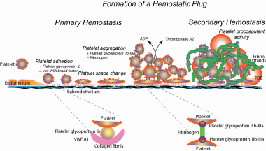
Platelet Membrane Glycoprotein IIb
Bone Marrow
NF-E2 Transcription Factor
Signal Transduction
Benzoates
NF-E2 Transcription Factor, p45 Subunit
Hematopoietic Cell Growth Factors
Platelet Glycoprotein GPIb-IX Complex
Cell Lineage
TYK2 Kinase
Polycythemia Vera
Culture Media, Serum-Free
Erythroid Precursor Cells
Platelet Aggregation
Receptors, Erythropoietin
Erythroid-Specific DNA-Binding Factors
Gab-family adapter proteins act downstream of cytokine and growth factor receptors and T- and B-cell antigen receptors. (1/338)
We previously found that the adapter protein Gab1 (110 kD) is tyrosine-phosphorylated and forms a complex with SHP-2 and PI-3 kinase upon stimulation through either the interleukin-3 receptor (IL-3R) or gp130, the common receptor subunit of IL-6-family cytokines. In this report, we identified another adapter molecule (100 kD) interacting with SHP-2 and PI-3 kinase in response to various stimuli. The molecule displays striking homology to Gab1 at the amino acid level; thus, we named it Gab2. It contains a PH domain, proline-rich sequences, and tyrosine residues that bind to SH2 domains when they are phosphorylated. Gab1 is phosphorylated on tyrosine upon stimulation through the thrombopoietin receptor (TPOR), stem cell factor receptor (SCFR), and T-cell and B-cell antigen receptors (TCR and BCR, respectively), in addition to IL-3R and gp130. Tyrosine phosphorylation of Gab2 was induced by stimulation through gp130, IL-2R, IL-3R, TPOR, SCFR, and TCR. Gab1 and Gab2 were shown to be substrates for SHP-2 in vitro. Overexpression of Gab2 enhanced the gp130 or Src-related kinases-mediated ERK2 activation as that of Gab1 did. These data indicate that Gab-family molecules act as adapters for transmitting various signals. (+info)Identification of mutations in the c-mpl gene in congenital amegakaryocytic thrombocytopenia. (2/338)
Congenital amegakaryocytic thrombocytopenia (CAMT) is a rare disorder expressed in infancy and characterized by isolated thrombocytopenia and megakaryocytopenia with no physical anomalies. Our previous hematological analysis indicated similarities between human CAMT and murine c-mpl (thrombopoietin receptor) deficiency. Because the c-mpl gene was considered as one of the candidate genes for this disorder, we analyzed the genomic sequence of the c-mpl gene of a 10-year-old Japanese girl with CAMT. We detected two heterozygous point mutations: a C-to-T transition at the cDNA nucleotide position 556 (Q186X) in exon 4 and a single nucleotide deletion of thymine at position 1,499 (1,499 delT) in exon 10. Both mutations were predicted to result in a prematurely terminated c-Mpl protein, which, if translated, lacks all intracellular domains essential for signal transduction. Each of the mutations was segregated from the patient's parents. Accordingly, the patient was a compound heterozygote for two mutations of the c-mpl gene, each derived from one of the parents. The present study suggests that at least a certain type of CAMT is caused by the c-mpl mutation, which disrupts the function of thrombopoietin receptor. (+info)Thrombopoietin stimulates endothelial cell motility and neoangiogenesis by a platelet-activating factor-dependent mechanism. (3/338)
In this study, we demonstrate that human umbilical cord vein-derived endothelial cells (HUVECs) expressed c-Mpl, the thrombopoietin (TPO) receptor, and that TPO activates HUVECs in vitro, as indicated by directional migration, synthesis of 1-alkyl-/1-acyl-platelet-activating factor (PAF) and interleukin-8 (IL-8), and phosphorylation of the signal transducers and activators of transcription (STAT) STAT1 and STAT5B. The observation that WEB 2170 and CV3988, 2 structurally unrelated PAF receptor antagonists, prevented the motility of HUVECs induced by TPO suggests a role of PAF as secondary mediator. Moreover, kinetic analysis of TPO-induced tyrosine phosphorylation of STAT demonstrated that STAT5B activation temporally correlated with the synthesis of PAF. PAF, in turn, induced a rapid tyrosine phosphorylation of STAT5B and PAF receptor blockade, by WEB 2170, preventing both TPO- and PAF-mediated STAT5B activation. The in vivo angiogenic effect of TPO, studied in a mouse model of Matrigel implantation, demonstrated that TPO induced a dose-dependent angiogenic response that required the presence of heparin. Moreover, the in vivo angiogenic effect of TPO was inhibited by the PAF receptor antagonist WEB 2170 but not by the anti-basic fibroblast growth factor neutralizing antibody. These results indicate that the effects of TPO are not restricted to cells of hematopoietic lineages, because TPO is able to activate endothelial cells and to induce an angiogenic response in which the recruitment of endothelial cells is mediated by the synthesis of PAF. Moreover, biochemical analysis supports the hypothesis that STAT5B may be involved in the signaling pathway leading to PAF-dependent angiogenesis. (+info)Apoptosis of erythroid precursors under stimulation with thrombopoietin: contribution to megakaryocytic lineage choice. (4/338)
Although the effect of thrombopoietin (TPO) on megakaryocyte production is well established, its role in the commitment of multipotential hematopoietic progenitors to the megakaryocytic lineage remains to be determined. In the present study, we attempted to clarify the determination process of megakaryocytic lineage as a terminal differentiation pathway under stimulation with TPO. Day 7 cultured cells grown by TPO derived from cord blood CD34+ cells were divided into four subpopulations on the basis of CD34 and CD41 expression. The CD34-/CD41- cells showed the labeling pattern of anti-CD42b and anti-CD9 antibodies closer to that of the CD34+/CD41- cells than the CD34+/CD41+ cells. Replating experiments revealed that approximately 40% of the CD34-/CD41- cells proliferated in response to a combination of growth factors, and more than 80% of them were pure erythroid precursors. However, this subpopulation failed to grow/survive and fell into apoptosis in the presence of TPO alone. In contrast, the CD34+/CD41+ cells, which predominantly contained megakaryocytic precursors, exerted a low but significant proliferative potential in the presence of TPO. The insufficient response to TPO of the CD34-/CD41- cells may result from the apparently low expression of c-MpI, as determined by flow cytometric analysis and reverse transcription-polymerase chain reaction analysis. Therefore, these results suggest that the apoptosis of hematopoietic precursors other than megakaryocytic precursors is related to the determination of the terminal differentiation under the influence of TPO. (+info)High-level expression of Mpl in platelets and megakaryocytes is independent of thrombopoietin. (5/338)
Thrombopoietin (TPO) is a hematopoietic growth factor that regulates megakaryocytopoiesis and platelet production through binding to its receptor, Mpl, encoded by the c-mpl proto-oncogene. Circulating levels of TPO are regulated by receptor-mediated uptake and degradation. To better understand this mode of TPO regulation, we examined whether expression of Mpl was regulated by its ligand. Using RNase protection analysis, we found no differences in the levels of c-mpl transcripts in megakaryocytes (MKs) produced in vitro either in the presence or absence of TPO and in platelets (PLTs) obtained from mice hyperstimulated in vivo by ectopic secretion of TPO. Similarly, Western blot analysis of MKs produced in the presence or absence of TPO showed no difference in Mpl levels. Levels of Mpl, GpIIb, or P-selectin were virtually identical in platelet lysates obtained from normal, TPO knockout and mildly TPO-stimulated mice. In contrast, the expression of Mpl was significantly reduced in PLTs from severely thrombocythemic mice. These results show that TPO does not have a major effect on the transcription or translation of Mpl. However, they do suggest that an excess of circulating TPO can lead to the disappearance of Mpl from PLTs via catabolism. (+info)Consequences of GATA-1 deficiency in megakaryocytes and platelets. (6/338)
In the absence of the hematopoietic transcription factor GATA-1, mice develop thrombocytopenia and an increased number of megakaryocytes characterized by marked ultrastructural abnormalities. These observations establish a critical role for GATA-1 in megakaryopoiesis and raise the question as to how GATA-1 influences megakaryocyte maturation and platelet production. To begin to address this, we have performed a more detailed examination of the megakaryocytes and platelets produced in mice that lack GATA-1 in this lineage. Our analysis demonstrates that compared with their normal counterparts, GATA-1-deficient primary megakaryocytes exhibit significant hyperproliferation in liquid culture, suggesting that the megakaryocytosis seen in animals is nonreactive. Morphologically, these mutant megakaryocytes are small and show evidence of retarded nuclear and cytoplasmic development. A significant proportion of these cells do not undergo endomitosis and express markedly lower levels of mRNA of all megakaryocyte-associated genes tested, including GPIbalpha, GPIbbeta, platelet factor 4 (PF4), c-mpl, and p45 NF-E2. These results are consistent with regulation of a program of megakaryocytic differentiation by GATA-1. Bleeding times are significantly prolonged in mutant animals. GATA-1-deficient platelets show abnormal ultrastructure, reminiscent of the megakaryocytes from which they are derived, and exhibit modest but selective defects in platelet activation in response to thrombin or to the combination of adenosine diphosphate (ADP) and epinephrine. Our findings indicate that GATA-1 serves multiple functions in megakaryocyte development, influencing both cellular growth and maturation. (+info)Deletion of the extracellular membrane-distal cytokine receptor homology module of Mpl results in constitutive cell growth and loss of thrombopoietin binding. (7/338)
The thrombopoietin receptor, Mpl, is a member of the cytokine receptor superfamily. The extracellular domain of Mpl contains two copies of the cytokine receptor homology module (CRM). Mpl is encoded by c-mpl, the cellular homologue of the oncogene v-mpl. The oncogenic potential of v-mpl may arise from deletion of all but the 43 most membrane-proximal amino acids of the extracellular domain of the wild-type receptor. To test the hypothesis that the extracellular domain of Mpl plays a role in controlling receptor activity, we created mutants of murine Mpl in which the membrane-distal CRM was either deleted or replaced by the membrane-proximal CRM. Introduction of these mutant receptors into factor-dependent BaF3 cells led to constitutive cell growth in the absence of growth factor. Both mutant receptors failed to bind 125I-Tpo. These results suggest that the membrane-distal CRM of Mpl acts as a brake on cell proliferation and that this region is required for ligand binding. (+info)Developmental expression of plasminogen activator inhibitor-1 associated with thrombopoietin-dependent megakaryocytic differentiation. (8/338)
Plasminogen activator inhibitor-1 (PAI-1) is present in the platelet alpha-granule and is released on activation. However, there is some debate as to whether the megakaryocyte and platelet synthesize PAI-1, take it up from plasma, or both. We examined the expression of PAI-1 in differentiating megakaryocytic progenitor cells (UT-7) and in CD34(+)/CD41(-) cells from cord blood. UT-7 cells differentiated with thrombopoietin (TPO) resembled megakaryocytes (UT-7/TPO) with respect to morphology, ploidy, and the expression of glycoprotein IIb-IIIa. PAI-1 messenger RNA (mRNA) expression was upregulated and PAI-1 protein synthesized in the UT-7/TPO cells accumulated in the cytoplasm without being released spontaneously. In contrast, erythropoietin (EPO)-stimulated UT-7 cells (UT-7/EPO) did not express PAI-1 mRNA after stimulation with TPO because they do not have endogenous c-Mpl. After cotransfection with human wild-type c-mpl, the cells (UT-7/EPO-MPL) responded to phorbol 12-myristate 13-acetate (PMA), tumor necrosis factor-alpha (TNF-alpha), and interleukin-1beta (IL-1beta) with enhanced PAI-1 mRNA expression within 24 to 48 hours. However, induction of PAI-1 mRNA in UT-7/EPO-MPL cells by TPO required at least 14-days stimulation. UT-7/EPO cells expressing c-Mpl changed their morphology and the other characteristics similar to the UT-7/TPO cells. TPO also differentiated human cord blood CD34(+)/CD41(-) cells to CD34(-)/CD41(+) cells, generated morphologically mature megakaryocytes, and induced the expression of PAI-1 mRNA. These results suggest that both PAI-1 mRNA and de novo PAI-1 protein synthesis is induced after differentiation of immature progenitor cells into megakaryocytes by TPO. (+info)Thrombopoietin (TPO) is a glycoprotein hormone that plays a crucial role in the regulation of platelet production, also known as thrombopoiesis. It is primarily produced by the liver and to some extent by megakaryocytes, which are the cells responsible for producing platelets.
TPO binds to its receptor, c-Mpl, on the surface of megakaryocytes and their precursor cells, stimulating their proliferation, differentiation, and maturation into platelets. By regulating the number of platelets in circulation, TPO helps maintain hemostasis, the process that prevents excessive bleeding after injury.
In addition to its role in thrombopoiesis, TPO has been shown to have potential effects on other cell types, including hematopoietic stem cells and certain immune cells. However, its primary function remains the regulation of platelet production.
Megakaryocytes are large, specialized bone marrow cells that are responsible for the production and release of platelets (also known as thrombocytes) into the bloodstream. Platelets play an essential role in blood clotting and hemostasis, helping to prevent excessive bleeding during injuries or trauma.
Megakaryocytes have a unique structure with multilobed nuclei and abundant cytoplasm rich in organelles called alpha-granules and dense granules, which store various proteins, growth factors, and enzymes necessary for platelet function. As megakaryocytes mature, they extend long cytoplasmic processes called proplatelets into the bone marrow sinuses, where these extensions fragment into individual platelets that are released into circulation.
Abnormalities in megakaryocyte number, size, or function can lead to various hematological disorders, such as thrombocytopenia (low platelet count), thrombocytosis (high platelet count), and certain types of leukemia.
Thrombopoiesis is the process of formation and development of thrombocytes or platelets, which are small, colorless cell fragments in our blood that play an essential role in clotting. Thrombopoiesis occurs inside the bone marrow, where stem cells differentiate into megakaryoblasts, then progressively develop into promegakaryocytes and megakaryocytes. These megakaryocytes subsequently undergo a process called cytoplasmic fragmentation to produce platelets.
The regulation of thrombopoiesis is primarily controlled by the hormone thrombopoietin (TPO), which is produced mainly in the liver and binds to the thrombopoietin receptor (c-Mpl) on megakaryocytes and their precursors. This binding stimulates the proliferation, differentiation, and maturation of megakaryocytes, leading to an increase in platelet production.
Abnormalities in thrombopoiesis can result in conditions such as thrombocytopenia (low platelet count) or thrombocytosis (high platelet count), which may be associated with bleeding disorders or increased risk of thrombosis, respectively.
Cytokine receptors are specialized protein molecules found on the surface of cells that selectively bind to specific cytokines. Cytokines are signaling molecules used for communication between cells, and they play crucial roles in regulating immune responses, inflammation, hematopoiesis, and cell survival.
Cytokine receptors have specific binding sites that recognize and interact with the corresponding cytokines. This interaction triggers a series of intracellular signaling events that ultimately lead to changes in gene expression and various cellular responses. Cytokine receptors can be found on many different types of cells, including immune cells, endothelial cells, and structural cells like fibroblasts.
Cytokine receptors are typically composed of multiple subunits, which may include both extracellular and intracellular domains. The extracellular domain is responsible for cytokine binding, while the intracellular domain is involved in signal transduction. Cytokine receptors can be classified into several families based on their structural features and signaling mechanisms, such as the hematopoietic cytokine receptor family, the interferon receptor family, the tumor necrosis factor receptor family, and the interleukin-1 receptor family.
Dysregulation of cytokine receptors and their signaling pathways has been implicated in various diseases, including autoimmune disorders, chronic inflammation, and cancer. Therefore, understanding the biology of cytokine receptors is essential for developing targeted therapies to treat these conditions.
Thrombocytopenia is a medical condition characterized by an abnormally low platelet count (thrombocytes) in the blood. Platelets are small cell fragments that play a crucial role in blood clotting, helping to stop bleeding when a blood vessel is damaged. A healthy adult typically has a platelet count between 150,000 and 450,000 platelets per microliter of blood. Thrombocytopenia is usually diagnosed when the platelet count falls below 150,000 platelets/µL.
Thrombocytopenia can be classified into three main categories based on its underlying cause:
1. Immune thrombocytopenia (ITP): An autoimmune disorder where the immune system mistakenly attacks and destroys its own platelets, leading to a decreased platelet count. ITP can be further divided into primary or secondary forms, depending on whether it occurs alone or as a result of another medical condition or medication.
2. Decreased production: Thrombocytopenia can occur when there is insufficient production of platelets in the bone marrow due to various causes, such as viral infections, chemotherapy, radiation therapy, leukemia, aplastic anemia, or vitamin B12 or folate deficiency.
3. Increased destruction or consumption: Thrombocytopenia can also result from increased platelet destruction or consumption due to conditions like disseminated intravascular coagulation (DIC), thrombotic thrombocytopenic purpura (TTP), hemolytic uremic syndrome (HUS), or severe bacterial infections.
Symptoms of thrombocytopenia may include easy bruising, prolonged bleeding from cuts, spontaneous nosebleeds, bleeding gums, blood in urine or stools, and skin rashes like petechiae (small red or purple spots) or purpura (larger patches). The severity of symptoms can vary depending on the degree of thrombocytopenia and the presence of any underlying conditions. Treatment for thrombocytopenia depends on the cause and may include medications, transfusions, or addressing the underlying condition.
A platelet count is a laboratory test that measures the number of platelets, also known as thrombocytes, in a sample of blood. Platelets are small, colorless cell fragments that circulate in the blood and play a crucial role in blood clotting. They help to stop bleeding by sticking together to form a plug at the site of an injured blood vessel.
A normal platelet count ranges from 150,000 to 450,000 platelets per microliter (µL) of blood. A lower than normal platelet count is called thrombocytopenia, while a higher than normal platelet count is known as thrombocytosis.
Abnormal platelet counts can be a sign of various medical conditions, including bleeding disorders, infections, certain medications, and some types of cancer. It is important to consult with a healthcare provider if you have any concerns about your platelet count or if you experience symptoms such as easy bruising, prolonged bleeding, or excessive menstrual flow.
Blood platelets, also known as thrombocytes, are small, colorless cell fragments in our blood that play an essential role in normal blood clotting. They are formed in the bone marrow from large cells called megakaryocytes and circulate in the blood in an inactive state until they are needed to help stop bleeding. When a blood vessel is damaged, platelets become activated and change shape, releasing chemicals that attract more platelets to the site of injury. These activated platelets then stick together to form a plug, or clot, that seals the wound and prevents further blood loss. In addition to their role in clotting, platelets also help to promote healing by releasing growth factors that stimulate the growth of new tissue.
Thrombocytosis is a medical condition characterized by an abnormally high platelet count (also known as thrombocytes) in the blood. Platelets are small cell fragments that play a crucial role in blood clotting. A normal platelet count ranges from 150,000 to 450,000 platelets per microliter of blood. Thrombocytosis is typically defined as a platelet count exceeding 450,000-500,000 platelets/µL.
Thrombocytosis can be classified into two types: reactive (or secondary) thrombocytosis and primary (or essential) thrombocytosis. Reactive thrombocytosis is more common and occurs as a response to an underlying condition, such as infection, inflammation, surgery, or certain types of cancer. Primary thrombocytosis, on the other hand, is caused by intrinsic abnormalities in the bone marrow cells responsible for platelet production (megakaryocytes), and it is often associated with myeloproliferative neoplasms like essential thrombocythemia.
While mild thrombocytosis may not cause any symptoms, higher platelet counts can increase the risk of blood clots (thrombosis) and bleeding disorders due to excessive platelet aggregation. Symptoms of thrombocytosis may include headaches, dizziness, visual disturbances, or chest pain if a blood clot forms in the brain or heart. Bleeding symptoms can manifest as easy bruising, nosebleeds, or gastrointestinal bleeding.
Treatment for thrombocytosis depends on the underlying cause and the severity of the condition. In cases of reactive thrombocytosis, treating the underlying disorder often resolves the high platelet count. For primary thrombocytosis, medications like aspirin or cytoreductive therapy (such as hydroxyurea) may be used to reduce the risk of blood clots and control platelet production. Regular monitoring of platelet counts is essential for managing this condition and preventing potential complications.
Hematopoietic stem cells (HSCs) are immature, self-renewing cells that give rise to all the mature blood and immune cells in the body. They are capable of both producing more hematopoietic stem cells (self-renewal) and differentiating into early progenitor cells that eventually develop into red blood cells, white blood cells, and platelets. HSCs are found in the bone marrow, umbilical cord blood, and peripheral blood. They have the ability to repair damaged tissues and offer significant therapeutic potential for treating various diseases, including hematological disorders, genetic diseases, and cancer.
Megakaryocyte progenitor cells are a type of hematopoietic (blood-forming) stem or progenitor cell that give rise to megakaryocytes, which are large cells found in the bone marrow. Megakaryocytes are responsible for producing platelets, also known as thrombocytes, which are small cell fragments that play a crucial role in blood clotting and hemostasis.
Megakaryocyte progenitor cells are characterized by their ability to differentiate into megakaryocytes and express specific surface markers, such as CD34, CD41, and CD61. They can be found in the bone marrow and peripheral blood and can be expanded and differentiated in vitro for therapeutic purposes, such as in platelet production for transfusion therapy.
Abnormalities in megakaryocyte progenitor cells can lead to various hematological disorders, including thrombocytopenia (low platelet count) and myeloproliferative neoplasms (abnormal blood cell growth). Therefore, understanding the biology and regulation of megakaryocyte progenitor cells is essential for developing new diagnostic and therapeutic strategies for these conditions.
Idiopathic Thrombocytopenic Purpura (ITP) is a medical condition characterized by a low platelet count (thrombocytopenia) in the blood without an identifiable cause. Platelets are small blood cells that help your body form clots to stop bleeding. When you don't have enough platelets, you may bleed excessively or spontaneously, causing purpura, which refers to purple-colored spots on the skin that result from bleeding under the skin.
In ITP, the immune system mistakenly attacks and destroys platelets, leading to their decreased levels in the blood. This condition can occur at any age but is more common in children following a viral infection, and in adults after the age of 30-40 years. Symptoms may include easy or excessive bruising, prolonged bleeding from cuts, spontaneous bleeding from the gums or nose, blood blisters, and small red or purple spots on the skin (petechiae).
Depending on the severity of thrombocytopenia and the presence of bleeding symptoms, ITP treatment may include observation, corticosteroids, intravenous immunoglobulin (IVIG), or other medications that modify the immune system's response. In severe cases or when other treatments are ineffective, surgical removal of the spleen (splenectomy) might be considered.
Hematopoiesis is the process of forming and developing blood cells. It occurs in the bone marrow and includes the production of red blood cells (erythropoiesis), white blood cells (leukopoiesis), and platelets (thrombopoiesis). This process is regulated by various growth factors, hormones, and cytokines. Hematopoiesis begins early in fetal development and continues throughout a person's life. Disorders of hematopoiesis can result in conditions such as anemia, leukopenia, leukocytosis, thrombocytopenia, or thrombocytosis.
Stem Cell Factor (SCF), also known as Kit Ligand or Steel Factor, is a growth factor that plays a crucial role in the regulation of hematopoiesis, which is the process of producing various blood cells. It is a glycoprotein that binds to the c-Kit receptor found on hematopoietic stem cells and progenitor cells, promoting their survival, proliferation, and differentiation into mature blood cells.
SCF is involved in the development and function of several types of blood cells, including red blood cells, white blood cells, and platelets. It also plays a role in the maintenance and self-renewal of hematopoietic stem cells, which are essential for the continuous production of new blood cells throughout an individual's lifetime.
In addition to its role in hematopoiesis, SCF has been implicated in various other biological processes, such as melanogenesis, gametogenesis, and tissue repair and regeneration. Dysregulation of SCF signaling has been associated with several diseases, including certain types of cancer, bone marrow failure disorders, and autoimmune diseases.
CD34 is a type of antigen that is found on the surface of certain cells in the human body. Specifically, CD34 antigens are present on hematopoietic stem cells, which are immature cells that can develop into different types of blood cells. These stem cells are found in the bone marrow and are responsible for producing red blood cells, white blood cells, and platelets.
CD34 antigens are a type of cell surface marker that is used in medical research and clinical settings to identify and isolate hematopoietic stem cells. They are also used in the development of stem cell therapies and transplantation procedures. CD34 antigens can be detected using various laboratory techniques, such as flow cytometry or immunohistochemistry.
It's important to note that while CD34 is a useful marker for identifying hematopoietic stem cells, it is not exclusive to these cells and can also be found on other cell types, such as endothelial cells that line blood vessels. Therefore, additional markers are often used in combination with CD34 to more specifically identify and isolate hematopoietic stem cells.
A neoplasm is a tumor or growth that is formed by an abnormal and excessive proliferation of cells, which can be benign or malignant. Neoplasm proteins are therefore any proteins that are expressed or produced in these neoplastic cells. These proteins can play various roles in the development, progression, and maintenance of neoplasms.
Some neoplasm proteins may contribute to the uncontrolled cell growth and division seen in cancer, such as oncogenic proteins that promote cell cycle progression or inhibit apoptosis (programmed cell death). Others may help the neoplastic cells evade the immune system, allowing them to proliferate undetected. Still others may be involved in angiogenesis, the formation of new blood vessels that supply the tumor with nutrients and oxygen.
Neoplasm proteins can also serve as biomarkers for cancer diagnosis, prognosis, or treatment response. For example, the presence or level of certain neoplasm proteins in biological samples such as blood or tissue may indicate the presence of a specific type of cancer, help predict the likelihood of cancer recurrence, or suggest whether a particular therapy will be effective.
Overall, understanding the roles and behaviors of neoplasm proteins can provide valuable insights into the biology of cancer and inform the development of new diagnostic and therapeutic strategies.
Proto-oncogene proteins are normal cellular proteins that play crucial roles in various cellular processes, such as signal transduction, cell cycle regulation, and apoptosis (programmed cell death). They are involved in the regulation of cell growth, differentiation, and survival under physiological conditions.
When proto-oncogene proteins undergo mutations or aberrations in their expression levels, they can transform into oncogenic forms, leading to uncontrolled cell growth and division. These altered proteins are then referred to as oncogene products or oncoproteins. Oncogenic mutations can occur due to various factors, including genetic predisposition, environmental exposures, and aging.
Examples of proto-oncogene proteins include:
1. Ras proteins: Involved in signal transduction pathways that regulate cell growth and differentiation. Activating mutations in Ras genes are found in various human cancers.
2. Myc proteins: Regulate gene expression related to cell cycle progression, apoptosis, and metabolism. Overexpression of Myc proteins is associated with several types of cancer.
3. EGFR (Epidermal Growth Factor Receptor): A transmembrane receptor tyrosine kinase that regulates cell proliferation, survival, and differentiation. Mutations or overexpression of EGFR are linked to various malignancies, such as lung cancer and glioblastoma.
4. Src family kinases: Intracellular tyrosine kinases that regulate signal transduction pathways involved in cell proliferation, survival, and migration. Dysregulation of Src family kinases is implicated in several types of cancer.
5. Abl kinases: Cytoplasmic tyrosine kinases that regulate various cellular processes, including cell growth, differentiation, and stress responses. Aberrant activation of Abl kinases, as seen in chronic myelogenous leukemia (CML), leads to uncontrolled cell proliferation.
Understanding the roles of proto-oncogene proteins and their dysregulation in cancer development is essential for developing targeted cancer therapies that aim to inhibit or modulate these aberrant signaling pathways.
Interleukin-3 (IL-3) is a type of cytokine, which is a small signaling protein that modulates the immune response, cell growth, and differentiation. IL-3 is primarily produced by activated T cells and mast cells. It plays an essential role in the survival, proliferation, and differentiation of hematopoietic stem cells, which give rise to all blood cell types. Specifically, IL-3 supports the development of myeloid lineage cells, including basophils, eosinophils, mast cells, megakaryocytes, and erythroid progenitors.
IL-3 binds to its receptor, the interleukin-3 receptor (IL-3R), which consists of two subunits: CD123 (the alpha chain) and CD131 (the beta chain). The binding of IL-3 to its receptor triggers a signaling cascade within the cell that ultimately leads to changes in gene expression, promoting cell growth and differentiation. Dysregulation of IL-3 production or signaling has been implicated in several hematological disorders, such as leukemia and myelodysplastic syndromes.
Hydrazines are not a medical term, but rather a class of organic compounds containing the functional group N-NH2. They are used in various industrial and chemical applications, including the production of polymers, pharmaceuticals, and agrochemicals. However, some hydrazines have been studied for their potential therapeutic uses, such as in the treatment of cancer and cardiovascular diseases. Exposure to high levels of hydrazines can be toxic and may cause damage to the liver, kidneys, and central nervous system. Therefore, medical professionals should be aware of the potential health hazards associated with hydrazine exposure.
Janus Kinase 2 (JAK2) is a tyrosine kinase enzyme that plays a crucial role in intracellular signal transduction. It is named after the Roman god Janus, who is depicted with two faces, as JAK2 has two similar phosphate-transferring domains. JAK2 is involved in various cytokine receptor-mediated signaling pathways and contributes to hematopoiesis, immune function, and cell growth.
Mutations in the JAK2 gene have been associated with several myeloproliferative neoplasms (MPNs), including polycythemia vera, essential thrombocythemia, and primary myelofibrosis. The most common mutation is JAK2 V617F, which results in a constitutively active enzyme that promotes uncontrolled cell proliferation and survival, contributing to the development of these MPNs.
Interleukin-11 (IL-11) is a type of cytokine, which is a small signaling protein involved in the immune response and hematopoiesis (the formation of blood cells). IL-11 is primarily produced by bone marrow stromal cells and is involved in regulating the production and function of platelets, which are cell fragments necessary for blood clotting.
IL-11 has a number of biological activities, including promoting the growth and differentiation of megakaryocytes (the precursor cells to platelets), stimulating the production of acute phase proteins during inflammation, and regulating the function of certain immune cells. In addition, IL-11 has been shown to have effects on other tissues, including promoting the growth and survival of some cancer cells.
Dysregulation of IL-11 signaling has been implicated in a number of diseases, including thrombocytopenia (low platelet count), certain types of anemia, and various cancers.
Bone marrow cells are the types of cells found within the bone marrow, which is the spongy tissue inside certain bones in the body. The main function of bone marrow is to produce blood cells. There are two types of bone marrow: red and yellow. Red bone marrow is where most blood cell production takes place, while yellow bone marrow serves as a fat storage site.
The three main types of bone marrow cells are:
1. Hematopoietic stem cells (HSCs): These are immature cells that can differentiate into any type of blood cell, including red blood cells, white blood cells, and platelets. They have the ability to self-renew, meaning they can divide and create more hematopoietic stem cells.
2. Red blood cell progenitors: These are immature cells that will develop into mature red blood cells, also known as erythrocytes. Red blood cells carry oxygen from the lungs to the body's tissues and carbon dioxide back to the lungs.
3. Myeloid and lymphoid white blood cell progenitors: These are immature cells that will develop into various types of white blood cells, which play a crucial role in the body's immune system by fighting infections and diseases. Myeloid progenitors give rise to granulocytes (neutrophils, eosinophils, and basophils), monocytes, and megakaryocytes (which eventually become platelets). Lymphoid progenitors differentiate into B cells, T cells, and natural killer (NK) cells.
Bone marrow cells are essential for maintaining a healthy blood cell count and immune system function. Abnormalities in bone marrow cells can lead to various medical conditions, such as anemia, leukopenia, leukocytosis, thrombocytopenia, or thrombocytosis, depending on the specific type of blood cell affected. Additionally, bone marrow cells are often used in transplantation procedures to treat patients with certain types of cancer, such as leukemia and lymphoma, or other hematologic disorders.
Myeloproliferative disorders (MPDs) are a group of rare, chronic blood cancers that originate from the abnormal proliferation or growth of one or more types of blood-forming cells in the bone marrow. These disorders result in an overproduction of mature but dysfunctional blood cells, which can lead to serious complications such as blood clots, bleeding, and organ damage.
There are several subtypes of MPDs, including:
1. Chronic Myeloid Leukemia (CML): A disorder characterized by the overproduction of mature granulocytes (a type of white blood cell) in the bone marrow, leading to an increased number of these cells in the blood. CML is caused by a genetic mutation that results in the formation of the BCR-ABL fusion protein, which drives uncontrolled cell growth and division.
2. Polycythemia Vera (PV): A disorder characterized by the overproduction of all three types of blood cells - red blood cells, white blood cells, and platelets - in the bone marrow. This can lead to an increased risk of blood clots, bleeding, and enlargement of the spleen.
3. Essential Thrombocythemia (ET): A disorder characterized by the overproduction of platelets in the bone marrow, leading to an increased risk of blood clots and bleeding.
4. Primary Myelofibrosis (PMF): A disorder characterized by the replacement of normal bone marrow tissue with scar tissue, leading to impaired blood cell production and anemia, enlargement of the spleen, and increased risk of infections and bleeding.
5. Chronic Neutrophilic Leukemia (CNL): A rare disorder characterized by the overproduction of neutrophils (a type of white blood cell) in the bone marrow, leading to an increased number of these cells in the blood. CNL can lead to an increased risk of infections and organ damage.
MPDs are typically treated with a combination of therapies, including chemotherapy, targeted therapy, immunotherapy, and stem cell transplantation. The choice of treatment depends on several factors, including the subtype of MPD, the patient's age and overall health, and the presence of any comorbidities.
Recombinant proteins are artificially created proteins produced through the use of recombinant DNA technology. This process involves combining DNA molecules from different sources to create a new set of genes that encode for a specific protein. The resulting recombinant protein can then be expressed, purified, and used for various applications in research, medicine, and industry.
Recombinant proteins are widely used in biomedical research to study protein function, structure, and interactions. They are also used in the development of diagnostic tests, vaccines, and therapeutic drugs. For example, recombinant insulin is a common treatment for diabetes, while recombinant human growth hormone is used to treat growth disorders.
The production of recombinant proteins typically involves the use of host cells, such as bacteria, yeast, or mammalian cells, which are engineered to express the desired protein. The host cells are transformed with a plasmid vector containing the gene of interest, along with regulatory elements that control its expression. Once the host cells are cultured and the protein is expressed, it can be purified using various chromatography techniques.
Overall, recombinant proteins have revolutionized many areas of biology and medicine, enabling researchers to study and manipulate proteins in ways that were previously impossible.
Fetal blood refers to the blood circulating in a fetus during pregnancy. It is essential for the growth and development of the fetus, as it carries oxygen and nutrients from the placenta to the developing tissues and organs. Fetal blood also removes waste products, such as carbon dioxide, from the fetal tissues and transports them to the placenta for elimination.
Fetal blood has several unique characteristics that distinguish it from adult blood. For example, fetal hemoglobin (HbF) is the primary type of hemoglobin found in fetal blood, whereas adults primarily have adult hemoglobin (HbA). Fetal hemoglobin has a higher affinity for oxygen than adult hemoglobin, which allows it to more efficiently extract oxygen from the maternal blood in the placenta.
Additionally, fetal blood contains a higher proportion of reticulocytes (immature red blood cells) and nucleated red blood cells compared to adult blood. These differences reflect the high turnover rate of red blood cells in the developing fetus and the need for rapid growth and development.
Examination of fetal blood can provide important information about the health and well-being of the fetus during pregnancy. For example, fetal blood sampling (also known as cordocentesis or percutaneous umbilical blood sampling) can be used to diagnose genetic disorders, infections, and other conditions that may affect fetal development. However, this procedure carries risks, including preterm labor, infection, and fetal loss, and is typically only performed when there is a significant risk of fetal compromise or when other diagnostic tests have been inconclusive.
Erythropoietin (EPO) is a hormone that is primarily produced by the kidneys and plays a crucial role in the production of red blood cells in the body. It works by stimulating the bone marrow to produce more red blood cells, which are essential for carrying oxygen to various tissues and organs.
EPO is a glycoprotein that is released into the bloodstream in response to low oxygen levels in the body. When the kidneys detect low oxygen levels, they release EPO, which then travels to the bone marrow and binds to specific receptors on immature red blood cells called erythroblasts. This binding triggers a series of events that promote the maturation and proliferation of erythroblasts, leading to an increase in the production of red blood cells.
In addition to its role in regulating red blood cell production, EPO has also been shown to have neuroprotective effects and may play a role in modulating the immune system. Abnormal levels of EPO have been associated with various medical conditions, including anemia, kidney disease, and certain types of cancer.
EPO is also used as a therapeutic agent for the treatment of anemia caused by chronic kidney disease, chemotherapy, or other conditions that affect red blood cell production. Recombinant human EPO (rhEPO) is a synthetic form of the hormone that is produced using genetic engineering techniques and is commonly used in clinical practice to treat anemia. However, misuse of rhEPO for performance enhancement in sports has been a subject of concern due to its potential to enhance oxygen-carrying capacity and improve endurance.
Primary myelofibrosis (PMF) is a rare, chronic bone marrow disorder characterized by the replacement of normal bone marrow tissue with fibrous scar tissue, leading to impaired production of blood cells. This results in cytopenias (anemia, leukopenia, thrombocytopenia), which can cause fatigue, infection susceptibility, and bleeding tendencies. Additionally, PMF is often accompanied by the proliferation of abnormal megakaryocytes (large, atypical bone marrow cells that produce platelets) and extramedullary hematopoiesis (blood cell formation outside the bone marrow, typically in the spleen and liver).
PMF is a type of myeloproliferative neoplasm (MPN), which is a group of clonal stem cell disorders characterized by excessive proliferation of one or more types of blood cells. PMF can present with various symptoms such as fatigue, weight loss, night sweats, abdominal discomfort due to splenomegaly (enlarged spleen), and bone pain. In some cases, PMF may progress to acute myeloid leukemia (AML).
The exact cause of PMF remains unclear; however, genetic mutations are known to play a significant role in its development. The Janus kinase 2 (JAK2), calreticulin (CALR), and MPL genes have been identified as commonly mutated in PMF patients. These genetic alterations contribute to the dysregulated production of blood cells and the activation of signaling pathways that promote fibrosis.
Diagnosis of PMF typically involves a combination of clinical evaluation, complete blood count (CBC), bone marrow aspiration and biopsy, cytogenetic analysis, and molecular testing to identify genetic mutations. Treatment options depend on the individual patient's symptoms, risk stratification, and disease progression. They may include observation, supportive care, medications to manage symptoms and control the disease (such as JAK inhibitors), and stem cell transplantation for eligible patients.
Essential thrombocythemia (ET) is a myeloproliferative neoplasm (MPN), a type of blood cancer characterized by the overproduction of platelets (thrombocytosis) in the bone marrow. In ET, there is an excessive proliferation of megakaryocytes, the precursor cells that produce platelets. This leads to increased platelet counts in the peripheral blood, which can increase the risk of blood clots (thrombosis) and bleeding episodes (hemorrhage).
The term "essential" is used to indicate that the cause of this condition is not known or idiopathic. ET is primarily a disease of older adults, but it can also occur in younger individuals. The diagnosis of essential thrombocythemia requires careful evaluation and exclusion of secondary causes of thrombocytosis, such as reactive conditions, inflammation, or other myeloproliferative neoplasms.
The clinical presentation of ET can vary widely among patients. Some individuals may be asymptomatic and discovered only during routine blood tests, while others may experience symptoms related to thrombosis or bleeding. Common symptoms include headaches, visual disturbances, dizziness, weakness, numbness, or tingling in the extremities, if there are complications due to blood clots in the brain or other parts of the body. Excessive bruising, nosebleeds, or blood in the stool can indicate bleeding complications.
Treatment for essential thrombocythemia is aimed at reducing the risk of thrombosis and managing symptoms. Hydroxyurea is a commonly used medication to lower platelet counts, while aspirin may be prescribed to decrease the risk of blood clots. In some cases, interferon-alpha or ruxolitinib might be considered as treatment options. Regular follow-up with a hematologist and monitoring of blood counts are essential for managing this condition and detecting potential complications early.
A Colony-Forming Units (CFU) assay is a type of laboratory test used to measure the number of viable, or living, cells in a sample. It is commonly used to enumerate bacteria, yeast, and other microorganisms. The test involves placing a known volume of the sample onto a nutrient-agar plate, which provides a solid growth surface for the cells. The plate is then incubated under conditions that allow the cells to grow and form colonies. Each colony that forms on the plate represents a single viable cell from the original sample. By counting the number of colonies and multiplying by the known volume of the sample, the total number of viable cells in the sample can be calculated. This information is useful in a variety of applications, including monitoring microbial populations, assessing the effectiveness of disinfection procedures, and studying microbial growth and survival.
Cell differentiation is the process by which a less specialized cell, or stem cell, becomes a more specialized cell type with specific functions and structures. This process involves changes in gene expression, which are regulated by various intracellular signaling pathways and transcription factors. Differentiation results in the development of distinct cell types that make up tissues and organs in multicellular organisms. It is a crucial aspect of embryonic development, tissue repair, and maintenance of homeostasis in the body.
A platelet transfusion is the process of medically administering platelets, which are small blood cells that help your body form clots to stop bleeding. Platelet transfusions are often given to patients with low platelet counts or dysfunctional platelets due to various reasons such as chemotherapy, bone marrow transplantation, disseminated intravascular coagulation (DIC), and other medical conditions leading to increased consumption or destruction of platelets. This procedure helps to prevent or treat bleeding complications in these patients. It's important to note that platelet transfusions should be given under the supervision of a healthcare professional, taking into account the patient's clinical condition, platelet count, and potential risks associated with transfusion reactions.
Acute Megakaryoblastic Leukemia (AMKL) is a type of cancer that affects the blood and bone marrow. Specifically, it is a subtype of acute myeloid leukemia (AML), which is characterized by the rapid growth of abnormal cells in the bone marrow that interfere with the production of normal blood cells.
In AMKL, the abnormal cells are megakaryoblasts, which are immature cells that should develop into platelet-producing cells called megakaryocytes. However, in AMKL, these cells do not mature properly and instead accumulate in the bone marrow and bloodstream, leading to a shortage of healthy blood cells.
Symptoms of AMKL may include fatigue, weakness, frequent infections, easy bruising or bleeding, and the appearance of small red spots on the skin (petechiae). Diagnosis typically involves a combination of physical exam, medical history, blood tests, bone marrow aspiration and biopsy, and sometimes imaging studies.
Treatment for AMKL usually involves a combination of chemotherapy, radiation therapy, and/or stem cell transplantation. The specific treatment plan will depend on several factors, including the patient's age, overall health, and the extent of the disease.
Congenital Upper Extremity Deformities refer to physical abnormalities or malformations of the upper limb (arm, elbow, forearm, wrist, and hand) that are present at birth. These deformities can vary greatly in severity, complexity, and impact on function and appearance. They may result from genetic factors, environmental influences, or a combination of both during fetal development. Examples of congenital upper extremity deformities include:
1. Radial club hand: A condition where the radius bone in the forearm is underdeveloped or absent, causing the hand to turn outward and the wrist to bend inward.
2. Club foot of the arm: Also known as congenital vertical talus, this deformity affects the ankle and foot, causing them to point upwards. In the upper extremity, it can lead to limited mobility and function.
3. Polydactyly: The presence of extra fingers or toes, which can be fully formed or rudimentary.
4. Syndactyly: Fusion or webbing of fingers or toes.
5. Radial longitudinal deficiency: A spectrum of radial ray anomalies that includes radial club hand and other associated malformations.
6. Ulnar longitudinal deficiency: Underdevelopment or absence of the ulna bone, which can lead to deformities in the forearm, wrist, and hand.
7. Amniotic band syndrome: A condition where fibrous bands in the amniotic sac entangle and restrict the growth of fetal parts, including the upper limbs.
8. Cleidocranial dysplasia: A genetic disorder characterized by underdeveloped or absent collarbones, delayed closing of the skull bones, and other skeletal abnormalities, including shortened or deformed upper extremities.
9. Arthrogryposis: A group of conditions characterized by joint contractures and stiffness, which can affect any part of the body, including the upper extremities.
Treatment for congenital upper extremity deformities typically involves a combination of surgical interventions, physical therapy, bracing, or prosthetics to improve function, appearance, and quality of life.
Pancytopenia is a medical condition characterized by a reduction in the number of all three types of blood cells in the peripheral blood: red blood cells (anemia), white blood cells (leukopenia), and platelets (thrombocytopenia). This condition can be caused by various underlying diseases, including bone marrow disorders, viral infections, exposure to toxic substances or radiation, vitamin deficiencies, and certain medications. Symptoms of pancytopenia may include fatigue, weakness, increased susceptibility to infections, and easy bruising or bleeding.
Cell division is the process by which a single eukaryotic cell (a cell with a true nucleus) divides into two identical daughter cells. This complex process involves several stages, including replication of DNA, separation of chromosomes, and division of the cytoplasm. There are two main types of cell division: mitosis and meiosis.
Mitosis is the type of cell division that results in two genetically identical daughter cells. It is a fundamental process for growth, development, and tissue repair in multicellular organisms. The stages of mitosis include prophase, prometaphase, metaphase, anaphase, and telophase, followed by cytokinesis, which divides the cytoplasm.
Meiosis, on the other hand, is a type of cell division that occurs in the gonads (ovaries and testes) during the production of gametes (sex cells). Meiosis results in four genetically unique daughter cells, each with half the number of chromosomes as the parent cell. This process is essential for sexual reproduction and genetic diversity. The stages of meiosis include meiosis I and meiosis II, which are further divided into prophase, prometaphase, metaphase, anaphase, and telophase.
In summary, cell division is the process by which a single cell divides into two daughter cells, either through mitosis or meiosis. This process is critical for growth, development, tissue repair, and sexual reproduction in multicellular organisms.
Osteosclerosis is a medical term that refers to an abnormal thickening and increased density of bone tissue. This condition can occur as a result of various diseases or conditions, such as certain types of bone cancer, Paget's disease of bone, fluoride poisoning, or chronic infection of the bone. Osteosclerosis can also be seen in some benign conditions, such as osteopetrosis, which is a rare genetic disorder characterized by an excessively hard and dense skeleton.
In some cases, osteosclerosis may not cause any symptoms and may only be discovered on X-rays or other imaging studies. However, in other cases, it can lead to complications such as bone pain, fractures, or deformities. Treatment for osteosclerosis depends on the underlying cause of the condition and may include medications, surgery, or other therapies.
Stat5 (Signal Transducer and Activator of Transcription 5) is a transcription factor that plays a crucial role in various cellular processes, including growth, survival, and differentiation. It exists in two closely related isoforms, Stat5a and Stat5b, which are encoded by separate genes but share significant sequence homology and functional similarity.
When activated through phosphorylation by receptor or non-receptor tyrosine kinases, Stat5 forms homodimers or heterodimers that translocate to the nucleus. Once in the nucleus, these dimers bind to specific DNA sequences called Stat-binding elements (SBEs) in the promoter regions of target genes, leading to their transcriptional activation or repression.
Stat5 is involved in various physiological and pathological conditions, such as hematopoiesis, lactation, immune response, and cancer progression. Dysregulation of Stat5 signaling has been implicated in several malignancies, including leukemias, lymphomas, and breast cancer, making it an attractive therapeutic target for these diseases.
"Cells, cultured" is a medical term that refers to cells that have been removed from an organism and grown in controlled laboratory conditions outside of the body. This process is called cell culture and it allows scientists to study cells in a more controlled and accessible environment than they would have inside the body. Cultured cells can be derived from a variety of sources, including tissues, organs, or fluids from humans, animals, or cell lines that have been previously established in the laboratory.
Cell culture involves several steps, including isolation of the cells from the tissue, purification and characterization of the cells, and maintenance of the cells in appropriate growth conditions. The cells are typically grown in specialized media that contain nutrients, growth factors, and other components necessary for their survival and proliferation. Cultured cells can be used for a variety of purposes, including basic research, drug development and testing, and production of biological products such as vaccines and gene therapies.
It is important to note that cultured cells may behave differently than they do in the body, and results obtained from cell culture studies may not always translate directly to human physiology or disease. Therefore, it is essential to validate findings from cell culture experiments using additional models and ultimately in clinical trials involving human subjects.
Milk proteins are a complex mixture of proteins that are naturally present in milk, consisting of casein and whey proteins. Casein makes up about 80% of the total milk protein and is divided into several types including alpha-, beta-, gamma- and kappa-casein. Whey proteins account for the remaining 20% and include beta-lactoglobulin, alpha-lactalbumin, bovine serum albumin, and immunoglobulins. These proteins are important sources of essential amino acids and play a crucial role in the nutrition of infants and young children. Additionally, milk proteins have various functional properties that are widely used in the food industry for their gelling, emulsifying, and foaming abilities.
Plateletpheresis is a medical procedure that involves the collection of platelets from a donor's blood through a process called apheresis. In this process, whole blood is withdrawn from the donor, and the platelets are separated from other blood components using a specialized machine. The separated platelets are then collected in a sterile bag, while the remaining blood components (red blood cells, white blood cells, and plasma) are returned to the donor's body.
Plateletpheresis is often used to collect platelets for transfusion purposes, particularly for patients who require large volumes of platelets due to conditions such as leukemia, aplastic anemia, or other forms of cancer. It is also used in the treatment of thrombocytopenia, a condition characterized by abnormally low levels of platelets in the blood.
The procedure typically takes between one to two hours and requires the use of a specialized machine and trained medical staff. Donors may experience mild side effects such as fatigue, bruising, or discomfort at the site where the needle was inserted, but serious complications are rare.
Glycoprotein IIb (also known as integrin αIIbβ3 or CD41/CD61) is a type of protein found on the surface of platelets, which are small cell fragments involved in blood clotting. This glycoprotein plays a crucial role in the final pathway of platelet activation and aggregation, which ultimately leads to the formation of a clot to stop bleeding.
More specifically, Glycoprotein IIb is responsible for binding fibrinogen, von Willebrand factor, and other adhesive proteins in the blood, allowing platelets to bind together and form a clot. Mutations or defects in this glycoprotein can lead to bleeding disorders such as Glanzmann thrombasthenia, which is characterized by abnormal platelet function and excessive bleeding.
Bone marrow is the spongy tissue found inside certain bones in the body, such as the hips, thighs, and vertebrae. It is responsible for producing blood-forming cells, including red blood cells, white blood cells, and platelets. There are two types of bone marrow: red marrow, which is involved in blood cell production, and yellow marrow, which contains fatty tissue.
Red bone marrow contains hematopoietic stem cells, which can differentiate into various types of blood cells. These stem cells continuously divide and mature to produce new blood cells that are released into the circulation. Red blood cells carry oxygen throughout the body, white blood cells help fight infections, and platelets play a crucial role in blood clotting.
Bone marrow also serves as a site for immune cell development and maturation. It contains various types of immune cells, such as lymphocytes, macrophages, and dendritic cells, which help protect the body against infections and diseases.
Abnormalities in bone marrow function can lead to several medical conditions, including anemia, leukopenia, thrombocytopenia, and various types of cancer, such as leukemia and multiple myeloma. Bone marrow aspiration and biopsy are common diagnostic procedures used to evaluate bone marrow health and function.
Ploidy is a term used in genetics to describe the number of sets of chromosomes in a cell or an organism. The ploidy level can have important implications for genetic inheritance and expression, as well as for evolutionary processes such as speciation and hybridization.
In most animals, including humans, the normal ploidy level is diploid, meaning that each cell contains two sets of chromosomes - one set inherited from each parent. However, there are also many examples of polyploidy, in which an organism has more than two sets of chromosomes.
Polyploidy can arise through various mechanisms, such as genome duplication or hybridization between different species. In some cases, polyploidy may confer evolutionary advantages, such as increased genetic diversity and adaptability to new environments. However, it can also lead to reproductive isolation and the formation of new species.
In plants, polyploidy is relatively common and has played a significant role in their evolution and diversification. Many crop plants are polyploids, including wheat, cotton, and tobacco. In some cases, artificial induction of polyploidy has been used to create new varieties with desirable traits for agriculture and horticulture.
Overall, ploidy is an important concept in genetics and evolution, with implications for a wide range of biological processes and phenomena.
Nuclear factor, erythroid-derived 2 (NFE2), also known as NF-E2 transcription factor, is a protein that plays a crucial role in the regulation of gene expression. It belongs to the cap'n'collar (CNC) subfamily of basic region-leucine zipper (bZIP) transcription factors.
NFE2 forms a heterodimer with small Maf proteins and binds to antioxidant response elements (AREs) in the promoter regions of target genes. These target genes are often involved in cellular defense against oxidative stress, electrophiles, and inflammation. NFE2 regulates the expression of various enzymes and proteins that protect cells from damage caused by reactive oxygen species (ROS) and other harmful substances.
Mutations in the NFE2 gene have been associated with several diseases, including chronic obstructive pulmonary disease (COPD), acute respiratory distress syndrome (ARDS), and certain types of cancer. Proper regulation of NFE2 is essential for maintaining cellular homeostasis and preventing the development of various pathological conditions.
Selenomethionine is an organic form of selenium, which is an essential trace element in human nutrition. It is incorporated into proteins in place of methionine, one of the 20 standard amino acids, and functions as an antioxidant by helping to prevent cellular damage from free radicals. Selenomethionine can be found in a variety of foods, including brazil nuts, fish, meat, and whole grains, and is also available as a dietary supplement.
Signal transduction is the process by which a cell converts an extracellular signal, such as a hormone or neurotransmitter, into an intracellular response. This involves a series of molecular events that transmit the signal from the cell surface to the interior of the cell, ultimately resulting in changes in gene expression, protein activity, or metabolism.
The process typically begins with the binding of the extracellular signal to a receptor located on the cell membrane. This binding event activates the receptor, which then triggers a cascade of intracellular signaling molecules, such as second messengers, protein kinases, and ion channels. These molecules amplify and propagate the signal, ultimately leading to the activation or inhibition of specific cellular responses.
Signal transduction pathways are highly regulated and can be modulated by various factors, including other signaling molecules, post-translational modifications, and feedback mechanisms. Dysregulation of these pathways has been implicated in a variety of diseases, including cancer, diabetes, and neurological disorders.
Benzoates are the salts and esters of benzoic acid. They are widely used as preservatives in foods, cosmetics, and pharmaceuticals to prevent the growth of microorganisms. The chemical formula for benzoic acid is C6H5COOH, and when it is combined with a base (like sodium or potassium), it forms a benzoate salt (e.g., sodium benzoate or potassium benzoate). When benzoic acid reacts with an alcohol, it forms a benzoate ester (e.g., methyl benzoate or ethyl benzoate).
Benzoates are generally considered safe for use in food and cosmetics in small quantities. However, some people may have allergies or sensitivities to benzoates, which can cause reactions such as hives, itching, or asthma symptoms. In addition, there is ongoing research into the potential health effects of consuming high levels of benzoates over time, particularly in relation to gut health and the development of certain diseases.
In a medical context, benzoates may also be used as a treatment for certain conditions. For example, sodium benzoate is sometimes given to people with elevated levels of ammonia in their blood (hyperammonemia) to help reduce those levels and prevent brain damage. This is because benzoates can bind with excess ammonia in the body and convert it into a form that can be excreted in urine.
Blood cells are the formed elements in the blood, including red blood cells (erythrocytes), white blood cells (leukocytes), and platelets (thrombocytes). These cells are produced in the bone marrow and play crucial roles in the body's functions. Red blood cells are responsible for carrying oxygen to tissues and carbon dioxide away from them, while white blood cells are part of the immune system and help defend against infection and disease. Platelets are cell fragments that are essential for normal blood clotting.
The NF-E2 (Nuclear Factor, Erythroid-derived 2) transcription factor is a heterodimeric protein that plays a crucial role in the regulation of gene expression. It is composed of two subunits: p18 and p45. The p45 subunit, also known as NFE2L2 or GABPalpha, is a member of the basic region-leucine zipper (bZIP) family of transcription factors.
The p45 subunit forms a complex with the p18 subunit, and this complex binds to specific DNA sequences called antioxidant response elements (AREs) or electrophile response elements (EpREs), which are present in the promoter regions of various genes involved in cellular defense against oxidative stress and xenobiotic metabolism.
The p45 subunit is responsible for recognizing and binding to the DNA sequence, while the p18 subunit stabilizes the complex and enhances its DNA-binding affinity. Together, they regulate the expression of genes involved in heme biosynthesis, cytochrome P450 activity, antioxidant defense, and other cellular processes.
Mutations in the NFE2L2 gene, which encodes the p45 subunit, have been associated with various diseases, including chronic obstructive pulmonary disease (COPD), neurodegenerative disorders, and cancer.
Hematopoietic cell growth factors are a group of glycoproteins that stimulate the proliferation, differentiation, and survival of hematopoietic cells, which are the precursor cells that give rise to all blood cells. These growth factors include colony-stimulating factors (CSFs) such as granulocyte-colony stimulating factor (G-CSF), granulocyte-macrophage colony-stimulating factor (GM-CSF), and macrophage colony-stimulating factor (M-CSF), as well as erythropoietin (EPO) and thrombopoietin (TPO).
G-CSF primarily stimulates the production of neutrophils, a type of white blood cell that plays a crucial role in the immune response to bacterial infections. GM-CSF stimulates the production of both granulocytes and monocytes/macrophages, while M-CSF specifically stimulates the production of monocytes/macrophages. EPO stimulates the production of red blood cells, while TPO stimulates the production of platelets.
Hematopoietic cell growth factors are used clinically to treat a variety of conditions associated with impaired hematopoiesis, such as chemotherapy-induced neutropenia, aplastic anemia, and congenital disorders of hematopoiesis. They can also be used to mobilize hematopoietic stem cells from the bone marrow into the peripheral blood for collection and transplantation.
The platelet glycoprotein GPIb-IX complex is a crucial receptor on the surface of platelets that plays a vital role in hemostasis and thrombosis. It is a heterotetrameric transmembrane protein complex composed of two disulfide-linked glycoprotein subunits, GPIbα, GPIbβ, GPV (Glycoprotein V), and GPIX (Glycoprotein IX).
The GPIb-IX complex is responsible for the initial interaction between platelets and von Willebrand factor (vWF) in the circulation. When blood vessels are damaged, exposed collagen recruits vWF to the site of injury, where it binds to the GPIbα subunit of the GPIb-IX complex, leading to platelet adhesion and activation. This interaction is critical for primary hemostasis, which helps prevent excessive blood loss from injured vessels.
Genetic mutations or deficiencies in the genes encoding these glycoproteins can lead to bleeding disorders such as Bernard-Soulier syndrome, a rare autosomal recessive disorder characterized by thrombocytopenia and large platelets with impaired vWF binding and platelet adhesion.
'Cell lineage' is a term used in biology and medicine to describe the developmental history or relationship of a cell or group of cells to other cells, tracing back to the original progenitor or stem cell. It refers to the series of cell divisions and differentiation events that give rise to specific types of cells in an organism over time.
In simpler terms, cell lineage is like a family tree for cells, showing how they are related to each other through a chain of cell division and specialization events. This concept is important in understanding the development, growth, and maintenance of tissues and organs in living beings.
TYK2 (Tyrosine Kinase 2) is a member of the Janus kinase (JAK) family of intracellular non-receptor protein tyrosine kinases. It plays a crucial role in the signaling of various cytokines and growth factors, including interferons, interleukin-6, -10, -12, and -23, by associating with their receptors and mediating downstream signal transduction.
The activation of TYK2 leads to the phosphorylation of signal transducers and activators of transcription (STAT) proteins, which then dimerize and translocate to the nucleus, where they regulate gene expression involved in various cellular processes such as immune responses, hematopoiesis, and cell growth. Dysregulation of TYK2 has been implicated in several autoimmune diseases and cancer, making it an attractive target for therapeutic intervention.
Polycythemia Vera is a type of myeloproliferative neoplasm, a group of rare blood cancers. In Polycythemia Vera, the body produces too many red blood cells, leading to an increased risk of blood clots and thickening of the blood, which can cause various symptoms such as fatigue, headache, dizziness, and itching. It can also lead to enlargement of the spleen. The exact cause of Polycythemia Vera is not known, but it is associated with genetic mutations in the JAK2 gene in most cases. It is a progressive disease that can lead to complications such as bleeding, thrombosis, and transformation into acute leukemia if left untreated.
"Serum-free culture media" refers to a type of nutrient medium used in cell culture and tissue engineering that does not contain fetal bovine serum (FBS) or other animal serums. Instead, it is supplemented with defined, chemically-defined components such as hormones, growth factors, vitamins, and amino acids.
The use of serum-free media offers several advantages over traditional media formulations that contain serum. For example, it reduces the risk of contamination with adventitious agents, such as viruses and prions, that may be present in animal serums. Additionally, it allows for greater control over the culture environment, as the concentration and composition of individual components can be carefully regulated. This is particularly important in applications where precise control over cell behavior is required, such as in the production of therapeutic proteins or in stem cell research.
However, serum-free media may not be suitable for all cell types, as some cells require the complex mixture of growth factors and other components found in animal serums to survive and proliferate. Therefore, it is important to carefully evaluate the needs of each specific cell type when selecting a culture medium.
Erythroid precursor cells, also known as erythroblasts or normoblasts, are early stage cells in the process of producing mature red blood cells (erythrocytes) in the bone marrow. These cells are derived from hematopoietic stem cells and undergo a series of maturation stages, including proerythroblast, basophilic erythroblast, polychromatophilic erythroblast, and orthochromatic erythroblast, before becoming reticulocytes and then mature red blood cells. During this maturation process, the cells lose their nuclei and become enucleated, taking on the biconcave shape and flexible membrane that allows them to move through small blood vessels and deliver oxygen to tissues throughout the body.
Platelet aggregation is the clumping together of platelets (thrombocytes) in the blood, which is an essential step in the process of hemostasis (the stopping of bleeding) after injury to a blood vessel. When the inner lining of a blood vessel is damaged, exposure of subendothelial collagen and tissue factor triggers platelet activation. Activated platelets change shape, become sticky, and release the contents of their granules, which include ADP (adenosine diphosphate).
ADP then acts as a chemical mediator to attract and bind additional platelets to the site of injury, leading to platelet aggregation. This forms a plug that seals the damaged vessel and prevents further blood loss. Platelet aggregation is also a crucial component in the formation of blood clots (thrombosis) within blood vessels, which can have pathological consequences such as heart attacks and strokes if they obstruct blood flow to vital organs.
Erythropoietin receptors are cell surface proteins found on immature red blood cell precursors in the bone marrow. They bind to the hormone erythropoietin (EPO), which is produced by the kidneys in response to low oxygen levels in the blood. When EPO binds to its receptor, it activates a signaling pathway that promotes the survival, proliferation, and differentiation of red blood cell precursors, leading to increased production of red blood cells. This process is critical for maintaining adequate oxygen delivery to tissues in the body. Mutations in the erythropoietin receptor gene can lead to various blood disorders, including anemia and polycythemia.
Erythroid-specific DNA-binding factors are transcription factors that bind to specific sequences of DNA and help regulate the expression of genes that are involved in the development and differentiation of erythroid cells, which are cells that mature to become red blood cells. These transcription factors play a crucial role in the production of hemoglobin, the protein in red blood cells that carries oxygen throughout the body. Examples of erythroid-specific DNA-binding factors include GATA-1 and KLF1.
Reticulin is a type of protein fiber that forms part of the extracellular matrix in various connective tissues in the body. It is composed of collagenous and non-collagenous proteins, and it has a reticular or network-like structure when viewed under a microscope. In histology (the study of the microscopic structure of tissues), reticulin fibers are often stained to help identify certain types of cells or structures.
In particular, reticulin fibers are often found in close association with certain types of cells, such as hematopoietic stem cells and neurons. They provide structural support and help regulate the function of these cells. In addition, reticulin fibers play a role in the immune response, wound healing, and tissue repair.
Abnormal accumulations of reticulin fibers can be seen in various disease states, such as fibrosis (excessive scarring) and certain types of cancer. For example, increased reticulin fibers are often found in the liver in patients with cirrhosis, a condition characterized by extensive scarring and damage to the liver. Similarly, abnormal reticulin fiber deposition is seen in some forms of lymphoma, a type of cancer that affects the lymphatic system.
 Thrombopoietin receptor
Thrombopoietin mimetics
Shionogi
Eltrombopag
Thrombopoietin
Avatrombopag
Fratricidins
Primary myelofibrosis
Cytokine
NUDC
Mir-708 microRNA precursor family
Mir-28 microRNA precursor family
Paracrine signaling
Kenneth Kaushansky
Endoreduplication
Megakaryocyte
Cirrhosis
Lusutrombopag
MYH9
Promegapoietin
Janus kinase 2
Type I cytokine receptor
SH2B3
List of biomolecules
Fostamatinib
Clonal hypereosinophilia
Harrington-Hollingsworth experiment
William Vainchenker
Congenital amegakaryocytic thrombocytopenia
RUNX1
Thrombopoietin receptor - Wikipedia
 Thrombopoietin Receptor Agonists in Pediatric Immune Thrombocytopenia
 Enhanced thrombopoietin but not G-CSF receptor stimulation induces self-renewing hematopoietic stem cell divisions in vivo ...
 Platelet Disorders: Overview of Platelet Disorders, Pathophysiology of Platelet Disorders, Autoimmune Thrombocytopenias
Platelet Disorders: Overview of Platelet Disorders, Pathophysiology of Platelet Disorders, Autoimmune Thrombocytopenias
 Deciphering predictive factors for choice of thrombopoietin receptor agonist, treatment free responses, and thrombotic events...
 A Phenotypic Screen for Small-Molecule Inhibitors of Constitutively Active Mutant Thrombopoietin Receptor Implicated in...
Thrombopoietin receptor agonists administration for acute exacerbation of chronic idiopathic thrombocytopenic purpura and...
Use of Thrombopoietin Receptor Agonists in Childhood Immune Thrombocytopenia - Fingerprint - Albert Einstein College of...
 MPL gene: MedlinePlus Genetics
Impaired expression of the thrombopoietin receptor by platelets from patients with polycythemia vera<...
Hepatitis C Medication: HCV Protease Inhibitors, HCV Polymerase Inhibitors, HCV NS5A Inhibitors, Combination Products,...
Modulation of the Human Thrombopoietin Receptor Conformation Uncouples JAK2 V617F-Driven from Cytokine-Induced Activation -...
Which medications in the drug class Thrombopoietic Agents are used in the treatment of Immune Thrombocytopenia (ITP)?
 Zinc | Memorial Sloan Kettering Cancer Center
 View of NATION-WIDE SURVEY ON THE USE OF THROMBOPOIETIN RECEPTOR AGONISTS (TPO-RA) FOR THE MANAGEMENT OF IMMUNE...
 A pilot phase I dose finding safety study of the thrombopoietin-receptor agonist, eltrombopag, in patients with myelodysplastic...
NMDSG10A. A pilot phase I dose finding safety study of a Thrombopoietin-receptor agonist, Eltrombopag, in patients with...
 Consensus guidelines for the management of adult immune thrombocytopenia in Australia and New Zealand | The Medical Journal of...
 Immune Thrombocytopenia (ITP) Clinical Research Trials | CenterWatch
 Vol. 105 No. 3 (2020): March, 2020
| Haematologica
Vol. 105 No. 3 (2020): March, 2020
| Haematologica
 Template:Cytokine receptor modulators - wikidoc
Death receptor 6 - wikidoc
Interferon gamma receptor 1 - wikidoc
 John E. Baker, PhD Research | Congenital Heart Surgery | Medical College of Wisconsin
 Dr Nadia Kershaw, Lab Head | WEHI Researcher Profile
 Jane Larae Liesveld, M.D. | UR Medicine
 Voltage-gated
 Hematopoietic Stem/Progenitor Cell Fate Specification in Health and Disease | Cooperative Center of Excellence in Hematology
Agonists10
- Why don't we move on now to the thrombopoietin receptor agonists [TPO-RA], which is a large class of drugs with now a broad experience. (onclive.com)
- I'd like to hear everybody's experiences and understanding of the TPO receptor agonists, so, why don't I go back to Cindy and ask about the pediatric population and the role of TPO receptor agonists. (onclive.com)
- Using in vivo high-resolution single HSC divisional tracking, we directly demonstrate that clinically applied thrombopoietin receptor but not granulocyte colony-stimulating factor (G-CSF) receptor agonists drive HSCs into self-renewing divisions leading to quantitative expansion of functional HSC as defined by their in vivo serial multilineage and long-term repopulating potential. (uzh.ch)
- Thrombopoietin receptor agonists administration for acute exacerbation of chronic idiopathic thrombocytopenic purpura and subsequent anticoagulant therapy for accompanying deep venous thrombosis of the lower limbs]. (psu.edu)
- Options with the most robust evidence include splenectomy, rituximab and thrombopoietin receptor agonists. (mja.com.au)
- Eltrombopag belongs to the class of medications called thrombopoietin receptor agonists. (medbroadcast.com)
- Lusutrombopag is in a class of medications called thrombopoietin (TPO) receptor agonists. (epnet.com)
- Usually started after oestrogen used to safely increase height thrombopoietin-receptor agonists may. (amberray.net)
- In this study, researchers investigated the combination of thrombopoietin receptor agonists (TPO-RAs) with immunosuppressive agents to treat refractory immune thrombocytopenia (ITP). (pdsa.org)
- Portal vein thrombosis has been reported in patients with chronic liver disease, and thromboembolic events (arterial and venous) have been reported in patients with chronic immune thrombocytopenia treated with TPO receptor agonists. (reachmd.com)
Agonist14
- Shared insight on thrombopoietin receptor agonist (TPO-RA) use in the pediatric setting of immune thrombocytopenia management. (onclive.com)
- Very few data exist on when a particular thrombopoietin-receptor agonist (TPO-RA) is favored in clinical practice for the treatment of patients with immune thrombocytopenia (ITP), about novel risk factors for vascular events (VE) with these drugs, nor about predictive factors for therapy free responses (TFR). (gencat.cat)
- Both were successfully managed with thrombopoietin receptor agonist (TPO-RA) administration. (psu.edu)
- however, severe and romiplostim, a thrombopoietin receptor agonist. (cdc.gov)
- Second-generation orally administered thrombopoietin receptor agonist (TPO-RA). (medscape.com)
- Oral thrombopoietin (TPO) receptor agonist. (medscape.com)
- A pilot phase I dose finding safety study of the thrombopoietin-receptor agonist, eltrombopag, in patients with myelodysplastic syndrome treated with azacitidine. (ox.ac.uk)
- Eltrombopag is a novel thrombopoietin receptor agonist, which also has been shown to inhibit proliferation of leukaemia cell lines in vitro. (ox.ac.uk)
- WASHINGTON -- The FDA approved another thrombopoietin receptor agonist on Monday, for an indication not covered by other drugs intended to raise platelet counts. (medpagetoday.com)
- They are cell-based trans-activation assays, and the principal application is in the screening of test samples to quantify any functional activity, either agonist or antagonist, that the compounds may exert against the nuclear receptors. (indigobiosciences.com)
- The primary objective is to assess the safety and efficacy of the oral thrombopoietin receptor agonist (TPO-R agonist) eltrombopag in aplastic anemia patients with immunosuppressive-therapy refractory thrombocytopenia. (drugpatentwatch.com)
- Through screening of a custom clinical compound library, we identified eltrombopag (DDL-701), a thrombopoietin receptor agonist, as having PL pro inhibitory activity that is sustained in the presence of the M pro inhibitor nirmatrelvir. (biorxiv.org)
- 4. Initiation of treatment with an erythropoiesis-stimulating agent (eg, erythropoietin), a thrombopoietin receptor agonist (eg, eltrombopag), or danazol within 28 days prior to the screening visit. (who.int)
- The older sibling soon recovered, but the younger child required a prolonged hospitalization for severe thrombocytopenia, ultimately receiving repeated doses of IVIG, steroids, and romiplostim, a thrombopoietin receptor agonist. (cdc.gov)
Cytokine receptor4
- We are studying this signalling system both from the outside (cytokine:receptor interactions) and the inside (JAK and proteins that regulate JAK function) of the cell. (edu.au)
- Titration calorimetric experiments show that hTPO(163) interacts with soluble c-Mpl containing the extracellular cytokine receptor homology domains with 1:2 stoichiometry with the binding constants of 3.3 x 10(9) M(-1) and 1.1 x 10(6) M(-1). (rcsb.org)
- Unveil insights into cellular signaling and immune system function with Cytokine Receptor Assay Kits from INDIGO Biosciences. (indigobiosciences.com)
- Therefore, cytokine receptor assays are crucial for developing targeted therapies and interventions for various diseases. (indigobiosciences.com)
TpoR2
- Background: Rather than a Janus Kinase 2 inhibitor (ruxolitinib), a specific thrombopoietin receptor (TpoR) inhibitor would be more specific for the treatment of myeloproliferative neoplasms due to TpoR mutations. (eurekaselect.com)
- Conclusion: This screen has identified a monoamine receptor inhibitor that can inhibit viability of cells with active TpoR or EpoR signalings. (eurekaselect.com)
Platelets9
- This receptor is especially important for the proliferation of certain blood cells called megakaryocytes, which produce platelets, the cells involved in blood clotting. (medlineplus.gov)
- Research suggests that the thrombopoietin receptor may also play a role in the maintenance of hematopoietic stem cells, which are stem cells located within the bone marrow that have the potential to develop into red blood cells, white blood cells, and platelets. (medlineplus.gov)
- Amino acid changes at position 505 or 515 result in a thrombopoietin receptor protein that is constantly turned on (constitutively activated), which, in essential thrombocythemia, leads to the overproduction of abnormal megakaryocytes and an increased number of platelets. (medlineplus.gov)
- Thrombopoietin is a hematopoietic growth factor that regulates the production of multipotent hematopoietic progenitor cells and platelets. (johnshopkins.edu)
- To evaluate the possibility that an abnormality in thrombopoietin-mediated signal transduction might be involved in the pathogenesis of polycythemia vera, we examined thrombopoietin-induced tyrosine phosphorylation of proteins and the expression of the thrombopoietin receptor in platelets from patients with the disease. (johnshopkins.edu)
- The platelets were exposed to either thrombopoietin or thrombin and then lysed for analysis of tyrosine phosphorylation of platelet proteins and the expression of the proteins by means of immunoblotting. (johnshopkins.edu)
- Expression of the thrombopoietin receptor (Mpl) by platelets and megakaryocytes was also assessed. (johnshopkins.edu)
- Thrombopoietin-mediated tyrosine phosphorylation of proteins was impaired in platelets from 20 patients with polycythemia vera and 3 with idiopathic myelofibrosis, but not in 4 patients with essential thrombocytosis, 3 with chronic myelogenous leukemia, 6 with secondary erythrocytosis, 2 with iron-deficiency anemia, 4 with hemochromatosis, or 5 normal subjects. (johnshopkins.edu)
- Moliterno, AR, David Hankins, W & Spivak, JL 1998, ' Impaired expression of the thrombopoietin receptor by platelets from patients with polycythemia vera ', New England Journal of Medicine , vol. 338, no. 9, pp. 572-580. (johnshopkins.edu)
Signal transduction1
- Through its death domain , this receptor interacts with TRADD protein, which is known to serve as an adaptor that mediates signal transduction of TNF-receptors. (wikidoc.org)
Hematopoietic receptor2
- Sequence data revealed that c-mpl encoded a protein that was homologous with members of the hematopoietic receptor superfamily. (wikipedia.org)
- The cytokine thrombopoietin (TPO), the ligand for the hematopoietic receptor c-Mpl, acts as a primary regulator of megakaryocytopoiesis and platelet production. (rcsb.org)
Ligand6
- The ligand for c-mpl, thrombopoietin, was cloned in 1994. (wikipedia.org)
- The gene IFNGR1 encodes IFN-γR1, which is the ligand-binding chain (alpha) of the heterodimeric gamma interferon receptor, which is found on macrophages . (wikidoc.org)
- IFNGR2 , encodes IFN-γR2, the non-ligand-binding partner of the heterodimeric receptor. (wikidoc.org)
- All other native NR functional/structural domains (ligand binding domain, hinge region, and various activation domains) are present in these hybrid receptors. (indigobiosciences.com)
- Consequently, once a bioactive compound associates with the ligand binding domain of the hybrid receptor, only the luciferase reporter gene is induced. (indigobiosciences.com)
- Ligand-activation of the hybrid receptor will not induce collateral expression of target genes that are otherwise regulated by the native nuclear receptor. (indigobiosciences.com)
Erythropoietin receptor1
- Preferential inhibition of viability was observed in Ba/F3 cells expressing erythropoietin receptor (EpoR) when stimulated with Epo compared to stimulation with interleukin-3 (IL3). (eurekaselect.com)
Interacts1
- Interacts with transmembrane domain of human TPO receptor and induces megakaryocyte proliferation and differentiation from bone marrow progenitor cells. (medscape.com)
Tyrosine phosphorylation1
- Impaired thrombopoietin- induced tyrosine phosphorylation of proteins in patients with these two diseases was uniformly associated with markedly reduced expression of Mpl or the lack of its expression. (johnshopkins.edu)
Romiplostim1
- Background: Romiplostim is a thrombopoietin mimetic ?peptibody? (omicsonline.org)
Gene7
- Myeloproliferative leukemia virus oncogene has been shown to interact with: ATXN2L JAK2 thrombopoietin Inactivating mutations in this gene have been shown to cause familial aplastic anemia. (wikipedia.org)
- The MPL gene provides instructions for making the thrombopoietin receptor protein, which promotes the growth and division (proliferation) of cells. (medlineplus.gov)
- MPL gene mutations associated with essential thrombocythemia change a single protein building block (amino acid) in the thrombopoietin receptor protein. (medlineplus.gov)
- The MPL gene mutations involved in CAMT lead to a reduced functioning or nonfunctioning thrombopoietin receptor protein. (medlineplus.gov)
- The protein encoded by this gene is a member of the TNF-receptor superfamily. (wikidoc.org)
- Interferon gamma receptor 1 ( IFNGR1 ) also known as CD119 ( C luster of D ifferentiation 119 ), is a protein that in humans is encoded by the IFNGR1 gene . (wikidoc.org)
- There are also congenital familial thrombocytoses such as those due to thrombopoietin and thrombopoietin receptor gene mutations. (msdmanuals.com)
Mutations lead2
- These mutations lead to the production of thrombopoietin receptors that are permanently activated, which results in the overproduction of abnormal megakaryocytes. (wikipedia.org)
- These mutations lead to a constitutively activated thrombopoietin receptor protein, which results in the overproduction of abnormal megakaryocytes. (medlineplus.gov)
Stimulates2
- The activated thrombopoietin receptor stimulates a signaling pathway called the JAK/STAT pathway, which transmits chemical signals from outside the cell to the cell's nucleus and is important for controlling the production of blood cells. (medlineplus.gov)
- Thrombopoietin (TPO) is the principal endogenous regulator of platelet production and also stimulates hematopoietic stem and progenitor cells. (drugpatentwatch.com)
Myeloproliferative2
- The thrombopoietin receptor also known as the myeloproliferative leukemia protein or CD110 (Cluster of Differentiation 110) is a protein that in humans is encoded by the MPL (myeloproliferative leukemia virus) oncogene. (wikipedia.org)
- we are studying the Thrombopoietin receptor, which is important in regulating blood development, and we are trying to develop new strategies for targeting the dysregulated Thrombopoietin signalling that can drive myeloproliferative disease. (edu.au)
Pathways2
- Death receptor 6 ( DR6 ), also known as tumor necrosis factor receptor superfamily member 21 ( TNFRSF21 ), is a cell surface receptor of the tumor necrosis factor receptor superfamily which activates the JNK and NF-κB pathways. (wikidoc.org)
- By modulating these signaling pathways, cytokine receptors help coordinate immune responses, inflammation, and other physiological processes. (indigobiosciences.com)
Increases platelet1
- An Fc-peptide fusion protein (peptibody) that increases platelet production through binding and activation of the thrombopoietin (TPO) receptor, a mechanism similar to endogenous TPO. (medscape.com)
Megakaryocyte1
- TPO-R deficient mice were severely thrombocytopenic, emphasizing the important role of CD110 and thrombopoietin in megakaryocyte and platelet formation. (wikipedia.org)
Amino acid1
- comprising a human immunoglobulin IgG1 Fc domain covalently linked at each of its two C-terminals to two 14-amino-acid peptides that bind to and stimulate the thrombopoietin receptor. (omicsonline.org)
Protein4
- Upon binding of thrombopoietin, CD110 is dimerized and the JAK family of non-receptor tyrosine kinases, as well as the STAT family, the MAPK family, the adaptor protein Shc and the receptors themselves become tyrosine phosphorylated. (wikipedia.org)
- Our compound specifically targets monoamine receptors in the rhodopsin-like receptor family of G protein-coupled receptor. (eurekaselect.com)
- The thrombopoietin receptor is turned on (activated) when a protein called thrombopoietin attaches (binds) to it. (medlineplus.gov)
- [2] The DR6 is an alpha-helical integral membrane receptor protein that shows evidence that it has something to do with the inhibition of blood vessels forming on tumors which would allow them to grow larger. (wikidoc.org)
Therapies1
- Recent advances in autologous chimeric antigen receptor (CAR)-T cell therapies have revolutionized the treatment of hematologic malignancies by taking advantage of the basic principles of T cell biology to engineer products that promote long-lasting T cell response. (bvsalud.org)
Binds2
- When a cytokine binds to its cognate receptor on the outside of the cell, it initiates a signalling cascade on the inside of the cell through a family tyrosine kinases known as the JAKs (Janus Kinases). (edu.au)
- SOCS3 binds specific receptor-JAK complexes to control cytokine signaling by direct kinase inhibition. (edu.au)
Regulator1
- Thrombopoietin was shown to be the major regulator of megakaryocytopoiesis and platelet formation. (wikipedia.org)
Interferon2
- Interferon gamma receptor 1 has been shown to interact with Interferon-gamma . (wikidoc.org)
- The human interferon-gamma receptor. (wikidoc.org)
Cytokines2
- These receptors are found on the surface of various cells and are activated by cytokines, which are small proteins involved in cell-to-cell communication. (indigobiosciences.com)
- When cytokines bind to their respective receptors on the cell surface, they initiate a cascade of events within the cell. (indigobiosciences.com)
Induce1
- This receptor has been shown to activate NF-κB and MAPK8 / JNK , and induce cell apoptosis . (wikidoc.org)
Severe1
- People with no thrombopoietin receptor function have a severe form of the condition called CAMT I. People with some remaining thrombopoietin receptor function have a milder form of the condition called CAMT II. (medlineplus.gov)
Activation2
- Evaluate amount of receptor activation quickly and easily. (indigobiosciences.com)
- Instead, the luciferase light response is measured which correlates to the activation status of the receptor (either activation or inhibition). (indigobiosciences.com)
HUMAN3
- We have determined the crystal structure of the receptor-binding domain of human TPO (hTPO(163)) to a 2.5-A resolution by complexation with a neutralizing Fab fragment. (rcsb.org)
- Structure of the receptor-binding domain of human thrombopoietin determined by complexation with a neutralizing antibody fragment. (expasy.org)
- INDIGO's nuclear receptor assays utilize proprietary human and non-human mammalian cells engineered to provide constitutive, high-level expression of the designated receptor. (indigobiosciences.com)
Stem Cells1
- The cytokine Thrombopoietin (Tpo) controls the maintenance of blood stem cells as well as the numbers of platelet in the blood. (edu.au)
Integral1
- Cytokine receptors are integral components of the immune system and play a crucial role in cell signaling. (indigobiosciences.com)
Apoptosis1
- Death receptor 6 gets a chemical message and starts a signaling pathway that causes apoptosis, also known as cell death, to occur. (wikidoc.org)
Native1
- Do INDIGO's nuclear receptor assays express the native, full-length receptor? (indigobiosciences.com)
Cell1
- These results suggest that thrombopoietin mimetics might be applicable to expand HSCs in vivo and to sensitize thrombopoietin receptor-expressing HSCs to cell cycle-dependent cytotoxic agents. (uzh.ch)