Pudendal Nerve
Pudendal Neuralgia
Urethra
Anal Canal
Lumbosacral Plexus
Perineum
Hypogastric Plexus
Peripheral Nerves
Fecal Incontinence
Trauma, Nervous System
Urinary Bladder
Nerve Transfer
Urinary Incontinence, Stress
Mononeuropathies
Reflex
Cats
Penis
Urinary Incontinence
Femoral Nerve
Pelvic Floor
Nerve Block
Urinary Bladder, Neurogenic
Spinal Nerves
Sciatic Nerve
Afferent Pathways
Electromyography
Pressure
Clitoris
Optic Nerve
Nerve Fibers
Electric Stimulation Therapy
Pelvic Organ Prolapse
Uterine Prolapse
Impotence, Vasculogenic
Evoked Potentials
Spinal Cord
Rats, Sprague-Dawley
Nerve Endings
Sural Nerve
Median Nerve
Spinal Cord Stimulation
Muscle, Smooth
Ischium
Facial Nerve
Tibial Nerve
Spinal Cord Injuries
Ulnar Nerve
Pelvis
Neural Conduction
Nerve Growth Factor
Trigeminal Nerve
Autonomic Pathways
Nerve Growth Factors
Phrenic Nerve
Radial Nerve
Cranial Nerves
Disease Models, Animal
Spinal Nerve Roots
Nerve Compression Syndromes
Feasibility of a femoral nerve motor branch for transfer to the pudendal nerve for restoring continence: a cadaveric study. (1/18)
(+info)Recruitment of intracavernously injected adipose-derived stem cells to the major pelvic ganglion improves erectile function in a rat model of cavernous nerve injury. (2/18)
(+info)Reinnervation of urethral and anal sphincters with femoral motor nerve to pudendal nerve transfer. (3/18)
(+info)Involvement of metabotropic glutamate receptor 5 in pudendal inhibition of nociceptive bladder activity in cats. (4/18)
(+info)Selective co-stimulation of pudendal afferents enhances reflex bladder activation. (5/18)
(+info)Value of the pudendal nerves terminal motor latency measurements in the diagnosis of occult stress urinary incontinence. (6/18)
BACKGROUND: Occult stress urinary incontinence may lead to de novo stress urinary incontinence after pelvic floor repair surgery. A measurement of pudendal nerve terminal motor latency can reflect the integrity of the nerves. We aimed to explore the value of pudendal nerve terminal motor latency in the diagnosis of occult stress urinary incontinence in pelvic organ prolapse patients. METHODS: Ten patients with stress urinary incontinence (SUI group), 10 with SUI and uterine or vaginal prolapse (POP + SUI group) and 10 with uncomplicated uterine or vaginal prolapse (POP group) were evaluated for their pudendal nerve terminal motor latency using a keypoint electromyogram. RESULTS: The amplitude of positive waves was between 0.1 and 0.2 mV. The nerve terminal motor latency was between 1.44 and 2.38 ms. There was no significant difference in the wave amplitudes of pudendal nerve evoked action potential among the three different groups (P > 0.05). The pudendal nerve latency of the SUI group, POP + SUI group and POP group were (2.9 +/- 0.7) seconds, (2.8 +/- 0.7) seconds and (1.9 +/- 0.5) seconds respectively. The difference between the SUI group and POP + SUI group was not statistically significant (P > 0.05), whereas the difference between the SUI and POP groups and between the POP + SUI and POP groups were statistically significant (P < 0.05). There was a positive correlation between pudendal nerve latency and the severity of SUI; the correlation coefficient was 0.720 (P < 0.01). CONCLUSIONS: Patients with SUI may have some nerve demyelination injuries in the pudendal nerve but the damage might not involve the nerve axons. The measurement of pudendal nerve latency may be useful for the diagnosis of SUI in POP patients. (+info)Both immediate and delayed intracavernous injection of autologous adipose-derived stromal vascular fraction enhances recovery of erectile function in a rat model of cavernous nerve injury. (7/18)
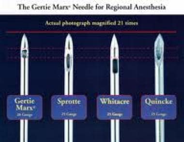
(+info)Labor analgesia. (8/18)
Regional analgesia has become the most common method of pain relief used during labor in the United States. Epidural and spinal analgesia are two types of regional analgesia. With epidural analgesia, an indwelling catheter is directed into the epidural space, and the patient receives a continuous infusion or multiple injections of local anesthetic. Spinal injections are usually single injections into the intrathecal space. A combination of epidural and spinal analgesia, known as a walking epidural, also is available. This technique combines the rapid pain relief from the spinal regional block with the constant and consistent effects from the epidural block. It allows sufficient motor function for patients to ambulate. Complications with regional analgesia are uncommon, but may include postdural puncture headache. Rare serious complications include neurologic injury, epidural hematoma, or deep epidural infection. Regional analgesia increases the risk of instrument-assisted vaginal delivery, and family physicians should understand the contraindications and risks of complications. Continuous labor support (e.g., doula), systemic opioid analgesia, pudendal blocks, water immersion, sterile water injections into the lumbosacral spine, self-taught hypnosis, and acupuncture are other options for pain management during labor. (+info)The Pudendal Nerve is a somatic nerve that carries sensory and motor fibers to the genital region in both males and females. It originates from the sacral plexus, specifically from nerves S2, S3, and S4. The pudendal nerve provides innervation to the skin of the perineum, labia majora/scrotum, and the lower portions of the vagina/penis. Additionally, it supplies motor function to the external anal and urethral sphincters, as well as to some muscles of the pelvic floor, such as the bulbospongiosus and ischiocavernosus muscles. The pudendal nerve plays a crucial role in sexual response and urinary and fecal continence.
Pudendal Neuralgia is a chronic pain condition characterized by the irritation or damage to the pudendal nerve, which supplies sensation and innervation to the perineum, genital region, and lower rectum. The symptoms often include burning pain, numbness, tingling, or shooting pain in these areas, which can be worsened by sitting or certain movements. It is important to note that Pudendal Neuralgia is not the same as Pudendal Nerve Entrapment (PNE), although PNE can lead to Pudendal Neuralgia. The diagnosis of this condition typically involves a thorough physical examination, medical history, and sometimes specialized tests like nerve blocks or electromyography (EMG) studies.
The urethra is the tube that carries urine from the bladder out of the body. In males, it also serves as the conduit for semen during ejaculation. The male urethra is longer than the female urethra and is divided into sections: the prostatic, membranous, and spongy (or penile) urethra. The female urethra extends from the bladder to the external urethral orifice, which is located just above the vaginal opening.
The anal canal is the terminal portion of the digestive tract, located between the rectum and the anus. It is a short tube-like structure that is about 1 to 1.5 inches long in adults. The main function of the anal canal is to provide a seal for the elimination of feces from the body while also preventing the leakage of intestinal contents.
The inner lining of the anal canal is called the mucosa, which is kept moist by the production of mucus. The walls of the anal canal contain specialized muscles that help control the passage of stool during bowel movements. These muscles include the internal and external sphincters, which work together to maintain continence and allow for the voluntary release of feces.
The anal canal is an important part of the digestive system and plays a critical role in maintaining bowel function and overall health.
The lumbosacral plexus is a complex network of nerves that arises from the lower part of the spinal cord, specifically the lumbar (L1-L5) and sacral (S1-S4) roots. This plexus is responsible for providing innervation to the lower extremities, including the legs, feet, and some parts of the abdomen and pelvis.
The lumbosacral plexus can be divided into several major branches:
1. The femoral nerve: It arises from the L2-L4 roots and supplies motor innervation to the muscles in the anterior compartment of the thigh, as well as sensation to the anterior and medial aspects of the leg and thigh.
2. The obturator nerve: It originates from the L2-L4 roots and provides motor innervation to the adductor muscles of the thigh and sensation to the inner aspect of the thigh.
3. The sciatic nerve: This is the largest nerve in the body, formed by the union of the tibial and common fibular (peroneal) nerves. It arises from the L4-S3 roots and supplies motor innervation to the muscles of the lower leg and foot, as well as sensation to the posterior aspect of the leg and foot.
4. The pudendal nerve: It originates from the S2-S4 roots and is responsible for providing motor innervation to the pelvic floor muscles and sensory innervation to the genital region.
5. Other smaller nerves, such as the ilioinguinal, iliohypogastric, and genitofemoral nerves, also arise from the lumbosacral plexus and supply sensation to various regions in the lower abdomen and pelvis.
Damage or injury to the lumbosacral plexus can result in significant neurological deficits, including muscle weakness, numbness, and pain in the lower extremities.
The perineum is the region between the anus and the genitals. In anatomical terms, it refers to the diamond-shaped area located in the lower part of the pelvis and extends from the coccyx (tailbone) to the pubic symphysis, which is the joint in the front where the two pubic bones meet. This region contains various muscles that support the pelvic floor and contributes to maintaining urinary and fecal continence. The perineum can be further divided into two triangular regions: the urogenital triangle (anterior) and the anal triangle (posterior).
The hypogastric plexus is a complex network of nerves located in the lower abdomen, near the aortic bifurcation. It plays a crucial role in the autonomic nervous system, primarily controlling the parasympathetic and sympathetic innervation to the pelvic viscera, including the descending colon, rectum, bladder, and reproductive organs. The hypogastric plexus is formed by the fusion of the superior and inferior hypogastric nerves, which originate from the lumbar and sacral spinal cord levels, respectively. Damage to this plexus can lead to various pelvic autonomic dysfunctions, such as urinary and fecal incontinence or sexual impairment.
Urination, also known as micturition, is the physiological process of excreting urine from the urinary bladder through the urethra. It is a complex process that involves several systems in the body, including the urinary system, nervous system, and muscular system.
In medical terms, urination is defined as the voluntary or involuntary discharge of urine from the urethra, which is the final pathway for the elimination of waste products from the body. The process is regulated by a complex interplay between the detrusor muscle of the bladder, the internal and external sphincters of the urethra, and the nervous system.
During urination, the detrusor muscle contracts, causing the bladder to empty, while the sphincters relax to allow the urine to flow through the urethra and out of the body. The nervous system plays a crucial role in coordinating these actions, with sensory receptors in the bladder sending signals to the brain when it is time to urinate.
Urination is essential for maintaining the balance of fluids and electrolytes in the body, as well as eliminating waste products such as urea, creatinine, and other metabolic byproducts. Abnormalities in urination can indicate underlying medical conditions, such as urinary tract infections, bladder dysfunction, or neurological disorders.
Peripheral nerves are nerve fibers that transmit signals between the central nervous system (CNS, consisting of the brain and spinal cord) and the rest of the body. These nerves convey motor, sensory, and autonomic information, enabling us to move, feel, and respond to changes in our environment. They form a complex network that extends from the CNS to muscles, glands, skin, and internal organs, allowing for coordinated responses and functions throughout the body. Damage or injury to peripheral nerves can result in various neurological symptoms, such as numbness, weakness, or pain, depending on the type and severity of the damage.
Fecal incontinence is the involuntary loss or leakage of stool (feces) from the rectum. It is also known as bowel incontinence. This condition can range from occasional leakage of stool when passing gas to a complete loss of bowel control. Fecal incontinence can be an embarrassing and distressing problem, but there are treatments available that can help improve symptoms and quality of life.
The causes of fecal incontinence can vary, but some common factors include:
* Damage to the muscles or nerves that control bowel function, such as from childbirth, surgery, spinal cord injury, or long-term constipation or diarrhea.
* Chronic digestive conditions, such as irritable bowel syndrome (IBS), inflammatory bowel disease (IBD), or celiac disease.
* Neurological conditions, such as multiple sclerosis, stroke, or spina bifida.
* Aging, which can lead to a decrease in muscle strength and control.
Treatment for fecal incontinence depends on the underlying cause of the condition. Treatments may include:
* Bowel training exercises to improve muscle strength and control.
* Changes in diet to help regulate bowel movements.
* Medications to treat constipation or diarrhea.
* Surgery to repair damaged muscles or nerves, or to create a new opening for stool to exit the body.
If you are experiencing symptoms of fecal incontinence, it is important to speak with your healthcare provider. They can help determine the cause of your symptoms and develop an appropriate treatment plan.
Nervous system trauma, also known as neurotrauma, refers to damage or injury to the nervous system, including the brain and spinal cord. This type of trauma can result from various causes, such as vehicular accidents, sports injuries, falls, violence, or penetrating traumas. Nervous system trauma can lead to temporary or permanent impairments in sensory, motor, or cognitive functions, depending on the severity and location of the injury.
Traumatic brain injury (TBI) is a common form of nervous system trauma that occurs when an external force causes brain dysfunction. TBIs can be classified as mild, moderate, or severe, based on factors such as loss of consciousness, memory loss, and neurological deficits. Mild TBIs, also known as concussions, may not cause long-term damage but still require medical attention to ensure proper healing and prevent further complications.
Spinal cord injuries (SCI) are another form of nervous system trauma that can have severe consequences. SCI occurs when the spinal cord is damaged due to a sudden, traumatic blow or cut, causing loss of motor function, sensation, or autonomic function below the level of injury. The severity and location of the injury determine the extent of impairment, which can range from partial to complete paralysis.
Immediate medical intervention is crucial in cases of nervous system trauma to minimize secondary damage, prevent complications, and optimize recovery outcomes. Treatment options may include surgery, medication, rehabilitation, or a combination of these approaches.
The urinary bladder is a muscular, hollow organ in the pelvis that stores urine before it is released from the body. It expands as it fills with urine and contracts when emptying. The typical adult bladder can hold between 400 to 600 milliliters of urine for about 2-5 hours before the urge to urinate occurs. The wall of the bladder contains several layers, including a mucous membrane, a layer of smooth muscle (detrusor muscle), and an outer fibrous adventitia. The muscles of the bladder neck and urethra remain contracted to prevent leakage of urine during filling, and they relax during voiding to allow the urine to flow out through the urethra.
A nerve transfer is a surgical procedure where a functioning nerve is connected to an injured nerve to restore movement, sensation or function. The functioning nerve, called the donor nerve, usually comes from another less critical location in the body and has spare nerve fibers that can be used to reinnervate the injured nerve, called the recipient nerve.
During the procedure, a small section of the donor nerve is carefully dissected and prepared for transfer. The recipient nerve is also prepared by removing any damaged or non-functioning portions. The two ends are then connected using microsurgical techniques under a microscope. Over time, the nerve fibers from the donor nerve grow along the recipient nerve and reinnervate the muscles or sensory structures that were previously innervated by the injured nerve.
Nerve transfers can be used to treat various types of nerve injuries, including brachial plexus injuries, facial nerve palsy, and peripheral nerve injuries. The goal of the procedure is to restore function as quickly and efficiently as possible, allowing for a faster recovery and improved quality of life for the patient.
Stress Urinary Incontinence (SUI) is a type of urinary incontinence that occurs when physical activities or movements, such as coughing, sneezing, laughing, exercising, or lifting heavy objects, put pressure on the bladder, causing unintentional leakage of urine. It is caused by weakened pelvic floor muscles and/or a malfunctioning urethral sphincter, which normally help maintain urinary continence. SUI is more common in women than men, especially those who have gone through pregnancy, childbirth, or menopause, but it can also affect older men with prostate gland issues.
A nerve crush injury is a type of peripheral nerve injury that occurs when there is excessive pressure or compression applied to a nerve, causing it to become damaged or dysfunctional. This can happen due to various reasons such as trauma from accidents, surgical errors, or prolonged pressure on the nerve from tight casts, clothing, or positions.
The compression disrupts the normal functioning of the nerve, leading to symptoms such as numbness, tingling, weakness, or pain in the affected area. In severe cases, a nerve crush injury can cause permanent damage to the nerve, leading to long-term disability or loss of function. Treatment for nerve crush injuries typically involves relieving the pressure on the nerve, providing supportive care, and in some cases, surgical intervention may be necessary to repair the damaged nerve.
Mononeuropathy is a medical condition that refers to damage or dysfunction affecting a single peripheral nerve, outside of the brain and spinal cord. This can result in weakness, numbness, or pain in the area served by that specific nerve. Mononeuropathies can occur due to various reasons such as trauma, compression, infection, or systemic diseases like diabetes. The symptoms and severity may vary depending on the type and location of the affected nerve.
A reflex is an automatic, involuntary and rapid response to a stimulus that occurs without conscious intention. In the context of physiology and neurology, it's a basic mechanism that involves the transmission of nerve impulses between neurons, resulting in a muscle contraction or glandular secretion.
Reflexes are important for maintaining homeostasis, protecting the body from harm, and coordinating movements. They can be tested clinically to assess the integrity of the nervous system, such as the knee-j jerk reflex, which tests the function of the L3-L4 spinal nerve roots and the sensitivity of the stretch reflex arc.
Peripheral nerve injuries refer to damage or trauma to the peripheral nerves, which are the nerves outside the brain and spinal cord. These nerves transmit information between the central nervous system (CNS) and the rest of the body, including sensory, motor, and autonomic functions. Peripheral nerve injuries can result in various symptoms, depending on the type and severity of the injury, such as numbness, tingling, weakness, or paralysis in the affected area.
Peripheral nerve injuries are classified into three main categories based on the degree of damage:
1. Neuropraxia: This is the mildest form of nerve injury, where the nerve remains intact but its function is disrupted due to a local conduction block. The nerve fiber is damaged, but the supporting structures remain intact. Recovery usually occurs within 6-12 weeks without any residual deficits.
2. Axonotmesis: In this type of injury, there is damage to both the axons and the supporting structures (endoneurium, perineurium). The nerve fibers are disrupted, but the connective tissue sheaths remain intact. Recovery can take several months or even up to a year, and it may be incomplete, with some residual deficits possible.
3. Neurotmesis: This is the most severe form of nerve injury, where there is complete disruption of the nerve fibers and supporting structures (endoneurium, perineurium, epineurium). Recovery is unlikely without surgical intervention, which may involve nerve grafting or repair.
Peripheral nerve injuries can be caused by various factors, including trauma, compression, stretching, lacerations, or chemical exposure. Treatment options depend on the type and severity of the injury and may include conservative management, such as physical therapy and pain management, or surgical intervention for more severe cases.
"Cat" is a common name that refers to various species of small carnivorous mammals that belong to the family Felidae. The domestic cat, also known as Felis catus or Felis silvestris catus, is a popular pet and companion animal. It is a subspecies of the wildcat, which is found in Europe, Africa, and Asia.
Domestic cats are often kept as pets because of their companionship, playful behavior, and ability to hunt vermin. They are also valued for their ability to provide emotional support and therapy to people. Cats are obligate carnivores, which means that they require a diet that consists mainly of meat to meet their nutritional needs.
Cats are known for their agility, sharp senses, and predatory instincts. They have retractable claws, which they use for hunting and self-defense. Cats also have a keen sense of smell, hearing, and vision, which allow them to detect prey and navigate their environment.
In medical terms, cats can be hosts to various parasites and diseases that can affect humans and other animals. Some common feline diseases include rabies, feline leukemia virus (FeLV), feline immunodeficiency virus (FIV), and toxoplasmosis. It is important for cat owners to keep their pets healthy and up-to-date on vaccinations and preventative treatments to protect both the cats and their human companions.
The penis is a part of the male reproductive and urinary systems. It has three parts: the root, the body, and the glans. The root attaches to the pelvic bone and the body makes up the majority of the free-hanging portion. The glans is the cone-shaped end that protects the urethra, the tube inside the penis that carries urine from the bladder and semen from the testicles.
The penis has a dual function - it acts as a conduit for both urine and semen. During sexual arousal, the penis becomes erect when blood fills two chambers inside its shaft. This process is facilitated by the relaxation of the smooth muscles in the arterial walls and the trappping of blood in the corpora cavernosa. The stiffness of the penis enables sexual intercourse. After ejaculation, or when the sexual arousal passes, the muscles contract and the blood flows out of the penis back into the body, causing it to become flaccid again.
The foreskin, a layer of skin that covers the glans, is sometimes removed in a procedure called circumcision. Circumcision is often performed for religious or cultural reasons, or as a matter of family custom. In some countries, it's also done for medical reasons, such as to treat conditions like phimosis (an inability to retract the foreskin) or balanitis (inflammation of the glans).
It's important to note that any changes in appearance, size, or function of the penis should be evaluated by a healthcare professional, as they could indicate an underlying medical condition.
Urinary incontinence is defined as the involuntary loss or leakage of urine that is sufficient to be a social or hygienic problem. It can occur due to various reasons such as weak pelvic muscles, damage to nerves that control the bladder, certain medications, and underlying medical conditions like diabetes, multiple sclerosis, or Parkinson's disease.
There are different types of urinary incontinence, including stress incontinence (leakage of urine during physical activities like coughing, sneezing, or exercising), urge incontinence (a sudden and strong need to urinate that results in leakage), overflow incontinence (constant dribbling of urine due to a bladder that doesn't empty completely), functional incontinence (inability to reach the bathroom in time due to physical or mental impairments), and mixed incontinence (a combination of any two or more types of incontinence).
Urinary incontinence can significantly impact a person's quality of life, causing embarrassment, social isolation, and depression. However, it is a treatable condition, and various treatment options are available, including bladder training, pelvic floor exercises, medications, medical devices, and surgery.
The femoral nerve is a major nerve in the thigh region of the human body. It originates from the lumbar plexus, specifically from the ventral rami (anterior divisions) of the second, third, and fourth lumbar nerves (L2-L4). The femoral nerve provides motor and sensory innervation to various muscles and areas in the lower limb.
Motor Innervation:
The femoral nerve is responsible for providing motor innervation to several muscles in the anterior compartment of the thigh, including:
1. Iliacus muscle
2. Psoas major muscle
3. Quadriceps femoris muscle (consisting of four heads: rectus femoris, vastus lateralis, vastus medialis, and vastus intermedius)
These muscles are involved in hip flexion, knee extension, and stabilization of the hip joint.
Sensory Innervation:
The sensory distribution of the femoral nerve includes:
1. Anterior and medial aspects of the thigh
2. Skin over the anterior aspect of the knee and lower leg (via the saphenous nerve, a branch of the femoral nerve)
The saphenous nerve provides sensation to the skin on the inner side of the leg and foot, as well as the medial malleolus (the bony bump on the inside of the ankle).
In summary, the femoral nerve is a crucial component of the lumbar plexus that controls motor functions in the anterior thigh muscles and provides sensory innervation to the anterior and medial aspects of the thigh and lower leg.
The pelvic floor is a group of muscles, ligaments, and connective tissues that form a sling or hammock across the bottom of the pelvis. It supports the organs in the pelvic cavity, including the bladder, rectum, and uterus or prostate. The pelvic floor helps control urination, defecation, and sexual function by relaxing and contracting to allow for the release of waste and during sexual activity. It also contributes to postural stability and balance. Weakness or damage to the pelvic floor can lead to various health issues such as incontinence, pelvic organ prolapse, and sexual dysfunction.
Electric stimulation, also known as electrical nerve stimulation or neuromuscular electrical stimulation, is a therapeutic treatment that uses low-voltage electrical currents to stimulate nerves and muscles. It is often used to help manage pain, promote healing, and improve muscle strength and mobility. The electrical impulses can be delivered through electrodes placed on the skin or directly implanted into the body.
In a medical context, electric stimulation may be used for various purposes such as:
1. Pain management: Electric stimulation can help to block pain signals from reaching the brain and promote the release of endorphins, which are natural painkillers produced by the body.
2. Muscle rehabilitation: Electric stimulation can help to strengthen muscles that have become weak due to injury, illness, or surgery. It can also help to prevent muscle atrophy and improve range of motion.
3. Wound healing: Electric stimulation can promote tissue growth and help to speed up the healing process in wounds, ulcers, and other types of injuries.
4. Urinary incontinence: Electric stimulation can be used to strengthen the muscles that control urination and reduce symptoms of urinary incontinence.
5. Migraine prevention: Electric stimulation can be used as a preventive treatment for migraines by applying electrical impulses to specific nerves in the head and neck.
It is important to note that electric stimulation should only be administered under the guidance of a qualified healthcare professional, as improper use can cause harm or discomfort.
A nerve block is a medical procedure in which an anesthetic or neurolytic agent is injected near a specific nerve or bundle of nerves to block the transmission of pain signals from that area to the brain. This technique can be used for both diagnostic and therapeutic purposes, such as identifying the source of pain, providing temporary or prolonged relief, or facilitating surgical procedures in the affected region.
The injection typically contains a local anesthetic like lidocaine or bupivacaine, which numbs the nerve, preventing it from transmitting pain signals. In some cases, steroids may also be added to reduce inflammation and provide longer-lasting relief. Depending on the type of nerve block and its intended use, the injection might be administered close to the spine (neuraxial blocks), at peripheral nerves (peripheral nerve blocks), or around the sympathetic nervous system (sympathetic nerve blocks).
While nerve blocks are generally safe, they can have side effects such as infection, bleeding, nerve damage, or in rare cases, systemic toxicity from the anesthetic agent. It is essential to consult with a qualified medical professional before undergoing this procedure to ensure proper evaluation, technique, and post-procedure care.
Neurogenic bladder is a term used to describe bladder dysfunction due to neurological damage or disease. The condition can result in problems with bladder storage and emptying, leading to symptoms such as urinary frequency, urgency, hesitancy, incontinence, and retention.
Neurogenic bladder can occur due to various medical conditions, including spinal cord injury, multiple sclerosis, Parkinson's disease, diabetic neuropathy, and stroke. The damage to the nerves that control bladder function can result in overactivity or underactivity of the bladder muscle, leading to urinary symptoms.
Management of neurogenic bladder typically involves a multidisciplinary approach, including medications, bladder training, catheterization, and surgery in some cases. The specific treatment plan depends on the underlying cause of the condition and the severity of the symptoms.
Spinal nerves are the bundles of nerve fibers that transmit signals between the spinal cord and the rest of the body. There are 31 pairs of spinal nerves in the human body, which can be divided into five regions: 8 cervical, 12 thoracic, 5 lumbar, 5 sacral, and 1 coccygeal. Each spinal nerve carries both sensory information (such as touch, temperature, and pain) from the periphery to the spinal cord, and motor information (such as muscle control) from the spinal cord to the muscles and other structures in the body. Spinal nerves also contain autonomic fibers that regulate involuntary functions such as heart rate, digestion, and blood pressure.
The sciatic nerve is the largest and longest nerve in the human body, running from the lower back through the buttocks and down the legs to the feet. It is formed by the union of the ventral rami (branches) of the L4 to S3 spinal nerves. The sciatic nerve provides motor and sensory innervation to various muscles and skin areas in the lower limbs, including the hamstrings, calf muscles, and the sole of the foot. Sciatic nerve disorders or injuries can result in symptoms such as pain, numbness, tingling, or weakness in the lower back, hips, legs, and feet, known as sciatica.
Afferent pathways, also known as sensory pathways, refer to the neural connections that transmit sensory information from the peripheral nervous system to the central nervous system (CNS), specifically to the brain and spinal cord. These pathways are responsible for carrying various types of sensory information, such as touch, temperature, pain, pressure, vibration, hearing, vision, and taste, to the CNS for processing and interpretation.
The afferent pathways begin with sensory receptors located throughout the body, which detect changes in the environment and convert them into electrical signals. These signals are then transmitted via afferent neurons, also known as sensory neurons, to the spinal cord or brainstem. Within the CNS, the information is further processed and integrated with other neural inputs before being relayed to higher cognitive centers for conscious awareness and response.
Understanding the anatomy and physiology of afferent pathways is essential for diagnosing and treating various neurological conditions that affect sensory function, such as neuropathies, spinal cord injuries, and brain disorders.
Nerve regeneration is the process of regrowth and restoration of functional nerve connections following damage or injury to the nervous system. This complex process involves various cellular and molecular events, such as the activation of support cells called glia, the sprouting of surviving nerve fibers (axons), and the reformation of neural circuits. The goal of nerve regeneration is to enable the restoration of normal sensory, motor, and autonomic functions impaired due to nerve damage or injury.
Electromyography (EMG) is a medical diagnostic procedure that measures the electrical activity of skeletal muscles during contraction and at rest. It involves inserting a thin needle electrode into the muscle to record the electrical signals generated by the muscle fibers. These signals are then displayed on an oscilloscope and may be heard through a speaker.
EMG can help diagnose various neuromuscular disorders, such as muscle weakness, numbness, or pain, and can distinguish between muscle and nerve disorders. It is often used in conjunction with other diagnostic tests, such as nerve conduction studies, to provide a comprehensive evaluation of the nervous system.
EMG is typically performed by a neurologist or a physiatrist, and the procedure may cause some discomfort or pain, although this is usually minimal. The results of an EMG can help guide treatment decisions and monitor the progression of neuromuscular conditions over time.
In medical terms, pressure is defined as the force applied per unit area on an object or body surface. It is often measured in millimeters of mercury (mmHg) in clinical settings. For example, blood pressure is the force exerted by circulating blood on the walls of the arteries and is recorded as two numbers: systolic pressure (when the heart beats and pushes blood out) and diastolic pressure (when the heart rests between beats).
Pressure can also refer to the pressure exerted on a wound or incision to help control bleeding, or the pressure inside the skull or spinal canal. High or low pressure in different body systems can indicate various medical conditions and require appropriate treatment.
The clitoris is an important female sex organ that is primarily responsible for sexual arousal and pleasure. It is a small, highly sensitive piece of tissue located at the front of the vulva, where the labia minora meet. The clitoris is made up of two parts: the visible part, known as the glans clitoris, and the hidden part, called the corpora cavernosa and crura.
The glans clitoris is a small knob-like structure that is covered by a hood, or prepuce, and is located at the top of the vulva. It contains a high concentration of nerve endings, making it highly sensitive to touch and stimulation. The corpora cavernosa and crura are the internal parts of the clitoris, which are made up of sponge-like erectile tissue that becomes engorged with blood during sexual arousal, leading to clitoral erection.
The clitoris plays a crucial role in female sexual response and pleasure. During sexual arousal, the clitoris swells and becomes more sensitive to touch, which can lead to orgasm. The clitoris is also an important source of sexual pleasure during masturbation and partnered sexual activity. Despite its importance in female sexuality, the clitoris has historically been overlooked or stigmatized in many cultures, leading to a lack of understanding and education about this vital organ.
Parturition is the process of giving birth, or the act of delivering newborn offspring. In medical terms, it refers to the expulsion of the products of conception (such as the fetus, placenta, and membranes) from the uterus of a pregnant woman during childbirth. This process is regulated by hormonal changes and involves complex interactions between the mother's body and the developing fetus. Parturition typically occurs after a full-term pregnancy, which is approximately 40 weeks in humans.
The optic nerve, also known as the second cranial nerve, is the nerve that transmits visual information from the retina to the brain. It is composed of approximately one million nerve fibers that carry signals related to vision, such as light intensity and color, from the eye's photoreceptor cells (rods and cones) to the visual cortex in the brain. The optic nerve is responsible for carrying this visual information so that it can be processed and interpreted by the brain, allowing us to see and perceive our surroundings. Damage to the optic nerve can result in vision loss or impairment.
Nerve fibers are specialized structures that constitute the long, slender processes (axons) of neurons (nerve cells). They are responsible for conducting electrical impulses, known as action potentials, away from the cell body and transmitting them to other neurons or effector organs such as muscles and glands. Nerve fibers are often surrounded by supportive cells called glial cells and are grouped together to form nerve bundles or nerves. These fibers can be myelinated (covered with a fatty insulating sheath called myelin) or unmyelinated, which influences the speed of impulse transmission.
Urodynamics is a medical test that measures the function and performance of the lower urinary tract, which includes the bladder, urethra, and sphincters. It involves the use of specialized equipment to record measurements such as bladder pressure, urine flow rate, and residual urine volume. The test can help diagnose various urinary problems, including incontinence, urinary retention, and overactive bladder.
During the test, a small catheter is inserted into the bladder through the urethra to measure bladder pressure while filling it with sterile water or saline solution. Another catheter may be placed in the rectum to record abdominal pressure. The patient is then asked to urinate, and the flow rate and any leaks are recorded.
Urodynamics can help identify the underlying cause of urinary symptoms and guide treatment decisions. It is often recommended for patients with complex or persistent urinary problems that have not responded to initial treatments.
Electric stimulation therapy, also known as neuromuscular electrical stimulation (NMES) or electromyostimulation, is a therapeutic treatment that uses electrical impulses to stimulate muscles and nerves. The electrical signals are delivered through electrodes placed on the skin near the target muscle group or nerve.
The therapy can be used for various purposes, including:
1. Pain management: Electric stimulation can help reduce pain by stimulating the release of endorphins, which are natural painkillers produced by the body. It can also help block the transmission of pain signals to the brain.
2. Muscle rehabilitation: NMES can be used to prevent muscle atrophy and maintain muscle tone in individuals who are unable to move their muscles due to injury or illness, such as spinal cord injuries or stroke.
3. Improving circulation: Electric stimulation can help improve blood flow and reduce swelling by contracting the muscles and promoting the movement of fluids in the body.
4. Wound healing: NMES can be used to promote wound healing by increasing blood flow, reducing swelling, and improving muscle function around the wound site.
5. Muscle strengthening: Electric stimulation can be used to strengthen muscles by causing them to contract and relax repeatedly, which can help improve muscle strength and endurance.
It is important to note that electric stimulation therapy should only be administered under the guidance of a trained healthcare professional, as improper use can cause harm or discomfort.
Pelvic Organ Prolapse (POP) is a medical condition where the supporting muscles and ligaments in a woman's pelvis weaken, causing one or more of the pelvic organs - including the bladder, uterus, rectum, or small intestine - to drop or press into or out of the vagina. This can result in various symptoms such as a feeling of heaviness or fullness in the pelvis, pressure or pain in the lower back, painful intercourse, and problems with urination or bowel movements. POP is often associated with childbirth, menopause, aging, and certain medical conditions that increase abdominal pressure, like obesity or chronic coughing. Treatment options can range from lifestyle changes and physical therapy to surgery.
Uterine prolapse is a condition where the uterus descends or slips down from its normal position in the pelvic cavity into or through the cervix and sometimes even outside the vaginal opening. This occurs due to the weakening of the muscles and ligaments that support the uterus, often as a result of childbirth, aging, menopause, obesity, or prior hysterectomy. Uterine prolapse can lead to various symptoms such as a feeling of heaviness in the pelvis, difficulty in urinating or having bowel movements, and uncomfortable sexual intercourse. The severity of the condition may vary from mild to severe, and treatment options range from lifestyle changes and physical therapy to surgery.
Manometry is a medical test that measures pressure inside various parts of the gastrointestinal tract. It is often used to help diagnose digestive disorders such as achalasia, gastroparesis, and irritable bowel syndrome. During the test, a thin, flexible tube called a manometer is inserted through the mouth or rectum and into the area being tested. The tube is connected to a machine that measures and records pressure readings. These readings can help doctors identify any abnormalities in muscle function or nerve reflexes within the digestive tract.
The vagina is the canal that joins the cervix (the lower part of the uterus) to the outside of the body. It also is known as the birth canal because babies pass through it during childbirth. The vagina is where sexual intercourse occurs and where menstrual blood exits the body. It has a flexible wall that can expand and retract. During sexual arousal, the vaginal walls swell with blood to become more elastic in order to accommodate penetration.
It's important to note that sometimes people use the term "vagina" to refer to the entire female genital area, including the external structures like the labia and clitoris. But technically, these are considered part of the vulva, not the vagina.
Motor neurons are specialized nerve cells in the brain and spinal cord that play a crucial role in controlling voluntary muscle movements. They transmit electrical signals from the brain to the muscles, enabling us to perform actions such as walking, talking, and swallowing. There are two types of motor neurons: upper motor neurons, which originate in the brain's motor cortex and travel down to the brainstem and spinal cord; and lower motor neurons, which extend from the brainstem and spinal cord to the muscles. Damage or degeneration of these motor neurons can lead to various neurological disorders, such as amyotrophic lateral sclerosis (ALS) and spinal muscular atrophy (SMA).
Vasculogenic impotence, also known as vasculogenic erectile dysfunction (VED), is a specific type of erectile dysfunction that is primarily caused by conditions that affect the blood flow in the penis. This means that the blood vessels that supply the penis with oxygenated blood necessary for an erection are not functioning properly.
The term "vasculogenic" refers to the origin or development of blood vessels, and in this context, it specifically relates to the dysfunction of the blood vessels responsible for erectile function. Common conditions that can lead to vasculogenic impotence include atherosclerosis (hardening of the arteries), hypertension (high blood pressure), diabetes, high cholesterol levels, and smoking.
In vasculogenic impotence, the smooth muscle in the penis does not relax properly, which restricts blood flow into the corpora cavernosa, the sponge-like erectile tissue inside the penis. As a result, an adequate erection cannot be achieved or maintained, leading to difficulty with sexual intercourse and overall sexual satisfaction.
Treatment for vasculogenic impotence typically involves addressing the underlying medical conditions that contribute to poor blood flow in the penis. This may include lifestyle modifications such as quitting smoking, exercising regularly, and adopting a healthy diet. Medications like phosphodiesterase-5 inhibitors (PDE5is) can also be prescribed to improve erectile function by increasing blood flow to the penis. In some cases, more invasive treatments like penile revascularization surgery may be considered for severe cases of vasculogenic impotence that do not respond to other forms of treatment.
Evoked potentials (EPs) are medical tests that measure the electrical activity in the brain or spinal cord in response to specific sensory stimuli, such as sight, sound, or touch. These tests are often used to help diagnose and monitor conditions that affect the nervous system, such as multiple sclerosis, brainstem tumors, and spinal cord injuries.
There are several types of EPs, including:
1. Visual Evoked Potentials (VEPs): These are used to assess the function of the visual pathway from the eyes to the back of the brain. A patient is typically asked to look at a patterned image or flashing light while electrodes placed on the scalp record the electrical responses.
2. Brainstem Auditory Evoked Potentials (BAEPs): These are used to evaluate the function of the auditory nerve and brainstem. Clicking sounds are presented to one or both ears, and electrodes placed on the scalp measure the response.
3. Somatosensory Evoked Potentials (SSEPs): These are used to assess the function of the peripheral nerves and spinal cord. Small electrical shocks are applied to a nerve at the wrist or ankle, and electrodes placed on the scalp record the response as it travels up the spinal cord to the brain.
4. Motor Evoked Potentials (MEPs): These are used to assess the function of the motor pathways in the brain and spinal cord. A magnetic or electrical stimulus is applied to the brain or spinal cord, and electrodes placed on a muscle measure the response as it travels down the motor pathway.
EPs can help identify abnormalities in the nervous system that may not be apparent through other diagnostic tests, such as imaging studies or clinical examinations. They are generally safe, non-invasive procedures with few risks or side effects.
The spinal cord is a major part of the nervous system, extending from the brainstem and continuing down to the lower back. It is a slender, tubular bundle of nerve fibers (axons) and support cells (glial cells) that carries signals between the brain and the rest of the body. The spinal cord primarily serves as a conduit for motor information, which travels from the brain to the muscles, and sensory information, which travels from the body to the brain. It also contains neurons that can independently process and respond to information within the spinal cord without direct input from the brain.
The spinal cord is protected by the bony vertebral column (spine) and is divided into 31 segments: 8 cervical, 12 thoracic, 5 lumbar, 5 sacral, and 1 coccygeal. Each segment corresponds to a specific region of the body and gives rise to pairs of spinal nerves that exit through the intervertebral foramina at each level.
The spinal cord is responsible for several vital functions, including:
1. Reflexes: Simple reflex actions, such as the withdrawal reflex when touching a hot surface, are mediated by the spinal cord without involving the brain.
2. Muscle control: The spinal cord carries motor signals from the brain to the muscles, enabling voluntary movement and muscle tone regulation.
3. Sensory perception: The spinal cord transmits sensory information, such as touch, temperature, pain, and vibration, from the body to the brain for processing and awareness.
4. Autonomic functions: The sympathetic and parasympathetic divisions of the autonomic nervous system originate in the thoracolumbar and sacral regions of the spinal cord, respectively, controlling involuntary physiological responses like heart rate, blood pressure, digestion, and respiration.
Damage to the spinal cord can result in various degrees of paralysis or loss of sensation below the level of injury, depending on the severity and location of the damage.
Sprague-Dawley rats are a strain of albino laboratory rats that are widely used in scientific research. They were first developed by researchers H.H. Sprague and R.C. Dawley in the early 20th century, and have since become one of the most commonly used rat strains in biomedical research due to their relatively large size, ease of handling, and consistent genetic background.
Sprague-Dawley rats are outbred, which means that they are genetically diverse and do not suffer from the same limitations as inbred strains, which can have reduced fertility and increased susceptibility to certain diseases. They are also characterized by their docile nature and low levels of aggression, making them easier to handle and study than some other rat strains.
These rats are used in a wide variety of research areas, including toxicology, pharmacology, nutrition, cancer, and behavioral studies. Because they are genetically diverse, Sprague-Dawley rats can be used to model a range of human diseases and conditions, making them an important tool in the development of new drugs and therapies.
Nerve endings, also known as terminal branches or sensory receptors, are the specialized structures present at the termination point of nerve fibers (axons) that transmit electrical signals to and from the central nervous system (CNS). They primarily function in detecting changes in the external environment or internal body conditions and converting them into electrical impulses.
There are several types of nerve endings, including:
1. Free Nerve Endings: These are unencapsulated nerve endings that respond to various stimuli like temperature, pain, and touch. They are widely distributed throughout the body, especially in the skin, mucous membranes, and visceral organs.
2. Encapsulated Nerve Endings: These are wrapped by specialized connective tissue sheaths, which can modify their sensitivity to specific stimuli. Examples include Pacinian corpuscles (responsible for detecting deep pressure and vibration), Meissner's corpuscles (for light touch), Ruffini endings (for stretch and pressure), and Merkel cells (for sustained touch).
3. Specialised Nerve Endings: These are nerve endings that respond to specific stimuli, such as auditory, visual, olfactory, gustatory, and vestibular information. They include hair cells in the inner ear, photoreceptors in the retina, taste buds in the tongue, and olfactory receptors in the nasal cavity.
Nerve endings play a crucial role in relaying sensory information to the CNS for processing and initiating appropriate responses, such as reflex actions or conscious perception of the environment.
The sural nerve is a purely sensory peripheral nerve in the lower leg and foot. It provides sensation to the outer ( lateral) aspect of the little toe and the adjacent side of the fourth toe, as well as a small portion of the skin on the back of the leg between the ankle and knee joints.
The sural nerve is formed by the union of branches from the tibial and common fibular nerves (branches of the sciatic nerve) in the lower leg. It runs down the calf, behind the lateral malleolus (the bony prominence on the outside of the ankle), and into the foot.
The sural nerve is often used as a donor nerve during nerve grafting procedures due to its consistent anatomy and relatively low risk for morbidity at the donor site.
The median nerve is one of the major nerves in the human body, providing sensation and motor function to parts of the arm and hand. It originates from the brachial plexus, a network of nerves that arise from the spinal cord in the neck. The median nerve travels down the arm, passing through the cubital tunnel at the elbow, and continues into the forearm and hand.
In the hand, the median nerve supplies sensation to the palm side of the thumb, index finger, middle finger, and half of the ring finger. It also provides motor function to some of the muscles that control finger movements, allowing for flexion of the fingers and opposition of the thumb.
Damage to the median nerve can result in a condition called carpal tunnel syndrome, which is characterized by numbness, tingling, and weakness in the hand and fingers.
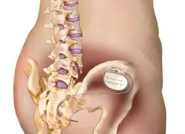
Spinal cord stimulation (SCS) is a medical procedure that involves the use of an implanted device to deliver electrical pulses to the spinal cord. The pulses are intended to interrupt or mask the transmission of pain signals to the brain, thereby reducing the perception of pain. SCS is typically offered as a treatment option for patients with chronic pain who have not found relief from other therapies, such as medication or surgery.
During the procedure, electrodes are placed in the epidural space of the spinal cord, and connected to a pulse generator that is implanted under the skin, usually in the abdomen or buttocks. The patient can use a remote control to adjust the intensity and location of the stimulation, allowing them to customize the therapy to their individual pain patterns.
SCS is generally considered safe, although there are some risks associated with the procedure, such as infection, bleeding, and nerve damage. It is important for patients to discuss these risks with their healthcare provider before deciding whether to undergo SCS.
Smooth muscle, also known as involuntary muscle, is a type of muscle that is controlled by the autonomic nervous system and functions without conscious effort. These muscles are found in the walls of hollow organs such as the stomach, intestines, bladder, and blood vessels, as well as in the eyes, skin, and other areas of the body.
Smooth muscle fibers are shorter and narrower than skeletal muscle fibers and do not have striations or sarcomeres, which give skeletal muscle its striped appearance. Smooth muscle is controlled by the autonomic nervous system through the release of neurotransmitters such as acetylcholine and norepinephrine, which bind to receptors on the smooth muscle cells and cause them to contract or relax.
Smooth muscle plays an important role in many physiological processes, including digestion, circulation, respiration, and elimination. It can also contribute to various medical conditions, such as hypertension, gastrointestinal disorders, and genitourinary dysfunction, when it becomes overactive or underactive.
The ischium is a part of the pelvic bone, specifically the lower and posterior portion. It is one of the three bones that fuse together to form each half of the pelvis, along with the ilium (the upper and largest portion) and the pubis (anteriorly).
The ischium has a thick, robust structure because it supports our body weight when we sit. Its main parts include:
1. The ischial tuberosity (sitting bone): This is the roughened, weight-bearing portion where you typically feel discomfort after sitting for long periods.
2. The ischial spine: A thin bony projection that serves as an attachment point for various muscles and ligaments.
3. The ramus of the ischium: The slender, curved part that extends downwards and joins with the pubis to form the inferior (lower) portion of the pelvic ring called the obturator foramen.
Together with the other components of the pelvis, the ischium plays a crucial role in providing stability, supporting the lower limbs, and protecting internal organs.
The facial nerve, also known as the seventh cranial nerve (CN VII), is a mixed nerve that carries both sensory and motor fibers. Its functions include controlling the muscles involved in facial expressions, taste sensation from the anterior two-thirds of the tongue, and secretomotor function to the lacrimal and salivary glands.
The facial nerve originates from the brainstem and exits the skull through the internal acoustic meatus. It then passes through the facial canal in the temporal bone before branching out to innervate various structures of the face. The main branches of the facial nerve include:
1. Temporal branch: Innervates the frontalis, corrugator supercilii, and orbicularis oculi muscles responsible for eyebrow movements and eyelid closure.
2. Zygomatic branch: Supplies the muscles that elevate the upper lip and wrinkle the nose.
3. Buccal branch: Innervates the muscles of the cheek and lips, allowing for facial expressions such as smiling and puckering.
4. Mandibular branch: Controls the muscles responsible for lower lip movement and depressing the angle of the mouth.
5. Cervical branch: Innervates the platysma muscle in the neck, which helps to depress the lower jaw and wrinkle the skin of the neck.
Damage to the facial nerve can result in various symptoms, such as facial weakness or paralysis, loss of taste sensation, and dry eyes or mouth due to impaired secretion.
The Tibial nerve is a major branch of the sciatic nerve that originates in the lower back and runs through the buttock and leg. It provides motor (nerve impulses that control muscle movement) and sensory (nerve impulses that convey information about touch, temperature, and pain) innervation to several muscles and skin regions in the lower limb.
More specifically, the Tibial nerve supplies the following structures:
1. Motor Innervation: The Tibial nerve provides motor innervation to the muscles in the back of the leg (posterior compartment), including the calf muscles (gastrocnemius and soleus) and the small muscles in the foot (intrinsic muscles). These muscles are responsible for plantarflexion (pointing the foot downward) and inversion (turning the foot inward) of the foot.
2. Sensory Innervation: The Tibial nerve provides sensory innervation to the skin on the sole of the foot, as well as the heel and some parts of the lower leg.
The Tibial nerve travels down the leg, passing behind the knee and through the calf, where it eventually joins with the common fibular (peroneal) nerve to form the tibial-fibular trunk. This trunk then divides into several smaller nerves that innervate the foot's intrinsic muscles and skin.
Damage or injury to the Tibial nerve can result in various symptoms, such as weakness or paralysis of the calf and foot muscles, numbness or tingling sensations in the sole of the foot, and difficulty walking or standing on tiptoes.
Spinal cord injuries (SCI) refer to damage to the spinal cord that results in a loss of function, such as mobility or feeling. This injury can be caused by direct trauma to the spine or by indirect damage resulting from disease or degeneration of surrounding bones, tissues, or blood vessels. The location and severity of the injury on the spinal cord will determine which parts of the body are affected and to what extent.
The effects of SCI can range from mild sensory changes to severe paralysis, including loss of motor function, autonomic dysfunction, and possible changes in sensation, strength, and reflexes below the level of injury. These injuries are typically classified as complete or incomplete, depending on whether there is any remaining function below the level of injury.
Immediate medical attention is crucial for spinal cord injuries to prevent further damage and improve the chances of recovery. Treatment usually involves immobilization of the spine, medications to reduce swelling and pressure, surgery to stabilize the spine, and rehabilitation to help regain lost function. Despite advances in treatment, SCI can have a significant impact on a person's quality of life and ability to perform daily activities.
The Ulnar nerve is one of the major nerves in the forearm and hand, which provides motor function to the majority of the intrinsic muscles of the hand (except for those innervated by the median nerve) and sensory innervation to the little finger and half of the ring finger. It originates from the brachial plexus, passes through the cubital tunnel at the elbow, and continues down the forearm, where it runs close to the ulna bone. The ulnar nerve then passes through the Guyon's canal in the wrist before branching out to innervate the hand muscles and provide sensation to the skin on the little finger and half of the ring finger.
Reaction time, in the context of medicine and physiology, refers to the time period between the presentation of a stimulus and the subsequent initiation of a response. This complex process involves the central nervous system, particularly the brain, which perceives the stimulus, processes it, and then sends signals to the appropriate muscles or glands to react.
There are different types of reaction times, including simple reaction time (responding to a single, expected stimulus) and choice reaction time (choosing an appropriate response from multiple possibilities). These measures can be used in clinical settings to assess various aspects of neurological function, such as cognitive processing speed, motor control, and alertness.
However, it is important to note that reaction times can be influenced by several factors, including age, fatigue, attention, and the use of certain medications or substances.
Urinary retention is a medical condition in which the bladder cannot empty completely or at all, resulting in the accumulation of urine in the bladder. This can lead to discomfort, pain, and difficulty in passing urine. Urinary retention can be acute (sudden onset) or chronic (long-term). Acute urinary retention is a medical emergency that requires immediate attention, while chronic urinary retention may be managed with medications or surgery. The causes of urinary retention include nerve damage, bladder muscle weakness, prostate gland enlargement, and side effects of certain medications.
Hematocolpos is a medical condition where the vagina gets filled with menstrual blood due to obstruction of the lower part of the genital tract, usually caused by an imperforate hymen or a transverse septum. It is often associated with hematometra (accumulation of menstrual blood in the uterus) and hematosalpinx (presence of menstrual blood in the fallopian tubes). This condition is typically found in adolescent females and can cause pelvic pain, difficulty walking, and a palpable mass in the lower abdomen. Surgical intervention is usually required to correct the obstruction and drain the accumulated blood.
The pelvis is the lower part of the trunk, located between the abdomen and the lower limbs. It is formed by the fusion of several bones: the ilium, ischium, and pubis (which together form the hip bone on each side), and the sacrum and coccyx in the back. The pelvis has several functions including supporting the weight of the upper body when sitting, protecting the lower abdominal organs, and providing attachment for muscles that enable movement of the lower limbs. In addition, it serves as a bony canal through which the reproductive and digestive tracts pass. The pelvic cavity contains several vital organs such as the bladder, parts of the large intestine, and in females, the uterus, ovaries, and fallopian tubes.
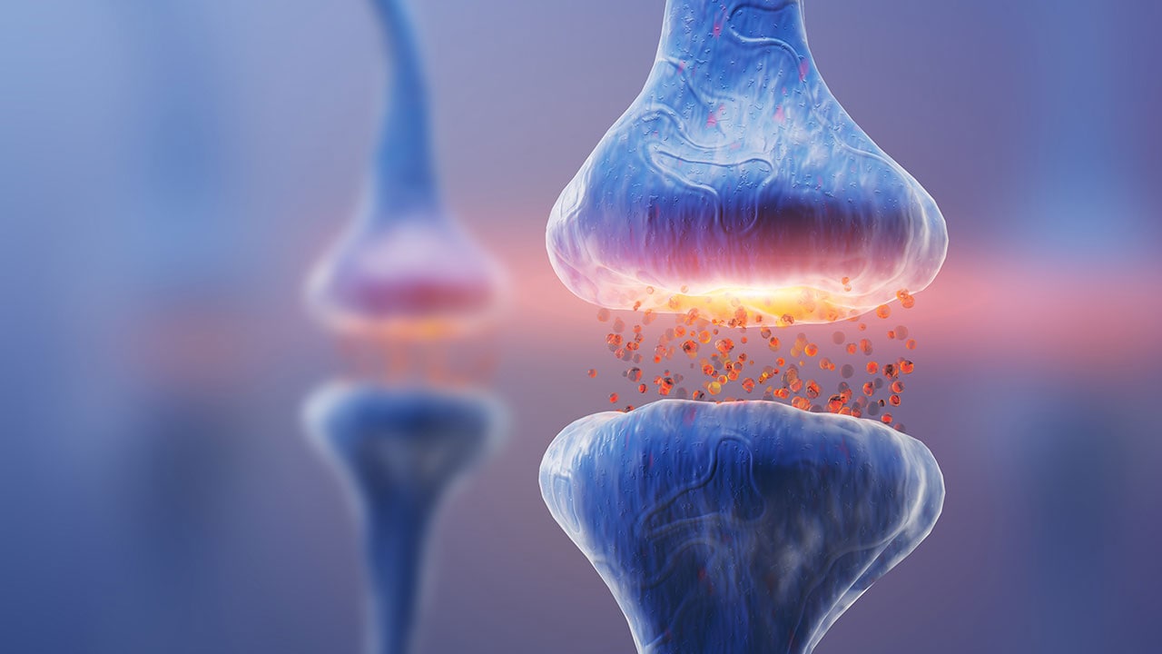
Neural conduction is the process by which electrical signals, known as action potentials, are transmitted along the axon of a neuron (nerve cell) to transmit information between different parts of the nervous system. This electrical impulse is generated by the movement of ions across the neuronal membrane, and it propagates down the length of the axon until it reaches the synapse, where it can then stimulate the release of neurotransmitters to communicate with other neurons or target cells. The speed of neural conduction can vary depending on factors such as the diameter of the axon, the presence of myelin sheaths (which act as insulation and allow for faster conduction), and the temperature of the environment.
Nerve Growth Factor (NGF) is a small secreted protein that is involved in the growth, maintenance, and survival of certain neurons (nerve cells). It was the first neurotrophin to be discovered and is essential for the development and function of the nervous system. NGF binds to specific receptors on the surface of nerve cells and helps to promote their differentiation, axonal growth, and synaptic plasticity. Additionally, NGF has been implicated in various physiological processes such as inflammation, immune response, and wound healing. Deficiencies or excesses of NGF have been linked to several neurological disorders, including Alzheimer's disease, Parkinson's disease, and pain conditions.
The trigeminal nerve, also known as the fifth cranial nerve or CNV, is a paired nerve that carries both sensory and motor information. It has three major branches: ophthalmic (V1), maxillary (V2), and mandibular (V3). The ophthalmic branch provides sensation to the forehead, eyes, and upper portion of the nose; the maxillary branch supplies sensation to the lower eyelid, cheek, nasal cavity, and upper lip; and the mandibular branch is responsible for sensation in the lower lip, chin, and parts of the oral cavity, as well as motor function to the muscles involved in chewing. The trigeminal nerve plays a crucial role in sensations of touch, pain, temperature, and pressure in the face and mouth, and it also contributes to biting, chewing, and swallowing functions.
The autonomic nervous system (ANS) is a component of the peripheral nervous system that regulates involuntary physiological functions, such as heart rate, digestion, respiratory rate, pupillary response, urination, and sexual arousal. The autonomic pathways refer to the neural connections and signaling processes that allow the ANS to carry out these functions.
The autonomic pathways consist of two main subdivisions: the sympathetic nervous system (SNS) and the parasympathetic nervous system (PNS). These systems have opposing effects on many organs, with the SNS generally stimulating activity and the PNS inhibiting it. The enteric nervous system, which controls gut function, is sometimes considered a third subdivision of the ANS.
The sympathetic pathway originates in the thoracic and lumbar regions of the spinal cord, with preganglionic neurons synapsing on postganglionic neurons in paravertebral ganglia or prevertebral ganglia. The parasympathetic pathway originates in the brainstem (cranial nerves III, VII, IX, and X) and the sacral region of the spinal cord (S2-S4), with preganglionic neurons synapsing on postganglionic neurons near or within the target organ.
Acetylcholine is the primary neurotransmitter used in both the sympathetic and parasympathetic pathways, although norepinephrine (noradrenaline) is also released by some postganglionic sympathetic neurons. The specific pattern of neural activation and inhibition within the autonomic pathways helps maintain homeostasis and allows for adaptive responses to changes in the internal and external environment.
Nerve Growth Factors (NGFs) are a family of proteins that play an essential role in the growth, maintenance, and survival of certain neurons (nerve cells). They were first discovered by Rita Levi-Montalcini and Stanley Cohen in 1956. NGF is particularly crucial for the development and function of the peripheral nervous system, which connects the central nervous system to various organs and tissues throughout the body.
NGF supports the differentiation and survival of sympathetic and sensory neurons during embryonic development. In adults, NGF continues to regulate the maintenance and repair of these neurons, contributing to neuroplasticity – the brain's ability to adapt and change over time. Additionally, NGF has been implicated in pain transmission and modulation, as well as inflammatory responses.
Abnormal levels or dysfunctional NGF signaling have been associated with various medical conditions, including neurodegenerative diseases (e.g., Alzheimer's and Parkinson's), chronic pain disorders, and certain cancers (e.g., small cell lung cancer). Therefore, understanding the role of NGF in physiological and pathological processes may provide valuable insights into developing novel therapeutic strategies for these conditions.
The phrenic nerve is a motor nerve that originates from the cervical spine (C3-C5) and descends through the neck to reach the diaphragm, which is the primary muscle used for breathing. The main function of the phrenic nerve is to innervate the diaphragm and control its contraction and relaxation, thereby enabling respiration.
Damage or injury to the phrenic nerve can result in paralysis of the diaphragm, leading to difficulty breathing and potentially causing respiratory failure. Certain medical conditions, such as neuromuscular disorders, spinal cord injuries, and tumors, can affect the phrenic nerve and impair its function.
The Radial nerve is a major peripheral nerve in the human body that originates from the brachial plexus, which is a network of nerves formed by the union of the ventral rami (anterior divisions) of spinal nerves C5-T1. The radial nerve provides motor function to extensor muscles of the upper limb and sensation to parts of the skin on the back of the arm, forearm, and hand.
More specifically, the radial nerve supplies motor innervation to:
* Extensor muscles of the shoulder (e.g., teres minor, infraspinatus)
* Rotator cuff muscles
* Elbow joint stabilizers (e.g., lateral head of the triceps)
* Extensors of the wrist, fingers, and thumb
The radial nerve also provides sensory innervation to:
* Posterior aspect of the upper arm (from the lower third of the humerus to the elbow)
* Lateral forearm (from the lateral epicondyle of the humerus to the wrist)
* Dorsum of the hand (skin over the radial side of the dorsum, including the first web space)
Damage or injury to the radial nerve may result in various symptoms, such as weakness or paralysis of the extensor muscles, numbness or tingling sensations in the affected areas, and difficulty with extension movements of the wrist, fingers, and thumb. Common causes of radial nerve injuries include fractures of the humerus bone, compression during sleep or prolonged pressure on the nerve (e.g., from crutches), and entrapment syndromes like radial tunnel syndrome.
Cranial nerves are a set of twelve pairs of nerves that originate from the brainstem and skull, rather than the spinal cord. These nerves are responsible for transmitting sensory information (such as sight, smell, hearing, and taste) to the brain, as well as controlling various muscles in the head and neck (including those involved in chewing, swallowing, and eye movement). Each cranial nerve has a specific function and is named accordingly. For example, the optic nerve (cranial nerve II) transmits visual information from the eyes to the brain, while the vagus nerve (cranial nerve X) controls parasympathetic functions in the body such as heart rate and digestion.
Animal disease models are specialized animals, typically rodents such as mice or rats, that have been genetically engineered or exposed to certain conditions to develop symptoms and physiological changes similar to those seen in human diseases. These models are used in medical research to study the pathophysiology of diseases, identify potential therapeutic targets, test drug efficacy and safety, and understand disease mechanisms.
The genetic modifications can include knockout or knock-in mutations, transgenic expression of specific genes, or RNA interference techniques. The animals may also be exposed to environmental factors such as chemicals, radiation, or infectious agents to induce the disease state.
Examples of animal disease models include:
1. Mouse models of cancer: Genetically engineered mice that develop various types of tumors, allowing researchers to study cancer initiation, progression, and metastasis.
2. Alzheimer's disease models: Transgenic mice expressing mutant human genes associated with Alzheimer's disease, which exhibit amyloid plaque formation and cognitive decline.
3. Diabetes models: Obese and diabetic mouse strains like the NOD (non-obese diabetic) or db/db mice, used to study the development of type 1 and type 2 diabetes, respectively.
4. Cardiovascular disease models: Atherosclerosis-prone mice, such as ApoE-deficient or LDLR-deficient mice, that develop plaque buildup in their arteries when fed a high-fat diet.
5. Inflammatory bowel disease models: Mice with genetic mutations affecting intestinal barrier function and immune response, such as IL-10 knockout or SAMP1/YitFc mice, which develop colitis.
Animal disease models are essential tools in preclinical research, but it is important to recognize their limitations. Differences between species can affect the translatability of results from animal studies to human patients. Therefore, researchers must carefully consider the choice of model and interpret findings cautiously when applying them to human diseases.
Spinal nerve roots are the initial parts of spinal nerves that emerge from the spinal cord through the intervertebral foramen, which are small openings between each vertebra in the spine. These nerve roots carry motor, sensory, and autonomic fibers to and from specific regions of the body. There are 31 pairs of spinal nerve roots in total, with 8 cervical, 12 thoracic, 5 lumbar, 5 sacral, and 1 coccygeal pair. Each root has a dorsal (posterior) and ventral (anterior) ramus that branch off to form the peripheral nervous system. Irritation or compression of these nerve roots can result in pain, numbness, weakness, or loss of reflexes in the affected area.
In the context of human anatomy, the thigh is the part of the lower limb that extends from the hip to the knee. It is the upper and largest portion of the leg and is primarily composed of the femur bone, which is the longest and strongest bone in the human body, as well as several muscles including the quadriceps femoris (front thigh), hamstrings (back thigh), and adductors (inner thigh). The major blood vessels and nerves that supply the lower limb also pass through the thigh.
Nerve compression syndromes refer to a group of conditions characterized by the pressure or irritation of a peripheral nerve, causing various symptoms such as pain, numbness, tingling, and weakness in the affected area. This compression can occur due to several reasons, including injury, repetitive motion, bone spurs, tumors, or swelling. Common examples of nerve compression syndromes include carpal tunnel syndrome, cubital tunnel syndrome, radial nerve compression, and ulnar nerve entrapment at the wrist or elbow. Treatment options may include physical therapy, splinting, medications, injections, or surgery, depending on the severity and underlying cause of the condition.
The ophthalmic nerve, also known as the first cranial nerve or CN I, is a sensory nerve that primarily transmits information about vision, including light intensity and color, and sensation in the eye and surrounding areas. It is responsible for the sensory innervation of the upper eyelid, conjunctiva, cornea, iris, ciliary body, and nasal cavity. The ophthalmic nerve has three major branches: the lacrimal nerve, frontal nerve, and nasociliary nerve. Damage to this nerve can result in various visual disturbances and loss of sensation in the affected areas.
 Pudendal nerve
Pudendal nerve entrapment
Pudendal plexus (nerves)
Pelvic pain
Obstructed defecation
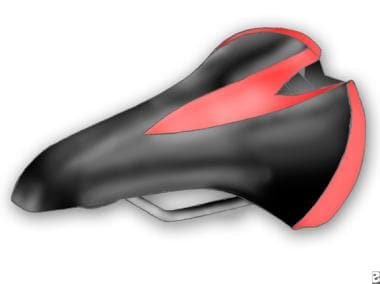
Cycling
Rectal prolapse
Dorsal nerve of the clitoris
Ischial spine
Vagina
Hip arthroscopy
Perineal nerve
Posterior labial nerves
Interventional radiology
Transverse perineal muscles
Perforating cutaneous nerve
Fecal incontinence
Pudendal canal
Vulva
Muscular branches of perineal nerve
Urethral sphincters
Onuf's nucleus
Internal pudendal veins
Human penis
Urogenital diaphragm
Prostate
Urethra
Chloroprocaine
Obstetric anesthesiology
Internal pudendal artery
Pudendal nerve - Wikipedia
 Transvaginal Pudendal Nerve Block: Overview, Indications, Contraindications
 Pudendal neuralgia caused by pressure on nerves | Clinton Daily News
 Pudendal Nerve Treatment Testimonials | IFAR Patient Stories
 Transvaginal Pudendal Nerve Blocks in Patients With Pudendal Neuralgia: Two-year Follow-up Results | Research Square
 Pudendal Nerve Entrapment/Neuropathy/Damage | Mayo Clinic Connect
 pudendal nerve pain Archives
 Pudendal Nerve Neuralgia Symptoms and Treatments
 Pudendal Nerve Irritation - Pelvic Health Solutions
 Pudendal nerve Archives - N2 Physical Therapy
 Pudendal Nerve<...
Transvaginal Pudendal Nerve Block: Overview, Indications, Contraindications
Transvaginal Pudendal Nerve Block: Overview, Indications, Contraindications
 Perineum Pain: Causes of Sore or Burning Perineum in Males/Females
 Dyspareunia Treated By Bilateral Pudendal Nerve Block
 PUDENDAL NERVE NEUROPATHY (PN) - Innovative Therapy Canada.
Urodynamic Studies for Urinary Incontinence: Overview, Patient Selection for Urodynamic Studies, Test Components
 Pudendal nerve entrapment syndrome (PNES) - An overview | Capsule Health
 Male Pudendal Nerve Entrapment - LINGAM WHISPERER BODYWORKLINGAM WHISPERER BODYWORK
 Pudendal Nerve Entrapment or Pudendal Neuralgia - Pain Care Clinic LTD
 Pudendal Neuralgia Treatment | Pudendal Nerve Pain Relief | Pain Spa, Bristol, UK
 Benzodiazepines Suppress Neuromodulatory Effects of Pudendal Nerve Stimulation on Rat Bladder Nociception. - AMiner
 Nerves that control bladder and bowel function
 Pudendal neuralgia after pelvic surgery using mesh: Case reports and laparoscopic pudendal nerve decompression | AVESİS
 Molecular Assessment of Neuroregenerative Response in the Pudendal Nerve: A Useful Tool in Regenerative Urology
Persistent genital arousal disorder: Treatment by neurolysis of dorsal branch of pudendal nerve<...
 Novel implantation technique for pudendal nerve peripheral nerve stimulation for treatment of chronic pelvic pain | Regional...
 Sonoanatomy of the ilioinguinal, iliohypogastric, genitofemoral, obturator, and pudendal nerves: a practical guide for US...
The 5 Things We Wish You Knew About Pudendal Neuralgia - Pelvic Guru #featured, loretta j. robertson, nerve pain, Pain with...
 back pain Archives - Erik Dalton Blog
Penis12
- 8 Inside the pudendal canal, the nerve divides into branches, first giving off the inferior rectal nerve, then the perineal nerve, before continuing as the dorsal nerve of the penis (in males) or the dorsal nerve of the clitoris (in females). (wikipedia.org)
- 1738 The pudendal nerve supplies sensation to the penis in males, and to the clitoris in females, which travels through the branches of both the dorsal nerve of the penis and the dorsal nerve of the clitoris. (wikipedia.org)
- 8 By providing sensation to the penis and the clitoris, the pudendal nerve is responsible for the afferent component of penile erection and clitoral erection. (wikipedia.org)
- Pudendal nerve irritation (neuralgia or nerve entrapment) is defined as pain in the distribution of the pudendal nerve, specifically the rectum, vagina, penis, perineum, and mons pubis. (pelvichealthsolutions.ca)
- Entrapment of the pudendal nerve can give rise to perineal pain involving the penis, scrotum, labia, perineum, or anorectal region. (painspa.co.uk)
- These can occur along its path between the sacrotuberous and the sacrospinous ligaments (interligamentous plane), through Alcock canal, or as the nerve passes through a tight osteofibrotic canal at the entrance to the base of the penis. (painspa.co.uk)
- Terminal branches are the dorsal nerve of the penis (clitoris), perineal nerve, and inferior anal nerve. (painspa.co.uk)
- Pudendal Neuralgia (PN) refers to pain along the distribution of the nerve - the pudendal nerve has 3 primary branches that go toward the anus, the perineum, and the clitoris or penis. (pelvicguru.com)
- It's entirely possible that someone's favorite cyclotron vibrator can shell-shock nerves, penis rings might lead to a grievous case of penile gangrene or those little vibrating beads could slip upstream and become tragicomically lost in bodily cavities while still in the 'on' position. (scientificamerican.com)
- The penis is innervated by the pudendal nerve (S2-S4). (medscape.com)
- This nerve eventually divides into the right and left dorsal nerves of the penis that pass under the pubis symphysis to travel just below the Buck fascia to supply the sensory innervation to the penis. (medscape.com)
- The right and left dorsal penile nerves should be blocked as proximally to the base of the penis as possible. (medscape.com)
Perineum10
- The pudendal nerve is the main nerve of the perineum. (wikipedia.org)
- A pudendal nerve block, also known as a saddle nerve block, is a local anesthesia technique used in an obstetric procedure to anesthetize the perineum during labor. (wikipedia.org)
- Blocking the pudendal nerve with injection of local anesthetic is used for vaginal deliveries and for minor surgeries of the vagina and perineum. (medscape.com)
- The sensory and motor innervation of the perineum is derived from the pudendal nerve, which is composed of the anterior primary divisions of the second, third, and fourth sacral nerves. (medscape.com)
- Your pudendal nerve runs from your lower back, along your pelvic floor muscles, out to your perineum (the skin between your pubic bone and your tailbone). (bhaskarhealth.com)
- The perineum lies near several nerves, muscles, and organs, so it's not uncommon to feel pain in this area. (healthline.com)
- It can also result in damage to the nerves and blood vessels in the perineum, which can cause bladder issues or problems during sex. (healthline.com)
- The primary symptom of pudendal nerve entrapment is ongoing pain somewhere in your pelvic region, including your perineum, scrotum, vulva, or rectum. (healthline.com)
- This includes the pudendal nerves and the cauda equina, which provides sensation and control of movement to the lower part of the body, including the bowel, anus, and perineum. (medicalnewstoday.com)
- The pudendal nerve is the primary sensory and motor nerve of the perineum and provides sensation from the genitalia and control of the urethral sphincter and anal sphincter. (naturalnews.com)
Pelvis13
- 215 : 157 The pudendal nerve passes between the piriformis muscle and coccygeus (ischiococcygeus) muscles and leaves the pelvis through the lower part of the greater sciatic foramen. (wikipedia.org)
- After reentering the pelvis, it accompanies the internal pudendal artery and internal pudendal vein upwards and forwards along the lateral wall of the ischiorectal fossa, being contained in a sheath of the obturator fascia termed the pudendal canal, along with the internal pudendal blood vessels. (wikipedia.org)
- This can occur as a result of a traumatic injury to the pelvis, where the pudendal nerve can become entrapped between the sacrospinous and sacrotuberous ligaments, or anywhere else along its pathway. (pelvichealthsolutions.ca)
- If you are going to consider a pudendal nerve block, you should see a pelvic floor physiotherapist first to ensure that the "container" of the pudendal nerve (muscles, connective tissue, and bony structures of the pelvis) are healthy so the effect of the nerve block can be accurately assessed. (pelvichealthsolutions.ca)
- The pudendal nerve is one of the primary nerves of your pelvis. (healthline.com)
- Uncommon chronic pain, in which the pudendal nerve (located in the pelvis) is entrapped or compressed. (capsulehealth.one)
- After re-entering the pelvis, it accompanies the internal pudendal artery and vein, coursing anterosuperiorly through the pudendal canal (also known as Alcock's canal - a structure formed by the fascia of the obturator internus muscle). (capsulehealth.one)
- There are pudendal nerves on the left and right sides of the body within the pelvis. (medicalnewstoday.com)
- The sacral plexus is a complex network of nerves situated at the back of the pelvis. (medicalnewstoday.com)
- The male pelvic floor is made up of three layers: deep, middle, and superficial, and has a complex relationship with the surrounding bony pelvis, fascia, ligaments and nerves. (physio-pedia.com)
- The anterior divisions of the upper four sacral nerves enter the pelvis through the anterior sacral foramina, that of the fifth between the sacrum and coccyx, while that of the coccygeal nerve curves forward below the rudimentary transverse process of the first piece of the coccyx. (bartleby.com)
- 829- Dissection of side wall of pelvis showing sacral and pudendal plexuses. (bartleby.com)
- The Superior Gluteal Nerve ( n. glutæus superior ) arises from the dorsal divisions of the fourth and fifth lumbar and first sacral nerves: it leaves the pelvis through the greater sciatic foramen above the Piriformis, accompanied by the superior gluteal vessels, and divides into a superior and an inferior branch. (bartleby.com)
Stimulation10
- Latest treatment using a peripheral nerve stimulator, which causes stimulation of the pudendal nerve in the ischioanal fossa. (capsulehealth.one)
- Benzodiazepines Suppress Neuromodulatory Effects of Pudendal Nerve Stimulation on Rat Bladder Nociception. (aminer.cn)
- Neuromodulation at a peripheral nerve target, in the form of bilateral electrical pudendal nerve stimulation (bPNS), has been shown to reduce bladder hypersensitivity in rats and anecdotally reduces pain in humans with pelvic pain of urological origin. (aminer.cn)
- bPNS was delivered as bilateral biphasic electrical stimulation of the mixed motor/sensory component of the pudendal nerves. (aminer.cn)
- CONCLUSIONS: This study suggests that inhibitory effects of bPNS on bladder pain could be suppressed in subjects receiving benzodiazepine therapy, suggesting that potential clinical testing of pudendal nerve stimulation for the treatment of painful bladder syndromes may be confounded by the use of benzodiazepines. (aminer.cn)
- Dorsal column spinal cord stimulation and dorsal root ganglion stimulation have had some success for managing CPP meanwhile peripheral nerve stimulators (PNS) have been suggested as another viable option. (bmj.com)
- applying this type of stimulation at the pudendal nerve could inhibit the pulses that lead to contraction of the urethral sphincter, and. (tudelft.nl)
- To examine the influence of autonomic receptor stimulation and blockade (noradrenaline, prazosin, terbutaline, propranolol, carbachol and atropine), and of pudendal nerve blockade on urethral stress relaxation. (nih.gov)
- The relief may be due to the stimulation of the pudendal nerve by the needle. (naturalnews.com)
- Thus, deep needling at these acupoints may increase the stimulation of the pudendal nerve, promoting the inhibition of local sensations going to the central nervous system. (naturalnews.com)
Neuropathy13
- Pudendal neuralgia (PN) is an extremely painful neuropathy of the pudendal nerve resulting in a negative impact on a patient's quality of life. (researchsquare.com)
- Pudendal neuralgia (PN) is an extremely painful neuropathy of the pudendal nerve. (researchsquare.com)
- Am wondering if anyone suffers from the monster Pudendal Nerve Entrapment/Neuropathy/Damage? (mayoclinic.org)
- Pudendal neuropathy can occur in both genders. (painspa.co.uk)
- Clinicians rarely associate these symptoms with pudendal neuropathy. (painspa.co.uk)
- Pudendal neuralgia is a peripheral neuropathy of the pudendal nerve that occurs in one or both sides in both genders. (painspa.co.uk)
- Pudendal neuropathy can be unilateral or bilateral. (painspa.co.uk)
- Stretch of the nerve by straining with constipation and childbirth can cause pudendal neuropathy. (painspa.co.uk)
- Radiation neuropathy following treatment of carcinoma of the prostate may relate to vascular impairment, inflammation within the nerve, or perineural desmoplastic reaction. (painspa.co.uk)
- Scrotalgia and vulvodynia occur with pudendal neuropathy. (painspa.co.uk)
- Neuropathy is a term used to describe several conditions that affect the nerves and can cause irritating and painful symptoms. (healthline.com)
- Research from 2021 shows that taking higher amounts can lead to nerve damage and cause symptoms of neuropathy. (healthline.com)
- It may raise energy levels, create healthy nerve cells, and reduce pain in people with neuropathy. (healthline.com)
Dysfunction3
- Pudendal neuralgia and pudendal nerve entrapment often involve a component of biomechanical dysfunction in the SI joint, Pelvic Floor Muscle Weakness and connective tissue dysfunction . (pelvichealthsolutions.ca)
- At our Clinic, we have witnessed many conditions exist whose symptoms imitate those of an actual pudendal nerve dysfunction, yet are not PN. (innovativetherapycanada.com)
- As with any disorder of somatic sensation, pudendal neuralgia can arise from dysfunction of the central nervous system, the nerve trunk, or the "end-organ. (painspa.co.uk)
Passes between the piriformis1
- Anatomical course and branches of the pudendal nerve: After its formation, the pudendal nerve descends and passes between the piriformis and ischiococcygeus muscles. (capsulehealth.one)
Fourth sacral nerves1
- Each receives a gray ramus communicans from the corresponding ganglion of the sympathetic trunk, while from the third and frequently from the second and the fourth sacral nerves, a white ramus communicans is given to the pelvic plexuses of the sympathetic. (bartleby.com)
Perineal4
- This nerve is accessed by 2 approaches, transvaginal and transcutaneous (or perineal). (medscape.com)
- Entrapment of the endopelvic part of the pudendal nerve may cause similar perineal or perianal pain with the extrapelvic pudendal nerve entrapment. (researchsquare.com)
- Pudendal neuralgia is characterized by perineal (and other pelvic) pain that is aggravated by sitting, generally decreased by standing and recumbence and typically reduced or relieved by sitting on a toilet seat. (painspa.co.uk)
- Her pudendal nerve also branches off to become the rectal, perineal, and clitoral nerves. (personallifemedia.com)
Clitoris1
- Conclusion: The relief of arousal symptoms by neurolysis of the dorsal nerve to the clitoris supports the hypothesis that PGAD is due to a minimal degree of compression of the dorsal branch of the pudendal nerve. (johnshopkins.edu)
Decompression surgery5
- Various treatment options, such as medication management, physiotherapy, nerve blocks, decompression surgery and neuromodulation, have been used, but the most appropriate treatment for pudendal neuralgia has not yet been determined. (comu.edu.tr)
- He's gone to the best medical centers all over the U.S. He's had pudendal nerve decompression surgery, numerous other procedures such as pudenda nerve blocks, pulsed radiofrequency ablation, and cryoablation to the pudendal nerves. (pelvicguru.com)
- This week's post is the second half of a Q&A on the controversial diagnosis of pudendal nerve entrapment and the decompression surgery associated with it. (pelvicpainrehab.com)
- When is a patient a good candidate for the pudendal nerve decompression surgery? (pelvicpainrehab.com)
- Should I have the Pudendal Nerve Decompression Surgery? (pelvicpainrehab.com)
Rectum1
- Above (proximal to) the dentate line, the rectum is supplied by stretch nerve fibers but not pain nerve fibers. (aafp.org)
Muscles6
- The pudendal nerve has both motor (control of muscles) and sensory functions. (wikipedia.org)
- There are many facilities around the country that treat this condition and the treatment is massaging, dialators, etc. of the muscles that could be entrapping the pudendal nerve. (mayoclinic.org)
- Pudendal neuralgia can come about when your pudendal nerve is exposed to traumas, the nerve is irritated, or compressed by bulky pelvic floor muscles or tight ligaments. (bhaskarhealth.com)
- The pudendal nerves stimulate the pelvic floor muscles, the anal sphincter, and pelvic organs. (medicalnewstoday.com)
- There are many reasons for this neuralgia - a local nerve irritation from inflammation, tight muscles/connective tissue, vascular compression, mechanical compression (i.e. sitting for too long in a position with too much pressure), etc. (pelvicguru.com)
- In women, stress relaxation in the urethra relies significantly on the pudendal nerve-innervated striated muscles. (nih.gov)
Cases of pudendal2
- therefore, interligamentous pudendal nerve entrapment cases have more benefits than the cases of pudendal nerve entrapment in the endopelvic part. (researchsquare.com)
- Most cases of pudendal neuralgia can be by a combination of physical therapies and pain medications but in some cases injection treatment may help with diagnosis as well as pain relief. (painspa.co.uk)
Neuralgia treatment1
- Watch incredible stories of transformation, told by patients just like you who came to us for life-changing pudendal neuralgia treatment. (advancedreconstruction.com)
Vagina1
- In this procedure, an anesthetic agent such as lidocaine is injected through the inner wall of the vagina about the pudendal nerve. (wikipedia.org)
Entrapment syndromes1
- The pudendal nerve entrapment syndromes subdivide into four types based on the level of compression. (capsulehealth.one)
Sciatic nerve2
- For example, the pudendal nerve may actually originate in the sciatic nerve. (wikipedia.org)
- Consequently, damage to the sciatic nerve can affect the pudendal nerve as well. (wikipedia.org)
Sacral plexus5
- 34 The nerve is a major branch of the sacral plexus,: 950 with fibers originating in Onuf's nucleus in the sacral region of the spinal cord. (wikipedia.org)
- The nerve trunk develops from the sacral plexus anterior to the piriformis muscle. (painspa.co.uk)
- There is a pudendal nerve for each side of the body, arising from the sacral plexus, the lowest part of the spine above the tailbone. (medicalnewstoday.com)
- The sacral plexus is formed by the lumbosacral trunk, the anterior division of the first, and portions of the anterior divisions of the second and third sacral nerves. (bartleby.com)
- The nerves forming the sacral plexus converge toward the lower part of the greater sciatic foramen, and unite to form a flattened band, from the anterior and posterior surfaces of which several branches arise. (bartleby.com)
Symptoms8
- However, during the recovery period I believe that something new kicked in which I believe is PNE (Pudendal Nerve Entrapment) as the symptoms are identical and to me it makes sense. (mayoclinic.org)
- End-organ pathologies capable of producing symptoms of pudendal neuralgia include tumors, dermatological, gynecological, urological and proctological conditions. (painspa.co.uk)
- The patient with unilateral decompression of the dorsal branch of the pudendal nerve was the only patient who had some, versus complete improvement in arousal symptoms. (johnshopkins.edu)
- Why are there so many misdiagnoses with pudendal symptoms? (pelvicguru.com)
- We are both pelvic physical therapists who see primarily pelvic pain, and more specifically, a high volume of patients with pudendal nerve symptoms. (pelvicguru.com)
- Based on his symptoms, it was good to know he found professionals who even knew about the pudendal nerve and types of treatments. (pelvicguru.com)
- (Here's another case based on symptoms of pain with sitting, but the wrong diagnosis based on MRI) A diagnosis of Pudendal Neuralgia truly involves a complete review of the patient history, diagnostic testing (if indicated) and a thorough exam. (pelvicguru.com)
- Furthermore, pudendal neuralgia is not just one thing - it can show up as a variety of symptoms, depending on the branch affected. (pelvicguru.com)
Several nerves3
- The pudendal nerve is one of several nerves supplying sensation to these areas. (wikipedia.org)
- The body has several nerves that work together to coordinate bladder function. (medicalnewstoday.com)
- Several nerves help control bowel function, digestion, and excretion. (medicalnewstoday.com)
Entrapments4
- The intervention was more effective in entrapments of the pudendal nerve between sacrospinous and sacrotuberous ligaments or below (Level-2) when compared to the injuries in the endopelvic part (Level-1). (researchsquare.com)
- I have nerve entrapment but not in that area, I have multiple nerve entrapments in my leg and recently had surgery to decompress the entrapments and insert a peripheral nerve stimulator in the event I still have pain after I heal. (mayoclinic.org)
- I have nerve entrapment but not in that area, I have multiple nerve entrapments in my. (mayoclinic.org)
- Many etiologies have been blamed including nerve injuries and entrapments. (comu.edu.tr)
Sensory3
- 274 It is a mixed (motor and sensory) nerve and also conveys sympathetic autonomic fibers. (wikipedia.org)
- The ilioinguinal, iliohypogastric, genitofemoral, obturator, and pudendal nerves are the major sensory nerves that may be involved in chronic groin and genital pain with a significant impact on the quality of life of patients . (bvsalud.org)
- This paper is aimed to serve as a practical technical guide for physicians to familiarize themselves with the ultrasound anatomy of the major inguinal sensory nerves and to enable successful ultrasound identification and ultrasound-guided diagnostic or therapeutic infiltrations for pain management of the ilioinguinal, iliohypogastric, genitofemoral, obturator, and pudendal nerves . (bvsalud.org)
Piriformis1
- Type I - Entrapment below the piriformis muscle as the pudendal nerve exits the greater sciatic notch. (capsulehealth.one)
Blood vessels1
- The penile shaft is composed of 3 erectile columns, the 2 corpora cavernosa and the corpus spongiosum, as well as the columns' enveloping fascial layers, nerves, lymphatics, and blood vessels, all covered by skin. (medscape.com)
Urethra3
- These nerves of the lower region of the spine stimulate the base of the bladder and urethra. (medicalnewstoday.com)
- The bladder and urethra are innervated by 3 sets of peripheral nerves arising from the autonomic nervous system (ANS) and somatic nervous system. (medscape.com)
- The drugs did not affect stress relaxation significantly, whereas the pudendal blockade produced a significant change along the length of the urethra characterized by a faster pressure decay following dilatation. (nih.gov)
Obturator3
- In this pictorial assay, we review the anatomy and clinical relevance of the ilioinguinal, iliohypogastric, genitofemoral, obturator, and pudendal nerves . (bvsalud.org)
- We summarize the various techniques for ultrasound identification, and present the ultrasound-guided infiltration techniques for injecting the ilioinguinal, iliohypogastric, genitofemoral, obturator, and pudendal nerves . (bvsalud.org)
- The Nerve to the Obturator Internus and Gemellus Superior arises from the ventral divisions of the fifth lumbar and first and second sacral nerves. (bartleby.com)
Vaginal2
- Use of this nerve block for vaginal delivery was reported as early as 1916. (medscape.com)
- In this article, we present two cases of postoperative pelvic pain thought to be secondary to injury or mechanical distortion of the pudendal nerve after rectocele repair using mesh and tension-free vaginal tape sling. (comu.edu.tr)
Autonomic1
- The pudendal nerve is a mixed nerve containing somatic and autonomic afferent and efferent fibers. (painspa.co.uk)
Bilateral2
- In this report, we present a patient with refractory superficial dyspareunia of unclear etiology, who was successfully managed with a bilateral pudendal nerve block. (symbiosisonlinepublishing.com)
- A bilateral pudendal nerve block alleviated the problem to three years follow-up. (symbiosisonlinepublishing.com)
Anatomy2
- 9 ] studied PN according to the segmental anatomy of the pudendal nerve. (researchsquare.com)
- The anatomy of the peripheral nerves can be successfully studied using imaging. (bvsalud.org)
Spinal cord5
- Various nerves control bladder and bowel function, including the spinal cord, cauda equina, pudendal nerves, and the enteric nervous system, a nerve network in the walls of the digestive tract. (medicalnewstoday.com)
- This is a group of nerve roots at the lower end of the spinal cord. (medicalnewstoday.com)
- These nerves begin at the sacral level of the spinal cord, which is the lowest part of the spine above the coccyx. (medicalnewstoday.com)
- The cauda equina is a group of nerves and nerve roots that stems from the lower end of the spinal cord and helps control the bladder. (medicalnewstoday.com)
- Methods: Sprague Dawley rats underwent unilateral pudendal nerve crush prior to spinal cord harvest and laser microdissection for separate collection of the injured and uninjured Onuf's nuclei (pudendal motor neuron cell bodies). (siftdesk.org)
Occur4
- Mechanical trauma such as compression, stretching, or irritation of the pudendal nerve may occur anywhere along the nerve pathway from the sacral roots to the end organ. (researchsquare.com)
- Damage to the pudendal nerve can occur suddenly as a result of trauma to the pelvic region, prolonged bicycling, fractures or from falls. (capsulehealth.one)
- Pain may occur anywhere in the pudendal territory. (painspa.co.uk)
- Part III of our "Demystifying Pudendal Neuralgia" Series How does PNE (pudendal nerve entrapment) occur? (pelvicpainrehab.com)
Anatomical4
- The anatomical basis for both approaches is to block the nerve proximal to its terminal branches. (medscape.com)
- The aim of this study is to evaluate the 2-years outcomes of repetitive doses of the transvaginal pudendal nerve injections (PNI) and to compare the success of the PNI concerning anatomical levels (endopelvic and extrapelvic portion) of the pudendal nerve pathology. (researchsquare.com)
- Pudendal neuralgia is a rare chronic neuropathic pain syndrome in the anatomical territory of the pudendal nerve. (comu.edu.tr)
- High-resolution ultrasound is increasingly used in the clinical setting for visualizing small peripheral nerves , and magnetic resonance imaging provides an anatomical overview of the relationship between small nerves and surrounding structures. (bvsalud.org)
Lesser sciatic1
- A pudendal nerve block targets the pudendal nerve trunk as it enters the lesser sciatic foramen, about 1 cm inferior and medial to the attachment of the sacrospinous ligament to the ischial spine. (medscape.com)
Branches3
- Pain in the territories of at least two out of the three terminal pudendal branches. (pelvichealthsolutions.ca)
- There are three branches of the pudendal nerve on each side of the body. (pelvichealthsolutions.ca)
- Branches of the pudendal nerve may innervate the pubococcygeus muscle. (painspa.co.uk)
Syndrome4
- You should go temporarily numb - if your pain is eliminated or significantly reduced, it indicates that the pudendal nerve is an important element in your pain syndrome. (bhaskarhealth.com)
- Pudendal neuralgia, or entrapment, has been compared to complex regional pain syndrome (CRPS), a sympathetically-mediated pain syndrome. (pelvichealthsolutions.ca)
- Synonyms include pudendal nerve entrapment or the syndrome of the pudendal canal (Alcock canal). (painspa.co.uk)
- Cauda equina syndrome (CES) may develop if the cauda equina nerves become compressed. (medicalnewstoday.com)
Ultrasound1
- Under X-ray or ultrasound, your doctor will inject a local anaesthetic into the canal the nerve travels through. (bhaskarhealth.com)
Anus1
- There are sensitive nerve endings around the anus. (personallifemedia.com)
Chronic1
- Pudendal neuralgia is chronic pain related to the pudendal nerve. (bhaskarhealth.com)
20201
- Klifto, K & Dellon, AL 2020, ' Persistent genital arousal disorder: Treatment by neurolysis of dorsal branch of pudendal nerve ', Microsurgery , vol. 40, no. 2, pp. 160-166. (johnshopkins.edu)
Pain12
- My wife has had pudendal nerve pain for 8 years. (mayoclinic.org)
- The pain areas in the pelvic region are exactly where the pudendal nerve goes. (mayoclinic.org)
- Pudendal neuralgia, like any pain condition, is managed through adapting your everyday life. (bhaskarhealth.com)
- If these criteria are not met, and the pain is predominately in the distribution of the pudendal nerve, then pudendal neuralgia is a more likely diagnosis. (pelvichealthsolutions.ca)
- in other words, the pudendal nerve block temporarily abolished or significantly reduced the typical pain that the patient is reporting. (pelvichealthsolutions.ca)
- Pudendal neuralgia (PN) is defined as the existence of pain in the distribution of the pudendal nerve. (painspa.co.uk)
- Pudendal neuralgia is a functional entrapment where pain occurs during a compression or stretch maneuver. (painspa.co.uk)
- We evaluate and treat patients from all over the world who are not finding help or differential diagnoses related to pelvic pain and pudendal neuralgia. (pelvicguru.com)
- It doesn't matter how many times a web search indicates pudendal neuralgia involves pain with sitting (or the Nantes Criteria indicates this), it does not mean you have PN if you have pain with sitting. (pelvicguru.com)
- Alpha-lipoic acid has been shown to have a positive effect on nerve conduction and to reduce neuropathic pain. (healthline.com)
- The risks of infection and blood clots are always present, and Ganz and his colleagues cite complications such as heterotopic ossification (new bone formation around the hip), nerve injuries, failure of the greater trochanter to heal back properly, persistent pain following the formation of scar tissue ( adhesions ) in the hip joint, and a small risk of damage of the blood supply to the femoral head. (wikipedia.org)
- Discover nerve gliding techniques for pain relief and enhanced mobility. (pinterest.ca)
Canal2
- The pudendal canal that carries the pudendal nerve is also known by the eponymous term "Alcock's canal", after Benjamin Alcock, an Irish anatomist who documented the canal in 1836. (wikipedia.org)
- The nerve follows the posterior surface of the sacrospinous ligament, passes anterior to the sacrotuberous ligament, and enters the Alcock canal. (painspa.co.uk)
Divides1
- The anterior division of the third sacral nerve divides into an upper and a lower branch, the former entering the sacral and the latter the pudendal plexus. (bartleby.com)
Urinary3
- If nerves that control the bladder or bowel become damaged, it can result in urinary or fecal incontinence. (medicalnewstoday.com)
- If the nerves become damaged, people may experience urinary retention or incontinence. (medicalnewstoday.com)
- Aims: Assessing pudendal nerve neuroregenerative response provides valuable insight into injuries and regenerative treatments related to urinary incontinence. (siftdesk.org)
Affect the nerves1
- Several conditions can affect the nerves that control bowel or bladder function. (medicalnewstoday.com)
Surgery2
- However, it is recommended to perform therapeutic nerve blocks even in patients with suspected endopelvic pudendal nerve pathology before the referral to surgery. (researchsquare.com)
- In cases of failed conservative treatment and of mesh removal surgery, laparoscopic pudendal nerve decompression and omental flap wrapping operation can be a treatment option for pudendal neuralgia. (comu.edu.tr)
Buttocks1
- Falls onto the buttocks can cause pudendal neuralgia. (painspa.co.uk)
Anesthesia2
- The nerve may be temporarily anesthetized, called pudendal anesthesia or pudendal block. (wikipedia.org)
- While neuroaxial analgesia continues to replace pudendal nerve block as the technique of choice, there are and will always remain situations in which anesthesia service is unavailable. (medscape.com)
Medial3
- Here, the nerve is medial to the internal pudendal vessels. (medscape.com)
- Results A cephalad to caudal-medial fluoroscopic guided approach as described within to successfully implant a percutaneous pudendal nerve PNS for management of CPP. (bmj.com)
- it appears at the medial margin of the Psoas major and runs downward over the pelvic brim to join the first sacral nerve. (bartleby.com)
Damage2
- Pudendal nerve entrapment is a type of nerve damage. (healthline.com)
- Without treatment, it can cause permanent nerve damage. (healthline.com)
Diagnosis1
- Why is the diagnosis of Pudendal Neuralgia confused with Pudendal Nerve Entrapment? (pelvicguru.com)
Sacrospinous ligament1
- The three roots become two cords when the middle and lower root join to form the lower cord, and these in turn unite to form the pudendal nerve proper just proximal to the sacrospinous ligament. (wikipedia.org)