Myelography
Meningocele
Iophendylate
Arachnoiditis
Intracranial Hypotension
Spinal Cord Compression
Subdural Effusion
Meningism
Laminectomy
Metrizamide
Spinal Nerve Roots
Spinal Cord Diseases
Brachial Plexus
Spinal Puncture
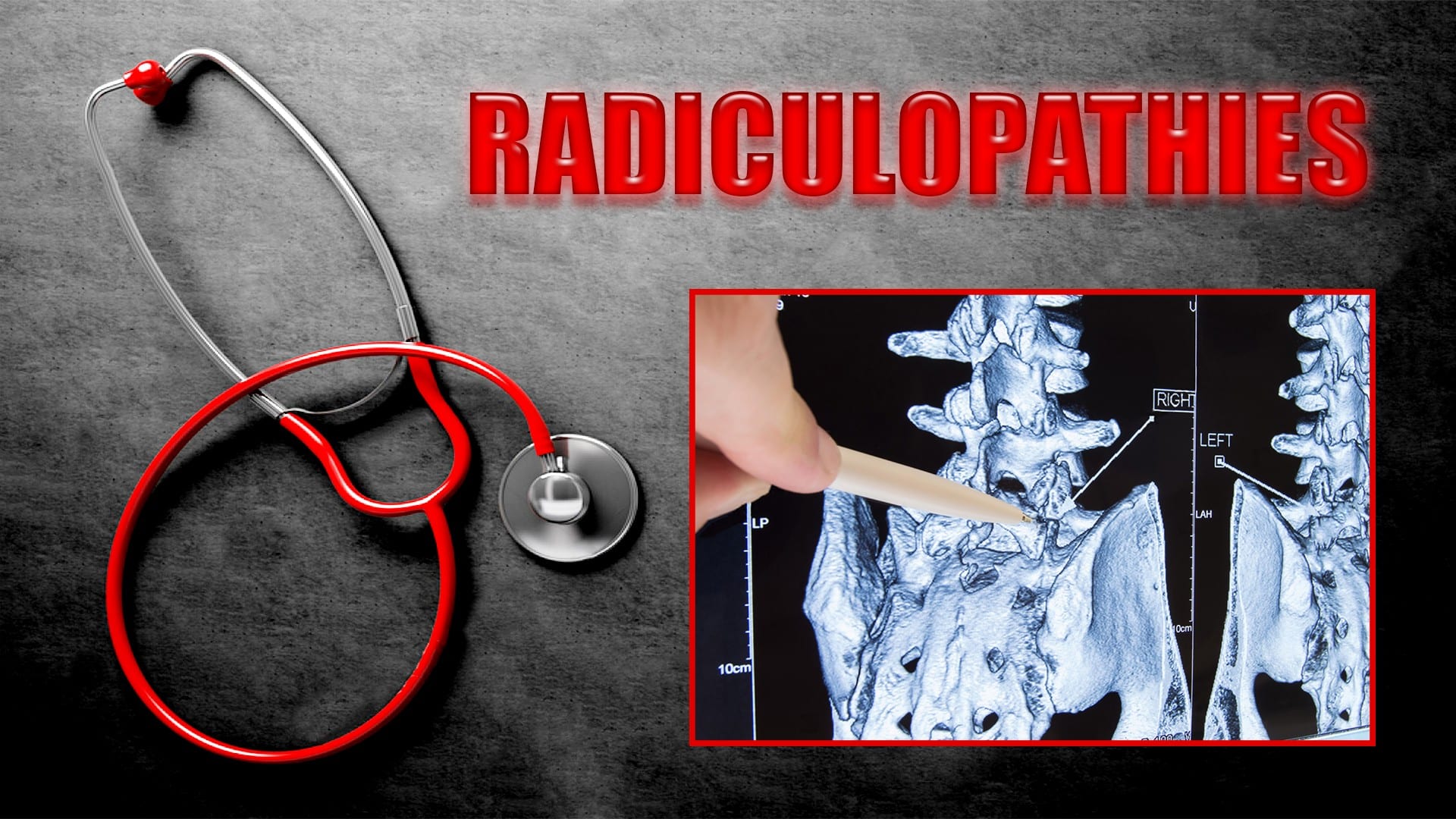
Nerve Compression Syndromes
Iopamidol
Radiography, Dual-Energy Scanned Projection
Dura Mater
Intervertebral Disc Displacement
Iothalamic Acid
Spinal Osteophytosis
Cervical Vertebrae
Cerebrospinal Fluid
Tomography, X-Ray Computed
Brachial Plexus Neuritis
Cerebrospinal Fluid Pressure
Neuroleptanalgesia
Arachnoid Cysts
Thoracic Vertebrae
Syringomyelia
Spinal Neoplasms
Lumbar Vertebrae
Iohexol
Spinal Cord Neoplasms
Cauda Equina
Meningomyelocele
Subarachnoid Space
Magnetic Resonance Imaging
Blood Patch, Epidural
Spinal Diseases
Paresthesia
Myelography is a medical imaging technique used to examine the spinal cord and surrounding structures, such as the spinal nerves, intervertebral discs, and the spinal column. This procedure involves the injection of a contrast dye into the subarachnoid space, which is the area surrounding the spinal cord filled with cerebrospinal fluid (CSF). The dye outlines the spinal structures, making them visible on X-ray or CT scan images.
The primary purpose of myelography is to diagnose various spinal conditions, including herniated discs, spinal stenosis, tumors, infection, and traumatic injuries. It can help identify any compression or irritation of the spinal cord or nerves that may be causing pain, numbness, weakness, or other neurological symptoms.
The procedure typically requires the patient to lie flat on their stomach or side while the radiologist inserts a thin needle into the subarachnoid space, usually at the lower lumbar level. Once the contrast dye is injected, the patient will be repositioned for various X-ray views or undergo a CT scan to capture detailed images of the spine. After the procedure, patients may experience headaches, nausea, or discomfort at the injection site, but these symptoms usually resolve within a few days.
A meningocele is a type of neural tube defect that results in the herniation of the meninges (the protective membranes covering the brain and spinal cord) through a defect in the vertebral column. The meninges protrude as a sac-like structure, which may be covered by skin or a thin layer of tissue. Meningoceles usually do not contain neural tissue, but cerebrospinal fluid is present within the sac. They are typically asymptomatic unless there is compression of surrounding structures or infection. Treatment generally involves surgical repair to prevent potential complications such as meningitis or neurological damage.
Iophendylate is not typically referred to as a medical definition, but it is the chemical name for a contrast agent that is used in radiology procedures. It is a type of oil-based contrast medium that is injected into the cerebrospinal fluid (CSF) during myelography, which is an imaging test used to visualize the spinal cord and surrounding structures.
Iophendylate, also known as Pantopaque, is a heavy oily substance that outlines the spinal canal and nerve roots on X-ray images, allowing radiologists to diagnose various conditions such as herniated discs, spinal stenosis, or tumors. However, due to the risks associated with its use, including chemical meningitis and potential neurological complications, it has largely been replaced by water-soluble contrast agents in current clinical practice.
Arachnoiditis is a medical condition that affects the arachnoid, one of the membranes that surround and protect the nerves of the central nervous system (the brain and spinal cord). The arachnoid becomes inflamed, often as a result of infection, direct injury, or complications from spinal surgery or chronic exposure to irritants such as steroids or contrast dyes.
The inflammation can cause the formation of scar tissue, which can lead to a variety of symptoms including:
1. Chronic pain in the back, legs, or arms
2. Numbness, tingling, or weakness in the limbs
3. Muscle cramps and spasms
4. Bladder and bowel dysfunction
5. Sexual dysfunction
In severe cases, arachnoiditis can cause permanent nerve damage and disability. Treatment typically focuses on managing symptoms and improving quality of life, as there is no cure for the condition.
Intracranial hypotension is a medical condition characterized by reduced pressure within the cranial cavity (the space containing brain and cerebrospinal fluid). This can occur due to several reasons, most commonly being a spontaneous or traumatic CSF leak (cerebrospinal fluid leak) from the dural membrane that surrounds the brain and spinal cord. The decrease in CSF pressure can cause various symptoms such as headaches (often positional), nausea, vomiting, neck pain, blurred vision, ringing in the ears, and cognitive impairment. Treatment typically involves identifying and addressing the underlying cause, which may include bed rest, hydration, caffeine, epidural blood patch procedures, or surgical repair of CSF leaks.
Spinal cord compression is a medical condition that refers to the narrowing of the spinal canal, which puts pressure on the spinal cord and the nerves that branch out from it. This can occur due to various reasons such as degenerative changes in the spine, herniated discs, bone spurs, tumors, or fractures. The compression can lead to a range of symptoms including pain, numbness, tingling, weakness, or loss of bladder and bowel control. In severe cases, it can cause paralysis. Treatment options depend on the underlying cause and may include physical therapy, medication, surgery, or radiation therapy.
A subdural effusion is an abnormal accumulation of fluid in the potential space between the dura mater (the outermost layer of the meninges that covers the brain and spinal cord) and the arachnoid membrane (one of the three layers of the meninges that surround the brain and spinal cord) in the subdural space.
Subdural effusions can occur due to various reasons, including head trauma, infection, or complications from neurosurgical procedures. The fluid accumulation may result from bleeding (subdural hematoma), inflammation, or increased cerebrospinal fluid pressure. Depending on the underlying cause and the amount of fluid accumulated, subdural effusions can cause various symptoms, such as headaches, altered mental status, or neurological deficits.
Subdural effusions are often asymptomatic and may resolve independently; however, in some cases, medical intervention might be necessary to alleviate the pressure on the brain or address the underlying condition. Imaging techniques like computed tomography (CT) or magnetic resonance imaging (MRI) scans are typically used to diagnose and monitor subdural effusions.
Meningismus is not a specific medical condition but rather a set of symptoms that suggest inflammation of the meninges, the membranes covering the brain and spinal cord. The term "meningitis" refers to the infection or inflammation of the meninges, while "meningism" describes the non-specific signs of this inflammation.
Meningismus symptoms include:
1. Stiff neck (nuchal rigidity) - resistance to passive flexion of the neck due to spasm of the neck muscles.
2. Photophobia - sensitivity to light.
3. Headache.
4. Fever.
5. Altered mental status, such as confusion or irritability.
Meningismus can be caused by various conditions, including bacterial and viral infections, fungal infections, chemical irritants, or non-infectious causes like autoimmune disorders or certain medications. It is essential to consult a healthcare professional if you suspect meningismus symptoms, as they may indicate a severe underlying condition requiring prompt medical attention.
A laminectomy is a surgical procedure that involves the removal of the lamina, which is the back part of the vertebra that covers the spinal canal. This procedure is often performed to relieve pressure on the spinal cord or nerves caused by conditions such as herniated discs, spinal stenosis, or tumors. By removing the lamina, the surgeon can access the affected area and alleviate the compression on the spinal cord or nerves, thereby reducing pain, numbness, or weakness in the back, legs, or arms.
Laminectomy may be performed as a standalone procedure or in combination with other surgical techniques such as discectomy, foraminotomy, or spinal fusion. The specific approach and extent of the surgery will depend on the patient's individual condition and symptoms.
Metrizamide is a contrast medium used in myelography, discography, and angiography to enhance the visibility of structures during X-ray or other radiological examinations. It's an ionic, water-soluble, non-ionic, monomeric contrast agent that belongs to the class of agents called triiodinated benzoic acid derivatives.
When administered, Metrizamide mixes with cerebrospinal fluid (CSF) and provides a clear image of the spinal cord and surrounding structures during myelography. It can also be used in discography to help diagnose disc-related pain by outlining the structure of intervertebral discs.
It's important to note that Metrizamide has been largely replaced by other contrast agents due to its potential side effects, including headache, nausea, vomiting, seizures, and in rare cases, brain damage or death.
Spinal nerve roots are the initial parts of spinal nerves that emerge from the spinal cord through the intervertebral foramen, which are small openings between each vertebra in the spine. These nerve roots carry motor, sensory, and autonomic fibers to and from specific regions of the body. There are 31 pairs of spinal nerve roots in total, with 8 cervical, 12 thoracic, 5 lumbar, 5 sacral, and 1 coccygeal pair. Each root has a dorsal (posterior) and ventral (anterior) ramus that branch off to form the peripheral nervous system. Irritation or compression of these nerve roots can result in pain, numbness, weakness, or loss of reflexes in the affected area.
Spinal cord diseases refer to a group of conditions that affect the spinal cord, which is a part of the central nervous system responsible for transmitting messages between the brain and the rest of the body. These diseases can cause damage to the spinal cord, leading to various symptoms such as muscle weakness, numbness, pain, bladder and bowel dysfunction, and difficulty with movement and coordination.
Spinal cord diseases can be congenital or acquired, and they can result from a variety of causes, including infections, injuries, tumors, degenerative conditions, autoimmune disorders, and genetic factors. Some examples of spinal cord diseases include multiple sclerosis, spina bifida, spinal cord injury, herniated discs, spinal stenosis, and motor neuron diseases such as amyotrophic lateral sclerosis (ALS).
The treatment for spinal cord diseases varies depending on the underlying cause and severity of the condition. Treatment options may include medication, physical therapy, surgery, and rehabilitation. In some cases, the damage to the spinal cord may be irreversible, leading to permanent disability or paralysis.
The brachial plexus is a network of nerves that originates from the spinal cord in the neck region and supplies motor and sensory innervation to the upper limb. It is formed by the ventral rami (branches) of the lower four cervical nerves (C5-C8) and the first thoracic nerve (T1). In some cases, contributions from C4 and T2 may also be included.
The brachial plexus nerves exit the intervertebral foramen, pass through the neck, and travel down the upper chest before branching out to form major peripheral nerves of the upper limb. These include the axillary, radial, musculocutaneous, median, and ulnar nerves, which further innervate specific muscles and sensory areas in the arm, forearm, and hand.
Damage to the brachial plexus can result in various neurological deficits, such as weakness or paralysis of the upper limb, numbness, or loss of sensation in the affected area, depending on the severity and location of the injury.
The spinal canal is the bony, protective channel within the vertebral column that contains and houses the spinal cord. It extends from the foramen magnum at the base of the skull to the sacrum, where the spinal cord ends and forms the cauda equina. The spinal canal is formed by a series of vertebral bodies stacked on top of each other, intervertebral discs in between them, and the laminae and spinous processes that form the posterior elements of the vertebrae. The spinal canal provides protection to the spinal cord from external trauma and contains cerebrospinal fluid (CSF) that circulates around the cord, providing nutrients and cushioning. Any narrowing or compression of the spinal canal, known as spinal stenosis, can cause various neurological symptoms due to pressure on the spinal cord or nerve roots.
A spinal puncture, also known as a lumbar puncture or a spinal tap, is a medical procedure in which a thin, hollow needle is inserted between two vertebrae in the lower back to extract cerebrospinal fluid (CSF) from the subarachnoid space. This procedure is typically performed to diagnose conditions affecting the central nervous system, such as meningitis, encephalitis, or subarachnoid hemorrhage, by analyzing the CSF for cells, chemicals, bacteria, or viruses. Additionally, spinal punctures can be used to administer medications or anesthetics directly into the CSF space, such as in the case of epidural anesthesia during childbirth.
The medical definition of a spinal puncture is: "A diagnostic and therapeutic procedure that involves introducing a thin needle into the subarachnoid space, typically at the lumbar level, to collect cerebrospinal fluid or administer medications."
Nerve compression syndromes refer to a group of conditions characterized by the pressure or irritation of a peripheral nerve, causing various symptoms such as pain, numbness, tingling, and weakness in the affected area. This compression can occur due to several reasons, including injury, repetitive motion, bone spurs, tumors, or swelling. Common examples of nerve compression syndromes include carpal tunnel syndrome, cubital tunnel syndrome, radial nerve compression, and ulnar nerve entrapment at the wrist or elbow. Treatment options may include physical therapy, splinting, medications, injections, or surgery, depending on the severity and underlying cause of the condition.
Iopamidol is a non-ionic, low-osmolar contrast media (LOCM) used in diagnostic imaging procedures such as X-rays, CT scans, and angiography. It is a type of radiocontrast agent that contains iodine atoms, which absorb X-rays and make the internal structures of the body visible on X-ray images. Iopamidol has a low osmolarity, which means it has fewer particles per unit volume compared to high-osmolar contrast media (HOCM). This makes it safer and more comfortable for patients as it reduces the risk of adverse reactions such as pain, vasodilation, and kidney damage. Iopamidol is elimated from the body primarily through the kidneys and excreted in the urine.
The epidural space is the potential space located outside the dura mater, which is the outermost of the three membranes covering the brain and spinal cord (the meninges). This space runs the entire length of the spinal canal and contains fatty tissue, blood vessels, and nerve roots. It is often used as a route for administering anesthesia during childbirth or surgery, as well as for pain management in certain medical conditions. The injection of medications into this space is called an epidural block.
Dual-energy scanned projection radiography is a specific type of medical imaging technique that uses two different X-ray energy spectra to create images of a patient's internal structures. This method allows for improved material decomposition and discrimination, making it particularly useful in identifying and distinguishing materials with similar attenuation properties, such as bone and calcium deposits.
In this technique, the X-ray tube emits two distinct energy peaks, which are absorbed differently by various tissues and materials within the body. The resulting images provide information about the composition and density of the imaged structures, enabling more accurate diagnosis and treatment planning. This method is often used in specialized applications like detecting uric acid deposits in gout or distinguishing between bone and metal implants.
Dura Mater is the thickest and outermost of the three membranes (meninges) that cover the brain and spinal cord. It provides protection and support to these delicate structures. The other two layers are called the Arachnoid Mater and the Pia Mater, which are thinner and more delicate than the Dura Mater. Together, these three layers form a protective barrier around the central nervous system.
Intervertebral disc displacement, also known as a slipped disc or herniated disc, is a medical condition where the inner, softer material (nucleus pulposus) of the intervertebral disc bulges or ruptures through its outer, tougher ring (annulus fibrosus). This can put pressure on nearby nerves and cause pain, numbness, tingling, or weakness in the affected area, often in the lower back or neck. The displacement may also lead to inflammation and irritation of the surrounding spinal structures, further exacerbating the symptoms. The condition is typically caused by age-related wear and tear (degenerative disc disease) or sudden trauma.
Spinal stenosis is a narrowing of the spinal canal or the neural foramina (the openings through which nerves exit the spinal column), typically in the lower back (lumbar) or neck (cervical) regions. This can put pressure on the spinal cord and/or nerve roots, causing pain, numbness, tingling, or weakness in the affected areas, often in the legs, arms, or hands. It's most commonly caused by age-related wear and tear, but can also be due to degenerative changes, herniated discs, tumors, or spinal injuries.
I'm sorry for any confusion, but "Pneumoradiography" is not a recognized term in the field of medicine or radiology. It seems that there might be a mix-up with the terminologies.
However, you may be referring to "Pneumonic Radiography," which means a chest X-ray used to diagnose pneumonia. Pneumonia is an infection or inflammation of the lung tissue, and a chest X-ray can help identify areas of increased density in the lungs that could indicate the presence of pneumonia.
If you meant something else, please provide more context or clarify your question, and I would be happy to help further.
I'm sorry for any confusion, but "Iothalamic Acid" doesn't appear to be a recognized term in medical or physiological literature. It's possible there may be a spelling error or it could be a highly specialized or obscure term used only in specific research contexts.
If you meant "Iothalamate," that is a compound used as a contrast agent in medical imaging, specifically in radiology for procedures like intravenous pyelograms (IVPs) and computed tomography (CT) scans. Iothalamate is not typically referred to as an acid, though.
Please double-check the term you're looking for, and if there's any chance you meant "Iothalamate," let me know so I can provide a more accurate response!
Spinal osteophytosis, also known as spinal osteophyte formation or bone spurs on the spine, refers to the abnormal growth of bony projections along the vertebral column's margins. These bony outgrowths develop due to degenerative changes, inflammation, or injury in the joints between the vertebrae (facet joints) and can cause stiffness, pain, and reduced mobility. In some cases, spinal osteophytosis may lead to complications such as spinal stenosis or nerve compression.
The cervical vertebrae are the seven vertebrae that make up the upper part of the spine, also known as the neck region. They are labeled C1 to C7, with C1 being closest to the skull and C7 connecting to the thoracic vertebrae in the chest region. The cervical vertebrae have unique structures to allow for a wide range of motion in the neck while also protecting the spinal cord and providing attachment points for muscles and ligaments.
Cerebrospinal fluid (CSF) is a clear, colorless fluid that surrounds and protects the brain and spinal cord. It acts as a shock absorber for the central nervous system and provides nutrients to the brain while removing waste products. CSF is produced by specialized cells called ependymal cells in the choroid plexus of the ventricles (fluid-filled spaces) inside the brain. From there, it circulates through the ventricular system and around the outside of the brain and spinal cord before being absorbed back into the bloodstream. CSF analysis is an important diagnostic tool for various neurological conditions, including infections, inflammation, and cancer.
X-ray computed tomography (CT or CAT scan) is a medical imaging method that uses computer-processed combinations of many X-ray images taken from different angles to produce cross-sectional (tomographic) images (virtual "slices") of the body. These cross-sectional images can then be used to display detailed internal views of organs, bones, and soft tissues in the body.
The term "computed tomography" is used instead of "CT scan" or "CAT scan" because the machines take a series of X-ray measurements from different angles around the body and then use a computer to process these data to create detailed images of internal structures within the body.
CT scanning is a noninvasive, painless medical test that helps physicians diagnose and treat medical conditions. CT imaging provides detailed information about many types of tissue including lung, bone, soft tissue and blood vessels. CT examinations can be performed on every part of the body for a variety of reasons including diagnosis, surgical planning, and monitoring of therapeutic responses.
In computed tomography (CT), an X-ray source and detector rotate around the patient, measuring the X-ray attenuation at many different angles. A computer uses this data to construct a cross-sectional image by the process of reconstruction. This technique is called "tomography". The term "computed" refers to the use of a computer to reconstruct the images.
CT has become an important tool in medical imaging and diagnosis, allowing radiologists and other physicians to view detailed internal images of the body. It can help identify many different medical conditions including cancer, heart disease, lung nodules, liver tumors, and internal injuries from trauma. CT is also commonly used for guiding biopsies and other minimally invasive procedures.
In summary, X-ray computed tomography (CT or CAT scan) is a medical imaging technique that uses computer-processed combinations of many X-ray images taken from different angles to produce cross-sectional images of the body. It provides detailed internal views of organs, bones, and soft tissues in the body, allowing physicians to diagnose and treat medical conditions.
Brachial plexus neuritis, also known as Parsonage-Turner syndrome or neuralgic amyotrophy, is a medical condition characterized by inflammation and damage to the brachial plexus. The brachial plexus is a network of nerves that originates from the spinal cord in the neck and travels down the arm, controlling movement and sensation in the shoulder, arm, and hand.
In Brachial plexus neuritis, the insulating covering of the nerves (myelin sheath) is damaged or destroyed, leading to impaired nerve function. The exact cause of this condition is not fully understood, but it can be associated with viral infections, trauma, surgery, or immunological disorders.
Symptoms of Brachial plexus neuritis may include sudden onset of severe pain in the shoulder and arm, followed by weakness or paralysis of the affected muscles. There may also be numbness, tingling, or loss of sensation in the affected areas. In some cases, recovery can occur spontaneously within a few months, while others may experience persistent weakness or disability. Treatment typically involves pain management, physical therapy, and in some cases, corticosteroids or other medications to reduce inflammation.
Cerebrospinal Fluid Pressure (CSFP) is the pressure exerted by the cerebrospinal fluid (CSF), a clear, colorless fluid that surrounds and protects the brain and spinal cord. CSF acts as a cushion for the brain, allowing it to float within the skull and protecting it from trauma.
The normal range of CSFP is typically between 6 and 18 cm of water (cm H2O) when measured in the lateral decubitus position (lying on one's side). Elevated CSFP can be a sign of various medical conditions, such as hydrocephalus, meningitis, or brain tumors. Conversely, low CSFP may indicate dehydration or other underlying health issues.
It is important to monitor and maintain normal CSFP levels, as abnormal pressure can lead to serious neurological complications, including damage to the optic nerve, cognitive impairment, and even death in severe cases. Regular monitoring of CSFP may be necessary for individuals with conditions that affect CSF production or absorption.
Neuroleptanalgesia is a clinical state produced by the combined use of a neuroleptic (a drug that dampens down the activity of the brain, leading to decreased awareness of one's surroundings and reduced ability to initiate movements) and an analgesic (a pain-relieving drug). This combination results in a state of dissociative analgesia, where the patient remains conscious but detached from their environment, with reduced perception of pain. It has been used in certain medical procedures as an alternative to general anesthesia.
The term 'neurolept' refers to drugs that have a pronounced effect on the nervous system, reducing psychomotor agitation and emotional reactivity. Examples of neuroleptic drugs include phenothiazines (such as chlorpromazine), butyrophenones (such as haloperidol), and diphenylbutylpiperidines (such as pimozide).
Analgesics, on the other hand, are medications that primarily target pain perception pathways in the nervous system. Common examples include opioids (such as morphine or fentanyl) and non-opioid analgesics (such as acetaminophen or ibuprofen).
The combination of neuroleptic and analgesic drugs is used to achieve a balance between pain relief, sedation, and preservation of the patient's ability to communicate and cooperate during medical procedures. However, due to potential side effects such as respiratory depression, neuroleptanalgesia requires careful monitoring and management by anesthesiologists or other trained medical professionals.
An Arachnoid cyst is a type of abnormal fluid-filled sac that develops between the brain or spinal cord and the arachnoid membrane, which is one of the three layers that cover and protect the central nervous system. These cysts are filled with cerebrospinal fluid (CSF), which is the same fluid that surrounds and cushions the brain and spinal cord.
Arachnoid cysts can vary in size and may be present at birth or develop later in life due to trauma, infection, or other factors. While many arachnoid cysts are asymptomatic and do not cause any problems, larger cysts or those that grow or shift over time can put pressure on the brain or spinal cord, leading to a range of neurological symptoms such as headaches, seizures, hearing or vision changes, balance or coordination difficulties, and cognitive impairments.
Treatment for arachnoid cysts depends on their size, location, and associated symptoms. In some cases, observation and monitoring may be sufficient, while in others, surgical intervention may be necessary to drain the cyst or create a connection between it and the surrounding CSF space to relieve pressure.
The thoracic vertebrae are the 12 vertebrae in the thoracic region of the spine, which is the portion between the cervical and lumbar regions. These vertebrae are numbered T1 to T12, with T1 being closest to the skull and T12 connecting to the lumbar region.
The main function of the thoracic vertebrae is to provide stability and support for the chest region, including protection for the vital organs within, such as the heart and lungs. Each thoracic vertebra has costal facets on its sides, which articulate with the heads of the ribs, forming the costovertebral joints. This connection between the spine and the ribcage allows for a range of movements while maintaining stability.
The thoracic vertebrae have a unique structure compared to other regions of the spine. They are characterized by having long, narrow bodies, small bony processes, and prominent spinous processes that point downwards. This particular shape and orientation of the thoracic vertebrae contribute to their role in limiting excessive spinal movement and providing overall trunk stability.
Syringomyelia is a medical condition characterized by the formation of a fluid-filled cavity or cavities (syrinx) within the spinal cord. This syrinx can lead to various symptoms depending on its size and location, which may include pain, muscle weakness, numbness, and stiffness in the neck, back, shoulders, arms, or legs. In some cases, it may also affect bladder and bowel function, sexual performance, and the ability to maintain normal body temperature. Syringomyelia is often associated with Chiari malformation, a condition where the lower part of the brain extends into the spinal canal. However, other conditions such as spinal cord injuries, tumors, or infections may also cause syringomyelia.
Spinal neoplasms refer to abnormal growths or tumors found within the spinal column, which can be benign (non-cancerous) or malignant (cancerous). These tumors can originate in the spine itself, called primary spinal neoplasms, or they can spread to the spine from other parts of the body, known as secondary or metastatic spinal neoplasms. Spinal neoplasms can cause various symptoms, such as back pain, neurological deficits, and even paralysis, depending on their location and size. Early diagnosis and treatment are crucial to prevent or minimize long-term complications and improve the patient's prognosis.
The lumbar vertebrae are the five largest and strongest vertebrae in the human spine, located in the lower back region. They are responsible for bearing most of the body's weight and providing stability during movement. The lumbar vertebrae have a characteristic shape, with a large body in the front, which serves as the main weight-bearing structure, and a bony ring in the back, formed by the pedicles, laminae, and processes. This ring encloses and protects the spinal cord and nerves. The lumbar vertebrae are numbered L1 to L5, starting from the uppermost one. They allow for flexion, extension, lateral bending, and rotation movements of the trunk.
Iohexol is a non-ionic, water-soluble contrast medium primarily used in radiographic imaging procedures such as computed tomography (CT) scans and angiography. It belongs to a class of medications known as radiocontrast agents. Iohexol works by increasing the X-ray absorption of body tissues, making them more visible on X-ray images. This helps healthcare professionals to better diagnose and assess various medical conditions, including injuries, tumors, and vascular diseases.
The chemical structure of iohexol consists of an iodine atom surrounded by organic molecules, which makes it safe for intravenous administration. It is eliminatted from the body primarily through urinary excretion. Iohexol has a low risk of allergic reactions compared to ionic contrast media and is generally well-tolerated in patients with normal renal function. However, its use should be avoided or closely monitored in individuals with impaired kidney function, as it may increase the risk of nephrotoxicity.
Spinal cord neoplasms refer to abnormal growths or tumors within the spinal cord. These can be benign (non-cancerous) or malignant (cancerous). They originate from the cells within the spinal cord itself (primary tumors), or they may spread to the spinal cord from other parts of the body (metastatic tumors). Spinal cord neoplasms can cause various symptoms depending on their location and size, including back pain, neurological deficits, and even paralysis. Treatment options include surgery, radiation therapy, and chemotherapy.
The Cauda Equina refers to a bundle of nerves at the lower end of the spinal cord within the vertebral column. It originates from the lumbar (L1-L5) and sacral (S1-S5) regions and looks like a horse's tail, hence the name "Cauda Equina" in Latin. These nerves are responsible for providing motor and sensory innervation to the lower extremities, bladder, bowel, and sexual organs. Any damage or compression to this region can lead to serious neurological deficits, such as bowel and bladder incontinence, sexual dysfunction, and lower limb weakness or paralysis.
Meningomyelocele is a type of neural tube defect that affects the development of the spinal cord and the surrounding membranes known as meninges. In this condition, a portion of the spinal cord and meninges protrude through an opening in the spine, creating a sac-like structure on the back. This sac is usually covered by skin, but it may be open in some cases.
Meningomyelocele can result in various neurological deficits, including muscle weakness, paralysis, and loss of sensation below the level of the lesion. It can also cause bladder and bowel dysfunction, as well as problems with sexual function. The severity of these symptoms depends on the location and extent of the spinal cord defect.
Early diagnosis and treatment are crucial for managing meningomyelocele and preventing further complications. Treatment typically involves surgical closure of the opening in the spine to protect the spinal cord and prevent infection. Physical therapy, occupational therapy, and other supportive care measures may also be necessary to help individuals with meningomyelocele achieve their full potential for mobility and independence.
The subarachnoid space is the area between the arachnoid mater and pia mater, which are two of the three membranes covering the brain and spinal cord (the third one being the dura mater). This space is filled with cerebrospinal fluid (CSF), which provides protection and cushioning to the central nervous system. The subarachnoid space also contains blood vessels that supply the brain and spinal cord with oxygen and nutrients. It's important to note that subarachnoid hemorrhage, a type of stroke, can occur when there is bleeding into this space.
Medical Definition:
Magnetic Resonance Imaging (MRI) is a non-invasive diagnostic imaging technique that uses a strong magnetic field and radio waves to create detailed cross-sectional or three-dimensional images of the internal structures of the body. The patient lies within a large, cylindrical magnet, and the scanner detects changes in the direction of the magnetic field caused by protons in the body. These changes are then converted into detailed images that help medical professionals to diagnose and monitor various medical conditions, such as tumors, injuries, or diseases affecting the brain, spinal cord, heart, blood vessels, joints, and other internal organs. MRI does not use radiation like computed tomography (CT) scans.
A blood patch, epidural is a medical procedure used to treat a post-dural puncture headache (PDPH), which can occur after a lumbar puncture or spinal anesthesia. During the procedure, a small amount of the patient's own blood is withdrawn and injected into the epidural space, forming a clot that seals the dural tear and alleviates the headache.
The blood patch procedure involves several steps:
1. The patient is typically placed in a lateral decubitus position (lying on their side) to widen the intervertebral space.
2. The area is cleaned and prepared for the injection, similar to other sterile procedures.
3. Using a local anesthetic, the skin and underlying tissues are numbed to minimize discomfort during the procedure.
4. A thin needle is inserted into the epidural space, usually at the same level as the original dural puncture.
5. Once the needle is in the correct position, a small amount of blood (usually around 10-20 mL) is drawn from a vein in the patient's arm.
6. The withdrawn blood is then slowly injected into the epidural space through the needle.
7. After the injection, the needle is removed, and the patient is monitored for any adverse reactions or complications.
The clot formed by the injected blood helps to seal the dural tear, preventing cerebrospinal fluid (CSF) from leaking into the epidural space and causing a headache. The blood patch procedure typically provides rapid relief from PDPH, with most patients experiencing significant improvement within 30 minutes to an hour after the injection. However, in some cases, multiple blood patches may be required to achieve complete resolution of the headache.
Spinal diseases refer to a range of medical conditions that affect the spinal column, which is made up of vertebrae (bones), intervertebral discs, facet joints, nerves, ligaments, and muscles. These diseases can cause pain, discomfort, stiffness, numbness, weakness, or even paralysis, depending on the severity and location of the condition. Here are some examples of spinal diseases:
1. Degenerative disc disease: This is a condition where the intervertebral discs lose their elasticity and height, leading to stiffness, pain, and decreased mobility.
2. Herniated disc: This occurs when the inner material of the intervertebral disc bulges or herniates out through a tear in the outer layer, causing pressure on the spinal nerves and resulting in pain, numbness, tingling, or weakness in the affected area.
3. Spinal stenosis: This is a narrowing of the spinal canal or the neural foramen (the openings where the spinal nerves exit the spinal column), which can cause pressure on the spinal cord or nerves and result in pain, numbness, tingling, or weakness.
4. Scoliosis: This is a curvature of the spine that can occur in children or adults, leading to an abnormal posture, back pain, and decreased lung function.
5. Osteoarthritis: This is a degenerative joint disease that affects the facet joints in the spine, causing pain, stiffness, and decreased mobility.
6. Ankylosing spondylitis: This is a chronic inflammatory disease that affects the spine and sacroiliac joints, leading to pain, stiffness, and fusion of the vertebrae.
7. Spinal tumors: These are abnormal growths that can occur in the spinal column, which can be benign or malignant, causing pain, neurological symptoms, or even paralysis.
8. Infections: Bacterial or viral infections can affect the spine, leading to pain, fever, and other systemic symptoms.
9. Trauma: Fractures, dislocations, or sprains of the spine can occur due to accidents, falls, or sports injuries, causing pain, neurological deficits, or even paralysis.
Paresthesia is a medical term that describes an abnormal sensation such as tingling, numbness, prickling, or burning, usually in the hands, feet, arms, or legs. These sensations can occur without any obvious cause, often described as "pins and needles" or falling asleep in a limb. However, persistent paresthesia can be a sign of an underlying medical condition, such as nerve damage, diabetes, multiple sclerosis, or a vitamin deficiency. It is important to consult with a healthcare professional if experiencing persistent paresthesia to determine the cause and appropriate treatment.
A headache is defined as pain or discomfort in the head, scalp, or neck. It can be a symptom of various underlying conditions such as stress, sinus congestion, migraine, or more serious issues like meningitis or concussion. Headaches can vary in intensity, ranging from mild to severe, and may be accompanied by other symptoms such as nausea, vomiting, or sensitivity to light and sound. There are over 150 different types of headaches, including tension headaches, cluster headaches, and sinus headaches, each with their own specific characteristics and causes.
 Myelography - Wikipedia
 Myelography: MedlinePlus Medical Test
 Intrathecal gadolinium-enhanced MR myelography and cisternography: a pilot study in human patients
 Image: Myelography - MSD Manual Consumer Version
Image: Myelography - Merck Manuals Consumer Version
Myelography CPT Coding Updates: Effects of 4 New Codes and Unintended Consequences | American Journal of Neuroradiology
 Myelography: Still helpful or too risky because of seizures?
 The Relative Sensitivity Of Computed Tomography And Myelography For Identification Of Thoracolumbar Intervertebral Disk...
MYELOGRAPHY - Nurse Info
Myelography for Spinal Cord Injuries
Myelography - Seattle, WA - Brain and Spine Surgery
 Cerebrospinal Fluid Leak Imaging: Practice Essentials, Computed Tomography, Magnetic Resonance Imaging
 Myelography and cisternography - BRAIN | Guide to Diagnostic Tests
 MRI Myelography Study Test | Upto 50% off | 9089089089 | HOD
Diagnostic Radiology Fluoroscopy Service
Spinal Stenosis | MedlinePlus
Similar articles for PMID: 4628822 - Search Results - PubMed
10137100 | Yearbooks 2020 | University of Pretoria
 Patterns of cerebrospinal fluid (CSF) distribution in patients with spontaneous intracranial hypotension: Assessed with...
 References | Isolation Precautions | Guidelines Library | Infection Control | CDC
In Re: Di Chiro G, Schellinger D. Computed Tomography of Spinal Cord after Lumbar lntrathecal Introduction of Metrizamide ...
 Lumbar Radiculopathy | Denver Health
 Arachnoiditis: Symptoms, types, causes, and treatment
 Spinal injuries / spinal surgery
 Our portfolio | Bracco
 Neuro Diagnostics
Sara Strauss, M.D. | Patient Care
NIOSHTIC-2 Search Results - Full View
 Spine Center | Vanderbilt Health Nashville, TN
Myelogram3
- Myelography, also called a myelogram, is an imaging test that checks for problems in your spinal canal. (medlineplus.gov)
- Myelography or myelogram is an X-ray of the spinal subarachnoid space taken after an opaque or air is injected into the spinal subarachnoid space through a spinal puncture. (nurseinfo.in)
- Myelography has largely been replaced by CT myelogram and MRI scans for the diagnosis of spinal cord diseases. (latinaproject.com)
Computed tomography3
- Myelography with computed tomography (CT) is used when doctors need detailed images of the spinal cord and MRI is not available. (msdmanuals.com)
- Objective -To determine intraobserver, interobserver, and intermethod agreement for results of myelography, computed tomography-myelography (CTM), and low-field magnetic resonance imaging (MRI) in dogs with disk-associated wobbler syndrome (DAWS). (avma.org)
- Myelography, computed tomography (CT) scanning, CT myelography, and magnetic resonance imaging (MRI) are added when indicated, as with compressive cord symptoms at the craniocervical and thoracolumbar junctions. (medscape.com)
Lumbar7
- Myelography is an imaging examination that involves introducing a spinal needle into the cervical or lumbar spinal canal and injecting contrast material in the subarachnoid space (space around the spinal cord). (dvm360.com)
- A recent study sought to establish the incidence of and risk factors for seizures after myelography with iohexol in dogs.1 In this retrospective case study, myelography was performed in 503 dogs by using iohexol (240 mg iodine/ml) injected into the cerebellomedullary cistern, lumbar cistern or both. (dvm360.com)
- Myelography is an imaging technique which involves the intrathecal instillation of contrast medium and shows its passage in the lumbar or cervical subarachnoid space around the spinal cord and nerve roots. (latinaproject.com)
- It was noted that even though both discography and myelography can reveal lumbar disc degeneration, the techniques may not determine which disc is responsible for the patient's symptoms. (cdc.gov)
- Spinal anaesthesia and myelography differ from diagnostic lumbar puncture because smaller gauge needles are used, smaller volumes of cerebrospinal fluid are removed, and other fluids can be introduced. (bmj.com)
- To be exposed to the techniques of myelography and lumbar puncture. (mun.ca)
- Apart from this, and form the residuals of the pantopaque myelography and hemilaminectomy, Dr. Langston fond no abnormalities of the lumbar spine. (ssa.gov)
Spine1
- Myelography is a comprehensive radiographic examination of the spine following intrathecal injection of iodinated contrast media, involving assessment of static structures such as the spinal canal and each exit foramen, and dynamic, real-time assessment of contrast injection and its flow dynamics under direct visualization. (ajnr.org)
Cisternography2
- Medicine Central , im.unboundmedicine.com/medicine/view/GDT/619427/all/Myelography_and_cisternography___BRAIN. (unboundmedicine.com)
- Isotope cisternography and myelography]. (nih.gov)
Scan5
- CT myelography: CT scan is performed after myelography. (latinaproject.com)
- Combining CT scan with myelography gives a better view of spinal cord and roots which appear as filling defect in the opaque subarachnoid space. (latinaproject.com)
- Low-dose CT myelography: The CT scan is performed with a relatively small amount of dilute contrast medium and thus has advantage of reducing radiation exposure as well as exposure to contrast media. (latinaproject.com)
- Newer CT scan with multidetector scanners are being used now a days, which quickly reform the images equivalent to traditional myelography projections. (latinaproject.com)
- When available, a CT scan may prove to be an alternative to myelography. (petplace.com)
Intervertebral disc2
- Myelography can also diagnose the spinal stenosis and herniation of intervertebral disc into spinal canal. (latinaproject.com)
- When this provided no relief, myelography was performed and surgery for a possible protruded intervertebral disc at L-5 was advised. (ssa.gov)
Disk herniation4
- The intrathecal gadolinium-enhanced MR myelography revealed disk herniation (n = 4), posttraumatic spinal stenosis (n = 3), postsurgical noncommunicating cyst (n = 1), myelitis (n = 1), intradural extramedullary mass formation (n = 1), and intradural vascular malformation (n = 1). (nih.gov)
- Criteria for patient selection included presurgical CT, myelography, or both and surgical or necropsy confirmation of disk herniation between the T3 and L6 vertebral articulations. (avmi.net)
- The relative sensitivity for locating the site of disk herniation was 83.6% when myelography was the first test performed and 81.8% when CT was the first test performed. (avmi.net)
- Both myelography and CT are reasonable diagnostic imaging modalities for locating the site of disk herniation. (avmi.net)
Radiographic examination1
- Myelography is a type of radiographic examination that uses a contrast medium to detect pathology of the spinal cord, including the location of a spinal cord injury, cysts, and tumors. (wikipedia.org)
Angiography1
- Patients with spinal cord injury without radiographic abnormality (SCIWORA) should undergo MRI testing of the suspected area, radiographic screening of the entire spinal cord, assessment of spinal stability with flexion-extension radiographs (in the acute setting and late follow-up, even with negative MRI), but neither spinal angiography or myelography is recommended. (medscape.com)
Scans1
- Today, myelography has largely been replaced by the use of MRI scans, although the technique is still sometimes used under certain circumstances - though now usually in conjunction with CT rather than X-ray projections. (wikipedia.org)
Subarachnoid space3
- In myelography, doctors do a spinal tap so that they can inject a radiopaque contrast agent into the subarachnoid space (the channel between the layers of tissue that cover the brain and spinal cord). (msdmanuals.com)
- A myelography is a specialised test that is used to visualise the spinal bones and the space around the spinal cord called the subarachnoid space. (seattleneuro.com)
- Because leaving the dye in the subarachnoid space resulted in inflammatory changes, lipiodol myelography was not enthusiastically endorsed (5) . (ajnr.org)
Spinal column1
- Myelography is an X-ray examination of the structures within the spinal column. (hoag.org)
Electrophysiology1
- Innovative radiological techniques, including advanced neuroimaging (MRI or CT myelography), electrophysiology and intraoperative monitoring. (spine-health.com)
Examination2
- The incidence of seizures with myelography is low, so this imaging examination technique is a relatively safe diagnostic procedure. (dvm360.com)
- An emulsion of ethyl iodophenylundecylate also is used in bronchography and in the examination of the spinal canal (myelography). (britannica.com)
Magnetic1
- Myelography has been largely replaced by magnetic resonance imaging (MRI), which usually produces more detailed images and is simpler and safer to do. (msdmanuals.com)
Fluoroscopy1
- Myelography uses a real-time form of X-ray called fluoroscopy. (latinaproject.com)
Diagnostic1
- One hundred and eighty-two dogs met the inclusion criteria, with 116 dogs having myelography performed as the initial diagnostic imaging modality and 66 dogs having CT performed as the initial modality. (avmi.net)
Contrast1
- We recently investigated three cases of bacterial meningitis that were reported from a midwestern radiology clinic where facemasks were not worn during spinal injection of contrast agent during myelography procedures. (cdc.gov)
Abnormalities2
- Myelography can be used to detect abnormalities of spinal cord, spinal canal and spinal nerve roots. (latinaproject.com)
- CT myelography is preferred for better evaluation of spinal canal abnormalities. (medscape.com)
Radiology1
- A myelography may be done at a radiology center or in the radiology department of a hospital. (medlineplus.gov)
Injection2
Detect1
- Also, it is using MRI Myelography (MRM) to detect them. (livingwitheagle.org)
Diseases1
- Myelography is used to look for conditions and diseases that affect the nerves, blood vessels, and structures in the spinal canal. (medlineplus.gov)
Cord2
- Subsequently, polytomography was used in conjunction with air myelography, and exquisite images of the spinal cord were obtained. (ajnr.org)
- If you experience a spinal cord injury, your doctor may use myelography to assess your condition. (healthline.com)
Procedures1
- Procedures -All dogs underwent myelography, CTM, and low-field MRI. (avma.org)
Dogs2
- A recent study sought to establish the incidence of and risk factors for seizures after myelography with iohexol in dogs. (dvm360.com)
- CT was more sensitive than myelography at detecting lesions in chronically affected dogs (P=0.025). (avmi.net)
Helpful1
- Myelography: Still helpful or too risky because of seizures? (dvm360.com)
Results1
- Results -There was very good to good intraobserver agreement for most variables assessed by myelography and only moderate intraobserver agreement for most variables assessed by CTM and low-field MRI. (avma.org)
Conditions1
- There are a few conditions for which conventional myelography is still used, such as suspected meningeal or arachnoid cyst. (latinaproject.com)