Proctocolectomy, Restorative
Colonic Pouches
Colitis, Ulcerative
Anal Canal
Ileostomy
Anastomosis, Surgical
Ileum
Adenomatous Polyposis Coli
Urinary Bladder Fistula
Vaginal Fistula
Phosphorothioate Oligonucleotides
Metronidazole
Defecography
Enema
Probiotics
Rifamycins
Chronic Disease
Pouchitis
Proteobacteria
Fecal Incontinence
Rectal Fistula
Gastrointestinal Agents
Intestinal Mucosa
Intestinal Polyps
Anus Diseases
Intestinal Fistula
Bacteroidetes
Administration, Rectal
Inflammatory Bowel Diseases
Capsule Endoscopy
Bacteria, Aerobic
Antibodies, Antineutrophil Cytoplasmic
Anti-Infective Agents
alpha-Defensins
Endoscopy, Gastrointestinal
Postoperative Complications
Biopsy
Budesonide
Crohn Disease
Colon
Treatment Outcome
Prednisolone
Feces
Drug Therapy, Combination
Follow-Up Studies
Fluorescent Antibody Technique, Indirect
Severity of Illness Index
Remission Induction
Quality of Life
Enzyme-Linked Immunosorbent Assay
Case-Control Studies
Incidence
Risk Factors
Antibiotic combination therapy in patients with chronic, treatment-resistant pouchitis. (1/67)
BACKGROUND: Pouchitis is the major long-term complication after ileal pouch-anal anastomosis for ulcerative colitis. About 15% of patients have a chronic, treatment-resistant disease. AIMS: To evaluate the efficacy of an antibiotic combination for chronic active, treatment-resistant pouchitis. PATIENTS AND METHODS: Eighteen patients were treated orally with rifaximin 1 g b.d. + ciprofloxacin 500 mg b.d. for 15 days. Symptoms assessment, endoscopic and histological evaluations were performed at screening and after 15 days using the Pouchitis Disease Activity Index (PDAI). Improvement was defined as a decrease of at least 3 points in PDAI score, and remission as a PDAI score of 0. Systemic absorption of rifaximin was determined by high performance liquid chromatography. Faecal samples were collected before and after antibiotic treatment for stool culture. RESULTS: Sixteen out of 18 patients (88.8%) either improved (n=10) or went into remission (n=6); the median PDAI scores before and after therapy were 11 (range 9-17) and 4 (range 0-16), respectively (P < 0.002). No side-effects were reported. Rifaximin plasma levels and urinary excretion were negligible, confirming its mainly topical activity. A significant decrease in total anaerobes and aerobes, enterococci, lactobacilli, bifidobacteria and bacteroides in faecal samples was observed, while the reduction in number of coliforms and Clostridium perfringens did not reach a statistical significance. CONCLUSIONS: A combination of rifaximin and ciprofloxacin was effective in patients with active chronic, treatment-resistant pouchitis, suggesting the need, in these patients, for treatment using antibiotic agents with wide antibacterial spectrum of activity. (+info)Ileoanal anastomosis with reservoirs: complications and long-term results. (2/67)
OBJECTIVE: To determine the rate of complications of ileoanal pouch anastomosis, their treatment and their influence on a successful outcome. DESIGN: A computerized database and chart review. SETTING: Three academic tertiary care health centres. PATIENTS: All 239 patients admitted for surgery between 1981 and 1994 with a diagnosis of ulcerative colitis and familial adenomatosis coli. INTERVENTIONS: Sphincter-saving total proctocolectomy and construction of either S-type of J-type ileoanal reservoir. OUTCOME MEASURES: Indications, early and late complications, incidence of pouch excision. RESULTS: Of the 239 patients, 228 (95.4%) were operated on for ulcerative colitis and 11 (4.6%) for familial polyposis coli. One patient in each group was found to have a carcinoma not previously diagnosed. Twenty-eight patients had poor results: in 17 (7.1%) the ileostomy was never closed or was re-established because of pelvic sepsis or complex fistulas, sclerosing cholangitis or severe diarrhea; 11 (4.6%) patients required excision of the pouch because of anal stenosis, perirectal abscess-fistula or rectovaginal fistula. Three patients died--of suicide, and complications of liver transplantation and HIV infection. Thus, 208 patients maintained a functioning pouch. The early complication rate (within 30 days of operation) was 57.7% (138 patients) and the late complication rate was 52.3% (125 patients). Pouchitis alone did not lead to failure or pouch excision. Emptying difficulties in 25 patients with anal stenosis were helped in 2 by resorting to intermittent catheterization. Patients with indeterminate colitis had a higher rate of anorectal septic complications, and all patients having Crohn's disease after pouch construction had complicated courses. CONCLUSIONS: The complication rate associated with ileoanal pouch anastomosis continues to be relatively high despite increasing experience with this technique. Overall, however, a satisfactory outcome was obtained in 87% of patients. (+info)Pouchitis: extracolonic manifestation of ulcerative colitis? (3/67)
Pouchitis is the most frequent complication of ileal pouch-anal anastomosis for treatment of ulcerative colitis. There are several possible explanations. Among them, we focus on the one that considers pouchitis as an extracolonic manifestation of ulcerative colitis. The aim of this study was to investigate the association between pouchitis and extra-intestinal manifestations (EIM), which are frequent in these patients. Sixty patients underwent restorative proctocolectomy with an ileal J pouch (IPAA) from September 1984 to December 1998. Pouchitis was defined by clinical, endoscopic, and histologic criteria. The following extra-intestinal manifestations were studied: articular, cutaneous, hepatobiliary, ocular, genitourinary, and growth failure. Thirteen patients, of which 10 were female (76.9%), developed one or more episodes of pouchitis. Twelve patients of this group (92.3%) presented some kind of extra-intestinal manifestation, 4 pre-operatively (exclusively), 2 post-operatively (exclusively), and 6 both pre- and post-operatively (1.7 per patient). Twenty patients (42.7%) of the 47 without pouchitis did not present extra-intestinal manifestations; 10/35 (28. 5%) of females had pouchitis, compared to 3/35 (12.0%) of men. Pouchitis was more frequent among females, though not statistically significant. EIM increases the risk of pouchitis. Pouchitis is related to EIM in 92.3 % of cases, corroborating the hypothesis that it could be an extracolonic manifestation of ulcerative colitis. (+info)Comparable expression of matrix metalloproteinases 1 and 2 in pouchitis and ulcerative colitis. (4/67)
BACKGROUND AND AIMS: Matrix metalloproteinases (MMPs) are implicated in the tissue destruction associated with inflammatory diseases. Proctocolectomy with ileo-anal pouch (IAP) anastomosis is associated with pouchitis, particularly in patients with ulcerative colitis (UC). The aim of this study was to quantify MMP-1 and MMP-2 in inflamed and uninflamed pouches of patients with UC compared with those with active UC. IAP patients with familial adenomatous polyposis (FAP) served as controls. METHODS: Biopsies were taken from 33 patients with IAP (UC, n=25; FAP, n=8) and from 10 UC patients. MMP-1 and MMP-2 were quantified using sandwich enzyme linked immunosorbent assays. In addition, northern and western blotting and in situ hybridisation experiments were performed. RESULTS: In pouchitis (n=11), MMP-1 and MMP-2 concentrations were increased compared with uninflamed pouches of patients with UC (n=14) or FAP (n=8) (MMP-1 17.7 ng/mg protein v 7.8 (UC) v 7.6 (FAP), p+info)Activation of signal-transducer and activator of transcription 1 (STAT1) in pouchitis. (5/67)
Activation of signal transducer and activator of transcription 1 (STAT1) is a hallmark of IFN-gamma receptor signal transduction but is also part of the signalling pathway of other cytokines/growth factor receptors. In ulcerative colitis, high levels of activation and expression of STAT1 have been observed in comparison with both Crohn's Disease and normal controls. Pouchitis develops in some patients after Ileal-Pouch-Anal-Anastomosis (IPAA). The pathophysiology and aetiology of pouchitis is still unclear. Recent studies have shown an increased production of proinflammatory cytokines including IFN-gamma. To investigate the expression and activation of STAT1 in pouchitis and the influence of treatment, patients were followed longitudinally from pouch operation. Diagnosis of pouchitis was made by clinical, endoscopic and histological criteria. Biopsies were obtained during routine endoscopy and snap frozen in liquid nitrogen. Nuclear and cytosolic extracts were prepared and the expression and activation of specific transcription factors were assessed by Western blot, electrophoretic mobility shift assay and immunofluorescence. Patients who develop pouchitis show highly increased levels of STAT1 alpha as well as STAT1 beta expression and activation in comparison with both normal pouch and normal ileal mucosa. Improvement of pouchitis during antibiotic therapy relates to a normalization of STAT1 expression and activation. We conclude that activation of STAT1 correlates to clinical disease activity and therefore STAT1 could play an important role in the pathophysiology of pouchitis. Similarities in the pattern of activation of STAT1 in pouchitis and ulcerative colitis may suggest a common pathway in the immunopathophysiology of both diseases. (+info)Functional results and visceral perception after ileo neo-rectal anastomosis in patients: a pilot study. (6/67)
INTRODUCTION: To reduce pouch related complications after restorative proctocolectomy, an alternative procedure was developed, the ileo neo-rectal anastomosis (INRA). This technique consists of rectal mucosa replacement by ileal mucosa and straight ileorectal anastomosis. Our study provides a detailed description of the functional results after INRA. PATIENTS AND METHODS: Eleven patients underwent an INRA procedure with a temporary ileostomy. Anorectal function tests were performed two months prior to and six and 12 months after closure of the ileostomy and comprised: anal manometry, ultrasound examination, rectal balloon distension, and transmucosal electrical nerve stimulation (TENS). Function was subsequently related to the histopathology of rectal biopsy samples. RESULTS: Median stool frequency decreased from 15/24 hours (10-25) to 6/24 hours (4-11) at one year. All patients reported full continence. Anal sensibility, and resting and squeeze pressures did not change after INRA. Rectal compliance decreased (2.1 (0.7-2.8) v 1.5 (0.4-2.2) and 1.4 (0.8-3.7) ml/mm Hg (p=0.03)) but the maximum tolerated volume increased (70 (50-118) v 96 (39-176) (NS) and 122 (56-185) ml (p=0.03)). Decreasing rectal sensitivity was found: the maximum tolerated pressure increased (14 (8-24) v 22 (8-34) (NS) and 26 (14-40) (p=0.02)) and the rectal threshold for TENS displayed a similar tendency. All patients displayed a low grade chronic inflammatory infiltrate in neorectal biopsy samples before closure of the ileostomy, with no change during follow up. CONCLUSIONS: The technique of INRA provides a safe alternative for restorative surgery. Stool frequency after INRA improves with time and seems to be related to decreasing sensitivity and not to histopathological changes in the neorectum. Furthermore, after the INRA procedure, all patients reported full continence. (+info)Ulcer associated cell lineage glands expressing trefoil peptide genes are induced by chronic ulceration in ileal pouch mucosa. (7/67)
BACKGROUND: Chronic ulcerative conditions in the gastrointestinal tract result in the appearance of the ulcer associated cell lineage (UACL). The glands of this new cell lineage secrete epidermal growth factor, transforming growth factor alpha, and the trefoil factor family (TFF) peptides, which are known to participate in repair processes. Pouchitis is the most frequent complication of ileal pouch-anal anastomosis. AIM: Our aim was to determine whether the mucosal ulceration present in pouchitis can induce the development of UACL glands. METHODS: Biopsies from ileal pouches with pouchitis (n=10), healthy pouches (n=5), and normal terminal ileum (n=5) were studied. Expression of TFF mRNA was assessed by in situ hybridisation. TFF1 and TFF2 proteins were localised by immunochemistry. RESULTS: UACL glands containing TFF1 and TFF2 were observed in six patients with pouchitis. In some glands, there was TFF3 mRNA as has been reported for Crohn's UACL. None of the biopsies from ileal reservoirs without pouchitis showed UACL glands (p<0.05). Neither TFF1 nor TFF2 expression was detected in ileal reservoirs without pouchitis. CONCLUSION: UACL glands arise de novo in ileal pouch mucosa of patients with pouchitis and express all three TFF peptide genes. Chronic inflammation alone, present in healthy pouches, is not enough to stimulate the growth of the UACL, and additional stimuli consequent on ulceration may be needed. (+info)Clinical use of Infliximab in Crohn's disease: the Edinburgh experience. (8/67)
Infliximab is an established treatment for steroid-resistant and fistulating Crohn's disease. Although efficacy has been shown in clinical trials, financial implications often limit its use and limited data exist regarding clinical practice. AIMS: To audit the clinical effectiveness of Infliximab. METHODS: We prospectively audited 50 consecutive patients [28 females; median age, 34 years (17-70 years)]. Disease activity and response rates were assessed by the Harvey-Bradshaw index. Clinical and disease data were collected and blood was taken for inflammatory markers, complement and double-stranded DNA antibodies. Patients received Infliximab at 5 mg/kg and were followed for 12 weeks. RESULTS: Indications for Infliximab were refractory Crohn's disease in 39 patients, fistulating Crohn's disease in six, pyoderma gangrenosum in one, pouchitis in two and coeliac disease in two. Thirty-one (79%) of the refractory Crohn's disease patients and four (66%) of the fistulating patients responded at 4 weeks. Twenty-one (54%) of the refractory Crohn's disease patients had a continued response at 12 weeks. Perianal disease was more prevalent in non-responders (7/8 vs. 12/31, P < 0.02). CONCLUSIONS: Response rates to Infliximab in our group are comparable to those of clinical trials. Despite the expense, it remains a useful adjunct to treatment in this otherwise difficult group of patients. Patients with perianal disease responded less well in our cohort. (+info)Restorative proctocolectomy, also known as ileal pouch-anal anastomosis (IPAA), is a surgical procedure used to treat ulcerative colitis and familial adenomatous polyposis. This procedure involves the removal of the colon, rectum, and anal canal while preserving the sphincter muscles that control fecal continence.
After removing the diseased tissues, the surgeon creates a pouch from the end of the small intestine (ileum) and attaches it to the anus, restoring the continuity of the gastrointestinal tract. The pouch serves as a reservoir for stool, allowing for more normal bowel movements compared to having a permanent ileostomy.
Restorative proctocolectomy can be performed in one or two stages, depending on the patient's condition and the surgeon's preference. In the two-stage procedure, an initial total colectomy with ileostomy is performed, followed by the creation of the pouch and closure of the ileostomy in a second operation. The single-stage procedure involves removing the colon, creating the pouch, and performing the anastomosis in one surgical setting.
While restorative proctocolectomy significantly improves quality of life for many patients with ulcerative colitis and familial adenomatous polyposis, potential complications include pouchitis (inflammation of the ileal pouch), anastomotic leakage, small bowel obstruction, and pelvic sepsis. Regular follow-up care is essential to monitor for these and other potential issues.
Colonic pouches, also known as pouch colon or reservoir, refer to an artificial structure created during a surgical procedure called restorative proctocolectomy. This is often performed in patients with certain types of inflammatory bowel disease like ulcerative colitis or familial adenomatous polyposis.
During the surgery, the entire colon and rectum are removed. A pouch is then created using the patient's own small intestine, which is folded back on itself and sewn together to form a reservoir. This pouch is connected to the anus, allowing the patient to have relatively normal bowel movements.
The most common type of colonic pouch is the J-pouch, so named because of its J-shaped design. Other types include the S-pouch and the W-pouch. The choice of pouch depends on various factors, including the patient's anatomy and the surgeon's preference.
The purpose of creating a colonic pouch is to restore intestinal continuity and function after removing the diseased colon and rectum, thereby improving the patient's quality of life. However, it's important to note that living with a colonic pouch also requires significant lifestyle adjustments and ongoing medical management.
Ileitis is a medical term that refers to inflammation of the ileum, which is the last part of the small intestine. The condition can have various causes, including infections, autoimmune disorders, and inflammatory bowel diseases such as Crohn's disease.
The symptoms of ileitis may include abdominal pain, diarrhea, fever, weight loss, and nausea or vomiting. The diagnosis of ileitis typically involves a combination of medical history, physical examination, laboratory tests, and imaging studies such as CT scans or MRI.
Treatment for ileitis depends on the underlying cause of the inflammation. In cases of infectious ileitis, antibiotics may be used to treat the infection. For autoimmune or inflammatory causes, medications that suppress the immune system may be necessary to reduce inflammation and manage symptoms.
In severe cases of ileitis, surgery may be required to remove damaged portions of the intestine or to drain abscesses. It is important to seek medical attention if you experience symptoms of ileitis, as early diagnosis and treatment can help prevent complications and improve outcomes.
Ulcerative colitis is a type of inflammatory bowel disease (IBD) that affects the lining of the large intestine (colon) and rectum. In ulcerative colitis, the lining of the colon becomes inflamed and develops ulcers or open sores that produce pus and mucous. The symptoms of ulcerative colitis include diarrhea, abdominal pain, and rectal bleeding.
The exact cause of ulcerative colitis is not known, but it is thought to be related to an abnormal immune response in which the body's immune system attacks the cells in the digestive tract. The inflammation can be triggered by environmental factors such as diet, stress, and infections.
Ulcerative colitis is a chronic condition that can cause symptoms ranging from mild to severe. It can also lead to complications such as anemia, malnutrition, and colon cancer. There is no cure for ulcerative colitis, but treatment options such as medications, lifestyle changes, and surgery can help manage the symptoms and prevent complications.
The anal canal is the terminal portion of the digestive tract, located between the rectum and the anus. It is a short tube-like structure that is about 1 to 1.5 inches long in adults. The main function of the anal canal is to provide a seal for the elimination of feces from the body while also preventing the leakage of intestinal contents.
The inner lining of the anal canal is called the mucosa, which is kept moist by the production of mucus. The walls of the anal canal contain specialized muscles that help control the passage of stool during bowel movements. These muscles include the internal and external sphincters, which work together to maintain continence and allow for the voluntary release of feces.
The anal canal is an important part of the digestive system and plays a critical role in maintaining bowel function and overall health.
An ileostomy is a surgical procedure in which the end of the small intestine, called the ileum, is brought through an opening in the abdominal wall (stoma) to create a path for waste material to leave the body. This procedure is typically performed when there is damage or removal of the colon, rectum, or anal canal due to conditions such as inflammatory bowel disease (Crohn's disease or ulcerative colitis), cancer, or trauma.
After an ileostomy, waste material from the small intestine exits the body through the stoma and collects in a pouch worn outside the body. The patient needs to empty the pouch regularly, typically every few hours, as the output is liquid or semi-liquid. Ileostomies can be temporary or permanent, depending on the underlying condition and the planned course of treatment. Proper care and management of the stoma and pouch are essential for maintaining good health and quality of life after an ileostomy.
Surgical anastomosis is a medical procedure that involves the connection of two tubular structures, such as blood vessels or intestines, to create a continuous passage. This technique is commonly used in various types of surgeries, including vascular, gastrointestinal, and orthopedic procedures.
During a surgical anastomosis, the ends of the two tubular structures are carefully prepared by removing any damaged or diseased tissue. The ends are then aligned and joined together using sutures, staples, or other devices. The connection must be secure and leak-free to ensure proper function and healing.
The success of a surgical anastomosis depends on several factors, including the patient's overall health, the location and condition of the structures being joined, and the skill and experience of the surgeon. Complications such as infection, bleeding, or leakage can occur, which may require additional medical intervention or surgery.
Proper postoperative care is also essential to ensure the success of a surgical anastomosis. This may include monitoring for signs of complications, administering medications to prevent infection and promote healing, and providing adequate nutrition and hydration.
The ileum is the third and final segment of the small intestine, located between the jejunum and the cecum (the beginning of the large intestine). It plays a crucial role in nutrient absorption, particularly for vitamin B12 and bile salts. The ileum is characterized by its thin, lined walls and the presence of Peyer's patches, which are part of the immune system and help surveil for pathogens.
Adenomatous Polyposis Coli (APC) is a genetic disorder characterized by the development of numerous adenomatous polyps in the colon and rectum. APC is caused by mutations in the APC gene, which is a tumor suppressor gene that helps regulate cell growth and division. When the APC gene is mutated, it can lead to uncontrolled cell growth and the development of polyps, which can eventually become cancerous.
Individuals with APC typically develop hundreds to thousands of polyps in their colon and rectum, usually beginning in adolescence or early adulthood. If left untreated, APC can lead to colorectal cancer in nearly all affected individuals by the age of 40.
APC is an autosomal dominant disorder, which means that a person has a 50% chance of inheriting the mutated gene from an affected parent. However, some cases of APC may also occur spontaneously due to new mutations in the APC gene. Treatment for APC typically involves surgical removal of the colon and rectum (colectomy) to prevent the development of colorectal cancer. Regular surveillance with colonoscopy is also recommended to monitor for the development of new polyps.
A urinary bladder fistula is an abnormal connection or passage between the urinary bladder and another organ or structure, such as the skin, intestine, or vagina. This condition can result from various factors, including surgery, injury, infection, inflammation, radiation therapy, or malignancy.
Bladder fistulas may lead to symptoms like continuous leakage of urine through the skin, frequent urinary tract infections, and fecal matter in the urine (when the fistula involves the intestine). The diagnosis typically involves imaging tests, such as a CT scan or cystogram, while treatment often requires surgical repair of the fistula.
A vaginal fistula is an abnormal opening or connection between the vagina and another organ, such as the bladder (resulting in a vesicovaginal fistula), the rectum (resulting in a rectovaginal fistula), or the colon (resulting in a colovaginal fistula). This condition can lead to various complications, including chronic urinary or fecal incontinence, infection, and difficulty with sexual intercourse.
Vaginal fistulas are often caused by obstetric trauma, such as prolonged labor, or may be the result of surgery, radiation therapy, injury, or infection. Symptoms can vary depending on the size and location of the fistula but typically include abnormal discharge, pain, and foul-smelling odor. Treatment usually involves surgical repair of the fistula, although smaller fistulas may sometimes heal on their own with proper care and management.
Phosphorothioate oligonucleotides are a type of synthetic oligonucleotide (a short chain of nucleotides) in which one of the non-bridging oxygen atoms in the phosphate group is replaced by a sulfur atom. This modification, known as phosphorothioation, confers increased resistance to degradation by endonucleases and exonucleases, thereby increasing the stability and half-life of the oligonucleotide in biological systems.
Phosphorothioate oligonucleotides have been widely used as antisense molecules, which can bind to complementary RNA sequences and inhibit gene expression through various mechanisms, such as RNase H-mediated degradation or steric hindrance of translation. They have also been explored for use in other applications, including aptamer development, vaccine adjuvants, and drug delivery systems.
However, it is important to note that phosphorothioate oligonucleotides can exhibit off-target effects, such as binding to proteins and activating the immune system, which may lead to undesirable side effects. Therefore, their use must be carefully evaluated in preclinical and clinical studies to ensure safety and efficacy.
Metronidazole is an antibiotic and antiprotozoal medication. It is primarily used to treat infections caused by anaerobic bacteria and certain parasites. Metronidazole works by interfering with the DNA of these organisms, which inhibits their ability to grow and multiply.
It is available in various forms, including tablets, capsules, creams, and gels, and is often used to treat conditions such as bacterial vaginosis, pelvic inflammatory disease, amebiasis, giardiasis, and pseudomembranous colitis.
Like all antibiotics, metronidazole should be taken only under the direction of a healthcare provider, as misuse can lead to antibiotic resistance and other complications.
Defecography is a medical diagnostic procedure that involves taking X-ray images of the rectum and anus while a person is defecating. Also known as evacuation proctography, this test assesses how well the muscles and structures of the pelvic floor perform during a bowel movement. It can help identify issues such as rectal prolapse, intussusception, or abnormalities in muscle function that may be causing difficulties with defecation or fecal incontinence.
During the procedure, the individual is usually given an enema containing a contrast material, which makes the contents of the rectum visible on X-ray images. The person then sits on a special toilet seat placed within the X-ray machine, and is asked to strain and evacuate as if having a bowel movement. Fluoroscopic X-ray imaging is used to capture real-time images of the pelvic floor and surrounding structures during this process. The resulting images can help healthcare providers diagnose and treat various anorectal conditions.
An enema is a medical procedure in which liquid is introduced into the lower part of the large intestine, specifically the sigmoid colon or rectum, through the anus using a special device called an enema kit. The liquid used can be plain water, saline solution, or a medicated solution, and it is typically retained for a short period of time before being expelled.
The purpose of an enema may vary, but it is often used to relieve constipation, prepare the bowel for medical procedures such as colonoscopy, or administer medications or nutrients that cannot be taken by mouth. Enemas can also be used for therapeutic purposes, such as to stimulate the immune system or promote relaxation.
It is important to follow proper instructions when administering an enema to avoid injury or discomfort. Possible side effects of enemas may include cramping, bloating, nausea, or electrolyte imbalances. If you have any health concerns or conditions that may be affected by an enema, it is recommended to consult with a healthcare professional before using one.
Probiotics are defined by the World Health Organization (WHO) as "live microorganisms which when administered in adequate amounts confer a health benefit on the host." They are often referred to as "good" or "friendly" bacteria because they help keep your gut healthy. Probiotics are naturally found in certain foods such as fermented foods like yogurt, sauerkraut, and some cheeses, or they can be taken as dietary supplements.
The most common groups of probiotics are lactic acid bacteria (like Lactobacillus) and bifidobacteria. They can help restore the balance of bacteria in your gut when it's been disrupted by things like illness, medication (such as antibiotics), or poor diet. Probiotics have been studied for their potential benefits in a variety of health conditions, including digestive issues, skin conditions, and even mental health disorders, although more research is needed to fully understand their effects and optimal uses.
Rifamycins are a class of antibiotics derived from the bacterium Amycolatopsis rifamycinica. They have a unique chemical structure and mechanism of action, which involves inhibiting bacterial DNA-dependent RNA polymerase. This leads to the prevention of bacterial transcription and ultimately results in bacteriostatic or bactericidal activity, depending on the drug concentration and the susceptibility of the bacteria.
Rifamycins are primarily used in the treatment of various types of infections caused by gram-positive and gram-negative bacteria, as well as mycobacteria. Some examples of rifamycin antibiotics include rifampin (also known as rifampicin), rifabutin, and rifapentine. These drugs are often used to treat tuberculosis, meningitis, and other serious infections. It is important to note that resistance to rifamycins can develop rapidly if the drugs are not used appropriately or if they are used to treat infections caused by bacteria that are already resistant to these antibiotics.
A chronic disease is a long-term medical condition that often progresses slowly over a period of years and requires ongoing management and care. These diseases are typically not fully curable, but symptoms can be managed to improve quality of life. Common chronic diseases include heart disease, stroke, cancer, diabetes, arthritis, and COPD (chronic obstructive pulmonary disease). They are often associated with advanced age, although they can also affect children and younger adults. Chronic diseases can have significant impacts on individuals' physical, emotional, and social well-being, as well as on healthcare systems and society at large.
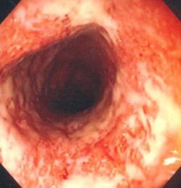
Pouchitis is a condition characterized by inflammation of the ileal pouch, a surgically created reservoir that is connected to the patient's anus in individuals who have undergone proctocolectomy with ileal pouch-anal anastomosis (IPAA). This procedure is often performed in patients with ulcerative colitis or familial adenomatous polyposis.
Pouchitis can present with symptoms such as diarrhea, abdominal cramps, urgency, and fecal incontinence. The exact cause of pouchitis remains unclear, but it is thought to be related to changes in the microbiota or an overactive immune response in the ileal pouch.
The diagnosis of pouchitis typically involves a combination of clinical symptoms, endoscopic findings, and histopathological examination of biopsies taken during endoscopy. Treatment options for pouchitis include antibiotics, anti-inflammatory medications, and probiotics, depending on the severity and frequency of the condition.
Proteobacteria is a major class of Gram-negative bacteria that includes a wide variety of pathogens and free-living organisms. This class is divided into six subclasses: Alpha, Beta, Gamma, Delta, Epsilon, and Zeta proteobacteria. Proteobacteria are characterized by their single circular chromosome and the presence of lipopolysaccharide (LPS) in their outer membrane. They can be found in a wide range of environments, including soil, water, and the gastrointestinal tracts of animals. Some notable examples of Proteobacteria include Escherichia coli, Salmonella enterica, and Yersinia pestis.
Ciprofloxacin is a fluoroquinolone antibiotic that is used to treat various types of bacterial infections, including respiratory, urinary, and skin infections. It works by inhibiting the bacterial DNA gyrase, which is an enzyme necessary for bacterial replication and transcription. This leads to bacterial cell death. Ciprofloxacin is available in oral and injectable forms and is usually prescribed to be taken twice a day. Common side effects include nausea, diarrhea, and headache. It may also cause serious adverse reactions such as tendinitis, tendon rupture, peripheral neuropathy, and central nervous system effects. It is important to note that ciprofloxacin should not be used in patients with a history of hypersensitivity to fluoroquinolones and should be used with caution in patients with a history of seizures, brain injury, or other neurological conditions.
Fecal incontinence is the involuntary loss or leakage of stool (feces) from the rectum. It is also known as bowel incontinence. This condition can range from occasional leakage of stool when passing gas to a complete loss of bowel control. Fecal incontinence can be an embarrassing and distressing problem, but there are treatments available that can help improve symptoms and quality of life.
The causes of fecal incontinence can vary, but some common factors include:
* Damage to the muscles or nerves that control bowel function, such as from childbirth, surgery, spinal cord injury, or long-term constipation or diarrhea.
* Chronic digestive conditions, such as irritable bowel syndrome (IBS), inflammatory bowel disease (IBD), or celiac disease.
* Neurological conditions, such as multiple sclerosis, stroke, or spina bifida.
* Aging, which can lead to a decrease in muscle strength and control.
Treatment for fecal incontinence depends on the underlying cause of the condition. Treatments may include:
* Bowel training exercises to improve muscle strength and control.
* Changes in diet to help regulate bowel movements.
* Medications to treat constipation or diarrhea.
* Surgery to repair damaged muscles or nerves, or to create a new opening for stool to exit the body.
If you are experiencing symptoms of fecal incontinence, it is important to speak with your healthcare provider. They can help determine the cause of your symptoms and develop an appropriate treatment plan.
The preoperative period is the time period before a surgical procedure during which various preparations are made to ensure the best possible outcome for the surgery. This includes evaluating the patient's overall health status, identifying and managing any underlying medical conditions that could increase the risk of complications, obtaining informed consent from the patient, and providing preoperative instructions regarding medications, food and drink intake, and other aspects of preparation for the surgery.
The specific activities that occur during the preoperative period may vary depending on the type and complexity of the surgical procedure, as well as the individual needs and medical history of the patient. However, some common elements of the preoperative period include:
* A thorough medical history and physical examination to assess the patient's overall health status and identify any potential risk factors for complications
* Diagnostic tests such as blood tests, imaging studies, or electrocardiograms (ECGs) to provide additional information about the patient's health status
* Consultation with anesthesia providers to determine the appropriate type and dosage of anesthesia for the procedure
* Preoperative teaching to help the patient understand what to expect before, during, and after the surgery
* Management of any underlying medical conditions such as diabetes, heart disease, or lung disease to reduce the risk of complications
* Administration of medications such as antibiotics or anti-coagulants to prevent infection or bleeding
* Fasting instructions to ensure that the stomach is empty during the surgery and reduce the risk of aspiration (inhalation of stomach contents into the lungs)
Overall, the preoperative period is a critical time for ensuring the safety and success of surgical procedures. By taking a thorough and systematic approach to preparing patients for surgery, healthcare providers can help to minimize the risks of complications and ensure the best possible outcomes for their patients.
A rectal fistula is an abnormal connection or tunnel that develops between the rectum, which is the lower end of the colon, and another organ or the skin surface surrounding the anus. This condition often results from inflammation, infection, trauma, or surgery in the anal area. The fistula can cause symptoms such as pain, discharge, irritation, and swelling around the anus. In some cases, it may also lead to complications like abscesses or recurrent infections if not treated promptly and effectively. Treatment options typically include surgical intervention to close the fistula and promote healing of the affected tissues.
Gastrointestinal agents are a class of pharmaceutical drugs that affect the gastrointestinal (GI) tract, which includes the organs involved in digestion such as the mouth, esophagus, stomach, small intestine, large intestine, and anus. These agents can have various effects on the GI tract, including:
1. Increasing gastric motility (promoting bowel movements) - laxatives, prokinetics
2. Decreasing gastric motility (reducing bowel movements) - antidiarrheal agents
3. Neutralizing gastric acid - antacids
4. Reducing gastric acid secretion - H2-blockers, proton pump inhibitors
5. Protecting the mucosal lining of the GI tract - sucralfate, misoprostol
6. Relieving symptoms associated with GI disorders such as bloating, abdominal pain, and nausea - antispasmodics, antiemetics
Examples of gastrointestinal agents include:
* Laxatives (e.g., psyllium, docusate)
* Prokinetics (e.g., metoclopramide)
* Antacids (e.g., calcium carbonate, aluminum hydroxide)
* H2-blockers (e.g., ranitidine, famotidine)
* Proton pump inhibitors (e.g., omeprazole, lansoprazole)
* Sucralfate
* Misoprostol
* Antispasmodics (e.g., hyoscyamine, dicyclomine)
* Antiemetics (e.g., ondansetron, promethazine)
It is important to note that gastrointestinal agents can have both therapeutic and adverse effects, and their use should be based on a careful evaluation of the patient's condition and medical history.
The intestinal mucosa is the innermost layer of the intestines, which comes into direct contact with digested food and microbes. It is a specialized epithelial tissue that plays crucial roles in nutrient absorption, barrier function, and immune defense. The intestinal mucosa is composed of several cell types, including absorptive enterocytes, mucus-secreting goblet cells, hormone-producing enteroendocrine cells, and immune cells such as lymphocytes and macrophages.
The surface of the intestinal mucosa is covered by a single layer of epithelial cells, which are joined together by tight junctions to form a protective barrier against harmful substances and microorganisms. This barrier also allows for the selective absorption of nutrients into the bloodstream. The intestinal mucosa also contains numerous lymphoid follicles, known as Peyer's patches, which are involved in immune surveillance and defense against pathogens.
In addition to its role in absorption and immunity, the intestinal mucosa is also capable of producing hormones that regulate digestion and metabolism. Dysfunction of the intestinal mucosa can lead to various gastrointestinal disorders, such as inflammatory bowel disease, celiac disease, and food allergies.
Intestinal polyps are abnormal growths that protrude from the lining of the intestines. They can occur in any part of the digestive tract, including the colon and rectum (colorectal polyps), small intestine, or stomach. These growths vary in size, shape, and number. Most intestinal polyps are benign, meaning they are not cancerous. However, some types of polyps, such as adenomatous polyps, can become cancerous over time if left untreated.
Intestinal polyps can be asymptomatic or cause symptoms like rectal bleeding, abdominal pain, changes in bowel habits, or anemia (in cases where there is chronic, slow bleeding). The exact cause of intestinal polyps is not fully understood, but factors such as age, family history, and certain genetic conditions can increase the risk of developing them. Regular screening exams, like colonoscopies, are essential for early detection and removal of polyps to prevent potential complications, including colorectal cancer.
The anus is the opening at the end of the digestive tract where feces are eliminated from the body. There are several diseases and conditions that can affect the anus, including:
1. Anal fissure: A small tear in the lining of the anus, which can cause pain and bleeding during bowel movements.
2. Hemorrhoids: Swollen veins in the rectum or anus that can cause discomfort, itching, and bleeding.
3. Perianal abscess: A collection of pus in the tissue surrounding the anus, which can cause pain, swelling, and redness.
4. Anal fistula: An abnormal connection between the anal canal and the skin around the anus, often resulting from a perianal abscess that did not heal properly.
5. Anal cancer: A rare form of cancer that develops in the cells lining the anus, usually affecting people over the age of 50.
6. Inflammatory bowel disease (IBD): A group of chronic inflammatory conditions of the intestine, including Crohn's disease and ulcerative colitis, which can affect the anus and cause symptoms such as pain, bleeding, and diarrhea.
7. Sexually transmitted infections (STIs): Certain STIs, such as herpes simplex virus, chlamydia, gonorrhea, and syphilis, can affect the anus and cause symptoms such as pain, discharge, and sores.
8. Fecal incontinence: The involuntary loss of bowel control, which can be caused by nerve damage, muscle weakness, or other medical conditions affecting the anus.
An intestinal fistula is an abnormal communication or connection between the intestines (or a portion of the intestine) and another organ or the skin surface. This connection forms a tract or passage, allowing the contents of the intestines, such as digestive enzymes, bacteria, and waste materials, to leak into other body areas or outside the body. Intestinal fistulas can develop due to various reasons, including inflammatory bowel diseases (like Crohn's disease), infections, complications from surgery, radiation therapy, or trauma. They can cause symptoms such as abdominal pain, diarrhea, skin irritation, and infection. Treatment of intestinal fistulas often involves a combination of medical management, nutritional support, and surgical intervention.
Bacteroidetes is a large phylum of gram-negative, predominantly anaerobic bacteria that are commonly found in the gastrointestinal tract of animals, including humans. They play an important role in the breakdown and fermentation of complex carbohydrates in the gut, producing short-chain fatty acids as a byproduct. Some species of Bacteroidetes have also been identified as opportunistic pathogens and can cause infections in immunocompromised individuals or under certain conditions.
The medical relevance of Bacteroidetes lies in their role in maintaining gut homeostasis, modulating the immune system, and protecting against pathogenic bacteria. Dysbiosis of the gut microbiota, including changes in the abundance and diversity of Bacteroidetes, has been associated with various diseases such as inflammatory bowel disease, obesity, diabetes, and cardiovascular disease. Therefore, understanding the ecology and function of Bacteroidetes is important for developing novel therapeutic strategies to target these conditions.
"Administration, Rectal" is a medical term that refers to the process of administering medication or other substances through the rectum. This route of administration is also known as "rectal suppository" or "suppository administration."
In this method, a solid dosage form called a suppository is inserted into the rectum using fingers or a special applicator. Once inside, the suppository melts or dissolves due to the body's temperature and releases the active drug or substance, which then gets absorbed into the bloodstream through the walls of the rectum.
Rectal administration is an alternative route of administration for people who have difficulty swallowing pills or liquids, or when rapid absorption of the medication is necessary. It can also be used to administer medications that are not well absorbed through other routes, such as the gastrointestinal tract. However, it may take longer for the medication to reach the bloodstream compared to intravenous (IV) administration.
Common examples of rectally administered medications include laxatives, antidiarrheal agents, analgesics, and some forms of hormonal therapy. It is important to follow the instructions provided by a healthcare professional when administering medication rectally, as improper administration can reduce the effectiveness of the medication or cause irritation or discomfort.
Inflammatory Bowel Diseases (IBD) are a group of chronic inflammatory conditions primarily affecting the gastrointestinal tract. The two main types of IBD are Crohn's disease and ulcerative colitis.
Crohn's disease can cause inflammation in any part of the digestive system, from the mouth to the anus, but it most commonly affects the lower part of the small intestine (the ileum) and/or the colon. The inflammation caused by Crohn's disease often spreads deep into the layers of affected bowel tissue.
Ulcerative colitis, on the other hand, is limited to the colon, specifically the innermost lining of the colon. It causes long-lasting inflammation and sores (ulcers) in the lining of the large intestine (colon) and rectum.
Symptoms can vary depending on the severity and location of inflammation but often include abdominal pain, diarrhea, fatigue, weight loss, and reduced appetite. IBD is not the same as irritable bowel syndrome (IBS), which is a functional gastrointestinal disorder.
The exact cause of IBD remains unknown, but it's thought to be a combination of genetic factors, an abnormal immune response, and environmental triggers. There is no cure for IBD, but treatments can help manage symptoms and reduce inflammation, potentially leading to long-term remission.
Capsule endoscopy is a medical procedure that uses a small, pill-sized camera to capture images of the digestive tract. The capsule is swallowed and transmits images wirelessly as it moves through the gastrointestinal (GI) tract, allowing doctors to examine the lining of the small intestine, which can be difficult to reach with traditional endoscopes.
The procedure is commonly used to diagnose and monitor conditions such as Crohn's disease, celiac disease, obscure gastrointestinal bleeding, and tumors in the small intestine. The images captured by the capsule are transmitted to a recorder worn by the patient, and then reviewed and analyzed by a healthcare professional.
Capsule endoscopy is generally considered safe and non-invasive, with few risks or side effects. However, it may not be suitable for everyone, including patients with swallowing difficulties, pacemakers, or certain gastrointestinal obstructions. It's important to consult with a healthcare provider to determine if capsule endoscopy is the right diagnostic tool for a particular condition.
A colectomy is a surgical procedure in which all or part of the large intestine (colon) is removed. This surgery may be performed to treat or prevent various medical conditions, including colon cancer, inflammatory bowel disease, diverticulitis, and severe obstructions or injuries of the colon.
There are several types of colectomies, depending on how much of the colon is removed:
* Total colectomy: Removal of the entire colon.
* Partial colectomy: Removal of a portion of the colon.
* Hemicolectomy: Removal of one half of the colon.
* Sigmoidectomy: Removal of the sigmoid colon, which is the part of the colon that is closest to the rectum.
After the affected portion of the colon is removed, the remaining ends of the intestine are reconnected, allowing stool to pass through the digestive system as usual. In some cases, a temporary or permanent colostomy may be necessary, in which a surgical opening (stoma) is created in the abdominal wall and the end of the colon is attached to it, allowing stool to be collected in a pouch outside the body.
Colectomies are major surgeries that require general anesthesia and hospitalization. The recovery time can vary depending on the type of colectomy performed and the individual's overall health, but typically ranges from several weeks to a few months. Complications of colectomy may include bleeding, infection, leakage from the surgical site, bowel obstruction, and changes in bowel habits or function.
Aerobic bacteria are a type of bacteria that require oxygen to live and grow. These bacteria use oxygen as the final electron acceptor in their respiratory chain to generate energy in the form of ATP (adenosine triphosphate). Aerobic bacteria can be found in various environments, including soil, water, and the air, as well as on the surfaces of living things. Some examples of aerobic bacteria include species of Pseudomonas, Bacillus, and Staphylococcus.
It's worth noting that some bacteria can switch between aerobic and anaerobic metabolism depending on the availability of oxygen. These bacteria are called facultative anaerobes. In contrast, obligate anaerobes are bacteria that cannot tolerate oxygen and will die in its presence.
Antineutrophil cytoplasmic antibodies (ANCAs) are a type of autoantibody that specifically target certain proteins in the cytoplasm of neutrophils, which are a type of white blood cell. These antibodies are associated with several types of vasculitis, which is inflammation of the blood vessels.
There are two main types of ANCAs: perinuclear ANCAs (p-ANCAs) and cytoplasmic ANCAs (c-ANCAs). p-ANCAs are directed against myeloperoxidase, a protein found in neutrophil granules, while c-ANCAs target proteinase 3, another protein found in neutrophil granules.
The presence of ANCAs in the blood can indicate an increased risk for developing certain types of vasculitis, such as granulomatosis with polyangiitis (GPA), eosinophilic granulomatosis with polyangiitis (EGPA), and microscopic polyangiitis (MPA). ANCA testing is often used in conjunction with other clinical findings to help diagnose and manage these conditions.
It's important to note that while the presence of ANCAs can indicate an increased risk for vasculitis, not everyone with ANCAs will develop the condition. Additionally, ANCAs can also be found in some individuals without any associated disease, so their presence should be interpreted in the context of other clinical findings.
Anti-infective agents are a class of medications that are used to treat infections caused by various microorganisms such as bacteria, viruses, fungi, and parasites. These agents work by either killing the microorganism or inhibiting its growth, thereby helping to control the infection and alleviate symptoms.
There are several types of anti-infective agents, including:
1. Antibiotics: These are medications that are used to treat bacterial infections. They work by either killing bacteria (bactericidal) or inhibiting their growth (bacteriostatic).
2. Antivirals: These are medications that are used to treat viral infections. They work by interfering with the replication of the virus, preventing it from spreading and causing further damage.
3. Antifungals: These are medications that are used to treat fungal infections. They work by disrupting the cell membrane of the fungus, killing it or inhibiting its growth.
4. Antiparasitics: These are medications that are used to treat parasitic infections. They work by either killing the parasite or inhibiting its growth and reproduction.
It is important to note that anti-infective agents are not effective against all types of infections, and it is essential to use them appropriately to avoid the development of drug-resistant strains of microorganisms.
Alpha-defensins are a type of defensin, which are small cationic host defense peptides that contribute to the innate immune system's response to microbial invasion. They are primarily produced by neutrophils, but can also be expressed by some epithelial cells and other immune cells. Alpha-defensins have broad-spectrum antimicrobial activity against bacteria, fungi, and enveloped viruses. They also play a role in modulating the inflammatory response and wound healing. There are six human alpha-defensin genes (DEFA1 to DEFA6) that encode six different peptides: Human Neutrophil Peptides 1-4 (HNP1-4) and Human Defensin 5 and 6 (HD5 and HD6). The HNPs are stored in the azurophilic granules of neutrophils and are released upon their activation, while HD5 and HD6 are found in the Paneth cells of the small intestine.
Gastrointestinal endoscopy is a medical procedure that allows direct visualization of the inner lining of the digestive tract, which includes the esophagus, stomach, small intestine, large intestine (colon), and sometimes the upper part of the small intestine (duodenum). This procedure is performed using an endoscope, a long, thin, flexible tube with a light and camera at its tip. The endoscope is inserted through the mouth for upper endoscopy or through the rectum for lower endoscopy (colonoscopy), and the images captured by the camera are transmitted to a monitor for the physician to view.
Gastrointestinal endoscopy can help diagnose various conditions, such as inflammation, ulcers, tumors, polyps, or bleeding in the digestive tract. It can also be used for therapeutic purposes, such as removing polyps, taking tissue samples (biopsies), treating bleeding, and performing other interventions to manage certain digestive diseases.
There are different types of gastrointestinal endoscopy procedures, including:
1. Upper Endoscopy (Esophagogastroduodenoscopy or EGD): This procedure examines the esophagus, stomach, and duodenum.
2. Colonoscopy: This procedure examines the colon and rectum.
3. Sigmoidoscopy: A limited examination of the lower part of the colon (sigmoid colon) using a shorter endoscope.
4. Enteroscopy: An examination of the small intestine, which can be performed using various techniques, such as push enteroscopy, single-balloon enteroscopy, or double-balloon enteroscopy.
5. Capsule Endoscopy: A procedure that involves swallowing a small capsule containing a camera, which captures images of the digestive tract as it passes through.
Gastrointestinal endoscopy is generally considered safe when performed by experienced medical professionals. However, like any medical procedure, there are potential risks and complications, such as bleeding, infection, perforation, or adverse reactions to sedatives used during the procedure. Patients should discuss these risks with their healthcare provider before undergoing gastrointestinal endoscopy.
Postoperative complications refer to any unfavorable condition or event that occurs during the recovery period after a surgical procedure. These complications can vary in severity and may include, but are not limited to:
1. Infection: This can occur at the site of the incision or inside the body, such as pneumonia or urinary tract infection.
2. Bleeding: Excessive bleeding (hemorrhage) can lead to a drop in blood pressure and may require further surgical intervention.
3. Blood clots: These can form in the deep veins of the legs (deep vein thrombosis) and can potentially travel to the lungs (pulmonary embolism).
4. Wound dehiscence: This is when the surgical wound opens up, which can lead to infection and further complications.
5. Pulmonary issues: These include atelectasis (collapsed lung), pneumonia, or respiratory failure.
6. Cardiovascular problems: These include abnormal heart rhythms (arrhythmias), heart attack, or stroke.
7. Renal failure: This can occur due to various reasons such as dehydration, blood loss, or the use of certain medications.
8. Pain management issues: Inadequate pain control can lead to increased stress, anxiety, and decreased mobility.
9. Nausea and vomiting: These can be caused by anesthesia, opioid pain medication, or other factors.
10. Delirium: This is a state of confusion and disorientation that can occur in the elderly or those with certain medical conditions.
Prompt identification and management of these complications are crucial to ensure the best possible outcome for the patient.
Recurrence, in a medical context, refers to the return of symptoms or signs of a disease after a period of improvement or remission. It indicates that the condition has not been fully eradicated and may require further treatment. Recurrence is often used to describe situations where a disease such as cancer comes back after initial treatment, but it can also apply to other medical conditions. The likelihood of recurrence varies depending on the type of disease and individual patient factors.
Anti-inflammatory agents are a class of drugs or substances that reduce inflammation in the body. They work by inhibiting the production of inflammatory mediators, such as prostaglandins and leukotrienes, which are released during an immune response and contribute to symptoms like pain, swelling, redness, and warmth.
There are two main types of anti-inflammatory agents: steroidal and nonsteroidal. Steroidal anti-inflammatory drugs (SAIDs) include corticosteroids, which mimic the effects of hormones produced by the adrenal gland. Nonsteroidal anti-inflammatory drugs (NSAIDs) are a larger group that includes both prescription and over-the-counter medications, such as aspirin, ibuprofen, naproxen, and celecoxib.
While both types of anti-inflammatory agents can be effective in reducing inflammation and relieving symptoms, they differ in their mechanisms of action, side effects, and potential risks. Long-term use of NSAIDs, for example, can increase the risk of gastrointestinal bleeding, kidney damage, and cardiovascular events. Corticosteroids can have significant side effects as well, particularly with long-term use, including weight gain, mood changes, and increased susceptibility to infections.
It's important to use anti-inflammatory agents only as directed by a healthcare provider, and to be aware of potential risks and interactions with other medications or health conditions.
A biopsy is a medical procedure in which a small sample of tissue is taken from the body to be examined under a microscope for the presence of disease. This can help doctors diagnose and monitor various medical conditions, such as cancer, infections, or autoimmune disorders. The type of biopsy performed will depend on the location and nature of the suspected condition. Some common types of biopsies include:
1. Incisional biopsy: In this procedure, a surgeon removes a piece of tissue from an abnormal area using a scalpel or other surgical instrument. This type of biopsy is often used when the lesion is too large to be removed entirely during the initial biopsy.
2. Excisional biopsy: An excisional biopsy involves removing the entire abnormal area, along with a margin of healthy tissue surrounding it. This technique is typically employed for smaller lesions or when cancer is suspected.
3. Needle biopsy: A needle biopsy uses a thin, hollow needle to extract cells or fluid from the body. There are two main types of needle biopsies: fine-needle aspiration (FNA) and core needle biopsy. FNA extracts loose cells, while a core needle biopsy removes a small piece of tissue.
4. Punch biopsy: In a punch biopsy, a round, sharp tool is used to remove a small cylindrical sample of skin tissue. This type of biopsy is often used for evaluating rashes or other skin abnormalities.
5. Shave biopsy: During a shave biopsy, a thin slice of tissue is removed from the surface of the skin using a sharp razor-like instrument. This technique is typically used for superficial lesions or growths on the skin.
After the biopsy sample has been collected, it is sent to a laboratory where a pathologist will examine the tissue under a microscope and provide a diagnosis based on their findings. The results of the biopsy can help guide further treatment decisions and determine the best course of action for managing the patient's condition.
Budesonide is a corticosteroid medication that is used to reduce inflammation in the body. It works by mimicking the effects of hormones produced naturally by the adrenal glands, which help regulate the immune system and suppress inflammatory responses. Budesonide is available as an inhaler, nasal spray, or oral tablet, and is used to treat a variety of conditions, including asthma, chronic obstructive pulmonary disease (COPD), rhinitis, and Crohn's disease.
When budesonide is inhaled or taken orally, it is absorbed into the bloodstream and travels throughout the body, where it can reduce inflammation in various tissues and organs. In the lungs, for example, budesonide can help prevent asthma attacks by reducing inflammation in the airways, making it easier to breathe.
Like other corticosteroid medications, budesonide can have side effects, particularly if used at high doses or for long periods of time. These may include thrush (a fungal infection in the mouth), hoarseness, sore throat, cough, headache, and easy bruising or skin thinning. Long-term use of corticosteroids can also lead to more serious side effects, such as adrenal suppression, osteoporosis, and increased risk of infections.
It is important to follow the dosage instructions provided by your healthcare provider when taking budesonide or any other medication, and to report any unusual symptoms or side effects promptly.
Crohn's disease is a type of inflammatory bowel disease (IBD) that can affect any part of the gastrointestinal tract, from the mouth to the anus. It is characterized by chronic inflammation of the digestive tract, which can lead to symptoms such as abdominal pain, diarrhea, fatigue, weight loss, and malnutrition.
The specific causes of Crohn's disease are not fully understood, but it is believed to be related to a combination of genetic, environmental, and immune system factors. The disease can affect people of any age, but it is most commonly diagnosed in young adults between the ages of 15 and 35.
There is no cure for Crohn's disease, but treatments such as medications, lifestyle changes, and surgery can help manage symptoms and prevent complications. Treatment options depend on the severity and location of the disease, as well as the individual patient's needs and preferences.
The colon, also known as the large intestine, is a part of the digestive system in humans and other vertebrates. It is an organ that eliminates waste from the body and is located between the small intestine and the rectum. The main function of the colon is to absorb water and electrolytes from digested food, forming and storing feces until they are eliminated through the anus.
The colon is divided into several regions, including the cecum, ascending colon, transverse colon, descending colon, sigmoid colon, rectum, and anus. The walls of the colon contain a layer of muscle that helps to move waste material through the organ by a process called peristalsis.
The inner surface of the colon is lined with mucous membrane, which secretes mucus to lubricate the passage of feces. The colon also contains a large population of bacteria, known as the gut microbiota, which play an important role in digestion and immunity.
Treatment outcome is a term used to describe the result or effect of medical treatment on a patient's health status. It can be measured in various ways, such as through symptoms improvement, disease remission, reduced disability, improved quality of life, or survival rates. The treatment outcome helps healthcare providers evaluate the effectiveness of a particular treatment plan and make informed decisions about future care. It is also used in clinical research to compare the efficacy of different treatments and improve patient care.
Prednisolone is a synthetic glucocorticoid drug, which is a class of steroid hormones. It is commonly used in the treatment of various inflammatory and autoimmune conditions due to its potent anti-inflammatory and immunosuppressive effects. Prednisolone works by binding to specific receptors in cells, leading to changes in gene expression that reduce the production of substances involved in inflammation, such as cytokines and prostaglandins.
Prednisolone is available in various forms, including tablets, syrups, and injectable solutions. It can be used to treat a wide range of medical conditions, including asthma, rheumatoid arthritis, inflammatory bowel disease, allergies, skin conditions, and certain types of cancer.
Like other steroid medications, prednisolone can have significant side effects if used in high doses or for long periods of time. These may include weight gain, mood changes, increased risk of infections, osteoporosis, diabetes, and adrenal suppression. As a result, the use of prednisolone should be closely monitored by a healthcare professional to ensure that its benefits outweigh its risks.
Feces are the solid or semisolid remains of food that could not be digested or absorbed in the small intestine, along with bacteria and other waste products. After being stored in the colon, feces are eliminated from the body through the rectum and anus during defecation. Feces can vary in color, consistency, and odor depending on a person's diet, health status, and other factors.
Combination drug therapy is a treatment approach that involves the use of multiple medications with different mechanisms of action to achieve better therapeutic outcomes. This approach is often used in the management of complex medical conditions such as cancer, HIV/AIDS, and cardiovascular diseases. The goal of combination drug therapy is to improve efficacy, reduce the risk of drug resistance, decrease the likelihood of adverse effects, and enhance the overall quality of life for patients.
In combining drugs, healthcare providers aim to target various pathways involved in the disease process, which may help to:
1. Increase the effectiveness of treatment by attacking the disease from multiple angles.
2. Decrease the dosage of individual medications, reducing the risk and severity of side effects.
3. Slow down or prevent the development of drug resistance, a common problem in chronic diseases like HIV/AIDS and cancer.
4. Improve patient compliance by simplifying dosing schedules and reducing pill burden.
Examples of combination drug therapy include:
1. Antiretroviral therapy (ART) for HIV treatment, which typically involves three or more drugs from different classes to suppress viral replication and prevent the development of drug resistance.
2. Chemotherapy regimens for cancer treatment, where multiple cytotoxic agents are used to target various stages of the cell cycle and reduce the likelihood of tumor cells developing resistance.
3. Cardiovascular disease management, which may involve combining medications such as angiotensin-converting enzyme (ACE) inhibitors, beta-blockers, diuretics, and statins to control blood pressure, heart rate, fluid balance, and cholesterol levels.
4. Treatment of tuberculosis, which often involves a combination of several antibiotics to target different aspects of the bacterial life cycle and prevent the development of drug-resistant strains.
When prescribing combination drug therapy, healthcare providers must carefully consider factors such as potential drug interactions, dosing schedules, adverse effects, and contraindications to ensure safe and effective treatment. Regular monitoring of patients is essential to assess treatment response, manage side effects, and adjust the treatment plan as needed.
Follow-up studies are a type of longitudinal research that involve repeated observations or measurements of the same variables over a period of time, in order to understand their long-term effects or outcomes. In medical context, follow-up studies are often used to evaluate the safety and efficacy of medical treatments, interventions, or procedures.
In a typical follow-up study, a group of individuals (called a cohort) who have received a particular treatment or intervention are identified and then followed over time through periodic assessments or data collection. The data collected may include information on clinical outcomes, adverse events, changes in symptoms or functional status, and other relevant measures.
The results of follow-up studies can provide important insights into the long-term benefits and risks of medical interventions, as well as help to identify factors that may influence treatment effectiveness or patient outcomes. However, it is important to note that follow-up studies can be subject to various biases and limitations, such as loss to follow-up, recall bias, and changes in clinical practice over time, which must be carefully considered when interpreting the results.
The Fluorescent Antibody Technique (FAT), Indirect is a type of immunofluorescence assay used to detect the presence of specific antigens in a sample. In this method, the sample is first incubated with a primary antibody that binds to the target antigen. After washing to remove unbound primary antibodies, a secondary fluorescently labeled antibody is added, which recognizes and binds to the primary antibody. This indirect labeling approach allows for amplification of the signal, making it more sensitive than direct methods. The sample is then examined under a fluorescence microscope to visualize the location and amount of antigen based on the emitted light from the fluorescent secondary antibody. It's commonly used in diagnostic laboratories for detection of various bacteria, viruses, and other antigens in clinical specimens.
Anti-bacterial agents, also known as antibiotics, are a type of medication used to treat infections caused by bacteria. These agents work by either killing the bacteria or inhibiting their growth and reproduction. There are several different classes of anti-bacterial agents, including penicillins, cephalosporins, fluoroquinolones, macrolides, and tetracyclines, among others. Each class of antibiotic has a specific mechanism of action and is used to treat certain types of bacterial infections. It's important to note that anti-bacterial agents are not effective against viral infections, such as the common cold or flu. Misuse and overuse of antibiotics can lead to antibiotic resistance, which is a significant global health concern.
A Severity of Illness Index is a measurement tool used in healthcare to assess the severity of a patient's condition and the risk of mortality or other adverse outcomes. These indices typically take into account various physiological and clinical variables, such as vital signs, laboratory values, and co-morbidities, to generate a score that reflects the patient's overall illness severity.
Examples of Severity of Illness Indices include the Acute Physiology and Chronic Health Evaluation (APACHE) system, the Simplified Acute Physiology Score (SAPS), and the Mortality Probability Model (MPM). These indices are often used in critical care settings to guide clinical decision-making, inform prognosis, and compare outcomes across different patient populations.
It is important to note that while these indices can provide valuable information about a patient's condition, they should not be used as the sole basis for clinical decision-making. Rather, they should be considered in conjunction with other factors, such as the patient's overall clinical presentation, treatment preferences, and goals of care.
Remission induction is a treatment approach in medicine, particularly in the field of oncology and hematology. It refers to the initial phase of therapy aimed at reducing or eliminating the signs and symptoms of active disease, such as cancer or autoimmune disorders. The primary goal of remission induction is to achieve a complete response (disappearance of all detectable signs of the disease) or a partial response (a decrease in the measurable extent of the disease). This phase of treatment is often intensive and may involve the use of multiple drugs or therapies, including chemotherapy, immunotherapy, or targeted therapy. After remission induction, patients may receive additional treatments to maintain the remission and prevent relapse, known as consolidation or maintenance therapy.
Quality of Life (QOL) is a broad, multidimensional concept that usually includes an individual's physical health, psychological state, level of independence, social relationships, personal beliefs, and their relationship to salient features of their environment. It reflects the impact of disease and treatment on a patient's overall well-being and ability to function in daily life.
The World Health Organization (WHO) defines QOL as "an individual's perception of their position in life in the context of the culture and value systems in which they live and in relation to their goals, expectations, standards and concerns." It is a subjective concept, meaning it can vary greatly from person to person.
In healthcare, QOL is often used as an outcome measure in clinical trials and other research studies to assess the impact of interventions or treatments on overall patient well-being.
Patient compliance, also known as medication adherence or patient adherence, refers to the degree to which a patient's behavior matches the agreed-upon recommendations from their healthcare provider. This includes taking medications as prescribed (including the correct dosage, frequency, and duration), following dietary restrictions, making lifestyle changes, and attending follow-up appointments. Poor patient compliance can negatively impact treatment outcomes and lead to worsening of symptoms, increased healthcare costs, and development of drug-resistant strains in the case of antibiotics. It is a significant challenge in healthcare and efforts are being made to improve patient education, communication, and support to enhance compliance.
An Enzyme-Linked Immunosorbent Assay (ELISA) is a type of analytical biochemistry assay used to detect and quantify the presence of a substance, typically a protein or peptide, in a liquid sample. It takes its name from the enzyme-linked antibodies used in the assay.
In an ELISA, the sample is added to a well containing a surface that has been treated to capture the target substance. If the target substance is present in the sample, it will bind to the surface. Next, an enzyme-linked antibody specific to the target substance is added. This antibody will bind to the captured target substance if it is present. After washing away any unbound material, a substrate for the enzyme is added. If the enzyme is present due to its linkage to the antibody, it will catalyze a reaction that produces a detectable signal, such as a color change or fluorescence. The intensity of this signal is proportional to the amount of target substance present in the sample, allowing for quantification.
ELISAs are widely used in research and clinical settings to detect and measure various substances, including hormones, viruses, and bacteria. They offer high sensitivity, specificity, and reproducibility, making them a reliable choice for many applications.
A case-control study is an observational research design used to identify risk factors or causes of a disease or health outcome. In this type of study, individuals with the disease or condition (cases) are compared with similar individuals who do not have the disease or condition (controls). The exposure history or other characteristics of interest are then compared between the two groups to determine if there is an association between the exposure and the disease.
Case-control studies are often used when it is not feasible or ethical to conduct a randomized controlled trial, as they can provide valuable insights into potential causes of diseases or health outcomes in a relatively short period of time and at a lower cost than other study designs. However, because case-control studies rely on retrospective data collection, they are subject to biases such as recall bias and selection bias, which can affect the validity of the results. Therefore, it is important to carefully design and conduct case-control studies to minimize these potential sources of bias.
In epidemiology, the incidence of a disease is defined as the number of new cases of that disease within a specific population over a certain period of time. It is typically expressed as a rate, with the number of new cases in the numerator and the size of the population at risk in the denominator. Incidence provides information about the risk of developing a disease during a given time period and can be used to compare disease rates between different populations or to monitor trends in disease occurrence over time.
Medical Definition:
"Risk factors" are any attribute, characteristic or exposure of an individual that increases the likelihood of developing a disease or injury. They can be divided into modifiable and non-modifiable risk factors. Modifiable risk factors are those that can be changed through lifestyle choices or medical treatment, while non-modifiable risk factors are inherent traits such as age, gender, or genetic predisposition. Examples of modifiable risk factors include smoking, alcohol consumption, physical inactivity, and unhealthy diet, while non-modifiable risk factors include age, sex, and family history. It is important to note that having a risk factor does not guarantee that a person will develop the disease, but rather indicates an increased susceptibility.
In the field of medicine, "time factors" refer to the duration of symptoms or time elapsed since the onset of a medical condition, which can have significant implications for diagnosis and treatment. Understanding time factors is crucial in determining the progression of a disease, evaluating the effectiveness of treatments, and making critical decisions regarding patient care.
For example, in stroke management, "time is brain," meaning that rapid intervention within a specific time frame (usually within 4.5 hours) is essential to administering tissue plasminogen activator (tPA), a clot-busting drug that can minimize brain damage and improve patient outcomes. Similarly, in trauma care, the "golden hour" concept emphasizes the importance of providing definitive care within the first 60 minutes after injury to increase survival rates and reduce morbidity.
Time factors also play a role in monitoring the progression of chronic conditions like diabetes or heart disease, where regular follow-ups and assessments help determine appropriate treatment adjustments and prevent complications. In infectious diseases, time factors are crucial for initiating antibiotic therapy and identifying potential outbreaks to control their spread.
Overall, "time factors" encompass the significance of recognizing and acting promptly in various medical scenarios to optimize patient outcomes and provide effective care.
Prospective studies, also known as longitudinal studies, are a type of cohort study in which data is collected forward in time, following a group of individuals who share a common characteristic or exposure over a period of time. The researchers clearly define the study population and exposure of interest at the beginning of the study and follow up with the participants to determine the outcomes that develop over time. This type of study design allows for the investigation of causal relationships between exposures and outcomes, as well as the identification of risk factors and the estimation of disease incidence rates. Prospective studies are particularly useful in epidemiology and medical research when studying diseases with long latency periods or rare outcomes.
 Pouchitis
 Pouchitis - Medical Dictionary online-medical-dictionary.org
 Pouchitis | The J-Pouch Group
 Fecal microbiota transplantation in the treatment of chronic pouchitis: A systematic review
- Aalborg University's...
Fecal microbiota transplantation in the treatment of chronic pouchitis: A systematic review
- Aalborg University's... EFFICACY OF BIOLOGICAL THERAPY FOR POUCHITIS AND OTHER INFLAMMATORY COMPLICATIONS OF THE POUCH, AND THE QUESTION OF A SECOND...
 Crohn's Disease Surgery: Types, After, and More
 News: Evaluation of endoscopic disease activity in pouchitis
 Case series describes pouchitis after immune checkpoint inhibitors | ACP Gastroenterology
Monthly
Case series describes pouchitis after immune checkpoint inhibitors | ACP Gastroenterology
Monthly Abordagem diagnóstica e de manuseio da pouchite em doença inflamatória intestinal<...
 Takeda Presents Positive Results from Phase 3 Study of Vedolizumab for Prevention of Intestinal Acute Graft-Versus-Host Disease...
 Sensitivity and specificity of diagnostic modalities in pouchitis: a systematic review and meta-analysis | Nevin Manimala
 Sidhartha Sinha
 Flagyl, Flagyl ER (metronidazole) dosing, indications, interactions, adverse effects, and more
 Successful treatment for pouchitis with rebamipide refractory to a combination of metronidazole (MNZ) and ciprofloxacin (CFX). ...
 Small and Large Intestine | Colon and Digestive Health Services for Lower Digestive Track Near Cleveland, Ohio | University...
 Takeda Announces Results From Phase 4 Vedolizumab Study in Patients With Chronic Pouchitis Published in New England Journal of...
 Dr. James Stinneford, MD, Gastroenterology Specialist - Elgin, IL | Sharecare
 European Crohn´s and Colitis Organisation - ECCO - ECCO News 3/2018
 JBPC Publications: 2011 - 2018 | Marine Biological Laboratory
![Jones S[au] - Search Results - PubMed](data:image/png;base64,iVBORw0KGgoAAAANSUhEUgAAABAAAAAQCAMAAAAoLQ9TAAAARVBMVEVHcEwoU45gYmYAUpQAUpRPYGVgYmZLXnJgYmYAUZUAUpRJXnIAUpQAUpRgYmYAUpRgYmZgYmZhYmYAUpQAUpQAUpRgYmaDiPJuAAAAFXRSTlMADOJ+6QewGO8/uTRqtH7GdFJ11p1bCL3TAAAAZUlEQVQYlV2PVw7AIAxDTeney7n/UcsoldX3E+VJOAboEi7MBpHWMs1ADlG8u7UYWauwyZFeRQVPOhG2o+aiwhByJxUx91Jxhje3iJSqGfHuLKI0+0TpXvY1twCOPlFh5pa/++MB0vIOBm+1zaoAAAAASUVORK5CYII=) Jones S[au] - Search Results - PubMed
Jones S[au] - Search Results - PubMed The American Journal of Gastroenterology
 Stoma surgery</span><i class="icon" aria-hidden=...