Epilepsy, Reflex
Photic Stimulation
Myoclonic Epilepsy, Juvenile
Electroencephalography
Myoclonus
Suprachiasmatic Nucleus
Occipital Lobe
Evoked Potentials, Visual
Visual Cortex
Epilepsy
Electric Stimulation Therapy
Stimulation, Chemical
Deep Brain Stimulation
Transcranial Magnetic Stimulation
Cells, Cultured
Transcutaneous Electric Nerve Stimulation
Signal Transduction
Calcium
Cats
Rats, Sprague-Dawley
Electrodes, Implanted
Muscle Contraction
Action Potentials
Phosphorylation
Vagus Nerve
Enzyme Activation
Dose-Response Relationship, Drug
Evoked Potentials
Lymphocyte Activation
Neurons
Evoked Potentials, Motor
RNA, Messenger
Isoproterenol
Cyclic AMP
Rats, Wistar
Reflex
Atropine
Norepinephrine
Synaptic Transmission
Electromyography
Motor Cortex
Subthalamic Nucleus
Afferent Pathways
Electrophysiology
Sympathetic Nervous System
Mice, Inbred C57BL
Neural Inhibition
Guinea Pigs
Competitive mechanisms subserve attention in macaque areas V2 and V4. (1/15800)
It is well established that attention modulates visual processing in extrastriate cortex. However, the underlying neural mechanisms are unknown. A consistent observation is that attention has its greatest impact on neuronal responses when multiple stimuli appear together within a cell's receptive field. One way to explain this is to assume that multiple stimuli activate competing populations of neurons and that attention biases this competition in favor of the attended stimulus. In the absence of competing stimuli, there is no competition to be resolved. Accordingly, attention has a more limited effect on the neuronal response to a single stimulus. To test this interpretation, we measured the responses of neurons in macaque areas V2 and V4 using a behavioral paradigm that allowed us to isolate automatic sensory processing mechanisms from attentional effects. First, we measured each cell's response to a single stimulus presented alone inside the receptive field or paired with a second receptive field stimulus, while the monkey attended to a location outside the receptive field. Adding the second stimulus typically caused the neuron's response to move toward the response that was elicited by the second stimulus alone. Then, we directed the monkey's attention to one element of the pair. This drove the neuron's response toward the response elicited when the attended stimulus appeared alone. These findings are consistent with the idea that attention biases competitive interactions among neurons, causing them to respond primarily to the attended stimulus. A quantitative neural model of attention is proposed to account for these results. (+info)The neuronal basis of a sensory analyser, the acridid movement detector system. I. Effects of simple incremental and decremental stimuli in light and dark adapted animals. (2/15800)
1. The response of the movement detector (MD) system to proportionally constant incremental and decremental stimuli has been studied at various degrees of light and dark adaptation. Action potentials in the descending contralateral movement detector neurone were taken as the indicator of response. 2. Over a range of at least six log10 units of adapting luminance, the MD system behaves as an ON/OFF unit, giving responses to both incremental and decremental changes in the illumination of a 5 degrees target. 3. With increasing amplitudes of stimuli, both the ON and OFF responses saturate rapidly. Saturation is reached sooner at higher levels of light adaptation. At all levels of light adaptation, the OFF response is greater than the ON. The ratio for saturating stimuli is approximately constant at around 3:2. 4. At the brightest adapting luminances used (20 000 cd/m2) the ON response is reduced but not lost. At the lowest (0-004 cd/m2) the OFF response to a 5 degrees disc fails, but can be regained by increasing the test area to 10 degrees. 5. From what is known of the retina of locusts and other insects, it is thought that light and dark adaptation in the MD system can be adequately explained by events at the retinula cell. (+info)MST neuronal responses to heading direction during pursuit eye movements. (3/15800)
As you move through the environment, you see a radial pattern of visual motion with a focus of expansion (FOE) that indicates your heading direction. When self-movement is combined with smooth pursuit eye movements, the turning of the eye distorts the retinal image of the FOE but somehow you still can perceive heading. We studied neurons in the medial superior temporal area (MST) of monkey visual cortex, recording responses to FOE stimuli presented during fixation and smooth pursuit eye movements. Almost all neurons showed significant changes in their FOE selective responses during pursuit eye movements. However, the vector average of all the neuronal responses indicated the direction of the FOE during both fixation and pursuit. Furthermore, the amplitude of the net vector increased with increasing FOE eccentricity. We conclude that neuronal population encoding in MST might contribute to pursuit-tolerant heading perception. (+info)Eye movement deficits following ibotenic acid lesions of the nucleus prepositus hypoglossi in monkeys II. Pursuit, vestibular, and optokinetic responses. (4/15800)
The eyes are moved by a combination of neural commands that code eye velocity and eye position. The eye position signal is supposed to be derived from velocity-coded command signals by mathematical integration via a single oculomotor neural integrator. For horizontal eye movements, the neural integrator is thought to reside in the rostral nucleus prepositus hypoglossi (nph) and project directly to the abducens nuclei. In a previous study, permanent, serial ibotenic acid lesions of the nph in three rhesus macaques compromised the neural integrator for fixation but saccades were not affected. In the present study, to determine further whether the nph is the neural substrate for a single oculomotor neural integrator, the effects of those lesions on smooth pursuit, the vestibulo-ocular reflex (VOR), vestibular nystagmus (VN), and optokinetic nystagmus (OKN) are documented. The lesions were correlated with long-lasting deficits in eye movements, indicated most clearly by the animals' inability to maintain steady gaze in the dark. However, smooth pursuit and sinusoidal VOR in the dark, like the saccades in the previous study, were affected minimally. The gain of horizontal smooth pursuit (eye movement/target movement) decreased slightly (<25%) and phase lead increased slightly for all frequencies (0.3-1.0 Hz, +/-10 degrees target tracking), most noticeably for higher frequencies (0.8-0.7 and approximately 20 degrees for 1.0-Hz tracking). Vertical smooth pursuit was not affected significantly. Surprisingly, horizontal sinusoidal VOR gain and phase also were not affected significantly. Lesions had complex effects on both VN and OKN. The plateau of per- and postrotatory VN was shortened substantially ( approximately 50%), whereas the initial response and the time constant of decay decreased slightly. The initial OKN response also decreased slightly, and the charging phase was prolonged transiently then recovered to below normal levels like the VN time constant. Maximum steady-state, slow eye velocity of OKN decreased progressively by approximately 30% over the course of the lesions. These results support the previous conclusion that the oculomotor neural integrator is not a single neural entity and that the mathematical integrative function for different oculomotor subsystems is most likely distributed among a number of nuclei. They also show that the nph apparently is not involved in integrating smooth pursuit signals and that lesions of the nph can fractionate the VOR and nystagmic responses to adequate stimuli. (+info)Light-induced calcium influx into retinal axons is regulated by presynaptic nicotinic acetylcholine receptor activity in vivo. (5/15800)
Visual activity is thought to be a critical factor in controlling the development of central retinal projections. Neuronal activity increases cytosolic calcium, which was hypothesized to regulate process outgrowth in neurons. We performed an in vivo imaging study in the retinotectal system of albino Xenopus laevis tadpoles with the fluorescent calcium indicator calcium green 1 dextran (CaGD) to test the role of calcium in regulating axon arbor development. We find that visual stimulus to the retina increased CaGD fluorescence intensity in retinal ganglion cell (RGC) axon arbors within the optic tectum and that branch additions to retinotectal axon arbors correlated with a local rise in calcium in the parent branch. We find three types of responses to visual stimulus, which roughly correlate with the ON, OFF, and SUSTAINED response types of RGC reported by physiological criteria. Imaging in bandscan mode indicated that patterns of calcium transients were nonuniform throughout the axons. We tested whether the increase in calcium in the retinotectal axons required synaptic activity in the retina; intraocular application of tetrodotoxin (10 microM) or nifedipine (1 and 10 microM) blocked the stimulus-induced increase in RGC axonal fluorescence. A second series of pharmacological investigations was designed to determine the mechanism of the calcium elevation in the axon terminals within the optic tectum. Injection of bis-(o-aminophenoxy)-N,N,N',N'-tetraacetic acid-AM (BAPTA-AM) (20 mM) into the tectal ventricle reduced axonal calcium levels, supporting the idea that visual stimulation increases axonal calcium. Injection of BAPTA (20 mM) into the tectal ventricle to chelate extracellular calcium also attenuated the calcium response to visual stimulation, indicating that calcium enters the axon from the extracellular medium. Caffeine (10 mM) caused a large increase in axonal calcium, indicating that intracellular stores contribute to the calcium signal. Presynaptic nicotinic acetylcholine receptors (nAChRs) may play a role in axon arbor development and the formation of the topographic retinotectal projection. Injection of nicotine (10 microM) into the tectal ventricle significantly elevated RGC axonal calcium levels, whereas application of the nAChR antagonist alphaBTX (100 nM) reduced the stimulus-evoked rise in RGC calcium fluorescence. These data suggest that light stimulus to the retina increases calcium in the axon terminal arbors through a mechanism that includes influx through nAChRs and amplification by calcium-induced calcium release from intracellular calcium stores. Such a mechanism may contribute to developmental plasticity of the retinotectal system by influencing both axon arbor elaboration and the strength of synaptic transmission. (+info)Correlated firing in rabbit retinal ganglion cells. (6/15800)
A ganglion cell's receptive field is defined as that region on the retinal surface in which a light stimulus will produce a response. While neighboring ganglion cells may respond to the same stimulus in a region where their receptive fields overlap, it generally has been assumed that each cell makes an independent decision about whether to fire. Recent recordings from cat and salamander retina using multiple electrodes have challenged this view of independent firing by showing that neighboring ganglion cells have an increased tendency to fire together within +/-5 ms. However, there is still uncertainty about which types of ganglion cells fire together, the mechanisms that produce coordinated spikes, and the overall function of coordinated firing. To address these issues, the responses of up to 80 rabbit retinal ganglion cells were recorded simultaneously using a multielectrode array. Of the 11 classes of rabbit ganglion cells previously identified, coordinated firing was observed in five. Plots of the spike train cross-correlation function suggested that coordinated firing occurred through two mechanisms. In the first mechanism, a spike in an interneuron diverged to produce simultaneous spikes in two ganglion cells. This mechanism predominated in four of the five classes including the ON brisk transient cells. In the second mechanism, ganglion cells appeared to activate each other reciprocally. This was the predominant pattern of correlated firing in OFF brisk transient cells. By comparing the receptive field profiles of ON and OFF brisk transient cells, a peripheral extension of the OFF brisk transient cell receptive field was identified that might be produced by lateral spike spread. Thus an individual OFF brisk transient cell can respond both to a light stimulus directed at the center of its receptive field and to stimuli that activate neighboring OFF brisk transient cells through their receptive field centers. (+info)Evidence for an eye-centered spherical representation of the visuomotor map. (7/15800)
During visually guided movement, visual coordinates of target location must be transformed into coordinates appropriate for movement. To investigate the representation of this visuomotor coordinate transformation, we examined changes in pointing behavior induced by a local visuomotor remapping. The visual feedback of finger position was limited to one location within the workspace, at which a discrepancy was introduced between the actual and visually perceived finger position. This remapping induced a change in pointing that extended over the entire workspace and was best captured by a spherical coordinate system centered near the eyes. (+info)Visuomotor processing as reflected in the directional discharge of premotor and primary motor cortex neurons. (8/15800)
Premotor and primary motor cortical neuronal firing was studied in two monkeys during an instructed delay, pursuit tracking task. The task included a premovement "cue period," during which the target was presented at the periphery of the workspace and moved to the center of the workspace along one of eight directions at one of four constant speeds. The "track period" consisted of a visually guided, error-constrained arm movement during which the animal tracked the target as it moved from the central start box along a line to the opposite periphery of the workspace. Behaviorally, the animals tracked the required directions and speeds with highly constrained trajectories. The eye movements consisted of saccades to the target at the onset of the cue period, followed by smooth pursuit intermingled with saccades throughout the cue and track periods. Initially, an analysis of variance (ANOVA) was used to test for direction and period effects in the firing. Subsequently, a linear regression analysis was used to fit the average firing from the cue and track periods to a cosine model. Directional tuning as determined by a significant fit to the cosine model was a prominent feature of the discharge during both the cue and track periods. However, the directional tuning of the firing of a single cell was not always constant across the cue and track periods. Approximately one-half of the neurons had differences in their preferred directions (PDs) of >45 degrees between cue and track periods. The PD in the cue or track period was not dependent on the target speed. A second linear regression analysis based on calculation of the preferred direction in 20-ms bins (i.e., the PD trajectory) was used to examine on a finer time scale the temporal evolution of this change in directional tuning. The PD trajectories in the cue period were not straight but instead rotated over the workspace to align with the track period PD. Both clockwise and counterclockwise rotations occurred. The PD trajectories were relatively straight during most of the track period. The rotation and eventual convergence of the PD trajectories in the cue period to the preferred direction of the track period may reflect the transformation of visual information into motor commands. The widely dispersed PD trajectories in the cue period would allow targets to be detected over a wide spatial aperture. The convergence of the PD trajectories occurring at the cue-track transition may serve as a "Go" signal to move that was not explicitly supplied by the paradigm. Furthermore, the rotation and convergence of the PD trajectories may provide a mechanism for nonstandard mapping. Standard mapping refers to a sensorimotor transformation in which the stimulus is the object of the reach. Nonstandard mapping is the mapping of an arbitrary stimulus into an arbitrary movement. The shifts in the PD may allow relevant visual information from any direction to be transformed into an appropriate movement direction, providing a neural substrate for nonstandard stimulus-response mappings. (+info)Reflex epilepsy is a type of epilepsy in which seizures are consistently triggered by specific, recurring sensory stimuli. These triggers can vary widely and may include visual patterns, flashes of light, touch, sound, or even emotional experiences. When the brain receives input from these triggers, it responds with an abnormal electrical discharge that can lead to a seizure.
Reflex epilepsy is relatively rare, accounting for only about 5-10% of all epilepsy cases. It's important to note that not everyone who experiences seizures in response to these triggers has reflex epilepsy; the defining characteristic of this condition is the consistent and reproducible nature of the seizure response to a specific stimulus.
There are several different types of reflex epilepsy, each characterized by its own unique set of triggers. For example, some people with this condition may experience seizures in response to visual patterns or flashes of light (known as photosensitive epilepsy), while others may have seizures triggered by certain sounds or tactile sensations.
Treatment for reflex epilepsy typically involves identifying and avoiding triggers whenever possible, as well as using medication to control seizures. In some cases, surgery may be recommended to remove the specific area of the brain that is responsible for the abnormal electrical activity. With proper treatment and management, many people with reflex epilepsy are able to lead full and active lives.
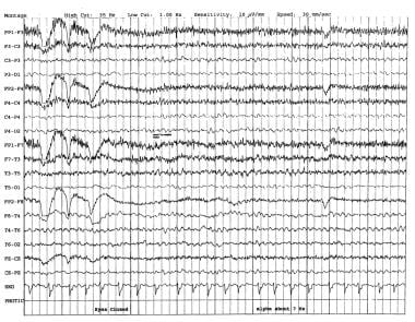
Photic stimulation is a medical term that refers to the exposure of the eyes to light, specifically repetitive pulses of light, which is used as a method in various research and clinical settings. In neuroscience, it's often used in studies related to vision, circadian rhythms, and brain function.
In a clinical context, photic stimulation is sometimes used in the diagnosis of certain medical conditions such as seizure disorders (like epilepsy). By observing the response of the brain to this light stimulus, doctors can gain valuable insights into the functioning of the brain and the presence of any neurological disorders.
However, it's important to note that photic stimulation should be conducted under the supervision of a trained healthcare professional, as improper use can potentially trigger seizures in individuals who are susceptible to them.
Juvenile Myoclonic Epilepsy (JME) is a genetic condition that is characterized by the occurrence of myoclonic seizures, which are sudden, brief, shock-like jerks of muscles typically occurring in the arms and legs. These seizures usually begin in adolescence or early adulthood, between 12 to 18 years of age.
JME is a type of generalized epilepsy, meaning that it involves abnormal electrical activity throughout the brain rather than just one area. In addition to myoclonic seizures, individuals with JME may also experience absence seizures (brief periods of staring and unresponsiveness) and/or tonic-clonic seizures (generalized convulsions).
The condition is often inherited in an autosomal dominant manner, meaning that a child has a 50% chance of inheriting the gene mutation from a parent with JME. However, not all cases are familial, and some may result from new genetic changes (mutations) that occur spontaneously.
JME is typically treated with anticonvulsant medications such as valproate or lamotrigine to control seizures. Lifestyle modifications, including avoiding sleep deprivation, stress, and excessive alcohol consumption, may also help reduce the frequency of seizures. With appropriate treatment, most individuals with JME can lead normal or near-normal lives.
Electroencephalography (EEG) is a medical procedure that records electrical activity in the brain. It uses small, metal discs called electrodes, which are attached to the scalp with paste or a specialized cap. These electrodes detect tiny electrical charges that result from the activity of brain cells, and the EEG machine then amplifies and records these signals.
EEG is used to diagnose various conditions related to the brain, such as seizures, sleep disorders, head injuries, infections, and degenerative diseases like Alzheimer's or Parkinson's. It can also be used during surgery to monitor brain activity and ensure that surgical procedures do not interfere with vital functions.
EEG is a safe and non-invasive procedure that typically takes about 30 minutes to an hour to complete, although longer recordings may be necessary in some cases. Patients are usually asked to relax and remain still during the test, as movement can affect the quality of the recording.
Myoclonus is a medical term that describes a quick, involuntary jerking muscle spasm. These spasms can happen once or repeat in a series, and they can range from mild to severe in nature. Myoclonus can affect any muscle in the body and can be caused by several different conditions, including certain neurological disorders, injuries, or diseases. In some cases, myoclonus may occur without an identifiable cause.
There are various types of myoclonus, classified based on their underlying causes, patterns of occurrence, and associated symptoms. Some common forms include:
1. Action myoclonus: Occurs during voluntary muscle movements
2. Stimulus-sensitive myoclonus: Triggered by external or internal stimuli, such as touch, sound, or light
3. Physiological myoclonus: Normal muscle jerks that occur during sleep onset (hypnic jerks) or during sleep (nocturnal myoclonus)
4. Reflex myoclonus: Result of a reflex arc activation due to a peripheral nerve stimulation
5. Epileptic myoclonus: Part of an epilepsy syndrome, often involving the brainstem or cortex
6. Symptomatic myoclonus: Occurs as a result of an underlying medical condition, such as metabolic disorders, infections, or neurodegenerative diseases
Treatment for myoclonus depends on the specific type and underlying cause. Medications, physical therapy, or lifestyle modifications may be recommended to help manage symptoms and improve quality of life.
The suprachiasmatic nucleus (SCN) is a small region located in the hypothalamus of the brain, just above the optic chiasm where the optic nerves from each eye cross. It is considered to be the primary circadian pacemaker in mammals, responsible for generating and maintaining the body's internal circadian rhythm, which is a roughly 24-hour cycle that regulates various physiological processes such as sleep-wake cycles, hormone release, and metabolism.
The SCN receives direct input from retinal ganglion cells, which are sensitive to light and dark signals. This information helps the SCN synchronize the internal circadian rhythm with the external environment, allowing it to adjust to changes in day length and other environmental cues. The SCN then sends signals to other parts of the brain and body to regulate various functions according to the time of day.
Disruption of the SCN's function can lead to a variety of circadian rhythm disorders, such as jet lag, shift work disorder, and advanced or delayed sleep phase syndrome.
The occipital lobe is the portion of the cerebral cortex that lies at the back of the brain (posteriorly) and is primarily involved in visual processing. It contains areas that are responsible for the interpretation and integration of visual stimuli, including color, form, movement, and recognition of objects. The occipital lobe is divided into several regions, such as the primary visual cortex (V1), secondary visual cortex (V2 to V5), and the visual association cortex, which work together to process different aspects of visual information. Damage to the occipital lobe can lead to various visual deficits, including blindness or partial loss of vision, known as a visual field cut.
In the context of medical terminology, "light" doesn't have a specific or standardized definition on its own. However, it can be used in various medical terms and phrases. For example, it could refer to:
1. Visible light: The range of electromagnetic radiation that can be detected by the human eye, typically between wavelengths of 400-700 nanometers. This is relevant in fields such as ophthalmology and optometry.
2. Therapeutic use of light: In some therapies, light is used to treat certain conditions. An example is phototherapy, which uses various wavelengths of ultraviolet (UV) or visible light for conditions like newborn jaundice, skin disorders, or seasonal affective disorder.
3. Light anesthesia: A state of reduced consciousness in which the patient remains responsive to verbal commands and physical stimulation. This is different from general anesthesia where the patient is completely unconscious.
4. Pain relief using light: Certain devices like transcutaneous electrical nerve stimulation (TENS) units have a 'light' setting, indicating lower intensity or frequency of electrical impulses used for pain management.
Without more context, it's hard to provide a precise medical definition of 'light'.
Evoked potentials, visual, also known as visually evoked potentials (VEPs), are electrical responses recorded from the brain following the presentation of a visual stimulus. These responses are typically measured using electroencephalography (EEG) and can provide information about the functioning of the visual pathways in the brain.
There are several types of VEPs, including pattern-reversal VEPs and flash VEPs. Pattern-reversal VEPs are elicited by presenting alternating checkerboard patterns, while flash VEPs are elicited by flashing a light. The responses are typically analyzed in terms of their latency (the time it takes for the response to occur) and amplitude (the size of the response).
VEPs are often used in clinical settings to help diagnose and monitor conditions that affect the visual system, such as multiple sclerosis, optic neuritis, and brainstem tumors. They can also be used in research to study the neural mechanisms underlying visual perception.
The visual cortex is the part of the brain that processes visual information. It is located in the occipital lobe, which is at the back of the brain. The visual cortex is responsible for receiving and interpreting signals from the retina, which are then transmitted through the optic nerve and optic tract.
The visual cortex contains several areas that are involved in different aspects of visual processing, such as identifying shapes, colors, and movements. These areas work together to help us recognize and understand what we see. Damage to the visual cortex can result in various visual impairments, such as blindness or difficulty with visual perception.
Epilepsy is a chronic neurological disorder characterized by recurrent, unprovoked seizures. These seizures are caused by abnormal electrical activity in the brain, which can result in a wide range of symptoms, including convulsions, loss of consciousness, and altered sensations or behaviors. Epilepsy can have many different causes, including genetic factors, brain injury, infection, or stroke. In some cases, the cause may be unknown.
There are many different types of seizures that can occur in people with epilepsy, and the specific type of seizure will depend on the location and extent of the abnormal electrical activity in the brain. Some people may experience only one type of seizure, while others may have several different types. Seizures can vary in frequency, from a few per year to dozens or even hundreds per day.
Epilepsy is typically diagnosed based on the patient's history of recurrent seizures and the results of an electroencephalogram (EEG), which measures the electrical activity in the brain. Imaging tests such as MRI or CT scans may also be used to help identify any structural abnormalities in the brain that may be contributing to the seizures.
While there is no cure for epilepsy, it can often be effectively managed with medication. In some cases, surgery may be recommended to remove the area of the brain responsible for the seizures. With proper treatment and management, many people with epilepsy are able to lead normal, productive lives.
Electric stimulation, also known as electrical nerve stimulation or neuromuscular electrical stimulation, is a therapeutic treatment that uses low-voltage electrical currents to stimulate nerves and muscles. It is often used to help manage pain, promote healing, and improve muscle strength and mobility. The electrical impulses can be delivered through electrodes placed on the skin or directly implanted into the body.
In a medical context, electric stimulation may be used for various purposes such as:
1. Pain management: Electric stimulation can help to block pain signals from reaching the brain and promote the release of endorphins, which are natural painkillers produced by the body.
2. Muscle rehabilitation: Electric stimulation can help to strengthen muscles that have become weak due to injury, illness, or surgery. It can also help to prevent muscle atrophy and improve range of motion.
3. Wound healing: Electric stimulation can promote tissue growth and help to speed up the healing process in wounds, ulcers, and other types of injuries.
4. Urinary incontinence: Electric stimulation can be used to strengthen the muscles that control urination and reduce symptoms of urinary incontinence.
5. Migraine prevention: Electric stimulation can be used as a preventive treatment for migraines by applying electrical impulses to specific nerves in the head and neck.
It is important to note that electric stimulation should only be administered under the guidance of a qualified healthcare professional, as improper use can cause harm or discomfort.
Electric stimulation therapy, also known as neuromuscular electrical stimulation (NMES) or electromyostimulation, is a therapeutic treatment that uses electrical impulses to stimulate muscles and nerves. The electrical signals are delivered through electrodes placed on the skin near the target muscle group or nerve.
The therapy can be used for various purposes, including:
1. Pain management: Electric stimulation can help reduce pain by stimulating the release of endorphins, which are natural painkillers produced by the body. It can also help block the transmission of pain signals to the brain.
2. Muscle rehabilitation: NMES can be used to prevent muscle atrophy and maintain muscle tone in individuals who are unable to move their muscles due to injury or illness, such as spinal cord injuries or stroke.
3. Improving circulation: Electric stimulation can help improve blood flow and reduce swelling by contracting the muscles and promoting the movement of fluids in the body.
4. Wound healing: NMES can be used to promote wound healing by increasing blood flow, reducing swelling, and improving muscle function around the wound site.
5. Muscle strengthening: Electric stimulation can be used to strengthen muscles by causing them to contract and relax repeatedly, which can help improve muscle strength and endurance.
It is important to note that electric stimulation therapy should only be administered under the guidance of a trained healthcare professional, as improper use can cause harm or discomfort.
A chemical stimulation in a medical context refers to the process of activating or enhancing physiological or psychological responses in the body using chemical substances. These chemicals can interact with receptors on cells to trigger specific reactions, such as neurotransmitters and hormones that transmit signals within the nervous system and endocrine system.
Examples of chemical stimulation include the use of medications, drugs, or supplements that affect mood, alertness, pain perception, or other bodily functions. For instance, caffeine can chemically stimulate the central nervous system to increase alertness and decrease feelings of fatigue. Similarly, certain painkillers can chemically stimulate opioid receptors in the brain to reduce the perception of pain.
It's important to note that while chemical stimulation can have therapeutic benefits, it can also have adverse effects if used improperly or in excessive amounts. Therefore, it's essential to follow proper dosing instructions and consult with a healthcare provider before using any chemical substances for stimulation purposes.
Deep brain stimulation (DBS) is a surgical procedure that involves the implantation of a medical device called a neurostimulator, which sends electrical impulses to specific targets in the brain. The impulses help to regulate abnormal brain activity, and can be used to treat a variety of neurological conditions, including Parkinson's disease, essential tremor, dystonia, and obsessive-compulsive disorder.
During the procedure, electrodes are implanted into the brain and connected to the neurostimulator, which is typically implanted in the chest. The neurostimulator can be programmed to deliver electrical impulses at varying frequencies, amplitudes, and pulse widths, depending on the specific needs of the patient.
DBS is generally considered a safe and effective treatment option for many patients with neurological conditions, although it does carry some risks, such as infection, bleeding, and hardware complications. It is typically reserved for patients who have not responded well to other forms of treatment, or who experience significant side effects from medication.
Physical stimulation, in a medical context, refers to the application of external forces or agents to the body or its tissues to elicit a response. This can include various forms of touch, pressure, temperature, vibration, or electrical currents. The purpose of physical stimulation may be therapeutic, as in the case of massage or physical therapy, or diagnostic, as in the use of reflex tests. It is also used in research settings to study physiological responses and mechanisms.
In a broader sense, physical stimulation can also refer to the body's exposure to physical activity or exercise, which can have numerous health benefits, including improving cardiovascular function, increasing muscle strength and flexibility, and reducing the risk of chronic diseases.
Transcranial Magnetic Stimulation (TMS) is a non-invasive form of brain stimulation where a magnetic field is generated via an electromagnetic coil placed on the scalp. This magnetic field induces an electric current in the underlying brain tissue, which can lead to neuronal activation or inhibition, depending on the frequency and intensity of the stimulation. TMS has been used as a therapeutic intervention for various neurological and psychiatric conditions, such as depression, migraine, and tinnitus, among others. It is also used in research settings to investigate brain function and connectivity.
"Cells, cultured" is a medical term that refers to cells that have been removed from an organism and grown in controlled laboratory conditions outside of the body. This process is called cell culture and it allows scientists to study cells in a more controlled and accessible environment than they would have inside the body. Cultured cells can be derived from a variety of sources, including tissues, organs, or fluids from humans, animals, or cell lines that have been previously established in the laboratory.
Cell culture involves several steps, including isolation of the cells from the tissue, purification and characterization of the cells, and maintenance of the cells in appropriate growth conditions. The cells are typically grown in specialized media that contain nutrients, growth factors, and other components necessary for their survival and proliferation. Cultured cells can be used for a variety of purposes, including basic research, drug development and testing, and production of biological products such as vaccines and gene therapies.
It is important to note that cultured cells may behave differently than they do in the body, and results obtained from cell culture studies may not always translate directly to human physiology or disease. Therefore, it is essential to validate findings from cell culture experiments using additional models and ultimately in clinical trials involving human subjects.
Transcutaneous Electrical Nerve Stimulation (TENS) is a non-invasive method of pain relief that involves the use of low-voltage electrical currents. A TENS device, which is usually small and portable, delivers these currents through electrodes that are placed on the skin near the site of pain. The electrical impulses stimulate nerve fibers, which can help to block the transmission of pain signals to the brain, thereby reducing the perception of pain.
TENS is thought to work through a number of different mechanisms, including the gate control theory of pain and the release of endorphins, which are natural painkillers produced by the body. It is generally considered safe, with few side effects, and can be used in conjunction with other forms of pain management.
TENS is often used to treat chronic pain conditions such as arthritis, fibromyalgia, and lower back pain, as well as acute pain from injuries or surgery. However, its effectiveness varies from person to person, and it may not work for everyone. It is important to consult with a healthcare provider before using TENS, particularly if you have any underlying medical conditions or are taking medication that could interact with the electrical currents.
In the field of medicine, "time factors" refer to the duration of symptoms or time elapsed since the onset of a medical condition, which can have significant implications for diagnosis and treatment. Understanding time factors is crucial in determining the progression of a disease, evaluating the effectiveness of treatments, and making critical decisions regarding patient care.
For example, in stroke management, "time is brain," meaning that rapid intervention within a specific time frame (usually within 4.5 hours) is essential to administering tissue plasminogen activator (tPA), a clot-busting drug that can minimize brain damage and improve patient outcomes. Similarly, in trauma care, the "golden hour" concept emphasizes the importance of providing definitive care within the first 60 minutes after injury to increase survival rates and reduce morbidity.
Time factors also play a role in monitoring the progression of chronic conditions like diabetes or heart disease, where regular follow-ups and assessments help determine appropriate treatment adjustments and prevent complications. In infectious diseases, time factors are crucial for initiating antibiotic therapy and identifying potential outbreaks to control their spread.
Overall, "time factors" encompass the significance of recognizing and acting promptly in various medical scenarios to optimize patient outcomes and provide effective care.
Signal transduction is the process by which a cell converts an extracellular signal, such as a hormone or neurotransmitter, into an intracellular response. This involves a series of molecular events that transmit the signal from the cell surface to the interior of the cell, ultimately resulting in changes in gene expression, protein activity, or metabolism.
The process typically begins with the binding of the extracellular signal to a receptor located on the cell membrane. This binding event activates the receptor, which then triggers a cascade of intracellular signaling molecules, such as second messengers, protein kinases, and ion channels. These molecules amplify and propagate the signal, ultimately leading to the activation or inhibition of specific cellular responses.
Signal transduction pathways are highly regulated and can be modulated by various factors, including other signaling molecules, post-translational modifications, and feedback mechanisms. Dysregulation of these pathways has been implicated in a variety of diseases, including cancer, diabetes, and neurological disorders.
Calcium is an essential mineral that is vital for various physiological processes in the human body. The medical definition of calcium is as follows:
Calcium (Ca2+) is a crucial cation and the most abundant mineral in the human body, with approximately 99% of it found in bones and teeth. It plays a vital role in maintaining structural integrity, nerve impulse transmission, muscle contraction, hormonal secretion, blood coagulation, and enzyme activation.
Calcium homeostasis is tightly regulated through the interplay of several hormones, including parathyroid hormone (PTH), calcitonin, and vitamin D. Dietary calcium intake, absorption, and excretion are also critical factors in maintaining optimal calcium levels in the body.
Hypocalcemia refers to low serum calcium levels, while hypercalcemia indicates high serum calcium levels. Both conditions can have detrimental effects on various organ systems and require medical intervention to correct.
"Cat" is a common name that refers to various species of small carnivorous mammals that belong to the family Felidae. The domestic cat, also known as Felis catus or Felis silvestris catus, is a popular pet and companion animal. It is a subspecies of the wildcat, which is found in Europe, Africa, and Asia.
Domestic cats are often kept as pets because of their companionship, playful behavior, and ability to hunt vermin. They are also valued for their ability to provide emotional support and therapy to people. Cats are obligate carnivores, which means that they require a diet that consists mainly of meat to meet their nutritional needs.
Cats are known for their agility, sharp senses, and predatory instincts. They have retractable claws, which they use for hunting and self-defense. Cats also have a keen sense of smell, hearing, and vision, which allow them to detect prey and navigate their environment.
In medical terms, cats can be hosts to various parasites and diseases that can affect humans and other animals. Some common feline diseases include rabies, feline leukemia virus (FeLV), feline immunodeficiency virus (FIV), and toxoplasmosis. It is important for cat owners to keep their pets healthy and up-to-date on vaccinations and preventative treatments to protect both the cats and their human companions.
Sprague-Dawley rats are a strain of albino laboratory rats that are widely used in scientific research. They were first developed by researchers H.H. Sprague and R.C. Dawley in the early 20th century, and have since become one of the most commonly used rat strains in biomedical research due to their relatively large size, ease of handling, and consistent genetic background.
Sprague-Dawley rats are outbred, which means that they are genetically diverse and do not suffer from the same limitations as inbred strains, which can have reduced fertility and increased susceptibility to certain diseases. They are also characterized by their docile nature and low levels of aggression, making them easier to handle and study than some other rat strains.
These rats are used in a wide variety of research areas, including toxicology, pharmacology, nutrition, cancer, and behavioral studies. Because they are genetically diverse, Sprague-Dawley rats can be used to model a range of human diseases and conditions, making them an important tool in the development of new drugs and therapies.
Implanted electrodes are medical devices that are surgically placed inside the body to interface directly with nerves, neurons, or other electrically excitable tissue for various therapeutic purposes. These electrodes can be used to stimulate or record electrical activity from specific areas of the body, depending on their design and application.
There are several types of implanted electrodes, including:
1. Deep Brain Stimulation (DBS) electrodes: These are placed deep within the brain to treat movement disorders such as Parkinson's disease, essential tremor, and dystonia. DBS electrodes deliver electrical impulses that modulate abnormal neural activity in targeted brain regions.
2. Spinal Cord Stimulation (SCS) electrodes: These are implanted along the spinal cord to treat chronic pain syndromes. SCS electrodes emit low-level electrical pulses that interfere with pain signals traveling to the brain, providing relief for patients.
3. Cochlear Implant electrodes: These are surgically inserted into the cochlea of the inner ear to restore hearing in individuals with severe to profound hearing loss. The electrodes stimulate the auditory nerve directly, bypassing damaged hair cells within the cochlea.
4. Retinal Implant electrodes: These are implanted in the retina to treat certain forms of blindness caused by degenerative eye diseases like retinitis pigmentosa. The electrodes convert visual information from a camera into electrical signals, which stimulate remaining retinal cells and transmit the information to the brain via the optic nerve.
5. Sacral Nerve Stimulation (SNS) electrodes: These are placed near the sacral nerves in the lower back to treat urinary or fecal incontinence and overactive bladder syndrome. SNS electrodes deliver electrical impulses that regulate the function of the affected muscles and nerves.
6. Vagus Nerve Stimulation (VNS) electrodes: These are wrapped around the vagus nerve in the neck to treat epilepsy and depression. VNS electrodes provide intermittent electrical stimulation to the vagus nerve, which has connections to various regions of the brain involved in these conditions.
Overall, implanted electrodes serve as a crucial component in many neuromodulation therapies, offering an effective treatment option for numerous neurological and sensory disorders.
Muscle contraction is the physiological process in which muscle fibers shorten and generate force, leading to movement or stability of a body part. This process involves the sliding filament theory where thick and thin filaments within the sarcomeres (the functional units of muscles) slide past each other, facilitated by the interaction between myosin heads and actin filaments. The energy required for this action is provided by the hydrolysis of adenosine triphosphate (ATP). Muscle contractions can be voluntary or involuntary, and they play a crucial role in various bodily functions such as locomotion, circulation, respiration, and posture maintenance.
A cell line is a culture of cells that are grown in a laboratory for use in research. These cells are usually taken from a single cell or group of cells, and they are able to divide and grow continuously in the lab. Cell lines can come from many different sources, including animals, plants, and humans. They are often used in scientific research to study cellular processes, disease mechanisms, and to test new drugs or treatments. Some common types of human cell lines include HeLa cells (which come from a cancer patient named Henrietta Lacks), HEK293 cells (which come from embryonic kidney cells), and HUVEC cells (which come from umbilical vein endothelial cells). It is important to note that cell lines are not the same as primary cells, which are cells that are taken directly from a living organism and have not been grown in the lab.
An action potential is a brief electrical signal that travels along the membrane of a nerve cell (neuron) or muscle cell. It is initiated by a rapid, localized change in the permeability of the cell membrane to specific ions, such as sodium and potassium, resulting in a rapid influx of sodium ions and a subsequent efflux of potassium ions. This ion movement causes a brief reversal of the electrical potential across the membrane, which is known as depolarization. The action potential then propagates along the cell membrane as a wave, allowing the electrical signal to be transmitted over long distances within the body. Action potentials play a crucial role in the communication and functioning of the nervous system and muscle tissue.
Phosphorylation is the process of adding a phosphate group (a molecule consisting of one phosphorus atom and four oxygen atoms) to a protein or other organic molecule, which is usually done by enzymes called kinases. This post-translational modification can change the function, localization, or activity of the target molecule, playing a crucial role in various cellular processes such as signal transduction, metabolism, and regulation of gene expression. Phosphorylation is reversible, and the removal of the phosphate group is facilitated by enzymes called phosphatases.
The vagus nerve, also known as the 10th cranial nerve (CN X), is the longest of the cranial nerves and extends from the brainstem to the abdomen. It has both sensory and motor functions and plays a crucial role in regulating various bodily functions such as heart rate, digestion, respiratory rate, speech, and sweating, among others.
The vagus nerve is responsible for carrying sensory information from the internal organs to the brain, and it also sends motor signals from the brain to the muscles of the throat and voice box, as well as to the heart, lungs, and digestive tract. The vagus nerve helps regulate the body's involuntary responses, such as controlling heart rate and blood pressure, promoting relaxation, and reducing inflammation.
Dysfunction in the vagus nerve can lead to various medical conditions, including gastroparesis, chronic pain, and autonomic nervous system disorders. Vagus nerve stimulation (VNS) is a therapeutic intervention that involves delivering electrical impulses to the vagus nerve to treat conditions such as epilepsy, depression, and migraine headaches.
Enzyme activation refers to the process by which an enzyme becomes biologically active and capable of carrying out its specific chemical or biological reaction. This is often achieved through various post-translational modifications, such as proteolytic cleavage, phosphorylation, or addition of cofactors or prosthetic groups to the enzyme molecule. These modifications can change the conformation or structure of the enzyme, exposing or creating a binding site for the substrate and allowing the enzymatic reaction to occur.
For example, in the case of proteolytic cleavage, an inactive precursor enzyme, known as a zymogen, is cleaved into its active form by a specific protease. This is seen in enzymes such as trypsin and chymotrypsin, which are initially produced in the pancreas as inactive precursors called trypsinogen and chymotrypsinogen, respectively. Once they reach the small intestine, they are activated by enteropeptidase, a protease that cleaves a specific peptide bond, releasing the active enzyme.
Phosphorylation is another common mechanism of enzyme activation, where a phosphate group is added to a specific serine, threonine, or tyrosine residue on the enzyme by a protein kinase. This modification can alter the conformation of the enzyme and create a binding site for the substrate, allowing the enzymatic reaction to occur.
Enzyme activation is a crucial process in many biological pathways, as it allows for precise control over when and where specific reactions take place. It also provides a mechanism for regulating enzyme activity in response to various signals and stimuli, such as hormones, neurotransmitters, or changes in the intracellular environment.
Acoustic stimulation refers to the use of sound waves or vibrations to elicit a response in an individual, typically for the purpose of assessing or treating hearing, balance, or neurological disorders. In a medical context, acoustic stimulation may involve presenting pure tones, speech sounds, or other types of auditory signals through headphones, speakers, or specialized devices such as bone conduction transducers.
The response to acoustic stimulation can be measured using various techniques, including electrophysiological tests like auditory brainstem responses (ABRs) or otoacoustic emissions (OAEs), behavioral observations, or functional imaging methods like fMRI. Acoustic stimulation is also used in therapeutic settings, such as auditory training programs for hearing impairment or vestibular rehabilitation for balance disorders.
It's important to note that acoustic stimulation should be administered under the guidance of a qualified healthcare professional to ensure safety and effectiveness.
A dose-response relationship in the context of drugs refers to the changes in the effects or symptoms that occur as the dose of a drug is increased or decreased. Generally, as the dose of a drug is increased, the severity or intensity of its effects also increases. Conversely, as the dose is decreased, the effects of the drug become less severe or may disappear altogether.
The dose-response relationship is an important concept in pharmacology and toxicology because it helps to establish the safe and effective dosage range for a drug. By understanding how changes in the dose of a drug affect its therapeutic and adverse effects, healthcare providers can optimize treatment plans for their patients while minimizing the risk of harm.
The dose-response relationship is typically depicted as a curve that shows the relationship between the dose of a drug and its effect. The shape of the curve may vary depending on the drug and the specific effect being measured. Some drugs may have a steep dose-response curve, meaning that small changes in the dose can result in large differences in the effect. Other drugs may have a more gradual dose-response curve, where larger changes in the dose are needed to produce significant effects.
In addition to helping establish safe and effective dosages, the dose-response relationship is also used to evaluate the potential therapeutic benefits and risks of new drugs during clinical trials. By systematically testing different doses of a drug in controlled studies, researchers can identify the optimal dosage range for the drug and assess its safety and efficacy.
In the context of medicine and pharmacology, "kinetics" refers to the study of how a drug moves throughout the body, including its absorption, distribution, metabolism, and excretion (often abbreviated as ADME). This field is called "pharmacokinetics."
1. Absorption: This is the process of a drug moving from its site of administration into the bloodstream. Factors such as the route of administration (e.g., oral, intravenous, etc.), formulation, and individual physiological differences can affect absorption.
2. Distribution: Once a drug is in the bloodstream, it gets distributed throughout the body to various tissues and organs. This process is influenced by factors like blood flow, protein binding, and lipid solubility of the drug.
3. Metabolism: Drugs are often chemically modified in the body, typically in the liver, through processes known as metabolism. These changes can lead to the formation of active or inactive metabolites, which may then be further distributed, excreted, or undergo additional metabolic transformations.
4. Excretion: This is the process by which drugs and their metabolites are eliminated from the body, primarily through the kidneys (urine) and the liver (bile).
Understanding the kinetics of a drug is crucial for determining its optimal dosing regimen, potential interactions with other medications or foods, and any necessary adjustments for special populations like pediatric or geriatric patients, or those with impaired renal or hepatic function.
Evoked potentials (EPs) are medical tests that measure the electrical activity in the brain or spinal cord in response to specific sensory stimuli, such as sight, sound, or touch. These tests are often used to help diagnose and monitor conditions that affect the nervous system, such as multiple sclerosis, brainstem tumors, and spinal cord injuries.
There are several types of EPs, including:
1. Visual Evoked Potentials (VEPs): These are used to assess the function of the visual pathway from the eyes to the back of the brain. A patient is typically asked to look at a patterned image or flashing light while electrodes placed on the scalp record the electrical responses.
2. Brainstem Auditory Evoked Potentials (BAEPs): These are used to evaluate the function of the auditory nerve and brainstem. Clicking sounds are presented to one or both ears, and electrodes placed on the scalp measure the response.
3. Somatosensory Evoked Potentials (SSEPs): These are used to assess the function of the peripheral nerves and spinal cord. Small electrical shocks are applied to a nerve at the wrist or ankle, and electrodes placed on the scalp record the response as it travels up the spinal cord to the brain.
4. Motor Evoked Potentials (MEPs): These are used to assess the function of the motor pathways in the brain and spinal cord. A magnetic or electrical stimulus is applied to the brain or spinal cord, and electrodes placed on a muscle measure the response as it travels down the motor pathway.
EPs can help identify abnormalities in the nervous system that may not be apparent through other diagnostic tests, such as imaging studies or clinical examinations. They are generally safe, non-invasive procedures with few risks or side effects.
Lymphocyte activation is the process by which B-cells and T-cells (types of lymphocytes) become activated to perform effector functions in an immune response. This process involves the recognition of specific antigens presented on the surface of antigen-presenting cells, such as dendritic cells or macrophages.
The activation of B-cells leads to their differentiation into plasma cells that produce antibodies, while the activation of T-cells results in the production of cytotoxic T-cells (CD8+ T-cells) that can directly kill infected cells or helper T-cells (CD4+ T-cells) that assist other immune cells.
Lymphocyte activation involves a series of intracellular signaling events, including the binding of co-stimulatory molecules and the release of cytokines, which ultimately result in the expression of genes involved in cell proliferation, differentiation, and effector functions. The activation process is tightly regulated to prevent excessive or inappropriate immune responses that can lead to autoimmunity or chronic inflammation.
Neurons, also known as nerve cells or neurocytes, are specialized cells that constitute the basic unit of the nervous system. They are responsible for receiving, processing, and transmitting information and signals within the body. Neurons have three main parts: the dendrites, the cell body (soma), and the axon. The dendrites receive signals from other neurons or sensory receptors, while the axon transmits these signals to other neurons, muscles, or glands. The junction between two neurons is called a synapse, where neurotransmitters are released to transmit the signal across the gap (synaptic cleft) to the next neuron. Neurons vary in size, shape, and structure depending on their function and location within the nervous system.
Evoked potentials, motor, are a category of tests used in clinical neurophysiology to measure the electrical activity generated by the nervous system in response to a stimulus that specifically activates the motor pathways. These tests can help assess the integrity and function of the motor neurons, which are responsible for controlling voluntary muscle movements.
During a motor evoked potentials test, electrodes are placed on the scalp or directly on the surface of the brain or spinal cord. A stimulus is then applied to the motor cortex or peripheral nerves, causing the muscles to contract. The resulting electrical signals are recorded and analyzed to evaluate the conduction velocity, amplitude, and latency of the motor responses.
Motor evoked potentials tests can be useful in diagnosing various neurological conditions, such as multiple sclerosis, spinal cord injuries, and motor neuron diseases. They can also help monitor the progression of these conditions and assess the effectiveness of treatments.
Messenger RNA (mRNA) is a type of RNA (ribonucleic acid) that carries genetic information copied from DNA in the form of a series of three-base code "words," each of which specifies a particular amino acid. This information is used by the cell's machinery to construct proteins, a process known as translation. After being transcribed from DNA, mRNA travels out of the nucleus to the ribosomes in the cytoplasm where protein synthesis occurs. Once the protein has been synthesized, the mRNA may be degraded and recycled. Post-transcriptional modifications can also occur to mRNA, such as alternative splicing and addition of a 5' cap and a poly(A) tail, which can affect its stability, localization, and translation efficiency.
Isoproterenol is a medication that belongs to a class of drugs called beta-adrenergic agonists. Medically, it is defined as a synthetic catecholamine with both alpha and beta adrenergic receptor stimulating properties. It is primarily used as a bronchodilator to treat conditions such as asthma and chronic obstructive pulmonary disease (COPD) by relaxing the smooth muscles in the airways, thereby improving breathing.
Isoproterenol can also be used in the treatment of bradycardia (abnormally slow heart rate), cardiac arrest, and heart blocks by increasing the heart rate and contractility. However, due to its non-selective beta-agonist activity, it may cause various side effects such as tremors, palpitations, and increased blood pressure. Its use is now limited due to the availability of more selective and safer medications.
Cyclic adenosine monophosphate (cAMP) is a key secondary messenger in many biological processes, including the regulation of metabolism, gene expression, and cellular excitability. It is synthesized from adenosine triphosphate (ATP) by the enzyme adenylyl cyclase and is degraded by the enzyme phosphodiesterase.
In the body, cAMP plays a crucial role in mediating the effects of hormones and neurotransmitters on target cells. For example, when a hormone binds to its receptor on the surface of a cell, it can activate a G protein, which in turn activates adenylyl cyclase to produce cAMP. The increased levels of cAMP then activate various effector proteins, such as protein kinases, which go on to regulate various cellular processes.
Overall, the regulation of cAMP levels is critical for maintaining proper cellular function and homeostasis, and abnormalities in cAMP signaling have been implicated in a variety of diseases, including cancer, diabetes, and neurological disorders.
"Wistar rats" are a strain of albino rats that are widely used in laboratory research. They were developed at the Wistar Institute in Philadelphia, USA, and were first introduced in 1906. Wistar rats are outbred, which means that they are genetically diverse and do not have a fixed set of genetic characteristics like inbred strains.
Wistar rats are commonly used as animal models in biomedical research because of their size, ease of handling, and relatively low cost. They are used in a wide range of research areas, including toxicology, pharmacology, nutrition, cancer, cardiovascular disease, and behavioral studies. Wistar rats are also used in safety testing of drugs, medical devices, and other products.
Wistar rats are typically larger than many other rat strains, with males weighing between 500-700 grams and females weighing between 250-350 grams. They have a lifespan of approximately 2-3 years. Wistar rats are also known for their docile and friendly nature, making them easy to handle and work with in the laboratory setting.
A reflex is an automatic, involuntary and rapid response to a stimulus that occurs without conscious intention. In the context of physiology and neurology, it's a basic mechanism that involves the transmission of nerve impulses between neurons, resulting in a muscle contraction or glandular secretion.
Reflexes are important for maintaining homeostasis, protecting the body from harm, and coordinating movements. They can be tested clinically to assess the integrity of the nervous system, such as the knee-j jerk reflex, which tests the function of the L3-L4 spinal nerve roots and the sensitivity of the stretch reflex arc.
Atropine is an anticholinergic drug that blocks the action of the neurotransmitter acetylcholine in the central and peripheral nervous system. It is derived from the belladonna alkaloids, which are found in plants such as deadly nightshade (Atropa belladonna), Jimson weed (Datura stramonium), and Duboisia spp.
In clinical medicine, atropine is used to reduce secretions, increase heart rate, and dilate the pupils. It is often used before surgery to dry up secretions in the mouth, throat, and lungs, and to reduce salivation during the procedure. Atropine is also used to treat certain types of nerve agent and pesticide poisoning, as well as to manage bradycardia (slow heart rate) and hypotension (low blood pressure) caused by beta-blockers or calcium channel blockers.
Atropine can have several side effects, including dry mouth, blurred vision, dizziness, confusion, and difficulty urinating. In high doses, it can cause delirium, hallucinations, and seizures. Atropine should be used with caution in patients with glaucoma, prostatic hypertrophy, or other conditions that may be exacerbated by its anticholinergic effects.
Norepinephrine, also known as noradrenaline, is a neurotransmitter and a hormone that is primarily produced in the adrenal glands and is released into the bloodstream in response to stress or physical activity. It plays a crucial role in the "fight-or-flight" response by preparing the body for action through increasing heart rate, blood pressure, respiratory rate, and glucose availability.
As a neurotransmitter, norepinephrine is involved in regulating various functions of the nervous system, including attention, perception, motivation, and arousal. It also plays a role in modulating pain perception and responding to stressful or emotional situations.
In medical settings, norepinephrine is used as a vasopressor medication to treat hypotension (low blood pressure) that can occur during septic shock, anesthesia, or other critical illnesses. It works by constricting blood vessels and increasing heart rate, which helps to improve blood pressure and perfusion of vital organs.
Synaptic transmission is the process by which a neuron communicates with another cell, such as another neuron or a muscle cell, across a junction called a synapse. It involves the release of neurotransmitters from the presynaptic terminal of the neuron, which then cross the synaptic cleft and bind to receptors on the postsynaptic cell, leading to changes in the electrical or chemical properties of the target cell. This process is critical for the transmission of signals within the nervous system and for controlling various physiological functions in the body.
Electromyography (EMG) is a medical diagnostic procedure that measures the electrical activity of skeletal muscles during contraction and at rest. It involves inserting a thin needle electrode into the muscle to record the electrical signals generated by the muscle fibers. These signals are then displayed on an oscilloscope and may be heard through a speaker.
EMG can help diagnose various neuromuscular disorders, such as muscle weakness, numbness, or pain, and can distinguish between muscle and nerve disorders. It is often used in conjunction with other diagnostic tests, such as nerve conduction studies, to provide a comprehensive evaluation of the nervous system.
EMG is typically performed by a neurologist or a physiatrist, and the procedure may cause some discomfort or pain, although this is usually minimal. The results of an EMG can help guide treatment decisions and monitor the progression of neuromuscular conditions over time.
The motor cortex is a region in the frontal lobe of the brain that is responsible for controlling voluntary movements. It is involved in planning, initiating, and executing movements of the limbs, body, and face. The motor cortex contains neurons called Betz cells, which have large cell bodies and are responsible for transmitting signals to the spinal cord to activate muscles. Damage to the motor cortex can result in various movement disorders such as hemiplegia or paralysis on one side of the body.
The subthalamic nucleus (STN) is a small, lens-shaped structure located in the basal ganglia of the brain. It plays a crucial role in motor control and has been identified as a key target for deep brain stimulation surgery in the treatment of Parkinson's disease and other movement disorders.
The STN is involved in the regulation of movement, balance, and posture, and helps to filter and coordinate signals that are sent from the cerebral cortex to the thalamus and then on to the motor neurons in the brainstem and spinal cord. In Parkinson's disease, abnormal activity in the STN can contribute to symptoms such as tremors, rigidity, and difficulty initiating movements.
Deep brain stimulation of the STN involves implanting electrodes into the nucleus and delivering electrical impulses that help to regulate its activity. This can lead to significant improvements in motor function and quality of life for some people with Parkinson's disease.
Afferent pathways, also known as sensory pathways, refer to the neural connections that transmit sensory information from the peripheral nervous system to the central nervous system (CNS), specifically to the brain and spinal cord. These pathways are responsible for carrying various types of sensory information, such as touch, temperature, pain, pressure, vibration, hearing, vision, and taste, to the CNS for processing and interpretation.
The afferent pathways begin with sensory receptors located throughout the body, which detect changes in the environment and convert them into electrical signals. These signals are then transmitted via afferent neurons, also known as sensory neurons, to the spinal cord or brainstem. Within the CNS, the information is further processed and integrated with other neural inputs before being relayed to higher cognitive centers for conscious awareness and response.
Understanding the anatomy and physiology of afferent pathways is essential for diagnosing and treating various neurological conditions that affect sensory function, such as neuropathies, spinal cord injuries, and brain disorders.
Electrophysiology is a branch of medicine that deals with the electrical activities of the body, particularly the heart. In a medical context, electrophysiology studies (EPS) are performed to assess abnormal heart rhythms (arrhythmias) and to evaluate the effectiveness of certain treatments, such as medication or pacemakers.
During an EPS, electrode catheters are inserted into the heart through blood vessels in the groin or neck. These catheters can record the electrical activity of the heart and stimulate it to help identify the source of the arrhythmia. The information gathered during the study can help doctors determine the best course of treatment for each patient.
In addition to cardiac electrophysiology, there are also other subspecialties within electrophysiology, such as neuromuscular electrophysiology, which deals with the electrical activity of the nervous system and muscles.
The sympathetic nervous system (SNS) is a part of the autonomic nervous system that operates largely below the level of consciousness, and it functions to produce appropriate physiological responses to perceived danger. It's often associated with the "fight or flight" response. The SNS uses nerve impulses to stimulate target organs, causing them to speed up (e.g., increased heart rate), prepare for action, or otherwise respond to stressful situations.
The sympathetic nervous system is activated due to stressful emotional or physical situations and it prepares the body for immediate actions. It dilates the pupils, increases heart rate and blood pressure, accelerates breathing, and slows down digestion. The primary neurotransmitter involved in this system is norepinephrine (also known as noradrenaline).
C57BL/6 (C57 Black 6) is an inbred strain of laboratory mouse that is widely used in biomedical research. The term "inbred" refers to a strain of animals where matings have been carried out between siblings or other closely related individuals for many generations, resulting in a population that is highly homozygous at most genetic loci.
The C57BL/6 strain was established in 1920 by crossing a female mouse from the dilute brown (DBA) strain with a male mouse from the black strain. The resulting offspring were then interbred for many generations to create the inbred C57BL/6 strain.
C57BL/6 mice are known for their robust health, longevity, and ease of handling, making them a popular choice for researchers. They have been used in a wide range of biomedical research areas, including studies of cancer, immunology, neuroscience, cardiovascular disease, and metabolism.
One of the most notable features of the C57BL/6 strain is its sensitivity to certain genetic modifications, such as the introduction of mutations that lead to obesity or impaired glucose tolerance. This has made it a valuable tool for studying the genetic basis of complex diseases and traits.
Overall, the C57BL/6 inbred mouse strain is an important model organism in biomedical research, providing a valuable resource for understanding the genetic and molecular mechanisms underlying human health and disease.
Neural inhibition is a process in the nervous system that decreases or prevents the activity of neurons (nerve cells) in order to regulate and control communication within the nervous system. It is a fundamental mechanism that allows for the balance of excitation and inhibition necessary for normal neural function. Inhibitory neurotransmitters, such as GABA (gamma-aminobutyric acid) and glycine, are released from the presynaptic neuron and bind to receptors on the postsynaptic neuron, reducing its likelihood of firing an action potential. This results in a decrease in neural activity and can have various effects depending on the specific neurons and brain regions involved. Neural inhibition is crucial for many functions including motor control, sensory processing, attention, memory, and emotional regulation.
I must clarify that the term "Guinea Pigs" is not typically used in medical definitions. However, in colloquial or informal language, it may refer to people who are used as the first to try out a new medical treatment or drug. This is known as being a "test subject" or "in a clinical trial."
In the field of scientific research, particularly in studies involving animals, guinea pigs are small rodents that are often used as experimental subjects due to their size, cost-effectiveness, and ease of handling. They are not actually pigs from Guinea, despite their name's origins being unclear. However, they do not exactly fit the description of being used in human medical experiments.
'Self-stimulation' is more commonly known as "autoeroticism" or "masturbation." It refers to the act of stimulating one's own genitals for sexual pleasure, which can lead to orgasm. This behavior is considered a normal part of human sexuality and is a safe way to explore one's body and sexual responses. Self-stimulation can also be used as a means of relieving sexual tension and promoting relaxation. It is important to note that self-stimulation should always be a consensual, private activity and not performed in public or against the will of another individual.
 Photic stimulation - Wikipedia
Sex and electroencephalographic synchronization after photic stimulation predict signal changes in the visual cortex on...
 Photic Stimulation - Omnium1
Photic Stimulation - Omnium1
Photic Stimulation - iMRS Prime
 Photic stimulation. Medical search
 Normalization as a canonical neural computation
Control of the superior colliculus by the lateral prefrontal cortex
 Does visual cortex lactate increase following photic stimulation in migraine without aura patients? A functional H-1-MRS study<...
 EEG in Dementia and Encephalopathy: Overview, Dementia, Vascular Dementia
 Browse subject: Light -- Theory | The Online Books Page
 Frontiers | How Energy Metabolism Supports Cerebral Function: Insights from 13C Magnetic Resonance Studies In vivo
EEG in Dementia and Encephalopathy: Overview, Dementia, Vascular Dementia
 Positive Health Online | Author - David Noton, Ph.D.
 PDF) Photic Preference of the Short-Tailed Opossum (Monodelphis domestica)
 Erowid.org: Erowid Reference 1499 : On some effects of lysergic acid diethylamide (L.S.D. 25) in normal volunteers : Bradley PB...
 EEG - Electroencephalogram | KidsHealth NZ
 critical care
Gina Vincent's research topics | Profiles RNS
Separation in the visual field has divergent effects on discriminating the speed and the direction of motion.
 Engage Therapeutics announces Phase 2a data from Staccato alprazolam study | Drug Discovery News
 iSchool Directory | Information School | University of Washington
Classics in the History of Psychology -- Yerkes & Morgulis (1909)
 Klinik und Poliklinik für Neurologie | Homepage » Clinical diagnostics » EEG
 Age-related circadian disorganization caused by sympathetic dysfunction in peripheral clock regulation | npj Aging
 Grand Mal Seizures | Management of Pediatric Medical Emergencies in the Dental Office | Continuing Education Course |...
 Lighting the Way - SRI Magazine 2013 - Sunnybrook Research Institute
 No health effects from wind turbines | REVE News of the wind sector in Spain and in the world
 John Libbey Eurotext - Epileptic Disorders - How to diagnose and classify idiopathic (genetic) generalized epilepsies
 Figures and data in DMT alters cortical travelling waves | eLife
Humans2
- The mechanism may be similar to photic sneezing in humans, in which staring into sunlight triggers sneezing episodes. (horsemansnews.com)
- Photic sneezing in humans is a heritable, non-allergic disorder. (horsemansnews.com)
Hyperventilation4
- Some recordings included photic stimulation and hyperventilation. (medscape.com)
- Conduct activation procedures including hyperventilation and photic stimulation used in the assessment of cerebral reactivity and sleep. (usajobs.gov)
- If a seizure disorder is suspected and the routine EEG is normal, maneuvers that electrically activate the cortex (eg, hyperventilation, photic stimulation, sleep, sleep deprivation) can sometimes elicit evidence of a seizure disorder. (msdmanuals.com)
- During the test, a strobe light will be flashed on and off (photic stimulation) and you may be asked to take repeated deep and fast breathes (hyperventilation). (beaumont.org)
Intermittent3
- Photic stimulation may refer to: Intermittent photic stimulation as diagnostic procedure Any stimulation involving photons This disambiguation page lists articles associated with the title Photic stimulation. (wikipedia.org)
- Intermittent photic stimulation did not evoke photoparoxysmal responses. (medlink.com)
- In 14 autistic boys, aged 6 14 years, free of drug treatment, with relatively intact verbal functions andwithout severe or moderate mental retardation (I.Q. 91.4±22.8), intermittent photic stimulation at 11 fixed frequencies of 3 24 Hz revealed latent deficiency of the right hemisphere in the photic driving reactivity, predominantly at the fast alpha and beta frequencies of stimulation. (fiocruz.br)
Perception1
- Behavioral and neurophysiological studies have shown an enhancement of visual perception in crossmodal audiovisual stimulation conditions, both for sensitivity and reaction times, when the stimulation in the two sensory modalities occurs in condition of space and time congruency. (edgehill.ac.uk)
Neurons2
Entrainment1
- Most of these studies examined photic stimulation (presented via flashing lights) or combined photic and BBT entrainment rather than BBT alone. (goodtherapy.org)
Reactivity1
- EEG photic driving: right-hemisphere reactivity deficit in childhood autism. (fiocruz.br)
Visual10
- Sex and electroencephalographic synchronization after photic stimulation predict signal changes in the visual cortex on functional MR images. (ajnr.org)
- Photic stimulation of the suprachiasmatic nucleus via the non-visual optic system. (nih.gov)
- To block off the anxiety-inducing sights and sounds of hospital surroundings and create a pleasing environment, the therapeutic potential of visual stimulation as a nursing intervention was investigated. (nih.gov)
- ECG, GSR, photic, auditory, visual or transcranial current stimulation), we offer our state-of-the art analysis pipeline and our powerful machine learning engine. (starlab.es)
- We sought to isolate the origin of age-related BOLD changes by comparing blood-flow and oxygen-metabolic constituents of the BOLD response using dual-echo arterial spin labeling during visual stimulation and CO 2 ingestion. (johnshopkins.edu)
- Results showed that TMS applied over posterior temporal cortex before scene onset (-200 and -100 msec) impaired the detection of object categories in subsequently presented scenes, relative to vertex and early visual cortex stimulation. (edgehill.ac.uk)
- Effects of Visual Stimulation. (nih.gov)
- The purpose of the present work is to verify whether congruent visual and acoustic stimulations can improve the detection of visual stimuli in people affected by low vision. (edgehill.ac.uk)
- Participants were asked to detect the presence of a visual stimulus (yes/no task) either presented in isolation (i.e., unimodal visual stimulation) or simultaneously with auditory stimuli, which could be placed in the same spatial position (i.e., crossmodal congruent conditions) or in different spatial positions (i.e., crossmodal incongruent conditions). (edgehill.ac.uk)
- This effect is selective for visual stimulation that occurs in the portion of visual field that is impaired, and disappears in the region of space in which vision is spared. (edgehill.ac.uk)
Synchronization1
- Next, photic stimulation was carried out, followed by automatic registration of synchronization between the given respiratory rhythm and heart rate. (scardio.ru)
Sensitivity1
- An abnormal sensitivity to photic stimulation at a very slow frequency is a suggestive finding. (bmj.com)
Frequency2
- In order to evaluate the stage of dementia, we focused attention on EEG rhythms and Hasegawa-dementia-rating scale (HDS-R). Frontal EEGs were recorded from dementia patients and normal controls during music and photo-image stimulations, and frequency analysis was performed. (berklee.edu)
- A few minutes after the end, necessary to restore heart rate and respiration at the initial level, the tests were repeated with a subsequent 5% increase in the frequency of expiratory photic. (scardio.ru)
Cerebral1
- Técnica de estudio utilizada con frecuencia durante la ELECTROENCEFALOGRAFÍA en la cual se usan una serie de destellos luminosos o patrones visuales para desencadenar la actividad cerebral. (bvsalud.org)
Relation1
- Tasaki, I. The strength-duration relation in photic stimulation of the dark-adapted human eye. (nih.gov)
Markedly2
- The P100 latency was markedly prolonged with mainly the left eye stimulation at an avrage of 143.62 msec and the amplitude of response decreased at 1.26 microvolts. (ispub.com)
- In the controls, profiles of rhythm pattern during music stimulation seemed to be markedly different from those during photo-image stimulation. (berklee.edu)
Flashing light1
- Myoclonus can occur when an affected person is at rest, and it is made worse by motion, excitement, or flashing light (photic stimulation). (medlineplus.gov)
Response2
- Horses with photic headshaking are suspected to experience a burning sensation or tingling of the muzzle (neuropathic pain) in response to bright sunlight. (horsemansnews.com)
- The response of photic headshakers to medical therapy is variable. (horsemansnews.com)
Term1
- However, a 2008 review of the BWE literature found that delta stimulation was associated with improvement in migraines and other headaches and reduction in short-term stress. (goodtherapy.org)
Subjects1
- Rhythmic photic stimulation enhanced the symptoms due to LSD (15-100 mcg orally) in 12 of 15 subjects. (erowid.org)
Brain1
- Starlab targets creating patient stratification biomarkers for ADHD and ASD through advanced machine learning techniques including frameworks for federated machine learning and colloborates with Neuroelectrics in the development of advanced brain stimulation protocols (multichannel tDCS). (starlab.es)
Condition1
- In 1995, it was noted that headshaking was triggered by natural sunlight and darkness provided relief from the condition (photic headshaking). (horsemansnews.com)
Alpha2
- A single session of alpha stimulation was associated with stress reduction in some settings, but not for those undergoing root canal. (goodtherapy.org)
- Alpha stimulation was also linked to pain relief. (goodtherapy.org)
Video1
- Seizures can also be brought on by photic stimulation such as flickering lights, TV, video games, or light shining through trees or reflecting off water or snow. (chp.edu)