Calcium Phosphates
Glucose-6-Phosphate
Glyceraldehyde-3-Phosphate Dehydrogenases
Sugar Phosphates
Inositol Phosphates
Glyceraldehyde 3-Phosphate
Pentose Phosphate Pathway
Glucosephosphate Dehydrogenase
Phosphate Transport Proteins
Pyridoxal Phosphate
Glucose-6-Phosphate Isomerase
Glucosephosphates
Glycerol-3-Phosphate O-Acyltransferase
Sphingosine
Pentosephosphates
Carbamyl Phosphate
Hexosephosphates
Lysophospholipids
Phosphate-Binding Proteins
Phosphatidylinositol Phosphates
Organophosphates
Aldehyde-Lyases
Glycerolphosphate Dehydrogenase
Fructosephosphates
Receptors, Lysosphingolipid
Receptor, IGF Type 2
Glutamine-Fructose-6-Phosphate Transaminase (Isomerizing)
Hydrogen-Ion Concentration
Phosphotransferases (Alcohol Group Acceptor)
Molecular Sequence Data
Organophosphorus Compounds
Trioses
Phosphotransferases
Adenosine Triphosphate
Glucosephosphate Dehydrogenase Deficiency
Escherichia coli
Phosphorus
UTP-Hexose-1-Phosphate Uridylyltransferase
Phosphoric Monoester Hydrolases
Amino Acid Sequence
Myo-Inositol-1-Phosphate Synthase
Glyceraldehyde-3-Phosphate Dehydrogenase (Phosphorylating)
Substrate Specificity
Sodium-Phosphate Cotransporter Proteins
NADP
Hyperphosphatemia
Transaldolase
Magnetic Resonance Spectroscopy
Phosphogluconate Dehydrogenase
Polyisoprenyl Phosphate Monosaccharides
Binding Sites
Glucose
Transferases (Other Substituted Phosphate Groups)
Glycolysis
Calcium
Tritolyl Phosphates
Magnesium
Carbohydrate Epimerases
Phosphatidylinositols
Mannose-6-Phosphate Isomerase
Propylene Glycols
Polyisoprenyl Phosphate Sugars
Alkaline Phosphatase
Erythritol
Phosphorylation
Phosphofructokinase-1
Phosphocreatine
Phosphotransferases (Phosphate Group Acceptor)
Mutation
3-Deoxy-7-Phosphoheptulonate Synthase
NAD
Inositol
Fructose-Bisphosphate Aldolase
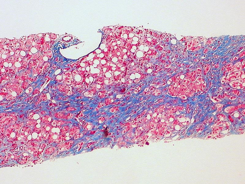
Liver
Phosphorus Radioisotopes
Carbamoyl-Phosphate Synthase (Ammonia)
Fructosediphosphates
Base Sequence
Phosphoenolpyruvate
Catalysis
Parathyroid Hormone
Adenosine Diphosphate
Models, Molecular
Protein Binding
Polyphosphates
UTP-Glucose-1-Phosphate Uridylyltransferase
Glucosamine 6-Phosphate N-Acetyltransferase
Buffers
Receptors, Lysophospholipid
Biological Transport
Hexokinase
Chromatography, High Pressure Liquid
3-Phosphoshikimate 1-Carboxyvinyltransferase
Rabbits
Sodium-Phosphate Cotransporter Proteins, Type IIa
Transketolase
Chromatography, Paper
Sodium-Phosphate Cotransporter Proteins, Type III
Hypophosphatemia, Familial
Phosphorus Isotopes
Durapatite
Phosphorus, Dietary
Cloning, Molecular
Sequence Homology, Amino Acid
Cattle
Adenine Nucleotides
Phosphate Acetyltransferase
Temperature
Vidarabine Phosphate
UDPglucose-Hexose-1-Phosphate Uridylyltransferase
Adenosine Monophosphate
Electrophoresis, Polyacrylamide Gel
Enzyme Activation
Triose-Phosphate Isomerase
Glycerol
Acid Phosphatase
Protein Conformation
Cells, Cultured
PHEX Phosphate Regulating Neutral Endopeptidase
Crystallography, X-Ray
Carbamoyl-Phosphate Synthase (Glutamine-Hydrolyzing)
Ketoses
Cell Membrane
Phosphoglucomutase
Isoenzymes
Oxidation-Reduction
Sodium-Phosphate Cotransporter Proteins, Type IIb
Culture Media
Transferases
Proton-Phosphate Symporters
Chemistry
Trehalose
Saccharomyces cerevisiae
Guanosine Diphosphate Mannose
Carbon Isotopes
Phytic Acid
Chemical Phenomena
Bone Substitutes
Phosphorus Metabolism Disorders
Glucose-6-Phosphatase
Gluconates
Molecular Structure
Dolichol Monophosphate Mannose
Phosphoric Diester Hydrolases
Rats, Inbred Strains
Kidney
Structure-Activity Relationship
Zinc Phosphate Cement
Chromatography, Ion Exchange
Signal Transduction
Phosphofructokinase-2
Chromatography, Gel
Fluorides
Chromatography, Thin Layer
Glucosyltransferases
Amino Acids
Phosphatidic Acids
Apatites
Carrier Proteins
Glyceraldehyde
Phospholipids
Models, Chemical
Models, Biological
Citrates
Pyruvates
Carbohydrate Metabolism
Heptoses
Biological Transport, Active
Dolichol
Glucosamine
1-Acylglycerol-3-Phosphate O-Acyltransferase
Carbohydrate Dehydrogenases
Mannose
A novel interaction mechanism accounting for different acylphosphatase effects on cardiac and fast twitch skeletal muscle sarcoplasmic reticulum calcium pumps. (1/8540)
In cardiac and skeletal muscle Ca2+ translocation from cytoplasm into sarcoplasmic reticulum (SR) is accomplished by different Ca2+-ATPases whose functioning involves the formation and decomposition of an acylphosphorylated phosphoenzyme intermediate (EP). In this study we found that acylphosphatase, an enzyme well represented in muscular tissues and which actively hydrolyzes EP, had different effects on heart (SERCA2a) and fast twitch skeletal muscle SR Ca2+-ATPase (SERCA1). With physiological acylphosphatase concentrations SERCA2a exhibited a parallel increase in the rates of both ATP hydrolysis and Ca2+ transport; in contrast, SERCA1 appeared to be uncoupled since the stimulation of ATP hydrolysis matched an inhibition of Ca2+ pump. These different effects probably depend on phospholamban, which is associated with SERCA2a but not SERCA1. Consistent with this view, the present study suggests that acylphosphatase-induced stimulation of SERCA2a, in addition to an enhanced EP hydrolysis, may be due to a displacement of phospholamban, thus to a removal of its inhibitory effect. (+info)The Golgi apparatus plays a significant role in the maintenance of Ca2+ homeostasis in the vps33Delta vacuolar biogenesis mutant of Saccharomyces cerevisiae. (2/8540)
The vacuole is the major site of intracellular Ca2+ storage in yeast and functions to maintain cytosolic Ca2+ levels within a narrow physiological range. In this study, we examined how cellular Ca2+ homeostasis is maintained in a vps33Delta vacuolar biogenesis mutant. We found that growth of the vps33Delta strain was sensitive to high or low extracellular Ca2+. This strain could not properly regulate cytosolic Ca2+ levels and was able to retain only a small fraction of its total cellular Ca2+ in a nonexchangeable intracellular pool. Surprisingly, the vps33Delta strain contained more total cellular Ca2+ than the wild type strain. Because most cellular Ca2+ is normally found within the vacuole, this suggested that other intracellular compartments compensated for the reduced capacity to store Ca2+ within the vacuole of this strain. To test this hypothesis, we examined the contribution of the Golgi-localized Ca2+ ATPase Pmr1p in the maintenance of cellular Ca2+ homeostasis. We found that a vps33Delta/pmr1Delta strain was hypersensitive to high extracellular Ca2+. In addition, certain combinations of mutations effecting both vacuolar and Golgi Ca2+ transport resulted in synthetic lethality. These results indicate that the Golgi apparatus plays a significant role in maintaining Ca2+ homeostasis when vacuolar biogenesis is compromised. (+info)Regulation of AMP deaminase from chicken erythrocytes. A kinetic study of the allosteric interactions. (3/8540)
The allosteric properties of AMP deaminase [EC 3.5.4.6] from chicken erythrocytes have been qualitatively and quantitatively accounted for by the concerted transition theory of Monod et al., on the assumption that this enzyme has different numbers of binding sites for each ligand. Theoretical curves yield a satisfactory fit for all experimental saturation functions with respect to activation by alkali metals and inhibition by Pi, assuming that the numbers of binding sites for AMP, alkali metals, and Pi are 4, 2, and 4, respectively. The enzyme was inhibited by concentrations of ATP and GTP below 0.1 and 0.25 mM, respectively, whereas activation of the enzyme was observed at ATP and GTP concentrations above 0.4 and 1.5 mM, respectively. These unusual kinetics with respect to ATP and GTP could be also accounted for by assuming 2 inhibitory and 4 activating sites for each ligand. (+info)Myocardial oxygenation during high work states in hearts with postinfarction remodeling. (4/8540)
BACKGROUND: Postinfarction left ventricular remodeling (LVR) is associated with reductions in myocardial high-energy phosphate (HEP) levels, which are more severe in animals that develop overt congestive heart failure (CHF). During high work states, further HEP loss occurs, which suggests demand-induced ischemia. This study tested the hypothesis that inadequate myocyte oxygen availability is the basis for these HEP abnormalities. METHODS AND RESULTS: Myocardial infarction was produced by left circumflex coronary artery ligation in swine. Studies were performed in 20 normal animals, 14 animals with compensated LVR, and 9 animals with CHF. Phosphocreatine (PCr)/ATP was determined with 31P NMR and deoxymyoglobin (Mb-delta) with 1H NMR in myocardium remote from the infarct. Basal PCr/ATP tended to be decreased in postinfarct hearts, and this was significant in animals with CHF. Infusion of dobutamine (20 microg x kg-1 x min-1 IV) caused doubling of the rate-pressure product in both normal and LVR hearts and resulted in comparable significant decreases of PCr/ATP in both groups. This decrease in PCr/ATP was not associated with detectable Mb-delta. In CHF hearts, rate-pressure product increased only 40% in response to dobutamine; this attenuated response also was not associated with detectable Mb-delta. CONCLUSIONS: Thus, the decrease of PCr/ATP during dobutamine infusion is not the result of insufficient myocardial oxygen availability. Furthermore, in CHF hearts, the low basal PCr/ATP and the attenuated response to dobutamine occurred in the absence of myocardial hypoxia, indicating that the HEP and contractile abnormalities were not the result of insufficient oxygen availability. (+info)Effects of phosphate intake on distribution of type II Na/Pi cotransporter mRNA in rat kidney. (5/8540)
BACKGROUND: Renal phosphate (Pi) reabsorption is regulated by dietary Pi intake, as well as in other ways. Changes in Pi reabsorption are associated with the modulation of sodium/Pi cotransporter type II (NaPi-2) protein abundance in the brush border membrane (BBM) of proximal tubules (PTs) and of renal NaPi-2 mRNA levels. In this study, we address whether the NaPi-2 protein and NaPi-2 mRNA distribution patterns in the renal cortex vary in parallel with changes of dietary Pi intake. METHODS: We investigated in cryosections of perfusion-fixed rat kidneys by in situ hybridization (ISH) and immunohistochemistry (IHC) the distribution patterns of NaPi-2 mRNA and of NaPi-2 protein one week, two hours, and four hours after changes in dietary Pi intake. RESULTS: NaPi-2 mRNA and NaPi-2 protein were present in PTs exclusively. In rats adapted to one week of high Pi intake, signals for NaPi-2 mRNA and NaPi-2 protein in cortical PTs were weak, except in the convoluted parts of PTs of juxtamedullary nephrons. After one week of low Pi intake, the ISH and IHC signals for NaPi-2 were high in PT segments in all cortical levels. The switch from a chronic high to a low Pi intake within two and four hours induced no increase and a slight increase, respectively, in the NaPi-2 mRNA signal in PTs of midcortical and of superficial nephrons, whereas in the BBM of these nephrons, NaPi-2 protein was markedly up-regulated. Two and four hours after switching from low to high Pi intake, the overall high ISH signal for NaPi-2 mRNA was unchanged, whereas NaPi-2 protein staining was drastically down-regulated in the BBM of PTs from superficial and midcortical nephrons. CONCLUSIONS: The marked changes in NaPi-2 protein abundance in the BBM, following altered dietary Pi intake, precede corresponding changes at the RNA level by several hours. Thus, the early adaptation to altered Pi intake involves mRNA-independent mechanisms. The up- or down-regulation of NaPi-2 protein abundance in the BBM and NaPi-2 mRNA in PT affects mainly midcortical and superficial nephrons. (+info)Biochemical indices of osteomalacia in pregnant Asian immigrants in Britain. (6/8540)
Serum calcium, phosphate and alkaline phosphatase, and urinary calcium excretion were examined during the second trimester of uncomplicated normal pregnancy in Asian immigrants to Britain and in local Caucasians. The mean serum calcium was significantly lower in Asians than in Caucasians, and the mean serum alkaline phosphatase was significantly higher in Asians. The geometric mean of the urinary calcium excretion was highly significantly lower in Asians than in Caucasians. The variances of the serum calcium, serum alkaline phosphatase, and urine calcium excretion did not differ significantly in the two populations. This indicates that there is a shift in values of immigrant Asians as a group compared with Caucasians. A comparison with figures obtained on normal nonpregnant persons of both suggests that the shift is not an inherent feature of the Asian population. (+info)Bound forms of Ca taken up by the synaptic plasma membrane. (7/8540)
Temperature dependent Ca-binding by the synaptic plasma membrane was increased in the presence of ATP and Mg++. Apparent Km for ATP was about 2.8 X 10(-5) M and optimal concentration of Mg++ was 2 mM in the presence of 2 mM ATP. After preincubation with nonradioactive Ca++, ATP and Mg++ to attain a steady state, addition of 45Ca resulted in remarkable labelling of the membrane, indicating rapid turnover of most of the membrane bound Ca. The presence of oxalate (60 mM) greatly increased Ca up-take on prolonged incubation. The Ca uptake in presence and absence of oxalate had similar substrate specificity and was similarly influenced by various monovalent cations. Furthermore, activities for Ca-uptake in the presence and absence of oxalate could not be separated by sucrose density gradient centrifugation of the synaptic plasma membrane fraction. Accordingly, it was considered that Ca++ in the medium was taken up by surface of the membrane, ATP- and temperature-dependently and then transferred into a cavity where the Ca-oxalate complex is formed. (+info)Mutations of Arg198 in sarcoplasmic reticulum Ca2+-ATPase cause inhibition of hydrolysis of the phosphoenzyme intermediate formed from inorganic phosphate. (8/8540)
Arg198 of sarcoplasmic reticulum Ca2+-ATPase was substituted with lysine, glutamine, glutamic acid, alanine, and isoleucine by site-directed mutagenesis. Kinetic analysis was performed with microsomal membranes isolated from COS-1 cells which were transfected with the mutated cDNAs. The rate of dephosphorylation of the ADP-insensitive phosphoenzyme was determined by first phosphorylating the Ca2+-ATPase with 32Pi and then diluting the sample with non-radioactive Pi. This rate was reduced substantially in the mutant R198Q, more strongly in the mutants R198A and R1981, and most strongly in the mutant R198E, but to a much lesser extent in R198K. The reduction in the rate of dephosphorylation was consistent with the observed decrease in the turnover rate of the Ca2+-ATPase accompanied by the steady-state accumulation of the ADP-insensitive phosphoenzyme formed from ATP. These results indicate that the positive charge and high hydrophilicity of Arg198 are critical for rapid hydrolysis of the ADP-insensitive phosphoenzyme. (+info)Phosphates, in a medical context, refer to the salts or esters of phosphoric acid. Phosphates play crucial roles in various biological processes within the human body. They are essential components of bones and teeth, where they combine with calcium to form hydroxyapatite crystals. Phosphates also participate in energy transfer reactions as phosphate groups attached to adenosine diphosphate (ADP) and adenosine triphosphate (ATP). Additionally, they contribute to buffer systems that help maintain normal pH levels in the body.
Abnormal levels of phosphates in the blood can indicate certain medical conditions. High phosphate levels (hyperphosphatemia) may be associated with kidney dysfunction, hyperparathyroidism, or excessive intake of phosphate-containing products. Low phosphate levels (hypophosphatemia) might result from malnutrition, vitamin D deficiency, or certain diseases affecting the small intestine or kidneys. Both hypophosphatemia and hyperphosphatemia can have significant impacts on various organ systems and may require medical intervention.
Calcium phosphates are a group of minerals that are important components of bones and teeth. They are also found in some foods and are used in dietary supplements and medical applications. Chemically, calcium phosphates are salts of calcium and phosphoric acid, and they exist in various forms, including hydroxyapatite, which is the primary mineral component of bone tissue. Other forms of calcium phosphates include monocalcium phosphate, dicalcium phosphate, and tricalcium phosphate, which are used as food additives and dietary supplements. Calcium phosphates are important for maintaining strong bones and teeth, and they also play a role in various physiological processes, such as nerve impulse transmission and muscle contraction.
Glucose-6-phosphate (G6P) is a vital intermediate compound in the metabolism of glucose, which is a simple sugar that serves as a primary source of energy for living organisms. G6P plays a critical role in both glycolysis and gluconeogenesis pathways, contributing to the regulation of blood glucose levels and energy production within cells.
In biochemistry, glucose-6-phosphate is defined as:
A hexose sugar phosphate ester formed by the phosphorylation of glucose at the 6th carbon atom by ATP in a reaction catalyzed by the enzyme hexokinase or glucokinase. This reaction is the first step in both glycolysis and glucose storage (glycogen synthesis) processes, ensuring that glucose can be effectively utilized for energy production or stored for later use.
G6P serves as a crucial metabolic branch point, leading to various pathways such as:
1. Glycolysis: In the presence of sufficient ATP and NAD+ levels, G6P is further metabolized through glycolysis to generate pyruvate, which enters the citric acid cycle for additional energy production in the form of ATP, NADH, and FADH2.
2. Gluconeogenesis: During periods of low blood glucose levels, G6P can be synthesized back into glucose through the gluconeogenesis pathway, primarily occurring in the liver and kidneys. This process helps maintain stable blood glucose concentrations and provides energy to cells when dietary intake is insufficient.
3. Pentose phosphate pathway (PPP): A portion of G6P can be shunted into the PPP, an alternative metabolic route that generates NADPH, ribose-5-phosphate for nucleotide synthesis, and erythrose-4-phosphate for aromatic amino acid production. The PPP is essential in maintaining redox balance within cells and supporting biosynthetic processes.
Overall, glucose-6-phosphate plays a critical role as a central metabolic intermediate, connecting various pathways to regulate energy homeostasis, redox balance, and biosynthesis in response to cellular demands and environmental cues.
Glyceraldehyde-3-phosphate dehydrogenase (GAPDH) is an enzyme that plays a crucial role in the metabolic pathway of glycolysis. Its primary function is to convert glyceraldehyde-3-phosphate (a triose sugar phosphate) into D-glycerate 1,3-bisphosphate, while also converting nicotinamide adenine dinucleotide (NAD+) into its reduced form NADH. This reaction is essential for the production of energy in the form of adenosine triphosphate (ATP) during cellular respiration. GAPDH has also been implicated in various non-metabolic processes, including DNA replication, repair, and transcription regulation, due to its ability to interact with different proteins and nucleic acids.
Sugar phosphates are organic compounds that play crucial roles in various biological processes, particularly in the field of genetics and molecular biology. They are formed by the attachment of a phosphate group to a sugar molecule, most commonly to the 5-carbon sugar ribose or deoxyribose.
In genetics, sugar phosphates form the backbone of nucleic acids, such as DNA and RNA. In DNA, the sugar phosphate backbone consists of alternating deoxyribose (a sugar) and phosphate groups, linked together by covalent bonds between the 5' carbon atom of one sugar molecule and the 3' carbon atom of another sugar molecule. This forms a long, twisted ladder-like structure known as a double helix.
Similarly, in RNA, the sugar phosphate backbone is formed by ribose (a sugar) and phosphate groups, creating a single-stranded structure that can fold back on itself to form complex shapes. These sugar phosphate backbones provide structural support for the nucleic acids and help to protect the genetic information stored within them.
Sugar phosphates also play important roles in energy metabolism, as they are involved in the formation and breakdown of high-energy compounds such as ATP (adenosine triphosphate) and GTP (guanosine triphosphate). These molecules serve as energy currency for cells, storing and releasing energy as needed to power various cellular processes.
Inositol phosphates are a family of molecules that consist of an inositol ring, which is a six-carbon heterocyclic compound, linked to one or more phosphate groups. These molecules play important roles as intracellular signaling intermediates and are involved in various cellular processes such as cell growth, differentiation, and metabolism.
Inositol hexakisphosphate (IP6), also known as phytic acid, is a form of inositol phosphate that is found in plant-based foods. IP6 has the ability to bind to minerals such as calcium, magnesium, and iron, which can reduce their bioavailability in the body.
Inositol phosphates have been implicated in several diseases, including cancer, diabetes, and neurodegenerative disorders. For example, altered levels of certain inositol phosphates have been observed in cancer cells, suggesting that they may play a role in tumor growth and progression. Additionally, mutations in enzymes involved in the metabolism of inositol phosphates have been associated with several genetic diseases.
Glyceraldehyde 3-phosphate (G3P) is a crucial intermediate in both glycolysis and gluconeogenesis metabolic pathways. It is an triose sugar phosphate, which means it contains three carbon atoms and has a phosphate group attached to it.
In the glycolysis process, G3P is produced during the third step of the process from the molecule dihydroxyacetone phosphate (DHAP) via the enzyme triosephosphate isomerase. In the following steps, G3P is converted into 1,3-bisphosphoglycerate, which eventually leads to the production of ATP and NADH.
In gluconeogenesis, G3P is produced from the reverse reaction of the glycolytic enzyme glyceraldehyde-3-phosphate dehydrogenase, using the molecule dihydroxyacetone phosphate (DHAP) as a starting point. G3P is then converted into glucose-6-phosphate, which can be further metabolized or released from the cell.
It's important to note that Glyceraldehyde 3-Phosphate plays a key role in energy production and carbohydrate metabolism.
The Pentose Phosphate Pathway (also known as the Hexose Monophosphate Shunt or HMP Shunt) is a metabolic pathway that runs parallel to glycolysis. It serves two major functions:
1. Providing reducing equivalents in the form of NADPH for reductive biosynthesis and detoxification processes.
2. Generating ribose-5-phosphate, a pentose sugar used in the synthesis of nucleotides and nucleic acids (DNA and RNA).
This pathway begins with the oxidation of glucose-6-phosphate to form 6-phosphogluconolactone, catalyzed by the enzyme glucose-6-phosphate dehydrogenase. The resulting NADPH is used in various anabolic reactions and antioxidant defense systems.
The Pentose Phosphate Pathway also includes a series of reactions called the non-oxidative branch, which interconverts various sugars to meet cellular needs for different types of monosaccharides. These conversions are facilitated by several enzymes including transketolase and transaldolase.
Glyceraldehyde-3-phosphate dehydrogenase (GAPDH), also known as Glucosephosphate Dehydrogenase, is an enzyme that plays a crucial role in cellular metabolism, particularly in the glycolytic pathway. It catalyzes the conversion of glyceraldehyde 3-phosphate (G3P) to 1,3-bisphosphoglycerate (1,3-BPG), while also converting nicotinamide adenine dinucleotide (NAD+) to its reduced form NADH. This reaction is essential for the production of energy in the form of adenosine triphosphate (ATP) during cellular respiration. GAPDH has been widely used as a housekeeping gene in molecular biology research due to its consistent expression across various tissues and cells, although recent studies have shown that its expression can vary under certain conditions.
Dihydroxyacetone Phosphate (DHAP) is a 3-carbon organic compound that plays a crucial role in the metabolic pathway called glycolysis. It is an intermediate molecule formed during the conversion of glucose into pyruvate, which ultimately produces energy in the form of ATP.
In the glycolytic process, DHAP is produced from glyceraldehyde 3-phosphate (G3P) in a reaction catalyzed by the enzyme triose phosphate isomerase. Then, DHAP is converted back to G3P in a subsequent step, which prepares it for further processing in the glycolytic pathway. This reversible conversion of DHAP and G3P helps maintain the equilibrium of the glycolytic process.
Apart from its role in energy metabolism, DHAP is also involved in other biochemical processes, such as the synthesis of glucose during gluconeogenesis and the formation of lipids in the liver.
Phosphate transport proteins are membrane-bound proteins responsible for the active transport of phosphate ions across cell membranes. They play a crucial role in maintaining appropriate phosphate concentrations within cells and between intracellular compartments, which is essential for various biological processes such as energy metabolism, signal transduction, and bone formation.
These proteins utilize the energy derived from ATP hydrolysis or other sources to move phosphate ions against their concentration gradient, thereby facilitating cellular uptake of phosphate even when extracellular concentrations are low. Phosphate transport proteins can be classified based on their structure, function, and localization into different types, including sodium-dependent and sodium-independent transporters, secondary active transporters, and channels.
Dysregulation of phosphate transport proteins has been implicated in several pathological conditions, such as renal Fanconi syndrome, tumoral calcinosis, and hypophosphatemic rickets. Therefore, understanding the molecular mechanisms underlying phosphate transport protein function is essential for developing targeted therapies to treat these disorders.
Pyridoxal phosphate (PLP) is the active form of vitamin B6 and functions as a cofactor in various enzymatic reactions in the human body. It plays a crucial role in the metabolism of amino acids, carbohydrates, lipids, and neurotransmitters. Pyridoxal phosphate is involved in more than 140 different enzyme-catalyzed reactions, making it one of the most versatile cofactors in human biochemistry.
As a cofactor, pyridoxal phosphate helps enzymes carry out their functions by facilitating chemical transformations in substrates (the molecules on which enzymes act). In particular, PLP is essential for transamination, decarboxylation, racemization, and elimination reactions involving amino acids. These processes are vital for the synthesis and degradation of amino acids, neurotransmitters, hemoglobin, and other crucial molecules in the body.
Pyridoxal phosphate is formed from the conversion of pyridoxal (a form of vitamin B6) by the enzyme pyridoxal kinase, using ATP as a phosphate donor. The human body obtains vitamin B6 through dietary sources such as whole grains, legumes, vegetables, nuts, and animal products like poultry, fish, and pork. It is essential to maintain adequate levels of pyridoxal phosphate for optimal enzymatic function and overall health.
Glucose-6-phosphate isomerase (GPI) is an enzyme involved in the glycolytic and gluconeogenesis pathways. It catalyzes the interconversion of glucose-6-phosphate (G6P) and fructose-6-phosphate (F6P), which are key metabolic intermediates in these pathways. This reaction is a reversible step that helps maintain the balance between the breakdown and synthesis of glucose in the cell.
In glycolysis, GPI converts G6P to F6P, which subsequently gets converted to fructose-1,6-bisphosphate (F1,6BP) by the enzyme phosphofructokinase-1 (PFK-1). In gluconeogenesis, the reaction is reversed, and F6P is converted back to G6P.
Deficiency or dysfunction of Glucose-6-phosphate isomerase can lead to various metabolic disorders, such as glycogen storage diseases and hereditary motor neuropathies.
Glucose phosphates are organic compounds that result from the reaction of glucose (a simple sugar) with phosphate groups. These compounds play a crucial role in various metabolic processes, particularly in energy metabolism within cells. The addition of phosphate groups to glucose makes it more reactive and enables it to undergo further reactions that lead to the formation of important molecules such as adenosine triphosphate (ATP), which is a primary source of energy for cellular functions.
One notable example of a glucose phosphate is glucose 1-phosphate, which is an intermediate in several metabolic pathways, including glycogenesis (the process of forming glycogen, a storage form of glucose) and glycolysis (the breakdown of glucose to release energy). Another example is glucose 6-phosphate, which is a key regulator of carbohydrate metabolism and serves as an important intermediate in the pentose phosphate pathway, a metabolic route that generates reducing equivalents (NADPH) and ribose sugars for nucleotide synthesis.
In summary, glucose phosphates are essential compounds in cellular metabolism, facilitating energy production, storage, and utilization.
In the context of medicine and pharmacology, "kinetics" refers to the study of how a drug moves throughout the body, including its absorption, distribution, metabolism, and excretion (often abbreviated as ADME). This field is called "pharmacokinetics."
1. Absorption: This is the process of a drug moving from its site of administration into the bloodstream. Factors such as the route of administration (e.g., oral, intravenous, etc.), formulation, and individual physiological differences can affect absorption.
2. Distribution: Once a drug is in the bloodstream, it gets distributed throughout the body to various tissues and organs. This process is influenced by factors like blood flow, protein binding, and lipid solubility of the drug.
3. Metabolism: Drugs are often chemically modified in the body, typically in the liver, through processes known as metabolism. These changes can lead to the formation of active or inactive metabolites, which may then be further distributed, excreted, or undergo additional metabolic transformations.
4. Excretion: This is the process by which drugs and their metabolites are eliminated from the body, primarily through the kidneys (urine) and the liver (bile).
Understanding the kinetics of a drug is crucial for determining its optimal dosing regimen, potential interactions with other medications or foods, and any necessary adjustments for special populations like pediatric or geriatric patients, or those with impaired renal or hepatic function.
Glycerol-3-Phosphate O-Acyltransferase (GPAT) is an enzyme that plays a crucial role in the biosynthesis of triacylglycerols and phospholipids, which are major components of cellular membranes and energy storage molecules. The GPAT enzyme catalyzes the initial and rate-limiting step in the glycerolipid synthesis pathway, specifically the transfer of an acyl group from an acyl-CoA donor to the sn-1 position of glycerol-3-phosphate, forming lysophosphatidic acid (LPA). This reaction is essential for the production of various glycerolipids, including phosphatidic acid, diacylglycerol, and triacylglycerol. There are four isoforms of GPAT (GPAT1-4) in humans, each with distinct subcellular localizations and functions. Dysregulation of GPAT activity has been implicated in several pathological conditions, such as metabolic disorders, cardiovascular diseases, and cancers.
Sphingosine is not a medical term per se, but rather a biological compound with importance in the field of medicine. It is a type of sphingolipid, a class of lipids that are crucial components of cell membranes. Sphingosine itself is a secondary alcohol with an amino group and two long-chain hydrocarbons.
Medically, sphingosine is significant due to its role as a precursor in the synthesis of other sphingolipids, such as ceramides, sphingomyelins, and gangliosides, which are involved in various cellular processes like signal transduction, cell growth, differentiation, and apoptosis (programmed cell death).
Moreover, sphingosine-1-phosphate (S1P), a derivative of sphingosine, is an important bioactive lipid mediator that regulates various physiological functions, including immune response, vascular maturation, and neuronal development. Dysregulation of S1P signaling has been implicated in several diseases, such as cancer, inflammation, and cardiovascular disorders.
In summary, sphingosine is a crucial biological compound with medical relevance due to its role as a precursor for various sphingolipids involved in cellular processes and as a precursor for the bioactive lipid mediator S1P.
Pentose phosphates are monosaccharides that contain five carbon atoms and one phosphate group. They play a crucial role in various metabolic pathways, including the pentose phosphate pathway (PPP), which is a major source of NADPH and ribose-5-phosphate for the synthesis of nucleotides.
The pentose phosphate pathway involves two main phases: the oxidative phase and the non-oxidative phase. In the oxidative phase, glucose-6-phosphate is converted to ribulose-5-phosphate, producing NADPH and CO2 as byproducts. Ribulose-5-phosphate can then be further metabolized in the non-oxidative phase to produce other pentose phosphates or converted back to glucose-6-phosphate through a series of reactions.
Pentose phosphates are also important intermediates in the synthesis of nucleotides, coenzymes, and other metabolites. Abnormalities in pentose phosphate pathway enzymes can lead to various metabolic disorders, such as defects in erythrocyte function and increased susceptibility to oxidative stress.
Glycerophosphates are esters of glycerol and phosphoric acid. In the context of biochemistry and medicine, glycerophosphates often refer to glycerol 3-phosphate (also known as glyceraldehyde 3-phosphate or glycerone phosphate) and its derivatives.
Glycerol 3-phosphate plays a crucial role in cellular metabolism, particularly in the process of energy production and storage. It is an important intermediate in both glycolysis (the breakdown of glucose to produce energy) and gluconeogenesis (the synthesis of glucose from non-carbohydrate precursors).
In addition, glycerophosphates are also involved in the formation of phospholipids, a major component of cell membranes. The esterification of glycerol 3-phosphate with fatty acids leads to the synthesis of phosphatidic acid, which is a key intermediate in the biosynthesis of other phospholipids.
Abnormalities in glycerophosphate metabolism have been implicated in various diseases, including metabolic disorders and neurological conditions.
Carbamyl Phosphate is a chemical compound that plays a crucial role in the biochemical process of nitrogen metabolism, particularly in the urea cycle. It is synthesized in the liver and serves as an important intermediate in the conversion of ammonia to urea, which is then excreted by the kidneys.
In medical terms, Carbamyl Phosphate Synthetase I (CPS I) deficiency is a rare genetic disorder that affects the production of Carbamyl Phosphate. This deficiency can lead to hyperammonemia, which is an excess of ammonia in the bloodstream, and can cause severe neurological symptoms and brain damage if left untreated.
It's important to note that while Carbamyl Phosphate is a critical component of the urea cycle, it is not typically used as a medication or therapeutic agent in clinical practice.
Hexose phosphates are organic compounds that consist of a hexose sugar molecule (a monosaccharide containing six carbon atoms, such as glucose or fructose) that has been phosphorylated, meaning that a phosphate group has been added to it. This process is typically facilitated by enzymes called kinases, which transfer a phosphate group from a donor molecule (usually ATP) to the sugar molecule.
Hexose phosphates play important roles in various metabolic pathways, including glycolysis, gluconeogenesis, and the pentose phosphate pathway. For example, glucose-6-phosphate is a key intermediate in both glycolysis and gluconeogenesis, while fructose-6-phosphate and fructose-1,6-bisphosphate are important intermediates in glycolysis. The pentose phosphate pathway, which is involved in the production of NADPH and ribose-5-phosphate, begins with the conversion of glucose-6-phosphate to 6-phosphogluconolactone by the enzyme glucose-6-phosphate dehydrogenase.
Overall, hexose phosphates are important metabolic intermediates that help regulate energy production and utilization in cells.
Lysophospholipids are a type of glycerophospholipid, which is a major component of cell membranes. They are characterized by having only one fatty acid chain attached to the glycerol backbone, as opposed to two in regular phospholipids. This results in a more polar and charged molecule, which can play important roles in cell signaling and regulation.
Lysophospholipids can be derived from the breakdown of regular phospholipids through the action of enzymes such as phospholipase A1 or A2. They can also be synthesized de novo in the cell. Some lysophospholipids, such as lysophosphatidic acid (LPA) and sphingosine-1-phosphate (S1P), have been found to act as signaling molecules that bind to specific G protein-coupled receptors and regulate various cellular processes, including proliferation, survival, and migration.
Abnormal levels of lysophospholipids have been implicated in several diseases, such as cancer, inflammation, and neurological disorders. Therefore, understanding the biology of lysophospholipids has important implications for developing new therapeutic strategies.
Phosphate-binding proteins are a type of protein that play a crucial role in regulating the concentration of phosphates in cells. They function by binding to phosphate ions and facilitating their transport, storage, or excretion. These proteins can be found in various organisms, including bacteria, plants, and animals.
In humans, one example of a phosphate-binding protein is the plasma protein known as fetuin-A. Fetuin-A helps regulate the amount of phosphate in the blood by binding to it and preventing it from forming insoluble precipitates with calcium, which can lead to the formation of kidney stones or calcifications in soft tissues.
Another example is the intracellular protein called alkaline phosphatase, which plays a role in removing phosphate groups from molecules within the cell. This enzyme helps regulate the levels of phosphates and other ions within the cell, as well as contributing to various metabolic processes.
Overall, phosphate-binding proteins are essential for maintaining proper phosphate homeostasis in the body, which is critical for numerous physiological functions, including energy metabolism, bone health, and signal transduction.
Ribose monophosphates are organic compounds that play a crucial role in the metabolism of cells, particularly in energy transfer and nucleic acid synthesis. A ribose monophosphate is formed by the attachment of a phosphate group to a ribose molecule, which is a type of sugar known as a pentose.
In biochemistry, there are two important ribose monophosphates:
1. Alpha-D-Ribose 5-Phosphate (ADP-Ribose): This compound serves as an essential substrate in various cellular processes, including DNA repair, chromatin remodeling, and protein modification. The enzyme that catalyzes the formation of ADP-ribose is known as poly(ADP-ribose) polymerase (PARP).
2. Ribulose 5-Phosphate: This compound is a key intermediate in the Calvin cycle, which is the process by which plants and some bacteria convert carbon dioxide into glucose during photosynthesis. Ribulose 5-phosphate is formed from ribose 5-phosphate through a series of enzymatic reactions.
Ribose monophosphates are essential for the proper functioning of cells and have implications in various physiological processes, as well as in certain disease states.
Phosphatidylinositol phosphates (PIPs) are a family of lipid molecules that play crucial roles as secondary messengers in intracellular signaling pathways. They are formed by the phosphorylation of the hydroxyl group on the inositol ring of phosphatidylinositol (PI), a fundamental component of cell membranes.
There are seven main types of PIPs, classified based on the number and position of phosphate groups attached to the inositol ring:
1. Phosphatidylinositol 4-monophosphate (PI4P) - one phosphate group at the 4th position
2. Phosphatidylinositol 5-monophosphate (PI5P) - one phosphate group at the 5th position
3. Phosphatidylinositol 3,4-bisphosphate (PI(3,4)P2) - two phosphate groups at the 3rd and 4th positions
4. Phosphatidylinositol 3,5-bisphosphate (PI(3,5)P2) - two phosphate groups at the 3rd and 5th positions
5. Phosphatidylinositol 4,5-bisphosphate [PI(4,5)P2] - two phosphate groups at the 4th and 5th positions
6. Phosphatidylinositol 3,4,5-trisphosphate [PI(3,4,5)P3] - three phosphate groups at the 3rd, 4th, and 5th positions
7. Phosphatidylinositol 3-phosphate (PI3P) - one phosphate group at the 3rd position
These PIPs are involved in various cellular processes such as membrane trafficking, cytoskeleton organization, cell survival, and metabolism. Dysregulation of PIP metabolism has been implicated in several diseases, including cancer, diabetes, and neurological disorders.
Organophosphates are a group of chemicals that include insecticides, herbicides, and nerve gases. They work by inhibiting an enzyme called acetylcholinesterase, which normally breaks down the neurotransmitter acetylcholine in the synapse between nerves. This leads to an overaccumulation of acetylcholine, causing overstimulation of the nervous system and resulting in a wide range of symptoms such as muscle twitching, nausea, vomiting, diarrhea, sweating, confusion, and potentially death due to respiratory failure. Organophosphates are highly toxic and their use is regulated due to the risks they pose to human health and the environment.
Aldehyde-lyases are a class of enzymes that catalyze the breakdown or synthesis of molecules involving an aldehyde group through a reaction known as lyase cleavage. This type of reaction results in the removal of a molecule, typically water or carbon dioxide, from the substrate.
In the case of aldehyde-lyases, these enzymes specifically catalyze reactions that involve the conversion of an aldehyde into a carboxylic acid or vice versa. These enzymes are important in various metabolic pathways and play a crucial role in the biosynthesis and degradation of several biomolecules, including carbohydrates, amino acids, and lipids.
The systematic name for this class of enzymes is "ald(e)hyde-lyases." They are classified under EC number 4.3.1 in the Enzyme Commission (EC) system.
Glycerol-3-phosphate dehydrogenase (GPD) is an enzyme that plays a crucial role in the metabolism of glucose and lipids. It catalyzes the conversion of dihydroxyacetone phosphate (DHAP) to glycerol-3-phosphate (G3P), which is a key intermediate in the synthesis of triglycerides, phospholipids, and other glycerophospholipids.
There are two main forms of GPD: a cytoplasmic form (GPD1) and a mitochondrial form (GPD2). The cytoplasmic form is involved in the production of NADH, which is used in various metabolic processes, while the mitochondrial form is involved in the production of ATP, the main energy currency of the cell.
Deficiencies or mutations in GPD can lead to a variety of metabolic disorders, including glycerol kinase deficiency and congenital muscular dystrophy. Elevated levels of GPD have been observed in certain types of cancer, suggesting that it may play a role in tumor growth and progression.
Fructose-1,6-bisphosphate (also known as fructose 1,6-diphosphate or Fru-1,6-BP) is the chemical compound that plays a crucial role in cellular respiration and glucose metabolism. It is not accurate to refer to "fructosephosphates" as a medical term, but fructose-1-phosphate and fructose-1,6-bisphosphate are important fructose phosphates with specific functions in the body.
Fructose-1-phosphate is an intermediate metabolite formed during the breakdown of fructose in the liver, while fructose-1,6-bisphosphate is a key regulator of glycolysis, the process by which glucose is broken down to produce energy in the form of ATP. Fructose-1,6-bisphosphate allosterically regulates the enzyme phosphofructokinase, which is the rate-limiting step in glycolysis, and its levels are tightly controlled to maintain proper glucose metabolism. Dysregulation of fructose metabolism has been implicated in various metabolic disorders, including insulin resistance, type 2 diabetes, and nonalcoholic fatty liver disease (NAFLD).
Lysosphingolipid receptors are a type of cell surface receptor that bind to lysosphingolipids, which are bioactive lipids derived from the degradation of sphingolipids. Sphingolipids are a class of lipids that play important roles in cell signaling and membrane structure.
Lysosphingolipids, such as lysosulfatide, lyso-Gb1 (lysoganglioside GM1), and lyso-PS (lysophosphatidylserine), have been implicated in various physiological and pathological processes, including cell proliferation, differentiation, inflammation, and apoptosis.
Lysosphingolipid receptors include several proteins, such as P2X7 receptor, G2A receptor, and Mas-related G protein-coupled receptor member X2 (MRGX2), that have been identified to interact with lysosphingolipids and mediate their downstream signaling.
Abnormal accumulation of lysosphingolipids has been associated with several diseases, including lysosomal storage disorders, neurodegenerative disorders, and cancer. Therefore, understanding the biology of lysosphingolipid receptors may provide insights into the development of new therapeutic strategies for these diseases.
IGF-2 (Insulin-like Growth Factor 2) receptor is a type of transmembrane protein that plays a role in cell growth, differentiation, and survival. Unlike other receptors in the insulin and IGF family, IGF-2 receptor does not mediate the activation of intracellular signaling pathways upon binding to its ligand (IGF-2). Instead, it acts as a clearance receptor that facilitates the removal of IGF-2 from circulation by transporting it to lysosomes for degradation.
The IGF-2 receptor is also known as cation-independent mannose-6-phosphate receptor (CI-M6PR) because it can also bind and transport mannose-6-phosphate-containing enzymes to lysosomes for degradation.
Mutations in the IGF-2 receptor gene have been associated with certain types of cancer, as well as developmental disorders such as Beckwith-Wiedemann syndrome.
I'm sorry for any confusion, but "Mannosephosphates" is not a widely recognized or established term in medicine or biochemistry. It seems that this term may be a combination of "mannose," which is a type of sugar (monosaccharide), and "phosphates," which are compounds containing phosphorus. However, without more context, it's difficult to provide an accurate medical definition for this term.
In biochemistry, mannose can be linked to phosphate groups in various ways, such as in the context of mannose-1-phosphate or mannose-6-phosphate, which are involved in different metabolic pathways. If you could provide more information about where you encountered this term, I might be able to give a more precise definition or explanation.
Polyisoprenyl phosphates are a type of organic compound that play a crucial role in the biosynthesis of various essential biomolecules in cells. They are formed by the addition of isoprene units, which are five-carbon molecules with a branched structure, to a phosphate group.
In medical terms, polyisoprenyl phosphates are primarily known for their role as intermediates in the biosynthesis of dolichols and farnesylated proteins. Dolichols are long-chain isoprenoids that function as lipid carriers in the synthesis of glycoproteins, which are proteins that contain carbohydrate groups attached to them. Farnesylated proteins, on the other hand, are proteins that have been modified with a farnesyl group, which is a 15-carbon isoprenoid. This modification plays a role in the localization and function of certain proteins within the cell.
Abnormalities in the biosynthesis of polyisoprenyl phosphates and their downstream products have been implicated in various diseases, including cancer, neurological disorders, and genetic syndromes. Therefore, understanding the biology and regulation of these compounds is an active area of research with potential therapeutic implications.
Hydrogen-ion concentration, also known as pH, is a measure of the acidity or basicity of a solution. It is defined as the negative logarithm (to the base 10) of the hydrogen ion activity in a solution. The standard unit of measurement is the pH unit. A pH of 7 is neutral, less than 7 is acidic, and greater than 7 is basic.
In medical terms, hydrogen-ion concentration is important for maintaining homeostasis within the body. For example, in the stomach, a high hydrogen-ion concentration (low pH) is necessary for the digestion of food. However, in other parts of the body such as blood, a high hydrogen-ion concentration can be harmful and lead to acidosis. Conversely, a low hydrogen-ion concentration (high pH) in the blood can lead to alkalosis. Both acidosis and alkalosis can have serious consequences on various organ systems if not corrected.
Molecular sequence data refers to the specific arrangement of molecules, most commonly nucleotides in DNA or RNA, or amino acids in proteins, that make up a biological macromolecule. This data is generated through laboratory techniques such as sequencing, and provides information about the exact order of the constituent molecules. This data is crucial in various fields of biology, including genetics, evolution, and molecular biology, allowing for comparisons between different organisms, identification of genetic variations, and studies of gene function and regulation.
Organophosphorus compounds are a class of chemical substances that contain phosphorus bonded to organic compounds. They are used in various applications, including as plasticizers, flame retardants, pesticides (insecticides, herbicides, and nerve gases), and solvents. In medicine, they are also used in the treatment of certain conditions such as glaucoma. However, organophosphorus compounds can be toxic to humans and animals, particularly those that affect the nervous system by inhibiting acetylcholinesterase, an enzyme that breaks down the neurotransmitter acetylcholine. Exposure to these compounds can cause symptoms such as nausea, vomiting, muscle weakness, and in severe cases, respiratory failure and death.
Trioses are simple sugars that contain three carbon atoms and a functional group called a ketone or aldehyde. They are the simplest type of sugar molecule, after monosaccharides such as glyceraldehyde and dihydroxyacetone.
Triose sugars can exist in two structural forms:
* Dihydroxyacetone (DHA), which is a ketotriose with the formula CH2OH-CO-CH2OH, and
* Glyceraldehyde (GA), which is an aldotriose with the formula HO-CHOH-CHO.
Trioses play important roles in various metabolic pathways, including glycolysis, gluconeogenesis, and the Calvin cycle of photosynthesis. In particular, DHA and GA are intermediates in the conversion of glucose to pyruvate during glycolysis, and they are also produced from pyruvate during gluconeogenesis.
Trioses can be synthesized chemically or biochemically through various methods, such as enzymatic reactions or microbial fermentation. They have potential applications in the food, pharmaceutical, and chemical industries, as they can serve as building blocks for more complex carbohydrates or as precursors for other organic compounds.
Phosphotransferases are a group of enzymes that catalyze the transfer of a phosphate group from a donor molecule to an acceptor molecule. This reaction is essential for various cellular processes, including energy metabolism, signal transduction, and biosynthesis.
The systematic name for this group of enzymes is phosphotransferase, which is derived from the general reaction they catalyze: D-donor + A-acceptor = D-donor minus phosphate + A-phosphate. The donor molecule can be a variety of compounds, such as ATP or a phosphorylated protein, while the acceptor molecule is typically a compound that becomes phosphorylated during the reaction.
Phosphotransferases are classified into several subgroups based on the type of donor and acceptor molecules they act upon. For example, kinases are a subgroup of phosphotransferases that transfer a phosphate group from ATP to a protein or other organic compound. Phosphatases, another subgroup, remove phosphate groups from molecules by transferring them to water.
Overall, phosphotransferases play a critical role in regulating many cellular functions and are important targets for drug development in various diseases, including cancer and neurological disorders.
Adenosine Triphosphate (ATP) is a high-energy molecule that stores and transports energy within cells. It is the main source of energy for most cellular processes, including muscle contraction, nerve impulse transmission, and protein synthesis. ATP is composed of a base (adenine), a sugar (ribose), and three phosphate groups. The bonds between these phosphate groups contain a significant amount of energy, which can be released when the bond between the second and third phosphate group is broken, resulting in the formation of adenosine diphosphate (ADP) and inorganic phosphate. This process is known as hydrolysis and can be catalyzed by various enzymes to drive a wide range of cellular functions. ATP can also be regenerated from ADP through various metabolic pathways, such as oxidative phosphorylation or substrate-level phosphorylation, allowing for the continuous supply of energy to cells.
Glucose-6-Phosphate Dehydrogenase (G6PD) deficiency is a genetic disorder that affects the normal functioning of an enzyme called G6PD. This enzyme is found in red blood cells and plays a crucial role in protecting them from damage.
In people with G6PD deficiency, the enzyme's activity is reduced or absent, making their red blood cells more susceptible to damage and destruction, particularly when they are exposed to certain triggers such as certain medications, infections, or foods. This can lead to a condition called hemolysis, where the red blood cells break down prematurely, leading to anemia, jaundice, and in severe cases, kidney failure.
G6PD deficiency is typically inherited from one's parents in an X-linked recessive pattern, meaning that males are more likely to be affected than females. While there is no cure for G6PD deficiency, avoiding triggers and managing symptoms can help prevent complications.
'Escherichia coli' (E. coli) is a type of gram-negative, facultatively anaerobic, rod-shaped bacterium that commonly inhabits the intestinal tract of humans and warm-blooded animals. It is a member of the family Enterobacteriaceae and one of the most well-studied prokaryotic model organisms in molecular biology.
While most E. coli strains are harmless and even beneficial to their hosts, some serotypes can cause various forms of gastrointestinal and extraintestinal illnesses in humans and animals. These pathogenic strains possess virulence factors that enable them to colonize and damage host tissues, leading to diseases such as diarrhea, urinary tract infections, pneumonia, and sepsis.
E. coli is a versatile organism with remarkable genetic diversity, which allows it to adapt to various environmental niches. It can be found in water, soil, food, and various man-made environments, making it an essential indicator of fecal contamination and a common cause of foodborne illnesses. The study of E. coli has contributed significantly to our understanding of fundamental biological processes, including DNA replication, gene regulation, and protein synthesis.
Phosphorus is an essential mineral that is required by every cell in the body for normal functioning. It is a key component of several important biomolecules, including adenosine triphosphate (ATP), which is the primary source of energy for cells, and deoxyribonucleic acid (DNA) and ribonucleic acid (RNA), which are the genetic materials in cells.
Phosphorus is also a major constituent of bones and teeth, where it combines with calcium to provide strength and structure. In addition, phosphorus plays a critical role in various metabolic processes, including energy production, nerve impulse transmission, and pH regulation.
The medical definition of phosphorus refers to the chemical element with the atomic number 15 and the symbol P. It is a highly reactive non-metal that exists in several forms, including white phosphorus, red phosphorus, and black phosphorus. In the body, phosphorus is primarily found in the form of organic compounds, such as phospholipids, phosphoproteins, and nucleic acids.
Abnormal levels of phosphorus in the body can lead to various health problems. For example, high levels of phosphorus (hyperphosphatemia) can occur in patients with kidney disease or those who consume large amounts of phosphorus-rich foods, and can contribute to the development of calcification of soft tissues and cardiovascular disease. On the other hand, low levels of phosphorus (hypophosphatemia) can occur in patients with malnutrition, vitamin D deficiency, or alcoholism, and can lead to muscle weakness, bone pain, and an increased risk of infection.
UTP-hexose-1-phosphate uridylyltransferase is an enzyme that catalyzes the transfer of a uridine monophosphate (UMP) group from a uridine triphosphate (UTP) molecule to a hexose-1-phosphate molecule, forming a UDP-hexose molecule. This reaction is an essential step in the biosynthesis of various glycosylated compounds, including glycoproteins and polysaccharides.
The systematic name for this enzyme is UTP:alpha-D-hexose-1-phosphate uridylyltransferase. It is also known as UDP-glucose pyrophosphorylase, which is a more specific name that refers to the formation of UDP-glucose from glucose-1-phosphate and UTP.
The enzyme plays a crucial role in carbohydrate metabolism and has been implicated in several diseases, including diabetes and cancer. Inhibitors of this enzyme have been explored as potential therapeutic agents for the treatment of these conditions.
Aldose-ketose isomerases are a group of enzymes that catalyze the interconversion between aldoses and ketoses, which are different forms of sugars. These enzymes play an essential role in carbohydrate metabolism by facilitating the reversible conversion of aldoses to ketoses and vice versa.
Aldoses are sugars that contain a carbonyl group (a functional group consisting of a carbon atom double-bonded to an oxygen atom) at the end of the carbon chain, while ketoses have their carbonyl group located in the middle of the chain. The isomerization process catalyzed by aldose-ketose isomerases helps maintain the balance between these two forms of sugars and enables cells to utilize them more efficiently for energy production and other metabolic processes.
There are several types of aldose-ketose isomerases, including:
1. Triose phosphate isomerase (TPI): This enzyme catalyzes the interconversion between dihydroxyacetone phosphate (a ketose) and D-glyceraldehyde 3-phosphate (an aldose), which are both trioses (three-carbon sugars). TPI plays a crucial role in glycolysis, the metabolic pathway that breaks down glucose to produce energy.
2. Xylulose kinase: This enzyme is involved in the pentose phosphate pathway, which is a metabolic route that generates reducing equivalents (NADPH) and pentoses for nucleic acid synthesis. Xylulose kinase catalyzes the conversion of D-xylulose (a ketose) to D-xylulose 5-phosphate, an important intermediate in the pentose phosphate pathway.
3. Ribulose-5-phosphate 3-epimerase: This enzyme is also part of the pentose phosphate pathway and catalyzes the interconversion between D-ribulose 5-phosphate (an aldose) and D-xylulose 5-phosphate (a ketose).
4. Phosphoglucomutase: This enzyme catalyzes the reversible conversion of glucose 1-phosphate (an aldose) to glucose 6-phosphate (an aldose), which is an important intermediate in both glycolysis and gluconeogenesis.
5. Phosphomannomutase: This enzyme catalyzes the reversible conversion of mannose 1-phosphate (a ketose) to mannose 6-phosphate (an aldose), which is involved in the biosynthesis of complex carbohydrates.
These are just a few examples of enzymes that catalyze the interconversion between aldoses and ketoses, highlighting their importance in various metabolic pathways.
Phosphoric monoester hydrolases are a class of enzymes that catalyze the hydrolysis of phosphoric monoesters into alcohol and phosphate. This class of enzymes includes several specific enzymes, such as phosphatases and nucleotidases, which play important roles in various biological processes, including metabolism, signal transduction, and regulation of cellular processes.
Phosphoric monoester hydrolases are classified under the EC number 3.1.3 by the Nomenclature Committee of the International Union of Biochemistry and Molecular Biology (IUBMB). The enzymes in this class share a common mechanism of action, which involves the nucleophilic attack on the phosphorus atom of the substrate by a serine or cysteine residue in the active site of the enzyme. This results in the formation of a covalent intermediate, which is then hydrolyzed to release the products.
Phosphoric monoester hydrolases are important therapeutic targets for the development of drugs that can modulate their activity. For example, inhibitors of phosphoric monoester hydrolases have been developed as potential treatments for various diseases, including cancer, neurodegenerative disorders, and infectious diseases.
An amino acid sequence is the specific order of amino acids in a protein or peptide molecule, formed by the linking of the amino group (-NH2) of one amino acid to the carboxyl group (-COOH) of another amino acid through a peptide bond. The sequence is determined by the genetic code and is unique to each type of protein or peptide. It plays a crucial role in determining the three-dimensional structure and function of proteins.
Myo-Inositol-1-Phosphate Synthase (MIPS) is an enzyme that catalyzes the conversion of glucose-6-phosphate to inositol 1,4-bisphosphate, which is the first and rate-limiting step in the biosynthesis of myo-inositol. Myo-inositol is a six-carbon cyclic polyol that serves as a precursor for various secondary messengers and structural lipids, including phosphatidylinositols and inositol phosphates, which play crucial roles in cell signaling pathways.
MIPS is widely distributed in nature and has been identified in bacteria, plants, fungi, and animals. In humans, MIPS is encoded by the ISO1 gene and is primarily localized in the cytoplasm of cells. Defects in MIPS have been associated with several diseases, including neurological disorders and cancer, highlighting its importance in maintaining cellular homeostasis.
Substrate specificity in the context of medical biochemistry and enzymology refers to the ability of an enzyme to selectively bind and catalyze a chemical reaction with a particular substrate (or a group of similar substrates) while discriminating against other molecules that are not substrates. This specificity arises from the three-dimensional structure of the enzyme, which has evolved to match the shape, charge distribution, and functional groups of its physiological substrate(s).
Substrate specificity is a fundamental property of enzymes that enables them to carry out highly selective chemical transformations in the complex cellular environment. The active site of an enzyme, where the catalysis takes place, has a unique conformation that complements the shape and charge distribution of its substrate(s). This ensures efficient recognition, binding, and conversion of the substrate into the desired product while minimizing unwanted side reactions with other molecules.
Substrate specificity can be categorized as:
1. Absolute specificity: An enzyme that can only act on a single substrate or a very narrow group of structurally related substrates, showing no activity towards any other molecule.
2. Group specificity: An enzyme that prefers to act on a particular functional group or class of compounds but can still accommodate minor structural variations within the substrate.
3. Broad or promiscuous specificity: An enzyme that can act on a wide range of structurally diverse substrates, albeit with varying catalytic efficiencies.
Understanding substrate specificity is crucial for elucidating enzymatic mechanisms, designing drugs that target specific enzymes or pathways, and developing biotechnological applications that rely on the controlled manipulation of enzyme activities.
Sodium-phosphate cotransporter proteins are membrane transport proteins that facilitate the active transport of sodium and inorganic phosphate ions across biological membranes. These proteins play a crucial role in maintaining phosphate homeostasis within the body by regulating the absorption and excretion of phosphate in the kidneys and intestines. They exist in two major types, type I (NaPi-I) and type II (NaPi-II), each having multiple subtypes with distinct tissue distributions and regulatory mechanisms.
Type I sodium-phosphate cotransporters are primarily expressed in the kidney's proximal tubules and play a significant role in reabsorbing phosphate from the primary urine back into the bloodstream. Type II sodium-phosphate cotransporters, on the other hand, are found in both the kidneys and intestines. In the kidneys, they contribute to phosphate reabsorption, while in the intestines, they facilitate phosphate absorption from food.
These proteins function by coupling the passive downhill movement of sodium ions (driven by the electrochemical gradient) with the active uphill transport of phosphate ions against their concentration gradient. This coupled transport process enables cells to maintain intracellular phosphate concentrations within a narrow range, despite fluctuations in dietary intake and renal function.
Dysregulation of sodium-phosphate cotransporter proteins has been implicated in various pathological conditions, such as chronic kidney disease (CKD), tumoral calcinosis, and certain genetic disorders affecting phosphate homeostasis.
NADP (Nicotinamide Adenine Dinucleotide Phosphate) is a coenzyme that plays a crucial role as an electron carrier in various redox reactions in the human body. It exists in two forms: NADP+, which functions as an oxidizing agent and accepts electrons, and NADPH, which serves as a reducing agent and donates electrons.
NADPH is particularly important in anabolic processes, such as lipid and nucleotide synthesis, where it provides the necessary reducing equivalents to drive these reactions forward. It also plays a critical role in maintaining the cellular redox balance by participating in antioxidant defense mechanisms that neutralize harmful reactive oxygen species (ROS).
In addition, NADP is involved in various metabolic pathways, including the pentose phosphate pathway and the Calvin cycle in photosynthesis. Overall, NADP and its reduced form, NADPH, are essential molecules for maintaining proper cellular function and energy homeostasis.
Ribulose phosphates are organic compounds that play a crucial role in the Calvin cycle, which is a part of photosynthesis. The Calvin cycle is the process by which plants, algae, and some bacteria convert carbon dioxide into glucose and other simple sugars.
Ribulose phosphates are sugar phosphates that contain five carbon atoms and have the chemical formula C5H10O5P. They exist in two forms: ribulose 5-phosphate (Ru5P) and ribulose 1,5-bisphosphate (RuBP).
Ribulose 1,5-bisphosphate is the starting point for carbon fixation in the Calvin cycle. In this process, an enzyme called RuBisCO (ribulose-1,5-bisphosphate carboxylase/oxygenase) catalyzes the reaction between RuBP and carbon dioxide to form two molecules of 3-phosphoglycerate, which are then converted into glucose and other sugars.
Ribulose phosphates are also involved in other metabolic pathways, such as the pentose phosphate pathway, which generates reducing power in the form of NADPH and produces ribose-5-phosphate, a precursor for nucleotide synthesis.
Hyperphosphatemia is a medical condition characterized by an excessively high level of phosphate (a form of the chemical element phosphorus) in the blood. Phosphate is an important component of various biological molecules, such as DNA, RNA, and ATP, and it plays a crucial role in many cellular processes, including energy metabolism and signal transduction.
In healthy individuals, the concentration of phosphate in the blood is tightly regulated within a narrow range to maintain normal physiological functions. However, when the phosphate level rises above this range (typically defined as a serum phosphate level greater than 4.5 mg/dL or 1.46 mmol/L), it can lead to hyperphosphatemia.
Hyperphosphatemia can result from various underlying medical conditions, including:
* Kidney dysfunction: The kidneys are responsible for filtering excess phosphate out of the blood and excreting it in the urine. When the kidneys fail to function properly, they may be unable to remove enough phosphate, leading to its accumulation in the blood.
* Hypoparathyroidism: The parathyroid glands produce a hormone called parathyroid hormone (PTH), which helps regulate calcium and phosphate levels in the body. In hypoparathyroidism, the production of PTH is insufficient, leading to an increase in phosphate levels.
* Hyperparathyroidism: In contrast, excessive production of PTH can also lead to hyperphosphatemia by increasing the release of phosphate from bones and decreasing its reabsorption in the kidneys.
* Excessive intake of phosphate-rich foods or supplements: Consuming large amounts of phosphate-rich foods, such as dairy products, nuts, and legumes, or taking phosphate supplements can raise blood phosphate levels.
* Tumor lysis syndrome: This is a complication that can occur after the treatment of certain types of cancer, particularly hematological malignancies. The rapid destruction of cancer cells releases large amounts of intracellular contents, including phosphate, into the bloodstream, leading to hyperphosphatemia.
* Rhabdomyolysis: This is a condition in which muscle tissue breaks down, releasing its contents, including phosphate, into the bloodstream. It can be caused by various factors, such as trauma, infection, or drug toxicity.
Hyperphosphatemia can have several adverse effects on the body, including calcification of soft tissues, kidney damage, and metabolic disturbances. Therefore, it is essential to diagnose and manage hyperphosphatemia promptly to prevent complications. Treatment options may include dietary modifications, medications that bind phosphate in the gastrointestinal tract, and dialysis in severe cases.
Transaldolase is not a medical term per se, but it is a term used in biochemistry and molecular biology. Transaldolase is an enzyme involved in the pentose phosphate pathway (PPP), which is a metabolic pathway that supplies reducing energy to cells by converting glucose-6-phosphate into ribulose-5-phosphate, a key intermediate in the synthesis of nucleotides.
The medical relevance of transaldolase lies in its role in maintaining cellular redox balance and providing precursors for nucleic acid synthesis. Defects in the PPP can lead to various metabolic disorders, including some forms of congenital cataracts, neurological dysfunction, and growth retardation. However, specific diseases or conditions directly attributed to transaldolase deficiency are not well-established.
Magnetic Resonance Spectroscopy (MRS) is a non-invasive diagnostic technique that provides information about the biochemical composition of tissues, including their metabolic state. It is often used in conjunction with Magnetic Resonance Imaging (MRI) to analyze various metabolites within body tissues, such as the brain, heart, liver, and muscles.
During MRS, a strong magnetic field, radio waves, and a computer are used to produce detailed images and data about the concentration of specific metabolites in the targeted tissue or organ. This technique can help detect abnormalities related to energy metabolism, neurotransmitter levels, pH balance, and other biochemical processes, which can be useful for diagnosing and monitoring various medical conditions, including cancer, neurological disorders, and metabolic diseases.
There are different types of MRS, such as Proton (^1^H) MRS, Phosphorus-31 (^31^P) MRS, and Carbon-13 (^13^C) MRS, each focusing on specific elements or metabolites within the body. The choice of MRS technique depends on the clinical question being addressed and the type of information needed for diagnosis or monitoring purposes.
Phosphogluconate dehydrogenase (PGD) is an enzyme that plays a crucial role in the pentose phosphate pathway, which is a metabolic pathway that supplies reducing energy to cells by converting glucose into ribose-5-phosphate and NADPH.
PGD catalyzes the third step of this pathway, in which 6-phosphogluconate is converted into ribulose-5-phosphate, with the concurrent reduction of NADP+ to NADPH. This reaction is essential for the generation of NADPH, which serves as a reducing agent in various cellular processes, including fatty acid synthesis and antioxidant defense.
Deficiencies in PGD can lead to several metabolic disorders, such as congenital nonspherocytic hemolytic anemia, which is characterized by the premature destruction of red blood cells due to a defect in the pentose phosphate pathway.
Polyisoprenyl phosphate monosaccharides are a type of glycosylated lipid intermediate molecule involved in the biosynthesis of isoprenoid-linked oligosaccharides, which are crucial for various cellular processes such as protein glycosylation and membrane trafficking.
These molecules consist of a polyisoprenyl phosphate tail, typically formed by the addition of multiple isoprene units (such as farnesyl or geranylgeranyl groups), which is attached to a single monosaccharide sugar moiety, such as glucose, mannose, or galactose.
The polyisoprenyl phosphate tail serves as a lipid anchor that helps tether the glycosylated molecule to cellular membranes during biosynthesis and transport. The monosaccharide component can be further modified by the addition of additional sugar residues, leading to the formation of more complex oligosaccharides that play important roles in various biological processes.
In the context of medical and biological sciences, a "binding site" refers to a specific location on a protein, molecule, or cell where another molecule can attach or bind. This binding interaction can lead to various functional changes in the original protein or molecule. The other molecule that binds to the binding site is often referred to as a ligand, which can be a small molecule, ion, or even another protein.
The binding between a ligand and its target binding site can be specific and selective, meaning that only certain ligands can bind to particular binding sites with high affinity. This specificity plays a crucial role in various biological processes, such as signal transduction, enzyme catalysis, or drug action.
In the case of drug development, understanding the location and properties of binding sites on target proteins is essential for designing drugs that can selectively bind to these sites and modulate protein function. This knowledge can help create more effective and safer therapeutic options for various diseases.
Glucose is a simple monosaccharide (or single sugar) that serves as the primary source of energy for living organisms. It's a fundamental molecule in biology, often referred to as "dextrose" or "grape sugar." Glucose has the molecular formula C6H12O6 and is vital to the functioning of cells, especially those in the brain and nervous system.
In the body, glucose is derived from the digestion of carbohydrates in food, and it's transported around the body via the bloodstream to cells where it can be used for energy. Cells convert glucose into a usable form through a process called cellular respiration, which involves a series of metabolic reactions that generate adenosine triphosphate (ATP)—the main currency of energy in cells.
Glucose is also stored in the liver and muscles as glycogen, a polysaccharide (multiple sugar) that can be broken down back into glucose when needed for energy between meals or during physical activity. Maintaining appropriate blood glucose levels is crucial for overall health, and imbalances can lead to conditions such as diabetes mellitus.
Glycolysis is a fundamental metabolic pathway that occurs in the cytoplasm of cells, consisting of a series of biochemical reactions. It's the process by which a six-carbon glucose molecule is broken down into two three-carbon pyruvate molecules. This process generates a net gain of two ATP molecules (the main energy currency in cells), two NADH molecules, and two water molecules.
Glycolysis can be divided into two stages: the preparatory phase (or 'energy investment' phase) and the payoff phase (or 'energy generation' phase). During the preparatory phase, glucose is phosphorylated twice to form glucose-6-phosphate and then converted to fructose-1,6-bisphosphate. These reactions consume two ATP molecules but set up the subsequent breakdown of fructose-1,6-bisphosphate into triose phosphates in the payoff phase. In this second stage, each triose phosphate is further oxidized and degraded to produce one pyruvate molecule, one NADH molecule, and one ATP molecule through substrate-level phosphorylation.
Glycolysis does not require oxygen to proceed; thus, it can occur under both aerobic (with oxygen) and anaerobic (without oxygen) conditions. In the absence of oxygen, the pyruvate produced during glycolysis is further metabolized through fermentation pathways such as lactic acid fermentation or alcohol fermentation to regenerate NAD+, which is necessary for glycolysis to continue.
In summary, glycolysis is a crucial process in cellular energy metabolism, allowing cells to convert glucose into ATP and other essential molecules while also serving as a starting point for various other biochemical pathways.
Calcium is an essential mineral that is vital for various physiological processes in the human body. The medical definition of calcium is as follows:
Calcium (Ca2+) is a crucial cation and the most abundant mineral in the human body, with approximately 99% of it found in bones and teeth. It plays a vital role in maintaining structural integrity, nerve impulse transmission, muscle contraction, hormonal secretion, blood coagulation, and enzyme activation.
Calcium homeostasis is tightly regulated through the interplay of several hormones, including parathyroid hormone (PTH), calcitonin, and vitamin D. Dietary calcium intake, absorption, and excretion are also critical factors in maintaining optimal calcium levels in the body.
Hypocalcemia refers to low serum calcium levels, while hypercalcemia indicates high serum calcium levels. Both conditions can have detrimental effects on various organ systems and require medical intervention to correct.
Tritolyl phosphates are not a medical term, but rather a class of industrial chemicals. They are organophosphate esters made from the reaction of toluene with phosphoric acid. These chemicals have various uses, including as plasticizers, flame retardants, and hydraulic fluids.
Exposure to high levels of tritolyl phosphates can cause irritation to the skin, eyes, and respiratory tract. However, they are not typically considered a significant health concern at the low levels encountered in most occupational or environmental settings. There is no known medical condition specifically associated with "tritolyl phosphates."
I'm sorry for any confusion, but "Mannitol Phosphates" is not a recognized medical term or concept in the way that, for example, "sodium chloride" is a specific chemical compound or "myocardial infarction" is a medical condition.
Mannitol is a sugar alcohol used as a medication, typically as an osmotic diuretic to reduce intracranial pressure or as a treatment for kidney failure. It can be administered intravenously or by mouth.
Phosphates, on the other hand, are a group of salts that contain the phosphate ion (PO43-). They have various uses in medicine and biology, such as in nutrition supplements, antacids, and laxatives.
It's possible that you may be looking for information on mannitol or phosphates separately, or there may be a specific medical context or compound that you have in mind. If you could provide more information about what you are looking for, I would be happy to help you further!
Magnesium is an essential mineral that plays a crucial role in various biological processes in the human body. It is the fourth most abundant cation in the body and is involved in over 300 enzymatic reactions, including protein synthesis, muscle and nerve function, blood glucose control, and blood pressure regulation. Magnesium also contributes to the structural development of bones and teeth.
In medical terms, magnesium deficiency can lead to several health issues, such as muscle cramps, weakness, heart arrhythmias, and seizures. On the other hand, excessive magnesium levels can cause symptoms like diarrhea, nausea, and muscle weakness. Magnesium supplements or magnesium-rich foods are often recommended to maintain optimal magnesium levels in the body.
Some common dietary sources of magnesium include leafy green vegetables, nuts, seeds, legumes, whole grains, and dairy products. Magnesium is also available in various forms as a dietary supplement, including magnesium oxide, magnesium citrate, magnesium chloride, and magnesium glycinate.
Carbohydrate epimerases are a group of enzymes that catalyze the interconversion of specific stereoisomers (epimers) of carbohydrates by the reversible oxidation and reduction of carbon atoms, usually at the fourth or fifth position. These enzymes play important roles in the biosynthesis and modification of various carbohydrate-containing molecules, such as glycoproteins, proteoglycans, and glycolipids, which are involved in numerous biological processes including cell recognition, signaling, and adhesion.
The reaction catalyzed by carbohydrate epimerases involves the transfer of a hydrogen atom and a proton between two adjacent carbon atoms, leading to the formation of new stereochemical configurations at these positions. This process can result in the conversion of one epimer into another, thereby expanding the structural diversity of carbohydrates and their derivatives.
Carbohydrate epimerases are classified based on the type of substrate they act upon and the specific stereochemical changes they induce. Some examples include UDP-glucose 4-epimerase, which interconverts UDP-glucose and UDP-galactose; UDP-N-acetylglucosamine 2-epimerase, which converts UDP-N-acetylglucosamine to UDP-N-acetylmannosamine; and GDP-fucose synthase, which catalyzes the conversion of GDP-mannose to GDP-fucose.
Understanding the function and regulation of carbohydrate epimerases is crucial for elucidating their roles in various biological processes and developing strategies for targeting them in therapeutic interventions.
Phosphatidylinositols (PIs) are a type of phospholipid that are abundant in the cell membrane. They contain a glycerol backbone, two fatty acid chains, and a head group consisting of myo-inositol, a cyclic sugar molecule, linked to a phosphate group.
Phosphatidylinositols can be phosphorylated at one or more of the hydroxyl groups on the inositol ring, forming various phosphoinositides (PtdInsPs) with different functions. These signaling molecules play crucial roles in regulating cellular processes such as membrane trafficking, cytoskeletal organization, and signal transduction pathways that control cell growth, differentiation, and survival.
Phosphatidylinositol 4,5-bisphosphate (PIP2) is a prominent phosphoinositide involved in the regulation of ion channels, enzymes, and cytoskeletal proteins. Upon activation of certain receptors, PIP2 can be cleaved by the enzyme phospholipase C into diacylglycerol (DAG) and inositol 1,4,5-trisphosphate (InsP3), which act as second messengers to trigger downstream signaling events.
Mannose-6-Phosphate Isomerase (MPI) is an enzyme that catalyzes the interconversion between mannose-6-phosphate and fructose-6-phosphate, which are both key metabolites in the glycolysis and gluconeogenesis pathways. This enzyme plays a crucial role in maintaining the balance between these two metabolic pathways, allowing cells to either break down or synthesize glucose depending on their energy needs.
The gene that encodes for MPI is called MPI1 and is located on chromosome 4 in humans. Defects in this gene can lead to a rare genetic disorder known as Mannose-6-Phosphate Isomerase Deficiency or Congenital Disorder of Glycosylation Type IIm, which is characterized by developmental delay, intellectual disability, seizures, and various other neurological symptoms.
Propylene glycol is not a medical term, but rather a chemical compound. Medically, it is classified as a humectant, which means it helps retain moisture. It is used in various pharmaceutical and cosmetic products as a solvent, preservative, and moisturizer. In medical settings, it can be found in topical creams, oral and injectable medications, and intravenous (IV) fluids.
The chemical definition of propylene glycol is:
Propylene glycol (IUPAC name: propan-1,2-diol) is a synthetic organic compound with the formula CH3CH(OH)CH2OH. It is a viscous, colorless, and nearly odorless liquid that is miscible with water, acetone, and chloroform. Propylene glycol is used as an antifreeze when mixed with water, as a solvent in the production of polymers, and as a moisturizer in various pharmaceutical and cosmetic products. It has a sweet taste and is considered generally recognized as safe (GRAS) by the U.S. Food and Drug Administration (FDA) for use as a food additive.
Arsenates are salts or esters of arsenic acid (AsO4). They contain the anion AsO4(3-), which consists of an arsenic atom bonded to four oxygen atoms in a tetrahedral arrangement. Arsenates can be found in various minerals, and they have been used in pesticides, wood preservatives, and other industrial applications. However, arsenic is highly toxic to humans and animals, so exposure to arsenates should be limited. Long-term exposure to arsenic can cause skin lesions, cancer, and damage to the nervous system, among other health problems.
Polyisoprenyl phosphate sugars are a type of glycosylated lipid that plays a crucial role in the biosynthesis of isoprenoid-derived natural products, including sterols and dolichols. These molecules consist of a polyisoprenyl phosphate group linked to one or more sugar moieties, such as glucose, mannose, or fructose. They serve as essential intermediates in the biosynthetic pathways that produce various isoprenoid-derived compounds, which have diverse functions in cellular metabolism and homeostasis.
The polyisoprenyl phosphate group is synthesized from isopentenyl pyrophosphate (IPP) and dimethylallyl pyrophosphate (DMAPP), the building blocks of isoprenoid biosynthesis, through a series of enzymatic reactions. The sugar moiety is then transferred to the polyisoprenyl phosphate group by specific glycosyltransferases, resulting in the formation of polyisoprenyl phosphate sugars.
These molecules are involved in various cellular processes, such as protein prenylation, where they serve as lipid anchors that facilitate the attachment of isoprenoid groups to proteins, thereby modulating their localization, stability, and activity. Additionally, polyisoprenyl phosphate sugars participate in the biosynthesis of bacterial cell wall components, such as peptidoglycan and lipopolysaccharides, highlighting their importance in both eukaryotic and prokaryotic organisms.
In summary, polyisoprenyl phosphate sugars are a class of glycosylated lipids that play a critical role in isoprenoid biosynthesis and related cellular processes, including protein prenylation and bacterial cell wall synthesis.
Hydrolysis is a chemical process, not a medical one. However, it is relevant to medicine and biology.
Hydrolysis is the breakdown of a chemical compound due to its reaction with water, often resulting in the formation of two or more simpler compounds. In the context of physiology and medicine, hydrolysis is a crucial process in various biological reactions, such as the digestion of food molecules like proteins, carbohydrates, and fats. Enzymes called hydrolases catalyze these hydrolysis reactions to speed up the breakdown process in the body.
Alkaline phosphatase (ALP) is an enzyme found in various body tissues, including the liver, bile ducts, digestive system, bones, and kidneys. It plays a role in breaking down proteins and minerals, such as phosphate, in the body.
The medical definition of alkaline phosphatase refers to its function as a hydrolase enzyme that removes phosphate groups from molecules at an alkaline pH level. In clinical settings, ALP is often measured through blood tests as a biomarker for various health conditions.
Elevated levels of ALP in the blood may indicate liver or bone diseases, such as hepatitis, cirrhosis, bone fractures, or cancer. Therefore, physicians may order an alkaline phosphatase test to help diagnose and monitor these conditions. However, it is essential to interpret ALP results in conjunction with other diagnostic tests and clinical findings for accurate diagnosis and treatment.
Hypophosphatemia is a medical condition characterized by abnormally low levels of phosphate (phosphorus) in the blood, specifically below 2.5 mg/dL. Phosphate is an essential electrolyte that plays a crucial role in various bodily functions such as energy production, bone formation, and maintaining acid-base balance.
Hypophosphatemia can result from several factors, including malnutrition, vitamin D deficiency, alcoholism, hormonal imbalances, and certain medications. Symptoms of hypophosphatemia may include muscle weakness, fatigue, bone pain, confusion, and respiratory failure in severe cases. Treatment typically involves correcting the underlying cause and administering phosphate supplements to restore normal levels.
Erythritol is a type of sugar alcohol (a carbohydrate that is metabolized differently than other sugars) used as a sugar substitute in food and drinks. It has about 0.24 calories per gram and contains almost no carbohydrates or sugar, making it a popular choice for people with diabetes or those following low-carb diets. Erythritol is naturally found in some fruits and fermented foods, but most commercial erythritol is made from cornstarch. It has a sweet taste similar to sugar but contains fewer calories and does not raise blood sugar levels.
Phosphorylation is the process of adding a phosphate group (a molecule consisting of one phosphorus atom and four oxygen atoms) to a protein or other organic molecule, which is usually done by enzymes called kinases. This post-translational modification can change the function, localization, or activity of the target molecule, playing a crucial role in various cellular processes such as signal transduction, metabolism, and regulation of gene expression. Phosphorylation is reversible, and the removal of the phosphate group is facilitated by enzymes called phosphatases.
Phosphofructokinase-1 (PFK-1) is a rate-limiting enzyme in the glycolytic pathway, which is the metabolic pathway that converts glucose into pyruvate, producing ATP and NADH as energy currency for the cell. PFK-1 plays a crucial role in regulating the rate of glycolysis by catalyzing the phosphorylation of fructose-6-phosphate to fructose-1,6-bisphosphate, using ATP as the phosphate donor.
PFK-1 is allosterically regulated by various metabolites, such as AMP, ADP, and ATP, which act as positive or negative effectors of the enzyme's activity. For example, an increase in the intracellular concentration of AMP or ADP can activate PFK-1, promoting glycolysis and energy production, while an increase in ATP levels can inhibit the enzyme's activity, conserving glucose for use under conditions of low energy demand.
Deficiencies in PFK-1 can lead to a rare genetic disorder called Tarui's disease or glycogen storage disease type VII, which is characterized by exercise intolerance, muscle cramps, and myoglobinuria (the presence of myoglobin in the urine due to muscle damage).
Phosphocreatine (PCr) is a high-energy phosphate compound found in the skeletal muscles, cardiac muscle, and brain. It plays a crucial role in energy metabolism and storage within cells. Phosphocreatine serves as an immediate energy reserve that helps regenerate ATP (adenosine triphosphate), the primary source of cellular energy, during short bursts of intense activity or stress. This process is facilitated by the enzyme creatine kinase, which catalyzes the transfer of a phosphate group from phosphocreatine to ADP (adenosine diphosphate) to form ATP.
In a medical context, phosphocreatine levels may be assessed in muscle biopsies or magnetic resonance spectroscopy (MRS) imaging to evaluate muscle energy metabolism and potential mitochondrial dysfunction in conditions such as muscular dystrophies, mitochondrial disorders, and neuromuscular diseases. Additionally, phosphocreatine depletion has been implicated in various pathological processes, including ischemia-reperfusion injury, neurodegenerative disorders, and heart failure.
A mutation is a permanent change in the DNA sequence of an organism's genome. Mutations can occur spontaneously or be caused by environmental factors such as exposure to radiation, chemicals, or viruses. They may have various effects on the organism, ranging from benign to harmful, depending on where they occur and whether they alter the function of essential proteins. In some cases, mutations can increase an individual's susceptibility to certain diseases or disorders, while in others, they may confer a survival advantage. Mutations are the driving force behind evolution, as they introduce new genetic variability into populations, which can then be acted upon by natural selection.
I'm sorry for any confusion, but "galactosephosphates" is not a widely recognized or established term in medicine or biochemistry. It seems that this term may be a combination of "galactose," which is a simple sugar, and "phosphate," which is a common ion found in biological systems. However, without more context, it's difficult to provide an accurate medical definition for this term.
Galactose is a monosaccharide that is metabolized in the body through the Leloir pathway, and defects in this pathway can lead to genetic disorders such as galactosemia. Phosphates are often found in biological molecules, including nucleic acids (DNA and RNA) and certain sugars (like glucose-1-phosphate).
Without further context or information about how "galactosephosphates" is being used, I would be cautious about assuming that it refers to a specific medical concept or condition.
3-Deoxy-7-phosphoheptulonate synthase (DAH7PS) is an enzyme that catalyzes the first step in the synthesis of the aromatic amino acids, phenylalanine, tyrosine, and tryptophan. The reaction it catalyzes is the condensation of erythrose-4-phosphate and phosphoenolpyruvate to form 3-deoxy-D-arabino-hept-2-ulose-7-phosphate (DAHP), also known as 3-deoxy-7-phosphoheptulonate.
The reaction catalyzed by DAH7PS is the first step in the shikimate pathway, which is a seven-step metabolic route used by bacteria, fungi, algae, parasites, and plants to produce aromatic amino acids and other important compounds. Mammals do not have this pathway, so enzymes of the shikimate pathway are potential targets for the development of antibiotics and herbicides.
DAH7PS is a regulatory enzyme in the shikimate pathway, and its activity is feedback inhibited by the aromatic amino acids phenylalanine and tyrosine. This helps to regulate the flow of carbon into the aromatic amino acid biosynthetic pathway based on the needs of the cell.
NAD (Nicotinamide Adenine Dinucleotide) is a coenzyme found in all living cells. It plays an essential role in cellular metabolism, particularly in redox reactions, where it acts as an electron carrier. NAD exists in two forms: NAD+, which accepts electrons and becomes reduced to NADH. This pairing of NAD+/NADH is involved in many fundamental biological processes such as generating energy in the form of ATP during cellular respiration, and serving as a critical cofactor for various enzymes that regulate cellular functions like DNA repair, gene expression, and cell death.
Maintaining optimal levels of NAD+/NADH is crucial for overall health and longevity, as it declines with age and in certain disease states. Therefore, strategies to boost NAD+ levels are being actively researched for their potential therapeutic benefits in various conditions such as aging, neurodegenerative disorders, and metabolic diseases.
Inositol is not considered a true "vitamin" because it can be created by the body from glucose. However, it is an important nutrient and is sometimes referred to as vitamin B8. It is a type of sugar alcohol that is found in both animals and plants. Inositol is involved in various biological processes, including:
1. Signal transduction: Inositol phospholipids are key components of cell membranes and play a crucial role in intracellular signaling pathways. They act as secondary messengers in response to hormones, neurotransmitters, and growth factors.
2. Insulin sensitivity: Inositol and its derivatives, such as myo-inositol and D-chiro-inositol, are involved in insulin signal transduction. Abnormalities in inositol metabolism have been linked to insulin resistance and conditions like polycystic ovary syndrome (PCOS).
3. Cerebral and ocular functions: Inositol is essential for the proper functioning of neurons and has been implicated in various neurological and psychiatric disorders, such as depression, anxiety, and bipolar disorder. It also plays a role in maintaining eye health.
4. Lipid metabolism: Inositol participates in the breakdown and transport of fats within the body.
5. Gene expression: Inositol and its derivatives are involved in regulating gene expression through epigenetic modifications.
Inositol can be found in various foods, including fruits, beans, grains, nuts, and vegetables. It is also available as a dietary supplement for those who wish to increase their intake.
Fructose-bisphosphate aldolase is a crucial enzyme in the glycolytic pathway, which is a metabolic process that breaks down glucose to produce energy. This enzyme catalyzes the conversion of fructose-1,6-bisphosphate into two triose sugars: dihydroxyacetone phosphate and glyceraldehyde-3-phosphate.
There are two main types of aldolase isoenzymes in humans, classified as aldolase A (or muscle type) and aldolase B (or liver type). Fructose-bisphosphate aldolase refers specifically to aldolase A, which is primarily found in the muscles, brain, and red blood cells. Aldolase B, on the other hand, is predominantly found in the liver, kidney, and small intestine.
Deficiency or dysfunction of fructose-bisphosphate aldolase can lead to metabolic disorders, such as hereditary fructose intolerance, which results from a deficiency in another enzyme called aldolase B. However, it is essential to note that the term "fructose-bisphosphate aldolase" typically refers to aldolase A and not aldolase B.
The liver is a large, solid organ located in the upper right portion of the abdomen, beneath the diaphragm and above the stomach. It plays a vital role in several bodily functions, including:
1. Metabolism: The liver helps to metabolize carbohydrates, fats, and proteins from the food we eat into energy and nutrients that our bodies can use.
2. Detoxification: The liver detoxifies harmful substances in the body by breaking them down into less toxic forms or excreting them through bile.
3. Synthesis: The liver synthesizes important proteins, such as albumin and clotting factors, that are necessary for proper bodily function.
4. Storage: The liver stores glucose, vitamins, and minerals that can be released when the body needs them.
5. Bile production: The liver produces bile, a digestive juice that helps to break down fats in the small intestine.
6. Immune function: The liver plays a role in the immune system by filtering out bacteria and other harmful substances from the blood.
Overall, the liver is an essential organ that plays a critical role in maintaining overall health and well-being.
Phosphorus radioisotopes are radioactive isotopes or variants of the element phosphorus that emit radiation. Phosphorus has several radioisotopes, with the most common ones being phosphorus-32 (^32P) and phosphorus-33 (^33P). These radioisotopes are used in various medical applications such as cancer treatment and diagnostic procedures.
Phosphorus-32 has a half-life of approximately 14.3 days and emits beta particles, making it useful for treating certain types of cancer, such as leukemia and lymphoma. It can also be used in brachytherapy, a type of radiation therapy that involves placing a radioactive source close to the tumor.
Phosphorus-33 has a shorter half-life of approximately 25.4 days and emits both beta particles and gamma rays. This makes it useful for diagnostic procedures, such as positron emission tomography (PET) scans, where the gamma rays can be detected and used to create images of the body's internal structures.
It is important to note that handling and using radioisotopes requires specialized training and equipment to ensure safety and prevent radiation exposure.
I'm sorry for any confusion, but "Fructosediphosphates" is not a recognized term in medicine or biochemistry. It's possible there may be a spelling mistake or misunderstanding in the term you're looking for.
If you meant "Fructose 1,6-bisphosphate," that is a key intermediate in carbohydrate metabolism. It's formed from fructose 6-phosphate in the process of glucose breakdown (glycolysis) and is then used in the generation of energy through the citric acid cycle.
If these terms are not what you were looking for, could you please provide more context or check the spelling? I'm here to help!
A base sequence in the context of molecular biology refers to the specific order of nucleotides in a DNA or RNA molecule. In DNA, these nucleotides are adenine (A), guanine (G), cytosine (C), and thymine (T). In RNA, uracil (U) takes the place of thymine. The base sequence contains genetic information that is transcribed into RNA and ultimately translated into proteins. It is the exact order of these bases that determines the genetic code and thus the function of the DNA or RNA molecule.
Phosphoenolpyruvate (PEP) is a key intermediate in the glycolysis pathway and other metabolic processes. It is a high-energy molecule that plays a crucial role in the transfer of energy during cellular respiration. Specifically, PEP is formed from the breakdown of fructose-1,6-bisphosphate and is then converted to pyruvate, releasing energy that is used to generate ATP, a major source of energy for cells.
Medically, abnormal levels of PEP may indicate issues with cellular metabolism or energy production, which can be associated with various medical conditions such as diabetes, mitochondrial disorders, and other metabolic diseases. However, direct measurement of PEP levels in clinical settings is not commonly performed due to technical challenges. Instead, clinicians typically assess overall metabolic function through a variety of other tests and measures.
Catalysis is the process of increasing the rate of a chemical reaction by adding a substance known as a catalyst, which remains unchanged at the end of the reaction. A catalyst lowers the activation energy required for the reaction to occur, thereby allowing the reaction to proceed more quickly and efficiently. This can be particularly important in biological systems, where enzymes act as catalysts to speed up metabolic reactions that are essential for life.
Parathyroid hormone (PTH) is a polypeptide hormone that plays a crucial role in the regulation of calcium and phosphate levels in the body. It is produced and secreted by the parathyroid glands, which are four small endocrine glands located on the back surface of the thyroid gland.
The primary function of PTH is to maintain normal calcium levels in the blood by increasing calcium absorption from the gut, mobilizing calcium from bones, and decreasing calcium excretion by the kidneys. PTH also increases phosphate excretion by the kidneys, which helps to lower serum phosphate levels.
In addition to its role in calcium and phosphate homeostasis, PTH has been shown to have anabolic effects on bone tissue, stimulating bone formation and preventing bone loss. However, chronic elevations in PTH levels can lead to excessive bone resorption and osteoporosis.
Overall, Parathyroid Hormone is a critical hormone that helps maintain mineral homeostasis and supports healthy bone metabolism.
Adenosine diphosphate (ADP) is a chemical compound that plays a crucial role in energy transfer within cells. It is a nucleotide, which consists of a adenosine molecule (a sugar molecule called ribose attached to a nitrogenous base called adenine) and two phosphate groups.
In the cell, ADP functions as an intermediate in the conversion of energy from one form to another. When a high-energy phosphate bond in ADP is broken, energy is released and ADP is converted to adenosine triphosphate (ATP), which serves as the main energy currency of the cell. Conversely, when ATP donates a phosphate group to another molecule, it is converted back to ADP, releasing energy for the cell to use.
ADP also plays a role in blood clotting and other physiological processes. In the coagulation cascade, ADP released from damaged red blood cells can help activate platelets and initiate the formation of a blood clot.
Molecular models are three-dimensional representations of molecular structures that are used in the field of molecular biology and chemistry to visualize and understand the spatial arrangement of atoms and bonds within a molecule. These models can be physical or computer-generated and allow researchers to study the shape, size, and behavior of molecules, which is crucial for understanding their function and interactions with other molecules.
Physical molecular models are often made up of balls (representing atoms) connected by rods or sticks (representing bonds). These models can be constructed manually using materials such as plastic or wooden balls and rods, or they can be created using 3D printing technology.
Computer-generated molecular models, on the other hand, are created using specialized software that allows researchers to visualize and manipulate molecular structures in three dimensions. These models can be used to simulate molecular interactions, predict molecular behavior, and design new drugs or chemicals with specific properties. Overall, molecular models play a critical role in advancing our understanding of molecular structures and their functions.
Protein binding, in the context of medical and biological sciences, refers to the interaction between a protein and another molecule (known as the ligand) that results in a stable complex. This process is often reversible and can be influenced by various factors such as pH, temperature, and concentration of the involved molecules.
In clinical chemistry, protein binding is particularly important when it comes to drugs, as many of them bind to proteins (especially albumin) in the bloodstream. The degree of protein binding can affect a drug's distribution, metabolism, and excretion, which in turn influence its therapeutic effectiveness and potential side effects.
Protein-bound drugs may be less available for interaction with their target tissues, as only the unbound or "free" fraction of the drug is active. Therefore, understanding protein binding can help optimize dosing regimens and minimize adverse reactions.
Polyphosphates are compounds consisting of many phosphate groups linked together in the form of chains or rings. They are often used in various medical and healthcare applications, such as:
* Dental care products: Polyphosphates can help prevent the formation of dental plaque and calculus by binding to calcium ions in saliva and inhibiting the growth of bacteria that cause tooth decay.
* Nutritional supplements: Polyphosphates are sometimes used as a source of phosphorus in nutritional supplements, particularly for people who have kidney disease or other medical conditions that require them to limit their intake of phosphorus from food sources.
* Medical devices: Polyphosphates may be used in the manufacture of medical devices, such as contact lenses and catheters, to improve their biocompatibility and resistance to bacterial growth.
It's worth noting that while polyphosphates have various medical uses, they can also be found in many non-medical products, such as food additives, water treatment chemicals, and cleaning agents.
UTP-Glucose-1-Phosphate Uridylyltransferase is an enzyme that catalyzes the reaction to form UDP-glucose from UTP and glucose-1-phosphate. This reaction plays a crucial role in the biosynthesis of various carbohydrates, glycoproteins, and glycolipids in the body. The enzyme is also known as UDP-glucose pyrophosphorylase or simply as UGPase.
The systematic name for this enzyme is glucose-1-phosphate:UTP uridylyltransferase, and its reaction can be represented as follows:
UTP + glucose-1-phosphate ⇌ UDP-glucose + pyrophosphate
The enzyme is widely distributed in nature and is found in various organisms, including bacteria, plants, and animals. In humans, UGPase is present in multiple tissues, such as the liver, kidney, and brain. Defects in this enzyme can lead to several metabolic disorders, highlighting its importance in maintaining normal bodily functions.
Glucosamine 6-phosphate N-acetyltransferase (GNA1, GNPNAT) is an enzyme involved in the biosynthesis of glycoproteins and proteoglycans. These are complex molecules made up of proteins combined with carbohydrates (sugars). They play crucial roles in various biological processes such as cell-cell recognition, cell signaling, and providing structural support to tissues and organs.
The enzyme Glucosamine 6-phosphate N-acetyltransferase specifically catalyzes the transfer of an acetyl group from acetyl-CoA to glucosamine 6-phosphate, forming N-acetylglucosamine 6-phosphate. This reaction is a critical step in the biosynthesis of glycoproteins and proteoglycans, as N-acetylglucosamine is a key component of these complex molecules.
Defects or mutations in the gene encoding Glucosamine 6-phosphate N-acetyltransferase can lead to congenital disorders of glycosylation (CDG), which are rare genetic diseases characterized by abnormal glycoprotein and proteoglycan synthesis. These disorders can result in a wide range of symptoms, including developmental delays, neurological impairments, and various physical abnormalities.
A buffer in the context of physiology and medicine refers to a substance or system that helps to maintain stable or neutral conditions, particularly in relation to pH levels, within the body or biological fluids.
Buffers are weak acids or bases that can react with strong acids or bases to minimize changes in the pH level. They do this by taking up excess hydrogen ions (H+) when acidity increases or releasing hydrogen ions when alkalinity increases, thereby maintaining a relatively constant pH.
In the human body, some of the key buffer systems include:
1. Bicarbonate buffer system: This is the major buffer in blood and extracellular fluids. It consists of bicarbonate ions (HCO3-) and carbonic acid (H2CO3). When there is an increase in acidity, the bicarbonate ion accepts a hydrogen ion to form carbonic acid, which then dissociates into water and carbon dioxide. The carbon dioxide can be exhaled, helping to remove excess acid from the body.
2. Phosphate buffer system: This is primarily found within cells. It consists of dihydrogen phosphate (H2PO4-) and monohydrogen phosphate (HPO42-) ions. When there is an increase in alkalinity, the dihydrogen phosphate ion donates a hydrogen ion to form monohydrogen phosphate, helping to neutralize the excess base.
3. Protein buffer system: Proteins, particularly histidine-rich proteins, can also act as buffers due to the presence of ionizable groups on their surfaces. These groups can bind or release hydrogen ions in response to changes in pH, thus maintaining a stable environment within cells and organelles.
Maintaining appropriate pH levels is crucial for various biological processes, including enzyme function, cell membrane stability, and overall homeostasis. Buffers play a vital role in preserving these balanced conditions despite internal or external challenges that might disrupt them.
Lysophospholipid receptors are a type of cell surface receptors that bind and respond to lysophospholipids, which are a class of lipid molecules with a single fatty acid chain attached to a glycerol backbone. These receptors play important roles in various physiological processes, including cell proliferation, survival, and migration.
There are several subtypes of lysophospholipid receptors, including:
1. G protein-coupled receptors (GPCRs): Lysophosphatidic acid (LPA) and sphingosine-1-phosphate (S1P) are two major lysophospholipids that bind to and activate GPCRs, which are seven-transmembrane domain receptors. These receptors are involved in various signaling pathways that regulate cellular responses such as proliferation, survival, and migration.
2. Enzyme-linked receptors: Lysophospholipids can also bind to enzyme-linked receptors, which contain intracellular tyrosine kinase domains. These receptors are involved in the activation of downstream signaling pathways that regulate cellular responses such as proliferation and survival.
3. Ion channels: Lysophospholipids can also bind to and modulate ion channel activity, which can affect various physiological processes such as neuronal excitability and muscle contraction.
Dysregulation of lysophospholipid receptor signaling has been implicated in various pathological conditions, including cancer, inflammation, and neurological disorders. Therefore, targeting these receptors has emerged as a potential therapeutic strategy for the treatment of these diseases.
Biological transport refers to the movement of molecules, ions, or solutes across biological membranes or through cells in living organisms. This process is essential for maintaining homeostasis, regulating cellular functions, and enabling communication between cells. There are two main types of biological transport: passive transport and active transport.
Passive transport does not require the input of energy and includes:
1. Diffusion: The random movement of molecules from an area of high concentration to an area of low concentration until equilibrium is reached.
2. Osmosis: The diffusion of solvent molecules (usually water) across a semi-permeable membrane from an area of lower solute concentration to an area of higher solute concentration.
3. Facilitated diffusion: The assisted passage of polar or charged substances through protein channels or carriers in the cell membrane, which increases the rate of diffusion without consuming energy.
Active transport requires the input of energy (in the form of ATP) and includes:
1. Primary active transport: The direct use of ATP to move molecules against their concentration gradient, often driven by specific transport proteins called pumps.
2. Secondary active transport: The coupling of the movement of one substance down its electrochemical gradient with the uphill transport of another substance, mediated by a shared transport protein. This process is also known as co-transport or counter-transport.
Molecular weight, also known as molecular mass, is the mass of a molecule. It is expressed in units of atomic mass units (amu) or daltons (Da). Molecular weight is calculated by adding up the atomic weights of each atom in a molecule. It is a useful property in chemistry and biology, as it can be used to determine the concentration of a substance in a solution, or to calculate the amount of a substance that will react with another in a chemical reaction.
Hexokinase is an enzyme that plays a crucial role in the initial step of glucose metabolism, which is the phosphorylation of glucose to form glucose-6-phosphate. This reaction is the first step in most glucose catabolic pathways, including glycolysis, pentose phosphate pathway, and glycogen synthesis.
Hexokinase has a high affinity for glucose, meaning it can bind and phosphorylate glucose even at low concentrations. This property makes hexokinase an important regulator of glucose metabolism in cells. There are four isoforms of hexokinase (I-IV) found in different tissues, with hexokinase IV (also known as glucokinase) being primarily expressed in the liver and pancreas.
In summary, hexokinase is a vital enzyme involved in glucose metabolism, catalyzing the conversion of glucose to glucose-6-phosphate, and playing a crucial role in regulating cellular energy homeostasis.
High-performance liquid chromatography (HPLC) is a type of chromatography that separates and analyzes compounds based on their interactions with a stationary phase and a mobile phase under high pressure. The mobile phase, which can be a gas or liquid, carries the sample mixture through a column containing the stationary phase.
In HPLC, the mobile phase is a liquid, and it is pumped through the column at high pressures (up to several hundred atmospheres) to achieve faster separation times and better resolution than other types of liquid chromatography. The stationary phase can be a solid or a liquid supported on a solid, and it interacts differently with each component in the sample mixture, causing them to separate as they travel through the column.
HPLC is widely used in analytical chemistry, pharmaceuticals, biotechnology, and other fields to separate, identify, and quantify compounds present in complex mixtures. It can be used to analyze a wide range of substances, including drugs, hormones, vitamins, pigments, flavors, and pollutants. HPLC is also used in the preparation of pure samples for further study or use.
3-Phosphoshikimate 1-Carboxyvinyltransferase (PCT) is an enzyme that catalyzes the sixth step in the biosynthesis of aromatic amino acids in plants and microorganisms. The reaction it catalyzes is the conversion of 3-phosphoshikimate (3PSM) and phosphoenolpyruvate (PEP) to 5-enolpyruvylshikimate-3-phosphate (EPSP). This step is a key control point in the aromatic amino acid biosynthetic pathway, and the enzyme is the target of several herbicides, including glyphosate. The gene that encodes this enzyme is also used as a molecular marker for plant systematics and evolutionary studies.
I believe there may be some confusion in your question. "Rabbits" is a common name used to refer to the Lagomorpha species, particularly members of the family Leporidae. They are small mammals known for their long ears, strong legs, and quick reproduction.
However, if you're referring to "rabbits" in a medical context, there is a term called "rabbit syndrome," which is a rare movement disorder characterized by repetitive, involuntary movements of the fingers, resembling those of a rabbit chewing. It is also known as "finger-chewing chorea." This condition is usually associated with certain medications, particularly antipsychotics, and typically resolves when the medication is stopped or adjusted.
Sodium-phosphate cotransporter proteins, type IIa (NaPi-IIa), are a subtype of membrane transport proteins that facilitate the active transport of sodium and phosphate ions across the cell membrane. They play a crucial role in maintaining phosphate homeostasis within the body by regulating the reabsorption of phosphate in the kidney's proximal tubules.
NaPi-IIa proteins are located on the brush border membrane of the proximal tubule cells and function to couple the movement of sodium ions down its electrochemical gradient into the cell with the influx of phosphate ions against its concentration gradient, from the lumen into the cell. This process is driven by the sodium-potassium ATPase pump, which maintains a low intracellular sodium concentration and a negative membrane potential.
NaPi-IIa proteins are encoded by the SLC34A1 gene in humans and are subject to regulation by various hormonal and physiological factors, such as parathyroid hormone (PTH), fibroblast growth factor 23 (FGF23), and dietary phosphate intake. Dysregulation of NaPi-IIa function has been implicated in several kidney diseases and disorders of phosphate homeostasis, such as hyperphosphatemia and hypophosphatemic rickets.
Transketolase is an enzyme found in most organisms, from bacteria to humans. It plays a crucial role in the pentose phosphate pathway (PPP), which is a metabolic pathway that runs alongside glycolysis in the cell cytoplasm. The PPP provides an alternative way of generating energy and also serves to provide building blocks for new cellular components, particularly nucleotides.
Transketolase functions by catalyzing the transfer of a two-carbon ketol group from a ketose (a sugar containing a ketone functional group) to an aldose (a sugar containing an aldehyde functional group). This reaction forms a new ketose and an aldose, effectively converting three-carbon sugars into five-carbon sugars, or vice versa.
In humans, transketolase is essential for the production of NADPH, an important reducing agent in the cell, and for the synthesis of certain amino acids and nucleotides. Deficiencies in this enzyme can lead to metabolic disorders such as pentosuria.
Paper chromatography is a type of chromatography technique that involves the separation and analysis of mixtures based on their components' ability to migrate differently upon capillary action on a paper medium. This simple and cost-effective method utilizes a paper, typically made of cellulose, as the stationary phase. The sample mixture is applied as a small spot near one end of the paper, and then the other end is dipped into a developing solvent or a mixture of solvents (mobile phase) in a shallow container.
As the mobile phase moves up the paper by capillary action, components within the sample mixture separate based on their partition coefficients between the stationary and mobile phases. The partition coefficient describes how much a component prefers to be in either the stationary or mobile phase. Components with higher partition coefficients in the mobile phase will move faster and further than those with lower partition coefficients.
Once separation is complete, the paper is dried and can be visualized under ultraviolet light or by using chemical reagents specific for the components of interest. The distance each component travels from the origin (point of application) and its corresponding solvent front position are measured, allowing for the calculation of Rf values (retardation factors). Rf is a dimensionless quantity calculated as the ratio of the distance traveled by the component to the distance traveled by the solvent front.
Rf = (distance traveled by component) / (distance traveled by solvent front)
Paper chromatography has been widely used in various applications, such as:
1. Identification and purity analysis of chemical compounds in pharmaceuticals, forensics, and research laboratories.
2. Separation and detection of amino acids, sugars, and other biomolecules in biological samples.
3. Educational purposes to demonstrate the principles of chromatography and separation techniques.
Despite its limitations, such as lower resolution compared to high-performance liquid chromatography (HPLC) and less compatibility with volatile or nonpolar compounds, paper chromatography remains a valuable tool for quick, qualitative analysis in various fields.
Sodium-phosphate cotransporter proteins, type III (NPTIII), are a subfamily of sodium-dependent phosphate transporters that play a crucial role in the regulation of phosphate homeostasis within the body. They are located primarily in the proximal tubule cells of the kidney and facilitate the active transport of inorganic phosphate (Pi) from the lumen into the cell, coupled with the movement of sodium ions (Na+) in the same direction.
The type III sodium-phosphate cotransporters consist of two isoforms, NaPi-IIa and NaPi-IIc, which are encoded by the SLC34A1 and SLC34A3 genes, respectively. These proteins have a molecular weight of approximately 80-90 kDa and contain 13 transmembrane domains, with both the N- and C-termini located intracellularly.
NaPi-IIa is responsible for the majority of sodium-dependent phosphate reabsorption in the kidney, while NaPi-IIc plays a modulatory role under conditions of high dietary phosphate intake or during development. Dysregulation of these cotransporters has been implicated in various pathological conditions, such as chronic kidney disease (CKD), tumoral calcinosis, and certain forms of hypophosphatemic rickets.
In summary, sodium-phosphate cotransporter proteins, type III, are essential for maintaining phosphate balance by mediating the active reabsorption of inorganic phosphate from the kidney tubular lumen into the bloodstream.
Familial Hypophosphatemia is a genetic disorder characterized by low levels of phosphate in the blood (hypophosphatemia) due to impaired absorption of phosphates in the gut. This condition results from mutations in the SLC34A3 gene, which provides instructions for making a protein called NaPi-IIc, responsible for reabsorbing phosphates from the filtrate in the kidney tubules back into the bloodstream.
In familial hypophosphatemia, the impaired function of NaPi-IIc leads to excessive loss of phosphate through urine, resulting in hypophosphatemia. This condition can cause rickets (a softening and weakening of bones) in children and osteomalacia (softening of bones) in adults. Symptoms may include bowed legs, bone pain, muscle weakness, and short stature.
Familial Hypophosphatemia is inherited as an autosomal recessive trait, meaning that an individual must inherit two copies of the mutated gene (one from each parent) to develop the condition.
Phosphorus isotopes are different forms of the element phosphorus that have different numbers of neutrons in their atomic nuclei, while the number of protons remains the same. The most common and stable isotope of phosphorus is 31P, which contains 15 protons and 16 neutrons. However, there are also several other isotopes of phosphorus that exist, including 32P and 33P, which are radioactive and have 15 protons and 17 or 18 neutrons, respectively. These radioactive isotopes are often used in medical research and treatment, such as in the form of radiopharmaceuticals to diagnose and treat various diseases.
Dura Mater: The tough, outer membrane that covers the brain and spinal cord.
Hydroxyapatite: A naturally occurring mineral form of calcium apatite, also known as dahllite, with the formula Ca5(PO4)3(OH), is the primary mineral component of biological apatites found in bones and teeth.
Therefore, "Durapatite" isn't a recognized medical term, but it seems like it might be a combination of "dura mater" and "hydroxyapatite." If you meant to ask about a material used in medical or dental applications that combines properties of both dura mater and hydroxyapatite, please provide more context.
Dietary Phosphorus is a mineral that is an essential nutrient for human health. It is required for the growth, maintenance, and repair of body tissues, including bones and teeth. Phosphorus is also necessary for the production of energy, the formation of DNA and RNA, and the regulation of various physiological processes.
In the diet, phosphorus is primarily found in protein-containing foods such as meat, poultry, fish, dairy products, legumes, and nuts. It can also be found in processed foods that contain additives such as phosphoric acid, which is used to enhance flavor or as a preservative.
The recommended daily intake of phosphorus for adults is 700 milligrams (mg) per day. However, it's important to note that excessive intake of phosphorus, particularly from supplements and fortified foods, can lead to health problems such as kidney damage and calcification of soft tissues. Therefore, it's recommended to obtain phosphorus primarily from whole foods rather than supplements.
Molecular cloning is a laboratory technique used to create multiple copies of a specific DNA sequence. This process involves several steps:
1. Isolation: The first step in molecular cloning is to isolate the DNA sequence of interest from the rest of the genomic DNA. This can be done using various methods such as PCR (polymerase chain reaction), restriction enzymes, or hybridization.
2. Vector construction: Once the DNA sequence of interest has been isolated, it must be inserted into a vector, which is a small circular DNA molecule that can replicate independently in a host cell. Common vectors used in molecular cloning include plasmids and phages.
3. Transformation: The constructed vector is then introduced into a host cell, usually a bacterial or yeast cell, through a process called transformation. This can be done using various methods such as electroporation or chemical transformation.
4. Selection: After transformation, the host cells are grown in selective media that allow only those cells containing the vector to grow. This ensures that the DNA sequence of interest has been successfully cloned into the vector.
5. Amplification: Once the host cells have been selected, they can be grown in large quantities to amplify the number of copies of the cloned DNA sequence.
Molecular cloning is a powerful tool in molecular biology and has numerous applications, including the production of recombinant proteins, gene therapy, functional analysis of genes, and genetic engineering.
Sequence homology, amino acid, refers to the similarity in the order of amino acids in a protein or a portion of a protein between two or more species. This similarity can be used to infer evolutionary relationships and functional similarities between proteins. The higher the degree of sequence homology, the more likely it is that the proteins are related and have similar functions. Sequence homology can be determined through various methods such as pairwise alignment or multiple sequence alignment, which compare the sequences and calculate a score based on the number and type of matching amino acids.
"Cattle" is a term used in the agricultural and veterinary fields to refer to domesticated animals of the genus *Bos*, primarily *Bos taurus* (European cattle) and *Bos indicus* (Zebu). These animals are often raised for meat, milk, leather, and labor. They are also known as bovines or cows (for females), bulls (intact males), and steers/bullocks (castrated males). However, in a strict medical definition, "cattle" does not apply to humans or other animals.
Adenine nucleotides are molecules that consist of a nitrogenous base called adenine, which is linked to a sugar molecule (ribose in the case of adenosine monophosphate or AMP, and deoxyribose in the case of adenosine diphosphate or ADP and adenosine triphosphate or ATP) and one, two, or three phosphate groups. These molecules play a crucial role in energy transfer and metabolism within cells.
AMP contains one phosphate group, while ADP contains two phosphate groups, and ATP contains three phosphate groups. When a phosphate group is removed from ATP, energy is released, which can be used to power various cellular processes such as muscle contraction, nerve impulse transmission, and protein synthesis. The reverse reaction, in which a phosphate group is added back to ADP or AMP to form ATP, requires energy input and often involves the breakdown of nutrients such as glucose or fatty acids.
In addition to their role in energy metabolism, adenine nucleotides also serve as precursors for other important molecules, including DNA and RNA, coenzymes, and signaling molecules.
Phosphate Acetyltransferase (PAT) is an enzyme involved in the metabolism of certain amino acids. It catalyzes the transfer of a phosphate group from acetyl phosphate to a variety of acceptor molecules, including carbon, nitrogen, and sulfur nucleophiles. This reaction plays a crucial role in several biochemical pathways, such as the biosynthesis of certain amino acids, vitamins, and cofactors.
The systematic name for this enzyme is acetylphosphate-protein phosphotransferase. It belongs to the family of transferases, specifically those transferring phosphorus-containing groups. The gene that encodes this enzyme in humans is called PAT1 or CABYR. Defects in this gene have been associated with certain neurological disorders.
Temperature, in a medical context, is a measure of the degree of hotness or coldness of a body or environment. It is usually measured using a thermometer and reported in degrees Celsius (°C), degrees Fahrenheit (°F), or kelvin (K). In the human body, normal core temperature ranges from about 36.5-37.5°C (97.7-99.5°F) when measured rectally, and can vary slightly depending on factors such as time of day, physical activity, and menstrual cycle. Elevated body temperature is a common sign of infection or inflammation, while abnormally low body temperature can indicate hypothermia or other medical conditions.
Bacterial proteins are a type of protein that are produced by bacteria as part of their structural or functional components. These proteins can be involved in various cellular processes, such as metabolism, DNA replication, transcription, and translation. They can also play a role in bacterial pathogenesis, helping the bacteria to evade the host's immune system, acquire nutrients, and multiply within the host.
Bacterial proteins can be classified into different categories based on their function, such as:
1. Enzymes: Proteins that catalyze chemical reactions in the bacterial cell.
2. Structural proteins: Proteins that provide structural support and maintain the shape of the bacterial cell.
3. Signaling proteins: Proteins that help bacteria to communicate with each other and coordinate their behavior.
4. Transport proteins: Proteins that facilitate the movement of molecules across the bacterial cell membrane.
5. Toxins: Proteins that are produced by pathogenic bacteria to damage host cells and promote infection.
6. Surface proteins: Proteins that are located on the surface of the bacterial cell and interact with the environment or host cells.
Understanding the structure and function of bacterial proteins is important for developing new antibiotics, vaccines, and other therapeutic strategies to combat bacterial infections.
Vidarabine phosphate is a antiviral medication used to treat herpes simplex encephalitis, a severe form of brain infection caused by the herpes simplex virus. It works by inhibiting the replication of the virus in human cells. Vidarabine phosphate is the salt of vidarabine, which is a nucleoside analogue that gets incorporated into viral DNA during replication, leading to termination of the DNA chain and preventing further viral reproduction. It is administered through intravenous (IV) infusion in a hospital setting.
UDP-glucose-hexose-1-phosphate uridylyltransferase is an enzyme that plays a role in the metabolism of carbohydrates. The systematic name for this enzyme is UDP-glucose:alpha-D-hexose-1-phosphate uridylyltransferase.
This enzyme catalyzes the following reaction:
UDP-glucose + alpha-D-hexose 1-phosphate glucose 1-phosphate + UDP-alpha-D-hexose
In simpler terms, this enzyme helps to transfer a uridylyl group (UDP) from UDP-glucose to another hexose sugar that is attached to a phosphate group. This reaction allows for the interconversion of different sugars in the cell and plays a role in various metabolic pathways, including the synthesis of glycogen and other complex carbohydrates.
Deficiencies or mutations in this enzyme can lead to various genetic disorders, such as congenital disorder of glycosylation type IIb (CDGIIb) and polycystic kidney disease.
Adenosine monophosphate (AMP) is a nucleotide that is the monophosphate ester of adenosine, consisting of the nitrogenous base adenine attached to the 1' carbon atom of ribose via a β-N9-glycosidic bond, which in turn is esterified to a phosphate group. It is an important molecule in biological systems as it plays a key role in cellular energy transfer and storage, serving as a precursor to other nucleotides such as ADP and ATP. AMP is also involved in various signaling pathways and can act as a neurotransmitter in the central nervous system.
Electrophoresis, polyacrylamide gel (EPG) is a laboratory technique used to separate and analyze complex mixtures of proteins or nucleic acids (DNA or RNA) based on their size and electrical charge. This technique utilizes a matrix made of cross-linked polyacrylamide, a type of gel, which provides a stable and uniform environment for the separation of molecules.
In this process:
1. The polyacrylamide gel is prepared by mixing acrylamide monomers with a cross-linking agent (bis-acrylamide) and a catalyst (ammonium persulfate) in the presence of a buffer solution.
2. The gel is then poured into a mold and allowed to polymerize, forming a solid matrix with uniform pore sizes that depend on the concentration of acrylamide used. Higher concentrations result in smaller pores, providing better resolution for separating smaller molecules.
3. Once the gel has set, it is placed in an electrophoresis apparatus containing a buffer solution. Samples containing the mixture of proteins or nucleic acids are loaded into wells on the top of the gel.
4. An electric field is applied across the gel, causing the negatively charged molecules to migrate towards the positive electrode (anode) while positively charged molecules move toward the negative electrode (cathode). The rate of migration depends on the size, charge, and shape of the molecules.
5. Smaller molecules move faster through the gel matrix and will migrate farther from the origin compared to larger molecules, resulting in separation based on size. Proteins and nucleic acids can be selectively stained after electrophoresis to visualize the separated bands.
EPG is widely used in various research fields, including molecular biology, genetics, proteomics, and forensic science, for applications such as protein characterization, DNA fragment analysis, cloning, mutation detection, and quality control of nucleic acid or protein samples.
Recombinant proteins are artificially created proteins produced through the use of recombinant DNA technology. This process involves combining DNA molecules from different sources to create a new set of genes that encode for a specific protein. The resulting recombinant protein can then be expressed, purified, and used for various applications in research, medicine, and industry.
Recombinant proteins are widely used in biomedical research to study protein function, structure, and interactions. They are also used in the development of diagnostic tests, vaccines, and therapeutic drugs. For example, recombinant insulin is a common treatment for diabetes, while recombinant human growth hormone is used to treat growth disorders.
The production of recombinant proteins typically involves the use of host cells, such as bacteria, yeast, or mammalian cells, which are engineered to express the desired protein. The host cells are transformed with a plasmid vector containing the gene of interest, along with regulatory elements that control its expression. Once the host cells are cultured and the protein is expressed, it can be purified using various chromatography techniques.
Overall, recombinant proteins have revolutionized many areas of biology and medicine, enabling researchers to study and manipulate proteins in ways that were previously impossible.
In the field of medicine, "time factors" refer to the duration of symptoms or time elapsed since the onset of a medical condition, which can have significant implications for diagnosis and treatment. Understanding time factors is crucial in determining the progression of a disease, evaluating the effectiveness of treatments, and making critical decisions regarding patient care.
For example, in stroke management, "time is brain," meaning that rapid intervention within a specific time frame (usually within 4.5 hours) is essential to administering tissue plasminogen activator (tPA), a clot-busting drug that can minimize brain damage and improve patient outcomes. Similarly, in trauma care, the "golden hour" concept emphasizes the importance of providing definitive care within the first 60 minutes after injury to increase survival rates and reduce morbidity.
Time factors also play a role in monitoring the progression of chronic conditions like diabetes or heart disease, where regular follow-ups and assessments help determine appropriate treatment adjustments and prevent complications. In infectious diseases, time factors are crucial for initiating antibiotic therapy and identifying potential outbreaks to control their spread.
Overall, "time factors" encompass the significance of recognizing and acting promptly in various medical scenarios to optimize patient outcomes and provide effective care.
Enzyme activation refers to the process by which an enzyme becomes biologically active and capable of carrying out its specific chemical or biological reaction. This is often achieved through various post-translational modifications, such as proteolytic cleavage, phosphorylation, or addition of cofactors or prosthetic groups to the enzyme molecule. These modifications can change the conformation or structure of the enzyme, exposing or creating a binding site for the substrate and allowing the enzymatic reaction to occur.
For example, in the case of proteolytic cleavage, an inactive precursor enzyme, known as a zymogen, is cleaved into its active form by a specific protease. This is seen in enzymes such as trypsin and chymotrypsin, which are initially produced in the pancreas as inactive precursors called trypsinogen and chymotrypsinogen, respectively. Once they reach the small intestine, they are activated by enteropeptidase, a protease that cleaves a specific peptide bond, releasing the active enzyme.
Phosphorylation is another common mechanism of enzyme activation, where a phosphate group is added to a specific serine, threonine, or tyrosine residue on the enzyme by a protein kinase. This modification can alter the conformation of the enzyme and create a binding site for the substrate, allowing the enzymatic reaction to occur.
Enzyme activation is a crucial process in many biological pathways, as it allows for precise control over when and where specific reactions take place. It also provides a mechanism for regulating enzyme activity in response to various signals and stimuli, such as hormones, neurotransmitters, or changes in the intracellular environment.
Triose-phosphate isomerase (TPI) is a crucial enzyme in the glycolytic pathway, which is a metabolic process that converts glucose into pyruvate, producing ATP and NADH as energy currency for the cell. TPI specifically catalyzes the reversible interconversion of the triose phosphates dihydroxyacetone phosphate (DHAP) and glyceraldehyde 3-phosphate (G3P). This interconversion is a vital step in maintaining the balance of metabolites in the glycolytic pathway.
The reaction catalyzed by TPI is as follows:
Dihydroxyacetone phosphate ↔ Glyceraldehyde 3-phosphate
Deficiency or mutations in the gene encoding triose-phosphate isomerase can lead to a severe autosomal recessive disorder known as Triose Phosphate Isomerase Deficiency (TID). This condition is characterized by chronic hemolytic anemia, neuromuscular symptoms, and shortened lifespan.
Glycerol, also known as glycerine or glycerin, is a simple polyol (a sugar alcohol) with a sweet taste and a thick, syrupy consistency. It is a colorless, odorless, viscous liquid that is slightly soluble in water and freely miscible with ethanol and ether.
In the medical field, glycerol is often used as a medication or supplement. It can be used as a laxative to treat constipation, as a source of calories and energy for people who cannot eat by mouth, and as a way to prevent dehydration in people with certain medical conditions.
Glycerol is also used in the production of various medical products, such as medications, skin care products, and vaccines. It acts as a humectant, which means it helps to keep things moist, and it can also be used as a solvent or preservative.
In addition to its medical uses, glycerol is also widely used in the food industry as a sweetener, thickening agent, and moisture-retaining agent. It is generally recognized as safe (GRAS) by the U.S. Food and Drug Administration (FDA).
Acid phosphatase is a type of enzyme that is found in various tissues and organs throughout the body, including the prostate gland, red blood cells, bone, liver, spleen, and kidneys. This enzyme plays a role in several biological processes, such as bone metabolism and the breakdown of molecules like nucleotides and proteins.
Acid phosphatase is classified based on its optimum pH level for activity. Acid phosphatases have an optimal activity at acidic pH levels (below 7.0), while alkaline phosphatases have an optimal activity at basic or alkaline pH levels (above 7.0).
In clinical settings, measuring the level of acid phosphatase in the blood can be useful as a tumor marker for prostate cancer. Elevated acid phosphatase levels may indicate the presence of metastatic prostate cancer or disease progression. However, it is important to note that acid phosphatase is not specific to prostate cancer and can also be elevated in other conditions, such as bone diseases, liver disorders, and some benign conditions. Therefore, acid phosphatase should be interpreted in conjunction with other diagnostic tests and clinical findings for a more accurate diagnosis.
Protein conformation refers to the specific three-dimensional shape that a protein molecule assumes due to the spatial arrangement of its constituent amino acid residues and their associated chemical groups. This complex structure is determined by several factors, including covalent bonds (disulfide bridges), hydrogen bonds, van der Waals forces, and ionic bonds, which help stabilize the protein's unique conformation.
Protein conformations can be broadly classified into two categories: primary, secondary, tertiary, and quaternary structures. The primary structure represents the linear sequence of amino acids in a polypeptide chain. The secondary structure arises from local interactions between adjacent amino acid residues, leading to the formation of recurring motifs such as α-helices and β-sheets. Tertiary structure refers to the overall three-dimensional folding pattern of a single polypeptide chain, while quaternary structure describes the spatial arrangement of multiple folded polypeptide chains (subunits) that interact to form a functional protein complex.
Understanding protein conformation is crucial for elucidating protein function, as the specific three-dimensional shape of a protein directly influences its ability to interact with other molecules, such as ligands, nucleic acids, or other proteins. Any alterations in protein conformation due to genetic mutations, environmental factors, or chemical modifications can lead to loss of function, misfolding, aggregation, and disease states like neurodegenerative disorders and cancer.
"Cells, cultured" is a medical term that refers to cells that have been removed from an organism and grown in controlled laboratory conditions outside of the body. This process is called cell culture and it allows scientists to study cells in a more controlled and accessible environment than they would have inside the body. Cultured cells can be derived from a variety of sources, including tissues, organs, or fluids from humans, animals, or cell lines that have been previously established in the laboratory.
Cell culture involves several steps, including isolation of the cells from the tissue, purification and characterization of the cells, and maintenance of the cells in appropriate growth conditions. The cells are typically grown in specialized media that contain nutrients, growth factors, and other components necessary for their survival and proliferation. Cultured cells can be used for a variety of purposes, including basic research, drug development and testing, and production of biological products such as vaccines and gene therapies.
It is important to note that cultured cells may behave differently than they do in the body, and results obtained from cell culture studies may not always translate directly to human physiology or disease. Therefore, it is essential to validate findings from cell culture experiments using additional models and ultimately in clinical trials involving human subjects.
PHEX (Phosphate Regulating Endopeptidase Homolog, X-Linked) is a gene that encodes for an enzyme called phosphate regulating neutral endopeptidase. This enzyme is primarily expressed in osteoblasts, which are cells responsible for bone formation.
The main function of the PHEX protein is to regulate the levels of a hormone called fibroblast growth factor 23 (FGF23) by breaking it down. FGF23 plays an essential role in maintaining phosphate homeostasis by regulating its reabsorption in the kidneys and its absorption from the gut.
Inactivating mutations in the PHEX gene can lead to X-linked hypophosphatemia (XLH), a genetic disorder characterized by low levels of phosphate in the blood, impaired bone mineralization, and rickets. In XLH, the production of FGF23 is increased due to the lack of regulation by PHEX, leading to excessive excretion of phosphate in the urine and decreased absorption from the gut. This results in hypophosphatemia, impaired bone mineralization, and other skeletal abnormalities.
X-ray crystallography is a technique used in structural biology to determine the three-dimensional arrangement of atoms in a crystal lattice. In this method, a beam of X-rays is directed at a crystal and diffracts, or spreads out, into a pattern of spots called reflections. The intensity and angle of each reflection are measured and used to create an electron density map, which reveals the position and type of atoms in the crystal. This information can be used to determine the molecular structure of a compound, including its shape, size, and chemical bonds. X-ray crystallography is a powerful tool for understanding the structure and function of biological macromolecules such as proteins and nucleic acids.
Nucleotidyltransferases are a class of enzymes that catalyze the transfer of nucleotides to an acceptor molecule, such as RNA or DNA. These enzymes play crucial roles in various biological processes, including DNA replication, repair, and recombination, as well as RNA synthesis and modification.
The reaction catalyzed by nucleotidyltransferases typically involves the donation of a nucleoside triphosphate (NTP) to an acceptor molecule, resulting in the formation of a phosphodiester bond between the nucleotides. The reaction can be represented as follows:
NTP + acceptor → NMP + pyrophosphate
where NTP is the nucleoside triphosphate donor and NMP is the nucleoside monophosphate product.
There are several subclasses of nucleotidyltransferases, including polymerases, ligases, and terminases. These enzymes have distinct functions and substrate specificities, but all share the ability to transfer nucleotides to an acceptor molecule.
Examples of nucleotidyltransferases include DNA polymerase, RNA polymerase, reverse transcriptase, telomerase, and ligase. These enzymes are essential for maintaining genome stability and function, and their dysregulation has been implicated in various diseases, including cancer and neurodegenerative disorders.
Ketosis is a metabolic state characterized by elevated levels of ketone bodies in the blood or urine. Ketone bodies are molecules produced from fatty acids during the breakdown of fats for energy, particularly when carbohydrate intake is low. This process occurs naturally in our body, and it's a part of normal metabolism. However, ketosis becomes significant under certain conditions such as:
1. Diabetic ketoacidosis (DKA): A serious complication in people with diabetes, typically type 1 diabetes, which happens when there are extremely high levels of ketones and blood sugar due to insulin deficiency or a severe infection. DKA is a medical emergency that requires immediate treatment.
2. Starvation or fasting: When the body doesn't receive enough carbohydrates from food, it starts breaking down fats for energy, leading to ketosis. This can occur during prolonged fasting or starvation.
3. Low-carbohydrate diets (LCDs) or ketogenic diets: Diets that restrict carbohydrate intake and emphasize high fat and protein consumption can induce a state of nutritional ketosis, where ketone bodies are used as the primary energy source. This type of ketosis is not harmful and can be beneficial for weight loss and managing certain medical conditions like epilepsy.
It's important to note that there is a difference between diabetic ketoacidosis (DKA), which is a dangerous condition, and nutritional ketosis, which is a normal metabolic process and can be achieved through dietary means without negative health consequences for most individuals.
Diphosphates, also known as pyrophosphates, are chemical compounds that contain two phosphate groups joined together by an oxygen atom. The general formula for a diphosphate is P~PO3~2-, where ~ represents a bond. Diphosphates play important roles in various biological processes, such as energy metabolism and cell signaling. In the context of nutrition, diphosphates can be found in some foods, including milk and certain vegetables.
A cell membrane, also known as the plasma membrane, is a thin semi-permeable phospholipid bilayer that surrounds all cells in animals, plants, and microorganisms. It functions as a barrier to control the movement of substances in and out of the cell, allowing necessary molecules such as nutrients, oxygen, and signaling molecules to enter while keeping out harmful substances and waste products. The cell membrane is composed mainly of phospholipids, which have hydrophilic (water-loving) heads and hydrophobic (water-fearing) tails. This unique structure allows the membrane to be flexible and fluid, yet selectively permeable. Additionally, various proteins are embedded in the membrane that serve as channels, pumps, receptors, and enzymes, contributing to the cell's overall functionality and communication with its environment.
Ribose is a simple carbohydrate, specifically a monosaccharide, which means it is a single sugar unit. It is a type of sugar known as a pentose, containing five carbon atoms. Ribose is a vital component of ribonucleic acid (RNA), one of the essential molecules in all living cells, involved in the process of transcribing and translating genetic information from DNA to proteins. The term "ribose" can also refer to any sugar alcohol derived from it, such as D-ribose or Ribitol.
Phosphoglucomutase (PGM) is an enzyme involved in carbohydrate metabolism, specifically in the glycolysis and gluconeogenesis pathways. It catalyzes the reversible conversion of glucose-6-phosphate (G6P) to glucose-1-phosphate (G1P), and vice versa.
In humans, there are three isoforms of phosphoglucomutase: PGM1, PGM2, and PGM3, which are encoded by different genes. These isoforms have distinct tissue distributions and functions. For example, PGM1 is widely expressed in various tissues, while PGM2 is primarily found in the brain and testis.
Phosphoglucomutase plays a crucial role in maintaining glucose homeostasis by interconverting G6P and G1P, which are precursors for glycogen synthesis and degradation, respectively. Deficiencies in phosphoglucomutase can lead to metabolic disorders such as muscle phosphorylase deficiency (McArdle disease) or type IV glycogen storage disease (GSD IV).
Isoenzymes, also known as isoforms, are multiple forms of an enzyme that catalyze the same chemical reaction but differ in their amino acid sequence, structure, and/or kinetic properties. They are encoded by different genes or alternative splicing of the same gene. Isoenzymes can be found in various tissues and organs, and they play a crucial role in biological processes such as metabolism, detoxification, and cell signaling. Measurement of isoenzyme levels in body fluids (such as blood) can provide valuable diagnostic information for certain medical conditions, including tissue damage, inflammation, and various diseases.
Oxidation-Reduction (redox) reactions are a type of chemical reaction involving a transfer of electrons between two species. The substance that loses electrons in the reaction is oxidized, and the substance that gains electrons is reduced. Oxidation and reduction always occur together in a redox reaction, hence the term "oxidation-reduction."
In biological systems, redox reactions play a crucial role in many cellular processes, including energy production, metabolism, and signaling. The transfer of electrons in these reactions is often facilitated by specialized molecules called electron carriers, such as nicotinamide adenine dinucleotide (NAD+/NADH) and flavin adenine dinucleotide (FAD/FADH2).
The oxidation state of an element in a compound is a measure of the number of electrons that have been gained or lost relative to its neutral state. In redox reactions, the oxidation state of one or more elements changes as they gain or lose electrons. The substance that is oxidized has a higher oxidation state, while the substance that is reduced has a lower oxidation state.
Overall, oxidation-reduction reactions are fundamental to the functioning of living organisms and are involved in many important biological processes.
Sodium-phosphate cotransporter proteins, type IIb (NaPi-IIb), are membrane transport proteins found in the kidney's brush border membrane of proximal tubule cells. They play a crucial role in reabsorbing inorganic phosphate from the primary urine back into the bloodstream. These cotransporters facilitate the active transport of phosphate ions (PO4^3-) coupled with sodium ions (Na+) through the cell membrane, using the energy derived from the electrochemical gradient of sodium ions.
Type IIb sodium-phosphate cotransporters are specifically expressed in the kidney and contribute to maintaining phosphate homeostasis in the body. Disorders in NaPi-IIb function can lead to abnormal phosphate levels, which may be associated with various medical conditions such as hypophosphatemia or hyperphosphatemia.
Culture media is a substance that is used to support the growth of microorganisms or cells in an artificial environment, such as a petri dish or test tube. It typically contains nutrients and other factors that are necessary for the growth and survival of the organisms being cultured. There are many different types of culture media, each with its own specific formulation and intended use. Some common examples include blood agar, which is used to culture bacteria; Sabouraud dextrose agar, which is used to culture fungi; and Eagle's minimum essential medium, which is used to culture animal cells.
Transferases are a class of enzymes that facilitate the transfer of specific functional groups (like methyl, acetyl, or phosphate groups) from one molecule (the donor) to another (the acceptor). This transfer of a chemical group can alter the physical or chemical properties of the acceptor molecule and is a crucial process in various metabolic pathways. Transferases play essential roles in numerous biological processes, such as biosynthesis, detoxification, and catabolism.
The classification of transferases is based on the type of functional group they transfer:
1. Methyltransferases - transfer a methyl group (-CH3)
2. Acetyltransferases - transfer an acetyl group (-COCH3)
3. Aminotransferases or Transaminases - transfer an amino group (-NH2 or -NHR, where R is a hydrogen atom or a carbon-containing group)
4. Glycosyltransferases - transfer a sugar moiety (a glycosyl group)
5. Phosphotransferases - transfer a phosphate group (-PO3H2)
6. Sulfotransferases - transfer a sulfo group (-SO3H)
7. Acyltransferases - transfer an acyl group (a fatty acid or similar molecule)
These enzymes are identified and named according to the systematic nomenclature of enzymes developed by the Nomenclature Committee of the International Union of Biochemistry and Molecular Biology (IUBMB). The naming convention includes the class of enzyme, the specific group being transferred, and the molecules involved in the transfer reaction. For example, the enzyme that transfers a phosphate group from ATP to glucose is named "glucokinase."
Proton-phosphate symporters are a type of transport protein that facilitate the movement of phosphate ions (PO4−3) into cells in exchange for protons (H+). This means that they co-transport both protons and phosphate ions in the same direction, usually into the cell. The energy needed for this transport is derived from the concentration gradient of the protons, which moves down its electrochemical gradient and drives the uptake of phosphate ions against their own concentration gradient. These transporters play important roles in various physiological processes, including nutrient uptake, pH regulation, and signal transduction.
A cell line is a culture of cells that are grown in a laboratory for use in research. These cells are usually taken from a single cell or group of cells, and they are able to divide and grow continuously in the lab. Cell lines can come from many different sources, including animals, plants, and humans. They are often used in scientific research to study cellular processes, disease mechanisms, and to test new drugs or treatments. Some common types of human cell lines include HeLa cells (which come from a cancer patient named Henrietta Lacks), HEK293 cells (which come from embryonic kidney cells), and HUVEC cells (which come from umbilical vein endothelial cells). It is important to note that cell lines are not the same as primary cells, which are cells that are taken directly from a living organism and have not been grown in the lab.
In the context of medicine, "chemistry" often refers to the field of study concerned with the properties, composition, and structure of elements and compounds, as well as their reactions with one another. It is a fundamental science that underlies much of modern medicine, including pharmacology (the study of drugs), toxicology (the study of poisons), and biochemistry (the study of the chemical processes that occur within living organisms).
In addition to its role as a basic science, chemistry is also used in medical testing and diagnosis. For example, clinical chemistry involves the analysis of bodily fluids such as blood and urine to detect and measure various substances, such as glucose, cholesterol, and electrolytes, that can provide important information about a person's health status.
Overall, chemistry plays a critical role in understanding the mechanisms of diseases, developing new treatments, and improving diagnostic tests and techniques.
Trehalose is a type of disaccharide, which is a sugar made up of two monosaccharides. It consists of two glucose molecules joined together in a way that makes it more stable and resistant to breakdown by enzymes and heat. This property allows trehalose to be used as a protectant for biological materials during freeze-drying and storage, as well as a food additive as a sweetener and preservative.
Trehalose is found naturally in some plants, fungi, insects, and microorganisms, where it serves as a source of energy and protection against environmental stresses such as drought, heat, and cold. In recent years, there has been interest in the potential therapeutic uses of trehalose for various medical conditions, including neurodegenerative diseases, diabetes, and cancer.
Medically speaking, trehalose may be used in some pharmaceutical formulations as an excipient or stabilizer, and it is also being investigated as a potential therapeutic agent for various diseases. However, its use as a medical treatment is still not widely established, and further research is needed to determine its safety and efficacy.
"Saccharomyces cerevisiae" is not typically considered a medical term, but it is a scientific name used in the field of microbiology. It refers to a species of yeast that is commonly used in various industrial processes, such as baking and brewing. It's also widely used in scientific research due to its genetic tractability and eukaryotic cellular organization.
However, it does have some relevance to medical fields like medicine and nutrition. For example, certain strains of S. cerevisiae are used as probiotics, which can provide health benefits when consumed. They may help support gut health, enhance the immune system, and even assist in the digestion of certain nutrients.
In summary, "Saccharomyces cerevisiae" is a species of yeast with various industrial and potential medical applications.
Guanosine diphosphate mannose (GDP-mannose) is a nucleotide sugar that plays a crucial role in the biosynthesis of various glycans, including those found on proteins and lipids. It is formed from mannose-1-phosphate through the action of the enzyme mannose-1-phosphate guanylyltransferase, using guanosine triphosphate (GTP) as a source of energy.
GDP-mannose serves as a donor substrate for several glycosyltransferases involved in the biosynthesis of complex carbohydrates, such as those found in glycoproteins and glycolipids. It is also used in the synthesis of certain polysaccharides, like bacterial cell wall components.
Defects in the metabolism or utilization of GDP-mannose can lead to various genetic disorders, such as congenital disorders of glycosylation (CDG), which can affect multiple organ systems and present with a wide range of clinical manifestations.
Carbon isotopes are variants of the chemical element carbon that have different numbers of neutrons in their atomic nuclei. The most common and stable isotope of carbon is carbon-12 (^{12}C), which contains six protons and six neutrons. However, carbon can also come in other forms, known as isotopes, which contain different numbers of neutrons.
Carbon-13 (^{13}C) is a stable isotope of carbon that contains seven neutrons in its nucleus. It makes up about 1.1% of all carbon found on Earth and is used in various scientific applications, such as in tracing the metabolic pathways of organisms or in studying the age of fossilized materials.
Carbon-14 (^{14}C), also known as radiocarbon, is a radioactive isotope of carbon that contains eight neutrons in its nucleus. It is produced naturally in the atmosphere through the interaction of cosmic rays with nitrogen gas. Carbon-14 has a half-life of about 5,730 years, which makes it useful for dating organic materials, such as archaeological artifacts or fossils, up to around 60,000 years old.
Carbon isotopes are important in many scientific fields, including geology, biology, and medicine, and are used in a variety of applications, from studying the Earth's climate history to diagnosing medical conditions.
Phytic acid, also known as phytate in its salt form, is a natural substance found in plant-based foods such as grains, legumes, nuts, and seeds. It's a storage form of phosphorus for the plant and is often referred to as an "anti-nutrient" because it can bind to certain minerals like calcium, iron, magnesium, and zinc in the gastrointestinal tract and prevent their absorption. This can potentially lead to mineral deficiencies if a diet is consistently high in phytic acid-rich foods and low in mineral-rich foods. However, it's important to note that phytic acid also has antioxidant properties and may have health benefits when consumed as part of a balanced diet.
The bioavailability of minerals from phytic acid-rich foods can be improved through various methods such as soaking, sprouting, fermenting, or cooking, which can help break down some of the phytic acid and release the bound minerals.
Chemical phenomena refer to the changes and interactions that occur at the molecular or atomic level when chemicals are involved. These phenomena can include chemical reactions, in which one or more substances (reactants) are converted into different substances (products), as well as physical properties that change as a result of chemical interactions, such as color, state of matter, and solubility. Chemical phenomena can be studied through various scientific disciplines, including chemistry, biochemistry, and physics.
Bone substitutes are materials that are used to replace missing or damaged bone in the body. They can be made from a variety of materials, including natural bone from other parts of the body or from animals, synthetic materials, or a combination of both. The goal of using bone substitutes is to provide structural support and promote the growth of new bone tissue.
Bone substitutes are often used in dental, orthopedic, and craniofacial surgery to help repair defects caused by trauma, tumors, or congenital abnormalities. They can also be used to augment bone volume in procedures such as spinal fusion or joint replacement.
There are several types of bone substitutes available, including:
1. Autografts: Bone taken from another part of the patient's body, such as the hip or pelvis.
2. Allografts: Bone taken from a deceased donor and processed to remove any cells and infectious materials.
3. Xenografts: Bone from an animal source, typically bovine or porcine, that has been processed to remove any cells and infectious materials.
4. Synthetic bone substitutes: Materials such as calcium phosphate ceramics, bioactive glass, and polymer-based materials that are designed to mimic the properties of natural bone.
The choice of bone substitute material depends on several factors, including the size and location of the defect, the patient's medical history, and the surgeon's preference. It is important to note that while bone substitutes can provide structural support and promote new bone growth, they may not have the same strength or durability as natural bone. Therefore, they may not be suitable for all applications, particularly those that require high load-bearing capacity.
A pentose is a monosaccharide (simple sugar) that contains five carbon atoms. The name "pentose" comes from the Greek word "pente," meaning five, and "ose," meaning sugar. Pentoses play important roles in various biological processes, such as serving as building blocks for nucleic acids (DNA and RNA) and other biomolecules.
Some common pentoses include:
1. D-Ribose - A naturally occurring pentose found in ribonucleic acid (RNA), certain coenzymes, and energy-carrying molecules like adenosine triphosphate (ATP).
2. D-Deoxyribose - A pentose that lacks a hydroxyl (-OH) group on the 2' carbon atom, making it a key component of deoxyribonucleic acid (DNA).
3. Xylose - A naturally occurring pentose found in various plants and woody materials; it is used as a sweetener and food additive.
4. Arabinose - Another plant-derived pentose, arabinose can be found in various fruits, vegetables, and grains. It has potential applications in the production of biofuels and other bioproducts.
5. Lyxose - A less common pentose that can be found in some polysaccharides and glycoproteins.
Pentoses are typically less sweet than hexoses (six-carbon sugars) like glucose or fructose, but they still contribute to the overall sweetness of many foods and beverages.
Phosphorus metabolism disorders refer to a group of conditions that affect the body's ability to properly regulate the levels and utilization of phosphorus. Phosphorus is an essential mineral that plays a critical role in many biological processes, including energy production, bone formation, and nerve function.
Disorders of phosphorus metabolism can result from genetic defects, kidney dysfunction, vitamin D deficiency, or other medical conditions. These disorders can lead to abnormal levels of phosphorus in the blood, which can cause a range of symptoms, including muscle weakness, bone pain, seizures, and respiratory failure.
Examples of phosphorus metabolism disorders include:
1. Hypophosphatemia: This is a condition characterized by low levels of phosphorus in the blood. It can be caused by various factors, such as malnutrition, vitamin D deficiency, and kidney dysfunction.
2. Hyperphosphatemia: This is a condition characterized by high levels of phosphorus in the blood. It can be caused by kidney failure, tumor lysis syndrome, and certain medications.
3. Hereditary hypophosphatemic rickets: This is a genetic disorder that affects the body's ability to regulate vitamin D and phosphorus metabolism. It can lead to weakened bones and skeletal deformities.
4. Oncogenic osteomalacia: This is a rare condition that occurs when tumors produce substances that interfere with phosphorus metabolism, leading to bone pain and weakness.
Treatment for phosphorus metabolism disorders depends on the underlying cause of the disorder and may include dietary changes, supplements, medications, or surgery.
Glucose-6-phosphatase is an enzyme that plays a crucial role in the regulation of glucose metabolism. It is primarily located in the endoplasmic reticulum of cells in liver, kidney, and intestinal mucosa. The main function of this enzyme is to remove the phosphate group from glucose-6-phosphate (G6P), converting it into free glucose, which can then be released into the bloodstream and used as a source of energy by cells throughout the body.
The reaction catalyzed by glucose-6-phosphatase is as follows:
Glucose-6-phosphate + H2O → Glucose + Pi (inorganic phosphate)
This enzyme is essential for maintaining normal blood glucose levels, particularly during periods of fasting or starvation. In these situations, the body needs to break down stored glycogen in the liver and convert it into glucose to supply energy to the brain and other vital organs. Glucose-6-phosphatase is a key enzyme in this process, allowing for the release of free glucose into the bloodstream.
Deficiencies or mutations in the gene encoding glucose-6-phosphatase can lead to several metabolic disorders, such as glycogen storage disease type I (von Gierke's disease) and other related conditions. These disorders are characterized by an accumulation of glycogen and/or fat in various organs, leading to impaired glucose metabolism, growth retardation, and increased risk of infection and liver dysfunction.
Gluconates are a group of salts and esters derived from gluconic acid, a weak organic acid that is naturally produced in the human body during the metabolism of carbohydrates. In medical contexts, gluconates are often used as a source of the essential mineral ions, such as calcium, magnesium, and iron, which are necessary for various bodily functions.
Gluconate salts are commonly used in pharmaceutical and nutritional supplements because they are highly soluble in water, making them easy to absorb and utilize by the body. For example, calcium gluconate is a common treatment for hypocalcemia (low blood calcium levels), while magnesium gluconate is used to treat magnesium deficiency.
Gluconates may also be used as preservatives in some medical products, such as intravenous solutions and eye drops, due to their ability to inhibit the growth of bacteria and other microorganisms. Overall, gluconates are a versatile class of compounds with important applications in medicine and health.
A muscle is a soft tissue in our body that contracts to produce force and motion. It is composed mainly of specialized cells called muscle fibers, which are bound together by connective tissue. There are three types of muscles: skeletal (voluntary), smooth (involuntary), and cardiac. Skeletal muscles attach to bones and help in movement, while smooth muscles are found within the walls of organs and blood vessels, helping with functions like digestion and circulation. Cardiac muscle is the specific type that makes up the heart, allowing it to pump blood throughout the body.
Molecular structure, in the context of biochemistry and molecular biology, refers to the arrangement and organization of atoms and chemical bonds within a molecule. It describes the three-dimensional layout of the constituent elements, including their spatial relationships, bond lengths, and angles. Understanding molecular structure is crucial for elucidating the functions and reactivities of biological macromolecules such as proteins, nucleic acids, lipids, and carbohydrates. Various experimental techniques, like X-ray crystallography, nuclear magnetic resonance (NMR) spectroscopy, and cryo-electron microscopy (cryo-EM), are employed to determine molecular structures at atomic resolution, providing valuable insights into their biological roles and potential therapeutic targets.
Dolichol monophosphate mannose (Dol-P-Man) is a type of glycosyl donor that plays a crucial role in the process of protein glycosylation within the endoplasmic reticulum (ER) of eukaryotic cells. Protein glycosylation is the enzymatic attachment of oligosaccharide chains to proteins, which can significantly affect their structure, stability, and function.
Dolichol monophosphate mannose consists of a dolichol molecule, a long-chain polyisoprenoid alcohol, linked to a mannose sugar via a phosphate group. The dolichol component serves as a lipid anchor, allowing Dol-P-Man to participate in the synthesis of oligosaccharides on the cytoplasmic side of the ER membrane.
In the first step of the process, mannose is transferred from a donor molecule, guanosine diphosphate mannose (GDP-Man), to dolichol phosphate (Dol-P) by the enzyme alpha-1,2-mannosyltransferase. This reaction forms Dol-P-Man, which then serves as a substrate for further glycosylation reactions in the ER lumen.
In summary, Dolichol monophosphate mannose is an essential intermediate in the biosynthesis of N-linked oligosaccharides, contributing to the proper folding and functioning of proteins within eukaryotic cells.
Phosphoric diester hydrolases are a class of enzymes that catalyze the hydrolysis of phosphoric diester bonds. These enzymes are also known as phosphatases or nucleotidases. They play important roles in various biological processes, such as signal transduction, metabolism, and regulation of cellular activities.
Phosphoric diester hydrolases can be further classified into several subclasses based on their substrate specificity and catalytic mechanism. For example, alkaline phosphatases (ALPs) are a group of phosphoric diester hydrolases that preferentially hydrolyze phosphomonoester bonds in a variety of organic molecules, releasing phosphate ions and alcohols. On the other hand, nucleotidases are a subclass of phosphoric diester hydrolases that specifically hydrolyze the phosphodiester bonds in nucleotides, releasing nucleosides and phosphate ions.
Overall, phosphoric diester hydrolases are essential for maintaining the balance of various cellular processes by regulating the levels of phosphorylated molecules and nucleotides.
"Inbred strains of rats" are genetically identical rodents that have been produced through many generations of brother-sister mating. This results in a high degree of homozygosity, where the genes at any particular locus in the genome are identical in all members of the strain.
Inbred strains of rats are widely used in biomedical research because they provide a consistent and reproducible genetic background for studying various biological phenomena, including the effects of drugs, environmental factors, and genetic mutations on health and disease. Additionally, inbred strains can be used to create genetically modified models of human diseases by introducing specific mutations into their genomes.
Some commonly used inbred strains of rats include the Wistar Kyoto (WKY), Sprague-Dawley (SD), and Fischer 344 (F344) rat strains. Each strain has its own unique genetic characteristics, making them suitable for different types of research.
A kidney, in medical terms, is one of two bean-shaped organs located in the lower back region of the body. They are essential for maintaining homeostasis within the body by performing several crucial functions such as:
1. Regulation of water and electrolyte balance: Kidneys help regulate the amount of water and various electrolytes like sodium, potassium, and calcium in the bloodstream to maintain a stable internal environment.
2. Excretion of waste products: They filter waste products from the blood, including urea (a byproduct of protein metabolism), creatinine (a breakdown product of muscle tissue), and other harmful substances that result from normal cellular functions or external sources like medications and toxins.
3. Endocrine function: Kidneys produce several hormones with important roles in the body, such as erythropoietin (stimulates red blood cell production), renin (regulates blood pressure), and calcitriol (activated form of vitamin D that helps regulate calcium homeostasis).
4. pH balance regulation: Kidneys maintain the proper acid-base balance in the body by excreting either hydrogen ions or bicarbonate ions, depending on whether the blood is too acidic or too alkaline.
5. Blood pressure control: The kidneys play a significant role in regulating blood pressure through the renin-angiotensin-aldosterone system (RAAS), which constricts blood vessels and promotes sodium and water retention to increase blood volume and, consequently, blood pressure.
Anatomically, each kidney is approximately 10-12 cm long, 5-7 cm wide, and 3 cm thick, with a weight of about 120-170 grams. They are surrounded by a protective layer of fat and connected to the urinary system through the renal pelvis, ureters, bladder, and urethra.
A Structure-Activity Relationship (SAR) in the context of medicinal chemistry and pharmacology refers to the relationship between the chemical structure of a drug or molecule and its biological activity or effect on a target protein, cell, or organism. SAR studies aim to identify patterns and correlations between structural features of a compound and its ability to interact with a specific biological target, leading to a desired therapeutic response or undesired side effects.
By analyzing the SAR, researchers can optimize the chemical structure of lead compounds to enhance their potency, selectivity, safety, and pharmacokinetic properties, ultimately guiding the design and development of novel drugs with improved efficacy and reduced toxicity.
Zinc phosphate cement is a type of dental cement that is created through the chemical reaction between zinc oxide and a phosphoric acid solution. It is commonly used as a base or liner under dental restorations such as crowns, bridges, and fillings. The setting process of zinc phosphate cement involves the formation of a hard, stable material that can effectively bond to tooth structure and provide a solid foundation for dental restorations.
Zinc phosphate cement has several desirable properties, including good compressive strength, resistance to dissolution in oral fluids, and low solubility in water. It is also relatively easy to manipulate and handle during dental procedures. However, it does have some limitations, such as a potential for shrinkage during setting, which can lead to marginal gaps and microleakage. Additionally, zinc phosphate cement may not be as durable or long-lasting as some newer types of dental cements.
Overall, zinc phosphate cement remains a widely used and reliable choice for many dental applications, particularly in cases where a strong, stable foundation is required for dental restorations.
Ion exchange chromatography is a type of chromatography technique used to separate and analyze charged molecules (ions) based on their ability to exchange bound ions in a solid resin or gel with ions of similar charge in the mobile phase. The stationary phase, often called an ion exchanger, contains fixed ated functional groups that can attract counter-ions of opposite charge from the sample mixture.
In this technique, the sample is loaded onto an ion exchange column containing the charged resin or gel. As the sample moves through the column, ions in the sample compete for binding sites on the stationary phase with ions already present in the column. The ions that bind most strongly to the stationary phase will elute (come off) slower than those that bind more weakly.
Ion exchange chromatography can be performed using either cation exchangers, which exchange positive ions (cations), or anion exchangers, which exchange negative ions (anions). The pH and ionic strength of the mobile phase can be adjusted to control the binding and elution of specific ions.
Ion exchange chromatography is widely used in various applications such as water treatment, protein purification, and chemical analysis.
Signal transduction is the process by which a cell converts an extracellular signal, such as a hormone or neurotransmitter, into an intracellular response. This involves a series of molecular events that transmit the signal from the cell surface to the interior of the cell, ultimately resulting in changes in gene expression, protein activity, or metabolism.
The process typically begins with the binding of the extracellular signal to a receptor located on the cell membrane. This binding event activates the receptor, which then triggers a cascade of intracellular signaling molecules, such as second messengers, protein kinases, and ion channels. These molecules amplify and propagate the signal, ultimately leading to the activation or inhibition of specific cellular responses.
Signal transduction pathways are highly regulated and can be modulated by various factors, including other signaling molecules, post-translational modifications, and feedback mechanisms. Dysregulation of these pathways has been implicated in a variety of diseases, including cancer, diabetes, and neurological disorders.
Phosphofructokinase-2 (PFK-2) is an enzyme that plays a crucial role in regulating the rate of glycolysis, which is the metabolic pathway responsible for the conversion of glucose into energy. PFK-2 catalyzes the phosphorylation of fructose-6-phosphate to form fructose-1,6-bisphosphate and subsequently fructose-2,6-bisphosphate (F-2,6-BP). F-2,6-BP is a potent allosteric activator of another enzyme called phosphofructokinase-1 (PFK-1), which is the rate-limiting enzyme in glycolysis.
PFK-2 exists as a complex with another enzyme, fructose-2,6-bisphosphatase (FBPase-2), and together they form a bifunctional enzyme called PFK-2/FBPase-2. This enzyme can reversibly convert F-6-P to F-2,6-BP and vice versa depending on the cellular energy status. When cells have high energy levels, FBPase-2 is activated, which leads to a decrease in F-2,6-BP levels and an inhibition of glycolysis. Conversely, when cells require more energy, PFK-2 is activated, leading to an increase in F-2,6-BP levels and an activation of glycolysis.
Regulation of PFK-2 activity occurs through various mechanisms, including allosteric regulation by metabolites such as AMP, citrate, and phosphate, as well as covalent modification by protein kinases and phosphatases. Dysregulation of PFK-2 has been implicated in several diseases, including diabetes, cancer, and neurological disorders.
Gel chromatography is a type of liquid chromatography that separates molecules based on their size or molecular weight. It uses a stationary phase that consists of a gel matrix made up of cross-linked polymers, such as dextran, agarose, or polyacrylamide. The gel matrix contains pores of various sizes, which allow smaller molecules to penetrate deeper into the matrix while larger molecules are excluded.
In gel chromatography, a mixture of molecules is loaded onto the top of the gel column and eluted with a solvent that moves down the column by gravity or pressure. As the sample components move down the column, they interact with the gel matrix and get separated based on their size. Smaller molecules can enter the pores of the gel and take longer to elute, while larger molecules are excluded from the pores and elute more quickly.
Gel chromatography is commonly used to separate and purify proteins, nucleic acids, and other biomolecules based on their size and molecular weight. It is also used in the analysis of polymers, colloids, and other materials with a wide range of applications in chemistry, biology, and medicine.
Fluorides are ionic compounds that contain the fluoride anion (F-). In the context of dental and public health, fluorides are commonly used in preventive measures to help reduce tooth decay. They can be found in various forms such as sodium fluoride, stannous fluoride, and calcium fluoride. When these compounds come into contact with saliva, they release fluoride ions that can be absorbed by tooth enamel. This process helps to strengthen the enamel and make it more resistant to acid attacks caused by bacteria in the mouth, which can lead to dental caries or cavities. Fluorides can be topically applied through products like toothpaste, mouth rinses, and fluoride varnishes, or systemically ingested through fluoridated water, salt, or supplements.
Thin-layer chromatography (TLC) is a type of chromatography used to separate, identify, and quantify the components of a mixture. In TLC, the sample is applied as a small spot onto a thin layer of adsorbent material, such as silica gel or alumina, which is coated on a flat, rigid support like a glass plate. The plate is then placed in a developing chamber containing a mobile phase, typically a mixture of solvents.
As the mobile phase moves up the plate by capillary action, it interacts with the stationary phase and the components of the sample. Different components of the mixture travel at different rates due to their varying interactions with the stationary and mobile phases, resulting in distinct spots on the plate. The distance each component travels can be measured and compared to known standards to identify and quantify the components of the mixture.
TLC is a simple, rapid, and cost-effective technique that is widely used in various fields, including forensics, pharmaceuticals, and research laboratories. It allows for the separation and analysis of complex mixtures with high resolution and sensitivity, making it an essential tool in many analytical applications.
Glucosyltransferases (GTs) are a group of enzymes that catalyze the transfer of a glucose molecule from an activated donor to an acceptor molecule, resulting in the formation of a glycosidic bond. These enzymes play crucial roles in various biological processes, including the biosynthesis of complex carbohydrates, cell wall synthesis, and protein glycosylation. In some cases, GTs can also contribute to bacterial pathogenesis by facilitating the attachment of bacteria to host tissues through the formation of glucans, which are polymers of glucose molecules.
GTs can be classified into several families based on their sequence similarities and catalytic mechanisms. The donor substrates for GTs are typically activated sugars such as UDP-glucose, TDP-glucose, or GDP-glucose, which serve as the source of the glucose moiety that is transferred to the acceptor molecule. The acceptor can be a wide range of molecules, including other sugars, proteins, lipids, or small molecules.
In the context of human health and disease, GTs have been implicated in various pathological conditions, such as cancer, inflammation, and microbial infections. For example, some GTs can modify proteins on the surface of cancer cells, leading to increased cell proliferation, migration, and invasion. Additionally, GTs can contribute to bacterial resistance to antibiotics by modifying the structure of bacterial cell walls or by producing biofilms that protect bacteria from host immune responses and antimicrobial agents.
Overall, Glucosyltransferases are essential enzymes involved in various biological processes, and their dysregulation has been associated with several human diseases. Therefore, understanding the structure, function, and regulation of GTs is crucial for developing novel therapeutic strategies to target these enzymes and treat related pathological conditions.
Amino acids are organic compounds that serve as the building blocks of proteins. They consist of a central carbon atom, also known as the alpha carbon, which is bonded to an amino group (-NH2), a carboxyl group (-COOH), a hydrogen atom (H), and a variable side chain (R group). The R group can be composed of various combinations of atoms such as hydrogen, oxygen, sulfur, nitrogen, and carbon, which determine the unique properties of each amino acid.
There are 20 standard amino acids that are encoded by the genetic code and incorporated into proteins during translation. These include:
1. Alanine (Ala)
2. Arginine (Arg)
3. Asparagine (Asn)
4. Aspartic acid (Asp)
5. Cysteine (Cys)
6. Glutamine (Gln)
7. Glutamic acid (Glu)
8. Glycine (Gly)
9. Histidine (His)
10. Isoleucine (Ile)
11. Leucine (Leu)
12. Lysine (Lys)
13. Methionine (Met)
14. Phenylalanine (Phe)
15. Proline (Pro)
16. Serine (Ser)
17. Threonine (Thr)
18. Tryptophan (Trp)
19. Tyrosine (Tyr)
20. Valine (Val)
Additionally, there are several non-standard or modified amino acids that can be incorporated into proteins through post-translational modifications, such as hydroxylation, methylation, and phosphorylation. These modifications expand the functional diversity of proteins and play crucial roles in various cellular processes.
Amino acids are essential for numerous biological functions, including protein synthesis, enzyme catalysis, neurotransmitter production, energy metabolism, and immune response regulation. Some amino acids can be synthesized by the human body (non-essential), while others must be obtained through dietary sources (essential).
Phosphatidic acids (PAs) are a type of phospholipid that are essential components of cell membranes. They are composed of a glycerol backbone linked to two fatty acid chains and a phosphate group. The phosphate group is esterified to another molecule, usually either serine, inositol, or choline, forming different types of phosphatidic acids.
PAs are particularly important as they serve as key regulators of many cellular processes, including signal transduction, membrane trafficking, and autophagy. They can act as signaling molecules by binding to and activating specific proteins, such as the enzyme phospholipase D, which generates second messengers involved in various signaling pathways.
PAs are also important intermediates in the synthesis of other phospholipids, such as phosphatidylcholine, phosphatidylethanolamine, and phosphatidylinositol. They are produced by the enzyme diacylglycerol kinase (DGK), which adds a phosphate group to diacylglycerol (DAG) to form PA.
Abnormal levels of PAs have been implicated in various diseases, including cancer, diabetes, and neurological disorders. Therefore, understanding the regulation and function of PAs is an active area of research with potential therapeutic implications.
Apatite is a group of phosphate minerals, primarily consisting of fluorapatite, chlorapatite, and hydroxylapatite. They are important constituents of rocks and bones, and they have a wide range of applications in various industries. In the context of medicine, apatites are most notable for their presence in human teeth and bones.
Hydroxylapatite is the primary mineral component of tooth enamel, making up about 97% of its weight. It provides strength and hardness to the enamel, enabling it to withstand the forces of biting and chewing. Fluorapatite, a related mineral that contains fluoride ions instead of hydroxyl ions, is also present in tooth enamel and helps to protect it from acid erosion caused by bacteria and dietary acids.
Chlorapatite has limited medical relevance but can be found in some pathological calcifications in the body.
In addition to their natural occurrence in teeth and bones, apatites have been synthesized for various medical applications, such as bone graft substitutes, drug delivery systems, and tissue engineering scaffolds. These synthetic apatites are biocompatible and can promote bone growth and regeneration, making them useful in dental and orthopedic procedures.
Carrier proteins, also known as transport proteins, are a type of protein that facilitates the movement of molecules across cell membranes. They are responsible for the selective and active transport of ions, sugars, amino acids, and other molecules from one side of the membrane to the other, against their concentration gradient. This process requires energy, usually in the form of ATP (adenosine triphosphate).
Carrier proteins have a specific binding site for the molecule they transport, and undergo conformational changes upon binding, which allows them to move the molecule across the membrane. Once the molecule has been transported, the carrier protein returns to its original conformation, ready to bind and transport another molecule.
Carrier proteins play a crucial role in maintaining the balance of ions and other molecules inside and outside of cells, and are essential for many physiological processes, including nerve impulse transmission, muscle contraction, and nutrient uptake.
Glyceraldehyde is a triose, a simple sugar consisting of three carbon atoms. It is a clear, colorless, sweet-tasting liquid that is used as a sweetener and preservative in the food industry. In the medical field, glyceraldehyde is used in research and diagnostics, particularly in the study of carbohydrate metabolism and enzyme function.
Glyceraldehyde is also an important intermediate in the glycolytic pathway, which is a series of reactions that convert glucose into pyruvate, producing ATP and NADH as energy-rich compounds. Glyceraldehyde 3-phosphate dehydrogenase (GAPDH) is an enzyme that catalyzes the conversion of glyceraldehyde 3-phosphate to 1,3-bisphosphoglycerate in this pathway.
In addition, glyceraldehyde has been studied for its potential role in the development of diabetic complications and other diseases associated with carbohydrate metabolism disorders.
Phospholipids are a major class of lipids that consist of a hydrophilic (water-attracting) head and two hydrophobic (water-repelling) tails. The head is composed of a phosphate group, which is often bound to an organic molecule such as choline, ethanolamine, serine or inositol. The tails are made up of two fatty acid chains.
Phospholipids are a key component of cell membranes and play a crucial role in maintaining the structural integrity and function of the cell. They form a lipid bilayer, with the hydrophilic heads facing outwards and the hydrophobic tails facing inwards, creating a barrier that separates the interior of the cell from the outside environment.
Phospholipids are also involved in various cellular processes such as signal transduction, intracellular trafficking, and protein function regulation. Additionally, they serve as emulsifiers in the digestive system, helping to break down fats in the diet.
A chemical model is a simplified representation or description of a chemical system, based on the laws of chemistry and physics. It is used to explain and predict the behavior of chemicals and chemical reactions. Chemical models can take many forms, including mathematical equations, diagrams, and computer simulations. They are often used in research, education, and industry to understand complex chemical processes and develop new products and technologies.
For example, a chemical model might be used to describe the way that atoms and molecules interact in a particular reaction, or to predict the properties of a new material. Chemical models can also be used to study the behavior of chemicals at the molecular level, such as how they bind to each other or how they are affected by changes in temperature or pressure.
It is important to note that chemical models are simplifications of reality and may not always accurately represent every aspect of a chemical system. They should be used with caution and validated against experimental data whenever possible.
Biological models, also known as physiological models or organismal models, are simplified representations of biological systems, processes, or mechanisms that are used to understand and explain the underlying principles and relationships. These models can be theoretical (conceptual or mathematical) or physical (such as anatomical models, cell cultures, or animal models). They are widely used in biomedical research to study various phenomena, including disease pathophysiology, drug action, and therapeutic interventions.
Examples of biological models include:
1. Mathematical models: These use mathematical equations and formulas to describe complex biological systems or processes, such as population dynamics, metabolic pathways, or gene regulation networks. They can help predict the behavior of these systems under different conditions and test hypotheses about their underlying mechanisms.
2. Cell cultures: These are collections of cells grown in a controlled environment, typically in a laboratory dish or flask. They can be used to study cellular processes, such as signal transduction, gene expression, or metabolism, and to test the effects of drugs or other treatments on these processes.
3. Animal models: These are living organisms, usually vertebrates like mice, rats, or non-human primates, that are used to study various aspects of human biology and disease. They can provide valuable insights into the pathophysiology of diseases, the mechanisms of drug action, and the safety and efficacy of new therapies.
4. Anatomical models: These are physical representations of biological structures or systems, such as plastic models of organs or tissues, that can be used for educational purposes or to plan surgical procedures. They can also serve as a basis for developing more sophisticated models, such as computer simulations or 3D-printed replicas.
Overall, biological models play a crucial role in advancing our understanding of biology and medicine, helping to identify new targets for therapeutic intervention, develop novel drugs and treatments, and improve human health.
Citrates are the salts or esters of citric acid, a weak organic acid that is naturally found in many fruits and vegetables. In a medical context, citrates are often used as a buffering agent in intravenous fluids to help maintain the pH balance of blood and other bodily fluids. They are also used in various medical tests and treatments, such as in urine alkalinization and as an anticoagulant in kidney dialysis solutions. Additionally, citrate is a component of some dietary supplements and medications.
Pyruvate is a negatively charged ion or group of atoms, called anion, with the chemical formula C3H3O3-. It is formed from the decomposition of glucose and other sugars in the process of cellular respiration. Pyruvate plays a crucial role in the metabolic pathways that generate energy for cells.
In the cytoplasm, pyruvate is produced through glycolysis, where one molecule of glucose is broken down into two molecules of pyruvate, releasing energy and producing ATP (adenosine triphosphate) and NADH (reduced nicotinamide adenine dinucleotide).
In the mitochondria, pyruvate can be further metabolized through the citric acid cycle (also known as the Krebs cycle) to produce more ATP. The process involves the conversion of pyruvate into acetyl-CoA, which then enters the citric acid cycle and undergoes a series of reactions that generate energy in the form of ATP, NADH, and FADH2 (reduced flavin adenine dinucleotide).
Overall, pyruvate is an important intermediate in cellular respiration and plays a central role in the production of energy for cells.
Potassium compounds refer to substances that contain the element potassium (chemical symbol: K) combined with one or more other elements. Potassium is an alkali metal that has the atomic number 19 and is highly reactive, so it is never found in its free form in nature. Instead, it is always found combined with other elements in the form of potassium compounds.
Potassium compounds can be ionic or covalent, depending on the properties of the other element(s) with which it is combined. In general, potassium forms ionic compounds with nonmetals and covalent compounds with other metals. Ionic potassium compounds are formed when potassium donates one electron to a nonmetal, forming a positively charged potassium ion (K+) and a negatively charged nonmetal ion.
Potassium compounds have many important uses in medicine, industry, and agriculture. For example, potassium chloride is used as a salt substitute and to treat or prevent low potassium levels in the blood. Potassium citrate is used to treat kidney stones and to alkalinize urine. Potassium iodide is used to treat thyroid disorders and to protect the thyroid gland from radioactive iodine during medical imaging procedures.
It's important to note that some potassium compounds can be toxic or even fatal if ingested in large quantities, so they should only be used under the supervision of a healthcare professional.
Carbohydrate metabolism is the process by which the body breaks down carbohydrates into glucose, which is then used for energy or stored in the liver and muscles as glycogen. This process involves several enzymes and chemical reactions that convert carbohydrates from food into glucose, fructose, or galactose, which are then absorbed into the bloodstream and transported to cells throughout the body.
The hormones insulin and glucagon regulate carbohydrate metabolism by controlling the uptake and storage of glucose in cells. Insulin is released from the pancreas when blood sugar levels are high, such as after a meal, and promotes the uptake and storage of glucose in cells. Glucagon, on the other hand, is released when blood sugar levels are low and signals the liver to convert stored glycogen back into glucose and release it into the bloodstream.
Disorders of carbohydrate metabolism can result from genetic defects or acquired conditions that affect the enzymes or hormones involved in this process. Examples include diabetes, hypoglycemia, and galactosemia. Proper management of these disorders typically involves dietary modifications, medication, and regular monitoring of blood sugar levels.
Heptoses are rare sugars that contain seven carbons in their structure. They are not as common as monosaccharides with 5 or 6 carbons, such as ribose or glucose. An example of a heptose is sedoheptulose, which can be found in some plants and honey. Heptoses can play a role in various biological processes, including cell signaling and metabolism, but they are not as widely studied or well-understood as other types of sugars.
Biological transport, active is the process by which cells use energy to move materials across their membranes from an area of lower concentration to an area of higher concentration. This type of transport is facilitated by specialized proteins called transporters or pumps that are located in the cell membrane. These proteins undergo conformational changes to physically carry the molecules through the lipid bilayer of the membrane, often against their concentration gradient.
Active transport requires energy because it works against the natural tendency of molecules to move from an area of higher concentration to an area of lower concentration, a process known as diffusion. Cells obtain this energy in the form of ATP (adenosine triphosphate), which is produced through cellular respiration.
Examples of active transport include the uptake of glucose and amino acids into cells, as well as the secretion of hormones and neurotransmitters. The sodium-potassium pump, which helps maintain resting membrane potential in nerve and muscle cells, is a classic example of an active transporter.
Dolichol is a type of lipid molecule that is involved in the process of protein glycosylation within the endoplasmic reticulum of eukaryotic cells. Glycosylation is the attachment of sugar molecules to proteins, and it plays a crucial role in various biological processes such as protein folding, trafficking, and cell-cell recognition.
Dolichols are long-chain polyisoprenoid alcohols that serve as carriers for the sugars during glycosylation. They consist of a hydrophobic tail made up of many isoprene units and a hydrophilic head group. The dolichol molecule is first activated by the addition of a diphosphate group to its terminal end, forming dolichyl pyrophosphate.
The sugars that will be attached to the protein are then transferred from their nucleotide sugar donors onto the dolichyl pyrophosphate carrier, creating a dolichol-linked oligosaccharide. This oligosaccharide is then transferred en bloc to the target protein in a process called "oligosaccharyltransferase" (OST) reaction.
Defects in dolichol biosynthesis or function can lead to various genetic disorders, such as congenital disorders of glycosylation (CDG), which are characterized by abnormal protein glycosylation and a wide range of clinical manifestations, including developmental delay, neurological impairment, and multi-systemic involvement.
Glucosamine is a natural compound found in the body, primarily in the fluid around joints. It is a building block of cartilage, which is the tissue that cushions bones and allows for smooth joint movement. Glucosamine can also be produced in a laboratory and is commonly sold as a dietary supplement.
Medical definitions of glucosamine describe it as a type of amino sugar that plays a crucial role in the formation and maintenance of cartilage, ligaments, tendons, and other connective tissues. It is often used as a supplement to help manage osteoarthritis symptoms, such as pain, stiffness, and swelling in the joints, by potentially reducing inflammation and promoting cartilage repair.
There are different forms of glucosamine available, including glucosamine sulfate, glucosamine hydrochloride, and N-acetyl glucosamine. Glucosamine sulfate is the most commonly used form in supplements and has been studied more extensively than other forms. While some research suggests that glucosamine may provide modest benefits for osteoarthritis symptoms, its effectiveness remains a topic of ongoing debate among medical professionals.
Xylulose is a ketopentose, which is a type of sugar (monosaccharide) with five carbon atoms and a ketone functional group. It is a less common sugar compared to glucose or fructose. Xylulose can be found in small amounts in some fruits and vegetables, and it can also be produced in the human body during the metabolism of certain substances like xylitol, a sugar alcohol used as a sweetener. In the body, xylulose is converted into xylulose-5-phosphate, which plays a role in the pentose phosphate pathway, a metabolic route that generates reducing power (NADPH) for biosynthesis and provides precursors for nucleotide synthesis.
1-Acylglycerol-3-Phosphate O-Acyltransferase is an enzyme that catalyzes the reaction of forming diacylglycerol phosphate (also known as phosphatidic acid) from 1-acylglycerol-3-phosphate and acyl-CoA. This enzyme plays a crucial role in the biosynthesis of glycerophospholipids, which are major components of biological membranes. The systematic name for this enzyme is 1-acylglycerol-3-phosphate O-acyltransferase; alternatively, it may also be referred to as lysophosphatidic acid acyltransferase or LPAAT.
Carbohydrate dehydrogenases are a group of enzymes that catalyze the oxidation of carbohydrates, including sugars and sugar alcohols. These enzymes play a crucial role in cellular metabolism by helping to convert these molecules into forms that can be used for energy or as building blocks for other biological compounds.
During the oxidation process, carbohydrate dehydrogenases remove hydrogen atoms from the carbohydrate substrate and transfer them to an electron acceptor, such as NAD+ or FAD. This results in the formation of a ketone or aldehyde group on the carbohydrate molecule and the reduction of the electron acceptor to NADH or FADH2.
Carbohydrate dehydrogenases are classified into several subgroups based on their substrate specificity, cofactor requirements, and other factors. Some examples include glucose dehydrogenase, galactose dehydrogenase, and sorbitol dehydrogenase.
These enzymes have important applications in various fields, including biotechnology, medicine, and industry. For example, they can be used to detect or quantify specific carbohydrates in biological samples, or to produce valuable chemical compounds through the oxidation of renewable resources such as plant-derived sugars.
Mannose is a simple sugar (monosaccharide) that is similar in structure to glucose. It is a hexose, meaning it contains six carbon atoms. Mannose is a stereoisomer of glucose, meaning it has the same chemical formula but a different structural arrangement of its atoms.
Mannose is not as commonly found in foods as other simple sugars, but it can be found in some fruits, such as cranberries, blueberries, and peaches, as well as in certain vegetables, like sweet potatoes and turnips. It is also found in some dietary fibers, such as those found in beans and whole grains.
In the body, mannose can be metabolized and used for energy, but it is also an important component of various glycoproteins and glycolipids, which are molecules that play critical roles in many biological processes, including cell recognition, signaling, and adhesion.
Mannose has been studied as a potential therapeutic agent for various medical conditions, including urinary tract infections (UTIs), because it can inhibit the attachment of certain bacteria to the cells lining the urinary tract. Additionally, mannose-binding lectins have been investigated for their potential role in the immune response to viral and bacterial infections.
Polyphloretin phosphate is not a widely recognized or established medical term. It appears to be a chemical compound that has been studied in the field of pharmacology and biochemistry, particularly for its potential antioxidant and anti-inflammatory effects. However, it does not have a specific medical definition as it is not a clinically used medication or a standard diagnostic term.
Polyphloretin phosphate is a derivative of polyphloretin, which is a polyphenolic compound found in the bark of trees such as apple and cherry. It has been suggested that this compound may have various health benefits due to its antioxidant properties, but more research is needed to confirm these effects and establish its safety and efficacy in clinical settings.
I'm sorry for any confusion, but "Hemiterpenes" is not a recognized term in medical or biochemistry terminology. The term "terpene" does refer to a large class of naturally occurring organic hydrocarbons, which are synthesized in various plants and animals. They are built from repeating units of isoprene (a five-carbon molecule), and can be further classified into monoterpenes (two isoprene units), sesquiterpenes (three isoprene units), diterpenes (four isoprene units), and so on.
However, the prefix "hemi-" means "half," which doesn't have a clear application in this context. It's possible there may be a misunderstanding or a typo in your question. If you meant to ask about a specific type of compound or a concept related to terpenes, please provide more context so I can give a more accurate answer.
Erythrocytes, also known as red blood cells (RBCs), are the most common type of blood cell in circulating blood in mammals. They are responsible for transporting oxygen from the lungs to the body's tissues and carbon dioxide from the tissues to the lungs.
Erythrocytes are formed in the bone marrow and have a biconcave shape, which allows them to fold and bend easily as they pass through narrow blood vessels. They do not have a nucleus or mitochondria, which makes them more flexible but also limits their ability to reproduce or repair themselves.
In humans, erythrocytes are typically disc-shaped and measure about 7 micrometers in diameter. They contain the protein hemoglobin, which binds to oxygen and gives blood its red color. The lifespan of an erythrocyte is approximately 120 days, after which it is broken down in the liver and spleen.
Abnormalities in erythrocyte count or function can lead to various medical conditions, such as anemia, polycythemia, and sickle cell disease.
Spectrophotometry is a technical analytical method used in the field of medicine and science to measure the amount of light absorbed or transmitted by a substance at specific wavelengths. This technique involves the use of a spectrophotometer, an instrument that measures the intensity of light as it passes through a sample.
In medical applications, spectrophotometry is often used in laboratory settings to analyze various biological samples such as blood, urine, and tissues. For example, it can be used to measure the concentration of specific chemicals or compounds in a sample by measuring the amount of light that is absorbed or transmitted at specific wavelengths.
In addition, spectrophotometry can also be used to assess the properties of biological tissues, such as their optical density and thickness. This information can be useful in the diagnosis and treatment of various medical conditions, including skin disorders, eye diseases, and cancer.
Overall, spectrophotometry is a valuable tool for medical professionals and researchers seeking to understand the composition and properties of various biological samples and tissues.
Fructose is a simple monosaccharide, also known as "fruit sugar." It is a naturally occurring carbohydrate that is found in fruits, vegetables, and honey. Fructose has the chemical formula C6H12O6 and is a hexose, or six-carbon sugar.
Fructose is absorbed directly into the bloodstream during digestion and is metabolized primarily in the liver. It is sweeter than other sugars such as glucose and sucrose (table sugar), which makes it a popular sweetener in many processed foods and beverages. However, consuming large amounts of fructose can have negative health effects, including increasing the risk of obesity, diabetes, and heart disease.
Uridine Diphosphate Glucose (UDP-glucose) is a nucleotide sugar that plays a crucial role in the synthesis and metabolism of carbohydrates in the body. It is formed from uridine triphosphate (UTP) and glucose-1-phosphate through the action of the enzyme UDP-glucose pyrophosphorylase.
UDP-glucose serves as a key intermediate in various biochemical pathways, including glycogen synthesis, where it donates glucose molecules to form glycogen, a large polymeric storage form of glucose found primarily in the liver and muscles. It is also involved in the biosynthesis of other carbohydrate-containing compounds such as proteoglycans and glycolipids.
Moreover, UDP-glucose is an essential substrate for the enzyme glucosyltransferase, which is responsible for adding glucose molecules to various acceptor molecules during the process of glycosylation. This post-translational modification is critical for the proper folding and functioning of many proteins.
Overall, UDP-glucose is a vital metabolic intermediate that plays a central role in carbohydrate metabolism and protein function.
Hexose diphosphates refer to a class of organic compounds that consist of a hexose sugar molecule (a monosaccharide containing six carbon atoms) linked to two phosphate groups. The most common examples of hexose diphosphates are glucose 1,6-bisphosphate and fructose 1,6-bisphosphate, which play important roles in cellular metabolism.
Glucose 1,6-bisphosphate is involved in the regulation of glycolysis, a process by which glucose is broken down to produce energy in the form of ATP. It acts as an allosteric regulator of several enzymes involved in this pathway and helps to maintain the balance between different metabolic processes.
Fructose 1,6-bisphosphate, on the other hand, is a key intermediate in gluconeogenesis, a process by which cells synthesize glucose from non-carbohydrate precursors. It is also involved in the regulation of glycolysis and helps to control the flow of metabolites through these pathways.
Overall, hexose diphosphates are important regulators of cellular metabolism and play a critical role in maintaining energy homeostasis in living organisms.
Stereoisomerism is a type of isomerism (structural arrangement of atoms) in which molecules have the same molecular formula and sequence of bonded atoms, but differ in the three-dimensional orientation of their atoms in space. This occurs when the molecule contains asymmetric carbon atoms or other rigid structures that prevent free rotation, leading to distinct spatial arrangements of groups of atoms around a central point. Stereoisomers can have different chemical and physical properties, such as optical activity, boiling points, and reactivities, due to differences in their shape and the way they interact with other molecules.
There are two main types of stereoisomerism: enantiomers (mirror-image isomers) and diastereomers (non-mirror-image isomers). Enantiomers are pairs of stereoisomers that are mirror images of each other, but cannot be superimposed on one another. Diastereomers, on the other hand, are non-mirror-image stereoisomers that have different physical and chemical properties.
Stereoisomerism is an important concept in chemistry and biology, as it can affect the biological activity of molecules, such as drugs and natural products. For example, some enantiomers of a drug may be active, while others are inactive or even toxic. Therefore, understanding stereoisomerism is crucial for designing and synthesizing effective and safe drugs.
Lactates, also known as lactic acid, are compounds that are produced by muscles during intense exercise or other conditions of low oxygen supply. They are formed from the breakdown of glucose in the absence of adequate oxygen to complete the full process of cellular respiration. This results in the production of lactate and a hydrogen ion, which can lead to a decrease in pH and muscle fatigue.
In a medical context, lactates may be measured in the blood as an indicator of tissue oxygenation and metabolic status. Elevated levels of lactate in the blood, known as lactic acidosis, can indicate poor tissue perfusion or hypoxia, and may be seen in conditions such as sepsis, cardiac arrest, and severe shock. It is important to note that lactates are not the primary cause of acidemia (low pH) in lactic acidosis, but rather a marker of the underlying process.
Acetates, in a medical context, most commonly refer to compounds that contain the acetate group, which is an functional group consisting of a carbon atom bonded to two hydrogen atoms and an oxygen atom (-COO-). An example of an acetate is sodium acetate (CH3COONa), which is a salt formed from acetic acid (CH3COOH) and is often used as a buffering agent in medical solutions.
Acetates can also refer to a group of medications that contain acetate as an active ingredient, such as magnesium acetate, which is used as a laxative, or calcium acetate, which is used to treat high levels of phosphate in the blood.
In addition, acetates can also refer to a process called acetylation, which is the addition of an acetyl group (-COCH3) to a molecule. This process can be important in the metabolism and regulation of various substances within the body.
Tertiary protein structure refers to the three-dimensional arrangement of all the elements (polypeptide chains) of a single protein molecule. It is the highest level of structural organization and results from interactions between various side chains (R groups) of the amino acids that make up the protein. These interactions, which include hydrogen bonds, ionic bonds, van der Waals forces, and disulfide bridges, give the protein its unique shape and stability, which in turn determines its function. The tertiary structure of a protein can be stabilized by various factors such as temperature, pH, and the presence of certain ions. Any changes in these factors can lead to denaturation, where the protein loses its tertiary structure and thus its function.
Physiologic calcification is the normal deposit of calcium salts in body tissues and organs. It is a natural process that occurs as part of the growth and development of the human body, as well as during the repair and remodeling of tissues.
Calcium is an essential mineral that plays a critical role in many bodily functions, including bone formation, muscle contraction, nerve impulse transmission, and blood clotting. In order to maintain proper levels of calcium in the body, excess calcium that is not needed for these functions may be deposited in various tissues as a normal part of the aging process.
Physiologic calcification typically occurs in areas such as the walls of blood vessels, the lungs, and the heart valves. While these calcifications are generally harmless, they can sometimes lead to complications, particularly if they occur in large amounts or in sensitive areas. For example, calcification of the coronary arteries can increase the risk of heart disease, while calcification of the lung tissue can cause respiratory symptoms.
It is important to note that pathologic calcification, on the other hand, refers to the abnormal deposit of calcium salts in tissues and organs, which can be caused by various medical conditions such as chronic kidney disease, hyperparathyroidism, and certain infections. Pathologic calcification is not a normal process and can lead to serious health complications if left untreated.
Acyltransferases are a group of enzymes that catalyze the transfer of an acyl group (a functional group consisting of a carbon atom double-bonded to an oxygen atom and single-bonded to a hydrogen atom) from one molecule to another. This transfer involves the formation of an ester bond between the acyl group donor and the acyl group acceptor.
Acyltransferases play important roles in various biological processes, including the biosynthesis of lipids, fatty acids, and other metabolites. They are also involved in the detoxification of xenobiotics (foreign substances) by catalyzing the addition of an acyl group to these compounds, making them more water-soluble and easier to excrete from the body.
Examples of acyltransferases include serine palmitoyltransferase, which is involved in the biosynthesis of sphingolipids, and cholesteryl ester transfer protein (CETP), which facilitates the transfer of cholesteryl esters between lipoproteins.
Acyltransferases are classified based on the type of acyl group they transfer and the nature of the acyl group donor and acceptor molecules. They can be further categorized into subclasses based on their sequence similarities, three-dimensional structures, and evolutionary relationships.
I believe there might be a slight misunderstanding in your question. "Glyceric acid" is not a widely recognized or established term in medicine or biochemistry. However, glyceric acid can refer to a specific compound with the chemical formula C3H8O4, also known as 2,3-dihydroxypropanoid acid or glycerol-3-phosphate when phosphorylated.
Glyceric acid is an organic compound that plays a crucial role in cellular metabolism, particularly in energy production pathways such as glycolysis and gluconeogenesis. It can be formed from the reduction of dihydroxyacetone phosphate (a glycolytic intermediate) or through the oxidation of glycerol.
If you were referring to a different term or concept, please provide more context so I can give a more accurate answer.
Cytosol refers to the liquid portion of the cytoplasm found within a eukaryotic cell, excluding the organelles and structures suspended in it. It is the site of various metabolic activities and contains a variety of ions, small molecules, and enzymes. The cytosol is where many biochemical reactions take place, including glycolysis, protein synthesis, and the regulation of cellular pH. It is also where some organelles, such as ribosomes and vesicles, are located. In contrast to the cytosol, the term "cytoplasm" refers to the entire contents of a cell, including both the cytosol and the organelles suspended within it.
Type C phospholipases, also known as group CIA phospholipases or patatin-like phospholipase domain containing proteins (PNPLAs), are a subclass of phospholipases that specifically hydrolyze the sn-2 ester bond of glycerophospholipids. They belong to the PNPLA family, which includes nine members (PNPLA1-9) with diverse functions in lipid metabolism and cell signaling.
Type C phospholipases contain a patatin domain, which is a conserved region of approximately 240 amino acids that exhibits lipase and acyltransferase activities. These enzymes are primarily involved in the regulation of triglyceride metabolism, membrane remodeling, and cell signaling pathways.
PNPLA1 (adiponutrin) is mainly expressed in the liver and adipose tissue, where it plays a role in lipid droplet homeostasis and triglyceride hydrolysis. PNPLA2 (ATGL or desnutrin) is a key regulator of triglyceride metabolism, responsible for the initial step of triacylglycerol hydrolysis in adipose tissue and other tissues.
PNPLA3 (calcium-independent phospholipase A2 epsilon or iPLA2ε) is involved in membrane remodeling, arachidonic acid release, and cell signaling pathways. Mutations in PNPLA3 have been associated with an increased risk of developing nonalcoholic fatty liver disease (NAFLD), alcoholic liver disease, and hepatic steatosis.
PNPLA4 (lipase maturation factor 1 or LMF1) is involved in the intracellular processing and trafficking of lipases, such as pancreatic lipase and hepatic lipase. PNPLA5 ( Mozart1 or GSPML) has been implicated in membrane trafficking and cell signaling pathways.
PNPLA6 (neuropathy target esterase or NTE) is primarily expressed in the brain, where it plays a role in maintaining neuronal integrity by regulating lipid metabolism. Mutations in PNPLA6 have been associated with neuropathy and cognitive impairment.
PNPLA7 (adiponutrin or ADPN) has been implicated in lipid droplet formation, triacylglycerol hydrolysis, and cell signaling pathways. Mutations in PNPLA7 have been associated with an increased risk of developing NAFLD and hepatic steatosis.
PNPLA8 (diglyceride lipase or DGLα) is involved in the regulation of intracellular triacylglycerol metabolism, particularly in adipocytes and muscle cells. PNPLA9 (calcium-independent phospholipase A2 gamma or iPLA2γ) has been implicated in membrane remodeling, arachidonic acid release, and cell signaling pathways.
PNPLA10 (calcium-independent phospholipase A2 delta or iPLA2δ) is involved in the regulation of intracellular triacylglycerol metabolism, particularly in adipocytes and muscle cells. PNPLA11 (calcium-independent phospholipase A2 epsilon or iPLA2ε) has been implicated in membrane remodeling, arachidonic acid release, and cell signaling pathways.
PNPLA12 (calcium-independent phospholipase A2 zeta or iPLA2ζ) is involved in the regulation of intracellular triacylglycerol metabolism, particularly in adipocytes and muscle cells. PNPLA13 (calcium-independent phospholipase A2 eta or iPLA2η) has been implicated in membrane remodeling, arachidonic acid release, and cell signaling pathways.
PNPLA14 (calcium-independent phospholipase A2 theta or iPLA2θ) is involved in the regulation of intracellular triacylglycerol metabolism, particularly in adipocytes and muscle cells. PNPLA15 (calcium-independent phospholipase A2 iota or iPLA2ι) has been implicated in membrane remodeling, arachidonic acid release, and cell signaling pathways.
PNPLA16 (calcium-independent phospholipase A2 kappa or iPLA2κ) is involved in the regulation of intracellular triacylglycerol metabolism, particularly in adipocytes and muscle cells. PNPLA17 (calcium-independent phospholipase A2 lambda or iPLA2λ) has been implicated in membrane remodeling, arachidonic acid release, and cell signaling pathways.
PNPLA18 (calcium-independent phospholipase A2 mu or iPLA2μ) is involved in the regulation of intracellular triacylglycerol metabolism, particularly in adipocytes and muscle cells. PNPLA19 (calcium-independent phospholipase A2 nu or iPLA2ν) has been implicated in membrane remodeling, arachidonic acid release, and cell signaling pathways.
PNPLA20 (calcium-independent phospholipase A2 xi or iPLA2ξ) is involved in the regulation of intracellular triacylglycerol metabolism, particularly in adipocytes and muscle cells. PNPLA21 (calcium-independent phospholipase A2 omicron or iPLA2ο) has been implicated in membrane remodeling, arachidonic acid release, and cell signaling pathways.
PNPLA22 (calcium-independent phospholipase A2 pi or iPLA2π) is involved in the regulation of intracellular triacylglycerol metabolism, particularly in adipocytes and muscle cells. PNPLA23 (calcium-independent phospholipase A2 rho or iPLA2ρ) has been implicated in membrane remodeling, arachidonic acid release, and cell signaling pathways.
PNPLA24 (calcium-independent phospholipase A2 sigma or iPLA2σ) is involved in the regulation of intracellular triacylglycerol metabolism, particularly in adipocytes and muscle cells. PNPLA25 (calcium-independent phospholipase A2 tau or iPLA2τ) has been implicated in membrane remodeling, arachidonic acid release, and cell signaling pathways.
PNPLA26 (calcium-independent phospholipase A2 upsilon or iPLA2υ) is involved in the regulation of intracellular triacylglycerol metabolism, particularly in adipocytes and muscle cells. PNPLA27 (calcium-independent phospholipase A2 phi or iPLA2φ) has been implicated in membrane remodeling, arachidonic acid release, and cell signaling pathways.
PNPLA28 (calcium-independent phospholipase A2 chi or iPLA2χ) is involved in the regulation of intracellular triacylglycerol metabolism, particularly in adipocytes and muscle cells. PNPLA29 (calcium-independent phospholipase A2 psi or iPLA2ψ) has been implicated in membrane remodeling, arachidonic acid release, and cell signaling pathways.
PNPLA30 (calcium-independent phospholipase A2 omega or iPLA2ω) is involved in the regulation of intracellular triacylglycerol metabolism, particularly in adipocytes and muscle cells. PNPLA31 (calcium-independent phospholipase A2 pi or iPLA2π) has been implicated in membrane remodeling, arachidonic acid release, and cell signaling pathways.
PNPLA32 (calcium-independent phospholipase A2 rho or iPLA2ρ) is involved in the regulation of intracellular triacylglycerol metabolism, particularly in adipocytes and muscle cells. PNPLA33 (calcium-independent phospholipase A2 sigma or iPLA2σ) has been implicated in membrane remodeling, ar
Phosphatidylinositol 4,5-Diphosphate (PIP2) is a phospholipid molecule that plays a crucial role as a secondary messenger in various cell signaling pathways. It is a constituent of the inner leaflet of the plasma membrane and is formed by the phosphorylation of Phosphatidylinositol 4-Phosphate (PIP) at the 5th position of the inositol ring by enzyme Phosphoinositide kinase.
PIP2 is involved in several cellular processes, including regulation of ion channels, cytoskeleton dynamics, and membrane trafficking. It also acts as a substrate for the generation of two important secondary messengers, Inositol 1,4,5-Trisphosphate (IP3) and Diacylglycerol (DAG), which are produced by the action of Phospholipase C enzyme in response to various extracellular signals. These second messengers then mediate a variety of cellular responses such as calcium mobilization, gene expression, and cell proliferation.
Glucuronidase is an enzyme that catalyzes the hydrolysis of glucuronic acid from various substrates, including molecules that have been conjugated with glucuronic acid as part of the detoxification process in the body. This enzyme plays a role in the breakdown and elimination of certain drugs, toxins, and endogenous compounds, such as bilirubin. It is found in various tissues and organisms, including humans, bacteria, and insects. In clinical contexts, glucuronidase activity may be measured to assess liver function or to identify the presence of certain bacterial infections.
Spectrophotometry, Ultraviolet (UV-Vis) is a type of spectrophotometry that measures how much ultraviolet (UV) and visible light is absorbed or transmitted by a sample. It uses a device called a spectrophotometer to measure the intensity of light at different wavelengths as it passes through a sample. The resulting data can be used to determine the concentration of specific components within the sample, identify unknown substances, or evaluate the physical and chemical properties of materials.
UV-Vis spectroscopy is widely used in various fields such as chemistry, biology, pharmaceuticals, and environmental science. It can detect a wide range of substances including organic compounds, metal ions, proteins, nucleic acids, and dyes. The technique is non-destructive, meaning that the sample remains unchanged after the measurement.
In UV-Vis spectroscopy, the sample is placed in a cuvette or other container, and light from a source is directed through it. The light then passes through a monochromator, which separates it into its component wavelengths. The monochromatic light is then directed through the sample, and the intensity of the transmitted or absorbed light is measured by a detector.
The resulting absorption spectrum can provide information about the concentration and identity of the components in the sample. For example, if a compound has a known absorption maximum at a specific wavelength, its concentration can be determined by measuring the absorbance at that wavelength and comparing it to a standard curve.
Overall, UV-Vis spectrophotometry is a versatile and powerful analytical technique for quantitative and qualitative analysis of various samples in different fields.
Glycogen is a complex carbohydrate that serves as the primary form of energy storage in animals, fungi, and bacteria. It is a polysaccharide consisting of long, branched chains of glucose molecules linked together by glycosidic bonds. Glycogen is stored primarily in the liver and muscles, where it can be quickly broken down to release glucose into the bloodstream during periods of fasting or increased metabolic demand.
In the liver, glycogen plays a crucial role in maintaining blood glucose levels by releasing glucose when needed, such as between meals or during exercise. In muscles, glycogen serves as an immediate energy source for muscle contractions during intense physical activity. The ability to store and mobilize glycogen is essential for the proper functioning of various physiological processes, including athletic performance, glucose homeostasis, and overall metabolic health.
Crystallization is a process in which a substance transitions from a liquid or dissolved state to a solid state, forming a crystal lattice. In the medical context, crystallization can refer to the formation of crystals within the body, which can occur under certain conditions such as changes in pH, temperature, or concentration of solutes. These crystals can deposit in various tissues and organs, leading to the formation of crystal-induced diseases or disorders.
For example, in patients with gout, uric acid crystals can accumulate in joints, causing inflammation, pain, and swelling. Similarly, in nephrolithiasis (kidney stones), minerals in the urine can crystallize and form stones that can obstruct the urinary tract. Crystallization can also occur in other medical contexts, such as in the formation of dental calculus or plaque, and in the development of cataracts in the eye.
Medical Definition of Monoglycerides:
Monoglycerides are types of glycerides that contain one molecule of fatty acid combined with a glycerol molecule through an ester linkage. They are often used as food additives, serving as emulsifiers to help blend together water and oil-based ingredients in foods such as baked goods, ice cream, and chocolate. Monoglycerides can also be found naturally in some foods, including certain vegetable oils.
In the context of human physiology, monoglycerides can serve as a source of energy and can also play a role in the absorption and transport of fatty acids in the body. However, they are not typically considered to be a major nutrient or component of the human diet.
Lysosomes are membrane-bound organelles found in the cytoplasm of eukaryotic cells. They are responsible for breaking down and recycling various materials, such as waste products, foreign substances, and damaged cellular components, through a process called autophagy or phagocytosis. Lysosomes contain hydrolytic enzymes that can break down biomolecules like proteins, nucleic acids, lipids, and carbohydrates into their basic building blocks, which can then be reused by the cell. They play a crucial role in maintaining cellular homeostasis and are often referred to as the "garbage disposal system" of the cell.
Sodium-phosphate cotransporter proteins, type II (NPTII), are a group of membrane transport proteins that facilitate the active transport of inorganic phosphate (Pi) and sodium ions (Na+) across the cell membrane. They play a crucial role in maintaining intracellular phosphate homeostasis and regulating various physiological processes, including energy metabolism, signal transduction, and bone mineralization.
The type II sodium-phosphate cotransporters are further divided into three subtypes: NPT2a, NPT2b, and NPT2c. These subtypes differ in their tissue distribution, substrate affinity, and regulatory mechanisms. NPT2a is primarily expressed in the kidney proximal tubules and plays a major role in reabsorbing phosphate from the glomerular filtrate. NPT2b is predominantly found in the small intestine and contributes to phosphate absorption from the diet. NPT2c is widely distributed, with significant expression in the kidney, brain, and testis, although its specific functions are not as well understood as those of NPT2a and NPT2b.
Dysregulation of sodium-phosphate cotransporter proteins, type II, has been implicated in several pathological conditions, such as renal phosphate wasting disorders, tumoral calcinosis, and certain forms of hyperparathyroidism.
Divalent cations are ions that carry a positive charge of +2. They are called divalent because they have two positive charges. Common examples of divalent cations include calcium (Ca²+), magnesium (Mg²+), and iron (Fe²+). These ions play important roles in various biological processes, such as muscle contraction, nerve impulse transmission, and bone metabolism. They can also interact with certain drugs and affect their absorption, distribution, and elimination in the body.
Oxidoreductases are a class of enzymes that catalyze oxidation-reduction reactions, which involve the transfer of electrons from one molecule (the reductant) to another (the oxidant). These enzymes play a crucial role in various biological processes, including energy production, metabolism, and detoxification.
The oxidoreductase-catalyzed reaction typically involves the donation of electrons from a reducing agent (donor) to an oxidizing agent (acceptor), often through the transfer of hydrogen atoms or hydride ions. The enzyme itself does not undergo any permanent chemical change during this process, but rather acts as a catalyst to lower the activation energy required for the reaction to occur.
Oxidoreductases are classified and named based on the type of electron donor or acceptor involved in the reaction. For example, oxidoreductases that act on the CH-OH group of donors are called dehydrogenases, while those that act on the aldehyde or ketone groups are called oxidases. Other examples include reductases, peroxidases, and catalases.
Understanding the function and regulation of oxidoreductases is important for understanding various physiological processes and developing therapeutic strategies for diseases associated with impaired redox homeostasis, such as cancer, neurodegenerative disorders, and cardiovascular disease.
Protein kinases are a group of enzymes that play a crucial role in many cellular processes by adding phosphate groups to other proteins, a process known as phosphorylation. This modification can activate or deactivate the target protein's function, thereby regulating various signaling pathways within the cell. Protein kinases are essential for numerous biological functions, including metabolism, signal transduction, cell cycle progression, and apoptosis (programmed cell death). Abnormal regulation of protein kinases has been implicated in several diseases, such as cancer, diabetes, and neurological disorders.
Manganese is not a medical condition, but it's an essential trace element that is vital for human health. Here is the medical definition of Manganese:
Manganese (Mn) is a trace mineral that is present in tiny amounts in the body. It is found mainly in bones, the liver, kidneys, and pancreas. Manganese helps the body form connective tissue, bones, blood clotting factors, and sex hormones. It also plays a role in fat and carbohydrate metabolism, calcium absorption, and blood sugar regulation. Manganese is also necessary for normal brain and nerve function.
The recommended dietary allowance (RDA) for manganese is 2.3 mg per day for adult men and 1.8 mg per day for adult women. Good food sources of manganese include nuts, seeds, legumes, whole grains, green leafy vegetables, and tea.
In some cases, exposure to high levels of manganese can cause neurological symptoms similar to Parkinson's disease, a condition known as manganism. However, this is rare and usually occurs in people who are occupationally exposed to manganese dust or fumes, such as welders.
Mersalyl is not a medical condition or diagnosis, but rather a pharmaceutical compound. It is a type of organic mercurial salt that was historically used in medicine as a diuretic and an antimicrobial agent. However, its use has been largely discontinued due to the toxic effects of mercury on the human body. Therefore, there isn't a medical definition for 'Mersalyl'.
Site-directed mutagenesis is a molecular biology technique used to introduce specific and targeted changes to a specific DNA sequence. This process involves creating a new variant of a gene or a specific region of interest within a DNA molecule by introducing a planned, deliberate change, or mutation, at a predetermined site within the DNA sequence.
The methodology typically involves the use of molecular tools such as PCR (polymerase chain reaction), restriction enzymes, and/or ligases to introduce the desired mutation(s) into a plasmid or other vector containing the target DNA sequence. The resulting modified DNA molecule can then be used to transform host cells, allowing for the production of large quantities of the mutated gene or protein for further study.
Site-directed mutagenesis is a valuable tool in basic research, drug discovery, and biotechnology applications where specific changes to a DNA sequence are required to understand gene function, investigate protein structure/function relationships, or engineer novel biological properties into existing genes or proteins.
Phosphorylases are enzymes that catalyze the phosphorolytic cleavage of a bond, often a glycosidic bond, in a carbohydrate molecule, releasing a sugar moiety and a phosphate group. This reaction is important in metabolic pathways such as glycogenolysis, where glycogen is broken down into glucose-1-phosphate by the action of glycogen phosphorylase. The resulting glucose-1-phosphate can then be further metabolized to produce energy. Phosphorylases are widely found in nature and play a crucial role in various biological processes, including energy metabolism and signal transduction.
Sphingolipids are a class of lipids that contain a sphingosine base, which is a long-chain amino alcohol with an unsaturated bond and an amino group. They are important components of animal cell membranes, particularly in the nervous system. Sphingolipids include ceramides, sphingomyelins, and glycosphingolipids.
Ceramides consist of a sphingosine base linked to a fatty acid through an amide bond. They play important roles in cell signaling, membrane structure, and apoptosis (programmed cell death).
Sphingomyelins are formed when ceramides combine with phosphorylcholine, resulting in the formation of a polar head group. Sphingomyelins are major components of the myelin sheath that surrounds nerve cells and are involved in signal transduction and membrane structure.
Glycosphingolipids contain one or more sugar residues attached to the ceramide backbone, forming complex structures that play important roles in cell recognition, adhesion, and signaling. Abnormalities in sphingolipid metabolism have been linked to various diseases, including neurological disorders, cancer, and cardiovascular disease.
Pyridoxamine is a form of vitamin B6, which is a water-soluble vitamin that plays an essential role in the body's protein metabolism, neurotransmitter synthesis, and hemoglobin production. Pyridoxamine is a specific chemical compound that is a derivative of pyridoxine, another form of vitamin B6.
Pyridoxamine functions as a cofactor for various enzymes involved in the metabolism of amino acids, the building blocks of proteins. It helps to convert harmful homocysteine into the essential amino acid methionine, which is important for maintaining normal levels of homocysteine and supporting cardiovascular health.
Pyridoxamine has been studied for its potential role in treating or preventing certain medical conditions, such as diabetic nephropathy and neurodegenerative diseases, due to its antioxidant properties and ability to protect against protein glycation, a process that can damage tissues and contribute to aging and disease. However, more research is needed to establish its safety and efficacy for these uses.
A dose-response relationship in the context of drugs refers to the changes in the effects or symptoms that occur as the dose of a drug is increased or decreased. Generally, as the dose of a drug is increased, the severity or intensity of its effects also increases. Conversely, as the dose is decreased, the effects of the drug become less severe or may disappear altogether.
The dose-response relationship is an important concept in pharmacology and toxicology because it helps to establish the safe and effective dosage range for a drug. By understanding how changes in the dose of a drug affect its therapeutic and adverse effects, healthcare providers can optimize treatment plans for their patients while minimizing the risk of harm.
The dose-response relationship is typically depicted as a curve that shows the relationship between the dose of a drug and its effect. The shape of the curve may vary depending on the drug and the specific effect being measured. Some drugs may have a steep dose-response curve, meaning that small changes in the dose can result in large differences in the effect. Other drugs may have a more gradual dose-response curve, where larger changes in the dose are needed to produce significant effects.
In addition to helping establish safe and effective dosages, the dose-response relationship is also used to evaluate the potential therapeutic benefits and risks of new drugs during clinical trials. By systematically testing different doses of a drug in controlled studies, researchers can identify the optimal dosage range for the drug and assess its safety and efficacy.
Galactose is a simple sugar or monosaccharide that is a constituent of lactose, the disaccharide found in milk and dairy products. It's structurally similar to glucose but with a different chemical structure, and it plays a crucial role in various biological processes.
Galactose can be metabolized in the body through the action of enzymes such as galactokinase, galactose-1-phosphate uridylyltransferase, and UDP-galactose 4'-epimerase. Inherited deficiencies in these enzymes can lead to metabolic disorders like galactosemia, which can cause serious health issues if not diagnosed and treated promptly.
In summary, Galactose is a simple sugar that plays an essential role in lactose metabolism and other biological processes.
Enzyme inhibitors are substances that bind to an enzyme and decrease its activity, preventing it from catalyzing a chemical reaction in the body. They can work by several mechanisms, including blocking the active site where the substrate binds, or binding to another site on the enzyme to change its shape and prevent substrate binding. Enzyme inhibitors are often used as drugs to treat various medical conditions, such as high blood pressure, abnormal heart rhythms, and bacterial infections. They can also be found naturally in some foods and plants, and can be used in research to understand enzyme function and regulation.
Diphosphoglycerates (also known as 2,3-diphosphoglycerates or 2,3-DPG) are organic molecules found in red blood cells. They play a crucial role in regulating the affinity of hemoglobin for oxygen. Hemoglobin is the protein in red blood cells that carries oxygen from the lungs to the body's tissues.
When the concentration of diphosphoglycerates in red blood cells increases, it reduces the ability of hemoglobin to bind with oxygen, which allows more oxygen to be released into the tissues. This is particularly important in conditions where there is low oxygen availability, such as at high altitudes or in diseases that cause poor oxygen delivery to the tissues, like heart failure and chronic obstructive pulmonary disease (COPD).
In summary, diphosphoglycerates are essential molecules that help regulate hemoglobin's affinity for oxygen, ensuring optimal oxygen delivery to the body's tissues.
Nucleotides are the basic structural units of nucleic acids, such as DNA and RNA. They consist of a nitrogenous base (adenine, guanine, cytosine, thymine or uracil), a pentose sugar (ribose in RNA and deoxyribose in DNA) and one to three phosphate groups. Nucleotides are linked together by phosphodiester bonds between the sugar of one nucleotide and the phosphate group of another, forming long chains known as polynucleotides. The sequence of these nucleotides determines the genetic information carried in DNA and RNA, which is essential for the functioning, reproduction and survival of all living organisms.
Messenger RNA (mRNA) is a type of RNA (ribonucleic acid) that carries genetic information copied from DNA in the form of a series of three-base code "words," each of which specifies a particular amino acid. This information is used by the cell's machinery to construct proteins, a process known as translation. After being transcribed from DNA, mRNA travels out of the nucleus to the ribosomes in the cytoplasm where protein synthesis occurs. Once the protein has been synthesized, the mRNA may be degraded and recycled. Post-transcriptional modifications can also occur to mRNA, such as alternative splicing and addition of a 5' cap and a poly(A) tail, which can affect its stability, localization, and translation efficiency.
Deoxyribonucleic acid (DNA) is the genetic material present in the cells of organisms where it is responsible for the storage and transmission of hereditary information. DNA is a long molecule that consists of two strands coiled together to form a double helix. Each strand is made up of a series of four nucleotide bases - adenine (A), guanine (G), cytosine (C), and thymine (T) - that are linked together by phosphate and sugar groups. The sequence of these bases along the length of the molecule encodes genetic information, with A always pairing with T and C always pairing with G. This base-pairing allows for the replication and transcription of DNA, which are essential processes in the functioning and reproduction of all living organisms.
Nucleoside diphosphate sugars (NDP-sugars) are essential activated sugars that play a crucial role in the biosynthesis of complex carbohydrates, such as glycoproteins and glycolipids. They consist of a sugar molecule linked to a nucleoside diphosphate, which is formed from a nucleotide by removal of one phosphate group.
NDP-sugars are created through the action of enzymes called nucleoside diphosphate sugars synthases or transferases, which transfer a sugar molecule from a donor to a nucleoside diphosphate, forming an NDP-sugar. The resulting NDP-sugar can then be used as a substrate for various glycosyltransferases that catalyze the addition of sugars to other molecules, such as proteins or lipids.
NDP-sugars are involved in many important biological processes, including cell signaling, protein targeting, and immune response. They also play a critical role in maintaining the structural integrity of cells and tissues.
Phosphoric acids are a group of mineral acids known chemically as orthophosphoric acid and its salts or esters. The chemical formula for orthophosphoric acid is H3PO4. It is a weak acid that partially dissociates in solution to release hydrogen ions (H+), making it acidic. Phosphoric acid has many uses in various industries, including food additives, fertilizers, and detergents.
In the context of medical definitions, phosphoric acids are not typically referred to directly. However, they can be relevant in certain medical contexts, such as:
* In dentistry, phosphoric acid is used as an etching agent to prepare tooth enamel for bonding with dental materials.
* In nutrition, phosphorus is an essential mineral that plays a crucial role in many bodily functions, including energy metabolism, bone and teeth formation, and nerve function. Phosphoric acid is one form of phosphorus found in some foods and beverages.
* In medical research, phosphoric acids can be used as buffers to maintain a stable pH in laboratory experiments or as reagents in various analytical techniques.
Calcium carbonate is a chemical compound with the formula CaCO3. It is a common substance found in rocks and in the shells of many marine animals. As a mineral, it is known as calcite or aragonite.
In the medical field, calcium carbonate is often used as a dietary supplement to prevent or treat calcium deficiency. It is also commonly used as an antacid to neutralize stomach acid and relieve symptoms of heartburn, acid reflux, and indigestion.
Calcium carbonate works by reacting with hydrochloric acid in the stomach to form water, carbon dioxide, and calcium chloride. This reaction helps to raise the pH level in the stomach and neutralize excess acid.
It is important to note that excessive use of calcium carbonate can lead to hypercalcemia, a condition characterized by high levels of calcium in the blood, which can cause symptoms such as nausea, vomiting, constipation, confusion, and muscle weakness. Therefore, it is recommended to consult with a healthcare provider before starting any new supplement regimen.
Sugar alcohol dehydrogenases (SADHs) are a group of enzymes that catalyze the interconversion between sugar alcohols and sugars, which involves the gain or loss of a pair of electrons, typically in the form of NAD(P)+/NAD(P)H. These enzymes play a crucial role in the metabolism of sugar alcohols, which are commonly found in various plants and some microorganisms.
Sugar alcohols, also known as polyols, are reduced forms of sugars that contain one or more hydroxyl groups instead of aldehyde or ketone groups. Examples of sugar alcohols include sorbitol, mannitol, xylitol, and erythritol. SADHs can interconvert these sugar alcohols to their corresponding sugars through a redox reaction that involves the transfer of hydrogen atoms.
The reaction catalyzed by SADHs is typically represented as follows:
R-CH(OH)-CH2OH + NAD(P)+ ↔ R-CO-CH2OH + NAD(P)H + H+
where R represents a carbon chain, and CH(OH)-CH2OH and CO-CH2OH represent the sugar alcohol and sugar forms, respectively.
SADHs are widely distributed in nature and have been found in various organisms, including bacteria, fungi, plants, and animals. These enzymes have attracted significant interest in biotechnology due to their potential applications in the production of sugar alcohols and other value-added products. Additionally, SADHs have been studied as targets for developing novel antimicrobial agents, as inhibiting these enzymes can disrupt the metabolism of certain pathogens that rely on sugar alcohols for growth and survival.
Dinucleoside phosphates are the chemical compounds that result from the linkage of two nucleosides through a phosphate group. Nucleosides themselves consist of a sugar molecule (ribose or deoxyribose) and a nitrogenous base (adenine, guanine, cytosine, thymine, or uracil). When two nucleosides are joined together by an ester bond between the phosphate group and the 5'-hydroxyl group of the sugar moiety, they form a dinucleoside phosphate.
These compounds play crucial roles in various biological processes, particularly in the context of DNA and RNA synthesis and repair. For instance, dinucleoside phosphates serve as building blocks for the formation of longer nucleic acid chains during replication and transcription. They are also involved in signaling pathways and energy transfer within cells.
It is worth noting that the term "dinucleotides" is sometimes used interchangeably with dinucleoside phosphates, although technically, dinucleotides refer to compounds formed by joining two nucleotides (nucleosides plus one or more phosphate groups) rather than just two nucleosides.
Carbon radioisotopes are radioactive isotopes of carbon, which is an naturally occurring chemical element with the atomic number 6. The most common and stable isotope of carbon is carbon-12 (^12C), but there are also several radioactive isotopes, including carbon-11 (^11C), carbon-14 (^14C), and carbon-13 (^13C). These radioisotopes have different numbers of neutrons in their nuclei, which makes them unstable and causes them to emit radiation.
Carbon-11 has a half-life of about 20 minutes and is used in medical imaging techniques such as positron emission tomography (PET) scans. It is produced by bombarding nitrogen-14 with protons in a cyclotron.
Carbon-14, also known as radiocarbon, has a half-life of about 5730 years and is used in archaeology and geology to date organic materials. It is produced naturally in the atmosphere by cosmic rays.
Carbon-13 is stable and has a natural abundance of about 1.1% in carbon. It is not radioactive, but it can be used as a tracer in medical research and in the study of metabolic processes.
An anion is an ion that has a negative electrical charge because it has more electrons than protons. The term "anion" is derived from the Greek word "anion," which means "to go up" or "to move upward." This name reflects the fact that anions are attracted to positively charged electrodes, or anodes, and will move toward them during electrolysis.
Anions can be formed when a neutral atom or molecule gains one or more extra electrons. For example, if a chlorine atom gains an electron, it becomes a chloride anion (Cl-). Anions are important in many chemical reactions and processes, including the conduction of electricity through solutions and the formation of salts.
In medicine, anions may be relevant in certain physiological processes, such as acid-base balance. For example, the concentration of anions such as bicarbonate (HCO3-) and chloride (Cl-) in the blood can affect the pH of the body fluids and help maintain normal acid-base balance. Abnormal levels of anions may indicate the presence of certain medical conditions, such as metabolic acidosis or alkalosis.
Chromatography is a technique used in analytical chemistry for the separation, identification, and quantification of the components of a mixture. It is based on the differential distribution of the components of a mixture between a stationary phase and a mobile phase. The stationary phase can be a solid or liquid, while the mobile phase is a gas, liquid, or supercritical fluid that moves through the stationary phase carrying the sample components.
The interaction between the sample components and the stationary and mobile phases determines how quickly each component will move through the system. Components that interact more strongly with the stationary phase will move more slowly than those that interact more strongly with the mobile phase. This difference in migration rates allows for the separation of the components, which can then be detected and quantified.
There are many different types of chromatography, including paper chromatography, thin-layer chromatography (TLC), gas chromatography (GC), liquid chromatography (LC), and high-performance liquid chromatography (HPLC). Each type has its own strengths and weaknesses, and is best suited for specific applications.
In summary, chromatography is a powerful analytical technique used to separate, identify, and quantify the components of a mixture based on their differential distribution between a stationary phase and a mobile phase.
A catalytic domain is a portion or region within a protein that contains the active site, where the chemical reactions necessary for the protein's function are carried out. This domain is responsible for the catalysis of biological reactions, hence the name "catalytic domain." The catalytic domain is often composed of specific amino acid residues that come together to form the active site, creating a unique three-dimensional structure that enables the protein to perform its specific function.
In enzymes, for example, the catalytic domain contains the residues that bind and convert substrates into products through chemical reactions. In receptors, the catalytic domain may be involved in signal transduction or other regulatory functions. Understanding the structure and function of catalytic domains is crucial to understanding the mechanisms of protein function and can provide valuable insights for drug design and therapeutic interventions.
Shikimic acid is not a medical term per se, but a chemical compound with significance in biochemistry and pharmacology. It is a cyclohexene derivative that plays a crucial role as an intermediate in the biosynthesis of aromatic amino acids (phenylalanine, tyrosine, and tryptophan) in plants and microorganisms.
Medically, shikimic acid is relevant due to its use as a precursor in the synthesis of antiviral drugs such as oseltamivir (Tamiflu), which is used for treating and preventing influenza A and B infections. It's important to note that shikimic acid itself does not have any direct medical applications, but its derivatives can be essential components in pharmaceutical products.
Chemical precipitation is a process in which a chemical compound becomes a solid, insoluble form, known as a precipitate, from a liquid solution. This occurs when the concentration of the compound in the solution exceeds its solubility limit and forms a separate phase. The reaction that causes the formation of the precipitate can be a result of various factors such as changes in temperature, pH, or the addition of another chemical reagent.
In the medical field, chemical precipitation is used in diagnostic tests to detect and measure the presence of certain substances in body fluids, such as blood or urine. For example, a common test for kidney function involves adding a chemical reagent to a urine sample, which causes the excess protein in the urine to precipitate out of solution. The amount of precipitate formed can then be measured and used to diagnose and monitor kidney disease.
Chemical precipitation is also used in the treatment of certain medical conditions, such as heavy metal poisoning. In this case, a chelating agent is administered to bind with the toxic metal ions in the body, forming an insoluble compound that can be excreted through the urine or feces. This process helps to reduce the amount of toxic metals in the body and alleviate symptoms associated with poisoning.
A cell-free system is a biochemical environment in which biological reactions can occur outside of an intact living cell. These systems are often used to study specific cellular processes or pathways, as they allow researchers to control and manipulate the conditions in which the reactions take place. In a cell-free system, the necessary enzymes, substrates, and cofactors for a particular reaction are provided in a test tube or other container, rather than within a whole cell.
Cell-free systems can be derived from various sources, including bacteria, yeast, and mammalian cells. They can be used to study a wide range of cellular processes, such as transcription, translation, protein folding, and metabolism. For example, a cell-free system might be used to express and purify a specific protein, or to investigate the regulation of a particular metabolic pathway.
One advantage of using cell-free systems is that they can provide valuable insights into the mechanisms of cellular processes without the need for time-consuming and resource-intensive cell culture or genetic manipulation. Additionally, because cell-free systems are not constrained by the limitations of a whole cell, they offer greater flexibility in terms of reaction conditions and the ability to study complex or transient interactions between biological molecules.
Overall, cell-free systems are an important tool in molecular biology and biochemistry, providing researchers with a versatile and powerful means of investigating the fundamental processes that underlie life at the cellular level.
Galactosemia is a rare metabolic disorder that affects the body's ability to metabolize the simple sugar galactose, which is found in milk and other dairy products. It is caused by deficiency or complete absence of one of the three enzymes needed to convert galactose into glucose:
1. Galactokinase (GALK) deficiency - also known as Galactokinase galactosemia, is a milder form of the disorder.
2. Galactose-1-phosphate uridylyltransferase (GALT) deficiency - the most common and severe form of classic galactosemia.
3. Galactose epimerase (GALE) deficiency - also known as Epimerase deficiency galactosemia, is a rare and milder form of the disorder.
The most severe form of the disorder, GALT deficiency, can lead to serious health problems such as cataracts, liver damage, mental retardation, and sepsis if left untreated. Treatment typically involves removing galactose from the diet, which requires avoiding all milk and dairy products. Early diagnosis and treatment are crucial for improving outcomes in individuals with galactosemia.
Phosphoribosyl Pyrophosphate (PRPP) is defined as a key intracellular nucleotide metabolite that plays an essential role in the biosynthesis of purine and pyrimidine nucleotides, which are the building blocks of DNA and RNA. PRPP is synthesized from ribose 5-phosphate and ATP by the enzyme PRPP synthase. It contributes a phosphoribosyl group in the conversion of purines and pyrimidines to their corresponding nucleotides, which are critical for various cellular processes such as DNA replication, repair, and gene expression. Abnormal levels of PRPP have been implicated in several genetic disorders, including Lesch-Nyhan syndrome and PRPP synthetase superactivity.
Ceramides are a type of lipid molecule that are found naturally in the outer layer of the skin (the stratum corneum). They play a crucial role in maintaining the barrier function and hydration of the skin. Ceramides help to seal in moisture, support the structure of the skin, and protect against environmental stressors such as pollution and bacteria.
In addition to their role in the skin, ceramides have also been studied for their potential therapeutic benefits in various medical conditions. For example, abnormal levels of ceramides have been implicated in several diseases, including diabetes, cardiovascular disease, and cancer. As a result, ceramide-based therapies are being investigated as potential treatments for these conditions.
Medically, ceramides may be mentioned in the context of skin disorders or diseases where there is a disruption in the skin's barrier function, such as eczema, psoriasis, and ichthyosis. In these cases, ceramide-based therapies may be used to help restore the skin's natural barrier and improve its overall health and appearance.
I'd be happy to help! However, I believe there might be a slight misunderstanding in your question. Mitochondria are organelles found in the cells of all complex living organisms, including humans, while the liver is a large, solid organ located in the upper right portion of the abdomen. They don't have a medical definition together. I can certainly provide you with separate definitions for each:
1. Mitochondria: These are double-membrane-bound cellular organelles that generate most of the chemical energy needed to power the cell's biochemical reactions. Commonly known as the "powerhouse of the cell," mitochondria convert organic substrates, such as glucose, fatty acids, and amino acids, into adenosine triphosphate (ATP) through a process called oxidative phosphorylation. Mitochondria are dynamic structures that can change their shape, size, and number through fission (division) and fusion (merging) processes. They play essential roles in various cellular functions, including calcium signaling, apoptosis (programmed cell death), and the regulation of cellular metabolism.
2. Liver: The liver is a large, lobulated organ that lies mainly in the upper right portion of the abdominal cavity, just below the diaphragm. It plays a crucial role in various physiological functions, such as detoxification, protein synthesis, metabolism, and nutrient storage. The liver is responsible for removing toxins from the bloodstream, producing bile to aid in digestion, regulating glucose levels, synthesizing plasma proteins, and storing glycogen, vitamins, and minerals. It also contributes to the metabolism of carbohydrates, lipids, and amino acids, helping maintain energy homeostasis in the body.
I hope this clarifies any confusion! If you have any further questions or need more information, please don't hesitate to ask.
In a medical context, "hot temperature" is not a standard medical term with a specific definition. However, it is often used in relation to fever, which is a common symptom of illness. A fever is typically defined as a body temperature that is higher than normal, usually above 38°C (100.4°F) for adults and above 37.5-38°C (99.5-101.3°F) for children, depending on the source.
Therefore, when a medical professional talks about "hot temperature," they may be referring to a body temperature that is higher than normal due to fever or other causes. It's important to note that a high environmental temperature can also contribute to an elevated body temperature, so it's essential to consider both the body temperature and the environmental temperature when assessing a patient's condition.
Hydroxyapatite is a calcium phosphate mineral that makes up about 70% of the inorganic component of bone and teeth in humans and other animals. It has the chemical formula Ca10(PO4)6(OH)2. Hydroxyapatite is a naturally occurring mineral form of calcium apatite, with the idealized crystal structure consisting of alternating calcium and phosphate layers.
In addition to its natural occurrence in bone and teeth, hydroxyapatite has various medical applications due to its biocompatibility and osteoconductive properties. It is used as a coating on orthopedic implants to promote bone growth and integration with the implant, and it is also used in dental and oral healthcare products for remineralization of tooth enamel. Furthermore, hydroxyapatite has been studied for its potential use in drug delivery systems, tissue engineering, and other biomedical applications.
A bacterial gene is a segment of DNA (or RNA in some viruses) that contains the genetic information necessary for the synthesis of a functional bacterial protein or RNA molecule. These genes are responsible for encoding various characteristics and functions of bacteria such as metabolism, reproduction, and resistance to antibiotics. They can be transmitted between bacteria through horizontal gene transfer mechanisms like conjugation, transformation, and transduction. Bacterial genes are often organized into operons, which are clusters of genes that are transcribed together as a single mRNA molecule.
It's important to note that the term "bacterial gene" is used to describe genetic elements found in bacteria, but not all genetic elements in bacteria are considered genes. For example, some DNA sequences may not encode functional products and are therefore not considered genes. Additionally, some bacterial genes may be plasmid-borne or phage-borne, rather than being located on the bacterial chromosome.
Nitrophenols are organic compounds that contain a hydroxyl group (-OH) attached to a phenyl ring (aromatic hydrocarbon) and one or more nitro groups (-NO2). They have the general structure R-C6H4-NO2, where R represents the hydroxyl group.
Nitrophenols are known for their distinctive yellow to brown color and can be found in various natural sources such as plants and microorganisms. Some common nitrophenols include:
* p-Nitrophenol (4-nitrophenol)
* o-Nitrophenol (2-nitrophenol)
* m-Nitrophenol (3-nitrophenol)
These compounds are used in various industrial applications, including dyes, pharmaceuticals, and agrochemicals. However, they can also be harmful to human health and the environment, as some nitrophenols have been identified as potential environmental pollutants and may pose risks to human health upon exposure.
Pyridoxine is the chemical name for Vitamin B6. According to the medical definition, Pyridoxine is a water-soluble vitamin that is part of the B-vitamin complex and is essential for the metabolism of proteins, carbohydrates, and fats. It plays a vital role in the regulation of homocysteine levels in the body, the formation of neurotransmitters such as serotonin and dopamine, and the synthesis of hemoglobin.
Pyridoxine can be found naturally in various foods, including whole grains, legumes, vegetables, nuts, seeds, meat, poultry, and fish. It is also available as a dietary supplement and may be prescribed by healthcare providers to treat or prevent certain medical conditions, such as vitamin B6 deficiency, anemia, seizures, and carpal tunnel syndrome.
Like other water-soluble vitamins, Pyridoxine cannot be stored in the body and must be replenished regularly through diet or supplementation. Excessive intake of Pyridoxine can lead to toxicity symptoms such as nerve damage, skin lesions, and light sensitivity.
Cathartics are a type of medication that stimulates bowel movements and evacuates the intestinal tract. They are often used to treat constipation or to prepare the bowel for certain medical procedures, such as colonoscopies. Common cathartic medications include laxatives, enemas, and suppositories.
Cathartics work by increasing the muscle contractions of the intestines, which helps to move stool through the digestive tract more quickly. They may also increase the amount of water in the stool, making it softer and easier to pass. Some cathartics, such as bulk-forming laxatives, work by absorbing water and swelling in the intestines, which helps to bulk up the stool and stimulate a bowel movement.
While cathartics can be effective at relieving constipation, they should be used with caution. Overuse of cathartics can lead to dependence on them for bowel movements, as well as electrolyte imbalances and other complications. It is important to follow the instructions carefully when using cathartic medications and to speak with a healthcare provider if constipation persists or worsens.
Cyclic adenosine monophosphate (cAMP) is a key secondary messenger in many biological processes, including the regulation of metabolism, gene expression, and cellular excitability. It is synthesized from adenosine triphosphate (ATP) by the enzyme adenylyl cyclase and is degraded by the enzyme phosphodiesterase.
In the body, cAMP plays a crucial role in mediating the effects of hormones and neurotransmitters on target cells. For example, when a hormone binds to its receptor on the surface of a cell, it can activate a G protein, which in turn activates adenylyl cyclase to produce cAMP. The increased levels of cAMP then activate various effector proteins, such as protein kinases, which go on to regulate various cellular processes.
Overall, the regulation of cAMP levels is critical for maintaining proper cellular function and homeostasis, and abnormalities in cAMP signaling have been implicated in a variety of diseases, including cancer, diabetes, and neurological disorders.
Multienzyme complexes are specialized protein structures that consist of multiple enzymes closely associated or bound together, often with other cofactors and regulatory subunits. These complexes facilitate the sequential transfer of substrates along a series of enzymatic reactions, also known as a metabolic pathway. By keeping the enzymes in close proximity, multienzyme complexes enhance reaction efficiency, improve substrate specificity, and maintain proper stoichiometry between different enzymes involved in the pathway. Examples of multienzyme complexes include the pyruvate dehydrogenase complex, the citrate synthase complex, and the fatty acid synthetase complex.
Isomerases are a class of enzymes that catalyze the interconversion of isomers of a single molecule. They do this by rearranging atoms within a molecule to form a new structural arrangement or isomer. Isomerases can act on various types of chemical bonds, including carbon-carbon and carbon-oxygen bonds.
There are several subclasses of isomerases, including:
1. Racemases and epimerases: These enzymes interconvert stereoisomers, which are molecules that have the same molecular formula but different spatial arrangements of their atoms in three-dimensional space.
2. Cis-trans isomerases: These enzymes interconvert cis and trans isomers, which differ in the arrangement of groups on opposite sides of a double bond.
3. Intramolecular oxidoreductases: These enzymes catalyze the transfer of electrons within a single molecule, resulting in the formation of different isomers.
4. Mutases: These enzymes catalyze the transfer of functional groups within a molecule, resulting in the formation of different isomers.
5. Tautomeres: These enzymes catalyze the interconversion of tautomers, which are isomeric forms of a molecule that differ in the location of a movable hydrogen atom and a double bond.
Isomerases play important roles in various biological processes, including metabolism, signaling, and regulation.
Energy metabolism is the process by which living organisms produce and consume energy to maintain life. It involves a series of chemical reactions that convert nutrients from food, such as carbohydrates, fats, and proteins, into energy in the form of adenosine triphosphate (ATP).
The process of energy metabolism can be divided into two main categories: catabolism and anabolism. Catabolism is the breakdown of nutrients to release energy, while anabolism is the synthesis of complex molecules from simpler ones using energy.
There are three main stages of energy metabolism: glycolysis, the citric acid cycle (also known as the Krebs cycle), and oxidative phosphorylation. Glycolysis occurs in the cytoplasm of the cell and involves the breakdown of glucose into pyruvate, producing a small amount of ATP and nicotinamide adenine dinucleotide (NADH). The citric acid cycle takes place in the mitochondria and involves the further breakdown of pyruvate to produce more ATP, NADH, and carbon dioxide. Oxidative phosphorylation is the final stage of energy metabolism and occurs in the inner mitochondrial membrane. It involves the transfer of electrons from NADH and other electron carriers to oxygen, which generates a proton gradient across the membrane. This gradient drives the synthesis of ATP, producing the majority of the cell's energy.
Overall, energy metabolism is a complex and essential process that allows organisms to grow, reproduce, and maintain their bodily functions. Disruptions in energy metabolism can lead to various diseases, including diabetes, obesity, and neurodegenerative disorders.
DEAE-cellulose chromatography is a method of purification and separation of biological molecules such as proteins, nucleic acids, and enzymes. DEAE stands for diethylaminoethyl, which is a type of charged functional group that is covalently bound to cellulose, creating a matrix with positive charges.
In this method, the mixture of biological molecules is applied to a column packed with DEAE-cellulose. The positively charged DEAE groups attract and bind negatively charged molecules in the mixture, such as nucleic acids and proteins, while allowing uncharged or neutrally charged molecules to pass through.
By adjusting the pH, ionic strength, or concentration of salt in the buffer solution used to elute the bound molecules from the column, it is possible to selectively elute specific molecules based on their charge and binding affinity to the DEAE-cellulose matrix. This makes DEAE-cellulose chromatography a powerful tool for purifying and separating biological molecules with high resolution and efficiency.
In genetics, sequence alignment is the process of arranging two or more DNA, RNA, or protein sequences to identify regions of similarity or homology between them. This is often done using computational methods to compare the nucleotide or amino acid sequences and identify matching patterns, which can provide insight into evolutionary relationships, functional domains, or potential genetic disorders. The alignment process typically involves adjusting gaps and mismatches in the sequences to maximize the similarity between them, resulting in an aligned sequence that can be visually represented and analyzed.
"Swine" is a common term used to refer to even-toed ungulates of the family Suidae, including domestic pigs and wild boars. However, in a medical context, "swine" often appears in the phrase "swine flu," which is a strain of influenza virus that typically infects pigs but can also cause illness in humans. The 2009 H1N1 pandemic was caused by a new strain of swine-origin influenza A virus, which was commonly referred to as "swine flu." It's important to note that this virus is not transmitted through eating cooked pork products; it spreads from person to person, mainly through respiratory droplets produced when an infected person coughs or sneezes.
Fosfomycin is an antibiotic that is primarily used to treat uncomplicated lower urinary tract infections. It works by inhibiting the bacterial enzyme responsible for the synthesis of the cell wall. The chemical name for fosfomycin is (E)-1,2-epoxypropylphosphonic acid.
Fosfomycin is available as an oral tablet and as a granule that can be dissolved in water for oral administration. It has a broad spectrum of activity against both gram-positive and gram-negative bacteria, including some strains that are resistant to other antibiotics.
Common side effects of fosfomycin include diarrhea, nausea, and headache. It is generally well tolerated and can be used in patients with impaired renal function. However, it should be avoided in people who have a history of allergic reactions to fosfomycin or any of its components.
It's important to note that the use of antibiotics like fosfomycin can lead to the development of bacterial resistance, so they should only be used when necessary and under the guidance of a healthcare professional.
Fibroblast Growth Factors (FGFs) are a family of growth factors that play crucial roles in various biological processes, including cell survival, proliferation, migration, and differentiation. They bind to specific tyrosine kinase receptors (FGFRs) on the cell surface, leading to intracellular signaling cascades that regulate gene expression and downstream cellular responses. FGFs are involved in embryonic development, tissue repair, and angiogenesis (the formation of new blood vessels). There are at least 22 distinct FGFs identified in humans, each with unique functions and patterns of expression. Some FGFs, like FGF1 and FGF2, have mitogenic effects on fibroblasts and other cell types, while others, such as FGF7 and FGF10, are essential for epithelial-mesenchymal interactions during organ development. Dysregulation of FGF signaling has been implicated in various pathological conditions, including cancer, fibrosis, and developmental disorders.
In the context of medical definitions, 'carbon' is not typically used as a standalone term. Carbon is an element with the symbol C and atomic number 6, which is naturally abundant in the human body and the environment. It is a crucial component of all living organisms, forming the basis of organic compounds, such as proteins, carbohydrates, lipids, and nucleic acids (DNA and RNA).
Carbon forms strong covalent bonds with various elements, allowing for the creation of complex molecules that are essential to life. In this sense, carbon is a fundamental building block of life on Earth. However, it does not have a specific medical definition as an isolated term.
Solubility is a fundamental concept in pharmaceutical sciences and medicine, which refers to the maximum amount of a substance (solute) that can be dissolved in a given quantity of solvent (usually water) at a specific temperature and pressure. Solubility is typically expressed as mass of solute per volume or mass of solvent (e.g., grams per liter, milligrams per milliliter). The process of dissolving a solute in a solvent results in a homogeneous solution where the solute particles are dispersed uniformly throughout the solvent.
Understanding the solubility of drugs is crucial for their formulation, administration, and therapeutic effectiveness. Drugs with low solubility may not dissolve sufficiently to produce the desired pharmacological effect, while those with high solubility might lead to rapid absorption and short duration of action. Therefore, optimizing drug solubility through various techniques like particle size reduction, salt formation, or solubilization is an essential aspect of drug development and delivery.
Pyruvate kinase is an enzyme that plays a crucial role in the final step of glycolysis, a process by which glucose is broken down to produce energy in the form of ATP (adenosine triphosphate). Specifically, pyruvate kinase catalyzes the transfer of a phosphate group from phosphoenolpyruvate (PEP) to adenosine diphosphate (ADP), resulting in the formation of pyruvate and ATP.
There are several isoforms of pyruvate kinase found in different tissues, including the liver, muscle, and brain. The type found in red blood cells is known as PK-RBC or PK-M2. Deficiencies in pyruvate kinase can lead to a genetic disorder called pyruvate kinase deficiency, which can result in hemolytic anemia due to the premature destruction of red blood cells.
Pyridoxal is a form of vitamin B6, specifically the alcohol form of pyridoxine. It is a cofactor for many enzymes involved in protein metabolism and synthesis of neurotransmitters. Pyridoxal can be converted to its active form, pyridoxal 5'-phosphate (PLP), which serves as a coenzyme in various biochemical reactions, including transamination, decarboxylation, and racemization/elimination reactions. Deficiency in vitamin B6 can lead to neurological disorders and impaired synthesis of amino acids and neurotransmitters.
'Escherichia coli (E. coli) proteins' refer to the various types of proteins that are produced and expressed by the bacterium Escherichia coli. These proteins play a critical role in the growth, development, and survival of the organism. They are involved in various cellular processes such as metabolism, DNA replication, transcription, translation, repair, and regulation.
E. coli is a gram-negative, facultative anaerobe that is commonly found in the intestines of warm-blooded organisms. It is widely used as a model organism in scientific research due to its well-studied genetics, rapid growth, and ability to be easily manipulated in the laboratory. As a result, many E. coli proteins have been identified, characterized, and studied in great detail.
Some examples of E. coli proteins include enzymes involved in carbohydrate metabolism such as lactase, sucrase, and maltose; proteins involved in DNA replication such as the polymerases, single-stranded binding proteins, and helicases; proteins involved in transcription such as RNA polymerase and sigma factors; proteins involved in translation such as ribosomal proteins, tRNAs, and aminoacyl-tRNA synthetases; and regulatory proteins such as global regulators, two-component systems, and transcription factors.
Understanding the structure, function, and regulation of E. coli proteins is essential for understanding the basic biology of this important organism, as well as for developing new strategies for combating bacterial infections and improving industrial processes involving bacteria.
Sugar acids are a type of organic acid that are derived from sugars through the process of hydrolysis or oxidation. They have complex structures and can be found in various natural sources such as fruits, vegetables, and honey. In the medical field, sugar acids may be used in the production of pharmaceuticals and other chemical products.
Some common examples of sugar acids include:
* Gluconic acid, which is derived from glucose and has applications in the food industry as a preservative and stabilizer.
* Lactic acid, which is produced by fermentation of carbohydrates and is used in the production of various pharmaceuticals, foods, and cosmetics.
* Citric acid, which is found in citrus fruits and is widely used as a flavoring agent, preservative, and chelating agent in food, beverages, and personal care products.
It's worth noting that while sugar acids have important applications in various industries, they can also contribute to tooth decay and other health problems when consumed in excess. Therefore, it's important to consume them in moderation as part of a balanced diet.
Glycerol kinase is an enzyme that plays a crucial role in the metabolism of glycerol, which is a simple carbohydrate. The enzyme catalyzes the conversion of glycerol to glycerol-3-phosphate by transferring a phosphate group from ATP to glycerol. This reaction is an essential step in the metabolic pathway that leads to the formation of glucose or other energy-rich compounds in the body.
There are two main forms of glycerol kinase found in humans, designated as GK1 and GK2. GK1 is primarily expressed in the liver, while GK2 is found in various tissues, including the brain, heart, and muscles. Deficiencies in glycerol kinase can lead to metabolic disorders such as hyperglycerolemia, which is characterized by high levels of glycerol in the blood.
Gene expression regulation in bacteria refers to the complex cellular processes that control the production of proteins from specific genes. This regulation allows bacteria to adapt to changing environmental conditions and ensure the appropriate amount of protein is produced at the right time.
Bacteria have a variety of mechanisms for regulating gene expression, including:
1. Operon structure: Many bacterial genes are organized into operons, which are clusters of genes that are transcribed together as a single mRNA molecule. The expression of these genes can be coordinately regulated by controlling the transcription of the entire operon.
2. Promoter regulation: Transcription is initiated at promoter regions upstream of the gene or operon. Bacteria have regulatory proteins called sigma factors that bind to the promoter and recruit RNA polymerase, the enzyme responsible for transcribing DNA into RNA. The binding of sigma factors can be influenced by environmental signals, allowing for regulation of transcription.
3. Attenuation: Some operons have regulatory regions called attenuators that control transcription termination. These regions contain hairpin structures that can form in the mRNA and cause transcription to stop prematurely. The formation of these hairpins is influenced by the concentration of specific metabolites, allowing for regulation of gene expression based on the availability of those metabolites.
4. Riboswitches: Some bacterial mRNAs contain regulatory elements called riboswitches that bind small molecules directly. When a small molecule binds to the riboswitch, it changes conformation and affects transcription or translation of the associated gene.
5. CRISPR-Cas systems: Bacteria use CRISPR-Cas systems for adaptive immunity against viruses and plasmids. These systems incorporate short sequences from foreign DNA into their own genome, which can then be used to recognize and cleave similar sequences in invading genetic elements.
Overall, gene expression regulation in bacteria is a complex process that allows them to respond quickly and efficiently to changing environmental conditions. Understanding these regulatory mechanisms can provide insights into bacterial physiology and help inform strategies for controlling bacterial growth and behavior.
Enzyme stability refers to the ability of an enzyme to maintain its structure and function under various environmental conditions, such as temperature, pH, and the presence of denaturants or inhibitors. A stable enzyme retains its activity and conformation over time and across a range of conditions, making it more suitable for industrial and therapeutic applications.
Enzymes can be stabilized through various methods, including chemical modification, immobilization, and protein engineering. Understanding the factors that affect enzyme stability is crucial for optimizing their use in biotechnology, medicine, and research.
I'm sorry for any confusion, but "Phosphites" is not a term commonly used in medical definitions. It is a term more frequently used in chemistry and biochemistry, where it refers to salts or esters of phosphorous acid in which the phosphorus has an oxidation state of +3. If you're looking for information on a medical topic, could you please provide more context or clarify what you're asking? I'm here to help!
Esters are organic compounds that are formed by the reaction between an alcohol and a carboxylic acid. They are widely found in nature and are used in various industries, including the production of perfumes, flavors, and pharmaceuticals. In the context of medical definitions, esters may be mentioned in relation to their use as excipients in medications or in discussions of organic chemistry and biochemistry. Esters can also be found in various natural substances such as fats and oils, which are triesters of glycerol and fatty acids.
Osmolar concentration is a measure of the total number of solute particles (such as ions or molecules) dissolved in a solution per liter of solvent (usually water), which affects the osmotic pressure. It is expressed in units of osmoles per liter (osmol/L). Osmolarity and osmolality are related concepts, with osmolarity referring to the number of osmoles per unit volume of solution, typically measured in liters, while osmolality refers to the number of osmoles per kilogram of solvent. In clinical contexts, osmolar concentration is often used to describe the solute concentration of bodily fluids such as blood or urine.
A plant root is not a medical term per se, but it is a term from botany, which is the study of plants. However, I can certainly provide a definition for you.
Plant roots are the underground organs of a plant that typically grow downward into the soil. They serve several important functions, including:
1. Anchorage: Roots help to stabilize the plant and keep it upright in the ground.
2. Absorption: Roots absorb water and nutrients from the soil, which are essential for the plant's growth and development.
3. Conduction: Roots conduct water and nutrients up to the above-ground parts of the plant, such as the stem and leaves.
4. Vegetative reproduction: Some plants can reproduce vegetatively through their roots, producing new plants from root fragments or specialized structures called rhizomes or tubers.
Roots are composed of several different tissues, including the epidermis, cortex, endodermis, and vascular tissue. The epidermis is the outermost layer of the root, which secretes a waxy substance called suberin that helps to prevent water loss. The cortex is the middle layer of the root, which contains cells that store carbohydrates and other nutrients. The endodermis is a thin layer of cells that surrounds the vascular tissue and regulates the movement of water and solutes into and out of the root. The vascular tissue consists of xylem and phloem, which transport water and nutrients throughout the plant.
X-ray diffraction (XRD) is not strictly a medical definition, but it is a technique commonly used in the field of medical research and diagnostics. XRD is a form of analytical spectroscopy that uses the phenomenon of X-ray diffraction to investigate the crystallographic structure of materials. When a beam of X-rays strikes a crystal, it is scattered in specific directions and with specific intensities that are determined by the arrangement of atoms within the crystal. By measuring these diffraction patterns, researchers can determine the crystal structures of various materials, including biological macromolecules such as proteins and viruses.
In the medical field, XRD is often used to study the structure of drugs and drug candidates, as well as to analyze the composition and structure of tissues and other biological samples. For example, XRD can be used to investigate the crystal structures of calcium phosphate minerals in bone tissue, which can provide insights into the mechanisms of bone formation and disease. Additionally, XRD is sometimes used in the development of new medical imaging techniques, such as phase-contrast X-ray imaging, which has the potential to improve the resolution and contrast of traditional X-ray images.
Microsomes are subcellular membranous vesicles that are obtained as a byproduct during the preparation of cellular homogenates. They are not naturally occurring structures within the cell, but rather formed due to fragmentation of the endoplasmic reticulum (ER) during laboratory procedures. Microsomes are widely used in various research and scientific studies, particularly in the fields of biochemistry and pharmacology.
Microsomes are rich in enzymes, including the cytochrome P450 system, which is involved in the metabolism of drugs, toxins, and other xenobiotics. These enzymes play a crucial role in detoxifying foreign substances and eliminating them from the body. As such, microsomes serve as an essential tool for studying drug metabolism, toxicity, and interactions, allowing researchers to better understand and predict the effects of various compounds on living organisms.
Ethylmaleimide is a chemical compound that is commonly used in research and scientific studies. Its chemical formula is C7H10N2S. It is known to modify proteins by forming covalent bonds with them, which can alter their function or structure. This property makes it a useful tool in the study of protein function and interactions.
In a medical context, Ethylmaleimide is not used as a therapeutic agent due to its reactivity and potential toxicity. However, it has been used in research to investigate various physiological processes, including the regulation of ion channels and the modulation of enzyme activity. It is important to note that the use of Ethylmaleimide in medical research should be carried out with appropriate precautions and safety measures due to its potential hazards.
Familial Hypophosphatemic Rickets (FHR) is a genetic disorder characterized by impaired reabsorption of phosphate in the kidneys, leading to low levels of phosphate in the blood (hypophosphatemia). This condition results in defective mineralization of bones and teeth, causing rickets in children and osteomalacia in adults.
FHR is typically caused by mutations in the PHEX gene, which encodes a protein that helps regulate phosphate levels in the body. In FHR, the mutation leads to an overproduction of a hormone called fibroblast growth factor 23 (FGF23), which increases phosphate excretion in the urine and decreases the activation of vitamin D, further contributing to hypophosphatemia.
Symptoms of FHR may include bowing of the legs, bone pain, muscle weakness, short stature, dental abnormalities, and skeletal deformities. Treatment typically involves oral phosphate supplements and active forms of vitamin D to correct the hypophosphatemia and improve bone mineralization. Regular monitoring of blood phosphate levels, kidney function, and bone health is essential for effective management of this condition.
I apologize for any confusion, but "plants" is not a term that has a medical definition. The term "plants" refers to a large and diverse group of organisms that obtain their energy through photosynthesis, which is the process of converting sunlight into chemical energy. Plants are typically characterized by having cells with cell walls containing cellulose, chloroplasts containing the pigment chlorophyll, and the ability to synthesize their own food through photosynthesis.
In a medical or biological context, you might be thinking of "plant-based" or "phytomedicine," which refer to the use of plants or plant extracts as a form of medicine or treatment. Phytomedicines have been used for thousands of years in many traditional systems of medicine, and some plant-derived compounds have been found to have therapeutic benefits in modern medicine as well. However, "plants" itself does not have a medical definition.
Uridine Diphosphate N-Acetylglucosamine (UDP-GlcNAc) is not a medical term per se, but rather a biochemical term. It is a form of nucleotide sugar that plays a crucial role in several biochemical processes in the human body.
To provide a more detailed definition: UDP-GlcNAc is a nucleotide sugar that serves as a donor substrate for various glycosyltransferases involved in the biosynthesis of glycoproteins, proteoglycans, and glycolipids. It is a key component in the process of N-linked and O-linked glycosylation, which are important post-translational modifications of proteins that occur within the endoplasmic reticulum and Golgi apparatus. UDP-GlcNAc also plays a role in the biosynthesis of hyaluronic acid, a major component of the extracellular matrix.
Abnormal levels or functioning of UDP-GlcNAc have been implicated in various disease states, including cancer and diabetes. However, it is not typically used as a diagnostic marker or therapeutic target in clinical medicine.
Edetic acid, also known as ethylenediaminetetraacetic acid (EDTA), is not a medical term per se, but a chemical compound with various applications in medicine. EDTA is a synthetic amino acid that acts as a chelating agent, which means it can bind to metallic ions and form stable complexes.
In medicine, EDTA is primarily used in the treatment of heavy metal poisoning, such as lead or mercury toxicity. It works by binding to the toxic metal ions in the body, forming a stable compound that can be excreted through urine. This helps reduce the levels of harmful metals in the body and alleviate their toxic effects.
EDTA is also used in some diagnostic tests, such as the determination of calcium levels in blood. Additionally, it has been explored as a potential therapy for conditions like atherosclerosis and Alzheimer's disease, although its efficacy in these areas remains controversial and unproven.
It is important to note that EDTA should only be administered under medical supervision due to its potential side effects and the need for careful monitoring of its use.
Dinitrophenols (DNP) are a class of chemical compounds that contain two nitro groups (-NO2) attached to a phenol group. Dinitrophenols have been used in the past as industrial dyes, wood preservatives, and pesticides. However, they have also been misused as weight loss supplements due to their ability to increase metabolic rate and cause weight loss.
The use of DNP for weight loss is dangerous and has been linked to several fatalities. DNP works by disrupting the normal functioning of the mitochondria in cells, which are responsible for producing energy. This disruption causes an increase in metabolic rate, leading to a rapid breakdown of fat and carbohydrates, and ultimately weight loss. However, this increased metabolism can also produce excessive heat, leading to hyperthermia, dehydration, and damage to organs such as the heart, liver, and kidneys.
Due to their potential for serious harm, DNP-containing products are banned in many countries, including the United States. Medical professionals should be aware of the dangers associated with DNP use and advise patients accordingly.
In the context of medicine and biology, sulfates are ions or compounds that contain the sulfate group (SO4−2). Sulfate is a polyatomic anion with the structure of a sphere. It consists of a central sulfur atom surrounded by four oxygen atoms in a tetrahedral arrangement.
Sulfates can be found in various biological molecules, such as glycosaminoglycans and proteoglycans, which are important components of connective tissue and the extracellular matrix. Sulfate groups play a crucial role in these molecules by providing negative charges that help maintain the structural integrity and hydration of tissues.
In addition to their biological roles, sulfates can also be found in various medications and pharmaceutical compounds. For example, some laxatives contain sulfate salts, such as magnesium sulfate (Epsom salt) or sodium sulfate, which work by increasing the water content in the intestines and promoting bowel movements.
It is important to note that exposure to high levels of sulfates can be harmful to human health, particularly in the form of sulfur dioxide (SO2), a common air pollutant produced by burning fossil fuels. Prolonged exposure to SO2 can cause respiratory problems and exacerbate existing lung conditions.
Sodium-phosphate cotransporter proteins, type I (NaPi-I), are a group of membrane transport proteins that facilitate the active transport of sodium and phosphate ions across the cell membrane. These proteins play a crucial role in regulating phosphate homeostasis in the body by reabsorbing phosphate from the glomerular filtrate in the kidney back into the bloodstream.
The type I sodium-phosphate cotransporters are composed of two subtypes, NaPi-IA and NaPi-IB, which share a similar structure and function. They consist of 13 transmembrane domains, with both the N- and C-termini located in the cytoplasm. These proteins are primarily expressed in the brush border membrane of the proximal tubule cells in the kidney.
NaPi-I proteins function as sodium-phosphate symporters, meaning they transport both sodium and phosphate ions in the same direction. The energy required for this active transport process comes from the electrochemical gradient of sodium ions across the cell membrane, which is maintained by the activity of the Na+/K+-ATPase pump.
Regulation of these proteins is critical for maintaining phosphate balance in the body. In response to changes in dietary phosphate intake or hormonal signals, such as parathyroid hormone (PTH) and fibroblast growth factor 23 (FGF23), the expression and activity of NaPi-I proteins can be modulated to adjust phosphate reabsorption in the kidney.
In summary, sodium-phosphate cotransporter proteins, type I, are essential membrane transport proteins that regulate phosphate homeostasis by facilitating the active reabsorption of phosphate from the glomerular filtrate in the kidney. Their expression and activity are tightly regulated to maintain proper phosphate balance in the body.
Gene expression regulation, enzymologic refers to the biochemical processes and mechanisms that control the transcription and translation of specific genes into functional proteins or enzymes. This regulation is achieved through various enzymatic activities that can either activate or repress gene expression at different levels, such as chromatin remodeling, transcription factor activation, mRNA processing, and protein degradation.
Enzymologic regulation of gene expression involves the action of specific enzymes that catalyze chemical reactions involved in these processes. For example, histone-modifying enzymes can alter the structure of chromatin to make genes more or less accessible for transcription, while RNA polymerase and its associated factors are responsible for transcribing DNA into mRNA. Additionally, various enzymes are involved in post-transcriptional modifications of mRNA, such as splicing, capping, and tailing, which can affect the stability and translation of the transcript.
Overall, the enzymologic regulation of gene expression is a complex and dynamic process that allows cells to respond to changes in their environment and maintain proper physiological function.
Sodium is an essential mineral and electrolyte that is necessary for human health. In a medical context, sodium is often discussed in terms of its concentration in the blood, as measured by serum sodium levels. The normal range for serum sodium is typically between 135 and 145 milliequivalents per liter (mEq/L).
Sodium plays a number of important roles in the body, including:
* Regulating fluid balance: Sodium helps to regulate the amount of water in and around your cells, which is important for maintaining normal blood pressure and preventing dehydration.
* Facilitating nerve impulse transmission: Sodium is involved in the generation and transmission of electrical signals in the nervous system, which is necessary for proper muscle function and coordination.
* Assisting with muscle contraction: Sodium helps to regulate muscle contractions by interacting with other minerals such as calcium and potassium.
Low sodium levels (hyponatremia) can cause symptoms such as confusion, seizures, and coma, while high sodium levels (hypernatremia) can lead to symptoms such as weakness, muscle cramps, and seizures. Both conditions require medical treatment to correct.
Phosphoglycerate Kinase (PGK) is an enzyme that plays a crucial role in the glycolytic pathway, which is a series of reactions that convert glucose into pyruvate, producing ATP and NADH as energy-rich compounds. PGK catalyzes the conversion of 1,3-bisphosphoglycerate (1,3-BPG) to 3-phosphoglycerate (3-PG), concomitantly transferring a phosphate group to ADP to form ATP. This reaction is the fourth step in the glycolytic pathway and is reversible under certain conditions.
In humans, there are two isoforms of PGK: PGK1 and PGK2. PGK1 is widely expressed in various tissues, while PGK2 is primarily found in sperm cells. Deficiencies or mutations in the PGK1 gene can lead to a rare metabolic disorder called Phosphoglycerate Kinase Deficiency (PGKD), which can present with hemolytic anemia and neurological symptoms.
Glucose-1-phosphate adenylyltransferase, also known as ADP-glucose pyrophosphorylase or AGPase, is an enzyme that plays a crucial role in carbohydrate metabolism, specifically in the synthesis of starch. It catalyzes the reaction between ATP and glucose-1-phosphate to produce ADP-glucose and pyrophosphate. This reaction is the first committed step in the biosynthetic pathway of starch in plants, algae, and some bacteria. In humans, defects in this enzyme can lead to a rare genetic disorder called glycogen storage disease type Ib.
Ornithine carbamoyltransferase (OCT or OAT) is an enzyme that plays a crucial role in the urea cycle, which is the biochemical pathway responsible for the removal of excess nitrogen from the body. Specifically, ornithine carbamoyltransferase catalyzes the transfer of a carbamoyl group from carbamoyl phosphate to ornithine, forming citrulline and releasing phosphate in the process. This reaction is essential for the production of urea, which can then be excreted by the kidneys.
Deficiency in ornithine carbamoyltransferase can lead to a genetic disorder called ornithine transcarbamylase deficiency (OTCD), which is characterized by hyperammonemia (elevated blood ammonia levels) and neurological symptoms. OTCD is one of the most common urea cycle disorders, and it primarily affects females due to its X-linked inheritance pattern.
Inositol 1,4,5-trisphosphate (IP3) is a intracellular signaling molecule that plays a crucial role in the release of calcium ions from the endoplasmic reticulum into the cytoplasm. It is a second messenger, which means it relays signals received by a cell's surface receptors to various effector proteins within the cell. IP3 is produced through the hydrolysis of phosphatidylinositol 4,5-bisphosphate (PIP2) by activated phospholipase C (PLC) enzymes in response to extracellular signals such as hormones and neurotransmitters. The binding of IP3 to its receptor on the endoplasmic reticulum triggers the release of calcium ions, which then activates various cellular processes like gene expression, metabolism, and muscle contraction.
1-Phosphatidylinositol 4-Kinase (PI4K) is a type of enzyme that belongs to the family of kinases, which are enzymes that transfer phosphate groups from high-energy donor molecules to specific target proteins or lipids in the cell. PI4K specifically phosphorylates the 4th position on the inositol ring of phosphatidylinositol (PI), a type of phospholipid found in the cell membrane, converting it to phosphatidylinositol 4-phosphate (PI4P).
PI4K has several isoforms, including PI4K alpha, beta, gamma, and delta, which are located in different cellular compartments and play distinct roles. For example, PI4K alpha and beta are primarily involved in vesicle trafficking and Golgi function, while PI4K gamma and delta are associated with the plasma membrane and regulate ion channels and other signaling pathways.
PI4P, the product of PI4K activity, is an important signaling molecule that regulates various cellular processes, including membrane trafficking, cytoskeleton organization, and protein sorting. Dysregulation of PI4K and its downstream pathways has been implicated in several human diseases, such as cancer, neurodegeneration, and viral infection.
Pyrophosphatases are enzymes that catalyze the hydrolysis or cleavage of pyrophosphate (PPi) into two inorganic phosphate (Pi) molecules. This reaction is essential for many biochemical processes, such as energy metabolism and biosynthesis pathways, where pyrophosphate is generated as a byproduct. By removing the pyrophosphate, pyrophosphatases help drive these reactions forward and maintain the thermodynamic equilibrium.
There are several types of pyrophosphatases found in various organisms and cellular compartments, including:
1. Inorganic Pyrophosphatase (PPiase): This enzyme is widely distributed across all kingdoms of life and is responsible for hydrolyzing inorganic pyrophosphate into two phosphates. It plays a crucial role in maintaining the cellular energy balance by ensuring that the reverse reaction, the formation of pyrophosphate from two phosphates, does not occur spontaneously.
2. Nucleotide Pyrophosphatases: These enzymes hydrolyze the pyrophosphate bond in nucleoside triphosphates (NTPs) and deoxynucleoside triphosphates (dNTPs), converting them into nucleoside monophosphates (NMPs) or deoxynucleoside monophosphates (dNMPs). This reaction is important for regulating the levels of NTPs and dNTPs in cells, which are necessary for DNA and RNA synthesis.
3. ATPases and GTPases: These enzymes belong to a larger family of P-loop NTPases that use the energy released from pyrophosphate bond hydrolysis to perform mechanical work or transport ions across membranes. Examples include the F1F0-ATP synthase, which synthesizes ATP using a proton gradient, and various molecular motors like myosin, kinesin, and dynein, which move along cytoskeletal filaments.
Overall, pyrophosphatases are essential for maintaining cellular homeostasis by regulating the levels of nucleotides and providing energy for various cellular processes.
Hexoses are simple sugars (monosaccharides) that contain six carbon atoms. The most common hexoses include glucose, fructose, and galactose. These sugars play important roles in various biological processes, such as serving as energy sources or forming complex carbohydrates like starch and cellulose. Hexoses are essential for the structure and function of living organisms, including humans.
APF, or Acidulated Phosphate Fluoride, is a dental product that contains fluoride ion in the form of sodium fluoride. It is used as a topical agent to prevent tooth decay by promoting remineralization and inhibiting demineralization of tooth enamel. The acidulated phosphate component helps to maintain a stable pH level and enhance fluoride absorption. It is typically applied in a dental office as a part of professional dental care.
Molecular conformation, also known as spatial arrangement or configuration, refers to the specific three-dimensional shape and orientation of atoms that make up a molecule. It describes the precise manner in which bonds between atoms are arranged around a molecular framework, taking into account factors such as bond lengths, bond angles, and torsional angles.
Conformational isomers, or conformers, are different spatial arrangements of the same molecule that can interconvert without breaking chemical bonds. These isomers may have varying energies, stability, and reactivity, which can significantly impact a molecule's biological activity and function. Understanding molecular conformation is crucial in fields such as drug design, where small changes in conformation can lead to substantial differences in how a drug interacts with its target.
A plasmid is a small, circular, double-stranded DNA molecule that is separate from the chromosomal DNA of a bacterium or other organism. Plasmids are typically not essential for the survival of the organism, but they can confer beneficial traits such as antibiotic resistance or the ability to degrade certain types of pollutants.
Plasmids are capable of replicating independently of the chromosomal DNA and can be transferred between bacteria through a process called conjugation. They often contain genes that provide resistance to antibiotics, heavy metals, and other environmental stressors. Plasmids have also been engineered for use in molecular biology as cloning vectors, allowing scientists to replicate and manipulate specific DNA sequences.
Plasmids are important tools in genetic engineering and biotechnology because they can be easily manipulated and transferred between organisms. They have been used to produce vaccines, diagnostic tests, and genetically modified organisms (GMOs) for various applications, including agriculture, medicine, and industry.
Uracil nucleotides are chemical compounds that play a crucial role in the synthesis, repair, and replication of DNA and RNA. Specifically, uracil nucleotides refer to the group of molecules that contain the nitrogenous base uracil, which is linked to a ribose sugar through a beta-glycosidic bond. This forms the nucleoside uridine, which can then be phosphorylated to create the uracil nucleotide.
Uracil nucleotides are important in the formation of RNA, where uracil base pairs with adenine through two hydrogen bonds during transcription. However, uracil is not typically found in DNA, and its presence in DNA can indicate damage or mutation. When uracil is found in DNA, it is usually the result of a process called deamination, where the nitrogenous base cytosine is spontaneously converted to uracil. This can lead to errors during replication, as uracil will pair with adenine instead of guanine, leading to a C-to-T or G-to-A mutation.
To prevent this type of mutation, cells have enzymes called uracil DNA glycosylases that recognize and remove uracil from DNA. This initiates the base excision repair pathway, which removes the damaged nucleotide and replaces it with a correct one. Overall, uracil nucleotides are essential for proper cellular function, but their misincorporation into DNA can have serious consequences for genome stability.
Macromolecular substances, also known as macromolecules, are large, complex molecules made up of repeating subunits called monomers. These substances are formed through polymerization, a process in which many small molecules combine to form a larger one. Macromolecular substances can be naturally occurring, such as proteins, DNA, and carbohydrates, or synthetic, such as plastics and synthetic fibers.
In the context of medicine, macromolecular substances are often used in the development of drugs and medical devices. For example, some drugs are designed to bind to specific macromolecules in the body, such as proteins or DNA, in order to alter their function and produce a therapeutic effect. Additionally, macromolecular substances may be used in the creation of medical implants, such as artificial joints and heart valves, due to their strength and durability.
It is important for healthcare professionals to have an understanding of macromolecular substances and how they function in the body, as this knowledge can inform the development and use of medical treatments.
The chemical element aluminum (or aluminium in British English) is a silvery-white, soft, non-magnetic, ductile metal. The atomic number of aluminum is 13 and its symbol on the periodic table is Al. It is the most abundant metallic element in the Earth's crust and is found in a variety of minerals such as bauxite.
Aluminum is resistant to corrosion due to the formation of a thin layer of aluminum oxide on its surface that protects it from further oxidation. It is lightweight, has good thermal and electrical conductivity, and can be easily formed and machined. These properties make aluminum a widely used metal in various industries such as construction, packaging, transportation, and electronics.
In the medical field, aluminum is used in some medications and medical devices. For example, aluminum hydroxide is commonly used as an antacid to neutralize stomach acid and treat heartburn, while aluminum salts are used as adjuvants in vaccines to enhance the immune response. However, excessive exposure to aluminum can be harmful and has been linked to neurological disorders such as Alzheimer's disease, although the exact relationship between aluminum and these conditions is not fully understood.
Sugar alcohols, also known as polyols, are carbohydrates that are chemically similar to sugar but have a different molecular structure. They occur naturally in some fruits and vegetables, but most sugar alcohols used in food products are manufactured.
The chemical structure of sugar alcohols contains a hydroxyl group (-OH) instead of a hydrogen and a ketone or aldehyde group, which makes them less sweet than sugar and have fewer calories. They are not completely absorbed by the body, so they do not cause a rapid increase in blood glucose levels, making them a popular sweetener for people with diabetes.
Common sugar alcohols used in food products include xylitol, sorbitol, mannitol, erythritol, and maltitol. They are often used as sweeteners in sugar-free and low-sugar foods such as candy, chewing gum, baked goods, and beverages.
However, consuming large amounts of sugar alcohols can cause digestive symptoms such as bloating, gas, and diarrhea, due to their partial absorption in the gut. Therefore, it is recommended to consume them in moderation.
Sodium Chloride is defined as the inorganic compound with the chemical formula NaCl, representing a 1:1 ratio of sodium and chloride ions. It is commonly known as table salt or halite, and it is used extensively in food seasoning and preservation due to its ability to enhance flavor and inhibit bacterial growth. In medicine, sodium chloride is used as a balanced electrolyte solution for rehydration and as a topical wound irrigant and antiseptic. It is also an essential component of the human body's fluid balance and nerve impulse transmission.
Succinates, in a medical context, most commonly refer to the salts or esters of succinic acid. Succinic acid is a dicarboxylic acid that is involved in the Krebs cycle, which is a key metabolic pathway in cells that generates energy through the oxidation of acetyl-CoA derived from carbohydrates, fats, and proteins.
Succinates can also be used as a buffer in medical solutions and as a pharmaceutical intermediate in the synthesis of various drugs. In some cases, succinate may be used as a nutritional supplement or as a component of parenteral nutrition formulations to provide energy and help maintain acid-base balance in patients who are unable to eat normally.
It's worth noting that there is also a condition called "succinic semialdehyde dehydrogenase deficiency" which is a genetic disorder that affects the metabolism of the amino acid gamma-aminobutyric acid (GABA). This condition can lead to an accumulation of succinic semialdehyde and other metabolic byproducts, which can cause neurological symptoms such as developmental delay, hypotonia, and seizures.
Tritium is not a medical term, but it is a term used in the field of nuclear physics and chemistry. Tritium (symbol: T or 3H) is a radioactive isotope of hydrogen with two neutrons and one proton in its nucleus. It is also known as heavy hydrogen or superheavy hydrogen.
Tritium has a half-life of about 12.3 years, which means that it decays by emitting a low-energy beta particle (an electron) to become helium-3. Due to its radioactive nature and relatively short half-life, tritium is used in various applications, including nuclear weapons, fusion reactors, luminous paints, and medical research.
In the context of medicine, tritium may be used as a radioactive tracer in some scientific studies or medical research, but it is not a term commonly used to describe a medical condition or treatment.
Terpenes are a large and diverse class of organic compounds produced by a variety of plants, including cannabis. They are responsible for the distinctive aromas and flavors found in different strains of cannabis. Terpenes have been found to have various therapeutic benefits, such as anti-inflammatory, analgesic, and antimicrobial properties. Some terpenes may also enhance the psychoactive effects of THC, the main psychoactive compound in cannabis. It's important to note that more research is needed to fully understand the potential medical benefits and risks associated with terpenes.
Allosteric regulation is a process that describes the way in which the binding of a molecule (known as a ligand) to an enzyme or protein at one site affects the ability of another molecule to bind to a different site on the same enzyme or protein. This interaction can either enhance (positive allosteric regulation) or inhibit (negative allosteric regulation) the activity of the enzyme or protein, depending on the nature of the ligand and its effect on the shape and/or conformation of the enzyme or protein.
In an allosteric regulatory system, the binding of the first molecule to the enzyme or protein causes a conformational change in the protein structure that alters the affinity of the second site for its ligand. This can result in changes in the activity of the enzyme or protein, allowing for fine-tuning of biochemical pathways and regulatory processes within cells.
Allosteric regulation is a fundamental mechanism in many biological systems, including metabolic pathways, signal transduction cascades, and gene expression networks. Understanding allosteric regulation can provide valuable insights into the mechanisms underlying various physiological and pathological processes, and can inform the development of novel therapeutic strategies for the treatment of disease.
Phosphoproteins are proteins that have been post-translationally modified by the addition of a phosphate group (-PO3H2) onto specific amino acid residues, most commonly serine, threonine, or tyrosine. This process is known as phosphorylation and is mediated by enzymes called kinases. Phosphoproteins play crucial roles in various cellular processes such as signal transduction, cell cycle regulation, metabolism, and gene expression. The addition or removal of a phosphate group can activate or inhibit the function of a protein, thereby serving as a switch to control its activity. Phosphoproteins can be detected and quantified using techniques such as Western blotting, mass spectrometry, and immunofluorescence.
A cation is a type of ion, which is a charged particle, that has a positive charge. In chemistry and biology, cations are formed when a neutral atom loses one or more electrons during chemical reactions. The removal of electrons results in the atom having more protons than electrons, giving it a net positive charge.
Cations are important in many biological processes, including nerve impulse transmission, muscle contraction, and enzyme function. For example, sodium (Na+), potassium (K+), calcium (Ca2+), and magnesium (Mg2+) are all essential cations that play critical roles in various physiological functions.
In medical contexts, cations can also be relevant in the diagnosis and treatment of various conditions. For instance, abnormal levels of certain cations, such as potassium or calcium, can indicate specific diseases or disorders. Additionally, medications used to treat various conditions may work by altering cation concentrations or activity within the body.
Ligases are a group of enzymes that catalyze the formation of a covalent bond between two molecules, usually involving the joining of two nucleotides in a DNA or RNA strand. They play a crucial role in various biological processes such as DNA replication, repair, and recombination. In DNA ligases, the enzyme seals nicks or breaks in the phosphodiester backbone of the DNA molecule by catalyzing the formation of an ester bond between the 3'-hydroxyl group and the 5'-phosphate group of adjacent nucleotides. This process is essential for maintaining genomic integrity and stability.
Glucokinase is an enzyme that plays a crucial role in regulating glucose metabolism. It is primarily found in the liver, pancreas, and brain. In the pancreas, glucokinase helps to trigger the release of insulin in response to rising blood glucose levels. In the liver, it plays a key role in controlling glucose storage and production.
Glucokinase has a unique property among hexokinases (enzymes that phosphorylate six-carbon sugars) in that it is not inhibited by its product, glucose-6-phosphate. This allows it to continue functioning even when glucose levels are high, making it an important regulator of glucose metabolism.
Defects in the gene that codes for glucokinase can lead to several types of inherited diabetes and other metabolic disorders.
A symporter is a type of transmembrane protein that functions to transport two or more molecules or ions across a biological membrane in the same direction, simultaneously. This process is called co-transport and it is driven by the concentration gradient of one of the substrates, which is usually an ion such as sodium (Na+) or proton (H+).
Symporters are classified based on the type of energy that drives the transport process. Primary active transporters, such as symporters, use the energy from ATP hydrolysis or from the electrochemical gradient of ions to move substrates against their concentration gradient. In contrast, secondary active transporters use the energy stored in an existing electrochemical gradient of one substrate to drive the transport of another substrate against its own concentration gradient.
Symporters play important roles in various physiological processes, including nutrient uptake, neurotransmitter reuptake, and ion homeostasis. For example, the sodium-glucose transporter (SGLT) is a symporter that co-transports glucose and sodium ions across the intestinal epithelium and the renal proximal tubule, contributing to glucose absorption and regulation of blood glucose levels. Similarly, the dopamine transporter (DAT) is a symporter that co-transports dopamine and sodium ions back into presynaptic neurons, terminating the action of dopamine in the synapse.
Lithium is not a medical term per se, but it is a chemical element with symbol Li and atomic number 3. In the field of medicine, lithium is most commonly referred to as a medication, specifically as "lithium carbonate" or "lithium citrate," which are used primarily to treat bipolar disorder. These medications work by stabilizing mood and reducing the severity and frequency of manic episodes.
Lithium is a naturally occurring substance, and it is an alkali metal. In its elemental form, lithium is highly reactive and flammable. However, when combined with carbonate or citrate ions to form lithium salts, it becomes more stable and safe for medical use.
It's important to note that lithium levels in the body must be closely monitored while taking this medication because too much lithium can lead to toxicity, causing symptoms such as tremors, nausea, diarrhea, and in severe cases, seizures, coma, or even death. Regular blood tests are necessary to ensure that lithium levels remain within the therapeutic range.
Cricetinae is a subfamily of rodents that includes hamsters, gerbils, and relatives. These small mammals are characterized by having short limbs, compact bodies, and cheek pouches for storing food. They are native to various parts of the world, particularly in Europe, Asia, and Africa. Some species are popular pets due to their small size, easy care, and friendly nature. In a medical context, understanding the biology and behavior of Cricetinae species can be important for individuals who keep them as pets or for researchers studying their physiology.
Potassium is a essential mineral and an important electrolyte that is widely distributed in the human body. The majority of potassium in the body (approximately 98%) is found within cells, with the remaining 2% present in blood serum and other bodily fluids. Potassium plays a crucial role in various physiological processes, including:
1. Regulation of fluid balance and maintenance of normal blood pressure through its effects on vascular tone and sodium excretion.
2. Facilitation of nerve impulse transmission and muscle contraction by participating in the generation and propagation of action potentials.
3. Protein synthesis, enzyme activation, and glycogen metabolism.
4. Regulation of acid-base balance through its role in buffering systems.
The normal serum potassium concentration ranges from 3.5 to 5.0 mEq/L (milliequivalents per liter) or mmol/L (millimoles per liter). Potassium levels outside this range can have significant clinical consequences, with both hypokalemia (low potassium levels) and hyperkalemia (high potassium levels) potentially leading to serious complications such as cardiac arrhythmias, muscle weakness, and respiratory failure.
Potassium is primarily obtained through the diet, with rich sources including fruits (e.g., bananas, oranges, and apricots), vegetables (e.g., leafy greens, potatoes, and tomatoes), legumes, nuts, dairy products, and meat. In cases of deficiency or increased needs, potassium supplements may be recommended under the guidance of a healthcare professional.
Malate Dehydrogenase (MDH) is an enzyme that plays a crucial role in the Krebs cycle, also known as the citric acid cycle or tricarboxylic acid (TCA) cycle. It catalyzes the reversible oxidation of malate to oxaloacetate, while simultaneously reducing NAD+ to NADH. This reaction is essential for energy production in the form of ATP and NADH within the cell.
There are two main types of Malate Dehydrogenase:
1. NAD-dependent Malate Dehydrogenase (MDH1): Found primarily in the cytoplasm, this isoform plays a role in the malate-aspartate shuttle, which helps transfer reducing equivalents between the cytoplasm and mitochondria.
2. FAD-dependent Malate Dehydrogenase (MDH2): Located within the mitochondrial matrix, this isoform is involved in the Krebs cycle for energy production.
Abnormal levels of Malate Dehydrogenase enzyme can be indicative of certain medical conditions or diseases, such as myocardial infarction (heart attack), muscle damage, or various types of cancer. Therefore, MDH enzyme activity is often assessed in diagnostic tests to help identify and monitor these health issues.
Lactic acid, also known as 2-hydroxypropanoic acid, is a chemical compound that plays a significant role in various biological processes. In the context of medicine and biochemistry, lactic acid is primarily discussed in relation to muscle metabolism and cellular energy production. Here's a medical definition for lactic acid:
Lactic acid (LA): A carboxylic acid with the molecular formula C3H6O3 that plays a crucial role in anaerobic respiration, particularly during strenuous exercise or conditions of reduced oxygen availability. It is formed through the conversion of pyruvate, catalyzed by the enzyme lactate dehydrogenase (LDH), when there is insufficient oxygen to complete the final step of cellular respiration in the Krebs cycle. The accumulation of lactic acid can lead to acidosis and muscle fatigue. Additionally, lactic acid serves as a vital intermediary in various metabolic pathways and is involved in the production of glucose through gluconeogenesis in the liver.
Dihydroxyacetone (DHA) is a simple sugar that is used as an ingredient in many self-tanning products. When applied to the skin, DHA reacts with amino acids in the dead layer of the skin to temporarily darken the skin color. This process is known as the Maillard reaction, which is a chemical reaction between an amino acid and a sugar. The effect of DHA is limited to the uppermost layer of the skin and it does not provide any protection against sunburn or UV radiation. The tanning effect produced by DHA usually lasts for about 5-7 days.
It's important to note that while DHA is considered safe for external use, it should not be inhaled or ingested, as it can cause irritation and other adverse effects. Additionally, some people may experience skin irritation or allergic reactions to products containing DHA, so it's always a good idea to do a patch test before using a new self-tanning product.
Sulfhydryl compounds, also known as thiol compounds, are organic compounds that contain a functional group consisting of a sulfur atom bonded to a hydrogen atom (-SH). This functional group is also called a sulfhydryl group. Sulfhydryl compounds can be found in various biological systems and play important roles in maintaining the structure and function of proteins, enzymes, and other biomolecules. They can also act as antioxidants and help protect cells from damage caused by reactive oxygen species. Examples of sulfhydryl compounds include cysteine, glutathione, and coenzyme A.
Fructose-bisphosphatase (FBPase) is an enzyme that plays a crucial role in the regulation of gluconeogenesis, which is the process of generating new glucose molecules from non-carbohydrate sources in the body. Specifically, FBPase is involved in the fourth step of gluconeogenesis, where it catalyzes the conversion of fructose-1,6-bisphosphate to fructose-6-phosphate.
Fructose-1,6-bisphosphate is a key intermediate in both glycolysis and gluconeogenesis, and its conversion to fructose-6-phosphate represents an important regulatory point in these pathways. FBPase is inhibited by high levels of energy charge (i.e., when the cell has plenty of ATP and low levels of ADP), as well as by certain metabolites such as citrate, which signals that there is abundant energy available from other sources.
There are two main isoforms of FBPase in humans: a cytoplasmic form found primarily in the liver and kidney, and a mitochondrial form found in various tissues including muscle and brain. Mutations in the gene that encodes the cytoplasmic form of FBPase can lead to a rare inherited metabolic disorder known as fructose-1,6-bisphosphatase deficiency, which is characterized by impaired gluconeogenesis and hypoglycemia.
I'm sorry for any confusion, but there seems to be a misunderstanding. Mathematics is not a medical term; it is a branch of science dedicated to the study of numbers, shapes, and structures. However, mathematics does have many applications in medicine, such as in modeling disease spread, analyzing medical images, or designing clinical trials. If you have any questions related to mathematics in a medical context, I'd be happy to help clarify those for you!
Iodoacetates are salts or esters of iodoacetic acid, an organic compound containing iodine. In medicine, iodoacetates have been used as topical antiseptics and anti-inflammatory agents. However, their use is limited due to potential skin irritation and the availability of safer alternatives.
In a broader context, iodoacetates are also known for their chemical properties. They can act as alkylating agents, which means they can react with proteins and enzymes in living organisms, disrupting their function. This property has been exploited in research to study various cellular processes.
Species specificity is a term used in the field of biology, including medicine, to refer to the characteristic of a biological entity (such as a virus, bacterium, or other microorganism) that allows it to interact exclusively or preferentially with a particular species. This means that the biological entity has a strong affinity for, or is only able to infect, a specific host species.
For example, HIV is specifically adapted to infect human cells and does not typically infect other animal species. Similarly, some bacterial toxins are species-specific and can only affect certain types of animals or humans. This concept is important in understanding the transmission dynamics and host range of various pathogens, as well as in developing targeted therapies and vaccines.
Drug stability refers to the ability of a pharmaceutical drug product to maintain its physical, chemical, and biological properties during storage and use, under specified conditions. A stable drug product retains its desired quality, purity, strength, and performance throughout its shelf life. Factors that can affect drug stability include temperature, humidity, light exposure, and container compatibility. Maintaining drug stability is crucial to ensure the safety and efficacy of medications for patients.
Acetate kinase is an enzyme that catalyzes the reversible phosphorylation of acetate to form acetyl phosphate and ADP (adenosine diphosphate) from ATP (adenosine triphosphate). The reaction is as follows:
Acetate + ATP -> Acetyl phosphate + ADP
This enzyme plays a role in the metabolism of certain bacteria and archaea, where it helps to generate energy in the form of ATP. It is not typically found in humans or other mammals.
Carbamates are a group of organic compounds that contain the carbamate functional group, which is a carbon atom double-bonded to oxygen and single-bonded to a nitrogen atom (> N-C=O). In the context of pharmaceuticals and agriculture, carbamates are a class of drugs and pesticides that have carbamate as their core structure.
Carbamate insecticides work by inhibiting the enzyme acetylcholinesterase, which is responsible for breaking down the neurotransmitter acetylcholine in the synapses of the nervous system. When this enzyme is inhibited, acetylcholine accumulates in the synaptic cleft, leading to overstimulation of the nervous system and ultimately causing paralysis and death in insects.
Carbamate drugs are used for a variety of medical indications, including as anticonvulsants, muscle relaxants, and psychotropic medications. They work by modulating various neurotransmitter systems in the brain, such as GABA, glutamate, and dopamine. Carbamates can also be used as anti- parasitic agents, such as ivermectin, which is effective against a range of parasites including nematodes, arthropods, and some protozoa.
It's important to note that carbamate pesticides can be toxic to non-target organisms, including humans, if not used properly. Therefore, it's essential to follow all safety guidelines when handling or using these products.
The myocardium is the middle layer of the heart wall, composed of specialized cardiac muscle cells that are responsible for pumping blood throughout the body. It forms the thickest part of the heart wall and is divided into two sections: the left ventricle, which pumps oxygenated blood to the rest of the body, and the right ventricle, which pumps deoxygenated blood to the lungs.
The myocardium contains several types of cells, including cardiac muscle fibers, connective tissue, nerves, and blood vessels. The muscle fibers are arranged in a highly organized pattern that allows them to contract in a coordinated manner, generating the force necessary to pump blood through the heart and circulatory system.
Damage to the myocardium can occur due to various factors such as ischemia (reduced blood flow), infection, inflammation, or genetic disorders. This damage can lead to several cardiac conditions, including heart failure, arrhythmias, and cardiomyopathy.
Carbon dioxide (CO2) is a colorless, odorless gas that is naturally present in the Earth's atmosphere. It is a normal byproduct of cellular respiration in humans, animals, and plants, and is also produced through the combustion of fossil fuels such as coal, oil, and natural gas.
In medical terms, carbon dioxide is often used as a respiratory stimulant and to maintain the pH balance of blood. It is also used during certain medical procedures, such as laparoscopic surgery, to insufflate (inflate) the abdominal cavity and create a working space for the surgeon.
Elevated levels of carbon dioxide in the body can lead to respiratory acidosis, a condition characterized by an increased concentration of carbon dioxide in the blood and a decrease in pH. This can occur in conditions such as chronic obstructive pulmonary disease (COPD), asthma, or other lung diseases that impair breathing and gas exchange. Symptoms of respiratory acidosis may include shortness of breath, confusion, headache, and in severe cases, coma or death.
Citric acid is a weak organic acid that is widely found in nature, particularly in citrus fruits such as lemons and oranges. Its chemical formula is C6H8O7, and it exists in a form known as a tribasic acid, which means it can donate three protons in chemical reactions.
In the context of medical definitions, citric acid may be mentioned in relation to various physiological processes, such as its role in the Krebs cycle (also known as the citric acid cycle), which is a key metabolic pathway involved in energy production within cells. Additionally, citric acid may be used in certain medical treatments or therapies, such as in the form of citrate salts to help prevent the formation of kidney stones. It may also be used as a flavoring agent or preservative in various pharmaceutical preparations.
Sprague-Dawley rats are a strain of albino laboratory rats that are widely used in scientific research. They were first developed by researchers H.H. Sprague and R.C. Dawley in the early 20th century, and have since become one of the most commonly used rat strains in biomedical research due to their relatively large size, ease of handling, and consistent genetic background.
Sprague-Dawley rats are outbred, which means that they are genetically diverse and do not suffer from the same limitations as inbred strains, which can have reduced fertility and increased susceptibility to certain diseases. They are also characterized by their docile nature and low levels of aggression, making them easier to handle and study than some other rat strains.
These rats are used in a wide variety of research areas, including toxicology, pharmacology, nutrition, cancer, and behavioral studies. Because they are genetically diverse, Sprague-Dawley rats can be used to model a range of human diseases and conditions, making them an important tool in the development of new drugs and therapies.
Borohydrides are a class of chemical compounds that contain boron and hydrogen ions (H-). The most common borohydride is sodium borohydride (NaBH4), which is a white, solid compound often used in chemistry as a reducing agent. Borohydrides are known for their ability to donate hydride ions (H:-) in chemical reactions, making them useful for reducing various organic and inorganic compounds. Other borohydrides include lithium borohydride (LiBH4), potassium borohydride (KBH4), and calcium borohydride (Ca(BH4)2).
'Arabidopsis' is a genus of small flowering plants that are part of the mustard family (Brassicaceae). The most commonly studied species within this genus is 'Arabidopsis thaliana', which is often used as a model organism in plant biology and genetics research. This plant is native to Eurasia and Africa, and it has a small genome that has been fully sequenced. It is known for its short life cycle, self-fertilization, and ease of growth, making it an ideal subject for studying various aspects of plant biology, including development, metabolism, and response to environmental stresses.
Mass spectrometry (MS) is an analytical technique used to identify and quantify the chemical components of a mixture or compound. It works by ionizing the sample, generating charged molecules or fragments, and then measuring their mass-to-charge ratio in a vacuum. The resulting mass spectrum provides information about the molecular weight and structure of the analytes, allowing for identification and characterization.
In simpler terms, mass spectrometry is a method used to determine what chemicals are present in a sample and in what quantities, by converting the chemicals into ions, measuring their masses, and generating a spectrum that shows the relative abundances of each ion type.
Cell surface receptors, also known as membrane receptors, are proteins located on the cell membrane that bind to specific molecules outside the cell, known as ligands. These receptors play a crucial role in signal transduction, which is the process of converting an extracellular signal into an intracellular response.
Cell surface receptors can be classified into several categories based on their structure and mechanism of action, including:
1. Ion channel receptors: These receptors contain a pore that opens to allow ions to flow across the cell membrane when they bind to their ligands. This ion flux can directly activate or inhibit various cellular processes.
2. G protein-coupled receptors (GPCRs): These receptors consist of seven transmembrane domains and are associated with heterotrimeric G proteins that modulate intracellular signaling pathways upon ligand binding.
3. Enzyme-linked receptors: These receptors possess an intrinsic enzymatic activity or are linked to an enzyme, which becomes activated when the receptor binds to its ligand. This activation can lead to the initiation of various signaling cascades within the cell.
4. Receptor tyrosine kinases (RTKs): These receptors contain intracellular tyrosine kinase domains that become activated upon ligand binding, leading to the phosphorylation and activation of downstream signaling molecules.
5. Integrins: These receptors are transmembrane proteins that mediate cell-cell or cell-matrix interactions by binding to extracellular matrix proteins or counter-receptors on adjacent cells. They play essential roles in cell adhesion, migration, and survival.
Cell surface receptors are involved in various physiological processes, including neurotransmission, hormone signaling, immune response, and cell growth and differentiation. Dysregulation of these receptors can contribute to the development of numerous diseases, such as cancer, diabetes, and neurological disorders.
Teichoic acids are complex polymers of glycerol or ribitol linked by phosphate groups, found in the cell wall of gram-positive bacteria. They play a crucial role in the bacterial cell's defense against hostile environments and can also contribute to virulence by helping the bacteria evade the host's immune system. Teichoic acids can be either linked to peptidoglycan (wall teichoic acids) or to membrane lipids (lipoteichoic acids). They can vary in structure and composition among different bacterial species, which can have implications for the design of antibiotics and other therapeutics.
Tetroses are a type of monosaccharides, which are simple sugars that cannot be broken down into simpler units by hydrolysis. Tetroses have four carbon atoms and are aldotetroses, meaning they contain an aldehyde functional group at the first carbon atom.
There are two naturally occurring tetroses: erythrose and threose. Erythrose has its hydroxyl groups on the second and fourth carbon atoms, while threose has its hydroxyl groups on the second and third carbon atoms. Tetroses can participate in various chemical reactions, including forming glycosidic bonds with other monosaccharides to create disaccharides or polysaccharides. However, tetroses are not as common as other monosaccharides, such as pentoses and hexoses.
Homeostasis is a fundamental concept in the field of medicine and physiology, referring to the body's ability to maintain a stable internal environment, despite changes in external conditions. It is the process by which biological systems regulate their internal environment to remain in a state of dynamic equilibrium. This is achieved through various feedback mechanisms that involve sensors, control centers, and effectors, working together to detect, interpret, and respond to disturbances in the system.
For example, the body maintains homeostasis through mechanisms such as temperature regulation (through sweating or shivering), fluid balance (through kidney function and thirst), and blood glucose levels (through insulin and glucagon secretion). When homeostasis is disrupted, it can lead to disease or dysfunction in the body.
In summary, homeostasis is the maintenance of a stable internal environment within biological systems, through various regulatory mechanisms that respond to changes in external conditions.
I'm sorry for any confusion, but "thermodynamics" is not a term that has a specific medical definition. It is a branch of physics that deals with the relationships between heat and other forms of energy. However, the principles of thermodynamics can be applied to biological systems, including those in the human body, such as in the study of metabolism or muscle function. But in a medical context, "thermodynamics" would not be a term used independently as a diagnosis, treatment, or any medical condition.
Mitochondria are specialized structures located inside cells that convert the energy from food into ATP (adenosine triphosphate), which is the primary form of energy used by cells. They are often referred to as the "powerhouses" of the cell because they generate most of the cell's supply of chemical energy. Mitochondria are also involved in various other cellular processes, such as signaling, differentiation, and apoptosis (programmed cell death).
Mitochondria have their own DNA, known as mitochondrial DNA (mtDNA), which is inherited maternally. This means that mtDNA is passed down from the mother to her offspring through the egg cells. Mitochondrial dysfunction has been linked to a variety of diseases and conditions, including neurodegenerative disorders, diabetes, and aging.
Membrane proteins are a type of protein that are embedded in the lipid bilayer of biological membranes, such as the plasma membrane of cells or the inner membrane of mitochondria. These proteins play crucial roles in various cellular processes, including:
1. Cell-cell recognition and signaling
2. Transport of molecules across the membrane (selective permeability)
3. Enzymatic reactions at the membrane surface
4. Energy transduction and conversion
5. Mechanosensation and signal transduction
Membrane proteins can be classified into two main categories: integral membrane proteins, which are permanently associated with the lipid bilayer, and peripheral membrane proteins, which are temporarily or loosely attached to the membrane surface. Integral membrane proteins can further be divided into three subcategories based on their topology:
1. Transmembrane proteins, which span the entire width of the lipid bilayer with one or more alpha-helices or beta-barrels.
2. Lipid-anchored proteins, which are covalently attached to lipids in the membrane via a glycosylphosphatidylinositol (GPI) anchor or other lipid modifications.
3. Monotopic proteins, which are partially embedded in the membrane and have one or more domains exposed to either side of the bilayer.
Membrane proteins are essential for maintaining cellular homeostasis and are targets for various therapeutic interventions, including drug development and gene therapy. However, their structural complexity and hydrophobicity make them challenging to study using traditional biochemical methods, requiring specialized techniques such as X-ray crystallography, nuclear magnetic resonance (NMR) spectroscopy, and single-particle cryo-electron microscopy (cryo-EM).
Acetylglucosamine is a type of sugar that is commonly found in the body and plays a crucial role in various biological processes. It is a key component of glycoproteins and proteoglycans, which are complex molecules made up of protein and carbohydrate components.
More specifically, acetylglucosamine is an amino sugar that is formed by the addition of an acetyl group to glucosamine. It can be further modified in the body through a process called acetylation, which involves the addition of additional acetyl groups.
Acetylglucosamine is important for maintaining the structure and function of various tissues in the body, including cartilage, tendons, and ligaments. It also plays a role in the immune system and has been studied as a potential therapeutic target for various diseases, including cancer and inflammatory conditions.
In summary, acetylglucosamine is a type of sugar that is involved in many important biological processes in the body, and has potential therapeutic applications in various diseases.
Mannitol is a type of sugar alcohol (a sugar substitute) used primarily as a diuretic to reduce brain swelling caused by traumatic brain injury or other causes that induce increased pressure in the brain. It works by drawing water out of the body through the urine. It's also used before surgeries in the heart, lungs, and kidneys to prevent fluid buildup.
In addition, mannitol is used in medical laboratories as a medium for growing bacteria and other microorganisms, and in some types of chemical research. In the clinic, it is also used as an osmotic agent in eye drops to reduce the pressure inside the eye in conditions such as glaucoma.
It's important to note that mannitol should be used with caution in patients with heart or kidney disease, as well as those who are dehydrated, because it can lead to electrolyte imbalances and other complications.
Aluminum compounds refer to chemical substances that are formed by the combination of aluminum with other elements. Aluminum is a naturally occurring metallic element, and it can combine with various non-metallic elements to form compounds with unique properties and uses. Some common aluminum compounds include:
1. Aluminum oxide (Al2O3): Also known as alumina, this compound is formed when aluminum combines with oxygen. It is a white, odorless powder that is highly resistant to heat and corrosion. Aluminum oxide is used in a variety of applications, including ceramics, abrasives, and refractories.
2. Aluminum sulfate (Al2(SO4)3): This compound is formed when aluminum combines with sulfuric acid. It is a white, crystalline powder that is highly soluble in water. Aluminum sulfate is used as a flocculant in water treatment, as well as in the manufacture of paper and textiles.
3. Aluminum chloride (AlCl3): This compound is formed when aluminum combines with chlorine. It is a white or yellowish-white solid that is highly deliquescent, meaning it readily absorbs moisture from the air. Aluminum chloride is used as a catalyst in chemical reactions, as well as in the production of various industrial chemicals.
4. Aluminum hydroxide (Al(OH)3): This compound is formed when aluminum combines with hydroxide ions. It is a white, powdery substance that is amphoteric, meaning it can react with both acids and bases. Aluminum hydroxide is used as an antacid and as a fire retardant.
5. Zinc oxide (ZnO) and aluminum hydroxide (Al(OH)3): This compound is formed when zinc oxide is combined with aluminum hydroxide. It is a white, powdery substance that is used as a filler in rubber and plastics, as well as in the manufacture of paints and coatings.
It's important to note that some aluminum compounds have been linked to health concerns, particularly when they are inhaled or ingested in large quantities. For example, aluminum chloride has been shown to be toxic to animals at high doses, while aluminum hydroxide has been associated with neurological disorders in some studies. However, the risks associated with exposure to these compounds are generally low, and they are considered safe for most industrial and consumer uses when used as directed.
'Bacillus subtilis' is a gram-positive, rod-shaped bacterium that is commonly found in soil and vegetation. It is a facultative anaerobe, meaning it can grow with or without oxygen. This bacterium is known for its ability to form durable endospores during unfavorable conditions, which allows it to survive in harsh environments for long periods of time.
'Bacillus subtilis' has been widely studied as a model organism in microbiology and molecular biology due to its genetic tractability and rapid growth. It is also used in various industrial applications, such as the production of enzymes, antibiotics, and other bioproducts.
Although 'Bacillus subtilis' is generally considered non-pathogenic, there have been rare cases of infection in immunocompromised individuals. It is important to note that this bacterium should not be confused with other pathogenic species within the genus Bacillus, such as B. anthracis (causative agent of anthrax) or B. cereus (a foodborne pathogen).
Lipid A is the biologically active component of lipopolysaccharides (LPS), which are found in the outer membrane of Gram-negative bacteria. It is responsible for the endotoxic activity of LPS and plays a crucial role in the pathogenesis of gram-negative bacterial infections. Lipid A is a glycophosphatidylinositol (GPI) anchor, consisting of a glucosamine disaccharide backbone with multiple fatty acid chains and phosphate groups attached to it. It can induce the release of proinflammatory cytokines, fever, and other symptoms associated with sepsis when introduced into the bloodstream.
Oxygen consumption, also known as oxygen uptake, is the amount of oxygen that is consumed or utilized by the body during a specific period of time, usually measured in liters per minute (L/min). It is a common measurement used in exercise physiology and critical care medicine to assess an individual's aerobic metabolism and overall health status.
In clinical settings, oxygen consumption is often measured during cardiopulmonary exercise testing (CPET) to evaluate cardiovascular function, pulmonary function, and exercise capacity in patients with various medical conditions such as heart failure, chronic obstructive pulmonary disease (COPD), and other respiratory or cardiac disorders.
During exercise, oxygen is consumed by the muscles to generate energy through a process called oxidative phosphorylation. The amount of oxygen consumed during exercise can provide important information about an individual's fitness level, exercise capacity, and overall health status. Additionally, measuring oxygen consumption can help healthcare providers assess the effectiveness of treatments and rehabilitation programs in patients with various medical conditions.
Medical definitions of water generally describe it as a colorless, odorless, tasteless liquid that is essential for all forms of life. It is a universal solvent, making it an excellent medium for transporting nutrients and waste products within the body. Water constitutes about 50-70% of an individual's body weight, depending on factors such as age, sex, and muscle mass.
In medical terms, water has several important functions in the human body:
1. Regulation of body temperature through perspiration and respiration.
2. Acting as a lubricant for joints and tissues.
3. Facilitating digestion by helping to break down food particles.
4. Transporting nutrients, oxygen, and waste products throughout the body.
5. Helping to maintain healthy skin and mucous membranes.
6. Assisting in the regulation of various bodily functions, such as blood pressure and heart rate.
Dehydration can occur when an individual does not consume enough water or loses too much fluid due to illness, exercise, or other factors. This can lead to a variety of symptoms, including dry mouth, fatigue, dizziness, and confusion. Severe dehydration can be life-threatening if left untreated.
Adenosine triphosphatases (ATPases) are a group of enzymes that catalyze the conversion of adenosine triphosphate (ATP) into adenosine diphosphate (ADP) and inorganic phosphate. This reaction releases energy, which is used to drive various cellular processes such as muscle contraction, transport of ions across membranes, and synthesis of proteins and nucleic acids.
ATPases are classified into several types based on their structure, function, and mechanism of action. Some examples include:
1. P-type ATPases: These ATPases form a phosphorylated intermediate during the reaction cycle and are involved in the transport of ions across membranes, such as the sodium-potassium pump and calcium pumps.
2. F-type ATPases: These ATPases are found in mitochondria, chloroplasts, and bacteria, and are responsible for generating a proton gradient across the membrane, which is used to synthesize ATP.
3. V-type ATPases: These ATPases are found in vacuolar membranes and endomembranes, and are involved in acidification of intracellular compartments.
4. A-type ATPases: These ATPases are found in the plasma membrane and are involved in various functions such as cell signaling and ion transport.
Overall, ATPases play a crucial role in maintaining the energy balance of cells and regulating various physiological processes.
"Bone" is the hard, dense connective tissue that makes up the skeleton of vertebrate animals. It provides support and protection for the body's internal organs, and serves as a attachment site for muscles, tendons, and ligaments. Bone is composed of cells called osteoblasts and osteoclasts, which are responsible for bone formation and resorption, respectively, and an extracellular matrix made up of collagen fibers and mineral crystals.
Bones can be classified into two main types: compact bone and spongy bone. Compact bone is dense and hard, and makes up the outer layer of all bones and the shafts of long bones. Spongy bone is less dense and contains large spaces, and makes up the ends of long bones and the interior of flat and irregular bones.
The human body has 206 bones in total. They can be further classified into five categories based on their shape: long bones, short bones, flat bones, irregular bones, and sesamoid bones.
I'm happy to help! However, I must clarify that "materials testing" is not a medical term per se. It is a term used in various fields, including engineering and materials science, to describe the process of subjecting materials to different tests to evaluate their properties and performance under various conditions.
In the context of medical devices or implants, materials testing may refer to the evaluation of the physical and mechanical properties of materials used in their construction. These tests can include assessments of strength, durability, biocompatibility, and other factors that are critical to ensuring the safety and efficacy of medical devices.
Medical device manufacturers must comply with regulatory standards for materials testing to ensure that their products meet specific requirements for performance, safety, and quality. These standards may vary depending on the type of device, its intended use, and the country or region in which it will be marketed and sold.
"Wistar rats" are a strain of albino rats that are widely used in laboratory research. They were developed at the Wistar Institute in Philadelphia, USA, and were first introduced in 1906. Wistar rats are outbred, which means that they are genetically diverse and do not have a fixed set of genetic characteristics like inbred strains.
Wistar rats are commonly used as animal models in biomedical research because of their size, ease of handling, and relatively low cost. They are used in a wide range of research areas, including toxicology, pharmacology, nutrition, cancer, cardiovascular disease, and behavioral studies. Wistar rats are also used in safety testing of drugs, medical devices, and other products.
Wistar rats are typically larger than many other rat strains, with males weighing between 500-700 grams and females weighing between 250-350 grams. They have a lifespan of approximately 2-3 years. Wistar rats are also known for their docile and friendly nature, making them easy to handle and work with in the laboratory setting.
Carbohydrates are a major nutrient class consisting of organic compounds that primarily contain carbon, hydrogen, and oxygen atoms. They are classified as saccharides, which include monosaccharides (simple sugars), disaccharides (double sugars), oligosaccharides (short-chain sugars), and polysaccharides (complex carbohydrates).
Monosaccharides, such as glucose, fructose, and galactose, are the simplest form of carbohydrates. They consist of a single sugar molecule that cannot be broken down further by hydrolysis. Disaccharides, like sucrose (table sugar), lactose (milk sugar), and maltose (malt sugar), are formed from two monosaccharide units joined together.
Oligosaccharides contain a small number of monosaccharide units, typically less than 20, while polysaccharides consist of long chains of hundreds to thousands of monosaccharide units. Polysaccharides can be further classified into starch (found in plants), glycogen (found in animals), and non-starchy polysaccharides like cellulose, chitin, and pectin.
Carbohydrates play a crucial role in providing energy to the body, with glucose being the primary source of energy for most cells. They also serve as structural components in plants (cellulose) and animals (chitin), participate in various metabolic processes, and contribute to the taste, texture, and preservation of foods.
Sodium fluoride is an inorganic compound with the chemical formula NaF. Medically, it is commonly used as a dental treatment to prevent tooth decay, as it is absorbed into the structure of teeth and helps to harden the enamel, making it more resistant to acid attacks from bacteria. It can also reduce the ability of bacteria to produce acid. Sodium fluoride is often found in toothpastes, mouth rinses, and various dental treatments. However, excessive consumption can lead to dental fluorosis and skeletal fluorosis, which cause changes in bone structure and might negatively affect health.
DNA primers are short single-stranded DNA molecules that serve as a starting point for DNA synthesis. They are typically used in laboratory techniques such as the polymerase chain reaction (PCR) and DNA sequencing. The primer binds to a complementary sequence on the DNA template through base pairing, providing a free 3'-hydroxyl group for the DNA polymerase enzyme to add nucleotides and synthesize a new strand of DNA. This allows for specific and targeted amplification or analysis of a particular region of interest within a larger DNA molecule.
Transaminases, also known as aminotransferases, are a group of enzymes found in various tissues of the body, particularly in the liver, heart, muscle, and kidneys. They play a crucial role in the metabolism of amino acids, the building blocks of proteins.
There are two major types of transaminases: aspartate aminotransferase (AST) and alanine aminotransferase (ALT). Both enzymes are normally present in low concentrations in the bloodstream. However, when tissues that contain these enzymes are damaged or injured, such as during liver disease or muscle damage, the levels of AST and ALT in the blood may significantly increase.
Measurement of serum transaminase levels is a common laboratory test used to assess liver function and detect liver injury or damage. Increased levels of these enzymes in the blood can indicate conditions such as hepatitis, liver cirrhosis, drug-induced liver injury, heart attack, and muscle disorders. It's important to note that while elevated transaminase levels may suggest liver disease, they do not specify the type or cause of the condition, and further diagnostic tests are often required for accurate diagnosis and treatment.
Tryptophan synthase is a bacterial enzyme that catalyzes the final step in the biosynthesis of the essential amino acid tryptophan. It is a complex enzyme composed of two types of subunits, α and β, which form an αββα tetrameric structure.
Tryptophan synthase catalyzes the conversion of indole-3-glycerol phosphate (IGP) and L-serine into tryptophan through two separate reactions that occur in a coordinated manner within the active site of the enzyme. In the first reaction, the α subunit catalyzes the breakdown of IGP into indole and glyceraldehyde-3-phosphate (G3P). The indole molecule then moves through a tunnel to the active site of the β subunit, where it is combined with L-serine to form tryptophan in the second reaction.
The overall reaction catalyzed by tryptophan synthase is:
Indole-3-glycerol phosphate + L-serine → L-tryptophan + glyceraldehyde-3-phosphate
Tryptophan synthase plays a critical role in the biosynthesis of tryptophan, which is an essential amino acid that cannot be synthesized by humans and must be obtained through diet. Defects in tryptophan synthase can lead to various genetic disorders, such as hyperbeta-alaninemia and tryptophanuria.
2,3-Diphosphoglycerate (2,3-DPG) is a molecule found in red blood cells that plays a crucial role in regulating the affinity of hemoglobin for oxygen. It is a byproduct of the glycolytic pathway, which is a series of biochemical reactions that convert glucose into energy.
In the tissues where oxygen demand is high, such as muscles and organs, 2,3-DPG concentrations are typically elevated. This molecule binds to deoxygenated hemoglobin at specific sites on the beta chains, reducing its affinity for oxygen and promoting the release of oxygen to the tissues.
Conversely, in the lungs where oxygen is abundant, 2,3-DPG concentrations are lower, allowing hemoglobin to bind more readily to oxygen and load up with oxygen for delivery to the tissues. Therefore, 2,3-DPG helps optimize the matching of oxygen supply and demand in the body.
Uridine diphosphate sugars (UDP-sugars) are nucleotide sugars that play a crucial role in the biosynthesis of glycans, which are complex carbohydrates found on the surface of many cell types. UDP-sugars consist of a uridine diphosphate molecule linked to a sugar moiety, such as glucose, galactose, or xylose. These molecules serve as activated donor substrates for glycosyltransferases, enzymes that catalyze the transfer of sugar residues to acceptor molecules, including proteins and other carbohydrates. UDP-sugars are essential for various biological processes, such as cell recognition, signaling, and protein folding. Dysregulation of UDP-sugar metabolism has been implicated in several diseases, including cancer and congenital disorders of glycosylation.
Aspartate carbamoyltransferase (ACT) is a crucial enzyme in the urea cycle, which is the biochemical pathway responsible for the elimination of excess nitrogen waste from the body. This enzyme catalyzes the second step of the urea cycle, where it facilitates the transfer of a carbamoyl group from carbamoyl phosphate to aspartic acid, forming N-acetylglutamic semialdehyde and releasing phosphate in the process.
The reaction catalyzed by aspartate carbamoyltransferase is as follows:
Carbamoyl phosphate + L-aspartate → N-acetylglutamic semialdehyde + P\_i + CO\_2
This enzyme plays a critical role in maintaining nitrogen balance and preventing the accumulation of toxic levels of ammonia in the body. Deficiencies or mutations in aspartate carbamoyltransferase can lead to serious metabolic disorders, such as citrullinemia and hyperammonemia, which can have severe neurological consequences if left untreated.
Oxidative phosphorylation is the metabolic process by which cells use enzymes to generate energy in the form of adenosine triphosphate (ATP) from the oxidation of nutrients, such as glucose or fatty acids. This process occurs in the inner mitochondrial membrane of eukaryotic cells and is facilitated by the electron transport chain, which consists of a series of protein complexes that transfer electrons from donor molecules to acceptor molecules. As the electrons are passed along the chain, they release energy that is used to pump protons across the membrane, creating a gradient. The ATP synthase enzyme then uses the flow of protons back across the membrane to generate ATP, which serves as the main energy currency for cellular processes.
Enzyme induction is a process by which the activity or expression of an enzyme is increased in response to some stimulus, such as a drug, hormone, or other environmental factor. This can occur through several mechanisms, including increasing the transcription of the enzyme's gene, stabilizing the mRNA that encodes the enzyme, or increasing the translation of the mRNA into protein.
In some cases, enzyme induction can be a beneficial process, such as when it helps the body to metabolize and clear drugs more quickly. However, in other cases, enzyme induction can have negative consequences, such as when it leads to the increased metabolism of important endogenous compounds or the activation of harmful procarcinogens.
Enzyme induction is an important concept in pharmacology and toxicology, as it can affect the efficacy and safety of drugs and other xenobiotics. It is also relevant to the study of drug interactions, as the induction of one enzyme by a drug can lead to altered metabolism and effects of another drug that is metabolized by the same enzyme.
Affinity chromatography is a type of chromatography technique used in biochemistry and molecular biology to separate and purify proteins based on their biological characteristics, such as their ability to bind specifically to certain ligands or molecules. This method utilizes a stationary phase that is coated with a specific ligand (e.g., an antibody, antigen, receptor, or enzyme) that selectively interacts with the target protein in a sample.
The process typically involves the following steps:
1. Preparation of the affinity chromatography column: The stationary phase, usually a solid matrix such as agarose beads or magnetic beads, is modified by covalently attaching the ligand to its surface.
2. Application of the sample: The protein mixture is applied to the top of the affinity chromatography column, allowing it to flow through the stationary phase under gravity or pressure.
3. Binding and washing: As the sample flows through the column, the target protein selectively binds to the ligand on the stationary phase, while other proteins and impurities pass through. The column is then washed with a suitable buffer to remove any unbound proteins and contaminants.
4. Elution of the bound protein: The target protein can be eluted from the column using various methods, such as changing the pH, ionic strength, or polarity of the buffer, or by introducing a competitive ligand that displaces the bound protein.
5. Collection and analysis: The eluted protein fraction is collected and analyzed for purity and identity, often through techniques like SDS-PAGE or mass spectrometry.
Affinity chromatography is a powerful tool in biochemistry and molecular biology due to its high selectivity and specificity, enabling the efficient isolation of target proteins from complex mixtures. However, it requires careful consideration of the binding affinity between the ligand and the protein, as well as optimization of the elution conditions to minimize potential damage or denaturation of the purified protein.
Arabinose is a simple sugar or monosaccharide that is a stereoisomer of xylose. It is a pentose, meaning it contains five carbon atoms, and is classified as a hexahydroxyhexital because it has six hydroxyl (-OH) groups attached to the carbon atoms. Arabinose is found in various plant polysaccharides, such as hemicelluloses, gums, and pectic substances. It can also be found in some bacteria and yeasts, where it plays a role in their metabolism. In humans, arabinose is not an essential nutrient and must be metabolized by specific enzymes if consumed.
Xylitol is a type of sugar alcohol used as a sugar substitute in various food and dental products. It has a sweet taste similar to sugar but with fewer calories and less impact on blood sugar levels, making it a popular choice for people with diabetes or those looking to reduce their sugar intake. Xylitol is also known to have dental benefits, as it can help prevent tooth decay by reducing the amount of bacteria in the mouth that cause cavities.
Medically speaking, xylitol is classified as a carbohydrate and has a chemical formula of C5H12O5. It occurs naturally in some fruits and vegetables, but most commercial xylitol is produced from corn cobs or other plant materials through a process called hydrogenation. While generally considered safe for human consumption, it can have a laxative effect in large amounts and may be harmful to dogs, so it's important to keep it out of reach of pets.
Ammonia is a colorless, pungent-smelling gas with the chemical formula NH3. It is a compound of nitrogen and hydrogen and is a basic compound, meaning it has a pH greater than 7. Ammonia is naturally found in the environment and is produced by the breakdown of organic matter, such as animal waste and decomposing plants. In the medical field, ammonia is most commonly discussed in relation to its role in human metabolism and its potential toxicity.
In the body, ammonia is produced as a byproduct of protein metabolism and is typically converted to urea in the liver and excreted in the urine. However, if the liver is not functioning properly or if there is an excess of protein in the diet, ammonia can accumulate in the blood and cause a condition called hyperammonemia. Hyperammonemia can lead to serious neurological symptoms, such as confusion, seizures, and coma, and is treated by lowering the level of ammonia in the blood through medications, dietary changes, and dialysis.
Transfection is a term used in molecular biology that refers to the process of deliberately introducing foreign genetic material (DNA, RNA or artificial gene constructs) into cells. This is typically done using chemical or physical methods, such as lipofection or electroporation. Transfection is widely used in research and medical settings for various purposes, including studying gene function, producing proteins, developing gene therapies, and creating genetically modified organisms. It's important to note that transfection is different from transduction, which is the process of introducing genetic material into cells using viruses as vectors.
An ion is an atom or molecule that has gained or lost one or more electrons, resulting in a net electric charge. Cations are positively charged ions, which have lost electrons, while anions are negatively charged ions, which have gained electrons. Ions can play a significant role in various physiological processes within the human body, including enzyme function, nerve impulse transmission, and maintenance of acid-base balance. They also contribute to the formation of salts and buffer systems that help regulate fluid composition and pH levels in different bodily fluids.
A genetic complementation test is a laboratory procedure used in molecular genetics to determine whether two mutated genes can complement each other's function, indicating that they are located at different loci and represent separate alleles. This test involves introducing a normal or wild-type copy of one gene into a cell containing a mutant version of the same gene, and then observing whether the presence of the normal gene restores the normal function of the mutated gene. If the introduction of the normal gene results in the restoration of the normal phenotype, it suggests that the two genes are located at different loci and can complement each other's function. However, if the introduction of the normal gene does not restore the normal phenotype, it suggests that the two genes are located at the same locus and represent different alleles of the same gene. This test is commonly used to map genes and identify genetic interactions in a variety of organisms, including bacteria, yeast, and animals.
Alkyl and aryl transferases are a group of enzymes that catalyze the transfer of alkyl or aryl groups from one molecule to another. These enzymes play a role in various biological processes, including the metabolism of drugs and other xenobiotics, as well as the biosynthesis of certain natural compounds.
Alkyl transferases typically catalyze the transfer of methyl or ethyl groups, while aryl transferases transfer larger aromatic rings. These enzymes often use cofactors such as S-adenosylmethionine (SAM) or acetyl-CoA to donate the alkyl or aryl group to a recipient molecule.
Examples of alkyl and aryl transferases include:
1. Methyltransferases: enzymes that transfer methyl groups from SAM to various acceptor molecules, such as DNA, RNA, proteins, and small molecules.
2. Histone methyltransferases: enzymes that methylate specific residues on histone proteins, which can affect chromatin structure and gene expression.
3. N-acyltransferases: enzymes that transfer acetyl or other acyl groups to amino groups in proteins or small molecules.
4. O-acyltransferases: enzymes that transfer acyl groups to hydroxyl groups in lipids, steroids, and other molecules.
5. Arylsulfatases: enzymes that remove sulfate groups from aromatic rings, releasing an alcohol and sulfate.
6. Glutathione S-transferases (GSTs): enzymes that transfer the tripeptide glutathione to electrophilic centers in xenobiotics and endogenous compounds, facilitating their detoxification and excretion.
Sucrose is a type of simple sugar, also known as a carbohydrate. It is a disaccharide, which means that it is made up of two monosaccharides: glucose and fructose. Sucrose occurs naturally in many fruits and vegetables and is often extracted and refined for use as a sweetener in food and beverages.
The chemical formula for sucrose is C12H22O11, and it has a molecular weight of 342.3 g/mol. In its pure form, sucrose is a white, odorless, crystalline solid that is highly soluble in water. It is commonly used as a reference compound for determining the sweetness of other substances, with a standard sucrose solution having a sweetness value of 1.0.
Sucrose is absorbed by the body through the small intestine and metabolized into glucose and fructose, which are then used for energy or stored as glycogen in the liver and muscles. While moderate consumption of sucrose is generally considered safe, excessive intake can contribute to weight gain, tooth decay, and other health problems.
Biocompatible materials are non-toxic and non-reacting substances that can be used in medical devices, tissue engineering, and drug delivery systems without causing harm or adverse reactions to living tissues or organs. These materials are designed to mimic the properties of natural tissues and are able to integrate with biological systems without being rejected by the body's immune system.
Biocompatible materials can be made from a variety of substances, including metals, ceramics, polymers, and composites. The specific properties of these materials, such as their mechanical strength, flexibility, and biodegradability, are carefully selected to meet the requirements of their intended medical application.
Examples of biocompatible materials include titanium used in dental implants and joint replacements, polyethylene used in artificial hips, and hydrogels used in contact lenses and drug delivery systems. The use of biocompatible materials has revolutionized modern medicine by enabling the development of advanced medical technologies that can improve patient outcomes and quality of life.
Microsomes, liver refers to a subcellular fraction of liver cells (hepatocytes) that are obtained during tissue homogenization and subsequent centrifugation. These microsomal fractions are rich in membranous structures known as the endoplasmic reticulum (ER), particularly the rough ER. They are involved in various important cellular processes, most notably the metabolism of xenobiotics (foreign substances) including drugs, toxins, and carcinogens.
The liver microsomes contain a variety of enzymes, such as cytochrome P450 monooxygenases, that are crucial for phase I drug metabolism. These enzymes help in the oxidation, reduction, or hydrolysis of xenobiotics, making them more water-soluble and facilitating their excretion from the body. Additionally, liver microsomes also host other enzymes involved in phase II conjugation reactions, where the metabolites from phase I are further modified by adding polar molecules like glucuronic acid, sulfate, or acetyl groups.
In summary, liver microsomes are a subcellular fraction of liver cells that play a significant role in the metabolism and detoxification of xenobiotics, contributing to the overall protection and maintenance of cellular homeostasis within the body.
Phosphorylase b is a form of the enzyme glycogen phosphorylase, which is involved in the breakdown of glycogen, a large polymer of glucose, to glucose-1-phosphate. This enzyme plays a crucial role in carbohydrate metabolism, particularly during muscle contraction and liver glycogenolysis (the process of breaking down glycogen in the liver to release glucose into the bloodstream).
Phosphorylase b is an inactive form of the enzyme that can be converted to its active form, phosphorylase a, through the addition of a phosphate group by another enzyme called phosphorylase kinase. This conversion is part of a signaling cascade that activates glycogen breakdown in response to hormonal signals (such as epinephrine or glucagon) and metabolic demands (like muscle contraction).
The interconversion between phosphorylase b and phosphorylase a is an essential mechanism for regulating glycogen metabolism, allowing the body to rapidly respond to changing energy needs.
Ribitol is a sugar alcohol, specifically a pentitol, that is a reduced form of the pentose sugar ribose. It is found in some fruits and vegetables, and it can also be produced synthetically. In biochemistry, ribitol plays a role as a component of certain coenzymes and as a constituent of the structural polysaccharides in the cell walls of some bacteria. It has been used in research and medical applications, such as in the study of bacterial metabolism and in the development of potential drugs.
Gene expression regulation in plants refers to the processes that control the production of proteins and RNA from the genes present in the plant's DNA. This regulation is crucial for normal growth, development, and response to environmental stimuli in plants. It can occur at various levels, including transcription (the first step in gene expression, where the DNA sequence is copied into RNA), RNA processing (such as alternative splicing, which generates different mRNA molecules from a single gene), translation (where the information in the mRNA is used to produce a protein), and post-translational modification (where proteins are chemically modified after they have been synthesized).
In plants, gene expression regulation can be influenced by various factors such as hormones, light, temperature, and stress. Plants use complex networks of transcription factors, chromatin remodeling complexes, and small RNAs to regulate gene expression in response to these signals. Understanding the mechanisms of gene expression regulation in plants is important for basic research, as well as for developing crops with improved traits such as increased yield, stress tolerance, and disease resistance.
Lanthanum is not a medical term itself, but it is a chemical element with the symbol "La" and atomic number 57. It is a soft, ductile, silvery-white metal that belongs to the lanthanide series in the periodic table.
However, in medical contexts, lanthanum may be mentioned as a component of certain medications or medical devices. For example, lanthanum carbonate (trade name Fosrenol) is a medication used to treat hyperphosphatemia (elevated levels of phosphate in the blood) in patients with chronic kidney disease. Lanthanum carbonate works by binding to phosphate in the gastrointestinal tract, preventing its absorption into the bloodstream.
It is important to note that lanthanum compounds are not biologically active and do not have any specific medical effects on their own. Any medical uses of lanthanum are related to its physical or chemical properties, rather than its biological activity.
A ligand, in the context of biochemistry and medicine, is a molecule that binds to a specific site on a protein or a larger biomolecule, such as an enzyme or a receptor. This binding interaction can modify the function or activity of the target protein, either activating it or inhibiting it. Ligands can be small molecules, like hormones or neurotransmitters, or larger structures, like antibodies. The study of ligand-protein interactions is crucial for understanding cellular processes and developing drugs, as many therapeutic compounds function by binding to specific targets within the body.
Glycogen synthase is an enzyme (EC 2.4.1.11) that plays a crucial role in the synthesis of glycogen, a polysaccharide that serves as the primary storage form of glucose in animals, fungi, and bacteria. This enzyme catalyzes the transfer of glucosyl residues from uridine diphosphate glucose (UDP-glucose) to the non-reducing end of an growing glycogen chain, thereby elongating it.
Glycogen synthase is regulated by several mechanisms, including allosteric regulation and covalent modification. The activity of this enzyme is inhibited by high levels of intracellular glucose-6-phosphate (G6P) and activated by the binding of glycogen or proteins that bind to glycogen, such as glycogenin. Phosphorylation of glycogen synthase by protein kinases, like glycogen synthase kinase-3 (GSK3), also reduces its activity, while dephosphorylation by protein phosphatases enhances it.
The regulation of glycogen synthase is critical for maintaining glucose homeostasis and energy balance in the body. Dysregulation of this enzyme has been implicated in several metabolic disorders, including type 2 diabetes and non-alcoholic fatty liver disease (NAFLD).
Lysine is an essential amino acid, which means that it cannot be synthesized by the human body and must be obtained through the diet. Its chemical formula is (2S)-2,6-diaminohexanoic acid. Lysine is necessary for the growth and maintenance of tissues in the body, and it plays a crucial role in the production of enzymes, hormones, and antibodies. It is also essential for the absorption of calcium and the formation of collagen, which is an important component of bones and connective tissue. Foods that are good sources of lysine include meat, poultry, fish, eggs, and dairy products.
Serine is an amino acid, which is a building block of proteins. More specifically, it is a non-essential amino acid, meaning that the body can produce it from other compounds, and it does not need to be obtained through diet. Serine plays important roles in the body, such as contributing to the formation of the protective covering of nerve fibers (myelin sheath), helping to synthesize another amino acid called tryptophan, and taking part in the metabolism of fatty acids. It is also involved in the production of muscle tissues, the immune system, and the forming of cell structures. Serine can be found in various foods such as soy, eggs, cheese, meat, peanuts, lentils, and many others.
Favism is a genetic disorder that results in a sensitivity to broad beans (Vicia faba) and related plants. It is most commonly found in populations from the Mediterranean, Middle East, and Asia. The disorder is caused by a deficiency of the enzyme glucose-6-phosphate dehydrogenase (G6PD), which is necessary for protecting red blood cells from damage.
When individuals with favism eat broad beans or inhale their pollen, the beans' metabolites can cause the release of harmful oxidative agents that destroy red blood cells, leading to hemolytic anemia. Symptoms of favism can include weakness, fatigue, abdominal pain, dark urine, and jaundice. In severe cases, it can lead to kidney failure, seizures, or even death.
Avoiding broad beans and related plants is the primary treatment for favism. In some cases, blood transfusions or medications that boost red blood cell production may be necessary to manage symptoms. It's important to note that not all people with G6PD deficiency will develop favism, and not all people with favism have G6PD deficiency.
Isomerism is a term used in chemistry and biochemistry, including the field of medicine, to describe the existence of molecules that have the same molecular formula but different structural formulas. This means that although these isomers contain the same number and type of atoms, they differ in the arrangement of these atoms in space.
There are several types of isomerism, including constitutional isomerism (also known as structural isomerism) and stereoisomerism. Constitutional isomers have different arrangements of atoms, while stereoisomers have the same arrangement of atoms but differ in the spatial arrangement of their atoms in three-dimensional space.
Stereoisomerism can be further divided into subcategories such as enantiomers (mirror-image stereoisomers), diastereomers (non-mirror-image stereoisomers), and conformational isomers (stereoisomers that can interconvert by rotating around single bonds).
In the context of medicine, isomerism can be important because different isomers of a drug may have different pharmacological properties. For example, some drugs may exist as pairs of enantiomers, and one enantiomer may be responsible for the desired therapeutic effect while the other enantiomer may be inactive or even harmful. In such cases, it may be important to develop methods for producing pure enantiomers of the drug in order to maximize its efficacy and minimize its side effects.
Plastids are membrane-bound organelles found in the cells of plants and algae. They are responsible for various cellular functions, including photosynthesis, storage of starch, lipids, and proteins, and the production of pigments that give plants their color. The most common types of plastids are chloroplasts (which contain chlorophyll and are involved in photosynthesis), chromoplasts (which contain pigments such as carotenoids and are responsible for the yellow, orange, and red colors of fruits and flowers), and leucoplasts (which do not contain pigments and serve mainly as storage organelles). Plastids have their own DNA and can replicate themselves within the cell.
Nephrocalcinosis is a medical condition characterized by the deposition of calcium salts in the renal parenchyma, specifically within the tubular epithelial cells and interstitium of the kidneys. This process can lead to chronic inflammation, tissue damage, and ultimately impaired renal function if left untreated.
The condition is often associated with metabolic disorders such as hyperparathyroidism, distal renal tubular acidosis, or hyperoxaluria; medications like loop diuretics, corticosteroids, or calcineurin inhibitors; and chronic kidney diseases. The diagnosis of nephrocalcinosis is typically made through imaging studies such as ultrasound, CT scan, or X-ray. Treatment usually involves addressing the underlying cause, modifying dietary habits, and administering medications to control calcium levels in the body.
A lyase is a type of enzyme that catalyzes the breaking of various chemical bonds in a molecule, often resulting in the formation of two new molecules. Lyases differ from other types of enzymes, such as hydrolases and oxidoreductases, because they create double bonds or rings as part of their reaction mechanism.
In the context of medical terminology, lyases are not typically discussed on their own, but rather as a type of enzyme that can be involved in various biochemical reactions within the body. For example, certain lyases play a role in the metabolism of carbohydrates, lipids, and amino acids, among other molecules.
One specific medical application of lyase enzymes is in the diagnosis of certain genetic disorders. For instance, individuals with hereditary fructose intolerance (HFI) lack the enzyme aldolase B, which is a type of lyase that helps break down fructose in the liver. By measuring the activity of aldolase B in a patient's blood or tissue sample, doctors can diagnose HFI and recommend appropriate dietary restrictions to manage the condition.
Overall, while lyases are not a medical diagnosis or condition themselves, they play important roles in various biochemical processes within the body and can be useful in the diagnosis of certain genetic disorders.
In the context of nutrition and health, minerals are inorganic elements that are essential for various bodily functions, such as nerve impulse transmission, muscle contraction, maintaining fluid and electrolyte balance, and bone structure. They are required in small amounts compared to macronutrients (carbohydrates, proteins, and fats) and are obtained from food and water.
Some of the major minerals include calcium, phosphorus, magnesium, sodium, potassium, and chloride, while trace minerals or microminerals are required in even smaller amounts and include iron, zinc, copper, manganese, iodine, selenium, and fluoride.
It's worth noting that the term "minerals" can also refer to geological substances found in the earth, but in medical terminology, it specifically refers to the essential inorganic elements required for human health.
I'm sorry for any confusion, but "Malates" is not a recognized term in medical terminology. It's possible there may be a spelling mistake or it could be a slang term or an abbreviation that is not widely recognized. If you have more context or information, I'd be happy to try and help further.
A cell wall is a rigid layer found surrounding the plasma membrane of plant cells, fungi, and many types of bacteria. It provides structural support and protection to the cell, maintains cell shape, and acts as a barrier against external factors such as chemicals and mechanical stress. The composition of the cell wall varies among different species; for example, in plants, it is primarily made up of cellulose, hemicellulose, and pectin, while in bacteria, it is composed of peptidoglycan.
6-Aminonicotinamide is a pharmacological compound that is an analog of nicotinamide, which is the amide form of vitamin B3 (niacin). Chemically, 6-Aminonicotinamide has a structure similar to nicotinamide, but with an amino group (-NH2) replacing a hydrogen atom at the 6th position of the pyridine ring.
This compound has been used in research to study the biochemical pathways related to nicotinamide and its role in cellular metabolism. It is known to inhibit the activity of certain enzymes, including nicotinamide phosphoribosyltransferase (NAMPT), which plays a crucial role in the biosynthesis of NAD+, an essential coenzyme involved in various redox reactions and energy metabolism in cells.
Due to its inhibitory effects on NAMPT, 6-Aminonicotinamide has been investigated as a potential therapeutic agent for cancer treatment, as disrupting NAD+ biosynthesis may selectively target and kill cancer cells with high metabolic demands. However, the use of 6-Aminonicotinamide in clinical settings is not yet established, and further research is needed to determine its safety and efficacy.
Subcellular fractions refer to the separation and collection of specific parts or components of a cell, including organelles, membranes, and other structures, through various laboratory techniques such as centrifugation and ultracentrifugation. These fractions can be used in further biochemical and molecular analyses to study the structure, function, and interactions of individual cellular components. Examples of subcellular fractions include nuclear extracts, mitochondrial fractions, microsomal fractions (membrane vesicles), and cytosolic fractions (cytoplasmic extracts).
Second messenger systems are a type of intracellular signaling pathway that allows cells to respond to external signals, such as hormones and neurotransmitters. When an extracellular signal binds to a specific receptor on the cell membrane, it activates a G-protein or an enzyme associated with the receptor. This activation leads to the production of a second messenger molecule inside the cell, which then propagates the signal and triggers various intracellular responses.
Examples of second messengers include cyclic adenosine monophosphate (cAMP), cyclic guanosine monophosphate (cGMP), inositol trisphosphate (IP3), diacylglycerol (DAG), and calcium ions (Ca2+). These second messengers activate or inhibit various downstream effectors, such as protein kinases, ion channels, and gene transcription factors, leading to changes in cellular functions, such as metabolism, gene expression, cell growth, differentiation, and apoptosis.
Second messenger systems play crucial roles in many physiological processes, including sensory perception, neurotransmission, hormonal regulation, immune response, and development. Dysregulation of these systems can contribute to various diseases, such as cancer, diabetes, cardiovascular disease, and neurological disorders.
"Competitive binding" is a term used in pharmacology and biochemistry to describe the behavior of two or more molecules (ligands) competing for the same binding site on a target protein or receptor. In this context, "binding" refers to the physical interaction between a ligand and its target.
When a ligand binds to a receptor, it can alter the receptor's function, either activating or inhibiting it. If multiple ligands compete for the same binding site, they will compete to bind to the receptor. The ability of each ligand to bind to the receptor is influenced by its affinity for the receptor, which is a measure of how strongly and specifically the ligand binds to the receptor.
In competitive binding, if one ligand is present in high concentrations, it can prevent other ligands with lower affinity from binding to the receptor. This is because the higher-affinity ligand will have a greater probability of occupying the binding site and blocking access to the other ligands. The competition between ligands can be described mathematically using equations such as the Langmuir isotherm, which describes the relationship between the concentration of ligand and the fraction of receptors that are occupied by the ligand.
Competitive binding is an important concept in drug development, as it can be used to predict how different drugs will interact with their targets and how they may affect each other's activity. By understanding the competitive binding properties of a drug, researchers can optimize its dosage and delivery to maximize its therapeutic effect while minimizing unwanted side effects.
Polyamines are organic compounds with more than one amino group (-NH2) and at least one carbon atom bonded to two or more amino groups. They are found in various tissues and fluids of living organisms and play important roles in many biological processes, such as cell growth, differentiation, and apoptosis (programmed cell death). Polyamines are also involved in the regulation of ion channels and transporters, DNA replication and gene expression. The most common polyamines found in mammalian cells are putrescine, spermidine, and spermine. They are derived from the decarboxylation of amino acids such as ornithine and methionine. Abnormal levels of polyamines have been associated with various pathological conditions, including cancer and neurodegenerative diseases.
Acetone is a colorless, volatile, and flammable liquid organic compound with the chemical formula (CH3)2CO. It is the simplest and smallest ketone, and its molecules consist of a carbonyl group linked to two methyl groups. Acetone occurs naturally in the human body and is produced as a byproduct of normal metabolic processes, particularly during fat burning.
In clinical settings, acetone can be measured in breath or blood to assess metabolic status, such as in cases of diabetic ketoacidosis, where an excess production of acetone and other ketones occurs due to insulin deficiency and high levels of fatty acid breakdown. High concentrations of acetone can lead to a sweet, fruity odor on the breath, often described as "fruity acetone" or "acetone breath."
Glutamine is defined as a conditionally essential amino acid in humans, which means that it can be produced by the body under normal circumstances, but may become essential during certain conditions such as stress, illness, or injury. It is the most abundant free amino acid found in the blood and in the muscles of the body.
Glutamine plays a crucial role in various biological processes, including protein synthesis, energy production, and acid-base balance. It serves as an important fuel source for cells in the intestines, immune system, and skeletal muscles. Glutamine has also been shown to have potential benefits in wound healing, gut function, and immunity, particularly during times of physiological stress or illness.
In summary, glutamine is a vital amino acid that plays a critical role in maintaining the health and function of various tissues and organs in the body.
Phosphorus-Oxygen Lyases are a class of enzymes that catalyze the breakdown of a substrate containing a phosphorus-oxygen bond, releasing a phosphate group and forming a new double bond in the process. This reaction is typically represented by the general formula:
Substrate-P-O + A acceptor ------> Substrate-O=A + P\_i
where "Substrate-P-O" represents the phosphorus-oxygen bond in the substrate, "A acceptor" is the molecule that accepts the phosphate group, and "P\_i" denotes inorganic phosphate. These enzymes play important roles in various biological processes, such as signal transduction, energy metabolism, and biosynthesis.
Examples of Phosphorus-Oxygen Lyases include:
1. Phospholipase D - catalyzes the hydrolysis of phosphatidylcholine to produce phosphatidic acid and choline.
2. ATP sulfurylase - catalyzes the formation of adenosine 5'-phosphosulfate (APS) from ATP and sulfate, which is an important intermediate in the biosynthesis of sulfur-containing amino acids.
3. Inositol polyphosphate 1-phosphatase - catalyzes the dephosphorylation of inositol polyphosphates, which are involved in intracellular signaling pathways.
4. UDP-glucose pyrophosphorylase - catalyzes the reversible conversion of UDP-glucose and pyrophosphate to glucose-1-phosphate and UTP, playing a crucial role in carbohydrate metabolism.
It is important to note that Phosphorus-Oxygen Lyases are distinct from Phosphoric Monoester Hydrolases, which also catalyze the hydrolysis of phosphorus-oxygen bonds but do not form new double bonds in the process.
Magnesium compounds refer to substances that contain magnesium (an essential mineral) combined with other elements. These compounds are formed when magnesium atoms chemically bond with atoms of other elements. Magnesium is an alkaline earth metal and it readily forms stable compounds with various elements due to its electron configuration.
Examples of magnesium compounds include:
1. Magnesium oxide (MgO): Also known as magnesia, it is formed by combining magnesium with oxygen. It has a high melting point and is used in various applications such as refractory materials, chemical production, and agricultural purposes.
2. Magnesium hydroxide (Mg(OH)2): Often called milk of magnesia, it is a common antacid and laxative. It is formed by combining magnesium with hydroxide ions.
3. Magnesium chloride (MgCl2): This compound is formed when magnesium reacts with chlorine gas. It has various uses, including as a de-icing agent, a component in fertilizers, and a mineral supplement.
4. Magnesium sulfate (MgSO4): Also known as Epsom salts, it is formed by combining magnesium with sulfur and oxygen. It is used as a bath salt, a laxative, and a fertilizer.
5. Magnesium carbonate (MgCO3): This compound is formed when magnesium reacts with carbon dioxide. It has various uses, including as a fire retardant, a food additive, and a dietary supplement.
These are just a few examples of the many different magnesium compounds that exist. Each compound has its unique properties and applications based on the elements it is combined with.
Anaerobiosis is a state in which an organism or a portion of an organism is able to live and grow in the absence of molecular oxygen (O2). In biological contexts, "anaerobe" refers to any organism that does not require oxygen for growth, and "aerobe" refers to an organism that does require oxygen for growth.
There are two types of anaerobes: obligate anaerobes, which cannot tolerate the presence of oxygen and will die if exposed to it; and facultative anaerobes, which can grow with or without oxygen but prefer to grow in its absence. Some organisms are able to switch between aerobic and anaerobic metabolism depending on the availability of oxygen, a process known as "facultative anaerobiosis."
Anaerobic respiration is a type of metabolic process that occurs in the absence of molecular oxygen. In this process, organisms use alternative electron acceptors other than oxygen to generate energy through the transfer of electrons during cellular respiration. Examples of alternative electron acceptors include nitrate, sulfate, and carbon dioxide.
Anaerobic metabolism is less efficient than aerobic metabolism in terms of energy production, but it allows organisms to survive in environments where oxygen is not available or is toxic. Anaerobic bacteria are important decomposers in many ecosystems, breaking down organic matter and releasing nutrients back into the environment. In the human body, anaerobic bacteria can cause infections and other health problems if they proliferate in areas with low oxygen levels, such as the mouth, intestines, or deep tissue wounds.
G-protein-coupled receptors (GPCRs) are a family of membrane receptors that play an essential role in cellular signaling and communication. These receptors possess seven transmembrane domains, forming a structure that spans the lipid bilayer of the cell membrane. They are called "G-protein-coupled" because they interact with heterotrimeric G proteins upon activation, which in turn modulate various downstream signaling pathways.
When an extracellular ligand binds to a GPCR, it causes a conformational change in the receptor's structure, leading to the exchange of guanosine diphosphate (GDP) for guanosine triphosphate (GTP) on the associated G protein's α subunit. This exchange triggers the dissociation of the G protein into its α and βγ subunits, which then interact with various effector proteins to elicit cellular responses.
There are four main families of GPCRs, classified based on their sequence similarities and downstream signaling pathways:
1. Gq-coupled receptors: These receptors activate phospholipase C (PLC), which leads to the production of inositol trisphosphate (IP3) and diacylglycerol (DAG). IP3 induces calcium release from intracellular stores, while DAG activates protein kinase C (PKC).
2. Gs-coupled receptors: These receptors activate adenylyl cyclase, which increases the production of cyclic adenosine monophosphate (cAMP) and subsequently activates protein kinase A (PKA).
3. Gi/o-coupled receptors: These receptors inhibit adenylyl cyclase, reducing cAMP levels and modulating PKA activity. Additionally, they can activate ion channels or regulate other signaling pathways through the βγ subunits.
4. G12/13-coupled receptors: These receptors primarily activate RhoGEFs, which in turn activate RhoA and modulate cytoskeletal organization and cellular motility.
GPCRs are involved in various physiological processes, including neurotransmission, hormone signaling, immune response, and sensory perception. Dysregulation of GPCR function has been implicated in numerous diseases, making them attractive targets for drug development.
Polyisoprenyl Phosphate Oligosaccharides are a type of molecule that play a role in the process of protein glycosylation, which is the attachment of sugar molecules to proteins. They consist of a polyisoprenyl phosphate molecule, which is a long-chain alcohol with isoprene units, linked to an oligosaccharide, which is a short chain of simple sugars. These molecules are involved in the transfer of the oligosaccharide to the protein during glycosylation, and they play a crucial role in the proper folding and functioning of many proteins in the body. They are found in various organisms, including bacteria, plants, and animals.
L-Lactate Dehydrogenase (LDH) is an enzyme found in various tissues within the body, including the heart, liver, kidneys, muscles, and brain. It plays a crucial role in the process of energy production, particularly during anaerobic conditions when oxygen levels are low.
In the presence of the coenzyme NADH, LDH catalyzes the conversion of pyruvate to lactate, generating NAD+ as a byproduct. Conversely, in the presence of NAD+, LDH can convert lactate back to pyruvate using NADH. This reversible reaction is essential for maintaining the balance between lactate and pyruvate levels within cells.
Elevated blood levels of LDH may indicate tissue damage or injury, as this enzyme can be released into the circulation following cellular breakdown. As a result, LDH is often used as a nonspecific biomarker for various medical conditions, such as myocardial infarction (heart attack), liver disease, muscle damage, and certain types of cancer. However, it's important to note that an isolated increase in LDH does not necessarily pinpoint the exact location or cause of tissue damage, and further diagnostic tests are usually required for confirmation.
The brain is the central organ of the nervous system, responsible for receiving and processing sensory information, regulating vital functions, and controlling behavior, movement, and cognition. It is divided into several distinct regions, each with specific functions:
1. Cerebrum: The largest part of the brain, responsible for higher cognitive functions such as thinking, learning, memory, language, and perception. It is divided into two hemispheres, each controlling the opposite side of the body.
2. Cerebellum: Located at the back of the brain, it is responsible for coordinating muscle movements, maintaining balance, and fine-tuning motor skills.
3. Brainstem: Connects the cerebrum and cerebellum to the spinal cord, controlling vital functions such as breathing, heart rate, and blood pressure. It also serves as a relay center for sensory information and motor commands between the brain and the rest of the body.
4. Diencephalon: A region that includes the thalamus (a major sensory relay station) and hypothalamus (regulates hormones, temperature, hunger, thirst, and sleep).
5. Limbic system: A group of structures involved in emotional processing, memory formation, and motivation, including the hippocampus, amygdala, and cingulate gyrus.
The brain is composed of billions of interconnected neurons that communicate through electrical and chemical signals. It is protected by the skull and surrounded by three layers of membranes called meninges, as well as cerebrospinal fluid that provides cushioning and nutrients.
Microvilli are small, finger-like projections that line the apical surface (the side facing the lumen) of many types of cells, including epithelial and absorptive cells. They serve to increase the surface area of the cell membrane, which in turn enhances the cell's ability to absorb nutrients, transport ions, and secrete molecules.
Microvilli are typically found in high density and are arranged in a brush-like border called the "brush border." They contain a core of actin filaments that provide structural support and allow for their movement and flexibility. The membrane surrounding microvilli contains various transporters, channels, and enzymes that facilitate specific functions related to absorption and secretion.
In summary, microvilli are specialized structures on the surface of cells that enhance their ability to interact with their environment by increasing the surface area for transport and secretory processes.
An apoenzyme is the protein component of an enzyme that is responsible for its catalytic activity. It combines with a cofactor, which can be either an organic or inorganic non-protein molecule, to form the active enzyme. The cofactor can be a metal ion or a small organic molecule called a coenzyme.
The term "apoenzyme" is used to describe the protein portion of an enzyme after it has lost its cofactor. When the apoenzyme combines with the cofactor, the active holoenzyme is formed, which is capable of carrying out the specific biochemical reaction for which the enzyme is responsible.
In some cases, the loss of a cofactor can result in the complete loss of enzymatic activity, while in other cases, the apoenzyme may retain some residual activity. The relationship between an apoenzyme and its cofactor is specific, meaning that each cofactor typically only binds to and activates one particular type of apoenzyme.
I'm sorry for any confusion, but "opossums" are not a medical term or a medical condition. Opossums are actually marsupials (pouched mammals) that are native to the Americas. They are often known for their "playing dead" behavior as a defense mechanism when threatened. If you have any questions about medical terminology or health-related topics, I'd be happy to help with those!
Oxygen is a colorless, odorless, tasteless gas that constitutes about 21% of the earth's atmosphere. It is a crucial element for human and most living organisms as it is vital for respiration. Inhaled oxygen enters the lungs and binds to hemoglobin in red blood cells, which carries it to tissues throughout the body where it is used to convert nutrients into energy and carbon dioxide, a waste product that is exhaled.
Medically, supplemental oxygen therapy may be provided to patients with conditions such as chronic obstructive pulmonary disease (COPD), pneumonia, heart failure, or other medical conditions that impair the body's ability to extract sufficient oxygen from the air. Oxygen can be administered through various devices, including nasal cannulas, face masks, and ventilators.
Enzymes are complex proteins that act as catalysts to speed up chemical reactions in the body. They help to lower activation energy required for reactions to occur, thereby enabling the reaction to happen faster and at lower temperatures. Enzymes work by binding to specific molecules, called substrates, and converting them into different molecules, called products. This process is known as catalysis.
Enzymes are highly specific and will only catalyze one particular reaction with a specific substrate. The shape of the enzyme's active site, where the substrate binds, determines this specificity. Enzymes can be regulated by various factors such as temperature, pH, and the presence of inhibitors or activators. They play a crucial role in many biological processes, including digestion, metabolism, and DNA replication.
Electrophoresis is a laboratory technique used in the field of molecular biology and chemistry to separate charged particles, such as DNA, RNA, or proteins, based on their size and charge. This technique uses an electric field to drive the movement of these charged particles through a medium, such as gel or liquid.
In electrophoresis, the sample containing the particles to be separated is placed in a matrix, such as a gel or a capillary tube, and an electric current is applied. The particles in the sample have a net charge, either positive or negative, which causes them to move through the matrix towards the oppositely charged electrode.
The rate at which the particles move through the matrix depends on their size and charge. Larger particles move more slowly than smaller ones, and particles with a higher charge-to-mass ratio move faster than those with a lower charge-to-mass ratio. By comparing the distance that each particle travels in the matrix, researchers can identify and quantify the different components of a mixture.
Electrophoresis has many applications in molecular biology and medicine, including DNA sequencing, genetic fingerprinting, protein analysis, and diagnosis of genetic disorders.
Histidine is an essential amino acid, meaning it cannot be synthesized by the human body and must be obtained through dietary sources. Its chemical formula is C6H9N3O2. Histidine plays a crucial role in several physiological processes, including:
1. Protein synthesis: As an essential amino acid, histidine is required for the production of proteins, which are vital components of various tissues and organs in the body.
2. Hemoglobin synthesis: Histidine is a key component of hemoglobin, the protein in red blood cells responsible for carrying oxygen throughout the body. The imidazole side chain of histidine acts as a proton acceptor/donor, facilitating the release and uptake of oxygen by hemoglobin.
3. Acid-base balance: Histidine is involved in maintaining acid-base homeostasis through its role in the biosynthesis of histamine, which is a critical mediator of inflammatory responses and allergies. The decarboxylation of histidine results in the formation of histamine, which can increase vascular permeability and modulate immune responses.
4. Metal ion binding: Histidine has a high affinity for metal ions such as zinc, copper, and iron. This property allows histidine to participate in various enzymatic reactions and maintain the structural integrity of proteins.
5. Antioxidant defense: Histidine-containing dipeptides, like carnosine and anserine, have been shown to exhibit antioxidant properties by scavenging reactive oxygen species (ROS) and chelating metal ions. These compounds may contribute to the protection of proteins and DNA from oxidative damage.
Dietary sources of histidine include meat, poultry, fish, dairy products, and wheat germ. Histidine deficiency is rare but can lead to growth retardation, anemia, and impaired immune function.
The proximal kidney tubule is the initial portion of the renal tubule in the nephron of the kidney. It is located in the renal cortex and is called "proximal" because it is closer to the glomerulus, compared to the distal tubule. The proximal tubule plays a crucial role in the reabsorption of water, electrolytes, and nutrients from the filtrate that has been formed by the glomerulus. It also helps in the secretion of waste products and other substances into the urine.
The proximal tubule is divided into two segments: the pars convoluta and the pars recta. The pars convoluta is the curved portion that receives filtrate from the Bowman's capsule, while the pars recta is the straight portion that extends deeper into the renal cortex.
The proximal tubule is lined with a simple cuboidal epithelium, and its cells are characterized by numerous mitochondria, which provide energy for active transport processes. The apical surface of the proximal tubular cells has numerous microvilli, forming a brush border that increases the surface area for reabsorption.
In summary, the proximal kidney tubule is a critical site for the reabsorption of water, electrolytes, and nutrients from the glomerular filtrate, contributing to the maintenance of fluid and electrolyte balance in the body.
In the context of medical research, "methods" refers to the specific procedures or techniques used in conducting a study or experiment. This includes details on how data was collected, what measurements were taken, and what statistical analyses were performed. The methods section of a medical paper allows other researchers to replicate the study if they choose to do so. It is considered one of the key components of a well-written research article, as it provides transparency and helps establish the validity of the findings.
Guanosine triphosphate (GTP) is a nucleotide that plays a crucial role in various cellular processes, such as protein synthesis, signal transduction, and regulation of enzymatic activities. It serves as an energy currency, similar to adenosine triphosphate (ATP), and undergoes hydrolysis to guanosine diphosphate (GDP) or guanosine monophosphate (GMP) to release energy required for these processes. GTP is also a precursor for the synthesis of other essential molecules, including RNA and certain signaling proteins. Additionally, it acts as a molecular switch in many intracellular signaling pathways by binding and activating specific GTPase proteins.
Hexosamines are amino sugars that are formed by the substitution of an amino group (-NH2) for a hydroxyl group (-OH) in a hexose sugar. The most common hexosamine is N-acetylglucosamine (GlcNAc), which is derived from glucose. Other hexosamines include galactosamine, mannosamine, and fucosamine.
Hexosamines play important roles in various biological processes, including the formation of glycosaminoglycans, proteoglycans, and glycoproteins. These molecules are involved in many cellular functions, such as cell signaling, cell adhesion, and protein folding. Abnormalities in hexosamine metabolism have been implicated in several diseases, including diabetes, cancer, and neurodegenerative disorders.
In the context of medicine, there is no specific medical definition for 'metals.' However, certain metals have significant roles in biological systems and are thus studied in physiology, pathology, and pharmacology. Some metals are essential to life, serving as cofactors for enzymatic reactions, while others are toxic and can cause harm at certain levels.
Examples of essential metals include:
1. Iron (Fe): It is a crucial component of hemoglobin, myoglobin, and various enzymes involved in energy production, DNA synthesis, and electron transport.
2. Zinc (Zn): This metal is vital for immune function, wound healing, protein synthesis, and DNA synthesis. It acts as a cofactor for over 300 enzymes.
3. Copper (Cu): Copper is essential for energy production, iron metabolism, antioxidant defense, and connective tissue formation. It serves as a cofactor for several enzymes.
4. Magnesium (Mg): Magnesium plays a crucial role in many biochemical reactions, including nerve and muscle function, protein synthesis, and blood pressure regulation.
5. Manganese (Mn): This metal is necessary for bone development, protein metabolism, and antioxidant defense. It acts as a cofactor for several enzymes.
6. Molybdenum (Mo): Molybdenum is essential for the function of certain enzymes involved in the metabolism of nucleic acids, proteins, and drugs.
7. Cobalt (Co): Cobalt is a component of vitamin B12, which plays a vital role in DNA synthesis, fatty acid metabolism, and nerve function.
Examples of toxic metals include:
1. Lead (Pb): Exposure to lead can cause neurological damage, anemia, kidney dysfunction, and developmental issues.
2. Mercury (Hg): Mercury is highly toxic and can cause neurological problems, kidney damage, and developmental issues.
3. Arsenic (As): Arsenic exposure can lead to skin lesions, cancer, neurological disorders, and cardiovascular diseases.
4. Cadmium (Cd): Cadmium is toxic and can cause kidney damage, bone demineralization, and lung irritation.
5. Chromium (Cr): Excessive exposure to chromium can lead to skin ulcers, respiratory issues, and kidney and liver damage.
Indole-3-glycerol-phosphate synthase (IGPS) is an enzyme that catalyzes the conversion of tryptophan into indole-3-glycerol phosphate, which is a key intermediate in the biosynthesis of various physiologically important compounds such as auxins (a type of plant hormone). In humans, defects in the IGPS enzyme have been associated with the disorder phenylketonuria (PKU), which is characterized by an inability to metabolize the amino acid phenylalanine. However, it's worth noting that IGPS primarily functions in the context of plant and microbial metabolism.
Scanning electron microscopy (SEM) is a type of electron microscopy that uses a focused beam of electrons to scan the surface of a sample and produce a high-resolution image. In SEM, a beam of electrons is scanned across the surface of a specimen, and secondary electrons are emitted from the sample due to interactions between the electrons and the atoms in the sample. These secondary electrons are then detected by a detector and used to create an image of the sample's surface topography. SEM can provide detailed images of the surface of a wide range of materials, including metals, polymers, ceramics, and biological samples. It is commonly used in materials science, biology, and electronics for the examination and analysis of surfaces at the micro- and nanoscale.
Calcinosis is a medical condition characterized by the abnormal deposit of calcium salts in various tissues of the body, commonly under the skin or in the muscles and tendons. These calcium deposits can form hard lumps or nodules that can cause pain, inflammation, and restricted mobility. Calcinosis can occur as a complication of other medical conditions, such as autoimmune disorders, kidney disease, and hypercalcemia (high levels of calcium in the blood). In some cases, the cause of calcinosis may be unknown. Treatment for calcinosis depends on the underlying cause and may include medications to manage calcium levels, physical therapy, and surgical removal of large deposits.
Sorbitol is a type of sugar alcohol used as a sweetener in food and drinks, with about half the calories of table sugar. In a medical context, sorbitol is often used as a laxative to treat constipation, or as a sugar substitute for people with diabetes. It's also used as a bulk sweetener and humectant (a substance that helps retain moisture) in various pharmaceutical and cosmetic products.
When consumed in large amounts, sorbitol can have a laxative effect because it's not fully absorbed by the body and draws water into the intestines, which can lead to diarrhea. It's important for people with certain digestive disorders, such as irritable bowel syndrome or fructose intolerance, to avoid sorbitol and other sugar alcohols, as they can cause gastrointestinal symptoms like bloating, gas, and diarrhea.
Uridine Monophosphate (UMP) is a nucleotide that is a constituent of RNA (Ribonucleic Acid). It consists of a nitrogenous base called Uridine, linked to a sugar molecule (ribose) and a phosphate group. UMP plays a crucial role in various biochemical reactions within the body, including energy transfer and cellular metabolism. It is also involved in the synthesis of other nucleotides and serves as an important precursor in the production of genetic material during cell division.
Western blotting is a laboratory technique used in molecular biology to detect and quantify specific proteins in a mixture of many different proteins. This technique is commonly used to confirm the expression of a protein of interest, determine its size, and investigate its post-translational modifications. The name "Western" blotting distinguishes this technique from Southern blotting (for DNA) and Northern blotting (for RNA).
The Western blotting procedure involves several steps:
1. Protein extraction: The sample containing the proteins of interest is first extracted, often by breaking open cells or tissues and using a buffer to extract the proteins.
2. Separation of proteins by electrophoresis: The extracted proteins are then separated based on their size by loading them onto a polyacrylamide gel and running an electric current through the gel (a process called sodium dodecyl sulfate-polyacrylamide gel electrophoresis or SDS-PAGE). This separates the proteins according to their molecular weight, with smaller proteins migrating faster than larger ones.
3. Transfer of proteins to a membrane: After separation, the proteins are transferred from the gel onto a nitrocellulose or polyvinylidene fluoride (PVDF) membrane using an electric current in a process called blotting. This creates a replica of the protein pattern on the gel but now immobilized on the membrane for further analysis.
4. Blocking: The membrane is then blocked with a blocking agent, such as non-fat dry milk or bovine serum albumin (BSA), to prevent non-specific binding of antibodies in subsequent steps.
5. Primary antibody incubation: A primary antibody that specifically recognizes the protein of interest is added and allowed to bind to its target protein on the membrane. This step may be performed at room temperature or 4°C overnight, depending on the antibody's properties.
6. Washing: The membrane is washed with a buffer to remove unbound primary antibodies.
7. Secondary antibody incubation: A secondary antibody that recognizes the primary antibody (often coupled to an enzyme or fluorophore) is added and allowed to bind to the primary antibody. This step may involve using a horseradish peroxidase (HRP)-conjugated or alkaline phosphatase (AP)-conjugated secondary antibody, depending on the detection method used later.
8. Washing: The membrane is washed again to remove unbound secondary antibodies.
9. Detection: A detection reagent is added to visualize the protein of interest by detecting the signal generated from the enzyme-conjugated or fluorophore-conjugated secondary antibody. This can be done using chemiluminescent, colorimetric, or fluorescent methods.
10. Analysis: The resulting image is analyzed to determine the presence and quantity of the protein of interest in the sample.
Western blotting is a powerful technique for identifying and quantifying specific proteins within complex mixtures. It can be used to study protein expression, post-translational modifications, protein-protein interactions, and more. However, it requires careful optimization and validation to ensure accurate and reproducible results.
Gene expression is the process by which the information encoded in a gene is used to synthesize a functional gene product, such as a protein or RNA molecule. This process involves several steps: transcription, RNA processing, and translation. During transcription, the genetic information in DNA is copied into a complementary RNA molecule, known as messenger RNA (mRNA). The mRNA then undergoes RNA processing, which includes adding a cap and tail to the mRNA and splicing out non-coding regions called introns. The resulting mature mRNA is then translated into a protein on ribosomes in the cytoplasm through the process of translation.
The regulation of gene expression is a complex and highly controlled process that allows cells to respond to changes in their environment, such as growth factors, hormones, and stress signals. This regulation can occur at various stages of gene expression, including transcriptional activation or repression, RNA processing, mRNA stability, and translation. Dysregulation of gene expression has been implicated in many diseases, including cancer, genetic disorders, and neurological conditions.
A peptide fragment is a short chain of amino acids that is derived from a larger peptide or protein through various biological or chemical processes. These fragments can result from the natural breakdown of proteins in the body during regular physiological processes, such as digestion, or they can be produced experimentally in a laboratory setting for research or therapeutic purposes.
Peptide fragments are often used in research to map the structure and function of larger peptides and proteins, as well as to study their interactions with other molecules. In some cases, peptide fragments may also have biological activity of their own and can be developed into drugs or diagnostic tools. For example, certain peptide fragments derived from hormones or neurotransmitters may bind to receptors in the body and mimic or block the effects of the full-length molecule.
Purine-nucleoside phosphorylase (PNP) is an enzyme that plays a crucial role in the metabolism of purines, which are essential components of nucleic acids (DNA and RNA). The medical definition of 'Purine-Nucleoside Phosphorylase' refers to the physiological function of this enzyme in the human body.
PNP is responsible for catalyzing the phosphorolytic cleavage of purine nucleosides, such as inosine and guanosine, into their respective purine bases (hypoxanthine and guanine) and ribose-1-phosphate. This reaction is essential for the recycling and salvage of purine bases, allowing the body to conserve energy and resources needed for de novo purine biosynthesis.
In a clinical or medical context, deficiencies in PNP activity can lead to serious consequences, particularly affecting the immune system and the nervous system. A genetic disorder called Purine-Nucleoside Phosphorylase Deficiency (PNP Deficiency) is characterized by significantly reduced or absent PNP enzyme activity, leading to an accumulation of toxic purine nucleosides and deoxypurine nucleosides. This accumulation can cause severe combined immunodeficiency (SCID), neurological impairments, and other complications, making it a critical area of study in medical research.
Secondary protein structure refers to the local spatial arrangement of amino acid chains in a protein, typically described as regular repeating patterns held together by hydrogen bonds. The two most common types of secondary structures are the alpha-helix (α-helix) and the beta-pleated sheet (β-sheet). In an α-helix, the polypeptide chain twists around itself in a helical shape, with each backbone atom forming a hydrogen bond with the fourth amino acid residue along the chain. This forms a rigid rod-like structure that is resistant to bending or twisting forces. In β-sheets, adjacent segments of the polypeptide chain run parallel or antiparallel to each other and are connected by hydrogen bonds, forming a pleated sheet-like arrangement. These secondary structures provide the foundation for the formation of tertiary and quaternary protein structures, which determine the overall three-dimensional shape and function of the protein.
Fatty acids are carboxylic acids with a long aliphatic chain, which are important components of lipids and are widely distributed in living organisms. They can be classified based on the length of their carbon chain, saturation level (presence or absence of double bonds), and other structural features.
The two main types of fatty acids are:
1. Saturated fatty acids: These have no double bonds in their carbon chain and are typically solid at room temperature. Examples include palmitic acid (C16:0) and stearic acid (C18:0).
2. Unsaturated fatty acids: These contain one or more double bonds in their carbon chain and can be further classified into monounsaturated (one double bond) and polyunsaturated (two or more double bonds) fatty acids. Examples of unsaturated fatty acids include oleic acid (C18:1, monounsaturated), linoleic acid (C18:2, polyunsaturated), and alpha-linolenic acid (C18:3, polyunsaturated).
Fatty acids play crucial roles in various biological processes, such as energy storage, membrane structure, and cell signaling. Some essential fatty acids cannot be synthesized by the human body and must be obtained through dietary sources.
Chelating agents are substances that can bind and form stable complexes with certain metal ions, preventing them from participating in chemical reactions. In medicine, chelating agents are used to remove toxic or excessive amounts of metal ions from the body. For example, ethylenediaminetetraacetic acid (EDTA) is a commonly used chelating agent that can bind with heavy metals such as lead and mercury, helping to eliminate them from the body and reduce their toxic effects. Other chelating agents include dimercaprol (BAL), penicillamine, and deferoxamine. These agents are used to treat metal poisoning, including lead poisoning, iron overload, and copper toxicity.
Fluorescence spectrometry is a type of analytical technique used to investigate the fluorescent properties of a sample. It involves the measurement of the intensity of light emitted by a substance when it absorbs light at a specific wavelength and then re-emits it at a longer wavelength. This process, known as fluorescence, occurs because the absorbed energy excites electrons in the molecules of the substance to higher energy states, and when these electrons return to their ground state, they release the excess energy as light.
Fluorescence spectrometry typically measures the emission spectrum of a sample, which is a plot of the intensity of emitted light versus the wavelength of emission. This technique can be used to identify and quantify the presence of specific fluorescent molecules in a sample, as well as to study their photophysical properties.
Fluorescence spectrometry has many applications in fields such as biochemistry, environmental science, and materials science. For example, it can be used to detect and measure the concentration of pollutants in water samples, to analyze the composition of complex biological mixtures, or to study the properties of fluorescent nanomaterials.
Gluconeogenesis is a metabolic pathway that occurs in the liver, kidneys, and to a lesser extent in the small intestine. It involves the synthesis of glucose from non-carbohydrate precursors such as lactate, pyruvate, glycerol, and certain amino acids. This process becomes particularly important during periods of fasting or starvation when glucose levels in the body begin to drop, and there is limited carbohydrate intake to replenish them.
Gluconeogenesis helps maintain blood glucose homeostasis by providing an alternative source of glucose for use by various tissues, especially the brain, which relies heavily on glucose as its primary energy source. It is a complex process that involves several enzymatic steps, many of which are regulated to ensure an adequate supply of glucose while preventing excessive production, which could lead to hyperglycemia.
Periodic acid is not a medical term per se, but it is a chemical reagent that is used in some laboratory tests and staining procedures in the field of pathology, which is a medical specialty.
Periodic acid is an oxidizing agent with the chemical formula HIO4 or H5IO6. It is often used in histology (the study of the microscopic structure of tissues) to perform a special staining technique called the periodic acid-Schiff (PAS) reaction. This reaction is used to identify certain types of carbohydrates, such as glycogen and some types of mucins, in tissues.
The periodic acid first oxidizes the carbohydrate molecules, creating aldehydes. These aldehydes then react with a Schiff reagent, which results in a pink or magenta color. This reaction can help pathologists identify and diagnose various medical conditions, such as cancer, infection, and inflammation.
C57BL/6 (C57 Black 6) is an inbred strain of laboratory mouse that is widely used in biomedical research. The term "inbred" refers to a strain of animals where matings have been carried out between siblings or other closely related individuals for many generations, resulting in a population that is highly homozygous at most genetic loci.
The C57BL/6 strain was established in 1920 by crossing a female mouse from the dilute brown (DBA) strain with a male mouse from the black strain. The resulting offspring were then interbred for many generations to create the inbred C57BL/6 strain.
C57BL/6 mice are known for their robust health, longevity, and ease of handling, making them a popular choice for researchers. They have been used in a wide range of biomedical research areas, including studies of cancer, immunology, neuroscience, cardiovascular disease, and metabolism.
One of the most notable features of the C57BL/6 strain is its sensitivity to certain genetic modifications, such as the introduction of mutations that lead to obesity or impaired glucose tolerance. This has made it a valuable tool for studying the genetic basis of complex diseases and traits.
Overall, the C57BL/6 inbred mouse strain is an important model organism in biomedical research, providing a valuable resource for understanding the genetic and molecular mechanisms underlying human health and disease.
Chronic kidney failure, also known as chronic kidney disease (CKD) stage 5 or end-stage renal disease (ESRD), is a permanent loss of kidney function that occurs gradually over a period of months to years. It is defined as a glomerular filtration rate (GFR) of less than 15 ml/min, which means the kidneys are filtering waste and excess fluids at less than 15% of their normal capacity.
CKD can be caused by various underlying conditions such as diabetes, hypertension, glomerulonephritis, polycystic kidney disease, and recurrent kidney infections. Over time, the damage to the kidneys can lead to a buildup of waste products and fluids in the body, which can cause a range of symptoms including fatigue, weakness, shortness of breath, nausea, vomiting, and confusion.
Treatment for chronic kidney failure typically involves managing the underlying condition, making lifestyle changes such as following a healthy diet, and receiving supportive care such as dialysis or a kidney transplant to replace lost kidney function.
Lipid metabolism is the process by which the body breaks down and utilizes lipids (fats) for various functions, such as energy production, cell membrane formation, and hormone synthesis. This complex process involves several enzymes and pathways that regulate the digestion, absorption, transport, storage, and consumption of fats in the body.
The main types of lipids involved in metabolism include triglycerides, cholesterol, phospholipids, and fatty acids. The breakdown of these lipids begins in the digestive system, where enzymes called lipases break down dietary fats into smaller molecules called fatty acids and glycerol. These molecules are then absorbed into the bloodstream and transported to the liver, which is the main site of lipid metabolism.
In the liver, fatty acids may be further broken down for energy production or used to synthesize new lipids. Excess fatty acids may be stored as triglycerides in specialized cells called adipocytes (fat cells) for later use. Cholesterol is also metabolized in the liver, where it may be used to synthesize bile acids, steroid hormones, and other important molecules.
Disorders of lipid metabolism can lead to a range of health problems, including obesity, diabetes, cardiovascular disease, and non-alcoholic fatty liver disease (NAFLD). These conditions may be caused by genetic factors, lifestyle habits, or a combination of both. Proper diagnosis and management of lipid metabolism disorders typically involves a combination of dietary changes, exercise, and medication.
Chloroplasts are specialized organelles found in the cells of green plants, algae, and some protists. They are responsible for carrying out photosynthesis, which is the process by which these organisms convert light energy from the sun into chemical energy in the form of organic compounds, such as glucose.
Chloroplasts contain the pigment chlorophyll, which absorbs light energy from the sun. They also contain a system of membranes and enzymes that convert carbon dioxide and water into glucose and oxygen through a series of chemical reactions known as the Calvin cycle. This process not only provides energy for the organism but also releases oxygen as a byproduct, which is essential for the survival of most life forms on Earth.
Chloroplasts are believed to have originated from ancient cyanobacteria that were engulfed by early eukaryotic cells and eventually became integrated into their host's cellular machinery through a process called endosymbiosis. Over time, chloroplasts evolved to become an essential component of plant and algal cells, contributing to their ability to carry out photosynthesis and thrive in a wide range of environments.
6-Phytase is an enzyme that catalyzes the hydrolysis of phytic acid (myo-inositol hexakisphosphate), a major storage form of phosphorus in plants, into inorganic phosphate and lower molecular weight myo-inositol phosphates. This enzymatic reaction releases phosphate and micronutrients, making them more available for absorption in the gastrointestinal tract of monogastric animals, such as pigs, poultry, and fish. The "6" in 6-Phytase refers to the position of the phosphate group that is cleaved from the myo-inositol ring. This enzyme has significant applications in animal nutrition and feed industry to improve nutrient utilization and reduce phosphorus pollution in the environment.
In medicine, "absorption" refers to the process by which substances, including nutrients, medications, or toxins, are taken up and assimilated into the body's tissues or bloodstream after they have been introduced into the body via various routes (such as oral, intravenous, or transdermal).
The absorption of a substance depends on several factors, including its chemical properties, the route of administration, and the presence of other substances that may affect its uptake. For example, some medications may be better absorbed when taken with food, while others may require an empty stomach for optimal absorption.
Once a substance is absorbed into the bloodstream, it can then be distributed to various tissues throughout the body, where it may exert its effects or be metabolized and eliminated by the body's detoxification systems. Understanding the process of absorption is crucial in developing effective medical treatments and determining appropriate dosages for medications.
Nucleic acid conformation refers to the three-dimensional structure that nucleic acids (DNA and RNA) adopt as a result of the bonding patterns between the atoms within the molecule. The primary structure of nucleic acids is determined by the sequence of nucleotides, while the conformation is influenced by factors such as the sugar-phosphate backbone, base stacking, and hydrogen bonding.
Two common conformations of DNA are the B-form and the A-form. The B-form is a right-handed helix with a diameter of about 20 Å and a pitch of 34 Å, while the A-form has a smaller diameter (about 18 Å) and a shorter pitch (about 25 Å). RNA typically adopts an A-form conformation.
The conformation of nucleic acids can have significant implications for their function, as it can affect their ability to interact with other molecules such as proteins or drugs. Understanding the conformational properties of nucleic acids is therefore an important area of research in molecular biology and medicine.
Ribonucleotides are organic compounds that consist of a ribose sugar, a phosphate group, and a nitrogenous base. They are the building blocks of RNA (ribonucleic acid), one of the essential molecules in all living organisms. The nitrogenous bases found in ribonucleotides include adenine, uracil, guanine, and cytosine. These molecules play crucial roles in various biological processes, such as protein synthesis, gene expression, and cellular energy production. Ribonucleotides can also be involved in cell signaling pathways and serve as important cofactors for enzymatic reactions.
A phenotype is the physical or biochemical expression of an organism's genes, or the observable traits and characteristics resulting from the interaction of its genetic constitution (genotype) with environmental factors. These characteristics can include appearance, development, behavior, and resistance to disease, among others. Phenotypes can vary widely, even among individuals with identical genotypes, due to differences in environmental influences, gene expression, and genetic interactions.
Fibroblasts are specialized cells that play a critical role in the body's immune response and wound healing process. They are responsible for producing and maintaining the extracellular matrix (ECM), which is the non-cellular component present within all tissues and organs, providing structural support and biochemical signals for surrounding cells.
Fibroblasts produce various ECM proteins such as collagens, elastin, fibronectin, and laminins, forming a complex network of fibers that give tissues their strength and flexibility. They also help in the regulation of tissue homeostasis by controlling the turnover of ECM components through the process of remodeling.
In response to injury or infection, fibroblasts become activated and start to proliferate rapidly, migrating towards the site of damage. Here, they participate in the inflammatory response, releasing cytokines and chemokines that attract immune cells to the area. Additionally, they deposit new ECM components to help repair the damaged tissue and restore its functionality.
Dysregulation of fibroblast activity has been implicated in several pathological conditions, including fibrosis (excessive scarring), cancer (where they can contribute to tumor growth and progression), and autoimmune diseases (such as rheumatoid arthritis).
Arginine is an α-amino acid that is classified as a semi-essential or conditionally essential amino acid, depending on the developmental stage and health status of the individual. The adult human body can normally synthesize sufficient amounts of arginine to meet its needs, but there are certain circumstances, such as periods of rapid growth or injury, where the dietary intake of arginine may become necessary.
The chemical formula for arginine is C6H14N4O2. It has a molecular weight of 174.20 g/mol and a pKa value of 12.48. Arginine is a basic amino acid, which means that it contains a side chain with a positive charge at physiological pH levels. The side chain of arginine is composed of a guanidino group, which is a functional group consisting of a nitrogen atom bonded to three methyl groups.
In the body, arginine plays several important roles. It is a precursor for the synthesis of nitric oxide, a molecule that helps regulate blood flow and immune function. Arginine is also involved in the detoxification of ammonia, a waste product produced by the breakdown of proteins. Additionally, arginine can be converted into other amino acids, such as ornithine and citrulline, which are involved in various metabolic processes.
Foods that are good sources of arginine include meat, poultry, fish, dairy products, nuts, seeds, and legumes. Arginine supplements are available and may be used for a variety of purposes, such as improving exercise performance, enhancing wound healing, and boosting immune function. However, it is important to consult with a healthcare provider before taking arginine supplements, as they can interact with certain medications and have potential side effects.
NADPH oxidase is an enzyme complex that plays a crucial role in the production of reactive oxygen species (ROS) in various cell types. The primary function of NADPH oxidase is to catalyze the transfer of electrons from NADPH to molecular oxygen, resulting in the formation of superoxide radicals. This enzyme complex consists of several subunits, including two membrane-bound components (gp91phox and p22phox) and several cytosolic components (p47phox, p67phox, p40phox, and rac1 or rac2). Upon activation, these subunits assemble to form a functional enzyme complex that generates ROS, which serve as important signaling molecules in various cellular processes. However, excessive or uncontrolled production of ROS by NADPH oxidase has been implicated in the pathogenesis of several diseases, such as cardiovascular disorders, neurodegenerative diseases, and cancer.
Quaternary ammonium compounds (QACs) are a group of disinfectants and antiseptics that contain a nitrogen atom surrounded by four organic groups, resulting in a charged "quat" structure. They are widely used in healthcare settings due to their broad-spectrum activity against bacteria, viruses, fungi, and spores. QACs work by disrupting the cell membrane of microorganisms, leading to their death. Common examples include benzalkonium chloride and cetyltrimethylammonium bromide. It is important to note that some microorganisms have developed resistance to QACs, and they may not be effective against all types of pathogens.
Indicators and reagents are terms commonly used in the field of clinical chemistry and laboratory medicine. Here are their definitions:
1. Indicator: An indicator is a substance that changes its color or other physical properties in response to a chemical change, such as a change in pH, oxidation-reduction potential, or the presence of a particular ion or molecule. Indicators are often used in laboratory tests to monitor or signal the progress of a reaction or to indicate the end point of a titration. A familiar example is the use of phenolphthalein as a pH indicator in acid-base titrations, which turns pink in basic solutions and colorless in acidic solutions.
2. Reagent: A reagent is a substance that is added to a system (such as a sample or a reaction mixture) to bring about a chemical reaction, test for the presence or absence of a particular component, or measure the concentration of a specific analyte. Reagents are typically chemicals with well-defined and consistent properties, allowing them to be used reliably in analytical procedures. Examples of reagents include enzymes, antibodies, dyes, metal ions, and organic compounds. In laboratory settings, reagents are often prepared and standardized according to strict protocols to ensure their quality and performance in diagnostic tests and research applications.
Genetic transcription is the process by which the information in a strand of DNA is used to create a complementary RNA molecule. This process is the first step in gene expression, where the genetic code in DNA is converted into a form that can be used to produce proteins or functional RNAs.
During transcription, an enzyme called RNA polymerase binds to the DNA template strand and reads the sequence of nucleotide bases. As it moves along the template, it adds complementary RNA nucleotides to the growing RNA chain, creating a single-stranded RNA molecule that is complementary to the DNA template strand. Once transcription is complete, the RNA molecule may undergo further processing before it can be translated into protein or perform its functional role in the cell.
Transcription can be either "constitutive" or "regulated." Constitutive transcription occurs at a relatively constant rate and produces essential proteins that are required for basic cellular functions. Regulated transcription, on the other hand, is subject to control by various intracellular and extracellular signals, allowing cells to respond to changing environmental conditions or developmental cues.
Xylose is a type of sugar that is commonly found in plants and wood. In the context of medical definitions, xylose is often used in tests to assess the function of the small intestine. The most common test is called the "xylose absorption test," which measures the ability of the small intestine to absorb this sugar.
In this test, a patient is given a small amount of xylose to drink, and then several blood and/or urine samples are collected over the next few hours. The amount of xylose that appears in these samples is measured and used to determine how well the small intestine is absorbing nutrients.
Abnormal results on a xylose absorption test can indicate various gastrointestinal disorders, such as malabsorption syndromes, celiac disease, or bacterial overgrowth in the small intestine.
Trypsin is a proteolytic enzyme, specifically a serine protease, that is secreted by the pancreas as an inactive precursor, trypsinogen. Trypsinogen is converted into its active form, trypsin, in the small intestine by enterokinase, which is produced by the intestinal mucosa.
Trypsin plays a crucial role in digestion by cleaving proteins into smaller peptides at specific arginine and lysine residues. This enzyme helps to break down dietary proteins into amino acids, allowing for their absorption and utilization by the body. Additionally, trypsin can activate other zymogenic pancreatic enzymes, such as chymotrypsinogen and procarboxypeptidases, thereby contributing to overall protein digestion.
Aspartic acid is an α-amino acid with the chemical formula HO2CCH(NH2)CO2H. It is one of the twenty standard amino acids, and it is a polar, negatively charged, and hydrophilic amino acid. In proteins, aspartic acid usually occurs in its ionized form, aspartate, which has a single negative charge.
Aspartic acid plays important roles in various biological processes, including metabolism, neurotransmitter synthesis, and energy production. It is also a key component of many enzymes and proteins, where it often contributes to the formation of ionic bonds and helps stabilize protein structure.
In addition to its role as a building block of proteins, aspartic acid is also used in the synthesis of other important biological molecules, such as nucleotides, which are the building blocks of DNA and RNA. It is also a component of the dipeptide aspartame, an artificial sweetener that is widely used in food and beverages.
Like other amino acids, aspartic acid is essential for human health, but it cannot be synthesized by the body and must be obtained through the diet. Foods that are rich in aspartic acid include meat, poultry, fish, dairy products, eggs, legumes, and some fruits and vegetables.
Thiamine, also known as vitamin B1, is a water-soluble vitamin that plays a crucial role in certain metabolic reactions, particularly in the conversion of carbohydrates into energy in the body. It is essential for the proper functioning of the heart, nerves, and digestive system. Thiamine acts as a cofactor for enzymes involved in the synthesis of neurotransmitters and the metabolism of carbohydrates, lipids, and proteins. Deficiency in thiamine can lead to serious health complications, such as beriberi (a disease characterized by peripheral neuropathy, muscle wasting, and heart failure) and Wernicke-Korsakoff syndrome (a neurological disorder often seen in alcoholics due to chronic thiamine deficiency). Thiamine is found in various foods, including whole grains, legumes, pork, beef, and fortified foods.
Glutamates are the salt or ester forms of glutamic acid, which is a naturally occurring amino acid and the most abundant excitatory neurotransmitter in the central nervous system. Glutamate plays a crucial role in various brain functions, such as learning, memory, and cognition. However, excessive levels of glutamate can lead to neuronal damage or death, contributing to several neurological disorders, including stroke, epilepsy, and neurodegenerative diseases like Alzheimer's and Parkinson's.
Glutamates are also commonly found in food as a natural flavor enhancer, often listed under the name monosodium glutamate (MSG). While MSG has been extensively studied, its safety remains a topic of debate, with some individuals reporting adverse reactions after consuming foods containing this additive.
Organophosphonates are a class of organic compounds characterized by the presence of a carbon-phosphorus bond. They contain a phosphonic acid group, which consists of a phosphorus atom bonded to four oxygen or nitrogen atoms, with one of those bonds being replaced by a carbon atom.
In a medical context, organophosphonates are commonly used as radiopharmaceuticals in diagnostic nuclear medicine procedures, such as bone scans. These compounds have the ability to bind to hydroxyapatite, the mineral component of bones, and can be labeled with radioactive isotopes for imaging purposes. They may also be used in therapeutic settings, including as treatments for conditions such as tumor-induced hypercalcemia and Paget's disease of bone.
It is important to note that organophosphonates are distinct from organophosphates, another class of compounds that contain a phosphorus atom bonded to three oxygen or sulfur atoms and one carbon atom. Organophosphates have been widely used as pesticides and chemical warfare agents, and can pose significant health risks due to their toxicity.
Cathepsin D is a lysosomal aspartic protease that plays a role in intracellular protein degradation and turnover. It is produced as an inactive precursor and is activated by cleavage into two subunits within the acidic environment of the lysosome. Cathepsin D is also known to be secreted by certain cells, where it can contribute to extracellular matrix remodeling and tissue degradation. In addition, abnormal levels or activity of cathepsin D have been implicated in various diseases, including cancer, neurodegenerative disorders, and infectious diseases.
An operon is a genetic unit in prokaryotic organisms (like bacteria) consisting of a cluster of genes that are transcribed together as a single mRNA molecule, which then undergoes translation to produce multiple proteins. This genetic organization allows for the coordinated regulation of genes that are involved in the same metabolic pathway or functional process. The unit typically includes promoter and operator regions that control the transcription of the operon, as well as structural genes encoding the proteins. Operons were first discovered in bacteria, but similar genetic organizations have been found in some eukaryotic organisms, such as yeast.
Sodium-phosphate cotransporter proteins, type IIc (NPTIIc), are a subtype of sodium-dependent phosphate transporters that play a crucial role in the regulation of phosphate homeostasis within the body. They are located primarily in the kidney's proximal tubule cells and intestinal epithelial cells.
NPTIIc proteins facilitate the active transport of inorganic phosphate (Pi) ions across the cell membrane, in conjunction with sodium ions (Na+). This symport mechanism allows for the movement of Pi against its concentration gradient, from areas of low concentration to high concentration. The energy required for this process is derived from the electrochemical gradient of sodium ions.
These transporters are essential for maintaining normal phosphate levels in the body, as they help reabsorb a significant portion of filtered phosphate in the kidneys and absorb dietary phosphate in the intestines. Dysregulation of NPTIIc proteins can lead to various disorders related to phosphate homeostasis, such as hypophosphatemia (low serum phosphate levels) or hyperphosphatemia (high serum phosphate levels), which can have detrimental effects on bone health, mineral metabolism, and overall body function.
Leuconostoc is a genus of gram-positive, facultatively anaerobic bacteria that belong to the family Leuconostocaceae. These bacteria are non-motile, non-spore forming, and occur as pairs or chains. They are catalase-negative and reduce nitrate to nitrite.
Leuconostoc species are commonly found in nature, particularly in plants, dairy products, and fermented foods. They play a significant role in the food industry, where they are used in the production of various fermented foods such as sauerkraut, pickles, and certain cheeses.
In clinical settings, Leuconostoc species can sometimes be associated with healthcare-associated infections, particularly in patients who have underlying medical conditions or who are immunocompromised. They can cause bacteremia, endocarditis, and device-related infections. However, these infections are relatively rare, and the majority of Leuconostoc species are considered to be non-pathogenic.
Phosphorus compounds refer to chemical substances that contain phosphorus (P) combined with one or more other elements. Phosphorus can form a variety of compounds due to its ability to exist in several oxidation states, most commonly +3 and +5.
In biological systems, phosphorus is an essential element for life, playing crucial roles in energy transfer, metabolism, and structural components of cells. Some common examples of phosphorus compounds include:
1. Phosphoric acid (H3PO4): A weak triprotic acid that forms salts called phosphates when combined with metal ions or basic radicals.
2. Phosphates (PO4^3-): The salt or ester form of phosphoric acid, widely found in nature and essential for various biological processes such as bone formation, energy metabolism, and nucleic acid synthesis.
3. Phosphorus pentachloride (PCl5): A pungent, white crystalline solid used in organic chemistry as a chlorinating agent.
4. Phosphorus trichloride (PCl3): A colorless liquid with a suffocating odor, used in the production of various chemical compounds, including pharmaceuticals and agrochemicals.
5. Dicalcium phosphate (CaHPO4): A calcium salt of phosphoric acid, commonly found in mineral supplements and used as a dietary supplement for animals and humans.
6. Adenosine triphosphate (ATP): A high-energy molecule that stores and transfers energy within cells, playing a critical role in metabolic processes such as muscle contraction and biosynthesis.
Phosphorus compounds have numerous applications across various industries, including agriculture, food processing, pharmaceuticals, and chemical manufacturing.
Phylogeny is the evolutionary history and relationship among biological entities, such as species or genes, based on their shared characteristics. In other words, it refers to the branching pattern of evolution that shows how various organisms have descended from a common ancestor over time. Phylogenetic analysis involves constructing a tree-like diagram called a phylogenetic tree, which depicts the inferred evolutionary relationships among organisms or genes based on molecular sequence data or other types of characters. This information is crucial for understanding the diversity and distribution of life on Earth, as well as for studying the emergence and spread of diseases.
Intracellular membranes refer to the membrane structures that exist within a eukaryotic cell (excluding bacteria and archaea, which are prokaryotic and do not have intracellular membranes). These membranes compartmentalize the cell, creating distinct organelles or functional regions with specific roles in various cellular processes.
Major types of intracellular membranes include:
1. Nuclear membrane (nuclear envelope): A double-membraned structure that surrounds and protects the genetic material within the nucleus. It consists of an outer and inner membrane, perforated by nuclear pores that regulate the transport of molecules between the nucleus and cytoplasm.
2. Endoplasmic reticulum (ER): An extensive network of interconnected tubules and sacs that serve as a major site for protein folding, modification, and lipid synthesis. The ER has two types: rough ER (with ribosomes on its surface) and smooth ER (without ribosomes).
3. Golgi apparatus/Golgi complex: A series of stacked membrane-bound compartments that process, sort, and modify proteins and lipids before they are transported to their final destinations within the cell or secreted out of the cell.
4. Lysosomes: Membrane-bound organelles containing hydrolytic enzymes for breaking down various biomolecules (proteins, carbohydrates, lipids, and nucleic acids) in the process called autophagy or from outside the cell via endocytosis.
5. Peroxisomes: Single-membrane organelles involved in various metabolic processes, such as fatty acid oxidation and detoxification of harmful substances like hydrogen peroxide.
6. Vacuoles: Membrane-bound compartments that store and transport various molecules, including nutrients, waste products, and enzymes. Plant cells have a large central vacuole for maintaining turgor pressure and storing metabolites.
7. Mitochondria: Double-membraned organelles responsible for generating energy (ATP) through oxidative phosphorylation and other metabolic processes, such as the citric acid cycle and fatty acid synthesis.
8. Chloroplasts: Double-membraned organelles found in plant cells that convert light energy into chemical energy during photosynthesis, producing oxygen and organic compounds (glucose) from carbon dioxide and water.
9. Endoplasmic reticulum (ER): A network of interconnected membrane-bound tubules involved in protein folding, modification, and transport; it is divided into two types: rough ER (with ribosomes on the surface) and smooth ER (without ribosomes).
10. Nucleus: Double-membraned organelle containing genetic material (DNA) and associated proteins involved in replication, transcription, RNA processing, and DNA repair. The nuclear membrane separates the nucleoplasm from the cytoplasm and contains nuclear pores for transporting molecules between the two compartments.
I believe there might be a misunderstanding in your question. "Dogs" is not a medical term or condition. It is the common name for a domesticated carnivore of the family Canidae, specifically the genus Canis, which includes wolves, foxes, and other extant and extinct species of mammals. Dogs are often kept as pets and companions, and they have been bred in a wide variety of forms and sizes for different purposes, such as hunting, herding, guarding, assisting police and military forces, and providing companionship and emotional support.
If you meant to ask about a specific medical condition or term related to dogs, please provide more context so I can give you an accurate answer.
Bicarbonates, also known as sodium bicarbonate or baking soda, is a chemical compound with the formula NaHCO3. In the context of medical definitions, bicarbonates refer to the bicarbonate ion (HCO3-), which is an important buffer in the body that helps maintain normal pH levels in blood and other bodily fluids.
The balance of bicarbonate and carbonic acid in the body helps regulate the acidity or alkalinity of the blood, a condition known as pH balance. Bicarbonates are produced by the body and are also found in some foods and drinking water. They work to neutralize excess acid in the body and help maintain the normal pH range of 7.35 to 7.45.
In medical testing, bicarbonate levels may be measured as part of an electrolyte panel or as a component of arterial blood gas (ABG) analysis. Low bicarbonate levels can indicate metabolic acidosis, while high levels can indicate metabolic alkalosis. Both conditions can have serious consequences if not treated promptly and appropriately.
Ribose-Phosphate Pyrophosphokinase (PRPS): It is an enzyme involved in the metabolic pathway of nucleotide synthesis. The systematic name for this enzyme is ribose-5-phosphate:ATP phosphotransferase. This enzyme catalyzes the conversion of ribose-5-phosphate and ATP to ribose-1,5-bisphosphate and AMP, plus inorganic pyrophosphate (PPi).
The reaction is:
ribose-5-phosphate + ATP -> ribose-1,5-bisphosphate + AMP + PPi
This enzyme plays a crucial role in the synthesis of purine nucleotides, which are essential for DNA and RNA synthesis. Deficiency or mutations in this enzyme can lead to serious medical conditions such as hereditary sensory neuropathy (HSN) and Arts syndrome.
"Plant proteins" refer to the proteins that are derived from plant sources. These can include proteins from legumes such as beans, lentils, and peas, as well as proteins from grains like wheat, rice, and corn. Other sources of plant proteins include nuts, seeds, and vegetables.
Plant proteins are made up of individual amino acids, which are the building blocks of protein. While animal-based proteins typically contain all of the essential amino acids that the body needs to function properly, many plant-based proteins may be lacking in one or more of these essential amino acids. However, by consuming a variety of plant-based foods throughout the day, it is possible to get all of the essential amino acids that the body needs from plant sources alone.
Plant proteins are often lower in calories and saturated fat than animal proteins, making them a popular choice for those following a vegetarian or vegan diet, as well as those looking to maintain a healthy weight or reduce their risk of chronic diseases such as heart disease and cancer. Additionally, plant proteins have been shown to have a number of health benefits, including improving gut health, reducing inflammation, and supporting muscle growth and repair.
Biocatalysis is the use of living organisms or their components, such as enzymes, to accelerate chemical reactions. In other words, it is the process by which biological systems, including cells, tissues, and organs, catalyze chemical transformations. Biocatalysts, such as enzymes, can increase the rate of a reaction by lowering the activation energy required for the reaction to occur. They are highly specific and efficient, making them valuable tools in various industries, including pharmaceuticals, food and beverage, and biofuels.
In medicine, biocatalysis is used in the production of drugs, such as antibiotics and hormones, as well as in diagnostic tests. Enzymes are also used in medical treatments, such as enzyme replacement therapy for genetic disorders that affect enzyme function. Overall, biocatalysis plays a critical role in many areas of medicine and healthcare.
Pyruvate, orthophosphate dikinase (PPDK) is an enzyme found in plants and some bacteria that plays a crucial role in carbohydrate metabolism. Its primary function is to catalyze the reversible conversion of phosphoenolpyruvate (PEP) to pyruvate, releasing inorganic phosphate (Pi) and generating a molecule of adenosine triphosphate (ATP) from adenosine diphosphate (ADP).
The reaction catalyzed by PPDK is as follows:
PEP + Pi + ATP ↔ Pyruvate + AMP + PPi (inorganic pyrophosphate)
This enzyme is particularly important in C4 and CAM plants, where it helps to fix carbon dioxide during photosynthesis. In these plant species, PPDK is primarily located in the bundle sheath cells, which are surrounding the vascular bundles of leaves. Here, it facilitates the transfer of fixed carbon from mesophyll cells to bundle sheath cells for further processing and eventual reduction into carbohydrates.
PPDK is subject to complex regulation, with its activity being controlled by various factors such as light, pH, and metabolite concentrations. The enzyme can be reversibly inactivated under low light conditions or during the night through a process called protein phosphorylation, which adds a phosphate group to specific residues on the enzyme. This modification reduces PPDK's catalytic activity and helps conserve energy when it is not needed for carbon fixation. Upon exposure to light, the phosphate group can be removed by a protein phosphatase, reactivating the enzyme and allowing it to participate in carbohydrate metabolism once again.
Oxygen isotopes are different forms or varieties of the element oxygen that have the same number of protons in their atomic nuclei, which is 8, but a different number of neutrons. The most common oxygen isotopes are oxygen-16 (^{16}O), which contains 8 protons and 8 neutrons, and oxygen-18 (^{18}O), which contains 8 protons and 10 neutrons.
The ratio of these oxygen isotopes can vary in different substances, such as water molecules, and can provide valuable information about the origins and history of those substances. For example, scientists can use the ratio of oxygen-18 to oxygen-16 in ancient ice cores or fossilized bones to learn about past climate conditions or the diets of ancient organisms.
In medical contexts, oxygen isotopes may be used in diagnostic tests or treatments, such as positron emission tomography (PET) scans, where a radioactive isotope of oxygen (such as oxygen-15) is introduced into the body and emits positrons that can be detected by specialized equipment to create detailed images of internal structures.
Alcohol oxidoreductases are a class of enzymes that catalyze the oxidation of alcohols to aldehydes or ketones, while reducing nicotinamide adenine dinucleotide (NAD+) to NADH. These enzymes play an important role in the metabolism of alcohols and other organic compounds in living organisms.
The most well-known example of an alcohol oxidoreductase is alcohol dehydrogenase (ADH), which is responsible for the oxidation of ethanol to acetaldehyde in the liver during the metabolism of alcoholic beverages. Other examples include aldehyde dehydrogenases (ALDH) and sorbitol dehydrogenase (SDH).
These enzymes are important targets for the development of drugs used to treat alcohol use disorder, as inhibiting their activity can help to reduce the rate of ethanol metabolism and the severity of its effects on the body.
Inosine monophosphate (IMP) is a nucleotide that plays a crucial role in the metabolic pathways of energy production and purine synthesis in cells. It is an ester of the nucleoside inosine and phosphoric acid. IMP is an important intermediate in the conversion of adenosine monophosphate (AMP) to guanosine monophosphate (GMP) in the purine nucleotide cycle, which is critical for maintaining the balance of purine nucleotides in the body. Additionally, IMP can be converted back to AMP through the action of the enzyme adenylosuccinate lyase. IMP has been studied for its potential therapeutic benefits in various medical conditions, including neurodegenerative disorders and ischemia-reperfusion injury.
Zinc is an essential mineral that is vital for the functioning of over 300 enzymes and involved in various biological processes in the human body, including protein synthesis, DNA synthesis, immune function, wound healing, and cell division. It is a component of many proteins and participates in the maintenance of structural integrity and functionality of proteins. Zinc also plays a crucial role in maintaining the sense of taste and smell.
The recommended daily intake of zinc varies depending on age, sex, and life stage. Good dietary sources of zinc include red meat, poultry, seafood, beans, nuts, dairy products, and fortified cereals. Zinc deficiency can lead to various health problems, including impaired immune function, growth retardation, and developmental delays in children. On the other hand, excessive intake of zinc can also have adverse effects on health, such as nausea, vomiting, and impaired immune function.
Glutaminase is an enzyme that catalyzes the conversion of L-glutamine, which is a type of amino acid, into glutamate and ammonia. This reaction is an essential part of nitrogen metabolism in many organisms, including humans. There are several forms of glutaminase found in different parts of the body, with varying properties and functions.
In humans, there are two major types of glutaminase: mitochondrial and cytosolic. Mitochondrial glutaminase is primarily found in the kidneys and brain, where it plays a crucial role in energy metabolism by converting glutamine into glutamate, which can then be further metabolized to produce ATP (adenosine triphosphate), a major source of cellular energy.
Cytosolic glutaminase, on the other hand, is found in many tissues throughout the body and is involved in various metabolic processes, including nucleotide synthesis and protein degradation.
Glutaminase activity has been implicated in several disease states, including cancer, where some tumors have been shown to have elevated levels of glutaminase expression, allowing them to use glutamine as a major source of energy and growth. Inhibitors of glutaminase are currently being investigated as potential therapeutic agents for the treatment of cancer.
Mannosyltransferases are a group of enzymes that catalyze the transfer of mannose (a type of sugar) to specific acceptor molecules during the process of glycosylation. Glycosylation is the attachment of carbohydrate groups, or glycans, to proteins and lipids, which plays a crucial role in various biological processes such as protein folding, quality control, trafficking, and cell-cell recognition.
In particular, mannosyltransferases are involved in the addition of mannose residues to the core oligosaccharide structure of N-linked glycans in the endoplasmic reticulum (ER) and Golgi apparatus of eukaryotic cells. These enzymes use a donor substrate, typically dolichol-phosphate-mannose (DPM), to add mannose molecules to the acceptor substrate, which is an asparagine residue within a growing glycan chain.
There are several classes of mannosyltransferases, each responsible for adding mannose to specific positions within the glycan structure. Defects in these enzymes can lead to various genetic disorders known as congenital disorders of glycosylation (CDG), which can affect multiple organ systems and result in a wide range of clinical manifestations.
In the context of medical terminology, "solutions" refers to a homogeneous mixture of two or more substances, in which one substance (the solute) is uniformly distributed within another substance (the solvent). The solvent is typically the greater component of the solution and is capable of dissolving the solute.
Solutions can be classified based on the physical state of the solvent and solute. For instance, a solution in which both the solvent and solute are liquids is called a liquid solution or simply a solution. A solid solution is one where the solvent is a solid and the solute is either a gas, liquid, or solid. Similarly, a gas solution refers to a mixture where the solvent is a gas and the solute can be a gas, liquid, or solid.
In medical applications, solutions are often used as vehicles for administering medications, such as intravenous (IV) fluids, oral rehydration solutions, eye drops, and topical creams or ointments. The composition of these solutions is carefully controlled to ensure the appropriate concentration and delivery of the active ingredients.
Proteins are complex, large molecules that play critical roles in the body's functions. They are made up of amino acids, which are organic compounds that are the building blocks of proteins. Proteins are required for the structure, function, and regulation of the body's tissues and organs. They are essential for the growth, repair, and maintenance of body tissues, and they play a crucial role in many biological processes, including metabolism, immune response, and cellular signaling. Proteins can be classified into different types based on their structure and function, such as enzymes, hormones, antibodies, and structural proteins. They are found in various foods, especially animal-derived products like meat, dairy, and eggs, as well as plant-based sources like beans, nuts, and grains.
Nitrogen is not typically referred to as a medical term, but it is an element that is crucial to medicine and human life.
In a medical context, nitrogen is often mentioned in relation to gas analysis, respiratory therapy, or medical gases. Nitrogen (N) is a colorless, odorless, and nonreactive gas that makes up about 78% of the Earth's atmosphere. It is an essential element for various biological processes, such as the growth and maintenance of organisms, because it is a key component of amino acids, nucleic acids, and other organic compounds.
In some medical applications, nitrogen is used to displace oxygen in a mixture to create a controlled environment with reduced oxygen levels (hypoxic conditions) for therapeutic purposes, such as in certain types of hyperbaric chambers. Additionally, nitrogen gas is sometimes used in cryotherapy, where extremely low temperatures are applied to tissues to reduce pain, swelling, and inflammation.
However, it's important to note that breathing pure nitrogen can be dangerous, as it can lead to unconsciousness and even death due to lack of oxygen (asphyxiation) within minutes.
Diacylglycerols (also known as diglycerides) are a type of glyceride, which is a compound that consists of glycerol and one or more fatty acids. Diacylglycerols contain two fatty acid chains bonded to a glycerol molecule through ester linkages. They are important intermediates in the metabolism of lipids and can be found in many types of food, including vegetable oils and dairy products. In the body, diacylglycerols can serve as a source of energy and can also play roles in cell signaling processes.
Lipids are a broad group of organic compounds that are insoluble in water but soluble in nonpolar organic solvents. They include fats, waxes, sterols, fat-soluble vitamins (such as vitamins A, D, E, and K), monoglycerides, diglycerides, triglycerides, and phospholipids. Lipids serve many important functions in the body, including energy storage, acting as structural components of cell membranes, and serving as signaling molecules. High levels of certain lipids, particularly cholesterol and triglycerides, in the blood are associated with an increased risk of cardiovascular disease.
Enzyme repression is a type of gene regulation in which the production of an enzyme is inhibited or suppressed, thereby reducing the rate of catalysis of the chemical reaction that the enzyme facilitates. This process typically occurs when the end product of the reaction binds to the regulatory protein, called a repressor, which then binds to the operator region of the operon (a group of genes that are transcribed together) and prevents transcription of the structural genes encoding for the enzyme. Enzyme repression helps maintain homeostasis within the cell by preventing the unnecessary production of enzymes when they are not needed, thus conserving energy and resources.
A "carbohydrate sequence" refers to the specific arrangement or order of monosaccharides (simple sugars) that make up a carbohydrate molecule, such as a polysaccharide or an oligosaccharide. Carbohydrates are often composed of repeating units of monosaccharides, and the sequence in which these units are arranged can have important implications for the function and properties of the carbohydrate.
For example, in glycoproteins (proteins that contain carbohydrate chains), the specific carbohydrate sequence can affect how the protein is processed and targeted within the cell, as well as its stability and activity. Similarly, in complex carbohydrates like starch or cellulose, the sequence of glucose units can determine whether the molecule is branched or unbranched, which can have implications for its digestibility and other properties.
Therefore, understanding the carbohydrate sequence is an important aspect of studying carbohydrate structure and function in biology and medicine.
The Citric Acid Cycle, also known as the Krebs cycle or tricarboxylic acid (TCA) cycle, is a crucial metabolic pathway in the cell's powerhouse, the mitochondria. It plays a central role in the oxidation of acetyl-CoA derived from carbohydrates, fats, and proteins, into carbon dioxide and high-energy electrons. This process generates energy in the form of ATP (adenosine triphosphate), reducing equivalents (NADH and FADH2), and water.
The cycle begins with the condensation of acetyl-CoA with oxaloacetate, forming citrate. Through a series of enzyme-catalyzed reactions, citrate is converted back to oxaloacetate, releasing two molecules of carbon dioxide, one GTP (guanosine triphosphate), three NADH, one FADH2, and regenerating oxaloacetate to continue the cycle. The reduced coenzymes (NADH and FADH2) then donate their electrons to the electron transport chain, driving ATP synthesis through chemiosmosis. Overall, the Citric Acid Cycle is a vital part of cellular respiration, connecting various catabolic pathways and generating energy for the cell's metabolic needs.
Coenzyme A, often abbreviated as CoA or sometimes holo-CoA, is a coenzyme that plays a crucial role in several important chemical reactions in the body, particularly in the metabolism of carbohydrates, fatty acids, and amino acids. It is composed of a pantothenic acid (vitamin B5) derivative called pantothenate, an adenosine diphosphate (ADP) molecule, and a terminal phosphate group.
Coenzyme A functions as a carrier molecule for acetyl groups, which are formed during the breakdown of carbohydrates, fatty acids, and some amino acids. The acetyl group is attached to the sulfur atom in CoA, forming acetyl-CoA, which can then be used as a building block for various biochemical pathways, such as the citric acid cycle (Krebs cycle) and fatty acid synthesis.
In summary, Coenzyme A is a vital coenzyme that helps facilitate essential metabolic processes by carrying and transferring acetyl groups in the body.
Phosphofructokinase (PFK) is an enzyme that plays a crucial role in regulating glycolysis, which is the metabolic pathway responsible for the conversion of glucose into energy. PFK catalyzes the phosphorylation of fructose-6-phosphate to fructose-1,6-bisphosphate, using a molecule of adenosine triphosphate (ATP) as a source of energy. This reaction is a key regulatory step in glycolysis and is subject to allosteric regulation by various metabolites, such as ATP, ADP, and citrate, that signal the cell's energy status.
There are several isoforms of PFK found in different tissues, including PFK-1 (or muscle PFK) and PFK-2 (or liver PFK), which exhibit tissue-specific patterns of expression and regulation. Mutations in the genes encoding PFK can result in various inherited metabolic disorders, such as Tarui's disease, characterized by exercise intolerance, muscle cramps, and myoglobinuria.
Pertussis toxin is an exotoxin produced by the bacterium Bordetella pertussis, which is responsible for causing whooping cough in humans. This toxin has several effects on the host organism, including:
1. Adenylyl cyclase activation: Pertussis toxin enters the host cell and modifies a specific G protein (Gαi), leading to the continuous activation of adenylyl cyclase. This results in increased levels of intracellular cAMP, which disrupts various cellular processes.
2. Inhibition of immune response: Pertussis toxin impairs the host's immune response by inhibiting the migration and function of immune cells like neutrophils and macrophages. It also interferes with antigen presentation and T-cell activation, making it difficult for the body to clear the infection.
3. Increased inflammation: The continuous activation of adenylyl cyclase by pertussis toxin leads to increased production of proinflammatory cytokines, contributing to the severe coughing fits and other symptoms associated with whooping cough.
Pertussis toxin is an essential virulence factor for Bordetella pertussis, and its effects contribute significantly to the pathogenesis of whooping cough. Vaccination against pertussis includes inactivated or genetically detoxified forms of pertussis toxin, which provide immunity without causing disease symptoms.
Ornithine is not a medical condition but a naturally occurring alpha-amino acid, which is involved in the urea cycle, a process that eliminates ammonia from the body. Here's a brief medical/biochemical definition of Ornithine:
Ornithine (NH₂-CH₂-CH₂-CH(NH₃)-COOH) is an α-amino acid without a carbon atom attached to the amino group, classified as a non-proteinogenic amino acid because it is not encoded by the standard genetic code and not commonly found in proteins. It plays a crucial role in the urea cycle, where it helps convert harmful ammonia into urea, which can then be excreted by the body through urine. Ornithine is produced from the breakdown of arginine, another amino acid, via the enzyme arginase. In some medical and nutritional contexts, ornithine supplementation may be recommended to support liver function, wound healing, or muscle growth, but its effectiveness for these uses remains a subject of ongoing research and debate.
Fourier Transform Infrared (FTIR) spectroscopy is a type of infrared spectroscopy that uses the Fourier transform mathematical technique to convert the raw data obtained from an interferometer into a more interpretable spectrum. This technique allows for the simultaneous collection of a wide range of wavelengths, resulting in increased sensitivity and speed compared to traditional dispersive infrared spectroscopy.
FTIR spectroscopy measures the absorption or transmission of infrared radiation by a sample as a function of frequency, providing information about the vibrational modes of the molecules present in the sample. This can be used for identification and quantification of chemical compounds, analysis of molecular structure, and investigation of chemical interactions and reactions.
In summary, FTIR spectroscopy is a powerful analytical technique that uses infrared radiation to study the vibrational properties of molecules, with increased sensitivity and speed due to the use of Fourier transform mathematical techniques and an interferometer.
I'm happy to help! However, it seems there might be a bit of confusion here. Starch is not typically considered a medical term, but rather a nutritional and biochemical concept. Here's a brief explanation:
Starch is a complex carbohydrate that serves as the primary form of energy storage in plants. It is made up of long chains of glucose molecules and can be found in various foods such as grains, legumes, fruits, and vegetables. Amylase, an enzyme present in our saliva and digestive system, helps break down starch into simpler sugars during the digestion process so that our bodies can absorb them for energy.
I hope this clarifies any confusion! If you have any other questions or need further information on a medical topic, please don't hesitate to ask.
Insulin is a hormone produced by the beta cells of the pancreatic islets, primarily in response to elevated levels of glucose in the circulating blood. It plays a crucial role in regulating blood glucose levels and facilitating the uptake and utilization of glucose by peripheral tissues, such as muscle and adipose tissue, for energy production and storage. Insulin also inhibits glucose production in the liver and promotes the storage of excess glucose as glycogen or triglycerides.
Deficiency in insulin secretion or action leads to impaired glucose regulation and can result in conditions such as diabetes mellitus, characterized by chronic hyperglycemia and associated complications. Exogenous insulin is used as a replacement therapy in individuals with diabetes to help manage their blood glucose levels and prevent long-term complications.
Ammonium sulfate is a chemical compound with the formula (NH4)2SO4. It is a white crystalline solid that is highly soluble in water and is commonly used in fertilizers due to its high nitrogen content. In a medical context, it can be used as a laxative or for lowering the pH of the gastrointestinal tract in certain medical conditions. It may also be used in the treatment of metabolic alkalosis, a condition characterized by an excessively high pH in the blood. However, its use in medical treatments is less common than its use in agricultural and industrial applications.
Recombinant fusion proteins are artificially created biomolecules that combine the functional domains or properties of two or more different proteins into a single protein entity. They are generated through recombinant DNA technology, where the genes encoding the desired protein domains are linked together and expressed as a single, chimeric gene in a host organism, such as bacteria, yeast, or mammalian cells.
The resulting fusion protein retains the functional properties of its individual constituent proteins, allowing for novel applications in research, diagnostics, and therapeutics. For instance, recombinant fusion proteins can be designed to enhance protein stability, solubility, or immunogenicity, making them valuable tools for studying protein-protein interactions, developing targeted therapies, or generating vaccines against infectious diseases or cancer.
Examples of recombinant fusion proteins include:
1. Etaglunatide (ABT-523): A soluble Fc fusion protein that combines the heavy chain fragment crystallizable region (Fc) of an immunoglobulin with the extracellular domain of the human interleukin-6 receptor (IL-6R). This fusion protein functions as a decoy receptor, neutralizing IL-6 and its downstream signaling pathways in rheumatoid arthritis.
2. Etanercept (Enbrel): A soluble TNF receptor p75 Fc fusion protein that binds to tumor necrosis factor-alpha (TNF-α) and inhibits its proinflammatory activity, making it a valuable therapeutic option for treating autoimmune diseases like rheumatoid arthritis, ankylosing spondylitis, and psoriasis.
3. Abatacept (Orencia): A fusion protein consisting of the extracellular domain of cytotoxic T-lymphocyte antigen 4 (CTLA-4) linked to the Fc region of an immunoglobulin, which downregulates T-cell activation and proliferation in autoimmune diseases like rheumatoid arthritis.
4. Belimumab (Benlysta): A monoclonal antibody that targets B-lymphocyte stimulator (BLyS) protein, preventing its interaction with the B-cell surface receptor and inhibiting B-cell activation in systemic lupus erythematosus (SLE).
5. Romiplostim (Nplate): A fusion protein consisting of a thrombopoietin receptor agonist peptide linked to an immunoglobulin Fc region, which stimulates platelet production in patients with chronic immune thrombocytopenia (ITP).
6. Darbepoetin alfa (Aranesp): A hyperglycosylated erythropoiesis-stimulating protein that functions as a longer-acting form of recombinant human erythropoietin, used to treat anemia in patients with chronic kidney disease or cancer.
7. Palivizumab (Synagis): A monoclonal antibody directed against the F protein of respiratory syncytial virus (RSV), which prevents RSV infection and is administered prophylactically to high-risk infants during the RSV season.
8. Ranibizumab (Lucentis): A recombinant humanized monoclonal antibody fragment that binds and inhibits vascular endothelial growth factor A (VEGF-A), used in the treatment of age-related macular degeneration, diabetic retinopathy, and other ocular disorders.
9. Cetuximab (Erbitux): A chimeric monoclonal antibody that binds to epidermal growth factor receptor (EGFR), used in the treatment of colorectal cancer and head and neck squamous cell carcinoma.
10. Adalimumab (Humira): A fully humanized monoclonal antibody that targets tumor necrosis factor-alpha (TNF-α), used in the treatment of various inflammatory diseases, including rheumatoid arthritis, psoriasis, and Crohn's disease.
11. Bevacizumab (Avastin): A recombinant humanized monoclonal antibody that binds to VEGF-A, used in the treatment of various cancers, including colorectal, lung, breast, and kidney cancer.
12. Trastuzumab (Herceptin): A humanized monoclonal antibody that targets HER2/neu receptor, used in the treatment of breast cancer.
13. Rituximab (Rituxan): A chimeric monoclonal antibody that binds to CD20 antigen on B cells, used in the treatment of non-Hodgkin's lymphoma and rheumatoid arthritis.
14. Palivizumab (Synagis): A humanized monoclonal antibody that binds to the F protein of respiratory syncytial virus, used in the prevention of respiratory syncytial virus infection in high-risk infants.
15. Infliximab (Remicade): A chimeric monoclonal antibody that targets TNF-α, used in the treatment of various inflammatory diseases, including Crohn's disease, ulcerative colitis, rheumatoid arthritis, and ankylosing spondylitis.
16. Natalizumab (Tysabri): A humanized monoclonal antibody that binds to α4β1 integrin, used in the treatment of multiple sclerosis and Crohn's disease.
17. Adalimumab (Humira): A fully human monoclonal antibody that targets TNF-α, used in the treatment of various inflammatory diseases, including rheumatoid arthritis, psoriatic arthritis, ankylosing spondylitis, Crohn's disease, and ulcerative colitis.
18. Golimumab (Simponi): A fully human monoclonal antibody that targets TNF-α, used in the treatment of rheumatoid arthritis, psoriatic arthritis, ankylosing spondylitis, and ulcerative colitis.
19. Certolizumab pegol (Cimzia): A PEGylated Fab' fragment of a humanized monoclonal antibody that targets TNF-α, used in the treatment of rheumatoid arthritis, psoriatic arthritis, ankylosing spondylitis, and Crohn's disease.
20. Ustekinumab (Stelara): A fully human monoclonal antibody that targets IL-12 and IL-23, used in the treatment of psoriasis, psoriatic arthritis, and Crohn's disease.
21. Secukinumab (Cosentyx): A fully human monoclonal antibody that targets IL-17A, used in the treatment of psoriasis, psoriatic arthritis, and ankylosing spondylitis.
22. Ixekizumab (Taltz): A fully human monoclonal antibody that targets IL-17A, used in the treatment of psoriasis and psoriatic arthritis.
23. Brodalumab (Siliq): A fully human monoclonal antibody that targets IL-17 receptor A, used in the treatment of psoriasis.
24. Sarilumab (Kevzara): A fully human monoclonal antibody that targets the IL-6 receptor, used in the treatment of rheumatoid arthritis.
25. Tocilizumab (Actemra): A humanized monoclonal antibody that targets the IL-6 receptor, used in the treatment of rheumatoid arthritis, systemic juvenile idiopathic arthritis, polyarticular juvenile idiopathic arthritis, giant cell arteritis, and chimeric antigen receptor T-cell-induced cytokine release syndrome.
26. Siltuximab (Sylvant): A chimeric monoclonal antibody that targets IL-6, used in the treatment of multicentric Castleman disease.
27. Satralizumab (Enspryng): A humanized monoclonal antibody that targets IL-6 receptor alpha, used in the treatment of neuromyelitis optica spectrum disorder.
28. Sirukumab (Plivensia): A human monoclonal antibody that targets IL-6, used in the treatment
Polyethylene glycols (PEGs) are a family of synthetic, water-soluble polymers with a wide range of molecular weights. They are commonly used in the medical field as excipients in pharmaceutical formulations due to their ability to improve drug solubility, stability, and bioavailability. PEGs can also be used as laxatives to treat constipation or as bowel cleansing agents prior to colonoscopy examinations. Additionally, some PEG-conjugated drugs have been developed for use in targeted cancer therapies.
In a medical context, PEGs are often referred to by their average molecular weight, such as PEG 300, PEG 400, PEG 1500, and so on. Higher molecular weight PEGs tend to be more viscous and have longer-lasting effects in the body.
It's worth noting that while PEGs are generally considered safe for use in medical applications, some people may experience allergic reactions or hypersensitivity to these compounds. Prolonged exposure to high molecular weight PEGs has also been linked to potential adverse effects, such as decreased fertility and developmental toxicity in animal studies. However, more research is needed to fully understand the long-term safety of PEGs in humans.
Pyruvic acid, also known as 2-oxopropanoic acid, is a key metabolic intermediate in both anaerobic and aerobic respiration. It is a carboxylic acid with a ketone functional group, making it a β-ketoacid. In the cytosol, pyruvate is produced from glucose during glycolysis, where it serves as a crucial link between the anaerobic breakdown of glucose and the aerobic process of cellular respiration in the mitochondria.
During low oxygen availability or high energy demands, pyruvate can be converted into lactate through anaerobic glycolysis, allowing for the continued production of ATP (adenosine triphosphate) without oxygen. In the presence of adequate oxygen and functional mitochondria, pyruvate is transported into the mitochondrial matrix where it undergoes oxidative decarboxylation to form acetyl-CoA by the enzyme pyruvate dehydrogenase complex (PDC). This reaction also involves the reduction of NAD+ to NADH and the release of CO2. Acetyl-CoA then enters the citric acid cycle, where it is further oxidized to produce energy in the form of ATP, NADH, FADH2, and GTP (guanosine triphosphate) through a series of enzymatic reactions.
In summary, pyruvic acid is a vital metabolic intermediate that plays a significant role in energy production pathways, connecting glycolysis to both anaerobic and aerobic respiration.
Saccharomyces cerevisiae proteins are the proteins that are produced by the budding yeast, Saccharomyces cerevisiae. This organism is a single-celled eukaryote that has been widely used as a model organism in scientific research for many years due to its relatively simple genetic makeup and its similarity to higher eukaryotic cells.
The genome of Saccharomyces cerevisiae has been fully sequenced, and it is estimated to contain approximately 6,000 genes that encode proteins. These proteins play a wide variety of roles in the cell, including catalyzing metabolic reactions, regulating gene expression, maintaining the structure of the cell, and responding to environmental stimuli.
Many Saccharomyces cerevisiae proteins have human homologs and are involved in similar biological processes, making this organism a valuable tool for studying human disease. For example, many of the proteins involved in DNA replication, repair, and recombination in yeast have human counterparts that are associated with cancer and other diseases. By studying these proteins in yeast, researchers can gain insights into their function and regulation in humans, which may lead to new treatments for disease.
Glycerides are esters formed from glycerol and one, two, or three fatty acids. They include monoglycerides (one fatty acid), diglycerides (two fatty acids), and triglycerides (three fatty acids). Triglycerides are the main constituents of natural fats and oils, and they are a major form of energy storage in animals and plants. High levels of triglycerides in the blood, also known as hypertriglyceridemia, can increase the risk of heart disease and stroke.
Oligosaccharides are complex carbohydrates composed of relatively small numbers (3-10) of monosaccharide units joined together by glycosidic linkages. They occur naturally in foods such as milk, fruits, vegetables, and legumes. In the body, oligosaccharides play important roles in various biological processes, including cell recognition, signaling, and protection against pathogens.
There are several types of oligosaccharides, classified based on their structures and functions. Some common examples include:
1. Disaccharides: These consist of two monosaccharide units, such as sucrose (glucose + fructose), lactose (glucose + galactose), and maltose (glucose + glucose).
2. Trisaccharides: These contain three monosaccharide units, like maltotriose (glucose + glucose + glucose) and raffinose (galactose + glucose + fructose).
3. Oligosaccharides found in human milk: Human milk contains unique oligosaccharides that serve as prebiotics, promoting the growth of beneficial bacteria in the gut. These oligosaccharides also help protect infants from pathogens by acting as decoy receptors and inhibiting bacterial adhesion to intestinal cells.
4. N-linked and O-linked glycans: These are oligosaccharides attached to proteins in the body, playing crucial roles in protein folding, stability, and function.
5. Plant-derived oligosaccharides: Fructooligosaccharides (FOS) and galactooligosaccharides (GOS) are examples of plant-derived oligosaccharides that serve as prebiotics, promoting the growth of beneficial gut bacteria.
Overall, oligosaccharides have significant impacts on human health and disease, particularly in relation to gastrointestinal function, immunity, and inflammation.
 Vanadium phosphates
Sugar phosphates
Challenge des phosphates
Phosphate
Compagnie des phosphates de Gafsa
Polyestriol phosphate
Triphenyl phosphate
Monosodium phosphate
Borate phosphate
Urea phosphate
Estrone phosphate
Phosphate phosphite
Phosphate mineral
Tolvaptan phosphate
Potassium phosphate
Phosphate nephropathy
Trimagnesium phosphate
Tripotassium phosphate
Ammonium phosphate
Boron phosphate
Monostarch phosphate
Glycerol phosphate
Trisodium phosphate
Phosphate glass
Vidarabine phosphate
Sodium phosphate
Butyryl phosphate
Hydrogen phosphate
Pyridoxal phosphate
Disodium phosphate
Feed phosphates
Phosphates in detergent
Phosphoric acids and phosphates
Betamethasone phosphate
Copper phosphate
Zinc phosphate
Oxalate phosphate
Phosphate binder
Vanadium phosphates - Wikipedia
 Phosphate Salts: MedlinePlus Supplements
Phosphate Ester Flame Retardants | ToxFAQs™ | ATSDR
 Disadvantages of Phosphate Fertilizers | ehow
 alpha-ribazole 5'-phosphate (CHEBI:16837)
 Sivextro (tedizolid phosphate) | CenterWatch
 Organophosphorus Insecticides: Dialkyl Phosphate Metabolites Factsheet | National Biomonitoring Program | CDC
 Potassium Phosphate/Sodium Phosphate: Uses, Side Effects
 Should You Take Calcium Phosphate?
N-ACETYL-D-GLUCOSAMINE-6-PHOSPHATE (CHEBI:49359)
 Phosphate Rock Fertilizer Imports to Poland
 PRIMAQUINE PHOSPHATE TABLETS, USP
DailyMed - CLINDAMYCIN PHOSPHATE gel
 Carbamoyl Phosphate Synthetase (CPS) Deficiency: Background, Pathophysiology, Epidemiology
 Phosphate Chemical Test Strips - Grainger Industrial Supply
 Jasmonate perception by inositol-phosphate-potentiated COI1-JAZ co-receptor | Nature
 Calcium Phosphate Transfection Kit Protocol
 α-Glucopyranose phosphate, hexakis-TMS
Phosphate (Phosphorus): Reference Range, Interpretation, Collection and Panels
 Tunisia's phosphate production to resume after protests end
 Undersea phosphate mine threatens Mexico's Gray whale nursery
 Argus Phosphates | Argus Media
 Phosphate project to get under way
 Morocco: A market mover in phosphates | CRU
 100g Disodium hydrogen phosphate
 Skip to Properties
 Structural and Electrical Properties of Novel Cr/Fe Mixed Transition-Metal Phosphates | Inorganic Chemistry
![GNPDA1 glucosamine-6-phosphate deaminase 1 [Homo sapiens (human)] - Gene - NCBI](data:image/png;base64,iVBORw0KGgoAAAANSUhEUgAAABAAAAAQCAYAAAAf8/9hAAAB1ElEQVQ4jaWSPWgTcRjGf/ehuWhobO2JxGJRY3taTTRV2yoqSpW6iIWO4iAoUsRBioNDKUWKLU7i4KA4OfhVREQnETRia03k7IdiS0LaQYKJQg3mLtfc30GySNUDn/V5nx/vy/vAf0pqad3db2xquiBJku93s2Tb2eEHdw1rTcsxol23sObTjN7oIp9KVmaU9kMdTxcLAyiqGtA0bfms+XKQULSdQG2EmnUx0q9ughAA8p/CFW0IN3Sv0vUI5p2zIMpUrd5JeP/Jii//80ZJUlrb9lyV8qn3zI5dB8A4MoBWtcITAKBmZe3eRmPzccYf9uIUsyzx6zQd7fMMAIjFdgxpkuPy4clFANbu6qa6fouybXtznxeAoqoBn0/zz5kvBqVQ5DBasJ5gXaPnDQAWFpwCkiwLZekyAMp2wTPAsqy5d8nEZcIHThPQo7jlIua9854BibdvekqKX8PouARAOn6F+c8pT4Bc7svz6U8f77O1cwDVV439PcPU4yHw8AUhhDPyOn4OfWOMuuZfBZp41INTLACorhC2/Jc2zsxMX8vl8lMcPBUHFL5mnpEZGa748sS42esKYS8WLtl2NjE22s/6fScIhtr48W2S5O0zIFwvp3vST6Z+myCvkaonAAAAAElFTkSuQmCC) GNPDA1 glucosamine-6-phosphate deaminase 1 [Homo sapiens (human)] - Gene - NCBI
GNPDA1 glucosamine-6-phosphate deaminase 1 [Homo sapiens (human)] - Gene - NCBI
 What is the chemical formula for nickel iii and phosphate? - Answers
Calcium30
- People use phosphate salts for bowel cleansing, low blood levels of phosphate, constipation, high blood levels of calcium, and heartburn. (medlineplus.gov)
- Aluminum phosphate and calcium phosphate are FDA-approved ingredients in OTC antacids. (medlineplus.gov)
- Taking phosphate salts by mouth is likely effective for treating high levels of calcium in the blood. (medlineplus.gov)
- But calcium phosphate should not be used since it contains calcium and can increase calcium levels. (medlineplus.gov)
- Taking potassium phosphate by mouth can help prevent calcium kidney stones from forming in people with high urine levels of calcium. (medlineplus.gov)
- Phosphate plays important roles in maintaining a steady state of calcium level, as a buffer in acid-base equilibrium, and in the excretion of hydrogen ion in the kidneys. (medicinenet.com)
- Increasing urinary phosphates reduces the pH levels and makes the urine more acidic, which helps keep the urinary calcium soluble, reduces rash and odor caused by ammoniacal urine, and increases the effectiveness of certain antibiotics used to treat urinary tract infections . (medicinenet.com)
- Should You Take Calcium Phosphate? (healthline.com)
- They're made of calcium phosphate, a compound of calcium and phosphorous. (healthline.com)
- Does this mean taking calcium phosphate supplements can give you healthier bones ? (healthline.com)
- Calcium phosphate - which you find as tricalcium phosphate in supplements - contains close to 39 percent elemental calcium. (healthline.com)
- Is calcium phosphate the answer? (healthline.com)
- In most cases, calcium phosphate offers no advantage over calcium carbonate or calcium citrate," said Dr. Roger Phipps, assistant professor at Husson University School of Pharmacy. (healthline.com)
- So calcium phosphate may be a more appropriate supplement in someone with phosphate deficiency. (healthline.com)
- Calcium phosphate transfection is a commonly used method for the introduction of DNA into eukaryotic cells. (sigmaaldrich.com)
- A DNA−calcium phosphate co-precipitate forms, which adheres to the cell surface and is taken up by the cell, presumably by endocytosis. (sigmaaldrich.com)
- The reagents supplied in the Calcium Phosphate Transfection Kit (Product No. CAPHOS) are sterilized by 0.2 µM filter and aseptically filled. (sigmaaldrich.com)
- Predicted to enable calcium ion binding activity and sn-glycerol-3-phosphate:ubiquinone-8 oxidoreductase activity. (nih.gov)
- An X-ray diffraction (XRD) method was developed to quantify the crystallographic character of coatings for implants in terms of phases present and amount of amorphous calcium phosphate or micro-crystalline phases included. (astm.org)
- The amount in stool varies depending on the amount of phosphate-binding compounds (mainly calcium) in the diet. (msdmanuals.com)
- Objective: to perform a histological analysis of nylon, carbon, and polypropylene fiber-enriched calcium phosphate cement (CPC) for use as bone substitute in the femur or rats. (bvsalud.org)
- Calcium Phosphate Cements. (bvsalud.org)
- The main effects of parathyroid hormone are to increase the concentration of plasma calcium by increasing the release of calcium and phosphate from bone matrix, increasing calcium reabsorption by the kidney, and increasing renal production of 1,25-dihydroxyvitamin D-3 (calcitriol), which increases intestinal absorption of calcium. (medscape.com)
- Bone formation in algae-derived and synthetic calcium phosphates with or without poloxamer. (bvsalud.org)
- Calcium phosphate ceramics such as hydroxyapatite (HA) and biphasic calcium phosphates are used clinically to repair bone defects. (bvsalud.org)
- These calcium phosphate ceramics can differ by composition, structure, and rate of degradation. (bvsalud.org)
- This study compared 3 calcium phosphate ceramics , 2 of which have similar structure but different composition 100% HA (algae derived) and HA/ß-tricalcium phosphate (ß-TCP) 20/80 (algae derived), and 2 with different structure but similar composition HA/ß-TCP 20/80 (algae derived) and HA/ß-TCP 15/85 (synthetic). (bvsalud.org)
- Calcium phosphate ceramics can be difficult to handle and contour during the surgeries. (bvsalud.org)
- All 3 calcium phosphate ceramics demonstrated osteoconductivity and performed similarly in supporting new bone formation , suggesting that the differences in their composition, structure, or degradation did not significantly affect their ability to promote bone healing in this application. (bvsalud.org)
- Incorporating P407 did not impede osteoconductivity as HA and biphasic calcium phosphate combined with P407 performed similarly as when used alone for craniofacial defect repair. (bvsalud.org)
Glucose-6-phosphate d4
- Severe glucose-6-phosphate dehydrogenase (G6PD) deficiency (see WARNINGS ). (nih.gov)
- Glucose-6-phosphate dehydrogenase in western Islamic Republic of Iran) to [G6PD] deficiency is a common X-linked find the prevalence of G6PD deficiency. (who.int)
- Genetic of glucose-6-phosphate dehydrogenase blood disorders survey in the Sultanate deficiency in the Fars province of Iran. (who.int)
- Title : Glucose-6-Phosphate Dehydrogenase Deficiency In Project Head Start Children Personal Author(s) : Kelly, S.;Almy, R. (cdc.gov)
Dihydrogen2
- Examples include the hydrogenphosphates, VOHPO4.4H2O and VO(HPO4).0.5H2O, as well as the dihydrogen phosphate VO(H2PO4)2. (wikipedia.org)
- The chemical name for clindamycin phosphate, USP is Methyl 7-chloro-6,7,8-trideoxy-6-(1‑ methyl- trans -4-propyl-L-2-pyrrolidinecarboxamido)-1-thio-L- threo -α-D- galacto ‑ octopyranoside 2-(dihydrogen phosphate). (nih.gov)
Potassium phosphate7
- Taking sodium or potassium phosphate by mouth or by IV is effective for preventing or treating low phosphate levels in the blood. (medlineplus.gov)
- What is potassium phosphate/sodium phosphate, and what is it used for? (medicinenet.com)
- Potassium phosphate/sodium phosphate is a medication used to correct lower than normal levels of phosphate in the blood (hypophosphatemia) and in the urine, and to acidify the urine. (medicinenet.com)
- Potassium phosphate is a combination salt of potassium and phosphorus and sodium is a combination of sodium and phosphorus. (medicinenet.com)
- Potassium phosphate/sodium phosphate helps restore normal phosphorus levels in patients deficient in phosphorous. (medicinenet.com)
- Potassium phosphate/sodium phosphate may cause a mild laxative effect in some people, particularly in the initial period of therapy. (medicinenet.com)
- What are the side effects of potassium phosphate/sodium phosphate? (medicinenet.com)
Carbamyl phosphate5
- at this step, N-acetylglutamate exerts its regulatory control on the mediating enzyme, carbamyl phosphate synthetase (CPS). (medscape.com)
- Carbamyl-phosphate-synthetase deficiency with neonatal onset of symptoms. (medscape.com)
- Association with hyperglycinemia and decreased levels of carbamyl phosphate synthetase. (medscape.com)
- Summar ML, Hall L, Christman B. Environmentally determined genetic expression: clinical correlates with molecular variants of carbamyl phosphate synthetase I. Mol Genet Metab . (medscape.com)
- Carbamyl phosphate synthetase-1 deficiency discovered after valproic acid-induced coma. (medscape.com)
Phosphorous1
- Phosphate salts (as phosphorous) are possibly unsafe when taken in doses higher than 4 grams daily for adults 70 years and younger, or in doses higher than 3 grams daily for people over 70 years. (medlineplus.gov)
Form of phosphate2
- In the diet and body, phosphorus usually binds to other minerals in the form of phosphate salts. (medlineplus.gov)
- Phosphorus in the form of phosphate is one of the essential electrolytes in the body that has many vital biochemical functions in metabolic processes and enzyme reactions in almost all organs and tissues. (medicinenet.com)
Esters9
- Phosphate esters are added to consumer and industrial products in order to reduce flammability. (cdc.gov)
- Some phosphate esters will deposit on wet and dry surfaces and others will be broken down by water. (cdc.gov)
- Most foods contain small amounts of phosphate esters and you may be exposed by eating contaminated food. (cdc.gov)
- Drinking water may contain phosphate esters due to leakage from plastics or industrial waste water discharge. (cdc.gov)
- The Department of Health and Human Services (DHHS) and the EPA have not classified the carcinogenic potential of the phosphate esters. (cdc.gov)
- Avoid installation or use of materials that are known to contain phosphate esters-based flame retardants in indoor environments to minimize exposure via air. (cdc.gov)
- potential of the phosphate esters. (cdc.gov)
- Prolonged storage at RT and delay testing/separation lead to hydrolysis of phosphate esters (eg, glucose phosphate, creatinine phosphate) and overestimation of phosphate concentration. (medscape.com)
- Fire tests shall be performed which measure and describe the properties of the phosphate esters in response to heat and flame under controlled laboratory conditions and should not be considered or used for the description or appraisal of the fire hazard of the fluids under actual fire conditions. (astm.org)
Sodium phosphate include1
- Brand names for sodium biphosphate/sodium phosphate include Fleet Enema , and Disposable Enema . (drugs.com)
20192
- In 2019, the country was number 6 comparing other countries in Phosphate Rock Fertilizer Imports at 1,179,128.4 Metric Tons. (nationmaster.com)
- This study on the global phosphates market analyzes the scenario for the period from 2019 to 2027, wherein 2018 is considered the base year. (bccresearch.com)
Deficiency12
- Phosphate deficiency is more common in those with celiac disease , Crohn's disease , kidney issues, alcohol use disorder , and those who take too many antacids. (healthline.com)
- Carbamoyl phosphate synthetase (CPS) deficiency is a urea cycle defect that results from a deficiency in an enzyme that mediates the normal path for incorporation of ammonia. (medscape.com)
- [ 9 ] Both patients probably had galactose-1-phosphate uridyltransferase (GALT) deficiency, which is the most common enzyme deficiency that causes hypergalactosemia. (medscape.com)
- Galactokinase converts galactose to galactose-1-phosphate and is not a common deficiency. (medscape.com)
- Incidence and molecular polymerase-chain-reaction amplification analysis of glucose-6-phosphate dehyd- of the entire coding region from genomic rogenase deficiency in the province of DNA. (who.int)
- 6-phosphate dehydrogenase (G6PD) of Physicians and Surgeons Pakistan , deficiency. (who.int)
- Understanding carbomyl phosphate synthetase deficiency: impact of clinical mutations on enzyme functionality. (medscape.com)
- Carbamoyl phosphate synthetase 1 deficiency in Italy: clinical and genetic findings in a heterogeneous cohort. (medscape.com)
- Postpartum coma and death due to carbamoyl-phosphate synthetase I deficiency. (medscape.com)
- Carbamyl phosphate synthase deficiency: diagnosed during pregnancy in a 41-year-old. (medscape.com)
- The frequent observation of evidence for nonsense-mediated decay in RNA from patients with caramyl phosphate synthetase I deficiency. (medscape.com)
- Prenatal diagnosis of carbamoyl phosphate synthetase I deficiency by identification of a missense mutation in CPS1. (medscape.com)
Hydroxyapatite1
- About 85% of the about 500 to 700 g of phosphate in the body is contained in bone, where it is an important constituent of crystalline hydroxyapatite. (msdmanuals.com)
World's3
- But this impressive, 50-foot mammal and its migratory feat are threatened by one of the world's first marine phosphate mines. (theecologist.org)
- Tunisia was once one of the world's largest producers of phosphate minerals, which are used to make fertilizers, but its market share fell after a 2011 uprising against the president Zine El-Abidine Ben Ali. (zawya.com)
- Morocco is a significant player in the phosphate rock and fertiliser sectors, with the country controlling over 70% of the world's known phosphate rock reserves. (crugroup.com)
Rock phosphate4
- Earlier this year, Montero entered into an agreement with niche investment firm Ovation Capital to fund the phosphate project from prefeasibility study through to bankable feasibility study, says Harwood, adding that the studies will be towards the potential establishment of a rock phosphate openpit mine. (miningweekly.com)
- Our vision is to develop a rock phosphate mine to feed an integrated fertiliser plant," says Harwood. (miningweekly.com)
- Further carbon emission savings will result when Chatham rock phosphate is applied to the land, as being a slow release fertiliser it can be applied less frequently. (newswire.ca)
- The other directly beneficial environmental effects of using Chatham rock phosphate that we have advised on numerous occasions are reduced run off, improved water quality, a healthier soil profile, and much lower cadmium being applied to soils. (newswire.ca)
Contain phosphate1
- Adenosine diphosphate (ADP) and adenosine triphosphate (ATP) contain phosphate and use chemical bonds between phosphate groups to store energy. (msdmanuals.com)
Hydrogen Phosphate2
- Due its agile phase change associated with heat absorption or release, disodium hydrogen phosphate dodecahydrate finds wide applications in the fields of solar thermal storage, waste heat recovery, building temperature control and electric power peak adjustment. (ecrater.com)
- In solution, dissolved disodium hydrogen phosphate forms basic solutions with pH ranging from 8.5 to 9.5, which makes it an alkaline phosphate salt. (ecrater.com)
Enzyme3
- Glucosamine-6-phosphate deaminase (EC 3.5.99.6) is an allosteric enzyme that catalyzes the reversible conversion of D-glucosamine-6-phosphate into D-fructose-6-phosphate and ammonium (Arreola et al. (nih.gov)
- Uridine phosphorylase (UP) is a key enzyme in the pyrimidine salvage pathway that catalyses the reversible phosphorolysis of uridine to uracil and ribose 1-phosphate. (rcsb.org)
- This enzyme catalyzes conversion of galactose-1-phosphate and UDP glucose to UDP galactose and glucose-1-phosphate. (medscape.com)
Ester16
- This fact sheet answers the most frequently asked health questions (FAQs) about phosphate ester flame retardants. (cdc.gov)
- The general population is primarily exposed to phosphate ester flame retardants by eating contaminated food. (cdc.gov)
- Phosphate ester flame retardants are human-made chemicals that are typically liquids at room temperature, although some are solids. (cdc.gov)
- What happens to phosphate ester flame retardants when they enter the environment? (cdc.gov)
- How might I be exposed to phosphate ester flame retardants? (cdc.gov)
- How can phosphate ester flame retardants affect my health? (cdc.gov)
- How likely are phosphate ester flame retardants to cause cancer? (cdc.gov)
- There is not enough information available to determine with certainty whether or not phosphate ester flame retardants produce cancer in humans. (cdc.gov)
- No studies have been conducted to examine the toxicity of phosphate ester flame retardants in children. (cdc.gov)
- How can families reduce the risk of exposure to phosphate ester flame retardants? (cdc.gov)
- Avoid food that is high in phosphate ester content. (cdc.gov)
- Phosphate ester flame retardants can be measured in retardants produce cancer in humans. (cdc.gov)
- How can phosphate ester flame for TnBP over an 8-hour workday and 40-hour work week. (cdc.gov)
- Clindamycin phosphate is a water soluble ester of the semi-synthetic antibiotic produced by a 7(S)-chloro-substitution of the 7(R)-hydroxyl group of the parent antibiotic lincomycin. (nih.gov)
- This specification covers the requirements for phosphate ester based fire resistant fluids for use in turbine lubrication. (astm.org)
- The use of this type of fluid is restricted to turbine systems that have been designed or modified to accommodate phosphate ester lubricants. (astm.org)
Ammonium1
- The most common assay for phosphate determination in clinical laboratories is based on a spectrophotometric method of complexing the serum inorganic phosphate with ammonium molibdate at low pH and formation of an absorbent phosphomolibdate complex that is measured by the spectrophotometer instrument. (medscape.com)
Phosphorus levels1
- Nirupama Vemuri, MD, of the South Florida Nephrology Group in Coral Springs, and her colleagues compared serum phosphorus levels at baseline (when patients were on a phosphate binder) and after 12 and 16 weeks of lanthanum carbonate (Fosrenol). (renalandurologynews.com)
Formula5
- Vanadium phosphates are inorganic compounds with the formula VOxPO4 as well related hydrates with the formula VOxPO4(H2O)n. (wikipedia.org)
- What is the chemical formula for nickel iii and phosphate? (answers.com)
- Nickel(III) phosphate has the formula NiPO4. (answers.com)
- In nickel (III) phosphate, the nickel ion has a charge of 3+ and the phosphate ion has a charge of 3-, so the formula for nickel (III) phosphate is NiPO4. (answers.com)
- Formula for gold III phosphate? (answers.com)
Urine4
- By measuring dialkyl phosphate metabolites in urine, scientists can estimate the amounts of organophosphorus pesticides that have entered peoples' bodies. (cdc.gov)
- Finding measurable amounts of dialkyl phosphate metabolites in urine does not imply that the levels of dialkyl phosphate metabolites cause an adverse health effect. (cdc.gov)
- Following multiple topical applications of clindamycin phosphate at a concentration equivalent to 10 mg clindamycin per mL in an isopropyl alcohol and water solution, very low levels of clindamycin are present in the serum (0 to 3 ng/mL) and less than 0.2% of the dose is recovered in urine as clindamycin. (nih.gov)
- Discontinue the use of primaquine phosphate promptly if signs suggestive of hemolytic anemia occur (darkening of the urine, marked fall of hemoglobin or erythrocytic count). (nih.gov)
Compounds2
- Compounds comprising the urea cycle are numbered sequentially, beginning with carbamyl phosphate (1). (medscape.com)
- In soft tissues, phosphate is mainly found in the intracellular compartment as an integral component of several organic compounds, including nucleic acids and cell membrane phospholipids. (msdmanuals.com)
Renal2
- ORLANDO-Switching end-stage renal disease patients to lanthanum carbonate monotherapy from a phosphate binder therapy maintains their previous serum phosphorus control, and aids patient and physician satisfaction. (renalandurologynews.com)
- Renal phosphate excretion roughly equals gastrointestinal absorption to maintain phosphate balance. (msdmanuals.com)
Concentration7
- Clindamycin phosphate gel USP, 1% contains clindamycin phosphate, USP at a concentration equivalent to 10 mg clindamycin per gram. (nih.gov)
- Phosphate concentration is characterized by a high physiologic variation, depending on age, gender, physiologic state (eg, pregnancy), and even season (due to the seasonal variation of vitamin D, which is directly involved in the regulation of phosphate concentration). (medscape.com)
- Phosphate concentration in plasma and serum will increased in unseparated specimens stored at room temperature for longer time. (medscape.com)
- As phosphates concentration is higher inside RBC, hemolysis will lead to increase in phosphates plasma/serum concentration. (medscape.com)
- Phosphate concentration increases by 4-5 mg/dl per day in hemolyzed samples stored at 4°C or RT. (medscape.com)
- Concentration of Phosphate will then be displayed. (palintest.com)
- The normal serum phosphate concentration in adults ranges from 2.5 to 4.5 mg/dL (0.81 to 1.45 mmol/L). Phosphate concentration is 50% higher in infants and 30% higher in children, possibly because of the important roles the phosphate-dependent processes play in growth. (msdmanuals.com)
Carbamoyl1
- Summar ML. Molecular genetic research into carbamoyl-phosphate synthase I: molecular defects and linkage markers. (medscape.com)
Binders1
- Through week 16, lanthanum carbonate maintained serum phosphorus at levels similar to those achieved with the previous phosphate binders. (renalandurologynews.com)
Reactions1
- We conclude that ATP hydrolysis by BchI+D is required for an activation step in the magnesium chelatase reaction, whereas ATPase activity of BchH and the phosphate exchange activity of BchI participate in subsequent reactions leading to the insertion of Mg2+ into protoporphyrin IX. (lu.se)
Defects1
- Molecular defects in human carbamoy phosphate synthetase I: mutational spectrum, diagnostic and protein structure considerations. (medscape.com)
Anion2
- In these species, the phosphate anion is singly or doubly protonated. (wikipedia.org)
- Phosphate is a major intracellular anion but is also present in plasma. (msdmanuals.com)
Kidneys1
- Phosphate depletion can occur in various disorders and normally results in conservation of phosphate by the kidneys. (msdmanuals.com)
Vanadium2
- Reduction of this compound with alcohols gives the vanadium(IV) phosphates. (wikipedia.org)
- Several vanadium(IV) phosphates are known. (wikipedia.org)
Dose3
- Giving a sodium phosphate enema to children OVER 2 years of age is likely safe when no more than one dose is given every 24 hours. (medlineplus.gov)
- But it is likely unsafe to give more than one dose every 24 hours, or to give a sodium phosphate enema to children who are under 2 years of age. (medlineplus.gov)
- Over 11 weeks, half of the participants received an additional dose of sodium phosphate in tablet form alongside their normal diet. (unibas.ch)
Kidney2
- But taking sodium phosphate can increase the risk of kidney damage in some people and should be used with caution. (medlineplus.gov)
- As a high phosphate level can lead, for example, to deposits in blood vessels, a low-phosphate diet has long been recommended for people with chronic kidney problems. (unibas.ch)
Enema1
- The cost for sodium biphosphate/sodium phosphate rectal enema (7 g-19 g) is around $10 for a supply of 133 milliliters, depending on the pharmacy you visit. (drugs.com)
Putative1
- It both stimulates intestinal phosphate absorption thus further increasing phosphate load, but also as putative cardioprotective effects. (unibas.ch)
Saline2
- The procedure is based on slow mixing of HEPES-buffered saline containing sodium phosphate with a CaCl 2 solution containing the DNA. (sigmaaldrich.com)
- Is IdeZ Protease active in Phosphate Buffered Saline (PBS)? (neb.com)
Amounts2
- Phosphate salts are possibly unsafe when taken in amounts above the tolerable upper intake level (UL). (medlineplus.gov)
- Most foods contain small amounts of phosphate lesions caused by tricresyl phosphate (TCP). (cdc.gov)
Ribose3
- ribose substituted at position 1 by a phosphate group. (chemspider.com)
- We have investigated UP-substrate interactions by determining the crystal structures of native Escherichia coli UP (two forms), and complexes with 5-fluorouracil/ribose 1-phosphate, 2-deoxyuridine/phosphate and thymidine/phosphate. (rcsb.org)
- The conservation of key residues and interactions with substrate in the phosphate and ribose binding pockets suggest that ribooxocarbenium ion formation during catalysis of UP may be similar to that proposed for E.coli PNP. (rcsb.org)
Binder1
- The second group took a phosphate binder that inhibits the substance's intake in the body. (unibas.ch)
Hydrolysis1
- Although clindamycin phosphate is inactive in vitro , rapid in vivo hydrolysis converts this compound to the antibacterially active clindamycin. (nih.gov)
Cell membrane1
- Phosphate is essential for the nucleic acid structure, cell membrane structure, energy storage and transfer, cell signaling, stable mineral levels, and bone mineralization. (medicinenet.com)
Salts7
- Phosphate salts can act as laxatives by causing more fluid to be drawn into the intestines and making the gut push out its contents faster. (medlineplus.gov)
- There is interest in using phosphate salts for a number of other purposes, but there isn't enough reliable information to say whether it might be helpful. (medlineplus.gov)
- Phosphate salts are likely safe when consumed in the diet, or when supplements are used appropriately for a short time. (medlineplus.gov)
- Phosphate salts might cause side effects such as stomach upset, diarrhea, headache, and others. (medlineplus.gov)
- Phosphate salts are likely safe for most people when inserted into the rectum appropriately and short-term. (medlineplus.gov)
- Phosphate salts are commonly consumed in the diet. (medlineplus.gov)
- Tridsodium Phosphate, Dode Cahydrate- Trisodium Phosphate is used in shampoos, cuticle softeners, bubble baths, and bath salts for its water-softening and cleaning actions. (chemistrystore.com)
Mining3
- These toxins may be consumed by fish that then arrive on our tables, making phosphate mining a potential source of radioactive contamination. (theecologist.org)
- The prefeasibility study at mineral exploration and development company Montero Mining & Exploration's phosphate project, in Saldanha Bay, the Western Cape, is expected to start soon, with the goal of completion in 2016, after which, if positive, the project will go into a bankable feasibility study, Montero president and CEO Dr Tony Harwood tells Mining Weekly . (miningweekly.com)
- Mining beyond phosphate rock is limited to small base and battery metal deposits that fall well behind other well-known mining regions. (crugroup.com)