Phlebography
Venous Insufficiency
Femoral Vein
Popliteal Vein
Iliac Vein
Relief of obstructive pelvic venous symptoms with endoluminal stenting. (1/608)
PURPOSE: To select patients for percutaneous transluminal stenting of chronic postthrombotic pelvic venous obstructions (CPPVO), we evaluated the clinical symptoms in a cohort of candidates and in a series of successfully treated patients. METHODS: The symptoms of 42 patients (39 women) with CPPVO (38 left iliac; average history, 18 years) were recorded, and the venous anatomy was studied by means of duplex scanning, subtraction venography, and computed tomography or magnetic resonance imaging. Successfully stented patients were controlled by means of duplex scanning and assessment of symptoms. RESULTS: The typical symptoms of CPPVO were reported spontaneously by 24% of patients and uncovered by means of a targeted interview in an additional 47%. Of 42 patients, 15 had venous claudication, four had neurogenic claudication (caused by dilated veins in the spinal canal that arise from the collateral circulation), and 11 had both symptoms. Twelve patients had no specific symptoms. Placement of a stent was found to be technically feasible in 25 patients (60%), was attempted in 14 patients, and was primarily successful in 12 patients. One stent occluded within the first week. All other stents were fully patent after a mean of 15 months (range, 1 to 43 months). Satisfaction was high in the patients who had the typical symptoms, but low in those who lacked them. CONCLUSION: Venous claudication and neurogenic claudication caused by venous collaterals in the spinal canal are typical clinical features of CPPVO. We recommend searching for these symptoms, because recanalization by means of stenting is often feasible and rewarding. (+info)Axillary vein transfer in trabeculated postthrombotic veins. (2/608)
PURPOSE: This study assessed whether axillary vein transfer can be successfully performed in trabeculated veins and whether patients with this severe form of postthrombotic syndrome can be helped by an aggressive approach. METHODS: A total of 102 axillary vein transfer procedures were carried out in 83 limbs with trabeculated veins. More than one venous segment was repaired in 38 limbs with a second axillary valve in 19, and a different technique was used in the remainder. The superficial and deep femoral veins were the most common target sites. "Bench repair" of leaky axillary valves was performed before the transfer in 32 cases. Venous stasis dermatitis or ulceration was present in 90% of the limbs. The operability rate and chance of successful valve reconstruction was high, even in the presence of severe venographic appearance. RESULTS: The actuarial transplant patency rate was 83% at 10 years. The actuarial freedom from recurrent ulceration rate was more than 60% at 10 years, similar to the results obtained in a matched group of axillary vein transfers to nontrabeculated veins. Severe preoperative ambulatory venous hypertension (venous filling time [VFT] of less than 5 seconds), which was present in 67% of patients, did not adversely affect outcome, but short VFTs that persisted after surgery did. VFT and VFI90 (venous filling index, air plethysmography) improved after valve transfer. Swelling disappeared or was significantly reduced in 55% of patients (11 of 20 patients) who had moderate or severe preoperative swelling. In 82% of patients (31 of 37 patients) who had mild or no preoperative swelling, the swelling remained stable after surgery, and in 18% of patients (6 of 37 patients), it became worse. Pain was significantly diminished in 70% of patients; 23% of patients with severe pain had complete resolution. CONCLUSION: Axillary vein transfer, in combination with other antirefluxive procedures when indicated, is safe, effective, and durable in patients with trabeculated veins and severe forms of postthrombotic syndrome. It may be considered as an option when conservative therapy or other types of surgery fail. (+info)Digital photoplethysmography in the diagnosis of suspected lower limb DVT: is it useful? (3/608)
OBJECTIVE: to determine the role of digital photoplethysmography (D-PPG) in the diagnosis of deep-vein thrombosis (DVT), in comparison to the "gold standard" of either contrast ascending venography (ACV) or colour-flow duplex imaging (CFDI). METHOD: prospective study of 100 hospital inpatients (103 legs) referred to the X-ray department for ACV or CFDI with clinically suspected lower limb DVT in a district general hospital. Each patient was assessed by either ACV or CFDI, and D-PPG. RESULTS: thirty-seven limbs were found to have DVT as demonstrated by ACV or CFDI. All patients with a venous refilling time (RT) of greater than 20 s and venous pump (VP) of greater than 35 had a normal ACV or CFDI. Using RT of less than 21 s as the optimal cut-off point, D-PPG achieved a sensitivity of 100%, negative-predictive value of 100%, specificity of 47% and positive-predictive value of 51%. By using VP of less than 36 as the optimal cut-off point, a sensitivity of 100%, a negative-predictive value of 100%, a specificity of 35% and positive-predictive value of 46% were achieved. CONCLUSIONS: these results validate the use of portable D-PPG as a useful screening tool for the diagnosis of clinically suspected lower limb DVT. A positive test requires further confirmation by one of the "gold standard" methods, whereas a negative test effectively excludes DVT. (+info)Venographic comparison of subcutaneous low-molecular weight heparin with oral anticoagulant therapy in the long-term treatment of deep venous thrombosis. (4/608)
PURPOSE: The primary objective of this study was to evaluate with venography the rate of thrombus regression after a fixed dose of low-molecular weight heparin (LMWH) per day for 3 months compared with oral anticoagulant therapy for deep venous thrombosis (DVT). Secondary endpoints were the comparisons of the efficacy and safety of both treatments. METHODS: This study was designed as an open randomized clinical study in a university hospital setting. Of the 165 patients finally enrolled in the study, 85 were assigned LMWH therapy and 80 were assigned oral anticoagulant therapy. In the group randomized to oral anticoagulant therapy, the patients first underwent treatment in the hospital with standard unfractionated heparin and then coumarin for 3 months. Doses were adjusted with laboratory monitoring to maintain the international normalized ratio between 2.0 and 3.0. Patients in the LMWH group were administered subcutaneous injections of fixed doses of 40 mg enoxaparin (4000 anti-Xa units) every 12 hours for 7 days, and after discharge from the hospital, they were administered 40 mg enoxaparin once daily at fixed doses for 3 months without a laboratory control assay. A quantitative venographic score (Marder score) was used to assess the extent of the venous thrombosis, with 0 points indicating no DVT and 40 points indicating total occlusion of all deep veins. The rate of thrombus reduction was defined as the difference in quantitative venographic scores after termination of LMWH or coumarin therapy as compared with the scores obtained on the initial venographic results. The efficacy was defined as the ability to prevent symptomatic extension or recurrence of venous thromboembolism (documented with venograms or serial lung scans). The safety was defined as the occurrence of hemorrhages. RESULTS: After 3 months of treatment, the mean Marder score was significantly decreased in both groups in comparison with the baseline score, although the effect of therapy was significantly better after LMWH therapy (49.4% reduction) than after coumarin therapy (24.5% reduction; P <.001). LMWH therapy and male gender were independently associated with an enhanced resolution of the thrombus. A lower frequency of symptomatic recurrent venous thromboembolism was also shown in patients who underwent treatment with LMWH therapy (9.5%) than with oral anticoagulant therapy (23.7%; P <.05), although this difference was entirely a result of recurrence of DVT. Bleeding complications were significantly fewer in the LMWH group than in the coumarin group (1. 1% vs 10%; P <.05). This difference was caused by minor hemorrhages. Coumarin therapy and cancer were independently associated with an enhanced risk of complications. Subcutaneous heparin therapy was well tolerated by all patients. CONCLUSION: The patients who were allocated to undergo enoxaparin therapy had a significantly greater improvement in their quantitative venographic score, a significantly lower recurrence rate of symptomatic venous thromboembolism, and a significantly lower incidence of bleeding than patients who underwent treatment with coumarin. LMWH can be used on an outpatient basis as a safer and more effective alternative to classical oral anticoagulant therapy for the secondary prophylaxis of selected patients with DVT. (+info)Comparison of early and delayed scintigraphy with 99mTc-apcitide and correlation with contrast-enhanced venography in detection of acute deep vein thrombosis. (5/608)
Preliminary studies with 99mTc-apcitide (99mTc-P280), a synthetic peptide that binds to glycoprotein IIb/IIIa receptors expressed on activated platelets, have shown promising results in the detection of acute deep vein thrombosis (ADVT). The purpose of this study was to compare the diagnostic value of early and delayed imaging with 99mTc-apcitide in patients with suspected ADVT, using contrast-enhanced venography as the gold standard. METHODS: Thirty-nine patients (17 women, 22 men; mean age 59 y) with signs or symptoms suggestive of ADVT (within 10 d of onset) and scheduled for contrast-enhanced venography were prospectively studied. The patients were injected with approximately 740 MBq (20 mCi) 99mTc-apcitide within 36 h of contrast-enhanced venography. Both anterior and posterior planar images (8-10 min/view) of the lower extremities using a dual-head gamma camera were obtained at 10, 60 and 120 min after the injection of 99mTc-apcitide. The three sets of images initially were interpreted randomly and separately by three experienced observers unaware of the clinical history, the site of ADVT and results of contrast-enhanced venography. All images from the three sets for a given patient were then analyzed together during a second session. Conventional contrast-enhanced venography was performed on 31 patients before 99mTc-apcitide scintigraphy and in the remaining 8 patients after 99mTc-apcitide scintigraphy. 99mTc-apcitide findings were considered positive forADVT when a focus of increased uptake was found to correspond to the location of a deep vein. Disagreements were resolved by consensus. RESULTS: Twenty-two patients had ADVT observed on contrast-enhanced venography, whereas 17 had normal findings. Six cases of ADVT were infrapopliteal. One patient did not complete the third set of images with 99mTc-apcitide. The sensitivity of 99mTc-apcitide in detecting ADVT was 63.6% (14/22), 68.2% (15/22), 76.2% (16/21) and 86.4% (19/22) for images obtained at 10, 60 and 120 min and for the three sets analyzed together, respectively. The specificity was 82.4% (14/17), 76.5% (13/17), 88.2% (15/17) and 88.2% (15/17) for images obtained at 10, 60 and 120 min and for the three sets of images together, respectively. CONCLUSION: Although the set of 99mTc-apcitide images obtained 120 min after injection showed good overall diagnostic accuracy, the combination of at least two sets of images provided the highest accuracy in detecting ADVT. (+info)Cerebral venous sinus thrombosis. (6/608)
Cerebral venous sinus thrombosis is a challenging condition because of its variability of clinical symptoms and signs. It is very often unrecognised at initial presentation. All age groups can be affected. Large sinuses such as the superior sagittal sinus are most frequently involved. Extensive collateral circulation within the cerebral venous system allows for a significant degree of compensation in the early stages of thrombus formation. Systemic inflammatory diseases and inherited as well as acquired coagulation disorders are frequent causes, although in up to 30% of cases no underlying cause can be identified. The oral contraceptive pill appears to be an important additional risk factor. The spectrum of clinical presentations ranges from headache with papilloedema to focal deficit, seizures and coma. Magnetic resonance imaging with venography is the investigation of choice; computed tomography alone will miss a significant number of cases. It has now been conclusively shown that intravenous heparin is the first-line treatment for cerebral venous sinus thrombosis because of its efficacy, safety and feasability. Local thrombolysis may be indicated in cases of deterioration, despite adequate heparinisation. This should be followed by oral anticoagulation for 3-6 months. The prognosis of cerebral venous sinus thrombosis is generally favourable. A high index of clinical suspicion is needed to diagnose this uncommon condition so that appropriate treatment can be initiated. (+info)Reoperation for recurrent saphenofemoral incompetence: a prospective randomised trial using a reflected flap of pectineus fascia. (7/608)
OBJECTIVE AND DESIGN: in 1978 Sheppard described using a flap of pectineus fascia in an attempt to reduce the further development of neovascularised veins at the saphenofemoral junction. The perceived benefits of this manoeuvre have not been tested by a prospective randomised trial. MATERIALS AND METHODS: consecutive patients with symptomatic recurrent varicose veins referred to a single consultant were examined for evidence of further reflux from the saphenofemoral junction. This was subsequently confirmed in forty limbs (thirty-seven patients) by descending venography. All had features of a neovascularised segment. These patients were treated by complete exposure and ligation of the recurrences arising from the common femoral vein, with or without the placement of a flap of pectineus fascia (prospectively randomised). The patients were assessed a minimum of eighteen months later by both clinical examination and duplex ultrasound scanning. RESULTS: six patients were lost to follow-up. This left seventeen limbs remaining in each half of the study. The characteristics in each group were broadly matched. CONCLUSIONS: this study failed to demonstrate any apparent benefit from the application of a flap of pectineus fascia. Most patients showed evidence of re-recurrence arising from the common femoral vein. (+info)Differential diagnosis between tumor-forming pancreatitis and pancreatic cancer by percutaneous transhepatic portography and selective direct pancreatic venography. (8/608)
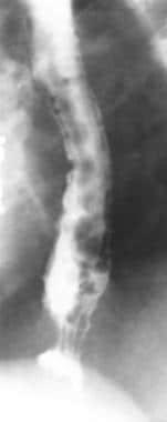
In 32 patients with tumor-forming pancreatitis and 109 patients with pancreatic cancer, the usefulness of percutaneous transhepatic portography (PTP) and selective pancreatic venography (SPV) for differential diagnosis of the two diseases was evaluated. The PTP images were type I in 53.1%, type II in 21.9%, type III in 12.5%, and type IV in 12.5% of the patients with tumor-forming pancreatitis and type I in 20.2%, type II in 23.9%, type III in 37.6%, and type IV in 18.3% of the patients with pancreatic cancer. Advanced images (type III or type IV) were observed in more than half the patients with pancreatic cancer. Mild images classified as type II were observed slightly more frequently in the patients with pancreatic cancer, but the differential diagnosis of the two diseases was difficult in patients showing type II PTP images. SPV findings were primarily hypervascularization (78.1%) and vasodilation (68.8%) in the patients with tumor-forming pancreatitis. Although encasement (smooth encasement) was noted in 31.3%, obstruction was found in only 3.1%. In the patients with pancreatic cancer, obstruction was observed in 85.3%, and encasement (irregular encasement) was noted in 78.9%. However, hypervascularization or vasodilatation was infrequent, and the tumor was characteristically imaged as a hypovascular area. PTP and SPV were considered to be useful for the differential diagnosis of tumor-forming pancreatitis and pancreatic cancer. (+info)Phlebography is a medical imaging technique used to visualize and assess the veins, particularly in the legs. It involves the injection of a contrast agent into the veins, followed by X-ray imaging to capture the flow of the contrast material through the veins. This allows doctors to identify any abnormalities such as blood clots, blockages, or malformations in the venous system.
There are different types of phlebography, including ascending phlebography (where the contrast agent is injected into a foot vein and travels up the leg) and descending phlebography (where the contrast agent is injected into a vein in the groin or neck and travels down the leg).
Phlebography is an invasive procedure that requires careful preparation and monitoring, and it is typically performed by radiologists or vascular specialists. It has largely been replaced by non-invasive imaging techniques such as ultrasound and CT angiography in many clinical settings.
Thrombophlebitis is a medical condition characterized by the inflammation and clotting of blood in a vein, usually in the legs. The term thrombophlebitis comes from two words: "thrombo" which means blood clot, and "phlebitis" which refers to inflammation of the vein.
The condition can occur in superficial or deep veins. Superficial thrombophlebitis affects the veins just below the skin's surface, while deep vein thrombophlebitis (DVT) occurs in the deeper veins. DVT is a more serious condition as it can lead to complications such as pulmonary embolism if the blood clot breaks off and travels to the lungs.
Symptoms of thrombophlebitis may include redness, warmth, pain, swelling, or discomfort in the affected area. In some cases, there may be visible surface veins that are hard, tender, or ropy to touch. If left untreated, thrombophlebitis can lead to chronic venous insufficiency and other long-term complications. Treatment typically involves medications such as anticoagulants, antiplatelet agents, or thrombolytics, along with compression stockings and other supportive measures.
Venous insufficiency is a medical condition that occurs when the veins, particularly in the legs, have difficulty returning blood back to the heart due to impaired valve function or obstruction in the vein. This results in blood pooling in the veins, leading to symptoms such as varicose veins, swelling, skin changes, and ulcers. Prolonged venous insufficiency can cause chronic pain and affect the quality of life if left untreated.
The femoral vein is the large vein that runs through the thigh and carries oxygen-depleted blood from the lower limbs back to the heart. It is located in the femoral triangle, along with the femoral artery and nerve. The femoral vein begins at the knee as the popliteal vein, which then joins with the deep vein of the thigh to form the femoral vein. As it moves up the leg, it is joined by several other veins, including the great saphenous vein, before it becomes the external iliac vein at the inguinal ligament in the groin.
The popliteal vein is the continuation of the tibial and fibular (or anterior and posterior tibial) veins, forming in the lower leg's back portion or popliteal fossa. It carries blood from the leg towards the heart. The popliteal vein is located deep within the body and is accompanied by the popliteal artery, which supplies oxygenated blood to the lower leg. This venous structure is a crucial part of the venous system in the lower extremities and is often assessed during physical examinations for signs of venous insufficiency or deep vein thrombosis (DVT).
In medical terms, the leg refers to the lower portion of the human body that extends from the knee down to the foot. It includes the thigh (femur), lower leg (tibia and fibula), foot, and ankle. The leg is primarily responsible for supporting the body's weight and enabling movements such as standing, walking, running, and jumping.
The leg contains several important structures, including bones, muscles, tendons, ligaments, blood vessels, nerves, and joints. These structures work together to provide stability, support, and mobility to the lower extremity. Common medical conditions that can affect the leg include fractures, sprains, strains, infections, peripheral artery disease, and neurological disorders.
The iliac veins are a pair of large veins in the human body that carry deoxygenated blood from the lower extremities and the pelvic area back to the heart. They are formed by the union of the common iliac veins, which receive blood from the lower abdomen and legs, at the level of the fifth lumbar vertebra.
The combined iliac vein is called the inferior vena cava, which continues upward to the right atrium of the heart. The iliac veins are located deep within the pelvis, lateral to the corresponding iliac arteries, and are accompanied by the iliac lymphatic vessels.
The left common iliac vein is longer than the right because it must cross the left common iliac artery to join the right common iliac vein. The external and internal iliac veins are the two branches of the common iliac vein, with the external iliac vein carrying blood from the lower limbs and the internal iliac vein carrying blood from the pelvic organs.
It is essential to maintain proper blood flow in the iliac veins to prevent deep vein thrombosis (DVT), a condition that can lead to serious complications such as pulmonary embolism.
Veins are blood vessels that carry deoxygenated blood from the tissues back to the heart. They have a lower pressure than arteries and contain valves to prevent the backflow of blood. Veins have a thin, flexible wall with a larger lumen compared to arteries, allowing them to accommodate more blood volume. The color of veins is often blue or green due to the absorption characteristics of light and the reduced oxygen content in the blood they carry.
 Dinker Belle Rai
Venous translucence
Timofei Moșneaga Republican Clinical Hospital
António Egas Moniz
Ovarian vein syndrome
Blood pool agent
Fibrinogen uptake test
Non-invasive procedure
Venography
Iopamidol
List of MeSH codes (E01)
ICD-9-CM Volume 3
IPG
 Phlebography | JAMA Surgery | JAMA Network
 Esophageal Varices Imaging and Diagnosis: Practice Essentials, Radiography, Computed Tomography
Dinker Belle Rai - Wikipedia
 Thrombotic complications with pacemakers
 PRIME PubMed | The role of surgery in the treatment of acute phlebitis in the lower members
The perfect crime? CCSVI not leaving a trace in MS | Journal of Neurology, Neurosurgery & Psychiatry
The perfect crime? CCSVI not leaving a trace in MS | Journal of Neurology, Neurosurgery & Psychiatry
 IndexCat
 Elephantiasis Treatment in Homeopathy | Homeopathic Medicine for Elephantiasis | Hpathy
 Thrombophlebitis
 The ORANGE is for More Than Juice-DIOSMIN Protects the Veins and Beyond | Ingredient Spotlight
 Regulation of Coagulation in Orthopaedic Surgery to Prevent Deep Vein Thrombosis and Pulmonary Embolism 4 - American College of...
 MeSH Browser
 Risk factors for chronic thromboembolic pulmonary hypertension | European Respiratory Society
 gram-, -gram-, -gram, -grammatic, -grammatical, -grammatically, -gramme, -grammic + - Word Information
 SciELO - Brazil - Gestação e varizes de membros inferiores: prevalência e fatores de risco Gestação e varizes de membros...
 How To Buy Solifenacin Generic Low Price, Order solifenacin us prices
NIOSHTIC-2 Search Results - Full View
 SNOMED CT Browser - FHIR Server FHIR Server
SNOMED CT Browser - FHIR Server FHIR Server
Detection of obstructions in venous blood flow<...
 Klinika Flebologii | leczenie żył | flebolog | Warszawa / Phlebology Clinic
High-resolution susceptibility-weighted imaging of clots in cerebral venous thrombosis<...
 Search results
 1 Vascular surgery clinic in Ensenada| Doctor.Global
 Iliac vein. Medical search
 Instruction for use: Dipyridamole - DR. DOPING
Development of Central Venous Stenosis Upon ICD Implantation in Dialysis Patients: A Non-Negligible Issue
Lower extremity1
- Methods for complete lower extremity phlebography are based on experience with over 600 examinations. (jamanetwork.com)
Venous2
- Phlebography and varicography (phlebography of varicose veins) are contrast-based techniques of venous system imaging. (klinikaflebologii.pl)
- Descending phlebography and varicography, whih we perform, allow for a precise evaluation of reflux and direction of venous blood flow in vessels. (klinikaflebologii.pl)
Venography1
- DVT can be ruled out by duplex sonography, phlebography, contrast enhanced CT venography, or MR venography. (hpathy.com)
Ultrasound2
- Provided there are any doubts following Doppler ultrasound and CT, phlebography is often a conclusive examination. (klinikaflebologii.pl)
- Three main types of vein damage diagnosed by ultrasound explorations and confirmed by cross-sectional imaging and selective phlebography were identified ( Table II ). (phlebolymphology.org)
Diagnosis1
- Phlebography can show filling defects and diverted blood flow and usually confirms the diagnosis. (health-care-clinic.org)
Vein1
- 2. A radiograph of a vein taken during phlebography. (wordinfo.info)
Patients2
- In all these patients phlebography about 19 months later revealed thrombotic complications, while 11 presented with clinical symptoms and signs. (nih.gov)
- Elle a inclus 50 patients soit 54 oreilles opérées pour otospongiose, explorés en préopératoire par une tomodensitométrie (TDM) des rochers et en postopératoire par des audiogrammes. (bvsalud.org)
Venography4
- For angiography throughout the cardiovascular system, including cerebral and peripheral arteriography, coronary arteriography and ventriculography, pediatric angiocardiography, selective visceral arteriography and aortography, peripheral venography (phlebography), and adult and pediatric intravenous excretory urography and intravenous adult and pediatric contrast enhancement of computed tomographic (CECT) head and body imaging. (renalandurologynews.com)
- If a patient has a number of risk factors, ultrasound, magnetic resonance imaging (MRI) or venography/phlebography (x-ray that identifies the veins and blood clots) are used to screen for DVT. (gwhospital.com)
- Adverse Reactions OMNIPAQUE 240 is indicated in adults for contrast enhancement for computed tomographic head imaging and peripheral venography (phlebography). (1dexlogistics.com)
- In addition, they are equipped to perform diagnostic tests such as duplex ultrasound, venography, and phlebography, which can help confirm PCS diagnosis. (newsfeed.com.sg)
Arteriography1
- This is done through Doppler ultrasound, phlebography and arteriography. (venart-surgery.com)
Ultrasound2
- STUDY DESIGN: This is a retrospective review of the patients followed up in the authors' center with diagnosis of NS based on clinical and imaging tests (ultrasound, computed tomography/magnetic resonance imaging, and phlebography). (bvsalud.org)
- For safe diagnosis, an ultrasound test (Duplex) and, where necessary, a phlebography are necessary. (seiter-klinik.com)
Adrenal1
- 2. [Adrenal phlebography]. (nih.gov)
Procedure1
- Also known as a phlebography, the procedure is essentially an x-ray used to gauge the flow of blood through the veins more effectively. (medicalhealthtests.com)
Veins1
- Phlebography (contrast phlebography) of the veins before the operation is often unpleasant and is only required as an exception. (beinklinik.de)
Pulmonary1
- 1- 8 Reasons include the high cost of accurate examinations, the different risk perception with techniques based on contrast media (like pulmonary angiography, 9 phlebography and spiral computed tomographic scan 10, 11 ), and the variability in terms of practical availability and even the performance of qualified people. (bmj.com)
Thrombosis1
- Patients with previouts deep veini thronmbosis-Samples of blood were obtained from 83 patients who had had a definite episode of deep vein thrombosis in the past, proved then or later by phlebography. (docksci.com)
Tests1
- The suspected lesion may be confirmed by various tests such as differential radioactive phosphorus uptake study, differential radioactive phosphorus uptake study, differential oximetry intramedullary pressure determinations, intraosseous phlebography, and biopsy for tissue microscopy and microroentgenography. (cdc.gov)