Phagocytosis
Opsonin Proteins
Macrophages
Neutrophils
Phagosomes
Receptors, IgG
Latex
Zymosan
Receptors, Fc
Macrophage-1 Antigen
Blood Bactericidal Activity
Respiratory Burst
Microspheres
Macrophages, Peritoneal
Monocytes
Receptors, Complement
Pinocytosis
Macrophages, Alveolar
Cells, Cultured
Erythrocytes
Immunoglobulin G
Complement C3b
Phagocyte Bactericidal Dysfunction
Complement System Proteins
Antigens, CD47
Phosphatidylserines
Leukocytes
Macrophage Activation
Actins
Complement C3
Receptors, Complement 3b
Microscopy, Electron
Apoptosis
Mannose-Binding Lectins
Staphylococcus aureus
Mice, Inbred C57BL
Lysosomes
Microglia
Luminescent Measurements
Flow Cytometry
Complement C1q
Cryptococcus
Cryptococcus neoformans
Granulocytes
Receptors, Immunologic
Pigment Epithelium of Eye
Superoxides
Peritoneal Cavity
Signal Transduction
Nitroblue Tetrazolium
Polystyrenes
Mice, Knockout
Escherichia coli
Cell Membrane
Candida albicans
Pulmonary Surfactant-Associated Protein A
Rod Cell Outer Segment
Antigen-Antibody Complex
Microscopy, Electron, Scanning
Lipopolysaccharides
Cytochalasin D
Fluorescein-5-isothiocyanate
rac GTP-Binding Proteins
Immunity, Innate
Complement Activation
Microscopy, Confocal
Mice, Inbred BALB C
Dictyostelium
Neutrophil Activation
Antigens, CD18
Virulence
Receptors, Cell Surface
Entamoeba histolytica
Chemotaxis, Leukocyte
rac1 GTP-Binding Protein
Bacterial Adhesion
Immunoglobulin Fc Fragments
Lectins, C-Type
Receptors, Scavenger
Sheep
Bacterial Capsules
Rabbits
Cytoskeleton
Microscopy, Fluorescence
Rosette Formation
Pseudopodia
Pulmonary Alveoli
Phagocytic acitivity of bovine leukocytes during pregnancy. (1/9754)
The phagocytic competence, measured as the total number of polymorphonuclear leukocytes per mm3 which phagocytosed Staphylococcus aureus, strain 321, in vitro, was determined in eight cows during complete pregnancies. Such leukocytes are referred to as "Active PMN'S". There was a gradual decline in the number of these cells from conception to a minimum between the 16th and 20th weeks of pregnancy, followed by a steady increase to the cessation of lactation when a marked drop occurred, after which there was an increase to a maximun during the second week prepartum. From this maximum there was a rapid decrease to an absolute minimum during the first week after parturition. From the second week postpartum there was a gradual increase to conception. The correlation coefficient (r) of number of active PMN'S with time before conception was -0.474 )p-0.01). There were significant differences (p=0.01) in numbers of active PMNS Among the eight cows. It was found that the cows fell into two groups, one whose members had, overall, significantly more active PMNs (p=0.001) than those in the second group. The between cow differences may have been due to 1) age, since the cows with the highest numbers of circulating active PMNs were younger than those in the other group of 2) the combined stress of pregnancy and lactation, as those cows which were both pregnant and milking had the lowest numbers of active PMNs. (+info)GM-CSF-deficient mice are susceptible to pulmonary group B streptococcal infection. (2/9754)
Granulocyte-macrophage colony-stimulating factor (GM-CSF) gene-targeted mice (GM-/-) cleared group B streptococcus (GBS) from the lungs more slowly than wild-type mice. Expression of GM-CSF in the respiratory epithelium of GM-/- mice improved bacterial clearance to levels greater than that in wild-type GM+/+ mice. Acute aerosolization of GM-CSF to GM+/+ mice significantly enhanced clearance of GBS at 24 hours. GBS infection was associated with increased neutrophilic infiltration in lungs of GM-/- mice, while macrophage infiltrates predominated in wild-type mice, suggesting an abnormality in macrophage clearance of bacteria in the absence of GM-CSF. While phagocytosis of GBS was unaltered, production of superoxide radicals and hydrogen peroxide was markedly deficient in macrophages from GM-/- mice. Lipid peroxidation, assessed by measuring the isoprostane 8-iso-PGF2alpha, was decreased in the lungs of GM-/- mice. GM-CSF plays an important role in GBS clearance in vivo, mediated in part by its role in enhancing superoxide and hydrogen peroxide production and bacterial killing by alveolar macrophages. (+info)Salmonella typhimurium and lipopolysaccharide stimulate extracellularly regulated kinase activation in macrophages by a mechanism involving phosphatidylinositol 3-kinase and phospholipase D as novel intermediates. (3/9754)
Activation of the extracellularly regulated kinase (ERK) pathway is part of the early biochemical events that follow lipopolysaccharide (LPS) treatment of macrophages or their infection by virulent and attenuated Salmonella strains. Phagocytosis as well as the secretion of invasion-associated proteins is dispensable for ERK activation by the pathogen. Furthermore, the pathways used by Salmonella and LPS to stimulate ERK are identical, suggesting that kinase activation might be solely mediated by LPS. Both stimuli activate ERK by a mechanism involving herbimycin-dependent tyrosine kinase(s) and phosphatidylinositol 3-kinase. Phospholipase D activation and stimulation of protein kinase C appear to be intermediates in this novel pathway of MEK/ERK activation. (+info)Treponema denticola outer membrane enhances the phagocytosis of collagen-coated beads by gingival fibroblasts. (4/9754)
Human gingival fibroblasts (HGFs) degrade collagen fibrils in physiological processes by phagocytosis. Since Treponema denticola outer membrane (OM) extract perturbs actin filaments, important structures in phagocytosis, we determined whether the OM affects collagen phagocytosis in vitro by HGFs. Phagocytosis was measured by flow cytometric assessment of internalized collagen-coated fluorescent latex beads. Confluent HGFs pretreated with T. denticola ATCC 35405 OM exhibited an increase in the percentage of collagen phagocytic cells (phagocytosis index [PI]) and in the number of beads per phagocytosing cell (phagocytic capacity [PC]) compared with untreated controls. The enhancement was swift (within 15 min) and was still evident after 1 day. PI and PC of HGFs for bovine serum albumin (BSA)-coated beads were also increased, indicating a global increase in phagocytic processes. These results contrasted those for control OM from Veillonella atypica ATCC 17744, which decreased phagocytosis. The T. denticola OM-induced increase in bead uptake was eliminated by heating the OM and by depolymerization of actin filaments by cytochalasin D treatment of HGFs. Fluid-phase accumulation of lucifer yellow was enhanced in a saturable, concentration-dependent, transient manner by the T. denticola OM. Our findings were not due to HGF detachment or cytotoxicity in response to the T. denticola OM treatment since the HGFs exhibited minimal detachment from the substratum; they did not take up propidium iodide; and there was no change in their size, granularity, or content of sub-G1 DNA. We conclude that a heat-sensitive component(s) in T. denticola OM extract stimulates collagen phagocytosis and other endocytic processes such as nonspecific phagocytosis and pinocytosis by HGFs. (+info)Safety and immunogenicity of a Pseudomonas aeruginosa hybrid outer membrane protein F-I vaccine in human volunteers. (5/9754)
A hybrid protein [Met-Ala-(His)6OprF190-342-OprI21-83] consisting of the mature outer membrane protein I (OprI) and amino acids 190 to 342 of OprF of Pseudomonas aeruginosa was expressed in Escherichia coli and purified by Ni2+ chelate-affinity chromatography. After safety and pyrogenicity evaluations in animals, four groups of eight adult human volunteers were vaccinated intramuscularly three times at 4-week intervals and revaccinated 6 months later with either 500, 100, 50, or 20 microg of OprF-OprI adsorbed onto A1(OH)3. All vaccinations were well tolerated. After the first vaccination, a significant rise of antibody titers against P. aeruginosa OprF and OprI was measured in volunteers receiving the 100- or the 500-microg dose. After the second vaccination, significant antibody titers were measured for all groups. Elevated antibody titers against OprF and OprI could still be measured 6 months after the third vaccination. The capacity of the elicited antibodies to promote complement binding and opsonization could be demonstrated by a C1q-binding assay and by the in vitro opsonophagocytic uptake of P. aeruginosa bacteria. These data support the continued development of an OprF-OprI vaccine for use in humans. (+info)An ultrastructural study of implantation in the golden hamster. II. Trophoblastic invasion and removal of the uterine epithelium. (6/9754)
Sixty six implantation sites from 18 golden hamsters were examined with light and electron microscopy between 4 and 5 1/2 days of pregnancy (post-ovulation). At 4 days some blastocysts began to invade the uterine epithelium, with trophoblastic processes penetrating and engulfing portions of the uterine epithelium. The majority of epithelial cells appeared normal before invasion, although at two implantation sites three or four adjoining epithelial cells were necrotic before penetration by the trophoblast. In general the epithelial cells were degenerating at the time the trophoblast invaded the epithelium. Inclusions, representing portions of the engulfed epithelium, and varying in size and electron density, were present throughout the invading trophoblast cells at 4 1/2 and 5 days of pregnancy. At 5 1/2 days the uterine epithelium had disappeared and the embryo was now almost completely surrounded by blood lacunae. (+info)Role of class B scavenger receptor type I in phagocytosis of apoptotic rat spermatogenic cells by Sertoli cells. (7/9754)
Rat Sertoli cells phagocytose apoptotic spermatogenic cells, which consist mostly of spermatocytes, in primary culture by recognizing phosphatidylserine (PS) exposed on the surface of degenerating spermatogenic cells. We compared the mode of phagocytosis using spermatogenic cells at different stages of spermatogenesis. Spermatogenic cells were separated into several groups based on their ploidy, with purities of 60-90%. When the fractionated spermatogenic cell populations were subjected to a phagocytosis assay, cells with ploidies of 1n, 2n, and 4n were almost equally phagocytosed by Sertoli cells. All the cell populations exposed PS on the cell surface, and phagocytosis of all cell populations was similarly inhibited by the addition of PS-containing liposomes. Class B scavenger receptor type I (SR-BI), a candidate for the PS receptor, was detected in Sertoli cells. Overexpression of the rat SR-BI cDNA increased the PS-mediated phagocytic activity of Sertoli cell-derived cell lines. Moreover, phagocytosis of spermatogenic cells by Sertoli cells was inhibited in the presence of an anti-SR-BI antibody. Finally, the addition of high density lipoprotein, a ligand specific for SR-BI, decreased both phagocytosis of spermatogenic cells and incorporation of PS-containing liposomes by Sertoli cells. In conclusion, SR-BI functions at least partly as a PS receptor, enabling Sertoli cells to recognize and phagocytose apoptotic spermatogenic cells at all stages of differentiation. (+info)Tissue distribution of dextran sulfate sodium (DSS) in the acute phase of murine DSS-induced colitis. (8/9754)
In the present study, we examined histochemically the tissue distribution of dextran sulfate sodium (DSS) in the acute phase of murine colitis induced by administering DSS in the drinking water. DSS was mainly observed in the Kupffer cells of the liver, in the macrophages of the mesenteric lymph node (MLN) and in the lamina propria of the large intestine after administration of DSS. We followed the time course of DSS distribution and found that DSS, which was considered as a large and negatively charged molecule that can not easily cross membranes, was distributed in the liver, the MLN, and the large intestine 1 day after the start of administration of DSS. (+info)Phagocytosis is the process by which certain cells in the body, known as phagocytes, engulf and destroy foreign particles, bacteria, or dead cells. This mechanism plays a crucial role in the immune system's response to infection and inflammation. Phagocytes, such as neutrophils, monocytes, and macrophages, have receptors on their surface that recognize and bind to specific molecules (known as antigens) on the target particles or microorganisms.
Once attached, the phagocyte extends pseudopodia (cell extensions) around the particle, forming a vesicle called a phagosome that completely encloses it. The phagosome then fuses with a lysosome, an intracellular organelle containing digestive enzymes and other chemicals. This fusion results in the formation of a phagolysosome, where the engulfed particle is broken down by the action of these enzymes, neutralizing its harmful effects and allowing for the removal of cellular debris or pathogens.
Phagocytosis not only serves as a crucial defense mechanism against infections but also contributes to tissue homeostasis by removing dead cells and debris.
Opsonins are proteins found in the blood that help enhance the immune system's response to foreign substances, such as bacteria and viruses. They do this by coating the surface of these pathogens, making them more recognizable to immune cells like neutrophils and macrophages. This process, known as opsonization, facilitates the phagocytosis (engulfing and destroying) of the pathogen by these immune cells.
There are two main types of opsonins:
1. IgG antibodies: These are a type of antibody produced by the immune system in response to an infection. They bind to specific antigens on the surface of the pathogen, marking them for destruction by phagocytic cells.
2. Complement proteins: The complement system is a group of proteins that work together to help eliminate pathogens. When activated, the complement system can produce various proteins that act as opsonins, including C3b and C4b. These proteins bind to the surface of the pathogen, making it easier for phagocytic cells to recognize and destroy them.
In summary, opsonin proteins are crucial components of the immune system's response to infections, helping to mark foreign substances for destruction by immune cells like neutrophils and macrophages.
Macrophages are a type of white blood cell that are an essential part of the immune system. They are large, specialized cells that engulf and destroy foreign substances, such as bacteria, viruses, parasites, and fungi, as well as damaged or dead cells. Macrophages are found throughout the body, including in the bloodstream, lymph nodes, spleen, liver, lungs, and connective tissues. They play a critical role in inflammation, immune response, and tissue repair and remodeling.
Macrophages originate from monocytes, which are a type of white blood cell produced in the bone marrow. When monocytes enter the tissues, they differentiate into macrophages, which have a larger size and more specialized functions than monocytes. Macrophages can change their shape and move through tissues to reach sites of infection or injury. They also produce cytokines, chemokines, and other signaling molecules that help coordinate the immune response and recruit other immune cells to the site of infection or injury.
Macrophages have a variety of surface receptors that allow them to recognize and respond to different types of foreign substances and signals from other cells. They can engulf and digest foreign particles, bacteria, and viruses through a process called phagocytosis. Macrophages also play a role in presenting antigens to T cells, which are another type of immune cell that helps coordinate the immune response.
Overall, macrophages are crucial for maintaining tissue homeostasis, defending against infection, and promoting wound healing and tissue repair. Dysregulation of macrophage function has been implicated in a variety of diseases, including cancer, autoimmune disorders, and chronic inflammatory conditions.
Neutrophils are a type of white blood cell that are part of the immune system's response to infection. They are produced in the bone marrow and released into the bloodstream where they circulate and are able to move quickly to sites of infection or inflammation in the body. Neutrophils are capable of engulfing and destroying bacteria, viruses, and other foreign substances through a process called phagocytosis. They are also involved in the release of inflammatory mediators, which can contribute to tissue damage in some cases. Neutrophils are characterized by the presence of granules in their cytoplasm, which contain enzymes and other proteins that help them carry out their immune functions.
A phagosome is a type of membrane-bound organelle that forms around a particle or microorganism following its engulfment by a cell, through the process of phagocytosis. This results in the formation of a vesicle containing the ingested material, which then fuses with another organelle called a lysosome to form a phago-lysosome. The lysosome contains enzymes that digest and break down the contents of the phagosome, allowing the cell to neutralize and dispose of potentially harmful substances or pathogens.
In summary, phagosomes are important organelles involved in the immune response, helping to protect the body against infection and disease.
IgG receptors, also known as Fcγ receptors (Fc gamma receptors), are specialized protein molecules found on the surface of various immune cells, such as neutrophils, monocytes, macrophages, and some lymphocytes. These receptors recognize and bind to the Fc region of IgG antibodies, one of the five classes of immunoglobulins in the human body.
IgG receptors play a crucial role in immune responses by mediating different effector functions, including:
1. Antibody-dependent cellular cytotoxicity (ADCC): IgG receptors on natural killer (NK) cells and other immune cells bind to IgG antibodies coated on the surface of virus-infected or cancer cells, leading to their destruction.
2. Phagocytosis: When IgG antibodies tag pathogens or foreign particles, phagocytes like neutrophils and macrophages recognize and bind to these immune complexes via IgG receptors, facilitating the engulfment and removal of the targeted particles.
3. Antigen presentation: IgG receptors on antigen-presenting cells (APCs) can internalize immune complexes, process the antigens, and present them to T cells, thereby initiating adaptive immune responses.
4. Inflammatory response regulation: IgG receptors can modulate inflammation by activating or inhibiting downstream signaling pathways in immune cells, depending on the specific type of Fcγ receptor and its activation state.
There are several types of IgG receptors (FcγRI, FcγRII, FcγRIII, and FcγRIV) with varying affinities for different subclasses of IgG antibodies (IgG1, IgG2, IgG3, and IgG4). The distinct functions and expression patterns of these receptors contribute to the complexity and fine-tuning of immune responses in the human body.
In a medical context, "latex" refers to the natural rubber milk-like substance that is tapped from the incisions made in the bark of the rubber tree (Hevea brasiliensis). This sap is then processed to create various products such as gloves, catheters, and balloons. It's important to note that some people may have a latex allergy, which can cause mild to severe reactions when they come into contact with latex products.
Zymosan is a type of substance that is derived from the cell walls of yeast and some types of fungi. It's often used in laboratory research as an agent to stimulate inflammation, because it can activate certain immune cells (such as neutrophils) and cause them to release pro-inflammatory chemicals.
In medical terms, Zymosan is sometimes used as a tool for studying the immune system and inflammation in experimental settings. It's important to note that Zymosan itself is not a medical condition or disease, but rather a research reagent with potential applications in understanding human health and disease.
Phagocytes are a type of white blood cell in the immune system that engulf and destroy foreign particles, microbes, and cellular debris. They play a crucial role in the body's defense against infection and tissue damage. There are several types of phagocytes, including neutrophils, monocytes, macrophages, and dendritic cells. These cells have receptors that recognize and bind to specific molecules on the surface of foreign particles or microbes, allowing them to engulf and digest the invaders. Phagocytosis is an important mechanism for maintaining tissue homeostasis and preventing the spread of infection.
Fc receptors (FcRs) are specialized proteins found on the surface of various immune cells, including neutrophils, monocytes, macrophages, eosinophils, basophils, mast cells, and B lymphocytes. They play a crucial role in the immune response by recognizing and binding to the Fc region of antibodies (IgG, IgA, and IgE) after they have interacted with their specific antigens.
FcRs can be classified into several types based on the class of antibody they bind:
1. FcγRs - bind to the Fc region of IgG antibodies
2. FcαRs - bind to the Fc region of IgA antibodies
3. FcεRs - bind to the Fc region of IgE antibodies
The binding of antibodies to Fc receptors triggers various cellular responses, such as phagocytosis, degranulation, and antibody-dependent cellular cytotoxicity (ADCC), which contribute to the elimination of pathogens, immune complexes, and other foreign substances. Dysregulation of Fc receptor function has been implicated in several diseases, including autoimmune disorders and allergies.
The Macrophage-1 Antigen (also known as Macrophage Antigen-1 or CD14) is a glycoprotein found on the surface of various cells, including monocytes, macrophages, and some dendritic cells. It functions as a receptor for complexes formed by lipopolysaccharides (LPS) and LPS-binding protein (LBP), which are involved in the immune response to gram-negative bacteria. CD14 plays a crucial role in activating immune cells and initiating the release of proinflammatory cytokines upon recognizing bacterial components.
In summary, Macrophage-1 Antigen is a cell surface receptor that contributes to the recognition and response against gram-negative bacteria by interacting with LPS-LBP complexes.
Blood bactericidal activity refers to the ability of an individual's blood to kill or inhibit the growth of bacteria. This is an important aspect of the body's immune system, as it helps to prevent infection and maintain overall health. The bactericidal activity of blood can be influenced by various factors, including the presence of antibodies, white blood cells (such as neutrophils), and complement proteins.
In medical terms, the term "bactericidal" specifically refers to an agent or substance that is capable of killing bacteria. Therefore, when we talk about blood bactericidal activity, we are referring to the collective ability of various components in the blood to kill or inhibit the growth of bacteria. This is often measured in laboratory tests as a way to assess a person's immune function and their susceptibility to infection.
It's worth noting that not all substances in the blood are bactericidal; some may simply inhibit the growth of bacteria without killing them. These substances are referred to as bacteriostatic. Both bactericidal and bacteriostatic agents play important roles in maintaining the body's defense against infection.
Respiratory burst is a term used in the field of biology, particularly in the context of immunology and cellular processes. It does not have a direct application to clinical medicine, but it is important for understanding certain physiological and pathophysiological mechanisms. Here's a definition of respiratory burst:
Respiratory burst is a rapid increase in oxygen consumption by phagocytic cells (like neutrophils, monocytes, and macrophages) following their activation in response to various stimuli, such as pathogens or inflammatory molecules. This process is part of the innate immune response and serves to eliminate invading microorganisms.
The respiratory burst involves the activation of NADPH oxidase, an enzyme complex present in the membrane of phagosomes (the compartment where pathogens are engulfed). Upon activation, NADPH oxidase catalyzes the reduction of oxygen to superoxide radicals, which then dismutate to form hydrogen peroxide. These reactive oxygen species (ROS) can directly kill or damage microorganisms and also serve as signaling molecules for other immune cells.
While respiratory burst is a crucial part of the immune response, excessive or dysregulated ROS production can contribute to tissue damage and chronic inflammation, which have implications in various pathological conditions, such as atherosclerosis, neurodegenerative diseases, and cancer.
Microspheres are tiny, spherical particles that range in size from 1 to 1000 micrometers in diameter. They are made of biocompatible and biodegradable materials such as polymers, glass, or ceramics. In medical terms, microspheres have various applications, including drug delivery systems, medical imaging, and tissue engineering.
In drug delivery, microspheres can be used to encapsulate drugs and release them slowly over time, improving the efficacy of the treatment while reducing side effects. They can also be used for targeted drug delivery, where the microspheres are designed to accumulate in specific tissues or organs.
In medical imaging, microspheres can be labeled with radioactive isotopes or magnetic materials and used as contrast agents to enhance the visibility of tissues or organs during imaging procedures such as X-ray, CT, MRI, or PET scans.
In tissue engineering, microspheres can serve as a scaffold for cell growth and differentiation, promoting the regeneration of damaged tissues or organs. Overall, microspheres have great potential in various medical applications due to their unique properties and versatility.
Peritoneal macrophages are a type of immune cell that are present in the peritoneal cavity, which is the space within the abdomen that contains the liver, spleen, stomach, and intestines. These macrophages play a crucial role in the body's defense against infection and injury by engulfing and destroying foreign substances such as bacteria, viruses, and other microorganisms.
Macrophages are large phagocytic cells that originate from monocytes, which are a type of white blood cell produced in the bone marrow. When monocytes enter tissue, they can differentiate into macrophages, which have a variety of functions depending on their location and activation state.
Peritoneal macrophages are involved in various physiological processes, including the regulation of inflammation, tissue repair, and the breakdown of foreign substances. They also play a role in the development and progression of certain diseases, such as cancer and autoimmune disorders.
These macrophages can be collected from animals or humans for research purposes by injecting a solution into the peritoneal cavity and then withdrawing the fluid, which contains the macrophages. These cells can then be studied in vitro to better understand their functions and potential therapeutic targets.
Monocytes are a type of white blood cell that are part of the immune system. They are large cells with a round or oval shape and a nucleus that is typically indented or horseshoe-shaped. Monocytes are produced in the bone marrow and then circulate in the bloodstream, where they can differentiate into other types of immune cells such as macrophages and dendritic cells.
Monocytes play an important role in the body's defense against infection and tissue damage. They are able to engulf and digest foreign particles, microorganisms, and dead or damaged cells, which helps to clear them from the body. Monocytes also produce cytokines, which are signaling molecules that help to coordinate the immune response.
Elevated levels of monocytes in the bloodstream can be a sign of an ongoing infection, inflammation, or other medical conditions such as cancer or autoimmune disorders.
Complement receptors are proteins found on the surface of various cells in the human body, including immune cells and some non-immune cells. They play a crucial role in the complement system, which is a part of the innate immune response that helps to eliminate pathogens and damaged cells from the body. Complement receptors bind to complement proteins or fragments that are generated during the activation of the complement system. This binding triggers various intracellular signaling events that can lead to diverse cellular responses, such as phagocytosis, inflammation, and immune regulation.
There are several types of complement receptors, including:
1. CR1 (CD35): A receptor found on erythrocytes, B cells, neutrophils, monocytes, macrophages, and glomerular podocytes. It functions in the clearance of immune complexes and regulates complement activation.
2. CR2 (CD21): Expressed mainly on B cells and follicular dendritic cells. It facilitates antigen presentation, B-cell activation, and immune regulation.
3. CR3 (CD11b/CD18, Mac-1): Present on neutrophils, monocytes, macrophages, and some T cells. It mediates cell adhesion, phagocytosis, and intracellular signaling.
4. CR4 (CD11c/CD18, p150,95): Expressed on neutrophils, monocytes, macrophages, and dendritic cells. It is involved in cell adhesion, phagocytosis, and intracellular signaling.
5. C5aR (CD88): Found on various immune cells, including neutrophils, monocytes, macrophages, mast cells, eosinophils, and dendritic cells. It binds to the complement protein C5a and mediates chemotaxis, degranulation, and inflammation.
6. C5L2 (GPR77): Present on various cell types, including immune cells. Its function is not well understood but may involve regulating C5a-mediated responses or acting as a receptor for other ligands.
These receptors play crucial roles in the immune response and inflammation by mediating various functions such as chemotaxis, phagocytosis, cell adhesion, and intracellular signaling. Dysregulation of these receptors has been implicated in several diseases, including autoimmune disorders, infections, and cancer.
Pinocytosis is a type of cellular process involving the ingestion and absorption of extracellular fluid and dissolved substances into a cell. It is a form of endocytosis, where the cell membrane surrounds and engulfs the extracellular fluid to form a vesicle containing the fluid and its contents within the cell cytoplasm.
In pinocytosis, the cell membrane invaginates and forms small vesicles (pinocytotic vesicles) that contain extracellular fluid and dissolved substances. These vesicles then detach from the cell membrane and move into the cytoplasm, where they fuse with endosomes or lysosomes to break down and digest the contents of the vesicle.
Pinocytosis is a non-selective process that allows cells to take up small amounts of extracellular fluid and dissolved substances from their environment. It plays an important role in various physiological processes, including nutrient uptake, cell signaling, and the regulation of extracellular matrix composition.
Alveolar macrophages are a type of macrophage (a large phagocytic cell) that are found in the alveoli of the lungs. They play a crucial role in the immune defense system of the lungs by engulfing and destroying any foreign particles, such as dust, microorganisms, and pathogens, that enter the lungs through the process of inhalation. Alveolar macrophages also produce cytokines, which are signaling molecules that help to coordinate the immune response. They are important for maintaining the health and function of the lungs by removing debris and preventing infection.
"Cells, cultured" is a medical term that refers to cells that have been removed from an organism and grown in controlled laboratory conditions outside of the body. This process is called cell culture and it allows scientists to study cells in a more controlled and accessible environment than they would have inside the body. Cultured cells can be derived from a variety of sources, including tissues, organs, or fluids from humans, animals, or cell lines that have been previously established in the laboratory.
Cell culture involves several steps, including isolation of the cells from the tissue, purification and characterization of the cells, and maintenance of the cells in appropriate growth conditions. The cells are typically grown in specialized media that contain nutrients, growth factors, and other components necessary for their survival and proliferation. Cultured cells can be used for a variety of purposes, including basic research, drug development and testing, and production of biological products such as vaccines and gene therapies.
It is important to note that cultured cells may behave differently than they do in the body, and results obtained from cell culture studies may not always translate directly to human physiology or disease. Therefore, it is essential to validate findings from cell culture experiments using additional models and ultimately in clinical trials involving human subjects.
Erythrocytes, also known as red blood cells (RBCs), are the most common type of blood cell in circulating blood in mammals. They are responsible for transporting oxygen from the lungs to the body's tissues and carbon dioxide from the tissues to the lungs.
Erythrocytes are formed in the bone marrow and have a biconcave shape, which allows them to fold and bend easily as they pass through narrow blood vessels. They do not have a nucleus or mitochondria, which makes them more flexible but also limits their ability to reproduce or repair themselves.
In humans, erythrocytes are typically disc-shaped and measure about 7 micrometers in diameter. They contain the protein hemoglobin, which binds to oxygen and gives blood its red color. The lifespan of an erythrocyte is approximately 120 days, after which it is broken down in the liver and spleen.
Abnormalities in erythrocyte count or function can lead to various medical conditions, such as anemia, polycythemia, and sickle cell disease.
Immunoglobulin G (IgG) is a type of antibody, which is a protective protein produced by the immune system in response to foreign substances like bacteria or viruses. IgG is the most abundant type of antibody in human blood, making up about 75-80% of all antibodies. It is found in all body fluids and plays a crucial role in fighting infections caused by bacteria, viruses, and toxins.
IgG has several important functions:
1. Neutralization: IgG can bind to the surface of bacteria or viruses, preventing them from attaching to and infecting human cells.
2. Opsonization: IgG coats the surface of pathogens, making them more recognizable and easier for immune cells like neutrophils and macrophages to phagocytose (engulf and destroy) them.
3. Complement activation: IgG can activate the complement system, a group of proteins that work together to help eliminate pathogens from the body. Activation of the complement system leads to the formation of the membrane attack complex, which creates holes in the cell membranes of bacteria, leading to their lysis (destruction).
4. Antibody-dependent cellular cytotoxicity (ADCC): IgG can bind to immune cells like natural killer (NK) cells and trigger them to release substances that cause target cells (such as virus-infected or cancerous cells) to undergo apoptosis (programmed cell death).
5. Immune complex formation: IgG can form immune complexes with antigens, which can then be removed from the body through various mechanisms, such as phagocytosis by immune cells or excretion in urine.
IgG is a critical component of adaptive immunity and provides long-lasting protection against reinfection with many pathogens. It has four subclasses (IgG1, IgG2, IgG3, and IgG4) that differ in their structure, function, and distribution in the body.
Hemocytes are specialized cells found in the open circulatory system of invertebrates, including insects, crustaceans, and mollusks. They play crucial roles in the immune response and defense mechanisms of these organisms. Hemocytes can be categorized into several types based on their functions and morphologies, such as phagocytic cells, encapsulating cells, and clotting cells. These cells are responsible for various immunological activities, including recognition and removal of foreign particles, pathogens, and debris; production of immune effector molecules; and contribution to the formation of blood clots to prevent excessive bleeding. In some invertebrates, hemocytes also participate in wound healing, tissue repair, and other physiological processes.
A cell line is a culture of cells that are grown in a laboratory for use in research. These cells are usually taken from a single cell or group of cells, and they are able to divide and grow continuously in the lab. Cell lines can come from many different sources, including animals, plants, and humans. They are often used in scientific research to study cellular processes, disease mechanisms, and to test new drugs or treatments. Some common types of human cell lines include HeLa cells (which come from a cancer patient named Henrietta Lacks), HEK293 cells (which come from embryonic kidney cells), and HUVEC cells (which come from umbilical vein endothelial cells). It is important to note that cell lines are not the same as primary cells, which are cells that are taken directly from a living organism and have not been grown in the lab.
Complement C3b is a protein fragment that plays a crucial role in the complement system, which is a part of the immune system that helps to clear pathogens and damaged cells from the body. C3b is generated during the activation of the complement system, particularly via the classical, lectin, and alternative pathways.
Once formed, C3b can bind covalently to the surface of microbes or other target particles, marking them for destruction by other components of the immune system. Additionally, C3b can interact with other proteins in the complement system to generate the membrane attack complex (MAC), which forms pores in the membranes of targeted cells, leading to their lysis and removal.
In summary, Complement C3b is a vital protein fragment involved in the recognition, tagging, and elimination of pathogens and damaged cells during the immune response.
Phagocyte bactericidal dysfunction refers to an impairment in the ability of certain types of immune cells, called phagocytes, to kill bacteria. Phagocytes, which include cells such as neutrophils and macrophages, play a critical role in the body's defense against infection by engulfing and destroying foreign invaders like bacteria.
Bactericidal dysfunction occurs when there is a problem with one or more of the bacterial killing mechanisms within the phagocyte. This can be due to genetic defects, acquired conditions, or medication side effects. As a result, the phagocytes are not able to effectively eliminate bacteria, leading to an increased risk of recurrent or chronic infections.
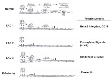
Examples of conditions associated with phagocyte bactericidal dysfunction include chronic granulomatous disease (CGD), leukocyte adhesion deficiency (LAD), and myeloperoxidase deficiency. These conditions are typically rare, but can have serious consequences if not properly diagnosed and managed.
The complement system is a group of proteins found in the blood and on the surface of cells that when activated, work together to help eliminate pathogens such as bacteria, viruses, and fungi from the body. The proteins are normally inactive in the bloodstream. When they encounter an invading microorganism or foreign substance, a series of reactions take place leading to the activation of the complement system. Activation results in the production of effector molecules that can punch holes in the cell membranes of pathogens, recruit and activate immune cells, and help remove debris and dead cells from the body.
There are three main pathways that can lead to complement activation: the classical pathway, the lectin pathway, and the alternative pathway. Each pathway involves a series of proteins that work together in a cascade-like manner to amplify the response and generate effector molecules. The three main effector molecules produced by the complement system are C3b, C4b, and C5b. These molecules can bind to the surface of pathogens, marking them for destruction by other immune cells.
Complement proteins also play a role in the regulation of the immune response. They help to prevent excessive activation of the complement system, which could damage host tissues. Dysregulation of the complement system has been implicated in a number of diseases, including autoimmune disorders and inflammatory conditions.
In summary, Complement System Proteins are a group of proteins that play a crucial role in the immune response by helping to eliminate pathogens and regulate the immune response. They can be activated through three different pathways, leading to the production of effector molecules that mark pathogens for destruction. Dysregulation of the complement system has been linked to various diseases.
CD47 is a cell surface protein that acts as a type of "marker" on certain cells in the body, including red blood cells and immune cells. It is sometimes referred to as an "antigen" because it can be recognized by other proteins called receptors, which can trigger various responses in the body.
CD47 plays a role in regulating the immune response and protecting healthy cells from being attacked by the immune system. It does this by binding to a receptor called SIRPα on certain immune cells, such as macrophages and dendritic cells. This interaction sends a "don't eat me" signal that helps prevent the immune cells from attacking and destroying the CD47-expressing cells.
CD47 has been studied in the context of various diseases, including cancer, because some cancer cells may overexpress CD47 as a way to evade the immune system. Inhibiting the interaction between CD47 and SIRPα has emerged as a potential strategy for enhancing the body's ability to fight off cancer cells.
Phosphatidylserines are a type of phospholipids that are essential components of the cell membrane, particularly in the brain. They play a crucial role in maintaining the fluidity and permeability of the cell membrane, and are involved in various cellular processes such as signal transduction, protein anchorage, and apoptosis (programmed cell death). Phosphatidylserines contain a polar head group made up of serine amino acids and two non-polar fatty acid tails. They are abundant in the inner layer of the cell membrane but can be externalized to the outer layer during apoptosis, where they serve as signals for recognition and removal of dying cells by the immune system. Phosphatidylserines have been studied for their potential benefits in various medical conditions, including cognitive decline, Alzheimer's disease, and depression.
Leukocytes, also known as white blood cells (WBCs), are a crucial component of the human immune system. They are responsible for protecting the body against infections and foreign substances. Leukocytes are produced in the bone marrow and circulate throughout the body in the bloodstream and lymphatic system.
There are several types of leukocytes, including:
1. Neutrophils - These are the most abundant type of leukocyte and are primarily responsible for fighting bacterial infections. They contain enzymes that can destroy bacteria.
2. Lymphocytes - These are responsible for producing antibodies and destroying virus-infected cells, as well as cancer cells. There are two main types of lymphocytes: B-lymphocytes and T-lymphocytes.
3. Monocytes - These are the largest type of leukocyte and help to break down and remove dead or damaged tissues, as well as microorganisms.
4. Eosinophils - These play a role in fighting parasitic infections and are also involved in allergic reactions and inflammation.
5. Basophils - These release histamine and other chemicals that cause inflammation in response to allergens or irritants.
An abnormal increase or decrease in the number of leukocytes can indicate an underlying medical condition, such as an infection, inflammation, or a blood disorder.
Macrophage activation is a process in which these immune cells become increasingly active and responsive to various stimuli, such as pathogens or inflammatory signals. This activation triggers a series of changes within the macrophages, allowing them to perform important functions like phagocytosis (ingesting and destroying foreign particles or microorganisms), antigen presentation (presenting microbial fragments to T-cells to stimulate an immune response), and production of cytokines and chemokines (signaling molecules that help coordinate the immune response).
There are two main types of macrophage activation: classical (or M1) activation and alternative (or M2) activation. Classical activation is typically induced by interferon-gamma (IFN-γ) and lipopolysaccharide (LPS), leading to a proinflammatory response, enhanced microbicidal activity, and the production of reactive oxygen and nitrogen species. Alternative activation, on the other hand, is triggered by cytokines like interleukin-4 (IL-4) and IL-13, resulting in an anti-inflammatory response, tissue repair, and the promotion of wound healing.
It's important to note that macrophage activation plays a crucial role in various physiological and pathological processes, including immune defense, inflammation, tissue remodeling, and even cancer progression. Dysregulation of macrophage activation has been implicated in several diseases, such as autoimmune disorders, chronic infections, and cancer.
Actin is a type of protein that forms part of the contractile apparatus in muscle cells, and is also found in various other cell types. It is a globular protein that polymerizes to form long filaments, which are important for many cellular processes such as cell division, cell motility, and the maintenance of cell shape. In muscle cells, actin filaments interact with another type of protein called myosin to enable muscle contraction. Actins can be further divided into different subtypes, including alpha-actin, beta-actin, and gamma-actin, which have distinct functions and expression patterns in the body.
Complement C3 is a protein that plays a central role in the complement system, which is a part of the immune system that helps to clear pathogens and damaged cells from the body. Complement C3 can be activated through three different pathways: the classical pathway, the lectin pathway, and the alternative pathway. Once activated, it breaks down into two fragments, C3a and C3b.
C3a is an anaphylatoxin that helps to recruit immune cells to the site of infection or injury, while C3b plays a role in opsonization, which is the process of coating pathogens or damaged cells with proteins to make them more recognizable to the immune system. Additionally, C3b can also activate the membrane attack complex (MAC), which forms a pore in the membrane of target cells leading to their lysis or destruction.
In summary, Complement C3 is an important protein in the complement system that helps to identify and eliminate pathogens and damaged cells from the body through various mechanisms.
Complement receptor 3b (CR3b or CD11b/CD18) is not a medical definition itself, but I can provide you with the relevant information regarding this term.
Complement receptor 3 (CR3) is a heterodimeric receptor consisting of two subunits, CD11b (also known as Mac-1 or CR3 alpha) and CD18 (also known as beta2 integrin). There are two forms of the CD11b/CD18 heterodimer: CR3a (CD11b/CD18) and CR3b (CD11b/CD18'). The difference between these two forms lies in the conformation of the CD11b subunit.
Complement receptor 3b (CR3b or CD11b/CD18') is a less common form of the CR3 receptor, which is primarily expressed on myeloid cells such as monocytes, macrophages, and neutrophils. CR3b has a higher affinity for complement component C3b and its fragments iC3b and C3dg compared to CR3a.
CR3b plays a role in various immune functions, including:
1. Phagocytosis: Binding of C3b or its fragments to CR3b facilitates the recognition and uptake of opsonized pathogens by phagocytes.
2. Adhesion: The integrin component of CR3b mediates cell-cell and cell-matrix interactions, contributing to leukocyte migration and recruitment to sites of inflammation or infection.
3. Intracellular signaling: Activation of CR3b can lead to intracellular signaling events that modulate immune responses, such as the release of pro-inflammatory cytokines and reactive oxygen species.
In summary, Complement receptor 3b (CR3b or CD11b/CD18') is a less common form of CR3 primarily expressed on myeloid cells that binds complement component C3b and its fragments with high affinity, mediating phagocytosis, adhesion, and intracellular signaling.
Electron microscopy (EM) is a type of microscopy that uses a beam of electrons to create an image of the sample being examined, resulting in much higher magnification and resolution than light microscopy. There are several types of electron microscopy, including transmission electron microscopy (TEM), scanning electron microscopy (SEM), and reflection electron microscopy (REM).
In TEM, a beam of electrons is transmitted through a thin slice of the sample, and the electrons that pass through the sample are focused to form an image. This technique can provide detailed information about the internal structure of cells, viruses, and other biological specimens, as well as the composition and structure of materials at the atomic level.
In SEM, a beam of electrons is scanned across the surface of the sample, and the electrons that are scattered back from the surface are detected to create an image. This technique can provide information about the topography and composition of surfaces, as well as the structure of materials at the microscopic level.
REM is a variation of SEM in which the beam of electrons is reflected off the surface of the sample, rather than scattered back from it. This technique can provide information about the surface chemistry and composition of materials.
Electron microscopy has a wide range of applications in biology, medicine, and materials science, including the study of cellular structure and function, disease diagnosis, and the development of new materials and technologies.
Apoptosis is a programmed and controlled cell death process that occurs in multicellular organisms. It is a natural process that helps maintain tissue homeostasis by eliminating damaged, infected, or unwanted cells. During apoptosis, the cell undergoes a series of morphological changes, including cell shrinkage, chromatin condensation, and fragmentation into membrane-bound vesicles called apoptotic bodies. These bodies are then recognized and engulfed by neighboring cells or phagocytic cells, preventing an inflammatory response. Apoptosis is regulated by a complex network of intracellular signaling pathways that involve proteins such as caspases, Bcl-2 family members, and inhibitors of apoptosis (IAPs).
Mannose-binding lectins (MBLs) are a group of proteins that belong to the collectin family and play a crucial role in the innate immune system. They are primarily produced by the liver and secreted into the bloodstream. MBLs have a specific affinity for mannose sugar residues found on the surface of various microorganisms, including bacteria, viruses, fungi, and parasites.
The primary function of MBLs is to recognize and bind to these mannose-rich structures, which triggers the complement system's activation through the lectin pathway. This process leads to the destruction of the microorganism by opsonization (coating the microbe to enhance phagocytosis) or direct lysis. MBLs also have the ability to neutralize certain viruses and inhibit the replication of others, further contributing to their antimicrobial activity.
Deficiencies in MBL levels or function have been associated with an increased susceptibility to infections, particularly in children and older adults. However, the clinical significance of MBL deficiency remains a subject of ongoing research.
Staphylococcus aureus is a type of gram-positive, round (coccal) bacterium that is commonly found on the skin and mucous membranes of warm-blooded animals and humans. It is a facultative anaerobe, which means it can grow in the presence or absence of oxygen.
Staphylococcus aureus is known to cause a wide range of infections, from mild skin infections such as pimples, impetigo, and furuncles (boils) to more severe and potentially life-threatening infections such as pneumonia, endocarditis, osteomyelitis, and sepsis. It can also cause food poisoning and toxic shock syndrome.
The bacterium is often resistant to multiple antibiotics, including methicillin, which has led to the emergence of methicillin-resistant Staphylococcus aureus (MRSA) strains that are difficult to treat. Proper hand hygiene and infection control practices are critical in preventing the spread of Staphylococcus aureus and MRSA.
C57BL/6 (C57 Black 6) is an inbred strain of laboratory mouse that is widely used in biomedical research. The term "inbred" refers to a strain of animals where matings have been carried out between siblings or other closely related individuals for many generations, resulting in a population that is highly homozygous at most genetic loci.
The C57BL/6 strain was established in 1920 by crossing a female mouse from the dilute brown (DBA) strain with a male mouse from the black strain. The resulting offspring were then interbred for many generations to create the inbred C57BL/6 strain.
C57BL/6 mice are known for their robust health, longevity, and ease of handling, making them a popular choice for researchers. They have been used in a wide range of biomedical research areas, including studies of cancer, immunology, neuroscience, cardiovascular disease, and metabolism.
One of the most notable features of the C57BL/6 strain is its sensitivity to certain genetic modifications, such as the introduction of mutations that lead to obesity or impaired glucose tolerance. This has made it a valuable tool for studying the genetic basis of complex diseases and traits.
Overall, the C57BL/6 inbred mouse strain is an important model organism in biomedical research, providing a valuable resource for understanding the genetic and molecular mechanisms underlying human health and disease.
Lysosomes are membrane-bound organelles found in the cytoplasm of eukaryotic cells. They are responsible for breaking down and recycling various materials, such as waste products, foreign substances, and damaged cellular components, through a process called autophagy or phagocytosis. Lysosomes contain hydrolytic enzymes that can break down biomolecules like proteins, nucleic acids, lipids, and carbohydrates into their basic building blocks, which can then be reused by the cell. They play a crucial role in maintaining cellular homeostasis and are often referred to as the "garbage disposal system" of the cell.
Microglia are a type of specialized immune cell found in the brain and spinal cord. They are part of the glial family, which provide support and protection to the neurons in the central nervous system (CNS). Microglia account for about 10-15% of all cells found in the CNS.
The primary role of microglia is to constantly survey their environment and eliminate any potentially harmful agents, such as pathogens, dead cells, or protein aggregates. They do this through a process called phagocytosis, where they engulf and digest foreign particles or cellular debris. In addition to their phagocytic function, microglia also release various cytokines, chemokines, and growth factors that help regulate the immune response in the CNS, promote neuronal survival, and contribute to synaptic plasticity.
Microglia can exist in different activation states depending on the nature of the stimuli they encounter. In a resting state, microglia have a small cell body with numerous branches that are constantly monitoring their surroundings. When activated by an injury, infection, or neurodegenerative process, microglia change their morphology and phenotype, retracting their processes and adopting an amoeboid shape to migrate towards the site of damage or inflammation. Based on the type of activation, microglia can release both pro-inflammatory and anti-inflammatory factors that contribute to either neuroprotection or neurotoxicity.
Dysregulation of microglial function has been implicated in several neurological disorders, including Alzheimer's disease, Parkinson's disease, multiple sclerosis, and Amyotrophic Lateral Sclerosis (ALS). Therefore, understanding the role of microglia in health and disease is crucial for developing novel therapeutic strategies to treat these conditions.
Luminescent measurements refer to the quantitative assessment of the emission of light from a substance that has been excited, typically through some form of energy input such as electrical energy or radiation. In the context of medical diagnostics and research, luminescent measurements can be used in various applications, including bioluminescence imaging, which is used to study biological processes at the cellular and molecular level.
Bioluminescence occurs when a chemical reaction produces light within a living organism, often through the action of enzymes such as luciferase. By introducing a luciferase gene into cells or organisms, researchers can use bioluminescent measurements to track cellular processes and monitor gene expression in real time.
Luminescent measurements may also be used in medical research to study the properties of materials used in medical devices, such as LEDs or optical fibers, or to develop new diagnostic tools based on light-emitting nanoparticles or other luminescent materials.
In summary, luminescent measurements are a valuable tool in medical research and diagnostics, providing a non-invasive way to study biological processes and develop new technologies for disease detection and treatment.
Ascitic fluid is defined as the abnormal accumulation of fluid in the peritoneal cavity, which is the space between the two layers of the peritoneum, a serous membrane that lines the abdominal cavity and covers the abdominal organs. This buildup of fluid, also known as ascites, can be caused by various medical conditions such as liver cirrhosis, cancer, heart failure, or infection. The fluid itself is typically straw-colored and clear, but it may also contain cells, proteins, and other substances depending on the underlying cause. Analysis of ascitic fluid can help doctors diagnose and manage the underlying condition causing the accumulation of fluid.
Flow cytometry is a medical and research technique used to measure physical and chemical characteristics of cells or particles, one cell at a time, as they flow in a fluid stream through a beam of light. The properties measured include:
* Cell size (light scatter)
* Cell internal complexity (granularity, also light scatter)
* Presence or absence of specific proteins or other molecules on the cell surface or inside the cell (using fluorescent antibodies or other fluorescent probes)
The technique is widely used in cell counting, cell sorting, protein engineering, biomarker discovery and monitoring disease progression, particularly in hematology, immunology, and cancer research.
Complement C1q is a protein that is part of the complement system, which is a group of proteins in the blood that help to eliminate pathogens and damaged cells from the body. C1q is the first component of the classical complement pathway, which is activated by the binding of C1q to antibodies that are attached to the surface of a pathogen or damaged cell.
C1q is composed of six identical polypeptide chains, each containing a collagen-like region and a globular head region. The globular heads can bind to various structures, including the Fc regions of certain antibodies, immune complexes, and some types of cells. When C1q binds to an activating surface, it triggers a series of proteolytic reactions that lead to the activation of other complement components and the formation of the membrane attack complex (MAC), which can punch holes in the membranes of pathogens or damaged cells, leading to their destruction.
In addition to its role in the immune system, C1q has also been found to have roles in various physiological processes, including tissue remodeling, angiogenesis, and the clearance of apoptotic cells. Dysregulation of the complement system, including abnormalities in C1q function, has been implicated in a variety of diseases, including autoimmune disorders, inflammatory diseases, and neurodegenerative conditions.
Cytochalasin B is a fungal metabolite that inhibits actin polymerization in cells, which can disrupt the cytoskeleton and affect various cellular processes such as cell division and motility. It is often used in research to study actin dynamics and cell shape.
'Cryptococcus' is a genus of encapsulated, budding yeast that are found in the environment, particularly in soil and bird droppings. The most common species that causes infection in humans is Cryptococcus neoformans, followed by Cryptococcus gattii.
Infection with Cryptococcus can occur when a person inhales the microscopic yeast cells, which can then lead to lung infections (pneumonia) or disseminated disease, particularly in people with weakened immune systems. The most common form of disseminated cryptococcal infection is meningitis, an inflammation of the membranes surrounding the brain and spinal cord.
Cryptococcal infections can be serious and even life-threatening, especially in individuals with HIV/AIDS or other conditions that weaken the immune system. Treatment typically involves antifungal medications, such as amphotericin B and fluconazole.
'Cryptococcus neoformans' is a species of encapsulated, budding yeast that is an important cause of fungal infections in humans and animals. The capsule surrounding the cell wall is composed of polysaccharides and is a key virulence factor, allowing the organism to evade host immune responses. C. neoformans is found worldwide in soil, particularly in association with bird droppings, and can be inhaled, leading to pulmonary infection. In people with weakened immune systems, such as those with HIV/AIDS, hematological malignancies, or organ transplants, C. neoformans can disseminate from the lungs to other sites, most commonly the central nervous system (CNS), causing meningitis. The infection can also affect other organs, including the skin, bones, and eyes.
The diagnosis of cryptococcosis typically involves microscopic examination and culture of clinical specimens, such as sputum, blood, or cerebrospinal fluid (CSF), followed by biochemical and molecular identification of the organism. Treatment usually consists of a combination of antifungal medications, such as amphotericin B and fluconazole, along with management of any underlying immunodeficiency. The prognosis of cryptococcosis depends on various factors, including the patient's immune status, the extent and severity of infection, and the timeliness and adequacy of treatment.
Granulocytes are a type of white blood cell that plays a crucial role in the body's immune system. They are called granulocytes because they contain small granules in their cytoplasm, which are filled with various enzymes and proteins that help them fight off infections and destroy foreign substances.
There are three types of granulocytes: neutrophils, eosinophils, and basophils. Neutrophils are the most abundant type and are primarily responsible for fighting bacterial infections. Eosinophils play a role in defending against parasitic infections and regulating immune responses. Basophils are involved in inflammatory reactions and allergic responses.
Granulocytes are produced in the bone marrow and released into the bloodstream, where they circulate and patrol for any signs of infection or foreign substances. When they encounter a threat, they quickly move to the site of infection or injury and release their granules to destroy the invading organisms or substances.
Abnormal levels of granulocytes in the blood can indicate an underlying medical condition, such as an infection, inflammation, or a bone marrow disorder.
Immunologic receptors are specialized proteins found on the surface of immune cells that recognize and bind to specific molecules, known as antigens, on the surface of pathogens or infected cells. This binding triggers a series of intracellular signaling events that activate the immune cell and initiate an immune response.
There are several types of immunologic receptors, including:
1. T-cell receptors (TCRs): These receptors are found on the surface of T cells and recognize antigens presented in the context of major histocompatibility complex (MHC) molecules.
2. B-cell receptors (BCRs): These receptors are found on the surface of B cells and recognize free antigens in solution.
3. Pattern recognition receptors (PRRs): These receptors are found inside immune cells and recognize conserved molecular patterns associated with pathogens, such as lipopolysaccharides and flagellin.
4. Fc receptors: These receptors are found on the surface of various immune cells and bind to the constant region of antibodies, mediating effector functions such as phagocytosis and antibody-dependent cellular cytotoxicity (ADCC).
Immunologic receptors play a critical role in the recognition and elimination of pathogens and infected cells, and dysregulation of these receptors can lead to immune disorders and diseases.
The pigment epithelium of the eye, also known as the retinal pigment epithelium (RPE), is a layer of cells located between the photoreceptor cells of the retina and the choroid, which is the vascular layer of the eye. The RPE plays a crucial role in maintaining the health and function of the photoreceptors by providing them with nutrients, removing waste products, and helping to regulate the light that enters the eye.
The RPE cells contain pigment granules that absorb excess light, preventing it from scattering within the eye and improving visual acuity. They also help to create a barrier between the retina and the choroid, which is important for maintaining the proper functioning of the photoreceptors. Additionally, the RPE plays a role in the regeneration of visual pigments in the photoreceptor cells, allowing us to see in different light conditions.
Damage to the RPE can lead to various eye diseases and conditions, including age-related macular degeneration (AMD), which is a leading cause of vision loss in older adults.
Superoxides are partially reduced derivatives of oxygen that contain one extra electron, giving them an overall charge of -1. They are highly reactive and unstable, with the most common superoxide being the hydroxyl radical (•OH-) and the superoxide anion (O2-). Superoxides are produced naturally in the body during metabolic processes, particularly within the mitochondria during cellular respiration. They play a role in various physiological processes, but when produced in excess or not properly neutralized, they can contribute to oxidative stress and damage to cells and tissues, potentially leading to the development of various diseases such as cancer, atherosclerosis, and neurodegenerative disorders.
The peritoneal cavity is the potential space within the abdominal and pelvic regions, bounded by the parietal peritoneum lining the inner aspect of the abdominal and pelvic walls, and the visceral peritoneum covering the abdominal and pelvic organs. It contains a small amount of serous fluid that allows for the gliding of organs against each other during normal physiological activities such as digestion and movement. This cavity can become pathologically involved in various conditions, including inflammation, infection, hemorrhage, or neoplasia, leading to symptoms like abdominal pain, distention, or tenderness.
Signal transduction is the process by which a cell converts an extracellular signal, such as a hormone or neurotransmitter, into an intracellular response. This involves a series of molecular events that transmit the signal from the cell surface to the interior of the cell, ultimately resulting in changes in gene expression, protein activity, or metabolism.
The process typically begins with the binding of the extracellular signal to a receptor located on the cell membrane. This binding event activates the receptor, which then triggers a cascade of intracellular signaling molecules, such as second messengers, protein kinases, and ion channels. These molecules amplify and propagate the signal, ultimately leading to the activation or inhibition of specific cellular responses.
Signal transduction pathways are highly regulated and can be modulated by various factors, including other signaling molecules, post-translational modifications, and feedback mechanisms. Dysregulation of these pathways has been implicated in a variety of diseases, including cancer, diabetes, and neurological disorders.
Nitroblue Tetrazolium (NBT) is not a medical term per se, but a chemical compound that is widely used in scientific research and diagnostic tests. It's primarily used as an electron acceptor in various biochemical assays to detect the presence of certain enzymes or reactive oxygen species (ROS).
In a medical context, NBT is often used in the NBT reduction test, which is a diagnostic procedure to identify patients with chronic granulomatous disease (CGD), an inherited immunodeficiency disorder. In this test, white blood cells called phagocytes from the patient's blood sample are incubated with NBT and a stimulus that triggers their respiratory burst, such as bacterial particles. If the phagocytes can produce superoxide radicals during the respiratory burst, these radicals reduce NBT to form a blue-black insoluble formazan precipitate. In CGD patients, who have impaired production of ROS, there is no or significantly reduced formazan formation, indicating an abnormal NBT reduction test result.
I'm sorry for any confusion, but "Polystyrenes" is not a medical term. Polystyrene is a type of synthetic aromatic hydrocarbon polymer that is used in a variety of commercial and industrial products, such as packaging materials, insulation, and disposable cutlery. It's important to note that some polystyrene products may contain potentially harmful chemicals, such as styrene, which can leach out into food or drink, posing potential health risks. However, the medical community primarily deals with the health effects of exposure to these chemicals rather than defining the material itself.
Thioglycolates are a group of chemical compounds that contain a thiol (sulfhydryl) group (-SH) bonded to a glycolate group. In the context of medical and cosmetic use, the term "thioglycolates" often refers to salts of thioglycolic acid, which are used as depilatories or hair-curling agents.
Thioglycolates work by breaking the disulfide bonds in keratin, the protein that makes up hair and nails. When applied to hair, thioglycolates reduce the disulfide bonds into sulfhydryl groups, making the hair more flexible and easier to shape or remove. This property is exploited in hair-curling products and depilatories (hair removal creams).
It's important to note that thioglycolates can cause skin irritation, allergic reactions, and respiratory issues in some individuals. Therefore, they should be used with caution, following the manufacturer's instructions, and in a well-ventilated area.
A "knockout" mouse is a genetically engineered mouse in which one or more genes have been deleted or "knocked out" using molecular biology techniques. This allows researchers to study the function of specific genes and their role in various biological processes, as well as potential associations with human diseases. The mice are generated by introducing targeted DNA modifications into embryonic stem cells, which are then used to create a live animal. Knockout mice have been widely used in biomedical research to investigate gene function, disease mechanisms, and potential therapeutic targets.
'Escherichia coli' (E. coli) is a type of gram-negative, facultatively anaerobic, rod-shaped bacterium that commonly inhabits the intestinal tract of humans and warm-blooded animals. It is a member of the family Enterobacteriaceae and one of the most well-studied prokaryotic model organisms in molecular biology.
While most E. coli strains are harmless and even beneficial to their hosts, some serotypes can cause various forms of gastrointestinal and extraintestinal illnesses in humans and animals. These pathogenic strains possess virulence factors that enable them to colonize and damage host tissues, leading to diseases such as diarrhea, urinary tract infections, pneumonia, and sepsis.
E. coli is a versatile organism with remarkable genetic diversity, which allows it to adapt to various environmental niches. It can be found in water, soil, food, and various man-made environments, making it an essential indicator of fecal contamination and a common cause of foodborne illnesses. The study of E. coli has contributed significantly to our understanding of fundamental biological processes, including DNA replication, gene regulation, and protein synthesis.
A cell membrane, also known as the plasma membrane, is a thin semi-permeable phospholipid bilayer that surrounds all cells in animals, plants, and microorganisms. It functions as a barrier to control the movement of substances in and out of the cell, allowing necessary molecules such as nutrients, oxygen, and signaling molecules to enter while keeping out harmful substances and waste products. The cell membrane is composed mainly of phospholipids, which have hydrophilic (water-loving) heads and hydrophobic (water-fearing) tails. This unique structure allows the membrane to be flexible and fluid, yet selectively permeable. Additionally, various proteins are embedded in the membrane that serve as channels, pumps, receptors, and enzymes, contributing to the cell's overall functionality and communication with its environment.
'Candida albicans' is a species of yeast that is commonly found in the human body, particularly in warm and moist areas such as the mouth, gut, and genital region. It is a part of the normal microbiota and usually does not cause any harm. However, under certain conditions like a weakened immune system, prolonged use of antibiotics or steroids, poor oral hygiene, or diabetes, it can overgrow and cause infections known as candidiasis. These infections can affect various parts of the body including the skin, nails, mouth (thrush), and genital area (yeast infection).
The medical definition of 'Candida albicans' is:
A species of yeast belonging to the genus Candida, which is commonly found as a commensal organism in humans. It can cause opportunistic infections when there is a disruption in the normal microbiota or when the immune system is compromised. The overgrowth of C. albicans can lead to various forms of candidiasis, such as oral thrush, vaginal yeast infection, and invasive candidiasis.
Pulmonary Surfactant-Associated Protein A (SP-A) is a protein that is a major component of pulmonary surfactant, which is a complex mixture of lipids and proteins found in the alveoli of the lungs. SP-A is produced by specialized cells called type II alveolar epithelial cells and has several important functions in the lung.
SP-A plays a role in innate immunity by binding to pathogens, such as bacteria and viruses, and facilitating their clearance from the lungs. It also helps to regulate surfactant homeostasis by participating in the reuptake and recycling of surfactant components. Additionally, SP-A has been shown to have anti-inflammatory effects and may help to modulate the immune response in the lung.
Deficiencies or mutations in SP-A have been associated with various respiratory diseases, including acute respiratory distress syndrome (ARDS), pulmonary fibrosis, and chronic obstructive pulmonary disease (COPD).
A rod cell outer segment is a specialized structure in the retina of the eye that is responsible for photoreception, or the conversion of light into electrical signals. Rod cells are one of the two types of photoreceptor cells in the retina, with the other type being cone cells. Rod cells are more sensitive to light than cone cells and are responsible for low-light vision and peripheral vision.
The outer segment of a rod cell is a long, thin structure that contains stacks of discs filled with the visual pigment rhodopsin. When light hits the rhodopsin molecules in the discs, it causes a chemical reaction that leads to the activation of a signaling pathway within the rod cell. This ultimately results in the generation of an electrical signal that is transmitted to the brain via the optic nerve.
The outer segment of a rod cell is constantly being regenerated and broken down through a process called shedding and renewal. The tips of the outer segments are shed and phagocytosed by cells called retinal pigment epithelial (RPE) cells, which help to maintain the health and function of the rod cells.
Cell adhesion refers to the binding of cells to extracellular matrices or to other cells, a process that is fundamental to the development, function, and maintenance of multicellular organisms. Cell adhesion is mediated by various cell surface receptors, such as integrins, cadherins, and immunoglobulin-like cell adhesion molecules (Ig-CAMs), which interact with specific ligands in the extracellular environment. These interactions lead to the formation of specialized junctions, such as tight junctions, adherens junctions, and desmosomes, that help to maintain tissue architecture and regulate various cellular processes, including proliferation, differentiation, migration, and survival. Disruptions in cell adhesion can contribute to a variety of diseases, including cancer, inflammation, and degenerative disorders.
An antigen-antibody complex is a type of immune complex that forms when an antibody binds to a specific antigen. An antigen is any substance that triggers an immune response, while an antibody is a protein produced by the immune system to neutralize or destroy foreign substances like antigens.
When an antibody binds to an antigen, it forms a complex that can be either soluble or insoluble. Soluble complexes are formed when the antigen is small and can move freely through the bloodstream. Insoluble complexes, on the other hand, are formed when the antigen is too large to move freely, such as when it is part of a bacterium or virus.
The formation of antigen-antibody complexes plays an important role in the immune response. Once formed, these complexes can be recognized and cleared by other components of the immune system, such as phagocytes, which help to prevent further damage to the body. However, in some cases, the formation of large numbers of antigen-antibody complexes can lead to inflammation and tissue damage, contributing to the development of certain autoimmune diseases.
Scanning electron microscopy (SEM) is a type of electron microscopy that uses a focused beam of electrons to scan the surface of a sample and produce a high-resolution image. In SEM, a beam of electrons is scanned across the surface of a specimen, and secondary electrons are emitted from the sample due to interactions between the electrons and the atoms in the sample. These secondary electrons are then detected by a detector and used to create an image of the sample's surface topography. SEM can provide detailed images of the surface of a wide range of materials, including metals, polymers, ceramics, and biological samples. It is commonly used in materials science, biology, and electronics for the examination and analysis of surfaces at the micro- and nanoscale.
Lipopolysaccharides (LPS) are large molecules found in the outer membrane of Gram-negative bacteria. They consist of a hydrophilic polysaccharide called the O-antigen, a core oligosaccharide, and a lipid portion known as Lipid A. The Lipid A component is responsible for the endotoxic activity of LPS, which can trigger a powerful immune response in animals, including humans. This response can lead to symptoms such as fever, inflammation, and septic shock, especially when large amounts of LPS are introduced into the bloodstream.
Cytochalasin D is a toxin produced by certain fungi that inhibits the polymerization and elongation of actin filaments, which are crucial components of the cytoskeleton in cells. This results in the disruption of various cellular processes such as cell division, motility, and shape maintenance. It is often used in research to study actin dynamics and cellular structure.
Fluorescein-5-isothiocyanate (FITC) is not a medical term per se, but a chemical compound commonly used in biomedical research and clinical diagnostics. Therefore, I will provide a general definition of this term:
Fluorescein-5-isothiocyanate (FITC) is a fluorescent dye with an absorption maximum at approximately 492-495 nm and an emission maximum at around 518-525 nm. It is widely used as a labeling reagent for various biological molecules, such as antibodies, proteins, and nucleic acids, to study their structure, function, and interactions in techniques like flow cytometry, immunofluorescence microscopy, and western blotting. The isothiocyanate group (-N=C=S) in the FITC molecule reacts with primary amines (-NH2) present in biological molecules to form a stable thiourea bond, enabling specific labeling of target molecules for detection and analysis.
Rac (Ras-related C3 botulinum toxin substrate) GTP-binding proteins are a subfamily of the Rho family of small GTPases, which function as molecular switches that regulate various cellular processes, including actin cytoskeleton organization, cell adhesion, and gene transcription.
Rac GTP-binding proteins cycle between an inactive GDP-bound state and an active GTP-bound state. When Rac is in its active state, it interacts with downstream effectors to regulate various signaling pathways that control cell behavior. Activation of Rac promotes the formation of lamellipodia and membrane ruffles, which are important for cell migration and invasion.
Rac GTP-binding proteins have been implicated in a variety of physiological and pathological processes, including embryonic development, immune function, and cancer. Dysregulation of Rac signaling has been associated with various diseases, such as inflammatory disorders, neurological disorders, and cancer. Therefore, understanding the regulation and function of Rac GTP-binding proteins is crucial for developing therapeutic strategies to target these diseases.
Innate immunity, also known as non-specific immunity or natural immunity, is the inherent defense mechanism that provides immediate protection against potentially harmful pathogens (like bacteria, viruses, fungi, and parasites) without the need for prior exposure. This type of immunity is present from birth and does not adapt to specific threats over time.
Innate immune responses involve various mechanisms such as:
1. Physical barriers: Skin and mucous membranes prevent pathogens from entering the body.
2. Chemical barriers: Enzymes, stomach acid, and lysozyme in tears, saliva, and sweat help to destroy or inhibit the growth of microorganisms.
3. Cellular responses: Phagocytic cells (neutrophils, monocytes, macrophages) recognize and engulf foreign particles and pathogens, while natural killer (NK) cells target and eliminate virus-infected or cancerous cells.
4. Inflammatory response: When an infection occurs, the innate immune system triggers inflammation to increase blood flow, recruit immune cells, and remove damaged tissue.
5. Complement system: A group of proteins that work together to recognize and destroy pathogens directly or enhance phagocytosis by coating them with complement components (opsonization).
Innate immunity plays a crucial role in initiating the adaptive immune response, which is specific to particular pathogens and provides long-term protection through memory cells. Both innate and adaptive immunity work together to maintain overall immune homeostasis and protect the body from infections and diseases.
Complement activation is the process by which the complement system, a part of the immune system, is activated to help eliminate pathogens and damaged cells from the body. The complement system consists of a group of proteins that work together to recognize and destroy foreign substances.
Activation of the complement system can occur through three different pathways: the classical pathway, the lectin pathway, and the alternative pathway. Each pathway involves a series of proteolytic reactions that ultimately result in the formation of the membrane attack complex (MAC), which creates a pore in the membrane of the target cell, leading to its lysis and removal.
The classical pathway is typically activated by the binding of antibodies to antigens on the surface of a pathogen or damaged cell. The lectin pathway is activated by the recognition of specific carbohydrate structures on the surface of microorganisms. The alternative pathway can be spontaneously activated and serves as an amplification loop for both the classical and lectin pathways.
Complement activation plays a crucial role in the immune response, but uncontrolled or excessive activation can also lead to tissue damage and inflammation. Dysregulation of complement activation has been implicated in various diseases, including autoimmune disorders, inflammatory conditions, and neurodegenerative diseases.
Confocal microscopy is a powerful imaging technique used in medical and biological research to obtain high-resolution, contrast-rich images of thick samples. This super-resolution technology provides detailed visualization of cellular structures and processes at various depths within a specimen.
In confocal microscopy, a laser beam focused through a pinhole illuminates a small spot within the sample. The emitted fluorescence or reflected light from this spot is then collected by a detector, passing through a second pinhole that ensures only light from the focal plane reaches the detector. This process eliminates out-of-focus light, resulting in sharp images with improved contrast compared to conventional widefield microscopy.
By scanning the laser beam across the sample in a raster pattern and collecting fluorescence at each point, confocal microscopy generates optical sections of the specimen. These sections can be combined to create three-dimensional reconstructions, allowing researchers to study cellular architecture and interactions within complex tissues.
Confocal microscopy has numerous applications in medical research, including studying protein localization, tracking intracellular dynamics, analyzing cell morphology, and investigating disease mechanisms at the cellular level. Additionally, it is widely used in clinical settings for diagnostic purposes, such as analyzing skin lesions or detecting pathogens in patient samples.
BALB/c is an inbred strain of laboratory mouse that is widely used in biomedical research. The strain was developed at the Institute of Cancer Research in London by Henry Baldwin and his colleagues in the 1920s, and it has since become one of the most commonly used inbred strains in the world.
BALB/c mice are characterized by their black coat color, which is determined by a recessive allele at the tyrosinase locus. They are also known for their docile and friendly temperament, making them easy to handle and work with in the laboratory.
One of the key features of BALB/c mice that makes them useful for research is their susceptibility to certain types of tumors and immune responses. For example, they are highly susceptible to developing mammary tumors, which can be induced by chemical carcinogens or viral infection. They also have a strong Th2-biased immune response, which makes them useful models for studying allergic diseases and asthma.
BALB/c mice are also commonly used in studies of genetics, neuroscience, behavior, and infectious diseases. Because they are an inbred strain, they have a uniform genetic background, which makes it easier to control for genetic factors in experiments. Additionally, because they have been bred in the laboratory for many generations, they are highly standardized and reproducible, making them ideal subjects for scientific research.
'Dictyostelium' is a genus of social amoebae that are commonly found in soil and decaying organic matter. These microscopic organisms have a unique life cycle, starting as individual cells that feed on bacteria. When food becomes scarce, the cells undergo a developmental process where they aggregate together to form a multicellular slug-like structure called a pseudoplasmodium or grex. This grex then moves and differentiates into a fruiting body that can release spores for further reproduction.
Dictyostelium discoideum is the most well-studied species in this genus, serving as a valuable model organism for research in various fields such as cell biology, developmental biology, and evolutionary biology. The study of Dictyostelium has contributed significantly to our understanding of fundamental biological processes like chemotaxis, signal transduction, and cell differentiation.
Neutrophil activation refers to the process by which neutrophils, a type of white blood cell, become activated in response to a signal or stimulus, such as an infection or inflammation. This activation triggers a series of responses within the neutrophil that enable it to carry out its immune functions, including:
1. Degranulation: The release of granules containing enzymes and other proteins that can destroy microbes.
2. Phagocytosis: The engulfment and destruction of microbes through the use of reactive oxygen species (ROS) and other toxic substances.
3. Formation of neutrophil extracellular traps (NETs): A process in which neutrophils release DNA and proteins to trap and kill microbes outside the cell.
4. Release of cytokines and chemokines: Signaling molecules that recruit other immune cells to the site of infection or inflammation.
Neutrophil activation is a critical component of the innate immune response, but excessive or uncontrolled activation can contribute to tissue damage and chronic inflammation.
Cytophagocytosis is a medical term that refers to the process in which certain types of cells, particularly immune cells like macrophages, engulf and digest other smaller cells or particles. This process helps the body to eliminate foreign substances, cellular debris, and pathogens such as bacteria, viruses, and fungi.
During cytophagocytosis, the macrophage extends its pseudopodia (cytoplasmic extensions) to surround and engulf the target cell or particle, forming a vesicle called a phagosome. The phagosome then fuses with a lysosome, an organelle containing digestive enzymes, which breaks down the contents of the phagosome into smaller molecules that can be used by the macrophage for energy or eliminated as waste products.
Cytophagocytosis is an essential part of the immune system's defense mechanisms and plays a crucial role in maintaining tissue homeostasis and preventing infection and disease.
CD18 is a type of protein called an integrin that is found on the surface of many different types of cells in the human body, including white blood cells (leukocytes). It plays a crucial role in the immune system by helping these cells to migrate through blood vessel walls and into tissues where they can carry out their various functions, such as fighting infection and inflammation.
CD18 forms a complex with another protein called CD11b, and together they are known as Mac-1 or CR3 (complement receptor 3). This complex is involved in the recognition and binding of various molecules, including bacterial proteins and fragments of complement proteins, which help to trigger an immune response.
CD18 has been implicated in a number of diseases, including certain types of cancer, inflammatory bowel disease, and rheumatoid arthritis. Mutations in the gene that encodes CD18 can lead to a rare disorder called leukocyte adhesion deficiency (LAD) type 1, which is characterized by recurrent bacterial infections and impaired wound healing.
Virulence, in the context of medicine and microbiology, refers to the degree or severity of damage or harm that a pathogen (like a bacterium, virus, fungus, or parasite) can cause to its host. It is often associated with the ability of the pathogen to invade and damage host tissues, evade or suppress the host's immune response, replicate within the host, and spread between hosts.
Virulence factors are the specific components or mechanisms that contribute to a pathogen's virulence, such as toxins, enzymes, adhesins, and capsules. These factors enable the pathogen to establish an infection, cause tissue damage, and facilitate its transmission between hosts. The overall virulence of a pathogen can be influenced by various factors, including host susceptibility, environmental conditions, and the specific strain or species of the pathogen.
Cell surface receptors, also known as membrane receptors, are proteins located on the cell membrane that bind to specific molecules outside the cell, known as ligands. These receptors play a crucial role in signal transduction, which is the process of converting an extracellular signal into an intracellular response.
Cell surface receptors can be classified into several categories based on their structure and mechanism of action, including:
1. Ion channel receptors: These receptors contain a pore that opens to allow ions to flow across the cell membrane when they bind to their ligands. This ion flux can directly activate or inhibit various cellular processes.
2. G protein-coupled receptors (GPCRs): These receptors consist of seven transmembrane domains and are associated with heterotrimeric G proteins that modulate intracellular signaling pathways upon ligand binding.
3. Enzyme-linked receptors: These receptors possess an intrinsic enzymatic activity or are linked to an enzyme, which becomes activated when the receptor binds to its ligand. This activation can lead to the initiation of various signaling cascades within the cell.
4. Receptor tyrosine kinases (RTKs): These receptors contain intracellular tyrosine kinase domains that become activated upon ligand binding, leading to the phosphorylation and activation of downstream signaling molecules.
5. Integrins: These receptors are transmembrane proteins that mediate cell-cell or cell-matrix interactions by binding to extracellular matrix proteins or counter-receptors on adjacent cells. They play essential roles in cell adhesion, migration, and survival.
Cell surface receptors are involved in various physiological processes, including neurotransmission, hormone signaling, immune response, and cell growth and differentiation. Dysregulation of these receptors can contribute to the development of numerous diseases, such as cancer, diabetes, and neurological disorders.
'Entamoeba histolytica' is a species of microscopic, single-celled protozoan parasites that can cause a range of human health problems, primarily in the form of intestinal and extra-intestinal infections. The medical definition of 'Entamoeba histolytica' is as follows:
Entamoeba histolytica: A species of pathogenic protozoan parasites belonging to the family Entamoebidae, order Amoebida, and phylum Sarcomastigophora. These microorganisms are typically found in the form of cysts or trophozoites and can infect humans through the ingestion of contaminated food, water, or feces.
Once inside the human body, 'Entamoeba histolytica' parasites can colonize the large intestine, where they may cause a range of symptoms, from mild diarrhea to severe dysentery, depending on the individual's immune response and the location of the infection. In some cases, these parasites can also invade other organs, such as the liver, lungs, or brain, leading to more serious health complications.
The life cycle of 'Entamoeba histolytica' involves two main stages: the cyst stage and the trophozoite stage. The cysts are the infective form, which can be transmitted from person to person through fecal-oral contact or by ingesting contaminated food or water. Once inside the human body, these cysts excyst in the small intestine, releasing the motile and feeding trophozoites.
The trophozoites then migrate to the large intestine, where they can multiply by binary fission and cause tissue damage through their ability to phagocytize host cells and release cytotoxic substances. Some of these trophozoites may transform back into cysts, which are excreted in feces and can then infect other individuals.
Diagnosis of 'Entamoeba histolytica' infection typically involves the examination of stool samples for the presence of cysts or trophozoites, as well as serological tests to detect antibodies against the parasite. Treatment usually involves the use of antiparasitic drugs such as metronidazole or tinidazole, which can kill the trophozoites and help to control the infection. However, it is important to note that these drugs do not affect the cysts, so proper sanitation and hygiene measures are crucial to prevent the spread of the parasite.
Chemotaxis, Leukocyte is the movement of leukocytes (white blood cells) towards a higher concentration of a particular chemical substance, known as a chemotactic factor. This process plays a crucial role in the immune system's response to infection and injury.
When there is an infection or tissue damage, certain cells release chemotactic factors, which are small molecules or proteins that can attract leukocytes to the site of inflammation. Leukocytes have receptors on their surface that can detect these chemotactic factors and move towards them through a process called chemotaxis.
Once they reach the site of inflammation, leukocytes can help eliminate pathogens or damaged cells by phagocytosis (engulfing and destroying) or releasing toxic substances that kill the invading microorganisms. Chemotaxis is an essential part of the immune system's defense mechanisms and helps to maintain tissue homeostasis and prevent the spread of infection.
Rac1 (Ras-related C3 botulinum toxin substrate 1) is a GTP-binding protein, which belongs to the Rho family of small GTPases. These proteins function as molecular switches that regulate various cellular processes such as actin cytoskeleton organization, gene expression, cell proliferation, and differentiation.
Rac1 cycles between an inactive GDP-bound state and an active GTP-bound state. When Rac1 is in its active form (GTP-bound), it interacts with various downstream effectors to modulate the actin cytoskeleton dynamics, cell adhesion, and motility. Activation of Rac1 has been implicated in several cellular responses, including cell migration, membrane ruffling, and filopodia formation.
Rac1 GTP-binding protein plays a crucial role in many physiological processes, such as embryonic development, angiogenesis, and wound healing. However, dysregulation of Rac1 activity has been associated with various pathological conditions, including cancer, inflammation, and neurological disorders.
Bacterial adhesion is the initial and crucial step in the process of bacterial colonization, where bacteria attach themselves to a surface or tissue. This process involves specific interactions between bacterial adhesins (proteins, fimbriae, or pili) and host receptors (glycoproteins, glycolipids, or extracellular matrix components). The attachment can be either reversible or irreversible, depending on the strength of interaction. Bacterial adhesion is a significant factor in initiating biofilm formation, which can lead to various infectious diseases and medical device-associated infections.
Immunoglobulin Fc fragments are the crystallizable fragment of an antibody that is responsible for effector functions such as engagement with Fc receptors on immune cells, activation of the complement system, and neutralization of toxins. The Fc region is located at the tail end of the Y-shaped immunoglobulin molecule, and it is made up of constant regions of the heavy chains of the antibody.
When an antibody binds to its target antigen, the Fc region can interact with other proteins in the immune system, leading to a variety of responses such as phagocytosis, antibody-dependent cellular cytotoxicity (ADCC), and complement activation. These effector functions help to eliminate pathogens and infected cells from the body.
Immunoglobulin Fc fragments can be produced artificially through enzymatic digestion of intact antibodies, resulting in a fragment that retains the ability to interact with Fc receptors and other proteins involved in immune responses. These fragments have potential therapeutic applications in a variety of diseases, including autoimmune disorders, inflammatory conditions, and cancer.
C-type lectins are a family of proteins that contain one or more carbohydrate recognition domains (CRDs) with a characteristic pattern of conserved sequence motifs. These proteins are capable of binding to specific carbohydrate structures in a calcium-dependent manner, making them important in various biological processes such as cell adhesion, immune recognition, and initiation of inflammatory responses.
C-type lectins can be further classified into several subfamilies based on their structure and function, including selectins, collectins, and immunoglobulin-like receptors. They play a crucial role in the immune system by recognizing and binding to carbohydrate structures on the surface of pathogens, facilitating their clearance by phagocytic cells. Additionally, C-type lectins are involved in various physiological processes such as cell development, tissue repair, and cancer progression.
It is important to note that some C-type lectins can also bind to self-antigens and contribute to autoimmune diseases. Therefore, understanding the structure and function of these proteins has important implications for developing new therapeutic strategies for various diseases.
Scavenger receptors are a class of cell surface receptors that play a crucial role in the recognition and clearance of various biomolecules, including modified self-molecules, pathogens, and apoptotic cells. These receptors are expressed mainly by phagocytic cells such as macrophages and dendritic cells, but they can also be found on other cell types, including endothelial cells and smooth muscle cells.
Scavenger receptors have broad specificity and can bind to a wide range of ligands, including oxidized low-density lipoprotein (oxLDL), polyanionic molecules, advanced glycation end products (AGEs), and pathogen-associated molecular patterns (PAMPs). The binding of ligands to scavenger receptors triggers various cellular responses, such as phagocytosis, endocytosis, signaling cascades, and the production of cytokines and chemokines.
Scavenger receptors are classified into several families based on their structural features and ligand specificity, including:
1. Class A (SR-A): This family includes SR-AI, SR-AII, and MARCO, which bind to oxLDL, bacteria, and apoptotic cells.
2. Class B (SR-B): This family includes SR-BI, CD36, and LIMPII, which bind to lipoproteins, phospholipids, and pathogens.
3. Class C (SR-C): This family includes DEC-205, MRC1, and LOX-1, which bind to various ligands, including apoptotic cells, bacteria, and oxLDL.
4. Class D (SR-D): This family includes SCARF1, which binds to PAMPs and damage-associated molecular patterns (DAMPs).
5. Class E (SR-E): This family includes CXCL16, which binds to chemokine CXCR6 and phosphatidylserine.
Scavenger receptors play a critical role in maintaining tissue homeostasis by removing damaged or altered molecules and cells, modulating immune responses, and regulating lipid metabolism. Dysregulation of scavenger receptor function has been implicated in various pathological conditions, including atherosclerosis, inflammation, infection, and cancer.
I apologize, but "sheep" is not a term that has a medical definition. It is a common animal with the scientific name Ovis aries. If you have any medical or health-related questions, I would be happy to try and help answer those for you.
Bacterial capsules are slimy, gel-like layers that surround many types of bacteria. They are made up of polysaccharides, proteins, or lipopolysaccharides and are synthesized by the bacterial cell. These capsules play a crucial role in the virulence and pathogenicity of bacteria as they help the bacteria to evade the host's immune system and promote their survival and colonization within the host. The presence of a capsule can also contribute to the bacteria's resistance to desiccation, phagocytosis, and antibiotics.
The chemical composition and structure of bacterial capsules vary among different species of bacteria, which is one factor that contributes to their serological specificity and allows for their identification and classification using methods such as the Quellung reaction or immunofluorescence microscopy.
I believe there may be some confusion in your question. "Rabbits" is a common name used to refer to the Lagomorpha species, particularly members of the family Leporidae. They are small mammals known for their long ears, strong legs, and quick reproduction.
However, if you're referring to "rabbits" in a medical context, there is a term called "rabbit syndrome," which is a rare movement disorder characterized by repetitive, involuntary movements of the fingers, resembling those of a rabbit chewing. It is also known as "finger-chewing chorea." This condition is usually associated with certain medications, particularly antipsychotics, and typically resolves when the medication is stopped or adjusted.
The cytoskeleton is a complex network of various protein filaments that provides structural support, shape, and stability to the cell. It plays a crucial role in maintaining cellular integrity, intracellular organization, and enabling cell movement. The cytoskeleton is composed of three major types of protein fibers: microfilaments (actin filaments), intermediate filaments, and microtubules. These filaments work together to provide mechanical support, participate in cell division, intracellular transport, and help maintain the cell's architecture. The dynamic nature of the cytoskeleton allows cells to adapt to changing environmental conditions and respond to various stimuli.
Fluorescence microscopy is a type of microscopy that uses fluorescent dyes or proteins to highlight and visualize specific components within a sample. In this technique, the sample is illuminated with high-energy light, typically ultraviolet (UV) or blue light, which excites the fluorescent molecules causing them to emit lower-energy, longer-wavelength light, usually visible light in the form of various colors. This emitted light is then collected by the microscope and detected to produce an image.
Fluorescence microscopy has several advantages over traditional brightfield microscopy, including the ability to visualize specific structures or molecules within a complex sample, increased sensitivity, and the potential for quantitative analysis. It is widely used in various fields of biology and medicine, such as cell biology, neuroscience, and pathology, to study the structure, function, and interactions of cells and proteins.
There are several types of fluorescence microscopy techniques, including widefield fluorescence microscopy, confocal microscopy, two-photon microscopy, and total internal reflection fluorescence (TIRF) microscopy, each with its own strengths and limitations. These techniques can provide valuable insights into the behavior of cells and proteins in health and disease.
Monoclonal antibodies are a type of antibody that are identical because they are produced by a single clone of cells. They are laboratory-produced molecules that act like human antibodies in the immune system. They can be designed to attach to specific proteins found on the surface of cancer cells, making them useful for targeting and treating cancer. Monoclonal antibodies can also be used as a therapy for other diseases, such as autoimmune disorders and inflammatory conditions.
Monoclonal antibodies are produced by fusing a single type of immune cell, called a B cell, with a tumor cell to create a hybrid cell, or hybridoma. This hybrid cell is then able to replicate indefinitely, producing a large number of identical copies of the original antibody. These antibodies can be further modified and engineered to enhance their ability to bind to specific targets, increase their stability, and improve their effectiveness as therapeutic agents.
Monoclonal antibodies have several mechanisms of action in cancer therapy. They can directly kill cancer cells by binding to them and triggering an immune response. They can also block the signals that promote cancer growth and survival. Additionally, monoclonal antibodies can be used to deliver drugs or radiation directly to cancer cells, increasing the effectiveness of these treatments while minimizing their side effects on healthy tissues.
Monoclonal antibodies have become an important tool in modern medicine, with several approved for use in cancer therapy and other diseases. They are continuing to be studied and developed as a promising approach to treating a wide range of medical conditions.
Rosette formation is a term used in pathology and histology, which refers to the circular arrangement of cells or structures around a central point, creating a pattern that resembles a rose flower. This phenomenon can be observed in various tissues and diseases. For example, in the context of cancer, rosette formation may be seen in certain types of tumors, such as medulloblastomas or retinoblastomas, where cancer cells cluster around blood vessels or form distinctive arrangements that are characteristic of these malignancies. In some cases, rosette formation can provide valuable clues for the diagnosis and classification of neoplasms. However, it is essential to consider other histological features and clinical context when interpreting rosette formation in diagnostic pathology.
Pseudopodia are temporary projections or extensions of the cytoplasm in certain types of cells, such as white blood cells (leukocytes) and some amoebas. They are used for locomotion and engulfing particles or other cells through a process called phagocytosis.
In simpler terms, pseudopodia are like "false feet" that some cells use to move around and interact with their environment. The term comes from the Greek words "pseudes," meaning false, and "podos," meaning foot.
Pulmonary alveoli, also known as air sacs, are tiny clusters of air-filled pouches located at the end of the bronchioles in the lungs. They play a crucial role in the process of gas exchange during respiration. The thin walls of the alveoli, called alveolar membranes, allow oxygen from inhaled air to pass into the bloodstream and carbon dioxide from the bloodstream to pass into the alveoli to be exhaled out of the body. This vital function enables the lungs to supply oxygen-rich blood to the rest of the body and remove waste products like carbon dioxide.
In the field of medicine, "time factors" refer to the duration of symptoms or time elapsed since the onset of a medical condition, which can have significant implications for diagnosis and treatment. Understanding time factors is crucial in determining the progression of a disease, evaluating the effectiveness of treatments, and making critical decisions regarding patient care.
For example, in stroke management, "time is brain," meaning that rapid intervention within a specific time frame (usually within 4.5 hours) is essential to administering tissue plasminogen activator (tPA), a clot-busting drug that can minimize brain damage and improve patient outcomes. Similarly, in trauma care, the "golden hour" concept emphasizes the importance of providing definitive care within the first 60 minutes after injury to increase survival rates and reduce morbidity.
Time factors also play a role in monitoring the progression of chronic conditions like diabetes or heart disease, where regular follow-ups and assessments help determine appropriate treatment adjustments and prevent complications. In infectious diseases, time factors are crucial for initiating antibiotic therapy and identifying potential outbreaks to control their spread.
Overall, "time factors" encompass the significance of recognizing and acting promptly in various medical scenarios to optimize patient outcomes and provide effective care.
 Phagocytosis
History of phagocytosis
Constantin Levaditi
Bacterial effector protein
Griffith Evans (bacteriologist)
Macrophage
Thomas P. Stossel
Cryptococcus neoformans
Osis
Siamon Gordon
Fever
Immunology
Naegleria
Active transport
Phagocyte
Tupanvirus
1882 in science
Platelet
Paul Franklin Clark
HIV vaccine development
Hartmann F. Stähelin
Emperipolesis
Phagoptosis
Gaseous signaling molecules
Élie Metchnikoff
Polydnavirus
List of people considered father or mother of a scientific field
Cafeteria roenbergensis virus
Fc receptor
Phagosome
Phagocytosis - Wikipedia
History of phagocytosis - Wikipedia
 Phagocytosis - Health Video: MedlinePlus Medical Encyclopedia
 Frontiers | Hemocyte-Mediated Phagocytosis in Crustaceans
Frontiers | Antibody-Dependent Cellular Phagocytosis in Antiviral Immune Responses
JCI - Efficient phagocytosis and laccase activity affect the outcome of HIV-associated cryptococcosis
 Macrophage phagocytosis of SARS-CoV-2-infected cells mediates potent plasmacytoid dendritic cell activation | Cellular &...
JCI - Phagocytosis of unopsonized Pseudomonas aeruginosa by murine macrophages is a two-step process requiring glucose.
 Requirement for Rho GTPases and PI 3-kinases during apoptotic cell phagocytosis by macrophages
 Quantification of Phagocytosis Using Flow Cytometry | Lund University Publications
 Studies of ARPE-19 Phagocytosis - OpenWetWare
 Phagocytosis: A marker of decreased immune response in radiation treated oral cancers - Amrita Vishwa Vidyapeetham
 Bacterial aggregate size determines phagocytosis efficiency of polymorphonuclear leukocytes - Resultat
 Bone Marrow Mesenchymal Stem Cell-Derived Dermcidin-Containing Migrasomes enhance LC3-Associated Phagocytosis of Pulmonary...
 QUANTITATIVE STUDIES OF PHAGOCYTOSIS | Journal of Cell Biology | Rockefeller University Press
 The influence of dietary supplementation with the leucine metabolite β-hydroxy-β-methylbutyrate (HMB) on the chemotaxis,...
However, RNA sequencing attributed these findings to increased expression of genes involved in cell migration and proliferation...
 404 Impact of PSMA antibody or chimeric antigen receptor on phagocytosis and tumor localization by wild-type and NF-κB p50...
 "Effects of SOCS1 and SOCS3 Peptide Mimetics on Macrophage Phagocytosis" by Colt Dylan Capan
![Rhymes with phagocytosis: ptosis fibrosis psychosis mitosis acidosis [438 more]](data:image/png;base64,iVBORw0KGgoAAAANSUhEUgAAABAAAAAQCAMAAAAoLQ9TAAAAeFBMVEX+//z///2LqsuGpslXhLXw9fZ1msLb5e3d7s18n8WCpMjW68Ll7PKjvNW4zN+Prc2xxtuYtNFnkLvi6/DV4OyqwdjA0eLJ2OY5bqj1+fXr9eD6/fXl8tlkjbrs9uObttK4zdn3/O7G46vS4OONrsOKxVSWtsPf7Ns322KEAAAAtUlEQVQYlS2P2XaDMBBDZ/E6MRhDElrsmJAu/P8flhLfR42kowHdj+uE/8Cbqxpvku520k2IJqKeO/KpCR+GPpecwVITlGddigPNLfPwYqG6jOv9XWsMP7fsXttXs0RP36Vefi7ltzstvWexCM7Vnc9QEKEVMNelBJ6PedgRyxXzshWYTN8DTkKRA9bdDqyGAQAHksRdFDOqRwhHi45Joj8wis4XMXBKayIf5zYWrfFGbvY8/wEwEQnhNwdmJwAAAABJRU5ErkJggg==) Rhymes with phagocytosis: ptosis fibrosis psychosis mitosis acidosis [438 more]
Rhymes with phagocytosis: ptosis fibrosis psychosis mitosis acidosis [438 more]
 phagocytosis | AAT Bioquest
 "Ethanol Impairs Mechanisms of Macrophage Phagocytosis and Cytokine Pro" by John Karavitis
 phagocytosis - Atlas of Science
 Serum amyloid P component (SAP) binds to late apoptotic cells and mediates their phagocytosis by macrophages | Arthritis...
 Phagocytosis - Online Pathology Lecture | Lecturio
Palavra-chave » Phagocytosis » Somos UFMG
Plus it
 Pinocytosis vs. Phagocytosis: What's the Difference?
phagocytosis Archivos | Connecting the growing brain
 Cancer immunotherapy: Macrophage-mediated phagocytosis | accela
Macrophages29
- Macrophages initiate phagocytosis by mannose receptors, scavenger receptors, Fcγ receptors and complement receptors 1, 3 and 4. (wikipedia.org)
- Macrophages are long-lived and can continue phagocytosis by forming new lysosomes. (wikipedia.org)
- By then evidences were mounting that leucocytes can perform cell eating just like protists, but it was not until Metchnikoff showed that specific leukocytes (in his case macrophages) eat cell that the role of phagocytosis in immunity was realised. (wikipedia.org)
- Phagocytosis of unopsonized Pseudomonas aeruginosa by murine macrophages is a two-step process requiring glucose. (jci.org)
- The purpose of the present study was to define factors that regulate the capacity of macrophages to mediate nonopsonic phagocytosis. (jci.org)
- Bone Marrow Mesenchymal Stem Cell-Derived Dermcidin-Containing Migrasomes enhance LC3-Associated Phagocytosis of Pulmonary Macrophages and Protect against Post-Stroke Pneumonia. (physiciansweekly.com)
- Methods For phagocytosis assays, bone marrow-derived macrophages (BMM) from WT or p50(-/-) mice were stimulated with IFNγ or IL-4 and combined with PSMA Ab or IgG control, or transduced with empty vector (EV) or PSMA.CAR. (bmj.com)
- Phagocytosis was confirmed using CFSE-labelled macrophages and pHrodo-Red-labeled MyC-CaP/PSMA cells. (bmj.com)
- This study examined the roles of SOCS1 and SOCS3 peptide mimetics on the phagocytosis of fluorescently labeled malignant target cell by RAW264.7 murine macrophages. (wright.edu)
- When M1 (pro-inflammatory) polarized macrophages were treated with a SOCS1 peptide mimetic, an increase in phagocytosis was observed, but a SOCS3 peptide mimetic had no effect on phagocytosis. (wright.edu)
- Neither SOCS1 nor 3 peptide mimetics showed a significant effect on the phagocytosis ability of the M2 (anti-inflammatory) polarized macrophages when target cells were stained with carboxyfluorescein succinimidyl ester (CFSE). (wright.edu)
- When target cells were treated with anti-CRT, phagocytosis was decreased by both M1 and M2 polarized macrophages. (wright.edu)
- When M2 macrophages were treated with SOCS3 peptide mimetic and target cells were blocked with anti-CRT, which initiates apoptosis, an increase in phagocytosis compared to unblocked target cells was observed. (wright.edu)
- Overall this study provides a mechanism by which SOCS1/3 peptide mimetics can enhance phenotypic specific phagocytosis of pro-inflammatory M1 macrophages and anti-inflammatory M2 macrophages. (wright.edu)
- Phagocytosis was performed by incubation of macrophages for 30 minutes with EA or LA cells in the presence of human serum (HS) and depicted as phagocytosis index (PI, number of Jurkatt internalized by 100 macrophages). (biomedcentral.com)
- SAP binds to late apoptotic cells and is involved in the phagocytosis of these cells by human monocyte derived macrophages. (biomedcentral.com)
- OBJECTIVE: To study the phagocytosis of macrophages on the early biofilm cells of E. coli on polydimethylsiloxane, a biomaterial with different surface hardness. (syr.edu)
- CFU cell counting, flow cytometry and inverted fluorescence microscope were used to detect the difference in macrophages Phagocytosis of E. coli biofilm cells on surface hardness materials. (syr.edu)
- CFU cell count was applied to detect macrophages phagocytosis of E. coli biofilm cells on materials with different surface hardness. (syr.edu)
- RESULTS AND CONCLUSION: (1) Bacteria were inoculated first and then macrophages were added to the experiment: CFU cell count, flow cytometry technology and inverted fluorescence microscope detection showed that as the surface hardness of the material increased, the phagocytosis of macrophages increased and the number of E. coli biofilm cells decreased. (syr.edu)
- 2) Macrophages were inoculated first and then bacteria were added to the experiment: CFU cell count test showed that as the surface hardness of the material increased, the phagocytosis of macrophages increased and the number of bacterial cells decreased. (syr.edu)
- 3) The results show that with the increase of the hardness of polydimethylsiloxane, the efficiency of macrophages phagocytosis of E. coli biofilm cells is higher. (syr.edu)
- Elevated P CO2 also decreased phagocytosis of opsonized polystyrene beads and heat-killed bacteria in THP-1 and human alveolar macrophages. (northwestern.edu)
- Excess α-synuclein compromises phagocytosis in iPSC-derived macrophages. (dementiasplatform.uk)
- In this study we investigated whether Amb and FLU had other, more subtle effects on C. neoformans that could contribute to their therapeutic efficacy, C. neoformans cells were grown in media with subinhibitory concentrations of either AmB or FLU and analyzed for cellular charge, phagocytosis by macrophages with antibody and complement opsonins, appearance by scanning electron and light microscopies, and release of the capsular polysaccharide glucuronoxylomannan into the culture medium. (elsevierpure.com)
- Phagocytosis studies demonstrated that exposure of C. neoformans to subinhibitory concentrations of Arab or FLU enhanced phagocytosis by macrophages. (elsevierpure.com)
- Mechanism involved in interleukin-21-induced phagocytosis in human monocytes and macrophages Clinical & Experimental Immunology , vol. 187 , nº 2. (inrs.ca)
- Previously, we reported that IL-21 enhances Fc gamma receptor (FcRγ)-mediated phagocytosis in human monocytes and in human monocyte-derived macrophages (HMDM) and identified Syk as a novel molecular target of IL-21. (inrs.ca)
- Additionally, phagocytosis, a function microglia share with other tissue macrophages, is critical to their role in brain disorders. (biomedcentral.com)
Receptors12
- Receptors for phagocytosis can be divided into two categories by recognised molecules. (wikipedia.org)
- In this review, we summarized the current knowledge of hemocyte-mediated phagocytosis, in particular focusing on the related receptors for recognition and internalization of pathogens as well as the downstream signal pathways and intracellular regulators involved in the process of hemocyte phagocytosis. (frontiersin.org)
- M phi binding and phagocytosis of unopsonized P. aeruginosa appeared to occur by a mechanism independent of complement receptor 3 and mannose receptors. (jci.org)
- In addition, these studies suggest that intracellular signaling via CR3 is usually proinflammatory and harmful in AD and that C3-brought on phagocytosis of A is beneficial when mediated by receptors other than CR3. (nos-nop.org)
- Efficient phagocytosis also depends on recognition of ligands, intracellular signaling by multiple receptors, successful endosomal trafficking, lysosomal digestion and product recycling, and protection of surrounding cells from bystander cytotoxic effects (46). (nos-nop.org)
- Microglial CR3-mediated phagocytosis has also been shown to be dependent on DAP12, PKC, DAG, cAMP, MLCK, and Rho/Rock signaling pathways, all of which could be modulated pharmacologically or by other endogenous receptors (49C52). (nos-nop.org)
- We now evaluate whether we can enhance wild-type (WT) or p50-IMC tumor localization and/or the phagocytosis of PSMA+ prostate cancer cells by expressing a prostate-specific membrane antigen (PSMA)-specific chimeric antigen receptor (CAR) on IMC, or by combining IMC with PSMA antibody (Ab), which binds to the surface of myeloid cells via their Fc receptors. (bmj.com)
- Serum components, like serum amyloid P component (SAP), together with membrane receptors on phagocytes play essential roles in the phagocytosis of apoptotic cells. (biomedcentral.com)
- UDP facilitates microglial phagocytosis through P2Y6 receptors. (elsevierpure.com)
- In the meanwhile, microglia express the metabotropic P2Y6 receptors, the activation of which by uridine 5'-diphosphate (UDP) triggers microglial phagocytosis in a concentration-dependent fashion. (elsevierpure.com)
- Phagocytosis in Sertoli cells was induced by stimulating phosphatidylserine receptors. (elsevierpure.com)
- Phagocytosis is initiated by the interaction of target particles with phagocytic receptors expressed on microglia. (biomedcentral.com)
Microglial phagocytosis6
- There is evidence for C1q-mediated microglial phagocytosis in the clearance of apoptotic cells (36, 37). (nos-nop.org)
- It has been shown that intracellular PI3K signaling brought on by Galectin-3 is crucial for activating CR3-mediated microglial phagocytosis of myelin (47, 48). (nos-nop.org)
- In this review, we examine the existing scientific literature to synthesize the hypothesis that microglial phagocytosis of amyloid beta (Aβ) aggregates in Alzheimer's disease (AD) can be selectively enhanced via metabolic interventions. (biomedcentral.com)
- Finally, we discuss the effects of metabolic factors on microglial phagocytosis of healthy synapses, a pathological process that also contributes to the progression of AD. (biomedcentral.com)
- We conclude by enlisting the current challenges that need to be addressed before strategies to harness microglial phagocytosis to clear pathological protein deposits in AD and other neurodegenerative disorders can be widely adopted. (biomedcentral.com)
- Additionally, an aberrant response of microglia to Aβ deposits and degenerating neurons can lead to inflammatory states that further contribute to neuronal damage [ 3 ] Thus, finding ways to preferentially enhance microglial phagocytosis of toxic deposits such as Aβ without degradation of healthy synapses or exaggerated inflammatory responses could be an effective preventive and therapeutic strategy in NDDs, such as AD (Fig. 1 ). (biomedcentral.com)
Phagocytes3
- The history of phagocytosis is an account of the discoveries of cells, known as phagocytes, that are capable of eating other cells or particles, and how that eventually established the science of immunology. (wikipedia.org)
- Our results highlight the mechanism by which phagocytosis tightly controls the activation of phagocytes by fungal pathogens and strongly suggest that actin cytoskeleton dynamics are an essential determinant of the host's susceptibility or resistance to invasive fungal infections. (etsu.edu)
- Phagocytes need to bind to microbial pathogens for the induction of phagocytosis during microbial pathogen infection. (mybubbaandme.com)
Neutrophils5
- Fcγ receptor mediated phagocytosis includes formation of protrusions of the cell called a 'phagocytic cup' and activates an oxidative burst in neutrophils. (wikipedia.org)
- Secretions from the primary granules of neutrophils stimulate the phagocytosis of IgG-antibody-coated bacteria. (mybubbaandme.com)
- [ 1 ] Immune adherence refers to the process by which bacteria coated with immunoglobulin G (IgG) or immunoglobulin M (IgM) antibody and C3b adhere to erythrocytes, which facilitates phagocytosis by neutrophils. (medscape.com)
- Microscope images confirmed that neutrophils appeared to have an important role in leishmania elimination through phagocytosis of amastigotes in the later stages of the disease process. (who.int)
- Results: In phagocytosis, both glucans significantly (P ≤0.05 level) increased the phagocytic activity of blood monocytes and neutrophils. (who.int)
Cellular phagocytosis3
- The Fc receptor-dependent function of antibody-dependent cellular phagocytosis (ADCP) provides mechanisms for clearance of virus and virus-infected cells, as well as for stimulation of downstream adaptive immune responses by facilitating antigen presentation, or by stimulating the secretion of inflammatory mediators. (frontiersin.org)
- Results support the hypothesis that surface of cell culture plates affects the cellular phagocytosis mechanism which play a critical role in the functions in many cases such as in vivo retina. (allbiosolution.com)
- In this study, we demonstrate that amphiphysin 1 is essential for cellular phagocytosis and that it is critical for actin polymerization. (elsevierpure.com)
Monocytes2
- Remarkably, whether phagocytosis of fungal β-glucan leads to an inflammatory response in human monocytes remains to be established. (etsu.edu)
- Unlike its ability to enhance phagocytosis of opsonized sheep red blood cells (SRBCs), IL-21 did not promote phagocytosis of Escherichia coli and zymosan by monocytes and did not alter the cell surface expression of CD16, CD32 and CD64. (inrs.ca)
Photoreceptor outer segment phagocytosis2
- Scholars@Duke publication: CERKL gene knockout disturbs photoreceptor outer segment phagocytosis and causes rod-cone dystrophy in zebrafish. (duke.edu)
- Analysis of Photoreceptor Outer Segment Phagocytosis by RPE Cells in Culture. (allbiosolution.com)
Studying phagocytosis of opsonized1
- As an example, we provide an experimental protocol for studying phagocytosis of opsonized Streptococcus pyogenes using the THP-1 cell line. (lu.se)
Macrophage1
- Additionally, acute in vivo (2.2 g/kg) or in vitro (50 mM) ethanol exposure transiently decreased AMs and RAW264.7 murine macrophage phagocytosis of Pseudomonas aeruginosa as early as 3 hours after initial exposure, with functional recovery by 24 hours. (luc.edu)
Existing phagocytosis assays1
- This approach is easily incorporated into most existing phagocytosis assays and allows for reproducible results with high sensitivity. (lu.se)
Intracellular3
- Neutrophil secretions increase phagocytosis and the formation of reactive oxygen compounds involved in intracellular killing. (mybubbaandme.com)
- In Staphylococcus aureus, rsbU down-regulates agr and stimulates production of staphyloxanthin (STX), an antioxidant that may contribute to intracellular survival after phagocytosis. (illinois.edu)
- At 1 week, cells were analyzed by means of flow cytometry for cell-surface marker expression (HLA-DR, CD80, CD86, Toll-like receptor 2, Toll-like receptor 4, mCD14, and CD16), phagocytosis (IgG-opsonized zymosan particles), and intracellular killing of Streptococcus pneumoniae. (cdc.gov)
Flow cytometry1
- By using dose-response curve principles for both experimental design and analysis, it is possible to increase the sensitivity and robustness, leading to accurate quantification of phagocytosis that is comparable across experiments and systems.Here, we describe how to quantify phagocytosis using flow cytometry with a robust, high-throughput, and easy-to-use approach. (lu.se)
Pseudomonas2
- Nonopsonic phagocytosis of Pseudomonas aeruginoas: insights from an infant with leukocyte adhesion deficiency. (ox.ac.uk)
- In vitro studies of phagocytic cells from an infant with leukocyte adhesion deficiency type I demonstrated that complement receptor 3 (CD18/CD11b) mediates nonopsonic phagocytosis of some Pseudomonas aeruginosa strains and might play a control role in the control of Pseudomonas infections at sites where there are low levels of opsonins. (ox.ac.uk)
Process of phagocytosis1
- The process of phagocytosis continues until the bacterium is completely internalized, surrounded by membrane in the phagosome. (mybubbaandme.com)
Pathogens6
- In a multicellular organism's immune system, phagocytosis is a major mechanism used to remove pathogens and cell debris. (wikipedia.org)
- Dendritic cells also reside in tissues and ingest pathogens by phagocytosis. (wikipedia.org)
- Phagocytosis currently is described as an endocytic process that endogenous foreign particles or pathogens larger than 0.5 μm were first recognized by phagocyte surface receptor and then uptaken and engulfed into a plasma-membrane device, known as phagosome, following initiation of a signaling cascade to generate phagolysosome by fusion of phagosome with lysosomes. (frontiersin.org)
- Phagocytosis has been considered as an essential defense mechanism of immune response to pathogens among eukaryotes, which are also implicated in diverse physiological processes, including development, apoptotic, tissue repair, and host defense ( 4 ). (frontiersin.org)
- After discovery of phagocytosis by Elie Metchnikoff in 1882, it has become an axiom that in human body white blood cells (leukocytes) are the main cells that engulf and destroy bacteria and other pathogens. (atlasofscience.org)
- Most bacterial pathogens responsible for such infections are enclosed by polysaccharide capsules that protect them from phagocytosis and complement- mediated killing, ensuring their persistence on the respiratory mucosa and survival in the bloodstream and deep body tissues. (cdc.gov)
Apoptotic7
- Phagocytosis is an ancient, highly conserved process in all multicellular organisms, through which the host can protect itself against invading microorganisms and environmental particles, as well as remove self-apoptotic cells/cell debris to maintain tissue homeostasis. (frontiersin.org)
- We show that inhibition of Rho GTPases by Clostridium difficile toxin B prevents apoptotic cell phagocytosis and inhibits the accumulation of both F-actin and phosphotyrosine. (nih.gov)
- The PI 3-kinase inhibitor LY294002 also prevents apoptotic cell phagocytosis but has no effect on the accumulation of F actin and phosphotyrosine. (nih.gov)
- These results indicate that both Rho GTPases and PI 3-kinases are involved in apoptotic cell phagocytosis but that they play distinct roles in this process. (nih.gov)
- We evaluated SAP binding to early and late apoptotic cells and whether this binding has functional consequences for the phagocytosis of these cells. (biomedcentral.com)
- This might have consequences for diseases in which phagocytosis of early apoptotic cells is decreased. (biomedcentral.com)
- Phagocytosis is involved in several functions including nutrient uptake, immune response, inflammation, tissue homeostasis, cellular apoptotic bodies and debris elimination. (allbiosolution.com)
Complement3
- Complement-mediated phagocytosis is not restricted to C3 opsonins and CR3 and/or CR4 engagement. (nos-nop.org)
- The complement anaphylatoxins (C3a and C5a) can also indirectly modulate phagocytosis. (nos-nop.org)
- It is involved in the activation of complement, enhancement of phagocytosis, and detoxification of substances released from damaged tissue. (cdc.gov)
Assays2
- Kit containing one vial of lyophilized solid sufficient for labeling 5 x10 7 target cells of choice along with wash and labeling buffers - suitable for 100-200 live-cell imaging phagocytosis assays in 96-well format. (sartorius.com)
- The pHrodo ® Cell Labeling Kit for Incucyte ® enables the labeling of your choice of target cells with a pH-sensitive fluorophore for use in Incucyte ® Phagocytosis Assays. (sartorius.com)
Phagocytic activity2
- Granulocytes from 15 oral cancer patients were collected and tested for phagocytic activity, by assessing the percent phagocytosis and phagocytic index of the granulocytes towards candida albicans before and after radiotherapy. (amrita.edu)
- Methodology: The technique employing phagocytosis of synthetic polymeric microspheres was used for evaluation of phagocytic activity. (who.int)
Bead phagocytosis2
- Following 15 minutes of IgG-coated bead phagocytosis, Rac activation increases by only 50% in ethanol exposed cells, compared to 175% in control conditions. (luc.edu)
- There was no significant difference between numbers of ARPE-19 cells growing on polystyrene and glass surface performing latex bead phagocytosis. (allbiosolution.com)
Quantify phagocytosis1
- The unique pHrodo ® -based system uses the acidic environment of the phagosome to quantify phagocytosis. (sartorius.com)
Retinal pigment ep2
- This suggested that CERKL may regulate the phagocytosis of OSs by the retinal pigment epithelium (RPE). (duke.edu)
- Renewal of photoreceptor outer segments and their phagocytosis by the retinal pigment epithelium. (allbiosolution.com)
Microglia4
- Our results show that maternal dietary n-3 PUFA deficiency increases microglia-mediated phagocytosis of synaptic elements in the rodent developing hippocampus, partly through the activation of 12/15-lipoxygenase (LOX)/12-HETE signaling, altering neuronal morphology and affecting cognitive performance of the offspring. (nature.com)
- For example, an study showed that whereas short-lived exposure to C3a increases A phagocytosis by primary microglia, chronic exposure attenuates A Tirbanibulin Mesylate phagocytosis, an effect that can be reversed by C3aR antagonists (45). (nos-nop.org)
- Microglia perform multiple functions in the central nervous system, including surveillance, phagocytosis and release of a variety of soluble factors. (biomedcentral.com)
- Notably, inflammatory response and phagocytosis are among the most crucial properties of microglia that are not only important for their physiological functions but also relevant to their implication in brain pathologies. (biomedcentral.com)
Particles3
- Phagocytosis is the process of sensing and taking in particles larger than 0.5 μm. (mybubbaandme.com)
- Our findings demonstrate that osteoclasts are capable of phagocytosing particles of a wide range of size, including particles of polymeric and metallic biomaterials found in periprosthetic tissues, and that after particle phagocytosis, they remain fully functional, hormone-responsive, bone-resorbing cells. (ox.ac.uk)
- The clearance mechanisms for particles that deposit in the respiratory bronchioles, alveolar ducts, and alveolar sacs operate on a much longer time scale (see discussion on 'phagocytosis' in Section 2.2 ). (cdc.gov)
Mechanism4
- The history of phagocytosis represents the scientific establishment of immunology as the process is the first immune response mechanism discovered and understood as such. (wikipedia.org)
- In crustacean, phagocytosis by hemocyte has also been well-recognized as a crucial defense mechanism for the host against infectious agents such as bacteria and viruses. (frontiersin.org)
- What is phagocytosis mechanism? (mybubbaandme.com)
- Antibody-dependent phagocytosis (ADP) is a potentially important immune mechanism to clear HIV. (usuhs.edu)
Élie Metchnikoff2
- Phagocytosis was noted by Canadian physician William Osler (1876), and later studied and named by Élie Metchnikoff (1880, 1883). (wikipedia.org)
- The fundamental theory of phagocytosis was first described by Élie Metchnikoff in 1882, which has been gradually established and well-understood over the past two centuries ( 1 ). (frontiersin.org)
Cells15
- The first demonstration of phagocytosis as a property of leucocytes, the immune cells, was from the German zoologist Ernst Haeckel. (wikipedia.org)
- It was the first direct evidence of phagocytosis by immune cells. (wikipedia.org)
- Although most cells are capable of phagocytosis, some cell types perform it as part of their main function. (wikipedia.org)
- Results PSMA Ab increased phagocytosis of PSMA+ MyC-CaP cells by WT or p50(-/-) BMM to an average of 14% in IFNγ and to 2.7% in IL-4, compared with 1.5% and 0.9% in IgG controls. (bmj.com)
- Conclusions Absence of NF-κB p50 does not enhance or impair low-level, basal phagocytosis of PSMA-expressing prostate cancer cells. (bmj.com)
- In ethanol treated murine AMs and the RAW264.7 cells, phagocytosis of IgG-coated beads demonstrated a 40 and 50% reduction in actin recruitment to the site of the phagosome compared control conditions. (luc.edu)
- These cells are then suitable for use in downstream applications such as phagocytosis of cells. (sartorius.com)
- tissue culture plate polystyrene and glass coverslip, on phagocytosis performed by RPE cells growing on each surface. (allbiosolution.com)
- ARPE-19 cells were incubated for 4 and 24 hours with 0.2 and 1 μl red fluorescent latex beads, and then images were taken to measure the average numbers of cells, cells performing no phagocytosis and phagocytosis and engulfed red fluorescent latex beads. (allbiosolution.com)
- Knocking out amphiphysin 1 by RNA interference in the cells resulted in the reduction of ruffle formation, actin polymerization, and phagocytosis. (elsevierpure.com)
- Phagocytosis was also drastically decreased in amph 1 (-/-) Sertoli cells. (elsevierpure.com)
- Here, we elucidate further how IL-21 promotes phagocytosis in these cells. (inrs.ca)
- Doxorubicin enhances the expression of "eat-me" signals by dying tumor cells, facilitating their phagocytosis by dendritic cells (DC). (aacrjournals.org)
- In vitro , doxorubicin microparticles were less cytotoxic to DCs than to B lymphoma cells, did not require internalization by tumor cells, and significantly enhanced phagocytosis of tumor cells by DCs as compared with soluble doxorubicin. (aacrjournals.org)
- Metronidazole selectively radiosensitizes of metronidazole (in a range of therapeutic hypoxic cells without influencing the ra- concentrations) on PMN-generated free diation response of normal well-oxygenated radicals during phagocytosis activity in cells [ 5,7-9 ]. (who.int)
Inflammation1
- Here, we show that phagocytosis of heat-killed Candida albicans is essential to trigger inflammation and cytokine release. (etsu.edu)
Unicellular organisms3
- Phagocytosis is broadly used in two ways in different organisms, for feeding in unicellular organisms (protists) and for immune response to protect the body against infections in metazoans. (wikipedia.org)
- Phagocytosis was first observed as a process by which unicellular organisms eat their food, usually smaller organisms like protists and bacteria. (wikipedia.org)
- How is phagocytosis used by unicellular organisms? (mybubbaandme.com)
Bacteria2
- Phagocytosis of P. aeruginosa killed by tobramycin or Formalin was glucose dependent, suggesting that the glucose exerted its effects on the M phi rather than the bacteria. (jci.org)
- The ability of bacteria to aggregate and form biofilms impairs phagocytosis by polymorphonuclear leukocytes (PMNs). (ku.dk)
Receptor mediated1
- Phagocytosis is defined as the receptor-mediated uptake of large particulate matter. (atlasofscience.org)
Hydrolytic enzymes1
- Phagosomes are vesicles formed around material that enters a cell by phagocytosis, and after a complex sequence of events, they fuse with lysosomes containing degradative hydrolytic enzymes. (mybubbaandme.com)
Immune4
- Phagocytosis is one main mechanisms of the innate immune defense. (wikipedia.org)
- Owing to its importance and contributions to the innate and adaptive immune function in relation to human and animal health, phagocytosis still remains of great interests to many scientists. (frontiersin.org)
- During the past few decades, hemocyte-mediated phagocytosis, as one of the most important innate cellular immune function, has also received great attention in crustacean, and a good progress in elucidating the involvement of hemocyte-mediated phagocytosis, as well as its protective roles and mechanisms, against bacterial and viral infections has been achieved. (frontiersin.org)
- CR1 participates in immune adherence and phagocytosis. (medscape.com)
Bacterial1
- The aim of this study was to examine if the size of aggregates is critical for successful phagocytosis and how bacterial biofilms evade phagocytosis. (ku.dk)
Synaptic1
- CR3-mediated phagocytosis is also implicated in synaptic phagocytosis Cdh15 in the APP mouse model, suggesting that this same pathway leading to clearance of A is also involved in synapse loss (28). (nos-nop.org)
Increases1
- What increases phagocytosis? (mybubbaandme.com)
Antigen1
- Both PSMA Ab or CAR expression increased phagocytosis, with CAR being more effective, which could favor tumor antigen cross-presentation. (bmj.com)
Zymosan1
- In HMDM, IL-21 was found to enhance phagocytosis of zymosan. (inrs.ca)
Deficiency1
- We conclude that CERKL deficiency in zebrafish may cause rod-cone dystrophy, but not cone-rod dystrophy, while interfering with the phagocytosis function of RPE associated with down-regulation of the expression of MERTK. (duke.edu)
Phagosome2
- Phagocytosis (from Ancient Greek φαγεῖν (phagein) 'to eat', and κύτος (kytos) 'cell') is the process by which a cell uses its plasma membrane to engulf a large particle (≥ 0.5 μm), giving rise to an internal compartment called the phagosome. (wikipedia.org)
- As nouns the difference between endosome and phagosome is that endosome is (biology) an endocytic vacuole through which molecules internalized during endocytosis pass en route to lysosomes while phagosome is a membrane-bound vacuole within a cell containing foreign material captured by phagocytosis. (mybubbaandme.com)
Microscopy1
- Osteoclast phagocytosis of these particle types was shown by light microscopy, energy-dispersive X-ray analysis and SEM. (ox.ac.uk)
Abstract1
- abstract = "Phagocytosis is relevant for many research fields and is often measured as a functional outcome. (lu.se)
Biomaterials2
Enhances1
- Using a pharmacological approach, we demonstrate that IL-21 enhances phagocytosis by activating some mitogen-activated protein kinases (MAPKs) and phosphoinositide 3-kinase (PI3K)-Akt and Janus kinase (JAK)-STAT pathways. (inrs.ca)
Cell5
- A cell that performs phagocytosis is called a phagocyte. (wikipedia.org)
- Recognition of the fungal cell wall carbohydrate β-glucan by the host receptor Dectin-1 elicits broad immunomodulatory responses, such as phagocytosis and activation of oxidative burst. (etsu.edu)
- Additionally, in ARPE-19 cell lines, knockdown of CERKL also decreased the mRNA and protein level of MERTK, as well as the ox-POS phagocytosis. (duke.edu)
- SNCA Triplication pMac, but not A53T pMac, show significantly reduced phagocytosis capability and this can be phenocopied by adding monomeric αS to the cell culture medium of control pMac. (dementiasplatform.uk)
- Phagocytosis in fresh RPE cell cultures was detected and recorded. (allbiosolution.com)
Aggregates1
- Aggregate size significantly affected phagocytosis outcome and larger aggregates were less likely to be phagocytized. (ku.dk)
Significantly reduced2
CD181
- The CD11/CD18 complex is part of the beta-2 integrin family and is important in adhesion and phagocytosis (see Table 1). (medscape.com)
Staphylococcus1
- Analysis of Staphylococcus aureus gene expression during PMN phagocytosis. (bvsalud.org)