Pelvic Floor
Pelvic Floor Disorders
Pelvic Organ Prolapse
Uterine Prolapse
Fecal Incontinence
Urinary Incontinence, Stress
Perineum
Urinary Incontinence
Defecography
Cystocele
Anal Canal
Rectal Prolapse
Pessaries
Biofeedback, Psychology
Episiotomy
Rectal Diseases
Constipation
Hernia
Delivery, Obstetric
Urethra
Mouth Floor
Dyspareunia
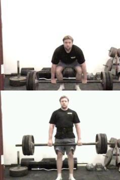
Exercise Therapy
Pelvis
Urologic Surgical Procedures
Imaging, Three-Dimensional
Urinary Bladder, Overactive
Female Urogenital Diseases
Diagnostic Techniques, Urological
Parity
Muscle Contraction
Palpation
Urinary Incontinence, Urge
Obstetric Labor Complications
Pelvimetry
Gastrointestinal Transit
Suburethral Slings
Gravidity
Principle-Based Ethics
Electric Stimulation Therapy
Surgical Mesh
Gynecological Examination
Quality of Life
Muscle Strength
Single blind, randomised controlled trial of pelvic floor exercises, electrical stimulation, vaginal cones, and no treatment in management of genuine stress incontinence in women. (1/336)
OBJECTIVE: To compare the effect of pelvic floor exercises, electrical stimulation, vaginal cones, and no treatment for genuine stress incontinence. DESIGN: Stratified, single blind, randomised controlled trial. SETTING: Multicentre. PARTICIPANTS: 107 women with clinically and urodynamically proved genuine stress incontinence. Mean (range) age was 49.5 (24-70) years, and mean (range) duration of symptoms 10.8 (1-45) years. INTERVENTIONS: Pelvic floor exercise (n=25) comprised 8-12 contractions 3 times a day and exercise in groups with skilled physical therapists once a week. The electrical stimulation group (n=25) used vaginal intermittent stimulation with the MS 106 Twin at 50 Hz 30 minutes a day. The vaginal cones group (n=27) used cones for 20 minutes a day. The untreated control group (n=30) was offered the use of a continence guard. Muscle strength was measured by vaginal squeeze pressure once a month. MAIN OUTCOME MEASURES: Pad test with standardised bladder volume, and self report of severity. RESULTS: Improvement in muscle strength was significantly greater (P=0.03) after pelvic floor exercises (11.0 cm H2O (95% confidence interval 7.7 to 14.3) before v 19.2 cm H2O (15.3 to 23.1) after) than either electrical stimulation (14.8 cm H2O (10. 9 to 18.7) v 18.6 cm H2O (13.3 to 23.9)) or vaginal cones (11.8 cm H2O (8.5 to 15.1) v 15.4 cm H2O (11.1 to 19.7)). Reduction in leakage on pad test was greater in the exercise group (-30.2 g; -43. 3 to 16.9) than in the electrical stimulation group (-7.4 g; -20.9 to 6.1) and the vaginal cones group (-14.7 g; -27.6 to -1.8). On completion of the trial one participant in the control group, 14 in the pelvic floor exercise group, three in the electrical stimulation group, and two in the vaginal cones group no longer considered themselves as having a problem. CONCLUSION: Training of the pelvic floor muscles is superior to electrical stimulation and vaginal cones in the treatment of genuine stress incontinence. (+info)Functional disorders of the anus and rectum. (2/336)
In this report the functional anorectal disorders, the etiology of which is currently unknown or related to the abnormal functioning of normally innervated and structurally intact muscles, are discussed. These disorders include functional fecal incontinence, functional anorectal pain, including levator ani syndrome and proctalgia fugax, and pelvic floor dyssynergia. The epidemiology of each disorder is defined and discussed, their pathophysiology is summarized and diagnostic approaches and treatment are suggested. Some suggestions for the direction of future research on these disorders are also given. (+info)The Virtual Pelvic Floor, a tele-immersive educational environment. (3/336)
This paper describes the development of the Virtual Pelvic Floor, a new method of teaching the complex anatomy of the pelvic region utilizing virtual reality and advanced networking technology. Virtual reality technology allows improved visualization of three-dimensional structures over conventional media because it supports stereo vision, viewer-centered perspective, large angles of view, and interactivity. Two or more ImmersaDesk systems, drafting table format virtual reality displays, are networked together providing an environment where teacher and students share a high quality three-dimensional anatomical model, and are able to converse, see each other, and to point in three dimensions to indicate areas of interest. This project was realized by the teamwork of surgeons, medical artists and sculptors, computer scientists, and computer visualization experts. It demonstrates the future of virtual reality for surgical education and applications for the Next Generation Internet. (+info)Manometric investigation of anorectal function in early and late stage Parkinson's disease. (4/336)
Abnormal gastrointestinal function is relatively frequent in Parkinson's disease, and constipation is a disturbing symptom in many patients. However, it remains to be established whether anorectal abnormalities are characteristic of the late stages of the disease. Clinical and anorectal manometric function were investigated in groups of early and late stage parkinsonian patients. Thirty one patients (19 men, 12 women, age range 22 to 89 years) entered the study. The disease severity was assessed by Hoehn and Yahr staging: there were four (12.9%) stage I, seven (22.6%) stage II, 10 (32.2%) stage III, and 10 (32.2%) stage IV patients. Anorectal variables were measured by standard manometric equipment and techniques. Values obtained in early stage patients (Hoehn and Yahr stage I and II) were compared with those obtained in late stage patients (Hoehn and Yahr stage III and IV). Overall, more than 70% of patients complained of chronic constipation, with chronic laxative use reported in more than 30%. Late stage patients were slightly older than their early stage counterparts. Pelvic floor dyssynergia was documented in more than 60% of patients. Manometric variables were not different in the two groups. In conclusion, defecatory dysfunction is frequent in Parkinson's disease, it is not confined to late stage patients, and it is found early in the course of the disease. This has potential implications for a targeted therapeutic approach. (+info)FES-biofeedback versus intensive pelvic floor muscle exercise for the prevention and treatment of genuine stress incontinence. (5/336)
We undertook this work to compare the treatment efficacies and the changes of quality of life after pelvic floor muscle (PFM) exercise and the functional electrical stimulation (FES)-biofeedback treatment, both of which are being widely used as conservative treatment methods for female urinary incontinence. We randomly selected 60 female incontinence patients who visited our department and divided them evenly into two groups. They were treated for a period of 6 weeks. The subjective changes in the severity of incontinence and discomfort in daily and social life were measured using a translated version of the questionnaire by Jackson. Objective changes of pelvic muscle contraction force were measured using a perineometer. Pre- and post-treatment maximal pelvic floor muscle contractile (PMC) pressure and changes in the severity of urinary incontinence and discomfort of the two groups showed statistically significant differences (p<0.001). In particular the FES-biofeedback group showed significantly increased maximal PMC pressure and a decreased severity of urinary incontinence and discomfort compared to the intensive PFM exercise group (p<0.001). In conclusion, FES-biofeedback proved more effective than simple PFM exercise. (+info)Pelvic floor muscle contraction during a cough and decreased vesical neck mobility. (6/336)
OBJECTIVE: To test the hypothesis that a voluntary pelvic muscle contraction initiated in preparation for a cough, a maneuver we call the Knack, significantly reduces vesical neck displacement. METHODS: A convenience sample of 22 women consisted of 11 young, continent nulliparas (mean age [+/- standard deviation] 24.8 +/- 7.0 years) and 11 older, incontinent paras (mean age [+/-SD] 66.9 +/- 3.9 years). With the use of perineal ultrasound, we quantified vesical neck displacement at rest and during coughs using caliper tracing and a coordinate system. The subjects coughed with and without voluntary pelvic floor muscle contraction. RESULTS: Vesical neck mobility during coughs was significantly decreased when voluntary contraction was used: from a median (range) of 5.4 (20.0) mm without volitional contraction to 2.9 (18.3) mm with volitional contraction (P <.001). The younger women demonstrated a median (range) decrease in excursion from 4.6 (19.5) to 0.0 (17.0) mm (P =.007), and the older incontinent women demonstrated a median (range) decrease from 6.2 (10.0) to 3.5 (15.4) mm (P =.003). At rest, the median vesical neck position in the group of older incontinent women was significantly further dorsocaudal (P =.001) than in the younger women. CONCLUSION: A pelvic floor muscle contraction in preparation for, and throughout, a cough can augment proximal urethra support during stress, thereby reducing the amount of dorsocaudal displacement. (+info)Predictors of intention to adhere to physiotherapy among women with urinary incontinence. (7/336)
During the last decade, pelvic floor muscle exercise (PFME) therapy has proved its short-term efficacy among women with urinary incontinence. Long-term success with PFME therapy is hampered by non-adherence. So far, specific knowledge on determinants of adherence behavior has been scarce. A cross-sectional study was conducted to elucidate the relative importance of determinants of the intention to adhere to PFME therapy in women with urinary incontinence. Based on behavioral theories, literature research and interviews, a questionnaire measuring determinants of the intention to adhere to PFME therapy was developed. In total, 129 women, aged 17 years or over, with symptoms of urinary incontinence, completed this questionnaire. Multiple regression analysis with backward elimination was carried out to identify determinants that predict intention. Significant predictors of the intention to adhere to PFME therapy were the amount to urinary loss per wet episode and women's perception of their ability to do the exercises as recommended under various circumstances. Building self-efficacy might be a good starting point for health education interventions aiming to promote adherence to PFME therapy, which can be used by physiotherapists and general practitioners. (+info)The functional anatomy of the female pelvic floor and stress continence control system. (8/336)
This paper provides an overview of the functional anatomy of the structures responsible for controlling urinary continence under stress. The stress continence control system can be divided into two parts: the system responsible for bladder neck support, and the system responsible for sphincteric closure. Age- and injury-related changes in each of these systems are discussed. Understanding the pathophysiology of incontinence on the anatomical level will help to lead to identification of specific defects, thereby allowing better individualized treatment for the incontinent patient. (+info)The pelvic floor is a group of muscles, ligaments, and connective tissues that form a sling or hammock across the bottom of the pelvis. It supports the organs in the pelvic cavity, including the bladder, rectum, and uterus or prostate. The pelvic floor helps control urination, defecation, and sexual function by relaxing and contracting to allow for the release of waste and during sexual activity. It also contributes to postural stability and balance. Weakness or damage to the pelvic floor can lead to various health issues such as incontinence, pelvic organ prolapse, and sexual dysfunction.
Pelvic floor disorders (PFD) refer to a group of conditions that affect the muscles and tissues supporting the pelvic organs, including the bladder, rectum, uterus, and vagina. These disorders can result in various symptoms such as urinary or fecal incontinence, pelvic organ prolapse, and painful sexual intercourse.
The causes of PFD are varied and may include childbirth, aging, obesity, chronic constipation, menopause, and certain neurological conditions. Treatment options for PFD depend on the severity and type of disorder but may include physical therapy, medication, surgery, or lifestyle changes such as weight loss and smoking cessation.
It is important to seek medical attention if you experience any symptoms of pelvic floor disorders, as early intervention can help prevent further damage and improve quality of life.
Pelvic Organ Prolapse (POP) is a medical condition where the supporting muscles and ligaments in a woman's pelvis weaken, causing one or more of the pelvic organs - including the bladder, uterus, rectum, or small intestine - to drop or press into or out of the vagina. This can result in various symptoms such as a feeling of heaviness or fullness in the pelvis, pressure or pain in the lower back, painful intercourse, and problems with urination or bowel movements. POP is often associated with childbirth, menopause, aging, and certain medical conditions that increase abdominal pressure, like obesity or chronic coughing. Treatment options can range from lifestyle changes and physical therapy to surgery.
Uterine prolapse is a condition where the uterus descends or slips down from its normal position in the pelvic cavity into or through the cervix and sometimes even outside the vaginal opening. This occurs due to the weakening of the muscles and ligaments that support the uterus, often as a result of childbirth, aging, menopause, obesity, or prior hysterectomy. Uterine prolapse can lead to various symptoms such as a feeling of heaviness in the pelvis, difficulty in urinating or having bowel movements, and uncomfortable sexual intercourse. The severity of the condition may vary from mild to severe, and treatment options range from lifestyle changes and physical therapy to surgery.
Fecal incontinence is the involuntary loss or leakage of stool (feces) from the rectum. It is also known as bowel incontinence. This condition can range from occasional leakage of stool when passing gas to a complete loss of bowel control. Fecal incontinence can be an embarrassing and distressing problem, but there are treatments available that can help improve symptoms and quality of life.
The causes of fecal incontinence can vary, but some common factors include:
* Damage to the muscles or nerves that control bowel function, such as from childbirth, surgery, spinal cord injury, or long-term constipation or diarrhea.
* Chronic digestive conditions, such as irritable bowel syndrome (IBS), inflammatory bowel disease (IBD), or celiac disease.
* Neurological conditions, such as multiple sclerosis, stroke, or spina bifida.
* Aging, which can lead to a decrease in muscle strength and control.
Treatment for fecal incontinence depends on the underlying cause of the condition. Treatments may include:
* Bowel training exercises to improve muscle strength and control.
* Changes in diet to help regulate bowel movements.
* Medications to treat constipation or diarrhea.
* Surgery to repair damaged muscles or nerves, or to create a new opening for stool to exit the body.
If you are experiencing symptoms of fecal incontinence, it is important to speak with your healthcare provider. They can help determine the cause of your symptoms and develop an appropriate treatment plan.
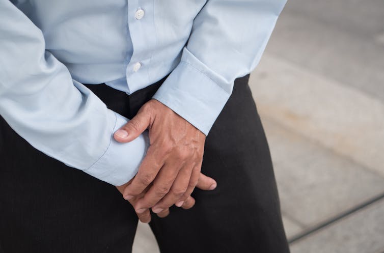
Stress Urinary Incontinence (SUI) is a type of urinary incontinence that occurs when physical activities or movements, such as coughing, sneezing, laughing, exercising, or lifting heavy objects, put pressure on the bladder, causing unintentional leakage of urine. It is caused by weakened pelvic floor muscles and/or a malfunctioning urethral sphincter, which normally help maintain urinary continence. SUI is more common in women than men, especially those who have gone through pregnancy, childbirth, or menopause, but it can also affect older men with prostate gland issues.
The perineum is the region between the anus and the genitals. In anatomical terms, it refers to the diamond-shaped area located in the lower part of the pelvis and extends from the coccyx (tailbone) to the pubic symphysis, which is the joint in the front where the two pubic bones meet. This region contains various muscles that support the pelvic floor and contributes to maintaining urinary and fecal continence. The perineum can be further divided into two triangular regions: the urogenital triangle (anterior) and the anal triangle (posterior).
A rectocele is a type of pelvic organ prolapse, which occurs when the rectum (the lower end of the colon) bulges into the back wall of the vagina. This condition most commonly affects women who have gone through childbirth, although it can also occur in older women or those with long-term constipation or other conditions that put pressure on the pelvic floor muscles.
Rectoceles can cause a variety of symptoms, including difficulty having bowel movements, feeling like something is sticking out of the vagina, and pain during sexual intercourse. In some cases, rectoceles may not cause any symptoms at all. Treatment options for rectoceles include pelvic floor physical therapy, lifestyle changes (such as avoiding heavy lifting or straining), and in severe cases, surgery.
The exact medical definition of a rectocele is: "A herniation of the rectal wall into the vaginal wall, often associated with disruption of the rectovaginal septum." This means that there is a protrusion or bulge of the rectal wall into the vaginal wall, which can be caused by a weakening or tearing of the tissue that separates the two structures.
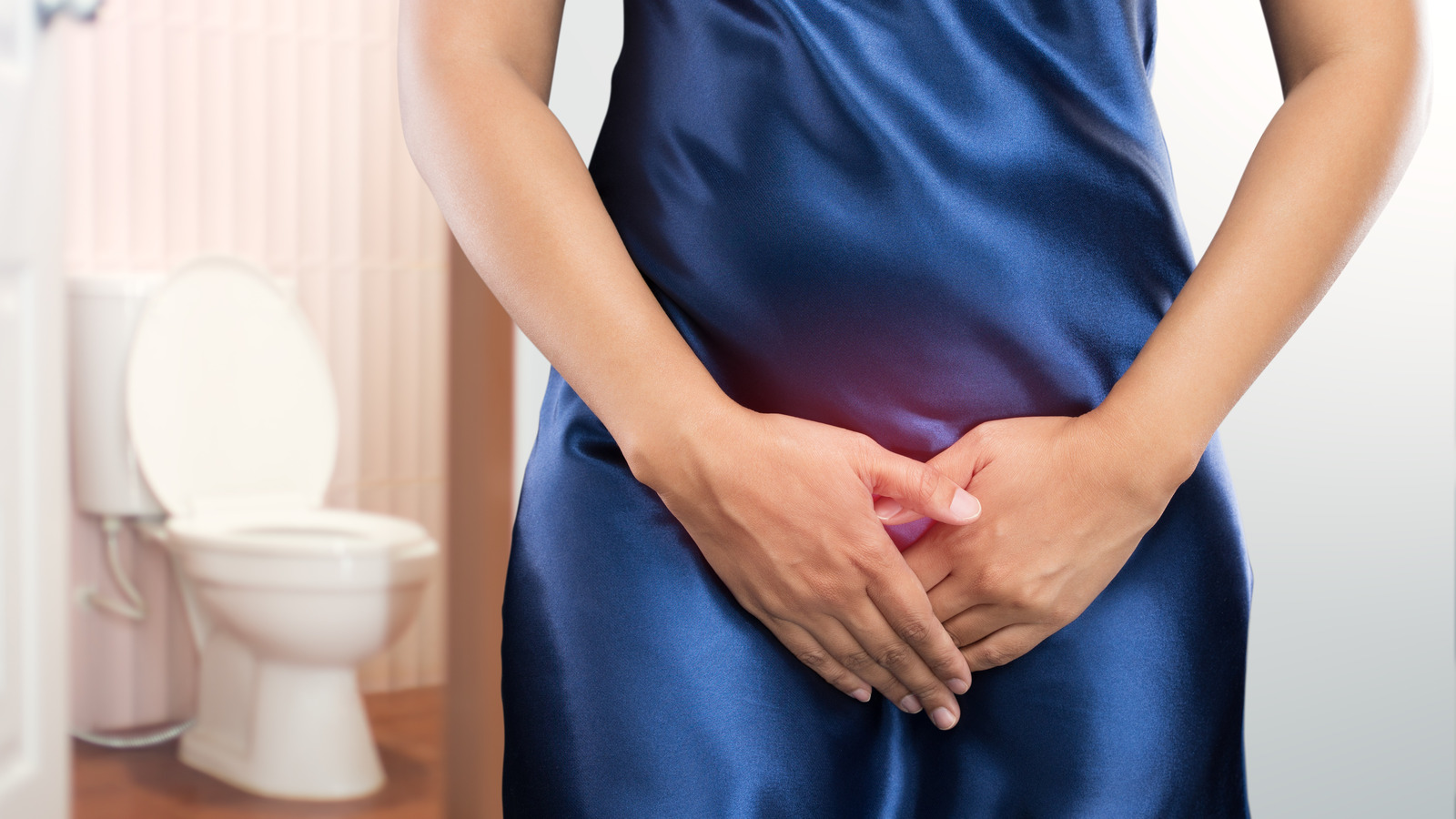
Urinary incontinence is defined as the involuntary loss or leakage of urine that is sufficient to be a social or hygienic problem. It can occur due to various reasons such as weak pelvic muscles, damage to nerves that control the bladder, certain medications, and underlying medical conditions like diabetes, multiple sclerosis, or Parkinson's disease.
There are different types of urinary incontinence, including stress incontinence (leakage of urine during physical activities like coughing, sneezing, or exercising), urge incontinence (a sudden and strong need to urinate that results in leakage), overflow incontinence (constant dribbling of urine due to a bladder that doesn't empty completely), functional incontinence (inability to reach the bathroom in time due to physical or mental impairments), and mixed incontinence (a combination of any two or more types of incontinence).
Urinary incontinence can significantly impact a person's quality of life, causing embarrassment, social isolation, and depression. However, it is a treatable condition, and various treatment options are available, including bladder training, pelvic floor exercises, medications, medical devices, and surgery.
Defecography is a medical diagnostic procedure that involves taking X-ray images of the rectum and anus while a person is defecating. Also known as evacuation proctography, this test assesses how well the muscles and structures of the pelvic floor perform during a bowel movement. It can help identify issues such as rectal prolapse, intussusception, or abnormalities in muscle function that may be causing difficulties with defecation or fecal incontinence.
During the procedure, the individual is usually given an enema containing a contrast material, which makes the contents of the rectum visible on X-ray images. The person then sits on a special toilet seat placed within the X-ray machine, and is asked to strain and evacuate as if having a bowel movement. Fluoroscopic X-ray imaging is used to capture real-time images of the pelvic floor and surrounding structures during this process. The resulting images can help healthcare providers diagnose and treat various anorectal conditions.
A cystocele is a type of pelvic organ prolapse that occurs when the wall between the bladder and the vagina weakens and allows the bladder to bulge into the vagina. This condition is also sometimes referred to as a "prolapsed bladder." Cystoceles can cause various symptoms, including urinary incontinence, difficulty emptying the bladder completely, and discomfort or pain during sexual activity. The severity of a cystocele can vary, and treatment options may include lifestyle changes, pelvic floor exercises, or surgery.
I'm sorry for any confusion, but "Floors and Floorcoverings" is not a medical term. It refers to the physical structures and materials used to cover floors in buildings or homes. If you have any medical terms that you would like me to define, please let me know!
A prolapse is a medical condition where an organ or tissue in the body slips from its normal position and drops down into a lower part of the body. This usually occurs when the muscles and ligaments that support the organ become weak or stretched. The most common types of prolapses include:
* Uterine prolapse: When the uterus slips down into or protrudes out of the vagina.
* Rectal prolapse: When the rectum (the lower end of the colon) slips outside the anus.
* Bladder prolapse (cystocele): When the bladder drops into the vagina.
* Small bowel prolapse (enterocele): When the small intestine bulges into the vagina.
Prolapses can cause various symptoms, such as discomfort, pain, pressure, and difficulty with urination or bowel movements. Treatment options depend on the severity of the prolapse and may include lifestyle changes, physical therapy, medication, or surgery.
The anal canal is the terminal portion of the digestive tract, located between the rectum and the anus. It is a short tube-like structure that is about 1 to 1.5 inches long in adults. The main function of the anal canal is to provide a seal for the elimination of feces from the body while also preventing the leakage of intestinal contents.
The inner lining of the anal canal is called the mucosa, which is kept moist by the production of mucus. The walls of the anal canal contain specialized muscles that help control the passage of stool during bowel movements. These muscles include the internal and external sphincters, which work together to maintain continence and allow for the voluntary release of feces.
The anal canal is an important part of the digestive system and plays a critical role in maintaining bowel function and overall health.
Rectal prolapse is a medical condition where the rectum, which is the lower end of the colon, slips outside the anus, the opening through which stool leaves the body. This usually occurs due to weakened muscles and supporting structures in the pelvic area, often as a result of aging, childbirth, or long-term constipation or diarrhea.
The rectal prolapse can be partial, where only a small portion of the rectum slips outside the anus, or complete, where the entire rectum protrudes. This condition can cause discomfort, pain, bleeding, and difficulty with bowel movements. Treatment options may include dietary changes, medication, or surgical intervention.
A pessary is a medical device that is inserted into the vagina to provide support for the uterus, vaginal vault, or bladder. It is often used in the management of pelvic organ prolapse, urinary incontinence, and other gynecological conditions. Pessaries come in various shapes and sizes, and they are typically made of silicone, rubber, or plastic. They can be worn for extended periods of time and are usually removable and cleanable. The selection and fitting of a pessary should be performed by a healthcare professional, such as a gynecologist or nurse midwife.
Biofeedback is a psychological and physiological intervention that involves the use of electronic devices to measure and provide real-time feedback to individuals about their bodily functions, such as heart rate, muscle tension, skin conductance, and brain activity. The goal of biofeedback is to help individuals gain awareness and control over these functions, with the aim of improving physical and mental health outcomes.
In psychology, biofeedback is often used as a treatment for a variety of conditions, including anxiety, stress, headaches, chronic pain, and mood disorders. By learning to regulate their physiological responses through biofeedback training, individuals can reduce symptoms and improve their overall well-being. The process typically involves working with a trained healthcare provider who guides the individual in practicing various relaxation techniques, such as deep breathing or progressive muscle relaxation, while monitoring their physiological responses using biofeedback equipment. Over time, the individual learns to associate these techniques with positive changes in their body and can use them to manage symptoms on their own.
An episiotomy is a surgical incision made in the perineum, the area between the vagina and the anus, during childbirth to widen the opening of the vagina and facilitate the delivery of the baby. It is typically performed when there is a risk of severe tearing or if the baby is showing signs of distress and needs to be delivered quickly. The incision is usually made with scissors or a scalpel, and it can be either midline (cut along the midline of the perineum) or mediolateral (cut diagonally from the vaginal opening toward the side of the buttocks). After delivery, the incision is stitched up.
Episiotomy was once a routine procedure during childbirth, but its use has become less common in recent years due to increasing evidence that it may not provide any significant benefits and can actually increase the risk of complications such as pain, infection, and difficulty with urination or bowel movements. Current guidelines recommend that episiotomies should only be performed when medically necessary and after informed consent from the mother.
Rectal diseases refer to conditions that affect the structure or function of the rectum, which is the lower end of the large intestine, just above the anus. The rectum serves as a storage area for stool before it is eliminated from the body. Some common rectal diseases include:
1. Hemorrhoids: Swollen veins in the rectum or anus that can cause pain, itching, bleeding, and discomfort.
2. Rectal cancer: Abnormal growth of cells in the rectum that can invade and destroy nearby tissue and spread to other parts of the body.
3. Anal fissures: Small tears in the lining of the anus that can cause pain, bleeding, and itching.
4. Rectal prolapse: A condition where the rectum slips outside the anus, causing discomfort, fecal incontinence, and other symptoms.
5. Inflammatory bowel disease (IBD): A group of chronic inflammatory conditions that affect the digestive tract, including the rectum, such as Crohn's disease and ulcerative colitis.
6. Rectal abscess: A collection of pus in the rectum caused by an infection, which can cause pain, swelling, and fever.
7. Fistula-in-ano: An abnormal connection between the rectum and the skin around the anus, which can cause drainage of pus or stool.
8. Rectal foreign bodies: Objects that are accidentally or intentionally inserted into the rectum and can cause injury, infection, or obstruction.
These are just a few examples of rectal diseases, and there are many other conditions that can affect the rectum. If you experience any symptoms related to the rectum, it is important to seek medical attention from a healthcare professional for proper diagnosis and treatment.
Constipation is a condition characterized by infrequent bowel movements or difficulty in passing stools that are often hard and dry. The medical definition of constipation varies, but it is generally defined as having fewer than three bowel movements in a week. In addition to infrequent bowel movements, other symptoms of constipation can include straining during bowel movements, feeling like you haven't completely evacuated your bowels, and experiencing hard or lumpy stools.
Constipation can have many causes, including a low-fiber diet, dehydration, certain medications, lack of physical activity, and underlying medical conditions such as irritable bowel syndrome or hypothyroidism. In most cases, constipation can be treated with lifestyle changes, such as increasing fiber intake, drinking more water, and getting regular exercise. However, if constipation is severe, persistent, or accompanied by other symptoms, it's important to seek medical attention to rule out any underlying conditions that may require treatment.
A hernia is a protrusion of an organ or tissue through a weakened area in the abdominal wall, often appearing as a bulge beneath the skin. This condition can occur in various parts of the body such as the groin (inguinal hernia), navel (umbilical hernia), or site of a previous surgical incision (incisional hernia). Hernias may cause discomfort or pain, especially when straining, lifting heavy objects, or during bowel movements. In some cases, they may lead to serious complications like intestinal obstruction or strangulation, requiring immediate medical attention.
Defecation is the medical term for the act of passing stools (feces) through the anus. It is a normal bodily function that involves the contraction of muscles in the colon and anal sphincter to release waste from the body. Defecation is usually a regular and daily occurrence, with the frequency varying from person to person.
The stool is made up of undigested food, bacteria, and other waste products that are eliminated from the body through the rectum and anus. The process of defecation is controlled by the autonomic nervous system, which regulates involuntary bodily functions such as heart rate and digestion.
Difficulties with defecation can occur due to various medical conditions, including constipation, irritable bowel syndrome, and inflammatory bowel disease. These conditions can cause symptoms such as hard or painful stools, straining during bowel movements, and a feeling of incomplete evacuation. If you are experiencing any problems with defecation, it is important to speak with your healthcare provider for proper diagnosis and treatment.
"Delivery, Obstetric" is a medical term that refers to the process of giving birth to a baby. It involves the passage of the fetus through the mother's vagina or via Caesarean section (C-section), which is a surgical procedure.
The obstetric delivery process typically includes three stages:
1. The first stage begins with the onset of labor and ends when the cervix is fully dilated.
2. The second stage starts with full dilation of the cervix and ends with the birth of the baby.
3. The third stage involves the delivery of the placenta, which is the organ that provides oxygen and nutrients to the developing fetus during pregnancy.
Obstetric delivery requires careful monitoring and management by healthcare professionals to ensure the safety and well-being of both the mother and the baby. Various interventions and techniques may be used during the delivery process to facilitate a safe and successful outcome, including the use of medications, assisted delivery with forceps or vacuum extraction, and C-section.
Urodynamics is a medical test that measures the function and performance of the lower urinary tract, which includes the bladder, urethra, and sphincters. It involves the use of specialized equipment to record measurements such as bladder pressure, urine flow rate, and residual urine volume. The test can help diagnose various urinary problems, including incontinence, urinary retention, and overactive bladder.
During the test, a small catheter is inserted into the bladder through the urethra to measure bladder pressure while filling it with sterile water or saline solution. Another catheter may be placed in the rectum to record abdominal pressure. The patient is then asked to urinate, and the flow rate and any leaks are recorded.
Urodynamics can help identify the underlying cause of urinary symptoms and guide treatment decisions. It is often recommended for patients with complex or persistent urinary problems that have not responded to initial treatments.
The urethra is the tube that carries urine from the bladder out of the body. In males, it also serves as the conduit for semen during ejaculation. The male urethra is longer than the female urethra and is divided into sections: the prostatic, membranous, and spongy (or penile) urethra. The female urethra extends from the bladder to the external urethral orifice, which is located just above the vaginal opening.
The term "mouth floor" is not a standard medical terminology. However, it might refer to the floor of the mouth, which is the part of the oral cavity located beneath the tongue and above the hyoid bone, which is a U-shaped bone in the front of the neck that helps support the tongue. The mouth floor contains several salivary glands, muscles, and nerves that are important for functions such as swallowing and speaking.
The vagina is the canal that joins the cervix (the lower part of the uterus) to the outside of the body. It also is known as the birth canal because babies pass through it during childbirth. The vagina is where sexual intercourse occurs and where menstrual blood exits the body. It has a flexible wall that can expand and retract. During sexual arousal, the vaginal walls swell with blood to become more elastic in order to accommodate penetration.
It's important to note that sometimes people use the term "vagina" to refer to the entire female genital area, including the external structures like the labia and clitoris. But technically, these are considered part of the vulva, not the vagina.
Dyspareunia is a medical term that describes painful sexual intercourse. This condition can affect both men and women, but it is more commonly reported by women. The pain can occur in various locations, such as the vaginal opening, deep inside the vagina, or in the pelvic region. It can be caused by a variety of factors, including physical conditions like vulvodynia, endometriosis, or vaginal infections, as well as psychological factors like anxiety, depression, or relationship issues. Treatment for dyspareunia depends on the underlying cause and may include medication, therapy, or lifestyle changes.
Manometry is a medical test that measures pressure inside various parts of the gastrointestinal tract. It is often used to help diagnose digestive disorders such as achalasia, gastroparesis, and irritable bowel syndrome. During the test, a thin, flexible tube called a manometer is inserted through the mouth or rectum and into the area being tested. The tube is connected to a machine that measures and records pressure readings. These readings can help doctors identify any abnormalities in muscle function or nerve reflexes within the digestive tract.
Exercise therapy is a type of medical treatment that uses physical movement and exercise to improve a patient's physical functioning, mobility, and overall health. It is often used as a component of rehabilitation programs for individuals who have experienced injuries, illnesses, or surgeries that have impaired their ability to move and function normally.
Exercise therapy may involve a range of activities, including stretching, strengthening, balance training, aerobic exercise, and functional training. The specific exercises used will depend on the individual's needs, goals, and medical condition.
The benefits of exercise therapy include:
* Improved strength and flexibility
* Increased endurance and stamina
* Enhanced balance and coordination
* Reduced pain and inflammation
* Improved cardiovascular health
* Increased range of motion and joint mobility
* Better overall physical functioning and quality of life.
Exercise therapy is typically prescribed and supervised by a healthcare professional, such as a physical therapist or exercise physiologist, who has experience working with individuals with similar medical conditions. The healthcare professional will create an individualized exercise program based on the patient's needs and goals, and will provide guidance and support to ensure that the exercises are performed safely and effectively.
The pelvis is the lower part of the trunk, located between the abdomen and the lower limbs. It is formed by the fusion of several bones: the ilium, ischium, and pubis (which together form the hip bone on each side), and the sacrum and coccyx in the back. The pelvis has several functions including supporting the weight of the upper body when sitting, protecting the lower abdominal organs, and providing attachment for muscles that enable movement of the lower limbs. In addition, it serves as a bony canal through which the reproductive and digestive tracts pass. The pelvic cavity contains several vital organs such as the bladder, parts of the large intestine, and in females, the uterus, ovaries, and fallopian tubes.
Urologic surgical procedures refer to various types of surgeries that are performed on the urinary system and male reproductive system. These surgeries can be invasive (requiring an incision) or minimally invasive (using small incisions or scopes). They may be performed to treat a range of conditions, including but not limited to:
1. Kidney stones: Procedures such as shock wave lithotripsy, ureteroscopy, and percutaneous nephrolithotomy are used to remove or break up kidney stones.
2. Urinary tract obstructions: Surgeries like pyeloplasty and urethral dilation can be done to correct blockages in the urinary tract.
3. Prostate gland issues: Transurethral resection of the prostate (TURP), simple prostatectomy, and robotic-assisted laparoscopic radical prostatectomy are some procedures used for benign prostatic hyperplasia (BPH) or prostate cancer.
4. Bladder problems: Procedures such as cystectomy (removal of the bladder), bladder augmentation, and implantation of an artificial urinary sphincter can be done for conditions like bladder cancer or incontinence.
5. Kidney diseases: Nephrectomy (removal of a kidney) may be necessary for severe kidney damage or cancer.
6. Testicular issues: Orchiectomy (removal of one or both testicles) can be performed for testicular cancer.
7. Pelvic organ prolapse: Surgeries like sacrocolpopexy and vaginal vault suspension can help correct this condition in women.
These are just a few examples; there are many other urologic surgical procedures available to treat various conditions affecting the urinary and reproductive systems.
Three-dimensional (3D) imaging in medicine refers to the use of technologies and techniques that generate a 3D representation of internal body structures, organs, or tissues. This is achieved by acquiring and processing data from various imaging modalities such as X-ray computed tomography (CT), magnetic resonance imaging (MRI), ultrasound, or confocal microscopy. The resulting 3D images offer a more detailed visualization of the anatomy and pathology compared to traditional 2D imaging techniques, allowing for improved diagnostic accuracy, surgical planning, and minimally invasive interventions.
In 3D imaging, specialized software is used to reconstruct the acquired data into a volumetric model, which can be manipulated and viewed from different angles and perspectives. This enables healthcare professionals to better understand complex anatomical relationships, detect abnormalities, assess disease progression, and monitor treatment response. Common applications of 3D imaging include neuroimaging, orthopedic surgery planning, cancer staging, dental and maxillofacial reconstruction, and interventional radiology procedures.
Overactive bladder (OAB) is a urological condition characterized by the involuntary contraction of the detrusor muscle of the urinary bladder, leading to symptoms such as urgency, frequency, and nocturia (the need to wake up at night to urinate), with or without urge incontinence (the involuntary loss of urine associated with a strong desire to void). It is important to note that OAB is not necessarily related to bladder volume or age-related changes, and it can significantly impact an individual's quality of life. The exact cause of OAB is not fully understood, but it may be associated with neurological disorders, certain medications, infections, or other underlying medical conditions. Treatment options for OAB include behavioral modifications, pelvic floor exercises, bladder training, medications, and, in some cases, surgical interventions.
Female urogenital diseases refer to a range of medical conditions that affect the female urinary and genital systems. These systems include the kidneys, ureters, bladder, urethra, vulva, vagina, and reproductive organs such as the ovaries and uterus.
Some common female urogenital diseases include:
1. Urinary tract infections (UTIs): These are infections that occur in any part of the urinary system, including the kidneys, ureters, bladder, or urethra.
2. Pelvic inflammatory disease (PID): This is an infection of the reproductive organs, including the uterus, fallopian tubes, and ovaries.
3. Endometriosis: This is a condition in which tissue similar to the lining of the uterus grows outside of the uterus, often on the ovaries, fallopian tubes, or other pelvic structures.
4. Ovarian cysts: These are fluid-filled sacs that form on the ovaries.
5. Uterine fibroids: These are noncancerous growths that develop in the muscular wall of the uterus.
6. Interstitial cystitis/bladder pain syndrome (IC/BPS): This is a chronic bladder condition characterized by pain, pressure, and discomfort in the bladder and pelvic area.
7. Sexually transmitted infections (STIs): These are infections that are passed from person to person during sexual contact. Common STIs include chlamydia, gonorrhea, syphilis, and HIV.
8. Vulvodynia: This is chronic pain or discomfort of the vulva, the external female genital area.
9. Cancers of the reproductive system, such as ovarian cancer, cervical cancer, and uterine cancer.
These are just a few examples of female urogenital diseases. It's important for women to receive regular medical care and screenings to detect and treat these conditions early, when they are often easier to manage and have better outcomes.
The Valsalva maneuver is a medical procedure that involves forced exhalation against a closed airway, typically by closing one's mouth, pinching the nose shut, and then blowing. This maneuver increases the pressure in the chest and affects the heart's filling and pumping capabilities, as well as the pressures within the ears and eyes.
It is often used during medical examinations to test for conditions such as heart murmurs or to help clear the ears during changes in air pressure (like when scuba diving or flying). It can also be used to help diagnose or monitor conditions related to the autonomic nervous system, such as orthostatic hypotension or dysautonomia.
However, it's important to perform the Valsalva maneuver correctly and under medical supervision, as improper technique or overdoing it can lead to adverse effects like increased heart rate, changes in blood pressure, or even damage to the eardrum.
Diagnostic techniques in urology are methods used to identify and diagnose various urological conditions affecting the urinary tract and male reproductive system. These techniques include:
1. Urinalysis: A laboratory examination of a urine sample to detect abnormalities such as infection, kidney stones, or other underlying medical conditions.
2. Urine Culture: A test used to identify and grow bacteria from the urine to determine the type of bacterial infection present in the urinary tract.
3. Imaging Studies: Various imaging techniques such as X-rays, ultrasound, CT scans, and MRI scans are used to visualize the internal structures of the urinary tract and identify any abnormalities.
4. Cystoscopy: A procedure that involves inserting a thin tube with a camera into the bladder through the urethra to examine the bladder and urethra for signs of disease or abnormality.
5. Urodynamics: A series of tests used to evaluate bladder function, including measuring bladder pressure and urine flow rate.
6. Biopsy: The removal and examination of tissue from the urinary tract or male reproductive system to diagnose conditions such as cancer.
7. Prostate-Specific Antigen (PSA) Test: A blood test used to screen for prostate cancer by measuring the level of PSA, a protein produced by the prostate gland.
8. Voiding Diary: A record of urinary habits, including the frequency and volume of urination, that can help diagnose conditions such as overactive bladder or urinary incontinence.
Parturition is the process of giving birth, or the act of delivering newborn offspring. In medical terms, it refers to the expulsion of the products of conception (such as the fetus, placenta, and membranes) from the uterus of a pregnant woman during childbirth. This process is regulated by hormonal changes and involves complex interactions between the mother's body and the developing fetus. Parturition typically occurs after a full-term pregnancy, which is approximately 40 weeks in humans.
The rectum is the lower end of the digestive tract, located between the sigmoid colon and the anus. It serves as a storage area for feces before they are eliminated from the body. The rectum is about 12 cm long in adults and is surrounded by layers of muscle that help control defecation. The mucous membrane lining the rectum allows for the detection of stool, which triggers the reflex to have a bowel movement.
In medical terms, parity refers to the number of times a woman has given birth to a viable fetus, usually defined as a pregnancy that reaches at least 20 weeks' gestation. It is often used in obstetrics and gynecology to describe a woman's childbearing history and to assess potential risks associated with childbirth.
Parity is typically categorized as follows:
* Nulliparous: A woman who has never given birth to a viable fetus.
* Primiparous: A woman who has given birth to one viable fetus.
* Multiparous: A woman who has given birth to more than one viable fetus.
In some cases, parity may also consider the number of pregnancies that resulted in stillbirths or miscarriages, although this is not always the case. It's important to note that parity does not necessarily reflect the total number of pregnancies a woman has had, only those that resulted in viable births.
Gynecologic surgical procedures refer to the operations that are performed on the female reproductive system and related organs. These surgeries can be either minimally invasive or open procedures, depending on the condition and the patient's health status.
The indications for gynecologic surgical procedures may include but are not limited to:
1. Diagnosis and treatment of various benign and malignant conditions such as uterine fibroids, ovarian cysts, endometriosis, and cancers of the reproductive organs.
2. Management of abnormal uterine bleeding, pelvic pain, and infertility.
3. Treatment of ectopic pregnancies and miscarriages.
4. Pelvic organ prolapse repair.
5. Sterilization procedures such as tubal ligation.
6. Investigation and treatment of suspicious lesions or abnormal Pap smears.
Some common gynecologic surgical procedures include hysterectomy (removal of the uterus), oophorectomy (removal of the ovary), salpingectomy (removal of the fallopian tube), cystectomy (removal of a cyst), myomectomy (removal of fibroids while preserving the uterus), and endometrial ablation (destruction of the lining of the uterus).
Minimally invasive surgical techniques such as laparoscopy and hysteroscopy have gained popularity in recent years due to their advantages over traditional open surgeries, including smaller incisions, less postoperative pain, quicker recovery times, and reduced risk of complications.
Muscle contraction is the physiological process in which muscle fibers shorten and generate force, leading to movement or stability of a body part. This process involves the sliding filament theory where thick and thin filaments within the sarcomeres (the functional units of muscles) slide past each other, facilitated by the interaction between myosin heads and actin filaments. The energy required for this action is provided by the hydrolysis of adenosine triphosphate (ATP). Muscle contractions can be voluntary or involuntary, and they play a crucial role in various bodily functions such as locomotion, circulation, respiration, and posture maintenance.
Palpation is a medical examination technique in which a healthcare professional uses their hands to feel the size, shape, and consistency of body parts, including organs, tissues, and bones. It is used to assess the patient's overall health, identify any abnormalities or areas of pain, monitor healing and disease progression, and guide diagnostic and treatment decisions.
During palpation, the healthcare professional applies gentle pressure with their fingers or hands to specific areas of the body, feeling for any changes in texture, temperature, moisture, or movement. The technique can be used to assess various bodily systems, including the cardiovascular, respiratory, gastrointestinal, musculoskeletal, and nervous systems.
Palpation is a valuable tool in physical examinations because it is non-invasive, relatively quick, and cost-effective. It can provide important information that helps healthcare professionals make accurate diagnoses and develop effective treatment plans for their patients.
Urge urinary incontinence is a type of urinary incontinence where there is a sudden, strong need to urinate that cannot be postponed, leading to an involuntary loss of urine. It is also known as overactive bladder (OAB) or detrusor instability. The underlying cause is often due to uninhibited contractions of the detrusor muscle, which is the main muscle in the bladder that helps with urination. This can be caused by various factors such as nerve damage, bladder infections, bladder stones, or certain medications. Treatment options may include behavioral modifications, pelvic floor exercises, medication, and in some cases, surgery.
Obstetric labor complications refer to any physical or physiological difficulties that arise during the process of childbirth (labor) and can pose risks to the health of the mother, baby, or both. These complications may result from various factors such as pre-existing medical conditions, fetal distress, prolonged labor, abnormal positioning of the fetus, or issues related to the size or weight of the baby.
Some examples of obstetric labor complications include:
1. Fetal distress: This occurs when the fetus is not receiving adequate oxygen supply or is in danger during labor. It can be caused by various factors such as umbilical cord compression, placental abruption, or maternal anemia.
2. Prolonged labor: When labor lasts for more than 20 hours in first-time mothers or more than 14 hours in subsequent pregnancies, it is considered prolonged labor. This can lead to fatigue, infection, and other complications for both the mother and baby.
3. Abnormal positioning of the fetus: Normally, the fetus should be positioned head-down (vertex) before delivery. However, if the fetus is in a breech or transverse position, it can lead to difficult labor and increased risk of complications during delivery.
4. Shoulder dystocia: This occurs when the baby's shoulders get stuck behind the mother's pubic bone during delivery, making it challenging to deliver the baby. It can cause injuries to both the mother and the baby.
5. Placental abruption: This is a serious complication where the placenta separates from the uterus before delivery, leading to bleeding and potential oxygen deprivation for the fetus.
6. Uterine rupture: A rare but life-threatening complication where the uterus tears during labor, causing severe bleeding and potentially endangering both the mother and baby's lives.
7. Preeclampsia/eclampsia: This is a pregnancy-related hypertensive disorder that can lead to complications such as seizures, organ failure, or even maternal death if left untreated.
8. Postpartum hemorrhage: Excessive bleeding after delivery can be life-threatening and requires immediate medical attention.
9. Infections: Maternal infections during pregnancy or childbirth can lead to complications for both the mother and baby, including preterm labor, low birth weight, and even fetal death.
10. Anesthesia complications: Adverse reactions to anesthesia during delivery can cause respiratory depression, allergic reactions, or other complications that may endanger the mother's life.
A laceration is a type of injury that results in a tear or ragged cut in the skin or mucous membrane, often caused by some form of trauma. This can include cuts from sharp objects, blunt force trauma, or accidents. Lacerations can vary greatly in severity, from minor injuries that only affect the top layer of skin to more serious wounds that penetrate deeper into underlying tissues and structures.
Lacerations are typically irregular in shape and may have jagged edges, unlike clean incisions caused by sharp objects. They can also be accompanied by bruising, swelling, and bleeding, depending on the severity of the injury. In some cases, lacerations may require medical attention to properly clean, close, and manage the wound to prevent infection and promote healing.
It is essential to assess the depth, location, and extent of a laceration to determine the appropriate course of action. Deeper lacerations that expose underlying tissues or structures, such as muscles, tendons, nerves, or blood vessels, may require sutures (stitches), staples, or adhesive strips to close the wound. In some instances, surgical intervention might be necessary to repair damaged tissues properly. Always consult a healthcare professional for proper evaluation and treatment of lacerations.
Pelvimetry is a medical measurement and evaluation of the size and shape of the pelvis, which can be performed in several ways:
1. Clinical pelvimetry: This involves physical examination to assess the dimensions of the pelvis by palpation and measurement of the distance between bony landmarks.
2. Radiological pelvimetry: This uses X-ray or CT imaging to obtain more accurate measurements of the pelvic diameters, including the anteroposterior, transverse, and oblique dimensions.
3. Magnetic resonance imaging (MRI) pelvimetry: This method is considered the most accurate for assessing the size and shape of the pelvis, as it provides detailed images without radiation exposure.
Pelvimetry is often used in obstetrics to evaluate whether a woman's pelvis can accommodate a fetus during childbirth (known as "obstetric pelvimetry"). It helps healthcare providers determine if a vaginal delivery is possible or if a cesarean section may be necessary. However, the use of pelvimetry in modern obstetrics has become less common due to its limited predictive value and the increasing focus on individualized birth management.
Gastrointestinal transit refers to the movement of food, digestive secretions, and waste products through the gastrointestinal tract, from the mouth to the anus. This process involves several muscles and nerves that work together to propel the contents through the stomach, small intestine, large intestine, and rectum.
The transit time can vary depending on factors such as the type and amount of food consumed, hydration levels, and overall health. Abnormalities in gastrointestinal transit can lead to various conditions, including constipation, diarrhea, and malabsorption. Therefore, maintaining normal gastrointestinal transit is essential for proper digestion, nutrient absorption, and overall health.
A suburethral sling is a type of surgical mesh used in the treatment of stress urinary incontinence (SUI) in women. It is a narrow strip of synthetic material or tissue that is placed under the urethra, the tube that carries urine from the bladder out of the body, to provide support and restore normal function.
The sling helps to keep the urethra in its proper position during physical activities, such as coughing, sneezing, or exercising, which can put pressure on the bladder and cause urine leakage in women with SUI. Suburethral slings are typically made of non-absorbable synthetic materials, such as polypropylene or polyester, and can be attached to surrounding tissue or bone for added support.
The procedure to implant a suburethral sling is usually performed on an outpatient basis, and most women are able to return to their normal activities within a few weeks. While suburethral slings have been shown to be effective in treating SUI, they are not without risks, including infection, bleeding, pain during sexual intercourse, and in rare cases, erosion of the mesh into surrounding tissues.
Gravidity is a medical term that refers to the number of times a woman has been pregnant, regardless of the outcome of the pregnancies. It's a way to quantify a woman's childbearing experience and is often used in obstetrics and gynecology to assess potential risks and complications during pregnancy and childbirth.
For example, a woman who has been pregnant once before would have a gravidity of 1, while a woman who has been pregnant twice would have a gravidity of 2. This term is distinct from parity, which refers to the number of pregnancies that have reached a viable gestational age and resulted in a live birth.
Obstetrical extraction refers to a medical procedure in obstetrics, where a fetus or a dead fetus is removed from the uterus through surgical means. This is typically performed when a vaginal delivery is not possible or safe due to various reasons such as obstructed labor, maternal or fetal distress, or prolonged pregnancy. The procedure may involve dilation and evacuation (D&E) or instrumental delivery using forceps or vacuum extractor. It is usually done under anesthesia in a hospital setting.
Principle-Based Ethics is a framework for moral decision-making that involves the application of several fundamental ethical principles. These principles include:
1. Respect for Autonomy: This principle recognizes and respects an individual's right to make their own decisions, as long as they do not harm others or infringe upon their rights.
2. Nonmaleficence: This principle requires that healthcare providers should not cause harm to their patients. They should avoid doing anything that could potentially harm their patients, unless the potential benefits of an action outweigh its risks.
3. Beneficence: This principle requires healthcare providers to act in the best interests of their patients and promote their well-being. Healthcare providers should take positive actions to benefit their patients and prevent harm.
4. Justice: This principle requires that healthcare resources be distributed fairly and equitably among all members of society, regardless of their social status or ability to pay.
These principles serve as a foundation for ethical decision-making in healthcare and provide guidance for making difficult moral choices. They are often used in conjunction with other ethical theories and frameworks, such as consequentialism and virtue ethics, to help healthcare providers make informed and responsible decisions that promote the well-being of their patients while also respecting their autonomy and rights.
Electric stimulation therapy, also known as neuromuscular electrical stimulation (NMES) or electromyostimulation, is a therapeutic treatment that uses electrical impulses to stimulate muscles and nerves. The electrical signals are delivered through electrodes placed on the skin near the target muscle group or nerve.
The therapy can be used for various purposes, including:
1. Pain management: Electric stimulation can help reduce pain by stimulating the release of endorphins, which are natural painkillers produced by the body. It can also help block the transmission of pain signals to the brain.
2. Muscle rehabilitation: NMES can be used to prevent muscle atrophy and maintain muscle tone in individuals who are unable to move their muscles due to injury or illness, such as spinal cord injuries or stroke.
3. Improving circulation: Electric stimulation can help improve blood flow and reduce swelling by contracting the muscles and promoting the movement of fluids in the body.
4. Wound healing: NMES can be used to promote wound healing by increasing blood flow, reducing swelling, and improving muscle function around the wound site.
5. Muscle strengthening: Electric stimulation can be used to strengthen muscles by causing them to contract and relax repeatedly, which can help improve muscle strength and endurance.
It is important to note that electric stimulation therapy should only be administered under the guidance of a trained healthcare professional, as improper use can cause harm or discomfort.
Urination, also known as micturition, is the physiological process of excreting urine from the urinary bladder through the urethra. It is a complex process that involves several systems in the body, including the urinary system, nervous system, and muscular system.
In medical terms, urination is defined as the voluntary or involuntary discharge of urine from the urethra, which is the final pathway for the elimination of waste products from the body. The process is regulated by a complex interplay between the detrusor muscle of the bladder, the internal and external sphincters of the urethra, and the nervous system.
During urination, the detrusor muscle contracts, causing the bladder to empty, while the sphincters relax to allow the urine to flow through the urethra and out of the body. The nervous system plays a crucial role in coordinating these actions, with sensory receptors in the bladder sending signals to the brain when it is time to urinate.
Urination is essential for maintaining the balance of fluids and electrolytes in the body, as well as eliminating waste products such as urea, creatinine, and other metabolic byproducts. Abnormalities in urination can indicate underlying medical conditions, such as urinary tract infections, bladder dysfunction, or neurological disorders.
Surgical mesh is a medical device that is used in various surgical procedures, particularly in reconstructive surgery, to provide additional support to weakened or damaged tissues. It is typically made from synthetic materials such as polypropylene or polyester, or from biological materials such as animal tissue or human cadaveric tissue.
The mesh is designed to be implanted into the body, where it can help to reinforce and repair damaged tissues. For example, it may be used in hernia repairs to support the weakened abdominal wall, or in pelvic floor reconstruction surgery to treat conditions such as pelvic organ prolapse or stress urinary incontinence.
Surgical mesh can come in different forms, including sheets, plugs, and patches, and may be either absorbable or non-absorbable. The choice of mesh material and type will depend on the specific surgical indication and the patient's individual needs. It is important for patients to discuss the risks and benefits of surgical mesh with their healthcare provider before undergoing any surgical procedure that involves its use.
A gynecological examination is a medical procedure performed by a healthcare professional, typically a gynecologist, to evaluate the female reproductive system. The examination may include a variety of tests and procedures, such as:
1. Medical history review: The doctor will ask questions about the patient's menstrual cycle, sexual activity, contraceptive use, pregnancy history, and any symptoms or concerns.
2. External examination: The doctor will inspect the external genitalia for any signs of infection, irritation, or abnormalities.
3. Speculum exam: A speculum, a medical instrument that resembles a duckbill, is inserted into the vagina to allow the doctor to visualize the cervix and vaginal walls. This helps in detecting any abnormalities such as cervical polyps, inflammation, or cancerous growths.
4. Pelvic exam: The doctor will insert gloved fingers into the patient's vagina while simultaneously pressing on the lower abdomen to assess the size, shape, and position of the reproductive organs, including the uterus, ovaries, and fallopian tubes.
5. Pap test: A sample of cells is collected from the cervix using a spatula or brush and sent to a laboratory for analysis. This helps in detecting any precancerous or cancerous changes in the cervical cells.
6. Other tests: Depending on the patient's age, medical history, and symptoms, additional tests such as STD screening, breast exam, or imaging studies (e.g., ultrasound, MRI) may be recommended.
The frequency and type of gynecological examinations vary depending on a woman's age, health status, and individual needs. Regular check-ups are essential for early detection and prevention of reproductive system-related issues, including sexually transmitted infections, cervical cancer, and other gynecological conditions.
Quality of Life (QOL) is a broad, multidimensional concept that usually includes an individual's physical health, psychological state, level of independence, social relationships, personal beliefs, and their relationship to salient features of their environment. It reflects the impact of disease and treatment on a patient's overall well-being and ability to function in daily life.
The World Health Organization (WHO) defines QOL as "an individual's perception of their position in life in the context of the culture and value systems in which they live and in relation to their goals, expectations, standards and concerns." It is a subjective concept, meaning it can vary greatly from person to person.
In healthcare, QOL is often used as an outcome measure in clinical trials and other research studies to assess the impact of interventions or treatments on overall patient well-being.
Muscle strength, in a medical context, refers to the amount of force a muscle or group of muscles can produce during contraction. It is the maximum amount of force that a muscle can generate through its full range of motion and is often measured in units of force such as pounds or newtons. Muscle strength is an important component of physical function and mobility, and it can be assessed through various tests, including manual muscle testing, dynamometry, and isokinetic testing. Factors that can affect muscle strength include age, sex, body composition, injury, disease, and physical activity level.
A hysterectomy is a surgical procedure that involves the removal of the uterus (womb). Depending on the specific medical condition and necessity, a hysterectomy may also include the removal of the ovaries, fallopian tubes, and surrounding tissues. There are different types of hysterectomies, including:
1. Total hysterectomy: The uterus and cervix are removed.
2. Supracervical (or subtotal) hysterectomy: Only the upper part of the uterus is removed, leaving the cervix intact.
3. Radical hysterectomy: This procedure involves removing the uterus, cervix, surrounding tissues, and the upper part of the vagina. It is typically performed in cases of cervical cancer.
4. Oophorectomy: The removal of one or both ovaries can be performed along with a hysterectomy depending on the patient's medical condition and age.
5. Salpingectomy: The removal of one or both fallopian tubes can also be performed along with a hysterectomy if needed.
The reasons for performing a hysterectomy may include but are not limited to: uterine fibroids, heavy menstrual bleeding, endometriosis, adenomyosis, pelvic prolapse, cervical or uterine cancer, and chronic pelvic pain. The choice of the type of hysterectomy depends on the patient's medical condition, age, and personal preferences.
The second stage of labor is the active phase of childbirth, during which the uterus continues to contract and the cervix fully dilates. This stage begins when the cervix is completely open (10 cm) and ends with the birth of the baby. During this stage, the mother typically experiences strong, regular contractions that help to push the baby down the birth canal.
The second stage of labor can be further divided into two phases: the latent phase and the pushing phase. The latent phase is the period between full dilation of the cervix and the beginning of active pushing. This phase can last anywhere from a few minutes to several hours, depending on various factors such as the position of the baby, the mother's exhaustion, and whether it is the mother's first baby or not.
The pushing phase is the period during which the mother actively pushes the baby out of the birth canal. This phase typically lasts between 20 minutes to an hour, although it can be longer in some cases. The healthcare provider will guide the mother through this process, instructing her when and how to push. Once the baby's head emerges, the healthcare provider will continue to support the delivery of the baby's shoulders and body.
It is important for the mother to receive appropriate support and guidance during the second stage of labor to ensure a safe and successful delivery.
 Pelvic floor
 Pelvic Support Problems | Pelvic Floor | MedlinePlus
 Pelvic Floor News, Research
 Pelvic Floor Health | Baylor Medicine
 Pelvic floor exercises: The best exercises for men and women
 Hypertonic Pelvic Floor: Symptoms, Causes & Treatment
 Pelvic Floor Rehabilitation | Cooper University Health Care
 Pelvic Floor Disorders: More Common Than You Think
 Pelvic Floor Exercisers | Kegel Exercise Trainers | Cult Beauty
 Urologic Management in Neurologic Disease: Overview, Neuroanatomy of Pelvic Floor, Neurophysiology of Pelvic Floor
 Urogynaecology and pelvic floor | Royal Free London
 Is Something Missing From Antenatal Education? A Survey of Pregnant Women's Knowledge of Pelvic Floor Disorders
 Analytica Expands Clinical Indication for Its Unique Pelvic Floor Exercise Technology
 Assessing muscle function of the male pelvic floor using real time ultrasound - Abstract
 Physical Therapy for Pelvic Floor Dysfunction | University of Utah Health
 Apstron Science Revolutionizes Pelvic Floor Health with Enhanced PelvicTron™ App
 6 Pelvic Floor Stretches To Release Overly Tight Muscles
 Ask the Expert - Pelvic Floor Physiotherapy | IMPACT Magazine
 The Pelvic Floor Electrotherapy Muscle Strengthener - Hammacher Schlemmer
 Pelvic Floor Tips for New Moms and Postpartum Hiking - Gossamer Gear
 Men have pelvic floors too - and can benefit when they exercise them regularly - CQUniversity
 CISH-Hysterectomy Without Destruction of the Pelvic Floor Support
 How Pelvic Floor Physical Therapy Can Help MS Patients
 Your Health | Female Pelvic Floor Health Quiz | Summa Health
 Rüdiger Anatomie Premium Female Skeleton with Pelvic Floor Muscles
 Pelvic Floor Dysfunction: Symptoms, Causes & Treatments | ATI
 Benefits of pelvic floor P.T. - Mayo Clinic Health System