Peanut Agglutinin
Arachis hypogaea
Agglutinins
Wheat Germ Agglutinins
Lectins
Plant Lectins
Receptors, Mitogen
Glycoconjugates
Antigens, Tumor-Associated, Carbohydrate
Retinal Cone Photoreceptor Cells
Neuraminidase
Carbohydrates
Concanavalin A
Histocytochemistry
2S Albumins, Plant
Galactose
Glycoproteins
Carbohydrate Metabolism
Glycosylation
Carbohydrate Sequence
Acrosome
Acrosome Reaction
Chromatography, Affinity
N-Acetylneuraminic Acid
Sialic Acids
Fluorescein-5-isothiocyanate
Cryoglobulins
Gastrointestinal Hormones
Thymus Gland
Gastrointestinal responses to a panel of lectins in rats maintained on total parenteral nutrition. (1/230)
Total parenteral nutrition (TPN) causes atrophy of gastrointestinal epithelia, so we asked whether lectins that stimulate epithelial proliferation can reverse this effect of TPN. Two lectins stimulate pancreatic proliferation by releasing CCK, so we asked whether lectins that stimulate gastrointestinal proliferation also release hormones that might mediate their effects. Six rats per group received continuous infusion of TPN and a once daily bolus dose of purified lectin (25 mg. rat-1. day-1) or vehicle alone (control group) for 4 days via an intragastric cannula. Proliferation rates were estimated by metaphase arrest, and hormones were measured by RIAs. Phytohemagglutinin (PHA) increased proliferation by 90% in the gastric fundus (P < 0.05), doubled proliferation in the small intestine (P < 0.001), and had a small effect in the midcolon (P < 0.05). Peanut agglutinin (PNA) had a minor trophic effect in the proximal small intestine (P < 0.05) and increased proliferation by 166% in the proximal colon (P < 0.001) and by 40% in the midcolon (P < 0.001). PNA elevated circulating gastrin and CCK by 97 (P < 0.05) and 81% (P < 0.01), respectively, and PHA elevated plasma enteroglucagon by 69% and CCK by 60% (both P < 0.05). Only wheat germ agglutinin increased the release of glucagon-like peptide-1 by 100% (P < 0.05). PHA and PNA consistently reverse the fall in gastrointestinal and pancreatic growth associated with TPN in rats. Both lectins stimulated the release of specific hormones that may have been responsible for the trophic effects. It is suggested that lectins could be used to prevent gastrointestinal atrophy during TPN. Their hormone-releasing effects might be involved. (+info)Lectin binding patterns in nonsensory regions of rat cochlea during postnatal development. (2/230)
The distribution of glycoconjugates was examined in the nonsensory regions of the rat cochlea during postnatal development using biotin-conjugated lectins. Temporal bones of rats at postnatal d 1 and at wk 2, 4 and 6 were fixed in 4% paraformaldehyde and 0.1% glutaraldehyde and processed for paraffin wax embedding. The dewaxed sections were incubated with 7 biotinylated lectins, followed by avidin-biotin-peroxidase complex. A different staining pattern was observed in the stria vascularis, spiral ligament and spiral limbus in the age groups examined. The staining intensity varied between lectins and the reaction product exhibited limited disparity. The staining intensity for WGA increased with age in all the 3 nonsensory regions. The staining patterns for the other lectins differed in the various nonsensory regions examined indicating tissue specificity. The limited variations in the lectin binding patterns after 2nd wk of postnatal life also indicate that the changes in the carbohydrate moieties are established during the fetal period of cochlear development and limited changes take place during postnatal maturation of the nonsensory regions. (+info)Changes in the distribution of peanut agglutinin (PNA) binding molecules during muscle reinnervation following nerve crush injury. (3/230)
Peanut agglutinin (PNA) staining during muscle reinnervation following a crushing injury of the sciatic nerve was performed in reference to the neural profiles immunolabeled with the PGP 9.5 antibody. PNA staining in the normal controls exhibited dots, granules, or lines along the length of the nerve fibers in the nerve trunk, but was faint or absent in the motor endplate. At seven days post-crush, PNA staining was detected around the vacuolated neural structures in the disorganized nerve trunk, but was still faint or absent in the motor endplate. At twenty-one days post-crush, when PGP 9.5-positive regenerating axons appeared in most of the motor endplates, PNA staining, either faint or strong, followed the pathway of the nerve fibers delineated by PGP 9.5-like immunoreactivity. During reinnervation to the motor endplates, PNA staining displayed signs of remodeling in the nerve trunk, such as marked variations in density and profile in the nerve fiber-associated dots or patches; it increased in intensity in the connective tissue covering the area of the motor endplate, as well as in the junctional myofiber surface. The structures recognizable by PNA coincided with components of the connective tissue such as collagen fibers and capillaries. Results suggest that: 1) the expression of PNA-binding molecules is dependent on the state of innervation, and 2) the spatiotemporal relationship between neural profiles and PNA staining provides sequences of axonal extension and subsequent nerve terminal maturation during regeneration in the motor endplate. (+info)Expression, purification and characterization of peanut (Arachis hypogaea) agglutinin (PNA) from baculovirus infected insect cells. (4/230)
Peanut (Arachis hypogaea) seed lectin, PNA is widely used to identify tumor specific antigen (T-antigen), Galbeta1-3GalNAc on the eukaryotic cell surface. The functional amino acid coding region of a cDNA clone, pBSH-PN was PCR amplified and cloned downstream of the polyhedrin promoter in the Autographa californica nucleopolyhedrovirus (AcNPV) based transfer vector pVL1393. Co-transfection of Spodoptera frugiperda cells (Sf9) with the transfer vector, pAcPNA and AcRP6 (a recombinant AcNPV having B-gal downstream of the polyhedrin promoter) DNAs produced a recombinant virus, AcPNA which expresses PNA. Infection of suspension culture of Sf9 cells with plaque purified AcPNA produced as much as 9.8 mg PNA per liter (2.0 x 10(6) cells/ml) of serum-free medium. Intracellularly expressed protein (re-PNA) was purified to apparent homogeneity by affinity chromatography using ECD-Sepharose. Polyclonal antibodies against natural PNA (n-PNA) cross-reacted with re-PNA. The subunit molecular weight (30 kDa), hemagglutination activity, and carbohydrate specificity of re-PNA were found to be identical to that of n-PNA, thus confirming the abundant production of a functionally active protein in the baculovirus expression system. (+info)Germinal centers without T cells. (5/230)
Germinal centers are critical for affinity maturation of antibody (Ab) responses. This process allows the production of high-efficiency neutralizing Ab that protects against virus infection and bacterial exotoxins. In germinal centers, responding B cells selectively mutate the genes that encode their receptors for antigen. This process can change Ab affinity and specificity. The mutated cells that produce high-affinity Ab are selected to become Ab-forming or memory B cells, whereas cells that have lost affinity or acquired autoreactivity are eliminated. Normally, T cells are critical for germinal center formation and subsequent B cell selection. Both processes involve engagement of CD40 on B cells by T cells. This report describes how high-affinity B cells can be induced to form large germinal centers in response to (4-hydroxy-3-nitrophenyl) acetyl (NP)-Ficoll in the absence of T cells or signaling through CD40 or CD28. This requires extensive cross-linking of the B cell receptors, and a frequency of antigen-specific B cells of at least 1 in 1,000. These germinal centers abort dramatically at the time when mutated high-affinity B cells are normally selected by T cells. Thus, there is a fail-safe mechanism against autoreactivity, even in the event of thymus-independent germinal center formation. (+info)Lymphotoxin-alpha-dependent spleen microenvironment supports the generation of memory B cells and is required for their subsequent antigen-induced activation. (6/230)
Lymphotoxin alpha-deficient (LTalpha-/-) mice show dramatically reduced IgG responses after either primary or secondary immunizations with sheep red blood cells (SRBC). When splenocytes from SRBC-primed wild-type donor mice were infused into irradiated naive wild-type recipient mice, they generated a robust memory IgG response, but not when infused into LTalpha-/- recipients, indicating that the microenvironment that develops in LTalpha-/- mice is incompetent to support the activation of this memory response. When irradiated wild-type mice were reconstituted with splenocytes from primed LTalpha-/- donors and then challenged with the same immunizing Ag, no memory response was observed, indicating further that memory cells could not be generated in the LTalpha-/- environment. To address which lymphocyte subsets were impaired in the LTalpha-/- mice, we performed reconstitution experiments using a hapten/carrier system and T cells and B cells from different primed donors. There was no detectable defect in either the generation or expression of memory T cells from LTalpha-/- donors. In contrast, B cells were not primed for memory in the microenvironment of LTalpha-/- mice. Additionally, primed wild-type memory B cells could not express a memory IgG response in the LTalpha-/- microenvironment. Thus, splenic white pulp structure, which depends on the expression of LTalpha for its development and maintenance, is needed to support the generation of memory B cells and to permit existing memory B cells to express an isotype switched memory Ig response following antigenic challenge. (+info)GP-83 and GP-39, two glycoproteins secreted by human epididymis are conjugated to spermatozoa during maturation. (7/230)
Surface glycoconjugates of spermatozoa are modified during epididymal maturation, which is closely related to the development of sperm function. In addition, recognition of surface glycoconjugates is one of very critical events in sperm-oocyte interaction. The binding of carbohydrate-specific lectins to the human sperm surface during epididymal maturation has been investigated. However, the glycoproteins responsible for lectin binding in sperm maturation are not well documented. This study used wheat germ agglutinin (WGA), peanut agglutinin (PNA) and concanavalin A (Con-A) to identify sperm maturation-related glycoproteins in human epididymis. Histochemical localization revealed that the binding sites of WGA, PNA and Con-A were mainly in the principal cells and luminal contents of the human epididymis, but not in the interstitial regions. Each lectin displayed a fairly distinct regional localization. On Western blots probed with WGA and Con-A, glycoproteins of 83 kDa (GP-83) and 39 kDa (GP-39) were identified in the sperm extracts, epididymal fluid and tissue extracts of the corpus and cauda epididymides, but not in the caput. PNA identified GP-83 in the same manner as WGA and Con-A, but did not recognize GP-39. These results suggest that lectin-binding glycoproteins GP-83 and GP-39 found on mature spermatozoa may be secreted by the principal cells of corpus and cauda epididymis, and conjugated to spermatozoa during their transit in human epididymis. (+info)Identification of murine germinal center B cell subsets defined by the expression of surface isotypes and differentiation antigens. (8/230)
Germinal centers (GCs) are inducible lymphoid microenvironments that support the generation of memory B cells, affinity maturation, and isotype switching. Previously, phenotypic transitions following in vivo B cell activation have been exploited to discriminate GC from non-GC B cells in the mouse and to delineate as many as seven distinct human peripheral B cell subsets. To better understand the differentiative processes occurring within murine GCs, we sought to identify subpopulations of GC B cells corresponding to discrete stages of GC B cell ontogeny. We performed multiparameter flow-cytometric analyses of GC B cells at consecutive time points following immunization of BALB/c mice with SRBC. We resolved the murine GC compartment into subsets based on the differential expression of activation markers, surface Ig isotypes, and differentiation Ags. Class-switched and nonswitched GC B cells emerged contemporaneously, and their relative frequencies remained nearly constant throughout the GC reaction, perhaps reflecting the establishment of a steady state. A significant percentage of the nonswitched B cells with a GC phenotype exhibited surface markers associated with naive B cells, including CD23, surface IgD, and high levels of CD38 consistent with either prolonged recruitment into the GC reaction or protracted expression of these markers during differentiation within the GC. Expression of the activation marker BLA-1 was dynamic over time, with all GC B cells being positive early after immunization, followed by progressive loss as the GC reaction matured into the second and third week. Implications of these results concerning GC evolution are discussed. (+info)Peanut agglutinin (PNA) is a lectin, a type of carbohydrate-binding protein, found in peanuts. It is known to bind specifically to Galβ1-3GalNAc, a disaccharide present on glycoproteins and glycolipids of various cells. PNA has been used in research as a tool for identifying and isolating specific cell types, such as immature red blood cells (reticulocytes) and certain types of cancer cells, due to its affinity for these structures. However, it's important to note that peanut agglutinin may also have potential implications in the development of allergies to peanuts.
'Arachis hypogaea' is the scientific name for the peanut plant. It is a legume crop that grows underground, which is why it is also known as a groundnut. The peanut plant produces flowers above ground, and when the flowers are pollinated, the ovary of the flower elongates and grows downwards into the soil where the peanut eventually forms and matures.
The peanut is not only an important food crop worldwide but also has various industrial uses, including the production of biodiesel, plastics, and animal feed. The plant is native to South America and was domesticated by indigenous peoples in what is now Brazil and Peru thousands of years ago. Today, peanuts are grown in many countries around the world, with China, India, and the United States being the largest producers.
Agglutinins are antibodies that cause the particles (such as red blood cells, bacteria, or viruses) to clump together. They recognize and bind to specific antigens on the surface of these particles, forming a bridge between them and causing them to agglutinate or clump. Agglutinins are an important part of the immune system's response to infection and help to eliminate pathogens from the body.
There are two main types of agglutinins:
1. Naturally occurring agglutinins: These are present in the blood serum of most individuals, even before exposure to an antigen. They can agglutinate some bacteria and red blood cells without prior sensitization. For example, anti-A and anti-B agglutinins are naturally occurring antibodies found in people with different blood groups (A, B, AB, or O).
2. Immune agglutinins: These are produced by the immune system after exposure to an antigen. They develop as part of the adaptive immune response and target specific antigens that the body has encountered before. Immunization with vaccines often leads to the production of immune agglutinins, which can provide protection against future infections.
Agglutination reactions are widely used in laboratory tests for various diagnostic purposes, such as blood typing, detecting bacterial or viral infections, and monitoring immune responses.
Wheat germ agglutinins (WGA) are proteins found in wheat germ that have the ability to bind to specific carbohydrate structures, such as N-acetylglucosamine and sialic acid, which are present on the surface of many cells in the human body. WGA is a type of lectin, a group of proteins that can agglutinate, or clump together, red blood cells and bind to specific sugars on cell membranes.
WGA has been studied for its potential effects on various biological processes, including inflammation, immune response, and gut barrier function. Some research suggests that WGA may interact with the gut epithelium and affect intestinal permeability, potentially contributing to the development of gastrointestinal symptoms in some individuals. However, more research is needed to fully understand the clinical significance of these findings.
It's worth noting that while WGA has been studied for its potential biological effects, it is not currently recognized as a major allergen or toxic component of wheat. However, some people may still choose to avoid foods containing WGA due to personal dietary preferences or sensitivities.
Lectins are a type of proteins that bind specifically to carbohydrates and have been found in various plant and animal sources. They play important roles in biological recognition events, such as cell-cell adhesion, and can also be involved in the immune response. Some lectins can agglutinate certain types of cells or precipitate glycoproteins, while others may have a more direct effect on cellular processes. In some cases, lectins from plants can cause adverse effects in humans if ingested, such as digestive discomfort or allergic reactions.
Plant lectins are proteins or glycoproteins that are abundantly found in various plant parts such as seeds, leaves, stems, and roots. They have the ability to bind specifically to carbohydrate structures present on cell membranes, known as glycoconjugates. This binding property of lectins is reversible and non-catalytic, meaning it does not involve any enzymatic activity.
Lectins play several roles in plants, including defense against predators, pathogens, and herbivores. They can agglutinate red blood cells, stimulate the immune system, and have been implicated in various biological processes such as cell growth, differentiation, and apoptosis (programmed cell death). Some lectins also exhibit mitogenic activity, which means they can stimulate the proliferation of certain types of cells.
In the medical field, plant lectins have gained attention due to their potential therapeutic applications. For instance, some lectins have been shown to possess anti-cancer properties and are being investigated as potential cancer treatments. However, it is important to note that some lectins can be toxic or allergenic to humans and animals, so they must be used with caution.
Peanut hypersensitivity, also known as peanut allergy, is an abnormal immune response to proteins found in peanuts. It is a type of IgE-mediated food hypersensitivity disorder. The body's immune system recognizes the peanut proteins as harmful and produces antibodies (IgE) against them. When the person comes into contact with peanuts again, these antibodies trigger the release of histamine and other chemicals, leading to a range of symptoms that can be mild or severe, including skin reactions, digestive problems, respiratory difficulties, and in some cases, anaphylaxis, which is a life-threatening emergency. It's important to note that peanut hypersensitivity should be diagnosed and managed by a medical professional.
Mitogen receptors are a type of cell surface receptor that become activated in response to the binding of mitogens, which are substances that stimulate mitosis (cell division) and therefore promote growth and proliferation of cells. The activation of mitogen receptors triggers a series of intracellular signaling events that ultimately lead to the transcription of genes involved in cell cycle progression and cell division.
Mitogen receptors include receptor tyrosine kinases (RTKs), G protein-coupled receptors (GPCRs), and cytokine receptors, among others. RTKs are transmembrane proteins that have an intracellular tyrosine kinase domain, which becomes activated upon ligand binding and phosphorylates downstream signaling molecules. GPCRs are seven-transmembrane domain proteins that activate heterotrimeric G proteins upon ligand binding, leading to the activation of various intracellular signaling pathways. Cytokine receptors are typically composed of multiple subunits and activate Janus kinases (JAKs) and signal transducer and activator of transcription (STAT) proteins upon ligand binding.
Abnormal activation of mitogen receptors has been implicated in the development and progression of various diseases, including cancer, autoimmune disorders, and inflammatory conditions. Therefore, understanding the mechanisms underlying mitogen receptor signaling is crucial for the development of targeted therapies for these diseases.
Glycoconjugates are a type of complex molecule that form when a carbohydrate (sugar) becomes chemically linked to a protein or lipid (fat) molecule. This linkage, known as a glycosidic bond, results in the formation of a new molecule that combines the properties and functions of both the carbohydrate and the protein or lipid component.
Glycoconjugates can be classified into several categories based on the type of linkage and the nature of the components involved. For example, glycoproteins are glycoconjugates that consist of a protein backbone with one or more carbohydrate chains attached to it. Similarly, glycolipids are molecules that contain a lipid anchor linked to one or more carbohydrate residues.
Glycoconjugates play important roles in various biological processes, including cell recognition, signaling, and communication. They are also involved in the immune response, inflammation, and the development of certain diseases such as cancer and infectious disorders. As a result, understanding the structure and function of glycoconjugates is an active area of research in biochemistry, cell biology, and medical science.
Tumor-associated carbohydrate antigens (TACAs) are a type of tumor antigen that are expressed on the surface of cancer cells. These antigens are abnormal forms of carbohydrates, also known as glycans, which are attached to proteins and lipids on the cell surface.
TACAs are often overexpressed or expressed in a different form on cancer cells compared to normal cells. This makes them attractive targets for cancer immunotherapy because they can be recognized by the immune system as foreign and elicit an immune response. Some examples of TACAs include gangliosides, fucosylated glycans, and sialylated glycans.
Tumor-associated carbohydrate antigens have been studied as potential targets for cancer vaccines, antibody therapies, and other immunotherapeutic approaches. However, their use as targets for cancer therapy is still in the early stages of research and development.
Retinal cone photoreceptor cells are specialized neurons located in the retina of the eye, responsible for visual phototransduction and color vision. They are one of the two types of photoreceptors, with the other being rods, which are more sensitive to low light levels. Cones are primarily responsible for high-acuity, color vision during daylight or bright-light conditions.
There are three types of cone cells, each containing different photopigments that absorb light at distinct wavelengths: short (S), medium (M), and long (L) wavelengths, which correspond to blue, green, and red light, respectively. The combination of signals from these three types of cones allows the human visual system to perceive a wide range of colors and discriminate between them. Cones are densely packed in the central region of the retina, known as the fovea, which provides the highest visual acuity.
Neuraminidase is an enzyme that occurs on the surface of influenza viruses. It plays a crucial role in the life cycle of the virus by helping it to infect host cells and to spread from cell to cell within the body. Neuraminidase works by cleaving sialic acid residues from glycoproteins, allowing the virus to detach from infected cells and to move through mucus and other bodily fluids. This enzyme is a major target of antiviral drugs used to treat influenza, such as oseltamivir (Tamiflu) and zanamivir (Relenza). Inhibiting the activity of neuraminidase can help to prevent the spread of the virus within the body and reduce the severity of symptoms.
Carbohydrates are a major nutrient class consisting of organic compounds that primarily contain carbon, hydrogen, and oxygen atoms. They are classified as saccharides, which include monosaccharides (simple sugars), disaccharides (double sugars), oligosaccharides (short-chain sugars), and polysaccharides (complex carbohydrates).
Monosaccharides, such as glucose, fructose, and galactose, are the simplest form of carbohydrates. They consist of a single sugar molecule that cannot be broken down further by hydrolysis. Disaccharides, like sucrose (table sugar), lactose (milk sugar), and maltose (malt sugar), are formed from two monosaccharide units joined together.
Oligosaccharides contain a small number of monosaccharide units, typically less than 20, while polysaccharides consist of long chains of hundreds to thousands of monosaccharide units. Polysaccharides can be further classified into starch (found in plants), glycogen (found in animals), and non-starchy polysaccharides like cellulose, chitin, and pectin.
Carbohydrates play a crucial role in providing energy to the body, with glucose being the primary source of energy for most cells. They also serve as structural components in plants (cellulose) and animals (chitin), participate in various metabolic processes, and contribute to the taste, texture, and preservation of foods.
Concanavalin A (Con A) is a type of protein known as a lectin, which is found in the seeds of the plant Canavalia ensiformis, also known as jack bean. It is often used in laboratory settings as a tool to study various biological processes, such as cell division and the immune response, due to its ability to bind specifically to certain sugars on the surface of cells. Con A has been extensively studied for its potential applications in medicine, including as a possible treatment for cancer and viral infections. However, more research is needed before these potential uses can be realized.
Histochemistry is the branch of pathology that deals with the microscopic localization of cellular or tissue components using specific chemical reactions. It involves the application of chemical techniques to identify and locate specific biomolecules within tissues, cells, and subcellular structures. This is achieved through the use of various staining methods that react with specific antigens or enzymes in the sample, allowing for their visualization under a microscope. Histochemistry is widely used in diagnostic pathology to identify different types of tissues, cells, and structures, as well as in research to study cellular and molecular processes in health and disease.
2S albumins are a type of protein found in plants. They are part of the larger family of storage proteins, which are abundant in seeds and provide nutrients to the developing plant embryo. 2S albumins are characterized by their small size, stable structure, and ability to resist digestion in the gut, making them important allergens in some plants.
The name "2S albumins" refers to their sedimentation coefficient, which is a measure of their size and shape in an ultracentrifuge. These proteins typically have a molecular weight of around 8-16 kDa and consist of two subunits held together by disulfide bonds. They are found in a wide variety of plant species, including legumes, cereals, and nuts.
In addition to their role as allergens, 2S albumins have been studied for their potential health benefits. Some studies suggest that they may have antimicrobial, antioxidant, and anti-inflammatory properties, although more research is needed to confirm these effects and understand their mechanisms of action.
Disaccharides are a type of carbohydrate that is made up of two monosaccharide units bonded together. Monosaccharides are simple sugars, such as glucose, fructose, or galactose. When two monosaccharides are joined together through a condensation reaction, they form a disaccharide.
The most common disaccharides include:
* Sucrose (table sugar), which is composed of one glucose molecule and one fructose molecule.
* Lactose (milk sugar), which is composed of one glucose molecule and one galactose molecule.
* Maltose (malt sugar), which is composed of two glucose molecules.
Disaccharides are broken down into their component monosaccharides during digestion by enzymes called disaccharidases, which are located in the brush border of the small intestine. These enzymes catalyze the hydrolysis of the glycosidic bond that links the two monosaccharides together, releasing them to be absorbed into the bloodstream and used for energy.
Disorders of disaccharide digestion and absorption can lead to various symptoms, such as bloating, diarrhea, and abdominal pain. For example, lactose intolerance is a common condition in which individuals lack sufficient levels of the enzyme lactase, leading to an inability to properly digest lactose and resulting in gastrointestinal symptoms.
Galactose is a simple sugar or monosaccharide that is a constituent of lactose, the disaccharide found in milk and dairy products. It's structurally similar to glucose but with a different chemical structure, and it plays a crucial role in various biological processes.
Galactose can be metabolized in the body through the action of enzymes such as galactokinase, galactose-1-phosphate uridylyltransferase, and UDP-galactose 4'-epimerase. Inherited deficiencies in these enzymes can lead to metabolic disorders like galactosemia, which can cause serious health issues if not diagnosed and treated promptly.
In summary, Galactose is a simple sugar that plays an essential role in lactose metabolism and other biological processes.
Glycoproteins are complex proteins that contain oligosaccharide chains (glycans) covalently attached to their polypeptide backbone. These glycans are linked to the protein through asparagine residues (N-linked) or serine/threonine residues (O-linked). Glycoproteins play crucial roles in various biological processes, including cell recognition, cell-cell interactions, cell adhesion, and signal transduction. They are widely distributed in nature and can be found on the outer surface of cell membranes, in extracellular fluids, and as components of the extracellular matrix. The structure and composition of glycoproteins can vary significantly depending on their function and location within an organism.
Carbohydrate metabolism is the process by which the body breaks down carbohydrates into glucose, which is then used for energy or stored in the liver and muscles as glycogen. This process involves several enzymes and chemical reactions that convert carbohydrates from food into glucose, fructose, or galactose, which are then absorbed into the bloodstream and transported to cells throughout the body.
The hormones insulin and glucagon regulate carbohydrate metabolism by controlling the uptake and storage of glucose in cells. Insulin is released from the pancreas when blood sugar levels are high, such as after a meal, and promotes the uptake and storage of glucose in cells. Glucagon, on the other hand, is released when blood sugar levels are low and signals the liver to convert stored glycogen back into glucose and release it into the bloodstream.
Disorders of carbohydrate metabolism can result from genetic defects or acquired conditions that affect the enzymes or hormones involved in this process. Examples include diabetes, hypoglycemia, and galactosemia. Proper management of these disorders typically involves dietary modifications, medication, and regular monitoring of blood sugar levels.
Glycosylation is the enzymatic process of adding a sugar group, or glycan, to a protein, lipid, or other organic molecule. This post-translational modification plays a crucial role in modulating various biological functions, such as protein stability, trafficking, and ligand binding. The structure and composition of the attached glycans can significantly influence the functional properties of the modified molecule, contributing to cell-cell recognition, signal transduction, and immune response regulation. Abnormal glycosylation patterns have been implicated in several disease states, including cancer, diabetes, and neurodegenerative disorders.
A "carbohydrate sequence" refers to the specific arrangement or order of monosaccharides (simple sugars) that make up a carbohydrate molecule, such as a polysaccharide or an oligosaccharide. Carbohydrates are often composed of repeating units of monosaccharides, and the sequence in which these units are arranged can have important implications for the function and properties of the carbohydrate.
For example, in glycoproteins (proteins that contain carbohydrate chains), the specific carbohydrate sequence can affect how the protein is processed and targeted within the cell, as well as its stability and activity. Similarly, in complex carbohydrates like starch or cellulose, the sequence of glucose units can determine whether the molecule is branched or unbranched, which can have implications for its digestibility and other properties.
Therefore, understanding the carbohydrate sequence is an important aspect of studying carbohydrate structure and function in biology and medicine.
The acrosome is a specialized structure located on the anterior part of the sperm head in many species of animals, including humans. It contains enzymes that help the sperm penetrate the outer covering of the egg (zona pellucida) during fertilization. The acrosome reaction is the process by which the acrosome releases its enzymes, allowing the sperm to digest a path through the zona pellucida and reach the egg plasma membrane for fusion and fertilization.
The acrosome is formed during spermatogenesis, the process of sperm production in the testis, from the Golgi apparatus, a cellular organelle involved in protein trafficking and modification. The acrosome contains hydrolytic enzymes such as hyaluronidase, acrosin, and proteases that are activated during the acrosome reaction to facilitate sperm-egg fusion.
Abnormalities in acrosome formation or function can lead to infertility in males.
The acrosome reaction is a crucial event in the fertilization process of many species, including humans. It occurs when the sperm makes contact with and binds to the zona pellucida, the glycoprotein-rich extracellular matrix that surrounds the egg. This interaction triggers a series of molecular events leading to the exocytosis of the acrosome, a membrane-bound organelle located at the tip of the sperm head.
The acrosome contains hydrolytic enzymes that help the sperm to penetrate the zona pellucida and reach the egg's plasma membrane. During the acrosome reaction, the outer acrosomal membrane fuses with the sperm plasma membrane, releasing these enzymes and causing the release of the inner acrosomal membrane, which then reorganizes to form a structure called the acrosomal cap.
The acrosome reaction exposes new proteins on the sperm surface that can interact with the egg's plasma membrane, allowing for the fusion of the two membranes and the entry of the sperm into the egg. This event is essential for successful fertilization and subsequent embryonic development.
Affinity chromatography is a type of chromatography technique used in biochemistry and molecular biology to separate and purify proteins based on their biological characteristics, such as their ability to bind specifically to certain ligands or molecules. This method utilizes a stationary phase that is coated with a specific ligand (e.g., an antibody, antigen, receptor, or enzyme) that selectively interacts with the target protein in a sample.
The process typically involves the following steps:
1. Preparation of the affinity chromatography column: The stationary phase, usually a solid matrix such as agarose beads or magnetic beads, is modified by covalently attaching the ligand to its surface.
2. Application of the sample: The protein mixture is applied to the top of the affinity chromatography column, allowing it to flow through the stationary phase under gravity or pressure.
3. Binding and washing: As the sample flows through the column, the target protein selectively binds to the ligand on the stationary phase, while other proteins and impurities pass through. The column is then washed with a suitable buffer to remove any unbound proteins and contaminants.
4. Elution of the bound protein: The target protein can be eluted from the column using various methods, such as changing the pH, ionic strength, or polarity of the buffer, or by introducing a competitive ligand that displaces the bound protein.
5. Collection and analysis: The eluted protein fraction is collected and analyzed for purity and identity, often through techniques like SDS-PAGE or mass spectrometry.
Affinity chromatography is a powerful tool in biochemistry and molecular biology due to its high selectivity and specificity, enabling the efficient isolation of target proteins from complex mixtures. However, it requires careful consideration of the binding affinity between the ligand and the protein, as well as optimization of the elution conditions to minimize potential damage or denaturation of the purified protein.
N-Acetylneuraminic Acid (Neu5Ac) is an organic compound that belongs to the family of sialic acids. It is a common terminal sugar found on many glycoproteins and glycolipids on the surface of animal cells. Neu5Ac plays crucial roles in various biological processes, including cell recognition, signaling, and intercellular interactions. It is also involved in the protection against pathogens by serving as a barrier to prevent their attachment to host cells. Additionally, Neu5Ac has been implicated in several disease conditions, such as cancer and inflammation, due to its altered expression and metabolism.
Sialic acids are a family of nine-carbon sugars that are commonly found on the outermost surface of many cell types, particularly on the glycoconjugates of mucins in various secretions and on the glycoproteins and glycolipids of cell membranes. They play important roles in a variety of biological processes, including cell recognition, immune response, and viral and bacterial infectivity. Sialic acids can exist in different forms, with N-acetylneuraminic acid being the most common one in humans.
Fluorescein-5-isothiocyanate (FITC) is not a medical term per se, but a chemical compound commonly used in biomedical research and clinical diagnostics. Therefore, I will provide a general definition of this term:
Fluorescein-5-isothiocyanate (FITC) is a fluorescent dye with an absorption maximum at approximately 492-495 nm and an emission maximum at around 518-525 nm. It is widely used as a labeling reagent for various biological molecules, such as antibodies, proteins, and nucleic acids, to study their structure, function, and interactions in techniques like flow cytometry, immunofluorescence microscopy, and western blotting. The isothiocyanate group (-N=C=S) in the FITC molecule reacts with primary amines (-NH2) present in biological molecules to form a stable thiourea bond, enabling specific labeling of target molecules for detection and analysis.
Cryoglobulins are immunoglobulins (a type of antibody) that precipitate or become insoluble at reduced temperatures, typically below 37°C (98.6°F), and re-dissolve when rewarmed. They can be found in various clinical conditions such as infections, inflammatory diseases, and lymphoproliferative disorders.
The presence of cryoglobulins in the blood can lead to a variety of symptoms, including purpura (a type of skin rash), arthralgias (joint pain), neuropathy (nerve damage), and glomerulonephritis (kidney inflammation). The diagnosis of cryoglobulinemia is made by detecting the presence of cryoglobulins in the serum, which requires special handling and processing of the blood sample. Treatment of cryoglobulinemia depends on the underlying cause and may include medications such as corticosteroids, immunosuppressive agents, or targeted therapies.
Agglutination is a medical term that refers to the clumping together of particles, such as cells, bacteria, or precipitates, in a liquid medium. It most commonly occurs due to the presence of antibodies in the fluid that bind to specific antigens on the surface of the particles, causing them to adhere to one another and form visible clumps.
In clinical laboratory testing, agglutination is often used as a diagnostic tool to identify the presence of certain antibodies or antigens in a patient's sample. For example, a common application of agglutination is in blood typing, where the presence of specific antigens on the surface of red blood cells causes them to clump together when mixed with corresponding antibodies.
Agglutination can also occur in response to certain infectious agents, such as bacteria or viruses, that display antigens on their surface. In these cases, the agglutination reaction can help diagnose an infection and guide appropriate treatment.
Gastrointestinal (GI) hormones are a group of hormones that are secreted by cells in the gastrointestinal tract in response to food intake and digestion. They play crucial roles in regulating various physiological processes, including appetite regulation, gastric acid secretion, motility of the gastrointestinal tract, insulin secretion, and pancreatic enzyme release.
Examples of GI hormones include:
* Gastrin: Secreted by G cells in the stomach, gastrin stimulates the release of hydrochloric acid from parietal cells in the stomach lining.
* Ghrelin: Produced by the stomach, ghrelin is often referred to as the "hunger hormone" because it stimulates appetite and food intake.
* Cholecystokinin (CCK): Secreted by I cells in the small intestine, CCK promotes digestion by stimulating the release of pancreatic enzymes and bile from the liver. It also inhibits gastric emptying and reduces appetite.
* Gastric inhibitory peptide (GIP): Produced by K cells in the small intestine, GIP promotes insulin secretion and inhibits glucagon release.
* Secretin: Released by S cells in the small intestine, secretin stimulates the pancreas to produce bicarbonate-rich fluid that neutralizes stomach acid in the duodenum.
* Motilin: Secreted by MO cells in the small intestine, motilin promotes gastrointestinal motility and regulates the migrating motor complex (MMC), which is responsible for cleaning out the small intestine between meals.
These hormones work together to regulate digestion and maintain homeostasis in the body. Dysregulation of GI hormones can contribute to various gastrointestinal disorders, such as gastroparesis, irritable bowel syndrome (IBS), and diabetes.
The thymus gland is an essential organ of the immune system, located in the upper chest, behind the sternum and surrounding the heart. It's primarily active until puberty and begins to shrink in size and activity thereafter. The main function of the thymus gland is the production and maturation of T-lymphocytes (T-cells), which are crucial for cell-mediated immunity, helping to protect the body from infection and cancer.
The thymus gland provides a protected environment where immune cells called pre-T cells develop into mature T cells. During this process, they learn to recognize and respond appropriately to foreign substances while remaining tolerant to self-tissues, which is crucial for preventing autoimmune diseases.
Additionally, the thymus gland produces hormones like thymosin that regulate immune cell activities and contribute to the overall immune response.
Semen preservation is the process of collecting, liquefying, testing, and storing semen samples for future use in assisted reproductive technologies (ART) such as artificial insemination (AI), in vitro fertilization (IVF), or intracytoplasmic sperm injection (ICSI). The semen sample is usually collected through masturbation, and then it is mixed with a cryoprotectant solution to prevent damage during the freezing and thawing process. After that, the sample is divided into straws or vials and frozen in liquid nitrogen tanks at temperatures below -196°C. Properly preserved semen can be stored for many years without significant loss of quality or fertility potential. Semen preservation is often recommended for men who are about to undergo medical treatments that may affect their sperm production or fertility, such as chemotherapy or radiation therapy, or for those who wish to postpone fatherhood for personal or medical reasons.
 Peanut agglutinin
Autoimmunity
Avadhesha Surolia
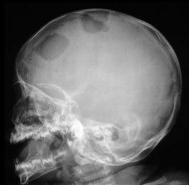
Langerhans cell histiocytosis
Sunil Kumar Podder
OMG (gene)
Capacitation
Leishmania
CD164
PNA
Soybean
Ricin
Genetically modified food controversies
Peanut agglutinin - Wikipedia
 Lectins: Peanut Agglutinin (PNA), Cy3 labeled
 Peanut Agglutinin | Profiles RNS
 Investigation of the interaction between peanut agglutinin and synthetic glycopolymeric multivalent ligands. - Department of...
Galactose - Page 3 of 4 - VectorLabs
 Histiocytosis: Practice Essentials, Pathophysiology, Epidemiology
 Lectins as markers for blood grouping
Differential expression of lectin-binding sites defines mouse intestinal M-cells
 Molecular Vision: Loss of the cone-enriched caspase-7 does not affect secondary cone death in retinitis pigmentosa
 The effect of parasitism by Cotesia congregata on the hemocytes of Diatraea saccharalis and Ostrinia nubilalis
 WikiGenes
Histiocytoses
 BJOC - Authors
 New test may help address costly parasite in sheep industry | ScienceDaily
 Whole-body exposures to radiofrequency-electromagnetic energy can cause DNA damage in mouse spermatozoa via an oxidative...
 Image: Differentiating Haemonchus contortus from other trichostrongyles - Merck Veterinary Manual
JCI - GMPPA defects cause a neuromuscular disorder with α-dystroglycan hyperglycosylation
 Gap junctions in lymphocyte ontogeny - Enlighten Theses
 Use of immobilized cryopreserved bovine semen in a blind artifical insemination trial - SINTEF
 JCI Insight -
VEGF/VEGFR2 blockade does not cause retinal atrophy in AMD-relevant models
JCI Insight -
VEGF/VEGFR2 blockade does not cause retinal atrophy in AMD-relevant models
 Goat Anti-Biotin, Peroxidase Conjugated, 1 mg - 1 Emb - Reactolab SA
Histiocytosis: Background, Pathophysiology, Epidemiology
 Galactosamine. Medical search. Definitions
 Distribution of lectin receptors sites in the zona pellucida of follicular and ovulated rat oocytes - Fingerprint
- Tel...
Distribution of lectin receptors sites in the zona pellucida of follicular and ovulated rat oocytes - Fingerprint
- Tel...
Peanut Butter on Keto Diet, Is it Good? - Keto Dieting Club
 Ciencias de la Salud - Resultados de investigación
- Universidad Nacional Mayor de San Marcos
Ciencias de la Salud - Resultados de investigación
- Universidad Nacional Mayor de San Marcos
Medicina Veterinaria - Research output
- Universidad Nacional Mayor de San Marcos
 US Patent for Compositions for use in the treatment and prevention of cardiovascular disorders resulting from cerebrovascular...
A solid-phase affinity labeling method for target-selective isolation and modification of proteins<...
Lectin peanut agglutinin2
- Neuraminidase-induced removal of sialic acid from natural substrates (desialylation) unmasks saccharides that are specifically recognized by the lectin peanut agglutinin (PNA). (nih.gov)
- The treatment was not cytotoxic and resulted in membrane desialylation as assessed by measurement of sialic acids released, along with an increased fixation of the galactose-specific lectin peanut agglutinin. (hal.science)
CD151
- A comparison with Leu-M1 (CD15), LN2 (CD74), peanut agglutinin, and Ber-H2 (CD30). (nih.gov)
Specificity2
- For example in PNA-affinity chromatography the binding specificity of peanut agglutinin is used to isolate glycosylated molecules which have the sugar sequence Gal-β(1-3)-GalNAc. (wikipedia.org)
- We developed a systematic approach, called outlier-motif analysis, for extracting fine-specificity information from glycan-array data, and we applied the method to the study of four commonly used lectins: two mannose binders (concanavalin A and Lens culinaris) and two galactose binders (Bauhinia purpurea and peanut agglutinin). (nih.gov)
Arachis4
- Peanut agglutinin (PNA) is plant lectin protein derived from the fruits of Arachis hypogaea. (wikipedia.org)
- Peanut agglutinin may also be referred to as Arachis hypogaea lectin. (wikipedia.org)
- Lectin purified from peanuts (ARACHIS HYPOGAEA). (reference.md)
- 16. Site-specific monoclonal antibodies against peanut agglutinin (PNA) from Arachis hypogaea. (nih.gov)
Binds2
- peanut agglutinin binds the carbohydrate sequence Gal-β(1-3)-GalNAc. (wikipedia.org)
- Available Structures of peanut agglutinin Because peanut agglutinin specifically binds a particular carbohydrate sequence it finds use in a range of methods for cell biology and biochemistry. (wikipedia.org)
Galactose1
- Peanut agglutinin activity is inhibited by lactose and galactose which compete for the binding site. (wikipedia.org)
Epithelium3
- In influenza-infected guinea-pigs, as assessed by reactivity with peanut agglutinin, the tracheal and lung epithelium, as well as alveolar cells were hyposialylated. (hal.science)
- Gut pathogens produce agglutinins that they use to attach to the heparan sulfate (HS), the predominant acid polysaccharide of the intestinal epithelium. (blogspot.com)
- Mast cells of the intestines normally release heparin, which is a mixture of HS fragments, to stick to the agglutinins and block attachment to the HS of the epithelium. (blogspot.com)
Cells4
- Specific binding of peanut lectin to a class of retinal photoreceptor cells. (wikipedia.org)
- Characterization of reactive and suppressive cells in the mouse embryonic liver by peanut agglutinin (PNA). (bgu.ac.il)
- Dive into the research topics of 'Characterization of reactive and suppressive cells in the mouse embryonic liver by peanut agglutinin (PNA). (bgu.ac.il)
- Leu-M1 and peanut agglutinin stain the neoplastic cells of Hodgkin's Disease. (bdbiosciences.com)