Partial Thromboplastin Time
Prothrombin Time
Blood Coagulation
Blood Coagulation Disorders
Thrombin Time
Thromboplastin
Heparin
Hemostasis
Antithrombins
Blood Coagulation Factors
Disseminated Intravascular Coagulation
Bleeding Time
Thrombelastography
Antithrombin III
Hirudin Therapy
Lupus Coagulation Inhibitor
Hirudins
Fibrinogen
Factor VIII
Pipecolic Acids
Kaolin
Hemorrhagic Disorders
Blood Coagulation Disorders, Inherited
Factor XI
Protein C
Factor Xa
Hemophilia A
Prothrombin
Hemophilia B
Factor XII
International Normalized Ratio
Factor IX
Coagulation Protein Disorders
Fibrin Fibrinogen Degradation Products
Factor XI Deficiency
Factor X
Factor VII
Vitamin K Deficiency
Solar Activity
Blood Cell Count
Carotid Artery Thrombosis
Factor XIa
Platelet Aggregation
Plasma
Thromboembolism
Factor V
Antiphospholipid Syndrome
Postoperative Hemorrhage
Heparinoids
4-Hydroxycoumarins
beta-Alanine
Factor VIIa
Protein S
Activated Protein C Resistance
Prekallikrein
Factor XIIa
Platelet Function Tests
Vitamin K
Fibrinopeptide A
Factor X Deficiency
Fibrin
Thrombophilia
Seaweed
Kininogens
Chromogenic Compounds
Heparin, Low-Molecular-Weight
Phaeophyta
Drug Monitoring
Hemodilution
Enoxaparin
Antibodies, Anticardiolipin
Factor IXa
Warfarin
Antifibrinolytic Agents
Dermatan Sulfate
Blood Platelets
Hydroxyethyl Starch Derivatives
Indicators and Reagents
Dogs
Dose-Response Relationship, Drug
von Willebrand Factor
Antibodies, Antiphospholipid
Dog Diseases
Infusions, Intravenous
Venous Thromboembolism
Aprotinin
Blood Transfusion
Cardiolipins
Prospective Studies
Cardiopulmonary Bypass
beta 2-Glycoprotein I
Tissue Plasminogen Activator
Rabbits
Low-molecular-weight heparin in outpatient treatment of DVT. (1/681)
Patients with a diagnosis of acute deep venous thrombosis have traditionally been hospitalized and treated with unfractionated heparin followed by oral anticoagulation therapy. Several clinical trials have shown that low-molecular-weight heparin is at least as safe and effective as unfractionated heparin in the treatment of uncomplicated deep venous thrombosis. The use of low-molecular-weight heparin in an outpatient program for the management of deep venous thrombosis provides a treatment alternative to hospitalization in selected patients. Use of low-molecular-weight heparin on an outpatient basis requires coordination of care, laboratory monitoring, and patient education and participation in treatment. Overlapping the initiation of warfarin permits long-term anticoagulation. Advantages include a decreased incidence of heparin-induced thrombocytopenia and fewer episodes of bleeding complications. Future clinical trials evaluating the safety and efficacy of low-molecular-weight heparin in the treatment of complicated deep venous thrombosis will further define appropriate indications for use and strategies for outpatient management. (+info)Structure and anticoagulant activity of sulfated fucans. Comparison between the regular, repetitive, and linear fucans from echinoderms with the more heterogeneous and branched polymers from brown algae. (2/681)
Sulfated fucans are among the most widely studied of all the sulfated polysaccharides of non-mammalian origin that exhibit biological activities in mammalian systems. Examples of these polysaccharides extracted from echinoderms have simple structures, composed of oligosaccharide repeating units within which the residues differ by specific patterns of sulfation among different species. In contrast the algal fucans may have some regular repeating structure but are clearly more heterogeneous when compared with the echinoderm fucans. The structures of the sulfated fucans from brown algae also vary from species to species. We compared the anticoagulant activity of the regular and repetitive fucans from echinoderms with that of the more heterogeneous fucans from three species of brown algae. Our results indicate that different structural features determine not only the anticoagulant potency of the sulfated fucans but also the mechanism by which they exert this activity. Thus, the branched fucans from brown algae are direct inhibitors of thrombin, whereas the linear fucans from echinoderms require the presence of antithrombin or heparin cofactor II for inhibition of thrombin, as reported for mammalian glycosaminoglycans. The linear sulfated fucans from echinoderms have an anticoagulant action resembling that of mammalian dermatan sulfate and a modest action through antithrombin. A single difference of one sulfate ester per tetrasaccharide repeating unit modifies the anticoagulant activity of the polysaccharide markedly. Possibly the spatial arrangements of sulfate esters in the repeating tetrasaccharide unit of the echinoderm fucan mimics the site in dermatan sulfate with high affinity for heparin cofactor II. (+info)Antithrombotic efficacy of thrombin inhibitor L-374,087: intravenous activity in a primate model of venous thrombus extension and oral activity in a canine model of primary venous and coronary artery thrombosis. (3/681)
The small molecule direct thrombin inhibitor L-374,087 was characterized across species in an in vitro activated partial thromboplastin clotting time (aPTT) assay and in vivo in rhesus monkey and dog thrombosis models. In vitro in rhesus, dog, and human plasma, L-374,087 concentrations eliciting 2-fold increases in aPTT were 0.25, 1.9, and 0.28 microM, respectively. In anesthetized rhesus monkeys, 300 microgram/kg bolus plus 12 microgram/kg/min and 300 microgram/kg bolus plus 30 microgram/kg/min L-374,087 i.v. infusions significantly reduced jugular vein thrombus extension, with both regimens limiting venous thrombus extension to 2-fold that of baseline thrombus mass compared with a 5-fold extension observed in the vehicle control group. Antithrombotic efficacy in the rhesus with the lower-dose regimen was achieved with 2.3- to 2.4-fold increases in aPTT and prothrombin time. In a conscious instrumented dog model of electrolytic vessel injury, the oral administration of two 10 mg/kg L-374,087 doses 12 h apart significantly reduced jugular vein thrombus mass, reduced the incidence of and delayed time to occlusive coronary artery thrombosis, and significantly reduced coronary artery thrombus mass and ensuing posterolateral myocardial infarct size. Antithrombotic efficacy in the dog was achieved with 1.6- to 2.0-fold increases in aPTT at 1 to 6 h after oral dosing with L-374,087. These results indicate significant antithrombotic efficacy against both venous and coronary arterial thrombosis with L-374,087 with only moderate elevations in aPTT or prothrombin time. The oral efficacy of L-374,087 characterizes this compound as a prototype for the further development of orally active direct thrombin inhibitors. (+info)Factor VIII and other hemostasis variables are related to incident diabetes in adults. The Atherosclerosis Risk in Communities (ARIC) Study. (4/681)
OBJECTIVE: Our objective was to evaluate whether selected hemostasis variables, some of which may reflect inflammation or endothelial dysfunction, are independently associated with the development of diabetes. RESEARCH DESIGN AND METHODS: We studied a biethnic cohort of 12,330 men and women, 45-64 years of age, of the Atherosclerosis Risk in Communities Study. New cases of diabetes were diagnosed by a reported physician diagnosis, hypoglycemic medication use, or a casual or fasting serum glucose level of > or = 11.1 or > or = 7 mmol/l, respectively. RESULTS: Over an average follow-up of 7 years, 1,335 new cases of diabetes were detected. The odds ratios (4th versus 1st quartile) of developing diabetes, adjusted by logistic regression for age, sex, race, study center, family history of diabetes, fasting glucose, physical activity, and smoking, were 1.2 (95% CI 1.0-1.5) for fibrinogen and 1.4 (1.1-1.6) for factor VII. Associations for factor VIII, von Willebrand factor, and activated partial thromboplastin time were found to be 1.8 (1.3-2.3), 1.4 (1.1-1.8), and 0.63 (0.49-0.82), respectively, in women. Although further adjustment for BMI and waist-to-hip ratio diminished the relationships, a highly statistically significant association (P = 0.001) remained for factor VIII (1.6 [1.2-2.1]) in women. CONCLUSIONS: Factor VIII and other hemostasis variables are associated with the development of diabetes in middle-aged adults. These findings support a role for inflammation and, particularly in women, endothelial dysfunction in the pathogenesis of type 2 diabetes. (+info)The effect of a low molecular mass thrombin inhibitor, inogatran, and heparin on thrombin generation and fibrin turnover in patients with unstable coronary artery disease. (5/681)
AIM: This study evaluated a novel specific thrombin inhibitor, inogatran, in comparison with unfractionated heparin, with regard to markers for coagulation activity in patients with unstable coronary artery disease. METHODS AND RESULTS: In the Thrombin Inhibition In Myocardial Ischaemia (TRIM) study patients were randomized to one of three different doses of inogatran or to unfractionated heparin, given intravenously over 72 h. In a subpopulation of 320 patients, markers for coagulation activity were measured at baseline, during and after the study infusion. Prothrombin fragment 1 + 2, indicating thrombin generation, decreased in the low, medium and high dose inogatran groups and in the heparin group during the first 6 h of treatment by 12%, 15%, 21% and 26%, respectively. From 6 h to 72 h after the start of infusion the levels changed by -7%, -6%, -4% and +34%, respectively. The increase in the heparin group continued after the infusion was stopped. Thrombin-antithrombin complex, also indicating thrombin generation, decreased by 0%, 2%, 18% and 22%, respectively, during the first 6 h of treatment. During the same period soluble fibrin, an intermediate in fibrin formation, increased both in the low and medium inogatran group by 9%, while a decrease by 4% and 18%, respectively, was seen in the high dose inogatran group and in the heparin group. Fibrin dissolution, as measured by fibrin D-dimer, decreased during the first 24 h of treatment by 20%, 18%, 18% and 20%, respectively. The first 24 h after discontinuation of infusion, fibrin D-dimer increased by 6%, 23%, 25% and 44%, respectively. After 72 h, at the end of infusion, patients treated with inogatran, to a larger extent than those given heparin, had suffered from death, myocardial infarction or refractory angina pectoris. After 7 days this trend was less marked. CONCLUSION: The more pronounced decrease in thrombin generation and fibrin turnover during the first 6 h of infusion, and the later increase in thrombin generation and fibrin turnover, in the heparin group, as compared to the inogatran groups, may be related to the lower clinical event rate during infusion with heparin compared with inogatran and the recurrence of ischaemic events, early after cessation of heparin infusion. (+info)Adenovirus-mediated local expression of human tissue factor pathway inhibitor eliminates shear stress-induced recurrent thrombosis in the injured carotid artery of the rabbit. (6/681)
The main cause of acute coronary syndrome may be recurrent thrombosis, which is initiated by the activation of the extrinsic coagulation pathway. Tissue factor (TF) pathway inhibitor (TFPI) efficiently inhibits an early step in this pathway by the formation of a complex with factor VIIa, TF, and factor Xa. We determined whether local TFPI gene transfer can inhibit thrombosis in an injured artery without inducing systemic side effects. Balloon-injured rabbit carotid arteries were infected with an adenoviral vector that expressed either human TFPI (AdCATFPI) or bacterial beta-galactosidase (AdCALacZ). Two to 6 days after gene transfer, thrombosis was induced by the production of constant stenosis of the artery, and blood flow was measured continuously with an electromagnetic flow probe. A cyclic flow variation, which is thought to reflect the recurrent formation and dislodgment of mural thrombi, was observed in all AdCALacZ-infected arteries as well as in saline-infused arteries. In contrast, no cyclic flow variation was detectable in AdCATFPI-transfected arteries, even in the presence of epinephrine (1 microg. kg-1. min-1 infusion). Prothrombin time, activated partial thromboplastin time, and the ex vivo platelet aggregation induced by either adenosine diphosphate or collagen were unaltered in AdCATFPI-infected rabbits. We found that in vivo TFPI gene transfer into an injured artery completely inhibits the recurrent thrombosis induced by shear stress even in the presence of catecholamine, without affecting systemic coagulation status. Adenovirus-mediated local expression of TFPI may have the potential for the treatment of human thrombosis. (+info)Impaired anticoagulant response to infusion of thrombin in atherosclerotic monkeys associated with acquired defects in the protein C system. (7/681)
To examine the effects of atherosclerosis on the protein C anticoagulant pathway in vivo, we measured anticoagulant responses to intravenous administration of human alpha-thrombin or activated protein C (APC) in cynomolgus monkeys. Two groups of monkeys were fed either a control diet (n=18) or an atherogenic diet (n=12) that produces both hypercholesterolemia and moderate hyperhomocyst(e)inemia. A third group (n=8) was fed an atherogenic diet for 15 months, and then fed the atherogenic diet supplemented with B vitamins for 6 months to correct the hyperhomocyst(e)inemia. The plasma homocyst(e)ine level was higher in monkeys fed the atherogenic diet (9.6+/-1.0 micromol/L) than in monkeys fed the control diet (3.7+/-0.2 micromol/L) or the atherogenic diet with B vitamins (3.6+/-0.2 micromol/L) (P<0.001). Infusion of thrombin produced a much greater prolongation of the activated partial thromboplastin time in monkeys fed the control diet (52+/-10 seconds) than in monkeys fed the atherogenic diet either with (24+/-4 seconds) or without (27+/-5 seconds) supplemental B vitamins (P<0.02). Thrombin-dependent generation of circulating APC was higher in control (294+/-17 U/mL) than in atherosclerotic (240+/-14 U/mL) monkeys (P<0.05), although levels of fibrinogen, plasminogen, D-dimer, and thrombin-antithrombin complexes were similar in each group. Injection of human APC produced a similar prolongation of the activated partial thromboplastin time in control (31+/-3 seconds) and atherosclerotic (29+/-2 seconds) monkeys. These findings provide evidence for impaired anticoagulation, due partly to decreased formation of APC, in atherosclerosis. The blunted anticoagulant response to thrombin in hypercholesterolemic monkeys was not corrected by supplementation with B vitamins. (+info)Large amounts of vascular endothelial growth factor at the site of hemostatic plug formation in vivo. (8/681)
Vascular endothelial growth factor (VEGF) is important for the proliferation, differentiation, and survival of microvascular endothelial cells. It is a potent angiogenic factor and a specific endothelial cell mitogen that increases fenestration and extravasation of plasma macromolecules. Recently, large quantities of VEGF were detected in human megakaryocytes. Incubation of human platelets with thrombin in vitro resulted in the release of large amounts of VEGF. To investigate whether VEGF is released from platelets during coagulation activation in vivo, we measured in human subjects VEGF at the site of plug formation, ie, in blood emerging from a standardized injury made to determine bleeding time (shed blood). VEGF was also determined in the same volunteers after treatment with the specific thrombin inhibitor recombinant hirudin (r-hirudin). In a double-blind, randomized, crossover study, 17 healthy male volunteers (aged 20 to 35 years) were investigated. VEGF concentrations were measured in venous blood and in shed blood by the use of an immunoassay 10 minutes after intravenous administration of r-hirudin (0.35 mg/kg of body weight) or physiological saline. Prothrombin fragment f1.2 (f1.2) and beta-thromboglobulin (beta-TG) were determined as indicators of coagulation and platelet activation, respectively. Concentrations of VEGF, f1.2, and beta-TG in shed blood 4 minutes after injury were significantly higher than in venous blood (VEGF, 55.8+/-9.2 versus <20 pg/mL, P<0.001; f1.2, 71.3+/-10.4 versus 0.78+/-0.03 nmol/L, P<0. 001; beta-TG, 2290+/-170 versus 53.2+/-14.0 ng/mL, P<0.001). Administration of r-hirudin caused a >50% inhibition of the beta-TG and f1.2 levels in shed blood. In a similar manner, much lower amounts of VEGF were detectable at the site of plug formation after r-hirudin treatment (69.0+/-9.5 versus 37.8+/-2.6 pg/mL per minute; P=0.0015). Our data indicate that substantial quantities of VEGF are released from platelets during the interaction with the injured vessel wall in vivo. This finding may be relevant with respect to wound healing and tissue repair, tumor vascularization, or arterial thrombus formation. (+info)Partial Thromboplastin Time (PTT) is a medical laboratory test that measures the time it takes for blood to clot. It's more specifically a measure of the intrinsic and common pathways of the coagulation cascade, which are the series of chemical reactions that lead to the formation of a clot.
The test involves adding a partial thromboplastin reagent (an activator of the intrinsic pathway) and calcium to plasma, and then measuring the time it takes for a fibrin clot to form. This is compared to a control sample, and the ratio of the two times is calculated.
The PTT test is often used to help diagnose bleeding disorders or abnormal blood clotting, such as hemophilia or disseminated intravascular coagulation (DIC). It can also be used to monitor the effectiveness of anticoagulant therapy, such as heparin. Prolonged PTT results may indicate a bleeding disorder or an increased risk of bleeding, while shortened PTT results may indicate a hypercoagulable state and an increased risk of thrombosis.
Prothrombin time (PT) is a medical laboratory test that measures the time it takes for blood to clot. It's often used to evaluate the functioning of the extrinsic and common pathways of the coagulation system, which is responsible for blood clotting. Specifically, PT measures how long it takes for prothrombin (a protein produced by the liver) to be converted into thrombin, an enzyme that converts fibrinogen into fibrin and helps form a clot.
Prolonged PT may indicate a bleeding disorder or a deficiency in coagulation factors, such as vitamin K deficiency or the use of anticoagulant medications like warfarin. It's important to note that PT is often reported with an international normalized ratio (INR), which allows for standardization and comparison of results across different laboratories and reagent types.
Blood coagulation tests, also known as coagulation studies or clotting tests, are a series of medical tests used to evaluate the blood's ability to clot. These tests measure the functioning of various clotting factors and regulatory proteins involved in the coagulation cascade, which is a complex process that leads to the formation of a blood clot to prevent excessive bleeding.
The most commonly performed coagulation tests include:
1. Prothrombin Time (PT): Measures the time it takes for a sample of plasma to clot after the addition of calcium and tissue factor, which activates the extrinsic pathway of coagulation. The PT is reported in seconds and can be converted to an International Normalized Ratio (INR) to monitor anticoagulant therapy.
2. Activated Partial Thromboplastin Time (aPTT): Measures the time it takes for a sample of plasma to clot after the addition of calcium, phospholipid, and a contact activator, which activates the intrinsic pathway of coagulation. The aPTT is reported in seconds and is used to monitor heparin therapy.
3. Thrombin Time (TT): Measures the time it takes for a sample of plasma to clot after the addition of thrombin, which directly converts fibrinogen to fibrin. The TT is reported in seconds and can be used to detect the presence of fibrin degradation products or abnormalities in fibrinogen function.
4. Fibrinogen Level: Measures the amount of fibrinogen, a protein involved in clot formation, present in the blood. The level is reported in grams per liter (g/L) and can be used to assess bleeding risk or the effectiveness of fibrinogen replacement therapy.
5. D-dimer Level: Measures the amount of D-dimer, a protein fragment produced during the breakdown of a blood clot, present in the blood. The level is reported in micrograms per milliliter (µg/mL) and can be used to diagnose or exclude venous thromboembolism (VTE), such as deep vein thrombosis (DVT) or pulmonary embolism (PE).
These tests are important for the diagnosis, management, and monitoring of various bleeding and clotting disorders. They can help identify the underlying cause of abnormal bleeding or clotting, guide appropriate treatment decisions, and monitor the effectiveness of therapy. It is essential to interpret these test results in conjunction with a patient's clinical presentation and medical history.
Blood coagulation, also known as blood clotting, is a complex process that occurs in the body to prevent excessive bleeding when a blood vessel is damaged. This process involves several different proteins and chemical reactions that ultimately lead to the formation of a clot.
The coagulation cascade is initiated when blood comes into contact with tissue factor, which is exposed after damage to the blood vessel wall. This triggers a series of enzymatic reactions that activate clotting factors, leading to the formation of a fibrin clot. Fibrin is a protein that forms a mesh-like structure that traps platelets and red blood cells to form a stable clot.
Once the bleeding has stopped, the coagulation process is regulated and inhibited to prevent excessive clotting. The fibrinolytic system degrades the clot over time, allowing for the restoration of normal blood flow.
Abnormalities in the blood coagulation process can lead to bleeding disorders or thrombotic disorders such as deep vein thrombosis and pulmonary embolism.
Blood coagulation disorders, also known as bleeding disorders or clotting disorders, refer to a group of medical conditions that affect the body's ability to form blood clots properly. Normally, when a blood vessel is injured, the body's coagulation system works to form a clot to stop the bleeding and promote healing.
In blood coagulation disorders, there can be either an increased tendency to bleed due to problems with the formation of clots (hemorrhagic disorder), or an increased tendency for clots to form inappropriately even without injury, leading to blockages in the blood vessels (thrombotic disorder).
Examples of hemorrhagic disorders include:
1. Hemophilia - a genetic disorder that affects the ability to form clots due to deficiencies in clotting factors VIII or IX.
2. Von Willebrand disease - another genetic disorder caused by a deficiency or abnormality of the von Willebrand factor, which helps platelets stick together to form a clot.
3. Liver diseases - can lead to decreased production of coagulation factors, increasing the risk of bleeding.
4. Disseminated intravascular coagulation (DIC) - a serious condition where clotting and bleeding occur simultaneously due to widespread activation of the coagulation system.
Examples of thrombotic disorders include:
1. Factor V Leiden mutation - a genetic disorder that increases the risk of inappropriate blood clot formation.
2. Antithrombin III deficiency - a genetic disorder that impairs the body's ability to break down clots, increasing the risk of thrombosis.
3. Protein C or S deficiencies - genetic disorders that lead to an increased risk of thrombosis due to impaired regulation of the coagulation system.
4. Antiphospholipid syndrome (APS) - an autoimmune disorder where the body produces antibodies against its own clotting factors, increasing the risk of thrombosis.
Treatment for blood coagulation disorders depends on the specific diagnosis and may include medications to manage bleeding or prevent clots, as well as lifestyle changes and monitoring to reduce the risk of complications.
Thrombin time (TT) is a medical laboratory test that measures the time it takes for a clot to form after thrombin, an enzyme that converts fibrinogen to fibrin in the final step of the coagulation cascade, is added to a plasma sample. This test is used to evaluate the efficiency of the conversion of fibrinogen to fibrin and can be used to detect the presence of abnormalities in the coagulation system, such as the presence of heparin or dysfibrinogenemia. Increased thrombin time may indicate the presence of a systemic anticoagulant or a deficiency in fibrinogen.
Thromboplastin is a substance that activates the coagulation cascade, leading to the formation of a clot (thrombus). It's primarily found in damaged or injured tissues and blood vessels, as well as in platelets (thrombocytes). There are two types of thromboplastin:
1. Extrinsic thromboplastin (also known as tissue factor): This is a transmembrane glycoprotein that is primarily found in subendothelial cells and released upon injury to the blood vessels. It initiates the extrinsic pathway of coagulation by binding to and activating Factor VII, ultimately leading to the formation of thrombin and fibrin clots.
2. Intrinsic thromboplastin (also known as plasma thromboplastin or factor III): This term is used less frequently and refers to a labile phospholipid component present in platelet membranes, which plays a role in the intrinsic pathway of coagulation.
In clinical settings, the term "thromboplastin" often refers to reagents used in laboratory tests like the prothrombin time (PT) and activated partial thromboplastin time (aPTT). These reagents contain a source of tissue factor and calcium ions to initiate and monitor the coagulation process.
Heparin is defined as a highly sulfated glycosaminoglycan (a type of polysaccharide) that is widely present in many tissues, but is most commonly derived from the mucosal tissues of mammalian lungs or intestinal mucosa. It is an anticoagulant that acts as an inhibitor of several enzymes involved in the blood coagulation cascade, primarily by activating antithrombin III which then neutralizes thrombin and other clotting factors.
Heparin is used medically to prevent and treat thromboembolic disorders such as deep vein thrombosis, pulmonary embolism, and certain types of heart attacks. It can also be used during hemodialysis, cardiac bypass surgery, and other medical procedures to prevent the formation of blood clots.
It's important to note that while heparin is a powerful anticoagulant, it does not have any fibrinolytic activity, meaning it cannot dissolve existing blood clots. Instead, it prevents new clots from forming and stops existing clots from growing larger.
Anticoagulants are a class of medications that work to prevent the formation of blood clots in the body. They do this by inhibiting the coagulation cascade, which is a series of chemical reactions that lead to the formation of a clot. Anticoagulants can be given orally, intravenously, or subcutaneously, depending on the specific drug and the individual patient's needs.
There are several different types of anticoagulants, including:
1. Heparin: This is a naturally occurring anticoagulant that is often used in hospitalized patients who require immediate anticoagulation. It works by activating an enzyme called antithrombin III, which inhibits the formation of clots.
2. Low molecular weight heparin (LMWH): LMWH is a form of heparin that has been broken down into smaller molecules. It has a longer half-life than standard heparin and can be given once or twice daily by subcutaneous injection.
3. Direct oral anticoagulants (DOACs): These are newer oral anticoagulants that work by directly inhibiting specific clotting factors in the coagulation cascade. Examples include apixaban, rivaroxaban, and dabigatran.
4. Vitamin K antagonists: These are older oral anticoagulants that work by inhibiting the action of vitamin K, which is necessary for the formation of clotting factors. Warfarin is an example of a vitamin K antagonist.
Anticoagulants are used to prevent and treat a variety of conditions, including deep vein thrombosis (DVT), pulmonary embolism (PE), atrial fibrillation, and prosthetic heart valve thrombosis. It is important to note that anticoagulants can increase the risk of bleeding, so they must be used with caution and regular monitoring of blood clotting times may be required.
Hemostasis is the physiological process that occurs to stop bleeding (bleeding control) when a blood vessel is damaged. This involves the interaction of platelets, vasoconstriction, and blood clotting factors leading to the formation of a clot. The ultimate goal of hemostasis is to maintain the integrity of the vascular system while preventing excessive blood loss.
Antithrombins are substances that prevent the formation or promote the dissolution of blood clots (thrombi). They include:
1. Anticoagulants: These are medications that reduce the ability of the blood to clot. Examples include heparin, warfarin, and direct oral anticoagulants (DOACs) such as apixaban, rivaroxaban, and dabigatran.
2. Thrombolytic agents: These are medications that break down existing blood clots. Examples include alteplase, reteplase, and tenecteplase.
3. Fibrinolytics: These are a type of thrombolytic agent that specifically target fibrin, a protein involved in the formation of blood clots.
4. Natural anticoagulants: These are substances produced by the body to regulate blood clotting. Examples include antithrombin III, protein C, and protein S.
Antithrombins are used in the prevention and treatment of various thromboembolic disorders, such as deep vein thrombosis (DVT), pulmonary embolism (PE), stroke, and myocardial infarction (heart attack). It is important to note that while antithrombins can help prevent or dissolve blood clots, they also increase the risk of bleeding, so their use must be carefully monitored.
Blood coagulation factors, also known as clotting factors, are a group of proteins that play a crucial role in the blood coagulation process. They are essential for maintaining hemostasis, which is the body's ability to stop bleeding after injury.
There are 13 known blood coagulation factors, and they are designated by Roman numerals I through XIII. These factors are produced in the liver and are normally present in an inactive form in the blood. When there is an injury to a blood vessel, the coagulation process is initiated, leading to the activation of these factors in a specific order.
The coagulation cascade involves two pathways: the intrinsic and extrinsic pathways. The intrinsic pathway is activated when there is damage to the blood vessel itself, while the extrinsic pathway is activated by tissue factor released from damaged tissues. Both pathways converge at the common pathway, leading to the formation of a fibrin clot.
Blood coagulation factors work together in a complex series of reactions that involve activation, binding, and proteolysis. When one factor is activated, it activates the next factor in the cascade, and so on. This process continues until a stable fibrin clot is formed.
Deficiencies or abnormalities in blood coagulation factors can lead to bleeding disorders such as hemophilia or thrombosis. Hemophilia is a genetic disorder that affects one or more of the coagulation factors, leading to excessive bleeding and difficulty forming clots. Thrombosis, on the other hand, occurs when there is an abnormal formation of blood clots in the blood vessels, which can lead to serious complications such as stroke or pulmonary embolism.
Disseminated Intravascular Coagulation (DIC) is a complex medical condition characterized by the abnormal activation of the coagulation cascade, leading to the formation of blood clots in small blood vessels throughout the body. This process can result in the consumption of clotting factors and platelets, which can then lead to bleeding complications. DIC can be caused by a variety of underlying conditions, including sepsis, trauma, cancer, and obstetric emergencies.
The term "disseminated" refers to the widespread nature of the clotting activation, while "intravascular" indicates that the clotting is occurring within the blood vessels. The condition can manifest as both bleeding and clotting complications, which can make it challenging to diagnose and manage.
The diagnosis of DIC typically involves laboratory tests that evaluate coagulation factors, platelet count, fibrin degradation products, and other markers of coagulation activation. Treatment is focused on addressing the underlying cause of the condition while also managing any bleeding or clotting complications that may arise.
Bleeding time is a medical test that measures the time it takes for a small blood vessel to stop bleeding after being cut. It's used to evaluate platelet function and the effectiveness of blood clotting. The most common method used to measure bleeding time is the Ivy method, which involves making a standardized incision on the forearm and measuring the time it takes for the bleeding to stop. A normal bleeding time ranges from 2 to 9 minutes, but this can vary depending on the specific method used. Prolonged bleeding time may indicate an impairment in platelet function or clotting factor deficiency.
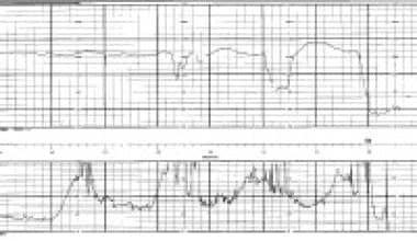
Thromboelastography (TEG) is a viscoelastic method used to assess the kinetics of clot formation, clot strength, and fibrinolysis in whole blood. It provides a global assessment of hemostasis by measuring the mechanical properties of a clot as it forms and dissolves over time. The TEG graph displays several parameters that reflect the different stages of clotting, including reaction time (R), clot formation time (K), angle of clot formation (α), maximum amplitude (MA), and percentage lysis at 30 minutes (LY30). These parameters can help guide transfusion therapy and inform decisions regarding the management of coagulopathy in various clinical settings, such as trauma, cardiac surgery, liver transplantation, and obstetrics.
Antithrombin III is a protein that inhibits the formation of blood clots (thrombi) in the body. It does this by inactivating several enzymes involved in coagulation, including thrombin and factor Xa. Antithrombin III is produced naturally by the liver and is also available as a medication for the prevention and treatment of thromboembolic disorders, such as deep vein thrombosis and pulmonary embolism. It works by binding to and neutralizing excess clotting factors in the bloodstream, thereby reducing the risk of clot formation.
Hirudin therapy, also known as leech therapy, is a type of treatment that uses the saliva of medicinal leeches (Hirudo medicinalis) to alleviate symptoms and promote healing. The saliva of these leeches contains various bioactive compounds, including hirudin, which is a potent anticoagulant that prevents blood clotting.
In hirudin therapy, leeches are applied to specific areas of the body, usually on congested tissues or sites of stasis, where they feed on the patient's blood and release their saliva into the bite site. The hirudin in the saliva helps to dissolve blood clots, improve circulation, reduce swelling, and relieve pain.
Hirudin therapy is used in various medical conditions, such as arterial and venous insufficiency, skin ulcers, joint diseases, and post-surgical recovery, particularly after reconstructive surgery or organ transplantation. It can also be used to treat thrombophlebitis, varicose veins, and other circulatory disorders.
It is essential to note that hirudin therapy should only be performed by trained medical professionals in a controlled environment due to the potential risks associated with infection transmission and bleeding complications.
I'm sorry for any confusion, but "Lupus Coagulation Inhibitor" is not a recognized medical term or concept in hematology or immunology.
Systemic Lupus Erythematosus (SLE), often referred to simply as lupus, is an autoimmune disease where the body's immune system mistakenly attacks healthy tissue. However, this does not result in a specific coagulation inhibitor.
If you're asking about lupus anticoagulants, these are antibodies that can interfere with clotting tests but paradoxically increase the risk of blood clots in vivo. They are sometimes seen in patients with SLE and other autoimmune diseases.
Please provide more context if you meant something else, so I can give a more accurate response.
Hirudin is not a medical term itself, but it is a specific substance with medical relevance. Hirudin is a naturally occurring anticoagulant that is found in the saliva of certain species of leeches (such as Hirudo medicinalis). This compound works by inhibiting the activity of thrombin, a key enzyme in the coagulation cascade, which ultimately results in preventing blood clot formation.
Medically, hirudin has been used in some research and therapeutic settings for its anticoagulant properties. For instance, recombinant hirudin (also known as lepirudin) is available for clinical use as an injectable anticoagulant to treat or prevent blood clots in specific medical conditions, such as heparin-induced thrombocytopenia (HIT).
In summary, Hirudins are a group of anticoagulant substances, primarily derived from leeches, that inhibit the activity of thrombin and have potential medical applications in preventing or treating blood clots.
Fibrinogen is a soluble protein present in plasma, synthesized by the liver. It plays an essential role in blood coagulation. When an injury occurs, fibrinogen gets converted into insoluble fibrin by the action of thrombin, forming a fibrin clot that helps to stop bleeding from the injured site. Therefore, fibrinogen is crucial for hemostasis, which is the process of stopping bleeding and starting the healing process after an injury.
Factor VIII is a protein in the blood that is essential for normal blood clotting. It is also known as antihemophilic factor (AHF). Deficiency or dysfunction of this protein results in hemophilia A, a genetic disorder characterized by prolonged bleeding and easy bruising. Factor VIII works together with other proteins to help form a clot and stop bleeding at the site of an injury. It acts as a cofactor for another clotting factor, IX, in the so-called intrinsic pathway of blood coagulation. Intravenous infusions of Factor VIII concentrate are used to treat and prevent bleeding episodes in people with hemophilia A.
Pipicolic acid is not a term that refers to a specific medical condition or disease. Instead, it is a metabolite that is involved in the body's metabolic processes.
Pipicolic acid is a type of organic compound called a cyclic amino acid, which is derived from the amino acid lysine. It is produced in the liver and is excreted in urine. Pipicolic acid has been found to have various functions in the body, including regulating the metabolism of lipids and bile acids.
Abnormal levels of pipicolic acid in the body may be associated with certain medical conditions, such as liver disease or genetic disorders that affect amino acid metabolism. However, pipicolic acid is not typically used as a diagnostic marker for these conditions.
In summary, pipicolic acid is a cyclic amino acid produced in the liver and involved in various metabolic processes in the body. Abnormal levels of pipicolic acid may be associated with certain medical conditions but are not typically used as diagnostic markers.
Kaolin is not a medical term per se, but it is a mineral that has various applications in the medical field. Medically, kaolin is used as an ingredient in some over-the-counter (OTC) medications and clinical products, particularly in oral and topical formulations.
Medical definition: Kaolin is a natural hydrated aluminum silicate clay mineral (with the chemical formula Al2Si2O5(OH)4) used in medical applications as an antidiarrheal agent and as a component in various dermatological products for its absorbent, protective, and soothing properties.
Hemorrhagic disorders are medical conditions characterized by abnormal bleeding due to impaired blood clotting. This can result from deficiencies in coagulation factors, platelet dysfunction, or the use of medications that interfere with normal clotting processes. Examples include hemophilia, von Willebrand disease, and disseminated intravascular coagulation (DIC). Treatment often involves replacing the missing clotting factor or administering medications to help control bleeding.
Blood coagulation disorders, inherited, also known as coagulopathies, are genetic conditions that affect the body's ability to form blood clots in response to injury or damage to blood vessels. These disorders can lead to excessive bleeding or hemorrhage, and in some cases, abnormal clotting.
There are several types of inherited blood coagulation disorders, including:
1. Hemophilia A and B: These are X-linked recessive disorders that affect the production of factors VIII and IX, respectively, which are essential for normal blood clotting. People with hemophilia may experience prolonged bleeding after injury or surgery, and spontaneous bleeding into joints and muscles.
2. Von Willebrand disease: This is the most common inherited coagulation disorder, affecting both men and women. It results from a deficiency or abnormality of von Willebrand factor, a protein that helps platelets stick to damaged blood vessels and assists in the activation of factor VIII. People with von Willebrand disease may experience excessive bleeding after injury, surgery, or dental work.
3. Factor XI deficiency: This is an autosomal recessive disorder that affects the production of factor XI, a protein involved in the intrinsic pathway of blood coagulation. People with factor XI deficiency may have a mild to moderate bleeding tendency, particularly after surgery or trauma.
4. Rare coagulation factor deficiencies: There are several other rare inherited coagulation disorders that affect the production of other clotting factors, such as factors II, V, VII, X, and XIII. These conditions can lead to a range of bleeding symptoms, from mild to severe.
Inherited blood coagulation disorders are usually diagnosed through a combination of medical history, physical examination, and laboratory tests that measure the levels and function of clotting factors in the blood. Treatment may include replacement therapy with purified clotting factor concentrates, medications to control bleeding, and management of bleeding symptoms as they arise.
A platelet count is a laboratory test that measures the number of platelets, also known as thrombocytes, in a sample of blood. Platelets are small, colorless cell fragments that circulate in the blood and play a crucial role in blood clotting. They help to stop bleeding by sticking together to form a plug at the site of an injured blood vessel.
A normal platelet count ranges from 150,000 to 450,000 platelets per microliter (µL) of blood. A lower than normal platelet count is called thrombocytopenia, while a higher than normal platelet count is known as thrombocytosis.
Abnormal platelet counts can be a sign of various medical conditions, including bleeding disorders, infections, certain medications, and some types of cancer. It is important to consult with a healthcare provider if you have any concerns about your platelet count or if you experience symptoms such as easy bruising, prolonged bleeding, or excessive menstrual flow.
Factor XI, also known as plasma thromboplastin antecedent (PTA) or antihemophilic factor C, is a protein involved in blood coagulation. It is one of the factors in the intrinsic pathway of coagulation, which is activated when blood comes into contact with negatively charged surfaces, such as damaged blood vessels.
When Factor XI is activated (usually by thrombin or activated Factor XII), it activates more Factor XI and also activates Factor IX, leading to the formation of a complex that converts Factor X to its active form, Factor Xa. This ultimately leads to the formation of a fibrin clot and helps to stop bleeding.
Deficiencies in Factor XI can lead to an increased risk of bleeding, although the severity of the bleeding disorder can vary widely among individuals with Factor XI deficiency. Treatment for Factor XI deficiency typically involves replacement therapy with fresh frozen plasma or recombinant Factor XI concentrate.
Hemorrhage is defined in the medical context as an excessive loss of blood from the circulatory system, which can occur due to various reasons such as injury, surgery, or underlying health conditions that affect blood clotting or the integrity of blood vessels. The bleeding may be internal, external, visible, or concealed, and it can vary in severity from minor to life-threatening, depending on the location and extent of the bleeding. Hemorrhage is a serious medical emergency that requires immediate attention and treatment to prevent further blood loss, organ damage, and potential death.
Protein C is a vitamin K-dependent protease that functions as an important regulator of coagulation and inflammation. It is a plasma protein produced in the liver that, when activated, degrades clotting factors Va and VIIIa to limit thrombus formation and prevent excessive blood clotting. Protein C also has anti-inflammatory properties by inhibiting the release of pro-inflammatory cytokines and reducing endothelial cell activation. Inherited or acquired deficiencies in Protein C can lead to an increased risk of thrombosis, a condition characterized by abnormal blood clot formation within blood vessels.
Thrombin is a serine protease enzyme that plays a crucial role in the coagulation cascade, which is a complex series of biochemical reactions that leads to the formation of a blood clot (thrombus) to prevent excessive bleeding during an injury. Thrombin is formed from its precursor protein, prothrombin, through a process called activation, which involves cleavage by another enzyme called factor Xa.
Once activated, thrombin converts fibrinogen, a soluble plasma protein, into fibrin, an insoluble protein that forms the structural framework of a blood clot. Thrombin also activates other components of the coagulation cascade, such as factor XIII, which crosslinks and stabilizes the fibrin network, and platelets, which contribute to the formation and growth of the clot.
Thrombin has several regulatory mechanisms that control its activity, including feedback inhibition by antithrombin III, a plasma protein that inactivates thrombin and other serine proteases, and tissue factor pathway inhibitor (TFPI), which inhibits the activation of factor Xa, thereby preventing further thrombin formation.
Overall, thrombin is an essential enzyme in hemostasis, the process that maintains the balance between bleeding and clotting in the body. However, excessive or uncontrolled thrombin activity can lead to pathological conditions such as thrombosis, atherosclerosis, and disseminated intravascular coagulation (DIC).
Factor Xa is a serine protease that plays a crucial role in the coagulation cascade, which is a series of reactions that lead to the formation of a blood clot. It is one of the activated forms of Factor X, a pro-protein that is converted to Factor Xa through the action of other enzymes in the coagulation cascade.
Factor Xa functions as a key component of the prothrombinase complex, which also includes calcium ions, phospholipids, and activated Factor V (also known as Activated Protein C or APC). This complex is responsible for converting prothrombin to thrombin, which then converts fibrinogen to fibrin, forming a stable clot.
Inhibitors of Factor Xa are used as anticoagulants in the prevention and treatment of thromboembolic disorders such as deep vein thrombosis and pulmonary embolism. These drugs work by selectively inhibiting Factor Xa, thereby preventing the formation of the prothrombinase complex and reducing the risk of clot formation.
Hemophilia A is a genetic bleeding disorder caused by a deficiency in clotting factor VIII. This results in impaired blood clotting and prolonged bleeding, particularly after injuries or surgeries. Symptoms can range from mild to severe, with the most severe form resulting in spontaneous bleeding into joints and muscles, leading to pain, swelling, and potential joint damage over time. Hemophilia A primarily affects males, as it is an X-linked recessive disorder, and is usually inherited from a carrier mother. However, about one third of cases result from a spontaneous mutation in the gene for factor VIII. Treatment typically involves replacement therapy with infusions of factor VIII concentrates to prevent or control bleeding episodes.
Fibrinolysis is the natural process in the body that leads to the dissolution of blood clots. It is a vital part of hemostasis, the process that regulates bleeding and wound healing. Fibrinolysis occurs when plasminogen activators convert plasminogen to plasmin, an enzyme that breaks down fibrin, the insoluble protein mesh that forms the structure of a blood clot. This process helps to prevent excessive clotting and maintains the fluidity of the blood. In medical settings, fibrinolysis can also refer to the therapeutic use of drugs that stimulate this process to dissolve unwanted or harmful blood clots, such as those that cause deep vein thrombosis or pulmonary embolism.
Prothrombin is a protein present in blood plasma, and it's also known as coagulation factor II. It plays a crucial role in the coagulation cascade, which is a complex series of reactions that leads to the formation of a blood clot.
When an injury occurs, the coagulation cascade is initiated to prevent excessive blood loss. Prothrombin is converted into its active form, thrombin, by another factor called factor Xa in the presence of calcium ions, phospholipids, and factor Va. Thrombin then catalyzes the conversion of fibrinogen into fibrin, forming a stable clot.
Prothrombin levels can be measured through a blood test, which is often used to diagnose or monitor conditions related to bleeding or coagulation disorders, such as liver disease or vitamin K deficiency.
Hypoprothrombinemia is a medical condition characterized by a decreased level of prothrombin (coagulation factor II) in the blood, which can lead to an increased bleeding tendency. Prothrombin is a protein involved in the coagulation cascade that helps to form blood clots and stop bleeding.
Hypoprothrombinemia can be caused by various factors, including vitamin K deficiency, liver disease, inherited or acquired disorders of prothrombin synthesis, or the use of certain medications such as warfarin. Symptoms may include easy bruising, prolonged bleeding from cuts or injuries, nosebleeds, and in severe cases, internal bleeding. Treatment typically involves addressing the underlying cause and may include vitamin K supplementation, fresh frozen plasma transfusions, or other specific therapies depending on the etiology of the condition.
Hemophilia B is a genetic disorder that affects the body's ability to control blood clotting, also known as coagulation. This condition is caused by a deficiency or dysfunction in Factor IX, one of the proteins essential for normal blood clotting. As a result, people with Hemophilia B experience prolonged bleeding and bruising after injuries, surgeries, or spontaneously, particularly in joints and muscles.
There are different degrees of severity, depending on how much Factor IX is missing or not functioning properly. Mild cases may only become apparent after significant trauma, surgery, or tooth extraction, while severe cases can lead to spontaneous bleeding into joints and muscles, causing pain, swelling, and potential long-term damage. Hemophilia B primarily affects males, as it is an X-linked recessive disorder, but females can be carriers of the condition and may experience mild symptoms.
Coagulants are substances that promote the process of coagulation or clotting. They are often used in medical settings to help control bleeding and promote healing. Coagulants work by encouraging the formation of a clot, which helps to stop the flow of blood from a wound or cut.
There are several different types of coagulants that may be used in medical treatments. Some coagulants are naturally occurring substances, such as vitamin K, which is essential for the production of certain clotting factors in the body. Other coagulants may be synthetic or semi-synthetic compounds, such as recombinant activated factor VII (rFVIIa), which is used to treat bleeding disorders and prevent excessive bleeding during surgery.
Coagulants are often administered through injection or infusion, but they can also be applied topically to wounds or cuts. In some cases, coagulants may be used in combination with other treatments, such as compression or cauterization, to help control bleeding and promote healing.
It is important to note that while coagulants can be helpful in controlling bleeding and promoting healing, they can also increase the risk of blood clots and other complications. As a result, they should only be used under the guidance and supervision of a qualified healthcare professional.
Factor XII, also known as Hageman factor, is a protein that plays a role in the coagulation cascade, which is the series of events that leads to the formation of a blood clot. It is one of the zymogens, or inactive precursor proteins, that becomes activated and helps to trigger the coagulation process.
When Factor XII comes into contact with negatively charged surfaces, such as damaged endothelial cells or artificial surfaces like those found on medical devices, it undergoes a conformational change and becomes activated. Activated Factor XII then activates other proteins in the coagulation cascade, including Factor XI, which ultimately leads to the formation of a fibrin clot.
Deficiencies in Factor XII are generally not associated with bleeding disorders, as the coagulation cascade can still proceed through other pathways. However, excessive activation of Factor XII has been implicated in certain thrombotic disorders, such as deep vein thrombosis and disseminated intravascular coagulation (DIC).
The International Normalized Ratio (INR) is a standardized measurement of the prothrombin time (PT), which is the time it takes for blood to clot. The INR is used to monitor and regulate the effects of anticoagulant medications, such as warfarin, that affect the blood's ability to clot.
The INR is calculated by dividing the patient's PT by a control value (the PT of normal, healthy blood), raised to the power of a sensitivity factor called the International Sensitivity Index (ISI). The ISI is specific to the thromboplastin reagent used in the PT assay.
The INR provides a consistent and comparable way to monitor anticoagulation therapy across different laboratories, regardless of the thromboplastin reagent used. This helps ensure that patients receive appropriate doses of anticoagulant medications and reduces the risk of bleeding or clotting complications.
In general, an INR range of 2.0 to 3.0 is recommended for most people taking anticoagulants for conditions such as atrial fibrillation, deep vein thrombosis, or pulmonary embolism. However, the target INR range may vary depending on individual patient factors and medical indications.
Heparin antagonists, also known as heparin neutralizers or reversal agents, are medications used to reverse the anticoagulant effects of heparin, a type of blood thinner. Heparin works by activating antithrombin III, which inactivates clotting factors IIa and Xa. Heparin antagonists, such as protamine sulfate, work by binding to heparin, forming a stable complex that is unable to bind to and activate antithrombin III, thereby neutralizing its anticoagulant effect.
Protamine sulfate is the most commonly used heparin antagonist. It is a highly basic protein derived from fish sperm that can neutralize the anticoagulant effects of heparin by forming a stable complex with it. The dose of protamine required to reverse the effects of heparin depends on the amount and type of heparin administered, as well as the timing of administration.
It is important to note that while heparin antagonists can reverse the anticoagulant effects of heparin, they do not reverse the underlying coagulation disorder or prevent further clot formation. Therefore, additional treatments may be necessary to manage the underlying condition and prevent recurrent thrombosis.
Thrombosis is the formation of a blood clot (thrombus) inside a blood vessel, obstructing the flow of blood through the circulatory system. When a clot forms in an artery, it can cut off the supply of oxygen and nutrients to the tissues served by that artery, leading to damage or tissue death. If a thrombus forms in the heart, it can cause a heart attack. If a thrombus breaks off and travels through the bloodstream, it can lodge in a smaller vessel, causing blockage and potentially leading to damage in the organ that the vessel supplies. This is known as an embolism.
Thrombosis can occur due to various factors such as injury to the blood vessel wall, abnormalities in blood flow, or changes in the composition of the blood. Certain medical conditions, medications, and lifestyle factors can increase the risk of thrombosis. Treatment typically involves anticoagulant or thrombolytic therapy to dissolve or prevent further growth of the clot, as well as addressing any underlying causes.
Factor IX is also known as Christmas factor, which is a protein that plays a crucial role in the coagulation cascade, a series of chemical reactions that leads to the formation of a blood clot. It is one of the essential components required for the proper functioning of the body's natural blood-clotting mechanism.
Factor IX is synthesized in the liver and activated when it comes into contact with an injured blood vessel. Once activated, it collaborates with other factors to convert factor X to its active form, which then converts prothrombin to thrombin. Thrombin is responsible for converting fibrinogen to fibrin, forming a stable fibrin clot that helps stop bleeding and promote healing.
Deficiencies in Factor IX can lead to hemophilia B, a genetic disorder characterized by prolonged bleeding and an increased risk of spontaneous bleeding. Hemophilia B is inherited in an X-linked recessive pattern, meaning it primarily affects males, while females serve as carriers of the disease. Treatment for hemophilia B typically involves replacing the missing or deficient Factor IX through infusions to prevent or manage bleeding episodes.
Coagulation protein disorders are a group of medical conditions that affect the body's ability to form blood clots properly. These disorders can be caused by genetic defects or acquired factors, such as liver disease or vitamin K deficiency.
The coagulation system is a complex process that involves various proteins called clotting factors. When there is an injury to a blood vessel, these clotting factors work together in a specific order to form a clot and prevent excessive bleeding. In coagulation protein disorders, one or more of these clotting factors are missing or not functioning properly, leading to abnormal bleeding or clotting.
There are several types of coagulation protein disorders, including:
1. Hemophilia: This is a genetic disorder that affects the clotting factor VIII or IX. People with hemophilia may experience prolonged bleeding after injuries, surgery, or dental work.
2. Von Willebrand disease: This is another genetic disorder that affects the von Willebrand factor, a protein that helps platelets stick together and form a clot. People with this condition may have nosebleeds, easy bruising, and excessive bleeding during menstruation or after surgery.
3. Factor XI deficiency: This is a rare genetic disorder that affects the clotting factor XI. People with this condition may experience prolonged bleeding after surgery or trauma.
4. Factor VII deficiency: This is a rare genetic disorder that affects the clotting factor VII. People with this condition may have nosebleeds, easy bruising, and excessive bleeding during menstruation or after surgery.
5. Acquired coagulation protein disorders: These are conditions that develop due to other medical factors, such as liver disease, vitamin K deficiency, or the use of certain medications. These disorders can affect one or more clotting factors and may cause abnormal bleeding or clotting.
Treatment for coagulation protein disorders depends on the specific condition and severity of symptoms. In some cases, replacement therapy with the missing clotting factor may be necessary to prevent excessive bleeding. Other treatments may include medications to control bleeding, such as desmopressin or antifibrinolytic agents, and lifestyle changes to reduce the risk of injury and bleeding.
Fibrin(ogen) degradation products (FDPs) are a group of proteins that result from the breakdown of fibrinogen and fibrin, which are key components of blood clots. This process occurs during the normal physiological process of fibrinolysis, where clots are dissolved to maintain blood flow.
FDPs can be measured in the blood as a marker for the activation of the coagulation and fibrinolytic systems. Elevated levels of FDPs may indicate the presence of a disorder that causes abnormal clotting or bleeding, such as disseminated intravascular coagulation (DIC), deep vein thrombosis (DVT), pulmonary embolism (PE), or certain types of cancer.
It is important to note that FDPs are not specific to any particular disorder and their measurement should be interpreted in conjunction with other clinical and laboratory findings.
Fibrinolytic agents are medications that dissolve or break down blood clots by activating plasminogen, which is converted into plasmin. Plasmin is a proteolytic enzyme that degrades fibrin, the structural protein in blood clots. Fibrinolytic agents are used medically to treat conditions such as acute ischemic stroke, deep vein thrombosis, pulmonary embolism, and myocardial infarction (heart attack) by restoring blood flow in occluded vessels. Examples of fibrinolytic agents include alteplase, reteplase, and tenecteplase. It is important to note that these medications carry a risk of bleeding complications and should be administered with caution.
Factor XI deficiency, also known as Hemophilia C or Rosenthal syndrome, is a rare bleeding disorder caused by a deficiency or dysfunction of coagulation factor XI. This protease plays an important role in the intrinsic pathway of blood coagulation. Factor XI deficiency can lead to prolonged bleeding after surgery, trauma, or menstruation, but it typically does not cause spontaneous bleeding like Hemophilia A and B. The severity of the condition varies widely among affected individuals. Inheritance is autosomal recessive, meaning that two defective copies of the gene (one from each parent) are necessary to have the disease.
Whole Blood Coagulation Time (WBCT) is not a standard term used in medical literature. However, I believe you may be referring to "bleeding time" or "coagulation time" which are tests used to evaluate the function of the blood's clotting system.
Bleeding time is a measure of how long it takes for bleeding to stop after a small cut is made in the skin. It helps assess the function of the platelets and the smaller blood vessels.
Coagulation time, on the other hand, measures the time it takes for a larger clot to form in whole blood. This test is not commonly used in clinical practice.
It's important to note that these tests have largely been replaced by more specific coagulation tests, such as prothrombin time (PT) and activated partial thromboplastin time (aPTT), which provide more detailed information about the different components of the clotting system.
Factor X is a protein that is essential for blood clotting, also known as coagulation. It is an enzyme that plays a crucial role in the coagulation cascade, which is a series of chemical reactions that lead to the formation of a blood clot. Factor X is activated by one of two pathways: the intrinsic pathway, which is initiated by damage to the blood vessels, or the extrinsic pathway, which is triggered by the release of tissue factor from damaged cells. Once activated, Factor X converts prothrombin to thrombin, which then converts fibrinogen to fibrin to form a stable clot.
Inherited deficiencies in Factor X can lead to bleeding disorders, while increased levels of Factor X have been associated with an increased risk of thrombosis or blood clots. Therefore, maintaining appropriate levels of Factor X is important for the proper balance between bleeding and clotting in the body.
Factor VII, also known as proconvertin, is a protein involved in the coagulation cascade, which is a series of chemical reactions that leads to the formation of a blood clot. Factor VII is synthesized in the liver and is activated when it comes into contact with tissue factor, which is exposed when blood vessels are damaged. Activated Factor VII then activates Factor X, leading to the formation of thrombin and ultimately a fibrin clot.
Inherited deficiencies or dysfunctions of Factor VII can lead to an increased risk of bleeding, while elevated levels of Factor VII have been associated with an increased risk of thrombosis (blood clots).
Vitamin K deficiency is a condition that occurs when the body lacks adequate amounts of Vitamin K, a fat-soluble vitamin essential for blood clotting and bone metabolism. This can lead to an increased risk of excessive bleeding (hemorrhage) and calcification of tissues.
Vitamin K is required for the activation of several proteins involved in blood clotting, known as coagulation factors II, VII, IX, and X. A deficiency in Vitamin K can result in these factors remaining in their inactive forms, leading to impaired blood clotting and an increased risk of bleeding.
Vitamin K deficiency can occur due to several reasons, including malnutrition, malabsorption disorders (such as cystic fibrosis or celiac disease), liver diseases, use of certain medications (such as antibiotics or anticoagulants), and prolonged use of warfarin therapy.
In newborns, Vitamin K deficiency can lead to a serious bleeding disorder known as hemorrhagic disease of the newborn. This is because newborns have low levels of Vitamin K at birth, and their gut bacteria, which are responsible for producing Vitamin K, are not yet fully developed. Therefore, it is recommended that newborns receive a dose of Vitamin K within the first few days of life to prevent this condition.
Symptoms of Vitamin K deficiency can include easy bruising, nosebleeds, bleeding gums, blood in urine or stools, and excessive menstrual bleeding. In severe cases, it can lead to life-threatening hemorrhage. Treatment typically involves administering Vitamin K supplements or injections to replenish the body's levels of this essential nutrient.
Thrombophlebitis is a medical condition characterized by the inflammation and clotting of blood in a vein, usually in the legs. The term thrombophlebitis comes from two words: "thrombo" which means blood clot, and "phlebitis" which refers to inflammation of the vein.
The condition can occur in superficial or deep veins. Superficial thrombophlebitis affects the veins just below the skin's surface, while deep vein thrombophlebitis (DVT) occurs in the deeper veins. DVT is a more serious condition as it can lead to complications such as pulmonary embolism if the blood clot breaks off and travels to the lungs.
Symptoms of thrombophlebitis may include redness, warmth, pain, swelling, or discomfort in the affected area. In some cases, there may be visible surface veins that are hard, tender, or ropy to touch. If left untreated, thrombophlebitis can lead to chronic venous insufficiency and other long-term complications. Treatment typically involves medications such as anticoagulants, antiplatelet agents, or thrombolytics, along with compression stockings and other supportive measures.
I'm sorry for any confusion, but "solar activity" is not a term typically used in medical definitions. Solar activity refers to the various phenomena that occur on the Sun, including solar flares, coronal mass ejections (CMEs), and solar wind. These events involve the release of energy and charged particles from the Sun's atmosphere and can have effects on space weather and technological systems in near-Earth space. If you have any questions related to medical terminology or health-related topics, I would be happy to help with those!
A "Blood Cell Count" is a medical laboratory test that measures the number of red blood cells (RBCs), white blood cells (WBCs), and platelets in a sample of blood. This test is often used as a part of a routine check-up or to help diagnose various medical conditions, such as anemia, infection, inflammation, and many others.
The RBC count measures the number of oxygen-carrying cells in the blood, while the WBC count measures the number of immune cells that help fight infections. The platelet count measures the number of cells involved in clotting. Abnormal results in any of these counts may indicate an underlying medical condition and further testing may be required for diagnosis and treatment.
Carotid artery thrombosis is a medical condition characterized by the formation of a blood clot (thrombus) inside the carotid artery, which is one of the major blood vessels that supplies oxygenated blood to the head and neck. This condition can lead to serious complications such as a stroke or transient ischemic attack (TIA), also known as a "mini-stroke," if the clot dislodges and travels to the brain, blocking the flow of blood and oxygen.
Carotid artery thrombosis can result from various factors, including atherosclerosis (the buildup of fats, cholesterol, and other substances in the artery walls), hypertension (high blood pressure), diabetes, smoking, and genetic predisposition. Symptoms may include neck pain or stiffness, weakness or numbness in the face or limbs, difficulty speaking or understanding speech, vision problems, and sudden severe headaches. Diagnosis typically involves imaging tests such as ultrasound, CT angiography, or MRI angiography. Treatment options may include anticoagulant or antiplatelet medications, endovascular procedures to remove the clot, or surgery to clean out the artery (carotid endarterectomy).
Factor XIa is a serine protease enzyme that plays a crucial role in blood coagulation. It is formed through the activation of Factor XI, which is one of the key components in the intrinsic pathway of the coagulation cascade. The activation of Factor XI to Factor XIa occurs via either autoactivation or through the action of thrombin. Once activated, Factor XIa can cleave and activate Factor IX, leading to the formation of Factor IXa, which further amplifies the coagulation cascade.
In summary, Factor XIa is a vital enzyme in the blood coagulation process, contributing to the formation of a stable fibrin clot that helps prevent excessive bleeding during injury or trauma.
Platelet aggregation is the clumping together of platelets (thrombocytes) in the blood, which is an essential step in the process of hemostasis (the stopping of bleeding) after injury to a blood vessel. When the inner lining of a blood vessel is damaged, exposure of subendothelial collagen and tissue factor triggers platelet activation. Activated platelets change shape, become sticky, and release the contents of their granules, which include ADP (adenosine diphosphate).
ADP then acts as a chemical mediator to attract and bind additional platelets to the site of injury, leading to platelet aggregation. This forms a plug that seals the damaged vessel and prevents further blood loss. Platelet aggregation is also a crucial component in the formation of blood clots (thrombosis) within blood vessels, which can have pathological consequences such as heart attacks and strokes if they obstruct blood flow to vital organs.
In the context of medicine, plasma refers to the clear, yellowish fluid that is the liquid component of blood. It's composed of water, enzymes, hormones, antibodies, clotting factors, and other proteins. Plasma serves as a transport medium for cells, nutrients, waste products, gases, and other substances throughout the body. Additionally, it plays a crucial role in the immune response and helps regulate various bodily functions.
Plasma can be collected from blood donors and processed into various therapeutic products, such as clotting factors for people with hemophilia or immunoglobulins for patients with immune deficiencies. This process is called plasma fractionation.
Thromboembolism is a medical condition that refers to the obstruction of a blood vessel by a thrombus (blood clot) that has formed elsewhere in the body and then been transported by the bloodstream to a narrower vessel, where it becomes lodged. This process can occur in various parts of the body, leading to different types of thromboembolisms:
1. Deep Vein Thrombosis (DVT): A thrombus forms in the deep veins, usually in the legs or pelvis, and then breaks off and travels to the lungs, causing a pulmonary embolism.
2. Pulmonary Embolism (PE): A thrombus formed elsewhere, often in the deep veins of the legs, dislodges and travels to the lungs, blocking one or more pulmonary arteries. This can lead to shortness of breath, chest pain, and potentially life-threatening complications if not treated promptly.
3. Cerebral Embolism: A thrombus formed in another part of the body, such as the heart or carotid artery, dislodges and travels to the brain, causing a stroke or transient ischemic attack (TIA).
4. Arterial Thromboembolism: A thrombus forms in an artery and breaks off, traveling to another part of the body and blocking blood flow to an organ or tissue, leading to potential damage or loss of function. Examples include mesenteric ischemia (intestinal damage due to blocked blood flow) and retinal artery occlusion (vision loss due to blocked blood flow in the eye).
Prevention, early detection, and appropriate treatment are crucial for managing thromboembolism and reducing the risk of severe complications.
Factor V, also known as proaccelerin or labile factor, is a protein involved in the coagulation cascade, which is a series of chemical reactions that leads to the formation of a blood clot. Factor V acts as a cofactor for the activation of Factor X to Factor Xa, which is a critical step in the coagulation cascade.
When blood vessels are damaged, the coagulation cascade is initiated to prevent excessive bleeding. During this process, Factor V is activated by thrombin, another protein involved in coagulation, and then forms a complex with activated Factor X and calcium ions on the surface of platelets or other cells. This complex converts prothrombin to thrombin, which then converts fibrinogen to fibrin to form a stable clot.
Deficiency or dysfunction of Factor V can lead to bleeding disorders such as hemophilia B or factor V deficiency, while mutations in the gene encoding Factor V can increase the risk of thrombosis, as seen in the Factor V Leiden mutation.
Antiphospholipid syndrome (APS) is an autoimmune disorder characterized by the presence of antiphospholipid antibodies in the blood. These antibodies are directed against phospholipids, a type of fat molecule found in cell membranes and plasma lipoproteins. The presence of these antibodies can lead to abnormal blood clotting, which can cause serious complications such as stroke, heart attack, deep vein thrombosis, and pulmonary embolism.
APS can occur either on its own (primary APS) or in conjunction with other autoimmune disorders, such as systemic lupus erythematosus (secondary APS). The exact cause of APS is not fully understood, but it is believed to involve a combination of genetic and environmental factors.
Symptoms of APS can vary widely depending on the location and severity of the blood clots. They may include:
* Recurrent miscarriages or stillbirths
* Blood clots in the legs, lungs, or other parts of the body
* Skin ulcers or lesions
* Headaches, seizures, or stroke-like symptoms
* Kidney problems
* Heart valve abnormalities
Diagnosis of APS typically involves blood tests to detect the presence of antiphospholipid antibodies. Treatment may include medications to prevent blood clots, such as anticoagulants and antiplatelet agents, as well as management of any underlying autoimmune disorders.
Postoperative hemorrhage is a medical term that refers to bleeding that occurs after a surgical procedure. This condition can range from minor oozing to severe, life-threatening bleeding. Postoperative hemorrhage can occur soon after surgery or even several days later, as the surgical site begins to heal.
The causes of postoperative hemorrhage can vary, but some common factors include:
1. Inadequate hemostasis during surgery: This means that all bleeding was not properly controlled during the procedure, leading to bleeding after surgery.
2. Blood vessel injury: During surgery, blood vessels may be accidentally cut or damaged, causing bleeding after the procedure.
3. Coagulopathy: This is a condition in which the body has difficulty forming blood clots, increasing the risk of postoperative hemorrhage.
4. Use of anticoagulant medications: Medications that prevent blood clots can increase the risk of bleeding after surgery.
5. Infection: An infection at the surgical site can cause inflammation and bleeding.
Symptoms of postoperative hemorrhage may include swelling, pain, warmth, or discoloration around the surgical site, as well as signs of shock such as rapid heartbeat, low blood pressure, and confusion. Treatment for postoperative hemorrhage depends on the severity of the bleeding and may include medications to control bleeding, transfusions of blood products, or additional surgery to stop the bleeding.
Heparinoids are a group of substances that have similar properties to heparin, a highly sulfated glycosaminoglycan found in mast cells and basophils. Heparin is a powerful anticoagulant that works by accelerating the action of an enzyme called antithrombin III, which inhibits the formation of blood clots.
Heparinoids are often used as alternative anticoagulants to heparin in clinical settings. They have similar mechanisms of action and can also inhibit the coagulation cascade, preventing the formation of blood clots. However, heparinoids have a lower anticoagulant activity than heparin and may have different side effect profiles.
Examples of heparinoids include low molecular weight heparins (LMWHs), fondaparinux, and danaparoid. LMWHs are derived from standard heparin by chemical or enzymatic depolymerization and have a lower molecular weight than heparin. They have a more predictable anticoagulant response and longer half-life than standard heparin, making them useful for outpatient treatment of deep vein thrombosis and pulmonary embolism.
Fondaparinux is a synthetic pentasaccharide that selectively binds to antithrombin III and enhances its inhibitory activity against factor Xa, a key enzyme in the coagulation cascade. It has a long half-life and predictable pharmacokinetics, making it useful for the prevention and treatment of venous thromboembolism.
Danaparoid is a mixture of heparan sulfate, dermatan sulfate, and chondroitin sulfate derived from pig intestinal mucosa. It has a lower anticoagulant activity than heparin but a longer half-life and less frequent dosing requirements. Danaparoid is used for the prevention and treatment of venous thromboembolism, as well as for the management of heparin-induced thrombocytopenia (HIT), a rare but serious complication of heparin therapy.
4-Hydroxycoumarins are a type of chemical compound that contains a hydroxy group (-OH) attached to the 4th carbon atom of the coumarin structure. Coumarins themselves are aromatic organic compounds, characterized by a benzene ring fused to a pyrone ring.
4-Hydroxycoumarins have gained attention in medical research due to their potential biological activities. For instance, some 4-hydroxycoumarins exhibit anticoagulant properties and are used as oral anticoagulant drugs, such as warfarin. These compounds work by inhibiting the vitamin K epoxide reductase enzyme, thereby interfering with the blood clotting process.
Additionally, 4-hydroxycoumarins have been investigated for their potential anticancer, anti-inflammatory, and antibacterial activities. However, more research is needed to fully understand their therapeutic potential and safety profiles.
Beta-alanine is a non-essential amino acid, which means that it is not required in the diet because the body can produce it from other amino acids. It is produced in the liver and is also found in some foods such as meat, poultry, and fish.
Beta-alanine plays a role in the production of carnosine, a dipeptide molecule that helps to regulate muscle pH and improve muscle function during high-intensity exercise. When muscles contract during intense exercise, they produce hydrogen ions, which can cause the muscle pH to decrease (become more acidic), leading to fatigue and reduced muscle function. Carnosine acts as a buffer against this acidity, helping to maintain optimal muscle pH levels and improve performance during high-intensity exercise.
Beta-alanine supplements have been shown to increase carnosine levels in muscles, which may lead to improved athletic performance, particularly in activities that require short bursts of intense effort, such as weightlifting or sprinting. However, more research is needed to fully understand the effects and potential benefits of beta-alanine supplementation.
It's important to note that while beta-alanine supplements are generally considered safe for most people, they can cause a tingling sensation in the skin (paresthesia) when taken in high doses. This is a harmless side effect and typically subsides within an hour or so of taking the supplement.
Factor VIIa is a protein involved in the coagulation cascade, which is a series of chemical reactions that leads to the formation of a blood clot. Factor VIIa is the activated form of factor VII, which is normally activated by tissue factor (TF) when there is damage to the blood vessels. Together, TF and Factor VIIa convert Factor X to its active form, Factor Xa, which then converts prothrombin to thrombin, leading to the formation of a fibrin clot.
In summary, Factor VIIa is an important protein in the coagulation cascade that helps to initiate the formation of a blood clot in response to injury.
Surgical hemostasis refers to the methods and techniques used during surgical procedures to stop bleeding or prevent hemorrhage. This can be achieved through various means, including the use of surgical instruments such as clamps, ligatures, or staples to physically compress blood vessels and stop the flow of blood. Electrosurgical tools like cautery may also be used to coagulate and seal off bleeding vessels using heat. Additionally, topical hemostatic agents can be applied to promote clotting and control bleeding in wounded tissues. Effective surgical hemostasis is crucial for ensuring a successful surgical outcome and minimizing the risk of complications such as excessive blood loss, infection, or delayed healing.
Protein S is a vitamin K-dependent protein found in the blood that functions as a natural anticoagulant. It plays a crucial role in regulating the body's clotting system by inhibiting the activation of coagulation factors, thereby preventing excessive blood clotting. Protein S also acts as a cofactor for activated protein C, which is another important anticoagulant protein.
Protein S exists in two forms: free and bound to a protein called C4b-binding protein (C4BP). Only the free form of Protein S has biological activity in inhibiting coagulation. Inherited or acquired deficiencies in Protein S can lead to an increased risk of thrombosis, or abnormal blood clot formation, which can cause various medical conditions such as deep vein thrombosis (DVT) and pulmonary embolism (PE). Regular monitoring of Protein S levels is essential for patients with a history of thrombotic events or those who have a family history of thrombophilia.
Activated Protein C (APC) resistance is a condition in which the body's natural anticoagulant system is impaired, leading to an increased risk of thrombosis or blood clot formation. APC is an enzyme that plays a crucial role in regulating blood coagulation by inactivating clotting factors Va and VIIIa.
APC resistance is most commonly caused by a genetic mutation in the Factor V gene, known as Factor V Leiden. This mutation results in the production of a variant form of Factor V called Factor V Leiden, which is resistant to APC-mediated inactivation. As a result, the body's ability to regulate blood clotting is impaired, leading to an increased risk of thrombosis.
APC resistance can be measured by performing a functional assay that compares the activity of APC in normal plasma versus plasma from a patient with suspected APC resistance. The assay measures the rate of inactivation of Factor Va by APC, and a reduced rate of inactivation indicates APC resistance.
It is important to note that not all individuals with APC resistance will develop thrombosis, and other factors such as age, obesity, pregnancy, oral contraceptive use, and smoking can increase the risk of thrombosis in individuals with APC resistance.
Prekallikrein is a zymogen, or inactive precursor, of the serine protease kallikrein. It is a protein that plays a role in the coagulation cascade and the kinin-kallikrein system. Prekallikrein is primarily produced in the liver and circulates in the bloodstream. When activated, prekallikrein is converted to kallikrein, which then participates in various physiological processes such as blood pressure regulation, inflammation, and fibrinolysis (the breakdown of blood clots). The activation of prekallikrein is facilitated by the surface of negatively charged activators like kininogen or collagen, in conjunction with factor XII (Hageman factor) in a positive feedback loop.
In summary, Prekallikrein is a crucial protein in the coagulation and kinin-kallikrein systems that becomes activated to kallikrein upon contact with negatively charged surfaces and factor XII, contributing to various physiological processes.
Factor XIIa is a protease enzyme that plays a role in the coagulation cascade, which is the series of events that leads to blood clotting. It is formed when Factor XII, also known as Hageman factor, is activated by contact with negatively charged surfaces such as damaged endothelial cells or artificial surfaces like medical devices.
Once activated, Factor XIIa can activate other components of the coagulation cascade, including Factor XI, which ultimately leads to the formation of a fibrin clot. While Factor XIIa is an important part of the coagulation system, it is not essential for normal hemostasis (the process that stops bleeding) in humans, as people with deficiencies in Factor XII do not have an increased risk of bleeding. However, excessive activation of Factor XIIa has been implicated in several pathological conditions, including thrombosis and inflammation.
Platelet function tests are laboratory tests that measure how well platelets, which are small blood cells responsible for clotting, function in preventing or stopping bleeding. These tests are often used to investigate the cause of abnormal bleeding or bruising, or to monitor the effectiveness of antiplatelet therapy in patients with certain medical conditions such as heart disease or stroke.
There are several types of platelet function tests available, including:
1. Platelet count: This test measures the number of platelets present in a sample of blood. A low platelet count can increase the risk of bleeding.
2. Bleeding time: This test measures how long it takes for a small cut to stop bleeding. It is used less frequently than other tests due to its invasiveness and variability.
3. Platelet aggregation tests: These tests measure how well platelets clump together (aggregate) in response to various agents that promote platelet activation, such as adenosine diphosphate (ADP), collagen, or epinephrine.
4. Platelet function analyzer (PFA): This test measures the time it takes for a blood sample to clot under shear stress, simulating the conditions in an injured blood vessel. The PFA can provide information about the overall platelet function and the effectiveness of antiplatelet therapy.
5. Thromboelastography (TEG) or rotational thromboelastometry (ROTEM): These tests measure the kinetics of clot formation, strength, and dissolution in whole blood samples. They provide information about both platelet function and coagulation factors.
These tests can help healthcare providers diagnose bleeding disorders, assess the risk of bleeding during surgery or other invasive procedures, monitor antiplatelet therapy, and guide treatment decisions for patients with abnormal platelet function.
Vitamin K is a fat-soluble vitamin that plays a crucial role in blood clotting and bone metabolism. It is essential for the production of several proteins involved in blood clotting, including factor II (prothrombin), factor VII, factor IX, and factor X. Additionally, Vitamin K is necessary for the synthesis of osteocalcin, a protein that contributes to bone health by regulating the deposition of calcium in bones.
There are two main forms of Vitamin K: Vitamin K1 (phylloquinone), which is found primarily in green leafy vegetables and some vegetable oils, and Vitamin K2 (menaquinones), which is produced by bacteria in the intestines and is also found in some fermented foods.
Vitamin K deficiency can lead to bleeding disorders such as hemorrhage and excessive bruising. While Vitamin K deficiency is rare in adults, it can occur in newborns who have not yet developed sufficient levels of the vitamin. Therefore, newborns are often given a Vitamin K injection shortly after birth to prevent bleeding problems.
Fibrinopeptide A is a small protein molecule that is cleaved and released from the larger fibrinogen protein during the blood clotting process. Specifically, it is removed by the enzyme thrombin as part of the conversion of fibrinogen to fibrin, which is the main structural component of a blood clot. The measurement of Fibrinopeptide A in the blood can be used as a marker for ongoing thrombin activation and fibrin formation, which are key events in coagulation and hemostasis. Increased levels of Fibrinopeptide A may indicate abnormal or excessive blood clotting, such as in disseminated intravascular coagulation (DIC) or deep vein thrombosis (DVT).
Factor X deficiency, also known as Stuart-Prower factor deficiency, is a rare bleeding disorder that affects the body's ability to form blood clots. It is caused by a mutation in the gene that provides instructions for making coagulation factor X, a protein involved in the coagulation cascade, which is a series of chemical reactions that lead to the formation of a blood clot.
People with factor X deficiency may experience excessive bleeding after injury or surgery, and they may also have an increased risk of spontaneous bleeding, such as nosebleeds, heavy menstrual periods, and joint bleeds. The severity of the condition can vary widely, from mild to severe, depending on the level of factor X activity in the blood.
Factor X deficiency can be inherited or acquired. Inherited forms of the disorder are caused by mutations in the F10 gene and are usually present at birth. Acquired forms of the disorder can develop later in life due to conditions such as liver disease, vitamin K deficiency, or the use of certain medications that interfere with coagulation.
Treatment for factor X deficiency typically involves replacement therapy with fresh frozen plasma or recombinant factor X concentrates to help restore normal clotting function. In some cases, other treatments such as antifibrinolytic agents or desmopressin may also be used to manage bleeding symptoms.
Fibrin is defined as a protein that is formed from fibrinogen during the clotting of blood. It plays an essential role in the formation of blood clots, also known as a clotting or coagulation cascade. When an injury occurs and bleeding starts, fibrin threads form a net-like structure that entraps platelets and red blood cells to create a stable clot, preventing further loss of blood.
The process of forming fibrin from fibrinogen is initiated by thrombin, another protein involved in the coagulation cascade. Thrombin cleaves fibrinogen into fibrin monomers, which then polymerize to form long strands of fibrin. These strands cross-link with each other through a process catalyzed by factor XIIIa, forming a stable clot that protects the wound and promotes healing.
It is important to note that abnormalities in fibrin formation or breakdown can lead to bleeding disorders or thrombotic conditions, respectively. Proper regulation of fibrin production and degradation is crucial for maintaining healthy hemostasis and preventing excessive clotting or bleeding.
Thrombophilia is a medical condition characterized by an increased tendency to form blood clots (thrombi) due to various genetic or acquired abnormalities in the coagulation system. These abnormalities can lead to a hypercoagulable state, which can cause thrombosis in both veins and arteries. Commonly identified thrombophilias include factor V Leiden mutation, prothrombin G20210A mutation, antithrombin deficiency, protein C deficiency, and protein S deficiency.
Acquired thrombophilias can be caused by various factors such as antiphospholipid antibody syndrome (APS), malignancies, pregnancy, oral contraceptive use, hormone replacement therapy, and certain medical conditions like inflammatory bowel disease or nephrotic syndrome.
It is essential to diagnose thrombophilia accurately, as it may influence the management of venous thromboembolism (VTE) events and guide decisions regarding prophylactic anticoagulation in high-risk situations.
A contusion is a medical term for a bruise. It's a type of injury that occurs when blood vessels become damaged or broken as a result of trauma to the body. This trauma can be caused by a variety of things, such as a fall, a blow, or a hit. When the blood vessels are damaged, blood leaks into the surrounding tissues, causing the area to become discolored and swollen.
Contusions can occur anywhere on the body, but they are most common in areas that are more likely to be injured, such as the knees, elbows, and hands. In some cases, a contusion may be accompanied by other injuries, such as fractures or sprains.
Most contusions will heal on their own within a few days or weeks, depending on the severity of the injury. Treatment typically involves rest, ice, compression, and elevation (RICE) to help reduce swelling and pain. In some cases, over-the-counter pain medications may also be recommended to help manage discomfort.
If you suspect that you have a contusion, it's important to seek medical attention if the injury is severe or if you experience symptoms such as difficulty breathing, chest pain, or loss of consciousness. These could be signs of a more serious injury and require immediate medical attention.
I'm sorry for any confusion, but seaweed is not typically referred to in medical definitions. Seaweed is a common name for various marine macroalgae, which are multicellular aquatic plants found in the ocean. They are often consumed as food and have been used in traditional medicine in some cultures. However, there is no widely accepted medical definition for seaweed. If you're looking for information about a specific compound or substance derived from seaweed that might be relevant to medical definitions, I would be happy to help if you could provide more details.
Kininogens are a group of proteins found in the blood plasma that play a crucial role in the inflammatory response and blood coagulation. They are precursors to bradykinin, a potent vasodilator and inflammatory mediator. There are two types of kininogens: high molecular weight kininogen (HMWK) and low molecular weight kininogen (LMWK). HMWK is involved in the intrinsic pathway of blood coagulation, while LMWK is responsible for the release of bradykinin. Both kininogens are important targets in the regulation of inflammation and hemostasis.
Chromogenic compounds are substances that can be converted into a colored product through a chemical reaction. These compounds are often used in various diagnostic tests, including microbiological assays and immunoassays, to detect the presence or absence of a specific analyte (such as a particular bacterium, enzyme, or antigen).
In these tests, a chromogenic substrate is added to the sample, and if the target analyte is present, it will react with the substrate and produce a colored product. The intensity of the color can often be correlated with the amount of analyte present in the sample, allowing for quantitative analysis.
Chromogenic compounds are widely used in clinical laboratories because they offer several advantages over other types of diagnostic tests. They are typically easy to use and interpret, and they can provide rapid results with high sensitivity and specificity. Additionally, chromogenic assays can be automated, which can help increase throughput and reduce the potential for human error.
Low-molecular-weight heparin (LMWH) is a type of heparin used as an anticoagulant, which refers to a group of medications that prevent the formation of blood clots. Heparin is a naturally occurring substance in the body, and low-molecular-weight heparins are obtained through the depolymerization of standard heparin.
LMWH has a lower molecular weight than standard heparin, which results in several pharmacological differences. LMWHs have a more predictable dose response, longer half-life, and higher bioavailability when administered subcutaneously compared to standard heparin. They also exhibit greater anti-factor Xa activity relative to their antithrombin (anti-IIa) activity, which contributes to their anticoagulant effects.
LMWHs are used for the prevention and treatment of deep vein thrombosis (DVT), pulmonary embolism (PE), and other thromboembolic disorders. Common LMWHs include enoxaparin, dalteparin, tinzaparin, and nadroparin.
It is essential to monitor the patient's kidney function when using LMWH since they are primarily cleared by the kidneys. In patients with renal impairment, dose adjustments or alternative anticoagulants may be necessary to reduce the risk of bleeding complications.
Phaeophyta is a taxonomic division that refers to a group of complex, multicellular brown algae found in marine environments. These algae are characterized by their pigmentation, which includes fucoxanthin, chlorophyll-a, and chlorophyll-c, giving them a brown color. They have diverse morphology, ranging from simple thread-like forms to large seaweeds.
Phaeophyta species are primarily found in cold, nutrient-rich waters and play an essential role in marine ecosystems as primary producers and habitats for various marine organisms. Some examples of Phaeophyta include kelps, rockweed, and bladderwrack. It's worth noting that the classification and nomenclature of algae are continually evolving, so different sources might use slightly different terminology or categorization.
Drug monitoring, also known as therapeutic drug monitoring (TDM), is a medical practice that involves testing blood or other bodily fluids to determine the concentration of a particular medication. This information is used to ensure that the patient is receiving an appropriate dosage and to help guide adjustments in medication therapy. It can be especially important for medications with a narrow therapeutic index, meaning that there is a small range between the effective dose and a toxic dose.
The goal of drug monitoring is to optimize medication effectiveness while minimizing potential side effects. This may involve measuring the concentration of a drug at various times after dosing to determine how quickly it is being metabolized or eliminated from the body, as well as to assess compliance with the prescribed treatment regimen.
Drug monitoring can be performed using a variety of methods, including immunoassays, chromatography, and mass spectrometry. The specific method used will depend on the drug being monitored and the level of sensitivity required. Results from drug monitoring tests are typically interpreted in conjunction with other clinical information, such as the patient's age, weight, renal function, liver function, and overall health status.
Hemodilution is a medical term that refers to the reduction in the concentration of certain components in the blood, usually referring to red blood cells (RBCs) or hemoglobin. This occurs when an individual's plasma volume expands due to the infusion of intravenous fluids or the body's own production of fluid, such as during severe infection or inflammation. As a result, the number of RBCs per unit of blood decreases, leading to a lower hematocrit and hemoglobin level. It is important to note that while hemodilution reduces the concentration of RBCs in the blood, it does not necessarily indicate anemia or blood loss.
Enoxaparin is a low molecular weight heparin (LMWH) medication that is used as an anticoagulant to prevent and treat blood clots. It works by binding to and inhibiting the activity of factor Xa, a clotting factor in the blood. This helps to reduce the risk of clot formation and can help to prevent conditions such as deep vein thrombosis (DVT) and pulmonary embolism (PE). Enoxaparin is typically given by injection under the skin (subcutaneously) and is available under the brand names Lovenox and Clexane, among others. It is important to follow the instructions of a healthcare professional when using enoxaparin, as it can increase the risk of bleeding.
Anticardiolipin antibodies are a type of autoantibody that targets and binds to cardiolipin, a phospholipid component found in the inner mitochondrial membrane of cells. These antibodies are clinically significant because they have been associated with a variety of autoimmune disorders, including antiphospholipid syndrome (APS).
APS is a condition characterized by recurrent blood clots, pregnancy losses, and thrombocytopenia (low platelet count). Anticardiolipin antibodies are one of the three main types of autoantibodies found in APS, along with lupus anticoagulant and anti-β2 glycoprotein I antibodies.
The presence of high levels of anticardiolipin antibodies in the blood can lead to abnormal blood clotting, which can cause serious complications such as deep vein thrombosis, pulmonary embolism, and stroke. Anticardiolipin antibodies can also contribute to pregnancy losses by causing placental insufficiency or abnormal blood clotting in the placenta.
Anticardiolipin antibodies are typically detected through a blood test that measures their levels in the serum. A positive result is usually confirmed with a second test performed at least 12 weeks later to establish persistence. Treatment for anticardiolipin antibody-related disorders typically involves anticoagulation therapy to prevent blood clots and other complications.
Factor IXa is a protein that plays a crucial role in the coagulation cascade, which is a series of biochemical reactions involved in blood clotting. It is an activated form of Factor IX, which is one of the coagulation factors that help convert prothrombin to thrombin, leading to the formation of a fibrin clot and stopping bleeding at the site of injury.
Factor IXa works by activating Factor X in the presence of calcium ions, phospholipids, and Factor VIIIa, which is another activated coagulation factor. This complex is called the tenase complex. The activation of Factor X leads to the formation of thrombin, which then converts fibrinogen to fibrin, forming a stable clot.
Deficiencies or dysfunctions in Factor IXa can lead to bleeding disorders such as hemophilia B, also known as Christmas disease, which is characterized by prolonged bleeding times and spontaneous bleeding episodes.
Warfarin is a anticoagulant medication that works by inhibiting the vitamin K-dependent activation of several coagulation factors (factors II, VII, IX, and X). This results in prolonged clotting times and reduced thrombus formation. It is commonly used to prevent and treat blood clots in conditions such as atrial fibrillation, deep vein thrombosis, and pulmonary embolism. Warfarin is also known by its brand names Coumadin and Jantoven.
It's important to note that warfarin has a narrow therapeutic index, meaning that the difference between an effective dose and a toxic one is small. Therefore, it requires careful monitoring of the patient's coagulation status through regular blood tests (INR) to ensure that the dosage is appropriate and to minimize the risk of bleeding complications.
Afibrinogenemia is a rare genetic disorder characterized by the complete absence or severely decreased levels of fibrinogen, a protein involved in blood clotting. This condition leads to an increased risk of excessive bleeding due to the inability to form proper blood clots. It is caused by mutations in the genes that provide instructions for making the three chains (Aα, Bβ, and γ) that make up the fibrinogen protein. Inheritance is autosomal recessive, meaning an individual must inherit two copies of the defective gene, one from each parent, to have the condition.
Hematologic tests, also known as hematology tests, are a group of diagnostic exams that evaluate the health and function of different components of blood, such as red and white blood cells, platelets, and clotting factors. These tests can detect various disorders, including anemia, infection, bleeding problems, and several types of cancer. Common hematologic tests include complete blood count (CBC), coagulation studies, peripheral smear examination, and erythrocyte sedimentation rate (ESR). The specific test or combination of tests ordered will depend on the patient's symptoms, medical history, and physical examination findings.
Thrombocytopenia is a medical condition characterized by an abnormally low platelet count (thrombocytes) in the blood. Platelets are small cell fragments that play a crucial role in blood clotting, helping to stop bleeding when a blood vessel is damaged. A healthy adult typically has a platelet count between 150,000 and 450,000 platelets per microliter of blood. Thrombocytopenia is usually diagnosed when the platelet count falls below 150,000 platelets/µL.
Thrombocytopenia can be classified into three main categories based on its underlying cause:
1. Immune thrombocytopenia (ITP): An autoimmune disorder where the immune system mistakenly attacks and destroys its own platelets, leading to a decreased platelet count. ITP can be further divided into primary or secondary forms, depending on whether it occurs alone or as a result of another medical condition or medication.
2. Decreased production: Thrombocytopenia can occur when there is insufficient production of platelets in the bone marrow due to various causes, such as viral infections, chemotherapy, radiation therapy, leukemia, aplastic anemia, or vitamin B12 or folate deficiency.
3. Increased destruction or consumption: Thrombocytopenia can also result from increased platelet destruction or consumption due to conditions like disseminated intravascular coagulation (DIC), thrombotic thrombocytopenic purpura (TTP), hemolytic uremic syndrome (HUS), or severe bacterial infections.
Symptoms of thrombocytopenia may include easy bruising, prolonged bleeding from cuts, spontaneous nosebleeds, bleeding gums, blood in urine or stools, and skin rashes like petechiae (small red or purple spots) or purpura (larger patches). The severity of symptoms can vary depending on the degree of thrombocytopenia and the presence of any underlying conditions. Treatment for thrombocytopenia depends on the cause and may include medications, transfusions, or addressing the underlying condition.
Antifibrinolytic agents are a class of medications that inhibit the breakdown of blood clots. They work by blocking the action of enzymes called plasminogen activators, which convert plasminogen to plasmin, the main enzyme responsible for breaking down fibrin, a protein that forms the framework of a blood clot.
By preventing the conversion of plasminogen to plasmin, antifibrinolytic agents help to stabilize existing blood clots and prevent their premature dissolution. These medications are often used in clinical settings where excessive bleeding is a concern, such as during or after surgery, childbirth, or trauma.
Examples of antifibrinolytic agents include tranexamic acid, aminocaproic acid, and epsilon-aminocaproic acid. While these medications can be effective in reducing bleeding, they also carry the risk of thromboembolic events, such as deep vein thrombosis or pulmonary embolism, due to their pro-coagulant effects. Therefore, they should be used with caution and only under the close supervision of a healthcare provider.
Dermatan sulfate is a type of glycosaminoglycan, which is a long, unbranched sugar chain found on the proteoglycan core protein in the extracellular matrix of animal tissues. It is composed of repeating disaccharide units of iduronic acid and N-acetylgalactosamine, with alternating sulfation at the 4-position of the iduronic acid and the 6-position of the galactosamine.
Dermatan sulfate is found in various tissues, including skin, heart valves, and blood vessels, where it plays important roles in regulating cell behavior, tissue development, and homeostasis. It also binds to a variety of growth factors, cytokines, and enzymes, modulating their activities and contributing to the regulation of various biological processes.
Abnormalities in dermatan sulfate metabolism can lead to several genetic disorders, such as Hunter syndrome and Hurler-Scheie syndrome, which are characterized by skeletal abnormalities, cardiac defects, and neurological impairment.
Blood platelets, also known as thrombocytes, are small, colorless cell fragments in our blood that play an essential role in normal blood clotting. They are formed in the bone marrow from large cells called megakaryocytes and circulate in the blood in an inactive state until they are needed to help stop bleeding. When a blood vessel is damaged, platelets become activated and change shape, releasing chemicals that attract more platelets to the site of injury. These activated platelets then stick together to form a plug, or clot, that seals the wound and prevents further blood loss. In addition to their role in clotting, platelets also help to promote healing by releasing growth factors that stimulate the growth of new tissue.
Hemostatic techniques refer to various methods used in medicine to stop bleeding or hemorrhage. The goal of these techniques is to promote the body's natural clotting process and prevent excessive blood loss. Some common hemostatic techniques include:
1. Mechanical compression: Applying pressure directly to the wound to physically compress blood vessels and stop the flow of blood. This can be done manually or with the use of medical devices such as clamps, tourniquets, or compression bandages.
2. Suturing or stapling: Closing a wound with stitches or staples to bring the edges of the wound together and allow the body's natural clotting process to occur.
3. Electrocautery: Using heat generated by an electrical current to seal off blood vessels and stop bleeding.
4. Hemostatic agents: Applying topical substances that promote clotting, such as fibrin glue, collagen, or gelatin sponges, to the wound site.
5. Vascular embolization: Inserting a catheter into a blood vessel and injecting a substance that blocks the flow of blood to a specific area, such as a bleeding tumor or aneurysm.
6. Surgical ligation: Tying off a bleeding blood vessel with suture material during surgery.
7. Arterial or venous repair: Repairing damaged blood vessels through surgical intervention to restore normal blood flow and prevent further bleeding.
Hydroxyethyl starch derivatives are modified starches that are used as plasma expanders in medicine. They are created by chemically treating corn, potato, or wheat starch with hydroxylethyl groups, which makes the starch more soluble and less likely to be broken down by enzymes in the body. This results in a large molecule that can remain in the bloodstream for an extended period, increasing intravascular volume and improving circulation.
These derivatives are available in different molecular weights and substitution patterns, which affect their pharmacokinetics and pharmacodynamics. They are used to treat or prevent hypovolemia (low blood volume) due to various causes such as bleeding, burns, or dehydration. Common brand names include Hetastarch, Pentastarch, and Voluven.
It's important to note that the use of hydroxyethyl starch derivatives has been associated with adverse effects, including kidney injury, coagulopathy, and pruritus (severe itching). Therefore, their use should be carefully monitored and restricted to specific clinical situations.
Indicators and reagents are terms commonly used in the field of clinical chemistry and laboratory medicine. Here are their definitions:
1. Indicator: An indicator is a substance that changes its color or other physical properties in response to a chemical change, such as a change in pH, oxidation-reduction potential, or the presence of a particular ion or molecule. Indicators are often used in laboratory tests to monitor or signal the progress of a reaction or to indicate the end point of a titration. A familiar example is the use of phenolphthalein as a pH indicator in acid-base titrations, which turns pink in basic solutions and colorless in acidic solutions.
2. Reagent: A reagent is a substance that is added to a system (such as a sample or a reaction mixture) to bring about a chemical reaction, test for the presence or absence of a particular component, or measure the concentration of a specific analyte. Reagents are typically chemicals with well-defined and consistent properties, allowing them to be used reliably in analytical procedures. Examples of reagents include enzymes, antibodies, dyes, metal ions, and organic compounds. In laboratory settings, reagents are often prepared and standardized according to strict protocols to ensure their quality and performance in diagnostic tests and research applications.
I believe there might be a misunderstanding in your question. "Dogs" is not a medical term or condition. It is the common name for a domesticated carnivore of the family Canidae, specifically the genus Canis, which includes wolves, foxes, and other extant and extinct species of mammals. Dogs are often kept as pets and companions, and they have been bred in a wide variety of forms and sizes for different purposes, such as hunting, herding, guarding, assisting police and military forces, and providing companionship and emotional support.
If you meant to ask about a specific medical condition or term related to dogs, please provide more context so I can give you an accurate answer.
A dose-response relationship in the context of drugs refers to the changes in the effects or symptoms that occur as the dose of a drug is increased or decreased. Generally, as the dose of a drug is increased, the severity or intensity of its effects also increases. Conversely, as the dose is decreased, the effects of the drug become less severe or may disappear altogether.
The dose-response relationship is an important concept in pharmacology and toxicology because it helps to establish the safe and effective dosage range for a drug. By understanding how changes in the dose of a drug affect its therapeutic and adverse effects, healthcare providers can optimize treatment plans for their patients while minimizing the risk of harm.
The dose-response relationship is typically depicted as a curve that shows the relationship between the dose of a drug and its effect. The shape of the curve may vary depending on the drug and the specific effect being measured. Some drugs may have a steep dose-response curve, meaning that small changes in the dose can result in large differences in the effect. Other drugs may have a more gradual dose-response curve, where larger changes in the dose are needed to produce significant effects.
In addition to helping establish safe and effective dosages, the dose-response relationship is also used to evaluate the potential therapeutic benefits and risks of new drugs during clinical trials. By systematically testing different doses of a drug in controlled studies, researchers can identify the optimal dosage range for the drug and assess its safety and efficacy.
Von Willebrand factor (vWF) is a large multimeric glycoprotein that plays a crucial role in hemostasis, the process which leads to the cessation of bleeding and the formation of a blood clot. It was named after Erik Adolf von Willebrand, a Finnish physician who first described the disorder associated with its deficiency, known as von Willebrand disease (vWD).
The primary functions of vWF include:
1. Platelet adhesion and aggregation: vWF mediates the initial attachment of platelets to damaged blood vessel walls by binding to exposed collagen fibers and then interacting with glycoprotein Ib (GPIb) receptors on the surface of platelets, facilitating platelet adhesion. Subsequently, vWF also promotes platelet-platelet interactions (aggregation) through its interaction with platelet glycoprotein IIb/IIIa (GPIIb/IIIa) receptors under high shear stress conditions found in areas of turbulent blood flow, such as arterioles and the capillary bed.
2. Transport and stabilization of coagulation factor VIII: vWF serves as a carrier protein for coagulation factor VIII (FVIII), protecting it from proteolytic degradation and maintaining its stability in circulation. This interaction between vWF and FVIII is essential for the proper functioning of the coagulation cascade, particularly in the context of vWD, where impaired FVIII function can lead to bleeding disorders.
3. Wound healing: vWF contributes to wound healing by promoting platelet adhesion and aggregation at the site of injury, which facilitates the formation of a provisional fibrin-based clot that serves as a scaffold for tissue repair and regeneration.
In summary, von Willebrand factor is a vital hemostatic protein involved in platelet adhesion, aggregation, coagulation factor VIII stabilization, and wound healing. Deficiencies or dysfunctions in vWF can lead to bleeding disorders such as von Willebrand disease.
Antiphospholipid antibodies are a type of autoantibody that targets and binds to certain proteins found in the blood that attach to phospholipids (a type of fat molecule). These antibodies are associated with an increased risk of developing antiphospholipid syndrome, a disorder characterized by abnormal blood clotting.
There are several types of antiphospholipid antibodies, including:
1. Lupus anticoagulant: This type of antiphospholipid antibody can interfere with blood clotting tests and may increase the risk of thrombosis (blood clots) in both arteries and veins.
2. Anticardiolipin antibodies: These antibodies target a specific phospholipid called cardiolipin, which is found in the inner membrane of mitochondria. High levels of anticardiolipin antibodies are associated with an increased risk of thrombosis and pregnancy complications such as recurrent miscarriage.
3. Anti-β2 glycoprotein I antibodies: These antibodies target a protein called β2 glycoprotein I, which binds to negatively charged phospholipids on the surface of cells. High levels of anti-β2 glycoprotein I antibodies are associated with an increased risk of thrombosis and pregnancy complications.
The exact mechanism by which antiphospholipid antibodies cause blood clotting is not fully understood, but it is thought to involve the activation of platelets, the inhibition of natural anticoagulants, and the promotion of inflammation. Antiphospholipid syndrome can be treated with medications that thin the blood or prevent clots from forming, such as aspirin, warfarin, or heparin.
Recombinant proteins are artificially created proteins produced through the use of recombinant DNA technology. This process involves combining DNA molecules from different sources to create a new set of genes that encode for a specific protein. The resulting recombinant protein can then be expressed, purified, and used for various applications in research, medicine, and industry.
Recombinant proteins are widely used in biomedical research to study protein function, structure, and interactions. They are also used in the development of diagnostic tests, vaccines, and therapeutic drugs. For example, recombinant insulin is a common treatment for diabetes, while recombinant human growth hormone is used to treat growth disorders.
The production of recombinant proteins typically involves the use of host cells, such as bacteria, yeast, or mammalian cells, which are engineered to express the desired protein. The host cells are transformed with a plasmid vector containing the gene of interest, along with regulatory elements that control its expression. Once the host cells are cultured and the protein is expressed, it can be purified using various chromatography techniques.
Overall, recombinant proteins have revolutionized many areas of biology and medicine, enabling researchers to study and manipulate proteins in ways that were previously impossible.
There is no medical definition for "dog diseases" as it is too broad a term. However, dogs can suffer from various health conditions and illnesses that are specific to their species or similar to those found in humans. Some common categories of dog diseases include:
1. Infectious Diseases: These are caused by viruses, bacteria, fungi, or parasites. Examples include distemper, parvovirus, kennel cough, Lyme disease, and heartworms.
2. Hereditary/Genetic Disorders: Some dogs may inherit certain genetic disorders from their parents. Examples include hip dysplasia, elbow dysplasia, progressive retinal atrophy (PRA), and degenerative myelopathy.
3. Age-Related Diseases: As dogs age, they become more susceptible to various health issues. Common age-related diseases in dogs include arthritis, dental disease, cancer, and cognitive dysfunction syndrome (CDS).
4. Nutritional Disorders: Malnutrition or improper feeding can lead to various health problems in dogs. Examples include obesity, malnutrition, and vitamin deficiencies.
5. Environmental Diseases: These are caused by exposure to environmental factors such as toxins, allergens, or extreme temperatures. Examples include heatstroke, frostbite, and toxicities from ingesting harmful substances.
6. Neurological Disorders: Dogs can suffer from various neurological conditions that affect their nervous system. Examples include epilepsy, intervertebral disc disease (IVDD), and vestibular disease.
7. Behavioral Disorders: Some dogs may develop behavioral issues due to various factors such as anxiety, fear, or aggression. Examples include separation anxiety, noise phobias, and resource guarding.
It's important to note that regular veterinary care, proper nutrition, exercise, and preventative measures can help reduce the risk of many dog diseases.
Intravenous (IV) infusion is a medical procedure in which liquids, such as medications, nutrients, or fluids, are delivered directly into a patient's vein through a needle or a catheter. This route of administration allows for rapid absorption and distribution of the infused substance throughout the body. IV infusions can be used for various purposes, including resuscitation, hydration, nutrition support, medication delivery, and blood product transfusion. The rate and volume of the infusion are carefully controlled to ensure patient safety and efficacy of treatment.
Venous Thromboembolism (VTE) is a medical condition that includes both deep vein thrombosis (DVT) and pulmonary embolism (PE). DVT is a blood clot that forms in the deep veins, usually in the legs, while PE occurs when a clot breaks off and travels to the lungs, blocking a pulmonary artery or one of its branches. This condition can be life-threatening if not diagnosed and treated promptly.
The medical definition of Venous Thromboembolism is:
"The formation of a blood clot (thrombus) in a deep vein, most commonly in the legs, which can then dislodge and travel to the lungs, causing a potentially life-threatening blockage of the pulmonary artery or one of its branches (pulmonary embolism). VTE is a complex disorder resulting from an interplay of genetic and environmental factors that affect the balance between thrombosis and fibrinolysis."
Some common risk factors for VTE include immobility, surgery, trauma, cancer, hormonal therapy, pregnancy, advanced age, and inherited or acquired thrombophilia. Symptoms of DVT may include swelling, pain, warmth, and redness in the affected limb, while symptoms of PE can range from shortness of breath and chest pain to coughing up blood or even sudden death. Diagnosis typically involves a combination of clinical assessment, imaging studies (such as ultrasound, CT scan, or MRI), and laboratory tests (such as D-dimer). Treatment usually includes anticoagulation therapy to prevent further clot formation and reduce the risk of recurrence.
Aprotinin is a medication that belongs to a class of drugs called serine protease inhibitors. It works by inhibiting the activity of certain enzymes in the body that can cause tissue damage and bleeding. Aprotinin is used in medical procedures such as heart bypass surgery to reduce blood loss and the need for blood transfusions. It is administered intravenously and its use is typically stopped a few days after the surgical procedure.
Aprotinin was first approved for use in the United States in 1993, but its use has been restricted or withdrawn in many countries due to concerns about its safety. In 2006, a study found an increased risk of kidney damage and death associated with the use of aprotinin during heart bypass surgery, leading to its withdrawal from the market in Europe and Canada. However, it is still available for use in the United States under a restricted access program.
It's important to note that the use of aprotinin should be carefully considered and discussed with the healthcare provider, taking into account the potential benefits and risks of the medication.
A blood transfusion is a medical procedure in which blood or its components are transferred from one individual (donor) to another (recipient) through a vein. The donated blood can be fresh whole blood, packed red blood cells, platelets, plasma, or cryoprecipitate, depending on the recipient's needs. Blood transfusions are performed to replace lost blood due to severe bleeding, treat anemia, support patients undergoing major surgeries, or manage various medical conditions such as hemophilia, thalassemia, and leukemia. The donated blood must be carefully cross-matched with the recipient's blood type to minimize the risk of transfusion reactions.
In the field of medicine, "time factors" refer to the duration of symptoms or time elapsed since the onset of a medical condition, which can have significant implications for diagnosis and treatment. Understanding time factors is crucial in determining the progression of a disease, evaluating the effectiveness of treatments, and making critical decisions regarding patient care.
For example, in stroke management, "time is brain," meaning that rapid intervention within a specific time frame (usually within 4.5 hours) is essential to administering tissue plasminogen activator (tPA), a clot-busting drug that can minimize brain damage and improve patient outcomes. Similarly, in trauma care, the "golden hour" concept emphasizes the importance of providing definitive care within the first 60 minutes after injury to increase survival rates and reduce morbidity.
Time factors also play a role in monitoring the progression of chronic conditions like diabetes or heart disease, where regular follow-ups and assessments help determine appropriate treatment adjustments and prevent complications. In infectious diseases, time factors are crucial for initiating antibiotic therapy and identifying potential outbreaks to control their spread.
Overall, "time factors" encompass the significance of recognizing and acting promptly in various medical scenarios to optimize patient outcomes and provide effective care.
Cardiolipins are a type of phospholipid that are primarily found in the inner mitochondrial membrane of cells. They play a crucial role in several important cellular processes, including energy production, apoptosis (programmed cell death), and maintenance of the structural integrity of the mitochondria.
Cardiolipins are unique because they contain four fatty acid chains, whereas most other phospholipids contain only two. This gives cardiolipins a distinctive conical shape that is important for their function in maintaining the curvature and stability of the inner mitochondrial membrane.
Cardiolipins have also been implicated in various diseases, including neurodegenerative disorders, cancer, and bacterial infections. For example, changes in cardiolipin composition or distribution have been linked to mitochondrial dysfunction in Parkinson's disease and other neurological conditions. Additionally, certain bacteria, such as Neisseria gonorrhoeae and Chlamydia trachomatis, can manipulate host cell cardiolipins to facilitate their own survival and replication.
In summary, cardiolipins are essential phospholipids found in the inner mitochondrial membrane that play a critical role in several cellular processes, and have been implicated in various diseases.
Prospective studies, also known as longitudinal studies, are a type of cohort study in which data is collected forward in time, following a group of individuals who share a common characteristic or exposure over a period of time. The researchers clearly define the study population and exposure of interest at the beginning of the study and follow up with the participants to determine the outcomes that develop over time. This type of study design allows for the investigation of causal relationships between exposures and outcomes, as well as the identification of risk factors and the estimation of disease incidence rates. Prospective studies are particularly useful in epidemiology and medical research when studying diseases with long latency periods or rare outcomes.
Intravenous injections are a type of medical procedure where medication or fluids are administered directly into a vein using a needle and syringe. This route of administration is also known as an IV injection. The solution injected enters the patient's bloodstream immediately, allowing for rapid absorption and onset of action. Intravenous injections are commonly used to provide quick relief from symptoms, deliver medications that are not easily absorbed by other routes, or administer fluids and electrolytes in cases of dehydration or severe illness. It is important that intravenous injections are performed using aseptic technique to minimize the risk of infection.
Cardiopulmonary bypass (CPB) is a medical procedure that temporarily takes over the functions of the heart and lungs during major heart surgery. It allows the surgeon to operate on a still, bloodless heart.
During CPB, the patient's blood is circulated outside the body with the help of a heart-lung machine. The machine pumps the blood through a oxygenator, where it is oxygenated and then returned to the body. This bypasses the heart and lungs, hence the name "cardiopulmonary bypass."
CPB involves several components, including a pump, oxygenator, heat exchanger, and tubing. The patient's blood is drained from the heart through cannulas (tubes) and passed through the oxygenator, where it is oxygenated and carbon dioxide is removed. The oxygenated blood is then warmed to body temperature in a heat exchanger before being pumped back into the body.
While on CPB, the patient's heart is stopped with the help of cardioplegia solution, which is infused directly into the coronary arteries. This helps to protect the heart muscle during surgery. The surgeon can then operate on a still and bloodless heart, allowing for more precise surgical repair.
After the surgery is complete, the patient is gradually weaned off CPB, and the heart is restarted with the help of electrical stimulation or medication. The patient's condition is closely monitored during this time to ensure that their heart and lungs are functioning properly.
While CPB has revolutionized heart surgery and allowed for more complex procedures to be performed, it is not without risks. These include bleeding, infection, stroke, kidney damage, and inflammation. However, with advances in technology and technique, the risks associated with CPB have been significantly reduced over time.
Beta 2-glycoprotein I, also known as apolipoprotein H, is a plasma protein that belongs to the family of proteins called immunoglobulin-binding proteins. It has a molecular weight of approximately 44 kDa and is composed of five domains with similar structures.
Beta 2-glycoprotein I is primarily produced in the liver and circulates in the bloodstream, where it plays a role in several physiological processes, including coagulation, complement activation, and lipid metabolism. It has been identified as an autoantigen in certain autoimmune disorders, such as antiphospholipid syndrome (APS), where autoantibodies against beta 2-glycoprotein I can cause blood clots, miscarriages, and other complications.
In medical terminology, the definition of "beta 2-glycoprotein I" is as follows:
A plasma protein that belongs to the family of immunoglobulin-binding proteins and has a molecular weight of approximately 44 kDa. It is primarily produced in the liver and circulates in the bloodstream, where it plays a role in several physiological processes, including coagulation, complement activation, and lipid metabolism. Autoantibodies against beta 2-glycoprotein I are associated with certain autoimmune disorders, such as antiphospholipid syndrome (APS), where they can cause blood clots, miscarriages, and other complications.
Serine proteinase inhibitors, also known as serine protease inhibitors or serpins, are a group of proteins that inhibit serine proteases, which are enzymes that cut other proteins in a process called proteolysis. Serine proteinases are important in many biological processes such as blood coagulation, fibrinolysis, inflammation and cell death. The inhibition of these enzymes by serpin proteins is an essential regulatory mechanism to maintain the balance and prevent uncontrolled proteolytic activity that can lead to diseases.
Serpins work by forming a covalent complex with their target serine proteinases, irreversibly inactivating them. The active site of serpins contains a reactive center loop (RCL) that mimics the protease's target protein sequence and acts as a bait for the enzyme. When the protease cleaves the RCL, it gets trapped within the serpin structure, leading to its inactivation.
Serpin proteinase inhibitors play crucial roles in various physiological processes, including:
1. Blood coagulation and fibrinolysis regulation: Serpins such as antithrombin, heparin cofactor II, and protease nexin-2 control the activity of enzymes involved in blood clotting and dissolution to prevent excessive or insufficient clot formation.
2. Inflammation modulation: Serpins like α1-antitrypsin, α2-macroglobulin, and C1 inhibitor regulate the activity of proteases released during inflammation, protecting tissues from damage.
3. Cell death regulation: Some serpins, such as PI-9/SERPINB9, control apoptosis (programmed cell death) by inhibiting granzyme B, a protease involved in this process.
4. Embryonic development and tissue remodeling: Serpins like plasminogen activator inhibitor-1 (PAI-1) and PAI-2 regulate the activity of enzymes involved in extracellular matrix degradation during embryonic development and tissue remodeling.
5. Neuroprotection: Serpins such as neuroserpin protect neurons from damage by inhibiting proteases released during neuroinflammation or neurodegenerative diseases.
Dysregulation of serpins has been implicated in various pathological conditions, including thrombosis, emphysema, Alzheimer's disease, and cancer. Understanding the roles of serpins in these processes may provide insights into potential therapeutic strategies for treating these diseases.
Venous thrombosis is a medical condition characterized by the formation of a blood clot (thrombus) in the deep veins, often in the legs (deep vein thrombosis or DVT), but it can also occur in other parts of the body such as the arms, pelvis, or lungs (pulmonary embolism).
The formation of a venous thrombus can be caused by various factors, including injury to the blood vessel wall, changes in blood flow, and alterations in the composition of the blood. These factors can lead to the activation of clotting factors and platelets, which can result in the formation of a clot that blocks the vein.
Symptoms of venous thrombosis may include swelling, pain, warmth, and redness in the affected area. In some cases, the clot can dislodge and travel to other parts of the body, causing potentially life-threatening complications such as pulmonary embolism.
Risk factors for venous thrombosis include advanced age, obesity, smoking, pregnancy, use of hormonal contraceptives or hormone replacement therapy, cancer, recent surgery or trauma, prolonged immobility, and a history of previous venous thromboembolism. Treatment typically involves the use of anticoagulant medications to prevent further clotting and dissolve existing clots.
Tissue Plasminogen Activator (tPA) is a thrombolytic enzyme, which means it dissolves blood clots. It is naturally produced by the endothelial cells that line the interior surface of blood vessels. tPA activates plasminogen, a zymogen, to convert it into plasmin, a protease that breaks down fibrin, the structural protein in blood clots. This enzyme is used medically as a thrombolytic drug under various brand names, such as Activase and Alteplase, to treat conditions like acute ischemic stroke, pulmonary embolism, and deep vein thrombosis by dissolving the clots and restoring blood flow.
Oral administration is a route of giving medications or other substances by mouth. This can be in the form of tablets, capsules, liquids, pastes, or other forms that can be swallowed. Once ingested, the substance is absorbed through the gastrointestinal tract and enters the bloodstream to reach its intended target site in the body. Oral administration is a common and convenient route of medication delivery, but it may not be appropriate for all substances or in certain situations, such as when rapid onset of action is required or when the patient has difficulty swallowing.
I believe there may be some confusion in your question. "Rabbits" is a common name used to refer to the Lagomorpha species, particularly members of the family Leporidae. They are small mammals known for their long ears, strong legs, and quick reproduction.
However, if you're referring to "rabbits" in a medical context, there is a term called "rabbit syndrome," which is a rare movement disorder characterized by repetitive, involuntary movements of the fingers, resembling those of a rabbit chewing. It is also known as "finger-chewing chorea." This condition is usually associated with certain medications, particularly antipsychotics, and typically resolves when the medication is stopped or adjusted.
Benzimidazoles are a class of heterocyclic compounds containing a benzene fused to a imidazole ring. They have a wide range of pharmacological activities and are used in the treatment of various diseases. Some of the benzimidazoles are used as antiparasitics, such as albendazole and mebendazole, which are effective against a variety of worm infestations. Other benzimidazoles have antifungal properties, such as thiabendazole and fuberidazole, and are used to treat fungal infections. Additionally, some benzimidazoles have been found to have anti-cancer properties and are being investigated for their potential use in cancer therapy.
Surgical blood loss is the amount of blood that is lost during a surgical procedure. It can occur through various routes such as incisions, punctures or during the removal of organs or tissues. The amount of blood loss can vary widely depending on the type and complexity of the surgery being performed.
Surgical blood loss can be classified into three categories:
1. Insensible losses: These are small amounts of blood that are lost through the skin, respiratory tract, or gastrointestinal tract during surgery. They are not usually significant enough to cause any clinical effects.
2. Visible losses: These are larger amounts of blood that can be seen and measured directly during surgery. They may require transfusion or other interventions to prevent hypovolemia (low blood volume) and its complications.
3. Hidden losses: These are internal bleeding that cannot be easily seen or measured during surgery. They can occur in the abdominal cavity, retroperitoneal space, or other areas of the body. They may require further exploration or imaging studies to diagnose and manage.
Surgical blood loss can lead to several complications such as hypovolemia, anemia, coagulopathy (disorders of blood clotting), and organ dysfunction. Therefore, it is essential to monitor and manage surgical blood loss effectively to ensure optimal patient outcomes.
Unstable angina is a term used in cardiology to describe chest pain or discomfort that occurs suddenly and unexpectedly, often at rest or with minimal physical exertion. It is caused by an insufficient supply of oxygen-rich blood to the heart muscle due to reduced blood flow, typically as a result of partial or complete blockage of the coronary arteries.
Unlike stable angina, which tends to occur predictably during physical activity and can be relieved with rest or nitroglycerin, unstable angina is more severe, unpredictable, and may not respond to traditional treatments. It is considered a medical emergency because it can be a sign of an impending heart attack or other serious cardiac event.
Unstable angina is often treated in the hospital with medications such as nitroglycerin, beta blockers, calcium channel blockers, and antiplatelet agents to improve blood flow to the heart and prevent further complications. In some cases, more invasive treatments such as coronary angioplasty or bypass surgery may be necessary to restore blood flow to the affected areas of the heart.
 Partial thromboplastin time
Thromboplastin
Hemostasis
Factor XII deficiency
Rabbit hemorrhagic disease
Tissue factor
Heparin
Bleeding diathesis
Theiler's disease
Plasma kallikrein
Celecoxib
Waldenström macroglobulinemia
Liver disease
Factor X deficiency
Kaolin clotting time
Red-bellied black snake
Pseudo gray platelet syndrome
King brown snake
Haemophilia C
Von Willebrand disease
Liver function tests
Protamine sulfate
Factor VII deficiency
Fibrinogen
Hypercoagulability in pregnancy
Noonan syndrome
Factor XII
Waterhouse-Friderichsen syndrome
Seratrodast
Mixing study
Partial thromboplastin time - Wikipedia
 Partial thromboplastin time (PTT): MedlinePlus Medical Encyclopedia
 Partial thromboplastin time (PTT)
 Blood Test: Partial Thromboplastin Time (PTT) (for Parents) - Cook Children's
Partial thromboplastin time (PTT)
 Activated Partial Thromboplastin Time Test: Procedure, Preparation & Risks
 ESC 365 - Anti-Xa concentration better than activated partial thromboplastin time for the monitoring of unfractionated heparin...
 activated Partial Thromboplastin Time - StrokeSciences
 Activated Partial Thromboplastin Time (aptt) Test In Sivakasi
Partial Thromboplastin Time, Activated: Reference Range, Interpretation, Collection and Panels
Partial Thromboplastin Time, Activated: Reference Range, Interpretation, Collection and Panels
Book Activated Partial Thromboplastin Time (APTT/PTTK) Test - GDL Pathology
 Activated Partial Thromboplastin Time (APTT) Test in Arrha @ 499 - Redcliffe Labs
 Hematologia / Hemostasia / Coagulação - SPINREACT
 Inferior Vena Cava Filters: Products, Design Features, Indications
Table A1 - Imported Lassa Fever, Pennsylvania, USA, 2010 - Volume 16, Number 10-October 2010 - Emerging Infectious Diseases...
 Programme Result - UK NEQAS
 The nontoxic mushroom Auricularia auricula contains a polysaccharide with anticoagulant activity mediated by antithrombin
 Carbon Tetrachloride Toxicity: Clinical Assessment - Laboratory Tests | Environmental Medicine | ATSDR
 Comparative Coagulation Submission Guidelines | Cornell University College of Veterinary Medicine
 Recent Advances in Hemocompatible Polymers for Biomedical Applications | SpringerLink
 APTT (Activated Partial Thromboplastin Time) - Xpress Pathlabs | Liver Function Test, Kidney Function Test, Full Body Checkup,...
Genetic associations for activated partial thromboplastin time and prothrombin time, their gene expression profiles, and risk...
Evaluation of Anti-Xa and Activated Partial Thromboplastin Time Monitoring of Heparin in Adult Patients Receiving...
 Health Articles | Difference Between - Part 5
JCI - Sex differences in thrombosis in mice are mediated by sex-specific growth hormone secretion patterns
 Aspirin Use Is Associated With Decreased Mechanical Ventilat... : Anesthesia & Analgesia
APTT8
- The partial thromboplastin time (PTT), also known as the activated partial thromboplastin time (aPTT or APTT), is a blood test that characterizes coagulation of the blood. (wikipedia.org)
- The activated partial thromboplastin time test or (APTT) is best for this. (medicalhealthtests.com)
- however, in aPTT, an activator is added that speeds up the clotting time and results in a narrower reference range. (medscape.com)
- Many drugs can change the results of the activated partial thromboplastin time (aPTT), including nonprescription drugs. (medscape.com)
- The activated partial thromboplastin time (APTT) test measures the time it takes for a blood clot to form. (redcliffelabs.com)
- This time, prioritize your health above everything else and confirm yourself with an APTT blood test in Arrha with Redcliffe Labs. (redcliffelabs.com)
- Activated partial thromboplastin time (aPTT) and prothrombin time (PT) are clinical tests commonly used to screen for coagulation-factor deficiencies. (hw.ac.uk)
- The objective of this study was to compare the correlation between anti-Xa and activated partial thromboplastin time (aPTT) with heparin dose and to describe their association with clinical events in adult ECMO patients. (elsevierpure.com)
Prothrombin time9
- The PTT is often used in conjunction with another measure of how quickly blood clotting takes place called the prothrombin time (PT). (wikipedia.org)
- A related blood test is prothrombin time (PT) , which measures the function of a different part of the clotting system. (medlineplus.gov)
- A related blood test is prothrombin time (PT) . (ucsfbenioffchildrens.org)
- Prothrombin time or PT is another blood clotting test that measures clotting factors. (medicalhealthtests.com)
- Prothrombin time and partial thromboplastin times are normal. (hawaii.edu)
- This page describes protocols used for hematology parameters (complete blood count with white cell differentials, reticulocyte and red blood cell indices, platelet count and indices), and coagulation parameters (fibrinogen, prothrombin time, partial thromboplastin time). (jax.org)
- Prolonged prothrombin time (PT) and international normalized ratio (INR) (24 to 72 hours after exposure) persisting for weeks to months, as determined by hospital laboratory tests. (cdc.gov)
- The presence of CVD, chronic kidney disease, prolonged prothrombin time and elevated lactate dehydrogenase (LDH) were associated with mortality. (who.int)
- Mortality from COVID-19 was associated with having CVD, chronic kidney disease, elevated LDH and prolonged prothrombin time. (who.int)
Fibrinogen1
- The PTT indirectly measures action of the following coagulation factors: I (fibrinogen), II (prothrombin), V (proaccelerin), VIII (anti-hemophilic factor), X (Stuart-Prower factor), XI (plasma thromboplastin antecedent), and XII (Hageman factor). (wikipedia.org)
Thrombin1
- TT - thrombin time. (medscimonit.com)
Coagulation1
- Finally, in order to activate the intrinsic pathway of coagulation, an activator (such as silica, celite, kaolin, ellagic acid) is added, and the time the sample takes to clot is measured optically. (wikipedia.org)
Heparin3
- Apart from detecting abnormalities in blood clotting, partial thromboplastin time is also used to monitor the treatment effect of heparin, a widely prescribed drug that reduces blood's tendency to clot. (wikipedia.org)
- Anti-Xa concentration better than activated partial thromboplastin time for the monitoring of unfractionated heparin treatment. (escardio.org)
- If the patient is receiving heparin by intermittent injection, the sample should be drawn 30-60 minutes before the next dose, while, if the patient is receiving a continuous heparin infusion, the sample can be drawn at any time. (medscape.com)
Clot7
- Partial thromboplastin time (PTT) is a blood test that looks at how long it takes for blood to clot. (medlineplus.gov)
- A partial thromboplastin time (PTT) test measures how long it takes for a clot to form in a blood sample. (kidshealth.org)
- This is a blood test to measure the kind of time blood takes to clot. (medicalhealthtests.com)
- Twelve factors of clotting are needed for the blood to coagulate.The activated partial thromboplastin time level test is important because it is necessary to know how long your blood needs to clot. (medicalhealthtests.com)
- The test measures the time blood takes to clot & helps finds blood clotting problems. (redcliffelabs.com)
- Calcium chloride is added to initiate clotting, and the time it takes for a clot to form is measured. (redcliffelabs.com)
- Critically ill dogs had significantly increased maximum amplitude, α angle, and global clot strength and significantly decreased clot formation time. (avma.org)
Antiphospholipid1
- This case demonstrates a transient antiphospholipid antibody syndrome as a rare presentation of bleeding diathesis in a previously healthy child, and should be considered in children with new onset bruising and prolonged activated partial thromboplastin time. (bmj.com)
Test6
- A historical name for this measure is the kaolin-cephalin clotting time (KCCT), reflecting kaolin and cephalin as materials historically used in the test. (wikipedia.org)
- The test is termed "partial" due to the absence of tissue factor from the reaction mixture. (wikipedia.org)
- What Is a Partial Thromboplastin Time Test? (kidshealth.org)
- Activated partial thromboplastin substitution test - diagnostic. (ucsfbenioffchildrens.org)
- The activated partial thromboplastin time level test can be done to check if substances known as inhibitors are affecting the blood clotting. (medicalhealthtests.com)
- This test is done with partial thromboplastin time test to check if the person has bleeding problems due to clotting factors. (medicalhealthtests.com)
Plasma2
- The blood is mixed, then centrifuged to separate blood cells from plasma (as partial thromboplastin time is most commonly measured using blood plasma). (wikipedia.org)
- L'agrégation plaquettaire induite par le collagène dans des échantillons de plasma riche en plaquettes de 14 lapins sains a été mesurée par turbidimétrie en utilisant un agrégomètre, avant et une heure après une injection intra- veineuse d'alun. (who.int)
Bleeding5
- Activated partial thromboplastin time range is generally used to check if the person has bleeding problems. (medicalhealthtests.com)
- This study investigated the in vivo effect of alum on platelet aggregation and bleeding time in rabbits. (who.int)
- Bleeding time from an ear puncture in 8 rabbits was also significantly prolonged after intravenous alum injection. (who.int)
- contradiction in the mechanism of action of alum, we evaluated the in vivo effect of Alum (aluminium potassium sulfate) is a alum in terms of collagen-induced platelet food additive and traditional remedy used to aggregation and bleeding time. (who.int)
- Abnormal assays for factors II and VII in patients with unexplained bleeding and a normal PT, partial thromboplastin time, or INR, as determined by hospital or commercial laboratory tests. (cdc.gov)
Collect1
- Used by Google Analytics to collect data on the number of times a user has visited the website as well as dates for the first and most recent visit. (spinreact.com)
Normal1
- Partial thromboplastin time is typically analyzed by a medical technologist or a laboratory technician on an automated instrument at 37 °C (as a nominal approximation of normal human body temperature). (wikipedia.org)
Laboratory1
- A clinically compatible case in which a high index of suspicion (credible threat or patient history regarding location and time) exists for a long-acting anticoagulant exposure, or an epidemiologic link exists between this case and a laboratory-confirmed case. (cdc.gov)
Functionality1
- You can change these settings at any time, but that may impair functionality on our websites. (clsi.org)
Significantly1
- Compared with nonsedated cats, cats that required sedation had a significantly shorter clotting time and greater α angle, whereas reaction time and maximum amplitude were not significantly different. (avma.org)
Change1
- You can at any time change or withdraw your consent from the Cookie Declaration on our website. (spinreact.com)
Seconds1
- Capillary refill time 2 seconds. (hawaii.edu)
Effect1
- ABSTRACT Traditionally known as a haemostatic agent, alum shows a paradoxical effect of increased prothrombin and partial thromboplastin times. (who.int)
Patients2
- Advise patients to take QINLOCK at the same time each day. (nih.gov)
- Ces résultats semblent indiquer que l'utilisation de l'alun en tant qu'antiplaquettaire oral pourrait faire l'objet d'études complémentaires, en tenant compte des effets secondaires éventuels notamment chez les patients dont la fonction rénale est altérée. (who.int)
Dose1
- If a dose of CALQUENCE is missed by more than 3 hours, it should be skipped and the next dose should be taken at its regularly scheduled time. (drugs.com)
Greater1
- The hemodynamic stabilization seemed to have a greater priority at the time. (hawaii.edu)
Prothrombin and Partial Thromboplastin Times1
- ABSTRACT Traditionally known as a haemostatic agent, alum shows a paradoxical effect of increased prothrombin and partial thromboplastin times. (who.int)
International norma1
- Laboratory studies revealed an elevated prothrombin time (PT) (42.5 seconds), partial thromboplastin time (PTT) (64.6 seconds), and international normalized ratio (INR) of 7. (medscape.com)
Reagent4
- During the laboratory test, a reagent is injected into the blood plasma, triggering the sequential formation of coagulation factors, after the incubation period, calcium chloride and partial thromboplastin are added to the mixture - the natural process of coagulation along the internal pathway is simulated. (medic-journal.com)
- The clotting time of platelet-free plasma with a reagent containing phospholipids is most often determined using an automatic analyzer. (medic-journal.com)
- It measures the time it takes for blood plasma to clot after the addition of a phospholipid-based reagent. (labtestsguide.com)
- An incomplete thromboplastin reagent and calcium are added to the patient's plasma. (labpedia.net)
Clotting Time5
- A historical name for this measure is the kaolin-cephalin clotting time (KCCT), reflecting kaolin and cephalin as materials historically used in the test. (wikipedia.org)
- To first differentiate FVIII inhibitor, ratio of 12 min/0 min of 4 parameters (clotting time, min1, min2, Aspect ratio (Ar:presentated in ISTH2019, ID PB1302) were assessed. (isth.org)
- In CWA with FVIII inhibitor sample, the clotting time ratio was increased and the other ratios were decreased significantly ( p (isth.org)
- By setting the different mixing ratios including further 2 patterns (sample:PNP = 9:1, 1:1, 1:9), the sensitivity of differentiation in inhibitor with lower ( 100 BU) was improved, although only clotting time ratio in cases with lower inhibitor titer showed no significant difference. (isth.org)
- Your doctor will determine your goal clotting time. (healthline.com)
Phospholipids3
- Citrated blood is mixed with a negatively charged activator (phospholipids as thromboplastin). (labpedia.net)
- Partial thromboplastin consists only of phospholipids to which a certain surface activator with negative surface charge (silicon dioxide, kaolin, ellagic acid, cephalin, etc.) has been added to activate the intrinsic pathway. (biochemia-medica.com)
- The phospholipids comprising partial thromboplastin may be isolated from the placental or brain tissue, or may be of plant origin. (biochemia-medica.com)
Clot formation2
- Note the time to clot formation. (labpedia.net)
- The time to the fibrin clot formation is measured in seconds. (biochemia-medica.com)
Activator3
- Finally, in order to activate the intrinsic pathway of coagulation, an activator (such as silica, celite, kaolin, ellagic acid) is added, and the time the sample takes to clot is measured optically. (wikipedia.org)
- It measures the time it takes for blood plasma to clot after the addition of an activator, such as kaolin or ellagic acid. (labtestsguide.com)
- They differ according to the partial thromboplastin sensitivity and choice of surface activator. (biochemia-medica.com)
Common pathway1
- PTT detects the intrinsic pathway deficiency of the thromboplastin system and the common pathway. (labpedia.net)
Urine protein1
- The multivariate Cox regression analysis revealed several risk factors significantly associated with survival, including the preoperative VAS score, postoperative chemotherapy, prothrombin time activity (PTA), albumin, lactate dehydrogenase and urine protein level. (spandidos-publications.com)
Diagnosis1
- Note: Patients who undergo either stereotactic biopsy or open biopsy for tissue diagnosis, or partial tumor resection, and who subsequently have a definitive surgical resection may still be eligible for inclusion, provided that randomization can occur within 16 weeks of the date of surgical resection. (dana-farber.org)
Concentration1
- The sensitivity of partial thromboplastin depends on its origin and concentration. (biochemia-medica.com)
Platelet aggregation1
- This study investigated the in vivo effect of alum on platelet aggregation and bleeding time in rabbits. (who.int)
Tests2
- Pathology tests: is the time for demand management ripe at last? (bmj.com)
- In some cases, additional time should be allowed for additional confirmatory or additional reflex tests. (labcorp.com)
Extrinsic1
- At the same time, the extrinsic system is activated. (labpedia.net)
Calcium1
- PTT (activated partial thromboplastin time) is the rate of formation of a blood clot in a plasma sample after adding calcium chloride and other reagents to it. (medic-journal.com)
Parameters1
- We report the major findings with 34 organs observed and 56 parameters analyzed at 11 time points for most organs. (hpathy.com)
Tissue1
- There is a complex formation between the tissue thromboplastin (Factor III) and factor VII. (labpedia.net)
Females1
- 2) timed- pregnant females (n=50) received 0 and 1525 mg/kg/day by oral gavage. (europa.eu)
Infusion1
- This is an injection under the skin that's given over a period of time using a device known as an infusion pump. (medicalnewstoday.com)
Survival time3
- The results revealed that PEG asparaginase had a comparable complete remission rate (95.65 vs. 90.79%), median overall survival time (14.07 vs. 16.29 months) and median relapse‑free survival time (10.00 vs. 8.57 months) with E. coli asparaginase. (spandidos-publications.com)
- The present retrospective study was designed to compare the pain relief, surgery duration, life quality, survival time and relative prognostic factors in multiple myeloma (MM) bone disease patients with different surgical sites. (spandidos-publications.com)
- The hospitalization time, preoperative duration of symptoms, method of surgery, complications, recurrence and survival time were evaluated and compared retrospectively. (spandidos-publications.com)
Study4
- A genetic association study of activated partial thromboplastin time in European Americans and African Americans: the ARIC Study. (nih.gov)
- Weng LC, Cushman M, Pankow JS, Basu S, Boerwinkle E, Folsom AR, Tang W. A genetic association study of activated partial thromboplastin time in European Americans and African Americans: the ARIC Study. (nih.gov)
- Patients of childbearing / reproductive potential should agree to use adequate birth control measures for 3 months, especially if they will undergo any radiotherapy treatment at any time during the study. (uniklinik-ulm.de)
- Patients must be 1-25 years of age at the time of entry into the study. (mayo.edu)
Sample1
- 2,3 The sample should be mixed immediately by gentle inversion at least six times to ensure adequate mixing of the anticoagulant with the blood. (labcorp.com)
Control2
- In patients receiving anticoagulant therapy, the reference range is 1.5-2.5 times the control value in seconds. (medscape.com)
- Males presented up to four times more large palpable tumors starting 600 days earlier than in the control group, in which only one tumor was noted. (hpathy.com)
Hemostasis1
- PTT (partial thromboplastin time) is one of the basic indicators of the hemostasis system. (medic-journal.com)
Process1
- Although this gradual process may allow time to secure antivenom, it may also lead the victim to erroneously conclude the bite was not venomous ("dry bite") or permit healthcare providers to prematurely discharge such victims from clinical observation. (medscape.com)
Liver1
- In treated males, liver congestions and necrosis were 2.5 to 5.5 times higher. (hpathy.com)
Levels1
- With regards to coagulopathies and warfarin reversal, outcome measures often include prothrombin time, which is heavily influenced by factor VII levels. (hindawi.com)
Monitor1
- Apart from detecting abnormalities in blood clotting, partial thromboplastin time is also used to monitor the treatment effect of heparin, a widely prescribed drug that reduces blood's tendency to clot. (wikipedia.org)
Factor2
- Heparin inactivates prothrombin (factor II) and prevents the formation of thromboplastin. (labpedia.net)
- Heparin inactivates prothrombin (Factor II) and will prevent the formation of thromboplastin. (labpedia.net)
Short1
- It's was a nice experience, the reception was good and the services are affordable, plus the fact that I could access my result online within a short period of time was incredible. (boodcheck.com)
Analysis1
- The timing of the analysis is no more than 1 working day. (medic-journal.com)
Include1
- These include PT (prothrombin time) and PTT (partial thromboplastin time). (stanfordchildrens.org)