Paraganglia, Chromaffin
Paraganglia, Nonchromaffin
Chromaffin Cells
Glomus Jugulare
Chromaffin System
Aortic Bodies
Paraganglioma
Chromaffin Granules
Paraganglioma, Extra-Adrenal
Adrenal Medulla
Pheochromocytoma
Adrenal Glands
Cattle
Exocytosis
Chromogranins
Chromogranin A
Islets of Langerhans Transplantation
Islets of Langerhans
Pancreas, Artificial
Encyclopedias as Topic
Diabetes Mellitus, Type 1
Insulin
Tumours of the adrenal gland and paraganglia. (1/8)
This classification is arranged in two parts in order to take into account the different origins, structures, and functions of the cortex and medulla. The tabular classification is a simplified version of that suggested for adrenal tumours in man, and includes cortical adenoma and carcinoma, phaeochromocytoma, chemodectoma, neurofibroma, ganglioneuroma and ganglioneuroblastoma, and neuroblastoma. A detailed functional classification is not given, since the hormonal activity of many adrenal tumours in animals is less well known than it is in man. Of the tumour-like lesions listed, cortical hyperplasia is particularly important in several species. (+info)Hypoxia alters gene expression in human neuroblastoma cells toward an immature and neural crest-like phenotype. (2/8)
Insufficient oxygen and nutrient supply often restrain solid tumor growth, and the hypoxia-inducible factors (HIF) 1 alpha and HIF-2 alpha are key transcription regulators of phenotypic adaptation to low oxygen levels. Moreover, mouse gene disruption studies have implicated HIF-2 alpha in embryonic regulation of tyrosine hydroxylase, a hallmark gene of the sympathetic nervous system. Neuroblastoma tumors originate from immature sympathetic cells, and therefore we investigated the effect of hypoxia on the differentiation status of human neuroblastoma cells. Hypoxia stabilized HIF-1 alpha and HIF-2 alpha proteins and activated the expression of known hypoxia-induced genes, such as vascular endothelial growth factor and tyrosine hydroxylase. These changes in gene expression also occurred in hypoxic regions of experimental neuroblastoma xenografts grown in mice. In contrast, hypoxia decreased the expression of several neuronal/neuroendocrine marker genes but induced genes expressed in neural crest sympathetic progenitors, for instance c-kit and Notch-1. Thus, hypoxia apparently causes dedifferentiation both in vitro and in vivo. These findings suggest a novel mechanism for selection of highly malignant tumor cells with stem-cell characteristics. (+info)An immunohistochemical study of human postnatal paraganglia associated with the urinary bladder. (3/8)
Histological and immunohistochemical methods were used to study pelvic paraganglia in a series of human postnatal specimens ranging in age from 1 month to 6 y. Up to 5 months of age, many of the encapsulated paraganglia contained small pacinian-like sensory corpuscles which occurred either singly or in small clusters, implying an unknown functional interrelationship during this period. In older specimens, this intimate association was not observed since pacinian corpuscles and small nonencapsulated clusters of paraganglion cells were observed only as separate structures. It is suggested that the paraganglion cells may induce the formation of the pacinian corpuscles during fetal development. Immunohistochemistry using the nerve marker protein gene product (PGP 9.5) demonstrated a rich plexus of varicose nerve fibres within the paraganglia which may directly innervate the paraganglion cells and/or be associated with the profuse vascular supply. A similar density of vasoactive intestinal polypeptide-containing nerves was also demonstrated while some of the nerves contained calcitonin gene related peptide or substance P. The paraganglion cells stained positively for tyrosine hydroxylase, dopamine-beta-hydroxylase and neuropeptide Y, but not for phenylethanolamine N-methyltransferase. This combination of immunostaining confirms them as a rich source of noradrenaline. (+info)Malignant paraganglioma of the mesentery: a case report and review of literature. (4/8)
(+info)Immunohistochemical evidence for the occurrence of vasoactive intestinal polypeptide (VIP)-containing nerve fibres in human fetal abdominal paraganglia. (5/8)
The abdominal paraganglia in man represent a major source of catecholamines, and perhaps peptide hormones, during the fetal period. The nature of the innervation of the abdominal paraganglia was studied immunohistochemically by utilising antibodies to vasoactive intestinal polypeptide, enkephalin, substance-P and somatostatin. The paraganglia showed an abundant network of VIP-immunoreactive fibres, and similar nerve fibres were found within nerve bundles of the preaortic sympathetic plexus. Occasionally, VIP-immunoreactive fibres were seen within the prevertebral ganglia, but stained cell bodies were never observed. It may be suggested that VIP-containing nerves could regulate a secretory response from fetal human abdominal paraganglia. (+info)A study of the distribution of chromaffin-positive (CH+) and small intensely fluorescent (SIF) cells in sympathetic ganglia of the rat at various ages. (6/8)
Small intensely fluorescent (SIF) and chromaffin-positive (CH+) cells were independently investigated by formol-induced fluorescence and by chromaffin techniques in the superior cervical, thoracic and coeliac-mesenteric ganglia of neonatal (2--10 days), adolescent (2--4 months) and adult (6--15 months) rats. Identification of CH+ cells was facilitated by glutaraldehyde fixation prior to chromatin. Intraganglionic blood vessels were displaced by antemortem injection of either India ink or the fluorescent dye Thioflavine-S. SIF and CH+ cells were randomly distributed through the ganglia, either singly or in pairs related to principal neurons, or in variably-sized, highly vascularized groups. In chromaffin preparations these groups either consisted entirely of CH+ cells or else they contained a mixture of CH+ and CH- cells. CH+ cells were present in some adolescent and adult ganglia of all types, and in the neonatal coeliac-mesenteric ganglion at 10 days. In neonatal material generally, SIF cells were mostly green fluorescent, occurring separately or in homogeneous or mixed groups, but both yellow and green cells occurred in coeliac-mesenteric ganglia at 10 days. All ganglia in adolescent and older animals contained both yellow and green cells. There were more green than yellow cells, and more SIF than CH+ cells in all ganglia studied. (+info)The ultrastructure of hypertrophied paraganglia in aged rats. (7/8)
The catecholamine-storing cells in the paraganglia of old rats showed structural characteristics common to adrenomedullary and paraganglionic cells of young animals. No sign of degeneration was found. Lipofuscin pigment was observed in most cells. The paraganglia were innervated and well supplied by fenestrated sinusoidal capillaries. Their fine structure suggests active endocrine function. An increase in the total bulk of the paraganglia in old rats suggests that they have a physiological role in senescence. (+info)IGF2 expression is a marker for paraganglionic/SIF cell differentiation in neuroblastoma. (8/8)
Neuroblastoma is a childhood tumor of the sympathetic nervous system. Observations in the Beckwith-Wiedemann syndrome suggest that sympathetic embryonal cells with an abundant expression of the insulin-like growth factor 2 gene (IGF2) may be involved in the genesis of low-malignant infant neuroblastomas. We have therefore compared the cell type-specific IGF2 expression of the human sympathetic nervous system during early development with that of neuroblastoma. An abundant expression in normal sympathetic tissue was specific to extra-adrenal chromaffin cells, ie, paraganglia and small intensely fluorescent (SIF) cells, whereas sympathetic neuronal cells were IGF2-negative. A subpopulation of neuroblastomas expressed IGF2, which correlated with an early age at diagnosis, an extra-adrenal tumor origin, and severe hemodynamic signs of catecholamine secretion. Histologically IGF2-expressing tumors displayed a lobular growth pattern, and expression was restricted to the most mature and least proliferative cells. Typically, these cells were morphologically and histochemically similar to paraganglia/SIF cells and formed distinct ring-like zones in the center of the lobules around a core of apoptosis-like tumor cells. The similarities found between IGF2-expressing neuroblastoma cells and paraganglia/SIF cells in terms of histological features, anatomical origin, and age-dependent growth suggest a paraganglionic/SIF cell lineage of most infant tumors and also of extra-adrenal tumors diagnosed after infancy. Furthermore, since paraganglia/SIF cells undergo postnatal involution, the same cellular mechanism may be responsible for spontaneous regression in infant neuroblastoma. (+info)Paraganglia, chromaffin are neuroendocrine tissues that are derived from the neural crest and are located outside the adrenal gland. They are capable of producing catecholamines, including epinephrine (adrenaline) and norepinephrine (noradrenaline), in response to various stimuli such as stress or changes in blood pressure.
Chromaffin paraganglia are named for their ability to undergo a chemical reaction that results in brown coloration when exposed to chromium salts, a characteristic known as "chromaffinity." These tissues are found throughout the body, but the majority of them are clustered around the sympathetic and parasympathetic ganglia of the autonomic nervous system.
Examples of chromaffin paraganglia include the adrenal medulla (the inner part of the adrenal gland), the sympathetic paraganglia (such as the organ of Zuckerkandl, which is located near the aorta and is particularly prominent in fetuses and young children), and the parasympathetic paraganglia (such as the carotid body, which is located near the bifurcation of the common carotid artery).
Abnormal growths or tumors of chromaffin paraganglia are called pheochromocytomas if they arise from the adrenal medulla and paragangliomas if they arise from extra-adrenal locations. These tumors can cause excessive production of catecholamines, leading to hypertension, tachycardia, sweating, and other symptoms associated with the "fight or flight" response.
Paraganglia, nonchromaffin are neuroendocrine tissues that originate from the neural crest and are widely distributed throughout the body. They are similar to chromaffin paraganglia (which contain catecholamines) but do not contain catecholamines or only contain them in trace amounts. Instead, they produce and secrete various neuropeptides and hormones, such as serotonin, somatostatin, and calcitonin gene-related peptide (CGRP).
Nonchromaffin paraganglia are divided into two main groups: the head and neck (HNP) and the thoracoabdominal (TAP) paraganglia. The HNP include the carotid body, jugular body, vagal body, and laryngeal paraganglia, while the TAP include the aorticopulmonary, organ of Zuckerkandl, and other abdominal and pelvic paraganglia.
Nonchromaffin paragangliomas are rare tumors that arise from these tissues. They can be functional or nonfunctional, depending on whether they produce and secrete hormones or not. Functional tumors can cause a variety of symptoms due to the excessive release of hormones, while nonfunctional tumors usually present as masses that may compress surrounding structures.
Chromaffin cells are specialized neuroendocrine cells that are responsible for the synthesis and release of catecholamines, which are hormones such as adrenaline (epinephrine) and noradrenaline (norepinephrine). These cells are located in the medulla of the adrenal gland and in some autonomic ganglia outside the central nervous system. Chromaffin cells contain secretory granules that stain brown with chromium salts, hence their name. They play a crucial role in the body's response to stress by releasing catecholamines into the bloodstream, which helps prepare the body for the "fight or flight" response.
Glomus jugulare is a small, highly vascular tumor that originates from the glomus body, which is a type of nerve ending involved in temperature regulation, located near the jugular bulb in the skull. These tumors are typically benign but can cause serious symptoms due to their location and effects on surrounding structures. Symptoms may include hearing loss, pulsatile tinnitus (a rhythmic buzzing or whooshing sound in the ear), dizziness, and difficulty swallowing. Treatment options include surgical removal and radiation therapy.
The chromaffin system is a part of the autonomic nervous system that consists of specialized cells called chromaffin cells. These cells are found in two main locations: the adrenal medulla, which is the inner portion of the adrenal glands located on top of the kidneys; and scattered throughout various nerve ganglia along the sympathetic trunk, a chain of ganglia that runs parallel to the spinal cord.
Chromaffin cells are responsible for synthesizing, storing, and releasing catecholamines, which are hormones and neurotransmitters that help regulate various bodily functions such as heart rate, blood pressure, and metabolism. The most well-known catecholamines are adrenaline (epinephrine) and noradrenaline (norepinephrine), which are released in response to stress or excitement.
The term "chromaffin" refers to the ability of these cells to take up chromium salts and produce a brown coloration, which is why they are called chromaffin cells. The chromaffin system plays an important role in the body's fight-or-flight response, helping to prepare the body for immediate action in response to perceived threats or stressors.
Aortic bodies, also known as aortic arch chemoreceptors or simply as carotid and aortic bodies, are small clusters of nerve cells located near the bifurcation of the common carotid artery (carotid body) and in the wall of the aortic arch (aortic body). They are part of the peripheral chemoreceptor system that responds to changes in chemical composition of the blood, particularly to decreases in oxygen levels, increases in carbon dioxide levels, and changes in pH. These receptors send signals to the brainstem, which in turn regulates breathing rate and depth to maintain adequate gas exchange and acid-base balance in the body.
Paraganglioma is a rare type of tumor that develops in the nervous system, specifically in the paraganglia. Paraganglia are clusters of specialized nerve cells throughout the body that release hormones in response to stress or physical activity. Most paragangliomas are benign (noncancerous), but some can be malignant (cancerous) and may spread to other parts of the body.
Paragangliomas can occur in various locations, including the head and neck region (called "head and neck paragangliomas") or near the spine, abdomen, or chest (called "extra-adrenal paragangliomas"). When they develop in the adrenal glands, which are located on top of each kidney, they are called pheochromocytomas.
Paragangliomas can produce and release hormones such as epinephrine (adrenaline) and norepinephrine, leading to symptoms like high blood pressure, rapid heart rate, sweating, anxiety, and headaches. Treatment typically involves surgical removal of the tumor, along with medications to manage symptoms and control hormone levels before and after surgery.
Chromaffin granules are membrane-bound organelles found in the cytoplasm of chromaffin cells, which are a type of neuroendocrine cell. These cells are located in the adrenal medulla and some sympathetic ganglia and play a crucial role in the body's stress response.
Chromaffin granules contain a variety of substances, including catecholamines such as epinephrine (adrenaline) and norepinephrine (noradrenaline), as well as proteins and other molecules. When the chromaffin cell is stimulated, the granules fuse with the cell membrane and release their contents into the extracellular space, where they can bind to receptors on nearby cells and trigger a variety of physiological responses.
The name "chromaffin" comes from the fact that these granules contain enzymes that can react with chromium salts to produce a brown color, which is why they are also sometimes referred to as "black-brown granules."
Paraganglioma, extra-adrenal, is a type of rare tumor that develops in the nervous system's paraganglia, which are groups of specialized cells that are responsible for regulating blood pressure and other bodily functions. Unlike adrenal paragangliomas, which form in the adrenal glands located on top of the kidneys, extra-adrenal paragangliomas develop outside of the adrenal glands, in various locations along the sympathetic and parasympathetic nervous systems. These tumors can be functional or nonfunctional, meaning they may or may not produce hormones such as catecholamines (epinephrine, norepinephrine, and dopamine). Functional extra-adrenal paragangliomas can cause symptoms related to excessive hormone production, including hypertension, sweating, headaches, and rapid heartbeat. Treatment typically involves surgical removal of the tumor, along with preoperative preparation to manage potential hormonal imbalances.
The adrenal medulla is the inner part of the adrenal gland, which is located on top of the kidneys. It is responsible for producing and releasing hormones such as epinephrine (also known as adrenaline) and norepinephrine (also known as noradrenaline). These hormones play a crucial role in the body's "fight or flight" response, preparing the body for immediate action in response to stress.
Epinephrine increases heart rate, blood pressure, and respiratory rate, while also increasing blood flow to muscles and decreasing blood flow to the skin and digestive system. Norepinephrine has similar effects but is generally less potent than epinephrine. Together, these hormones help to prepare the body for physical activity and increase alertness and focus.
Disorders of the adrenal medulla can lead to a variety of symptoms, including high blood pressure, rapid heart rate, anxiety, and tremors. Some conditions that affect the adrenal medulla include pheochromocytoma, a tumor that causes excessive production of epinephrine and norepinephrine, and neuroblastoma, a cancerous tumor that arises from immature nerve cells in the adrenal gland.
Pheochromocytoma is a rare type of tumor that develops in the adrenal glands, which are triangular-shaped glands located on top of each kidney. These tumors produce excessive amounts of hormones called catecholamines, including adrenaline and noradrenaline. This can lead to a variety of symptoms such as high blood pressure, sweating, headaches, rapid heartbeat, and anxiety.
Pheochromocytomas are typically slow-growing and can be benign or malignant (cancerous). While the exact cause of these tumors is not always known, some genetic factors have been identified that may increase a person's risk. Treatment usually involves surgical removal of the tumor, along with medications to manage symptoms and control blood pressure before and after surgery.
Adrenal gland neoplasms refer to abnormal growths or tumors in the adrenal glands. These glands are located on top of each kidney and are responsible for producing hormones that regulate various bodily functions such as metabolism, blood pressure, and stress response. Adrenal gland neoplasms can be benign (non-cancerous) or malignant (cancerous).
Benign adrenal tumors are called adenomas and are usually small and asymptomatic. However, some adenomas may produce excessive amounts of hormones, leading to symptoms such as high blood pressure, weight gain, and mood changes.
Malignant adrenal tumors are called adrenocortical carcinomas and are rare but aggressive cancers that can spread to other parts of the body. Symptoms of adrenocortical carcinoma may include abdominal pain, weight loss, and hormonal imbalances.
It is important to diagnose and treat adrenal gland neoplasms early to prevent complications and improve outcomes. Diagnostic tests may include imaging studies such as CT scans or MRIs, as well as hormone level testing and biopsy. Treatment options may include surgery, radiation therapy, chemotherapy, or a combination of these approaches.
Catecholamines are a group of hormones and neurotransmitters that are derived from the amino acid tyrosine. The most well-known catecholamines are dopamine, norepinephrine (also known as noradrenaline), and epinephrine (also known as adrenaline). These hormones are produced by the adrenal glands and are released into the bloodstream in response to stress. They play important roles in the "fight or flight" response, increasing heart rate, blood pressure, and alertness. In addition to their role as hormones, catecholamines also function as neurotransmitters, transmitting signals in the nervous system. Disorders of catecholamine regulation can lead to a variety of medical conditions, including hypertension, mood disorders, and neurological disorders.
The adrenal glands are a pair of endocrine glands that are located on top of the kidneys. Each gland has two parts: the outer cortex and the inner medulla. The adrenal cortex produces hormones such as cortisol, aldosterone, and androgens, which regulate metabolism, blood pressure, and other vital functions. The adrenal medulla produces catecholamines, including epinephrine (adrenaline) and norepinephrine (noradrenaline), which help the body respond to stress by increasing heart rate, blood pressure, and alertness.
"Cattle" is a term used in the agricultural and veterinary fields to refer to domesticated animals of the genus *Bos*, primarily *Bos taurus* (European cattle) and *Bos indicus* (Zebu). These animals are often raised for meat, milk, leather, and labor. They are also known as bovines or cows (for females), bulls (intact males), and steers/bullocks (castrated males). However, in a strict medical definition, "cattle" does not apply to humans or other animals.
Exocytosis is the process by which cells release molecules, such as hormones or neurotransmitters, to the extracellular space. This process involves the transport of these molecules inside vesicles (membrane-bound sacs) to the cell membrane, where they fuse and release their contents to the outside of the cell. It is a crucial mechanism for intercellular communication and the regulation of various physiological processes in the body.
Chromogranins are a group of proteins that are stored in the secretory vesicles of neuroendocrine cells, including neurons and endocrine cells. These proteins are co-released with neurotransmitters and hormones upon stimulation of the cells. Chromogranin A is the most abundant and best studied member of this protein family.
Chromogranins have several functions in the body. They play a role in the biogenesis, processing, and storage of neuropeptides and neurotransmitters within secretory vesicles. Additionally, chromogranins can be cleaved into smaller peptides, some of which have hormonal or regulatory activities. For example, vasostatin-1, a peptide derived from chromogranin A, has been shown to have vasodilatory and cardioprotective effects.
Measurement of chromogranin levels in blood can be used as a biomarker for the diagnosis and monitoring of neuroendocrine tumors, which are characterized by excessive secretion of chromogranins and other neuroendocrine markers.
Chromogranin A is a protein that is widely used as a marker for neuroendocrine tumors. These are tumors that arise from cells of the neuroendocrine system, which is a network of cells throughout the body that produce hormones and help to regulate various bodily functions. Chromogranin A is stored in secretory granules within these cells and is released into the bloodstream when the cells are stimulated to release their hormones.
Chromogranin A is measured in the blood as a way to help diagnose neuroendocrine tumors, monitor the effectiveness of treatment, and track the progression of the disease. Elevated levels of chromogranin A in the blood may indicate the presence of a neuroendocrine tumor, although other factors can also cause an increase in this protein.
It's important to note that while chromogranin A is a useful marker for neuroendocrine tumors, it is not specific to any one type of tumor and should be used in conjunction with other diagnostic tests and clinical evaluation.
Islets of Langerhans transplantation is a surgical procedure that involves the transplantation of isolated islets from a deceased donor's pancreas into another person with type 1 diabetes. The islets of Langerhans are clusters of cells within the pancreas that produce hormones, including insulin, which regulates blood sugar levels.
In type 1 diabetes, the body's immune system mistakenly attacks and destroys these insulin-producing cells, leading to high blood sugar levels. Islet transplantation aims to replace the damaged islets with healthy ones from a donor, allowing the recipient's body to produce and regulate its own insulin again.
The procedure involves extracting the islets from the donor pancreas and infusing them into the recipient's liver through a small incision in the abdomen. Once inside the liver, the islets can sense glucose levels in the bloodstream and release insulin as needed to maintain normal blood sugar levels.
Islet transplantation has shown promising results in improving blood sugar control and reducing the risk of severe hypoglycemia (low blood sugar) in people with type 1 diabetes. However, it requires long-term immunosuppressive therapy to prevent rejection of the transplanted islets, which can have side effects and increase the risk of infections.
The Islets of Langerhans are clusters of specialized cells within the pancreas, an organ located behind the stomach. These islets are named after Paul Langerhans, who first identified them in 1869. They constitute around 1-2% of the total mass of the pancreas and are distributed throughout its substance.
The Islets of Langerhans contain several types of cells, including:
1. Alpha (α) cells: These produce and release glucagon, a hormone that helps to regulate blood sugar levels by promoting the conversion of glycogen to glucose in the liver when blood sugar levels are low.
2. Beta (β) cells: These produce and release insulin, a hormone that promotes the uptake and utilization of glucose by cells throughout the body, thereby lowering blood sugar levels.
3. Delta (δ) cells: These produce and release somatostatin, a hormone that inhibits the release of both insulin and glucagon and helps regulate their secretion in response to changing blood sugar levels.
4. PP cells (gamma or γ cells): These produce and release pancreatic polypeptide, which plays a role in regulating digestive enzyme secretion and gastrointestinal motility.
Dysfunction of the Islets of Langerhans can lead to various endocrine disorders, such as diabetes mellitus, where insulin-producing beta cells are damaged or destroyed, leading to impaired blood sugar regulation.
An artificial pancreas is not a literal organ like a biological pancreas. Instead, it refers to a closed-loop system that integrates a continuous glucose monitor (CGM) with an insulin pump to automatically regulate blood glucose levels in individuals with diabetes. This system mimics the functions of a healthy pancreas by constantly monitoring blood sugar levels and delivering the appropriate amount of insulin as needed, without requiring manual input from the user.
The artificial pancreas is still an area of active research and development, and various prototypes and systems are being tested in clinical trials to improve their accuracy, safety, and effectiveness. The ultimate goal of developing an artificial pancreas is to provide a more effective and convenient way to manage diabetes, reduce the risk of complications, and improve quality of life for people with diabetes.
An encyclopedia is a comprehensive reference work containing articles on various topics, usually arranged in alphabetical order. In the context of medicine, a medical encyclopedia is a collection of articles that provide information about a wide range of medical topics, including diseases and conditions, treatments, tests, procedures, and anatomy and physiology. Medical encyclopedias may be published in print or electronic formats and are often used as a starting point for researching medical topics. They can provide reliable and accurate information on medical subjects, making them useful resources for healthcare professionals, students, and patients alike. Some well-known examples of medical encyclopedias include the Merck Manual and the Stedman's Medical Dictionary.
Pancreas transplantation is a surgical procedure that involves implanting a healthy pancreas from a deceased donor into a recipient with diabetes. The primary goal of this procedure is to restore the recipient's insulin production and eliminate the need for insulin injections, thereby improving their quality of life and reducing the risk of long-term complications associated with diabetes.
There are three main types of pancreas transplantation:
1. Simultaneous pancreas-kidney (SPK) transplantation: This is the most common type of pancreas transplant, performed simultaneously with a kidney transplant in patients with diabetes and end-stage renal disease (ESRD). The new pancreas not only restores insulin production but also helps prevent further kidney damage.
2. Pancreas after kidney (PAK) transplantation: In this procedure, a patient receives a kidney transplant first, followed by a pancreas transplant at a later time. This is typically performed in patients who have already undergone a successful kidney transplant and wish to improve their diabetes management.
3. Pancreas transplantation alone (PTA): In rare cases, a pancreas transplant may be performed without a concurrent kidney transplant. This is usually considered for patients with brittle diabetes who experience severe hypoglycemic episodes despite optimal medical management and lifestyle modifications.
The success of pancreas transplantation has significantly improved over the years, thanks to advancements in surgical techniques, immunosuppressive medications, and post-transplant care. However, it is essential to weigh the benefits against the risks, such as potential complications related to surgery, infection, rejection, and long-term use of immunosuppressive drugs. Ultimately, the decision to undergo pancreas transplantation should be made in consultation with a multidisciplinary team of healthcare professionals, considering each patient's unique medical history and personal circumstances.
Diabetes Mellitus, Type 1 is a chronic autoimmune disease characterized by the destruction of insulin-producing beta cells in the pancreas, leading to an absolute deficiency of insulin. This results in an inability to regulate blood glucose levels, causing hyperglycemia (high blood sugar). Type 1 diabetes typically presents in childhood or early adulthood, although it can develop at any age. It is usually managed with regular insulin injections or the use of an insulin pump, along with monitoring of blood glucose levels and adjustments to diet and physical activity. Uncontrolled type 1 diabetes can lead to serious complications such as kidney damage, nerve damage, blindness, and cardiovascular disease.
Insulin is a hormone produced by the beta cells of the pancreatic islets, primarily in response to elevated levels of glucose in the circulating blood. It plays a crucial role in regulating blood glucose levels and facilitating the uptake and utilization of glucose by peripheral tissues, such as muscle and adipose tissue, for energy production and storage. Insulin also inhibits glucose production in the liver and promotes the storage of excess glucose as glycogen or triglycerides.
Deficiency in insulin secretion or action leads to impaired glucose regulation and can result in conditions such as diabetes mellitus, characterized by chronic hyperglycemia and associated complications. Exogenous insulin is used as a replacement therapy in individuals with diabetes to help manage their blood glucose levels and prevent long-term complications.
 Chromaffin cell
Alfred Kohn
Paraganglioma
Glomus cell
Chromogranin A
Paraganglion
Pheochromocytoma
List of MeSH codes (A06)
Adrenal tumor
Organ of Zuckerkandl
PC12 cell line
Aortic body
Catecholamine
Peripheral chemoreceptors
Chromaffin cell - Wikipedia
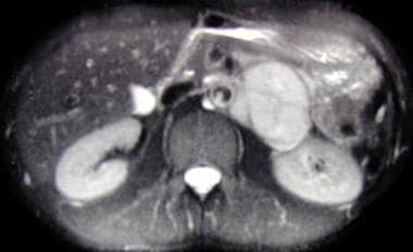
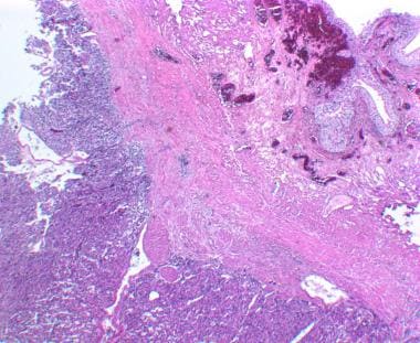
 Pediatric Pheochromocytoma: Background, Pathophysiology, Etiology
 Multimedia | Portal Regional da BVS
Pediatric Pheochromocytoma: Background, Pathophysiology, Etiology
 Hypertensive Emergencies: A Review | CE Article | NursingCenter
 MeSH Browser
 chromaffin cells | NAL Agricultural Thesaurus
English-Armenian Medical - Terms starting with 'P' - MEDINDEX.AM
Chromaffin Cells | Profiles RNS
 Chromogranin A ELISA Kit (ChgA)
Pediatric Pheochromocytoma: Background, Pathophysiology, Etiology
Pediatric Pheochromocytoma: Background, Pathophysiology, Etiology
 Pesquisa | Portal Regional da BVS
Enterochromaffin-like Cells | Profiles RNS
 Lack of an adrenal cortex in Sf1 mutant mice is compatible with the generation and differentiation of chromaffin cells |...
 Potential Biomarkers of Metastasizing Paragangliomas and Pheochromocytomas | Encyclopedia MDPI
 Cunningham's textbook of anatomy - Daniel John Cunningham - Google Livres
 Gastric paraganglioma: a case report and a review of the literature
 DeCS
DeCS
 Pheochromocytoma - Endocrine and Metabolic Disorders - MSD Manual Professional Edition
 A Not-So-Simple Thyroid Nodule | Endocrinology | JN Learning | AMA Ed Hub
Pancreatic islets - Wikipedia
Gonad - Wikipedia, ang malayang ensiklopedya
Pheochromocytoma and Paraganglioma Incidence Over 70 Years
![HRP Anti-SDHB antibody [EPR10880] (ab198329) | Abcam](data:image/png;base64,iVBORw0KGgoAAAANSUhEUgAAABAAAAAQCAYAAAAf8/9hAAABm0lEQVQ4jaWTv0tbURTHP/cl75lqTIiNRFyEJIiUxNB2qf+D6NIuDg7WwcXFxU2yOznYte2klFIqpXVqoXQqgTYZKhURUURTlajJy++Xdx1eeJrmTc/vcuHc8/3cc869V8ileBZkClcSOcW9GUCmFPdmS16n4PfjKp/2KxQbJsmwxnwywOs/RUoNCcB0rI+xAbUb0DQls9tnbO7qHcC3OyWOigbn1RYA0aDXGbD8o8Dmro6qCOYS/Tx6qPHztMbGXx3Zzll5FuJppKe7hULNZD17jQA+TA0xGe21Nh4HmRj2sfjtAoDno36iQdUG2EPM5Gs0WpJ4SL01t7UwHqBPdZ63HTVMaxWOaWBK6Ri3AU8iPSgC9i6bfDmodCS9yhWpGs4AT3piIA3Qrykc6y1+ndV5v1cmX25xWGqy9vua1cyVbRh84GEk4CXk81gVy6WYja4YkpnP/9jaL3eckghrnOgGhZrV57vJCC9G/cB/19jrFXycHuLrUZXtgwpXdZPUoMbLZIA3dx5SMnx7jR0VuNG9/4ICIufaLWT2BlLHjkWr+SchAAAAAElFTkSuQmCC) HRP Anti-SDHB antibody [EPR10880] (ab198329) | Abcam
HRP Anti-SDHB antibody [EPR10880] (ab198329) | Abcam
 TREE NUMBER DESCRIPTOR
Publication Detail
 Denaturing high performance liquid chromatography detection of SDHB, SDHD, and VHL germline mutations in pheochromocytoma -...
MeSH Browser
MeSH Browser
The basal release of endothelium-derived catecholamines regulates the contractions of Chelonoidis carbonaria aorta caused by...
DeCS
 C C177536 GDC Property Terminology C129439 Medulloblastoma, Molecularly Defined A term that refers to the classification of...
Pesquisa | Portal Regional da BVS
Pediatric Pheochromocytoma: Background, Pathophysiology, Etiology
Pediatric Pheochromocytoma: Background, Pathophysiology, Etiology
Indian Pediatrics Case Reports : Table of Contents
 Scegli la categoria - lookformedical.com
Pheochromocytoma | Palmetto Profiles
Enterochromaffin Cells | Profiles RNS
 World Journal of Endocrine Surgery
 Coverage Policy Manual - Arkansas Blue Cross and Blue Shield
 Hereditary Paraganglioma-Pheochromocytoma Syndromes - GeneReviews® - NCBI Bookshelf
 Pulsatilla. | Henriette's Herbal Homepage
Descriptors in 2013 MeSH. Preferred term only. December 14, 2012
Classification-Index
 Chromaffin System
 Diseases of the adrenal glands | Thyroid hormones and thyroid cancer
Tumors7
- The biosynthesis and storage of catecholamines in chromaffin cell tumors may differ from the biosynthesis and storage in the normal medulla. (medscape.com)
- Pheochromoctyomas are catecholamine-producing neuroendocrine tumors that arise from chromaffin cells of the adrenal medulla or extra-adrenal paraganglia. (hopkinsguides.com)
- Extra-adrenal chromaffin tissue tumors are referred to as paragangliomas or extra-adrenal pheochromocytomas. (hopkinsguides.com)
- The tumors that arise from the largest sympathetic paraganglia forming the adrenal medulla are called pheochromocytomas (PHEOs). (encyclopedia.pub)
- Tumors developing from paraganglia outside the adrenal gland are termed paragangliomas (PGLs). (encyclopedia.pub)
- AIM: Paragangliomas are neural crest-derived neuroendocrine tumors, originating from paraganglia, which are dispersed neuroendocrine organs characterized by catecholamine and peptide-producing cells. (unime.it)
- DISCUSSION: Pheochromocytoma indicates exclusively tumors arising from the adrenal medulla, while the extra-adrenal paraganglioma suggests tumors of the chromaffin cells with other locations. (unime.it)
Paragangliomas3
- Neoplasms arising from these cells are pheochromocytomas (also called chromaffin or sympathetic paragangliomas, in contrast to non-chromaffin or parasympathetic paragangliomas of glomus cells). (wikipedia.org)
- Pheochromocytomas are derived from chromaffin cells of the adrenal medulla, while paragangliomas arise from the extra-adrenal autonomic paraganglia. (bvsalud.org)
- Paragangliomas can derive from either parasympathetic or sympathetic paraganglia. (bvsalud.org)
Extra-adrenal chromaffin1
- In lower concentrations, extra-adrenal chromaffin cells also reside in the bladder wall, prostate, and behind the liver. (wikipedia.org)
Vagus nerve3
- Chromaffin cells also settle near the vagus nerve and carotid arteries. (wikipedia.org)
- the chromaffin cells settle near the sympathetic ganglia, the vagus nerve, paraganglia, and carotid arteries. (medscape.com)
- Parasympathetic paraganglia include supracardiac paraganglia, paraganglia of the carotid body, middle ear, and larynx, as well as paraganglia distributed along the vagus nerve and several other smaller paraganglia [ 2 ] . (encyclopedia.pub)
Catecholamine-secre1
- A pheochromocytoma is a catecholamine-secreting tumor of chromaffin cells typically located in the adrenals. (msdmanuals.com)
Paraganglioma1
- These terms can be used interchangeably but usually paraganglioma refer to a tumor originating from chromaffin cells outside the adrenal gland, which can also be called extra-adrenal pheochromocytoma, whereas pheochromocytoma typically refer to a tumor originating from the chromaffin cells within the adrenal gland. (wikipedia.org)
Parasympathetic2
- Paraganglia represent groups of paraneurons derived from neural crest cells during embryonic development and are divided into sympathetic and parasympathetic. (encyclopedia.pub)
- Sympathetic paraganglia consist of chromaffin cells and are involved in the secretion of catecholamines (norepinephrine, epinephrine, and dopamine), while parasympathetic paraganglia consist of glomus (nonchromaffin) cells and act as chemoreceptors [ 1 ] . (encyclopedia.pub)
Catecholamines3
- The chromaffin cells release catecholamines: ~80% of adrenaline (epinephrine) and ~20% of noradrenaline (norepinephrine) into systemic circulation for systemic effects on multiple organs (similarly to secretory neurones of the hypothalamus), and can also send paracrine signals. (wikipedia.org)
- This increased sympathetic activity leads to chronically increased synthesis and secretion of catecholamines from the adrenal chromaffin cells. (wikipedia.org)
- This chronic increase of epinephrine and norepinephrine secretion causes desensitization of the chromaffin cells to catecholamines resulting in a decrease in production and presence of α2 adrenergic receptors on their cell membrane. (wikipedia.org)
Ganglia of the sympathetic2
- There are (i.) a series of isolated masses, the paraganglia, associated singly or in groups with the ganglia of the sympathetic nervous system, (ii. (co.ma)
- The paraganglia are rounded masses of chromaphil tissue, 1-3 mm. in diameter, placed inside, half inside, or immediately outside the capsules of the ganglia of the sympathetic system. (co.ma)
Cells14
- Chromaffin cells, also called pheochromocytes (or phaeochromocytes), are neuroendocrine cells found mostly in the medulla of the adrenal glands in mammals. (wikipedia.org)
- In order to activate chromaffin cells, the splanchnic nerve of the sympathetic nervous system releases acetylcholine, which then binds to nicotinic acetylcholine receptors on the adrenal medulla. (wikipedia.org)
- 2) Chromaffin cells (or pheochromocytes): These cells will migrate to the area adjacent to the sympathetic ganglia (hence the name paraganglia) and to the adrenal medulla where they will be the most abundant type of cells. (wikipedia.org)
- The largest extra-adrenal cluster of chromaffin cells in mammals is the organ of Zuckerkandl. (wikipedia.org)
- In non-mammals, chromaffin cells are found in a variety of places, generally not organised as an individual organ, and may be without innervation, relying only on endocrine or paracrine signals for secretion. (wikipedia.org)
- Chromaffin cells of the adrenal medulla are innervated by the splanchnic nerve and secrete adrenaline (epinephrine), noradrenaline (norepinephrine), some dopamine, enkephalin and enkephalin-containing peptides, and a few other hormones into the blood stream. (wikipedia.org)
- Small bodies containing chromaffin cells occurring outside of the adrenal medulla, most commonly near the sympathetic ganglia and in organs such as the kidney, liver, heart and gonads. (nih.gov)
- Chromaffin Cells" is a descriptor in the National Library of Medicine's controlled vocabulary thesaurus, MeSH (Medical Subject Headings) . (jefferson.edu)
- This graph shows the total number of publications written about "Chromaffin Cells" by people in this website by year, and whether "Chromaffin Cells" was a major or minor topic of these publications. (jefferson.edu)
- Below are the most recent publications written about "Chromaffin Cells" by people in Profiles. (jefferson.edu)
- chromaffin cells of the medulla, adrenal, enterochromaffin-like, paraganglia and beta cells of the pancreas. (elisakits.co.uk)
- The diversification of neural-crest-derived sympathoadrenal (SA) progenitor cells into sympathetic neurons and neuroendocrine adrenal chromaffin cells was thought to be largely understood. (silverchair.com)
- In-vitro studies with isolated SA progenitor cells had suggested that chromaffin cell differentiation depends crucially on glucocorticoids provided by adrenal cortical cells. (silverchair.com)
- However, analysis of mice lacking the glucocorticoid receptor gene had revealed that adrenal chromaffin cells develop mostly normally in these mice. (silverchair.com)
Medulla1
- Characteristically, they are located in the adrenal medulla and paraganglia of the sympathetic nervous system. (usda.gov)
Typically1
- Typically one paraganglion, exceptionally a pair of paraganglia, is associated with each ganglion of the gangliated trunks and with each ganglion of the cœliac, renal, suprarenal, aortic, and hypogastric plexuses. (co.ma)
Tissue1
- From seven to seventy masses of chromaphil tissue are developed in relation to the abdominal sympathetic plexuses, independently of the ganglia and in addition to the paraganglia. (co.ma)
Common1
- These paraganglia are common throughout the body, but most are found in the head and neck area [ 3 ] . (encyclopedia.pub)
Adrenal medulla10
- In order to activate chromaffin cells, the splanchnic nerve of the sympathetic nervous system releases acetylcholine, which then binds to nicotinic acetylcholine receptors on the adrenal medulla. (wikipedia.org)
- 2) Chromaffin cells (or pheochromocytes): These cells will migrate to the area adjacent to the sympathetic ganglia (hence the name paraganglia) and to the adrenal medulla where they will be the most abundant type of cells. (wikipedia.org)
- Chromaffin cells of the adrenal medulla are innervated by the splanchnic nerve and secrete adrenaline (epinephrine), noradrenaline (norepinephrine), some dopamine, enkephalin and enkephalin-containing peptides, and a few other hormones into the blood stream. (wikipedia.org)
- [ 1 ] Pheochromocytomas (PCCs) originate from the adrenal medulla, whereas paragangliomas (PGLs) arise from extra-adrenal paraganglia. (medscape.com)
- Pheochromocytomas are neuroendocrine tumors of chromaffin cell origin which arise from the adrenal medulla and less commonly the extra-adrenal sympathetic paraganglia. (nih.gov)
- Small bodies containing chromaffin cells occurring outside of the adrenal medulla, most commonly near the sympathetic ganglia and in organs such as the kidney, liver, heart and gonads. (nih.gov)
- Objective: Pheochromocytoma is a rare catecholamine-producing neuroendocrine tumour originating from the chromaffin cells of the adrenal medulla or extra-adrenal paraganglia. (bvsalud.org)
- Termination of the adrenal medulla growth was found to be associated with decreased chromaffin cell proliferation, activation of canonical Wnt-signaling pathway, and enhanced expression of Sonic Hedgehog ligand. (bvsalud.org)
- A usually benign, well-encapsulated, lobular, vascular tumor of chromaffin tissue of the ADRENAL MEDULLA or sympathetic paraganglia. (musc.edu)
- Pheochromocytomas (PCCs) are rare catecholamine-secreting tumors of the chromaffin cells of the adrenal medulla or paraganglia at other places. (wjoes.com)
Originating from the chromaffin cells2
- These terms can be used interchangeably but usually paraganglioma refer to a tumor originating from chromaffin cells outside the adrenal gland, which can also be called extra-adrenal pheochromocytoma, whereas pheochromocytoma typically refer to a tumor originating from the chromaffin cells within the adrenal gland. (wikipedia.org)
- Pheochromocytomas and paragangliomas (PPGLs) are rare neuroendocrine tumors originating from the chromaffin cells of the autonomic nervous system. (medscape.com)
Pheochromocytomas1
- Neoplasms arising from these cells are pheochromocytomas (also called chromaffin or sympathetic paragangliomas, in contrast to non-chromaffin or parasympathetic paragangliomas of glomus cells). (wikipedia.org)
Neuroendocrine1
- Chromaffin cells, also called pheochromocytes (or phaeochromocytes), are neuroendocrine cells found mostly in the medulla of the adrenal glands in mammals. (wikipedia.org)