Oxidative Phosphorylation
Electron Transport
Mitochondria
Electron Transport Chain Complex Proteins
Uncoupling Agents
Electron Transport Complex I
Oxygen Consumption
Adenosine Triphosphate
Microscopy, Electron
Electron Transport Complex IV
Biological Transport
Electrons
Biological Transport, Active
Antimycin A
Electron Transport Complex III
Mitochondria, Liver
Oligomycins
Oxidation-Reduction
Phosphorylation
Cell Respiration
Glycolysis
Molecular Sequence Data
Serine
Amino Acid Sequence
Microscopy, Electron, Scanning
Photosynthesis
Mutation
Succinates
Energy Metabolism
Electron Transport Complex II
Mitochondria, Muscle
Mitochondrial Proteins
Cytochromes
Mitochondrial Diseases
Models, Biological
Cyanides
Oxygen
Adenosine Diphosphate
Tyrosine
Oxidative Phosphorylation Coupling Factors
Cells, Cultured
DNA, Mitochondrial
Enzyme Activation
NAD
NADH Dehydrogenase
Protein-Serine-Threonine Kinases
Protein Transport
Reactive Oxygen Species
Ubiquinone
2,4-Dinitrophenol
Glucose
Carbonyl Cyanide p-Trifluoromethoxyphenylhydrazone
Cell Membrane
Phosphoproteins
Protein Kinases
Succinate Dehydrogenase
Oxidoreductases
Carrier Proteins
Protein Binding
Mitochondrial ADP, ATP Translocases
Carbonyl Cyanide m-Chlorophenyl Hydrazone
Enzyme Inhibitors
Mitochondrial Proton-Translocating ATPases
Amobarbital
Hydrogen-Ion Concentration
Membrane Transport Proteins
Calcium
Succinic Acid
Threonine
Blotting, Western
Membrane Proteins
Atractyloside
Axonal Transport
Protons
Ion Transport
Chlorophyll
Polarography
Malates
Signal Transduction
Binding Sites
Membrane Potential, Mitochondrial
Adenosine Triphosphatases
Microscopy, Electron, Transmission
Protein-Tyrosine Kinases
Proton-Translocating ATPases
Citric Acid Cycle
Potassium Cyanide
Hydroxyquinolines
Protein Kinase C
Submitochondrial Particles
Muscle, Skeletal
Cattle
Intracellular Membranes
Plastoquinone
Base Sequence
Transfection
Escherichia coli
Electron Spin Resonance Spectroscopy
Cyclic AMP-Dependent Protein Kinases
Protein Structure, Tertiary
NADH, NADPH Oxidoreductases
Tetramethylphenylenediamine
Protein Processing, Post-Translational
Electrophoresis, Polyacrylamide Gel
Rats, Wistar
Oxidative Stress
Photosystem II Protein Complex
Proto-Oncogene Proteins c-akt
Anaerobiosis
Cell Nucleus
Chloroplasts
Cytochrome c Group
Rats, Sprague-Dawley
Phosphopeptides
ATP Synthetase Complexes
Proteins
Phosphotyrosine
Multienzyme Complexes
HeLa Cells
Dicumarol
Immunoblotting
Gene Expression Regulation
Thylakoids
Myocardium
Mutagenesis, Site-Directed
Valinomycin
Mitochondrial Swelling
Membrane Potentials
Apoptosis
Mitochondrial Membranes
Saccharomyces cerevisiae
Photosystem I Protein Complex
Dicyclohexylcarbodiimide
Monosaccharide Transport Proteins
Rabbits
Adenine Nucleotide Translocator 1
Sodium Cyanide
Liver
Dose-Response Relationship, Drug
Phosphocreatine
Pyruvic Acid
RNA, Messenger
Hydrogen Peroxide
Cytochrome b6f Complex
Spectrophotometry
Substrate Specificity
Photosynthetic Reaction Center Complex Proteins
Mitogen-Activated Protein Kinases
Transcription Factors
Lactic Acid
Phosphothreonine
Sequence Homology, Amino Acid
Fibroblasts
Insulin
Cytoplasm
Quinones
Temperature
NADP
Peptide Mapping
Pyruvates
Sodium Azide
Transcription, Genetic
Recombinant Fusion Proteins
Sodium
Hydrogen
Adenine Nucleotides
DNA-Binding Proteins
Casein Kinase II
Cytosol
Models, Molecular
src-Family Kinases
Cardiolipins
Phosphorus Radioisotopes
Microscopy, Fluorescence
Intracellular Signaling Peptides and Proteins
Proto-Oncogene Proteins
Phosphoprotein Phosphatases
Rats, Inbred Strains
Tumor Cells, Cultured
Amino Acids
Citrate (si)-Synthase
Oxidative phosphorylation is the metabolic process by which cells use enzymes to generate energy in the form of adenosine triphosphate (ATP) from the oxidation of nutrients, such as glucose or fatty acids. This process occurs in the inner mitochondrial membrane of eukaryotic cells and is facilitated by the electron transport chain, which consists of a series of protein complexes that transfer electrons from donor molecules to acceptor molecules. As the electrons are passed along the chain, they release energy that is used to pump protons across the membrane, creating a gradient. The ATP synthase enzyme then uses the flow of protons back across the membrane to generate ATP, which serves as the main energy currency for cellular processes.
The Electron Transport Chain (ETC) is a series of complexes in the inner mitochondrial membrane that are involved in the process of cellular respiration. It is the final pathway for electrons derived from the oxidation of nutrients such as glucose, fatty acids, and amino acids to be transferred to molecular oxygen. This transfer of electrons drives the generation of a proton gradient across the inner mitochondrial membrane, which is then used by ATP synthase to produce ATP, the main energy currency of the cell.
The electron transport chain consists of four complexes (I-IV) and two mobile electron carriers (ubiquinone and cytochrome c). Electrons from NADH and FADH2 are transferred to Complex I and Complex II respectively, which then pass them along to ubiquinone. Ubiquinone then transfers the electrons to Complex III, which passes them on to cytochrome c. Finally, cytochrome c transfers the electrons to Complex IV, where they combine with oxygen and protons to form water.
The transfer of electrons through the ETC is accompanied by the pumping of protons from the mitochondrial matrix to the intermembrane space, creating a proton gradient. The flow of protons back across the inner membrane through ATP synthase drives the synthesis of ATP from ADP and inorganic phosphate.
Overall, the electron transport chain is a crucial process for generating energy in the form of ATP in the cell, and it plays a key role in many metabolic pathways.
Mitochondria are specialized structures located inside cells that convert the energy from food into ATP (adenosine triphosphate), which is the primary form of energy used by cells. They are often referred to as the "powerhouses" of the cell because they generate most of the cell's supply of chemical energy. Mitochondria are also involved in various other cellular processes, such as signaling, differentiation, and apoptosis (programmed cell death).
Mitochondria have their own DNA, known as mitochondrial DNA (mtDNA), which is inherited maternally. This means that mtDNA is passed down from the mother to her offspring through the egg cells. Mitochondrial dysfunction has been linked to a variety of diseases and conditions, including neurodegenerative disorders, diabetes, and aging.
The Electron Transport Chain (ETC) is a series of complexes in the inner mitochondrial membrane that are involved in the process of cellular respiration, through which the majority of energy is generated for the cell. The ETC complex proteins are a group of transmembrane protein complexes that facilitate the transfer of electrons from electron donors to electron acceptors via redox reactions. This transfer of electrons drives the generation of a proton gradient across the inner mitochondrial membrane, which is then used by ATP synthase to generate ATP, the primary energy currency of the cell.
The ETC complex proteins consist of four main complexes: Complex I (NADH-Q oxidoreductase), Complex II (succinate-Q oxidoreductase), Complex III (cytochrome bc1 complex or CoQ:cytochrome c oxidoreductase), and Complex IV (cytochrome c oxidase). Each complex contains a number of subunits, many of which are encoded by both the nuclear and mitochondrial genomes.
In summary, Electron Transport Chain Complex Proteins are a group of transmembrane protein complexes located in the inner mitochondrial membrane that facilitate the transfer of electrons from electron donors to electron acceptors, driving the generation of a proton gradient and ultimately ATP synthesis during cellular respiration.
Uncoupling agents are chemicals that interfere with the normal process of oxidative phosphorylation in cells. In this process, the energy from food is converted into ATP (adenosine triphosphate), which is the main source of energy for cellular functions. Uncouplers disrupt this process by preventing the transfer of high-energy electrons to oxygen, which normally drives the production of ATP.
Instead, the energy from these electrons is released as heat, leading to an increase in body temperature. This effect is similar to what happens during shivering or exercise, when the body generates heat to maintain its core temperature. Uncoupling agents are therefore also known as "mitochondrial protonophores" because they allow protons to leak across the inner mitochondrial membrane, bypassing the ATP synthase enzyme that would normally use the energy from this proton gradient to produce ATP.
Uncoupling agents have been studied for their potential therapeutic uses, such as in weight loss and the treatment of metabolic disorders. However, they can also be toxic at high doses, and their long-term effects on health are not well understood.
Electron Transport Complex I, also known as NADH:ubiquinone oxidoreductase, is a large protein complex located in the inner mitochondrial membrane of eukaryotic cells and the cytoplasmic membrane of prokaryotic cells. It is the first complex in the electron transport chain, a series of protein complexes that transfer electrons from NADH to oxygen, driving the synthesis of ATP through chemiosmosis.
Complex I consists of multiple subunits, including a flavin mononucleotide (FMN) cofactor and several iron-sulfur clusters, which facilitate the oxidation of NADH and the reduction of ubiquinone (coenzyme Q). The energy released during this electron transfer process is used to pump protons across the membrane, creating a proton gradient that drives ATP synthesis.
Defects in Complex I can lead to various mitochondrial diseases, including neurological disorders and muscle weakness.
Oxygen consumption, also known as oxygen uptake, is the amount of oxygen that is consumed or utilized by the body during a specific period of time, usually measured in liters per minute (L/min). It is a common measurement used in exercise physiology and critical care medicine to assess an individual's aerobic metabolism and overall health status.
In clinical settings, oxygen consumption is often measured during cardiopulmonary exercise testing (CPET) to evaluate cardiovascular function, pulmonary function, and exercise capacity in patients with various medical conditions such as heart failure, chronic obstructive pulmonary disease (COPD), and other respiratory or cardiac disorders.
During exercise, oxygen is consumed by the muscles to generate energy through a process called oxidative phosphorylation. The amount of oxygen consumed during exercise can provide important information about an individual's fitness level, exercise capacity, and overall health status. Additionally, measuring oxygen consumption can help healthcare providers assess the effectiveness of treatments and rehabilitation programs in patients with various medical conditions.
Adenosine Triphosphate (ATP) is a high-energy molecule that stores and transports energy within cells. It is the main source of energy for most cellular processes, including muscle contraction, nerve impulse transmission, and protein synthesis. ATP is composed of a base (adenine), a sugar (ribose), and three phosphate groups. The bonds between these phosphate groups contain a significant amount of energy, which can be released when the bond between the second and third phosphate group is broken, resulting in the formation of adenosine diphosphate (ADP) and inorganic phosphate. This process is known as hydrolysis and can be catalyzed by various enzymes to drive a wide range of cellular functions. ATP can also be regenerated from ADP through various metabolic pathways, such as oxidative phosphorylation or substrate-level phosphorylation, allowing for the continuous supply of energy to cells.
Electron microscopy (EM) is a type of microscopy that uses a beam of electrons to create an image of the sample being examined, resulting in much higher magnification and resolution than light microscopy. There are several types of electron microscopy, including transmission electron microscopy (TEM), scanning electron microscopy (SEM), and reflection electron microscopy (REM).
In TEM, a beam of electrons is transmitted through a thin slice of the sample, and the electrons that pass through the sample are focused to form an image. This technique can provide detailed information about the internal structure of cells, viruses, and other biological specimens, as well as the composition and structure of materials at the atomic level.
In SEM, a beam of electrons is scanned across the surface of the sample, and the electrons that are scattered back from the surface are detected to create an image. This technique can provide information about the topography and composition of surfaces, as well as the structure of materials at the microscopic level.
REM is a variation of SEM in which the beam of electrons is reflected off the surface of the sample, rather than scattered back from it. This technique can provide information about the surface chemistry and composition of materials.
Electron microscopy has a wide range of applications in biology, medicine, and materials science, including the study of cellular structure and function, disease diagnosis, and the development of new materials and technologies.
Electron Transport Complex IV is also known as Cytochrome c oxidase. It is the last complex in the electron transport chain, located in the inner mitochondrial membrane of eukaryotic cells and the plasma membrane of prokaryotic cells. This complex contains 13 subunits, two heme groups (a and a3), and three copper centers (A, B, and C).
In the electron transport chain, Complex IV receives electrons from cytochrome c and transfers them to molecular oxygen, reducing it to water. This process is accompanied by the pumping of protons across the membrane, contributing to the generation of a proton gradient that drives ATP synthesis via ATP synthase (Complex V). The overall reaction catalyzed by Complex IV can be summarized as follows:
4e- + 4H+ + O2 → 2H2O
Defects in Cytochrome c oxidase can lead to various diseases, including mitochondrial encephalomyopathies and neurodegenerative disorders.
Biological transport refers to the movement of molecules, ions, or solutes across biological membranes or through cells in living organisms. This process is essential for maintaining homeostasis, regulating cellular functions, and enabling communication between cells. There are two main types of biological transport: passive transport and active transport.
Passive transport does not require the input of energy and includes:
1. Diffusion: The random movement of molecules from an area of high concentration to an area of low concentration until equilibrium is reached.
2. Osmosis: The diffusion of solvent molecules (usually water) across a semi-permeable membrane from an area of lower solute concentration to an area of higher solute concentration.
3. Facilitated diffusion: The assisted passage of polar or charged substances through protein channels or carriers in the cell membrane, which increases the rate of diffusion without consuming energy.
Active transport requires the input of energy (in the form of ATP) and includes:
1. Primary active transport: The direct use of ATP to move molecules against their concentration gradient, often driven by specific transport proteins called pumps.
2. Secondary active transport: The coupling of the movement of one substance down its electrochemical gradient with the uphill transport of another substance, mediated by a shared transport protein. This process is also known as co-transport or counter-transport.
An electron is a subatomic particle, symbol e-, with a negative electric charge. Electrons are fundamental components of atoms and are responsible for the chemical bonding between atoms to form molecules. They are located in an atom's electron cloud, which is the outermost region of an atom and contains negatively charged electrons that surround the positively charged nucleus.
Electrons have a mass that is much smaller than that of protons or neutrons, making them virtually weightless on the atomic scale. They are also known to exhibit both particle-like and wave-like properties, which is a fundamental concept in quantum mechanics. Electrons play a crucial role in various physical phenomena, such as electricity, magnetism, and chemical reactions.
Biological transport, active is the process by which cells use energy to move materials across their membranes from an area of lower concentration to an area of higher concentration. This type of transport is facilitated by specialized proteins called transporters or pumps that are located in the cell membrane. These proteins undergo conformational changes to physically carry the molecules through the lipid bilayer of the membrane, often against their concentration gradient.
Active transport requires energy because it works against the natural tendency of molecules to move from an area of higher concentration to an area of lower concentration, a process known as diffusion. Cells obtain this energy in the form of ATP (adenosine triphosphate), which is produced through cellular respiration.
Examples of active transport include the uptake of glucose and amino acids into cells, as well as the secretion of hormones and neurotransmitters. The sodium-potassium pump, which helps maintain resting membrane potential in nerve and muscle cells, is a classic example of an active transporter.
Antimycin A is an antibiotic substance produced by various species of Streptomyces bacteria. It is known to inhibit the electron transport chain in mitochondria, which can lead to cellular dysfunction and death. Antimycin A has been used in research to study the mechanisms of cellular respiration and oxidative phosphorylation.
In a medical context, antimycin A is not used as a therapeutic agent due to its toxicity to mammalian cells. However, it may be used in laboratory settings to investigate various biological processes or to develop new therapies for diseases related to mitochondrial dysfunction.
In the context of medicine and pharmacology, "kinetics" refers to the study of how a drug moves throughout the body, including its absorption, distribution, metabolism, and excretion (often abbreviated as ADME). This field is called "pharmacokinetics."
1. Absorption: This is the process of a drug moving from its site of administration into the bloodstream. Factors such as the route of administration (e.g., oral, intravenous, etc.), formulation, and individual physiological differences can affect absorption.
2. Distribution: Once a drug is in the bloodstream, it gets distributed throughout the body to various tissues and organs. This process is influenced by factors like blood flow, protein binding, and lipid solubility of the drug.
3. Metabolism: Drugs are often chemically modified in the body, typically in the liver, through processes known as metabolism. These changes can lead to the formation of active or inactive metabolites, which may then be further distributed, excreted, or undergo additional metabolic transformations.
4. Excretion: This is the process by which drugs and their metabolites are eliminated from the body, primarily through the kidneys (urine) and the liver (bile).
Understanding the kinetics of a drug is crucial for determining its optimal dosing regimen, potential interactions with other medications or foods, and any necessary adjustments for special populations like pediatric or geriatric patients, or those with impaired renal or hepatic function.
Electron Transport Complex III, also known as cytochrome bc1 complex or ubiquinol-cytochrome c reductase, is a protein complex located in the inner mitochondrial membrane of eukaryotic cells and the cytoplasmic membrane of prokaryotic cells. It plays a crucial role in the electron transport chain (ETC), a series of complexes that generate energy in the form of ATP through a process called oxidative phosphorylation.
In ETC, Electron Transport Complex III accepts electrons from ubiquinol and transfers them to cytochrome c. This electron transfer is coupled with the translocation of protons (H+ ions) across the membrane, creating an electrochemical gradient. The energy stored in this gradient drives the synthesis of ATP by ATP synthase.
Electron Transport Complex III consists of several subunits, including cytochrome b, cytochrome c1, and the Rieske iron-sulfur protein. These subunits work together to facilitate the electron transfer and proton translocation processes.
I'd be happy to help! However, I believe there might be a slight misunderstanding in your question. Mitochondria are organelles found in the cells of all complex living organisms, including humans, while the liver is a large, solid organ located in the upper right portion of the abdomen. They don't have a medical definition together. I can certainly provide you with separate definitions for each:
1. Mitochondria: These are double-membrane-bound cellular organelles that generate most of the chemical energy needed to power the cell's biochemical reactions. Commonly known as the "powerhouse of the cell," mitochondria convert organic substrates, such as glucose, fatty acids, and amino acids, into adenosine triphosphate (ATP) through a process called oxidative phosphorylation. Mitochondria are dynamic structures that can change their shape, size, and number through fission (division) and fusion (merging) processes. They play essential roles in various cellular functions, including calcium signaling, apoptosis (programmed cell death), and the regulation of cellular metabolism.
2. Liver: The liver is a large, lobulated organ that lies mainly in the upper right portion of the abdominal cavity, just below the diaphragm. It plays a crucial role in various physiological functions, such as detoxification, protein synthesis, metabolism, and nutrient storage. The liver is responsible for removing toxins from the bloodstream, producing bile to aid in digestion, regulating glucose levels, synthesizing plasma proteins, and storing glycogen, vitamins, and minerals. It also contributes to the metabolism of carbohydrates, lipids, and amino acids, helping maintain energy homeostasis in the body.
I hope this clarifies any confusion! If you have any further questions or need more information, please don't hesitate to ask.
Oligomycins are a group of antibiotics produced by various species of Streptomyces bacteria. They are characterized by their ability to inhibit the function of ATP synthase, an enzyme that plays a crucial role in energy production within cells. By binding to the F1 component of ATP synthase, oligomycins prevent the synthesis of ATP, which is a key source of energy for cellular processes.
These antibiotics have been used in research to study the mechanisms of ATP synthase and mitochondrial function. However, their therapeutic use as antibiotics is limited due to their toxicity to mammalian cells. Oligomycin A is one of the most well-known and studied members of this group of antibiotics.
Oxidation-Reduction (redox) reactions are a type of chemical reaction involving a transfer of electrons between two species. The substance that loses electrons in the reaction is oxidized, and the substance that gains electrons is reduced. Oxidation and reduction always occur together in a redox reaction, hence the term "oxidation-reduction."
In biological systems, redox reactions play a crucial role in many cellular processes, including energy production, metabolism, and signaling. The transfer of electrons in these reactions is often facilitated by specialized molecules called electron carriers, such as nicotinamide adenine dinucleotide (NAD+/NADH) and flavin adenine dinucleotide (FAD/FADH2).
The oxidation state of an element in a compound is a measure of the number of electrons that have been gained or lost relative to its neutral state. In redox reactions, the oxidation state of one or more elements changes as they gain or lose electrons. The substance that is oxidized has a higher oxidation state, while the substance that is reduced has a lower oxidation state.
Overall, oxidation-reduction reactions are fundamental to the functioning of living organisms and are involved in many important biological processes.
Phosphorylation is the process of adding a phosphate group (a molecule consisting of one phosphorus atom and four oxygen atoms) to a protein or other organic molecule, which is usually done by enzymes called kinases. This post-translational modification can change the function, localization, or activity of the target molecule, playing a crucial role in various cellular processes such as signal transduction, metabolism, and regulation of gene expression. Phosphorylation is reversible, and the removal of the phosphate group is facilitated by enzymes called phosphatases.
Cell respiration is the process by which cells convert biochemical energy from nutrients into adenosine triphosphate (ATP), and then release waste products. The three main stages of cell respiration are glycolysis, the citric acid cycle (also known as the Krebs cycle), and the electron transport chain.
During glycolysis, which takes place in the cytoplasm, glucose is broken down into two molecules of pyruvate, producing a small amount of ATP and reducing power in the form of NADH.
The citric acid cycle occurs in the mitochondria and involves the breakdown of acetyl-CoA (formed from pyruvate) to produce more ATP, NADH, and FADH2.
Finally, the electron transport chain, also located in the mitochondria, uses the energy from NADH and FADH2 to pump protons across the inner mitochondrial membrane, creating a proton gradient. The flow of protons back across the membrane drives the synthesis of ATP, which is used as a source of energy by the cell.
Cell respiration is a crucial process that allows cells to generate the energy they need to perform various functions and maintain homeostasis.
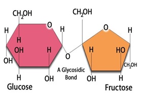
Glycolysis is a fundamental metabolic pathway that occurs in the cytoplasm of cells, consisting of a series of biochemical reactions. It's the process by which a six-carbon glucose molecule is broken down into two three-carbon pyruvate molecules. This process generates a net gain of two ATP molecules (the main energy currency in cells), two NADH molecules, and two water molecules.
Glycolysis can be divided into two stages: the preparatory phase (or 'energy investment' phase) and the payoff phase (or 'energy generation' phase). During the preparatory phase, glucose is phosphorylated twice to form glucose-6-phosphate and then converted to fructose-1,6-bisphosphate. These reactions consume two ATP molecules but set up the subsequent breakdown of fructose-1,6-bisphosphate into triose phosphates in the payoff phase. In this second stage, each triose phosphate is further oxidized and degraded to produce one pyruvate molecule, one NADH molecule, and one ATP molecule through substrate-level phosphorylation.
Glycolysis does not require oxygen to proceed; thus, it can occur under both aerobic (with oxygen) and anaerobic (without oxygen) conditions. In the absence of oxygen, the pyruvate produced during glycolysis is further metabolized through fermentation pathways such as lactic acid fermentation or alcohol fermentation to regenerate NAD+, which is necessary for glycolysis to continue.
In summary, glycolysis is a crucial process in cellular energy metabolism, allowing cells to convert glucose into ATP and other essential molecules while also serving as a starting point for various other biochemical pathways.
Molecular sequence data refers to the specific arrangement of molecules, most commonly nucleotides in DNA or RNA, or amino acids in proteins, that make up a biological macromolecule. This data is generated through laboratory techniques such as sequencing, and provides information about the exact order of the constituent molecules. This data is crucial in various fields of biology, including genetics, evolution, and molecular biology, allowing for comparisons between different organisms, identification of genetic variations, and studies of gene function and regulation.
Serine is an amino acid, which is a building block of proteins. More specifically, it is a non-essential amino acid, meaning that the body can produce it from other compounds, and it does not need to be obtained through diet. Serine plays important roles in the body, such as contributing to the formation of the protective covering of nerve fibers (myelin sheath), helping to synthesize another amino acid called tryptophan, and taking part in the metabolism of fatty acids. It is also involved in the production of muscle tissues, the immune system, and the forming of cell structures. Serine can be found in various foods such as soy, eggs, cheese, meat, peanuts, lentils, and many others.
An amino acid sequence is the specific order of amino acids in a protein or peptide molecule, formed by the linking of the amino group (-NH2) of one amino acid to the carboxyl group (-COOH) of another amino acid through a peptide bond. The sequence is determined by the genetic code and is unique to each type of protein or peptide. It plays a crucial role in determining the three-dimensional structure and function of proteins.
Rotenone is not strictly a medical term, but it is a pesticide that is used in some medical situations. According to the National Pesticide Information Center, rotenone is a pesticide derived from the roots and stems of several plants, including Derris Eliptica, Lonchocarpus utilis, and Tephrosia vogelii. It is used as a pesticide to control insects, mites, and fish in both agricultural and residential settings.
In medical contexts, rotenone has been studied for its potential effects on human health, particularly in relation to Parkinson's disease. Some research suggests that exposure to rotenone may increase the risk of developing Parkinson's disease, although more studies are needed to confirm this link. Rotenone works by inhibiting the mitochondria in cells, which can lead to cell death and neurodegeneration.
It is important to note that rotenone is highly toxic and should be handled with care. It can cause skin and eye irritation, respiratory problems, and gastrointestinal symptoms if ingested or inhaled. Therefore, it is recommended to use personal protective equipment when handling rotenone and to follow all label instructions carefully.
I'm happy to help! However, it seems like there are two terms combined into one request: "Mitochondria" and "Heart." I will provide separate definitions for each.
Mitochondrion (singular) or Mitochondria (plural): These are specialized structures found in the cytoplasm of most eukaryotic cells (cells with a true nucleus), including human cells. They are often referred to as the "powerhouse" of the cell because they generate energy in the form of ATP (adenosine triphosphate) through a process called oxidative phosphorylation. Mitochondria contain their own DNA, which is distinct from the nuclear DNA, and are believed to have originated from ancient bacteria that established a symbiotic relationship with primitive eukaryotic cells.
Heart: In human anatomy, the heart is a muscular organ responsible for pumping blood throughout the body. It is located in the thoracic cavity, slightly left of the center, and is enclosed by the pericardium, a double-walled sac that provides protection and lubrication for the heart's movement. The human heart is divided into four chambers: two atria on the top and two ventricles on the bottom. The right side of the heart receives deoxygenated blood from the body and pumps it to the lungs, while the left side receives oxygenated blood from the lungs and pumps it to the rest of the body. The heart's pumping action is regulated by electrical signals that originate in a group of specialized cardiac muscle cells called the sinoatrial node (SA node).
Scanning electron microscopy (SEM) is a type of electron microscopy that uses a focused beam of electrons to scan the surface of a sample and produce a high-resolution image. In SEM, a beam of electrons is scanned across the surface of a specimen, and secondary electrons are emitted from the sample due to interactions between the electrons and the atoms in the sample. These secondary electrons are then detected by a detector and used to create an image of the sample's surface topography. SEM can provide detailed images of the surface of a wide range of materials, including metals, polymers, ceramics, and biological samples. It is commonly used in materials science, biology, and electronics for the examination and analysis of surfaces at the micro- and nanoscale.
Photosynthesis is not strictly a medical term, but it is a fundamental biological process with significant implications for medicine, particularly in understanding energy production in cells and the role of oxygen in sustaining life. Here's a general biological definition:
Photosynthesis is a process by which plants, algae, and some bacteria convert light energy, usually from the sun, into chemical energy in the form of organic compounds, such as glucose (or sugar), using water and carbon dioxide. This process primarily takes place in the chloroplasts of plant cells, specifically in structures called thylakoids. The overall reaction can be summarized as:
6 CO2 + 6 H2O + light energy → C6H12O6 + 6 O2
In this equation, carbon dioxide (CO2) and water (H2O) are the reactants, while glucose (C6H12O6) and oxygen (O2) are the products. Photosynthesis has two main stages: the light-dependent reactions and the light-independent reactions (Calvin cycle). The light-dependent reactions occur in the thylakoid membrane and involve the conversion of light energy into ATP and NADPH, which are used to power the Calvin cycle. The Calvin cycle takes place in the stroma of chloroplasts and involves the synthesis of glucose from CO2 and water using the ATP and NADPH generated during the light-dependent reactions.
Understanding photosynthesis is crucial for understanding various biological processes, including cellular respiration, plant metabolism, and the global carbon cycle. Additionally, research into artificial photosynthesis has potential applications in renewable energy production and environmental remediation.
A mutation is a permanent change in the DNA sequence of an organism's genome. Mutations can occur spontaneously or be caused by environmental factors such as exposure to radiation, chemicals, or viruses. They may have various effects on the organism, ranging from benign to harmful, depending on where they occur and whether they alter the function of essential proteins. In some cases, mutations can increase an individual's susceptibility to certain diseases or disorders, while in others, they may confer a survival advantage. Mutations are the driving force behind evolution, as they introduce new genetic variability into populations, which can then be acted upon by natural selection.
Dinitrophenols (DNP) are a class of chemical compounds that contain two nitro groups (-NO2) attached to a phenol group. Dinitrophenols have been used in the past as industrial dyes, wood preservatives, and pesticides. However, they have also been misused as weight loss supplements due to their ability to increase metabolic rate and cause weight loss.
The use of DNP for weight loss is dangerous and has been linked to several fatalities. DNP works by disrupting the normal functioning of the mitochondria in cells, which are responsible for producing energy. This disruption causes an increase in metabolic rate, leading to a rapid breakdown of fat and carbohydrates, and ultimately weight loss. However, this increased metabolism can also produce excessive heat, leading to hyperthermia, dehydration, and damage to organs such as the heart, liver, and kidneys.
Due to their potential for serious harm, DNP-containing products are banned in many countries, including the United States. Medical professionals should be aware of the dangers associated with DNP use and advise patients accordingly.
Succinates, in a medical context, most commonly refer to the salts or esters of succinic acid. Succinic acid is a dicarboxylic acid that is involved in the Krebs cycle, which is a key metabolic pathway in cells that generates energy through the oxidation of acetyl-CoA derived from carbohydrates, fats, and proteins.
Succinates can also be used as a buffer in medical solutions and as a pharmaceutical intermediate in the synthesis of various drugs. In some cases, succinate may be used as a nutritional supplement or as a component of parenteral nutrition formulations to provide energy and help maintain acid-base balance in patients who are unable to eat normally.
It's worth noting that there is also a condition called "succinic semialdehyde dehydrogenase deficiency" which is a genetic disorder that affects the metabolism of the amino acid gamma-aminobutyric acid (GABA). This condition can lead to an accumulation of succinic semialdehyde and other metabolic byproducts, which can cause neurological symptoms such as developmental delay, hypotonia, and seizures.
Energy metabolism is the process by which living organisms produce and consume energy to maintain life. It involves a series of chemical reactions that convert nutrients from food, such as carbohydrates, fats, and proteins, into energy in the form of adenosine triphosphate (ATP).
The process of energy metabolism can be divided into two main categories: catabolism and anabolism. Catabolism is the breakdown of nutrients to release energy, while anabolism is the synthesis of complex molecules from simpler ones using energy.
There are three main stages of energy metabolism: glycolysis, the citric acid cycle (also known as the Krebs cycle), and oxidative phosphorylation. Glycolysis occurs in the cytoplasm of the cell and involves the breakdown of glucose into pyruvate, producing a small amount of ATP and nicotinamide adenine dinucleotide (NADH). The citric acid cycle takes place in the mitochondria and involves the further breakdown of pyruvate to produce more ATP, NADH, and carbon dioxide. Oxidative phosphorylation is the final stage of energy metabolism and occurs in the inner mitochondrial membrane. It involves the transfer of electrons from NADH and other electron carriers to oxygen, which generates a proton gradient across the membrane. This gradient drives the synthesis of ATP, producing the majority of the cell's energy.
Overall, energy metabolism is a complex and essential process that allows organisms to grow, reproduce, and maintain their bodily functions. Disruptions in energy metabolism can lead to various diseases, including diabetes, obesity, and neurodegenerative disorders.
Electron Transport Complex II, also known as succinate-Q oxidoreductase, is a key component of the electron transport chain in the inner mitochondrial membrane. It plays a crucial role in the process of cellular respiration, where it facilitates the transfer of electrons from succinate to ubiquinone (Q), thereby generating a proton gradient across the membrane. This gradient drives the synthesis of ATP, which is the primary source of energy for the cell.
The complex is composed of four core subunits: flavoprotein (Fp), iron-sulfur protein (Ip), cytochrome b (Cyb), and ubiquinone-binding protein (Qp). Electrons from succinate are accepted by FAD in the Fp subunit, and then transferred to the Ip subunit containing iron-sulfur clusters. From there, the electrons are moved to heme groups in the Cyb subunit, and finally passed on to ubiquinone at the Qp subunit.
In addition to its role in the electron transport chain, Complex II has been implicated in various cellular processes such as regulation of reactive oxygen species (ROS) production and modulation of apoptosis. Mutations in genes encoding Complex II subunits have been associated with several human diseases, including neurodegenerative disorders and cancer.
A cell line is a culture of cells that are grown in a laboratory for use in research. These cells are usually taken from a single cell or group of cells, and they are able to divide and grow continuously in the lab. Cell lines can come from many different sources, including animals, plants, and humans. They are often used in scientific research to study cellular processes, disease mechanisms, and to test new drugs or treatments. Some common types of human cell lines include HeLa cells (which come from a cancer patient named Henrietta Lacks), HEK293 cells (which come from embryonic kidney cells), and HUVEC cells (which come from umbilical vein endothelial cells). It is important to note that cell lines are not the same as primary cells, which are cells that are taken directly from a living organism and have not been grown in the lab.
Mitochondria in muscle, also known as the "powerhouses" of the cell, are organelles that play a crucial role in generating energy for muscle cells through a process called cellular respiration. They convert the chemical energy found in glucose and oxygen into ATP (adenosine triphosphate), which is the main source of energy used by cells.
Muscle cells contain a high number of mitochondria due to their high energy demands for muscle contraction and relaxation. The number and size of mitochondria in muscle fibers can vary depending on the type of muscle fiber, with slow-twitch, aerobic fibers having more numerous and larger mitochondria than fast-twitch, anaerobic fibers.
Mitochondrial dysfunction has been linked to various muscle disorders, including mitochondrial myopathies, which are characterized by muscle weakness, exercise intolerance, and other symptoms related to impaired energy production in the muscle cells.
Mitochondrial proteins are any proteins that are encoded by the nuclear genome or mitochondrial genome and are located within the mitochondria, an organelle found in eukaryotic cells. These proteins play crucial roles in various cellular processes including energy production, metabolism of lipids, amino acids, and steroids, regulation of calcium homeostasis, and programmed cell death or apoptosis.
Mitochondrial proteins can be classified into two main categories based on their origin:
1. Nuclear-encoded mitochondrial proteins (NEMPs): These are proteins that are encoded by genes located in the nucleus, synthesized in the cytoplasm, and then imported into the mitochondria through specific import pathways. NEMPs make up about 99% of all mitochondrial proteins and are involved in various functions such as oxidative phosphorylation, tricarboxylic acid (TCA) cycle, fatty acid oxidation, and mitochondrial dynamics.
2. Mitochondrial DNA-encoded proteins (MEPs): These are proteins that are encoded by the mitochondrial genome, synthesized within the mitochondria, and play essential roles in the electron transport chain (ETC), a key component of oxidative phosphorylation. The human mitochondrial genome encodes only 13 proteins, all of which are subunits of complexes I, III, IV, and V of the ETC.
Defects in mitochondrial proteins can lead to various mitochondrial disorders, which often manifest as neurological, muscular, or metabolic symptoms due to impaired energy production. These disorders are usually caused by mutations in either nuclear or mitochondrial genes that encode mitochondrial proteins.
Cytochromes are a type of hemeprotein found in the mitochondria and other cellular membranes of organisms. They contain a heme group, which is a prosthetic group composed of an iron atom surrounded by a porphyrin ring. This structure allows cytochromes to participate in redox reactions, acting as electron carriers in various biological processes.
There are several types of cytochromes, classified based on the type of heme they contain and their absorption spectra. Some of the most well-known cytochromes include:
* Cytochrome c: a small, mobile protein found in the inner mitochondrial membrane that plays a crucial role in the electron transport chain during cellular respiration.
* Cytochrome P450: a large family of enzymes involved in the metabolism of drugs, toxins, and other xenobiotics. They are found in various tissues, including the liver, lungs, and skin.
* Cytochrome b: a component of several electron transport chains, including those found in mitochondria, bacteria, and chloroplasts.
Cytochromes play essential roles in energy production, detoxification, and other metabolic processes, making them vital for the survival and function of living organisms.
Mitochondrial diseases are a group of disorders caused by dysfunctions in the mitochondria, which are the energy-producing structures in cells. These diseases can affect people of any age and can manifest in various ways, depending on which organs or systems are affected. Common symptoms include muscle weakness, neurological problems, cardiac disease, diabetes, and vision/hearing loss. Mitochondrial diseases can be inherited from either the mother's or father's side, or they can occur spontaneously due to genetic mutations. They can range from mild to severe and can even be life-threatening in some cases.
Biological models, also known as physiological models or organismal models, are simplified representations of biological systems, processes, or mechanisms that are used to understand and explain the underlying principles and relationships. These models can be theoretical (conceptual or mathematical) or physical (such as anatomical models, cell cultures, or animal models). They are widely used in biomedical research to study various phenomena, including disease pathophysiology, drug action, and therapeutic interventions.
Examples of biological models include:
1. Mathematical models: These use mathematical equations and formulas to describe complex biological systems or processes, such as population dynamics, metabolic pathways, or gene regulation networks. They can help predict the behavior of these systems under different conditions and test hypotheses about their underlying mechanisms.
2. Cell cultures: These are collections of cells grown in a controlled environment, typically in a laboratory dish or flask. They can be used to study cellular processes, such as signal transduction, gene expression, or metabolism, and to test the effects of drugs or other treatments on these processes.
3. Animal models: These are living organisms, usually vertebrates like mice, rats, or non-human primates, that are used to study various aspects of human biology and disease. They can provide valuable insights into the pathophysiology of diseases, the mechanisms of drug action, and the safety and efficacy of new therapies.
4. Anatomical models: These are physical representations of biological structures or systems, such as plastic models of organs or tissues, that can be used for educational purposes or to plan surgical procedures. They can also serve as a basis for developing more sophisticated models, such as computer simulations or 3D-printed replicas.
Overall, biological models play a crucial role in advancing our understanding of biology and medicine, helping to identify new targets for therapeutic intervention, develop novel drugs and treatments, and improve human health.
Cyanides are a group of chemical compounds that contain the cyano group, -CN, which consists of a carbon atom triple-bonded to a nitrogen atom. They are highly toxic and can cause rapid death due to the inhibition of cellular respiration. Cyanide ions (CN-) bind to the ferric iron in cytochrome c oxidase, a crucial enzyme in the electron transport chain, preventing the flow of electrons and the production of ATP, leading to cellular asphyxiation.
Common sources of cyanides include industrial chemicals such as hydrogen cyanide (HCN) and potassium cyanide (KCN), as well as natural sources like certain fruits, nuts, and plants. Exposure to high levels of cyanides can occur through inhalation, ingestion, or skin absorption, leading to symptoms such as headache, dizziness, nausea, vomiting, rapid heartbeat, seizures, coma, and ultimately death. Treatment for cyanide poisoning typically involves the use of antidotes that bind to cyanide ions and convert them into less toxic forms, such as thiosulfate and rhodanese.
Oxygen is a colorless, odorless, tasteless gas that constitutes about 21% of the earth's atmosphere. It is a crucial element for human and most living organisms as it is vital for respiration. Inhaled oxygen enters the lungs and binds to hemoglobin in red blood cells, which carries it to tissues throughout the body where it is used to convert nutrients into energy and carbon dioxide, a waste product that is exhaled.
Medically, supplemental oxygen therapy may be provided to patients with conditions such as chronic obstructive pulmonary disease (COPD), pneumonia, heart failure, or other medical conditions that impair the body's ability to extract sufficient oxygen from the air. Oxygen can be administered through various devices, including nasal cannulas, face masks, and ventilators.
Adenosine diphosphate (ADP) is a chemical compound that plays a crucial role in energy transfer within cells. It is a nucleotide, which consists of a adenosine molecule (a sugar molecule called ribose attached to a nitrogenous base called adenine) and two phosphate groups.
In the cell, ADP functions as an intermediate in the conversion of energy from one form to another. When a high-energy phosphate bond in ADP is broken, energy is released and ADP is converted to adenosine triphosphate (ATP), which serves as the main energy currency of the cell. Conversely, when ATP donates a phosphate group to another molecule, it is converted back to ADP, releasing energy for the cell to use.
ADP also plays a role in blood clotting and other physiological processes. In the coagulation cascade, ADP released from damaged red blood cells can help activate platelets and initiate the formation of a blood clot.
Tyrosine is an non-essential amino acid, which means that it can be synthesized by the human body from another amino acid called phenylalanine. Its name is derived from the Greek word "tyros," which means cheese, as it was first isolated from casein, a protein found in cheese.
Tyrosine plays a crucial role in the production of several important substances in the body, including neurotransmitters such as dopamine, norepinephrine, and epinephrine, which are involved in various physiological processes, including mood regulation, stress response, and cognitive functions. It also serves as a precursor to melanin, the pigment responsible for skin, hair, and eye color.
In addition, tyrosine is involved in the structure of proteins and is essential for normal growth and development. Some individuals may require tyrosine supplementation if they have a genetic disorder that affects tyrosine metabolism or if they are phenylketonurics (PKU), who cannot metabolize phenylalanine, which can lead to elevated tyrosine levels in the blood. However, it is important to consult with a healthcare professional before starting any supplementation regimen.
Oxidative phosphorylation coupling factors are a group of proteins that play a crucial role in the process of oxidative phosphorylation, which is a metabolic pathway that generates energy in the form of ATP (adenosine triphosphate) through the transfer of electrons from NADH or FADH2 to oxygen, resulting in water.
The coupling factors are involved in the regulation and coordination of two key processes: electron transport and phosphorylation of ADP to ATP. These factors include:
1. Complex I (NADH-Q reductase): This complex transfers electrons from NADH to coenzyme Q10, and also contributes to the generation of a proton gradient across the inner mitochondrial membrane.
2. Complex II (Succinate-Q reductase): This complex transfers electrons from FADH2 to coenzyme Q10 and also participates in the citric acid cycle.
3. Complex III (Q-cytochrome c reductase): This complex transfers electrons from coenzyme Q10 to cytochrome c, and also contributes to the generation of a proton gradient across the inner mitochondrial membrane.
4. Complex IV (Cytochrome c oxidase): This complex transfers electrons from cytochrome c to oxygen, generating water and contributing to the generation of a proton gradient across the inner mitochondrial membrane.
5. ATP synthase: Also known as Complex V, this enzyme uses the energy generated by the proton gradient to synthesize ATP from ADP and inorganic phosphate.
Together, these coupling factors work to efficiently convert the energy stored in nutrients into a form that can be used by cells for various functions, such as muscle contraction, nerve impulse transmission, and biosynthesis.
"Cells, cultured" is a medical term that refers to cells that have been removed from an organism and grown in controlled laboratory conditions outside of the body. This process is called cell culture and it allows scientists to study cells in a more controlled and accessible environment than they would have inside the body. Cultured cells can be derived from a variety of sources, including tissues, organs, or fluids from humans, animals, or cell lines that have been previously established in the laboratory.
Cell culture involves several steps, including isolation of the cells from the tissue, purification and characterization of the cells, and maintenance of the cells in appropriate growth conditions. The cells are typically grown in specialized media that contain nutrients, growth factors, and other components necessary for their survival and proliferation. Cultured cells can be used for a variety of purposes, including basic research, drug development and testing, and production of biological products such as vaccines and gene therapies.
It is important to note that cultured cells may behave differently than they do in the body, and results obtained from cell culture studies may not always translate directly to human physiology or disease. Therefore, it is essential to validate findings from cell culture experiments using additional models and ultimately in clinical trials involving human subjects.
Mitochondrial DNA (mtDNA) is the genetic material present in the mitochondria, which are specialized structures within cells that generate energy. Unlike nuclear DNA, which is present in the cell nucleus and inherited from both parents, mtDNA is inherited solely from the mother.
MtDNA is a circular molecule that contains 37 genes, including 13 genes that encode for proteins involved in oxidative phosphorylation, a process that generates energy in the form of ATP. The remaining genes encode for rRNAs and tRNAs, which are necessary for protein synthesis within the mitochondria.
Mutations in mtDNA can lead to a variety of genetic disorders, including mitochondrial diseases, which can affect any organ system in the body. These mutations can also be used in forensic science to identify individuals and establish biological relationships.
Enzyme activation refers to the process by which an enzyme becomes biologically active and capable of carrying out its specific chemical or biological reaction. This is often achieved through various post-translational modifications, such as proteolytic cleavage, phosphorylation, or addition of cofactors or prosthetic groups to the enzyme molecule. These modifications can change the conformation or structure of the enzyme, exposing or creating a binding site for the substrate and allowing the enzymatic reaction to occur.
For example, in the case of proteolytic cleavage, an inactive precursor enzyme, known as a zymogen, is cleaved into its active form by a specific protease. This is seen in enzymes such as trypsin and chymotrypsin, which are initially produced in the pancreas as inactive precursors called trypsinogen and chymotrypsinogen, respectively. Once they reach the small intestine, they are activated by enteropeptidase, a protease that cleaves a specific peptide bond, releasing the active enzyme.
Phosphorylation is another common mechanism of enzyme activation, where a phosphate group is added to a specific serine, threonine, or tyrosine residue on the enzyme by a protein kinase. This modification can alter the conformation of the enzyme and create a binding site for the substrate, allowing the enzymatic reaction to occur.
Enzyme activation is a crucial process in many biological pathways, as it allows for precise control over when and where specific reactions take place. It also provides a mechanism for regulating enzyme activity in response to various signals and stimuli, such as hormones, neurotransmitters, or changes in the intracellular environment.
NAD (Nicotinamide Adenine Dinucleotide) is a coenzyme found in all living cells. It plays an essential role in cellular metabolism, particularly in redox reactions, where it acts as an electron carrier. NAD exists in two forms: NAD+, which accepts electrons and becomes reduced to NADH. This pairing of NAD+/NADH is involved in many fundamental biological processes such as generating energy in the form of ATP during cellular respiration, and serving as a critical cofactor for various enzymes that regulate cellular functions like DNA repair, gene expression, and cell death.
Maintaining optimal levels of NAD+/NADH is crucial for overall health and longevity, as it declines with age and in certain disease states. Therefore, strategies to boost NAD+ levels are being actively researched for their potential therapeutic benefits in various conditions such as aging, neurodegenerative disorders, and metabolic diseases.
NADH dehydrogenase, also known as Complex I, is an enzyme complex in the electron transport chain located in the inner mitochondrial membrane. It catalyzes the oxidation of NADH to NAD+ and the reduction of coenzyme Q to ubiquinol, playing a crucial role in cellular respiration and energy production. The reaction involves the transfer of electrons from NADH to coenzyme Q, which contributes to the generation of a proton gradient across the membrane, ultimately leading to ATP synthesis. Defects in NADH dehydrogenase can result in various mitochondrial diseases and disorders.
Protein-Serine-Threonine Kinases (PSTKs) are a type of protein kinase that catalyzes the transfer of a phosphate group from ATP to the hydroxyl side chains of serine or threonine residues on target proteins. This phosphorylation process plays a crucial role in various cellular signaling pathways, including regulation of metabolism, gene expression, cell cycle progression, and apoptosis. PSTKs are involved in many physiological and pathological processes, and their dysregulation has been implicated in several diseases, such as cancer, diabetes, and neurodegenerative disorders.
Protein transport, in the context of cellular biology, refers to the process by which proteins are actively moved from one location to another within or between cells. This is a crucial mechanism for maintaining proper cell function and regulation.
Intracellular protein transport involves the movement of proteins within a single cell. Proteins can be transported across membranes (such as the nuclear envelope, endoplasmic reticulum, Golgi apparatus, or plasma membrane) via specialized transport systems like vesicles and transport channels.
Intercellular protein transport refers to the movement of proteins from one cell to another, often facilitated by exocytosis (release of proteins in vesicles) and endocytosis (uptake of extracellular substances via membrane-bound vesicles). This is essential for communication between cells, immune response, and other physiological processes.
It's important to note that any disruption in protein transport can lead to various diseases, including neurological disorders, cancer, and metabolic conditions.
Reactive Oxygen Species (ROS) are highly reactive molecules containing oxygen, including peroxides, superoxide, hydroxyl radical, and singlet oxygen. They are naturally produced as byproducts of normal cellular metabolism in the mitochondria, and can also be generated by external sources such as ionizing radiation, tobacco smoke, and air pollutants. At low or moderate concentrations, ROS play important roles in cell signaling and homeostasis, but at high concentrations, they can cause significant damage to cell structures, including lipids, proteins, and DNA, leading to oxidative stress and potential cell death.
Ubiquinone, also known as coenzyme Q10 (CoQ10), is a lipid-soluble benzoquinone that plays a crucial role in the mitochondrial electron transport chain as an essential component of Complexes I, II, and III. It functions as an electron carrier, assisting in the transfer of electrons from reduced nicotinamide adenine dinucleotide (NADH) and flavin adenine dinucleotide (FADH2) to molecular oxygen during oxidative phosphorylation, thereby contributing to the generation of adenosine triphosphate (ATP), the primary energy currency of the cell.
Additionally, ubiquinone acts as a potent antioxidant in both membranes and lipoproteins, protecting against lipid peroxidation and oxidative damage to proteins and DNA. Its antioxidant properties stem from its ability to donate electrons and regenerate other antioxidants like vitamin E. Ubiquinone is synthesized endogenously in all human cells, with the highest concentrations found in tissues with high energy demands, such as the heart, liver, kidneys, and skeletal muscles.
Deficiency in ubiquinone can result from genetic disorders, aging, or certain medications (such as statins), leading to impaired mitochondrial function and increased oxidative stress. Supplementation with ubiquinone has been explored as a potential therapeutic strategy for various conditions associated with mitochondrial dysfunction and oxidative stress, including cardiovascular diseases, neurodegenerative disorders, and cancer.
2,4-Dinitrophenol (DNP) is a chemical compound with the formula C6H4N2O5. It is an organic compound that contains two nitro groups (-NO2) attached to a phenol molecule. DNP is a yellow, crystalline solid that is slightly soluble in water and more soluble in organic solvents.
In the medical field, DNP has been used in the past as a weight loss agent due to its ability to disrupt mitochondrial function and increase metabolic rate. However, its use as a weight loss drug was banned in the United States in the 1930s due to serious side effects, including cataracts, skin lesions, and hyperthermia, which can lead to death.
Exposure to DNP can occur through ingestion, inhalation, or skin contact. Acute exposure to high levels of DNP can cause symptoms such as nausea, vomiting, sweating, dizziness, headache, and rapid heartbeat. Chronic exposure to lower levels of DNP can lead to cataracts, skin lesions, and damage to the nervous system, liver, and kidneys.
It is important to note that DNP is not approved for use as a weight loss agent or any other medical purpose in the United States. Its use as a dietary supplement or weight loss aid is illegal and can be dangerous.
Glucose is a simple monosaccharide (or single sugar) that serves as the primary source of energy for living organisms. It's a fundamental molecule in biology, often referred to as "dextrose" or "grape sugar." Glucose has the molecular formula C6H12O6 and is vital to the functioning of cells, especially those in the brain and nervous system.
In the body, glucose is derived from the digestion of carbohydrates in food, and it's transported around the body via the bloodstream to cells where it can be used for energy. Cells convert glucose into a usable form through a process called cellular respiration, which involves a series of metabolic reactions that generate adenosine triphosphate (ATP)—the main currency of energy in cells.
Glucose is also stored in the liver and muscles as glycogen, a polysaccharide (multiple sugar) that can be broken down back into glucose when needed for energy between meals or during physical activity. Maintaining appropriate blood glucose levels is crucial for overall health, and imbalances can lead to conditions such as diabetes mellitus.
Carbonyl cyanide p-trifluoromethoxyphenylhydrazone (CCP) is a chemical compound that functions as an ionophore, which is a type of molecule that can transport ions across biological membranes. CCP is specifically known to transport protons (H+) and has been used in research as a tool to study the role of proton transport in various cellular processes.
CCP is also a potent mitochondrial uncoupler, which means that it disrupts the normal functioning of the mitochondria, the energy-producing structures in cells. By doing so, CCP can cause a rapid and irreversible decline in ATP (adenosine triphosphate) production, leading to cell death.
Due to its potent toxicity, CCP is not used as a therapeutic agent but rather as a research tool to study mitochondrial function and cellular metabolism. It is important to handle this compound with care and follow appropriate safety protocols when working with it in the laboratory.
A cell membrane, also known as the plasma membrane, is a thin semi-permeable phospholipid bilayer that surrounds all cells in animals, plants, and microorganisms. It functions as a barrier to control the movement of substances in and out of the cell, allowing necessary molecules such as nutrients, oxygen, and signaling molecules to enter while keeping out harmful substances and waste products. The cell membrane is composed mainly of phospholipids, which have hydrophilic (water-loving) heads and hydrophobic (water-fearing) tails. This unique structure allows the membrane to be flexible and fluid, yet selectively permeable. Additionally, various proteins are embedded in the membrane that serve as channels, pumps, receptors, and enzymes, contributing to the cell's overall functionality and communication with its environment.
Phosphoproteins are proteins that have been post-translationally modified by the addition of a phosphate group (-PO3H2) onto specific amino acid residues, most commonly serine, threonine, or tyrosine. This process is known as phosphorylation and is mediated by enzymes called kinases. Phosphoproteins play crucial roles in various cellular processes such as signal transduction, cell cycle regulation, metabolism, and gene expression. The addition or removal of a phosphate group can activate or inhibit the function of a protein, thereby serving as a switch to control its activity. Phosphoproteins can be detected and quantified using techniques such as Western blotting, mass spectrometry, and immunofluorescence.
Protein kinases are a group of enzymes that play a crucial role in many cellular processes by adding phosphate groups to other proteins, a process known as phosphorylation. This modification can activate or deactivate the target protein's function, thereby regulating various signaling pathways within the cell. Protein kinases are essential for numerous biological functions, including metabolism, signal transduction, cell cycle progression, and apoptosis (programmed cell death). Abnormal regulation of protein kinases has been implicated in several diseases, such as cancer, diabetes, and neurological disorders.
Diuron is a pesticide and herbicide that is used to control weeds in various settings, such as agriculture, landscaping, and forestry. Its chemical name is 3-(3,4-dichlorophenyl)-1,1-dimethylurea. Diuron works by inhibiting photosynthesis in plants, which prevents them from growing and eventually kills them.
While diuron is effective at controlling weeds, it can also have harmful effects on non-target organisms, including aquatic life and pollinators. Additionally, there are concerns about the potential for diuron to contaminate water sources and pose risks to human health. As a result, its use is regulated in many countries, and there are restrictions on how it can be applied and disposed of.
It's worth noting that Diuron is not a medical term or a drug used for treating any medical condition in humans or animals.
Succinate dehydrogenase (SDH) is an enzyme complex that plays a crucial role in the process of cellular respiration, specifically in the citric acid cycle (also known as the Krebs cycle) and the electron transport chain. It is located in the inner mitochondrial membrane of eukaryotic cells.
SDH catalyzes the oxidation of succinate to fumarate, converting it into a molecule of fadaquate in the process. During this reaction, two electrons are transferred from succinate to the FAD cofactor within the SDH enzyme complex, reducing it to FADH2. These electrons are then passed on to ubiquinone (CoQ), which is a mobile electron carrier in the electron transport chain, leading to the generation of ATP, the main energy currency of the cell.
SDH is also known as mitochondrial complex II because it is the second complex in the electron transport chain. Mutations in the genes encoding SDH subunits or associated proteins have been linked to various human diseases, including hereditary paragangliomas, pheochromocytomas, gastrointestinal stromal tumors (GISTs), and some forms of neurodegenerative disorders.
Oxidoreductases are a class of enzymes that catalyze oxidation-reduction reactions, which involve the transfer of electrons from one molecule (the reductant) to another (the oxidant). These enzymes play a crucial role in various biological processes, including energy production, metabolism, and detoxification.
The oxidoreductase-catalyzed reaction typically involves the donation of electrons from a reducing agent (donor) to an oxidizing agent (acceptor), often through the transfer of hydrogen atoms or hydride ions. The enzyme itself does not undergo any permanent chemical change during this process, but rather acts as a catalyst to lower the activation energy required for the reaction to occur.
Oxidoreductases are classified and named based on the type of electron donor or acceptor involved in the reaction. For example, oxidoreductases that act on the CH-OH group of donors are called dehydrogenases, while those that act on the aldehyde or ketone groups are called oxidases. Other examples include reductases, peroxidases, and catalases.
Understanding the function and regulation of oxidoreductases is important for understanding various physiological processes and developing therapeutic strategies for diseases associated with impaired redox homeostasis, such as cancer, neurodegenerative disorders, and cardiovascular disease.
Phosphates, in a medical context, refer to the salts or esters of phosphoric acid. Phosphates play crucial roles in various biological processes within the human body. They are essential components of bones and teeth, where they combine with calcium to form hydroxyapatite crystals. Phosphates also participate in energy transfer reactions as phosphate groups attached to adenosine diphosphate (ADP) and adenosine triphosphate (ATP). Additionally, they contribute to buffer systems that help maintain normal pH levels in the body.
Abnormal levels of phosphates in the blood can indicate certain medical conditions. High phosphate levels (hyperphosphatemia) may be associated with kidney dysfunction, hyperparathyroidism, or excessive intake of phosphate-containing products. Low phosphate levels (hypophosphatemia) might result from malnutrition, vitamin D deficiency, or certain diseases affecting the small intestine or kidneys. Both hypophosphatemia and hyperphosphatemia can have significant impacts on various organ systems and may require medical intervention.
Carrier proteins, also known as transport proteins, are a type of protein that facilitates the movement of molecules across cell membranes. They are responsible for the selective and active transport of ions, sugars, amino acids, and other molecules from one side of the membrane to the other, against their concentration gradient. This process requires energy, usually in the form of ATP (adenosine triphosphate).
Carrier proteins have a specific binding site for the molecule they transport, and undergo conformational changes upon binding, which allows them to move the molecule across the membrane. Once the molecule has been transported, the carrier protein returns to its original conformation, ready to bind and transport another molecule.
Carrier proteins play a crucial role in maintaining the balance of ions and other molecules inside and outside of cells, and are essential for many physiological processes, including nerve impulse transmission, muscle contraction, and nutrient uptake.
Protein binding, in the context of medical and biological sciences, refers to the interaction between a protein and another molecule (known as the ligand) that results in a stable complex. This process is often reversible and can be influenced by various factors such as pH, temperature, and concentration of the involved molecules.
In clinical chemistry, protein binding is particularly important when it comes to drugs, as many of them bind to proteins (especially albumin) in the bloodstream. The degree of protein binding can affect a drug's distribution, metabolism, and excretion, which in turn influence its therapeutic effectiveness and potential side effects.
Protein-bound drugs may be less available for interaction with their target tissues, as only the unbound or "free" fraction of the drug is active. Therefore, understanding protein binding can help optimize dosing regimens and minimize adverse reactions.
Mitochondrial ADP/ATP translocases, also known as adenine nucleotide translocators (ANT), are a group of proteins located in the inner mitochondrial membrane that play a crucial role in cellular energy production. These translocases facilitate the exchange of adenosine diphosphate (ADP) and adenosine triphosphate (ATP) across the mitochondrial membrane, which is essential for oxidative phosphorylation and thus, energy homeostasis in the cell.
In more detail, during oxidative phosphorylation, ATP is produced within the mitochondria as a result of the electron transport chain's activity. This ATP must be exported to the cytosol for use by the cell's various processes. Simultaneously, the mitochondria need a continuous supply of ADP to sustain the production of ATP. The mitochondrial ADP/ATP translocases facilitate this exchange, allowing for the import of ADP into the mitochondria and the export of ATP to the cytosol.
There are multiple isoforms of the ADP/ATP translocase in humans (ANT1, ANT2, ANT3, and ANT4), encoded by different genes, with varying tissue distributions and functions. Dysfunction of these translocases has been implicated in several pathological conditions, including neurodegenerative diseases, ischemia-reperfusion injury, and cancer.
Carbonyl cyanide m-chlorophenyl hydrazone (CCCP) is a chemical compound that is often used in research and scientific studies. It is an ionophore, which is a type of molecule that can transport ions across biological membranes. CCCP specifically transports protons (H+ ions) across membranes.
In biochemistry and cell biology, CCCP is commonly used as an uncoupler of oxidative phosphorylation. This is a process by which cells generate energy in the form of ATP (adenosine triphosphate) using the energy from the electron transport chain. By disrupting the proton gradient across the inner mitochondrial membrane, CCCP prevents the synthesis of ATP and causes a rapid depletion of cellular energy stores.
The medical relevance of CCCP is primarily limited to its use as a research tool in laboratory studies. It is not used as a therapeutic agent in clinical medicine.
Enzyme inhibitors are substances that bind to an enzyme and decrease its activity, preventing it from catalyzing a chemical reaction in the body. They can work by several mechanisms, including blocking the active site where the substrate binds, or binding to another site on the enzyme to change its shape and prevent substrate binding. Enzyme inhibitors are often used as drugs to treat various medical conditions, such as high blood pressure, abnormal heart rhythms, and bacterial infections. They can also be found naturally in some foods and plants, and can be used in research to understand enzyme function and regulation.
Mitochondrial proton-translocating ATPases, also known as F1F0-ATP synthase or complex V, are enzyme complexes found in the inner mitochondrial membrane of eukaryotic cells. They play a crucial role in the process of oxidative phosphorylation, which generates ATP (adenosine triphosphate), the primary energy currency of the cell.
These enzyme complexes consist of two main parts: F1 and F0. The F1 portion is located on the matrix side of the inner mitochondrial membrane and contains the catalytic sites for ATP synthesis. It is composed of three α, three β, and one γ subunits, along with additional subunits that regulate its activity.
The F0 portion spans the inner mitochondrial membrane and functions as a proton channel. It is composed of multiple subunits, including a, b, and c subunits, which form a rotor-stator structure. As protons flow through this channel due to the electrochemical gradient established by the electron transport chain, the rotation of the F0 rotor drives the synthesis of ATP in the F1 portion.
Mitochondrial proton-translocating ATPases are highly conserved across different species and play a vital role in maintaining energy homeostasis within the cell. Dysfunction in these enzyme complexes can lead to various mitochondrial disorders and diseases, such as neurodegenerative disorders, muscle weakness, and metabolic abnormalities.
Amobarbital is a barbiturate drug that is primarily used as a sedative and sleep aid. It works by depressing the central nervous system, which can lead to relaxation, drowsiness, and reduced anxiety. Amobarbital is also sometimes used as an anticonvulsant to help control seizures.
Like other barbiturates, amobarbital has a high potential for abuse and addiction, and it can be dangerous or even fatal when taken in large doses or mixed with alcohol or other drugs. It is typically prescribed only for short-term use due to the risk of tolerance and dependence.
It's important to note that the use of barbiturates like amobarbital has declined in recent years due to the development of safer and more effective alternatives, such as benzodiazepines and non-benzodiazepine sleep aids.
Hydrogen-ion concentration, also known as pH, is a measure of the acidity or basicity of a solution. It is defined as the negative logarithm (to the base 10) of the hydrogen ion activity in a solution. The standard unit of measurement is the pH unit. A pH of 7 is neutral, less than 7 is acidic, and greater than 7 is basic.
In medical terms, hydrogen-ion concentration is important for maintaining homeostasis within the body. For example, in the stomach, a high hydrogen-ion concentration (low pH) is necessary for the digestion of food. However, in other parts of the body such as blood, a high hydrogen-ion concentration can be harmful and lead to acidosis. Conversely, a low hydrogen-ion concentration (high pH) in the blood can lead to alkalosis. Both acidosis and alkalosis can have serious consequences on various organ systems if not corrected.
Membrane transport proteins are specialized biological molecules, specifically integral membrane proteins, that facilitate the movement of various substances across the lipid bilayer of cell membranes. They are responsible for the selective and regulated transport of ions, sugars, amino acids, nucleotides, and other molecules into and out of cells, as well as within different cellular compartments. These proteins can be categorized into two main types: channels and carriers (or pumps). Channels provide a passive transport mechanism, allowing ions or small molecules to move down their electrochemical gradient, while carriers actively transport substances against their concentration gradient, requiring energy usually in the form of ATP. Membrane transport proteins play a crucial role in maintaining cell homeostasis, signaling processes, and many other physiological functions.
Phosphoserine is not a medical term per se, but rather a biochemical term. It refers to a post-translationally modified amino acid called serine that has a phosphate group attached to its side chain. This modification plays a crucial role in various cellular processes, including signal transduction and regulation of protein function. In medical contexts, abnormalities in the regulation of phosphorylation (the addition of a phosphate group) and dephosphorylation (the removal of a phosphate group) have been implicated in several diseases, such as cancer and neurological disorders.
Calcium is an essential mineral that is vital for various physiological processes in the human body. The medical definition of calcium is as follows:
Calcium (Ca2+) is a crucial cation and the most abundant mineral in the human body, with approximately 99% of it found in bones and teeth. It plays a vital role in maintaining structural integrity, nerve impulse transmission, muscle contraction, hormonal secretion, blood coagulation, and enzyme activation.
Calcium homeostasis is tightly regulated through the interplay of several hormones, including parathyroid hormone (PTH), calcitonin, and vitamin D. Dietary calcium intake, absorption, and excretion are also critical factors in maintaining optimal calcium levels in the body.
Hypocalcemia refers to low serum calcium levels, while hypercalcemia indicates high serum calcium levels. Both conditions can have detrimental effects on various organ systems and require medical intervention to correct.
Succinic acid, also known as butanedioic acid, is an organic compound with the chemical formula HOOC(CH2)2COOH. It is a white crystalline powder that is soluble in water and has a slightly acerbic taste. In medicine, succinic acid is not used as a treatment for any specific condition. However, it is a naturally occurring substance found in the body and plays a role in the citric acid cycle, which is a key process in energy production within cells. It can also be found in some foods and is used in the manufacturing of various products such as pharmaceuticals, resins, and perfumes.
Threonine is an essential amino acid, meaning it cannot be synthesized by the human body and must be obtained through the diet. Its chemical formula is HO2CCH(NH2)CH(OH)CH3. Threonine plays a crucial role in various biological processes, including protein synthesis, immune function, and fat metabolism. It is particularly important for maintaining the structural integrity of proteins, as it is often found in their hydroxyl-containing regions. Foods rich in threonine include animal proteins such as meat, dairy products, and eggs, as well as plant-based sources like lentils and soybeans.
Western blotting is a laboratory technique used in molecular biology to detect and quantify specific proteins in a mixture of many different proteins. This technique is commonly used to confirm the expression of a protein of interest, determine its size, and investigate its post-translational modifications. The name "Western" blotting distinguishes this technique from Southern blotting (for DNA) and Northern blotting (for RNA).
The Western blotting procedure involves several steps:
1. Protein extraction: The sample containing the proteins of interest is first extracted, often by breaking open cells or tissues and using a buffer to extract the proteins.
2. Separation of proteins by electrophoresis: The extracted proteins are then separated based on their size by loading them onto a polyacrylamide gel and running an electric current through the gel (a process called sodium dodecyl sulfate-polyacrylamide gel electrophoresis or SDS-PAGE). This separates the proteins according to their molecular weight, with smaller proteins migrating faster than larger ones.
3. Transfer of proteins to a membrane: After separation, the proteins are transferred from the gel onto a nitrocellulose or polyvinylidene fluoride (PVDF) membrane using an electric current in a process called blotting. This creates a replica of the protein pattern on the gel but now immobilized on the membrane for further analysis.
4. Blocking: The membrane is then blocked with a blocking agent, such as non-fat dry milk or bovine serum albumin (BSA), to prevent non-specific binding of antibodies in subsequent steps.
5. Primary antibody incubation: A primary antibody that specifically recognizes the protein of interest is added and allowed to bind to its target protein on the membrane. This step may be performed at room temperature or 4°C overnight, depending on the antibody's properties.
6. Washing: The membrane is washed with a buffer to remove unbound primary antibodies.
7. Secondary antibody incubation: A secondary antibody that recognizes the primary antibody (often coupled to an enzyme or fluorophore) is added and allowed to bind to the primary antibody. This step may involve using a horseradish peroxidase (HRP)-conjugated or alkaline phosphatase (AP)-conjugated secondary antibody, depending on the detection method used later.
8. Washing: The membrane is washed again to remove unbound secondary antibodies.
9. Detection: A detection reagent is added to visualize the protein of interest by detecting the signal generated from the enzyme-conjugated or fluorophore-conjugated secondary antibody. This can be done using chemiluminescent, colorimetric, or fluorescent methods.
10. Analysis: The resulting image is analyzed to determine the presence and quantity of the protein of interest in the sample.
Western blotting is a powerful technique for identifying and quantifying specific proteins within complex mixtures. It can be used to study protein expression, post-translational modifications, protein-protein interactions, and more. However, it requires careful optimization and validation to ensure accurate and reproducible results.
Membrane proteins are a type of protein that are embedded in the lipid bilayer of biological membranes, such as the plasma membrane of cells or the inner membrane of mitochondria. These proteins play crucial roles in various cellular processes, including:
1. Cell-cell recognition and signaling
2. Transport of molecules across the membrane (selective permeability)
3. Enzymatic reactions at the membrane surface
4. Energy transduction and conversion
5. Mechanosensation and signal transduction
Membrane proteins can be classified into two main categories: integral membrane proteins, which are permanently associated with the lipid bilayer, and peripheral membrane proteins, which are temporarily or loosely attached to the membrane surface. Integral membrane proteins can further be divided into three subcategories based on their topology:
1. Transmembrane proteins, which span the entire width of the lipid bilayer with one or more alpha-helices or beta-barrels.
2. Lipid-anchored proteins, which are covalently attached to lipids in the membrane via a glycosylphosphatidylinositol (GPI) anchor or other lipid modifications.
3. Monotopic proteins, which are partially embedded in the membrane and have one or more domains exposed to either side of the bilayer.
Membrane proteins are essential for maintaining cellular homeostasis and are targets for various therapeutic interventions, including drug development and gene therapy. However, their structural complexity and hydrophobicity make them challenging to study using traditional biochemical methods, requiring specialized techniques such as X-ray crystallography, nuclear magnetic resonance (NMR) spectroscopy, and single-particle cryo-electron microscopy (cryo-EM).
Atractyloside is a toxic diterpene compound that can be found in various plants, including Atractylis gummifera (commonly known as gum cistus or rabbit-ear cistus) and other members of the Asteraceae family. This toxin is known to inhibit the mitochondrial ADP/ATP translocase, which plays a crucial role in cellular energy production.
Inhibition of this translocase leads to a disruption in the balance of adenine nucleotides inside the mitochondria, resulting in a decrease in ATP synthesis and an increase in the formation of reactive oxygen species (ROS). This can ultimately cause cell damage and even cell death.
Atractyloside poisoning can lead to various symptoms, such as gastrointestinal distress, liver and kidney damage, neurological issues, and, in severe cases, multi-organ failure. It is essential to seek immediate medical attention if atractyloside poisoning is suspected.
Axonal transport is the controlled movement of materials and organelles within axons, which are the nerve fibers of neurons (nerve cells). This intracellular transport system is essential for maintaining the structural and functional integrity of axons, particularly in neurons with long axonal processes. There are two types of axonal transport: anterograde transport, which moves materials from the cell body toward the synaptic terminals, and retrograde transport, which transports materials from the synaptic terminals back to the cell body. Anterograde transport is typically slower than retrograde transport and can be divided into fast and slow components based on velocity. Fast anterograde transport moves vesicles containing neurotransmitters and their receptors, as well as mitochondria and other organelles, at speeds of up to 400 mm/day. Slow anterograde transport moves cytoskeletal elements, proteins, and RNA at speeds of 1-10 mm/day. Retrograde transport is primarily responsible for recycling membrane components, removing damaged organelles, and transmitting signals from the axon terminal to the cell body. Dysfunctions in axonal transport have been implicated in various neurodegenerative disorders, such as Alzheimer's disease, Parkinson's disease, and amyotrophic lateral sclerosis (ALS).
In the context of medicine, particularly in relation to cancer treatment, protons refer to positively charged subatomic particles found in the nucleus of an atom. Proton therapy, a type of radiation therapy, uses a beam of protons to target and destroy cancer cells with high precision, minimizing damage to surrounding healthy tissue. The concentrated dose of radiation is delivered directly to the tumor site, reducing side effects and improving quality of life during treatment.
Ion transport refers to the active or passive movement of ions, such as sodium (Na+), potassium (K+), chloride (Cl-), and calcium (Ca2+) ions, across cell membranes. This process is essential for various physiological functions, including nerve impulse transmission, muscle contraction, and maintenance of resting membrane potential.
Ion transport can occur through several mechanisms, including:
1. Diffusion: the passive movement of ions down their concentration gradient, from an area of high concentration to an area of low concentration.
2. Facilitated diffusion: the passive movement of ions through specialized channels or transporters in the cell membrane.
3. Active transport: the energy-dependent movement of ions against their concentration gradient, requiring the use of ATP. This process is often mediated by ion pumps, such as the sodium-potassium pump (Na+/K+-ATPase).
4. Co-transport or symport: the coupled transport of two or more different ions or molecules in the same direction, often driven by an electrochemical gradient.
5. Counter-transport or antiport: the coupled transport of two or more different ions or molecules in opposite directions, also often driven by an electrochemical gradient.
Abnormalities in ion transport can lead to various medical conditions, such as cystic fibrosis (which involves defective chloride channel function), hypertension (which may be related to altered sodium transport), and certain forms of heart disease (which can result from abnormal calcium handling).
Chlorophyll is a green pigment found in the chloroplasts of photosynthetic plants, algae, and some bacteria. It plays an essential role in light-dependent reactions of photosynthesis by absorbing light energy, primarily from the blue and red parts of the electromagnetic spectrum, and converting it into chemical energy to fuel the synthesis of carbohydrates from carbon dioxide and water. The structure of chlorophyll includes a porphyrin ring, which binds a central magnesium ion, and a long phytol tail. There are several types of chlorophyll, including chlorophyll a and chlorophyll b, which have distinct absorption spectra and slightly different structures. Chlorophyll is crucial for the process of photosynthesis, enabling the conversion of sunlight into chemical energy and the release of oxygen as a byproduct.
Polarography is a type of electrochemical analysis technique used to determine the concentration of an ion or electron-transferring species in a solution. It involves measuring the current that flows through an electrode as the voltage is varied, which can provide information about the redox potential and the number of electrons transferred during a reaction. The technique is particularly useful for analyzing complex mixtures and for detecting trace amounts of substances.
In polarography, a dropping mercury electrode (DME) is typically used as the working electrode. As the mercury droplets fall from the electrode, they create fresh surfaces for analysis, which helps to minimize interference from surface-adsorbed species. The DME is immersed in a solution containing the analyte along with a supporting electrolyte, and a potential is applied between the DME and a reference electrode.
As the potential is scanned, reduction or oxidation of the analyte occurs at the DME surface, leading to a current that can be measured. The resulting polarogram (a plot of current vs. voltage) shows peaks or waves corresponding to the redox potentials of the analyte, which can be used to identify and quantify the species present in the solution.
Polarography is a sensitive and selective technique that has been widely used in fields such as environmental analysis, pharmaceuticals, and biochemistry. However, it has largely been replaced by more modern electrochemical techniques, such as cyclic voltammetry and differential pulse voltammetry, which offer higher sensitivity and better resolution of complex mixtures.
In the context of medical terminology, "light" doesn't have a specific or standardized definition on its own. However, it can be used in various medical terms and phrases. For example, it could refer to:
1. Visible light: The range of electromagnetic radiation that can be detected by the human eye, typically between wavelengths of 400-700 nanometers. This is relevant in fields such as ophthalmology and optometry.
2. Therapeutic use of light: In some therapies, light is used to treat certain conditions. An example is phototherapy, which uses various wavelengths of ultraviolet (UV) or visible light for conditions like newborn jaundice, skin disorders, or seasonal affective disorder.
3. Light anesthesia: A state of reduced consciousness in which the patient remains responsive to verbal commands and physical stimulation. This is different from general anesthesia where the patient is completely unconscious.
4. Pain relief using light: Certain devices like transcutaneous electrical nerve stimulation (TENS) units have a 'light' setting, indicating lower intensity or frequency of electrical impulses used for pain management.
Without more context, it's hard to provide a precise medical definition of 'light'.
I'm sorry for any confusion, but "Malates" is not a recognized term in medical terminology. It's possible there may be a spelling mistake or it could be a slang term or an abbreviation that is not widely recognized. If you have more context or information, I'd be happy to try and help further.
Signal transduction is the process by which a cell converts an extracellular signal, such as a hormone or neurotransmitter, into an intracellular response. This involves a series of molecular events that transmit the signal from the cell surface to the interior of the cell, ultimately resulting in changes in gene expression, protein activity, or metabolism.
The process typically begins with the binding of the extracellular signal to a receptor located on the cell membrane. This binding event activates the receptor, which then triggers a cascade of intracellular signaling molecules, such as second messengers, protein kinases, and ion channels. These molecules amplify and propagate the signal, ultimately leading to the activation or inhibition of specific cellular responses.
Signal transduction pathways are highly regulated and can be modulated by various factors, including other signaling molecules, post-translational modifications, and feedback mechanisms. Dysregulation of these pathways has been implicated in a variety of diseases, including cancer, diabetes, and neurological disorders.
In the context of medical and biological sciences, a "binding site" refers to a specific location on a protein, molecule, or cell where another molecule can attach or bind. This binding interaction can lead to various functional changes in the original protein or molecule. The other molecule that binds to the binding site is often referred to as a ligand, which can be a small molecule, ion, or even another protein.
The binding between a ligand and its target binding site can be specific and selective, meaning that only certain ligands can bind to particular binding sites with high affinity. This specificity plays a crucial role in various biological processes, such as signal transduction, enzyme catalysis, or drug action.
In the case of drug development, understanding the location and properties of binding sites on target proteins is essential for designing drugs that can selectively bind to these sites and modulate protein function. This knowledge can help create more effective and safer therapeutic options for various diseases.
Mitochondrial membrane potential is the electric potential difference (voltage) across the inner mitochondrial membrane. It is negative inside the mitochondria and positive outside. This electrical gradient is established by the active transport of hydrogen ions (protons) out of the mitochondrial matrix and into the intermembrane space by complexes in the electron transport chain during oxidative phosphorylation. The energy stored in this electrochemical gradient is used to generate ATP, which is the main source of energy for cellular metabolism.
Adenosine triphosphatases (ATPases) are a group of enzymes that catalyze the conversion of adenosine triphosphate (ATP) into adenosine diphosphate (ADP) and inorganic phosphate. This reaction releases energy, which is used to drive various cellular processes such as muscle contraction, transport of ions across membranes, and synthesis of proteins and nucleic acids.
ATPases are classified into several types based on their structure, function, and mechanism of action. Some examples include:
1. P-type ATPases: These ATPases form a phosphorylated intermediate during the reaction cycle and are involved in the transport of ions across membranes, such as the sodium-potassium pump and calcium pumps.
2. F-type ATPases: These ATPases are found in mitochondria, chloroplasts, and bacteria, and are responsible for generating a proton gradient across the membrane, which is used to synthesize ATP.
3. V-type ATPases: These ATPases are found in vacuolar membranes and endomembranes, and are involved in acidification of intracellular compartments.
4. A-type ATPases: These ATPases are found in the plasma membrane and are involved in various functions such as cell signaling and ion transport.
Overall, ATPases play a crucial role in maintaining the energy balance of cells and regulating various physiological processes.
Transmission electron microscopy (TEM) is a type of microscopy in which an electron beam is transmitted through a ultra-thin specimen, interacting with it as it passes through. An image is formed from the interaction of the electrons with the specimen; the image is then magnified and visualized on a fluorescent screen or recorded on an electronic detector (or photographic film in older models).
TEM can provide high-resolution, high-magnification images that can reveal the internal structure of specimens including cells, viruses, and even molecules. It is widely used in biological and materials science research to investigate the ultrastructure of cells, tissues and materials. In medicine, TEM is used for diagnostic purposes in fields such as virology and bacteriology.
It's important to note that preparing a sample for TEM is a complex process, requiring specialized techniques to create thin (50-100 nm) specimens. These include cutting ultrathin sections of embedded samples using an ultramicrotome, staining with heavy metal salts, and positive staining or negative staining methods.
In the field of medicine, "time factors" refer to the duration of symptoms or time elapsed since the onset of a medical condition, which can have significant implications for diagnosis and treatment. Understanding time factors is crucial in determining the progression of a disease, evaluating the effectiveness of treatments, and making critical decisions regarding patient care.
For example, in stroke management, "time is brain," meaning that rapid intervention within a specific time frame (usually within 4.5 hours) is essential to administering tissue plasminogen activator (tPA), a clot-busting drug that can minimize brain damage and improve patient outcomes. Similarly, in trauma care, the "golden hour" concept emphasizes the importance of providing definitive care within the first 60 minutes after injury to increase survival rates and reduce morbidity.
Time factors also play a role in monitoring the progression of chronic conditions like diabetes or heart disease, where regular follow-ups and assessments help determine appropriate treatment adjustments and prevent complications. In infectious diseases, time factors are crucial for initiating antibiotic therapy and identifying potential outbreaks to control their spread.
Overall, "time factors" encompass the significance of recognizing and acting promptly in various medical scenarios to optimize patient outcomes and provide effective care.
Protein-Tyrosine Kinases (PTKs) are a type of enzyme that plays a crucial role in various cellular functions, including signal transduction, cell growth, differentiation, and metabolism. They catalyze the transfer of a phosphate group from ATP to the tyrosine residues of proteins, thereby modifying their activity, localization, or interaction with other molecules.
PTKs can be divided into two main categories: receptor tyrosine kinases (RTKs) and non-receptor tyrosine kinases (NRTKs). RTKs are transmembrane proteins that become activated upon binding to specific ligands, such as growth factors or hormones. NRTKs, on the other hand, are intracellular enzymes that can be activated by various signals, including receptor-mediated signaling and intracellular messengers.
Dysregulation of PTK activity has been implicated in several diseases, such as cancer, diabetes, and inflammatory disorders. Therefore, PTKs are important targets for drug development and therapy.
Proton-translocating ATPases are complex, multi-subunit enzymes found in the membranes of many organisms, from bacteria to humans. They play a crucial role in energy transduction processes within cells.
In simpler terms, these enzymes help convert chemical energy into a form that can be used to perform mechanical work, such as moving molecules across membranes against their concentration gradients. This is achieved through a process called chemiosmosis, where the movement of ions (in this case, protons or hydrogen ions) down their electrochemical gradient drives the synthesis of ATP, an essential energy currency for cellular functions.
Proton-translocating ATPases consist of two main domains: a catalytic domain responsible for ATP binding and hydrolysis, and a membrane domain that contains the ion transport channel. The enzyme operates in either direction depending on the energy status of the cell: it can use ATP to pump protons out of the cell when there's an excess of chemical energy or utilize the proton gradient to generate ATP during times of energy deficit.
These enzymes are essential for various biological processes, including nutrient uptake, pH regulation, and maintaining ion homeostasis across membranes. In humans, they are primarily located in the inner mitochondrial membrane (forming the F0F1-ATP synthase) and plasma membranes of certain cells (as V-type ATPases). Dysfunction of these enzymes has been linked to several diseases, including neurological disorders and cancer.
The Citric Acid Cycle, also known as the Krebs cycle or tricarboxylic acid (TCA) cycle, is a crucial metabolic pathway in the cell's powerhouse, the mitochondria. It plays a central role in the oxidation of acetyl-CoA derived from carbohydrates, fats, and proteins, into carbon dioxide and high-energy electrons. This process generates energy in the form of ATP (adenosine triphosphate), reducing equivalents (NADH and FADH2), and water.
The cycle begins with the condensation of acetyl-CoA with oxaloacetate, forming citrate. Through a series of enzyme-catalyzed reactions, citrate is converted back to oxaloacetate, releasing two molecules of carbon dioxide, one GTP (guanosine triphosphate), three NADH, one FADH2, and regenerating oxaloacetate to continue the cycle. The reduced coenzymes (NADH and FADH2) then donate their electrons to the electron transport chain, driving ATP synthesis through chemiosmosis. Overall, the Citric Acid Cycle is a vital part of cellular respiration, connecting various catabolic pathways and generating energy for the cell's metabolic needs.
Potassium Cyanide (C6H5KN) is defined as a white, water-soluble, crystalline salt that is highly toxic. It is used in fumigation, electroplating, and metal cleaning. When combined with acids, it releases the deadly gas hydrogen cyanide. It can cause immediate death by inhibiting cellular respiration. It is also known as Cyanide of Potassium or Potassium Salt of Hydrocyanic Acid.
Hydroxyquinolines are a group of synthetic antimicrobial agents that contain a hydroxyl group (-OH) attached to a quinoline ring. They have been used in the treatment of various bacterial, fungal, and parasitic infections. Some common examples of hydroxyquinolines include chloroquine, hydroxychloroquine, and quinacrine. These agents work by inhibiting the growth and multiplication of microorganisms, although their exact mechanisms of action may vary. Chloroquine and hydroxychloroquine, for example, are known to interfere with the replication of the malaria parasite within red blood cells, while quinacrine has been used to treat certain types of protozoal infections.
It is important to note that the use of hydroxyquinolines is associated with a number of potential side effects and risks, including gastrointestinal disturbances, visual disturbances, and cardiac toxicity. As such, they should only be used under the close supervision of a healthcare professional.
Protein Kinase C (PKC) is a family of serine-threonine kinases that play crucial roles in various cellular signaling pathways. These enzymes are activated by second messengers such as diacylglycerol (DAG) and calcium ions (Ca2+), which result from the activation of cell surface receptors like G protein-coupled receptors (GPCRs) and receptor tyrosine kinases (RTKs).
Once activated, PKC proteins phosphorylate downstream target proteins, thereby modulating their activities. This regulation is involved in numerous cellular processes, including cell growth, differentiation, apoptosis, and membrane trafficking. There are at least 10 isoforms of PKC, classified into three subfamilies based on their second messenger requirements and structural features: conventional (cPKC; α, βI, βII, and γ), novel (nPKC; δ, ε, η, and θ), and atypical (aPKC; ζ and ι/λ). Dysregulation of PKC signaling has been implicated in several diseases, such as cancer, diabetes, and neurological disorders.
Submitochondrial particles, also known as "submitochondrial vesicles" or "inner membrane fragments," are small particles that consist of the inner mitochondrial membrane and the associated components. They are obtained through sonication or other methods of disrupting mitochondria, which results in breaking down the outer membrane while leaving the inner membrane intact. These particles can be used in various biochemical studies to investigate the structure, function, and composition of the inner mitochondrial membrane and its components, such as the electron transport chain and ATP synthase complexes.
Skeletal muscle, also known as striated or voluntary muscle, is a type of muscle that is attached to bones by tendons or aponeuroses and functions to produce movements and support the posture of the body. It is composed of long, multinucleated fibers that are arranged in parallel bundles and are characterized by alternating light and dark bands, giving them a striped appearance under a microscope. Skeletal muscle is under voluntary control, meaning that it is consciously activated through signals from the nervous system. It is responsible for activities such as walking, running, jumping, and lifting objects.
"Cattle" is a term used in the agricultural and veterinary fields to refer to domesticated animals of the genus *Bos*, primarily *Bos taurus* (European cattle) and *Bos indicus* (Zebu). These animals are often raised for meat, milk, leather, and labor. They are also known as bovines or cows (for females), bulls (intact males), and steers/bullocks (castrated males). However, in a strict medical definition, "cattle" does not apply to humans or other animals.
Intracellular membranes refer to the membrane structures that exist within a eukaryotic cell (excluding bacteria and archaea, which are prokaryotic and do not have intracellular membranes). These membranes compartmentalize the cell, creating distinct organelles or functional regions with specific roles in various cellular processes.
Major types of intracellular membranes include:
1. Nuclear membrane (nuclear envelope): A double-membraned structure that surrounds and protects the genetic material within the nucleus. It consists of an outer and inner membrane, perforated by nuclear pores that regulate the transport of molecules between the nucleus and cytoplasm.
2. Endoplasmic reticulum (ER): An extensive network of interconnected tubules and sacs that serve as a major site for protein folding, modification, and lipid synthesis. The ER has two types: rough ER (with ribosomes on its surface) and smooth ER (without ribosomes).
3. Golgi apparatus/Golgi complex: A series of stacked membrane-bound compartments that process, sort, and modify proteins and lipids before they are transported to their final destinations within the cell or secreted out of the cell.
4. Lysosomes: Membrane-bound organelles containing hydrolytic enzymes for breaking down various biomolecules (proteins, carbohydrates, lipids, and nucleic acids) in the process called autophagy or from outside the cell via endocytosis.
5. Peroxisomes: Single-membrane organelles involved in various metabolic processes, such as fatty acid oxidation and detoxification of harmful substances like hydrogen peroxide.
6. Vacuoles: Membrane-bound compartments that store and transport various molecules, including nutrients, waste products, and enzymes. Plant cells have a large central vacuole for maintaining turgor pressure and storing metabolites.
7. Mitochondria: Double-membraned organelles responsible for generating energy (ATP) through oxidative phosphorylation and other metabolic processes, such as the citric acid cycle and fatty acid synthesis.
8. Chloroplasts: Double-membraned organelles found in plant cells that convert light energy into chemical energy during photosynthesis, producing oxygen and organic compounds (glucose) from carbon dioxide and water.
9. Endoplasmic reticulum (ER): A network of interconnected membrane-bound tubules involved in protein folding, modification, and transport; it is divided into two types: rough ER (with ribosomes on the surface) and smooth ER (without ribosomes).
10. Nucleus: Double-membraned organelle containing genetic material (DNA) and associated proteins involved in replication, transcription, RNA processing, and DNA repair. The nuclear membrane separates the nucleoplasm from the cytoplasm and contains nuclear pores for transporting molecules between the two compartments.
Plastoquinone is a lipid-soluble electron carrier in the photosynthetic electron transport chain located in the thylakoid membrane of chloroplasts. It plays a crucial role in both the light-dependent reactions of photosynthesis and cyclic photophosphorylation.
In more detail, plastoquinone exists in an oxidized (PQ) and reduced form (PQH2). In its oxidized state, it accepts electrons from cytochrome b6f complex during the transfer of electrons from photosystem II to photosystem I. Once plastoquinone accepts two electrons and two protons, it converts into its reduced form, plastoquinol (PQH2). Plastoquinol then donates the electrons to the cytochrome b6f complex, which in turn passes them on to the next carrier in the electron transport chain.
Plastoquinone is a member of the quinone family and is synthesized via the methylerythritol 4-phosphate (MEP) pathway, also known as the non-mevalonate pathway.
A base sequence in the context of molecular biology refers to the specific order of nucleotides in a DNA or RNA molecule. In DNA, these nucleotides are adenine (A), guanine (G), cytosine (C), and thymine (T). In RNA, uracil (U) takes the place of thymine. The base sequence contains genetic information that is transcribed into RNA and ultimately translated into proteins. It is the exact order of these bases that determines the genetic code and thus the function of the DNA or RNA molecule.
Transfection is a term used in molecular biology that refers to the process of deliberately introducing foreign genetic material (DNA, RNA or artificial gene constructs) into cells. This is typically done using chemical or physical methods, such as lipofection or electroporation. Transfection is widely used in research and medical settings for various purposes, including studying gene function, producing proteins, developing gene therapies, and creating genetically modified organisms. It's important to note that transfection is different from transduction, which is the process of introducing genetic material into cells using viruses as vectors.
"Mycobacterium phlei" is not a recognized medical condition or disease. Mycobacterium phlei is actually a species of non-tuberculous mycobacteria (NTM) that is commonly found in the environment, such as in soil and water. It is often used in laboratory settings as a reference strain for mycobacterial identification and research. This bacterium is not known to cause disease in humans and is generally considered to be non-pathogenic.
An azide is a chemical compound that contains the functional group -N=N+=N-, which consists of three nitrogen atoms joined by covalent bonds. In organic chemistry, azides are often used as reagents in various chemical reactions, such as the azide-alkyne cycloaddition (also known as the "click reaction").
In medical terminology, azides may refer to a class of drugs that contain an azido group and are used for their pharmacological effects. For example, sodium nitroprusside is a vasodilator drug that contains an azido group and is used to treat hypertensive emergencies.
However, it's worth noting that azides can also be toxic and potentially explosive under certain conditions, so they must be handled with care in laboratory settings.
'Escherichia coli' (E. coli) is a type of gram-negative, facultatively anaerobic, rod-shaped bacterium that commonly inhabits the intestinal tract of humans and warm-blooded animals. It is a member of the family Enterobacteriaceae and one of the most well-studied prokaryotic model organisms in molecular biology.
While most E. coli strains are harmless and even beneficial to their hosts, some serotypes can cause various forms of gastrointestinal and extraintestinal illnesses in humans and animals. These pathogenic strains possess virulence factors that enable them to colonize and damage host tissues, leading to diseases such as diarrhea, urinary tract infections, pneumonia, and sepsis.
E. coli is a versatile organism with remarkable genetic diversity, which allows it to adapt to various environmental niches. It can be found in water, soil, food, and various man-made environments, making it an essential indicator of fecal contamination and a common cause of foodborne illnesses. The study of E. coli has contributed significantly to our understanding of fundamental biological processes, including DNA replication, gene regulation, and protein synthesis.
Electron Spin Resonance (ESR) Spectroscopy, also known as Electron Paramagnetic Resonance (EPR) Spectroscopy, is a technique used to investigate materials with unpaired electrons. It is based on the principle of absorption of energy by the unpaired electrons when they are exposed to an external magnetic field and microwave radiation.
In this technique, a sample is placed in a magnetic field and microwave radiation is applied. The unpaired electrons in the sample absorb energy and change their spin state when the energy of the microwaves matches the energy difference between the spin states. This absorption of energy is recorded as a function of the magnetic field strength, producing an ESR spectrum.
ESR spectroscopy can provide information about the number, type, and behavior of unpaired electrons in a sample, as well as the local environment around the electron. It is widely used in physics, chemistry, and biology to study materials such as free radicals, transition metal ions, and defects in solids.
Cyclic AMP (cAMP)-dependent protein kinases, also known as protein kinase A (PKA), are a family of enzymes that play a crucial role in intracellular signaling pathways. These enzymes are responsible for the regulation of various cellular processes, including metabolism, gene expression, and cell growth and differentiation.
PKA is composed of two regulatory subunits and two catalytic subunits. When cAMP binds to the regulatory subunits, it causes a conformational change that leads to the dissociation of the catalytic subunits. The freed catalytic subunits then phosphorylate specific serine and threonine residues on target proteins, thereby modulating their activity.
The cAMP-dependent protein kinases are activated in response to a variety of extracellular signals, such as hormones and neurotransmitters, that bind to G protein-coupled receptors (GPCRs) or receptor tyrosine kinases (RTKs). These signals lead to the activation of adenylyl cyclase, which catalyzes the conversion of ATP to cAMP. The resulting increase in intracellular cAMP levels triggers the activation of PKA and the downstream phosphorylation of target proteins.
Overall, cAMP-dependent protein kinases are essential regulators of many fundamental cellular processes and play a critical role in maintaining normal physiology and homeostasis. Dysregulation of these enzymes has been implicated in various diseases, including cancer, diabetes, and neurological disorders.
Tertiary protein structure refers to the three-dimensional arrangement of all the elements (polypeptide chains) of a single protein molecule. It is the highest level of structural organization and results from interactions between various side chains (R groups) of the amino acids that make up the protein. These interactions, which include hydrogen bonds, ionic bonds, van der Waals forces, and disulfide bridges, give the protein its unique shape and stability, which in turn determines its function. The tertiary structure of a protein can be stabilized by various factors such as temperature, pH, and the presence of certain ions. Any changes in these factors can lead to denaturation, where the protein loses its tertiary structure and thus its function.
NADH, NADPH oxidoreductases are a class of enzymes that catalyze the redox reaction between NADH or NADPH and various electron acceptors. These enzymes play a crucial role in cellular metabolism by transferring electrons from NADH or NADPH to other molecules, which is essential for many biochemical reactions.
NADH (nicotinamide adenine dinucleotide hydrogen) and NADPH (nicotinamide adenine dinucleotide phosphate hydrogen) are coenzymes that act as electron carriers in redox reactions. They consist of a nicotinamide ring, which undergoes reduction or oxidation by accepting or donating electrons and a proton (H+).
NADH, NADPH oxidoreductases are classified based on their structure and mechanism of action. Some examples include:
1. Dehydrogenases: These enzymes catalyze the oxidation of NADH or NADPH to NAD+ or NADP+ while reducing an organic substrate. Examples include lactate dehydrogenase, alcohol dehydrogenase, and malate dehydrogenase.
2. Oxidases: These enzymes catalyze the oxidation of NADH or NADPH to NAD+ or NADP+ while reducing molecular oxygen (O2) to water (H2O). Examples include NADH oxidase and NADPH oxidase.
3. Reductases: These enzymes catalyze the reduction of various electron acceptors using NADH or NADPH as a source of electrons. Examples include glutathione reductase, thioredoxin reductase, and nitrate reductase.
Overall, NADH, NADPH oxidoreductases are essential for maintaining the redox balance in cells and play a critical role in various metabolic pathways, including energy production, detoxification, and biosynthesis.
Tetramethylphenylenediamine (TMPD) is not typically considered a medical term, but it is a chemical compound that is used in some scientific and research contexts. It's a type of aromatic amine, which is a class of organic compounds characterized by the presence of one or more amino groups (-NH2) attached to an aromatic hydrocarbon ring.
In biochemistry and molecular biology, TMPD is sometimes used as an electron carrier in experiments that involve redox reactions, such as those that occur during cellular respiration. It can also be used as a catalyst or reagent in various chemical reactions. However, it's important to note that TMPD is not a substance that is typically encountered in medical practice or patient care.
Post-translational protein processing refers to the modifications and changes that proteins undergo after their synthesis on ribosomes, which are complex molecular machines responsible for protein synthesis. These modifications occur through various biochemical processes and play a crucial role in determining the final structure, function, and stability of the protein.
The process begins with the translation of messenger RNA (mRNA) into a linear polypeptide chain, which is then subjected to several post-translational modifications. These modifications can include:
1. Proteolytic cleavage: The removal of specific segments or domains from the polypeptide chain by proteases, resulting in the formation of mature, functional protein subunits.
2. Chemical modifications: Addition or modification of chemical groups to the side chains of amino acids, such as phosphorylation (addition of a phosphate group), glycosylation (addition of sugar moieties), methylation (addition of a methyl group), acetylation (addition of an acetyl group), and ubiquitination (addition of a ubiquitin protein).
3. Disulfide bond formation: The oxidation of specific cysteine residues within the polypeptide chain, leading to the formation of disulfide bonds between them. This process helps stabilize the three-dimensional structure of proteins, particularly in extracellular environments.
4. Folding and assembly: The acquisition of a specific three-dimensional conformation by the polypeptide chain, which is essential for its function. Chaperone proteins assist in this process to ensure proper folding and prevent aggregation.
5. Protein targeting: The directed transport of proteins to their appropriate cellular locations, such as the nucleus, mitochondria, endoplasmic reticulum, or plasma membrane. This is often facilitated by specific signal sequences within the protein that are recognized and bound by transport machinery.
Collectively, these post-translational modifications contribute to the functional diversity of proteins in living organisms, allowing them to perform a wide range of cellular processes, including signaling, catalysis, regulation, and structural support.
Recombinant proteins are artificially created proteins produced through the use of recombinant DNA technology. This process involves combining DNA molecules from different sources to create a new set of genes that encode for a specific protein. The resulting recombinant protein can then be expressed, purified, and used for various applications in research, medicine, and industry.
Recombinant proteins are widely used in biomedical research to study protein function, structure, and interactions. They are also used in the development of diagnostic tests, vaccines, and therapeutic drugs. For example, recombinant insulin is a common treatment for diabetes, while recombinant human growth hormone is used to treat growth disorders.
The production of recombinant proteins typically involves the use of host cells, such as bacteria, yeast, or mammalian cells, which are engineered to express the desired protein. The host cells are transformed with a plasmid vector containing the gene of interest, along with regulatory elements that control its expression. Once the host cells are cultured and the protein is expressed, it can be purified using various chromatography techniques.
Overall, recombinant proteins have revolutionized many areas of biology and medicine, enabling researchers to study and manipulate proteins in ways that were previously impossible.
Electrophoresis, polyacrylamide gel (EPG) is a laboratory technique used to separate and analyze complex mixtures of proteins or nucleic acids (DNA or RNA) based on their size and electrical charge. This technique utilizes a matrix made of cross-linked polyacrylamide, a type of gel, which provides a stable and uniform environment for the separation of molecules.
In this process:
1. The polyacrylamide gel is prepared by mixing acrylamide monomers with a cross-linking agent (bis-acrylamide) and a catalyst (ammonium persulfate) in the presence of a buffer solution.
2. The gel is then poured into a mold and allowed to polymerize, forming a solid matrix with uniform pore sizes that depend on the concentration of acrylamide used. Higher concentrations result in smaller pores, providing better resolution for separating smaller molecules.
3. Once the gel has set, it is placed in an electrophoresis apparatus containing a buffer solution. Samples containing the mixture of proteins or nucleic acids are loaded into wells on the top of the gel.
4. An electric field is applied across the gel, causing the negatively charged molecules to migrate towards the positive electrode (anode) while positively charged molecules move toward the negative electrode (cathode). The rate of migration depends on the size, charge, and shape of the molecules.
5. Smaller molecules move faster through the gel matrix and will migrate farther from the origin compared to larger molecules, resulting in separation based on size. Proteins and nucleic acids can be selectively stained after electrophoresis to visualize the separated bands.
EPG is widely used in various research fields, including molecular biology, genetics, proteomics, and forensic science, for applications such as protein characterization, DNA fragment analysis, cloning, mutation detection, and quality control of nucleic acid or protein samples.
"Wistar rats" are a strain of albino rats that are widely used in laboratory research. They were developed at the Wistar Institute in Philadelphia, USA, and were first introduced in 1906. Wistar rats are outbred, which means that they are genetically diverse and do not have a fixed set of genetic characteristics like inbred strains.
Wistar rats are commonly used as animal models in biomedical research because of their size, ease of handling, and relatively low cost. They are used in a wide range of research areas, including toxicology, pharmacology, nutrition, cancer, cardiovascular disease, and behavioral studies. Wistar rats are also used in safety testing of drugs, medical devices, and other products.
Wistar rats are typically larger than many other rat strains, with males weighing between 500-700 grams and females weighing between 250-350 grams. They have a lifespan of approximately 2-3 years. Wistar rats are also known for their docile and friendly nature, making them easy to handle and work with in the laboratory setting.
Oxidative stress is defined as an imbalance between the production of reactive oxygen species (free radicals) and the body's ability to detoxify them or repair the damage they cause. This imbalance can lead to cellular damage, oxidation of proteins, lipids, and DNA, disruption of cellular functions, and activation of inflammatory responses. Prolonged or excessive oxidative stress has been linked to various health conditions, including cancer, cardiovascular diseases, neurodegenerative disorders, and aging-related diseases.
Photosystem II Protein Complex is a crucial component of the photosynthetic apparatus in plants, algae, and cyanobacteria. It is a multi-subunit protein complex located in the thylakoid membrane of the chloroplasts. Photosystem II plays a vital role in light-dependent reactions of photosynthesis, where it absorbs sunlight and uses its energy to drive the oxidation of water molecules into oxygen, electrons, and protons.
The protein complex consists of several subunits, including the D1 and D2 proteins, which form the reaction center, and several antenna proteins that capture light energy and transfer it to the reaction center. Photosystem II also contains various cofactors, such as pigments (chlorophylls and carotenoids), redox-active metal ions (manganese and calcium), and quinones, which facilitate the charge separation and electron transfer processes during photosynthesis.
Photosystem II Protein Complex is responsible for the initial charge separation event in photosynthesis, which sets off a series of redox reactions that ultimately lead to the reduction of NADP+ to NADPH and the synthesis of ATP, providing energy for the carbon fixation reactions in the Calvin cycle. Additionally, Photosystem II Protein Complex is involved in oxygen evolution, contributing to the Earth's atmosphere's oxygen levels and making it an essential component of global carbon fixation and oxygen production.
A cell line that is derived from tumor cells and has been adapted to grow in culture. These cell lines are often used in research to study the characteristics of cancer cells, including their growth patterns, genetic changes, and responses to various treatments. They can be established from many different types of tumors, such as carcinomas, sarcomas, and leukemias. Once established, these cell lines can be grown and maintained indefinitely in the laboratory, allowing researchers to conduct experiments and studies that would not be feasible using primary tumor cells. It is important to note that tumor cell lines may not always accurately represent the behavior of the original tumor, as they can undergo genetic changes during their time in culture.
Protein-kinase B, also known as AKT, is a group of intracellular proteins that play a crucial role in various cellular processes such as glucose metabolism, apoptosis, cell proliferation, transcription, and cell migration. The AKT family includes three isoforms: AKT1, AKT2, and AKT3, which are encoded by the genes PKBalpha, PKBbeta, and PKBgamma, respectively.
Proto-oncogene proteins c-AKT refer to the normal, non-mutated forms of these proteins that are involved in the regulation of cell growth and survival under physiological conditions. However, when these genes are mutated or overexpressed, they can become oncogenes, leading to uncontrolled cell growth and cancer development.
Activation of c-AKT occurs through a signaling cascade that begins with the binding of extracellular ligands such as insulin-like growth factor 1 (IGF-1) or epidermal growth factor (EGF) to their respective receptors on the cell surface. This triggers a series of phosphorylation events that ultimately lead to the activation of c-AKT, which then phosphorylates downstream targets involved in various cellular processes.
In summary, proto-oncogene proteins c-AKT are normal intracellular proteins that play essential roles in regulating cell growth and survival under physiological conditions. However, their dysregulation can contribute to cancer development and progression.
Anaerobiosis is a state in which an organism or a portion of an organism is able to live and grow in the absence of molecular oxygen (O2). In biological contexts, "anaerobe" refers to any organism that does not require oxygen for growth, and "aerobe" refers to an organism that does require oxygen for growth.
There are two types of anaerobes: obligate anaerobes, which cannot tolerate the presence of oxygen and will die if exposed to it; and facultative anaerobes, which can grow with or without oxygen but prefer to grow in its absence. Some organisms are able to switch between aerobic and anaerobic metabolism depending on the availability of oxygen, a process known as "facultative anaerobiosis."
Anaerobic respiration is a type of metabolic process that occurs in the absence of molecular oxygen. In this process, organisms use alternative electron acceptors other than oxygen to generate energy through the transfer of electrons during cellular respiration. Examples of alternative electron acceptors include nitrate, sulfate, and carbon dioxide.
Anaerobic metabolism is less efficient than aerobic metabolism in terms of energy production, but it allows organisms to survive in environments where oxygen is not available or is toxic. Anaerobic bacteria are important decomposers in many ecosystems, breaking down organic matter and releasing nutrients back into the environment. In the human body, anaerobic bacteria can cause infections and other health problems if they proliferate in areas with low oxygen levels, such as the mouth, intestines, or deep tissue wounds.
Mitochondrial myopathies are a group of genetic disorders caused by mutations in the mitochondrial DNA or nuclear DNA that affect the function of the mitochondria, which are the energy-producing structures in cells. These mutations can result in impaired muscle function and other symptoms, depending on the specific type and severity of the disorder.
Mitochondrial myopathies can present at any age and can cause a range of symptoms, including muscle weakness, exercise intolerance, fatigue, muscle pain, and difficulty with coordination and balance. Some people with mitochondrial myopathies may also experience neurological symptoms such as seizures, developmental delays, and hearing or vision loss.
The diagnosis of mitochondrial myopathies typically involves a combination of clinical evaluation, muscle biopsy, genetic testing, and other diagnostic tests to assess mitochondrial function. Treatment is generally supportive and may include physical therapy, medications to manage symptoms, and nutritional support. In some cases, specific therapies such as vitamin or coenzyme Q10 supplementation may be recommended based on the underlying genetic defect.
The cell nucleus is a membrane-bound organelle found in the eukaryotic cells (cells with a true nucleus). It contains most of the cell's genetic material, organized as DNA molecules in complex with proteins, RNA molecules, and histones to form chromosomes.
The primary function of the cell nucleus is to regulate and control the activities of the cell, including growth, metabolism, protein synthesis, and reproduction. It also plays a crucial role in the process of mitosis (cell division) by separating and protecting the genetic material during this process. The nuclear membrane, or nuclear envelope, surrounding the nucleus is composed of two lipid bilayers with numerous pores that allow for the selective transport of molecules between the nucleoplasm (nucleus interior) and the cytoplasm (cell exterior).
The cell nucleus is a vital structure in eukaryotic cells, and its dysfunction can lead to various diseases, including cancer and genetic disorders.
Chloroplasts are specialized organelles found in the cells of green plants, algae, and some protists. They are responsible for carrying out photosynthesis, which is the process by which these organisms convert light energy from the sun into chemical energy in the form of organic compounds, such as glucose.
Chloroplasts contain the pigment chlorophyll, which absorbs light energy from the sun. They also contain a system of membranes and enzymes that convert carbon dioxide and water into glucose and oxygen through a series of chemical reactions known as the Calvin cycle. This process not only provides energy for the organism but also releases oxygen as a byproduct, which is essential for the survival of most life forms on Earth.
Chloroplasts are believed to have originated from ancient cyanobacteria that were engulfed by early eukaryotic cells and eventually became integrated into their host's cellular machinery through a process called endosymbiosis. Over time, chloroplasts evolved to become an essential component of plant and algal cells, contributing to their ability to carry out photosynthesis and thrive in a wide range of environments.
Cytochrome c is a small protein that is involved in the electron transport chain, a key part of cellular respiration in which cells generate energy in the form of ATP. Cytochrome c contains a heme group, which binds to and transports electrons. The cytochrome c group refers to a class of related cytochromes that have similar structures and functions. These proteins are found in the mitochondria of eukaryotic cells (such as those of plants and animals) and in the inner membranes of bacteria. They play a crucial role in the production of energy within the cell, and are also involved in certain types of programmed cell death (apoptosis).
Sprague-Dawley rats are a strain of albino laboratory rats that are widely used in scientific research. They were first developed by researchers H.H. Sprague and R.C. Dawley in the early 20th century, and have since become one of the most commonly used rat strains in biomedical research due to their relatively large size, ease of handling, and consistent genetic background.
Sprague-Dawley rats are outbred, which means that they are genetically diverse and do not suffer from the same limitations as inbred strains, which can have reduced fertility and increased susceptibility to certain diseases. They are also characterized by their docile nature and low levels of aggression, making them easier to handle and study than some other rat strains.
These rats are used in a wide variety of research areas, including toxicology, pharmacology, nutrition, cancer, and behavioral studies. Because they are genetically diverse, Sprague-Dawley rats can be used to model a range of human diseases and conditions, making them an important tool in the development of new drugs and therapies.
Phosphopeptides are short peptide sequences that contain one or more phosphorylated amino acid residues, most commonly serine, threonine, or tyrosine. Phosphorylation is a post-translational modification that plays a crucial role in regulating various cellular processes such as signal transduction, protein-protein interactions, enzyme activity, and protein degradation. The addition of a phosphate group to a peptide can alter its charge, conformation, stability, and interaction with other molecules, thereby modulating its function in the cell. Phosphopeptides are often generated by proteolytic digestion of phosphorylated proteins and are used as biomarkers or probes to study protein phosphorylation and signaling pathways in various biological systems.
ATP synthetase complexes are molecular machines found in the inner membrane of mitochondria and the bacterial cell membrane that catalyze the synthesis of Adenosine Triphosphate (ATP) from ADP and inorganic phosphate. They are also known as F1F0-ATPases or complex V of the electron transport chain.
The complex is composed of two main parts: the F1 portion, which is located on the matrix side of the inner mitochondrial membrane and contains the catalytic site for ATP synthesis; and the F0 portion, which is embedded in the membrane and acts as a proton channel.
The process of ATP synthesis is coupled to the flow of protons across the membrane, driven by the electrochemical gradient generated by the electron transport chain. As protons flow through the F0 portion, they cause the F1 portion to rotate, which in turn drives the synthesis of ATP from ADP and Pi at the catalytic site.
ATP synthetase complexes are essential for cellular energy production and are conserved across all kingdoms of life.
Proteins are complex, large molecules that play critical roles in the body's functions. They are made up of amino acids, which are organic compounds that are the building blocks of proteins. Proteins are required for the structure, function, and regulation of the body's tissues and organs. They are essential for the growth, repair, and maintenance of body tissues, and they play a crucial role in many biological processes, including metabolism, immune response, and cellular signaling. Proteins can be classified into different types based on their structure and function, such as enzymes, hormones, antibodies, and structural proteins. They are found in various foods, especially animal-derived products like meat, dairy, and eggs, as well as plant-based sources like beans, nuts, and grains.
Phosphotyrosine is not a medical term per se, but rather a biochemical term used in the field of medicine and life sciences.
Phosphotyrosine is a post-translational modification of tyrosine residues in proteins, where a phosphate group is added to the hydroxyl side chain of tyrosine by protein kinases. This modification plays a crucial role in intracellular signaling pathways and regulates various cellular processes such as cell growth, differentiation, and apoptosis. Abnormalities in phosphotyrosine-mediated signaling have been implicated in several diseases, including cancer and diabetes.
Multienzyme complexes are specialized protein structures that consist of multiple enzymes closely associated or bound together, often with other cofactors and regulatory subunits. These complexes facilitate the sequential transfer of substrates along a series of enzymatic reactions, also known as a metabolic pathway. By keeping the enzymes in close proximity, multienzyme complexes enhance reaction efficiency, improve substrate specificity, and maintain proper stoichiometry between different enzymes involved in the pathway. Examples of multienzyme complexes include the pyruvate dehydrogenase complex, the citrate synthase complex, and the fatty acid synthetase complex.
HeLa cells are a type of immortalized cell line used in scientific research. They are derived from a cancer that developed in the cervical tissue of Henrietta Lacks, an African-American woman, in 1951. After her death, cells taken from her tumor were found to be capable of continuous division and growth in a laboratory setting, making them an invaluable resource for medical research.
HeLa cells have been used in a wide range of scientific studies, including research on cancer, viruses, genetics, and drug development. They were the first human cell line to be successfully cloned and are able to grow rapidly in culture, doubling their population every 20-24 hours. This has made them an essential tool for many areas of biomedical research.
It is important to note that while HeLa cells have been instrumental in numerous scientific breakthroughs, the story of their origin raises ethical questions about informed consent and the use of human tissue in research.
Dicumarol is an anticoagulant medication that belongs to a class of compounds known as coumarins. It works by inhibiting the action of vitamin K, which is necessary for the production of certain clotting factors in the liver. This results in a decrease in blood clotting ability and helps prevent the formation of harmful blood clots.
Dicumarol is primarily used to treat and prevent deep vein thrombosis (DVT), pulmonary embolism, and other conditions that may require anticoagulation therapy. It is also used in the management of atrial fibrillation, valvular heart disease, and certain types of heart attacks.
It's important to note that dicumarol has a narrow therapeutic index, meaning that the difference between an effective dose and a toxic dose is relatively small. Therefore, it requires careful monitoring of blood clotting times (INR) to ensure that the drug is working effectively without causing excessive bleeding.
Dicumarol is available in oral form and is typically taken once or twice daily. Common side effects include nausea, vomiting, diarrhea, skin rash, and abnormal liver function tests. Rare but serious side effects include severe bleeding, necrosis of the skin and other tissues, and allergic reactions.
Dicumarol is a prescription medication that should only be used under the guidance of a healthcare professional. It interacts with many other medications and foods, so it's important to inform your doctor about all the drugs you are taking and any dietary changes you may make while on this medication.
Immunoblotting, also known as western blotting, is a laboratory technique used in molecular biology and immunogenetics to detect and quantify specific proteins in a complex mixture. This technique combines the electrophoretic separation of proteins by gel electrophoresis with their detection using antibodies that recognize specific epitopes (protein fragments) on the target protein.
The process involves several steps: first, the protein sample is separated based on size through sodium dodecyl sulfate-polyacrylamide gel electrophoresis (SDS-PAGE). Next, the separated proteins are transferred onto a nitrocellulose or polyvinylidene fluoride (PVDF) membrane using an electric field. The membrane is then blocked with a blocking agent to prevent non-specific binding of antibodies.
After blocking, the membrane is incubated with a primary antibody that specifically recognizes the target protein. Following this, the membrane is washed to remove unbound primary antibodies and then incubated with a secondary antibody conjugated to an enzyme such as horseradish peroxidase (HRP) or alkaline phosphatase (AP). The enzyme catalyzes a colorimetric or chemiluminescent reaction that allows for the detection of the target protein.
Immunoblotting is widely used in research and clinical settings to study protein expression, post-translational modifications, protein-protein interactions, and disease biomarkers. It provides high specificity and sensitivity, making it a valuable tool for identifying and quantifying proteins in various biological samples.
'Gene expression regulation' refers to the processes that control whether, when, and where a particular gene is expressed, meaning the production of a specific protein or functional RNA encoded by that gene. This complex mechanism can be influenced by various factors such as transcription factors, chromatin remodeling, DNA methylation, non-coding RNAs, and post-transcriptional modifications, among others. Proper regulation of gene expression is crucial for normal cellular function, development, and maintaining homeostasis in living organisms. Dysregulation of gene expression can lead to various diseases, including cancer and genetic disorders.
Thylakoids are membrane-bound structures located in the chloroplasts of plant cells and some protists. They are the site of the light-dependent reactions of photosynthesis, where light energy is converted into chemical energy in the form of ATP (adenosine triphosphate) and NADPH (nicotinamide adenine dinucleotide phosphate). Thylakoids have a characteristic stacked or disc-like structure, called grana, and are interconnected by unstacked regions called stroma lamellae. The arrangement of thylakoids in grana increases the surface area for absorption of light energy, allowing for more efficient photosynthesis.
The myocardium is the middle layer of the heart wall, composed of specialized cardiac muscle cells that are responsible for pumping blood throughout the body. It forms the thickest part of the heart wall and is divided into two sections: the left ventricle, which pumps oxygenated blood to the rest of the body, and the right ventricle, which pumps deoxygenated blood to the lungs.
The myocardium contains several types of cells, including cardiac muscle fibers, connective tissue, nerves, and blood vessels. The muscle fibers are arranged in a highly organized pattern that allows them to contract in a coordinated manner, generating the force necessary to pump blood through the heart and circulatory system.
Damage to the myocardium can occur due to various factors such as ischemia (reduced blood flow), infection, inflammation, or genetic disorders. This damage can lead to several cardiac conditions, including heart failure, arrhythmias, and cardiomyopathy.
Site-directed mutagenesis is a molecular biology technique used to introduce specific and targeted changes to a specific DNA sequence. This process involves creating a new variant of a gene or a specific region of interest within a DNA molecule by introducing a planned, deliberate change, or mutation, at a predetermined site within the DNA sequence.
The methodology typically involves the use of molecular tools such as PCR (polymerase chain reaction), restriction enzymes, and/or ligases to introduce the desired mutation(s) into a plasmid or other vector containing the target DNA sequence. The resulting modified DNA molecule can then be used to transform host cells, allowing for the production of large quantities of the mutated gene or protein for further study.
Site-directed mutagenesis is a valuable tool in basic research, drug discovery, and biotechnology applications where specific changes to a DNA sequence are required to understand gene function, investigate protein structure/function relationships, or engineer novel biological properties into existing genes or proteins.
Valinomycin is not a medical condition or treatment, but rather it is a naturally occurring antibiotic compound that is produced by certain strains of bacteria. Valinomycin is a cyclic depsipeptide, which means it is made up of a ring of amino acids and alcohols.
Valinomycin is known for its ability to selectively bind to potassium ions (K+) with high affinity and transport them across biological membranes. This property makes valinomycin useful in laboratory research as a tool for studying ion transport and membrane permeability. However, it has no direct medical application in humans or animals.
Aerobiosis is the process of living, growing, and functioning in the presence of oxygen. It refers to the metabolic processes that require oxygen to break down nutrients and produce energy in cells. This is in contrast to anaerobiosis, which is the ability to live and grow in the absence of oxygen.
In medical terms, aerobiosis is often used to describe the growth of microorganisms, such as bacteria and fungi, that require oxygen to survive and multiply. These organisms are called aerobic organisms, and they play an important role in many biological processes, including decomposition and waste breakdown.
However, some microorganisms are unable to grow in the presence of oxygen and are instead restricted to environments where oxygen is absent or limited. These organisms are called anaerobic organisms, and their growth and metabolism are referred to as anaerobiosis.
Mitochondrial swelling is a pathological change in the structure of mitochondria, which are the energy-producing organelles found in cells. This condition is characterized by an increase in the volume of the mitochondrial matrix, which is the space inside the mitochondrion that contains enzymes and other molecules involved in energy production.
Mitochondrial swelling can occur as a result of various cellular stressors, such as oxidative damage, calcium overload, or decreased levels of adenosine triphosphate (ATP), which is the primary energy currency of the cell. This swelling can lead to disruption of the mitochondrial membrane and release of cytochrome c, a protein involved in apoptosis or programmed cell death.
Mitochondrial swelling has been implicated in several diseases, including neurodegenerative disorders, ischemia-reperfusion injury, and drug toxicity. It can be observed under an electron microscope as part of an ultrastructural analysis of tissue samples or detected through biochemical assays that measure changes in mitochondrial membrane potential or matrix volume.
Membrane potential is the electrical potential difference across a cell membrane, typically for excitable cells such as nerve and muscle cells. It is the difference in electric charge between the inside and outside of a cell, created by the selective permeability of the cell membrane to different ions. The resting membrane potential of a typical animal cell is around -70 mV, with the interior being negative relative to the exterior. This potential is generated and maintained by the active transport of ions across the membrane, primarily through the action of the sodium-potassium pump. Membrane potentials play a crucial role in many physiological processes, including the transmission of nerve impulses and the contraction of muscle cells.
Apoptosis is a programmed and controlled cell death process that occurs in multicellular organisms. It is a natural process that helps maintain tissue homeostasis by eliminating damaged, infected, or unwanted cells. During apoptosis, the cell undergoes a series of morphological changes, including cell shrinkage, chromatin condensation, and fragmentation into membrane-bound vesicles called apoptotic bodies. These bodies are then recognized and engulfed by neighboring cells or phagocytic cells, preventing an inflammatory response. Apoptosis is regulated by a complex network of intracellular signaling pathways that involve proteins such as caspases, Bcl-2 family members, and inhibitors of apoptosis (IAPs).
Mitochondrial membranes refer to the double-layered structure that surrounds the mitochondrion, an organelle found in the cells of most eukaryotes. The outer mitochondrial membrane is a smooth, porous membrane that allows small molecules and ions to pass through freely, while the inner mitochondrial membrane is highly folded and selectively permeable, controlling the movement of larger molecules and maintaining the electrochemical gradient necessary for ATP synthesis. The space between the two membranes is called the intermembrane space, and the space within the inner membrane is called the matrix. Together, these membranes play a crucial role in energy production, metabolism, and cellular homeostasis.
"Saccharomyces cerevisiae" is not typically considered a medical term, but it is a scientific name used in the field of microbiology. It refers to a species of yeast that is commonly used in various industrial processes, such as baking and brewing. It's also widely used in scientific research due to its genetic tractability and eukaryotic cellular organization.
However, it does have some relevance to medical fields like medicine and nutrition. For example, certain strains of S. cerevisiae are used as probiotics, which can provide health benefits when consumed. They may help support gut health, enhance the immune system, and even assist in the digestion of certain nutrients.
In summary, "Saccharomyces cerevisiae" is a species of yeast with various industrial and potential medical applications.
Bacterial proteins are a type of protein that are produced by bacteria as part of their structural or functional components. These proteins can be involved in various cellular processes, such as metabolism, DNA replication, transcription, and translation. They can also play a role in bacterial pathogenesis, helping the bacteria to evade the host's immune system, acquire nutrients, and multiply within the host.
Bacterial proteins can be classified into different categories based on their function, such as:
1. Enzymes: Proteins that catalyze chemical reactions in the bacterial cell.
2. Structural proteins: Proteins that provide structural support and maintain the shape of the bacterial cell.
3. Signaling proteins: Proteins that help bacteria to communicate with each other and coordinate their behavior.
4. Transport proteins: Proteins that facilitate the movement of molecules across the bacterial cell membrane.
5. Toxins: Proteins that are produced by pathogenic bacteria to damage host cells and promote infection.
6. Surface proteins: Proteins that are located on the surface of the bacterial cell and interact with the environment or host cells.
Understanding the structure and function of bacterial proteins is important for developing new antibiotics, vaccines, and other therapeutic strategies to combat bacterial infections.
Photosystem I Protein Complex, also known as PsaA/B-Protein or Photosystem I reaction center, is a large protein complex found in the thylakoid membrane of plant chloroplasts and cyanobacteria. It plays a crucial role in light-dependent reactions of photosynthesis, where it absorbs light energy and converts it into chemical energy in the form of NADPH.
The complex is composed of several subunits, including PsaA and PsaB, which are the core components that bind to chlorophyll a and bacteriochlorophyll a pigments. These pigments absorb light energy and transfer it to the reaction center, where it is used to drive the electron transport chain and generate a proton gradient across the membrane. This gradient is then used to produce ATP, which provides energy for the carbon fixation reactions in photosynthesis.
Photosystem I Protein Complex is also involved in cyclic electron flow, where electrons are recycled within the complex to generate additional ATP without producing NADPH. This process helps regulate the balance between ATP and NADPH production in the chloroplast and optimizes the efficiency of photosynthesis.
Dicyclohexylcarbodiimide (DCC) is a chemical compound with the formula (C6H11)2NCO. It is a white to off-white solid that is used as a dehydrating agent in organic synthesis, particularly in the formation of peptide bonds. DCC works by activating carboxylic acids to form an active ester intermediate, which can then react with amines to form amides.
It's important to note that Dicyclohexylcarbodiimide is a hazardous chemical and should be handled with appropriate safety precautions, including the use of personal protective equipment (PPE) such as gloves, lab coats, and eye protection. It can cause skin and eye irritation, and prolonged exposure can lead to respiratory problems. Additionally, it can react violently with water and strong oxidizing agents.
It's also important to note that Dicyclohexylcarbodiimide is not a medical term or a substance used in medical treatment, but rather a chemical reagent used in laboratory settings for research purposes.
Monosaccharide transport proteins are a type of membrane transport protein that facilitate the passive or active transport of monosaccharides, such as glucose, fructose, and galactose, across cell membranes. These proteins play a crucial role in the absorption, distribution, and metabolism of carbohydrates in the body.
There are two main types of monosaccharide transport proteins: facilitated diffusion transporters and active transporters. Facilitated diffusion transporters, also known as glucose transporters (GLUTs), passively transport monosaccharides down their concentration gradient without the need for energy. In contrast, active transporters, such as the sodium-glucose cotransporter (SGLT), use energy in the form of ATP to actively transport monosaccharides against their concentration gradient.
Monosaccharide transport proteins are found in various tissues throughout the body, including the intestines, kidneys, liver, and brain. They play a critical role in maintaining glucose homeostasis by regulating the uptake and release of glucose into and out of cells. Dysfunction of these transporters has been implicated in several diseases, such as diabetes, cancer, and neurological disorders.
I believe there may be some confusion in your question. "Rabbits" is a common name used to refer to the Lagomorpha species, particularly members of the family Leporidae. They are small mammals known for their long ears, strong legs, and quick reproduction.
However, if you're referring to "rabbits" in a medical context, there is a term called "rabbit syndrome," which is a rare movement disorder characterized by repetitive, involuntary movements of the fingers, resembling those of a rabbit chewing. It is also known as "finger-chewing chorea." This condition is usually associated with certain medications, particularly antipsychotics, and typically resolves when the medication is stopped or adjusted.
Adenine Nucleotide Translocator 1 (ANT1) is a protein found in the inner mitochondrial membrane of cells. It plays a crucial role in cellular energy metabolism by facilitating the exchange of adenosine diphosphate (ADP) and adenosine triphosphate (ATP) across the mitochondrial membrane.
In simpler terms, ANT1 helps to transport ATP, which is a major source of energy for cells, out of the mitochondria and exchange it for ADP, which can be converted back into ATP through cellular respiration. This process is essential for maintaining the energy balance within the cell and supporting various physiological functions.
Mutations in the gene that encodes ANT1 have been associated with certain mitochondrial disorders, such as autosomal recessive progressive external ophthalmoplegia (arPEO) and maternally inherited diabetes and deafness (MIDD). These genetic conditions can result in a range of symptoms, including muscle weakness, exercise intolerance, and neurological problems.
Sodium cyanide is a highly toxic chemical compound with the formula NaCN. It is a white solid that is readily soluble in water, and it has a bitter, almond-like odor that some people can detect. Sodium cyanide is used in various industrial processes, including metal cleaning and electroplating, but it is perhaps best known as a poison.
Cyanide ions (CN-) are extremely toxic because they bind to the ferric iron (Fe3+) in cytochrome c oxidase, a crucial enzyme in the mitochondria that is responsible for cellular respiration and energy production. When cyanide ions bind to this enzyme, it becomes unable to function, leading to a rapid depletion of ATP (adenosine triphosphate) and an accumulation of lactic acid, which can cause metabolic acidosis, coma, and death within minutes to hours.
It is important to note that sodium cyanide should be handled with extreme care and only by trained professionals who are familiar with its hazards and proper safety protocols. Exposure to this compound can cause severe health effects, including respiratory failure, convulsions, and cardiac arrest.
The liver is a large, solid organ located in the upper right portion of the abdomen, beneath the diaphragm and above the stomach. It plays a vital role in several bodily functions, including:
1. Metabolism: The liver helps to metabolize carbohydrates, fats, and proteins from the food we eat into energy and nutrients that our bodies can use.
2. Detoxification: The liver detoxifies harmful substances in the body by breaking them down into less toxic forms or excreting them through bile.
3. Synthesis: The liver synthesizes important proteins, such as albumin and clotting factors, that are necessary for proper bodily function.
4. Storage: The liver stores glucose, vitamins, and minerals that can be released when the body needs them.
5. Bile production: The liver produces bile, a digestive juice that helps to break down fats in the small intestine.
6. Immune function: The liver plays a role in the immune system by filtering out bacteria and other harmful substances from the blood.
Overall, the liver is an essential organ that plays a critical role in maintaining overall health and well-being.
A dose-response relationship in the context of drugs refers to the changes in the effects or symptoms that occur as the dose of a drug is increased or decreased. Generally, as the dose of a drug is increased, the severity or intensity of its effects also increases. Conversely, as the dose is decreased, the effects of the drug become less severe or may disappear altogether.
The dose-response relationship is an important concept in pharmacology and toxicology because it helps to establish the safe and effective dosage range for a drug. By understanding how changes in the dose of a drug affect its therapeutic and adverse effects, healthcare providers can optimize treatment plans for their patients while minimizing the risk of harm.
The dose-response relationship is typically depicted as a curve that shows the relationship between the dose of a drug and its effect. The shape of the curve may vary depending on the drug and the specific effect being measured. Some drugs may have a steep dose-response curve, meaning that small changes in the dose can result in large differences in the effect. Other drugs may have a more gradual dose-response curve, where larger changes in the dose are needed to produce significant effects.
In addition to helping establish safe and effective dosages, the dose-response relationship is also used to evaluate the potential therapeutic benefits and risks of new drugs during clinical trials. By systematically testing different doses of a drug in controlled studies, researchers can identify the optimal dosage range for the drug and assess its safety and efficacy.
Phosphocreatine (PCr) is a high-energy phosphate compound found in the skeletal muscles, cardiac muscle, and brain. It plays a crucial role in energy metabolism and storage within cells. Phosphocreatine serves as an immediate energy reserve that helps regenerate ATP (adenosine triphosphate), the primary source of cellular energy, during short bursts of intense activity or stress. This process is facilitated by the enzyme creatine kinase, which catalyzes the transfer of a phosphate group from phosphocreatine to ADP (adenosine diphosphate) to form ATP.
In a medical context, phosphocreatine levels may be assessed in muscle biopsies or magnetic resonance spectroscopy (MRS) imaging to evaluate muscle energy metabolism and potential mitochondrial dysfunction in conditions such as muscular dystrophies, mitochondrial disorders, and neuromuscular diseases. Additionally, phosphocreatine depletion has been implicated in various pathological processes, including ischemia-reperfusion injury, neurodegenerative disorders, and heart failure.
Pyruvic acid, also known as 2-oxopropanoic acid, is a key metabolic intermediate in both anaerobic and aerobic respiration. It is a carboxylic acid with a ketone functional group, making it a β-ketoacid. In the cytosol, pyruvate is produced from glucose during glycolysis, where it serves as a crucial link between the anaerobic breakdown of glucose and the aerobic process of cellular respiration in the mitochondria.
During low oxygen availability or high energy demands, pyruvate can be converted into lactate through anaerobic glycolysis, allowing for the continued production of ATP (adenosine triphosphate) without oxygen. In the presence of adequate oxygen and functional mitochondria, pyruvate is transported into the mitochondrial matrix where it undergoes oxidative decarboxylation to form acetyl-CoA by the enzyme pyruvate dehydrogenase complex (PDC). This reaction also involves the reduction of NAD+ to NADH and the release of CO2. Acetyl-CoA then enters the citric acid cycle, where it is further oxidized to produce energy in the form of ATP, NADH, FADH2, and GTP (guanosine triphosphate) through a series of enzymatic reactions.
In summary, pyruvic acid is a vital metabolic intermediate that plays a significant role in energy production pathways, connecting glycolysis to both anaerobic and aerobic respiration.
Messenger RNA (mRNA) is a type of RNA (ribonucleic acid) that carries genetic information copied from DNA in the form of a series of three-base code "words," each of which specifies a particular amino acid. This information is used by the cell's machinery to construct proteins, a process known as translation. After being transcribed from DNA, mRNA travels out of the nucleus to the ribosomes in the cytoplasm where protein synthesis occurs. Once the protein has been synthesized, the mRNA may be degraded and recycled. Post-transcriptional modifications can also occur to mRNA, such as alternative splicing and addition of a 5' cap and a poly(A) tail, which can affect its stability, localization, and translation efficiency.
Hydrogen peroxide (H2O2) is a colorless, odorless, clear liquid with a slightly sweet taste, although drinking it is harmful and can cause poisoning. It is a weak oxidizing agent and is used as an antiseptic and a bleaching agent. In diluted form, it is used to disinfect wounds and kill bacteria and viruses on the skin; in higher concentrations, it can be used to bleach hair or remove stains from clothing. It is also used as a propellant in rocketry and in certain industrial processes. Chemically, hydrogen peroxide is composed of two hydrogen atoms and two oxygen atoms, and it is structurally similar to water (H2O), with an extra oxygen atom. This gives it its oxidizing properties, as the additional oxygen can be released and used to react with other substances.
The cytochrome b6f complex is a protein complex in the thylakoid membrane of the chloroplasts in plants, algae, and cyanobacteria. It plays a crucial role in the light-dependent reactions of photosynthesis by facilitating the transfer of electrons from photosystem II to photosystem I.
The complex is composed of four subunits: cytochrome b6, subunit IV, and two Rieske iron-sulfur proteins. Cytochrome b6 is a heme protein that contains two heme groups, while subunit IV helps anchor the complex in the thylakoid membrane. The Rieske iron-sulfur proteins contain a 2Fe-2S cluster and are responsible for transferring electrons between cytochrome b6 and plastoquinone, a mobile electron carrier.
The cytochrome b6f complex functions in the Q-cycle, which is a mechanism that increases the efficiency of electron transfer and generates a proton gradient across the thylakoid membrane. This proton gradient drives the synthesis of ATP, an essential energy currency for the cell. Overall, the cytochrome b6f complex is a vital component of the photosynthetic machinery, enabling the conversion of light energy into chemical energy in the form of ATP and NADPH.
Spectrophotometry is a technical analytical method used in the field of medicine and science to measure the amount of light absorbed or transmitted by a substance at specific wavelengths. This technique involves the use of a spectrophotometer, an instrument that measures the intensity of light as it passes through a sample.
In medical applications, spectrophotometry is often used in laboratory settings to analyze various biological samples such as blood, urine, and tissues. For example, it can be used to measure the concentration of specific chemicals or compounds in a sample by measuring the amount of light that is absorbed or transmitted at specific wavelengths.
In addition, spectrophotometry can also be used to assess the properties of biological tissues, such as their optical density and thickness. This information can be useful in the diagnosis and treatment of various medical conditions, including skin disorders, eye diseases, and cancer.
Overall, spectrophotometry is a valuable tool for medical professionals and researchers seeking to understand the composition and properties of various biological samples and tissues.
Substrate specificity in the context of medical biochemistry and enzymology refers to the ability of an enzyme to selectively bind and catalyze a chemical reaction with a particular substrate (or a group of similar substrates) while discriminating against other molecules that are not substrates. This specificity arises from the three-dimensional structure of the enzyme, which has evolved to match the shape, charge distribution, and functional groups of its physiological substrate(s).
Substrate specificity is a fundamental property of enzymes that enables them to carry out highly selective chemical transformations in the complex cellular environment. The active site of an enzyme, where the catalysis takes place, has a unique conformation that complements the shape and charge distribution of its substrate(s). This ensures efficient recognition, binding, and conversion of the substrate into the desired product while minimizing unwanted side reactions with other molecules.
Substrate specificity can be categorized as:
1. Absolute specificity: An enzyme that can only act on a single substrate or a very narrow group of structurally related substrates, showing no activity towards any other molecule.
2. Group specificity: An enzyme that prefers to act on a particular functional group or class of compounds but can still accommodate minor structural variations within the substrate.
3. Broad or promiscuous specificity: An enzyme that can act on a wide range of structurally diverse substrates, albeit with varying catalytic efficiencies.
Understanding substrate specificity is crucial for elucidating enzymatic mechanisms, designing drugs that target specific enzymes or pathways, and developing biotechnological applications that rely on the controlled manipulation of enzyme activities.
Photosynthetic Reaction Center (RC) Complex Proteins are specialized protein-pigment structures that play a crucial role in the primary process of light-driven electron transport during photosynthesis. They are present in the thylakoid membranes of cyanobacteria, algae, and higher plants.
The Photosynthetic Reaction Center Complex Proteins are composed of two major components: the light-harvesting complex (LHC) and the reaction center (RC). The LHC contains antenna pigments like chlorophylls and carotenoids that absorb sunlight and transfer the excitation energy to the RC. The RC is a multi-subunit protein complex containing cofactors such as bacteriochlorophyll, pheophytin, quinones, and iron-sulfur clusters.
When a photon of light is absorbed by the antenna pigments in the LHC, the energy is transferred to the RC, where it initiates a charge separation event. This results in the transfer of an electron from a donor molecule to an acceptor molecule, creating a flow of electrical charge and generating a transmembrane electrochemical gradient. The energy stored in this gradient is then used to synthesize ATP and reduce NADP+, which are essential for carbon fixation and other metabolic processes in the cell.
In summary, Photosynthetic Reaction Center Complex Proteins are specialized protein structures involved in capturing light energy and converting it into chemical energy during photosynthesis, ultimately driving the synthesis of ATP and NADPH for use in carbon fixation and other metabolic processes.
Mitogen-Activated Protein Kinases (MAPKs) are a family of serine/threonine protein kinases that play crucial roles in various cellular processes, including proliferation, differentiation, transformation, and apoptosis, in response to diverse stimuli such as mitogens, growth factors, hormones, cytokines, and environmental stresses. They are highly conserved across eukaryotes and consist of a three-tiered kinase module composed of MAPK kinase kinases (MAP3Ks), MAPK kinases (MKKs or MAP2Ks), and MAPKs.
Activation of MAPKs occurs through a sequential phosphorylation and activation cascade, where MAP3Ks phosphorylate and activate MKKs, which in turn phosphorylate and activate MAPKs at specific residues (Thr-X-Tyr or Ser-Pro motifs). Once activated, MAPKs can further phosphorylate and regulate various downstream targets, including transcription factors and other protein kinases.
There are four major groups of MAPKs in mammals: extracellular signal-regulated kinases (ERK1/2), c-Jun N-terminal kinases (JNK1/2/3), p38 MAPKs (p38α/β/γ/δ), and ERK5/BMK1. Each group of MAPKs has distinct upstream activators, downstream targets, and cellular functions, allowing for a high degree of specificity in signal transduction and cellular responses. Dysregulation of MAPK signaling pathways has been implicated in various human diseases, including cancer, diabetes, neurodegenerative disorders, and inflammatory diseases.
Transcription factors are proteins that play a crucial role in regulating gene expression by controlling the transcription of DNA to messenger RNA (mRNA). They function by binding to specific DNA sequences, known as response elements, located in the promoter region or enhancer regions of target genes. This binding can either activate or repress the initiation of transcription, depending on the properties and interactions of the particular transcription factor. Transcription factors often act as part of a complex network of regulatory proteins that determine the precise spatiotemporal patterns of gene expression during development, differentiation, and homeostasis in an organism.
Lactic acid, also known as 2-hydroxypropanoic acid, is a chemical compound that plays a significant role in various biological processes. In the context of medicine and biochemistry, lactic acid is primarily discussed in relation to muscle metabolism and cellular energy production. Here's a medical definition for lactic acid:
Lactic acid (LA): A carboxylic acid with the molecular formula C3H6O3 that plays a crucial role in anaerobic respiration, particularly during strenuous exercise or conditions of reduced oxygen availability. It is formed through the conversion of pyruvate, catalyzed by the enzyme lactate dehydrogenase (LDH), when there is insufficient oxygen to complete the final step of cellular respiration in the Krebs cycle. The accumulation of lactic acid can lead to acidosis and muscle fatigue. Additionally, lactic acid serves as a vital intermediary in various metabolic pathways and is involved in the production of glucose through gluconeogenesis in the liver.
Phosphothreonine is not a medical term per se, but rather a biochemical term that refers to a specific post-translational modification of the amino acid threonine. In this modification, a phosphate group is added to the hydroxyl side chain of threonine, which can affect the function and regulation of proteins in which it occurs.
In medical or clinical contexts, phosphothreonine may be mentioned in relation to various disease processes or signaling pathways that involve protein kinases, enzymes that add phosphate groups to specific amino acids (including threonine) in proteins. For example, abnormal regulation of protein kinases and phosphatases (enzymes that remove phosphate groups) can contribute to the development of cancer, neurological disorders, and other diseases.
Sequence homology, amino acid, refers to the similarity in the order of amino acids in a protein or a portion of a protein between two or more species. This similarity can be used to infer evolutionary relationships and functional similarities between proteins. The higher the degree of sequence homology, the more likely it is that the proteins are related and have similar functions. Sequence homology can be determined through various methods such as pairwise alignment or multiple sequence alignment, which compare the sequences and calculate a score based on the number and type of matching amino acids.
Fibroblasts are specialized cells that play a critical role in the body's immune response and wound healing process. They are responsible for producing and maintaining the extracellular matrix (ECM), which is the non-cellular component present within all tissues and organs, providing structural support and biochemical signals for surrounding cells.
Fibroblasts produce various ECM proteins such as collagens, elastin, fibronectin, and laminins, forming a complex network of fibers that give tissues their strength and flexibility. They also help in the regulation of tissue homeostasis by controlling the turnover of ECM components through the process of remodeling.
In response to injury or infection, fibroblasts become activated and start to proliferate rapidly, migrating towards the site of damage. Here, they participate in the inflammatory response, releasing cytokines and chemokines that attract immune cells to the area. Additionally, they deposit new ECM components to help repair the damaged tissue and restore its functionality.
Dysregulation of fibroblast activity has been implicated in several pathological conditions, including fibrosis (excessive scarring), cancer (where they can contribute to tumor growth and progression), and autoimmune diseases (such as rheumatoid arthritis).
Insulin is a hormone produced by the beta cells of the pancreatic islets, primarily in response to elevated levels of glucose in the circulating blood. It plays a crucial role in regulating blood glucose levels and facilitating the uptake and utilization of glucose by peripheral tissues, such as muscle and adipose tissue, for energy production and storage. Insulin also inhibits glucose production in the liver and promotes the storage of excess glucose as glycogen or triglycerides.
Deficiency in insulin secretion or action leads to impaired glucose regulation and can result in conditions such as diabetes mellitus, characterized by chronic hyperglycemia and associated complications. Exogenous insulin is used as a replacement therapy in individuals with diabetes to help manage their blood glucose levels and prevent long-term complications.
Cytoplasm is the material within a eukaryotic cell (a cell with a true nucleus) that lies between the nuclear membrane and the cell membrane. It is composed of an aqueous solution called cytosol, in which various organelles such as mitochondria, ribosomes, endoplasmic reticulum, Golgi apparatus, lysosomes, and vacuoles are suspended. Cytoplasm also contains a variety of dissolved nutrients, metabolites, ions, and enzymes that are involved in various cellular processes such as metabolism, signaling, and transport. It is where most of the cell's metabolic activities take place, and it plays a crucial role in maintaining the structure and function of the cell.
Arsenates are salts or esters of arsenic acid (AsO4). They contain the anion AsO4(3-), which consists of an arsenic atom bonded to four oxygen atoms in a tetrahedral arrangement. Arsenates can be found in various minerals, and they have been used in pesticides, wood preservatives, and other industrial applications. However, arsenic is highly toxic to humans and animals, so exposure to arsenates should be limited. Long-term exposure to arsenic can cause skin lesions, cancer, and damage to the nervous system, among other health problems.
Quinones are a class of organic compounds that contain a fully conjugated diketone structure. This structure consists of two carbonyl groups (C=O) separated by a double bond (C=C). Quinones can be found in various biological systems and synthetic compounds. They play important roles in many biochemical processes, such as electron transport chains and redox reactions. Some quinones are also known for their antimicrobial and anticancer properties. However, some quinones can be toxic or mutagenic at high concentrations.
Temperature, in a medical context, is a measure of the degree of hotness or coldness of a body or environment. It is usually measured using a thermometer and reported in degrees Celsius (°C), degrees Fahrenheit (°F), or kelvin (K). In the human body, normal core temperature ranges from about 36.5-37.5°C (97.7-99.5°F) when measured rectally, and can vary slightly depending on factors such as time of day, physical activity, and menstrual cycle. Elevated body temperature is a common sign of infection or inflammation, while abnormally low body temperature can indicate hypothermia or other medical conditions.
Ferricyanides are a class of chemical compounds that contain the ferricyanide ion (Fe(CN)6−3). The ferricyanide ion is composed of a central iron atom in the +3 oxidation state, surrounded by six cyanide ligands. Ferricyanides are strong oxidizing agents and are used in various chemical reactions, including analytical chemistry and as reagents in organic synthesis.
It's important to note that while ferricyanides themselves are not highly toxic, they can release cyanide ions if they are decomposed or reduced under certain conditions. Therefore, they should be handled with care and used in well-ventilated areas.
NADP (Nicotinamide Adenine Dinucleotide Phosphate) is a coenzyme that plays a crucial role as an electron carrier in various redox reactions in the human body. It exists in two forms: NADP+, which functions as an oxidizing agent and accepts electrons, and NADPH, which serves as a reducing agent and donates electrons.
NADPH is particularly important in anabolic processes, such as lipid and nucleotide synthesis, where it provides the necessary reducing equivalents to drive these reactions forward. It also plays a critical role in maintaining the cellular redox balance by participating in antioxidant defense mechanisms that neutralize harmful reactive oxygen species (ROS).
In addition, NADP is involved in various metabolic pathways, including the pentose phosphate pathway and the Calvin cycle in photosynthesis. Overall, NADP and its reduced form, NADPH, are essential molecules for maintaining proper cellular function and energy homeostasis.
Peptide mapping is a technique used in proteomics and analytical chemistry to analyze and identify the sequence and structure of peptides or proteins. This method involves breaking down a protein into smaller peptide fragments using enzymatic or chemical digestion, followed by separation and identification of these fragments through various analytical techniques such as liquid chromatography (LC) and mass spectrometry (MS).
The resulting peptide map serves as a "fingerprint" of the protein, providing information about its sequence, modifications, and structure. Peptide mapping can be used for a variety of applications, including protein identification, characterization of post-translational modifications, and monitoring of protein degradation or cleavage.
In summary, peptide mapping is a powerful tool in proteomics that enables the analysis and identification of proteins and their modifications at the peptide level.
Pyruvate is a negatively charged ion or group of atoms, called anion, with the chemical formula C3H3O3-. It is formed from the decomposition of glucose and other sugars in the process of cellular respiration. Pyruvate plays a crucial role in the metabolic pathways that generate energy for cells.
In the cytoplasm, pyruvate is produced through glycolysis, where one molecule of glucose is broken down into two molecules of pyruvate, releasing energy and producing ATP (adenosine triphosphate) and NADH (reduced nicotinamide adenine dinucleotide).
In the mitochondria, pyruvate can be further metabolized through the citric acid cycle (also known as the Krebs cycle) to produce more ATP. The process involves the conversion of pyruvate into acetyl-CoA, which then enters the citric acid cycle and undergoes a series of reactions that generate energy in the form of ATP, NADH, and FADH2 (reduced flavin adenine dinucleotide).
Overall, pyruvate is an important intermediate in cellular respiration and plays a central role in the production of energy for cells.
Sodium azide is a chemical compound with the formula NaN3. Medically, it is not used as a treatment, but it can be found in some pharmaceutical and laboratory settings. It is a white crystalline powder that is highly soluble in water and has a relatively low melting point.
Sodium azide is well known for its ability to release nitrogen gas upon decomposition, which makes it useful as a propellant in airbags and as a preservative in laboratory settings to prevent bacterial growth. However, this property also makes it highly toxic to both animals and humans if ingested or inhaled, as it can cause rapid respiratory failure due to the release of nitrogen gas in the body. Therefore, it should be handled with great care and appropriate safety measures.
Genetic transcription is the process by which the information in a strand of DNA is used to create a complementary RNA molecule. This process is the first step in gene expression, where the genetic code in DNA is converted into a form that can be used to produce proteins or functional RNAs.
During transcription, an enzyme called RNA polymerase binds to the DNA template strand and reads the sequence of nucleotide bases. As it moves along the template, it adds complementary RNA nucleotides to the growing RNA chain, creating a single-stranded RNA molecule that is complementary to the DNA template strand. Once transcription is complete, the RNA molecule may undergo further processing before it can be translated into protein or perform its functional role in the cell.
Transcription can be either "constitutive" or "regulated." Constitutive transcription occurs at a relatively constant rate and produces essential proteins that are required for basic cellular functions. Regulated transcription, on the other hand, is subject to control by various intracellular and extracellular signals, allowing cells to respond to changing environmental conditions or developmental cues.
Recombinant fusion proteins are artificially created biomolecules that combine the functional domains or properties of two or more different proteins into a single protein entity. They are generated through recombinant DNA technology, where the genes encoding the desired protein domains are linked together and expressed as a single, chimeric gene in a host organism, such as bacteria, yeast, or mammalian cells.
The resulting fusion protein retains the functional properties of its individual constituent proteins, allowing for novel applications in research, diagnostics, and therapeutics. For instance, recombinant fusion proteins can be designed to enhance protein stability, solubility, or immunogenicity, making them valuable tools for studying protein-protein interactions, developing targeted therapies, or generating vaccines against infectious diseases or cancer.
Examples of recombinant fusion proteins include:
1. Etaglunatide (ABT-523): A soluble Fc fusion protein that combines the heavy chain fragment crystallizable region (Fc) of an immunoglobulin with the extracellular domain of the human interleukin-6 receptor (IL-6R). This fusion protein functions as a decoy receptor, neutralizing IL-6 and its downstream signaling pathways in rheumatoid arthritis.
2. Etanercept (Enbrel): A soluble TNF receptor p75 Fc fusion protein that binds to tumor necrosis factor-alpha (TNF-α) and inhibits its proinflammatory activity, making it a valuable therapeutic option for treating autoimmune diseases like rheumatoid arthritis, ankylosing spondylitis, and psoriasis.
3. Abatacept (Orencia): A fusion protein consisting of the extracellular domain of cytotoxic T-lymphocyte antigen 4 (CTLA-4) linked to the Fc region of an immunoglobulin, which downregulates T-cell activation and proliferation in autoimmune diseases like rheumatoid arthritis.
4. Belimumab (Benlysta): A monoclonal antibody that targets B-lymphocyte stimulator (BLyS) protein, preventing its interaction with the B-cell surface receptor and inhibiting B-cell activation in systemic lupus erythematosus (SLE).
5. Romiplostim (Nplate): A fusion protein consisting of a thrombopoietin receptor agonist peptide linked to an immunoglobulin Fc region, which stimulates platelet production in patients with chronic immune thrombocytopenia (ITP).
6. Darbepoetin alfa (Aranesp): A hyperglycosylated erythropoiesis-stimulating protein that functions as a longer-acting form of recombinant human erythropoietin, used to treat anemia in patients with chronic kidney disease or cancer.
7. Palivizumab (Synagis): A monoclonal antibody directed against the F protein of respiratory syncytial virus (RSV), which prevents RSV infection and is administered prophylactically to high-risk infants during the RSV season.
8. Ranibizumab (Lucentis): A recombinant humanized monoclonal antibody fragment that binds and inhibits vascular endothelial growth factor A (VEGF-A), used in the treatment of age-related macular degeneration, diabetic retinopathy, and other ocular disorders.
9. Cetuximab (Erbitux): A chimeric monoclonal antibody that binds to epidermal growth factor receptor (EGFR), used in the treatment of colorectal cancer and head and neck squamous cell carcinoma.
10. Adalimumab (Humira): A fully humanized monoclonal antibody that targets tumor necrosis factor-alpha (TNF-α), used in the treatment of various inflammatory diseases, including rheumatoid arthritis, psoriasis, and Crohn's disease.
11. Bevacizumab (Avastin): A recombinant humanized monoclonal antibody that binds to VEGF-A, used in the treatment of various cancers, including colorectal, lung, breast, and kidney cancer.
12. Trastuzumab (Herceptin): A humanized monoclonal antibody that targets HER2/neu receptor, used in the treatment of breast cancer.
13. Rituximab (Rituxan): A chimeric monoclonal antibody that binds to CD20 antigen on B cells, used in the treatment of non-Hodgkin's lymphoma and rheumatoid arthritis.
14. Palivizumab (Synagis): A humanized monoclonal antibody that binds to the F protein of respiratory syncytial virus, used in the prevention of respiratory syncytial virus infection in high-risk infants.
15. Infliximab (Remicade): A chimeric monoclonal antibody that targets TNF-α, used in the treatment of various inflammatory diseases, including Crohn's disease, ulcerative colitis, rheumatoid arthritis, and ankylosing spondylitis.
16. Natalizumab (Tysabri): A humanized monoclonal antibody that binds to α4β1 integrin, used in the treatment of multiple sclerosis and Crohn's disease.
17. Adalimumab (Humira): A fully human monoclonal antibody that targets TNF-α, used in the treatment of various inflammatory diseases, including rheumatoid arthritis, psoriatic arthritis, ankylosing spondylitis, Crohn's disease, and ulcerative colitis.
18. Golimumab (Simponi): A fully human monoclonal antibody that targets TNF-α, used in the treatment of rheumatoid arthritis, psoriatic arthritis, ankylosing spondylitis, and ulcerative colitis.
19. Certolizumab pegol (Cimzia): A PEGylated Fab' fragment of a humanized monoclonal antibody that targets TNF-α, used in the treatment of rheumatoid arthritis, psoriatic arthritis, ankylosing spondylitis, and Crohn's disease.
20. Ustekinumab (Stelara): A fully human monoclonal antibody that targets IL-12 and IL-23, used in the treatment of psoriasis, psoriatic arthritis, and Crohn's disease.
21. Secukinumab (Cosentyx): A fully human monoclonal antibody that targets IL-17A, used in the treatment of psoriasis, psoriatic arthritis, and ankylosing spondylitis.
22. Ixekizumab (Taltz): A fully human monoclonal antibody that targets IL-17A, used in the treatment of psoriasis and psoriatic arthritis.
23. Brodalumab (Siliq): A fully human monoclonal antibody that targets IL-17 receptor A, used in the treatment of psoriasis.
24. Sarilumab (Kevzara): A fully human monoclonal antibody that targets the IL-6 receptor, used in the treatment of rheumatoid arthritis.
25. Tocilizumab (Actemra): A humanized monoclonal antibody that targets the IL-6 receptor, used in the treatment of rheumatoid arthritis, systemic juvenile idiopathic arthritis, polyarticular juvenile idiopathic arthritis, giant cell arteritis, and chimeric antigen receptor T-cell-induced cytokine release syndrome.
26. Siltuximab (Sylvant): A chimeric monoclonal antibody that targets IL-6, used in the treatment of multicentric Castleman disease.
27. Satralizumab (Enspryng): A humanized monoclonal antibody that targets IL-6 receptor alpha, used in the treatment of neuromyelitis optica spectrum disorder.
28. Sirukumab (Plivensia): A human monoclonal antibody that targets IL-6, used in the treatment
Sodium is an essential mineral and electrolyte that is necessary for human health. In a medical context, sodium is often discussed in terms of its concentration in the blood, as measured by serum sodium levels. The normal range for serum sodium is typically between 135 and 145 milliequivalents per liter (mEq/L).
Sodium plays a number of important roles in the body, including:
* Regulating fluid balance: Sodium helps to regulate the amount of water in and around your cells, which is important for maintaining normal blood pressure and preventing dehydration.
* Facilitating nerve impulse transmission: Sodium is involved in the generation and transmission of electrical signals in the nervous system, which is necessary for proper muscle function and coordination.
* Assisting with muscle contraction: Sodium helps to regulate muscle contractions by interacting with other minerals such as calcium and potassium.
Low sodium levels (hyponatremia) can cause symptoms such as confusion, seizures, and coma, while high sodium levels (hypernatremia) can lead to symptoms such as weakness, muscle cramps, and seizures. Both conditions require medical treatment to correct.
I'm sorry for any confusion, but "Hydrogen" is not a medical term. It is a chemical element with the symbol H and atomic number 1. It is the lightest and most abundant chemical element in the universe, making up about 75% of its elemental mass.
In a medical context, hydrogen can be discussed in terms of molecular hydrogen (H2) which has been studied for potential therapeutic benefits. Some research explores its use as an antioxidant and anti-inflammatory agent, but more studies are needed to confirm these effects and understand the mechanisms behind them.
Adenine nucleotides are molecules that consist of a nitrogenous base called adenine, which is linked to a sugar molecule (ribose in the case of adenosine monophosphate or AMP, and deoxyribose in the case of adenosine diphosphate or ADP and adenosine triphosphate or ATP) and one, two, or three phosphate groups. These molecules play a crucial role in energy transfer and metabolism within cells.
AMP contains one phosphate group, while ADP contains two phosphate groups, and ATP contains three phosphate groups. When a phosphate group is removed from ATP, energy is released, which can be used to power various cellular processes such as muscle contraction, nerve impulse transmission, and protein synthesis. The reverse reaction, in which a phosphate group is added back to ADP or AMP to form ATP, requires energy input and often involves the breakdown of nutrients such as glucose or fatty acids.
In addition to their role in energy metabolism, adenine nucleotides also serve as precursors for other important molecules, including DNA and RNA, coenzymes, and signaling molecules.
Lactates, also known as lactic acid, are compounds that are produced by muscles during intense exercise or other conditions of low oxygen supply. They are formed from the breakdown of glucose in the absence of adequate oxygen to complete the full process of cellular respiration. This results in the production of lactate and a hydrogen ion, which can lead to a decrease in pH and muscle fatigue.
In a medical context, lactates may be measured in the blood as an indicator of tissue oxygenation and metabolic status. Elevated levels of lactate in the blood, known as lactic acidosis, can indicate poor tissue perfusion or hypoxia, and may be seen in conditions such as sepsis, cardiac arrest, and severe shock. It is important to note that lactates are not the primary cause of acidemia (low pH) in lactic acidosis, but rather a marker of the underlying process.
DNA-binding proteins are a type of protein that have the ability to bind to DNA (deoxyribonucleic acid), the genetic material of organisms. These proteins play crucial roles in various biological processes, such as regulation of gene expression, DNA replication, repair and recombination.
The binding of DNA-binding proteins to specific DNA sequences is mediated by non-covalent interactions, including electrostatic, hydrogen bonding, and van der Waals forces. The specificity of binding is determined by the recognition of particular nucleotide sequences or structural features of the DNA molecule.
DNA-binding proteins can be classified into several categories based on their structure and function, such as transcription factors, histones, and restriction enzymes. Transcription factors are a major class of DNA-binding proteins that regulate gene expression by binding to specific DNA sequences in the promoter region of genes and recruiting other proteins to modulate transcription. Histones are DNA-binding proteins that package DNA into nucleosomes, the basic unit of chromatin structure. Restriction enzymes are DNA-binding proteins that recognize and cleave specific DNA sequences, and are widely used in molecular biology research and biotechnology applications.
Casein Kinase II (CK2) is a serine/threonine protein kinase that is widely expressed in eukaryotic cells and is involved in the regulation of various cellular processes. It is a heterotetrameric enzyme, consisting of two catalytic subunits (alpha and alpha') and two regulatory subunits (beta).
CK2 phosphorylates a wide range of substrates, including transcription factors, signaling proteins, and other kinases. It is known to play roles in cell cycle regulation, apoptosis, DNA damage response, and protein stability, among others. CK2 activity is often found to be elevated in various types of cancer, making it a potential target for cancer therapy.
Cytosol refers to the liquid portion of the cytoplasm found within a eukaryotic cell, excluding the organelles and structures suspended in it. It is the site of various metabolic activities and contains a variety of ions, small molecules, and enzymes. The cytosol is where many biochemical reactions take place, including glycolysis, protein synthesis, and the regulation of cellular pH. It is also where some organelles, such as ribosomes and vesicles, are located. In contrast to the cytosol, the term "cytoplasm" refers to the entire contents of a cell, including both the cytosol and the organelles suspended within it.
Molecular models are three-dimensional representations of molecular structures that are used in the field of molecular biology and chemistry to visualize and understand the spatial arrangement of atoms and bonds within a molecule. These models can be physical or computer-generated and allow researchers to study the shape, size, and behavior of molecules, which is crucial for understanding their function and interactions with other molecules.
Physical molecular models are often made up of balls (representing atoms) connected by rods or sticks (representing bonds). These models can be constructed manually using materials such as plastic or wooden balls and rods, or they can be created using 3D printing technology.
Computer-generated molecular models, on the other hand, are created using specialized software that allows researchers to visualize and manipulate molecular structures in three dimensions. These models can be used to simulate molecular interactions, predict molecular behavior, and design new drugs or chemicals with specific properties. Overall, molecular models play a critical role in advancing our understanding of molecular structures and their functions.
SRC-family kinases (SFKs) are a group of non-receptor tyrosine kinases that play important roles in various cellular processes, including cell proliferation, differentiation, survival, and migration. They are named after the founding member, SRC, which was first identified as an oncogene in Rous sarcoma virus.
SFKs share a common structure, consisting of an N-terminal unique domain, a SH3 domain, a SH2 domain, a catalytic kinase domain, and a C-terminal regulatory tail with a negative regulatory tyrosine residue (Y527 in human SRC). In their inactive state, SFKs are maintained in a closed conformation through intramolecular interactions between the SH3 domain, SH2 domain, and the phosphorylated C-terminal tyrosine.
Upon activation by various signals, such as growth factors, cytokines, or integrin engagement, SFKs are activated through a series of events that involve dephosphorylation of the regulatory tyrosine residue, recruitment to membrane receptors via their SH2 and SH3 domains, and trans-autophosphorylation of the activation loop in the kinase domain.
Once activated, SFKs can phosphorylate a wide range of downstream substrates, including other protein kinases, adaptor proteins, and cytoskeletal components, thereby regulating various signaling pathways that control cell behavior. Dysregulation of SFK activity has been implicated in various diseases, including cancer, inflammation, and neurological disorders.
Cardiolipins are a type of phospholipid that are primarily found in the inner mitochondrial membrane of cells. They play a crucial role in several important cellular processes, including energy production, apoptosis (programmed cell death), and maintenance of the structural integrity of the mitochondria.
Cardiolipins are unique because they contain four fatty acid chains, whereas most other phospholipids contain only two. This gives cardiolipins a distinctive conical shape that is important for their function in maintaining the curvature and stability of the inner mitochondrial membrane.
Cardiolipins have also been implicated in various diseases, including neurodegenerative disorders, cancer, and bacterial infections. For example, changes in cardiolipin composition or distribution have been linked to mitochondrial dysfunction in Parkinson's disease and other neurological conditions. Additionally, certain bacteria, such as Neisseria gonorrhoeae and Chlamydia trachomatis, can manipulate host cell cardiolipins to facilitate their own survival and replication.
In summary, cardiolipins are essential phospholipids found in the inner mitochondrial membrane that play a critical role in several cellular processes, and have been implicated in various diseases.
Phosphorus radioisotopes are radioactive isotopes or variants of the element phosphorus that emit radiation. Phosphorus has several radioisotopes, with the most common ones being phosphorus-32 (^32P) and phosphorus-33 (^33P). These radioisotopes are used in various medical applications such as cancer treatment and diagnostic procedures.
Phosphorus-32 has a half-life of approximately 14.3 days and emits beta particles, making it useful for treating certain types of cancer, such as leukemia and lymphoma. It can also be used in brachytherapy, a type of radiation therapy that involves placing a radioactive source close to the tumor.
Phosphorus-33 has a shorter half-life of approximately 25.4 days and emits both beta particles and gamma rays. This makes it useful for diagnostic procedures, such as positron emission tomography (PET) scans, where the gamma rays can be detected and used to create images of the body's internal structures.
It is important to note that handling and using radioisotopes requires specialized training and equipment to ensure safety and prevent radiation exposure.
Fluorescence microscopy is a type of microscopy that uses fluorescent dyes or proteins to highlight and visualize specific components within a sample. In this technique, the sample is illuminated with high-energy light, typically ultraviolet (UV) or blue light, which excites the fluorescent molecules causing them to emit lower-energy, longer-wavelength light, usually visible light in the form of various colors. This emitted light is then collected by the microscope and detected to produce an image.
Fluorescence microscopy has several advantages over traditional brightfield microscopy, including the ability to visualize specific structures or molecules within a complex sample, increased sensitivity, and the potential for quantitative analysis. It is widely used in various fields of biology and medicine, such as cell biology, neuroscience, and pathology, to study the structure, function, and interactions of cells and proteins.
There are several types of fluorescence microscopy techniques, including widefield fluorescence microscopy, confocal microscopy, two-photon microscopy, and total internal reflection fluorescence (TIRF) microscopy, each with its own strengths and limitations. These techniques can provide valuable insights into the behavior of cells and proteins in health and disease.
Intracellular signaling peptides and proteins are molecules that play a crucial role in transmitting signals within cells, which ultimately lead to changes in cell behavior or function. These signals can originate from outside the cell (extracellular) or within the cell itself. Intracellular signaling molecules include various types of peptides and proteins, such as:
1. G-protein coupled receptors (GPCRs): These are seven-transmembrane domain receptors that bind to extracellular signaling molecules like hormones, neurotransmitters, or chemokines. Upon activation, they initiate a cascade of intracellular signals through G proteins and secondary messengers.
2. Receptor tyrosine kinases (RTKs): These are transmembrane receptors that bind to growth factors, cytokines, or hormones. Activation of RTKs leads to autophosphorylation of specific tyrosine residues, creating binding sites for intracellular signaling proteins such as adapter proteins, phosphatases, and enzymes like Ras, PI3K, and Src family kinases.
3. Second messenger systems: Intracellular second messengers are small molecules that amplify and propagate signals within the cell. Examples include cyclic adenosine monophosphate (cAMP), cyclic guanosine monophosphate (cGMP), diacylglycerol (DAG), inositol triphosphate (IP3), calcium ions (Ca2+), and nitric oxide (NO). These second messengers activate or inhibit various downstream effectors, leading to changes in cellular responses.
4. Signal transduction cascades: Intracellular signaling proteins often form complex networks of interacting molecules that relay signals from the plasma membrane to the nucleus. These cascades involve kinases (protein kinases A, B, C, etc.), phosphatases, and adapter proteins, which ultimately regulate gene expression, cell cycle progression, metabolism, and other cellular processes.
5. Ubiquitination and proteasome degradation: Intracellular signaling pathways can also control protein stability by modulating ubiquitin-proteasome degradation. E3 ubiquitin ligases recognize specific substrates and conjugate them with ubiquitin molecules, targeting them for proteasomal degradation. This process regulates the abundance of key signaling proteins and contributes to signal termination or amplification.
In summary, intracellular signaling pathways involve a complex network of interacting proteins that relay signals from the plasma membrane to various cellular compartments, ultimately regulating gene expression, metabolism, and other cellular processes. Dysregulation of these pathways can contribute to disease development and progression, making them attractive targets for therapeutic intervention.
Proto-oncogene proteins are normal cellular proteins that play crucial roles in various cellular processes, such as signal transduction, cell cycle regulation, and apoptosis (programmed cell death). They are involved in the regulation of cell growth, differentiation, and survival under physiological conditions.
When proto-oncogene proteins undergo mutations or aberrations in their expression levels, they can transform into oncogenic forms, leading to uncontrolled cell growth and division. These altered proteins are then referred to as oncogene products or oncoproteins. Oncogenic mutations can occur due to various factors, including genetic predisposition, environmental exposures, and aging.
Examples of proto-oncogene proteins include:
1. Ras proteins: Involved in signal transduction pathways that regulate cell growth and differentiation. Activating mutations in Ras genes are found in various human cancers.
2. Myc proteins: Regulate gene expression related to cell cycle progression, apoptosis, and metabolism. Overexpression of Myc proteins is associated with several types of cancer.
3. EGFR (Epidermal Growth Factor Receptor): A transmembrane receptor tyrosine kinase that regulates cell proliferation, survival, and differentiation. Mutations or overexpression of EGFR are linked to various malignancies, such as lung cancer and glioblastoma.
4. Src family kinases: Intracellular tyrosine kinases that regulate signal transduction pathways involved in cell proliferation, survival, and migration. Dysregulation of Src family kinases is implicated in several types of cancer.
5. Abl kinases: Cytoplasmic tyrosine kinases that regulate various cellular processes, including cell growth, differentiation, and stress responses. Aberrant activation of Abl kinases, as seen in chronic myelogenous leukemia (CML), leads to uncontrolled cell proliferation.
Understanding the roles of proto-oncogene proteins and their dysregulation in cancer development is essential for developing targeted cancer therapies that aim to inhibit or modulate these aberrant signaling pathways.
Phosphoprotein phosphatases (PPPs) are a family of enzymes that play a crucial role in the regulation of various cellular processes by removing phosphate groups from serine, threonine, and tyrosine residues on proteins. Phosphorylation is a post-translational modification that regulates protein function, localization, and stability, and dephosphorylation by PPPs is essential for maintaining the balance of this regulation.
The PPP family includes several subfamilies, such as PP1, PP2A, PP2B (also known as calcineurin), PP4, PP5, and PP6. Each subfamily has distinct substrate specificities and regulatory mechanisms. For example, PP1 and PP2A are involved in the regulation of metabolism, signal transduction, and cell cycle progression, while PP2B is involved in immune response and calcium signaling.
Dysregulation of PPPs has been implicated in various diseases, including cancer, neurodegenerative disorders, and cardiovascular disease. Therefore, understanding the function and regulation of PPPs is important for developing therapeutic strategies to target these diseases.
"Inbred strains of rats" are genetically identical rodents that have been produced through many generations of brother-sister mating. This results in a high degree of homozygosity, where the genes at any particular locus in the genome are identical in all members of the strain.
Inbred strains of rats are widely used in biomedical research because they provide a consistent and reproducible genetic background for studying various biological phenomena, including the effects of drugs, environmental factors, and genetic mutations on health and disease. Additionally, inbred strains can be used to create genetically modified models of human diseases by introducing specific mutations into their genomes.
Some commonly used inbred strains of rats include the Wistar Kyoto (WKY), Sprague-Dawley (SD), and Fischer 344 (F344) rat strains. Each strain has its own unique genetic characteristics, making them suitable for different types of research.
'Tumor cells, cultured' refers to the process of removing cancerous cells from a tumor and growing them in controlled laboratory conditions. This is typically done by isolating the tumor cells from a patient's tissue sample, then placing them in a nutrient-rich environment that promotes their growth and multiplication.
The resulting cultured tumor cells can be used for various research purposes, including the study of cancer biology, drug development, and toxicity testing. They provide a valuable tool for researchers to better understand the behavior and characteristics of cancer cells outside of the human body, which can lead to the development of more effective cancer treatments.
It is important to note that cultured tumor cells may not always behave exactly the same way as they do in the human body, so findings from cell culture studies must be validated through further research, such as animal models or clinical trials.
Amino acids are organic compounds that serve as the building blocks of proteins. They consist of a central carbon atom, also known as the alpha carbon, which is bonded to an amino group (-NH2), a carboxyl group (-COOH), a hydrogen atom (H), and a variable side chain (R group). The R group can be composed of various combinations of atoms such as hydrogen, oxygen, sulfur, nitrogen, and carbon, which determine the unique properties of each amino acid.
There are 20 standard amino acids that are encoded by the genetic code and incorporated into proteins during translation. These include:
1. Alanine (Ala)
2. Arginine (Arg)
3. Asparagine (Asn)
4. Aspartic acid (Asp)
5. Cysteine (Cys)
6. Glutamine (Gln)
7. Glutamic acid (Glu)
8. Glycine (Gly)
9. Histidine (His)
10. Isoleucine (Ile)
11. Leucine (Leu)
12. Lysine (Lys)
13. Methionine (Met)
14. Phenylalanine (Phe)
15. Proline (Pro)
16. Serine (Ser)
17. Threonine (Thr)
18. Tryptophan (Trp)
19. Tyrosine (Tyr)
20. Valine (Val)
Additionally, there are several non-standard or modified amino acids that can be incorporated into proteins through post-translational modifications, such as hydroxylation, methylation, and phosphorylation. These modifications expand the functional diversity of proteins and play crucial roles in various cellular processes.
Amino acids are essential for numerous biological functions, including protein synthesis, enzyme catalysis, neurotransmitter production, energy metabolism, and immune response regulation. Some amino acids can be synthesized by the human body (non-essential), while others must be obtained through dietary sources (essential).
I believe there may be a slight misunderstanding in your question. "Plant leaves" are not a medical term, but rather a general biological term referring to a specific organ found in plants.
Leaves are organs that are typically flat and broad, and they are the primary site of photosynthesis in most plants. They are usually green due to the presence of chlorophyll, which is essential for capturing sunlight and converting it into chemical energy through photosynthesis.
While leaves do not have a direct medical definition, understanding their structure and function can be important in various medical fields, such as pharmacognosy (the study of medicinal plants) or environmental health. For example, certain plant leaves may contain bioactive compounds that have therapeutic potential, while others may produce allergens or toxins that can impact human health.
"Plant proteins" refer to the proteins that are derived from plant sources. These can include proteins from legumes such as beans, lentils, and peas, as well as proteins from grains like wheat, rice, and corn. Other sources of plant proteins include nuts, seeds, and vegetables.
Plant proteins are made up of individual amino acids, which are the building blocks of protein. While animal-based proteins typically contain all of the essential amino acids that the body needs to function properly, many plant-based proteins may be lacking in one or more of these essential amino acids. However, by consuming a variety of plant-based foods throughout the day, it is possible to get all of the essential amino acids that the body needs from plant sources alone.
Plant proteins are often lower in calories and saturated fat than animal proteins, making them a popular choice for those following a vegetarian or vegan diet, as well as those looking to maintain a healthy weight or reduce their risk of chronic diseases such as heart disease and cancer. Additionally, plant proteins have been shown to have a number of health benefits, including improving gut health, reducing inflammation, and supporting muscle growth and repair.
Phosphatidylinositol 3-Kinases (PI3Ks) are a family of enzymes that play a crucial role in intracellular signal transduction. They phosphorylate the 3-hydroxyl group of the inositol ring in phosphatidylinositol and its derivatives, which results in the production of second messengers that regulate various cellular processes such as cell growth, proliferation, differentiation, motility, and survival.
PI3Ks are divided into three classes based on their structure and substrate specificity. Class I PI3Ks are further subdivided into two categories: class IA and class IB. Class IA PI3Ks are heterodimers consisting of a catalytic subunit (p110α, p110β, or p110δ) and a regulatory subunit (p85α, p85β, p55γ, or p50γ). They are primarily activated by receptor tyrosine kinases and G protein-coupled receptors. Class IB PI3Ks consist of a catalytic subunit (p110γ) and a regulatory subunit (p101 or p84/87). They are mainly activated by G protein-coupled receptors.
Dysregulation of PI3K signaling has been implicated in various human diseases, including cancer, diabetes, and autoimmune disorders. Therefore, PI3Ks have emerged as important targets for drug development in these areas.
Mitogen-Activated Protein Kinase 1 (MAPK1), also known as Extracellular Signal-Regulated Kinase 2 (ERK2), is a protein kinase that plays a crucial role in intracellular signal transduction pathways. It is a member of the MAPK family, which regulates various cellular processes such as proliferation, differentiation, apoptosis, and stress response.
MAPK1 is activated by a cascade of phosphorylation events initiated by upstream activators like MAPKK (Mitogen-Activated Protein Kinase Kinase) in response to various extracellular signals such as growth factors, hormones, and mitogens. Once activated, MAPK1 phosphorylates downstream targets, including transcription factors and other protein kinases, thereby modulating their activities and ultimately influencing gene expression and cellular responses.
MAPK1 is widely expressed in various tissues and cells, and its dysregulation has been implicated in several pathological conditions, including cancer, inflammation, and neurodegenerative diseases. Therefore, understanding the regulation and function of MAPK1 signaling pathways has important implications for developing therapeutic strategies to treat these disorders.
Cytochrome b is a type of cytochrome, which is a class of proteins that contain heme as a cofactor and are involved in electron transfer. Cytochromes are classified based on the type of heme they contain and their absorption spectra.
The cytochrome b group includes several subfamilies of cytochromes, including cytochrome b5, cytochrome b2, and cytochrome bc1 (also known as complex III). These cytochromes are involved in various biological processes, such as fatty acid desaturation, steroid metabolism, and the electron transport chain.
The electron transport chain is a series of protein complexes in the inner mitochondrial membrane that generates most of the ATP (adenosine triphosphate) required for cellular energy production. Cytochrome bc1 is a key component of the electron transport chain, where it functions as a dimer and catalyzes the transfer of electrons from ubiquinol to cytochrome c while simultaneously pumping protons across the membrane. This creates an electrochemical gradient that drives ATP synthesis.
Deficiencies or mutations in cytochrome b genes can lead to various diseases, such as mitochondrial disorders and cancer.
Isoenzymes, also known as isoforms, are multiple forms of an enzyme that catalyze the same chemical reaction but differ in their amino acid sequence, structure, and/or kinetic properties. They are encoded by different genes or alternative splicing of the same gene. Isoenzymes can be found in various tissues and organs, and they play a crucial role in biological processes such as metabolism, detoxification, and cell signaling. Measurement of isoenzyme levels in body fluids (such as blood) can provide valuable diagnostic information for certain medical conditions, including tissue damage, inflammation, and various diseases.
C57BL/6 (C57 Black 6) is an inbred strain of laboratory mouse that is widely used in biomedical research. The term "inbred" refers to a strain of animals where matings have been carried out between siblings or other closely related individuals for many generations, resulting in a population that is highly homozygous at most genetic loci.
The C57BL/6 strain was established in 1920 by crossing a female mouse from the dilute brown (DBA) strain with a male mouse from the black strain. The resulting offspring were then interbred for many generations to create the inbred C57BL/6 strain.
C57BL/6 mice are known for their robust health, longevity, and ease of handling, making them a popular choice for researchers. They have been used in a wide range of biomedical research areas, including studies of cancer, immunology, neuroscience, cardiovascular disease, and metabolism.
One of the most notable features of the C57BL/6 strain is its sensitivity to certain genetic modifications, such as the introduction of mutations that lead to obesity or impaired glucose tolerance. This has made it a valuable tool for studying the genetic basis of complex diseases and traits.
Overall, the C57BL/6 inbred mouse strain is an important model organism in biomedical research, providing a valuable resource for understanding the genetic and molecular mechanisms underlying human health and disease.
'Onium compounds' is a general term used in chemistry and biochemistry to describe a class of organic compounds that contain a positively charged functional group. The name 'onium' refers to the positive charge, which is usually located on a nitrogen or phosphorus atom.
The most common onium compounds are ammonium compounds (positive charge on a nitrogen atom) and phosphonium compounds (positive charge on a phosphorus atom). Other examples include sulfonium compounds (positive charge on a sulfur atom) and oxonium compounds (positive charge on an oxygen atom).
In the context of medical research, onium compounds may be studied for their potential use as drugs or diagnostic agents. For example, certain ammonium compounds have been shown to have antimicrobial properties and are used in some disinfectants and sanitizers. Phosphonium compounds have been investigated for their potential use as anti-cancer agents, while sulfonium compounds have been studied for their potential as enzyme inhibitors.
It's worth noting that onium compounds can also be found in nature, including in some biological systems. For example, certain enzymes and signaling molecules contain onium groups that are important for their function.
The Proton-Motive Force (PMF) is not a medical term per se, but it is a fundamental concept in the field of biochemistry and cellular physiology. It is primarily used to describe a key mechanism in bacterial cells and mitochondria that drives the synthesis of ATP (adenosine triphosphate), an essential energy currency for many cellular processes.
PMF is the electrochemical gradient of protons (H+ ions) across a biological membrane, such as the inner mitochondrial membrane or the bacterial cytoplasmic membrane. This gradient consists of two components:
1. A chemical component, which arises from the difference in proton concentration [H+] between the two sides of the membrane. Protons tend to move from an area of higher concentration (more acidic) to an area of lower concentration (less acidic).
2. An electrical component, which is due to the separation of charges across the membrane. The movement of protons generates a charge difference, creating an electric field that drives the flow of charged particles, such as ions.
The PMF stores energy in the form of this electrochemical gradient, and it can be harnessed by special enzymes called ATP synthases to produce ATP through a process called chemiosmosis. When protons flow back across the membrane through these enzymes, they release their stored energy, which is then used to convert ADP (adenosine diphosphate) and inorganic phosphate into ATP.
While PMF is not a medical term per se, understanding its role in cellular energy production is crucial for grasping various aspects of cell biology, bioenergetics, and related medical fields such as molecular biology, microbiology, and mitochondrial disorders.
Two-dimensional (2D) gel electrophoresis is a type of electrophoretic technique used in the separation and analysis of complex protein mixtures. This method combines two types of electrophoresis – isoelectric focusing (IEF) and sodium dodecyl sulfate polyacrylamide gel electrophoresis (SDS-PAGE) – to separate proteins based on their unique physical and chemical properties in two dimensions.
In the first dimension, IEF separates proteins according to their isoelectric points (pI), which is the pH at which a protein carries no net electrical charge. The proteins are focused into narrow zones along a pH gradient established within a gel strip. In the second dimension, SDS-PAGE separates the proteins based on their molecular weights by applying an electric field perpendicular to the first dimension.
The separated proteins form distinct spots on the 2D gel, which can be visualized using various staining techniques. The resulting protein pattern provides valuable information about the composition and modifications of the protein mixture, enabling researchers to identify and compare different proteins in various samples. Two-dimensional gel electrophoresis is widely used in proteomics research, biomarker discovery, and quality control in protein production.
Antimetabolites are a class of drugs that interfere with the normal metabolic processes of cells, particularly those involved in DNA replication and cell division. They are commonly used as chemotherapeutic agents to treat various types of cancer because many cancer cells divide more rapidly than normal cells. Antimetabolites work by mimicking natural substances needed for cell growth and division, such as nucleotides or amino acids, and getting incorporated into the growing cells' DNA or protein structures, which ultimately leads to the termination of cell division and death of the cancer cells. Examples of antimetabolites include methotrexate, 5-fluorouracil, and capecitabine.
Molecular weight, also known as molecular mass, is the mass of a molecule. It is expressed in units of atomic mass units (amu) or daltons (Da). Molecular weight is calculated by adding up the atomic weights of each atom in a molecule. It is a useful property in chemistry and biology, as it can be used to determine the concentration of a substance in a solution, or to calculate the amount of a substance that will react with another in a chemical reaction.
A proton pump is a specialized protein structure that functions as an enzyme, known as a proton pump ATPase, which actively transports hydrogen ions (protons) across a membrane. This process creates a gradient of hydrogen ions, resulting in an electrochemical potential difference, also known as a proton motive force. The main function of proton pumps is to generate and maintain this gradient, which can be used for various purposes, such as driving the synthesis of ATP (adenosine triphosphate) or transporting other molecules against their concentration gradients.
In the context of gastric physiology, the term "proton pump" often refers to the H+/K+-ATPase present in the parietal cells of the stomach. This proton pump is responsible for secreting hydrochloric acid into the stomach lumen, contributing to the digestion and sterilization of ingested food. Inhibiting this specific proton pump with medications like proton pump inhibitors (PPIs) is a common treatment strategy for gastric acid-related disorders such as gastroesophageal reflux disease (GERD), peptic ulcers, and Zollinger-Ellison syndrome.
Potassium is a essential mineral and an important electrolyte that is widely distributed in the human body. The majority of potassium in the body (approximately 98%) is found within cells, with the remaining 2% present in blood serum and other bodily fluids. Potassium plays a crucial role in various physiological processes, including:
1. Regulation of fluid balance and maintenance of normal blood pressure through its effects on vascular tone and sodium excretion.
2. Facilitation of nerve impulse transmission and muscle contraction by participating in the generation and propagation of action potentials.
3. Protein synthesis, enzyme activation, and glycogen metabolism.
4. Regulation of acid-base balance through its role in buffering systems.
The normal serum potassium concentration ranges from 3.5 to 5.0 mEq/L (milliequivalents per liter) or mmol/L (millimoles per liter). Potassium levels outside this range can have significant clinical consequences, with both hypokalemia (low potassium levels) and hyperkalemia (high potassium levels) potentially leading to serious complications such as cardiac arrhythmias, muscle weakness, and respiratory failure.
Potassium is primarily obtained through the diet, with rich sources including fruits (e.g., bananas, oranges, and apricots), vegetables (e.g., leafy greens, potatoes, and tomatoes), legumes, nuts, dairy products, and meat. In cases of deficiency or increased needs, potassium supplements may be recommended under the guidance of a healthcare professional.
Ferredoxins are iron-sulfur proteins that play a crucial role in electron transfer reactions in various biological systems, particularly in photosynthesis and nitrogen fixation. They contain one or more clusters of iron and sulfur atoms (known as the iron-sulfur cluster) that facilitate the movement of electrons between different molecules during metabolic processes.
Ferredoxins have a relatively simple structure, consisting of a polypeptide chain that binds to the iron-sulfur cluster. This simple structure allows ferredoxins to participate in a wide range of redox reactions and makes them versatile electron carriers in biological systems. They can accept electrons from various donors and transfer them to different acceptors, depending on the needs of the cell.
In photosynthesis, ferredoxins play a critical role in the light-dependent reactions by accepting electrons from photosystem I and transferring them to NADP+, forming NADPH. This reduced form of nicotinamide adenine dinucleotide phosphate (NADPH) is then used in the Calvin cycle for carbon fixation and the production of glucose.
In nitrogen fixation, ferredoxins help transfer electrons to the nitrogenase enzyme complex, which reduces atmospheric nitrogen gas (N2) into ammonia (NH3), making it available for assimilation by plants and other organisms.
Overall, ferredoxins are essential components of many metabolic pathways, facilitating electron transfer and energy conversion in various biological systems.
Protein conformation refers to the specific three-dimensional shape that a protein molecule assumes due to the spatial arrangement of its constituent amino acid residues and their associated chemical groups. This complex structure is determined by several factors, including covalent bonds (disulfide bridges), hydrogen bonds, van der Waals forces, and ionic bonds, which help stabilize the protein's unique conformation.
Protein conformations can be broadly classified into two categories: primary, secondary, tertiary, and quaternary structures. The primary structure represents the linear sequence of amino acids in a polypeptide chain. The secondary structure arises from local interactions between adjacent amino acid residues, leading to the formation of recurring motifs such as α-helices and β-sheets. Tertiary structure refers to the overall three-dimensional folding pattern of a single polypeptide chain, while quaternary structure describes the spatial arrangement of multiple folded polypeptide chains (subunits) that interact to form a functional protein complex.
Understanding protein conformation is crucial for elucidating protein function, as the specific three-dimensional shape of a protein directly influences its ability to interact with other molecules, such as ligands, nucleic acids, or other proteins. Any alterations in protein conformation due to genetic mutations, environmental factors, or chemical modifications can lead to loss of function, misfolding, aggregation, and disease states like neurodegenerative disorders and cancer.
Fumarates are the salts or esters of fumaric acid, a naturally occurring organic compound with the formula HO2C-CH=CH-CO2H. In the context of medical therapy, fumarates are used as medications for the treatment of psoriasis and multiple sclerosis.
One such medication is dimethyl fumarate (DMF), which is a stable salt of fumaric acid. DMF has anti-inflammatory and immunomodulatory properties, and it's used to treat relapsing forms of multiple sclerosis (MS) and moderate-to-severe plaque psoriasis.
The exact mechanism of action of fumarates in these conditions is not fully understood, but they are thought to modulate the immune system and have antioxidant effects. Common side effects of fumarate therapy include gastrointestinal symptoms such as diarrhea, nausea, and abdominal pain, as well as flushing and skin reactions.
A "knockout" mouse is a genetically engineered mouse in which one or more genes have been deleted or "knocked out" using molecular biology techniques. This allows researchers to study the function of specific genes and their role in various biological processes, as well as potential associations with human diseases. The mice are generated by introducing targeted DNA modifications into embryonic stem cells, which are then used to create a live animal. Knockout mice have been widely used in biomedical research to investigate gene function, disease mechanisms, and potential therapeutic targets.
COS cells are a type of cell line that are commonly used in molecular biology and genetic research. The name "COS" is an acronym for "CV-1 in Origin," as these cells were originally derived from the African green monkey kidney cell line CV-1. COS cells have been modified through genetic engineering to express high levels of a protein called SV40 large T antigen, which allows them to efficiently take up and replicate exogenous DNA.
There are several different types of COS cells that are commonly used in research, including COS-1, COS-3, and COS-7 cells. These cells are widely used for the production of recombinant proteins, as well as for studies of gene expression, protein localization, and signal transduction.
It is important to note that while COS cells have been a valuable tool in scientific research, they are not without their limitations. For example, because they are derived from monkey kidney cells, there may be differences in the way that human genes are expressed or regulated in these cells compared to human cells. Additionally, because COS cells express SV40 large T antigen, they may have altered cell cycle regulation and other phenotypic changes that could affect experimental results. Therefore, it is important to carefully consider the choice of cell line when designing experiments and interpreting results.
Cyclic adenosine monophosphate (cAMP) is a key secondary messenger in many biological processes, including the regulation of metabolism, gene expression, and cellular excitability. It is synthesized from adenosine triphosphate (ATP) by the enzyme adenylyl cyclase and is degraded by the enzyme phosphodiesterase.
In the body, cAMP plays a crucial role in mediating the effects of hormones and neurotransmitters on target cells. For example, when a hormone binds to its receptor on the surface of a cell, it can activate a G protein, which in turn activates adenylyl cyclase to produce cAMP. The increased levels of cAMP then activate various effector proteins, such as protein kinases, which go on to regulate various cellular processes.
Overall, the regulation of cAMP levels is critical for maintaining proper cellular function and homeostasis, and abnormalities in cAMP signaling have been implicated in a variety of diseases, including cancer, diabetes, and neurological disorders.
Cell survival refers to the ability of a cell to continue living and functioning normally, despite being exposed to potentially harmful conditions or treatments. This can include exposure to toxins, radiation, chemotherapeutic drugs, or other stressors that can damage cells or interfere with their normal processes.
In scientific research, measures of cell survival are often used to evaluate the effectiveness of various therapies or treatments. For example, researchers may expose cells to a particular drug or treatment and then measure the percentage of cells that survive to assess its potential therapeutic value. Similarly, in toxicology studies, measures of cell survival can help to determine the safety of various chemicals or substances.
It's important to note that cell survival is not the same as cell proliferation, which refers to the ability of cells to divide and multiply. While some treatments may promote cell survival, they may also inhibit cell proliferation, making them useful for treating diseases such as cancer. Conversely, other treatments may be designed to specifically target and kill cancer cells, even if it means sacrificing some healthy cells in the process.
A precipitin test is a type of immunodiagnostic test used to detect and measure the presence of specific antibodies or antigens in a patient's serum. The test is based on the principle of antigen-antibody interaction, where the addition of an antigen to a solution containing its corresponding antibody results in the formation of an insoluble immune complex known as a precipitin.
In this test, a small amount of the patient's serum is added to a solution containing a known antigen or antibody. If the patient has antibodies or antigens that correspond to the added reagent, they will bind and form a visible precipitate. The size and density of the precipitate can be used to quantify the amount of antibody or antigen present in the sample.
Precipitin tests are commonly used in the diagnosis of various infectious diseases, autoimmune disorders, and allergies. They can also be used in forensic science to identify biological samples. However, they have largely been replaced by more modern immunological techniques such as enzyme-linked immunosorbent assays (ELISAs) and radioimmunoassays (RIAs).
Photophosphorylation is the process by which ATP (adenosine triphosphate) is produced during photosynthesis, utilizing light energy to add a phosphate group to ADP (adenosine diphosphate). This process occurs in the chloroplasts of plant cells and cyanobacteria, in a series of steps that are catalyzed by several complexes of proteins. There are two types of photophosphorylation: cyclic and non-cyclic. Cyclic photophosphorylation involves the use of only one photosystem and results in the production of ATP, while non-cyclic photophosphorylation involves the use of two photosystems and leads to the production of both ATP and NADPH, as well as the reduction of NADP+ to NADPH. Overall, photophosphorylation plays a crucial role in providing energy for various metabolic processes in plant cells and is essential for life on Earth.
Fluorescence is not a medical term per se, but it is widely used in the medical field, particularly in diagnostic tests, medical devices, and research. Fluorescence is a physical phenomenon where a substance absorbs light at a specific wavelength and then emits light at a longer wavelength. This process, often referred to as fluorescing, results in the emission of visible light that can be detected and measured.
In medical terms, fluorescence is used in various applications such as:
1. In-vivo imaging: Fluorescent dyes or probes are introduced into the body to highlight specific structures, cells, or molecules during imaging procedures. This technique can help doctors detect and diagnose diseases such as cancer, inflammation, or infection.
2. Microscopy: Fluorescence microscopy is a powerful tool for visualizing biological samples at the cellular and molecular level. By labeling specific proteins, nucleic acids, or other molecules with fluorescent dyes, researchers can observe their distribution, interactions, and dynamics within cells and tissues.
3. Surgical guidance: Fluorescence-guided surgery is a technique where surgeons use fluorescent markers to identify critical structures such as blood vessels, nerves, or tumors during surgical procedures. This helps ensure precise and safe surgical interventions.
4. Diagnostic tests: Fluorescence-based assays are used in various diagnostic tests to detect and quantify specific biomarkers or analytes. These assays can be performed using techniques such as enzyme-linked immunosorbent assay (ELISA), polymerase chain reaction (PCR), or flow cytometry.
In summary, fluorescence is a physical process where a substance absorbs and emits light at different wavelengths. In the medical field, this phenomenon is harnessed for various applications such as in-vivo imaging, microscopy, surgical guidance, and diagnostic tests.
Calcium-calmodulin-dependent protein kinases (CAMKs) are a family of enzymes that play a crucial role in intracellular signaling pathways. They are activated by the binding of calcium ions and calmodulin, a ubiquitous calcium-binding protein, to their regulatory domain.
Once activated, CAMKs phosphorylate specific serine or threonine residues on target proteins, thereby modulating their activity, localization, or stability. This post-translational modification is essential for various cellular processes, including synaptic plasticity, gene expression, metabolism, and cell cycle regulation.
There are several subfamilies of CAMKs, including CaMKI, CaMKII, CaMKIII (also known as CaMKIV), and CaMK kinase (CaMKK). Each subfamily has distinct structural features, substrate specificity, and regulatory mechanisms. Dysregulation of CAMK signaling has been implicated in various pathological conditions, such as neurodegenerative diseases, cancer, and cardiovascular disorders.
Glycogen Synthase Kinase 3 (GSK-3) is a serine/threonine protein kinase that plays a crucial role in the regulation of several cellular processes, including glycogen metabolism, cell signaling, gene transcription, and apoptosis. It was initially discovered as a key enzyme involved in glycogen metabolism due to its ability to phosphorylate and inhibit glycogen synthase, an enzyme responsible for the synthesis of glycogen from glucose.
GSK-3 exists in two isoforms, GSK-3α and GSK-3β, which share a high degree of sequence similarity and are widely expressed in various tissues. Both isoforms are constitutively active under normal conditions and are regulated through inhibitory phosphorylation by several upstream signaling pathways, such as insulin, Wnt, and Hedgehog signaling.
Dysregulation of GSK-3 has been implicated in the pathogenesis of various diseases, including diabetes, neurodegenerative disorders, and cancer. In recent years, GSK-3 has emerged as an attractive therapeutic target for the development of novel drugs to treat these conditions.
Saccharomyces cerevisiae proteins are the proteins that are produced by the budding yeast, Saccharomyces cerevisiae. This organism is a single-celled eukaryote that has been widely used as a model organism in scientific research for many years due to its relatively simple genetic makeup and its similarity to higher eukaryotic cells.
The genome of Saccharomyces cerevisiae has been fully sequenced, and it is estimated to contain approximately 6,000 genes that encode proteins. These proteins play a wide variety of roles in the cell, including catalyzing metabolic reactions, regulating gene expression, maintaining the structure of the cell, and responding to environmental stimuli.
Many Saccharomyces cerevisiae proteins have human homologs and are involved in similar biological processes, making this organism a valuable tool for studying human disease. For example, many of the proteins involved in DNA replication, repair, and recombination in yeast have human counterparts that are associated with cancer and other diseases. By studying these proteins in yeast, researchers can gain insights into their function and regulation in humans, which may lead to new treatments for disease.
Hexokinase is an enzyme that plays a crucial role in the initial step of glucose metabolism, which is the phosphorylation of glucose to form glucose-6-phosphate. This reaction is the first step in most glucose catabolic pathways, including glycolysis, pentose phosphate pathway, and glycogen synthesis.
Hexokinase has a high affinity for glucose, meaning it can bind and phosphorylate glucose even at low concentrations. This property makes hexokinase an important regulator of glucose metabolism in cells. There are four isoforms of hexokinase (I-IV) found in different tissues, with hexokinase IV (also known as glucokinase) being primarily expressed in the liver and pancreas.
In summary, hexokinase is a vital enzyme involved in glucose metabolism, catalyzing the conversion of glucose to glucose-6-phosphate, and playing a crucial role in regulating cellular energy homeostasis.
Immunoprecipitation (IP) is a research technique used in molecular biology and immunology to isolate specific antigens or antibodies from a mixture. It involves the use of an antibody that recognizes and binds to a specific antigen, which is then precipitated out of solution using various methods, such as centrifugation or chemical cross-linking.
In this technique, an antibody is first incubated with a sample containing the antigen of interest. The antibody specifically binds to the antigen, forming an immune complex. This complex can then be captured by adding protein A or G agarose beads, which bind to the constant region of the antibody. The beads are then washed to remove any unbound proteins, leaving behind the precipitated antigen-antibody complex.
Immunoprecipitation is a powerful tool for studying protein-protein interactions, post-translational modifications, and signal transduction pathways. It can also be used to detect and quantify specific proteins in biological samples, such as cells or tissues, and to identify potential biomarkers of disease.
Mass spectrometry (MS) is an analytical technique used to identify and quantify the chemical components of a mixture or compound. It works by ionizing the sample, generating charged molecules or fragments, and then measuring their mass-to-charge ratio in a vacuum. The resulting mass spectrum provides information about the molecular weight and structure of the analytes, allowing for identification and characterization.
In simpler terms, mass spectrometry is a method used to determine what chemicals are present in a sample and in what quantities, by converting the chemicals into ions, measuring their masses, and generating a spectrum that shows the relative abundances of each ion type.
DNA primers are short single-stranded DNA molecules that serve as a starting point for DNA synthesis. They are typically used in laboratory techniques such as the polymerase chain reaction (PCR) and DNA sequencing. The primer binds to a complementary sequence on the DNA template through base pairing, providing a free 3'-hydroxyl group for the DNA polymerase enzyme to add nucleotides and synthesize a new strand of DNA. This allows for specific and targeted amplification or analysis of a particular region of interest within a larger DNA molecule.
Naphthoquinones are a type of organic compound that consists of a naphthalene ring (two benzene rings fused together) with two ketone functional groups (=O) at the 1 and 2 positions. They exist in several forms, including natural and synthetic compounds. Some well-known naphthoquinones include vitamin K1 (phylloquinone) and K2 (menaquinone), which are important for blood clotting and bone metabolism. Other naphthoquinones have been studied for their potential medicinal properties, including anticancer, antibacterial, and anti-inflammatory activities. However, some naphthoquinones can also be toxic or harmful to living organisms, so they must be used with caution.
In the context of medicine and physiology, permeability refers to the ability of a tissue or membrane to allow the passage of fluids, solutes, or gases. It is often used to describe the property of the capillary walls, which control the exchange of substances between the blood and the surrounding tissues.
The permeability of a membrane can be influenced by various factors, including its molecular structure, charge, and the size of the molecules attempting to pass through it. A more permeable membrane allows for easier passage of substances, while a less permeable membrane restricts the movement of substances.
In some cases, changes in permeability can have significant consequences for health. For example, increased permeability of the blood-brain barrier (a specialized type of capillary that regulates the passage of substances into the brain) has been implicated in a number of neurological conditions, including multiple sclerosis, Alzheimer's disease, and traumatic brain injury.
Mitogen-Activated Protein Kinase 3 (MAPK3), also known as extracellular signal-regulated kinase 1 (ERK1), is a serine/threonine protein kinase that plays a crucial role in intracellular signal transduction pathways. It is involved in the regulation of various cellular processes, including proliferation, differentiation, and survival, in response to extracellular stimuli such as growth factors, hormones, and stress.
MAPK3 is activated through a phosphorylation cascade that involves the activation of upstream MAPK kinases (MKK or MEK). Once activated, MAPK3 can phosphorylate and activate various downstream targets, including transcription factors, to regulate gene expression. Dysregulation of MAPK3 signaling has been implicated in several diseases, including cancer and neurological disorders.
Small interfering RNA (siRNA) is a type of short, double-stranded RNA molecule that plays a role in the RNA interference (RNAi) pathway. The RNAi pathway is a natural cellular process that regulates gene expression by targeting and destroying specific messenger RNA (mRNA) molecules, thereby preventing the translation of those mRNAs into proteins.
SiRNAs are typically 20-25 base pairs in length and are generated from longer double-stranded RNA precursors called hairpin RNAs or dsRNAs by an enzyme called Dicer. Once generated, siRNAs associate with a protein complex called the RNA-induced silencing complex (RISC), which uses one strand of the siRNA (the guide strand) to recognize and bind to complementary sequences in the target mRNA. The RISC then cleaves the target mRNA, leading to its degradation and the inhibition of protein synthesis.
SiRNAs have emerged as a powerful tool for studying gene function and have shown promise as therapeutic agents for a variety of diseases, including viral infections, cancer, and genetic disorders. However, their use as therapeutics is still in the early stages of development, and there are challenges associated with delivering siRNAs to specific cells and tissues in the body.
Iodoacetates are salts or esters of iodoacetic acid, an organic compound containing iodine. In medicine, iodoacetates have been used as topical antiseptics and anti-inflammatory agents. However, their use is limited due to potential skin irritation and the availability of safer alternatives.
In a broader context, iodoacetates are also known for their chemical properties. They can act as alkylating agents, which means they can react with proteins and enzymes in living organisms, disrupting their function. This property has been exploited in research to study various cellular processes.
In medical terms, membranes refer to thin layers of tissue that cover or line various structures in the body. They are composed of connective tissue and epithelial cells, and they can be found lining the outer surface of the body, internal organs, blood vessels, and nerves. There are several types of membranes in the human body, including:
1. Serous Membranes: These membranes line the inside of body cavities and cover the organs contained within them. They produce a lubricating fluid that reduces friction between the organ and the cavity wall. Examples include the pleura (lungs), pericardium (heart), and peritoneum (abdominal cavity).
2. Mucous Membranes: These membranes line the respiratory, gastrointestinal, and genitourinary tracts, as well as the inner surface of the eyelids and the nasal passages. They produce mucus to trap particles, bacteria, and other substances, which helps protect the body from infection.
3. Synovial Membranes: These membranes line the joint cavities and produce synovial fluid, which lubricates the joints and allows for smooth movement.
4. Meninges: These are three layers of membranes that cover and protect the brain and spinal cord. They include the dura mater (outermost layer), arachnoid mater (middle layer), and pia mater (innermost layer).
5. Amniotic Membrane: This is a thin, transparent membrane that surrounds and protects the fetus during pregnancy. It produces amniotic fluid, which provides a cushion for the developing baby and helps regulate its temperature.
Cricetinae is a subfamily of rodents that includes hamsters, gerbils, and relatives. These small mammals are characterized by having short limbs, compact bodies, and cheek pouches for storing food. They are native to various parts of the world, particularly in Europe, Asia, and Africa. Some species are popular pets due to their small size, easy care, and friendly nature. In a medical context, understanding the biology and behavior of Cricetinae species can be important for individuals who keep them as pets or for researchers studying their physiology.
A protein subunit refers to a distinct and independently folding polypeptide chain that makes up a larger protein complex. Proteins are often composed of multiple subunits, which can be identical or different, that come together to form the functional unit of the protein. These subunits can interact with each other through non-covalent interactions such as hydrogen bonds, ionic bonds, and van der Waals forces, as well as covalent bonds like disulfide bridges. The arrangement and interaction of these subunits contribute to the overall structure and function of the protein.
An amino acid substitution is a type of mutation in which one amino acid in a protein is replaced by another. This occurs when there is a change in the DNA sequence that codes for a particular amino acid in a protein. The genetic code is redundant, meaning that most amino acids are encoded by more than one codon (a sequence of three nucleotides). As a result, a single base pair change in the DNA sequence may not necessarily lead to an amino acid substitution. However, if a change does occur, it can have a variety of effects on the protein's structure and function, depending on the nature of the substituted amino acids. Some substitutions may be harmless, while others may alter the protein's activity or stability, leading to disease.
Homeostasis is a fundamental concept in the field of medicine and physiology, referring to the body's ability to maintain a stable internal environment, despite changes in external conditions. It is the process by which biological systems regulate their internal environment to remain in a state of dynamic equilibrium. This is achieved through various feedback mechanisms that involve sensors, control centers, and effectors, working together to detect, interpret, and respond to disturbances in the system.
For example, the body maintains homeostasis through mechanisms such as temperature regulation (through sweating or shivering), fluid balance (through kidney function and thirst), and blood glucose levels (through insulin and glucagon secretion). When homeostasis is disrupted, it can lead to disease or dysfunction in the body.
In summary, homeostasis is the maintenance of a stable internal environment within biological systems, through various regulatory mechanisms that respond to changes in external conditions.
A phenotype is the physical or biochemical expression of an organism's genes, or the observable traits and characteristics resulting from the interaction of its genetic constitution (genotype) with environmental factors. These characteristics can include appearance, development, behavior, and resistance to disease, among others. Phenotypes can vary widely, even among individuals with identical genotypes, due to differences in environmental influences, gene expression, and genetic interactions.
Dibromothymoquinone is not a medical term, but a chemical compound with the formula C10H8Br2O2. It is an orange crystalline powder that is slightly soluble in water and more soluble in organic solvents. Dibromothymoquinone is used as a intermediate in the synthesis of various pharmaceuticals and other chemical products.
It does not have any specific medical use or indication, but it may have some biological activities that could be explored for potential therapeutic applications. For example, some studies suggest that dibromothymoquinone has anticancer, anti-inflammatory, and antioxidant properties, although more research is needed to confirm these findings and determine the safety and efficacy of this compound in humans.
Therefore, it is important to consult with a healthcare professional or a chemist for more information about dibromothymoquinone and its potential uses or risks.
The brain is the central organ of the nervous system, responsible for receiving and processing sensory information, regulating vital functions, and controlling behavior, movement, and cognition. It is divided into several distinct regions, each with specific functions:
1. Cerebrum: The largest part of the brain, responsible for higher cognitive functions such as thinking, learning, memory, language, and perception. It is divided into two hemispheres, each controlling the opposite side of the body.
2. Cerebellum: Located at the back of the brain, it is responsible for coordinating muscle movements, maintaining balance, and fine-tuning motor skills.
3. Brainstem: Connects the cerebrum and cerebellum to the spinal cord, controlling vital functions such as breathing, heart rate, and blood pressure. It also serves as a relay center for sensory information and motor commands between the brain and the rest of the body.
4. Diencephalon: A region that includes the thalamus (a major sensory relay station) and hypothalamus (regulates hormones, temperature, hunger, thirst, and sleep).
5. Limbic system: A group of structures involved in emotional processing, memory formation, and motivation, including the hippocampus, amygdala, and cingulate gyrus.
The brain is composed of billions of interconnected neurons that communicate through electrical and chemical signals. It is protected by the skull and surrounded by three layers of membranes called meninges, as well as cerebrospinal fluid that provides cushioning and nutrients.
Gene expression is the process by which the information encoded in a gene is used to synthesize a functional gene product, such as a protein or RNA molecule. This process involves several steps: transcription, RNA processing, and translation. During transcription, the genetic information in DNA is copied into a complementary RNA molecule, known as messenger RNA (mRNA). The mRNA then undergoes RNA processing, which includes adding a cap and tail to the mRNA and splicing out non-coding regions called introns. The resulting mature mRNA is then translated into a protein on ribosomes in the cytoplasm through the process of translation.
The regulation of gene expression is a complex and highly controlled process that allows cells to respond to changes in their environment, such as growth factors, hormones, and stress signals. This regulation can occur at various stages of gene expression, including transcriptional activation or repression, RNA processing, mRNA stability, and translation. Dysregulation of gene expression has been implicated in many diseases, including cancer, genetic disorders, and neurological conditions.
"Malonates" is not a recognized medical term. However, in chemistry, malonates refer to salts or esters of malonic acid, a dicarboxylic acid with the formula CH2(COOH)2. Malonic acid and its derivatives have been used in the synthesis of various pharmaceuticals and chemicals, but they are not typically associated with any specific medical condition or treatment. If you have encountered the term "malonates" in a medical context, it may be helpful to provide more information or seek clarification from the source.
Nuclear proteins are a category of proteins that are primarily found in the nucleus of a eukaryotic cell. They play crucial roles in various nuclear functions, such as DNA replication, transcription, repair, and RNA processing. This group includes structural proteins like lamins, which form the nuclear lamina, and regulatory proteins, such as histones and transcription factors, that are involved in gene expression. Nuclear localization signals (NLS) often help target these proteins to the nucleus by interacting with importin proteins during active transport across the nuclear membrane.
Deoxyglucose is a glucose molecule that has had one oxygen atom removed, resulting in the absence of a hydroxyl group (-OH) at the 2' position of the carbon chain. It is used in research and medical settings as a metabolic tracer to study glucose uptake and metabolism in cells and organisms.
Deoxyglucose can be taken up by cells through glucose transporters, but it cannot be further metabolized by glycolysis or other glucose-utilizing pathways. This leads to the accumulation of deoxyglucose within the cell, which can interfere with normal cellular processes and cause toxicity in high concentrations.
In medical research, deoxyglucose is sometimes labeled with radioactive isotopes such as carbon-14 or fluorine-18 to create radiolabeled deoxyglucose (FDG), which can be used in positron emission tomography (PET) scans to visualize and measure glucose uptake in tissues. This technique is commonly used in cancer imaging, as tumors often have increased glucose metabolism compared to normal tissue.
Subcellular fractions refer to the separation and collection of specific parts or components of a cell, including organelles, membranes, and other structures, through various laboratory techniques such as centrifugation and ultracentrifugation. These fractions can be used in further biochemical and molecular analyses to study the structure, function, and interactions of individual cellular components. Examples of subcellular fractions include nuclear extracts, mitochondrial fractions, microsomal fractions (membrane vesicles), and cytosolic fractions (cytoplasmic extracts).
Cell proliferation is the process by which cells increase in number, typically through the process of cell division. In the context of biology and medicine, it refers to the reproduction of cells that makes up living tissue, allowing growth, maintenance, and repair. It involves several stages including the transition from a phase of quiescence (G0 phase) to an active phase (G1 phase), DNA replication in the S phase, and mitosis or M phase, where the cell divides into two daughter cells.
Abnormal or uncontrolled cell proliferation is a characteristic feature of many diseases, including cancer, where deregulated cell cycle control leads to excessive and unregulated growth of cells, forming tumors that can invade surrounding tissues and metastasize to distant sites in the body.
Extracellular signal-regulated mitogen-activated protein kinases (ERKs or Extracellular signal-regulated kinases) are a subfamily of the MAPK (mitogen-activated protein kinase) family, which are serine/threonine protein kinases that regulate various cellular processes such as proliferation, differentiation, migration, and survival in response to extracellular signals.
ERKs are activated by a cascade of phosphorylation events initiated by the binding of growth factors, hormones, or other extracellular molecules to their respective receptors. This activation results in the formation of a complex signaling pathway that involves the sequential activation of several protein kinases, including Ras, Raf, MEK (MAPK/ERK kinase), and ERK.
Once activated, ERKs translocate to the nucleus where they phosphorylate and activate various transcription factors, leading to changes in gene expression that ultimately result in the appropriate cellular response. Dysregulation of the ERK signaling pathway has been implicated in a variety of diseases, including cancer, diabetes, and neurological disorders.
Superoxides are partially reduced derivatives of oxygen that contain one extra electron, giving them an overall charge of -1. They are highly reactive and unstable, with the most common superoxide being the hydroxyl radical (•OH-) and the superoxide anion (O2-). Superoxides are produced naturally in the body during metabolic processes, particularly within the mitochondria during cellular respiration. They play a role in various physiological processes, but when produced in excess or not properly neutralized, they can contribute to oxidative stress and damage to cells and tissues, potentially leading to the development of various diseases such as cancer, atherosclerosis, and neurodegenerative disorders.
A Structure-Activity Relationship (SAR) in the context of medicinal chemistry and pharmacology refers to the relationship between the chemical structure of a drug or molecule and its biological activity or effect on a target protein, cell, or organism. SAR studies aim to identify patterns and correlations between structural features of a compound and its ability to interact with a specific biological target, leading to a desired therapeutic response or undesired side effects.
By analyzing the SAR, researchers can optimize the chemical structure of lead compounds to enhance their potency, selectivity, safety, and pharmacokinetic properties, ultimately guiding the design and development of novel drugs with improved efficacy and reduced toxicity.
Cyanobacteria, also known as blue-green algae, are a type of bacteria that obtain their energy through photosynthesis, similar to plants. They can produce oxygen and contain chlorophyll a, which gives them a greenish color. Some species of cyanobacteria can produce toxins that can be harmful to humans and animals if ingested or inhaled. They are found in various aquatic environments such as freshwater lakes, ponds, and oceans, as well as in damp soil and on rocks. Cyanobacteria are important contributors to the Earth's oxygen-rich atmosphere and play a significant role in the global carbon cycle.
Mitogen-activated protein kinase (MAPK) signaling system is a crucial pathway for the transmission and regulation of various cellular responses in eukaryotic cells. It plays a significant role in several biological processes, including proliferation, differentiation, apoptosis, inflammation, and stress response. The MAPK cascade consists of three main components: MAP kinase kinase kinase (MAP3K or MEKK), MAP kinase kinase (MAP2K or MEK), and MAP kinase (MAPK).
The signaling system is activated by various extracellular stimuli, such as growth factors, cytokines, hormones, and stress signals. These stimuli initiate a phosphorylation cascade that ultimately leads to the activation of MAPKs. The activated MAPKs then translocate into the nucleus and regulate gene expression by phosphorylating various transcription factors and other regulatory proteins.
There are four major MAPK families: extracellular signal-regulated kinases (ERK1/2), c-Jun N-terminal kinases (JNK1/2/3), p38 MAPKs (p38α/β/γ/δ), and ERK5. Each family has distinct functions, substrates, and upstream activators. Dysregulation of the MAPK signaling system can lead to various diseases, including cancer, diabetes, cardiovascular diseases, and neurological disorders. Therefore, understanding the molecular mechanisms underlying this pathway is crucial for developing novel therapeutic strategies.
Adaptor proteins are a type of protein that play a crucial role in intracellular signaling pathways by serving as a link between different components of the signaling complex. Specifically, "signal transducing adaptor proteins" refer to those adaptor proteins that are involved in signal transduction processes, where they help to transmit signals from the cell surface receptors to various intracellular effectors. These proteins typically contain modular domains that allow them to interact with multiple partners, thereby facilitating the formation of large signaling complexes and enabling the integration of signals from different pathways.
Signal transducing adaptor proteins can be classified into several families based on their structural features, including the Src homology 2 (SH2) domain, the Src homology 3 (SH3) domain, and the phosphotyrosine-binding (PTB) domain. These domains enable the adaptor proteins to recognize and bind to specific motifs on other signaling molecules, such as receptor tyrosine kinases, G protein-coupled receptors, and cytokine receptors.
One well-known example of a signal transducing adaptor protein is the growth factor receptor-bound protein 2 (Grb2), which contains an SH2 domain that binds to phosphotyrosine residues on activated receptor tyrosine kinases. Grb2 also contains an SH3 domain that interacts with proline-rich motifs on other signaling proteins, such as the guanine nucleotide exchange factor SOS. This interaction facilitates the activation of the Ras small GTPase and downstream signaling pathways involved in cell growth, differentiation, and survival.
Overall, signal transducing adaptor proteins play a critical role in regulating various cellular processes by modulating intracellular signaling pathways in response to extracellular stimuli. Dysregulation of these proteins has been implicated in various diseases, including cancer and inflammatory disorders.
Cell membrane permeability refers to the ability of various substances, such as molecules and ions, to pass through the cell membrane. The cell membrane, also known as the plasma membrane, is a thin, flexible barrier that surrounds all cells, controlling what enters and leaves the cell. Its primary function is to protect the cell's internal environment and maintain homeostasis.
The permeability of the cell membrane depends on its structure, which consists of a phospholipid bilayer interspersed with proteins. The hydrophilic (water-loving) heads of the phospholipids face outward, while the hydrophobic (water-fearing) tails face inward, creating a barrier that is generally impermeable to large, polar, or charged molecules.
However, specific proteins within the membrane, called channels and transporters, allow certain substances to cross the membrane. Channels are protein structures that span the membrane and provide a pore for ions or small uncharged molecules to pass through. Transporters, on the other hand, are proteins that bind to specific molecules and facilitate their movement across the membrane, often using energy in the form of ATP.
The permeability of the cell membrane can be influenced by various factors, such as temperature, pH, and the presence of certain chemicals or drugs. Changes in permeability can have significant consequences for the cell's function and survival, as they can disrupt ion balances, nutrient uptake, waste removal, and signal transduction.
Fatty acids are carboxylic acids with a long aliphatic chain, which are important components of lipids and are widely distributed in living organisms. They can be classified based on the length of their carbon chain, saturation level (presence or absence of double bonds), and other structural features.
The two main types of fatty acids are:
1. Saturated fatty acids: These have no double bonds in their carbon chain and are typically solid at room temperature. Examples include palmitic acid (C16:0) and stearic acid (C18:0).
2. Unsaturated fatty acids: These contain one or more double bonds in their carbon chain and can be further classified into monounsaturated (one double bond) and polyunsaturated (two or more double bonds) fatty acids. Examples of unsaturated fatty acids include oleic acid (C18:1, monounsaturated), linoleic acid (C18:2, polyunsaturated), and alpha-linolenic acid (C18:3, polyunsaturated).
Fatty acids play crucial roles in various biological processes, such as energy storage, membrane structure, and cell signaling. Some essential fatty acids cannot be synthesized by the human body and must be obtained through dietary sources.
Cytochromes c are a group of small heme proteins found in the mitochondria of cells, involved in the electron transport chain and play a crucial role in cellular respiration. They accept and donate electrons during the process of oxidative phosphorylation, which generates ATP, the main energy currency of the cell. Cytochromes c contain a heme group, an organic compound that includes iron, which facilitates the transfer of electrons. The "c" in cytochromes c refers to the type of heme group they contain (cyt c has heme c). They are highly conserved across species and have been widely used as a molecular marker for evolutionary studies.
Reverse Transcriptase Polymerase Chain Reaction (RT-PCR) is a laboratory technique used in molecular biology to amplify and detect specific DNA sequences. This technique is particularly useful for the detection and quantification of RNA viruses, as well as for the analysis of gene expression.
The process involves two main steps: reverse transcription and polymerase chain reaction (PCR). In the first step, reverse transcriptase enzyme is used to convert RNA into complementary DNA (cDNA) by reading the template provided by the RNA molecule. This cDNA then serves as a template for the PCR amplification step.
In the second step, the PCR reaction uses two primers that flank the target DNA sequence and a thermostable polymerase enzyme to repeatedly copy the targeted cDNA sequence. The reaction mixture is heated and cooled in cycles, allowing the primers to anneal to the template, and the polymerase to extend the new strand. This results in exponential amplification of the target DNA sequence, making it possible to detect even small amounts of RNA or cDNA.
RT-PCR is a sensitive and specific technique that has many applications in medical research and diagnostics, including the detection of viruses such as HIV, hepatitis C virus, and SARS-CoV-2 (the virus that causes COVID-19). It can also be used to study gene expression, identify genetic mutations, and diagnose genetic disorders.
Cell fractionation is a laboratory technique used to separate different cellular components or organelles based on their size, density, and other physical properties. This process involves breaking open the cell (usually through homogenization), and then separating the various components using various methods such as centrifugation, filtration, and ultracentrifugation.
The resulting fractions can include the cytoplasm, mitochondria, nuclei, endoplasmic reticulum, Golgi apparatus, lysosomes, peroxisomes, and other organelles. Each fraction can then be analyzed separately to study the biochemical and functional properties of the individual components.
Cell fractionation is a valuable tool in cell biology research, allowing scientists to study the structure, function, and interactions of various cellular components in a more detailed and precise manner.
Magnesium is an essential mineral that plays a crucial role in various biological processes in the human body. It is the fourth most abundant cation in the body and is involved in over 300 enzymatic reactions, including protein synthesis, muscle and nerve function, blood glucose control, and blood pressure regulation. Magnesium also contributes to the structural development of bones and teeth.
In medical terms, magnesium deficiency can lead to several health issues, such as muscle cramps, weakness, heart arrhythmias, and seizures. On the other hand, excessive magnesium levels can cause symptoms like diarrhea, nausea, and muscle weakness. Magnesium supplements or magnesium-rich foods are often recommended to maintain optimal magnesium levels in the body.
Some common dietary sources of magnesium include leafy green vegetables, nuts, seeds, legumes, whole grains, and dairy products. Magnesium is also available in various forms as a dietary supplement, including magnesium oxide, magnesium citrate, magnesium chloride, and magnesium glycinate.
Culture media is a substance that is used to support the growth of microorganisms or cells in an artificial environment, such as a petri dish or test tube. It typically contains nutrients and other factors that are necessary for the growth and survival of the organisms being cultured. There are many different types of culture media, each with its own specific formulation and intended use. Some common examples include blood agar, which is used to culture bacteria; Sabouraud dextrose agar, which is used to culture fungi; and Eagle's minimum essential medium, which is used to culture animal cells.
I apologize for any confusion, but "plants" is not a term that has a medical definition. The term "plants" refers to a large and diverse group of organisms that obtain their energy through photosynthesis, which is the process of converting sunlight into chemical energy. Plants are typically characterized by having cells with cell walls containing cellulose, chloroplasts containing the pigment chlorophyll, and the ability to synthesize their own food through photosynthesis.
In a medical or biological context, you might be thinking of "plant-based" or "phytomedicine," which refer to the use of plants or plant extracts as a form of medicine or treatment. Phytomedicines have been used for thousands of years in many traditional systems of medicine, and some plant-derived compounds have been found to have therapeutic benefits in modern medicine as well. However, "plants" itself does not have a medical definition.
Plastocyanin is a small, copper-containing protein that plays a crucial role in the photosynthetic electron transport chain. It functions as an electron carrier, facilitating the movement of electrons between two key protein complexes (cytochrome b6f and photosystem I) located in the thylakoid membrane of chloroplasts. Plastocyanin is a soluble protein found in the lumen of the thylakoids, and its copper ion serves as the site for electron transfer. The oxidized form of plastocyanin accepts an electron from cytochrome b6f and then donates it to photosystem I, helping to maintain the flow of electrons during light-dependent reactions in photosynthesis.
I'm sorry for any confusion, but "Active Transport, Cell Nucleus" is not a widely recognized or established medical term. Active transport typically refers to the energy-dependent process by which cells move molecules across their membranes against their concentration gradient. This process is facilitated by transport proteins and requires ATP as an energy source. However, this process primarily occurs in the cell membrane and not in the cell nucleus.
The cell nucleus, on the other hand, contains genetic material (DNA) and is responsible for controlling various cellular activities such as gene expression, replication, and repair. While there are transport processes that occur within the nucleus, they do not typically involve active transport in the same way that it occurs at the cell membrane.
Therefore, a medical definition of "Active Transport, Cell Nucleus" would not be applicable or informative in this context.
Down-regulation is a process that occurs in response to various stimuli, where the number or sensitivity of cell surface receptors or the expression of specific genes is decreased. This process helps maintain homeostasis within cells and tissues by reducing the ability of cells to respond to certain signals or molecules.
In the context of cell surface receptors, down-regulation can occur through several mechanisms:
1. Receptor internalization: After binding to their ligands, receptors can be internalized into the cell through endocytosis. Once inside the cell, these receptors may be degraded or recycled back to the cell surface in smaller numbers.
2. Reduced receptor synthesis: Down-regulation can also occur at the transcriptional level, where the expression of genes encoding for specific receptors is decreased, leading to fewer receptors being produced.
3. Receptor desensitization: Prolonged exposure to a ligand can lead to a decrease in receptor sensitivity or affinity, making it more difficult for the cell to respond to the signal.
In the context of gene expression, down-regulation refers to the decreased transcription and/or stability of specific mRNAs, leading to reduced protein levels. This process can be induced by various factors, including microRNA (miRNA)-mediated regulation, histone modification, or DNA methylation.
Down-regulation is an essential mechanism in many physiological processes and can also contribute to the development of several diseases, such as cancer and neurodegenerative disorders.
Protein biosynthesis is the process by which cells generate new proteins. It involves two major steps: transcription and translation. Transcription is the process of creating a complementary RNA copy of a sequence of DNA. This RNA copy, or messenger RNA (mRNA), carries the genetic information to the site of protein synthesis, the ribosome. During translation, the mRNA is read by transfer RNA (tRNA) molecules, which bring specific amino acids to the ribosome based on the sequence of nucleotides in the mRNA. The ribosome then links these amino acids together in the correct order to form a polypeptide chain, which may then fold into a functional protein. Protein biosynthesis is essential for the growth and maintenance of all living organisms.
Magnetic Resonance Spectroscopy (MRS) is a non-invasive diagnostic technique that provides information about the biochemical composition of tissues, including their metabolic state. It is often used in conjunction with Magnetic Resonance Imaging (MRI) to analyze various metabolites within body tissues, such as the brain, heart, liver, and muscles.
During MRS, a strong magnetic field, radio waves, and a computer are used to produce detailed images and data about the concentration of specific metabolites in the targeted tissue or organ. This technique can help detect abnormalities related to energy metabolism, neurotransmitter levels, pH balance, and other biochemical processes, which can be useful for diagnosing and monitoring various medical conditions, including cancer, neurological disorders, and metabolic diseases.
There are different types of MRS, such as Proton (^1^H) MRS, Phosphorus-31 (^31^P) MRS, and Carbon-13 (^13^C) MRS, each focusing on specific elements or metabolites within the body. The choice of MRS technique depends on the clinical question being addressed and the type of information needed for diagnosis or monitoring purposes.
Ion channels are specialized transmembrane proteins that form hydrophilic pores or gaps in the lipid bilayer of cell membranes. They regulate the movement of ions (such as sodium, potassium, calcium, and chloride) across the cell membrane by allowing these charged particles to pass through selectively in response to various stimuli, including voltage changes, ligand binding, mechanical stress, or temperature changes. This ion movement is essential for many physiological processes, including electrical signaling, neurotransmission, muscle contraction, and maintenance of resting membrane potential. Ion channels can be categorized based on their activation mechanisms, ion selectivity, and structural features. Dysfunction of ion channels can lead to various diseases, making them important targets for drug development.
Molecular cloning is a laboratory technique used to create multiple copies of a specific DNA sequence. This process involves several steps:
1. Isolation: The first step in molecular cloning is to isolate the DNA sequence of interest from the rest of the genomic DNA. This can be done using various methods such as PCR (polymerase chain reaction), restriction enzymes, or hybridization.
2. Vector construction: Once the DNA sequence of interest has been isolated, it must be inserted into a vector, which is a small circular DNA molecule that can replicate independently in a host cell. Common vectors used in molecular cloning include plasmids and phages.
3. Transformation: The constructed vector is then introduced into a host cell, usually a bacterial or yeast cell, through a process called transformation. This can be done using various methods such as electroporation or chemical transformation.
4. Selection: After transformation, the host cells are grown in selective media that allow only those cells containing the vector to grow. This ensures that the DNA sequence of interest has been successfully cloned into the vector.
5. Amplification: Once the host cells have been selected, they can be grown in large quantities to amplify the number of copies of the cloned DNA sequence.
Molecular cloning is a powerful tool in molecular biology and has numerous applications, including the production of recombinant proteins, gene therapy, functional analysis of genes, and genetic engineering.
Gene expression profiling is a laboratory technique used to measure the activity (expression) of thousands of genes at once. This technique allows researchers and clinicians to identify which genes are turned on or off in a particular cell, tissue, or organism under specific conditions, such as during health, disease, development, or in response to various treatments.
The process typically involves isolating RNA from the cells or tissues of interest, converting it into complementary DNA (cDNA), and then using microarray or high-throughput sequencing technologies to determine which genes are expressed and at what levels. The resulting data can be used to identify patterns of gene expression that are associated with specific biological states or processes, providing valuable insights into the underlying molecular mechanisms of diseases and potential targets for therapeutic intervention.
In recent years, gene expression profiling has become an essential tool in various fields, including cancer research, drug discovery, and personalized medicine, where it is used to identify biomarkers of disease, predict patient outcomes, and guide treatment decisions.
Carbon dioxide (CO2) is a colorless, odorless gas that is naturally present in the Earth's atmosphere. It is a normal byproduct of cellular respiration in humans, animals, and plants, and is also produced through the combustion of fossil fuels such as coal, oil, and natural gas.
In medical terms, carbon dioxide is often used as a respiratory stimulant and to maintain the pH balance of blood. It is also used during certain medical procedures, such as laparoscopic surgery, to insufflate (inflate) the abdominal cavity and create a working space for the surgeon.
Elevated levels of carbon dioxide in the body can lead to respiratory acidosis, a condition characterized by an increased concentration of carbon dioxide in the blood and a decrease in pH. This can occur in conditions such as chronic obstructive pulmonary disease (COPD), asthma, or other lung diseases that impair breathing and gas exchange. Symptoms of respiratory acidosis may include shortness of breath, confusion, headache, and in severe cases, coma or death.
I'm sorry for any confusion, but "Wolinella" is not a medical term or a specific condition in human medicine. It is actually the name of a genus of bacteria that are commonly found in the digestive tracts of animals, including humans. These bacteria are named after the Polish microbiologist Michaił Łaszkowski, whose last name was transcribed as "Wolin" in German scientific literature.
The bacteria in this genus are gram-negative, non-spore forming, and can be curved or spiral-shaped. They are often associated with gastrointestinal diseases in animals, but their role in human health and disease is not fully understood. Some research suggests that they may be involved in certain digestive disorders, such as inflammatory bowel disease, but more study is needed to confirm these associations and understand their significance.
Light-harvesting protein complexes are specialized structures in photosynthetic organisms, such as plants, algae, and some bacteria, that capture and transfer light energy to the reaction centers where the initial chemical reactions of photosynthesis occur. These complexes consist of proteins and pigments (primarily chlorophylls and carotenoids) arranged in a way that allows them to absorb light most efficiently. The absorbed light energy is then converted into electrical charges, which are transferred to the reaction centers for further chemical reactions leading to the production of organic compounds and oxygen. The light-harvesting protein complexes play a crucial role in initiating the process of photosynthesis and optimizing its efficiency by capturing and distributing light energy.
AMP-activated protein kinases (AMPK) are a group of heterotrimeric enzymes that play a crucial role in cellular energy homeostasis. They are composed of a catalytic subunit (α) and two regulatory subunits (β and γ). AMPK is activated under conditions of low energy charge, such as ATP depletion, hypoxia, or exercise, through an increase in the AMP:ATP ratio.
Once activated, AMPK phosphorylates and regulates various downstream targets involved in metabolic pathways, including glycolysis, fatty acid oxidation, and protein synthesis. This results in the inhibition of energy-consuming processes and the promotion of energy-producing processes, ultimately helping to restore cellular energy balance.
AMPK has been implicated in a variety of physiological processes, including glucose and lipid metabolism, autophagy, mitochondrial biogenesis, and inflammation. Dysregulation of AMPK activity has been linked to several diseases, such as diabetes, obesity, cancer, and neurodegenerative disorders. Therefore, AMPK is an attractive target for therapeutic interventions in these conditions.
Tetradecanoylphorbol acetate (TPA) is defined as a pharmacological agent that is a derivative of the phorbol ester family. It is a potent tumor promoter and activator of protein kinase C (PKC), a group of enzymes that play a role in various cellular processes such as signal transduction, proliferation, and differentiation. TPA has been widely used in research to study PKC-mediated signaling pathways and its role in cancer development and progression. It is also used in topical treatments for skin conditions such as psoriasis.
Trans-activators are proteins that increase the transcriptional activity of a gene or a set of genes. They do this by binding to specific DNA sequences and interacting with the transcription machinery, thereby enhancing the recruitment and assembly of the complexes needed for transcription. In some cases, trans-activators can also modulate the chromatin structure to make the template more accessible to the transcription machinery.
In the context of HIV (Human Immunodeficiency Virus) infection, the term "trans-activator" is often used specifically to refer to the Tat protein. The Tat protein is a viral regulatory protein that plays a critical role in the replication of HIV by activating the transcription of the viral genome. It does this by binding to a specific RNA structure called the Trans-Activation Response Element (TAR) located at the 5' end of all nascent HIV transcripts, and recruiting cellular cofactors that enhance the processivity and efficiency of RNA polymerase II, leading to increased viral gene expression.
Cell cycle proteins are a group of regulatory proteins that control the progression of the cell cycle, which is the series of events that take place in a eukaryotic cell leading to its division and duplication. These proteins can be classified into several categories based on their functions during different stages of the cell cycle.
The major groups of cell cycle proteins include:
1. Cyclin-dependent kinases (CDKs): CDKs are serine/threonine protein kinases that regulate key transitions in the cell cycle. They require binding to a regulatory subunit called cyclin to become active. Different CDK-cyclin complexes are activated at different stages of the cell cycle.
2. Cyclins: Cyclins are a family of regulatory proteins that bind and activate CDKs. Their levels fluctuate throughout the cell cycle, with specific cyclins expressed during particular phases. For example, cyclin D is important for the G1 to S phase transition, while cyclin B is required for the G2 to M phase transition.
3. CDK inhibitors (CKIs): CKIs are regulatory proteins that bind to and inhibit CDKs, thereby preventing their activation. CKIs can be divided into two main families: the INK4 family and the Cip/Kip family. INK4 family members specifically inhibit CDK4 and CDK6, while Cip/Kip family members inhibit a broader range of CDKs.
4. Anaphase-promoting complex/cyclosome (APC/C): APC/C is an E3 ubiquitin ligase that targets specific proteins for degradation by the 26S proteasome. During the cell cycle, APC/C regulates the metaphase to anaphase transition and the exit from mitosis by targeting securin and cyclin B for degradation.
5. Other regulatory proteins: Several other proteins play crucial roles in regulating the cell cycle, such as p53, a transcription factor that responds to DNA damage and arrests the cell cycle, and the polo-like kinases (PLKs), which are involved in various aspects of mitosis.
Overall, cell cycle proteins work together to ensure the proper progression of the cell cycle, maintain genomic stability, and prevent uncontrolled cell growth, which can lead to cancer.
p38 Mitogen-Activated Protein Kinases (p38 MAPKs) are a family of conserved serine-threonine protein kinases that play crucial roles in various cellular processes, including inflammation, immune response, differentiation, apoptosis, and stress responses. They are activated by diverse stimuli such as cytokines, ultraviolet radiation, heat shock, osmotic stress, and lipopolysaccharides (LPS).
Once activated, p38 MAPKs phosphorylate and regulate several downstream targets, including transcription factors and other protein kinases. This regulation leads to the expression of genes involved in inflammation, cell cycle arrest, and apoptosis. Dysregulation of p38 MAPK signaling has been implicated in various diseases, such as cancer, neurodegenerative disorders, and autoimmune diseases. Therefore, p38 MAPKs are considered promising targets for developing new therapeutic strategies to treat these conditions.
Mersalyl is not a medical condition or diagnosis, but rather a pharmaceutical compound. It is a type of organic mercurial salt that was historically used in medicine as a diuretic and an antimicrobial agent. However, its use has been largely discontinued due to the toxic effects of mercury on the human body. Therefore, there isn't a medical definition for 'Mersalyl'.
Transport vesicles are membrane-bound sacs or containers within cells that are responsible for the intracellular transport of proteins, lipids, and other cargo. These vesicles form when a portion of a donor membrane buds off, enclosing the cargo inside. There are different types of transport vesicles, including:
1. Endoplasmic reticulum (ER) vesicles: These vesicles form from the ER and transport proteins to the Golgi apparatus for further processing.
2. Golgi-derived vesicles: After proteins have been processed in the Golgi, they are packaged into transport vesicles that can deliver them to their final destinations within the cell or to the plasma membrane for secretion.
3. Endocytic vesicles: These vesicles form when a portion of the plasma membrane invaginates and pinches off, engulfing extracellular material or fluid. Examples include clathrin-coated vesicles and caveolae.
4. Lysosomal vesicles: These vesicles transport materials to lysosomes for degradation.
5. Secretory vesicles: These vesicles store proteins and other molecules that will be secreted from the cell. When stimulated, these vesicles fuse with the plasma membrane, releasing their contents to the extracellular space.
A kidney, in medical terms, is one of two bean-shaped organs located in the lower back region of the body. They are essential for maintaining homeostasis within the body by performing several crucial functions such as:
1. Regulation of water and electrolyte balance: Kidneys help regulate the amount of water and various electrolytes like sodium, potassium, and calcium in the bloodstream to maintain a stable internal environment.
2. Excretion of waste products: They filter waste products from the blood, including urea (a byproduct of protein metabolism), creatinine (a breakdown product of muscle tissue), and other harmful substances that result from normal cellular functions or external sources like medications and toxins.
3. Endocrine function: Kidneys produce several hormones with important roles in the body, such as erythropoietin (stimulates red blood cell production), renin (regulates blood pressure), and calcitriol (activated form of vitamin D that helps regulate calcium homeostasis).
4. pH balance regulation: Kidneys maintain the proper acid-base balance in the body by excreting either hydrogen ions or bicarbonate ions, depending on whether the blood is too acidic or too alkaline.
5. Blood pressure control: The kidneys play a significant role in regulating blood pressure through the renin-angiotensin-aldosterone system (RAAS), which constricts blood vessels and promotes sodium and water retention to increase blood volume and, consequently, blood pressure.
Anatomically, each kidney is approximately 10-12 cm long, 5-7 cm wide, and 3 cm thick, with a weight of about 120-170 grams. They are surrounded by a protective layer of fat and connected to the urinary system through the renal pelvis, ureters, bladder, and urethra.
"Swine" is a common term used to refer to even-toed ungulates of the family Suidae, including domestic pigs and wild boars. However, in a medical context, "swine" often appears in the phrase "swine flu," which is a strain of influenza virus that typically infects pigs but can also cause illness in humans. The 2009 H1N1 pandemic was caused by a new strain of swine-origin influenza A virus, which was commonly referred to as "swine flu." It's important to note that this virus is not transmitted through eating cooked pork products; it spreads from person to person, mainly through respiratory droplets produced when an infected person coughs or sneezes.
RNA interference (RNAi) is a biological process in which RNA molecules inhibit the expression of specific genes. This process is mediated by small RNA molecules, including microRNAs (miRNAs) and small interfering RNAs (siRNAs), that bind to complementary sequences on messenger RNA (mRNA) molecules, leading to their degradation or translation inhibition.
RNAi plays a crucial role in regulating gene expression and defending against foreign genetic elements, such as viruses and transposons. It has also emerged as an important tool for studying gene function and developing therapeutic strategies for various diseases, including cancer and viral infections.
Neurons, also known as nerve cells or neurocytes, are specialized cells that constitute the basic unit of the nervous system. They are responsible for receiving, processing, and transmitting information and signals within the body. Neurons have three main parts: the dendrites, the cell body (soma), and the axon. The dendrites receive signals from other neurons or sensory receptors, while the axon transmits these signals to other neurons, muscles, or glands. The junction between two neurons is called a synapse, where neurotransmitters are released to transmit the signal across the gap (synaptic cleft) to the next neuron. Neurons vary in size, shape, and structure depending on their function and location within the nervous system.
Vanadates are salts or esters of vanadic acid (HVO3), which contains the vanadium(V) ion. They contain the vanadate ion (VO3-), which consists of one vanadium atom and three oxygen atoms. Vanadates have been studied for their potential insulin-mimetic and antidiabetic effects, as well as their possible cardiovascular benefits. However, more research is needed to fully understand their mechanisms of action and potential therapeutic uses in medicine.
Adenylate kinase is an enzyme (EC 2.7.4.3) that catalyzes the reversible transfer of a phosphate group between adenine nucleotides, specifically between ATP and AMP to form two ADP molecules. This reaction plays a crucial role in maintaining the energy charge of the cell by interconverting these important energy currency molecules.
The general reaction catalyzed by adenylate kinase is:
AMP + ATP ↔ 2ADP
This enzyme is widely distributed in various organisms and tissues, including mammalian cells. In humans, there are several isoforms of adenylate kinase, located in different cellular compartments such as the cytosol, mitochondria, and nucleus. These isoforms have distinct roles in maintaining energy homeostasis and protecting cells under stress conditions. Dysregulation of adenylate kinase activity has been implicated in several pathological processes, including neurodegenerative diseases, ischemia-reperfusion injury, and cancer.
Metabolism is the complex network of chemical reactions that occur within our bodies to maintain life. It involves two main types of processes: catabolism, which is the breaking down of molecules to release energy, and anabolism, which is the building up of molecules using energy. These reactions are necessary for the body to grow, reproduce, respond to environmental changes, and repair itself. Metabolism is a continuous process that occurs at the cellular level and is regulated by enzymes, hormones, and other signaling molecules. It is influenced by various factors such as age, genetics, diet, physical activity, and overall health status.
Confocal microscopy is a powerful imaging technique used in medical and biological research to obtain high-resolution, contrast-rich images of thick samples. This super-resolution technology provides detailed visualization of cellular structures and processes at various depths within a specimen.
In confocal microscopy, a laser beam focused through a pinhole illuminates a small spot within the sample. The emitted fluorescence or reflected light from this spot is then collected by a detector, passing through a second pinhole that ensures only light from the focal plane reaches the detector. This process eliminates out-of-focus light, resulting in sharp images with improved contrast compared to conventional widefield microscopy.
By scanning the laser beam across the sample in a raster pattern and collecting fluorescence at each point, confocal microscopy generates optical sections of the specimen. These sections can be combined to create three-dimensional reconstructions, allowing researchers to study cellular architecture and interactions within complex tissues.
Confocal microscopy has numerous applications in medical research, including studying protein localization, tracking intracellular dynamics, analyzing cell morphology, and investigating disease mechanisms at the cellular level. Additionally, it is widely used in clinical settings for diagnostic purposes, such as analyzing skin lesions or detecting pathogens in patient samples.
Cell division is the process by which a single eukaryotic cell (a cell with a true nucleus) divides into two identical daughter cells. This complex process involves several stages, including replication of DNA, separation of chromosomes, and division of the cytoplasm. There are two main types of cell division: mitosis and meiosis.
Mitosis is the type of cell division that results in two genetically identical daughter cells. It is a fundamental process for growth, development, and tissue repair in multicellular organisms. The stages of mitosis include prophase, prometaphase, metaphase, anaphase, and telophase, followed by cytokinesis, which divides the cytoplasm.
Meiosis, on the other hand, is a type of cell division that occurs in the gonads (ovaries and testes) during the production of gametes (sex cells). Meiosis results in four genetically unique daughter cells, each with half the number of chromosomes as the parent cell. This process is essential for sexual reproduction and genetic diversity. The stages of meiosis include meiosis I and meiosis II, which are further divided into prophase, prometaphase, metaphase, anaphase, and telophase.
In summary, cell division is the process by which a single cell divides into two daughter cells, either through mitosis or meiosis. This process is critical for growth, development, tissue repair, and sexual reproduction in multicellular organisms.
Okadaic acid is a type of toxin that is produced by certain species of marine algae, including Dinophysis and Prorocentrum. It is a potent inhibitor of protein phosphatases 1 and 2A, which are important enzymes that help regulate cellular processes in the body.
Okadaic acid can accumulate in shellfish that feed on these algae, and consumption of contaminated seafood can lead to a serious illness known as diarrhetic shellfish poisoning (DSP). Symptoms of DSP include nausea, vomiting, diarrhea, and abdominal cramps. In severe cases, it can also cause neurological symptoms such as dizziness, disorientation, and tingling or numbness in the lips, tongue, and fingers.
It is important to note that okadaic acid is not only a marine toxin but also used in scientific research as a tool to study the role of protein phosphatases in cellular processes. However, exposure to this compound should be avoided due to its toxic effects.
In genetics, sequence alignment is the process of arranging two or more DNA, RNA, or protein sequences to identify regions of similarity or homology between them. This is often done using computational methods to compare the nucleotide or amino acid sequences and identify matching patterns, which can provide insight into evolutionary relationships, functional domains, or potential genetic disorders. The alignment process typically involves adjusting gaps and mismatches in the sequences to maximize the similarity between them, resulting in an aligned sequence that can be visually represented and analyzed.
Proline is an organic compound that is classified as a non-essential amino acid, meaning it can be produced by the human body and does not need to be obtained through the diet. It is encoded in the genetic code as the codon CCU, CCC, CCA, or CCG. Proline is a cyclic amino acid, containing an unusual secondary amine group, which forms a ring structure with its carboxyl group.
In proteins, proline acts as a structural helix breaker, disrupting the alpha-helix structure and leading to the formation of turns and bends in the protein chain. This property is important for the proper folding and function of many proteins. Proline also plays a role in the stability of collagen, a major structural protein found in connective tissues such as tendons, ligaments, and skin.
In addition to its role in protein structure, proline has been implicated in various cellular processes, including signal transduction, apoptosis, and oxidative stress response. It is also a precursor for the synthesis of other biologically important compounds such as hydroxyproline, which is found in collagen and elastin, and glutamate, an excitatory neurotransmitter in the brain.
Ethylmaleimide is a chemical compound that is commonly used in research and scientific studies. Its chemical formula is C7H10N2S. It is known to modify proteins by forming covalent bonds with them, which can alter their function or structure. This property makes it a useful tool in the study of protein function and interactions.
In a medical context, Ethylmaleimide is not used as a therapeutic agent due to its reactivity and potential toxicity. However, it has been used in research to investigate various physiological processes, including the regulation of ion channels and the modulation of enzyme activity. It is important to note that the use of Ethylmaleimide in medical research should be carried out with appropriate precautions and safety measures due to its potential hazards.
Myosin light chains are regulatory proteins that bind to the myosin head region of myosin molecules, which are involved in muscle contraction. There are two types of myosin light chains, essential and regulatory, that have different functions. The essential light chains are necessary for the assembly and stability of the myosin filaments, while the regulatory light chains control the calcium-sensitive activation of the myosin ATPase activity during muscle contraction. Phosphorylation of the regulatory light chains plays a critical role in regulating muscle contraction and relaxation.
Ascorbic acid is the chemical name for Vitamin C. It is a water-soluble vitamin that is essential for human health. Ascorbic acid is required for the synthesis of collagen, a protein that plays a role in the structure of bones, tendons, ligaments, and blood vessels. It also functions as an antioxidant, helping to protect cells from damage caused by free radicals.
Ascorbic acid cannot be produced by the human body and must be obtained through diet or supplementation. Good food sources of vitamin C include citrus fruits, strawberries, bell peppers, broccoli, and spinach.
In the medical field, ascorbic acid is used to treat or prevent vitamin C deficiency and related conditions, such as scurvy. It may also be used in the treatment of various other health conditions, including common cold, cancer, and cardiovascular disease, although its effectiveness for these uses is still a matter of scientific debate.
A plasmid is a small, circular, double-stranded DNA molecule that is separate from the chromosomal DNA of a bacterium or other organism. Plasmids are typically not essential for the survival of the organism, but they can confer beneficial traits such as antibiotic resistance or the ability to degrade certain types of pollutants.
Plasmids are capable of replicating independently of the chromosomal DNA and can be transferred between bacteria through a process called conjugation. They often contain genes that provide resistance to antibiotics, heavy metals, and other environmental stressors. Plasmids have also been engineered for use in molecular biology as cloning vectors, allowing scientists to replicate and manipulate specific DNA sequences.
Plasmids are important tools in genetic engineering and biotechnology because they can be easily manipulated and transferred between organisms. They have been used to produce vaccines, diagnostic tests, and genetically modified organisms (GMOs) for various applications, including agriculture, medicine, and industry.
Mitochondrial genes are a type of gene that is located in the DNA (deoxyribonucleic acid) found in the mitochondria, which are small organelles present in the cytoplasm of eukaryotic cells (cells with a true nucleus). Mitochondria are responsible for generating energy for the cell through a process called oxidative phosphorylation.
The human mitochondrial genome is a circular DNA molecule that contains 37 genes, including 13 genes that encode for proteins involved in oxidative phosphorylation, 22 genes that encode for transfer RNAs (tRNAs), and 2 genes that encode for ribosomal RNAs (rRNAs). Mutations in mitochondrial genes can lead to a variety of inherited mitochondrial disorders, which can affect any organ system in the body and can present at any age.
Mitochondrial DNA is maternally inherited, meaning that it is passed down from the mother to her offspring through the egg cell. This is because during fertilization, only the sperm's nucleus enters the egg, while the mitochondria remain outside. As a result, all of an individual's mitochondrial DNA comes from their mother.
Macromolecular substances, also known as macromolecules, are large, complex molecules made up of repeating subunits called monomers. These substances are formed through polymerization, a process in which many small molecules combine to form a larger one. Macromolecular substances can be naturally occurring, such as proteins, DNA, and carbohydrates, or synthetic, such as plastics and synthetic fibers.
In the context of medicine, macromolecular substances are often used in the development of drugs and medical devices. For example, some drugs are designed to bind to specific macromolecules in the body, such as proteins or DNA, in order to alter their function and produce a therapeutic effect. Additionally, macromolecular substances may be used in the creation of medical implants, such as artificial joints and heart valves, due to their strength and durability.
It is important for healthcare professionals to have an understanding of macromolecular substances and how they function in the body, as this knowledge can inform the development and use of medical treatments.
The Golgi apparatus, also known as the Golgi complex or simply the Golgi, is a membrane-bound organelle found in the cytoplasm of most eukaryotic cells. It plays a crucial role in the processing, sorting, and packaging of proteins and lipids for transport to their final destinations within the cell or for secretion outside the cell.
The Golgi apparatus consists of a series of flattened, disc-shaped sacs called cisternae, which are stacked together in a parallel arrangement. These stacks are often interconnected by tubular structures called tubules or vesicles. The Golgi apparatus has two main faces: the cis face, which is closest to the endoplasmic reticulum (ER) and receives proteins and lipids directly from the ER; and the trans face, which is responsible for sorting and dispatching these molecules to their final destinations.
The Golgi apparatus performs several essential functions in the cell:
1. Protein processing: After proteins are synthesized in the ER, they are transported to the cis face of the Golgi apparatus, where they undergo various post-translational modifications, such as glycosylation (the addition of sugar molecules) and sulfation. These modifications help determine the protein's final structure, function, and targeting.
2. Lipid modification: The Golgi apparatus also modifies lipids by adding or removing different functional groups, which can influence their properties and localization within the cell.
3. Protein sorting and packaging: Once proteins and lipids have been processed, they are sorted and packaged into vesicles at the trans face of the Golgi apparatus. These vesicles then transport their cargo to various destinations, such as lysosomes, plasma membrane, or extracellular space.
4. Intracellular transport: The Golgi apparatus serves as a central hub for intracellular trafficking, coordinating the movement of vesicles and other transport carriers between different organelles and cellular compartments.
5. Cell-cell communication: Some proteins that are processed and packaged in the Golgi apparatus are destined for secretion, playing crucial roles in cell-cell communication and maintaining tissue homeostasis.
In summary, the Golgi apparatus is a vital organelle involved in various cellular processes, including post-translational modification, sorting, packaging, and intracellular transport of proteins and lipids. Its proper functioning is essential for maintaining cellular homeostasis and overall organismal health.
Shewanella is a genus of gram-negative, facultatively anaerobic bacteria that are widely distributed in various environments such as aquatic habitats, sediments, and occasionally in association with animals or humans. The bacteria are known for their ability to reduce a variety of substances, including metals, which can have implications in bioremediation and corrosion processes. Some species of Shewanella have been associated with human infections, typically occurring in individuals with underlying health conditions or compromised immune systems. However, these cases are relatively rare.
Diffusion, in the context of medicine and physiology, refers to the process by which molecules move from an area of high concentration to an area of low concentration until they are evenly distributed throughout a space or solution. This passive transport mechanism does not require energy and relies solely on the random motion of particles. Diffusion is a vital process in many biological systems, including the exchange of gases in the lungs, the movement of nutrients and waste products across cell membranes, and the spread of drugs and other substances throughout tissues.
Iron-sulfur proteins are a group of metalloproteins that contain iron and sulfur atoms in their active centers. These clusters of iron and sulfur atoms, also known as iron-sulfur clusters, can exist in various forms, including Fe-S, 2Fe-2S, 3Fe-4S, and 4Fe-4S structures. The iron atoms are coordinated to the protein through cysteine residues, while the sulfur atoms can be in the form of sulfide (S2-) or sulfane (-S-).
These proteins play crucial roles in many biological processes, such as electron transfer, redox reactions, and enzyme catalysis. They are found in various organisms, from bacteria to humans, and are involved in a wide range of cellular functions, including energy metabolism, photosynthesis, nitrogen fixation, and DNA repair.
Iron-sulfur proteins can be classified into several categories based on their structure and function, such as ferredoxins, Rieske proteins, high-potential iron-sulfur proteins (HiPIPs), and radical SAM enzymes. Dysregulation or mutations in iron-sulfur protein genes have been linked to various human diseases, including neurodegenerative disorders, cancer, and mitochondrial disorders.
3T3 cells are a type of cell line that is commonly used in scientific research. The name "3T3" is derived from the fact that these cells were developed by treating mouse embryo cells with a chemical called trypsin and then culturing them in a flask at a temperature of 37 degrees Celsius.
Specifically, 3T3 cells are a type of fibroblast, which is a type of cell that is responsible for producing connective tissue in the body. They are often used in studies involving cell growth and proliferation, as well as in toxicity tests and drug screening assays.
One particularly well-known use of 3T3 cells is in the 3T3-L1 cell line, which is a subtype of 3T3 cells that can be differentiated into adipocytes (fat cells) under certain conditions. These cells are often used in studies of adipose tissue biology and obesity.
It's important to note that because 3T3 cells are a type of immortalized cell line, they do not always behave exactly the same way as primary cells (cells that are taken directly from a living organism). As such, researchers must be careful when interpreting results obtained using 3T3 cells and consider any potential limitations or artifacts that may arise due to their use.
Cytochrome f is a type of cytochrome protein that contains heme as a cofactor and plays a role in the electron transport chain during photosynthesis. It is specifically located in the cytochrome b6f complex, which is found in the thylakoid membrane of chloroplasts in plants and algae.
Cytochrome f functions as a ubiquinol-plastoquinone oxidoreductase, accepting electrons from ubiquinol and transferring them to plastoquinone. This electron transfer process is an essential step in the generation of a proton gradient across the thylakoid membrane, which drives the synthesis of ATP during photosynthesis.
Deficiency or mutation in cytochrome f can lead to impaired photosynthetic efficiency and reduced growth in plants.
I'm sorry for any confusion, but "Trityl compounds" are not a recognized medical term or concept in the field of medicine. Trityl groups or compounds are terms used in chemistry and biochemistry to refer to organic compounds that contain a trityl group (Ph3C=), where Ph represents a phenyl group (a benzene ring).
Trityl groups are often used as protecting groups in chemical reactions, particularly in the synthesis of complex carbohydrates, nucleotides, and other biomolecules. They can also be used in various applications such as radiopharmaceuticals for medical imaging. However, they do not have a specific medical definition or relevance on their own.
If you have any questions related to the chemistry or use of trityl compounds, I would recommend consulting a chemistry or biochemistry resource or expert.
Phosphofructokinase (PFK) is an enzyme that plays a crucial role in regulating glycolysis, which is the metabolic pathway responsible for the conversion of glucose into energy. PFK catalyzes the phosphorylation of fructose-6-phosphate to fructose-1,6-bisphosphate, using a molecule of adenosine triphosphate (ATP) as a source of energy. This reaction is a key regulatory step in glycolysis and is subject to allosteric regulation by various metabolites, such as ATP, ADP, and citrate, that signal the cell's energy status.
There are several isoforms of PFK found in different tissues, including PFK-1 (or muscle PFK) and PFK-2 (or liver PFK), which exhibit tissue-specific patterns of expression and regulation. Mutations in the genes encoding PFK can result in various inherited metabolic disorders, such as Tarui's disease, characterized by exercise intolerance, muscle cramps, and myoglobinuria.
A peptide fragment is a short chain of amino acids that is derived from a larger peptide or protein through various biological or chemical processes. These fragments can result from the natural breakdown of proteins in the body during regular physiological processes, such as digestion, or they can be produced experimentally in a laboratory setting for research or therapeutic purposes.
Peptide fragments are often used in research to map the structure and function of larger peptides and proteins, as well as to study their interactions with other molecules. In some cases, peptide fragments may also have biological activity of their own and can be developed into drugs or diagnostic tools. For example, certain peptide fragments derived from hormones or neurotransmitters may bind to receptors in the body and mimic or block the effects of the full-length molecule.
Gene deletion is a type of mutation where a segment of DNA, containing one or more genes, is permanently lost or removed from a chromosome. This can occur due to various genetic mechanisms such as homologous recombination, non-homologous end joining, or other types of genomic rearrangements.
The deletion of a gene can have varying effects on the organism, depending on the function of the deleted gene and its importance for normal physiological processes. If the deleted gene is essential for survival, the deletion may result in embryonic lethality or developmental abnormalities. However, if the gene is non-essential or has redundant functions, the deletion may not have any noticeable effects on the organism's phenotype.
Gene deletions can also be used as a tool in genetic research to study the function of specific genes and their role in various biological processes. For example, researchers may use gene deletion techniques to create genetically modified animal models to investigate the impact of gene deletion on disease progression or development.
CDC2 protein kinase, also known as cell division cycle 2 or CDK1, is a type of enzyme that plays a crucial role in the regulation of the cell cycle. The cell cycle is the series of events that cells undergo as they grow, replicate their DNA, and divide into two daughter cells.
CDC2 protein kinase is a member of the cyclin-dependent kinase (CDK) family, which are serine/threonine protein kinases that are activated by binding to regulatory subunits called cyclins. CDC2 protein kinase is primarily associated with the regulation of the G2 phase and the entry into mitosis, the stage of the cell cycle where nuclear and cytoplasmic division occur.
CDC2 protein kinase functions by phosphorylating various target proteins, which alters their activity and contributes to the coordination of the different events that occur during the cell cycle. The activity of CDC2 protein kinase is tightly regulated through a variety of mechanisms, including phosphorylation and dephosphorylation, as well as the binding and destruction of cyclin subunits.
Dysregulation of CDC2 protein kinase has been implicated in various human diseases, including cancer, where uncontrolled cell division can lead to the formation of tumors. Therefore, understanding the regulation and function of CDC2 protein kinase is an important area of research in molecular biology and medicine.
Protein kinase inhibitors (PKIs) are a class of drugs that work by interfering with the function of protein kinases. Protein kinases are enzymes that play a crucial role in many cellular processes by adding a phosphate group to specific proteins, thereby modifying their activity, localization, or interaction with other molecules. This process of adding a phosphate group is known as phosphorylation and is a key mechanism for regulating various cellular functions, including signal transduction, metabolism, and cell division.
In some diseases, such as cancer, protein kinases can become overactive or mutated, leading to uncontrolled cell growth and division. Protein kinase inhibitors are designed to block the activity of these dysregulated kinases, thereby preventing or slowing down the progression of the disease. These drugs can be highly specific, targeting individual protein kinases or families of kinases, making them valuable tools for targeted therapy in cancer and other diseases.
Protein kinase inhibitors can work in various ways to block the activity of protein kinases. Some bind directly to the active site of the enzyme, preventing it from interacting with its substrates. Others bind to allosteric sites, changing the conformation of the enzyme and making it inactive. Still, others target upstream regulators of protein kinases or interfere with their ability to form functional complexes.
Examples of protein kinase inhibitors include imatinib (Gleevec), which targets the BCR-ABL kinase in chronic myeloid leukemia, and gefitinib (Iressa), which inhibits the EGFR kinase in non-small cell lung cancer. These drugs have shown significant clinical benefits in treating these diseases and have become important components of modern cancer therapy.
Catalysis is the process of increasing the rate of a chemical reaction by adding a substance known as a catalyst, which remains unchanged at the end of the reaction. A catalyst lowers the activation energy required for the reaction to occur, thereby allowing the reaction to proceed more quickly and efficiently. This can be particularly important in biological systems, where enzymes act as catalysts to speed up metabolic reactions that are essential for life.
Up-regulation is a term used in molecular biology and medicine to describe an increase in the expression or activity of a gene, protein, or receptor in response to a stimulus. This can occur through various mechanisms such as increased transcription, translation, or reduced degradation of the molecule. Up-regulation can have important functional consequences, for example, enhancing the sensitivity or response of a cell to a hormone, neurotransmitter, or drug. It is a normal physiological process that can also be induced by disease or pharmacological interventions.
Peptides are short chains of amino acid residues linked by covalent bonds, known as peptide bonds. They are formed when two or more amino acids are joined together through a condensation reaction, which results in the elimination of a water molecule and the formation of an amide bond between the carboxyl group of one amino acid and the amino group of another.
Peptides can vary in length from two to about fifty amino acids, and they are often classified based on their size. For example, dipeptides contain two amino acids, tripeptides contain three, and so on. Oligopeptides typically contain up to ten amino acids, while polypeptides can contain dozens or even hundreds of amino acids.
Peptides play many important roles in the body, including serving as hormones, neurotransmitters, enzymes, and antibiotics. They are also used in medical research and therapeutic applications, such as drug delivery and tissue engineering.
I'm sorry for any confusion, but "Synechocystis" is not a medical term. It is actually a genus of cyanobacteria (also known as blue-green algae), which are gram-negative bacteria capable of photosynthesis. The most studied species, Synechocystis sp. PCC 6803, is often used as a model organism in research related to photosynthesis, carbon metabolism, and bioenergy.
If you have any medical terms or concepts that you would like me to define or explain, please let me know!
Actin is a type of protein that forms part of the contractile apparatus in muscle cells, and is also found in various other cell types. It is a globular protein that polymerizes to form long filaments, which are important for many cellular processes such as cell division, cell motility, and the maintenance of cell shape. In muscle cells, actin filaments interact with another type of protein called myosin to enable muscle contraction. Actins can be further divided into different subtypes, including alpha-actin, beta-actin, and gamma-actin, which have distinct functions and expression patterns in the body.
Adenosine monophosphate (AMP) is a nucleotide that is the monophosphate ester of adenosine, consisting of the nitrogenous base adenine attached to the 1' carbon atom of ribose via a β-N9-glycosidic bond, which in turn is esterified to a phosphate group. It is an important molecule in biological systems as it plays a key role in cellular energy transfer and storage, serving as a precursor to other nucleotides such as ADP and ATP. AMP is also involved in various signaling pathways and can act as a neurotransmitter in the central nervous system.
Gene expression regulation, enzymologic refers to the biochemical processes and mechanisms that control the transcription and translation of specific genes into functional proteins or enzymes. This regulation is achieved through various enzymatic activities that can either activate or repress gene expression at different levels, such as chromatin remodeling, transcription factor activation, mRNA processing, and protein degradation.
Enzymologic regulation of gene expression involves the action of specific enzymes that catalyze chemical reactions involved in these processes. For example, histone-modifying enzymes can alter the structure of chromatin to make genes more or less accessible for transcription, while RNA polymerase and its associated factors are responsible for transcribing DNA into mRNA. Additionally, various enzymes are involved in post-transcriptional modifications of mRNA, such as splicing, capping, and tailing, which can affect the stability and translation of the transcript.
Overall, the enzymologic regulation of gene expression is a complex and dynamic process that allows cells to respond to changes in their environment and maintain proper physiological function.
Cytochrome reductases are a group of enzymes that play a crucial role in the electron transport chain, a process that occurs in the mitochondria of cells and is responsible for generating energy in the form of ATP (adenosine triphosphate). Specifically, cytochrome reductases are responsible for transferring electrons from one component of the electron transport chain to another, specifically to cytochromes.
There are several types of cytochrome reductases, including NADH dehydrogenase (also known as Complex I), succinate dehydrogenase (also known as Complex II), and ubiquinone-cytochrome c reductase (also known as Complex III). These enzymes help to facilitate the flow of electrons through the electron transport chain, which is essential for the production of ATP and the maintenance of cellular homeostasis.
Defects in cytochrome reductases can lead to a variety of mitochondrial diseases, which can affect multiple organ systems and may be associated with symptoms such as muscle weakness, developmental delays, and cardiac dysfunction.
In the context of medical definitions, 'carbon' is not typically used as a standalone term. Carbon is an element with the symbol C and atomic number 6, which is naturally abundant in the human body and the environment. It is a crucial component of all living organisms, forming the basis of organic compounds, such as proteins, carbohydrates, lipids, and nucleic acids (DNA and RNA).
Carbon forms strong covalent bonds with various elements, allowing for the creation of complex molecules that are essential to life. In this sense, carbon is a fundamental building block of life on Earth. However, it does not have a specific medical definition as an isolated term.
Hydroxybutyrates are compounds that contain a hydroxyl group (-OH) and a butyric acid group. More specifically, in the context of clinical medicine and biochemistry, β-hydroxybutyrate (BHB) is often referred to as a "ketone body."
Ketone bodies are produced by the liver during periods of low carbohydrate availability, such as during fasting, starvation, or a high-fat, low-carbohydrate diet. BHB is one of three major ketone bodies, along with acetoacetate and acetone. These molecules serve as alternative energy sources for the brain and other tissues when glucose levels are low.
In some pathological states, such as diabetic ketoacidosis, the body produces excessive amounts of ketone bodies, leading to a life-threatening metabolic acidosis. Elevated levels of BHB can also be found in other conditions like alcoholism, severe illnesses, and high-fat diets.
It is important to note that while BHB is a hydroxybutyrate, not all hydroxybutyrates are ketone bodies. The term "hydroxybutyrates" can refer to any compound containing both a hydroxyl group (-OH) and a butyric acid group.
Transgenic mice are genetically modified rodents that have incorporated foreign DNA (exogenous DNA) into their own genome. This is typically done through the use of recombinant DNA technology, where a specific gene or genetic sequence of interest is isolated and then introduced into the mouse embryo. The resulting transgenic mice can then express the protein encoded by the foreign gene, allowing researchers to study its function in a living organism.
The process of creating transgenic mice usually involves microinjecting the exogenous DNA into the pronucleus of a fertilized egg, which is then implanted into a surrogate mother. The offspring that result from this procedure are screened for the presence of the foreign DNA, and those that carry the desired genetic modification are used to establish a transgenic mouse line.
Transgenic mice have been widely used in biomedical research to model human diseases, study gene function, and test new therapies. They provide a valuable tool for understanding complex biological processes and developing new treatments for a variety of medical conditions.
Metabolic networks and pathways refer to the complex interconnected series of biochemical reactions that occur within cells to maintain life. These reactions are catalyzed by enzymes and are responsible for the conversion of nutrients into energy, as well as the synthesis and breakdown of various molecules required for cellular function.
A metabolic pathway is a series of chemical reactions that occur in a specific order, with each reaction being catalyzed by a different enzyme. These pathways are often interconnected, forming a larger network of interactions known as a metabolic network.
Metabolic networks can be represented as complex diagrams or models, which show the relationships between different pathways and the flow of matter and energy through the system. These networks can help researchers to understand how cells regulate their metabolism in response to changes in their environment, and how disruptions to these networks can lead to disease.
Some common examples of metabolic pathways include glycolysis, the citric acid cycle (also known as the Krebs cycle), and the pentose phosphate pathway. Each of these pathways plays a critical role in maintaining cellular homeostasis and providing energy for cellular functions.
Alpha-ketoglutaric acid, also known as 2-oxoglutarate, is not an acid in the traditional sense but is instead a key molecule in the Krebs cycle (citric acid cycle), which is a central metabolic pathway involved in cellular respiration. Alpha-ketoglutaric acid is a crucial intermediate in the process of converting carbohydrates, fats, and proteins into energy through oxidation. It plays a vital role in amino acid synthesis and the breakdown of certain amino acids. Additionally, it serves as an essential cofactor for various enzymes involved in numerous biochemical reactions within the body. Any medical conditions or disorders related to alpha-ketoglutaric acid would typically be linked to metabolic dysfunctions or genetic defects affecting the Krebs cycle.
Anoxia is a medical condition that refers to the absence or complete lack of oxygen supply in the body or a specific organ, tissue, or cell. This can lead to serious health consequences, including damage or death of cells and tissues, due to the vital role that oxygen plays in supporting cellular metabolism and energy production.
Anoxia can occur due to various reasons, such as respiratory failure, cardiac arrest, severe blood loss, carbon monoxide poisoning, or high altitude exposure. Prolonged anoxia can result in hypoxic-ischemic encephalopathy, a serious condition that can cause brain damage and long-term neurological impairments.
Medical professionals use various diagnostic tests, such as blood gas analysis, pulse oximetry, and electroencephalography (EEG), to assess oxygen levels in the body and diagnose anoxia. Treatment for anoxia typically involves addressing the underlying cause, providing supplemental oxygen, and supporting vital functions, such as breathing and circulation, to prevent further damage.
'Cercopithecus aethiops' is the scientific name for the monkey species more commonly known as the green monkey. It belongs to the family Cercopithecidae and is native to western Africa. The green monkey is omnivorous, with a diet that includes fruits, nuts, seeds, insects, and small vertebrates. They are known for their distinctive greenish-brown fur and long tail. Green monkeys are also important animal models in biomedical research due to their susceptibility to certain diseases, such as SIV (simian immunodeficiency virus), which is closely related to HIV.
"Spinacia oleracea" is the scientific name for a plant species, not a medical term. It is commonly known as spinach, a leafy green vegetable. While spinach has many health benefits and is often recommended as part of a balanced diet, it does not have a specific medical definition.
Spinach is rich in various nutrients such as iron, calcium, vitamin A, vitamin C, and folic acid. It can contribute to overall health, support immune function, and provide antioxidant benefits. However, it is important to note that 'Spinacia oleracea' itself does not have a medical definition.
Physiological adaptation refers to the changes or modifications that occur in an organism's biological functions or structures as a result of environmental pressures or changes. These adaptations enable the organism to survive and reproduce more successfully in its environment. They can be short-term, such as the constriction of blood vessels in response to cold temperatures, or long-term, such as the evolution of longer limbs in animals that live in open environments.
In the context of human physiology, examples of physiological adaptation include:
1. Acclimatization: The process by which the body adjusts to changes in environmental conditions, such as altitude or temperature. For example, when a person moves to a high-altitude location, their body may produce more red blood cells to compensate for the lower oxygen levels, leading to improved oxygen delivery to tissues.
2. Exercise adaptation: Regular physical activity can lead to various physiological adaptations, such as increased muscle strength and endurance, enhanced cardiovascular function, and improved insulin sensitivity.
3. Hormonal adaptation: The body can adjust hormone levels in response to changes in the environment or internal conditions. For instance, during prolonged fasting, the body releases stress hormones like cortisol and adrenaline to help maintain energy levels and prevent muscle wasting.
4. Sensory adaptation: Our senses can adapt to different stimuli over time. For example, when we enter a dark room after being in bright sunlight, it takes some time for our eyes to adjust to the new light level. This process is known as dark adaptation.
5. Aging-related adaptations: As we age, various physiological changes occur that help us adapt to the changing environment and maintain homeostasis. These include changes in body composition, immune function, and cognitive abilities.
The cell cycle is a series of events that take place in a cell leading to its division and duplication. It consists of four main phases: G1 phase, S phase, G2 phase, and M phase.
During the G1 phase, the cell grows in size and synthesizes mRNA and proteins in preparation for DNA replication. In the S phase, the cell's DNA is copied, resulting in two complete sets of chromosomes. During the G2 phase, the cell continues to grow and produces more proteins and organelles necessary for cell division.
The M phase is the final stage of the cell cycle and consists of mitosis (nuclear division) and cytokinesis (cytoplasmic division). Mitosis results in two genetically identical daughter nuclei, while cytokinesis divides the cytoplasm and creates two separate daughter cells.
The cell cycle is regulated by various checkpoints that ensure the proper completion of each phase before progressing to the next. These checkpoints help prevent errors in DNA replication and division, which can lead to mutations and cancer.
Amino acid motifs are recurring patterns or sequences of amino acids in a protein molecule. These motifs can be identified through various sequence analysis techniques and often have functional or structural significance. They can be as short as two amino acids in length, but typically contain at least three to five residues.
Some common examples of amino acid motifs include:
1. Active site motifs: These are specific sequences of amino acids that form the active site of an enzyme and participate in catalyzing chemical reactions. For example, the catalytic triad in serine proteases consists of three residues (serine, histidine, and aspartate) that work together to hydrolyze peptide bonds.
2. Signal peptide motifs: These are sequences of amino acids that target proteins for secretion or localization to specific organelles within the cell. For example, a typical signal peptide consists of a positively charged n-region, a hydrophobic h-region, and a polar c-region that directs the protein to the endoplasmic reticulum membrane for translocation.
3. Zinc finger motifs: These are structural domains that contain conserved sequences of amino acids that bind zinc ions and play important roles in DNA recognition and regulation of gene expression.
4. Transmembrane motifs: These are sequences of hydrophobic amino acids that span the lipid bilayer of cell membranes and anchor transmembrane proteins in place.
5. Phosphorylation sites: These are specific serine, threonine, or tyrosine residues that can be phosphorylated by protein kinases to regulate protein function.
Understanding amino acid motifs is important for predicting protein structure and function, as well as for identifying potential drug targets in disease-associated proteins.
Ionophores are compounds that have the ability to form complexes with ions and facilitate their transportation across biological membranes. They can be either organic or inorganic molecules, and they play important roles in various physiological processes, including ion homeostasis, signal transduction, and antibiotic activity. In medicine and research, ionophores are used as tools to study ion transport, modulate cellular functions, and as therapeutic agents, especially in the treatment of bacterial and fungal infections.
Proteomics is the large-scale study and analysis of proteins, including their structures, functions, interactions, modifications, and abundance, in a given cell, tissue, or organism. It involves the identification and quantification of all expressed proteins in a biological sample, as well as the characterization of post-translational modifications, protein-protein interactions, and functional pathways. Proteomics can provide valuable insights into various biological processes, diseases, and drug responses, and has applications in basic research, biomedicine, and clinical diagnostics. The field combines various techniques from molecular biology, chemistry, physics, and bioinformatics to study proteins at a systems level.
'Phospho-specific antibodies' are a type of antibody that binds to a specific phosphorylated amino acid residue on a protein. Phosphorylation is a post-translational modification that plays a crucial role in regulating various cellular processes, such as signal transduction, cell cycle progression, and metabolism. The addition of a phosphate group to a protein can change its structure, function, and interactions with other molecules, thereby modulating its activity.
Phospho-specific antibodies are used in research and diagnostic applications to detect and quantify the levels of specific phosphorylated proteins or phosphorylation sites in biological samples. These antibodies are generated by immunizing animals with a synthetic peptide or protein that contains a phosphorylated residue, which stimulates the production of antibodies that recognize and bind to the phosphorylated epitope. Phospho-specific antibodies can be used in various techniques, such as Western blotting, immunoprecipitation, immunofluorescence, and enzyme-linked immunosorbent assays (ELISAs), to study the regulation and function of phosphorylation in cells and tissues.
It is important to note that the specificity and sensitivity of phospho-specific antibodies can vary depending on several factors, such as the quality and purity of the antigen used for immunization, the affinity and cross-reactivity of the generated antibodies, and the experimental conditions used for detection. Therefore, it is essential to validate and optimize the use of phospho-specific antibodies for each application to ensure accurate and reliable results.
Protein Tyrosine Phosphatases (PTPs) are a group of enzymes that play a crucial role in the regulation of various cellular processes, including cell growth, differentiation, and signal transduction. PTPs function by removing phosphate groups from tyrosine residues on proteins, thereby counteracting the effects of tyrosine kinases, which add phosphate groups to tyrosine residues to activate proteins.
PTPs are classified into several subfamilies based on their structure and function, including classical PTPs, dual-specificity PTPs (DSPs), and low molecular weight PTPs (LMW-PTPs). Each subfamily has distinct substrate specificities and regulatory mechanisms.
Classical PTPs are further divided into receptor-like PTPs (RPTPs) and non-receptor PTPs (NRPTPs). RPTPs contain a transmembrane domain and extracellular regions that mediate cell-cell interactions, while NRPTPs are soluble enzymes located in the cytoplasm.
DSPs can dephosphorylate both tyrosine and serine/threonine residues on proteins and play a critical role in regulating various signaling pathways, including the mitogen-activated protein kinase (MAPK) pathway.
LMW-PTPs are a group of small molecular weight PTPs that localize to different cellular compartments, such as the endoplasmic reticulum and mitochondria, and regulate various cellular processes, including protein folding and apoptosis.
Overall, PTPs play a critical role in maintaining the balance of phosphorylation and dephosphorylation events in cells, and dysregulation of PTP activity has been implicated in various diseases, including cancer, diabetes, and neurological disorders.
Ferredoxin-NADP Reductase (FDNR) is an enzyme that catalyzes the electron transfer from ferredoxin to NADP+, reducing it to NADPH. This reaction plays a crucial role in several metabolic pathways, including photosynthesis and nitrogen fixation.
In photosynthesis, FDNR is located in the stroma of chloroplasts and receives electrons from ferredoxin, which is reduced by photosystem I. The enzyme then transfers these electrons to NADP+, generating NADPH, which is used in the Calvin cycle for carbon fixation.
In nitrogen fixation, FDNR is found in the nitrogen-fixing bacteria and receives electrons from ferredoxin, which is reduced by nitrogenase. The enzyme then transfers these electrons to NADP+, generating NADPH, which is used in the reduction of nitrogen gas (N2) to ammonia (NH3).
FDNR is a flavoprotein that contains a FAD cofactor and an iron-sulfur cluster. The enzyme catalyzes the electron transfer through a series of conformational changes that bring ferredoxin and NADP+ in close proximity, allowing for efficient electron transfer.
Mitochondrial membrane transport proteins are a type of integral membrane proteins located in the inner and outer mitochondrial membranes. They play a crucial role in the regulation of molecule exchange between the cytosol and the mitochondrial matrix, allowing only specific ions and molecules to pass through while maintaining the structural and functional integrity of the mitochondria.
The inner mitochondrial membrane transport proteins, also known as the mitochondrial carrier proteins or the solute carriers, are a family of about 50 different types of proteins that facilitate the passage of various metabolites, such as nucleotides, amino acids, fatty acids, and inorganic ions (like calcium, sodium, and potassium). These transport proteins usually function as exchangers or uniporters, moving one type of solute in one direction in exchange for another type of solute or a proton.
The outer mitochondrial membrane is more permeable than the inner membrane due to the presence of voltage-dependent anion channels (VDACs) and other porins that allow small molecules, ions, and metabolites to pass through. VDACs are the most abundant proteins in the outer mitochondrial membrane and play a significant role in controlling the flow of metabolites between the cytosol and the intermembrane space.
In summary, mitochondrial membrane transport proteins are essential for maintaining the proper functioning of mitochondria by regulating the movement of molecules across the inner and outer membranes. They facilitate the exchange of nutrients, metabolites, and ions required for oxidative phosphorylation, energy production, and other cellular processes.
Immunohistochemistry (IHC) is a technique used in pathology and laboratory medicine to identify specific proteins or antigens in tissue sections. It combines the principles of immunology and histology to detect the presence and location of these target molecules within cells and tissues. This technique utilizes antibodies that are specific to the protein or antigen of interest, which are then tagged with a detection system such as a chromogen or fluorophore. The stained tissue sections can be examined under a microscope, allowing for the visualization and analysis of the distribution and expression patterns of the target molecule in the context of the tissue architecture. Immunohistochemistry is widely used in diagnostic pathology to help identify various diseases, including cancer, infectious diseases, and immune-mediated disorders.
14-3-3 proteins are a family of conserved regulatory molecules found in eukaryotic cells. They are involved in various cellular processes, such as signal transduction, cell cycle regulation, and apoptosis (programmed cell death). These proteins bind to specific phosphoserine-containing motifs on their target proteins, thereby modulating their activity, localization, or stability. Dysregulation of 14-3-3 proteins has been implicated in several human diseases, including cancer, neurodegenerative disorders, and diabetes.
HEK293 cells, also known as human embryonic kidney 293 cells, are a line of cells used in scientific research. They were originally derived from human embryonic kidney cells and have been adapted to grow in a lab setting. HEK293 cells are widely used in molecular biology and biochemistry because they can be easily transfected (a process by which DNA is introduced into cells) and highly express foreign genes. As a result, they are often used to produce proteins for structural and functional studies. It's important to note that while HEK293 cells are derived from human tissue, they have been grown in the lab for many generations and do not retain the characteristics of the original embryonic kidney cells.
Antioxidants are substances that can prevent or slow damage to cells caused by free radicals, which are unstable molecules that the body produces as a reaction to environmental and other pressures. Antioxidants are able to neutralize free radicals by donating an electron to them, thus stabilizing them and preventing them from causing further damage to the cells.
Antioxidants can be found in a variety of foods, including fruits, vegetables, nuts, and grains. Some common antioxidants include vitamins C and E, beta-carotene, and selenium. Antioxidants are also available as dietary supplements.
In addition to their role in protecting cells from damage, antioxidants have been studied for their potential to prevent or treat a number of health conditions, including cancer, heart disease, and age-related macular degeneration. However, more research is needed to fully understand the potential benefits and risks of using antioxidant supplements.
Animal disease models are specialized animals, typically rodents such as mice or rats, that have been genetically engineered or exposed to certain conditions to develop symptoms and physiological changes similar to those seen in human diseases. These models are used in medical research to study the pathophysiology of diseases, identify potential therapeutic targets, test drug efficacy and safety, and understand disease mechanisms.
The genetic modifications can include knockout or knock-in mutations, transgenic expression of specific genes, or RNA interference techniques. The animals may also be exposed to environmental factors such as chemicals, radiation, or infectious agents to induce the disease state.
Examples of animal disease models include:
1. Mouse models of cancer: Genetically engineered mice that develop various types of tumors, allowing researchers to study cancer initiation, progression, and metastasis.
2. Alzheimer's disease models: Transgenic mice expressing mutant human genes associated with Alzheimer's disease, which exhibit amyloid plaque formation and cognitive decline.
3. Diabetes models: Obese and diabetic mouse strains like the NOD (non-obese diabetic) or db/db mice, used to study the development of type 1 and type 2 diabetes, respectively.
4. Cardiovascular disease models: Atherosclerosis-prone mice, such as ApoE-deficient or LDLR-deficient mice, that develop plaque buildup in their arteries when fed a high-fat diet.
5. Inflammatory bowel disease models: Mice with genetic mutations affecting intestinal barrier function and immune response, such as IL-10 knockout or SAMP1/YitFc mice, which develop colitis.
Animal disease models are essential tools in preclinical research, but it is important to recognize their limitations. Differences between species can affect the translatability of results from animal studies to human patients. Therefore, researchers must carefully consider the choice of model and interpret findings cautiously when applying them to human diseases.
Nitrates are chemical compounds that consist of a nitrogen atom bonded to three oxygen atoms (NO3-). In the context of medical science, nitrates are often discussed in relation to their use as medications or their presence in food and water.
As medications, nitrates are commonly used to treat angina (chest pain) caused by coronary artery disease. Nitrates work by relaxing and widening blood vessels, which improves blood flow and reduces the workload on the heart. Some examples of nitrate medications include nitroglycerin, isosorbide dinitrate, and isosorbide mononitrate.
In food and water, nitrates are naturally occurring compounds that can be found in a variety of vegetables, such as spinach, beets, and lettuce. They can also be present in fertilizers and industrial waste, which can contaminate groundwater and surface water sources. While nitrates themselves are not harmful, they can be converted into potentially harmful compounds called nitrites under certain conditions, particularly in the digestive system of young children or in the presence of bacteria such as those found in unpasteurized foods. Excessive levels of nitrites can react with hemoglobin in the blood to form methemoglobin, which cannot transport oxygen effectively and can lead to a condition called methemoglobinemia.
'Arabidopsis' is a genus of small flowering plants that are part of the mustard family (Brassicaceae). The most commonly studied species within this genus is 'Arabidopsis thaliana', which is often used as a model organism in plant biology and genetics research. This plant is native to Eurasia and Africa, and it has a small genome that has been fully sequenced. It is known for its short life cycle, self-fertilization, and ease of growth, making it an ideal subject for studying various aspects of plant biology, including development, metabolism, and response to environmental stresses.
Quinone reductases are a group of enzymes that catalyze the reduction of quinones to hydroquinones, using NADH or NADPH as an electron donor. This reaction is important in the detoxification of quinones, which are potentially toxic compounds produced during the metabolism of certain drugs, chemicals, and endogenous substances.
There are two main types of quinone reductases: NQO1 (NAD(P)H:quinone oxidoreductase 1) and NQO2 (NAD(P)H:quinone oxidoreductase 2). NQO1 is a cytosolic enzyme that can reduce a wide range of quinones, while NQO2 is a mitochondrial enzyme with a narrower substrate specificity.
Quinone reductases have been studied for their potential role in cancer prevention and treatment, as they may help to protect cells from oxidative stress and DNA damage caused by quinones and other toxic compounds. Additionally, some quinone reductase inhibitors have been developed as chemotherapeutic agents, as they can enhance the cytotoxicity of certain drugs that require quinone reduction for activation.
Focal adhesion protein-tyrosine kinases (FAKs) are a group of non-receptor tyrosine kinases that play crucial roles in the regulation of various cellular processes, including cell adhesion, migration, proliferation, and survival. They are primarily localized at focal adhesions, which are specialized structures formed at the sites of integrin-mediated attachment of cells to the extracellular matrix (ECM).
FAKs consist of two major domains: an N-terminal FERM (4.1 protein, ezrin, radixin, moesin) domain and a C-terminal kinase domain. The FERM domain is responsible for the interaction with various proteins, including integrins, growth factor receptors, and cytoskeletal components, while the kinase domain possesses enzymatic activity that phosphorylates tyrosine residues on target proteins.
FAKs are activated in response to various extracellular signals, such as ECM stiffness, growth factors, and integrin engagement. Once activated, FAKs initiate a cascade of intracellular signaling events that ultimately regulate cell behavior. Dysregulation of FAK signaling has been implicated in several pathological conditions, including cancer, fibrosis, and cardiovascular diseases.
In summary, focal adhesion protein-tyrosine kinases are essential regulators of cellular processes that localize to focal adhesions and modulate intracellular signaling pathways in response to extracellular cues.
Vesicular transport proteins are specialized proteins that play a crucial role in the intracellular trafficking and transportation of various biomolecules, such as proteins and lipids, within eukaryotic cells. These proteins facilitate the formation, movement, and fusion of membrane-bound vesicles, which are small, spherical structures that carry cargo between different cellular compartments or organelles.
There are several types of vesicular transport proteins involved in this process:
1. Coat Proteins (COPs): These proteins form a coat around the vesicle membrane and help shape it into its spherical form during the budding process. They also participate in selecting and sorting cargo for transportation. Two main types of COPs exist: COPI, which is involved in transport between the Golgi apparatus and the endoplasmic reticulum (ER), and COPII, which mediates transport from the ER to the Golgi apparatus.
2. SNARE Proteins: These proteins are responsible for the specific recognition and docking of vesicles with their target membranes. They form complexes that bring the vesicle and target membranes close together, allowing for fusion and the release of cargo into the target organelle. There are two types of SNARE proteins: v-SNAREs (vesicle SNAREs) and t-SNAREs (target SNAREs), which interact to form a stable complex during membrane fusion.
3. Rab GTPases: These proteins act as molecular switches that regulate the recruitment of coat proteins, motor proteins, and SNAREs during vesicle transport. They cycle between an active GTP-bound state and an inactive GDP-bound state, controlling the various stages of vesicular trafficking, such as budding, transport, tethering, and fusion.
4. Tethering Proteins: These proteins help to bridge the gap between vesicles and their target membranes before SNARE-mediated fusion occurs. They play a role in ensuring specificity during vesicle docking and may also contribute to regulating the timing of membrane fusion events.
5. Soluble N-ethylmaleimide-sensitive factor Attachment Protein Receptors (SNAREs): These proteins are involved in intracellular transport, particularly in the trafficking of vesicles between organelles. They consist of a family of coiled-coil domain-containing proteins that form complexes to mediate membrane fusion events.
Overall, these various classes of proteins work together to ensure the specificity and efficiency of vesicular transport in eukaryotic cells. Dysregulation or mutation of these proteins can lead to various diseases, including neurodegenerative disorders and cancer.
Free radicals are molecules or atoms that have one or more unpaired electrons in their outermost shell, making them highly reactive. They can be formed naturally in the body through processes such as metabolism and exercise, or they can come from external sources like pollution, radiation, and certain chemicals. Free radicals can cause damage to cells and contribute to the development of various diseases, including cancer, cardiovascular disease, and neurodegenerative disorders. Antioxidants are substances that can neutralize free radicals and help protect against their harmful effects.
I'm sorry for any confusion, but there seems to be a misunderstanding. Mathematics is not a medical term; it is a branch of science dedicated to the study of numbers, shapes, and structures. However, mathematics does have many applications in medicine, such as in modeling disease spread, analyzing medical images, or designing clinical trials. If you have any questions related to mathematics in a medical context, I'd be happy to help clarify those for you!
Ribosomal Protein S6 Kinases, 90-kDa (RSKs) are a group of serine/threonine protein kinases that play a crucial role in signal transduction pathways linked to cell growth, proliferation, and survival. They are so named because they were initially discovered as protein kinases that phosphorylate the 40S ribosomal protein S6, a component of the ribosome involved in translation regulation.
RSKs consist of four isoforms (RSK1-4) encoded by separate genes but sharing similar structures and functions. They have an N-terminal kinase domain, a C-terminal kinase domain, and a linker region containing several regulatory phosphorylation sites. RSKs are activated through the Ras/MAPK (Mitogen-Activated Protein Kinase) signaling cascade, where Ras activates Raf, which in turn activates MEK, ultimately leading to the activation of ERK. Activated ERK then phosphorylates and activates RSKs by promoting a conformational change that allows for autophosphorylation and full kinase activity.
Once activated, RSKs can phosphorylate various substrates involved in transcriptional regulation, cytoskeletal reorganization, protein synthesis, and cell cycle progression. Dysregulation of RSK signaling has been implicated in several diseases, including cancer, where they contribute to tumor growth, metastasis, and drug resistance. Therefore, RSKs are considered potential therapeutic targets for cancer treatment.
Deoxyribonucleic acid (DNA) is the genetic material present in the cells of organisms where it is responsible for the storage and transmission of hereditary information. DNA is a long molecule that consists of two strands coiled together to form a double helix. Each strand is made up of a series of four nucleotide bases - adenine (A), guanine (G), cytosine (C), and thymine (T) - that are linked together by phosphate and sugar groups. The sequence of these bases along the length of the molecule encodes genetic information, with A always pairing with T and C always pairing with G. This base-pairing allows for the replication and transcription of DNA, which are essential processes in the functioning and reproduction of all living organisms.
The proteome is the entire set of proteins produced or present in an organism, system, organ, or cell at a certain time under specific conditions. It is a dynamic collection of protein species that changes over time, responding to various internal and external stimuli such as disease, stress, or environmental factors. The study of the proteome, known as proteomics, involves the identification and quantification of these protein components and their post-translational modifications, providing valuable insights into biological processes, functional pathways, and disease mechanisms.
Methylglucosides are not a medical term, but rather a chemical term referring to a type of compound known as glycosides, where a methanol molecule is linked to a glucose molecule. They do not have a specific medical relevance, but they can be used in various industrial and laboratory applications, including as sweetening agents or intermediates in chemical reactions.
However, if you meant "Methylglucamine," it is a related term that has medical significance. Methylglucamine is an organic compound used as an excipient (an inactive substance that serves as a vehicle or medium for a drug) in some pharmaceutical formulations. It is often used as a solubilizing agent to improve the solubility and absorption of certain drugs, particularly those that are poorly soluble in water. Methylglucamine is generally considered safe and non-toxic, although it can cause gastrointestinal symptoms such as diarrhea or nausea in some individuals if taken in large amounts.
Mitosis is a type of cell division in which the genetic material of a single cell, called the mother cell, is equally distributed into two identical daughter cells. It's a fundamental process that occurs in multicellular organisms for growth, maintenance, and repair, as well as in unicellular organisms for reproduction.
The process of mitosis can be broken down into several stages: prophase, prometaphase, metaphase, anaphase, and telophase. During prophase, the chromosomes condense and become visible, and the nuclear envelope breaks down. In prometaphase, the nuclear membrane is completely disassembled, and the mitotic spindle fibers attach to the chromosomes at their centromeres.
During metaphase, the chromosomes align at the metaphase plate, an imaginary line equidistant from the two spindle poles. In anaphase, sister chromatids are pulled apart by the spindle fibers and move toward opposite poles of the cell. Finally, in telophase, new nuclear envelopes form around each set of chromosomes, and the chromosomes decondense and become less visible.
Mitosis is followed by cytokinesis, a process that divides the cytoplasm of the mother cell into two separate daughter cells. The result of mitosis and cytokinesis is two genetically identical cells, each with the same number and kind of chromosomes as the original parent cell.
CHO cells, or Chinese Hamster Ovary cells, are a type of immortalized cell line that are commonly used in scientific research and biotechnology. They were originally derived from the ovaries of a female Chinese hamster (Cricetulus griseus) in the 1950s.
CHO cells have several characteristics that make them useful for laboratory experiments. They can grow and divide indefinitely under appropriate conditions, which allows researchers to culture large quantities of them for study. Additionally, CHO cells are capable of expressing high levels of recombinant proteins, making them a popular choice for the production of therapeutic drugs, vaccines, and other biologics.
In particular, CHO cells have become a workhorse in the field of biotherapeutics, with many approved monoclonal antibody-based therapies being produced using these cells. The ability to genetically modify CHO cells through various methods has further expanded their utility in research and industrial applications.
It is important to note that while CHO cells are widely used in scientific research, they may not always accurately represent human cell behavior or respond to drugs and other compounds in the same way as human cells do. Therefore, results obtained using CHO cells should be validated in more relevant systems when possible.
Glucose Transporter Type 1 (GLUT1) is a specific type of protein called a glucose transporter, which is responsible for facilitating the transport of glucose across the blood-brain barrier and into the brain cells. It is encoded by the SLC2A1 gene and is primarily found in the endothelial cells of the blood-brain barrier, as well as in other tissues such as the erythrocytes (red blood cells), placenta, and kidney.
GLUT1 plays a critical role in maintaining normal glucose levels in the brain, as it is the main mechanism for glucose uptake into the brain. Disorders of GLUT1 can lead to impaired glucose transport, which can result in neurological symptoms such as seizures, developmental delay, and movement disorders. These disorders are known as GLUT1 deficiency syndromes.
Benzoquinones are a type of chemical compound that contain a benzene ring (a cyclic arrangement of six carbon atoms) with two ketone functional groups (-C=O) in the 1,4-positions. They exist in two stable forms, namely ortho-benzoquinone and para-benzoquinone, depending on the orientation of the ketone groups relative to each other.
Benzoquinones are important intermediates in various biological processes and are also used in industrial applications such as dyes, pigments, and pharmaceuticals. They can be produced synthetically or obtained naturally from certain plants and microorganisms.
In the medical field, benzoquinones have been studied for their potential therapeutic effects, particularly in the treatment of cancer and infectious diseases. However, they are also known to exhibit toxicity and may cause adverse reactions in some individuals. Therefore, further research is needed to fully understand their mechanisms of action and potential risks before they can be safely used as drugs or therapies.
Epithelial cells are types of cells that cover the outer surfaces of the body, line the inner surfaces of organs and glands, and form the lining of blood vessels and body cavities. They provide a protective barrier against the external environment, regulate the movement of materials between the internal and external environments, and are involved in the sense of touch, temperature, and pain. Epithelial cells can be squamous (flat and thin), cuboidal (square-shaped and of equal height), or columnar (tall and narrow) in shape and are classified based on their location and function.
Pharmacology is the branch of medicine and biology concerned with the study of drugs, their actions, and their uses. It involves understanding how drugs interact with biological systems to produce desired effects, as well as any adverse or unwanted effects. This includes studying the absorption, distribution, metabolism, and excretion of drugs (often referred to as ADME), the receptors and biochemical pathways that drugs affect, and the therapeutic benefits and risks of drug use. Pharmacologists may also be involved in the development and testing of new medications.
A chemical model is a simplified representation or description of a chemical system, based on the laws of chemistry and physics. It is used to explain and predict the behavior of chemicals and chemical reactions. Chemical models can take many forms, including mathematical equations, diagrams, and computer simulations. They are often used in research, education, and industry to understand complex chemical processes and develop new products and technologies.
For example, a chemical model might be used to describe the way that atoms and molecules interact in a particular reaction, or to predict the properties of a new material. Chemical models can also be used to study the behavior of chemicals at the molecular level, such as how they bind to each other or how they are affected by changes in temperature or pressure.
It is important to note that chemical models are simplifications of reality and may not always accurately represent every aspect of a chemical system. They should be used with caution and validated against experimental data whenever possible.
Cell death is the process by which cells cease to function and eventually die. There are several ways that cells can die, but the two most well-known and well-studied forms of cell death are apoptosis and necrosis.
Apoptosis is a programmed form of cell death that occurs as a normal and necessary process in the development and maintenance of healthy tissues. During apoptosis, the cell's DNA is broken down into small fragments, the cell shrinks, and the membrane around the cell becomes fragmented, allowing the cell to be easily removed by phagocytic cells without causing an inflammatory response.
Necrosis, on the other hand, is a form of cell death that occurs as a result of acute tissue injury or overwhelming stress. During necrosis, the cell's membrane becomes damaged and the contents of the cell are released into the surrounding tissue, causing an inflammatory response.
There are also other forms of cell death, such as autophagy, which is a process by which cells break down their own organelles and proteins to recycle nutrients and maintain energy homeostasis, and pyroptosis, which is a form of programmed cell death that occurs in response to infection and involves the activation of inflammatory caspases.
Cell death is an important process in many physiological and pathological processes, including development, tissue homeostasis, and disease. Dysregulation of cell death can contribute to the development of various diseases, including cancer, neurodegenerative disorders, and autoimmune diseases.
Trialkyltin compounds are a category of organotin (oceanic) chemicals, characterized by the presence of three alkyl groups bonded to a tin atom. The general formula for these compounds is (CnH2n+1)3Sn, where n represents the number of carbon atoms in each alkyl group.
These compounds have been used in various industrial applications such as biocides, heat stabilizers, and PVC plasticizers. However, due to their high toxicity, environmental persistence, and potential bioaccumulation, their use has been restricted or banned in many countries.
Examples of trialkyltin compounds include tributyltin (TBT) and triphenyltin (TPT). TBT was widely used as an antifouling agent in marine paints to prevent the growth of barnacles, algae, and other organisms on ship hulls. However, due to its detrimental effects on marine life, particularly on shellfish and mollusks, its use has been largely phased out.
Trialkyltin compounds can have toxic effects on both aquatic and terrestrial organisms, including humans. They can cause neurological damage, impaired immune function, reproductive issues, and developmental abnormalities in various species.
In the context of medicine, iron is an essential micromineral and key component of various proteins and enzymes. It plays a crucial role in oxygen transport, DNA synthesis, and energy production within the body. Iron exists in two main forms: heme and non-heme. Heme iron is derived from hemoglobin and myoglobin in animal products, while non-heme iron comes from plant sources and supplements.
The recommended daily allowance (RDA) for iron varies depending on age, sex, and life stage:
* For men aged 19-50 years, the RDA is 8 mg/day
* For women aged 19-50 years, the RDA is 18 mg/day
* During pregnancy, the RDA increases to 27 mg/day
* During lactation, the RDA for breastfeeding mothers is 9 mg/day
Iron deficiency can lead to anemia, characterized by fatigue, weakness, and shortness of breath. Excessive iron intake may result in iron overload, causing damage to organs such as the liver and heart. Balanced iron levels are essential for maintaining optimal health.
Protein Phosphatase 2 (PP2A) is a type of serine/threonine protein phosphatase that plays a crucial role in the regulation of various cellular processes, including signal transduction, cell cycle progression, and metabolism. PP2A is a heterotrimeric enzyme composed of a catalytic subunit (C), a regulatory subunit A (A), and a variable regulatory subunit B (B). The different combinations of the B subunits confer specificity to PP2A, allowing it to regulate a diverse array of cellular targets.
PP2A is responsible for dephosphorylating many proteins that have been previously phosphorylated by protein kinases. This function is essential for maintaining the balance of phosphorylation and dephosphorylation in cells, which is necessary for proper protein function and cell signaling. Dysregulation of PP2A has been implicated in various diseases, including cancer, neurodegenerative disorders, and cardiovascular disease.
Cation transport proteins are a type of membrane protein that facilitate the movement of cations (positively charged ions) across biological membranes. These proteins play a crucial role in maintaining ion balance and electrical excitability within cells, as well as in various physiological processes such as nutrient uptake, waste elimination, and signal transduction.
There are several types of cation transport proteins, including:
1. Ion channels: These are specialized protein structures that form a pore or channel through the membrane, allowing ions to pass through rapidly and selectively. They can be either voltage-gated or ligand-gated, meaning they open in response to changes in electrical potential or binding of specific molecules, respectively.
2. Ion pumps: These are active transport proteins that use energy from ATP hydrolysis to move ions against their electrochemical gradient, effectively pumping them from one side of the membrane to the other. Examples include the sodium-potassium pump (Na+/K+-ATPase) and calcium pumps (Ca2+ ATPase).
3. Ion exchangers: These are antiporter proteins that facilitate the exchange of one ion for another across the membrane, maintaining electroneutrality. For example, the sodium-proton exchanger (NHE) moves a proton into the cell in exchange for a sodium ion being moved out.
4. Symporters: These are cotransporter proteins that move two or more ions together in the same direction, often coupled with the transport of a solute molecule. An example is the sodium-glucose cotransporter (SGLT), which facilitates glucose uptake into cells by coupling its movement with that of sodium ions.
Collectively, cation transport proteins help maintain ion homeostasis and contribute to various cellular functions, including electrical signaling, enzyme regulation, and metabolic processes. Dysfunction in these proteins can lead to a range of diseases, such as neurological disorders, cardiovascular disease, and kidney dysfunction.
Hydrolysis is a chemical process, not a medical one. However, it is relevant to medicine and biology.
Hydrolysis is the breakdown of a chemical compound due to its reaction with water, often resulting in the formation of two or more simpler compounds. In the context of physiology and medicine, hydrolysis is a crucial process in various biological reactions, such as the digestion of food molecules like proteins, carbohydrates, and fats. Enzymes called hydrolases catalyze these hydrolysis reactions to speed up the breakdown process in the body.
Gene expression regulation in bacteria refers to the complex cellular processes that control the production of proteins from specific genes. This regulation allows bacteria to adapt to changing environmental conditions and ensure the appropriate amount of protein is produced at the right time.
Bacteria have a variety of mechanisms for regulating gene expression, including:
1. Operon structure: Many bacterial genes are organized into operons, which are clusters of genes that are transcribed together as a single mRNA molecule. The expression of these genes can be coordinately regulated by controlling the transcription of the entire operon.
2. Promoter regulation: Transcription is initiated at promoter regions upstream of the gene or operon. Bacteria have regulatory proteins called sigma factors that bind to the promoter and recruit RNA polymerase, the enzyme responsible for transcribing DNA into RNA. The binding of sigma factors can be influenced by environmental signals, allowing for regulation of transcription.
3. Attenuation: Some operons have regulatory regions called attenuators that control transcription termination. These regions contain hairpin structures that can form in the mRNA and cause transcription to stop prematurely. The formation of these hairpins is influenced by the concentration of specific metabolites, allowing for regulation of gene expression based on the availability of those metabolites.
4. Riboswitches: Some bacterial mRNAs contain regulatory elements called riboswitches that bind small molecules directly. When a small molecule binds to the riboswitch, it changes conformation and affects transcription or translation of the associated gene.
5. CRISPR-Cas systems: Bacteria use CRISPR-Cas systems for adaptive immunity against viruses and plasmids. These systems incorporate short sequences from foreign DNA into their own genome, which can then be used to recognize and cleave similar sequences in invading genetic elements.
Overall, gene expression regulation in bacteria is a complex process that allows them to respond quickly and efficiently to changing environmental conditions. Understanding these regulatory mechanisms can provide insights into bacterial physiology and help inform strategies for controlling bacterial growth and behavior.
DNA damage refers to any alteration in the structure or composition of deoxyribonucleic acid (DNA), which is the genetic material present in cells. DNA damage can result from various internal and external factors, including environmental exposures such as ultraviolet radiation, tobacco smoke, and certain chemicals, as well as normal cellular processes such as replication and oxidative metabolism.
Examples of DNA damage include base modifications, base deletions or insertions, single-strand breaks, double-strand breaks, and crosslinks between the two strands of the DNA helix. These types of damage can lead to mutations, genomic instability, and chromosomal aberrations, which can contribute to the development of diseases such as cancer, neurodegenerative disorders, and aging-related conditions.
The body has several mechanisms for repairing DNA damage, including base excision repair, nucleotide excision repair, mismatch repair, and double-strand break repair. However, if the damage is too extensive or the repair mechanisms are impaired, the cell may undergo apoptosis (programmed cell death) to prevent the propagation of potentially harmful mutations.
Paxillin is a adaptor protein that plays a crucial role in the organization of signaling complexes at focal adhesions, which are specialized structures formed at sites of integrin-mediated cell attachment to the extracellular matrix. It contains multiple binding sites for various proteins involved in signal transduction, cytoskeletal organization, and cell adhesion. Paxillin has been implicated in several biological processes such as cell migration, proliferation, differentiation, and survival, and its dysregulation has been associated with the development of various diseases including cancer.
A cell-free system is a biochemical environment in which biological reactions can occur outside of an intact living cell. These systems are often used to study specific cellular processes or pathways, as they allow researchers to control and manipulate the conditions in which the reactions take place. In a cell-free system, the necessary enzymes, substrates, and cofactors for a particular reaction are provided in a test tube or other container, rather than within a whole cell.
Cell-free systems can be derived from various sources, including bacteria, yeast, and mammalian cells. They can be used to study a wide range of cellular processes, such as transcription, translation, protein folding, and metabolism. For example, a cell-free system might be used to express and purify a specific protein, or to investigate the regulation of a particular metabolic pathway.
One advantage of using cell-free systems is that they can provide valuable insights into the mechanisms of cellular processes without the need for time-consuming and resource-intensive cell culture or genetic manipulation. Additionally, because cell-free systems are not constrained by the limitations of a whole cell, they offer greater flexibility in terms of reaction conditions and the ability to study complex or transient interactions between biological molecules.
Overall, cell-free systems are an important tool in molecular biology and biochemistry, providing researchers with a versatile and powerful means of investigating the fundamental processes that underlie life at the cellular level.
Vitamin K is a fat-soluble vitamin that plays a crucial role in blood clotting and bone metabolism. It is essential for the production of several proteins involved in blood clotting, including factor II (prothrombin), factor VII, factor IX, and factor X. Additionally, Vitamin K is necessary for the synthesis of osteocalcin, a protein that contributes to bone health by regulating the deposition of calcium in bones.
There are two main forms of Vitamin K: Vitamin K1 (phylloquinone), which is found primarily in green leafy vegetables and some vegetable oils, and Vitamin K2 (menaquinones), which is produced by bacteria in the intestines and is also found in some fermented foods.
Vitamin K deficiency can lead to bleeding disorders such as hemorrhage and excessive bruising. While Vitamin K deficiency is rare in adults, it can occur in newborns who have not yet developed sufficient levels of the vitamin. Therefore, newborns are often given a Vitamin K injection shortly after birth to prevent bleeding problems.
Acetates, in a medical context, most commonly refer to compounds that contain the acetate group, which is an functional group consisting of a carbon atom bonded to two hydrogen atoms and an oxygen atom (-COO-). An example of an acetate is sodium acetate (CH3COONa), which is a salt formed from acetic acid (CH3COOH) and is often used as a buffering agent in medical solutions.
Acetates can also refer to a group of medications that contain acetate as an active ingredient, such as magnesium acetate, which is used as a laxative, or calcium acetate, which is used to treat high levels of phosphate in the blood.
In addition, acetates can also refer to a process called acetylation, which is the addition of an acetyl group (-COCH3) to a molecule. This process can be important in the metabolism and regulation of various substances within the body.
The cytoskeleton is a complex network of various protein filaments that provides structural support, shape, and stability to the cell. It plays a crucial role in maintaining cellular integrity, intracellular organization, and enabling cell movement. The cytoskeleton is composed of three major types of protein fibers: microfilaments (actin filaments), intermediate filaments, and microtubules. These filaments work together to provide mechanical support, participate in cell division, intracellular transport, and help maintain the cell's architecture. The dynamic nature of the cytoskeleton allows cells to adapt to changing environmental conditions and respond to various stimuli.
Muscle proteins are a type of protein that are found in muscle tissue and are responsible for providing structure, strength, and functionality to muscles. The two major types of muscle proteins are:
1. Contractile proteins: These include actin and myosin, which are responsible for the contraction and relaxation of muscles. They work together to cause muscle movement by sliding along each other and shortening the muscle fibers.
2. Structural proteins: These include titin, nebulin, and desmin, which provide structural support and stability to muscle fibers. Titin is the largest protein in the human body and acts as a molecular spring that helps maintain the integrity of the sarcomere (the basic unit of muscle contraction). Nebulin helps regulate the length of the sarcomere, while desmin forms a network of filaments that connects adjacent muscle fibers together.
Overall, muscle proteins play a critical role in maintaining muscle health and function, and their dysregulation can lead to various muscle-related disorders such as muscular dystrophy, myopathies, and sarcopenia.
I'm sorry for any confusion, but "Venturicidins" is not a recognized term in medical terminology or pharmacology. It appears to be a made-up term, possibly related to "ventricular" (referring to the heart's lower chambers) and "-cidin" (a suffix often used in antibiotics, meaning "killing"). However, without more context, it is impossible to provide an accurate definition or explanation. If you have more information about where this term comes from or its intended meaning, I'd be happy to try to help further!
Medical definitions of "oxidants" refer to them as oxidizing agents or substances that can gain electrons and be reduced. They are capable of accepting electrons from other molecules in chemical reactions, leading to the production of oxidation products. In biological systems, oxidants play a crucial role in various cellular processes such as energy production and immune responses. However, an imbalance between oxidant and antioxidant levels can lead to a state of oxidative stress, which has been linked to several diseases, including cancer, cardiovascular disease, and neurodegenerative disorders. Examples of oxidants include reactive oxygen species (ROS), such as superoxide anion, hydrogen peroxide, and hydroxyl radical, as well as reactive nitrogen species (RNS), such as nitric oxide and peroxynitrite.
I am not aware of a medical definition for the term "darkness." In general, darkness refers to the absence of light. It is not a term that is commonly used in the medical field, and it does not have a specific clinical meaning. If you have a question about a specific medical term or concept, I would be happy to try to help you understand it.
Focal Adhesion Kinase 1 (FAK1), also known as Protein Tyrosine Kinase 2 (PTK2), is a cytoplasmic tyrosine kinase that plays a crucial role in cellular processes such as cell adhesion, migration, and survival. It is recruited to focal adhesions, which are specialized structures that form at the sites of integrin-mediated attachment of the cell to the extracellular matrix (ECM).
FAK1 becomes activated through autophosphorylation upon integrin clustering and ECM binding. Once activated, FAK1 can phosphorylate various downstream substrates, leading to the activation of several signaling pathways that regulate cell behavior. These pathways include the Ras/MAPK, PI3K/AKT, and JNK signaling cascades, which are involved in cell proliferation, survival, and motility.
FAK1 has been implicated in various physiological and pathological processes, including embryonic development, wound healing, angiogenesis, and tumorigenesis. Dysregulation of FAK1 signaling has been associated with several diseases, such as cancer, fibrosis, and neurological disorders. Therefore, FAK1 is considered a potential therapeutic target for the treatment of these conditions.
Protein Phosphatase 1 (PP1) is a type of serine/threonine protein phosphatase that plays a crucial role in the regulation of various cellular processes, including metabolism, signal transduction, and cell cycle progression. PP1 functions by removing phosphate groups from specific serine and threonine residues on target proteins, thereby reversing the effects of protein kinases and controlling protein activity, localization, and stability.
PP1 is a highly conserved enzyme found in eukaryotic cells and is composed of a catalytic subunit associated with one or more regulatory subunits that determine its substrate specificity, subcellular localization, and regulation. The human genome encodes several isoforms of the PP1 catalytic subunit, including PP1α, PP1β/δ, and PP1γ, which share a high degree of sequence similarity and functional redundancy.
PP1 has been implicated in various physiological processes, such as muscle contraction, glycogen metabolism, DNA replication, transcription, and RNA processing. Dysregulation of PP1 activity has been associated with several pathological conditions, including neurodegenerative diseases, cancer, and diabetes. Therefore, understanding the molecular mechanisms that regulate PP1 function is essential for developing novel therapeutic strategies to treat these disorders.
Creatine kinase (CK) is a muscle enzyme that is normally present in small amounts in the blood. It is primarily found in tissues that require a lot of energy, such as the heart, brain, and skeletal muscles. When these tissues are damaged or injured, CK is released into the bloodstream, causing the levels to rise.
Creatine kinase exists in several forms, known as isoenzymes, which can be measured in the blood to help identify the location of tissue damage. The three main isoenzymes are:
1. CK-MM: Found primarily in skeletal muscle
2. CK-MB: Found primarily in heart muscle
3. CK-BB: Found primarily in the brain
Elevated levels of creatine kinase, particularly CK-MB, can indicate damage to the heart muscle, such as occurs with a heart attack. Similarly, elevated levels of CK-BB may suggest brain injury or disease. Overall, measuring creatine kinase levels is a useful diagnostic tool for assessing tissue damage and determining the severity of injuries or illnesses.
Glutathione is a tripeptide composed of three amino acids: cysteine, glutamic acid, and glycine. It is a vital antioxidant that plays an essential role in maintaining cellular health and function. Glutathione helps protect cells from oxidative stress by neutralizing free radicals, which are unstable molecules that can damage cells and contribute to aging and diseases such as cancer, heart disease, and dementia. It also supports the immune system, detoxifies harmful substances, and regulates various cellular processes, including DNA synthesis and repair.
Glutathione is found in every cell of the body, with particularly high concentrations in the liver, lungs, and eyes. The body can produce its own glutathione, but levels may decline with age, illness, or exposure to toxins. As such, maintaining optimal glutathione levels through diet, supplementation, or other means is essential for overall health and well-being.
A point mutation is a type of genetic mutation where a single nucleotide base (A, T, C, or G) in DNA is altered, deleted, or substituted with another nucleotide. Point mutations can have various effects on the organism, depending on the location of the mutation and whether it affects the function of any genes. Some point mutations may not have any noticeable effect, while others might lead to changes in the amino acids that make up proteins, potentially causing diseases or altering traits. Point mutations can occur spontaneously due to errors during DNA replication or be inherited from parents.
High-performance liquid chromatography (HPLC) is a type of chromatography that separates and analyzes compounds based on their interactions with a stationary phase and a mobile phase under high pressure. The mobile phase, which can be a gas or liquid, carries the sample mixture through a column containing the stationary phase.
In HPLC, the mobile phase is a liquid, and it is pumped through the column at high pressures (up to several hundred atmospheres) to achieve faster separation times and better resolution than other types of liquid chromatography. The stationary phase can be a solid or a liquid supported on a solid, and it interacts differently with each component in the sample mixture, causing them to separate as they travel through the column.
HPLC is widely used in analytical chemistry, pharmaceuticals, biotechnology, and other fields to separate, identify, and quantify compounds present in complex mixtures. It can be used to analyze a wide range of substances, including drugs, hormones, vitamins, pigments, flavors, and pollutants. HPLC is also used in the preparation of pure samples for further study or use.
Nerve tissue proteins are specialized proteins found in the nervous system that provide structural and functional support to nerve cells, also known as neurons. These proteins include:
1. Neurofilaments: These are type IV intermediate filaments that provide structural support to neurons and help maintain their shape and size. They are composed of three subunits - NFL (light), NFM (medium), and NFH (heavy).
2. Neuronal Cytoskeletal Proteins: These include tubulins, actins, and spectrins that provide structural support to the neuronal cytoskeleton and help maintain its integrity.
3. Neurotransmitter Receptors: These are specialized proteins located on the postsynaptic membrane of neurons that bind neurotransmitters released by presynaptic neurons, triggering a response in the target cell.
4. Ion Channels: These are transmembrane proteins that regulate the flow of ions across the neuronal membrane and play a crucial role in generating and transmitting electrical signals in neurons.
5. Signaling Proteins: These include enzymes, receptors, and adaptor proteins that mediate intracellular signaling pathways involved in neuronal development, differentiation, survival, and death.
6. Adhesion Proteins: These are cell surface proteins that mediate cell-cell and cell-matrix interactions, playing a crucial role in the formation and maintenance of neural circuits.
7. Extracellular Matrix Proteins: These include proteoglycans, laminins, and collagens that provide structural support to nerve tissue and regulate neuronal migration, differentiation, and survival.
Acclimatization is the process by which an individual organism adjusts to a change in its environment, enabling it to maintain its normal physiological functions and thus survive and reproduce. In the context of medicine, acclimatization often refers to the body's adaptation to changes in temperature, altitude, or other environmental factors that can affect health.
For example, when a person moves from a low-altitude area to a high-altitude area, their body may undergo several physiological changes to adapt to the reduced availability of oxygen at higher altitudes. These changes may include increased breathing rate and depth, increased heart rate, and altered blood chemistry, among others. This process of acclimatization can take several days or even weeks, depending on the individual and the degree of environmental change.
Similarly, when a person moves from a cold climate to a hot climate, their body may adjust by increasing its sweat production and reducing its heat production, in order to maintain a stable body temperature. This process of acclimatization can help prevent heat-related illnesses such as heat exhaustion and heat stroke.
Overall, acclimatization is an important physiological process that allows organisms to adapt to changing environments and maintain their health and well-being.
Complementary DNA (cDNA) is a type of DNA that is synthesized from a single-stranded RNA molecule through the process of reverse transcription. In this process, the enzyme reverse transcriptase uses an RNA molecule as a template to synthesize a complementary DNA strand. The resulting cDNA is therefore complementary to the original RNA molecule and is a copy of its coding sequence, but it does not contain non-coding regions such as introns that are present in genomic DNA.
Complementary DNA is often used in molecular biology research to study gene expression, protein function, and other genetic phenomena. For example, cDNA can be used to create cDNA libraries, which are collections of cloned cDNA fragments that represent the expressed genes in a particular cell type or tissue. These libraries can then be screened for specific genes or gene products of interest. Additionally, cDNA can be used to produce recombinant proteins in heterologous expression systems, allowing researchers to study the structure and function of proteins that may be difficult to express or purify from their native sources.
Oligonucleotide Array Sequence Analysis is a type of microarray analysis that allows for the simultaneous measurement of the expression levels of thousands of genes in a single sample. In this technique, oligonucleotides (short DNA sequences) are attached to a solid support, such as a glass slide, in a specific pattern. These oligonucleotides are designed to be complementary to specific target mRNA sequences from the sample being analyzed.
During the analysis, labeled RNA or cDNA from the sample is hybridized to the oligonucleotide array. The level of hybridization is then measured and used to determine the relative abundance of each target sequence in the sample. This information can be used to identify differences in gene expression between samples, which can help researchers understand the underlying biological processes involved in various diseases or developmental stages.
It's important to note that this technique requires specialized equipment and bioinformatics tools for data analysis, as well as careful experimental design and validation to ensure accurate and reproducible results.
Nigericin is not typically considered to have a "medical definition" as it is not a medication or therapeutic agent used in human medicine. However, it is a chemical compound that has been studied in laboratory research for its potential effects on various biological processes.
Nigericin is a polyether antibiotic produced by the bacterium Streptomyces hygroscopicus. It functions as an ionophore, which is a type of molecule that can transport ions across cell membranes. Specifically, nigericin can transport potassium (K+) and hydrogen (H+) ions across membranes, which can affect the balance of these ions inside and outside of cells.
In laboratory research, nigericin has been used to study various cellular processes, including the regulation of intracellular pH, mitochondrial function, and inflammation. However, it is not used as a therapeutic agent in clinical medicine due to its potential toxicity and narrow therapeutic window.
Phenazines are a class of heterocyclic aromatic organic compounds that consist of two nitrogen atoms connected by a five-membered ring. They are naturally occurring in various species of bacteria and fungi, where they play a role in chemical defense and communication. Some phenazines have been found to have antibiotic, antifungal, and antiparasitic properties. Synthetic phenazines are also used in various industrial applications, such as dyes and pigments, and as components in some pharmaceuticals and agrochemicals.
Carbohydrate metabolism is the process by which the body breaks down carbohydrates into glucose, which is then used for energy or stored in the liver and muscles as glycogen. This process involves several enzymes and chemical reactions that convert carbohydrates from food into glucose, fructose, or galactose, which are then absorbed into the bloodstream and transported to cells throughout the body.
The hormones insulin and glucagon regulate carbohydrate metabolism by controlling the uptake and storage of glucose in cells. Insulin is released from the pancreas when blood sugar levels are high, such as after a meal, and promotes the uptake and storage of glucose in cells. Glucagon, on the other hand, is released when blood sugar levels are low and signals the liver to convert stored glycogen back into glucose and release it into the bloodstream.
Disorders of carbohydrate metabolism can result from genetic defects or acquired conditions that affect the enzymes or hormones involved in this process. Examples include diabetes, hypoglycemia, and galactosemia. Proper management of these disorders typically involves dietary modifications, medication, and regular monitoring of blood sugar levels.
Anion transport proteins are specialized membrane transport proteins that facilitate the movement of negatively charged ions, known as anions, across biological membranes. These proteins play a crucial role in maintaining ionic balance and regulating various physiological processes within the body.
There are several types of anion transport proteins, including:
1. Cl-/HCO3- exchangers (also known as anion exchangers or band 3 proteins): These transporters facilitate the exchange of chloride (Cl-) and bicarbonate (HCO3-) ions across the membrane. They are widely expressed in various tissues, including the red blood cells, gastrointestinal tract, and kidneys, where they help regulate pH, fluid balance, and electrolyte homeostasis.
2. Sulfate permeases: These transporters facilitate the movement of sulfate ions (SO42-) across membranes. They are primarily found in the epithelial cells of the kidneys, intestines, and choroid plexus, where they play a role in sulfur metabolism and absorption.
3. Cl- channels: These proteins form ion channels that allow chloride ions to pass through the membrane. They are involved in various physiological processes, such as neuronal excitability, transepithelial fluid transport, and cell volume regulation.
4. Cation-chloride cotransporters: These transporters move both cations (positively charged ions) and chloride anions together across the membrane. They are involved in regulating neuronal excitability, cell volume, and ionic balance in various tissues.
Dysfunction of anion transport proteins has been implicated in several diseases, such as cystic fibrosis (due to mutations in the CFTR Cl- channel), distal renal tubular acidosis (due to defects in Cl-/HCO3- exchangers), and some forms of epilepsy (due to abnormalities in cation-chloride cotransporters).
Microtubules are hollow, cylindrical structures composed of tubulin proteins in the cytoskeleton of eukaryotic cells. They play crucial roles in various cellular processes such as maintaining cell shape, intracellular transport, and cell division (mitosis and meiosis). Microtubules are dynamic, undergoing continuous assembly and disassembly, which allows them to rapidly reorganize in response to cellular needs. They also form part of important cellular structures like centrioles, basal bodies, and cilia/flagella.
Insulin Receptor Substrate (IRS) proteins are a family of cytoplasmic signaling proteins that play a crucial role in the insulin signaling pathway. There are four main isoforms in humans, namely IRS-1, IRS-2, IRS-3, and IRS-4, which contain several conserved domains for interacting with various signaling molecules.
When insulin binds to its receptor, the intracellular tyrosine kinase domain of the receptor becomes activated and phosphorylates specific tyrosine residues on IRS proteins. This leads to the recruitment and activation of downstream effectors, such as PI3K and Grb2/SOS, which ultimately result in metabolic responses (e.g., glucose uptake, glycogen synthesis) and mitogenic responses (e.g., cell proliferation, differentiation).
Dysregulation of the IRS-mediated insulin signaling pathway has been implicated in several pathological conditions, including insulin resistance, type 2 diabetes, and certain types of cancer.
Cytoskeletal proteins are a type of structural proteins that form the cytoskeleton, which is the internal framework of cells. The cytoskeleton provides shape, support, and structure to the cell, and plays important roles in cell division, intracellular transport, and maintenance of cell shape and integrity.
There are three main types of cytoskeletal proteins: actin filaments, intermediate filaments, and microtubules. Actin filaments are thin, rod-like structures that are involved in muscle contraction, cell motility, and cell division. Intermediate filaments are thicker than actin filaments and provide structural support to the cell. Microtubules are hollow tubes that are involved in intracellular transport, cell division, and maintenance of cell shape.
Cytoskeletal proteins are composed of different subunits that polymerize to form filamentous structures. These proteins can be dynamically assembled and disassembled, allowing cells to change their shape and move. Mutations in cytoskeletal proteins have been linked to various human diseases, including cancer, neurological disorders, and muscular dystrophies.
Iodoacetic acid is not typically defined in the context of medical terminology, but rather it is a chemical compound with the formula CH2ICO2H. It is a colorless, oily liquid that is used in organic synthesis as an alkylating agent and also has been studied for its potential antibacterial and antifungal properties.
In medical contexts, iodoacetic acid may be mentioned in relation to its use in research or in the discussion of certain chemical reactions that may occur in the body. For example, it can inhibit the enzyme glyceraldehyde 3-phosphate dehydrogenase (GAPDH), which plays a crucial role in energy metabolism. However, iodoacetic acid itself is not a medical treatment or therapy.
Fungal proteins are a type of protein that is specifically produced and present in fungi, which are a group of eukaryotic organisms that include microorganisms such as yeasts and molds. These proteins play various roles in the growth, development, and survival of fungi. They can be involved in the structure and function of fungal cells, metabolism, pathogenesis, and other cellular processes. Some fungal proteins can also have important implications for human health, both in terms of their potential use as therapeutic targets and as allergens or toxins that can cause disease.
Fungal proteins can be classified into different categories based on their functions, such as enzymes, structural proteins, signaling proteins, and toxins. Enzymes are proteins that catalyze chemical reactions in fungal cells, while structural proteins provide support and protection for the cell. Signaling proteins are involved in communication between cells and regulation of various cellular processes, and toxins are proteins that can cause harm to other organisms, including humans.
Understanding the structure and function of fungal proteins is important for developing new treatments for fungal infections, as well as for understanding the basic biology of fungi. Research on fungal proteins has led to the development of several antifungal drugs that target specific fungal enzymes or other proteins, providing effective treatment options for a range of fungal diseases. Additionally, further study of fungal proteins may reveal new targets for drug development and help improve our ability to diagnose and treat fungal infections.
Species specificity is a term used in the field of biology, including medicine, to refer to the characteristic of a biological entity (such as a virus, bacterium, or other microorganism) that allows it to interact exclusively or preferentially with a particular species. This means that the biological entity has a strong affinity for, or is only able to infect, a specific host species.
For example, HIV is specifically adapted to infect human cells and does not typically infect other animal species. Similarly, some bacterial toxins are species-specific and can only affect certain types of animals or humans. This concept is important in understanding the transmission dynamics and host range of various pathogens, as well as in developing targeted therapies and vaccines.
Aging is a complex, progressive and inevitable process of bodily changes over time, characterized by the accumulation of cellular damage and degenerative changes that eventually lead to increased vulnerability to disease and death. It involves various biological, genetic, environmental, and lifestyle factors that contribute to the decline in physical and mental functions. The medical field studies aging through the discipline of gerontology, which aims to understand the underlying mechanisms of aging and develop interventions to promote healthy aging and extend the human healthspan.
Phosphotransferases are a group of enzymes that catalyze the transfer of a phosphate group from a donor molecule to an acceptor molecule. This reaction is essential for various cellular processes, including energy metabolism, signal transduction, and biosynthesis.
The systematic name for this group of enzymes is phosphotransferase, which is derived from the general reaction they catalyze: D-donor + A-acceptor = D-donor minus phosphate + A-phosphate. The donor molecule can be a variety of compounds, such as ATP or a phosphorylated protein, while the acceptor molecule is typically a compound that becomes phosphorylated during the reaction.
Phosphotransferases are classified into several subgroups based on the type of donor and acceptor molecules they act upon. For example, kinases are a subgroup of phosphotransferases that transfer a phosphate group from ATP to a protein or other organic compound. Phosphatases, another subgroup, remove phosphate groups from molecules by transferring them to water.
Overall, phosphotransferases play a critical role in regulating many cellular functions and are important targets for drug development in various diseases, including cancer and neurological disorders.
Hexoses are simple sugars (monosaccharides) that contain six carbon atoms. The most common hexoses include glucose, fructose, and galactose. These sugars play important roles in various biological processes, such as serving as energy sources or forming complex carbohydrates like starch and cellulose. Hexoses are essential for the structure and function of living organisms, including humans.
A muscle is a soft tissue in our body that contracts to produce force and motion. It is composed mainly of specialized cells called muscle fibers, which are bound together by connective tissue. There are three types of muscles: skeletal (voluntary), smooth (involuntary), and cardiac. Skeletal muscles attach to bones and help in movement, while smooth muscles are found within the walls of organs and blood vessels, helping with functions like digestion and circulation. Cardiac muscle is the specific type that makes up the heart, allowing it to pump blood throughout the body.
Medical definitions of water generally describe it as a colorless, odorless, tasteless liquid that is essential for all forms of life. It is a universal solvent, making it an excellent medium for transporting nutrients and waste products within the body. Water constitutes about 50-70% of an individual's body weight, depending on factors such as age, sex, and muscle mass.
In medical terms, water has several important functions in the human body:
1. Regulation of body temperature through perspiration and respiration.
2. Acting as a lubricant for joints and tissues.
3. Facilitating digestion by helping to break down food particles.
4. Transporting nutrients, oxygen, and waste products throughout the body.
5. Helping to maintain healthy skin and mucous membranes.
6. Assisting in the regulation of various bodily functions, such as blood pressure and heart rate.
Dehydration can occur when an individual does not consume enough water or loses too much fluid due to illness, exercise, or other factors. This can lead to a variety of symptoms, including dry mouth, fatigue, dizziness, and confusion. Severe dehydration can be life-threatening if left untreated.
'Escherichia coli (E. coli) proteins' refer to the various types of proteins that are produced and expressed by the bacterium Escherichia coli. These proteins play a critical role in the growth, development, and survival of the organism. They are involved in various cellular processes such as metabolism, DNA replication, transcription, translation, repair, and regulation.
E. coli is a gram-negative, facultative anaerobe that is commonly found in the intestines of warm-blooded organisms. It is widely used as a model organism in scientific research due to its well-studied genetics, rapid growth, and ability to be easily manipulated in the laboratory. As a result, many E. coli proteins have been identified, characterized, and studied in great detail.
Some examples of E. coli proteins include enzymes involved in carbohydrate metabolism such as lactase, sucrase, and maltose; proteins involved in DNA replication such as the polymerases, single-stranded binding proteins, and helicases; proteins involved in transcription such as RNA polymerase and sigma factors; proteins involved in translation such as ribosomal proteins, tRNAs, and aminoacyl-tRNA synthetases; and regulatory proteins such as global regulators, two-component systems, and transcription factors.
Understanding the structure, function, and regulation of E. coli proteins is essential for understanding the basic biology of this important organism, as well as for developing new strategies for combating bacterial infections and improving industrial processes involving bacteria.
Epidermal Growth Factor (EGF) is a small polypeptide that plays a significant role in various biological processes, including cell growth, proliferation, differentiation, and survival. It primarily binds to the Epidermal Growth Factor Receptor (EGFR) on the surface of target cells, leading to the activation of intracellular signaling pathways that regulate these functions.
EGF is naturally produced in various tissues, such as the skin, and is involved in wound healing, tissue regeneration, and maintaining the integrity of epithelial tissues. In addition to its physiological roles, EGF has been implicated in several pathological conditions, including cancer, where it can contribute to tumor growth and progression by promoting cell proliferation and survival.
As a result, EGF and its signaling pathways have become targets for therapeutic interventions in various diseases, particularly cancer. Inhibitors of EGFR or downstream signaling components are used in the treatment of several types of malignancies, such as non-small cell lung cancer, colorectal cancer, and head and neck cancer.
Scanning transmission electron microscopy (STEM) is a type of electron microscopy that uses a focused beam of electrons to transmit through a specimen and create an image based on the interactions between the electrons and the sample. In STEM, the electron beam is scanned across the sample in a raster pattern, similar to how a television or computer monitor displays an image. As the electrons pass through the sample, they interact with the atoms in the material, causing scattering and energy loss. By detecting these scattered and energy-loss electrons, a high-resolution image of the sample can be created.
Scanning transmission electron microscopy is particularly useful for imaging thin specimens with high resolution, making it an important tool in materials science, biology, and other fields where detailed information about the structure and composition of materials is needed. The technique can provide information about the crystal structure, chemical composition, and electronic properties of materials at the atomic level.
Overall, scanning transmission electron microscopy is a powerful tool for characterizing materials and understanding their properties at the nanoscale and atomic level.
I'm sorry for any confusion, but "thermodynamics" is not a term that has a specific medical definition. It is a branch of physics that deals with the relationships between heat and other forms of energy. However, the principles of thermodynamics can be applied to biological systems, including those in the human body, such as in the study of metabolism or muscle function. But in a medical context, "thermodynamics" would not be a term used independently as a diagnosis, treatment, or any medical condition.
Mitogen-Activated Protein Kinase Kinases (MAP2K or MEK) are a group of protein kinases that play a crucial role in intracellular signal transduction pathways. They are so named because they are activated by mitogens, which are substances that stimulate cell division, and other extracellular signals.
MAP2Ks are positioned upstream of the Mitogen-Activated Protein Kinases (MAPK) in a three-tiered kinase cascade. Once activated, MAP2Ks phosphorylate and activate MAPKs, which then go on to regulate various cellular processes such as proliferation, differentiation, survival, and apoptosis.
There are several subfamilies of MAP2Ks, including MEK1/2, MEK3/6 (also known as MKK3/6), MEK4/7 (also known as MKK4/7), and MEK5. Each MAP2K is specific to activating a particular MAPK, and they are activated by different MAP3Ks (MAP kinase kinase kinases) in response to various extracellular signals.
Dysregulation of the MAPK/MAP2K signaling pathways has been implicated in numerous diseases, including cancer, cardiovascular disease, and neurological disorders. Therefore, targeting these pathways with therapeutic agents has emerged as a promising strategy for treating various diseases.
Cardiac myocytes are the muscle cells that make up the heart muscle, also known as the myocardium. These specialized cells are responsible for contracting and relaxing in a coordinated manner to pump blood throughout the body. They differ from skeletal muscle cells in several ways, including their ability to generate their own electrical impulses, which allows the heart to function as an independent rhythmical pump. Cardiac myocytes contain sarcomeres, the contractile units of the muscle, and are connected to each other by intercalated discs that help coordinate contraction and ensure the synchronous beating of the heart.
The endoplasmic reticulum (ER) is a network of interconnected tubules and sacs that are present in the cytoplasm of eukaryotic cells. It is a continuous membranous organelle that plays a crucial role in the synthesis, folding, modification, and transport of proteins and lipids.
The ER has two main types: rough endoplasmic reticulum (RER) and smooth endoplasmic reticulum (SER). RER is covered with ribosomes, which give it a rough appearance, and is responsible for protein synthesis. On the other hand, SER lacks ribosomes and is involved in lipid synthesis, drug detoxification, calcium homeostasis, and steroid hormone production.
In summary, the endoplasmic reticulum is a vital organelle that functions in various cellular processes, including protein and lipid metabolism, calcium regulation, and detoxification.
Fluoroacetates are organic compounds that contain a fluorine atom and an acetic acid group. The most well-known and notorious compound in this family is sodium fluoroacetate, also known as 1080 or compound 1080, which is a potent metabolic poison. It works by interfering with the citric acid cycle, a critical process that generates energy in cells. Specifically, fluoroacetates are converted into fluorocitrate, which inhibits an enzyme called aconitase, leading to disruption of cellular metabolism and ultimately cell death.
Fluoroacetates have been used as rodenticides and pesticides, but their use is highly regulated due to their high toxicity to non-target species, including humans. Exposure to fluoroacetates can cause a range of symptoms, including nausea, vomiting, seizures, and cardiac arrest, and can be fatal if not treated promptly.
I believe there might be a misunderstanding in your question. "Dogs" is not a medical term or condition. It is the common name for a domesticated carnivore of the family Canidae, specifically the genus Canis, which includes wolves, foxes, and other extant and extinct species of mammals. Dogs are often kept as pets and companions, and they have been bred in a wide variety of forms and sizes for different purposes, such as hunting, herding, guarding, assisting police and military forces, and providing companionship and emotional support.
If you meant to ask about a specific medical condition or term related to dogs, please provide more context so I can give you an accurate answer.
Medical Definition:
Superoxide dismutase (SOD) is an enzyme that catalyzes the dismutation of superoxide radicals (O2-) into oxygen (O2) and hydrogen peroxide (H2O2). This essential antioxidant defense mechanism helps protect the body's cells from damage caused by reactive oxygen species (ROS), which are produced during normal metabolic processes and can lead to oxidative stress when their levels become too high.
There are three main types of superoxide dismutase found in different cellular locations:
1. Copper-zinc superoxide dismutase (CuZnSOD or SOD1) - Present mainly in the cytoplasm of cells.
2. Manganese superoxide dismutase (MnSOD or SOD2) - Located within the mitochondrial matrix.
3. Extracellular superoxide dismutase (EcSOD or SOD3) - Found in the extracellular spaces, such as blood vessels and connective tissues.
Imbalances in SOD levels or activity have been linked to various pathological conditions, including neurodegenerative diseases, cancer, and aging-related disorders.
2,6-Dichloroindophenol is a chemical compound that is used as an indicator in various analytical procedures, particularly in the field of biochemistry and microbiology. It is a derivative of indophenol, which contains two chlorine atoms at the 2nd and 6th positions of the benzene ring.
The chemical formula for 2,6-Dichloroindophenol is C8H6Cl2O2. This compound is a deep blue color in its oxidized state and turns colorless when reduced. The reduction potential of this compound makes it useful as an indicator in various redox reactions, including the determination of the concentration of reducing agents such as ascorbic acid (vitamin C) and other antioxidants.
It is important to note that 2,6-Dichloroindophenol is a hazardous chemical and should be handled with care. It can cause skin and eye irritation, and prolonged exposure may lead to more serious health effects. Therefore, it is essential to follow proper safety precautions when working with this compound.
Pentachlorophenol is not primarily a medical term, but rather a chemical compound with some uses and applications in the medical field. Medically, it's important to understand what pentachlorophenol is due to its potential health implications.
Pentachlorophenol (PCP) is an organochlorine compound that has been widely used as a pesticide, wood preservative, and disinfectant. Its chemical formula is C6HCl5O. It is a white crystalline solid with a distinct, somewhat unpleasant odor. In the environment, pentachlorophenol can be found in soil, water, and air as well as in various organisms, including humans.
Pentachlorophenol has been associated with several potential health risks. It is classified as a probable human carcinogen by the International Agency for Research on Cancer (IARC) and as a possible human carcinogen by the United States Environmental Protection Agency (EPA). Exposure to pentachlorophenol can occur through inhalation, skin contact, or ingestion. Potential health effects include irritation of the skin, eyes, and respiratory tract; damage to the liver and kidneys; neurological issues; and reproductive problems.
In a medical context, pentachlorophenol might be relevant in cases where individuals have been exposed to this compound through occupational or environmental sources. Medical professionals may need to assess potential health risks, diagnose related health issues, and provide appropriate treatment.
Phospholipids are a major class of lipids that consist of a hydrophilic (water-attracting) head and two hydrophobic (water-repelling) tails. The head is composed of a phosphate group, which is often bound to an organic molecule such as choline, ethanolamine, serine or inositol. The tails are made up of two fatty acid chains.
Phospholipids are a key component of cell membranes and play a crucial role in maintaining the structural integrity and function of the cell. They form a lipid bilayer, with the hydrophilic heads facing outwards and the hydrophobic tails facing inwards, creating a barrier that separates the interior of the cell from the outside environment.
Phospholipids are also involved in various cellular processes such as signal transduction, intracellular trafficking, and protein function regulation. Additionally, they serve as emulsifiers in the digestive system, helping to break down fats in the diet.
Arabidopsis proteins refer to the proteins that are encoded by the genes in the Arabidopsis thaliana plant, which is a model organism commonly used in plant biology research. This small flowering plant has a compact genome and a short life cycle, making it an ideal subject for studying various biological processes in plants.
Arabidopsis proteins play crucial roles in many cellular functions, such as metabolism, signaling, regulation of gene expression, response to environmental stresses, and developmental processes. Research on Arabidopsis proteins has contributed significantly to our understanding of plant biology and has provided valuable insights into the molecular mechanisms underlying various agronomic traits.
Some examples of Arabidopsis proteins include transcription factors, kinases, phosphatases, receptors, enzymes, and structural proteins. These proteins can be studied using a variety of techniques, such as biochemical assays, protein-protein interaction studies, and genetic approaches, to understand their functions and regulatory mechanisms in plants.
Thiazoles are organic compounds that contain a heterocyclic ring consisting of a nitrogen atom and a sulfur atom, along with two carbon atoms and two hydrogen atoms. They have the chemical formula C3H4NS. Thiazoles are present in various natural and synthetic substances, including some vitamins, drugs, and dyes. In the context of medicine, thiazole derivatives have been developed as pharmaceuticals for their diverse biological activities, such as anti-inflammatory, antifungal, antibacterial, and antihypertensive properties. Some well-known examples include thiazide diuretics (e.g., hydrochlorothiazide) used to treat high blood pressure and edema, and the antidiabetic drug pioglitazone.
A genetic complementation test is a laboratory procedure used in molecular genetics to determine whether two mutated genes can complement each other's function, indicating that they are located at different loci and represent separate alleles. This test involves introducing a normal or wild-type copy of one gene into a cell containing a mutant version of the same gene, and then observing whether the presence of the normal gene restores the normal function of the mutated gene. If the introduction of the normal gene results in the restoration of the normal phenotype, it suggests that the two genes are located at different loci and can complement each other's function. However, if the introduction of the normal gene does not restore the normal phenotype, it suggests that the two genes are located at the same locus and represent different alleles of the same gene. This test is commonly used to map genes and identify genetic interactions in a variety of organisms, including bacteria, yeast, and animals.
Tobacco is not a medical term, but it refers to the leaves of the plant Nicotiana tabacum that are dried and fermented before being used in a variety of ways. Medically speaking, tobacco is often referred to in the context of its health effects. According to the World Health Organization (WHO), "tobacco" can also refer to any product prepared from the leaf of the tobacco plant for smoking, sucking, chewing or snuffing.
Tobacco use is a major risk factor for a number of diseases, including cancer, heart disease, stroke, lung disease, and various other medical conditions. The smoke produced by burning tobacco contains thousands of chemicals, many of which are toxic and can cause serious health problems. Nicotine, one of the primary active constituents in tobacco, is highly addictive and can lead to dependence.
Protein isoforms are different forms or variants of a protein that are produced from a single gene through the process of alternative splicing, where different exons (or parts of exons) are included in the mature mRNA molecule. This results in the production of multiple, slightly different proteins that share a common core structure but have distinct sequences and functions. Protein isoforms can also arise from genetic variations such as single nucleotide polymorphisms or mutations that alter the protein-coding sequence of a gene. These differences in protein sequence can affect the stability, localization, activity, or interaction partners of the protein isoform, leading to functional diversity and specialization within cells and organisms.
Cell hypoxia, also known as cellular hypoxia or tissue hypoxia, refers to a condition in which the cells or tissues in the body do not receive an adequate supply of oxygen. Oxygen is essential for the production of energy in the form of ATP (adenosine triphosphate) through a process called oxidative phosphorylation. When the cells are deprived of oxygen, they switch to anaerobic metabolism, which produces lactic acid as a byproduct and can lead to acidosis.
Cell hypoxia can result from various conditions, including:
1. Low oxygen levels in the blood (hypoxemia) due to lung diseases such as chronic obstructive pulmonary disease (COPD), pneumonia, or high altitude.
2. Reduced blood flow to tissues due to cardiovascular diseases such as heart failure, peripheral artery disease, or shock.
3. Anemia, which reduces the oxygen-carrying capacity of the blood.
4. Carbon monoxide poisoning, which binds to hemoglobin and prevents it from carrying oxygen.
5. Inadequate ventilation due to trauma, drug overdose, or other causes that can lead to respiratory failure.
Cell hypoxia can cause cell damage, tissue injury, and organ dysfunction, leading to various clinical manifestations depending on the severity and duration of hypoxia. Treatment aims to correct the underlying cause and improve oxygen delivery to the tissues.
Heme is not a medical term per se, but it is a term used in the field of medicine and biology. Heme is a prosthetic group found in hemoproteins, which are proteins that contain a heme iron complex. This complex plays a crucial role in various biological processes, including oxygen transport (in hemoglobin), electron transfer (in cytochromes), and chemical catalysis (in peroxidases and catalases).
The heme group consists of an organic component called a porphyrin ring, which binds to a central iron atom. The iron atom can bind or release electrons, making it essential for redox reactions in the body. Heme is also vital for the formation of hemoglobin and myoglobin, proteins responsible for oxygen transport and storage in the blood and muscles, respectively.
In summary, heme is a complex organic-inorganic structure that plays a critical role in several biological processes, particularly in electron transfer and oxygen transport.
Nitric oxide (NO) is a molecule made up of one nitrogen atom and one oxygen atom. In the body, it is a crucial signaling molecule involved in various physiological processes such as vasodilation, immune response, neurotransmission, and inhibition of platelet aggregation. It is produced naturally by the enzyme nitric oxide synthase (NOS) from the amino acid L-arginine. Inhaled nitric oxide is used medically to treat pulmonary hypertension in newborns and adults, as it helps to relax and widen blood vessels, improving oxygenation and blood flow.
The Adenine Nucleotide Translocator 3 (ANT3) is a protein found in the inner mitochondrial membrane. It plays a crucial role in cellular energy metabolism by facilitating the exchange of adenosine diphosphate (ADP) and adenosine triphosphate (ATP) across the mitochondrial membrane.
More specifically, ANT3 transports ATP from the mitochondrial matrix to the cytoplasm, where it can be used for various cellular processes, while simultaneously transporting ADP in the opposite direction. This exchange is essential for maintaining the balance of adenine nucleotides and ensuring the proper functioning of the energy-producing machinery within the mitochondria.
ANT3 has also been implicated in the regulation of apoptosis or programmed cell death, as it can interact with pro-apoptotic proteins to facilitate the release of cytochrome c from the mitochondria, leading to caspase activation and cell death. Dysregulation of ANT3 function has been linked to various pathological conditions, including neurodegenerative diseases and cancer.
Glycerol, also known as glycerine or glycerin, is a simple polyol (a sugar alcohol) with a sweet taste and a thick, syrupy consistency. It is a colorless, odorless, viscous liquid that is slightly soluble in water and freely miscible with ethanol and ether.
In the medical field, glycerol is often used as a medication or supplement. It can be used as a laxative to treat constipation, as a source of calories and energy for people who cannot eat by mouth, and as a way to prevent dehydration in people with certain medical conditions.
Glycerol is also used in the production of various medical products, such as medications, skin care products, and vaccines. It acts as a humectant, which means it helps to keep things moist, and it can also be used as a solvent or preservative.
In addition to its medical uses, glycerol is also widely used in the food industry as a sweetener, thickening agent, and moisture-retaining agent. It is generally recognized as safe (GRAS) by the U.S. Food and Drug Administration (FDA).
Atovaquone is an antiprotozoal medication used for the treatment and prevention of certain parasitic infections. It works by inhibiting the mitochondria of the parasites, disrupting their energy production and ultimately leading to their death. Atovaquone is available as a oral suspension or coated tablets and is often prescribed for conditions such as Pneumocystis pneumonia (PCP), Toxoplasma gondii encephalitis, and babesiosis. It is also used for the prevention of PCP in people with weakened immune systems due to HIV/AIDS or other causes.
The medical definition of Atovaquone can be stated as:
"Atovaquone is an antiprotozoal medication (synthetic hydroxynaphthoquinone) that exhibits activity against a variety of protozoa, including Plasmodium falciparum (the parasite responsible for malaria), Pneumocystis jirovecii (the causative agent of PCP), Toxoplasma gondii, and Babesia microti. It is used primarily for the treatment and prevention of PCP in individuals with compromised immune systems, as well as for the treatment of babesiosis and toxoplasmosis."
Dimerization is a process in which two molecules, usually proteins or similar structures, bind together to form a larger complex. This can occur through various mechanisms, such as the formation of disulfide bonds, hydrogen bonding, or other non-covalent interactions. Dimerization can play important roles in cell signaling, enzyme function, and the regulation of gene expression.
In the context of medical research and therapy, dimerization is often studied in relation to specific proteins that are involved in diseases such as cancer. For example, some drugs have been developed to target and inhibit the dimerization of certain proteins, with the goal of disrupting their function and slowing or stopping the progression of the disease.
Ribosomal Protein S6 Kinases (RSKs) are a family of serine/threonine protein kinases that play a crucial role in the regulation of cell growth, proliferation, and survival. They are so named because they phosphorylate and regulate the function of the ribosomal protein S6, which is a component of the 40S ribosomal subunit involved in protein synthesis.
RSKs are activated by various signals, including growth factors, hormones, and mitogens, through a cascade of phosphorylation events involving several upstream kinases such as MAPK/ERK kinase (MEK) and extracellular signal-regulated kinase (ERK). Once activated, RSKs phosphorylate a wide range of downstream targets, including transcription factors, regulators of translation, and cytoskeletal proteins, thereby modulating their activities and functions.
There are four isoforms of RSKs in humans, namely RSK1, RSK2, RSK3, and RSK4, which share a common structural organization and functional domains, including an N-terminal kinase domain, a C-terminal kinase domain, and a linker region that contains several regulatory motifs. Dysregulation of RSKs has been implicated in various pathological conditions, including cancer, cardiovascular diseases, neurological disorders, and diabetes, making them attractive targets for therapeutic intervention.
Osmosis is a physiological process in which solvent molecules move from an area of lower solute concentration to an area of higher solute concentration, through a semi-permeable membrane, with the goal of equalizing the solute concentrations on the two sides. This process occurs naturally and is essential for the functioning of cells and biological systems.
In medical terms, osmosis plays a crucial role in maintaining water balance and regulating the distribution of fluids within the body. For example, it helps to control the flow of water between the bloodstream and the tissues, and between the different fluid compartments within the body. Disruptions in osmotic balance can lead to various medical conditions, such as dehydration, swelling, and electrolyte imbalances.
Glutamine is defined as a conditionally essential amino acid in humans, which means that it can be produced by the body under normal circumstances, but may become essential during certain conditions such as stress, illness, or injury. It is the most abundant free amino acid found in the blood and in the muscles of the body.
Glutamine plays a crucial role in various biological processes, including protein synthesis, energy production, and acid-base balance. It serves as an important fuel source for cells in the intestines, immune system, and skeletal muscles. Glutamine has also been shown to have potential benefits in wound healing, gut function, and immunity, particularly during times of physiological stress or illness.
In summary, glutamine is a vital amino acid that plays a critical role in maintaining the health and function of various tissues and organs in the body.
Casein kinases are a family of protein kinases that play important roles in various cellular processes, including signal transduction, cell cycle regulation, and DNA damage repair. These enzymes phosphorylate serine and threonine residues on their target proteins by transferring a phosphate group from ATP to the hydroxyl side chain of these amino acids.
There are several isoforms of casein kinases, including Casein Kinase 1 (CK1) and Casein Kinase 2 (CK2), which differ in their structure, substrate specificity, and cellular functions. CK1 is involved in various signaling pathways, such as the Wnt signaling pathway, and regulates processes such as gene transcription, DNA repair, and circadian rhythm. CK2, on the other hand, is a highly conserved serine/threonine protein kinase that plays a role in many cellular processes, including cell division, apoptosis, and transcriptional regulation.
Dysregulation of casein kinases has been implicated in various diseases, including cancer, neurodegenerative disorders, and cardiovascular disease. Therefore, these enzymes are considered important targets for the development of new therapeutic strategies.
Citrates are the salts or esters of citric acid, a weak organic acid that is naturally found in many fruits and vegetables. In a medical context, citrates are often used as a buffering agent in intravenous fluids to help maintain the pH balance of blood and other bodily fluids. They are also used in various medical tests and treatments, such as in urine alkalinization and as an anticoagulant in kidney dialysis solutions. Additionally, citrate is a component of some dietary supplements and medications.
Flavoproteins are a type of protein molecule that contain noncovalently bound flavin mononucleotide (FMN) or flavin adenine dinucleotide (FAD) as cofactors. These flavin cofactors play a crucial role in redox reactions, acting as electron carriers in various metabolic pathways such as cellular respiration and oxidative phosphorylation. Flavoproteins are involved in several biological processes, including the breakdown of fatty acids, amino acids, and carbohydrates, as well as the synthesis of steroids and other lipids. They can also function as enzymes that catalyze various redox reactions, such as oxidases, dehydrogenases, and reductases. Flavoproteins are widely distributed in nature and found in many organisms, from bacteria to humans.
Chlorides are simple inorganic ions consisting of a single chlorine atom bonded to a single charged hydrogen ion (H+). Chloride is the most abundant anion (negatively charged ion) in the extracellular fluid in the human body. The normal range for chloride concentration in the blood is typically between 96-106 milliequivalents per liter (mEq/L).
Chlorides play a crucial role in maintaining electrical neutrality, acid-base balance, and osmotic pressure in the body. They are also essential for various physiological processes such as nerve impulse transmission, maintenance of membrane potentials, and digestion (as hydrochloric acid in the stomach).
Chloride levels can be affected by several factors, including diet, hydration status, kidney function, and certain medical conditions. Increased or decreased chloride levels can indicate various disorders, such as dehydration, kidney disease, Addison's disease, or diabetes insipidus. Therefore, monitoring chloride levels is essential for assessing a person's overall health and diagnosing potential medical issues.
Cyclin-dependent kinases (CDKs) are a family of serine/threonine protein kinases that play crucial roles in regulating the cell cycle, transcription, and other cellular processes. They are activated by binding to cyclin proteins, which accumulate and degrade at specific stages of the cell cycle. The activation of CDKs leads to phosphorylation of various downstream target proteins, resulting in the promotion or inhibition of different cell cycle events. Dysregulation of CDKs has been implicated in several human diseases, including cancer, and they are considered important targets for drug development.
L-Lactate Dehydrogenase (LDH) is an enzyme found in various tissues within the body, including the heart, liver, kidneys, muscles, and brain. It plays a crucial role in the process of energy production, particularly during anaerobic conditions when oxygen levels are low.
In the presence of the coenzyme NADH, LDH catalyzes the conversion of pyruvate to lactate, generating NAD+ as a byproduct. Conversely, in the presence of NAD+, LDH can convert lactate back to pyruvate using NADH. This reversible reaction is essential for maintaining the balance between lactate and pyruvate levels within cells.
Elevated blood levels of LDH may indicate tissue damage or injury, as this enzyme can be released into the circulation following cellular breakdown. As a result, LDH is often used as a nonspecific biomarker for various medical conditions, such as myocardial infarction (heart attack), liver disease, muscle damage, and certain types of cancer. However, it's important to note that an isolated increase in LDH does not necessarily pinpoint the exact location or cause of tissue damage, and further diagnostic tests are usually required for confirmation.
TOR (Target Of Rapamycin) Serine-Threonine Kinases are a family of conserved protein kinases that play crucial roles in the regulation of cell growth, proliferation, and metabolism in response to various environmental cues such as nutrients, growth factors, and energy status. They are named after their ability to phosphorylate serine and threonine residues on target proteins.
Mammalian cells express two distinct TOR kinases, mTORC1 and mTORC2, which have different protein compositions and functions. mTORC1 is rapamycin-sensitive and regulates cell growth, proliferation, and metabolism by phosphorylating downstream targets such as p70S6 kinase and 4E-BP1, thereby controlling protein synthesis, autophagy, and lysosome biogenesis. mTORC2 is rapamycin-insensitive and regulates cell survival, cytoskeleton organization, and metabolism by phosphorylating AGC kinases such as AKT and PKCα.
Dysregulation of TOR Serine-Threonine Kinases has been implicated in various human diseases, including cancer, diabetes, and neurological disorders. Therefore, targeting TOR kinases has emerged as a promising therapeutic strategy for the treatment of these diseases.
Glutamates are the salt or ester forms of glutamic acid, which is a naturally occurring amino acid and the most abundant excitatory neurotransmitter in the central nervous system. Glutamate plays a crucial role in various brain functions, such as learning, memory, and cognition. However, excessive levels of glutamate can lead to neuronal damage or death, contributing to several neurological disorders, including stroke, epilepsy, and neurodegenerative diseases like Alzheimer's and Parkinson's.
Glutamates are also commonly found in food as a natural flavor enhancer, often listed under the name monosodium glutamate (MSG). While MSG has been extensively studied, its safety remains a topic of debate, with some individuals reporting adverse reactions after consuming foods containing this additive.
The Fluorescent Antibody Technique (FAT) is a type of immunofluorescence assay used in laboratory medicine and pathology for the detection and localization of specific antigens or antibodies in tissues, cells, or microorganisms. In this technique, a fluorescein-labeled antibody is used to selectively bind to the target antigen or antibody, forming an immune complex. When excited by light of a specific wavelength, the fluorescein label emits light at a longer wavelength, typically visualized as green fluorescence under a fluorescence microscope.
The FAT is widely used in diagnostic microbiology for the identification and characterization of various bacteria, viruses, fungi, and parasites. It has also been applied in the diagnosis of autoimmune diseases and certain cancers by detecting specific antibodies or antigens in patient samples. The main advantage of FAT is its high sensitivity and specificity, allowing for accurate detection and differentiation of various pathogens and disease markers. However, it requires specialized equipment and trained personnel to perform and interpret the results.
JNK (c-Jun N-terminal kinase) Mitogen-Activated Protein Kinases are a subgroup of the Ser/Thr protein kinases that are activated by stress stimuli and play important roles in various cellular processes, including inflammation, apoptosis, and differentiation. They are involved in the regulation of gene expression through phosphorylation of transcription factors such as c-Jun. JNKs are activated by a variety of upstream kinases, including MAP2Ks (MKK4/SEK1 and MKK7), which are in turn activated by MAP3Ks (such as ASK1, MEKK1, MLKs, and TAK1). JNK signaling pathways have been implicated in various diseases, including cancer, neurodegenerative disorders, and inflammatory diseases.
Leucine is an essential amino acid, meaning it cannot be produced by the human body and must be obtained through the diet. It is one of the three branched-chain amino acids (BCAAs), along with isoleucine and valine. Leucine is critical for protein synthesis and muscle growth, and it helps to regulate blood sugar levels, promote wound healing, and produce growth hormones.
Leucine is found in various food sources such as meat, dairy products, eggs, and certain plant-based proteins like soy and beans. It is also available as a dietary supplement for those looking to increase their intake for athletic performance or muscle recovery purposes. However, it's important to consult with a healthcare professional before starting any new supplement regimen.
Bacterial chromatophores are membranous structures within certain bacteria that contain pigments and are involved in light absorption. They are primarily found in photosynthetic bacteria, where they play a crucial role in the process of photosynthesis by capturing light energy and converting it into chemical energy.
The term "chromatophore" is derived from the Greek words "chroma," meaning color, and "phoros," meaning bearer. In bacteria, chromatophores are typically composed of one or more membrane-bound vesicles called thylakoids, which contain various pigments such as bacteriochlorophylls and carotenoids.
Bacterial chromatophores can be found in several groups of photosynthetic bacteria, including cyanobacteria, green sulfur bacteria, purple sulfur bacteria, and purple nonsulfur bacteria. The specific arrangement and composition of the pigments within the chromatophores determine the type of light that is absorbed and the wavelengths that are utilized for photosynthesis.
Overall, bacterial chromatophores are essential organelles for the survival and growth of many photosynthetic bacteria, allowing them to harness the energy from sunlight to fuel their metabolic processes.
A mitochondrial genome refers to the genetic material present in the mitochondria, which are small organelles found in the cytoplasm of eukaryotic cells (cells with a true nucleus). The mitochondrial genome is typically circular and contains a relatively small number of genes compared to the nuclear genome.
Mitochondrial DNA (mtDNA) encodes essential components of the electron transport chain, which is vital for cellular respiration and energy production. MtDNA also contains genes that code for some mitochondrial tRNAs and rRNAs needed for protein synthesis within the mitochondria.
In humans, the mitochondrial genome is about 16.6 kilobases in length and consists of 37 genes: 2 ribosomal RNA (rRNA) genes, 22 transfer RNA (tRNA) genes, and 13 protein-coding genes. The mitochondrial genome is inherited maternally, as sperm contribute very few or no mitochondria during fertilization. Mutations in the mitochondrial genome can lead to various genetic disorders, often affecting tissues with high energy demands, such as muscle and nerve cells.
Geobacter is not a medical term, but a genus of delta-proteobacteria that are capable of metal reduction and play a significant role in the biogeochemical cycling of metals in the environment. They are commonly found in soil, freshwater sediments, and groundwater, where they can facilitate the remediation of contaminants such as uranium, technetium, and petroleum products. While Geobacter species have no direct relevance to human medical conditions, understanding their metabolic capabilities and ecological roles can contribute to broader knowledge in microbiology, environmental science, and bioremediation.
Aminoimidazole carboxamide is a compound that is involved in the metabolic pathways of nucleotide synthesis in cells. It is also known as AICA ribonucleotide, and is a precursor to an important energy molecule in the body called adenosine triphosphate (ATP).
In medical terms, aminoimidazole carboxamide is sometimes used as a research tool to study cellular metabolism and has been investigated for its potential therapeutic use in various conditions such as neurodegenerative disorders and ischemia-reperfusion injury. However, it is not commonly used as a medication in clinical practice.
In a medical context, "hot temperature" is not a standard medical term with a specific definition. However, it is often used in relation to fever, which is a common symptom of illness. A fever is typically defined as a body temperature that is higher than normal, usually above 38°C (100.4°F) for adults and above 37.5-38°C (99.5-101.3°F) for children, depending on the source.
Therefore, when a medical professional talks about "hot temperature," they may be referring to a body temperature that is higher than normal due to fever or other causes. It's important to note that a high environmental temperature can also contribute to an elevated body temperature, so it's essential to consider both the body temperature and the environmental temperature when assessing a patient's condition.
Lipid metabolism is the process by which the body breaks down and utilizes lipids (fats) for various functions, such as energy production, cell membrane formation, and hormone synthesis. This complex process involves several enzymes and pathways that regulate the digestion, absorption, transport, storage, and consumption of fats in the body.
The main types of lipids involved in metabolism include triglycerides, cholesterol, phospholipids, and fatty acids. The breakdown of these lipids begins in the digestive system, where enzymes called lipases break down dietary fats into smaller molecules called fatty acids and glycerol. These molecules are then absorbed into the bloodstream and transported to the liver, which is the main site of lipid metabolism.
In the liver, fatty acids may be further broken down for energy production or used to synthesize new lipids. Excess fatty acids may be stored as triglycerides in specialized cells called adipocytes (fat cells) for later use. Cholesterol is also metabolized in the liver, where it may be used to synthesize bile acids, steroid hormones, and other important molecules.
Disorders of lipid metabolism can lead to a range of health problems, including obesity, diabetes, cardiovascular disease, and non-alcoholic fatty liver disease (NAFLD). These conditions may be caused by genetic factors, lifestyle habits, or a combination of both. Proper diagnosis and management of lipid metabolism disorders typically involves a combination of dietary changes, exercise, and medication.
Cysteine is a semi-essential amino acid, which means that it can be produced by the human body under normal circumstances, but may need to be obtained from external sources in certain conditions such as illness or stress. Its chemical formula is HO2CCH(NH2)CH2SH, and it contains a sulfhydryl group (-SH), which allows it to act as a powerful antioxidant and participate in various cellular processes.
Cysteine plays important roles in protein structure and function, detoxification, and the synthesis of other molecules such as glutathione, taurine, and coenzyme A. It is also involved in wound healing, immune response, and the maintenance of healthy skin, hair, and nails.
Cysteine can be found in a variety of foods, including meat, poultry, fish, dairy products, eggs, legumes, nuts, seeds, and some grains. It is also available as a dietary supplement and can be used in the treatment of various medical conditions such as liver disease, bronchitis, and heavy metal toxicity. However, excessive intake of cysteine may have adverse effects on health, including gastrointestinal disturbances, nausea, vomiting, and headaches.
Promoter regions in genetics refer to specific DNA sequences located near the transcription start site of a gene. They serve as binding sites for RNA polymerase and various transcription factors that regulate the initiation of gene transcription. These regulatory elements help control the rate of transcription and, therefore, the level of gene expression. Promoter regions can be composed of different types of sequences, such as the TATA box and CAAT box, and their organization and composition can vary between different genes and species.
Fluorescent dyes are substances that emit light upon excitation by absorbing light of a shorter wavelength. In a medical context, these dyes are often used in various diagnostic tests and procedures to highlight or mark certain structures or substances within the body. For example, fluorescent dyes may be used in imaging techniques such as fluorescence microscopy or fluorescence angiography to help visualize cells, tissues, or blood vessels. These dyes can also be used in flow cytometry to identify and sort specific types of cells. The choice of fluorescent dye depends on the specific application and the desired properties, such as excitation and emission spectra, quantum yield, and photostability.
Cytochromes b are a group of electron transport proteins that contain a heme c group, which is the prosthetic group responsible for their redox activity. They play a crucial role in the electron transport chain (ETC) located in the inner mitochondrial membrane of eukaryotic cells and in the plasma membrane of prokaryotic cells.
The cytochromes b are part of Complex III, also known as the cytochrome bc1 complex or ubiquinol-cytochrome c reductase, in the ETC. In this complex, they function as electron carriers between ubiquinone (Q) and cytochrome c, participating in the process of oxidative phosphorylation to generate ATP.
There are multiple isoforms of cytochromes b found in various organisms, with different numbers of subunits and structures. However, they all share a common function as essential components of the electron transport chain, facilitating the transfer of electrons during cellular respiration and energy production.
A "cell line, transformed" is a type of cell culture that has undergone a stable genetic alteration, which confers the ability to grow indefinitely in vitro, outside of the organism from which it was derived. These cells have typically been immortalized through exposure to chemical or viral carcinogens, or by introducing specific oncogenes that disrupt normal cell growth regulation pathways.
Transformed cell lines are widely used in scientific research because they offer a consistent and renewable source of biological material for experimentation. They can be used to study various aspects of cell biology, including signal transduction, gene expression, drug discovery, and toxicity testing. However, it is important to note that transformed cells may not always behave identically to their normal counterparts, and results obtained using these cells should be validated in more physiologically relevant systems when possible.
Androstadienes are a class of steroid hormones that are derived from androstenedione, which is a weak male sex hormone. Androstadienes include various compounds such as androstadiene-3,17-dione and androstanedione, which are intermediate products in the biosynthesis of more potent androgens like testosterone and dihydrotestosterone.
Androstadienes are present in both males and females but are found in higher concentrations in men. They can be detected in various bodily fluids, including blood, urine, sweat, and semen. In addition to their role in steroid hormone synthesis, androstadienes have been studied for their potential use as biomarkers of physiological processes and disease states.
It's worth noting that androstadienes are sometimes referred to as "androstenes" in the literature, although this term can also refer to other related compounds.
Ribosomal Protein S6 (RP S6) is a protein component of the 40S subunit of eukaryotic ribosomes, which are complexes responsible for protein synthesis in cells. Specifically, RP S6 is part of the heterodimer that makes up the head of the 40S subunit.
RP S6 plays a role in regulating translation, the process by which mRNA (messenger RNA) molecules are decoded to produce proteins. It has been found to be involved in the initiation and elongation steps of translation, particularly in response to various cellular signals such as growth factors, hormones, and nutrients.
Phosphorylation of RP S6 is a key regulatory mechanism that modulates its activity during translation. This phosphorylation can be mediated by several kinases, including the p70S6 kinase (p70S6K), which is activated in response to growth factor signaling and nutrient availability.
Abnormalities in RP S6 regulation have been implicated in various diseases, such as cancer, where increased RP S6 phosphorylation has been observed in many tumor types, suggesting a role in promoting cell proliferation and survival.
Nitriles, in a medical context, refer to a class of organic compounds that contain a cyano group (-CN) bonded to a carbon atom. They are widely used in the chemical industry and can be found in various materials, including certain plastics and rubber products.
In some cases, nitriles can pose health risks if ingested, inhaled, or come into contact with the skin. Short-term exposure to high levels of nitriles can cause irritation to the eyes, nose, throat, and respiratory tract. Prolonged or repeated exposure may lead to more severe health effects, such as damage to the nervous system, liver, and kidneys.
However, it's worth noting that the medical use of nitriles is not very common. Some nitrile gloves are used in healthcare settings due to their resistance to many chemicals and because they can provide a better barrier against infectious materials compared to latex or vinyl gloves. But beyond this application, nitriles themselves are not typically used as medications or therapeutic agents.
Physiological stress is a response of the body to a demand or threat that disrupts homeostasis and activates the autonomic nervous system and hypothalamic-pituitary-adrenal (HPA) axis. This results in the release of stress hormones such as adrenaline, cortisol, and noradrenaline, which prepare the body for a "fight or flight" response. Increased heart rate, rapid breathing, heightened sensory perception, and increased alertness are some of the physiological changes that occur during this response. Chronic stress can have negative effects on various bodily functions, including the immune, cardiovascular, and nervous systems.
Molecular structure, in the context of biochemistry and molecular biology, refers to the arrangement and organization of atoms and chemical bonds within a molecule. It describes the three-dimensional layout of the constituent elements, including their spatial relationships, bond lengths, and angles. Understanding molecular structure is crucial for elucidating the functions and reactivities of biological macromolecules such as proteins, nucleic acids, lipids, and carbohydrates. Various experimental techniques, like X-ray crystallography, nuclear magnetic resonance (NMR) spectroscopy, and cryo-electron microscopy (cryo-EM), are employed to determine molecular structures at atomic resolution, providing valuable insights into their biological roles and potential therapeutic targets.
Green Fluorescent Protein (GFP) is not a medical term per se, but a scientific term used in the field of molecular biology. GFP is a protein that exhibits bright green fluorescence when exposed to light, particularly blue or ultraviolet light. It was originally discovered in the jellyfish Aequorea victoria.
In medical and biological research, scientists often use recombinant DNA technology to introduce the gene for GFP into other organisms, including bacteria, plants, and animals, including humans. This allows them to track the expression and localization of specific genes or proteins of interest in living cells, tissues, or even whole organisms.
The ability to visualize specific cellular structures or processes in real-time has proven invaluable for a wide range of research areas, from studying the development and function of organs and organ systems to understanding the mechanisms of diseases and the effects of therapeutic interventions.
An insulin receptor is a transmembrane protein found on the surface of cells, primarily in the liver, muscle, and adipose tissue. It plays a crucial role in regulating glucose metabolism in the body. When insulin binds to its receptor, it triggers a series of intracellular signaling events that promote the uptake and utilization of glucose by cells, as well as the storage of excess glucose as glycogen or fat.
Insulin receptors are composed of two extracellular alpha subunits and two transmembrane beta subunits, which are linked together by disulfide bonds. The binding of insulin to the alpha subunits activates the tyrosine kinase activity of the beta subunits, leading to the phosphorylation of intracellular proteins and the initiation of downstream signaling pathways.
Abnormalities in insulin receptor function or number can contribute to the development of insulin resistance and type 2 diabetes.
Endocytosis is the process by which cells absorb substances from their external environment by engulfing them in membrane-bound structures, resulting in the formation of intracellular vesicles. This mechanism allows cells to take up large molecules, such as proteins and lipids, as well as small particles, like bacteria and viruses. There are two main types of endocytosis: phagocytosis (cell eating) and pinocytosis (cell drinking). Phagocytosis involves the engulfment of solid particles, while pinocytosis deals with the uptake of fluids and dissolved substances. Other specialized forms of endocytosis include receptor-mediated endocytosis and caveolae-mediated endocytosis, which allow for the specific internalization of molecules through the interaction with cell surface receptors.
Chlamydomonas reinhardtii is a species of single-celled, freshwater green algae. It is commonly used as a model organism in scientific research due to its simple unicellular structure and the ease with which it can be genetically manipulated. C. reinhardtii has a single, large chloroplast that contains both photosynthetic pigments and a nucleomorph, a remnant of a secondary endosymbiotic event where another alga was engulfed by an ancestral eukaryote. This species is capable of both phototactic and photophobic responses, allowing it to move towards or away from light sources. Additionally, C. reinhardtii has two flagella for locomotion, making it a popular subject for ciliary and flagellar research. It undergoes closed mitosis within its single, diploid nucleus, which is surrounded by a cell wall composed of glycoproteins. The genome of C. reinhardtii has been fully sequenced, providing valuable insights into the molecular mechanisms underlying photosynthesis, flagellar assembly, and other fundamental biological processes.
Osmolar concentration is a measure of the total number of solute particles (such as ions or molecules) dissolved in a solution per liter of solvent (usually water), which affects the osmotic pressure. It is expressed in units of osmoles per liter (osmol/L). Osmolarity and osmolality are related concepts, with osmolarity referring to the number of osmoles per unit volume of solution, typically measured in liters, while osmolality refers to the number of osmoles per kilogram of solvent. In clinical contexts, osmolar concentration is often used to describe the solute concentration of bodily fluids such as blood or urine.
Mutagenesis is the process by which the genetic material (DNA or RNA) of an organism is changed in a way that can alter its phenotype, or observable traits. These changes, known as mutations, can be caused by various factors such as chemicals, radiation, or viruses. Some mutations may have no effect on the organism, while others can cause harm, including diseases and cancer. Mutagenesis is a crucial area of study in genetics and molecular biology, with implications for understanding evolution, genetic disorders, and the development of new medical treatments.
Glutamic acid is an alpha-amino acid, which is one of the 20 standard amino acids in the genetic code. The systematic name for this amino acid is (2S)-2-Aminopentanedioic acid. Its chemical formula is HO2CCH(NH2)CH2CH2CO2H.
Glutamic acid is a crucial excitatory neurotransmitter in the human brain, and it plays an essential role in learning and memory. It's also involved in the metabolism of sugars and amino acids, the synthesis of proteins, and the removal of waste nitrogen from the body.
Glutamic acid can be found in various foods such as meat, fish, beans, eggs, dairy products, and vegetables. In the human body, glutamic acid can be converted into gamma-aminobutyric acid (GABA), another important neurotransmitter that has a calming effect on the nervous system.
Flavonoids are a type of plant compounds with antioxidant properties that are beneficial to health. They are found in various fruits, vegetables, grains, and wine. Flavonoids have been studied for their potential to prevent chronic diseases such as heart disease and cancer due to their ability to reduce inflammation and oxidative stress.
There are several subclasses of flavonoids, including:
1. Flavanols: Found in tea, chocolate, grapes, and berries. They have been shown to improve blood flow and lower blood pressure.
2. Flavones: Found in parsley, celery, and citrus fruits. They have anti-inflammatory and antioxidant properties.
3. Flavanonols: Found in citrus fruits, onions, and tea. They have been shown to improve blood flow and reduce inflammation.
4. Isoflavones: Found in soybeans and legumes. They have estrogen-like effects and may help prevent hormone-related cancers.
5. Anthocyanidins: Found in berries, grapes, and other fruits. They have antioxidant properties and may help improve vision and memory.
It is important to note that while flavonoids have potential health benefits, they should not be used as a substitute for medical treatment or a healthy lifestyle. It is always best to consult with a healthcare professional before starting any new supplement regimen.
I'm sorry for any confusion, but "mitochondrial turnover" is not a widely recognized or established medical term with a specific definition. Mitochondria are the powerhouses of the cell, responsible for producing energy in the form of ATP through a process called oxidative phosphorylation.
The term "turnover," when used in a biological context, generally refers to the process by which cells replace or regenerate their components over time. Therefore, one might infer that "mitochondrial turnover" could refer to the replacement and regeneration of mitochondria within cells. However, this is not a standardized term, and its precise meaning could vary depending on the context.
Mitochondria are known to undergo dynamic processes such as fusion (combining) and fission (dividing), which allow them to change their size, shape, and distribution in response to cellular needs. Additionally, damaged or dysfunctional mitochondria can be removed through a process called mitophagy, where they're targeted for degradation within lysosomes. New, healthy mitochondria are generated through biogenesis, which involves the production of new mitochondrial proteins and membranes.
In summary, while "mitochondrial turnover" is not a standard medical term, it could be used to describe the ongoing processes of mitochondrial dynamics, mitophagy, and biogenesis that contribute to the replacement and regeneration of mitochondria within cells over time.
Ouabain is defined as a cardiac glycoside, a type of steroid, that is found in the seeds and roots of certain plants native to Africa. It is used in medicine as a digitalis-like agent to increase the force of heart contractions and slow the heart rate, particularly in the treatment of congestive heart failure and atrial fibrillation. Ouabain functions by inhibiting the sodium-potassium pump (Na+/K+-ATPase) in the cell membrane, leading to an increase in intracellular sodium and calcium ions, which ultimately enhances cardiac muscle contractility. It is also known as g-strophanthin or ouabaine.
STAT3 (Signal Transducer and Activator of Transcription 3) is a transcription factor protein that plays a crucial role in signal transduction and gene regulation. It is activated through phosphorylation by various cytokines and growth factors, which leads to its dimerization, nuclear translocation, and binding to specific DNA sequences. Once bound to the DNA, STAT3 regulates the expression of target genes involved in various cellular processes such as proliferation, differentiation, survival, and angiogenesis. Dysregulation of STAT3 has been implicated in several diseases, including cancer, autoimmune disorders, and inflammatory conditions.
Phosphoric monoester hydrolases are a class of enzymes that catalyze the hydrolysis of phosphoric monoesters into alcohol and phosphate. This class of enzymes includes several specific enzymes, such as phosphatases and nucleotidases, which play important roles in various biological processes, including metabolism, signal transduction, and regulation of cellular processes.
Phosphoric monoester hydrolases are classified under the EC number 3.1.3 by the Nomenclature Committee of the International Union of Biochemistry and Molecular Biology (IUBMB). The enzymes in this class share a common mechanism of action, which involves the nucleophilic attack on the phosphorus atom of the substrate by a serine or cysteine residue in the active site of the enzyme. This results in the formation of a covalent intermediate, which is then hydrolyzed to release the products.
Phosphoric monoester hydrolases are important therapeutic targets for the development of drugs that can modulate their activity. For example, inhibitors of phosphoric monoester hydrolases have been developed as potential treatments for various diseases, including cancer, neurodegenerative disorders, and infectious diseases.
Enzymes are complex proteins that act as catalysts to speed up chemical reactions in the body. They help to lower activation energy required for reactions to occur, thereby enabling the reaction to happen faster and at lower temperatures. Enzymes work by binding to specific molecules, called substrates, and converting them into different molecules, called products. This process is known as catalysis.
Enzymes are highly specific and will only catalyze one particular reaction with a specific substrate. The shape of the enzyme's active site, where the substrate binds, determines this specificity. Enzymes can be regulated by various factors such as temperature, pH, and the presence of inhibitors or activators. They play a crucial role in many biological processes, including digestion, metabolism, and DNA replication.
Alanine is an alpha-amino acid that is used in the biosynthesis of proteins. The molecular formula for alanine is C3H7NO2. It is a non-essential amino acid, which means that it can be produced by the human body through the conversion of other nutrients, such as pyruvate, and does not need to be obtained directly from the diet.
Alanine is classified as an aliphatic amino acid because it contains a simple carbon side chain. It is also a non-polar amino acid, which means that it is hydrophobic and tends to repel water. Alanine plays a role in the metabolism of glucose and helps to regulate blood sugar levels. It is also involved in the transfer of nitrogen between tissues and helps to maintain the balance of nitrogen in the body.
In addition to its role as a building block of proteins, alanine is also used as a neurotransmitter in the brain and has been shown to have a calming effect on the nervous system. It is found in many foods, including meats, poultry, fish, eggs, dairy products, and legumes.
I'm sorry for any confusion, but "Pyridines" is not a medical term. It is a chemical term that refers to a class of organic compounds with the chemical structure of a six-membered ring containing one nitrogen atom and five carbon atoms (heterocyclic aromatic compound).
In a biological or medical context, pyridine derivatives can be found in various natural and synthetic substances. For example, some medications contain pyridine rings as part of their chemical structure. However, "Pyridines" itself is not a medical term or condition.
"Energy transfer" is a general term used in the field of physics and physiology, including medical sciences, to describe the process by which energy is passed from one system, entity, or location to another. In the context of medicine, energy transfer often refers to the ways in which cells and organ systems exchange and utilize various forms of energy for proper functioning and maintenance of life.
In a more specific sense, "energy transfer" may refer to:
1. Bioenergetics: This is the study of energy flow through living organisms, including the conversion, storage, and utilization of energy in biological systems. Key processes include cellular respiration, photosynthesis, and metabolic pathways that transform energy into forms useful for growth, maintenance, and reproduction.
2. Electron transfer: In biochemistry, electrons are transferred between molecules during redox reactions, which play a crucial role in energy production and consumption within cells. Examples include the electron transport chain (ETC) in mitochondria, where high-energy electrons from NADH and FADH2 are passed along a series of protein complexes to generate an electrochemical gradient that drives ATP synthesis.
3. Heat transfer: This is the exchange of thermal energy between systems or objects due to temperature differences. In medicine, heat transfer can be relevant in understanding how body temperature is regulated and maintained, as well as in therapeutic interventions such as hyperthermia or cryotherapy.
4. Mechanical energy transfer: This refers to the transmission of mechanical force or motion from one part of the body to another. For instance, muscle contractions generate forces that are transmitted through tendons and bones to produce movement and maintain posture.
5. Radiation therapy: In oncology, ionizing radiation is used to treat cancer by transferring energy to malignant cells, causing damage to their DNA and leading to cell death or impaired function.
6. Magnetic resonance imaging (MRI): This non-invasive diagnostic technique uses magnetic fields and radio waves to excite hydrogen nuclei in the body, which then release energy as they return to their ground state. The resulting signals are used to generate detailed images of internal structures and tissues.
In summary, "energy transfer" is a broad term that encompasses various processes by which different forms of energy (thermal, mechanical, electromagnetic, etc.) are exchanged or transmitted between systems or objects in the context of medicine and healthcare.
Glycogen is a complex carbohydrate that serves as the primary form of energy storage in animals, fungi, and bacteria. It is a polysaccharide consisting of long, branched chains of glucose molecules linked together by glycosidic bonds. Glycogen is stored primarily in the liver and muscles, where it can be quickly broken down to release glucose into the bloodstream during periods of fasting or increased metabolic demand.
In the liver, glycogen plays a crucial role in maintaining blood glucose levels by releasing glucose when needed, such as between meals or during exercise. In muscles, glycogen serves as an immediate energy source for muscle contractions during intense physical activity. The ability to store and mobilize glycogen is essential for the proper functioning of various physiological processes, including athletic performance, glucose homeostasis, and overall metabolic health.
Cyclin-Dependent Kinase 5 (CDK5) is a type of protein kinase that plays crucial roles in the regulation of various cellular processes, particularly in neurons. Unlike other cyclin-dependent kinases, CDK5 is activated by associating with regulatory subunits called cyclins, specifically cyclin I and cyclin D1, but not during the cell cycle.
CDK5 activity is primarily involved in the development and functioning of the nervous system, where it regulates neuronal migration, differentiation, and synaptic plasticity. It has been implicated in several neurological disorders, including Alzheimer's disease, Parkinson's disease, and various neurodevelopmental conditions.
CDK5 activity is tightly regulated by phosphorylation and interacting partners. Dysregulation of CDK5 can lead to abnormal neuronal function and contribute to the pathogenesis of neurological disorders.
Casein Kinase 1 (CK1) is a type of serine/threonine protein kinase that plays a crucial role in various cellular processes, including the regulation of circadian rhythms, signal transduction, and DNA damage response. CK1 phosphorylates specific serine or threonine residues on its target proteins, thereby modulating their activity, localization, or stability.
There are several isoforms of CK1, including CK1α, CK1δ, CK1ε, and CK1γ, which exhibit distinct subcellular distributions and functions. Dysregulation of CK1 has been implicated in several human diseases, such as cancer, neurodegenerative disorders, and metabolic syndromes. Therefore, understanding the molecular mechanisms underlying CK1 function is essential for developing novel therapeutic strategies to treat these conditions.
Electrochemistry is a branch of chemistry that deals with the interconversion of electrical energy and chemical energy. It involves the study of chemical processes that cause electrons to move, resulting in the transfer of electrical charge, and the reverse processes by which electrical energy can be used to drive chemical reactions. This field encompasses various phenomena such as the generation of electricity from chemical sources (as in batteries), the electrolysis of substances, and corrosion. Electrochemical reactions are fundamental to many technologies, including energy storage and conversion, environmental protection, and medical diagnostics.
Ferrocyanides are salts or complex ions containing the ferrocyanide ion (Fe(CN)2-4). The ferrocyanide ion is a stable, soluble, and brightly colored complex that contains iron in the +2 oxidation state coordinated to four cyanide ligands. Ferrocyanides are commonly used in various industrial applications such as water treatment, chemical synthesis, and photography due to their stability and reactivity. However, they can be toxic if ingested or inhaled in large quantities, so proper handling and disposal procedures should be followed.
Phosphorus is an essential mineral that is required by every cell in the body for normal functioning. It is a key component of several important biomolecules, including adenosine triphosphate (ATP), which is the primary source of energy for cells, and deoxyribonucleic acid (DNA) and ribonucleic acid (RNA), which are the genetic materials in cells.
Phosphorus is also a major constituent of bones and teeth, where it combines with calcium to provide strength and structure. In addition, phosphorus plays a critical role in various metabolic processes, including energy production, nerve impulse transmission, and pH regulation.
The medical definition of phosphorus refers to the chemical element with the atomic number 15 and the symbol P. It is a highly reactive non-metal that exists in several forms, including white phosphorus, red phosphorus, and black phosphorus. In the body, phosphorus is primarily found in the form of organic compounds, such as phospholipids, phosphoproteins, and nucleic acids.
Abnormal levels of phosphorus in the body can lead to various health problems. For example, high levels of phosphorus (hyperphosphatemia) can occur in patients with kidney disease or those who consume large amounts of phosphorus-rich foods, and can contribute to the development of calcification of soft tissues and cardiovascular disease. On the other hand, low levels of phosphorus (hypophosphatemia) can occur in patients with malnutrition, vitamin D deficiency, or alcoholism, and can lead to muscle weakness, bone pain, and an increased risk of infection.
Cell differentiation is the process by which a less specialized cell, or stem cell, becomes a more specialized cell type with specific functions and structures. This process involves changes in gene expression, which are regulated by various intracellular signaling pathways and transcription factors. Differentiation results in the development of distinct cell types that make up tissues and organs in multicellular organisms. It is a crucial aspect of embryonic development, tissue repair, and maintenance of homeostasis in the body.
SRC homology domains, often abbreviated as SH domains, are conserved protein modules that were first identified in the SRC family of non-receptor tyrosine kinases. These domains are involved in various intracellular signaling processes and mediate protein-protein interactions. There are several types of SH domains, including:
1. SH2 domain: This domain is approximately 100 amino acids long and binds to specific phosphotyrosine-containing motifs in other proteins, thereby mediating signal transduction.
2. SH3 domain: This domain is about 60 amino acids long and recognizes proline-rich sequences in target proteins, playing a role in protein-protein interactions and intracellular signaling.
3. SH1 domain: Also known as the tyrosine kinase catalytic domain, this region contains the active site responsible for transferring a phosphate group from ATP to specific tyrosine residues on target proteins.
4. SH4 domain: This domain is present in some SRC family members and serves as a membrane-targeting module by interacting with lipids or transmembrane proteins.
These SH domains allow SRC kinases and other proteins containing them to participate in complex signaling networks that regulate various cellular processes, such as proliferation, differentiation, survival, and migration.
Electron Probe Microanalysis (EPMA) is a technique used in materials science and geology to analyze the chemical composition of materials at very small scales, typically on the order of microns or less. In this technique, a focused beam of electrons is directed at a sample, causing the emission of X-rays that are characteristic of the elements present in the sample. By analyzing the energy and intensity of these X-rays, researchers can determine the concentration of different elements in the sample with high precision and accuracy.
EPMA is typically performed using a specialized instrument called an electron probe microanalyzer (EPMA), which consists of an electron column for generating and focusing the electron beam, an X-ray spectrometer for analyzing the emitted X-rays, and a stage for positioning and manipulating the sample. The technique is widely used in fields such as mineralogy, geochemistry, metallurgy, and materials science to study the composition and structure of minerals, alloys, semiconductors, and other materials.
One of the key advantages of EPMA is its ability to analyze the chemical composition of small regions within a sample, even in cases where there are spatial variations in composition or where the sample is heterogeneous. This makes it an ideal technique for studying the distribution and behavior of trace elements in minerals, the microstructure of alloys and other materials, and the composition of individual grains or phases within a polyphase material. Additionally, EPMA can be used to analyze both conductive and non-conductive samples, making it a versatile tool for a wide range of applications.
Ribulose-1,5-bisphosphate carboxylase/oxygenase (RuBisCO) is a crucial enzyme in the Calvin cycle, which is a process that plants use to convert carbon dioxide into glucose during photosynthesis. RuBisCO catalyzes the reaction between ribulose-1,5-bisphosphate and carbon dioxide, resulting in the formation of two molecules of 3-phosphoglycerate, which can then be converted into glucose.
RuBisCO is considered to be the most abundant enzyme on Earth, making up as much as 50% of the soluble protein found in leaves. It is a large and complex enzyme, consisting of eight small subunits and eight large subunits that are arranged in a barrel-shaped structure. The active site of the enzyme, where the reaction between ribulose-1,5-bisphosphate and carbon dioxide takes place, is located at the interface between two large subunits.
RuBisCO also has a secondary function as an oxygenase, which can lead to the production of glycolate, a toxic compound for plants. This reaction occurs when the enzyme binds with oxygen instead of carbon dioxide and is more prevalent in environments with low carbon dioxide concentrations and high oxygen concentrations. The glycolate produced during this process needs to be recycled through a series of reactions known as photorespiration, which can result in significant energy loss for the plant.
Thenoyltrifluoroacetone (TTA) is not typically considered a medical compound, but rather a chemical compound with potential uses in various fields including chemistry, biology, and materials science. However, I can provide a general definition:
Thenoyltrifluoroacetone is an organofluorine compound with the formula C6H3F3O2S. It is a colorless liquid that is used as a fluorinated ligand in coordination chemistry and as a reagent in organic synthesis. Its unique chemical properties, such as its ability to act as a metal chelator and its volatility, have led to its use in various applications. Please consult relevant technical or scientific literature for more specific information regarding its uses and properties.
Tau proteins are a type of microtubule-associated protein (MAP) found primarily in neurons of the central nervous system. They play a crucial role in maintaining the stability and structure of microtubules, which are essential components of the cell's cytoskeleton. Tau proteins bind to and stabilize microtubules, helping to regulate their assembly and disassembly.
In Alzheimer's disease and other neurodegenerative disorders known as tauopathies, tau proteins can become abnormally hyperphosphorylated, leading to the formation of insoluble aggregates called neurofibrillary tangles (NFTs) within neurons. These aggregates disrupt the normal function of microtubules and contribute to the degeneration and death of nerve cells, ultimately leading to cognitive decline and other symptoms associated with these disorders.
Phosphorus isotopes are different forms of the element phosphorus that have different numbers of neutrons in their atomic nuclei, while the number of protons remains the same. The most common and stable isotope of phosphorus is 31P, which contains 15 protons and 16 neutrons. However, there are also several other isotopes of phosphorus that exist, including 32P and 33P, which are radioactive and have 15 protons and 17 or 18 neutrons, respectively. These radioactive isotopes are often used in medical research and treatment, such as in the form of radiopharmaceuticals to diagnose and treat various diseases.
Gene knockdown techniques are methods used to reduce the expression or function of specific genes in order to study their role in biological processes. These techniques typically involve the use of small RNA molecules, such as siRNAs (small interfering RNAs) or shRNAs (short hairpin RNAs), which bind to and promote the degradation of complementary mRNA transcripts. This results in a decrease in the production of the protein encoded by the targeted gene.
Gene knockdown techniques are often used as an alternative to traditional gene knockout methods, which involve completely removing or disrupting the function of a gene. Knockdown techniques allow for more subtle and reversible manipulation of gene expression, making them useful for studying genes that are essential for cell survival or have redundant functions.
These techniques are widely used in molecular biology research to investigate gene function, genetic interactions, and disease mechanisms. However, it is important to note that gene knockdown can have off-target effects and may not completely eliminate the expression of the targeted gene, so results should be interpreted with caution.
Histones are highly alkaline proteins found in the chromatin of eukaryotic cells. They are rich in basic amino acid residues, such as arginine and lysine, which give them their positive charge. Histones play a crucial role in packaging DNA into a more compact structure within the nucleus by forming a complex with it called a nucleosome. Each nucleosome contains about 146 base pairs of DNA wrapped around an octamer of eight histone proteins (two each of H2A, H2B, H3, and H4). The N-terminal tails of these histones are subject to various post-translational modifications, such as methylation, acetylation, and phosphorylation, which can influence chromatin structure and gene expression. Histone variants also exist, which can contribute to the regulation of specific genes and other nuclear processes.
Cell movement, also known as cell motility, refers to the ability of cells to move independently and change their location within tissue or inside the body. This process is essential for various biological functions, including embryonic development, wound healing, immune responses, and cancer metastasis.
There are several types of cell movement, including:
1. **Crawling or mesenchymal migration:** Cells move by extending and retracting protrusions called pseudopodia or filopodia, which contain actin filaments. This type of movement is common in fibroblasts, immune cells, and cancer cells during tissue invasion and metastasis.
2. **Amoeboid migration:** Cells move by changing their shape and squeezing through tight spaces without forming protrusions. This type of movement is often observed in white blood cells (leukocytes) as they migrate through the body to fight infections.
3. **Pseudopodial extension:** Cells extend pseudopodia, which are temporary cytoplasmic projections containing actin filaments. These protrusions help the cell explore its environment and move forward.
4. **Bacterial flagellar motion:** Bacteria use a whip-like structure called a flagellum to propel themselves through their environment. The rotation of the flagellum is driven by a molecular motor in the bacterial cell membrane.
5. **Ciliary and ependymal movement:** Ciliated cells, such as those lining the respiratory tract and fallopian tubes, have hair-like structures called cilia that beat in coordinated waves to move fluids or mucus across the cell surface.
Cell movement is regulated by a complex interplay of signaling pathways, cytoskeletal rearrangements, and adhesion molecules, which enable cells to respond to environmental cues and navigate through tissues.
Amino acid transport systems refer to the various membrane transport proteins that are responsible for the active or passive translocation of amino acids across cell membranes in the body. These transport systems play a crucial role in maintaining amino acid homeostasis within cells and regulating their availability for protein synthesis, neurotransmission, and other physiological processes.
There are several distinct amino acid transport systems, each with its own specificity for particular types of amino acids or related molecules. These systems can be classified based on their energy requirements, substrate specificity, and membrane localization. Some of the major amino acid transport systems include:
1. System A - This is a sodium-dependent transport system that primarily transports small, neutral amino acids such as alanine, serine, and proline. It has several subtypes (ASC, A, and AN) with different substrate affinities and kinetic properties.
2. System L - This is a sodium-independent transport system that transports large, neutral amino acids such as leucine, isoleucine, valine, phenylalanine, and tryptophan. It has several subtypes (L1, L2, and y+L) with different substrate specificities and transport mechanisms.
3. System B0 - This is a sodium-dependent transport system that transports both neutral and basic amino acids such as arginine, lysine, and ornithine. It has several subtypes (B0,+, B0-, and b0,+) with different substrate affinities and kinetic properties.
4. System y+ - This is a sodium-independent transport system that transports primarily basic amino acids such as arginine, lysine, and ornithine. It has several subtypes (y+L, y+, b0,+) with different substrate specificities and transport mechanisms.
5. System X-AG - This is a sodium-independent antiporter system that exchanges glutamate and aspartate for neutral amino acids such as cystine, serine, and threonine. It plays an essential role in maintaining redox homeostasis by regulating the intracellular levels of cysteine, a precursor of glutathione.
These transport systems are critical for maintaining cellular homeostasis and regulating various physiological processes such as protein synthesis, neurotransmission, and immune function. Dysregulation of these transport systems has been implicated in several diseases, including cancer, neurological disorders, and cardiovascular disease. Therefore, understanding the molecular mechanisms underlying these transport systems is essential for developing novel therapeutic strategies to treat these conditions.
ATP-binding cassette (ABC) transporters are a family of membrane proteins that utilize the energy from ATP hydrolysis to transport various substrates across extra- and intracellular membranes. These transporters play crucial roles in several biological processes, including detoxification, drug resistance, nutrient uptake, and regulation of cellular cholesterol homeostasis.
The structure of ABC transporters consists of two nucleotide-binding domains (NBDs) that bind and hydrolyze ATP, and two transmembrane domains (TMDs) that form the substrate-translocation pathway. The NBDs are typically located adjacent to each other in the cytoplasm, while the TMDs can be either integral membrane domains or separate structures associated with the membrane.
The human genome encodes 48 distinct ABC transporters, which are classified into seven subfamilies (ABCA-ABCG) based on their sequence similarity and domain organization. Some well-known examples of ABC transporters include P-glycoprotein (ABCB1), multidrug resistance protein 1 (ABCC1), and breast cancer resistance protein (ABCG2).
Dysregulation or mutations in ABC transporters have been implicated in various diseases, such as cystic fibrosis, neurological disorders, and cancer. In cancer, overexpression of certain ABC transporters can contribute to drug resistance by actively effluxing chemotherapeutic agents from cancer cells, making them less susceptible to treatment.
Muscle contraction is the physiological process in which muscle fibers shorten and generate force, leading to movement or stability of a body part. This process involves the sliding filament theory where thick and thin filaments within the sarcomeres (the functional units of muscles) slide past each other, facilitated by the interaction between myosin heads and actin filaments. The energy required for this action is provided by the hydrolysis of adenosine triphosphate (ATP). Muscle contractions can be voluntary or involuntary, and they play a crucial role in various bodily functions such as locomotion, circulation, respiration, and posture maintenance.
The Epidermal Growth Factor Receptor (EGFR) is a type of receptor found on the surface of many cells in the body, including those of the epidermis or outer layer of the skin. It is a transmembrane protein that has an extracellular ligand-binding domain and an intracellular tyrosine kinase domain.
EGFR plays a crucial role in various cellular processes such as proliferation, differentiation, migration, and survival. When EGF (Epidermal Growth Factor) or other ligands bind to the extracellular domain of EGFR, it causes the receptor to dimerize and activate its intrinsic tyrosine kinase activity. This leads to the autophosphorylation of specific tyrosine residues on the receptor, which in turn recruits and activates various downstream signaling molecules, resulting in a cascade of intracellular signaling events that ultimately regulate gene expression and cell behavior.
Abnormal activation of EGFR has been implicated in several human diseases, including cancer. Overexpression or mutation of EGFR can lead to uncontrolled cell growth and division, angiogenesis, and metastasis, making it an important target for cancer therapy.
The proximal kidney tubule is the initial portion of the renal tubule in the nephron of the kidney. It is located in the renal cortex and is called "proximal" because it is closer to the glomerulus, compared to the distal tubule. The proximal tubule plays a crucial role in the reabsorption of water, electrolytes, and nutrients from the filtrate that has been formed by the glomerulus. It also helps in the secretion of waste products and other substances into the urine.
The proximal tubule is divided into two segments: the pars convoluta and the pars recta. The pars convoluta is the curved portion that receives filtrate from the Bowman's capsule, while the pars recta is the straight portion that extends deeper into the renal cortex.
The proximal tubule is lined with a simple cuboidal epithelium, and its cells are characterized by numerous mitochondria, which provide energy for active transport processes. The apical surface of the proximal tubular cells has numerous microvilli, forming a brush border that increases the surface area for reabsorption.
In summary, the proximal kidney tubule is a critical site for the reabsorption of water, electrolytes, and nutrients from the glomerular filtrate, contributing to the maintenance of fluid and electrolyte balance in the body.
Rhodamines are not a medical term, but rather a class of chemical compounds that are commonly used as dyes and fluorescent tracers in various fields, including biology, chemistry, and material science. They absorb light at one wavelength and emit it at another, longer wavelength, which makes them useful for tracking and visualizing processes in living cells and tissues.
In a medical context, rhodamines may be used as part of diagnostic tests or procedures, such as in fluorescence microscopy or flow cytometry, to label and detect specific cells or molecules of interest. However, they are not typically used as therapeutic agents themselves.
RNA (Ribonucleic Acid) is a single-stranded, linear polymer of ribonucleotides. It is a nucleic acid present in the cells of all living organisms and some viruses. RNAs play crucial roles in various biological processes such as protein synthesis, gene regulation, and cellular signaling. There are several types of RNA including messenger RNA (mRNA), ribosomal RNA (rRNA), transfer RNA (tRNA), small nuclear RNA (snRNA), microRNA (miRNA), and long non-coding RNA (lncRNA). These RNAs differ in their structure, function, and location within the cell.
Proto-oncogene proteins c-bcl-2 are a group of proteins that play a role in regulating cell death (apoptosis). The c-bcl-2 gene produces one of these proteins, which helps to prevent cells from undergoing apoptosis. This protein is located on the membrane of mitochondria and endoplasmic reticulum and it can inhibit the release of cytochrome c, a key player in the activation of caspases, which are enzymes that trigger apoptosis.
In normal cells, the regulation of c-bcl-2 protein helps to maintain a balance between cell proliferation and cell death, ensuring proper tissue homeostasis. However, when the c-bcl-2 gene is mutated or its expression is dysregulated, it can contribute to cancer development by allowing cancer cells to survive and proliferate. High levels of c-bcl-2 protein have been found in many types of cancer, including leukemia, lymphoma, and carcinomas, and are often associated with a poor prognosis.
Fluorescence spectrometry is a type of analytical technique used to investigate the fluorescent properties of a sample. It involves the measurement of the intensity of light emitted by a substance when it absorbs light at a specific wavelength and then re-emits it at a longer wavelength. This process, known as fluorescence, occurs because the absorbed energy excites electrons in the molecules of the substance to higher energy states, and when these electrons return to their ground state, they release the excess energy as light.
Fluorescence spectrometry typically measures the emission spectrum of a sample, which is a plot of the intensity of emitted light versus the wavelength of emission. This technique can be used to identify and quantify the presence of specific fluorescent molecules in a sample, as well as to study their photophysical properties.
Fluorescence spectrometry has many applications in fields such as biochemistry, environmental science, and materials science. For example, it can be used to detect and measure the concentration of pollutants in water samples, to analyze the composition of complex biological mixtures, or to study the properties of fluorescent nanomaterials.
Heat-shock proteins (HSPs) are a group of conserved proteins that are produced by cells in response to stressful conditions, such as increased temperature, exposure to toxins, or infection. They play an essential role in protecting cells and promoting their survival under stressful conditions by assisting in the proper folding and assembly of other proteins, preventing protein aggregation, and helping to refold or degrade damaged proteins. HSPs are named according to their molecular weight, for example, HSP70 and HSP90. They are found in all living organisms, from bacteria to humans, indicating their fundamental importance in cellular function and survival.
An oocyte, also known as an egg cell or female gamete, is a large specialized cell found in the ovary of female organisms. It contains half the number of chromosomes as a normal diploid cell, as it is the product of meiotic division. Oocytes are surrounded by follicle cells and are responsible for the production of female offspring upon fertilization with sperm. The term "oocyte" specifically refers to the immature egg cell before it reaches full maturity and is ready for fertilization, at which point it is referred to as an ovum or egg.
Ethanol is the medical term for pure alcohol, which is a colorless, clear, volatile, flammable liquid with a characteristic odor and burning taste. It is the type of alcohol that is found in alcoholic beverages and is produced by the fermentation of sugars by yeasts.
In the medical field, ethanol is used as an antiseptic and disinfectant, and it is also used as a solvent for various medicinal preparations. It has central nervous system depressant properties and is sometimes used as a sedative or to induce sleep. However, excessive consumption of ethanol can lead to alcohol intoxication, which can cause a range of negative health effects, including impaired judgment, coordination, and memory, as well as an increased risk of accidents, injuries, and chronic diseases such as liver disease and addiction.
Phylogeny is the evolutionary history and relationship among biological entities, such as species or genes, based on their shared characteristics. In other words, it refers to the branching pattern of evolution that shows how various organisms have descended from a common ancestor over time. Phylogenetic analysis involves constructing a tree-like diagram called a phylogenetic tree, which depicts the inferred evolutionary relationships among organisms or genes based on molecular sequence data or other types of characters. This information is crucial for understanding the diversity and distribution of life on Earth, as well as for studying the emergence and spread of diseases.
Ribonucleotides are organic compounds that consist of a ribose sugar, a phosphate group, and a nitrogenous base. They are the building blocks of RNA (ribonucleic acid), one of the essential molecules in all living organisms. The nitrogenous bases found in ribonucleotides include adenine, uracil, guanine, and cytosine. These molecules play crucial roles in various biological processes, such as protein synthesis, gene expression, and cellular energy production. Ribonucleotides can also be involved in cell signaling pathways and serve as important cofactors for enzymatic reactions.
Rhodobacter sphaeroides is not a medical term, but rather a scientific name for a type of bacteria. It belongs to the class of proteobacteria and is commonly found in soil, fresh water, and the ocean. This bacterium is capable of photosynthesis, and it can use light as an energy source, converting it into chemical energy. Rhodobacter sphaeroides is often studied in research settings due to its unique metabolic capabilities and potential applications in biotechnology.
In a medical context, Rhodobacter sphaeroides may be mentioned in relation to rare cases of infection, particularly in individuals with weakened immune systems. However, it is not considered a significant human pathogen, and there are no specific medical definitions associated with this bacterium.
Rhodamine 123 is not a medical term, but a chemical compound. It's a fluorescent dye used in various scientific and research applications, particularly in the field of cell biology. Rhodamine 123 has an affinity for mitochondria, the energy-producing structures in cells, making it useful as a marker to study mitochondrial function and distribution within cells.
In summary, Rhodamine 123 is not a medical definition itself, but it can be used in medical research contexts to investigate cellular processes.
An anion is an ion that has a negative electrical charge because it has more electrons than protons. The term "anion" is derived from the Greek word "anion," which means "to go up" or "to move upward." This name reflects the fact that anions are attracted to positively charged electrodes, or anodes, and will move toward them during electrolysis.
Anions can be formed when a neutral atom or molecule gains one or more extra electrons. For example, if a chlorine atom gains an electron, it becomes a chloride anion (Cl-). Anions are important in many chemical reactions and processes, including the conduction of electricity through solutions and the formation of salts.
In medicine, anions may be relevant in certain physiological processes, such as acid-base balance. For example, the concentration of anions such as bicarbonate (HCO3-) and chloride (Cl-) in the blood can affect the pH of the body fluids and help maintain normal acid-base balance. Abnormal levels of anions may indicate the presence of certain medical conditions, such as metabolic acidosis or alkalosis.
A symporter is a type of transmembrane protein that functions to transport two or more molecules or ions across a biological membrane in the same direction, simultaneously. This process is called co-transport and it is driven by the concentration gradient of one of the substrates, which is usually an ion such as sodium (Na+) or proton (H+).
Symporters are classified based on the type of energy that drives the transport process. Primary active transporters, such as symporters, use the energy from ATP hydrolysis or from the electrochemical gradient of ions to move substrates against their concentration gradient. In contrast, secondary active transporters use the energy stored in an existing electrochemical gradient of one substrate to drive the transport of another substrate against its own concentration gradient.
Symporters play important roles in various physiological processes, including nutrient uptake, neurotransmitter reuptake, and ion homeostasis. For example, the sodium-glucose transporter (SGLT) is a symporter that co-transports glucose and sodium ions across the intestinal epithelium and the renal proximal tubule, contributing to glucose absorption and regulation of blood glucose levels. Similarly, the dopamine transporter (DAT) is a symporter that co-transports dopamine and sodium ions back into presynaptic neurons, terminating the action of dopamine in the synapse.
Perfusion, in medical terms, refers to the process of circulating blood through the body's organs and tissues to deliver oxygen and nutrients and remove waste products. It is a measure of the delivery of adequate blood flow to specific areas or tissues in the body. Perfusion can be assessed using various methods, including imaging techniques like computed tomography (CT) scans, magnetic resonance imaging (MRI), and perfusion scintigraphy.
Perfusion is critical for maintaining proper organ function and overall health. When perfusion is impaired or inadequate, it can lead to tissue hypoxia, acidosis, and cell death, which can result in organ dysfunction or failure. Conditions that can affect perfusion include cardiovascular disease, shock, trauma, and certain surgical procedures.
Chloramphenicol is an antibiotic medication that is used to treat a variety of bacterial infections. It works by inhibiting the ability of bacteria to synthesize proteins, which essential for their growth and survival. This helps to stop the spread of the infection and allows the body's immune system to clear the bacteria from the body.
Chloramphenicol is a broad-spectrum antibiotic, which means that it is effective against many different types of bacteria. It is often used to treat serious infections that have not responded to other antibiotics. However, because of its potential for serious side effects, including bone marrow suppression and gray baby syndrome, chloramphenicol is usually reserved for use in cases where other antibiotics are not effective or are contraindicated.
Chloramphenicol can be given by mouth, injection, or applied directly to the skin in the form of an ointment or cream. It is important to take or use chloramphenicol exactly as directed by a healthcare provider, and to complete the full course of treatment even if symptoms improve before all of the medication has been taken. This helps to ensure that the infection is fully treated and reduces the risk of antibiotic resistance.
Transcriptional activation is the process by which a cell increases the rate of transcription of specific genes from DNA to RNA. This process is tightly regulated and plays a crucial role in various biological processes, including development, differentiation, and response to environmental stimuli.
Transcriptional activation occurs when transcription factors (proteins that bind to specific DNA sequences) interact with the promoter region of a gene and recruit co-activator proteins. These co-activators help to remodel the chromatin structure around the gene, making it more accessible for the transcription machinery to bind and initiate transcription.
Transcriptional activation can be regulated at multiple levels, including the availability and activity of transcription factors, the modification of histone proteins, and the recruitment of co-activators or co-repressors. Dysregulation of transcriptional activation has been implicated in various diseases, including cancer and genetic disorders.
"Xenopus laevis" is not a medical term itself, but it refers to a specific species of African clawed frog that is often used in scientific research, including biomedical and developmental studies. Therefore, its relevance to medicine comes from its role as a model organism in laboratories.
In a broader sense, Xenopus laevis has contributed significantly to various medical discoveries, such as the understanding of embryonic development, cell cycle regulation, and genetic research. For instance, the Nobel Prize in Physiology or Medicine was awarded in 1963 to John R. B. Gurdon and Sir Michael J. Bishop for their discoveries concerning the genetic mechanisms of organism development using Xenopus laevis as a model system.
Mitochondrial Encephalomyopathies are a group of genetic disorders that primarily affect the mitochondria, which are the energy-producing structures in cells. "Encephalo" refers to the brain, while "myopathy" refers to muscle disease. Therefore, Mitochondrial Encephalomyopathies are conditions that cause both neurological and muscular symptoms due to impaired mitochondrial function.
These disorders can affect any organ in the body, but they primarily impact the brain, nerves, and muscles. Symptoms may include muscle weakness, seizures, developmental delays, hearing loss, vision loss, heart problems, and lactic acidosis (a buildup of lactic acid in the blood).
Mitochondrial Encephalomyopathies can be caused by mutations in either the mitochondrial DNA or nuclear DNA. They are often inherited from the mother, as mitochondria are passed down through the maternal line. However, some cases can also result from new mutations that occur spontaneously.
Due to the complex nature of these disorders and their varying symptoms, diagnosis and treatment can be challenging. Treatment typically focuses on managing specific symptoms and may include medications, dietary changes, and physical therapy.
Bongkrekic acid is a toxic compound that is produced by certain strains of the bacterium Pseudomonas cocovenenans. This bacterium can contaminate foods, particularly coconut products such as tempeh, a traditional Indonesian soybean fermented food. Bongkrekic acid inhibits the function of the mitochondria, the energy-producing structures in cells, leading to cell death and potentially serious illness or death in humans. Consumption of food contaminated with bongkrekic acid can cause a severe form of food poisoning known as bongkrek fever, which is characterized by symptoms such as nausea, vomiting, diarrhea, abdominal pain, and neurological symptoms such as confusion, seizures, and coma. Bongkrek fever is often fatal if not treated promptly and effectively. It is important to handle and store food properly to prevent contamination with bongkrekic acid and other harmful bacteria.
Ferric compounds are inorganic compounds that contain the iron(III) cation, Fe3+. Iron(III) is a transition metal and can form stable compounds with various anions. Ferric compounds are often colored due to the d-d transitions of the iron ion. Examples of ferric compounds include ferric chloride (FeCl3), ferric sulfate (Fe2(SO4)3), and ferric oxide (Fe2O3). Ferric compounds have a variety of uses, including as catalysts, in dye production, and in medical applications.
Medical Definition of "Multiprotein Complexes" :
Multiprotein complexes are large molecular assemblies composed of two or more proteins that interact with each other to carry out specific cellular functions. These complexes can range from relatively simple dimers or trimers to massive structures containing hundreds of individual protein subunits. They are formed through a process known as protein-protein interaction, which is mediated by specialized regions on the protein surface called domains or motifs.
Multiprotein complexes play critical roles in many cellular processes, including signal transduction, gene regulation, DNA replication and repair, protein folding and degradation, and intracellular transport. The formation of these complexes is often dynamic and regulated in response to various stimuli, allowing for precise control of their function.
Disruption of multiprotein complexes can lead to a variety of diseases, including cancer, neurodegenerative disorders, and infectious diseases. Therefore, understanding the structure, composition, and regulation of these complexes is an important area of research in molecular biology and medicine.
Calmodulin is a small, ubiquitous calcium-binding protein that plays a critical role in various intracellular signaling pathways. It functions as a calcium sensor, binding to and regulating the activity of numerous target proteins upon calcium ion (Ca^2+^) binding. Calmodulin is expressed in all eukaryotic cells and participates in many cellular processes, including muscle contraction, neurotransmitter release, gene expression, metabolism, and cell cycle progression.
The protein contains four EF-hand motifs that can bind Ca^2+^ ions. Upon calcium binding, conformational changes occur in the calmodulin structure, exposing hydrophobic surfaces that facilitate its interaction with target proteins. Calmodulin's targets include enzymes (such as protein kinases and phosphatases), ion channels, transporters, and cytoskeletal components. By modulating the activity of these proteins, calmodulin helps regulate essential cellular functions in response to changes in intracellular Ca^2+^ concentrations.
Calmodulin's molecular weight is approximately 17 kDa, and it consists of a single polypeptide chain with 148-150 amino acid residues. The protein can be found in both the cytoplasm and the nucleus of cells. In addition to its role as a calcium sensor, calmodulin has been implicated in various pathological conditions, including cancer, neurodegenerative diseases, and cardiovascular disorders.
Leigh Disease, also known as Subacute Necrotizing Encephalomyelopathy (SNE), is a rare inherited neurometabolic disorder that affects the central nervous system. It is characterized by progressive degeneration of the brain and spinal cord. The condition typically appears in infancy or early childhood, although it can develop in adolescence or adulthood.
Leigh Disease is caused by mutations in mitochondrial DNA or nuclear genes that disrupt the function of the oxidative phosphorylation system, a part of the cellular energy production process. This results in decreased ATP (adenosine triphosphate) production and an accumulation of lactic acid in the body.
The symptoms of Leigh Disease can vary widely but often include vomiting, seizures, developmental delays, muscle weakness, loss of muscle tone, and difficulty swallowing and breathing. The condition can also cause lesions to form on the brainstem and basal ganglia, which can lead to further neurological problems.
There is no cure for Leigh Disease, and treatment is focused on managing symptoms and supporting affected individuals as they cope with the progression of the disease.
Calcium-binding proteins (CaBPs) are a diverse group of proteins that have the ability to bind calcium ions (Ca^2+^) with high affinity and specificity. They play crucial roles in various cellular processes, including signal transduction, muscle contraction, neurotransmitter release, and protection against oxidative stress.
The binding of calcium ions to these proteins induces conformational changes that can either activate or inhibit their functions. Some well-known CaBPs include calmodulin, troponin C, S100 proteins, and parvalbumins. These proteins are essential for maintaining calcium homeostasis within cells and for mediating the effects of calcium as a second messenger in various cellular signaling pathways.
A sequence deletion in a genetic context refers to the removal or absence of one or more nucleotides (the building blocks of DNA or RNA) from a specific region in a DNA or RNA molecule. This type of mutation can lead to the loss of genetic information, potentially resulting in changes in the function or expression of a gene. If the deletion involves a critical portion of the gene, it can cause diseases, depending on the role of that gene in the body. The size of the deleted sequence can vary, ranging from a single nucleotide to a large segment of DNA.
Dihydrostreptomycin sulfate is an antibiotic that is derived from streptomycin, a naturally occurring antibiotic produced by the bacterium Streptomyces griseus. Dihydrostreptomycin is a semi-synthetic derivative of streptomycin, in which one of the amino groups has been reduced to a hydroxyl group, resulting in improved water solubility and stability compared to streptomycin.
Dihydrostreptomycin sulfate is used primarily to treat severe infections caused by gram-negative bacteria, such as tuberculosis, typhoid fever, and other bacterial infections that are resistant to other antibiotics. It works by binding to the 30S subunit of the bacterial ribosome, inhibiting protein synthesis and ultimately leading to bacterial cell death.
Like all antibiotics, dihydrostreptomycin sulfate should be used only under the direction of a healthcare provider, as misuse can lead to antibiotic resistance and other serious health consequences.
A bacterial gene is a segment of DNA (or RNA in some viruses) that contains the genetic information necessary for the synthesis of a functional bacterial protein or RNA molecule. These genes are responsible for encoding various characteristics and functions of bacteria such as metabolism, reproduction, and resistance to antibiotics. They can be transmitted between bacteria through horizontal gene transfer mechanisms like conjugation, transformation, and transduction. Bacterial genes are often organized into operons, which are clusters of genes that are transcribed together as a single mRNA molecule.
It's important to note that the term "bacterial gene" is used to describe genetic elements found in bacteria, but not all genetic elements in bacteria are considered genes. For example, some DNA sequences may not encode functional products and are therefore not considered genes. Additionally, some bacterial genes may be plasmid-borne or phage-borne, rather than being located on the bacterial chromosome.
"Newborn animals" refers to the very young offspring of animals that have recently been born. In medical terminology, newborns are often referred to as "neonates," and they are classified as such from birth until about 28 days of age. During this time period, newborn animals are particularly vulnerable and require close monitoring and care to ensure their survival and healthy development.
The specific needs of newborn animals can vary widely depending on the species, but generally, they require warmth, nutrition, hydration, and protection from harm. In many cases, newborns are unable to regulate their own body temperature or feed themselves, so they rely heavily on their mothers for care and support.
In medical settings, newborn animals may be examined and treated by veterinarians to ensure that they are healthy and receiving the care they need. This can include providing medical interventions such as feeding tubes, antibiotics, or other treatments as needed to address any health issues that arise. Overall, the care and support of newborn animals is an important aspect of animal medicine and conservation efforts.
Aurovertins are a group of naturally occurring organic compounds that are produced by certain species of fungi. They belong to a class of molecules known as bisanthraquinones, which contain two anthraquinone units joined together. Aurovertins have been found to inhibit the activity of certain enzymes in the body, and they have been studied for their potential use as pharmaceuticals. However, they are also known to be toxic to some organisms, so their therapeutic use is still being explored.
There are several different aurovertins that have been identified, including aurovertin A, B, C, and D. These compounds differ from one another in their chemical structure and biological activity. For example, aurovertin A has been found to inhibit the activity of an enzyme called ATP synthase, which is involved in energy production within cells. This has led to interest in the potential use of aurovertin A as a research tool for studying cellular metabolism and as a possible therapeutic agent for diseases that are associated with mitochondrial dysfunction.
It is important to note that aurovertins have not been approved for use as drugs, and they should only be used in a laboratory setting under the supervision of trained professionals. Further research is needed to determine the safety and efficacy of these compounds before they can be considered for therapeutic use in humans.
In medical terms, the heart is a muscular organ located in the thoracic cavity that functions as a pump to circulate blood throughout the body. It's responsible for delivering oxygen and nutrients to the tissues and removing carbon dioxide and other wastes. The human heart is divided into four chambers: two atria on the top and two ventricles on the bottom. The right side of the heart receives deoxygenated blood from the body and pumps it to the lungs, while the left side receives oxygenated blood from the lungs and pumps it out to the rest of the body. The heart's rhythmic contractions and relaxations are regulated by a complex electrical conduction system.
Catalase is a type of enzyme that is found in many living organisms, including humans. Its primary function is to catalyze the decomposition of hydrogen peroxide (H2O2) into water (H2O) and oxygen (O2). This reaction helps protect cells from the harmful effects of hydrogen peroxide, which can be toxic at high concentrations.
The chemical reaction catalyzed by catalase can be represented as follows:
H2O2 + Catalase → H2O + O2 + Catalase
Catalase is a powerful antioxidant enzyme that plays an important role in protecting cells from oxidative damage. It is found in high concentrations in tissues that produce or are exposed to hydrogen peroxide, such as the liver, kidneys, and erythrocytes (red blood cells).
Deficiency in catalase activity has been linked to several diseases, including cancer, neurodegenerative disorders, and aging. On the other hand, overexpression of catalase has been shown to have potential therapeutic benefits in various disease models, such as reducing inflammation and oxidative stress.
Calcium-calmodulin-dependent protein kinase type 2 (CAMK2) is a type of serine/threonine protein kinase that plays a crucial role in signal transduction pathways related to synaptic plasticity, learning, and memory. It is composed of four subunits, each with a catalytic domain and a regulatory domain that contains an autoinhibitory region and a calmodulin-binding site.
The activation of CAMK2 requires the binding of calcium ions (Ca^2+^) to calmodulin, which then binds to the regulatory domain of CAMK2, relieving the autoinhibition and allowing the kinase to phosphorylate its substrates. Once activated, CAMK2 can also undergo a process called autophosphorylation, which results in a persistent activation state that can last for hours or even days.
CAMK2 has many downstream targets, including ion channels, transcription factors, and other protein kinases. Dysregulation of CAMK2 signaling has been implicated in various neurological disorders, such as Alzheimer's disease, Parkinson's disease, and epilepsy.
Galactose is a simple sugar or monosaccharide that is a constituent of lactose, the disaccharide found in milk and dairy products. It's structurally similar to glucose but with a different chemical structure, and it plays a crucial role in various biological processes.
Galactose can be metabolized in the body through the action of enzymes such as galactokinase, galactose-1-phosphate uridylyltransferase, and UDP-galactose 4'-epimerase. Inherited deficiencies in these enzymes can lead to metabolic disorders like galactosemia, which can cause serious health issues if not diagnosed and treated promptly.
In summary, Galactose is a simple sugar that plays an essential role in lactose metabolism and other biological processes.
Myosins are a large family of motor proteins that play a crucial role in various cellular processes, including muscle contraction and intracellular transport. They consist of heavy chains, which contain the motor domain responsible for generating force and motion, and light chains, which regulate the activity of the myosin. Based on their structural and functional differences, myosins are classified into over 35 classes, with classes II, V, and VI being the most well-studied.
Class II myosins, also known as conventional myosins, are responsible for muscle contraction in skeletal, cardiac, and smooth muscles. They form filaments called thick filaments, which interact with actin filaments to generate force and movement during muscle contraction.
Class V myosins, also known as unconventional myosins, are involved in intracellular transport and organelle positioning. They have a long tail that can bind to various cargoes, such as vesicles, mitochondria, and nuclei, and a motor domain that moves along actin filaments to transport the cargoes to their destinations.
Class VI myosins are also unconventional myosins involved in intracellular transport and organelle positioning. They have two heads connected by a coiled-coil tail, which can bind to various cargoes. Class VI myosins move along actin filaments in a unique hand-over-hand motion, allowing them to transport their cargoes efficiently.
Overall, myosins are essential for many cellular functions and have been implicated in various diseases, including cardiovascular diseases, neurological disorders, and cancer.
NF-κB (Nuclear Factor kappa-light-chain-enhancer of activated B cells) is a protein complex that plays a crucial role in regulating the immune response to infection and inflammation, as well as in cell survival, differentiation, and proliferation. It is composed of several subunits, including p50, p52, p65 (RelA), c-Rel, and RelB, which can form homodimers or heterodimers that bind to specific DNA sequences called κB sites in the promoter regions of target genes.
Under normal conditions, NF-κB is sequestered in the cytoplasm by inhibitory proteins known as IκBs (inhibitors of κB). However, upon stimulation by various signals such as cytokines, bacterial or viral products, and stress, IκBs are phosphorylated, ubiquitinated, and degraded, leading to the release and activation of NF-κB. Activated NF-κB then translocates to the nucleus, where it binds to κB sites and regulates the expression of target genes involved in inflammation, immunity, cell survival, and proliferation.
Dysregulation of NF-κB signaling has been implicated in various pathological conditions such as cancer, chronic inflammation, autoimmune diseases, and neurodegenerative disorders. Therefore, targeting NF-κB signaling has emerged as a potential therapeutic strategy for the treatment of these diseases.
Cell compartmentation, also known as intracellular compartmentalization, refers to the organization of cells into distinct functional and spatial domains. This is achieved through the separation of cellular components and biochemical reactions into membrane-bound organelles or compartments. Each compartment has its unique chemical composition and environment, allowing for specific biochemical reactions to occur efficiently and effectively without interfering with other processes in the cell.
Some examples of membrane-bound organelles include the nucleus, mitochondria, chloroplasts, endoplasmic reticulum, Golgi apparatus, lysosomes, peroxisomes, and vacuoles. These organelles have specific functions, such as energy production (mitochondria), protein synthesis and folding (endoplasmic reticulum and Golgi apparatus), waste management (lysosomes), and lipid metabolism (peroxisomes).
Cell compartmentation is essential for maintaining cellular homeostasis, regulating metabolic pathways, protecting the cell from potentially harmful substances, and enabling complex biochemical reactions to occur in a controlled manner. Dysfunction of cell compartmentation can lead to various diseases, including neurodegenerative disorders, cancer, and metabolic disorders.
Calcium signaling is the process by which cells regulate various functions through changes in intracellular calcium ion concentrations. Calcium ions (Ca^2+^) are crucial second messengers that play a critical role in many cellular processes, including muscle contraction, neurotransmitter release, gene expression, and programmed cell death (apoptosis).
Intracellular calcium levels are tightly regulated by a complex network of channels, pumps, and exchangers located on the plasma membrane and intracellular organelles such as the endoplasmic reticulum (ER) and mitochondria. These proteins control the influx, efflux, and storage of calcium ions within the cell.
Calcium signaling is initiated when an external signal, such as a hormone or neurotransmitter, binds to a specific receptor on the plasma membrane. This interaction triggers the opening of ion channels, allowing extracellular Ca^2+^ to flow into the cytoplasm. In some cases, this influx of calcium ions is sufficient to activate downstream targets directly. However, in most instances, the increase in intracellular Ca^2+^ serves as a trigger for the release of additional calcium from internal stores, such as the ER.
The release of calcium from the ER is mediated by ryanodine receptors (RyRs) and inositol trisphosphate receptors (IP3Rs), which are activated by specific second messengers generated in response to the initial external signal. The activation of these channels leads to a rapid increase in cytoplasmic Ca^2+^, creating a transient intracellular calcium signal known as a "calcium spark" or "calcium puff."
These localized increases in calcium concentration can then propagate throughout the cell as waves of elevated calcium, allowing for the spatial and temporal coordination of various cellular responses. The duration and amplitude of these calcium signals are finely tuned by the interplay between calcium-binding proteins, pumps, and exchangers, ensuring that appropriate responses are elicited in a controlled manner.
Dysregulation of intracellular calcium signaling has been implicated in numerous pathological conditions, including neurodegenerative diseases, cardiovascular disorders, and cancer. Therefore, understanding the molecular mechanisms governing calcium homeostasis and signaling is crucial for the development of novel therapeutic strategies targeting these diseases.
Chromones are a type of chemical compound that contain a benzopyran ring, which is a structural component made up of a benzene ring fused to a pyran ring. They can be found in various plants and have been used in medicine for their anti-inflammatory, antimicrobial, and antitussive (cough suppressant) properties. Some chromones are also known to have estrogenic activity and have been studied for their potential use in hormone replacement therapy. Additionally, some synthetic chromones have been developed as drugs for the treatment of asthma and other respiratory disorders.
Flavins are a group of naturally occurring organic compounds that contain a characteristic isoalloxazine ring, which is a tricyclic aromatic structure. The most common and well-known flavin is flavin adenine dinucleotide (FAD), which plays a crucial role as a coenzyme in various biological oxidation-reduction reactions. FAD accepts electrons and hydrogens to form the reduced form, flavin adenine dinucleotide hydride (FADH2). Another important flavin is flavin mononucleotide (FMN), which is derived from FAD and functions similarly as a coenzyme. Flavins are yellow in color and can be found in various biological systems, including animals, plants, and microorganisms. They are involved in several metabolic pathways, such as the electron transport chain, where they contribute to energy production.
3-O-Methylglucose is a form of glucose that has a methyl group (-CH3) attached to the third hydroxyl group (-OH) on the glucose molecule. It is a non-metabolizable sugar analog, which means it cannot be broken down and used for energy by the body's cells.
This compound is sometimes used in scientific research as a marker to study the absorption and transport of glucose in the body. Since 3-O-Methylglucose is not metabolized, it can be detected and measured in various tissues and fluids after it has been absorbed, allowing researchers to track its movement through the body.
It's important to note that 3-O-Methylglucose should not be confused with 3-O-Methyldopa, which is a medication used to treat high blood pressure.
Protein Kinase C-delta (PKC-δ) is a specific isoform of the Protein Kinase C (PKC) family, which are serine/threonine protein kinases that play crucial roles in various cellular signaling pathways. PKC-δ is involved in several cellular processes such as proliferation, differentiation, apoptosis, and motility. It is activated by second messengers like diacylglycerol (DAG) and calcium ions (Ca2+), and its activation leads to the phosphorylation of specific target proteins, thereby modulating their functions. Aberrant regulation of PKC-δ has been implicated in various diseases, including cancer and neurodegenerative disorders.
Eukaryotic Initiation Factor-2 (eIF-2) is a crucial protein complex in the process of protein synthesis, also known as translation, in eukaryotic cells. It plays a role in the initiation phase of translation, where it helps to recruit and position the initiator tRNA (tRNAiMet) at the start codon on the mRNA molecule.
The eIF-2 complex is made up of three subunits: α, β, and γ. Phosphorylation of the α subunit (eIF-2α) plays a regulatory role in protein synthesis. When eIF-2α is phosphorylated by one of several eIF-2 kinases in response to various stress signals, it leads to a decrease in global protein synthesis, allowing the cell to conserve resources and survive during times of stress. This process is known as the integrated stress response (ISR).
In summary, Eukaryotic Initiation Factor-2 (eIF-2) is a protein complex that plays a critical role in the initiation phase of protein synthesis in eukaryotic cells, and its activity can be regulated by phosphorylation of the α subunit.
I'm sorry for any confusion, but "Morpholines" is not a medical term. It is a chemical term that refers to a class of heterocyclic organic compounds containing one nitrogen atom and one oxygen atom in the ring. They are widely used as intermediates in the synthesis of various pharmaceuticals, agrochemicals, and dyes. If you have any questions about a medical issue or term, I'd be happy to try to help answer those for you!
"Tilia" is a genus of trees commonly known as lime trees or basswoods. In a medical context, "Tilia" often refers to the use of these trees in herbal medicine. The flowers and leaves are used to make teas and extracts that have been traditionally used for their calming and sedative effects, to help with anxiety, sleep problems, and digestive complaints. However, it's important to note that the scientific evidence supporting these uses is generally weak, and more research is needed before strong recommendations can be made.
Cell adhesion refers to the binding of cells to extracellular matrices or to other cells, a process that is fundamental to the development, function, and maintenance of multicellular organisms. Cell adhesion is mediated by various cell surface receptors, such as integrins, cadherins, and immunoglobulin-like cell adhesion molecules (Ig-CAMs), which interact with specific ligands in the extracellular environment. These interactions lead to the formation of specialized junctions, such as tight junctions, adherens junctions, and desmosomes, that help to maintain tissue architecture and regulate various cellular processes, including proliferation, differentiation, migration, and survival. Disruptions in cell adhesion can contribute to a variety of diseases, including cancer, inflammation, and degenerative disorders.
CREB (Cyclic AMP Response Element-Binding Protein) is a transcription factor that plays a crucial role in regulating gene expression in response to various cellular signals. CREB binds to the cAMP response element (CRE) sequence in the promoter region of target genes and regulates their transcription.
When activated, CREB undergoes phosphorylation at a specific serine residue (Ser-133), which leads to its binding to the coactivator protein CBP/p300 and recruitment of additional transcriptional machinery to the promoter region. This results in the activation of target gene transcription.
CREB is involved in various cellular processes, including metabolism, differentiation, survival, and memory formation. Dysregulation of CREB has been implicated in several diseases, such as cancer, neurodegenerative disorders, and mood disorders.
Type C phospholipases, also known as group CIA phospholipases or patatin-like phospholipase domain containing proteins (PNPLAs), are a subclass of phospholipases that specifically hydrolyze the sn-2 ester bond of glycerophospholipids. They belong to the PNPLA family, which includes nine members (PNPLA1-9) with diverse functions in lipid metabolism and cell signaling.
Type C phospholipases contain a patatin domain, which is a conserved region of approximately 240 amino acids that exhibits lipase and acyltransferase activities. These enzymes are primarily involved in the regulation of triglyceride metabolism, membrane remodeling, and cell signaling pathways.
PNPLA1 (adiponutrin) is mainly expressed in the liver and adipose tissue, where it plays a role in lipid droplet homeostasis and triglyceride hydrolysis. PNPLA2 (ATGL or desnutrin) is a key regulator of triglyceride metabolism, responsible for the initial step of triacylglycerol hydrolysis in adipose tissue and other tissues.
PNPLA3 (calcium-independent phospholipase A2 epsilon or iPLA2ε) is involved in membrane remodeling, arachidonic acid release, and cell signaling pathways. Mutations in PNPLA3 have been associated with an increased risk of developing nonalcoholic fatty liver disease (NAFLD), alcoholic liver disease, and hepatic steatosis.
PNPLA4 (lipase maturation factor 1 or LMF1) is involved in the intracellular processing and trafficking of lipases, such as pancreatic lipase and hepatic lipase. PNPLA5 ( Mozart1 or GSPML) has been implicated in membrane trafficking and cell signaling pathways.
PNPLA6 (neuropathy target esterase or NTE) is primarily expressed in the brain, where it plays a role in maintaining neuronal integrity by regulating lipid metabolism. Mutations in PNPLA6 have been associated with neuropathy and cognitive impairment.
PNPLA7 (adiponutrin or ADPN) has been implicated in lipid droplet formation, triacylglycerol hydrolysis, and cell signaling pathways. Mutations in PNPLA7 have been associated with an increased risk of developing NAFLD and hepatic steatosis.
PNPLA8 (diglyceride lipase or DGLα) is involved in the regulation of intracellular triacylglycerol metabolism, particularly in adipocytes and muscle cells. PNPLA9 (calcium-independent phospholipase A2 gamma or iPLA2γ) has been implicated in membrane remodeling, arachidonic acid release, and cell signaling pathways.
PNPLA10 (calcium-independent phospholipase A2 delta or iPLA2δ) is involved in the regulation of intracellular triacylglycerol metabolism, particularly in adipocytes and muscle cells. PNPLA11 (calcium-independent phospholipase A2 epsilon or iPLA2ε) has been implicated in membrane remodeling, arachidonic acid release, and cell signaling pathways.
PNPLA12 (calcium-independent phospholipase A2 zeta or iPLA2ζ) is involved in the regulation of intracellular triacylglycerol metabolism, particularly in adipocytes and muscle cells. PNPLA13 (calcium-independent phospholipase A2 eta or iPLA2η) has been implicated in membrane remodeling, arachidonic acid release, and cell signaling pathways.
PNPLA14 (calcium-independent phospholipase A2 theta or iPLA2θ) is involved in the regulation of intracellular triacylglycerol metabolism, particularly in adipocytes and muscle cells. PNPLA15 (calcium-independent phospholipase A2 iota or iPLA2ι) has been implicated in membrane remodeling, arachidonic acid release, and cell signaling pathways.
PNPLA16 (calcium-independent phospholipase A2 kappa or iPLA2κ) is involved in the regulation of intracellular triacylglycerol metabolism, particularly in adipocytes and muscle cells. PNPLA17 (calcium-independent phospholipase A2 lambda or iPLA2λ) has been implicated in membrane remodeling, arachidonic acid release, and cell signaling pathways.
PNPLA18 (calcium-independent phospholipase A2 mu or iPLA2μ) is involved in the regulation of intracellular triacylglycerol metabolism, particularly in adipocytes and muscle cells. PNPLA19 (calcium-independent phospholipase A2 nu or iPLA2ν) has been implicated in membrane remodeling, arachidonic acid release, and cell signaling pathways.
PNPLA20 (calcium-independent phospholipase A2 xi or iPLA2ξ) is involved in the regulation of intracellular triacylglycerol metabolism, particularly in adipocytes and muscle cells. PNPLA21 (calcium-independent phospholipase A2 omicron or iPLA2ο) has been implicated in membrane remodeling, arachidonic acid release, and cell signaling pathways.
PNPLA22 (calcium-independent phospholipase A2 pi or iPLA2π) is involved in the regulation of intracellular triacylglycerol metabolism, particularly in adipocytes and muscle cells. PNPLA23 (calcium-independent phospholipase A2 rho or iPLA2ρ) has been implicated in membrane remodeling, arachidonic acid release, and cell signaling pathways.
PNPLA24 (calcium-independent phospholipase A2 sigma or iPLA2σ) is involved in the regulation of intracellular triacylglycerol metabolism, particularly in adipocytes and muscle cells. PNPLA25 (calcium-independent phospholipase A2 tau or iPLA2τ) has been implicated in membrane remodeling, arachidonic acid release, and cell signaling pathways.
PNPLA26 (calcium-independent phospholipase A2 upsilon or iPLA2υ) is involved in the regulation of intracellular triacylglycerol metabolism, particularly in adipocytes and muscle cells. PNPLA27 (calcium-independent phospholipase A2 phi or iPLA2φ) has been implicated in membrane remodeling, arachidonic acid release, and cell signaling pathways.
PNPLA28 (calcium-independent phospholipase A2 chi or iPLA2χ) is involved in the regulation of intracellular triacylglycerol metabolism, particularly in adipocytes and muscle cells. PNPLA29 (calcium-independent phospholipase A2 psi or iPLA2ψ) has been implicated in membrane remodeling, arachidonic acid release, and cell signaling pathways.
PNPLA30 (calcium-independent phospholipase A2 omega or iPLA2ω) is involved in the regulation of intracellular triacylglycerol metabolism, particularly in adipocytes and muscle cells. PNPLA31 (calcium-independent phospholipase A2 pi or iPLA2π) has been implicated in membrane remodeling, arachidonic acid release, and cell signaling pathways.
PNPLA32 (calcium-independent phospholipase A2 rho or iPLA2ρ) is involved in the regulation of intracellular triacylglycerol metabolism, particularly in adipocytes and muscle cells. PNPLA33 (calcium-independent phospholipase A2 sigma or iPLA2σ) has been implicated in membrane remodeling, ar
Microfilament proteins are a type of structural protein that form part of the cytoskeleton in eukaryotic cells. They are made up of actin monomers, which polymerize to form long, thin filaments. These filaments are involved in various cellular processes such as muscle contraction, cell division, and cell motility. Microfilament proteins also interact with other cytoskeletal components like intermediate filaments and microtubules to maintain the overall shape and integrity of the cell. Additionally, they play a crucial role in the formation of cell-cell junctions and cell-matrix adhesions, which are essential for tissue structure and function.
I'm sorry for any confusion, but "Oxazoles" is not a medical term, it is a chemical term. Oxazoles are heterocyclic aromatic organic compounds that contain a five-membered ring made up of one nitrogen atom, one oxygen atom, and three carbon atoms. They have the molecular formula C4H4NO.
Oxazoles do not have specific medical relevance, but they can be found in some natural and synthetic substances, including certain drugs and bioactive molecules. Some oxazole-containing compounds have been studied for their potential medicinal properties, such as anti-inflammatory, antimicrobial, and anticancer activities. However, these studies are primarily within the field of chemistry and pharmacology, not medicine itself.
Staurosporine is an alkaloid compound that is derived from the bacterium Streptomyces staurosporeus. It is a potent and broad-spectrum protein kinase inhibitor, which means it can bind to and inhibit various types of protein kinases, including protein kinase C (PKC), cyclin-dependent kinases (CDKs), and tyrosine kinases.
Protein kinases are enzymes that play a crucial role in cell signaling by adding phosphate groups to other proteins, thereby modulating their activity. The inhibition of protein kinases by staurosporine can disrupt these signaling pathways and lead to various biological effects, such as the induction of apoptosis (programmed cell death) and the inhibition of cell proliferation.
Staurosporine has been widely used in research as a tool to study the roles of protein kinases in various cellular processes and diseases, including cancer, neurodegenerative disorders, and inflammation. However, its use as a therapeutic agent is limited due to its lack of specificity and high toxicity.
Maleimides are a class of chemical compounds that contain a maleimide functional group, which is characterized by a five-membered ring containing two carbon atoms and three nitrogen atoms. The double bond in the maleimide ring makes it highly reactive towards nucleophiles, particularly thiol groups found in cysteine residues of proteins.
In medical and biological contexts, maleimides are often used as cross-linking agents to modify or label proteins, peptides, and other biomolecules. For example, maleimide-functionalized probes such as fluorescent dyes, biotin, or radioisotopes can be covalently attached to thiol groups in proteins for various applications, including protein detection, purification, and imaging.
However, it is important to note that maleimides can also react with other nucleophiles such as amines, although at a slower rate. Therefore, careful control of reaction conditions is necessary to ensure specificity towards thiol groups.
Polymerase Chain Reaction (PCR) is a laboratory technique used to amplify specific regions of DNA. It enables the production of thousands to millions of copies of a particular DNA sequence in a rapid and efficient manner, making it an essential tool in various fields such as molecular biology, medical diagnostics, forensic science, and research.
The PCR process involves repeated cycles of heating and cooling to separate the DNA strands, allow primers (short sequences of single-stranded DNA) to attach to the target regions, and extend these primers using an enzyme called Taq polymerase, resulting in the exponential amplification of the desired DNA segment.
In a medical context, PCR is often used for detecting and quantifying specific pathogens (viruses, bacteria, fungi, or parasites) in clinical samples, identifying genetic mutations or polymorphisms associated with diseases, monitoring disease progression, and evaluating treatment effectiveness.
Myosin-Light-Chain Kinase (MLCK) is an enzyme that plays a crucial role in muscle contraction. It phosphorylates the regulatory light chains of myosin, a protein involved in muscle contraction, leading to the activation of myosin and the initiation of the contractile process. MLCK is activated by calcium ions and calmodulin, and its activity is essential for various cellular processes, including cytokinesis, cell motility, and maintenance of cell shape. In addition to its role in muscle contraction, MLCK has been implicated in several pathological conditions, such as hypertension, atherosclerosis, and cancer.
Sodium-Potassium-Exchanging ATPase (also known as Na+/K+ ATPase) is a type of active transporter found in the cell membrane of many types of cells. It plays a crucial role in maintaining the electrochemical gradient and membrane potential of animal cells by pumping sodium ions (Na+) out of the cell and potassium ions (K+) into the cell, using energy derived from ATP hydrolysis.
This transporter is composed of two main subunits: a catalytic α-subunit that contains the binding sites for Na+, K+, and ATP, and a regulatory β-subunit that helps in the proper targeting and functioning of the pump. The Na+/K+ ATPase plays a critical role in various physiological processes, including nerve impulse transmission, muscle contraction, and kidney function.
In summary, Sodium-Potassium-Exchanging ATPase is an essential membrane protein that uses energy from ATP to transport sodium and potassium ions across the cell membrane, thereby maintaining ionic gradients and membrane potentials necessary for normal cellular function.
Ribosomal Protein S6 Kinases, 70-kDa (p70S6K or RPS6KB1) are serine/threonine protein kinases that play a crucial role in the regulation of cell growth and metabolism. They are so named because they phosphorylate the 40S ribosomal protein S6, which is a component of the small ribosomal subunit. This phosphorylation event is believed to contribute to the control of protein synthesis rates in response to various cellular signals, including growth factors and nutrients.
p70S6K is activated by the PI3K/AKT/mTOR signaling pathway, which is a critical regulator of cell growth, proliferation, and survival. The activation of p70S6K involves a series of phosphorylation events, primarily by mTORC1 (mammalian target of rapamycin complex 1). Once activated, p70S6K promotes several processes related to cell growth, such as:
1. Translation initiation and elongation: Phosphorylation of ribosomal protein S6 and other translation factors enhances the translation of specific mRNAs involved in cell cycle progression, ribosome biogenesis, and metabolic enzymes.
2. Nucleolar formation and rRNA transcription: p70S6K promotes nucleolar formation and increases rRNA transcription by phosphorylating upstream binding factor (UBF), a critical transcriptional regulator of rDNA.
3. mRNA stability: Phosphorylation of certain RNA-binding proteins, such as 4E-BP1, by p70S6K can lead to increased mRNA stability and translation efficiency.
Abnormal regulation of p70S6K has been implicated in various diseases, including cancer, diabetes, and cardiovascular disorders. Therefore, understanding the function and regulation of p70S6K is essential for developing novel therapeutic strategies targeting these conditions.
Nitrate reductase is an enzyme that catalyzes the reduction of nitrate (NO3-) to nitrite (NO2-). It is widely distributed in nature and plays a crucial role in the nitrogen cycle, particularly in the process of nitrate assimilation by plants, fungi, and some bacteria.
In plants, nitrate reductase is primarily located in the cytoplasm and chloroplasts of plant cells. It requires reduced forms of nicotinamide adenine dinucleotide phosphate (NADPH) or flavin adenine dinucleotide (FADH2) as electron donors to facilitate the reduction of nitrate to nitrite.
The reaction catalyzed by nitrate reductase can be summarized as follows:
NO3- + NAD(P)H + H+ -> NO2- + NAD(P)+ + H2O
It is worth noting that there are different types of nitrate reductases, each with distinct properties and functions. For example, some nitrate reductases require molybdenum cofactor as a prosthetic group for their catalytic activity, while others do not. Additionally, some nitrate reductases are membrane-bound, while others are soluble enzymes.
Overall, nitrate reductase is an essential enzyme in the global nitrogen cycle and has significant implications for agriculture, environmental science, and microbiology.
Skeletal muscle fibers, also known as striated muscle fibers, are the type of muscle cells that make up skeletal muscles, which are responsible for voluntary movements of the body. These muscle fibers are long, cylindrical, and multinucleated, meaning they contain multiple nuclei. They are surrounded by a connective tissue layer called the endomysium, and many fibers are bundled together into fascicles, which are then surrounded by another layer of connective tissue called the perimysium.
Skeletal muscle fibers are composed of myofibrils, which are long, thread-like structures that run the length of the fiber. Myofibrils contain repeating units called sarcomeres, which are responsible for the striated appearance of skeletal muscle fibers. Sarcomeres are composed of thick and thin filaments, which slide past each other during muscle contraction to shorten the sarcomere and generate force.
Skeletal muscle fibers can be further classified into two main types based on their contractile properties: slow-twitch (type I) and fast-twitch (type II). Slow-twitch fibers have a high endurance capacity and are used for sustained, low-intensity activities such as maintaining posture. Fast-twitch fibers, on the other hand, have a higher contractile speed and force generation capacity but fatigue more quickly and are used for powerful, explosive movements.
I couldn't find a medical definition for "Depression, Chemical" as it is not a recognized medical term. However, I can provide you with information about chemical imbalances in the brain that are associated with depression.
Major depressive disorder (MDD), commonly referred to as depression, is believed to be caused by a combination of genetic, environmental, and physiological factors. While there is no definitive evidence that depression is solely caused by a "chemical imbalance," neurotransmitter irregularities in the brain are associated with depressive symptoms. Neurotransmitters are chemical messengers that transmit signals in the brain and other parts of the body. Some of the primary neurotransmitters involved in mood regulation include serotonin, norepinephrine, and dopamine.
In depression, it is thought that there may be alterations in the functioning of these neurotransmitter systems, leading to an imbalance. For example:
1. Serotonin: Low levels of serotonin are associated with depressive symptoms. Selective serotonin reuptake inhibitors (SSRIs), a common class of antidepressants, work by increasing the availability of serotonin in the synapse (the space between neurons) to improve communication between brain cells.
2. Norepinephrine: Imbalances in norepinephrine levels can contribute to depressive symptoms and anxiety. Norepinephrine reuptake inhibitors (NRIs), tricyclic antidepressants (TCAs), and monoamine oxidase inhibitors (MAOIs) are medications that target norepinephrine to help alleviate depression.
3. Dopamine: Deficiencies in dopamine can lead to depressive symptoms, anhedonia (the inability to feel pleasure), and motivation loss. Some antidepressants, like bupropion, work by increasing dopamine levels in the brain.
In summary, while "Chemical Depression" is not a recognized medical term, chemical imbalances in neurotransmitter systems are associated with depressive symptoms. However, depression is a complex disorder that cannot be solely attributed to a single cause or a simple chemical imbalance. It is essential to consider multiple factors when diagnosing and treating depression.
Trypsin is a proteolytic enzyme, specifically a serine protease, that is secreted by the pancreas as an inactive precursor, trypsinogen. Trypsinogen is converted into its active form, trypsin, in the small intestine by enterokinase, which is produced by the intestinal mucosa.
Trypsin plays a crucial role in digestion by cleaving proteins into smaller peptides at specific arginine and lysine residues. This enzyme helps to break down dietary proteins into amino acids, allowing for their absorption and utilization by the body. Additionally, trypsin can activate other zymogenic pancreatic enzymes, such as chymotrypsinogen and procarboxypeptidases, thereby contributing to overall protein digestion.
Coloring agents, also known as food dyes or color additives, are substances that are added to foods, medications, and cosmetics to improve their appearance by giving them a specific color. These agents can be made from both synthetic and natural sources. They must be approved by regulatory agencies such as the U.S. Food and Drug Administration (FDA) before they can be used in products intended for human consumption.
Coloring agents are used for various reasons, including:
* To replace color lost during food processing or preparation
* To make foods more visually appealing
* To help consumers easily identify certain types of food
* To indicate the flavor of a product (e.g., fruit-flavored candies)
It's important to note that while coloring agents can enhance the appearance of products, they do not affect their taste or nutritional value. Some people may have allergic reactions to certain coloring agents, so it's essential to check product labels if you have any known allergies. Additionally, excessive consumption of some synthetic coloring agents has been linked to health concerns, so moderation is key.
NADPH oxidase is an enzyme complex that plays a crucial role in the production of reactive oxygen species (ROS) in various cell types. The primary function of NADPH oxidase is to catalyze the transfer of electrons from NADPH to molecular oxygen, resulting in the formation of superoxide radicals. This enzyme complex consists of several subunits, including two membrane-bound components (gp91phox and p22phox) and several cytosolic components (p47phox, p67phox, p40phox, and rac1 or rac2). Upon activation, these subunits assemble to form a functional enzyme complex that generates ROS, which serve as important signaling molecules in various cellular processes. However, excessive or uncontrolled production of ROS by NADPH oxidase has been implicated in the pathogenesis of several diseases, such as cardiovascular disorders, neurodegenerative diseases, and cancer.
Proto-oncogene proteins, such as c-Fyn, are normal cellular proteins that play crucial roles in various cellular processes, including signal transduction, cell growth, differentiation, and survival. They are involved in the regulation of the cell cycle and apoptosis (programmed cell death). Proto-oncogenes can become oncogenes when they undergo mutations or aberrant regulations, leading to uncontrolled cell growth and tumor formation.
The c-Fyn protein is a member of the Src family of non-receptor tyrosine kinases. It is encoded by the FYN gene, which is a proto-oncogene. The c-Fyn protein is involved in various signaling pathways that regulate cellular functions, such as:
1. Cell adhesion and motility: c-Fyn helps to regulate the formation of focal adhesions, structures that allow cells to interact with the extracellular matrix and move.
2. Immune response: c-Fyn is essential for T-cell activation and signaling, contributing to the immune response.
3. Neuronal development and function: c-Fyn plays a role in neurite outgrowth, synaptic plasticity, and learning and memory processes.
4. Cell proliferation and survival: c-Fyn can contribute to the regulation of cell cycle progression and apoptosis, depending on the context and specific signaling pathways it is involved in.
Dysregulation or mutations in the FYN gene or its protein product, c-Fyn, have been implicated in several diseases, including cancer, neurodegenerative disorders, and immune system dysfunctions.
Fluorometry is not a medical term per se, but it is a scientific technique that has applications in the medical field. Fluorometry refers to the measurement of the intensity of fluorescence emitted by a substance when it absorbs light at a specific wavelength. This technique is widely used in various fields such as biochemistry, molecular biology, and clinical chemistry.
In the medical context, fluorometry is often used in diagnostic tests to detect and measure the concentration of certain substances in biological samples such as blood, urine, or tissues. For example, fluorometric assays are commonly used to measure the levels of enzymes, hormones, vitamins, and other biomolecules that exhibit fluorescence.
Fluorometry is also used in research and clinical settings to study various biological processes at the cellular and molecular level. For instance, fluorescent probes can be used to label specific proteins or organelles within cells, allowing researchers to track their movement, localization, and interactions in real-time.
Overall, fluorometry is a valuable tool in medical research and diagnostics, providing sensitive and specific measurements of various biological molecules and processes.
Phospholipase C gamma (PLCγ) is an enzyme that plays a crucial role in intracellular signaling transduction pathways, particularly in the context of growth factor receptor-mediated signals and immune cell activation. It is a member of the phospholipase C family, which hydrolyzes phospholipids into secondary messengers to mediate various cellular responses.
PLCγ has two isoforms, PLCγ1 and PLCγ2, encoded by separate genes. These isoforms share structural similarities but have distinct expression patterns and functions. PLCγ1 is widely expressed in various tissues, while PLCγ2 is primarily found in hematopoietic cells.
PLCγ is activated through tyrosine phosphorylation by receptor tyrosine kinases (RTKs) or non-receptor tyrosine kinases such as Src and Syk family kinases. Once activated, PLCγ hydrolyzes the membrane phospholipid, phosphatidylinositol 4,5-bisphosphate (PIP2), into two secondary messengers: inositol 1,4,5-trisphosphate (IP3) and diacylglycerol (DAG). IP3 stimulates the release of calcium ions from intracellular stores, while DAG activates protein kinase C (PKC), leading to a cascade of downstream signaling events that regulate cell proliferation, differentiation, survival, and migration.
In summary, Phospholipase C gamma (PLCγ) is an enzyme involved in intracellular signaling pathways by generating secondary messengers IP3 and DAG upon activation through tyrosine phosphorylation, ultimately regulating various cellular responses.
Aconitate hydratase is an enzyme that catalyzes the reversible conversion of citrate to isocitrate in the Krebs cycle (also known as the tricarboxylic acid cycle or TCA cycle), which is a central metabolic pathway in the cell. This enzyme is also called aconitase or aconitate dehydratase.
The reaction catalyzed by aconitate hydratase involves two steps: first, the removal of a water molecule from citrate to form cis-aconitate; and second, the addition of a water molecule to cis-aconitate to form isocitrate. The enzyme binds to the substrate in such a way that it stabilizes the transition state between citrate and cis-aconitate, making the reaction more favorable.
Aconitate hydratase plays an important role in energy metabolism, as it helps generate NADH and FADH2, which are used to produce ATP through oxidative phosphorylation. Additionally, aconitate hydratase has been implicated in various diseases, including neurodegenerative disorders, cancer, and bacterial infections.
Medical Definition:
Microtubule-associated proteins (MAPs) are a diverse group of proteins that bind to microtubules, which are key components of the cytoskeleton in eukaryotic cells. MAPs play crucial roles in regulating microtubule dynamics and stability, as well as in mediating interactions between microtubules and other cellular structures. They can be classified into several categories based on their functions, including:
1. Microtubule stabilizers: These MAPs promote the assembly of microtubules and protect them from disassembly by enhancing their stability. Examples include tau proteins and MAP2.
2. Microtubule dynamics regulators: These MAPs modulate the rate of microtubule polymerization and depolymerization, allowing for dynamic reorganization of the cytoskeleton during cell division and other processes. Examples include stathmin and XMAP215.
3. Microtubule motor proteins: These MAPs use energy from ATP hydrolysis to move along microtubules, transporting various cargoes within the cell. Examples include kinesin and dynein.
4. Adapter proteins: These MAPs facilitate interactions between microtubules and other cellular structures, such as membranes, organelles, or signaling molecules. Examples include MAP4 and CLASPs.
Dysregulation of MAPs has been implicated in several diseases, including neurodegenerative disorders like Alzheimer's disease (where tau proteins form abnormal aggregates called neurofibrillary tangles) and cancer (where altered microtubule dynamics can contribute to uncontrolled cell division).
Glyceraldehyde-3-phosphate dehydrogenase (GAPDH) is an enzyme that plays a crucial role in the metabolic pathway of glycolysis. Its primary function is to convert glyceraldehyde-3-phosphate (a triose sugar phosphate) into D-glycerate 1,3-bisphosphate, while also converting nicotinamide adenine dinucleotide (NAD+) into its reduced form NADH. This reaction is essential for the production of energy in the form of adenosine triphosphate (ATP) during cellular respiration. GAPDH has also been implicated in various non-metabolic processes, including DNA replication, repair, and transcription regulation, due to its ability to interact with different proteins and nucleic acids.
Methanosarcina is a genus of archaea, which are single-celled microorganisms that lack a nucleus and other membrane-bound organelles. These archaea are characterized by their ability to produce methane as a metabolic byproduct during the process of anaerobic respiration or fermentation. Methanosarcina species are found in various environments, including freshwater and marine sediments, waste treatment facilities, and the digestive tracts of animals. They are capable of degrading a wide range of organic compounds, such as acetate, methanol, and methylamines, to produce methane. It's important to note that while Methanosarcina species can be beneficial in certain environments, they may also contribute to the release of greenhouse gases, particularly methane, which is a potent contributor to climate change.
The endothelium is a thin layer of simple squamous epithelial cells that lines the interior surface of blood vessels, lymphatic vessels, and heart chambers. The vascular endothelium, specifically, refers to the endothelial cells that line the blood vessels. These cells play a crucial role in maintaining vascular homeostasis by regulating vasomotor tone, coagulation, platelet activation, inflammation, and permeability of the vessel wall. They also contribute to the growth and repair of the vascular system and are involved in various pathological processes such as atherosclerosis, hypertension, and diabetes.
Epithelium is the tissue that covers the outer surface of the body, lines the internal cavities and organs, and forms various glands. It is composed of one or more layers of tightly packed cells that have a uniform shape and size, and rest on a basement membrane. Epithelial tissues are avascular, meaning they do not contain blood vessels, and are supplied with nutrients by diffusion from the underlying connective tissue.
Epithelial cells perform a variety of functions, including protection, secretion, absorption, excretion, and sensation. They can be classified based on their shape and the number of cell layers they contain. The main types of epithelium are:
1. Squamous epithelium: composed of flat, scalelike cells that fit together like tiles on a roof. It forms the lining of blood vessels, air sacs in the lungs, and the outermost layer of the skin.
2. Cuboidal epithelium: composed of cube-shaped cells with equal height and width. It is found in glands, tubules, and ducts.
3. Columnar epithelium: composed of tall, rectangular cells that are taller than they are wide. It lines the respiratory, digestive, and reproductive tracts.
4. Pseudostratified epithelium: appears stratified or layered but is actually made up of a single layer of cells that vary in height. The nuclei of these cells appear at different levels, giving the tissue a stratified appearance. It lines the respiratory and reproductive tracts.
5. Transitional epithelium: composed of several layers of cells that can stretch and change shape to accommodate changes in volume. It is found in the urinary bladder and ureters.
Epithelial tissue provides a barrier between the internal and external environments, protecting the body from physical, chemical, and biological damage. It also plays a crucial role in maintaining homeostasis by regulating the exchange of substances between the body and its environment.
Methacrylates are a group of chemical compounds that contain the methacrylate functional group, which is a vinyl group (CH2=CH-) with a carbonyl group (C=O) at the β-position. This structure gives them unique chemical and physical properties, such as low viscosity, high reactivity, and resistance to heat and chemicals.
In medical terms, methacrylates are used in various biomedical applications, such as dental restorative materials, bone cements, and drug delivery systems. For example, methacrylate-based resins are commonly used in dentistry for fillings, crowns, and bridges due to their excellent mechanical properties and adhesion to tooth structures.
However, there have been concerns about the potential toxicity of methacrylates, particularly their ability to release monomers that can cause allergic reactions, irritation, or even mutagenic effects in some individuals. Therefore, it is essential to use these materials with caution and follow proper handling and safety protocols.
Tumor suppressor proteins are a type of regulatory protein that helps control the cell cycle and prevent cells from dividing and growing in an uncontrolled manner. They work to inhibit tumor growth by preventing the formation of tumors or slowing down their progression. These proteins can repair damaged DNA, regulate gene expression, and initiate programmed cell death (apoptosis) if the damage is too severe for repair.
Mutations in tumor suppressor genes, which provide the code for these proteins, can lead to a decrease or loss of function in the resulting protein. This can result in uncontrolled cell growth and division, leading to the formation of tumors and cancer. Examples of tumor suppressor proteins include p53, Rb (retinoblastoma), and BRCA1/2.
Longevity, in a medical context, refers to the condition of living for a long period of time. It is often used to describe individuals who have reached a advanced age, such as 85 years or older, and is sometimes associated with the study of aging and factors that contribute to a longer lifespan.
It's important to note that longevity can be influenced by various genetic and environmental factors, including family history, lifestyle choices, and access to quality healthcare. Some researchers are also studying the potential impact of certain medical interventions, such as stem cell therapies and caloric restriction, on lifespan and healthy aging.
Showdomycin is not a recognized or established term in medical science, pharmacology, or microbiology. It does not have a medical definition. The term "showdomycin" appears to be a misspelling of "showdomicin," which is also not a recognized term in medical science.
It's possible that the term was intended to refer to showdomicin, a type of antibiotic that has been studied for its potential therapeutic uses. Showdomicin is a glycopeptide antibiotic that is produced by certain strains of Streptomyces showdoensis bacteria. It has been shown to have activity against a variety of Gram-positive bacteria, including methicillin-resistant Staphylococcus aureus (MRSA) and vancomycin-resistant Enterococci (VRE). However, it is not currently approved for use in clinical practice.
It's important to note that the use of unapproved or experimental drugs should only be done under the supervision of qualified medical professionals and in the context of well-designed clinical trials.
Colforsin is a drug that belongs to a class of medications called phosphodiesterase inhibitors. It works by increasing the levels of a chemical called cyclic AMP (cyclic adenosine monophosphate) in the body, which helps to relax and widen blood vessels.
Colforsin is not approved for use in humans in many countries, including the United States. However, it has been used in research settings to study its potential effects on heart function and other physiological processes. In animals, colforsin has been shown to have positive inotropic (contractility-enhancing) and lusitropic (relaxation-enhancing) effects on the heart, making it a potential therapeutic option for heart failure and other cardiovascular conditions.
It is important to note that while colforsin has shown promise in preclinical studies, more research is needed to establish its safety and efficacy in humans. Therefore, it should only be used under the supervision of a qualified healthcare professional and in the context of a clinical trial or research study.
Indole is not strictly a medical term, but it is a chemical compound that can be found in the human body and has relevance to medical and biological research. Indoles are organic compounds that contain a bicyclic structure consisting of a six-membered benzene ring fused to a five-membered pyrrole ring.
In the context of medicine, indoles are particularly relevant due to their presence in certain hormones and other biologically active molecules. For example, the neurotransmitter serotonin contains an indole ring, as does the hormone melatonin. Indoles can also be found in various plant-based foods, such as cruciferous vegetables (e.g., broccoli, kale), and have been studied for their potential health benefits.
Some indoles, like indole-3-carbinol and diindolylmethane, are found in these vegetables and can have anti-cancer properties by modulating estrogen metabolism, reducing inflammation, and promoting cell death (apoptosis) in cancer cells. However, it is essential to note that further research is needed to fully understand the potential health benefits and risks associated with indoles.
Flavin-Adenine Dinucleotide (FAD) is a coenzyme that plays a crucial role in various metabolic processes, particularly in the electron transport chain where it functions as an electron carrier in oxidation-reduction reactions. FAD is composed of a flavin moiety, riboflavin or vitamin B2, and adenine dinucleotide. It can exist in two forms: an oxidized form (FAD) and a reduced form (FADH2). The reduction of FAD to FADH2 involves the gain of two electrons and two protons, which is accompanied by a significant conformational change that allows FADH2 to donate its electrons to subsequent components in the electron transport chain, ultimately leading to the production of ATP, the main energy currency of the cell.
Genetically modified plants (GMPs) are plants that have had their DNA altered through genetic engineering techniques to exhibit desired traits. These modifications can be made to enhance certain characteristics such as increased resistance to pests, improved tolerance to environmental stresses like drought or salinity, or enhanced nutritional content. The process often involves introducing genes from other organisms, such as bacteria or viruses, into the plant's genome. Examples of GMPs include Bt cotton, which has a gene from the bacterium Bacillus thuringiensis that makes it resistant to certain pests, and golden rice, which is engineered to contain higher levels of beta-carotene, a precursor to vitamin A. It's important to note that genetically modified plants are subject to rigorous testing and regulation to ensure their safety for human consumption and environmental impact before they are approved for commercial use.
Voltage-Dependent Anion Channels (VDACs) are large protein channels found in the outer mitochondrial membrane. They play a crucial role in the regulation of metabolite and ion exchange between the cytosol and the mitochondria. VDACs are permeable to anions such as chloride, phosphate, and bicarbonate ions, as well as to small molecules and metabolites like ATP, ADP, NADH, and others.
The voltage-dependent property of these channels arises from the fact that their permeability can be modulated by changes in the membrane potential across the outer mitochondrial membrane. At low membrane potentials, VDACs are predominantly open and facilitate the flow of metabolites and ions. However, as the membrane potential becomes more positive, VDACs can transition to a closed or partially closed state, which restricts ion and metabolite movement.
VDACs have been implicated in various cellular processes, including apoptosis, calcium homeostasis, and energy metabolism. Dysregulation of VDAC function has been associated with several pathological conditions, such as neurodegenerative diseases, cancer, and ischemia-reperfusion injury.
Cyclic ethers are a type of organic compound that contain an ether functional group (-O-) within a cyclic (ring-shaped) structure. In a cyclic ether, one or more oxygen atoms are part of the ring, which can consist of various numbers of carbon atoms. The simplest example of a cyclic ether is oxirane, also known as ethylene oxide, which contains a three-membered ring with two carbon atoms and one oxygen atom.
Cyclic ethers have diverse applications in the chemical industry, including their use as building blocks for the synthesis of other chemicals, pharmaceuticals, and materials. Some cyclic ethers, like tetrahydrofuran (THF), are common solvents due to their ability to dissolve a wide range of organic compounds. However, some cyclic ethers can be hazardous or toxic, so they must be handled with care during laboratory work and industrial processes.
Copper is a chemical element with the symbol Cu (from Latin: *cuprum*) and atomic number 29. It is a soft, malleable, and ductile metal with very high thermal and electrical conductivity. Copper is found as a free element in nature, and it is also a constituent of many minerals such as chalcopyrite and bornite.
In the human body, copper is an essential trace element that plays a role in various physiological processes, including iron metabolism, energy production, antioxidant defense, and connective tissue synthesis. Copper is found in a variety of foods, such as shellfish, nuts, seeds, whole grains, and organ meats. The recommended daily intake of copper for adults is 900 micrograms (mcg) per day.
Copper deficiency can lead to anemia, neutropenia, impaired immune function, and abnormal bone development. Copper toxicity, on the other hand, can cause nausea, vomiting, abdominal pain, diarrhea, and in severe cases, liver damage and neurological symptoms. Therefore, it is important to maintain a balanced copper intake through diet and supplements if necessary.
Glutathione transferases (GSTs) are a group of enzymes involved in the detoxification of xenobiotics and endogenous compounds. They facilitate the conjugation of these compounds with glutathione, a tripeptide consisting of cysteine, glutamic acid, and glycine, which results in more water-soluble products that can be easily excreted from the body.
GSTs play a crucial role in protecting cells against oxidative stress and chemical injury by neutralizing reactive electrophilic species and peroxides. They are found in various tissues, including the liver, kidneys, lungs, and intestines, and are classified into several families based on their structure and function.
Abnormalities in GST activity have been associated with increased susceptibility to certain diseases, such as cancer, neurological disorders, and respiratory diseases. Therefore, GSTs have become a subject of interest in toxicology, pharmacology, and clinical research.
Protein Kinase C-alpha (PKC-α) is a specific isoform of the Protein Kinase C (PKC) family, which are serine/threonine protein kinases that play crucial roles in various cellular processes such as proliferation, differentiation, and apoptosis. PKC-α is activated by diacylglycerol (DAG) and calcium ions (Ca2+). It is involved in signal transduction pathways related to cell growth, differentiation, and oncogenic transformation. Mutations or dysregulation of PKC-alpha have been implicated in several diseases including cancer, diabetes, and neurological disorders.
A computer simulation is a process that involves creating a model of a real-world system or phenomenon on a computer and then using that model to run experiments and make predictions about how the system will behave under different conditions. In the medical field, computer simulations are used for a variety of purposes, including:
1. Training and education: Computer simulations can be used to create realistic virtual environments where medical students and professionals can practice their skills and learn new procedures without risk to actual patients. For example, surgeons may use simulation software to practice complex surgical techniques before performing them on real patients.
2. Research and development: Computer simulations can help medical researchers study the behavior of biological systems at a level of detail that would be difficult or impossible to achieve through experimental methods alone. By creating detailed models of cells, tissues, organs, or even entire organisms, researchers can use simulation software to explore how these systems function and how they respond to different stimuli.
3. Drug discovery and development: Computer simulations are an essential tool in modern drug discovery and development. By modeling the behavior of drugs at a molecular level, researchers can predict how they will interact with their targets in the body and identify potential side effects or toxicities. This information can help guide the design of new drugs and reduce the need for expensive and time-consuming clinical trials.
4. Personalized medicine: Computer simulations can be used to create personalized models of individual patients based on their unique genetic, physiological, and environmental characteristics. These models can then be used to predict how a patient will respond to different treatments and identify the most effective therapy for their specific condition.
Overall, computer simulations are a powerful tool in modern medicine, enabling researchers and clinicians to study complex systems and make predictions about how they will behave under a wide range of conditions. By providing insights into the behavior of biological systems at a level of detail that would be difficult or impossible to achieve through experimental methods alone, computer simulations are helping to advance our understanding of human health and disease.
A missense mutation is a type of point mutation in which a single nucleotide change results in the substitution of a different amino acid in the protein that is encoded by the affected gene. This occurs when the altered codon (a sequence of three nucleotides that corresponds to a specific amino acid) specifies a different amino acid than the original one. The function and/or stability of the resulting protein may be affected, depending on the type and location of the missense mutation. Missense mutations can have various effects, ranging from benign to severe, depending on the importance of the changed amino acid for the protein's structure or function.
I'm sorry for any confusion, but "spectrum analysis" is not a commonly used medical term. Spectrum analysis is a term that is more frequently used in the fields of physics, mathematics, and engineering to describe the process of breaking down a signal or a wave into its different frequencies and amplitudes, creating a visual representation called a spectrum.
If you have any concerns about a medical issue, I would recommend consulting with a healthcare professional for accurate information and guidance.
'Azotobacter' is a genus of free-living nitrogen-fixing bacteria commonly found in soil and water. These bacteria are capable of converting atmospheric nitrogen into ammonia, a process known as nitrogen fixation, which can then be used by plants for growth. The name 'Azotobacter' comes from the Greek words "azoto," meaning without life, and "bakterion," meaning little rod.
The bacteria are characterized by their ability to form cysts or thick-walled resting stages that allow them to survive in unfavorable conditions such as dryness or high temperatures. They are also known for their large size, typically ranging from 1.5 to 2.5 micrometers in diameter, and their motility, which is powered by a single polar flagellum.
'Azotobacter' species are important contributors to the nitrogen cycle in soil and play a crucial role in maintaining soil fertility. They have also been studied for their potential use in various industrial applications, such as the production of biofuels, bioplastics, and enzymes.
I'm sorry for any confusion, but "photochemistry" is not a term typically used in medical definitions. Photochemistry is a branch of chemistry that deals with the chemical effects of light. It involves the absorption of light by a substance, which can lead to the promotion of an electron to a higher energy state, and subsequently result in various chemical reactions.
In a medical context, photochemical processes might be discussed in relation to certain therapies or diagnostic techniques, such as photodynamic therapy for cancer treatment, where a photosensitizing agent is used that reacts with light to produce singlet oxygen or other reactive species to destroy nearby cells. However, it's not a term used to define a specific medical condition or concept in the same way that one might define "inflammation" or "metabolism."
Tyrothricin is not typically considered a medical term, but it is a chemical compound with some medical applications. Medically, tyrothricin is often referred to as a polypeptide antibiotic, which is derived from the gram-positive bacteria Bacillus brevis. It is a complex mixture of several chemically related polypeptides, including tyrocidine and gramicidin. Tyrothricin has broad-spectrum antimicrobial activity against many gram-positive and gram-negative bacteria, as well as some fungi and viruses. However, its clinical use is limited due to its potential toxicity and the availability of safer and more effective antibiotics.
Membrane glycoproteins are proteins that contain oligosaccharide chains (glycans) covalently attached to their polypeptide backbone. They are integral components of biological membranes, spanning the lipid bilayer and playing crucial roles in various cellular processes.
The glycosylation of these proteins occurs in the endoplasmic reticulum (ER) and Golgi apparatus during protein folding and trafficking. The attached glycans can vary in structure, length, and composition, which contributes to the diversity of membrane glycoproteins.
Membrane glycoproteins can be classified into two main types based on their orientation within the lipid bilayer:
1. Type I (N-linked): These glycoproteins have a single transmembrane domain and an extracellular N-terminus, where the oligosaccharides are predominantly attached via asparagine residues (Asn-X-Ser/Thr sequon).
2. Type II (C-linked): These glycoproteins possess two transmembrane domains and an intracellular C-terminus, with the oligosaccharides linked to tryptophan residues via a mannose moiety.
Membrane glycoproteins are involved in various cellular functions, such as:
* Cell adhesion and recognition
* Receptor-mediated signal transduction
* Enzymatic catalysis
* Transport of molecules across membranes
* Cell-cell communication
* Immunological responses
Some examples of membrane glycoproteins include cell surface receptors (e.g., growth factor receptors, cytokine receptors), adhesion molecules (e.g., integrins, cadherins), and transporters (e.g., ion channels, ABC transporters).
Carbon isotopes are variants of the chemical element carbon that have different numbers of neutrons in their atomic nuclei. The most common and stable isotope of carbon is carbon-12 (^{12}C), which contains six protons and six neutrons. However, carbon can also come in other forms, known as isotopes, which contain different numbers of neutrons.
Carbon-13 (^{13}C) is a stable isotope of carbon that contains seven neutrons in its nucleus. It makes up about 1.1% of all carbon found on Earth and is used in various scientific applications, such as in tracing the metabolic pathways of organisms or in studying the age of fossilized materials.
Carbon-14 (^{14}C), also known as radiocarbon, is a radioactive isotope of carbon that contains eight neutrons in its nucleus. It is produced naturally in the atmosphere through the interaction of cosmic rays with nitrogen gas. Carbon-14 has a half-life of about 5,730 years, which makes it useful for dating organic materials, such as archaeological artifacts or fossils, up to around 60,000 years old.
Carbon isotopes are important in many scientific fields, including geology, biology, and medicine, and are used in a variety of applications, from studying the Earth's climate history to diagnosing medical conditions.
Bioelectric energy sources refer to the electrical energy generated through biological processes within living organisms. This energy is produced by the conversion of chemical energy into electrical energy, typically through the use of cell membranes and ions. A common example of a bioelectric energy source is the action potential generated by nerve cells, or neurons, in order to communicate signals throughout the body. Another example is the electrical energy generated by cardiac muscle cells during each heartbeat. These endogenous electrical signals can be harnessed and used for various medical and therapeutic purposes, such as in the use of pacemakers and cochlear implants. Additionally, there is ongoing research into developing bioelectric devices that can interface with living tissues to monitor or manipulate biological processes, such as tissue regeneration and cancer treatment.
F344 is a strain code used to designate an outbred stock of rats that has been inbreeded for over 100 generations. The F344 rats, also known as Fischer 344 rats, were originally developed at the National Institutes of Health (NIH) and are now widely used in biomedical research due to their consistent and reliable genetic background.
Inbred strains, like the F344, are created by mating genetically identical individuals (siblings or parents and offspring) for many generations until a state of complete homozygosity is reached, meaning that all members of the strain have identical genomes. This genetic uniformity makes inbred strains ideal for use in studies where consistent and reproducible results are important.
F344 rats are known for their longevity, with a median lifespan of around 27-31 months, making them useful for aging research. They also have a relatively low incidence of spontaneous tumors compared to other rat strains. However, they may be more susceptible to certain types of cancer and other diseases due to their inbred status.
It's important to note that while F344 rats are often used as a standard laboratory rat strain, there can still be some genetic variation between individual animals within the same strain, particularly if they come from different suppliers or breeding colonies. Therefore, it's always important to consider the source and history of any animal model when designing experiments and interpreting results.
Succinate cytochrome c oxidoreductase, also known as complex II or succinate-Q-reductase, is an enzyme complex in the electron transport chain that plays a crucial role in cellular respiration. It is located in the inner mitochondrial membrane of eukaryotic cells and the cytoplasmic membrane of prokaryotic cells.
Complex II consists of four subunits ( flavoprotein, iron-sulfur protein, and two cytochromes ) that catalyze the oxidation of succinate to fumarate, reducing FAD to FADH2 in the process. The FADH2 then transfers its electrons to the iron-sulfur protein and subsequently to ubiquinone (Q), reducing it to ubiquinol (QH2). This transfer of electrons drives the proton pumping across the membrane, contributing to the formation of a proton gradient that is used for ATP synthesis.
Complex II is unique among the electron transport chain complexes because it can operate independently of the other complexes and does not span the entire width of the inner mitochondrial membrane. It also plays a role in the regulation of reactive oxygen species (ROS) production, making it an important target for understanding various diseases, including neurodegenerative disorders and cancer.
Microvilli are small, finger-like projections that line the apical surface (the side facing the lumen) of many types of cells, including epithelial and absorptive cells. They serve to increase the surface area of the cell membrane, which in turn enhances the cell's ability to absorb nutrients, transport ions, and secrete molecules.
Microvilli are typically found in high density and are arranged in a brush-like border called the "brush border." They contain a core of actin filaments that provide structural support and allow for their movement and flexibility. The membrane surrounding microvilli contains various transporters, channels, and enzymes that facilitate specific functions related to absorption and secretion.
In summary, microvilli are specialized structures on the surface of cells that enhance their ability to interact with their environment by increasing the surface area for transport and secretory processes.
'Drosophila proteins' refer to the proteins that are expressed in the fruit fly, Drosophila melanogaster. This organism is a widely used model system in genetics, developmental biology, and molecular biology research. The study of Drosophila proteins has contributed significantly to our understanding of various biological processes, including gene regulation, cell signaling, development, and aging.
Some examples of well-studied Drosophila proteins include:
1. HSP70 (Heat Shock Protein 70): A chaperone protein involved in protein folding and protection from stress conditions.
2. TUBULIN: A structural protein that forms microtubules, important for cell division and intracellular transport.
3. ACTIN: A cytoskeletal protein involved in muscle contraction, cell motility, and maintenance of cell shape.
4. BETA-GALACTOSIDASE (LACZ): A reporter protein often used to monitor gene expression patterns in transgenic flies.
5. ENDOGLIN: A protein involved in the development of blood vessels during embryogenesis.
6. P53: A tumor suppressor protein that plays a crucial role in preventing cancer by regulating cell growth and division.
7. JUN-KINASE (JNK): A signaling protein involved in stress response, apoptosis, and developmental processes.
8. DECAPENTAPLEGIC (DPP): A member of the TGF-β (Transforming Growth Factor Beta) superfamily, playing essential roles in embryonic development and tissue homeostasis.
These proteins are often studied using various techniques such as biochemistry, genetics, molecular biology, and structural biology to understand their functions, interactions, and regulation within the cell.
Hydrogenase is not a medical term per se, but a biochemical term. It is used to describe an enzyme that catalyzes the reversible conversion between molecular hydrogen (H2) and protons (H+) or vice versa. These enzymes are found in certain bacteria, algae, and archaea, and they play a crucial role in their energy metabolism, particularly in processes like hydrogen production and consumption.
While not directly related to medical terminology, understanding the function of hydrogenase can be important in fields such as microbiology, molecular biology, and environmental science, which can have implications for human health in areas like infectious diseases, biofuels, and waste management.
Insulin resistance is a condition in which the body's cells become less responsive to insulin, a hormone produced by the pancreas that regulates blood sugar levels. In response to this decreased sensitivity, the pancreas produces more insulin to help glucose enter the cells. However, over time, the pancreas may not be able to keep up with the increased demand for insulin, leading to high levels of glucose in the blood and potentially resulting in type 2 diabetes, prediabetes, or other health issues such as metabolic syndrome, cardiovascular disease, and non-alcoholic fatty liver disease. Insulin resistance is often associated with obesity, physical inactivity, and genetic factors.
Cytochrome c2 is a type of cytochrome, which is a small water-soluble protein involved in electron transport chains and associated with the inner membrane of mitochondria. Cytochrome c2 specifically contains heme as a cofactor and plays a role in the respiratory chain of certain bacteria, contributing to their energy production through oxidative phosphorylation. It is not found in human or mammalian cells.
Bovine Serum Albumin (BSA) is not a medical term per se, but a biochemical term. It is widely used in medical and biological research. Here's the definition:
Bovine Serum Albumin is a serum albumin protein derived from cows. It is often used as a stabilizer, an emulsifier, or a protein source in various laboratory and industrial applications, including biochemical experiments, cell culture media, and diagnostic kits. BSA has a high solubility in water and can bind to many different types of molecules, making it useful for preventing unwanted interactions between components in a solution. It also has a consistent composition and is relatively inexpensive compared to human serum albumin, which are factors that contribute to its widespread use.
Genistein is defined as a type of isoflavone, which is a plant-derived compound with estrogen-like properties. It is found in soybeans and other legumes. Genistein acts as a phytoestrogen, meaning it can bind to estrogen receptors and have both weak estrogenic and anti-estrogenic effects in the body.
In addition to its estrogenic activity, genistein has been found to have various biological activities, such as antioxidant, anti-inflammatory, and anticancer properties. It has been studied for its potential role in preventing or treating a variety of health conditions, including cancer, cardiovascular disease, osteoporosis, and menopausal symptoms. However, more research is needed to fully understand the potential benefits and risks of genistein supplementation.
Immunoelectron microscopy (IEM) is a specialized type of electron microscopy that combines the principles of immunochemistry and electron microscopy to detect and localize specific antigens within cells or tissues at the ultrastructural level. This technique allows for the visualization and identification of specific proteins, viruses, or other antigenic structures with a high degree of resolution and specificity.
In IEM, samples are first fixed, embedded, and sectioned to prepare them for electron microscopy. The sections are then treated with specific antibodies that have been labeled with electron-dense markers, such as gold particles or ferritin. These labeled antibodies bind to the target antigens in the sample, allowing for their visualization under an electron microscope.
There are several different methods of IEM, including pre-embedding and post-embedding techniques. Pre-embedding involves labeling the antigens before embedding the sample in resin, while post-embedding involves labeling the antigens after embedding. Post-embedding techniques are generally more commonly used because they allow for better preservation of ultrastructure and higher resolution.
IEM is a valuable tool in many areas of research, including virology, bacteriology, immunology, and cell biology. It can be used to study the structure and function of viruses, bacteria, and other microorganisms, as well as the distribution and localization of specific proteins and antigens within cells and tissues.
Eukaryota is a domain that consists of organisms whose cells have a true nucleus and complex organelles. This domain includes animals, plants, fungi, and protists. The term "eukaryote" comes from the Greek words "eu," meaning true or good, and "karyon," meaning nut or kernel. In eukaryotic cells, the genetic material is housed within a membrane-bound nucleus, and the DNA is organized into chromosomes. This is in contrast to prokaryotic cells, which do not have a true nucleus and have their genetic material dispersed throughout the cytoplasm.
Eukaryotic cells are generally larger and more complex than prokaryotic cells. They have many different organelles, including mitochondria, chloroplasts, endoplasmic reticulum, and Golgi apparatus, that perform specific functions to support the cell's metabolism and survival. Eukaryotic cells also have a cytoskeleton made up of microtubules, actin filaments, and intermediate filaments, which provide structure and shape to the cell and allow for movement of organelles and other cellular components.
Eukaryotes are diverse and can be found in many different environments, ranging from single-celled organisms that live in water or soil to multicellular organisms that live on land or in aquatic habitats. Some eukaryotes are unicellular, meaning they consist of a single cell, while others are multicellular, meaning they consist of many cells that work together to form tissues and organs.
In summary, Eukaryota is a domain of organisms whose cells have a true nucleus and complex organelles. This domain includes animals, plants, fungi, and protists, and the eukaryotic cells are generally larger and more complex than prokaryotic cells.
"Cold temperature" is a relative term and its definition can vary depending on the context. In general, it refers to temperatures that are lower than those normally experienced or preferred by humans and other warm-blooded animals. In a medical context, cold temperature is often defined as an environmental temperature that is below 16°C (60.8°F).
Exposure to cold temperatures can have various physiological effects on the human body, such as vasoconstriction of blood vessels near the skin surface, increased heart rate and metabolic rate, and shivering, which helps to generate heat and maintain body temperature. Prolonged exposure to extreme cold temperatures can lead to hypothermia, a potentially life-threatening condition characterized by a drop in core body temperature below 35°C (95°F).
It's worth noting that some people may have different sensitivities to cold temperatures due to factors such as age, health status, and certain medical conditions. For example, older adults, young children, and individuals with circulatory or neurological disorders may be more susceptible to the effects of cold temperatures.
PC12 cells are a type of rat pheochromocytoma cell line, which are commonly used in scientific research. Pheochromocytomas are tumors that develop from the chromaffin cells of the adrenal gland, and PC12 cells are a subtype of these cells.
PC12 cells have several characteristics that make them useful for research purposes. They can be grown in culture and can be differentiated into a neuron-like phenotype when treated with nerve growth factor (NGF). This makes them a popular choice for studies involving neuroscience, neurotoxicity, and neurodegenerative disorders.
PC12 cells are also known to express various neurotransmitter receptors, ion channels, and other proteins that are relevant to neuronal function, making them useful for studying the mechanisms of drug action and toxicity. Additionally, PC12 cells can be used to study the regulation of cell growth and differentiation, as well as the molecular basis of cancer.
NIH 3T3 cells are a type of mouse fibroblast cell line that was developed by the National Institutes of Health (NIH). The "3T3" designation refers to the fact that these cells were derived from embryonic Swiss mouse tissue and were able to be passaged (i.e., subcultured) more than three times in tissue culture.
NIH 3T3 cells are widely used in scientific research, particularly in studies involving cell growth and differentiation, signal transduction, and gene expression. They have also been used as a model system for studying the effects of various chemicals and drugs on cell behavior. NIH 3T3 cells are known to be relatively easy to culture and maintain, and they have a stable, flat morphology that makes them well-suited for use in microscopy studies.
It is important to note that, as with any cell line, it is essential to verify the identity and authenticity of NIH 3T3 cells before using them in research, as contamination or misidentification can lead to erroneous results.
According to the medical definition, ultraviolet (UV) rays are invisible radiations that fall in the range of the electromagnetic spectrum between 100-400 nanometers. UV rays are further divided into three categories: UVA (320-400 nm), UVB (280-320 nm), and UVC (100-280 nm).
UV rays have various sources, including the sun and artificial sources like tanning beds. Prolonged exposure to UV rays can cause damage to the skin, leading to premature aging, eye damage, and an increased risk of skin cancer. UVA rays penetrate deeper into the skin and are associated with skin aging, while UVB rays primarily affect the outer layer of the skin and are linked to sunburns and skin cancer. UVC rays are the most harmful but fortunately, they are absorbed by the Earth's atmosphere and do not reach the surface.
Healthcare professionals recommend limiting exposure to UV rays, wearing protective clothing, using broad-spectrum sunscreen with an SPF of at least 30, and avoiding tanning beds to reduce the risk of UV-related health problems.
Rho-associated kinases (ROCKs) are serine/threonine kinases that are involved in the regulation of various cellular processes, including actin cytoskeleton organization, cell migration, and gene expression. They are named after their association with the small GTPase RhoA, which activates them upon binding.
ROCKs exist as two isoforms, ROCK1 and ROCK2, which share a high degree of sequence homology and have similar functions. They contain several functional domains, including a kinase domain, a coiled-coil region that mediates protein-protein interactions, and a Rho-binding domain (RBD) that binds to active RhoA.
Once activated by RhoA, ROCKs phosphorylate a variety of downstream targets, including myosin light chain (MLC), LIM kinase (LIMK), and moesin, leading to the regulation of actomyosin contractility, stress fiber formation, and focal adhesion turnover. Dysregulation of ROCK signaling has been implicated in various pathological conditions, such as cancer, cardiovascular diseases, neurological disorders, and fibrosis. Therefore, ROCKs have emerged as promising therapeutic targets for the treatment of these diseases.
I apologize, but I am not able to provide a medical definition for "Egtazic Acid" because it is not a term that is recognized in the field of medicine or pharmacology. It is possible that you may have meant "Egтарic Acid," which also does not have a specific medical meaning, or "Ethylene Glycol Tetraacetic Acid (EGTA)," which is a chemical compound used in research and medicine for its ability to bind calcium ions. If you have any other questions, I would be happy to try to help answer them.
Digitonin is a type of saponin, which is a natural substance found in some plants. It is often used in laboratory settings as a detergent to disrupt cell membranes and make it easier to study the contents of cells. Digitonin specifically binds to cholesterol in cell membranes, making it a useful tool for studying cholesterol-rich structures such as lipid rafts. It is not used as a medication in humans.
Glycerophosphates are esters of glycerol and phosphoric acid. In the context of biochemistry and medicine, glycerophosphates often refer to glycerol 3-phosphate (also known as glyceraldehyde 3-phosphate or glycerone phosphate) and its derivatives.
Glycerol 3-phosphate plays a crucial role in cellular metabolism, particularly in the process of energy production and storage. It is an important intermediate in both glycolysis (the breakdown of glucose to produce energy) and gluconeogenesis (the synthesis of glucose from non-carbohydrate precursors).
In addition, glycerophosphates are also involved in the formation of phospholipids, a major component of cell membranes. The esterification of glycerol 3-phosphate with fatty acids leads to the synthesis of phosphatidic acid, which is a key intermediate in the biosynthesis of other phospholipids.
Abnormalities in glycerophosphate metabolism have been implicated in various diseases, including metabolic disorders and neurological conditions.
Arginine is an α-amino acid that is classified as a semi-essential or conditionally essential amino acid, depending on the developmental stage and health status of the individual. The adult human body can normally synthesize sufficient amounts of arginine to meet its needs, but there are certain circumstances, such as periods of rapid growth or injury, where the dietary intake of arginine may become necessary.
The chemical formula for arginine is C6H14N4O2. It has a molecular weight of 174.20 g/mol and a pKa value of 12.48. Arginine is a basic amino acid, which means that it contains a side chain with a positive charge at physiological pH levels. The side chain of arginine is composed of a guanidino group, which is a functional group consisting of a nitrogen atom bonded to three methyl groups.
In the body, arginine plays several important roles. It is a precursor for the synthesis of nitric oxide, a molecule that helps regulate blood flow and immune function. Arginine is also involved in the detoxification of ammonia, a waste product produced by the breakdown of proteins. Additionally, arginine can be converted into other amino acids, such as ornithine and citrulline, which are involved in various metabolic processes.
Foods that are good sources of arginine include meat, poultry, fish, dairy products, nuts, seeds, and legumes. Arginine supplements are available and may be used for a variety of purposes, such as improving exercise performance, enhancing wound healing, and boosting immune function. However, it is important to consult with a healthcare provider before taking arginine supplements, as they can interact with certain medications and have potential side effects.
Creatine kinase (CK), also known as creatine phosphokinase (CPK), is an enzyme found in various tissues in the body, including the heart, brain, and skeletal muscles. It plays a crucial role in energy metabolism by catalyzing the conversion of creatine and adenosine triphosphate (ATP) to phosphocreatine and adenosine diphosphate (ADP). This reaction helps regenerate ATP, which is the primary source of energy for cellular functions.
There are three main forms of CK found in the body: CK-MM (muscle form), CK-BB (brain form), and CK-MB (mixture of muscle and brain forms). Additionally, there is a mitochondrial form of creatine kinase called CKmt or CK-MT, which is primarily located within the mitochondria.
Mitochondrial creatine kinase (CKmt) has two main isoforms: ubiquitous CKmt1 and sarcomeric CKmt2. These isoforms are responsible for catalyzing the transfer of high-energy phosphates between ATP and phosphocreatine within the mitochondria, which helps maintain energy homeostasis in the cell.
Abnormal levels of creatine kinase, including the mitochondrial form, can indicate tissue damage or disease. For example, increased CKmt levels may be associated with mitochondrial disorders, neurodegenerative diseases, or muscle-wasting conditions. However, measuring CKmt specifically is not as common in clinical practice as measuring other CK isoforms, and its interpretation requires specialized knowledge and context.
Carbodiimides are a class of chemical compounds with the general formula R-N=C=N-R, where R can be an organic group. They are widely used in the synthesis of various chemical and biological products due to their ability to act as dehydrating agents, promoting the formation of amide bonds between carboxylic acids and amines.
In the context of medical research and biochemistry, carbodiimides are often used to modify proteins, peptides, and other biological molecules for various purposes, such as labeling, cross-linking, or functionalizing. For example, the carbodiimide cross-linker EDC (1-ethyl-3-(3-dimethylaminopropyl)carbodiimide) is commonly used to create stable amide bonds between proteins and other molecules in a process known as "EDC coupling."
It's important to note that carbodiimides can be potentially toxic and should be handled with care. They can cause irritation to the skin, eyes, and respiratory tract, and prolonged exposure can lead to more serious health effects. Therefore, appropriate safety precautions should be taken when working with these compounds in a laboratory setting.
Benzyl viologen is a type of chemical compound that belongs to the class of viologens, which are derivatives of the parent compound methylviologen. The chemical name for benzyl viologen is N,N'-diphenyl-4,4'-bipyridinium dichloride.
Viologens are reduced forms of heterocyclic aromatic amines that can undergo reversible reduction and oxidation reactions. In their reduced form, they act as strong reducing agents, while in their oxidized form, they act as strong oxidizing agents. Benzyl viologen is commonly used as an electron carrier or mediator in various chemical and biological systems, such as redox reactions, photochemical processes, and bioelectrochemical devices like fuel cells and biosensors.
The compound consists of two benzyl groups attached to the nitrogen atoms of a bipyridinium cation, which is then complexed with chloride ions. The reduction potential of benzyl viologen is around -0.45 V vs. standard hydrogen electrode (SHE), making it a useful electron carrier in various applications where an easily reducible and oxidizable compound is required.
It's worth noting that while benzyl viologen has potential uses in scientific research, it may also have toxic effects on living organisms, so appropriate safety precautions should be taken when handling this chemical.
Yeasts are single-celled microorganisms that belong to the fungus kingdom. They are characterized by their ability to reproduce asexually through budding or fission, and they obtain nutrients by fermenting sugars and other organic compounds. Some species of yeast can cause infections in humans, known as candidiasis or "yeast infections." These infections can occur in various parts of the body, including the skin, mouth, genitals, and internal organs. Common symptoms of a yeast infection may include itching, redness, irritation, and discharge. Yeast infections are typically treated with antifungal medications.
Endothelial cells are the type of cells that line the inner surface of blood vessels, lymphatic vessels, and heart chambers. They play a crucial role in maintaining vascular homeostasis by controlling vasomotor tone, coagulation, platelet activation, and inflammation. Endothelial cells also regulate the transport of molecules between the blood and surrounding tissues, and contribute to the maintenance of the structural integrity of the vasculature. They are flat, elongated cells with a unique morphology that allows them to form a continuous, nonthrombogenic lining inside the vessels. Endothelial cells can be isolated from various tissues and cultured in vitro for research purposes.
Secondary protein structure refers to the local spatial arrangement of amino acid chains in a protein, typically described as regular repeating patterns held together by hydrogen bonds. The two most common types of secondary structures are the alpha-helix (α-helix) and the beta-pleated sheet (β-sheet). In an α-helix, the polypeptide chain twists around itself in a helical shape, with each backbone atom forming a hydrogen bond with the fourth amino acid residue along the chain. This forms a rigid rod-like structure that is resistant to bending or twisting forces. In β-sheets, adjacent segments of the polypeptide chain run parallel or antiparallel to each other and are connected by hydrogen bonds, forming a pleated sheet-like arrangement. These secondary structures provide the foundation for the formation of tertiary and quaternary protein structures, which determine the overall three-dimensional shape and function of the protein.
"Spin labels" are a term used in the field of magnetic resonance, including nuclear magnetic resonance (NMR) and electron paramagnetic resonance (EPR). They refer to molecules or atoms that have been chemically attached to a system of interest and possess a stable, unpaired electron. This unpaired electron behaves like a tiny magnet and can be manipulated using magnetic fields and radiofrequency pulses in EPR experiments. The resulting changes in the electron's spin state can provide information about the local environment, dynamics, and structure of the system to which it is attached. Spin labels are often used in biochemistry and materials science to study complex biological systems or materials at the molecular level.
Guanosine triphosphate (GTP) is a nucleotide that plays a crucial role in various cellular processes, such as protein synthesis, signal transduction, and regulation of enzymatic activities. It serves as an energy currency, similar to adenosine triphosphate (ATP), and undergoes hydrolysis to guanosine diphosphate (GDP) or guanosine monophosphate (GMP) to release energy required for these processes. GTP is also a precursor for the synthesis of other essential molecules, including RNA and certain signaling proteins. Additionally, it acts as a molecular switch in many intracellular signaling pathways by binding and activating specific GTPase proteins.
Dichloroacetic acid (DCA) is a chemical compound with the formula CCl2CO2H. It is a colorless liquid that is used as a reagent in organic synthesis and as a laboratory research tool. DCA is also a byproduct of water chlorination and has been found to occur in low levels in some chlorinated drinking waters.
In the medical field, DCA has been studied for its potential anticancer effects. Preclinical studies have suggested that DCA may be able to selectively kill cancer cells by inhibiting the activity of certain enzymes involved in cell metabolism. However, more research is needed to determine whether DCA is safe and effective as a cancer treatment in humans.
It is important to note that DCA is not currently approved by regulatory agencies such as the U.S. Food and Drug Administration (FDA) for use as a cancer treatment. It should only be used in clinical trials or under the supervision of a qualified healthcare professional.
Imidazoles are a class of heterocyclic organic compounds that contain a double-bonded nitrogen atom and two additional nitrogen atoms in the ring. They have the chemical formula C3H4N2. In a medical context, imidazoles are commonly used as antifungal agents. Some examples of imidazole-derived antifungals include clotrimazole, miconazole, and ketoconazole. These medications work by inhibiting the synthesis of ergosterol, a key component of fungal cell membranes, leading to increased permeability and death of the fungal cells. Imidazoles may also have anti-inflammatory, antibacterial, and anticancer properties.
Neoplastic cell transformation is a process in which a normal cell undergoes genetic alterations that cause it to become cancerous or malignant. This process involves changes in the cell's DNA that result in uncontrolled cell growth and division, loss of contact inhibition, and the ability to invade surrounding tissues and metastasize (spread) to other parts of the body.
Neoplastic transformation can occur as a result of various factors, including genetic mutations, exposure to carcinogens, viral infections, chronic inflammation, and aging. These changes can lead to the activation of oncogenes or the inactivation of tumor suppressor genes, which regulate cell growth and division.
The transformation of normal cells into cancerous cells is a complex and multi-step process that involves multiple genetic and epigenetic alterations. It is characterized by several hallmarks, including sustained proliferative signaling, evasion of growth suppressors, resistance to cell death, enabling replicative immortality, induction of angiogenesis, activation of invasion and metastasis, reprogramming of energy metabolism, and evading immune destruction.
Neoplastic cell transformation is a fundamental concept in cancer biology and is critical for understanding the molecular mechanisms underlying cancer development and progression. It also has important implications for cancer diagnosis, prognosis, and treatment, as identifying the specific genetic alterations that underlie neoplastic transformation can help guide targeted therapies and personalized medicine approaches.
Butadienes are a class of organic compounds that contain a chemical structure consisting of two carbon-carbon double bonds arranged in a conjugated system. The most common butadiene is 1,3-butadiene, which is an important industrial chemical used in the production of synthetic rubber and plastics.
1,3-Butadiene is a colorless gas that is highly flammable and has a mild sweet odor. It is produced as a byproduct of petroleum refining and is also released during the combustion of fossil fuels. Exposure to butadienes can occur through inhalation, skin contact, or ingestion, and prolonged exposure has been linked to an increased risk of cancer, particularly leukemia.
Other forms of butadiene include 1,2-butadiene and 1,4-butadiene, which have different chemical properties and uses. Overall, butadienes are important industrial chemicals with a wide range of applications, but their potential health hazards require careful handling and regulation.
Phosphoamino acids are post-translationally modified amino acid residues that contain a phosphate group (-HPO3) covalently attached to the side chain. The addition of a phosphate group can significantly alter the chemical and physical properties of an amino acid, such as its charge, hydrophilicity, and ability to participate in specific interactions within proteins.
In particular, phosphoamino acids are commonly found in proteins that regulate various cellular processes, including signal transduction, metabolism, and gene expression. The most prevalent phosphoamino acids are phosphoserine (pSer), phosphothreonine (pThr), and phosphotyrosine (pTyr). These modifications are introduced by specific enzymes called kinases, which transfer a phosphate group from ATP to the hydroxyl side chain of serine, threonine, or tyrosine residues.
Phosphoamino acids can be reversibly modified by opposing enzymes called phosphatases, which remove the phosphate group and restore the original amino acid. This dynamic regulation allows for precise control over protein function in response to various intracellular and extracellular signals.
Abnormalities in phosphoamino acid metabolism or signaling have been implicated in several diseases, including cancer, diabetes, and neurological disorders. Therefore, understanding the role of phosphoamino acids in cellular regulation is crucial for developing novel therapeutic strategies to treat these conditions.
Cyclic N-oxides are a class of organic compounds that contain a cyclic structure with a nitrogen atom bonded to an oxygen atom as an N-oxide. An N-oxide is a compound in which the nitrogen atom has a positive charge and the oxygen atom has a negative charge, forming a polar covalent bond. In cyclic N-oxides, this N-O group is part of a ring structure, which can be composed of various combinations of carbon, nitrogen, and other atoms. These compounds have been studied for their potential use in pharmaceuticals, agrochemicals, and materials science.
NADPH Dehydrogenase (also known as Nicotinamide Adenine Dinucleotide Phosphate Hydrogen Dehydrogenase) is an enzyme that plays a crucial role in the electron transport chain within the mitochondria of cells. It catalyzes the oxidation of NADPH to NADP+, which is a vital step in the process of cellular respiration where energy is produced in the form of ATP (Adenosine Triphosphate).
There are multiple forms of this enzyme, including both membrane-bound and soluble varieties. The membrane-bound NADPH Dehydrogenase is a complex I protein found in the inner mitochondrial membrane, while the soluble form is located in the cytosol.
Mutations in genes encoding for this enzyme can lead to various medical conditions, such as mitochondrial disorders and neurological diseases.
Gene expression regulation in plants refers to the processes that control the production of proteins and RNA from the genes present in the plant's DNA. This regulation is crucial for normal growth, development, and response to environmental stimuli in plants. It can occur at various levels, including transcription (the first step in gene expression, where the DNA sequence is copied into RNA), RNA processing (such as alternative splicing, which generates different mRNA molecules from a single gene), translation (where the information in the mRNA is used to produce a protein), and post-translational modification (where proteins are chemically modified after they have been synthesized).
In plants, gene expression regulation can be influenced by various factors such as hormones, light, temperature, and stress. Plants use complex networks of transcription factors, chromatin remodeling complexes, and small RNAs to regulate gene expression in response to these signals. Understanding the mechanisms of gene expression regulation in plants is important for basic research, as well as for developing crops with improved traits such as increased yield, stress tolerance, and disease resistance.
GTP-binding proteins, also known as G proteins, are a family of molecular switches present in many organisms, including humans. They play a crucial role in signal transduction pathways, particularly those involved in cellular responses to external stimuli such as hormones, neurotransmitters, and sensory signals like light and odorants.
G proteins are composed of three subunits: α, β, and γ. The α-subunit binds GTP (guanosine triphosphate) and acts as the active component of the complex. When a G protein-coupled receptor (GPCR) is activated by an external signal, it triggers a conformational change in the associated G protein, allowing the α-subunit to exchange GDP (guanosine diphosphate) for GTP. This activation leads to dissociation of the G protein complex into the GTP-bound α-subunit and the βγ-subunit pair. Both the α-GTP and βγ subunits can then interact with downstream effectors, such as enzymes or ion channels, to propagate and amplify the signal within the cell.
The intrinsic GTPase activity of the α-subunit eventually hydrolyzes the bound GTP to GDP, which leads to re-association of the α and βγ subunits and termination of the signal. This cycle of activation and inactivation makes G proteins versatile signaling elements that can respond quickly and precisely to changing environmental conditions.
Defects in G protein-mediated signaling pathways have been implicated in various diseases, including cancer, neurological disorders, and cardiovascular diseases. Therefore, understanding the function and regulation of GTP-binding proteins is essential for developing targeted therapeutic strategies.
Quinacrine is a medication that belongs to the class of drugs called antimalarials. It is primarily used in the treatment and prevention of malaria caused by Plasmodium falciparum and P. vivax parasites. Quinacrine works by inhibiting the growth of the malarial parasites in the red blood cells.
In addition to its antimalarial properties, quinacrine has been used off-label for various other medical conditions, including the treatment of rheumatoid arthritis and discoid lupus erythematosus (DLE), a type of skin lupus. However, its use in these conditions is not approved by regulatory authorities such as the US Food and Drug Administration (FDA) due to limited evidence and potential side effects.
Quinacrine has several known side effects, including gastrointestinal disturbances, skin rashes, headache, dizziness, and potential neuropsychiatric symptoms like depression, anxiety, or confusion. Long-term use of quinacrine may also lead to yellowing of the skin and eyes (known as quinacrine jaundice) and other eye-related issues. It is essential to consult a healthcare professional before starting quinacrine or any other medication for appropriate dosage, duration, and potential side effects.
Creatine is a organic acid that is produced naturally in the liver, kidneys and pancreas. It is also found in small amounts in certain foods such as meat and fish. The chemical formula for creatine is C4H9N3O2. In the body, creatine is converted into creatine phosphate, which is used to help produce energy during high-intensity exercise, such as weightlifting or sprinting.
Creatine can also be taken as a dietary supplement, in the form of creatine monohydrate, with the goal of increasing muscle creatine and phosphocreatine levels, which may improve athletic performance and help with muscle growth. However, it is important to note that while some studies have found that creatine supplementation can improve exercise performance and muscle mass in certain populations, others have not found significant benefits.
Creatine supplements are generally considered safe when used as directed, but they can cause side effects such as weight gain, stomach discomfort, and muscle cramps in some people. It is always recommended to consult a healthcare professional before starting any new supplement regimen.
Janus Kinase 2 (JAK2) is a tyrosine kinase enzyme that plays a crucial role in intracellular signal transduction. It is named after the Roman god Janus, who is depicted with two faces, as JAK2 has two similar phosphate-transferring domains. JAK2 is involved in various cytokine receptor-mediated signaling pathways and contributes to hematopoiesis, immune function, and cell growth.
Mutations in the JAK2 gene have been associated with several myeloproliferative neoplasms (MPNs), including polycythemia vera, essential thrombocythemia, and primary myelofibrosis. The most common mutation is JAK2 V617F, which results in a constitutively active enzyme that promotes uncontrolled cell proliferation and survival, contributing to the development of these MPNs.
"Shewanella putrefaciens" is a gram-negative, facultatively anaerobic, rod-shaped bacterium that is commonly found in marine and freshwater environments. It is capable of causing various types of infections in humans, including wound infections, respiratory tract infections, and bacteremia (bloodstream infection). This organism is also known for its ability to degrade proteins and produce foul-smelling compounds, which can lead to food spoilage. It is not considered a particularly virulent pathogen, but it can cause serious infections in individuals with weakened immune systems or underlying medical conditions. Proper identification and treatment of "Shewanella putrefaciens" infections typically involves the use of antibiotics that are effective against gram-negative bacteria.
"Solanum tuberosum" is the scientific name for a plant species that is commonly known as the potato. According to medical and botanical definitions, Solanum tuberosum refers to the starchy, edible tubers that grow underground from this plant. Potatoes are native to the Andes region of South America and are now grown worldwide. They are an important food source for many people and are used in a variety of culinary applications.
Potatoes contain several essential nutrients, including carbohydrates, fiber, protein, vitamin C, and some B vitamins. However, they can also be high in calories, especially when prepared with added fats like butter or oil. Additionally, potatoes are often consumed in forms that are less healthy, such as French fries and potato chips, which can contribute to weight gain and other health problems if consumed excessively.
In a medical context, potatoes may also be discussed in relation to food allergies or intolerances. While uncommon, some people may have adverse reactions to potatoes, including skin rashes, digestive symptoms, or difficulty breathing. These reactions are typically caused by an immune response to proteins found in the potato plant, rather than the tubers themselves.
Carnitine is a naturally occurring substance in the body that plays a crucial role in energy production. It transports long-chain fatty acids into the mitochondria, where they can be broken down to produce energy. Carnitine is also available as a dietary supplement and is often used to treat or prevent carnitine deficiency.
The medical definition of Carnitine is:
"A quaternary ammonium compound that occurs naturally in animal tissues, especially in muscle, heart, brain, and liver. It is essential for the transport of long-chain fatty acids into the mitochondria, where they can be oxidized to produce energy. Carnitine also functions as an antioxidant and has been studied as a potential treatment for various conditions, including heart disease, diabetes, and kidney disease."
Carnitine is also known as L-carnitine or levocarnitine. It can be found in foods such as red meat, dairy products, fish, poultry, and tempeh. In the body, carnitine is synthesized from the amino acids lysine and methionine with the help of vitamin C and iron. Some people may have a deficiency in carnitine due to genetic factors, malnutrition, or certain medical conditions, such as kidney disease or liver disease. In these cases, supplementation may be necessary to prevent or treat symptoms of carnitine deficiency.
The Pyruvate Dehydrogenase Complex (PDC) is a multi-enzyme complex that plays a crucial role in cellular energy metabolism. It is located in the mitochondrial matrix and catalyzes the oxidative decarboxylation of pyruvate, the end product of glycolysis, into acetyl-CoA. This reaction links the carbohydrate metabolism (glycolysis) to the citric acid cycle (Krebs cycle), enabling the continuation of energy production in the form of ATP through oxidative phosphorylation.
The Pyruvate Dehydrogenase Complex consists of three main enzymes: pyruvate dehydrogenase (E1), dihydrolipoyl transacetylase (E2), and dihydrolipoyl dehydrogenase (E3). Additionally, two regulatory enzymes are associated with the complex: pyruvate dehydrogenase kinase (PDK) and pyruvate dehydrogenase phosphatase (PDP). These regulatory enzymes control the activity of the PDC through reversible phosphorylation and dephosphorylation, allowing the cell to adapt to varying energy demands and substrate availability.
Deficiencies or dysfunctions in the Pyruvate Dehydrogenase Complex can lead to various metabolic disorders, such as pyruvate dehydrogenase deficiency, which may result in neurological impairments and lactic acidosis due to disrupted energy metabolism.
I'm sorry for any confusion, but "Tin" does not have a medical definition. Tin is a chemical element with the symbol Sn and atomic number 50. It is a malleable, ductile, silvery-white post-transition metal. It is found in nature mainly as tin oxides and is obtained from the mineral cassiterite through mining and processing.
Tin has no known biological role in humans, animals, or plants, and it is not considered an essential nutrient. Small amounts of tin can be found in some foods and drinking water, but these levels are generally low and not considered harmful. High levels of tin can be toxic to the human body, causing symptoms such as nausea, vomiting, and diarrhea.
If you have any questions about a medical condition or treatment, I would recommend consulting with a healthcare professional for accurate information and guidance.
Carbon radioisotopes are radioactive isotopes of carbon, which is an naturally occurring chemical element with the atomic number 6. The most common and stable isotope of carbon is carbon-12 (^12C), but there are also several radioactive isotopes, including carbon-11 (^11C), carbon-14 (^14C), and carbon-13 (^13C). These radioisotopes have different numbers of neutrons in their nuclei, which makes them unstable and causes them to emit radiation.
Carbon-11 has a half-life of about 20 minutes and is used in medical imaging techniques such as positron emission tomography (PET) scans. It is produced by bombarding nitrogen-14 with protons in a cyclotron.
Carbon-14, also known as radiocarbon, has a half-life of about 5730 years and is used in archaeology and geology to date organic materials. It is produced naturally in the atmosphere by cosmic rays.
Carbon-13 is stable and has a natural abundance of about 1.1% in carbon. It is not radioactive, but it can be used as a tracer in medical research and in the study of metabolic processes.
Cell surface receptors, also known as membrane receptors, are proteins located on the cell membrane that bind to specific molecules outside the cell, known as ligands. These receptors play a crucial role in signal transduction, which is the process of converting an extracellular signal into an intracellular response.
Cell surface receptors can be classified into several categories based on their structure and mechanism of action, including:
1. Ion channel receptors: These receptors contain a pore that opens to allow ions to flow across the cell membrane when they bind to their ligands. This ion flux can directly activate or inhibit various cellular processes.
2. G protein-coupled receptors (GPCRs): These receptors consist of seven transmembrane domains and are associated with heterotrimeric G proteins that modulate intracellular signaling pathways upon ligand binding.
3. Enzyme-linked receptors: These receptors possess an intrinsic enzymatic activity or are linked to an enzyme, which becomes activated when the receptor binds to its ligand. This activation can lead to the initiation of various signaling cascades within the cell.
4. Receptor tyrosine kinases (RTKs): These receptors contain intracellular tyrosine kinase domains that become activated upon ligand binding, leading to the phosphorylation and activation of downstream signaling molecules.
5. Integrins: These receptors are transmembrane proteins that mediate cell-cell or cell-matrix interactions by binding to extracellular matrix proteins or counter-receptors on adjacent cells. They play essential roles in cell adhesion, migration, and survival.
Cell surface receptors are involved in various physiological processes, including neurotransmission, hormone signaling, immune response, and cell growth and differentiation. Dysregulation of these receptors can contribute to the development of numerous diseases, such as cancer, diabetes, and neurological disorders.
"Xenopus" is not a medical term, but it is a genus of highly invasive aquatic frogs native to sub-Saharan Africa. They are often used in scientific research, particularly in developmental biology and genetics. The most commonly studied species is Xenopus laevis, also known as the African clawed frog.
In a medical context, Xenopus might be mentioned when discussing their use in research or as a model organism to study various biological processes or diseases.
The term "Theoretical Models" is used in various scientific fields, including medicine, to describe a representation of a complex system or phenomenon. It is a simplified framework that explains how different components of the system interact with each other and how they contribute to the overall behavior of the system. Theoretical models are often used in medical research to understand and predict the outcomes of diseases, treatments, or public health interventions.
A theoretical model can take many forms, such as mathematical equations, computer simulations, or conceptual diagrams. It is based on a set of assumptions and hypotheses about the underlying mechanisms that drive the system. By manipulating these variables and observing the effects on the model's output, researchers can test their assumptions and generate new insights into the system's behavior.
Theoretical models are useful for medical research because they allow scientists to explore complex systems in a controlled and systematic way. They can help identify key drivers of disease or treatment outcomes, inform the design of clinical trials, and guide the development of new interventions. However, it is important to recognize that theoretical models are simplifications of reality and may not capture all the nuances and complexities of real-world systems. Therefore, they should be used in conjunction with other forms of evidence, such as experimental data and observational studies, to inform medical decision-making.
Autophagy is a fundamental cellular process that involves the degradation and recycling of damaged or unnecessary cellular components, such as proteins and organelles. The term "autophagy" comes from the Greek words "auto" meaning self and "phagy" meaning eating. It is a natural process that occurs in all types of cells and helps maintain cellular homeostasis by breaking down and recycling these components.
There are several different types of autophagy, including macroautophagy, microautophagy, and chaperone-mediated autophagy (CMA). Macroautophagy is the most well-known form and involves the formation of a double-membraned vesicle called an autophagosome, which engulfs the cellular component to be degraded. The autophagosome then fuses with a lysosome, an organelle containing enzymes that break down and recycle the contents of the autophagosome.
Autophagy plays important roles in various cellular processes, including adaptation to starvation, removal of damaged organelles, clearance of protein aggregates, and regulation of programmed cell death (apoptosis). Dysregulation of autophagy has been implicated in a number of diseases, including cancer, neurodegenerative disorders, and infectious diseases.
Tandem mass spectrometry (MS/MS) is a technique used to identify and quantify specific molecules, such as proteins or metabolites, within complex mixtures. This method uses two or more sequential mass analyzers to first separate ions based on their mass-to-charge ratio and then further fragment the selected ions into smaller pieces for additional analysis. The fragmentation patterns generated in MS/MS experiments can be used to determine the structure and identity of the original molecule, making it a powerful tool in various fields such as proteomics, metabolomics, and forensic science.
Polyenes are a group of antibiotics that contain a long, unsaturated hydrocarbon chain with alternating double and single bonds. They are characterized by their ability to bind to ergosterol, a steroid found in fungal cell membranes, forming pores that increase the permeability of the membrane and lead to fungal cell death.
The most well-known polyene antibiotic is amphotericin B, which is used to treat serious systemic fungal infections such as candidiasis, aspergillosis, and cryptococcosis. Other polyenes include nystatin and natamycin, which are primarily used to treat topical fungal infections of the skin or mucous membranes.
While polyenes are effective antifungal agents, they can also cause significant side effects, particularly when used systemically. These may include kidney damage, infusion reactions, and electrolyte imbalances. Therefore, their use is typically reserved for severe fungal infections that are unresponsive to other treatments.
Nitrate reductases are a group of enzymes that catalyze the reduction of nitrate (NO3-) to nitrite (NO2-). This process is an essential part of the nitrogen cycle, where nitrate serves as a terminal electron acceptor in anaerobic respiration for many bacteria and archaea. In plants, this enzyme plays a crucial role in nitrogen assimilation by reducing nitrate to ammonium (NH4+), which can then be incorporated into organic compounds. Nitrate reductases require various cofactors, such as molybdenum, heme, and/or FAD, for their activity. There are three main types of nitrate reductases: membrane-bound (which use menaquinol as an electron donor), cytoplasmic (which use NADH or NADPH as an electron donor), and assimilatory (which also use NADH or NADPH as an electron donor).
Inborn errors of metabolism (IEM) refer to a group of genetic disorders caused by defects in enzymes or transporters that play a role in the body's metabolic processes. These disorders result in the accumulation or deficiency of specific chemicals within the body, which can lead to various clinical manifestations, such as developmental delay, intellectual disability, seizures, organ damage, and in some cases, death.
Examples of IEM include phenylketonuria (PKU), maple syrup urine disease (MSUD), galactosemia, and glycogen storage diseases, among many others. These disorders are typically inherited in an autosomal recessive manner, meaning that an affected individual has two copies of the mutated gene, one from each parent.
Early diagnosis and management of IEM are crucial to prevent or minimize complications and improve outcomes. Treatment options may include dietary modifications, supplementation with missing enzymes or cofactors, medication, and in some cases, stem cell transplantation or gene therapy.
Hepatocytes are the predominant type of cells in the liver, accounting for about 80% of its cytoplasmic mass. They play a key role in protein synthesis, protein storage, transformation of carbohydrates, synthesis of cholesterol, bile salts and phospholipids, detoxification, modification, and excretion of exogenous and endogenous substances, initiation of formation and secretion of bile, and enzyme production. Hepatocytes are essential for the maintenance of homeostasis in the body.
Neoplasms are abnormal growths of cells or tissues in the body that serve no physiological function. They can be benign (non-cancerous) or malignant (cancerous). Benign neoplasms are typically slow growing and do not spread to other parts of the body, while malignant neoplasms are aggressive, invasive, and can metastasize to distant sites.
Neoplasms occur when there is a dysregulation in the normal process of cell division and differentiation, leading to uncontrolled growth and accumulation of cells. This can result from genetic mutations or other factors such as viral infections, environmental exposures, or hormonal imbalances.
Neoplasms can develop in any organ or tissue of the body and can cause various symptoms depending on their size, location, and type. Treatment options for neoplasms include surgery, radiation therapy, chemotherapy, immunotherapy, and targeted therapy, among others.
Isoquinolines are not a medical term per se, but a chemical classification. They refer to a class of organic compounds that consist of a benzene ring fused to a piperidine ring. This structure is similar to that of quinoline, but with the nitrogen atom located at a different position in the ring.
Isoquinolines have various biological activities and can be found in some natural products, including certain alkaloids. Some isoquinoline derivatives have been developed as drugs for the treatment of various conditions, such as cardiovascular diseases, neurological disorders, and cancer. However, specific medical definitions related to isoquinolines typically refer to the use or effects of these specific drugs rather than the broader class of compounds.
Ataxia telangiectasia mutated (ATM) proteins are a type of protein that play a crucial role in the maintenance and repair of DNA in cells. The ATM gene produces these proteins, which are involved in several important cellular processes such as:
1. DNA damage response: When DNA is damaged, ATM proteins help to detect and respond to the damage by activating various signaling pathways that lead to DNA repair or apoptosis (programmed cell death) if the damage is too severe.
2. Cell cycle regulation: ATM proteins regulate the cell cycle by controlling checkpoints that ensure proper DNA replication and division. This helps prevent the propagation of cells with damaged DNA.
3. Telomere maintenance: ATM proteins help maintain telomeres, which are the protective caps at the ends of chromosomes. Telomeres shorten as cells divide, and when they become too short, cells can no longer divide and enter a state of senescence or die.
Mutations in the ATM gene can lead to Ataxia-telangiectasia (A-T), a rare inherited disorder characterized by neurological problems, immune system dysfunction, increased risk of cancer, and sensitivity to ionizing radiation. People with A-T have defective ATM proteins that cannot properly respond to DNA damage, leading to genomic instability and increased susceptibility to disease.
Repressor proteins are a type of regulatory protein in molecular biology that suppress the transcription of specific genes into messenger RNA (mRNA) by binding to DNA. They function as part of gene regulation processes, often working in conjunction with an operator region and a promoter region within the DNA molecule. Repressor proteins can be activated or deactivated by various signals, allowing for precise control over gene expression in response to changing cellular conditions.
There are two main types of repressor proteins:
1. DNA-binding repressors: These directly bind to specific DNA sequences (operator regions) near the target gene and prevent RNA polymerase from transcribing the gene into mRNA.
2. Allosteric repressors: These bind to effector molecules, which then cause a conformational change in the repressor protein, enabling it to bind to DNA and inhibit transcription.
Repressor proteins play crucial roles in various biological processes, such as development, metabolism, and stress response, by controlling gene expression patterns in cells.
Organ specificity, in the context of immunology and toxicology, refers to the phenomenon where a substance (such as a drug or toxin) or an immune response primarily affects certain organs or tissues in the body. This can occur due to various reasons such as:
1. The presence of specific targets (like antigens in the case of an immune response or receptors in the case of drugs) that are more abundant in these organs.
2. The unique properties of certain cells or tissues that make them more susceptible to damage.
3. The way a substance is metabolized or cleared from the body, which can concentrate it in specific organs.
For example, in autoimmune diseases, organ specificity describes immune responses that are directed against antigens found only in certain organs, such as the thyroid gland in Hashimoto's disease. Similarly, some toxins or drugs may have a particular affinity for liver cells, leading to liver damage or specific drug interactions.
Gene expression regulation in fungi refers to the complex cellular processes that control the production of proteins and other functional gene products in response to various internal and external stimuli. This regulation is crucial for normal growth, development, and adaptation of fungal cells to changing environmental conditions.
In fungi, gene expression is regulated at multiple levels, including transcriptional, post-transcriptional, translational, and post-translational modifications. Key regulatory mechanisms include:
1. Transcription factors (TFs): These proteins bind to specific DNA sequences in the promoter regions of target genes and either activate or repress their transcription. Fungi have a diverse array of TFs that respond to various signals, such as nutrient availability, stress, developmental cues, and quorum sensing.
2. Chromatin remodeling: The organization and compaction of DNA into chromatin can influence gene expression. Fungi utilize ATP-dependent chromatin remodeling complexes and histone modifying enzymes to alter chromatin structure, thereby facilitating or inhibiting the access of transcriptional machinery to genes.
3. Non-coding RNAs: Small non-coding RNAs (sncRNAs) play a role in post-transcriptional regulation of gene expression in fungi. These sncRNAs can guide RNA-induced transcriptional silencing (RITS) complexes to specific target loci, leading to the repression of gene expression through histone modifications and DNA methylation.
4. Alternative splicing: Fungi employ alternative splicing mechanisms to generate multiple mRNA isoforms from a single gene, thereby increasing proteome diversity. This process can be regulated by RNA-binding proteins that recognize specific sequence motifs in pre-mRNAs and promote or inhibit splicing events.
5. Protein stability and activity: Post-translational modifications (PTMs) of proteins, such as phosphorylation, ubiquitination, and sumoylation, can influence their stability, localization, and activity. These PTMs play a crucial role in regulating various cellular processes, including signal transduction, stress response, and cell cycle progression.
Understanding the complex interplay between these regulatory mechanisms is essential for elucidating the molecular basis of fungal development, pathogenesis, and drug resistance. This knowledge can be harnessed to develop novel strategies for combating fungal infections and improving agricultural productivity.
Bicarbonates, also known as sodium bicarbonate or baking soda, is a chemical compound with the formula NaHCO3. In the context of medical definitions, bicarbonates refer to the bicarbonate ion (HCO3-), which is an important buffer in the body that helps maintain normal pH levels in blood and other bodily fluids.
The balance of bicarbonate and carbonic acid in the body helps regulate the acidity or alkalinity of the blood, a condition known as pH balance. Bicarbonates are produced by the body and are also found in some foods and drinking water. They work to neutralize excess acid in the body and help maintain the normal pH range of 7.35 to 7.45.
In medical testing, bicarbonate levels may be measured as part of an electrolyte panel or as a component of arterial blood gas (ABG) analysis. Low bicarbonate levels can indicate metabolic acidosis, while high levels can indicate metabolic alkalosis. Both conditions can have serious consequences if not treated promptly and appropriately.
X-ray crystallography is a technique used in structural biology to determine the three-dimensional arrangement of atoms in a crystal lattice. In this method, a beam of X-rays is directed at a crystal and diffracts, or spreads out, into a pattern of spots called reflections. The intensity and angle of each reflection are measured and used to create an electron density map, which reveals the position and type of atoms in the crystal. This information can be used to determine the molecular structure of a compound, including its shape, size, and chemical bonds. X-ray crystallography is a powerful tool for understanding the structure and function of biological macromolecules such as proteins and nucleic acids.
Eukaryotic Initiation Factor-4E (eIF4E) is a protein that plays a crucial role in the initiation phase of protein synthesis in eukaryotic cells. It is a subunit of the eIF4F complex, which also includes eIF4A and eIF4G proteins.
The primary function of eIF4E is to recognize and bind to the 5' cap structure (m7GpppN) of messenger RNA (mRNA), a modified guanine nucleotide that is added to the 5' end of mRNA during transcription. This binding event helps recruit other initiation factors, including eIF4A and eIF4G, to form the eIF4F complex, which subsequently binds to the small ribosomal subunit and promotes the scanning of the 5' untranslated region (5' UTR) of mRNA for the start codon (AUG).
The activity of eIF4E is tightly regulated through various post-translational modifications, such as phosphorylation, and interactions with other regulatory proteins. Dysregulation of eIF4E has been implicated in several human diseases, including cancer, where increased eIF4E expression and activity have been associated with poor prognosis and resistance to therapy.
MAPKKK1 or Mitogen-Activated Protein Kinase Kinase Kinase 1 is a serine/threonine protein kinase that belongs to the MAP3K family. It plays a crucial role in intracellular signal transduction pathways, particularly in the MAPK/ERK cascade, which is involved in various cellular processes such as proliferation, differentiation, and survival.
MAPKKK1 activates MAPKKs (Mitogen-Activated Protein Kinase Kinases) through phosphorylation of specific serine and threonine residues. In turn, activated MAPKKs phosphorylate and activate MAPKs (Mitogen-Activated Protein Kinases), which then regulate the activity of various transcription factors and other downstream targets to elicit appropriate cellular responses.
Mutations in MAPKKK1 have been implicated in several human diseases, including cancer and developmental disorders. Therefore, understanding its function and regulation is essential for developing novel therapeutic strategies to treat these conditions.
Manganese is not a medical condition, but it's an essential trace element that is vital for human health. Here is the medical definition of Manganese:
Manganese (Mn) is a trace mineral that is present in tiny amounts in the body. It is found mainly in bones, the liver, kidneys, and pancreas. Manganese helps the body form connective tissue, bones, blood clotting factors, and sex hormones. It also plays a role in fat and carbohydrate metabolism, calcium absorption, and blood sugar regulation. Manganese is also necessary for normal brain and nerve function.
The recommended dietary allowance (RDA) for manganese is 2.3 mg per day for adult men and 1.8 mg per day for adult women. Good food sources of manganese include nuts, seeds, legumes, whole grains, green leafy vegetables, and tea.
In some cases, exposure to high levels of manganese can cause neurological symptoms similar to Parkinson's disease, a condition known as manganism. However, this is rare and usually occurs in people who are occupationally exposed to manganese dust or fumes, such as welders.
A ligand, in the context of biochemistry and medicine, is a molecule that binds to a specific site on a protein or a larger biomolecule, such as an enzyme or a receptor. This binding interaction can modify the function or activity of the target protein, either activating it or inhibiting it. Ligands can be small molecules, like hormones or neurotransmitters, or larger structures, like antibodies. The study of ligand-protein interactions is crucial for understanding cellular processes and developing drugs, as many therapeutic compounds function by binding to specific targets within the body.
A mutant protein is a protein that has undergone a genetic mutation, resulting in an altered amino acid sequence and potentially changed structure and function. These changes can occur due to various reasons such as errors during DNA replication, exposure to mutagenic substances, or inherited genetic disorders. The alterations in the protein's structure and function may have no significant effects, lead to benign phenotypic variations, or cause diseases, depending on the type and location of the mutation. Some well-known examples of diseases caused by mutant proteins include cystic fibrosis, sickle cell anemia, and certain types of cancer.
Signal Transducer and Activator of Transcription 1 (STAT1) is a transcription factor that plays a crucial role in the regulation of gene expression in response to cytokines and interferons. It is activated through phosphorylation by Janus kinases (JAKs) upon binding of cytokines to their respective receptors. Once activated, STAT1 forms homodimers or heterodimers with other STAT family members, translocates to the nucleus, and binds to specific DNA sequences called gamma-activated sites (GAS) in the promoter regions of target genes. This results in the modulation of gene expression involved in various cellular processes such as immune responses, differentiation, apoptosis, and cell cycle control. STAT1 also plays a critical role in the antiviral response by mediating the transcription of interferon-stimulated genes (ISGs).
Antibodies are proteins produced by the immune system in response to the presence of a foreign substance, such as a bacterium or virus. They are capable of identifying and binding to specific antigens (foreign substances) on the surface of these invaders, marking them for destruction by other immune cells. Antibodies are also known as immunoglobulins and come in several different types, including IgA, IgD, IgE, IgG, and IgM, each with a unique function in the immune response. They are composed of four polypeptide chains, two heavy chains and two light chains, that are held together by disulfide bonds. The variable regions of the heavy and light chains form the antigen-binding site, which is specific to a particular antigen.
A cation is a type of ion, which is a charged particle, that has a positive charge. In chemistry and biology, cations are formed when a neutral atom loses one or more electrons during chemical reactions. The removal of electrons results in the atom having more protons than electrons, giving it a net positive charge.
Cations are important in many biological processes, including nerve impulse transmission, muscle contraction, and enzyme function. For example, sodium (Na+), potassium (K+), calcium (Ca2+), and magnesium (Mg2+) are all essential cations that play critical roles in various physiological functions.
In medical contexts, cations can also be relevant in the diagnosis and treatment of various conditions. For instance, abnormal levels of certain cations, such as potassium or calcium, can indicate specific diseases or disorders. Additionally, medications used to treat various conditions may work by altering cation concentrations or activity within the body.
Flavodoxin is not strictly a medical term, but it is a term used in biochemistry and molecular biology. Flavodoxins are small electron transfer proteins that contain a non-heme iron atom bound to a organic molecule called flavin mononucleotide (FMN). They play a role in various biological processes such as photosynthesis, nitrogen fixation and respiration where they function as electron carriers. Flavodoxins can undergo reversible oxidation and reduction, and this property allows them to transfer electrons between different enzymes during metabolic reactions. They are not specific to human physiology, but can be found in various organisms including bacteria, algae, and plants.
Intracellular fluid (ICF) refers to the fluid that is contained within the cells of the body. It makes up about two-thirds of the total body water and is found in the cytosol, which is the liquid inside the cell's membrane. The intracellular fluid contains various ions, nutrients, waste products, and other molecules that are necessary for the proper functioning of the cell.
The main ions present in the ICF include potassium (K+), magnesium (Mg2+), and phosphate (HPO42-). The concentration of these ions inside the cell is different from their concentration outside the cell, which creates an electrochemical gradient that plays a crucial role in various physiological processes such as nerve impulse transmission, muscle contraction, and cell volume regulation.
Maintaining the balance of intracellular fluid is essential for normal cell function, and any disruption in this balance can lead to various health issues. Factors that can affect the ICF balance include changes in hydration status, electrolyte imbalances, and certain medical conditions such as kidney disease or heart failure.
Solubility is a fundamental concept in pharmaceutical sciences and medicine, which refers to the maximum amount of a substance (solute) that can be dissolved in a given quantity of solvent (usually water) at a specific temperature and pressure. Solubility is typically expressed as mass of solute per volume or mass of solvent (e.g., grams per liter, milligrams per milliliter). The process of dissolving a solute in a solvent results in a homogeneous solution where the solute particles are dispersed uniformly throughout the solvent.
Understanding the solubility of drugs is crucial for their formulation, administration, and therapeutic effectiveness. Drugs with low solubility may not dissolve sufficiently to produce the desired pharmacological effect, while those with high solubility might lead to rapid absorption and short duration of action. Therefore, optimizing drug solubility through various techniques like particle size reduction, salt formation, or solubilization is an essential aspect of drug development and delivery.
Myosin-Light-Chain Phosphatase (MLCP) is an enzyme complex that plays a crucial role in the regulation of muscle contraction and relaxation. It is responsible for dephosphorylating the myosin light chains, which are key regulatory components of the contractile apparatus in muscles.
The phosphorylation state of the myosin light chains regulates the interaction between actin and myosin filaments, which is necessary for muscle contraction. When the myosin light chains are phosphorylated, they bind more strongly to actin, leading to increased contractile force. Conversely, when the myosin light chains are dephosphorylated by MLCP, the interaction between actin and myosin is weakened, allowing for muscle relaxation.
MLCP is composed of three subunits: a catalytic subunit (PP1cδ), a regulatory subunit (MYPT1), and a small subunit (M20). The regulatory subunit contains binding sites for various signaling molecules that can modulate the activity of MLCP, such as calcium/calmodulin, protein kinase C, and Rho-associated protein kinase (ROCK). Dysregulation of MLCP has been implicated in various muscle disorders, including hypertrophic cardiomyopathy, dilated cardiomyopathy, and muscle atrophy.
Sulfur is not typically referred to in the context of a medical definition, as it is an element found in nature and not a specific medical condition or concept. However, sulfur does have some relevance to certain medical topics:
* Sulfur is an essential element that is a component of several amino acids (the building blocks of proteins) and is necessary for the proper functioning of enzymes and other biological processes in the body.
* Sulfur-containing compounds, such as glutathione, play important roles in antioxidant defense and detoxification in the body.
* Some medications and supplements contain sulfur or sulfur-containing compounds, such as dimethyl sulfoxide (DMSO), which is used topically for pain relief and inflammation.
* Sulfur baths and other forms of sulfur-based therapies have been used historically in alternative medicine to treat various conditions, although their effectiveness is not well-established by scientific research.
It's important to note that while sulfur itself is not a medical term, it can be relevant to certain medical topics and should be discussed with a healthcare professional if you have any questions or concerns about its use in medications, supplements, or therapies.
'Ovum transport' refers to the movement of an egg or ovum from the mature follicle within the ovary, through the fallopian tube, and ultimately to the uterus. This process is a critical part of the female reproductive system and occurs during each menstrual cycle.
The ovulation phase of the menstrual cycle triggers the release of a mature egg from the follicle in the ovary. The fimbriated end of the fallopian tube captures the egg and transports it into the tube, where it may encounter sperm for fertilization. Cilia lining the inside of the fallopian tubes create wave-like motions that help propel the egg towards the uterus.
If fertilization occurs, the resulting zygote will continue to travel down the fallopian tube and implant itself into the uterine lining, initiating pregnancy. If fertilization does not occur, the egg will be shed along with the uterine lining during menstruation.
Caspases are a family of protease enzymes that play essential roles in programmed cell death, also known as apoptosis. These enzymes are produced as inactive precursors and are activated when cells receive signals to undergo apoptosis. Once activated, caspases cleave specific protein substrates, leading to the characteristic morphological changes and DNA fragmentation associated with apoptotic cell death. Caspases also play roles in other cellular processes, including inflammation and differentiation. There are two types of caspases: initiator caspases (caspase-2, -8, -9, and -10) and effector caspases (caspase-3, -6, and -7). Initiator caspases are activated in response to various apoptotic signals and then activate the effector caspases, which carry out the proteolytic cleavage of cellular proteins. Dysregulation of caspase activity has been implicated in a variety of diseases, including neurodegenerative disorders, ischemic injury, and cancer.
Proto-oncogene proteins c-RAF, also known as RAF kinases, are serine/threonine protein kinases that play crucial roles in regulating cell growth, differentiation, and survival. They are part of the RAS/RAF/MEK/ERK signaling pathway, which is a key intracellular signaling cascade that conveys signals from various extracellular stimuli, such as growth factors and hormones, to the nucleus.
The c-RAF protein exists in three isoforms: A-RAF, B-RAF, and C-RAF (also known as RAF-1). These isoforms share a common structure, consisting of several functional domains, including an N-terminal regulatory region, a central kinase domain, and a C-terminal autoinhibitory region. In their inactive state, c-RAF proteins are bound to the cell membrane through interactions with RAS GTPases and other regulatory proteins.
Upon activation of RAS GTPases by upstream signals, c-RAF becomes recruited to the plasma membrane, where it undergoes a conformational change that leads to its activation. Activated c-RAF then phosphorylates and activates MEK (MAPK/ERK kinase) proteins, which in turn phosphorylate and activate ERK (Extracellular Signal-Regulated Kinase) proteins. Activated ERK proteins can translocate to the nucleus and regulate the expression of various genes involved in cell growth, differentiation, and survival.
Mutations in c-RAF proto-oncogenes can lead to their constitutive activation, resulting in uncontrolled cell growth and division, which can contribute to the development of various types of cancer. In particular, B-RAF mutations have been identified in several human malignancies, including melanoma, colorectal cancer, and thyroid cancer.
Rhodopseudomonas is a genus of gram-negative, rod-shaped bacteria that are capable of photosynthesis. These bacteria contain bacteriochlorophyll and can use light as an energy source in the absence of oxygen, which makes them facultative anaerobes. They typically inhabit freshwater and soil environments, and some species are able to fix nitrogen gas. Rhodopseudomonas species are known to cause various infections in humans, including bacteremia, endocarditis, and respiratory tract infections, particularly in immunocompromised individuals. However, such infections are relatively rare.
I'm sorry for any confusion, but "Formates" is not a recognized term in medical terminology. It is possible that you may be referring to "formic acid" or "formate," which are terms used in chemistry and biochemistry. Formic acid is a colorless, pungent, and corrosive liquid with the chemical formula HCOOH. Its salts are called formates.
Formate is the anion (negatively charged ion) of formic acid, with the chemical formula HCOO-. Formate can be found in various biological systems and is involved in several metabolic processes. If you could provide more context or clarify your question, I would be happy to help further.
"Cricetulus" is a genus of rodents that includes several species of hamsters. These small, burrowing animals are native to Asia and have a body length of about 8-15 centimeters, with a tail that is usually shorter than the body. They are characterized by their large cheek pouches, which they use to store food. Some common species in this genus include the Chinese hamster (Cricetulus griseus) and the Daurian hamster (Cricetulus dauuricus). These animals are often kept as pets or used in laboratory research.
Herbicides are a type of pesticide used to control or kill unwanted plants, also known as weeds. They work by interfering with the growth processes of the plant, such as inhibiting photosynthesis, disrupting cell division, or preventing the plant from producing certain essential proteins.
Herbicides can be classified based on their mode of action, chemical composition, and the timing of their application. Some herbicides are selective, meaning they target specific types of weeds while leaving crops unharmed, while others are non-selective and will kill any plant they come into contact with.
It's important to use herbicides responsibly and according to the manufacturer's instructions, as they can have negative impacts on the environment and human health if not used properly.
DNA Sequence Analysis is the systematic determination of the order of nucleotides in a DNA molecule. It is a critical component of modern molecular biology, genetics, and genetic engineering. The process involves determining the exact order of the four nucleotide bases - adenine (A), guanine (G), cytosine (C), and thymine (T) - in a DNA molecule or fragment. This information is used in various applications such as identifying gene mutations, studying evolutionary relationships, developing molecular markers for breeding, and diagnosing genetic diseases.
The process of DNA Sequence Analysis typically involves several steps, including DNA extraction, PCR amplification (if necessary), purification, sequencing reaction, and electrophoresis. The resulting data is then analyzed using specialized software to determine the exact sequence of nucleotides.
In recent years, high-throughput DNA sequencing technologies have revolutionized the field of genomics, enabling the rapid and cost-effective sequencing of entire genomes. This has led to an explosion of genomic data and new insights into the genetic basis of many diseases and traits.
Nucleotides are the basic structural units of nucleic acids, such as DNA and RNA. They consist of a nitrogenous base (adenine, guanine, cytosine, thymine or uracil), a pentose sugar (ribose in RNA and deoxyribose in DNA) and one to three phosphate groups. Nucleotides are linked together by phosphodiester bonds between the sugar of one nucleotide and the phosphate group of another, forming long chains known as polynucleotides. The sequence of these nucleotides determines the genetic information carried in DNA and RNA, which is essential for the functioning, reproduction and survival of all living organisms.
Autosomal dominant optic atrophy (ADOA) is a genetic disorder that affects the optic nerve, which transmits visual information from the eye to the brain. The term "optic atrophy" refers to degeneration or damage to the optic nerve. In ADOA, this condition is inherited in an autosomal dominant manner, meaning that only one copy of the mutated gene, located on one of the autosomal chromosomes (not a sex chromosome), needs to be present for the individual to develop the disorder.
The most common form of ADOA is caused by mutations in the OPA1 gene, which provides instructions for making a protein involved in the maintenance of mitochondria, the energy-producing structures in cells. The exact role of this protein in optic nerve function is not fully understood, but it is thought to play a critical role in maintaining the health and function of retinal ganglion cells, which are the neurons that make up the optic nerve.
In ADOA, mutations in the OPA1 gene lead to progressive degeneration of retinal ganglion cells and their axons (nerve fibers) within the optic nerve. This results in decreased visual acuity, color vision deficits, and a characteristic visual field defect called centrocecal scotoma, which is an area of blindness near the center of the visual field. The onset and severity of these symptoms can vary widely among individuals with ADOA.
It's important to note that medical definitions may contain complex terminology. In simpler terms, autosomal dominant optic atrophy (ADOA) is a genetic condition affecting the optic nerve, leading to decreased visual acuity and other vision problems due to degeneration of retinal ganglion cells. The disorder is inherited in an autosomal dominant manner, meaning only one copy of the mutated gene is needed for the individual to develop ADOA.
Erythrocytes, also known as red blood cells (RBCs), are the most common type of blood cell in circulating blood in mammals. They are responsible for transporting oxygen from the lungs to the body's tissues and carbon dioxide from the tissues to the lungs.
Erythrocytes are formed in the bone marrow and have a biconcave shape, which allows them to fold and bend easily as they pass through narrow blood vessels. They do not have a nucleus or mitochondria, which makes them more flexible but also limits their ability to reproduce or repair themselves.
In humans, erythrocytes are typically disc-shaped and measure about 7 micrometers in diameter. They contain the protein hemoglobin, which binds to oxygen and gives blood its red color. The lifespan of an erythrocyte is approximately 120 days, after which it is broken down in the liver and spleen.
Abnormalities in erythrocyte count or function can lead to various medical conditions, such as anemia, polycythemia, and sickle cell disease.
Cell adhesion molecules (CAMs) are a type of protein found on the surface of cells that mediate the attachment or adhesion of cells to either other cells or to the extracellular matrix (ECM), which is the network of proteins and carbohydrates that provides structural and biochemical support to surrounding cells.
CAMs play crucial roles in various biological processes, including tissue development, differentiation, repair, and maintenance of tissue architecture and function. They are also involved in cell signaling, migration, and regulation of the immune response.
There are several types of CAMs, classified based on their structure and function, such as immunoglobulin-like CAMs (IgCAMs), cadherins, integrins, and selectins. Dysregulation of CAMs has been implicated in various diseases, including cancer, inflammation, and neurological disorders.
Spermatozoa are the male reproductive cells, or gametes, that are produced in the testes. They are microscopic, flagellated (tail-equipped) cells that are highly specialized for fertilization. A spermatozoon consists of a head, neck, and tail. The head contains the genetic material within the nucleus, covered by a cap-like structure called the acrosome which contains enzymes to help the sperm penetrate the female's egg (ovum). The long, thin tail propels the sperm forward through fluid, such as semen, enabling its journey towards the egg for fertilization.
Jurkat cells are a type of human immortalized T lymphocyte (a type of white blood cell) cell line that is commonly used in scientific research. They were originally isolated from the peripheral blood of a patient with acute T-cell leukemia. Jurkat cells are widely used as a model system to study T-cell activation, signal transduction, and apoptosis (programmed cell death). They are also used in the study of HIV infection and replication, as they can be infected with the virus and used to investigate viral replication and host cell responses.
Malate Dehydrogenase (MDH) is an enzyme that plays a crucial role in the Krebs cycle, also known as the citric acid cycle or tricarboxylic acid (TCA) cycle. It catalyzes the reversible oxidation of malate to oxaloacetate, while simultaneously reducing NAD+ to NADH. This reaction is essential for energy production in the form of ATP and NADH within the cell.
There are two main types of Malate Dehydrogenase:
1. NAD-dependent Malate Dehydrogenase (MDH1): Found primarily in the cytoplasm, this isoform plays a role in the malate-aspartate shuttle, which helps transfer reducing equivalents between the cytoplasm and mitochondria.
2. FAD-dependent Malate Dehydrogenase (MDH2): Located within the mitochondrial matrix, this isoform is involved in the Krebs cycle for energy production.
Abnormal levels of Malate Dehydrogenase enzyme can be indicative of certain medical conditions or diseases, such as myocardial infarction (heart attack), muscle damage, or various types of cancer. Therefore, MDH enzyme activity is often assessed in diagnostic tests to help identify and monitor these health issues.
Antineoplastic agents are a class of drugs used to treat malignant neoplasms or cancer. These agents work by inhibiting the growth and proliferation of cancer cells, either by killing them or preventing their division and replication. Antineoplastic agents can be classified based on their mechanism of action, such as alkylating agents, antimetabolites, topoisomerase inhibitors, mitotic inhibitors, and targeted therapy agents.
Alkylating agents work by adding alkyl groups to DNA, which can cause cross-linking of DNA strands and ultimately lead to cell death. Antimetabolites interfere with the metabolic processes necessary for DNA synthesis and replication, while topoisomerase inhibitors prevent the relaxation of supercoiled DNA during replication. Mitotic inhibitors disrupt the normal functioning of the mitotic spindle, which is essential for cell division. Targeted therapy agents are designed to target specific molecular abnormalities in cancer cells, such as mutated oncogenes or dysregulated signaling pathways.
It's important to note that antineoplastic agents can also affect normal cells and tissues, leading to various side effects such as nausea, vomiting, hair loss, and myelosuppression (suppression of bone marrow function). Therefore, the use of these drugs requires careful monitoring and management of their potential adverse effects.
I-kappa B kinase (IKK) is a protein complex that plays a crucial role in the activation of NF-kB (nuclear factor kappa-light-chain-enhancer of activated B cells), a transcription factor involved in the regulation of immune response, inflammation, cell survival, and proliferation.
The IKK complex is composed of two catalytic subunits, IKKα and IKKβ, and a regulatory subunit, IKKγ (also known as NEMO). Upon stimulation by various signals such as cytokines, pathogens, or stress, the IKK complex becomes activated and phosphorylates I-kappa B (IkB), an inhibitor protein that keeps NF-kB in an inactive state in the cytoplasm.
Once IkB is phosphorylated by the IKK complex, it undergoes ubiquitination and degradation, leading to the release and nuclear translocation of NF-kB, where it can bind to specific DNA sequences and regulate gene expression. Dysregulation of IKK activity has been implicated in various pathological conditions, including chronic inflammation, autoimmune diseases, and cancer.
"Chickens" is a common term used to refer to the domesticated bird, Gallus gallus domesticus, which is widely raised for its eggs and meat. However, in medical terms, "chickens" is not a standard term with a specific definition. If you have any specific medical concern or question related to chickens, such as food safety or allergies, please provide more details so I can give a more accurate answer.
GTP (Guanosine Triphosphate) Phosphohydrolases are a group of enzymes that catalyze the hydrolysis of GTP to GDP (Guanosine Diphosphate) and inorganic phosphate. This reaction plays a crucial role in regulating various cellular processes, including signal transduction pathways, protein synthesis, and vesicle trafficking.
The human genome encodes several different types of GTP Phosphohydrolases, such as GTPase-activating proteins (GAPs), GTPase effectors, and G protein-coupled receptors (GPCRs). These enzymes share a common mechanism of action, in which they utilize the energy released from GTP hydrolysis to drive conformational changes that enable them to interact with downstream effector molecules and modulate their activity.
Dysregulation of GTP Phosphohydrolases has been implicated in various human diseases, including cancer, neurodegenerative disorders, and infectious diseases. Therefore, understanding the structure, function, and regulation of these enzymes is essential for developing novel therapeutic strategies to target these conditions.
Methylglycosides are not a recognized medical term or concept. However, in chemistry, methylglycosides refer to glycosidic compounds in which the glycosidic linkage is formed between a hemiacetal or hemiketal of a monosaccharide and a methanol molecule. These compounds are not typically associated with medical definitions or applications, but rather fall under the broader categories of organic chemistry or biochemistry.
Muscle cells, also known as muscle fibers, are specialized cells that have the ability to contract and generate force, allowing for movement of the body and various internal organ functions. There are three main types of muscle tissue: skeletal, cardiac, and smooth.
Skeletal muscle cells are voluntary striated muscles attached to bones, enabling body movements and posture. They are multinucleated, with numerous nuclei located at the periphery of the cell. These cells are often called muscle fibers and can be quite large, extending the entire length of the muscle.
Cardiac muscle cells form the contractile tissue of the heart. They are also striated but have a single nucleus per cell and are interconnected by specialized junctions called intercalated discs, which help coordinate contraction throughout the heart.
Smooth muscle cells are found in various internal organs such as the digestive, respiratory, and urinary tracts, blood vessels, and the reproductive system. They are involuntary, non-striated muscles that control the internal organ functions. Smooth muscle cells have a single nucleus per cell and can either be spindle-shaped or stellate (star-shaped).
In summary, muscle cells are specialized contractile cells responsible for movement and various internal organ functions in the human body. They can be categorized into three types: skeletal, cardiac, and smooth, based on their structure, location, and function.
Blood platelets, also known as thrombocytes, are small, colorless cell fragments in our blood that play an essential role in normal blood clotting. They are formed in the bone marrow from large cells called megakaryocytes and circulate in the blood in an inactive state until they are needed to help stop bleeding. When a blood vessel is damaged, platelets become activated and change shape, releasing chemicals that attract more platelets to the site of injury. These activated platelets then stick together to form a plug, or clot, that seals the wound and prevents further blood loss. In addition to their role in clotting, platelets also help to promote healing by releasing growth factors that stimulate the growth of new tissue.
'Drosophila melanogaster' is the scientific name for a species of fruit fly that is commonly used as a model organism in various fields of biological research, including genetics, developmental biology, and evolutionary biology. Its small size, short generation time, large number of offspring, and ease of cultivation make it an ideal subject for laboratory studies. The fruit fly's genome has been fully sequenced, and many of its genes have counterparts in the human genome, which facilitates the understanding of genetic mechanisms and their role in human health and disease.
Here is a brief medical definition:
Drosophila melanogaster (droh-suh-fih-luh meh-lon-guh-ster): A species of fruit fly used extensively as a model organism in genetic, developmental, and evolutionary research. Its genome has been sequenced, revealing many genes with human counterparts, making it valuable for understanding genetic mechanisms and their role in human health and disease.
Chlorophyta is a division of green algae, also known as green plants. This group includes a wide variety of simple, aquatic organisms that contain chlorophylls a and b, which gives them their characteristic green color. They are a diverse group, ranging from unicellular forms to complex multicellular seaweeds. Chlorophyta is a large and varied division with approximately 7,00
GTPase-activating proteins (GAPs) are a group of regulatory proteins that play a crucial role in the regulation of intracellular signaling pathways, particularly those involving GTP-binding proteins. GTPases are enzymes that can bind and hydrolyze guanosine triphosphate (GTP) to guanosine diphosphate (GDP). This biochemical reaction is essential for the regulation of various cellular processes, such as signal transduction, vesicle trafficking, and cytoskeleton organization.
GAPs function as negative regulators of GTPases by accelerating the rate of GTP hydrolysis, thereby promoting the inactive GDP-bound state of the GTPase. By doing so, GAPs help terminate GTPase-mediated signaling events and ensure proper control of downstream cellular responses.
There are various families of GAPs, each with specificity towards particular GTPases. Some well-known GAP families include:
1. p50/RhoGAP: Regulates Rho GTPases involved in cytoskeleton organization and cell migration.
2. GIT (G protein-coupled receptor kinase interactor 1) family: Regulates Arf GTPases involved in vesicle trafficking and actin remodeling.
3. IQGAPs (IQ motif-containing GTPase-activating proteins): Regulate Rac and Cdc42 GTPases, which are involved in cell adhesion, migration, and cytoskeleton organization.
In summary, GTPase-activating proteins (GAPs) are regulatory proteins that accelerate the GTP hydrolysis of GTPases, thereby acting as negative regulators of various intracellular signaling pathways and ensuring proper control of downstream cellular responses.
Alkaloids are a type of naturally occurring organic compounds that contain mostly basic nitrogen atoms. They are often found in plants, and are known for their complex ring structures and diverse pharmacological activities. Many alkaloids have been used in medicine for their analgesic, anti-inflammatory, and therapeutic properties. Examples of alkaloids include morphine, quinine, nicotine, and caffeine.
Adaptor proteins play a crucial role in vesicular transport, which is the process by which materials are transported within cells in membrane-bound sacs called vesicles. These adaptor proteins serve as a bridge between vesicle membranes and cytoskeletal elements or other cellular structures, facilitating the movement of vesicles throughout the cell.
There are several different types of adaptor proteins involved in vesicular transport, each with specific functions and localizations within the cell. Some examples include:
1. Clathrin Adaptor Protein Complex (AP-1, AP-2, AP-3, AP-4): These complexes are responsible for recruiting clathrin to membranes during vesicle formation, which helps to shape and stabilize the vesicle. They also play a role in sorting cargo into specific vesicles.
2. Coat Protein Complex I (COPI): This complex is involved in the transport of proteins between the endoplasmic reticulum (ER) and the Golgi apparatus, as well as within the Golgi itself. COPI-coated vesicles are formed by the assembly of coatomer proteins around the membrane, which helps to deform the membrane into a vesicle shape.
3. Coat Protein Complex II (COPII): This complex is involved in the transport of proteins from the ER to the Golgi apparatus. COPII-coated vesicles are formed by the assembly of Sar1, Sec23/24, and Sec13/31 proteins around the membrane, which helps to select cargo and form a vesicle.
4. BAR (Bin/Amphiphysin/Rvs) Domain Proteins: These proteins are involved in shaping and stabilizing membranes during vesicle formation. They can sense and curve membranes, recruiting other proteins to help form the vesicle.
5. SNARE Proteins: While not strictly adaptor proteins, SNAREs play a critical role in vesicle fusion by forming complexes that bring the vesicle and target membrane together. These complexes provide the energy required for membrane fusion, allowing for the release of cargo into the target compartment.
Overall, adaptor proteins are essential components of the cellular machinery that regulates intracellular trafficking. They help to select cargo, deform membranes, and facilitate vesicle formation, ensuring that proteins and lipids reach their correct destinations within the cell.
BCL-associated death protein, often referred to as BAD, is a type of protein that belongs to the BCL-2 family. These proteins play a crucial role in regulating programmed cell death, also known as apoptosis. Specifically, BAD is a pro-apoptotic protein, meaning it promotes cell death under certain conditions.
The function of BAD is tightly regulated through various post-translational modifications and interactions with other BCL-2 family members. When activated, BAD can bind to and inhibit anti-apoptotic proteins like BCL-2 or BCL-XL, thereby releasing pro-apoptotic proteins such as BAX and BAK, which form pores in the mitochondrial membrane and initiate the apoptotic cascade.
Dysregulation of BAD and other BCL-2 family members has been implicated in several diseases, including cancer and neurodegenerative disorders. For instance, overexpression of anti-apoptotic proteins or downregulation of pro-apoptotic proteins like BAD can contribute to tumor development and resistance to chemotherapy. Therefore, understanding the role of BAD and other BCL-2 family members in apoptosis regulation is essential for developing novel therapeutic strategies in cancer and other diseases.
Nitrogen is not typically referred to as a medical term, but it is an element that is crucial to medicine and human life.
In a medical context, nitrogen is often mentioned in relation to gas analysis, respiratory therapy, or medical gases. Nitrogen (N) is a colorless, odorless, and nonreactive gas that makes up about 78% of the Earth's atmosphere. It is an essential element for various biological processes, such as the growth and maintenance of organisms, because it is a key component of amino acids, nucleic acids, and other organic compounds.
In some medical applications, nitrogen is used to displace oxygen in a mixture to create a controlled environment with reduced oxygen levels (hypoxic conditions) for therapeutic purposes, such as in certain types of hyperbaric chambers. Additionally, nitrogen gas is sometimes used in cryotherapy, where extremely low temperatures are applied to tissues to reduce pain, swelling, and inflammation.
However, it's important to note that breathing pure nitrogen can be dangerous, as it can lead to unconsciousness and even death due to lack of oxygen (asphyxiation) within minutes.
Calcium-transporting ATPases, also known as calcium pumps, are a type of enzyme that use the energy from ATP (adenosine triphosphate) hydrolysis to transport calcium ions across membranes against their concentration gradient. This process helps maintain low intracellular calcium concentrations and is essential for various cellular functions, including muscle contraction, neurotransmitter release, and gene expression.
There are two main types of calcium-transporting ATPases: the sarcoplasmic/endoplasmic reticulum Ca^2+^-ATPase (SERCA) and the plasma membrane Ca^2+^-ATPase (PMCA). SERCA is found in the sarcoplasmic reticulum of muscle cells and endoplasmic reticulum of other cell types, where it pumps calcium ions into these organelles to initiate muscle relaxation or signal transduction. PMCA, on the other hand, is located in the plasma membrane and extrudes calcium ions from the cell to maintain low cytosolic calcium concentrations.
Calcium-transporting ATPases play a crucial role in maintaining calcium homeostasis in cells and are important targets for drug development in various diseases, including heart failure, hypertension, and neurological disorders.
An electrode is a medical device that can conduct electrical currents and is used to transmit or receive electrical signals, often in the context of medical procedures or treatments. In a medical setting, electrodes may be used for a variety of purposes, such as:
1. Recording electrical activity in the body: Electrodes can be attached to the skin or inserted into body tissues to measure electrical signals produced by the heart, brain, muscles, or nerves. This information can be used to diagnose medical conditions, monitor the effectiveness of treatments, or guide medical procedures.
2. Stimulating nerve or muscle activity: Electrodes can be used to deliver electrical impulses to nerves or muscles, which can help to restore function or alleviate symptoms in people with certain medical conditions. For example, electrodes may be used to stimulate the nerves that control bladder function in people with spinal cord injuries, or to stimulate muscles in people with muscle weakness or paralysis.
3. Administering treatments: Electrodes can also be used to deliver therapeutic treatments, such as transcranial magnetic stimulation (TMS) for depression or deep brain stimulation (DBS) for movement disorders like Parkinson's disease. In these procedures, electrodes are implanted in specific areas of the brain and connected to a device that generates electrical impulses, which can help to regulate abnormal brain activity and improve symptoms.
Overall, electrodes play an important role in many medical procedures and treatments, allowing healthcare professionals to diagnose and treat a wide range of conditions that affect the body's electrical systems.
Northern blotting is a laboratory technique used in molecular biology to detect and analyze specific RNA molecules (such as mRNA) in a mixture of total RNA extracted from cells or tissues. This technique is called "Northern" blotting because it is analogous to the Southern blotting method, which is used for DNA detection.
The Northern blotting procedure involves several steps:
1. Electrophoresis: The total RNA mixture is first separated based on size by running it through an agarose gel using electrical current. This separates the RNA molecules according to their length, with smaller RNA fragments migrating faster than larger ones.
2. Transfer: After electrophoresis, the RNA bands are denatured (made single-stranded) and transferred from the gel onto a nitrocellulose or nylon membrane using a technique called capillary transfer or vacuum blotting. This step ensures that the order and relative positions of the RNA fragments are preserved on the membrane, similar to how they appear in the gel.
3. Cross-linking: The RNA is then chemically cross-linked to the membrane using UV light or heat treatment, which helps to immobilize the RNA onto the membrane and prevent it from washing off during subsequent steps.
4. Prehybridization: Before adding the labeled probe, the membrane is prehybridized in a solution containing blocking agents (such as salmon sperm DNA or yeast tRNA) to minimize non-specific binding of the probe to the membrane.
5. Hybridization: A labeled nucleic acid probe, specific to the RNA of interest, is added to the prehybridization solution and allowed to hybridize (form base pairs) with its complementary RNA sequence on the membrane. The probe can be either a DNA or an RNA molecule, and it is typically labeled with a radioactive isotope (such as ³²P) or a non-radioactive label (such as digoxigenin).
6. Washing: After hybridization, the membrane is washed to remove unbound probe and reduce background noise. The washing conditions (temperature, salt concentration, and detergent concentration) are optimized based on the stringency required for specific hybridization.
7. Detection: The presence of the labeled probe is then detected using an appropriate method, depending on the type of label used. For radioactive probes, this typically involves exposing the membrane to X-ray film or a phosphorimager screen and analyzing the resulting image. For non-radioactive probes, detection can be performed using colorimetric, chemiluminescent, or fluorescent methods.
8. Data analysis: The intensity of the signal is quantified and compared to controls (such as housekeeping genes) to determine the relative expression level of the RNA of interest. This information can be used for various purposes, such as identifying differentially expressed genes in response to a specific treatment or comparing gene expression levels across different samples or conditions.
Glucose Transporter Type 4 (GLUT4) is a type of glucose transporter protein that plays a crucial role in regulating insulin-mediated glucose uptake into cells, particularly in muscle and fat tissues. GLUT4 is primarily located in intracellular vesicles within these cell types and moves to the plasma membrane upon stimulation by insulin or muscle contraction, facilitating the influx of glucose into the cell. Dysfunction in GLUT4 regulation has been implicated in various metabolic disorders, including type 2 diabetes and insulin resistance.
The hippocampus is a complex, curved formation in the brain that resembles a seahorse (hence its name, from the Greek word "hippos" meaning horse and "kampos" meaning sea monster). It's part of the limbic system and plays crucial roles in the formation of memories, particularly long-term ones.
This region is involved in spatial navigation and cognitive maps, allowing us to recognize locations and remember how to get to them. Additionally, it's one of the first areas affected by Alzheimer's disease, which often results in memory loss as an early symptom.
Anatomically, it consists of two main parts: the Ammon's horn (or cornu ammonis) and the dentate gyrus. These structures are made up of distinct types of neurons that contribute to different aspects of learning and memory.
Myocardial reperfusion injury is a pathological process that occurs when blood flow is restored to the heart muscle (myocardium) after a period of ischemia or reduced oxygen supply, such as during a myocardial infarction (heart attack). The restoration of blood flow, although necessary to salvage the dying tissue, can itself cause further damage to the heart muscle. This paradoxical phenomenon is known as myocardial reperfusion injury.
The mechanisms behind myocardial reperfusion injury are complex and involve several processes, including:
1. Oxidative stress: The sudden influx of oxygen into the previously ischemic tissue leads to an overproduction of reactive oxygen species (ROS), which can damage cellular structures, such as proteins, lipids, and DNA.
2. Calcium overload: During reperfusion, there is an increase in calcium influx into the cardiomyocytes (heart muscle cells). This elevated intracellular calcium level can disrupt normal cellular functions, leading to further damage.
3. Inflammation: Reperfusion triggers an immune response, with the recruitment of inflammatory cells, such as neutrophils and monocytes, to the site of injury. These cells release cytokines and other mediators that can exacerbate tissue damage.
4. Mitochondrial dysfunction: The restoration of blood flow can cause mitochondria, the powerhouses of the cell, to malfunction, leading to the release of pro-apoptotic factors and contributing to cell death.
5. Vasoconstriction and microvascular obstruction: During reperfusion, there may be vasoconstriction of the small blood vessels (microvasculature) in the heart, which can further limit blood flow and contribute to tissue damage.
Myocardial reperfusion injury is a significant concern because it can negate some of the benefits of early reperfusion therapy, such as thrombolysis or primary percutaneous coronary intervention (PCI), used to treat acute myocardial infarction. Strategies to minimize myocardial reperfusion injury are an area of active research and include pharmacological interventions, ischemic preconditioning, and remote ischemic conditioning.
Mitochondrial dynamics refer to the processes that regulate the shape, size, distribution, and quality control of mitochondria within cells. These dynamic processes include:
1. Mitochondrial Fusion: This is the process by which two adjacent mitochondria merge together to form a single, elongated organelle. Fusion allows for the exchange of mitochondrial content, including DNA, proteins, and metabolites, which helps maintain genetic stability and promote bioenergetic efficiency.
2. Mitochondrial Fission: This is the process by which a single mitochondrion divides into two separate organelles. Fission plays a crucial role in mitochondrial division, inheritance, and quality control, as it enables the segregation of damaged or dysfunctional mitochondria for degradation via autophagy (mitophagy).
3. Mitochondrial Transport: This is the active movement of mitochondria within cells, facilitated by cytoskeletal motor proteins. Mitochondrial transport enables organelles to be distributed evenly throughout the cell and to reach specific subcellular locations where their energy demands are high.
4. Mitochondrial Dynamics Regulation: The regulation of mitochondrial dynamics involves a complex interplay between various proteins, lipids, and signaling pathways that control fusion, fission, transport, and quality control processes. These regulatory mechanisms help maintain the balance between mitochondrial biogenesis (the generation of new organelles) and mitophagy (the removal of damaged ones), ensuring proper cellular homeostasis and function.
Dysregulation of mitochondrial dynamics has been implicated in various pathological conditions, including neurodegenerative diseases, metabolic disorders, and aging-related processes.
Organometallic compounds are a type of chemical compound that contain at least one metal-carbon bond. This means that the metal is directly attached to carbon atom(s) from an organic molecule. These compounds can be synthesized through various methods, and they have found widespread use in industrial and medicinal applications, including catalysis, polymerization, and pharmaceuticals.
It's worth noting that while organometallic compounds contain metal-carbon bonds, not all compounds with metal-carbon bonds are considered organometallic. For example, in classical inorganic chemistry, simple salts of metal carbonyls (M(CO)n) are not typically classified as organometallic, but rather as metal carbonyl complexes. The distinction between these classes of compounds can sometimes be subtle and is a matter of ongoing debate among chemists.
Endosomes are membrane-bound compartments within eukaryotic cells that play a critical role in intracellular trafficking and sorting of various cargoes, including proteins and lipids. They are formed by the invagination of the plasma membrane during endocytosis, resulting in the internalization of extracellular material and cell surface receptors.
Endosomes can be classified into early endosomes, late endosomes, and recycling endosomes based on their morphology, molecular markers, and functional properties. Early endosomes are the initial sorting stations for internalized cargoes, where they undergo sorting and processing before being directed to their final destinations. Late endosomes are more acidic compartments that mature from early endosomes and are responsible for the transport of cargoes to lysosomes for degradation.
Recycling endosomes, on the other hand, are involved in the recycling of internalized cargoes back to the plasma membrane or to other cellular compartments. Endosomal sorting and trafficking are regulated by a complex network of molecular interactions involving various proteins, lipids, and intracellular signaling pathways.
Defects in endosomal function have been implicated in various human diseases, including neurodegenerative disorders, developmental abnormalities, and cancer. Therefore, understanding the mechanisms underlying endosomal trafficking and sorting is of great importance for developing therapeutic strategies to treat these conditions.
A chemical stimulation in a medical context refers to the process of activating or enhancing physiological or psychological responses in the body using chemical substances. These chemicals can interact with receptors on cells to trigger specific reactions, such as neurotransmitters and hormones that transmit signals within the nervous system and endocrine system.
Examples of chemical stimulation include the use of medications, drugs, or supplements that affect mood, alertness, pain perception, or other bodily functions. For instance, caffeine can chemically stimulate the central nervous system to increase alertness and decrease feelings of fatigue. Similarly, certain painkillers can chemically stimulate opioid receptors in the brain to reduce the perception of pain.
It's important to note that while chemical stimulation can have therapeutic benefits, it can also have adverse effects if used improperly or in excessive amounts. Therefore, it's essential to follow proper dosing instructions and consult with a healthcare provider before using any chemical substances for stimulation purposes.
The transcriptome refers to the complete set of RNA molecules, including messenger RNA (mRNA), ribosomal RNA (rRNA), transfer RNA (tRNA), and other non-coding RNAs, that are present in a cell or a population of cells at a given point in time. It reflects the genetic activity and provides information about which genes are being actively transcribed and to what extent. The transcriptome can vary under different conditions, such as during development, in response to environmental stimuli, or in various diseases, making it an important area of study in molecular biology and personalized medicine.
Sucrose is a type of simple sugar, also known as a carbohydrate. It is a disaccharide, which means that it is made up of two monosaccharides: glucose and fructose. Sucrose occurs naturally in many fruits and vegetables and is often extracted and refined for use as a sweetener in food and beverages.
The chemical formula for sucrose is C12H22O11, and it has a molecular weight of 342.3 g/mol. In its pure form, sucrose is a white, odorless, crystalline solid that is highly soluble in water. It is commonly used as a reference compound for determining the sweetness of other substances, with a standard sucrose solution having a sweetness value of 1.0.
Sucrose is absorbed by the body through the small intestine and metabolized into glucose and fructose, which are then used for energy or stored as glycogen in the liver and muscles. While moderate consumption of sucrose is generally considered safe, excessive intake can contribute to weight gain, tooth decay, and other health problems.
Hydroquinones are a type of chemical compound that belong to the group of phenols. In a medical context, hydroquinones are often used as topical agents for skin lightening and the treatment of hyperpigmentation disorders such as melasma, age spots, and freckles. They work by inhibiting the enzyme tyrosinase, which is necessary for the production of melanin, the pigment that gives skin its color.
It's important to note that hydroquinones can have side effects, including skin irritation, redness, and contact dermatitis. Prolonged use or high concentrations may also cause ochronosis, a condition characterized by blue-black discoloration of the skin. Therefore, they should be used under the supervision of a healthcare provider and for limited periods of time.
I must clarify that the term "Guinea Pigs" is not typically used in medical definitions. However, in colloquial or informal language, it may refer to people who are used as the first to try out a new medical treatment or drug. This is known as being a "test subject" or "in a clinical trial."
In the field of scientific research, particularly in studies involving animals, guinea pigs are small rodents that are often used as experimental subjects due to their size, cost-effectiveness, and ease of handling. They are not actually pigs from Guinea, despite their name's origins being unclear. However, they do not exactly fit the description of being used in human medical experiments.
3-Phosphoinositide-Dependent Protein Kinases (PDPKs) are a family of serine/threonine protein kinases that play crucial roles in regulating various cellular processes, including cell survival, proliferation, and metabolism. They are named after their ability to phosphorylate and activate downstream targets in response to the binding of 3-phosphoinositides, which are lipid second messengers generated by the activation of phosphatidylinositol 3-kinases (PI3Ks).
PDPKs consist of two main isoforms: PDPK1 and PDK2. PDPK1 is also known as the mammalian target of rapamycin complex 2 (mTORC2) associated protein, mSin1 kinase, or Rictor-binding protein. It primarily phosphorylates and activates AGC kinases, such as Akt/PKB, p70 S6 kinase, and protein kinase C (PKC). PDK2, on the other hand, is also known as ILK-associated kinase (ILKAP) or PDPK2. It primarily phosphorylates and activates PKC isoforms.
PDPKs are often deregulated in various human diseases, including cancer, diabetes, and neurological disorders. Therefore, they represent potential therapeutic targets for the development of novel drugs to treat these conditions.
Manometry is a medical test that measures pressure inside various parts of the gastrointestinal tract. It is often used to help diagnose digestive disorders such as achalasia, gastroparesis, and irritable bowel syndrome. During the test, a thin, flexible tube called a manometer is inserted through the mouth or rectum and into the area being tested. The tube is connected to a machine that measures and records pressure readings. These readings can help doctors identify any abnormalities in muscle function or nerve reflexes within the digestive tract.
Neutral amino acid transport systems refer to a group of membrane transporters that facilitate the movement of neutral amino acids across cell membranes. Neutral amino acids are those that have a neutral charge at physiological pH and include amino acids such as alanine, serine, threonine, valine, leucine, isoleucine, methionine, cysteine, tyrosine, phenylalanine, and tryptophan.
There are several different transport systems that have been identified for neutral amino acids, each with its own specificity and affinity for different amino acids. Some of the major neutral amino acid transport systems include:
1. System A: This transporter preferentially transports small, neutral amino acids such as alanine, serine, and threonine. It is found in many tissues, including the intestines, kidneys, and brain.
2. System B0+: This transporter preferentially transports large, neutral amino acids such as leucine, isoleucine, valine, methionine, and phenylalanine. It is found in many tissues, including the intestines, kidneys, and brain.
3. System L: This transporter preferentially transports large, neutral amino acids such as leucine, isoleucine, valine, methionine, and phenylalanine. It is found in many tissues, including the intestines, kidneys, and brain.
4. System y+: This transporter preferentially transports cationic amino acids such as lysine and arginine, but it can also transport some neutral amino acids. It is found in many tissues, including the intestines, kidneys, and brain.
5. System b0,+: This transporter preferentially transports cationic amino acids such as lysine and arginine, but it can also transport some neutral amino acids. It is found in many tissues, including the intestines, kidneys, and brain.
These transport systems play important roles in maintaining amino acid homeostasis in the body, as well as in various physiological processes such as protein synthesis, neurotransmitter synthesis, and cell signaling. Dysregulation of these transport systems has been implicated in several diseases, including cancer, neurological disorders, and metabolic disorders.
In the context of medicine and biology, sulfates are ions or compounds that contain the sulfate group (SO4−2). Sulfate is a polyatomic anion with the structure of a sphere. It consists of a central sulfur atom surrounded by four oxygen atoms in a tetrahedral arrangement.
Sulfates can be found in various biological molecules, such as glycosaminoglycans and proteoglycans, which are important components of connective tissue and the extracellular matrix. Sulfate groups play a crucial role in these molecules by providing negative charges that help maintain the structural integrity and hydration of tissues.
In addition to their biological roles, sulfates can also be found in various medications and pharmaceutical compounds. For example, some laxatives contain sulfate salts, such as magnesium sulfate (Epsom salt) or sodium sulfate, which work by increasing the water content in the intestines and promoting bowel movements.
It is important to note that exposure to high levels of sulfates can be harmful to human health, particularly in the form of sulfur dioxide (SO2), a common air pollutant produced by burning fossil fuels. Prolonged exposure to SO2 can cause respiratory problems and exacerbate existing lung conditions.
Protein synthesis inhibitors are a class of medications or chemical substances that interfere with the process of protein synthesis in cells. Protein synthesis is the biological process by which cells create proteins, essential components for the structure, function, and regulation of tissues and organs. This process involves two main stages: transcription and translation.
Translation is the stage where the genetic information encoded in messenger RNA (mRNA) is translated into a specific sequence of amino acids, resulting in a protein molecule. Protein synthesis inhibitors work by targeting various components of the translation machinery, such as ribosomes, transfer RNAs (tRNAs), or translation factors, thereby preventing or disrupting the formation of new proteins.
These inhibitors have clinical applications in treating various conditions, including bacterial and viral infections, cancer, and autoimmune disorders. Some examples of protein synthesis inhibitors include:
1. Antibiotics: Certain antibiotics, like tetracyclines, macrolides, aminoglycosides, and chloramphenicol, target bacterial ribosomes and inhibit their ability to synthesize proteins, thereby killing or inhibiting the growth of bacteria.
2. Antiviral drugs: Protein synthesis inhibitors are used to treat viral infections by targeting various stages of the viral replication cycle, including protein synthesis. For example, ribavirin is an antiviral drug that can inhibit viral RNA-dependent RNA polymerase and mRNA capping, which are essential for viral protein synthesis.
3. Cancer therapeutics: Some chemotherapeutic agents target rapidly dividing cancer cells by interfering with their protein synthesis machinery. For instance, puromycin is an aminonucleoside antibiotic that can be incorporated into elongating polypeptide chains during translation, causing premature termination and inhibiting overall protein synthesis in cancer cells.
4. Immunosuppressive drugs: Protein synthesis inhibitors are also used as immunosuppressants to treat autoimmune disorders and prevent organ rejection after transplantation. For example, tacrolimus and cyclosporine bind to and inhibit the activity of calcineurin, a protein phosphatase that plays a crucial role in T-cell activation and cytokine production.
In summary, protein synthesis inhibitors are valuable tools for treating various diseases, including bacterial and viral infections, cancer, and autoimmune disorders. By targeting the protein synthesis machinery of pathogens or abnormal cells, these drugs can selectively inhibit their growth and proliferation while minimizing harm to normal cells.
Glucose Transporter Proteins, Facilitative (GLUTs) are a group of membrane proteins that facilitate the passive transport of glucose and other simple sugars across the cell membrane. They are also known as solute carrier family 2 (SLC2A) members. These proteins play a crucial role in maintaining glucose homeostasis within the body by regulating the uptake of glucose into cells. Unlike active transport, facilitative diffusion does not require energy and occurs down its concentration gradient. Different GLUT isoforms have varying tissue distributions and substrate specificities, allowing them to respond to different physiological needs. For example, GLUT1 is widely expressed and is responsible for basal glucose uptake in most tissues, while GLUT4 is primarily found in insulin-sensitive tissues such as muscle and adipose tissue, where it mediates the increased glucose uptake in response to insulin signaling.
Chloromercuribenzoates are organic compounds that contain a mercury atom bonded to a benzene ring and a chlorine atom. They are primarily used in research as reagents for the determination of various chemical properties, such as the presence of certain functional groups or the ability to act as a reducing agent.
The compound is typically prepared by reacting mercuric chloride with a benzoic acid derivative, resulting in the formation of a mercury-carbon bond. The presence of the mercury atom makes these compounds highly reactive and useful for chemical analysis. However, due to their toxicity and environmental persistence, they are not used in clinical or industrial settings.
I'm sorry for any confusion, but "peas" is not a term typically used in medical definitions. Peas are a type of legume that is commonly consumed as a vegetable. They are rich in nutrients such as protein, fiber, vitamin C, and vitamin K. If you have any questions about the health benefits or potential risks of consuming peas, I would be happy to try to help with that.
Iron-binding proteins, also known as transferrins, are a type of protein responsible for the transport and storage of iron in the body. They play a crucial role in maintaining iron homeostasis by binding free iron ions and preventing them from participating in harmful chemical reactions that can produce reactive oxygen species (ROS) and cause cellular damage.
Transferrin is the primary iron-binding protein found in blood plasma, while lactoferrin is found in various exocrine secretions such as milk, tears, and saliva. Both transferrin and lactoferrin have a similar structure, consisting of two lobes that can bind one ferric ion (Fe3+) each. When iron is bound to these proteins, they are called holo-transferrin or holo-lactoferrin; when they are unbound, they are referred to as apo-transferrin or apo-lactoferrin.
Iron-binding proteins have a high affinity for iron and can regulate the amount of free iron available in the body. They help prevent iron overload, which can lead to oxidative stress and cellular damage, as well as iron deficiency, which can result in anemia and other health problems.
In summary, iron-binding proteins are essential for maintaining iron homeostasis by transporting and storing iron ions, preventing them from causing harm to the body's cells.
"Mycobacterium" is a genus of gram-positive, aerobic, rod-shaped bacteria that are characterized by their complex cell walls containing large amounts of lipids. This genus includes several species that are significant in human and animal health, most notably Mycobacterium tuberculosis, which causes tuberculosis, and Mycobacterium leprae, which causes leprosy. Other species of Mycobacterium can cause various diseases in humans, including skin and soft tissue infections, lung infections, and disseminated disease in immunocompromised individuals. These bacteria are often resistant to common disinfectants and antibiotics, making them difficult to treat.
Lipid peroxidation is a process in which free radicals, such as reactive oxygen species (ROS), steal electrons from lipids containing carbon-carbon double bonds, particularly polyunsaturated fatty acids (PUFAs). This results in the formation of lipid hydroperoxides, which can decompose to form a variety of compounds including reactive carbonyl compounds, aldehydes, and ketones.
Malondialdehyde (MDA) is one such compound that is commonly used as a marker for lipid peroxidation. Lipid peroxidation can cause damage to cell membranes, leading to changes in their fluidity and permeability, and can also result in the modification of proteins and DNA, contributing to cellular dysfunction and ultimately cell death. It is associated with various pathological conditions such as atherosclerosis, neurodegenerative diseases, and cancer.
Adipose tissue, also known as fatty tissue, is a type of connective tissue that is composed mainly of adipocytes (fat cells). It is found throughout the body, but is particularly abundant in the abdominal cavity, beneath the skin, and around organs such as the heart and kidneys.
Adipose tissue serves several important functions in the body. One of its primary roles is to store energy in the form of fat, which can be mobilized and used as an energy source during periods of fasting or exercise. Adipose tissue also provides insulation and cushioning for the body, and produces hormones that help regulate metabolism, appetite, and reproductive function.
There are two main types of adipose tissue: white adipose tissue (WAT) and brown adipose tissue (BAT). WAT is the more common form and is responsible for storing energy as fat. BAT, on the other hand, contains a higher number of mitochondria and is involved in heat production and energy expenditure.
Excessive accumulation of adipose tissue can lead to obesity, which is associated with an increased risk of various health problems such as diabetes, heart disease, and certain types of cancer.
In the context of medicine and toxicology, sulfides refer to inorganic or organic compounds containing the sulfide ion (S2-). Sulfides can be found in various forms such as hydrogen sulfide (H2S), metal sulfides, and organic sulfides (also known as thioethers).
Hydrogen sulfide is a toxic gas with a characteristic rotten egg smell. It can cause various adverse health effects, including respiratory irritation, headaches, nausea, and, at high concentrations, loss of consciousness or even death. Metal sulfides, such as those found in some minerals, can also be toxic and may release hazardous sulfur dioxide (SO2) when heated or reacted with acidic substances.
Organic sulfides, on the other hand, are a class of organic compounds containing a sulfur atom bonded to two carbon atoms. They can occur naturally in some plants and animals or be synthesized in laboratories. Some organic sulfides have medicinal uses, while others may pose health risks depending on their concentration and route of exposure.
It is important to note that the term "sulfide" has different meanings in various scientific contexts, so it is essential to consider the specific context when interpreting this term.
"Solidago" is the genus name for a group of flowering plants commonly known as goldenrods. These plants are native to North America and are known for their tall, slender stems and bright yellow flowers that bloom in the late summer and fall. While "Solidago" is a scientific name and not a medical term per se, some species of Solidago have been used in traditional medicine for their alleged medicinal properties. For example, Solidago virgaurea (European goldenrod) has been used in herbal medicine as a diuretic, astringent, and anti-inflammatory agent. However, it's important to note that the effectiveness of Solidago for medicinal purposes is not well-established by scientific research, and its use as a treatment for any medical condition should be discussed with a healthcare provider.
I'm sorry for any confusion, but "Carcinoma, Ehrlich Tumor" is not a recognized medical term or a valid medical definition. The term "Ehrlich tumor" is sometimes used to refer to a type of transplantable tumor that was first developed by the German physician Paul Ehrlich in the early 20th century for cancer research purposes. However, it's important to note that this type of tumor is not a naturally occurring cancer and is typically used only in laboratory experiments.
Carcinoma, on the other hand, is a medical term that refers to a type of cancer that starts in cells that line the inner or outer surfaces of organs. Carcinomas can develop in various parts of the body, including the lungs, breasts, colon, and skin.
If you have any specific questions about cancer or a particular medical condition, I would be happy to try to help answer them for you.
Flow cytometry is a medical and research technique used to measure physical and chemical characteristics of cells or particles, one cell at a time, as they flow in a fluid stream through a beam of light. The properties measured include:
* Cell size (light scatter)
* Cell internal complexity (granularity, also light scatter)
* Presence or absence of specific proteins or other molecules on the cell surface or inside the cell (using fluorescent antibodies or other fluorescent probes)
The technique is widely used in cell counting, cell sorting, protein engineering, biomarker discovery and monitoring disease progression, particularly in hematology, immunology, and cancer research.
Caco-2 cells are a type of human epithelial colorectal adenocarcinoma cell line that is commonly used in scientific research, particularly in the field of drug development and toxicology. These cells are capable of forming a monolayer with tight junctions, which makes them an excellent model for studying intestinal absorption, transport, and metabolism of drugs and other xenobiotic compounds.
Caco-2 cells express many of the transporters and enzymes that are found in the human small intestine, making them a valuable tool for predicting drug absorption and bioavailability in humans. They are also used to study the mechanisms of drug transport across the intestinal epithelium, including passive diffusion and active transport by various transporters.
In addition to their use in drug development, Caco-2 cells are also used to study the toxicological effects of various compounds on human intestinal cells. They can be used to investigate the mechanisms of toxicity, as well as to evaluate the potential for drugs and other compounds to induce intestinal damage or inflammation.
Overall, Caco-2 cells are a widely used and valuable tool in both drug development and toxicology research, providing important insights into the absorption, transport, metabolism, and toxicity of various compounds in the human body.
Research, in the context of medicine, is a systematic and rigorous process of collecting, analyzing, and interpreting information in order to increase our understanding, develop new knowledge, or evaluate current practices and interventions. It can involve various methodologies such as observational studies, experiments, surveys, or literature reviews. The goal of medical research is to advance health care by identifying new treatments, improving diagnostic techniques, and developing prevention strategies. Medical research is typically conducted by teams of researchers including clinicians, scientists, and other healthcare professionals. It is subject to ethical guidelines and regulations to ensure that it is conducted responsibly and with the best interests of patients in mind.
Amino acid transport systems are specialized cellular mechanisms responsible for the active transport of amino acids across cell membranes. These systems are essential for maintaining proper amino acid homeostasis within cells and organisms. They consist of several types of transporters that can be categorized based on their energy source, electrochemical gradient, substrate specificity, and functional characteristics.
The term 'basic' in this context typically refers to the fundamental understanding of these transport systems, including their structure, function, regulation, and physiological roles. Amino acid transport systems play a crucial role in various biological processes, such as protein synthesis, neurotransmission, cell signaling, and energy metabolism.
There are two primary types of amino acid transport systems:
1. **Na+-dependent transporters:** These transporters utilize the sodium gradient across the cell membrane to drive the uptake of amino acids. They can be further divided into subtypes based on their substrate specificity and functional properties, such as system A, system ASC, system B0, system B, system L, and system y+.
2. **Na+-independent transporters:** These transporters do not rely on the sodium gradient for amino acid transport. Instead, they use other energy sources like proton gradients or direct coupling to membrane potential. Examples of Na+-independent transporters include system L, system y+, and system x-AG.
Understanding the basic aspects of amino acid transport systems is essential for elucidating their roles in health and disease. Dysregulation of these systems has been implicated in various pathological conditions, such as neurological disorders, cancer, and metabolic diseases.
In medical terms, the skin is the largest organ of the human body. It consists of two main layers: the epidermis (outer layer) and dermis (inner layer), as well as accessory structures like hair follicles, sweat glands, and oil glands. The skin plays a crucial role in protecting us from external factors such as bacteria, viruses, and environmental hazards, while also regulating body temperature and enabling the sense of touch.
Histochemistry is the branch of pathology that deals with the microscopic localization of cellular or tissue components using specific chemical reactions. It involves the application of chemical techniques to identify and locate specific biomolecules within tissues, cells, and subcellular structures. This is achieved through the use of various staining methods that react with specific antigens or enzymes in the sample, allowing for their visualization under a microscope. Histochemistry is widely used in diagnostic pathology to identify different types of tissues, cells, and structures, as well as in research to study cellular and molecular processes in health and disease.
I'm sorry for any confusion, but "Molybdenum" is not a medical term. It is an element with the symbol Mo and atomic number 42 on the periodic table. Molybdenum is used in various industries, including medicine, for example in the production of surgical instruments and some prosthetics due to its strength and resistance to corrosion. However, it is not a term used to describe a medical condition or bodily process. If you have any questions related to elements and their uses in medicine, I'd be happy to help with those!
RNA transport refers to the process by which messenger RNA (mRNA) molecules are transferred from the nucleus to the cytoplasm in eukaryotic cells. After being transcribed in the nucleus, mRNA molecules must be transported to the cytoplasm where they can be translated into proteins on ribosomes. This process is essential for gene expression and involves a complex network of proteins and RNA-binding factors that facilitate the recognition, packaging, and transport of mRNA through the nuclear pore complex.
The transport of mRNA is a highly regulated process that ensures the proper localization and translation of specific mRNAs in response to various cellular signals. Abnormalities in RNA transport have been implicated in several neurological disorders, including amyotrophic lateral sclerosis (ALS) and spinal muscular atrophy (SMA).
Lipids are a broad group of organic compounds that are insoluble in water but soluble in nonpolar organic solvents. They include fats, waxes, sterols, fat-soluble vitamins (such as vitamins A, D, E, and K), monoglycerides, diglycerides, triglycerides, and phospholipids. Lipids serve many important functions in the body, including energy storage, acting as structural components of cell membranes, and serving as signaling molecules. High levels of certain lipids, particularly cholesterol and triglycerides, in the blood are associated with an increased risk of cardiovascular disease.
Methylphenazonium methosulfate is not a medication itself, but rather a reagent used in the production and pharmacological research of certain medications. It's commonly used as a redox mediator, which means it helps to facilitate electron transfer in chemical reactions. In medical contexts, it may be used in the laboratory synthesis or testing of some drugs.
It's important to note that methylphenazonium methosulfate is not intended for direct medical use in humans or animals. Always consult with a healthcare professional or trusted medical source for information regarding specific medications and their uses.
A lung is a pair of spongy, elastic organs in the chest that work together to enable breathing. They are responsible for taking in oxygen and expelling carbon dioxide through the process of respiration. The left lung has two lobes, while the right lung has three lobes. The lungs are protected by the ribcage and are covered by a double-layered membrane called the pleura. The trachea divides into two bronchi, which further divide into smaller bronchioles, leading to millions of tiny air sacs called alveoli, where the exchange of gases occurs.
A catalytic domain is a portion or region within a protein that contains the active site, where the chemical reactions necessary for the protein's function are carried out. This domain is responsible for the catalysis of biological reactions, hence the name "catalytic domain." The catalytic domain is often composed of specific amino acid residues that come together to form the active site, creating a unique three-dimensional structure that enables the protein to perform its specific function.
In enzymes, for example, the catalytic domain contains the residues that bind and convert substrates into products through chemical reactions. In receptors, the catalytic domain may be involved in signal transduction or other regulatory functions. Understanding the structure and function of catalytic domains is crucial to understanding the mechanisms of protein function and can provide valuable insights for drug design and therapeutic interventions.
A conserved sequence in the context of molecular biology refers to a pattern of nucleotides (in DNA or RNA) or amino acids (in proteins) that has remained relatively unchanged over evolutionary time. These sequences are often functionally important and are highly conserved across different species, indicating strong selection pressure against changes in these regions.
In the case of protein-coding genes, the corresponding amino acid sequence is deduced from the DNA sequence through the genetic code. Conserved sequences in proteins may indicate structurally or functionally important regions, such as active sites or binding sites, that are critical for the protein's activity. Similarly, conserved non-coding sequences in DNA may represent regulatory elements that control gene expression.
Identifying conserved sequences can be useful for inferring evolutionary relationships between species and for predicting the function of unknown genes or proteins.
Organelles are specialized structures within cells that perform specific functions essential for the cell's survival and proper functioning. They can be thought of as the "organs" of the cell, and they are typically membrane-bound to separate them from the rest of the cellular cytoplasm. Examples of organelles include the nucleus (which contains the genetic material), mitochondria (which generate energy for the cell), ribosomes (which synthesize proteins), endoplasmic reticulum (which is involved in protein and lipid synthesis), Golgi apparatus (which modifies, sorts, and packages proteins and lipids for transport), lysosomes (which break down waste materials and cellular debris), peroxisomes (which detoxify harmful substances and produce certain organic compounds), and vacuoles (which store nutrients and waste products). The specific organelles present in a cell can vary depending on the type of cell and its function.
Phosphoenolpyruvate (PEP) is a key intermediate in the glycolysis pathway and other metabolic processes. It is a high-energy molecule that plays a crucial role in the transfer of energy during cellular respiration. Specifically, PEP is formed from the breakdown of fructose-1,6-bisphosphate and is then converted to pyruvate, releasing energy that is used to generate ATP, a major source of energy for cells.
Medically, abnormal levels of PEP may indicate issues with cellular metabolism or energy production, which can be associated with various medical conditions such as diabetes, mitochondrial disorders, and other metabolic diseases. However, direct measurement of PEP levels in clinical settings is not commonly performed due to technical challenges. Instead, clinicians typically assess overall metabolic function through a variety of other tests and measures.
Fructose is a simple monosaccharide, also known as "fruit sugar." It is a naturally occurring carbohydrate that is found in fruits, vegetables, and honey. Fructose has the chemical formula C6H12O6 and is a hexose, or six-carbon sugar.
Fructose is absorbed directly into the bloodstream during digestion and is metabolized primarily in the liver. It is sweeter than other sugars such as glucose and sucrose (table sugar), which makes it a popular sweetener in many processed foods and beverages. However, consuming large amounts of fructose can have negative health effects, including increasing the risk of obesity, diabetes, and heart disease.
Matrix-Assisted Laser Desorption/Ionization Mass Spectrometry (MALDI-MS) is a type of mass spectrometry that is used to analyze large biomolecules such as proteins and peptides. In this technique, the sample is mixed with a matrix compound, which absorbs laser energy and helps to vaporize and ionize the analyte molecules.
The matrix-analyte mixture is then placed on a target plate and hit with a laser beam, causing the matrix and analyte molecules to desorb from the plate and become ionized. The ions are then accelerated through an electric field and into a mass analyzer, which separates them based on their mass-to-charge ratio.
The separated ions are then detected and recorded as a mass spectrum, which can be used to identify and quantify the analyte molecules present in the sample. MALDI-MS is particularly useful for the analysis of complex biological samples, such as tissue extracts or biological fluids, because it allows for the detection and identification of individual components within those mixtures.
Cytochrome-c oxidase deficiency is a genetic disorder that affects the function of the mitochondria, which are the energy-producing structures in cells. Specifically, it is a deficiency in cytochrome-c oxidase (COX), also known as complex IV, which is an enzyme located in the inner membrane of the mitochondria that plays a critical role in the electron transport chain and oxidative phosphorylation.
Cytochrome-c oxidase deficiency can be caused by mutations in any of the genes that encode the subunits or assembly factors of COX. The severity of the disorder and the specific symptoms can vary widely, depending on the extent of the enzyme deficiency and the particular tissues and organs that are affected.
Symptoms of cytochrome-c oxidase deficiency may include muscle weakness, developmental delay, hypotonia (low muscle tone), seizures, lactic acidosis, and cardiac and neurological problems. In some cases, the disorder can be life-threatening in infancy or early childhood.
There is no cure for cytochrome-c oxidase deficiency, and treatment is generally supportive and aimed at addressing specific symptoms. Antioxidant therapy, such as vitamin C and E supplements, may help to reduce oxidative stress and improve mitochondrial function in some cases. In severe cases, a heart or liver transplant may be considered.
Thin-layer chromatography (TLC) is a type of chromatography used to separate, identify, and quantify the components of a mixture. In TLC, the sample is applied as a small spot onto a thin layer of adsorbent material, such as silica gel or alumina, which is coated on a flat, rigid support like a glass plate. The plate is then placed in a developing chamber containing a mobile phase, typically a mixture of solvents.
As the mobile phase moves up the plate by capillary action, it interacts with the stationary phase and the components of the sample. Different components of the mixture travel at different rates due to their varying interactions with the stationary and mobile phases, resulting in distinct spots on the plate. The distance each component travels can be measured and compared to known standards to identify and quantify the components of the mixture.
TLC is a simple, rapid, and cost-effective technique that is widely used in various fields, including forensics, pharmaceuticals, and research laboratories. It allows for the separation and analysis of complex mixtures with high resolution and sensitivity, making it an essential tool in many analytical applications.
I apologize for any confusion, but "Mammea" is not a recognized medical term or abbreviation in the English language. It might be a reference to a type of plant, as "Mammea americana" is a species of tree native to the Neotropics. The bark and fruits of this tree have been used in traditional medicine in some cultures. However, without more context, it's difficult for me to provide a precise definition related to the medical field. If you could provide more information or clarify your question, I would be happy to help further.
The actin cytoskeleton is a complex, dynamic network of filamentous (threadlike) proteins that provides structural support and shape to cells, allows for cell movement and division, and plays a role in intracellular transport. Actin filaments are composed of actin monomers that polymerize to form long, thin fibers. These filaments can be organized into different structures, such as stress fibers, which provide tension and support, or lamellipodia and filopodia, which are involved in cell motility. The actin cytoskeleton is constantly remodeling in response to various intracellular and extracellular signals, allowing for changes in cell shape and behavior.
Tyrphostins are a class of synthetic compounds that act as tyrosine kinase inhibitors. They were initially developed as research tools to study the role of tyrosine kinases in cell signaling pathways, but some have also been investigated for their potential therapeutic use in cancer and other diseases.
Tyrphostins work by binding to and inhibiting the activity of tyrosine kinases, which are enzymes that add a phosphate group to tyrosine residues on proteins, thereby activating or deactivating various cellular processes. By blocking this activity, tyrphostins can disrupt abnormal signaling pathways that contribute to the development and progression of diseases such as cancer.
There are several different subclasses of tyrphostins, each with varying levels of specificity for different tyrosine kinases. Some examples include genistein, erbstatin, and lavendustin A. While tyrphostins have been useful in basic research, their clinical use is limited due to issues such as poor bioavailability, lack of specificity, and toxicity. However, they continue to be important tools for studying the functions of tyrosine kinases and developing new therapeutic strategies.
Ruthenium Red is not a medical term itself, but it is a chemical compound that has been used in some medical research and procedures. Ruthenium Red is a dye that is used as a marker in electron microscopy to stain and highlight cellular structures, particularly mitochondria, the energy-producing organelles of cells. It can also be used in experimental treatments for conditions such as heart failure and neurodegenerative diseases.
In summary, Ruthenium Red is a chemical compound with potential medical applications as a research tool and experimental treatment, rather than a standalone medical condition or diagnosis.
Tricarboxylic acids, also known as TCA cycle or citric acid cycle, is a series of chemical reactions used by all living cells to generate energy through the oxidation of acetyl-CoA derived from carbohydrates, fats, and proteins into carbon dioxide and water in the form of ATP. This process is an important part of cellular respiration and occurs in the mitochondria. The cycle involves eight steps that result in the production of two molecules of ATP, reduced coenzymes NADH and FADH2, and the release of three molecules of carbon dioxide.
The tricarboxylic acids involved in this cycle are:
1. Citric acid (also known as citrate)
2. Cis-aconitic acid
3. Isocitric acid
4. Oxalosuccinic acid (an intermediate that is not regenerated)
5. α-Ketoglutaric acid (also known as alpha-ketoglutarate)
6. Succinyl-CoA
7. Succinic acid (also known as succinate)
8. Fumaric acid
9. Malic acid
10. Oxaloacetic acid (also known as oxalacetate)
These acids play a crucial role in the energy production and metabolism of living organisms.
Glycogen synthase kinases (GSKs) are a family of enzymes that play a crucial role in the regulation of glycogen metabolism. Glycogen is a complex carbohydrate that serves as a primary energy storage form in animals, fungi, and bacteria.
GSKs function as serine/threonine protein kinases, which means they add phosphate groups to specific serine or threonine residues on their target proteins. In the case of glycogen synthase kinases, their primary target is glycogen synthase, an enzyme responsible for synthesizing glycogen from glucose-1-phosphate during the process of glycogenesis (glycogen synthesis).
There are several isoforms of GSKs identified in humans, including GSK3α and GSK3β. These kinases are involved in various cellular processes, such as:
1. Regulation of glycogen metabolism: By phosphorylating and inhibiting glycogen synthase, GSKs help control the balance between glycogen storage and glucose utilization.
2. Cell signaling pathways: GSKs participate in several intracellular signaling cascades, including the Wnt signaling pathway, insulin signaling pathway, and the PI3K/AKT pathway, which regulate various cellular functions such as proliferation, differentiation, survival, and metabolism.
3. Regulation of gene expression: GSKs can modulate transcription factors' activity, thereby influencing gene expression and contributing to various cellular responses.
4. Neuronal function: In the brain, GSKs are involved in regulating synaptic plasticity, learning, and memory processes.
5. Disease pathogenesis: Dysregulation of GSKs has been implicated in several diseases, such as diabetes, neurodegenerative disorders (e.g., Alzheimer's disease), and cancer.
In summary, glycogen synthase kinases are a family of protein kinases that regulate glycogen metabolism and participate in various cell signaling pathways, influencing numerous cellular functions and being implicated in several diseases.
Liposomes are artificially prepared, small, spherical vesicles composed of one or more lipid bilayers that enclose an aqueous compartment. They can encapsulate both hydrophilic and hydrophobic drugs, making them useful for drug delivery applications in the medical field. The lipid bilayer structure of liposomes is similar to that of biological membranes, which allows them to merge with and deliver their contents into cells. This property makes liposomes a valuable tool in delivering drugs directly to targeted sites within the body, improving drug efficacy while minimizing side effects.
Palmitic acid is a type of saturated fatty acid, which is a common component in many foods and also produced naturally by the human body. Its chemical formula is C16H32O2. It's named after palm trees because it was first isolated from palm oil, although it can also be found in other vegetable oils, animal fats, and dairy products.
In the human body, palmitic acid plays a role in energy production and storage. However, consuming large amounts of this fatty acid has been linked to an increased risk of heart disease due to its association with elevated levels of bad cholesterol (LDL). The World Health Organization recommends limiting the consumption of saturated fats, including palmitic acid, to less than 10% of total energy intake.
A plant root is not a medical term per se, but it is a term from botany, which is the study of plants. However, I can certainly provide a definition for you.
Plant roots are the underground organs of a plant that typically grow downward into the soil. They serve several important functions, including:
1. Anchorage: Roots help to stabilize the plant and keep it upright in the ground.
2. Absorption: Roots absorb water and nutrients from the soil, which are essential for the plant's growth and development.
3. Conduction: Roots conduct water and nutrients up to the above-ground parts of the plant, such as the stem and leaves.
4. Vegetative reproduction: Some plants can reproduce vegetatively through their roots, producing new plants from root fragments or specialized structures called rhizomes or tubers.
Roots are composed of several different tissues, including the epidermis, cortex, endodermis, and vascular tissue. The epidermis is the outermost layer of the root, which secretes a waxy substance called suberin that helps to prevent water loss. The cortex is the middle layer of the root, which contains cells that store carbohydrates and other nutrients. The endodermis is a thin layer of cells that surrounds the vascular tissue and regulates the movement of water and solutes into and out of the root. The vascular tissue consists of xylem and phloem, which transport water and nutrients throughout the plant.
Fumarate hydratase (FH) is an enzyme that plays a crucial role in the citric acid cycle, also known as the Krebs cycle or tricarboxylic acid (TCA) cycle. The citric acid cycle is a series of chemical reactions used by all living cells to generate energy through the oxidation of acetyl-CoA derived from carbohydrates, fats, and proteins into adenosine triphosphate (ATP), carbon dioxide, and water.
Fumarate hydratase is specifically responsible for catalyzing the conversion of fumarate to malate in this cycle. A deficiency or dysfunction of this enzyme can lead to various metabolic disorders and hereditary diseases, such as fumarate hydratase deficiency, which may manifest as neurological issues, hemolytic anemia, and an increased risk of developing renal cell carcinoma.
Bacteria are single-celled microorganisms that are among the earliest known life forms on Earth. They are typically characterized as having a cell wall and no membrane-bound organelles. The majority of bacteria have a prokaryotic organization, meaning they lack a nucleus and other membrane-bound organelles.
Bacteria exist in diverse environments and can be found in every habitat on Earth, including soil, water, and the bodies of plants and animals. Some bacteria are beneficial to their hosts, while others can cause disease. Beneficial bacteria play important roles in processes such as digestion, nitrogen fixation, and biogeochemical cycling.
Bacteria reproduce asexually through binary fission or budding, and some species can also exchange genetic material through conjugation. They have a wide range of metabolic capabilities, with many using organic compounds as their source of energy, while others are capable of photosynthesis or chemosynthesis.
Bacteria are highly adaptable and can evolve rapidly in response to environmental changes. This has led to the development of antibiotic resistance in some species, which poses a significant public health challenge. Understanding the biology and behavior of bacteria is essential for developing strategies to prevent and treat bacterial infections and diseases.
A two-hybrid system technique is a type of genetic screening method used in molecular biology to identify protein-protein interactions within an organism, most commonly baker's yeast (Saccharomyces cerevisiae) or Escherichia coli. The name "two-hybrid" refers to the fact that two separate proteins are being examined for their ability to interact with each other.
The technique is based on the modular nature of transcription factors, which typically consist of two distinct domains: a DNA-binding domain (DBD) and an activation domain (AD). In a two-hybrid system, one protein of interest is fused to the DBD, while the second protein of interest is fused to the AD. If the two proteins interact, the DBD and AD are brought in close proximity, allowing for transcriptional activation of a reporter gene that is linked to a specific promoter sequence recognized by the DBD.
The main components of a two-hybrid system include:
1. Bait protein (fused to the DNA-binding domain)
2. Prey protein (fused to the activation domain)
3. Reporter gene (transcribed upon interaction between bait and prey proteins)
4. Promoter sequence (recognized by the DBD when brought in proximity due to interaction)
The two-hybrid system technique has several advantages, including:
1. Ability to screen large libraries of potential interacting partners
2. High sensitivity for detecting weak or transient interactions
3. Applicability to various organisms and protein types
4. Potential for high-throughput analysis
However, there are also limitations to the technique, such as false positives (interactions that do not occur in vivo) and false negatives (lack of detection of true interactions). Additionally, the fusion proteins may not always fold or localize correctly, leading to potential artifacts. Despite these limitations, two-hybrid system techniques remain a valuable tool for studying protein-protein interactions and have contributed significantly to our understanding of various cellular processes.
NADPH-ferrihemoprotein reductase, also known as diaphorase or NO synthase reductase, is an enzyme that catalyzes the reduction of ferrihemoproteins using NADPH as a reducing cofactor. This reaction plays a crucial role in various biological processes such as the detoxification of certain compounds and the regulation of cellular signaling pathways.
The systematic name for this enzyme is NADPH:ferrihemoprotein oxidoreductase, and it belongs to the family of oxidoreductases that use NADH or NADPH as electron donors. The reaction catalyzed by this enzyme can be represented as follows:
NADPH + H+ + ferrihemoprotein ↔ NADP+ + ferrohemoprotein
In this reaction, the ferric (FeIII) form of hemoproteins is reduced to its ferrous (FeII) form by accepting electrons from NADPH. This enzyme is widely distributed in various tissues and organisms, including bacteria, fungi, plants, and animals. It has been identified as a component of several multi-enzyme complexes involved in different metabolic pathways, such as nitric oxide synthase (NOS) and cytochrome P450 reductase.
In summary, NADPH-ferrihemoprotein reductase is an essential enzyme that catalyzes the reduction of ferrihemoproteins using NADPH as a reducing agent, playing a critical role in various biological processes and metabolic pathways.
Calmodulin-binding proteins are a diverse group of proteins that have the ability to bind to calmodulin, a ubiquitous calcium-binding protein found in eukaryotic cells. Calmodulin plays a critical role in various cellular processes by regulating the activity of its target proteins in a calcium-dependent manner.
Calmodulin-binding proteins contain specific domains or motifs that enable them to interact with calmodulin. These domains can be classified into two main categories: IQ motifs and CaM motifs. The IQ motif is a short amino acid sequence that contains the consensus sequence IQXXXRGXXR, where X represents any amino acid. This motif binds to the C-lobe of calmodulin in a calcium-dependent manner. On the other hand, CaM motifs are longer sequences that can bind to both lobes of calmodulin with high affinity and in a calcium-dependent manner.
Calmodulin-binding proteins play crucial roles in various cellular functions, including signal transduction, gene regulation, cytoskeleton organization, and ion channel regulation. For example, calmodulin-binding proteins such as calcineurin and CaM kinases are involved in the regulation of immune responses, learning, and memory. Similarly, myosin regulatory light chains, which contain IQ motifs, play a critical role in muscle contraction by regulating the interaction between actin and myosin filaments.
In summary, calmodulin-binding proteins are a diverse group of proteins that interact with calmodulin to regulate various cellular processes. They contain specific domains or motifs that enable them to bind to calmodulin in a calcium-dependent manner, thereby modulating the activity of their target proteins.
Inbred strains of mice are defined as lines of mice that have been brother-sister mated for at least 20 consecutive generations. This results in a high degree of homozygosity, where the mice of an inbred strain are genetically identical to one another, with the exception of spontaneous mutations.
Inbred strains of mice are widely used in biomedical research due to their genetic uniformity and stability, which makes them useful for studying the genetic basis of various traits, diseases, and biological processes. They also provide a consistent and reproducible experimental system, as compared to outbred or genetically heterogeneous populations.
Some commonly used inbred strains of mice include C57BL/6J, BALB/cByJ, DBA/2J, and 129SvEv. Each strain has its own unique genetic background and phenotypic characteristics, which can influence the results of experiments. Therefore, it is important to choose the appropriate inbred strain for a given research question.
Chloroplast proton-translocating ATPases, also known as CF1-CF0 ATP synthase, are complex enzymes found in the thylakoid membrane of chloroplasts. They play a crucial role in the process of photosynthesis by converting the energy generated from sunlight into chemical energy in the form of ATP (adenosine triphosphate).
The CF1 portion of the enzyme is located on the stromal side of the thylakoid membrane and contains the catalytic sites for ATP synthesis. The CF0 portion, on the other hand, spans the membrane and contains a proton channel that allows for the movement of protons (H+) across the membrane.
The process of ATP synthesis is driven by a proton gradient that is established across the thylakoid membrane during the light-dependent reactions of photosynthesis. As protons flow through the CF0 channel, they drive the rotation of a subunit within the enzyme complex, which in turn triggers the conversion of ADP (adenosine diphosphate) and phosphate into ATP at the CF1 catalytic sites.
Overall, chloroplast proton-translocating ATPases are essential for the generation of ATP in plants and other photosynthetic organisms, and play a critical role in maintaining the energy balance of the cell.
Potassium chloride is an essential electrolyte that is often used in medical settings as a medication. It's a white, crystalline salt that is highly soluble in water and has a salty taste. In the body, potassium chloride plays a crucial role in maintaining fluid and electrolyte balance, nerve function, and muscle contraction.
Medically, potassium chloride is commonly used to treat or prevent low potassium levels (hypokalemia) in the blood. Hypokalemia can occur due to various reasons such as certain medications, kidney diseases, vomiting, diarrhea, or excessive sweating. Potassium chloride is available in various forms, including tablets, capsules, and liquids, and it's usually taken by mouth.
It's important to note that potassium chloride should be used with caution and under the supervision of a healthcare provider, as high levels of potassium (hyperkalemia) can be harmful and even life-threatening. Hyperkalemia can cause symptoms such as muscle weakness, irregular heartbeat, and cardiac arrest.
Hypoxia-Inducible Factor 1 (HIF-1) is a transcription factor that plays a crucial role in the body's response to low oxygen levels, also known as hypoxia. HIF-1 is a heterodimeric protein composed of two subunits: an alpha subunit (HIF-1α) and a beta subunit (HIF-1β).
The alpha subunit, HIF-1α, is the regulatory subunit that is subject to oxygen-dependent degradation. Under normal oxygen conditions (normoxia), HIF-1α is constantly produced in the cell but is rapidly degraded by proteasomes due to hydroxylation of specific proline residues by prolyl hydroxylase domain-containing proteins (PHDs). This hydroxylation reaction requires oxygen as a substrate, and under hypoxic conditions, the activity of PHDs is inhibited, leading to the stabilization and accumulation of HIF-1α.
Once stabilized, HIF-1α translocates to the nucleus, where it heterodimerizes with HIF-1β and binds to hypoxia-responsive elements (HREs) in the promoter regions of target genes. This binding results in the activation of gene transcription programs that promote cellular adaptation to low oxygen levels. These adaptive responses include increased erythropoiesis, angiogenesis, glucose metabolism, and pH regulation, among others.
Therefore, HIF-1α is a critical regulator of the body's response to hypoxia, and its dysregulation has been implicated in various pathological conditions, including cancer, cardiovascular disease, and neurodegenerative disorders.
DNA replication is the biological process by which DNA makes an identical copy of itself during cell division. It is a fundamental mechanism that allows genetic information to be passed down from one generation of cells to the next. During DNA replication, each strand of the double helix serves as a template for the synthesis of a new complementary strand. This results in the creation of two identical DNA molecules. The enzymes responsible for DNA replication include helicase, which unwinds the double helix, and polymerase, which adds nucleotides to the growing strands.
Sirolimus is a medication that belongs to a class of drugs called immunosuppressants. It is also known as rapamycin. Sirolimus works by inhibiting the mammalian target of rapamycin (mTOR), which is a protein that plays a key role in cell growth and division.
Sirolimus is primarily used to prevent rejection of transplanted organs, such as kidneys, livers, and hearts. It works by suppressing the activity of the immune system, which can help to reduce the risk of the body rejecting the transplanted organ. Sirolimus is often used in combination with other immunosuppressive drugs, such as corticosteroids and calcineurin inhibitors.
Sirolimus is also being studied for its potential therapeutic benefits in a variety of other conditions, including cancer, tuberous sclerosis complex, and lymphangioleiomyomatosis. However, more research is needed to fully understand the safety and efficacy of sirolimus in these contexts.
It's important to note that sirolimus can have significant side effects, including increased risk of infections, mouth sores, high blood pressure, and kidney damage. Therefore, it should only be used under the close supervision of a healthcare provider.
Vacuoles are membrane-bound organelles found in the cells of most eukaryotic organisms. They are essentially fluid-filled sacs that store various substances, such as enzymes, waste products, and nutrients. In plants, vacuoles often contain water, ions, and various organic compounds, while in fungi, they may store lipids or pigments. Vacuoles can also play a role in maintaining the turgor pressure of cells, which is critical for cell shape and function.
In animal cells, vacuoles are typically smaller and less numerous than in plant cells. Animal cells have lysosomes, which are membrane-bound organelles that contain digestive enzymes and break down waste materials, cellular debris, and foreign substances. Lysosomes can be considered a type of vacuole, but they are more specialized in their function.
Overall, vacuoles are essential for maintaining the health and functioning of cells by providing a means to store and dispose of various substances.
Congenital Generalized Lipodystrophy (CGL) is a rare genetic disorder characterized by the near or complete absence of body fat at birth or in early childhood. It is also known as Berardinelli-Seip congenital lipodystrophy. The condition is caused by mutations in genes responsible for the development and function of adipose tissue (fat).
Individuals with CGL typically have a lack of subcutaneous fat, which gives them a muscular appearance, but they may have excess fat accumulation in other areas such as the neck, face, and liver. This can lead to various metabolic complications, including insulin resistance, diabetes mellitus, hypertriglyceridemia (high levels of triglycerides in the blood), and hepatic steatosis (fatty liver disease).
CGL is a genetic disorder that is inherited in an autosomal recessive pattern. This means that an individual must inherit two copies of the mutated gene, one from each parent, to develop the condition. The diagnosis of CGL is typically based on clinical features and confirmed by genetic testing. Treatment for CGL focuses on managing the metabolic complications associated with the disorder.
A neoplasm is a tumor or growth that is formed by an abnormal and excessive proliferation of cells, which can be benign or malignant. Neoplasm proteins are therefore any proteins that are expressed or produced in these neoplastic cells. These proteins can play various roles in the development, progression, and maintenance of neoplasms.
Some neoplasm proteins may contribute to the uncontrolled cell growth and division seen in cancer, such as oncogenic proteins that promote cell cycle progression or inhibit apoptosis (programmed cell death). Others may help the neoplastic cells evade the immune system, allowing them to proliferate undetected. Still others may be involved in angiogenesis, the formation of new blood vessels that supply the tumor with nutrients and oxygen.
Neoplasm proteins can also serve as biomarkers for cancer diagnosis, prognosis, or treatment response. For example, the presence or level of certain neoplasm proteins in biological samples such as blood or tissue may indicate the presence of a specific type of cancer, help predict the likelihood of cancer recurrence, or suggest whether a particular therapy will be effective.
Overall, understanding the roles and behaviors of neoplasm proteins can provide valuable insights into the biology of cancer and inform the development of new diagnostic and therapeutic strategies.
"Neurospora crassa" is not a medical term, but it is a scientific name used in the field of biology. It refers to a type of filamentous fungus that belongs to the phylum Ascomycota. This organism is commonly found in the environment and has been widely used as a model system for studying various biological processes, including genetics, cell biology, and molecular biology.
"Neurospora crassa" has a characteristic red pigment that makes it easy to identify, and it reproduces sexually through the formation of specialized structures called ascocarps or "fruiting bodies." The fungus undergoes meiosis inside these structures, resulting in the production of ascospores, which are haploid spores that can germinate and form new individuals.
The genome of "Neurospora crassa" was one of the first fungal genomes to be sequenced, and it has served as an important tool for understanding fundamental biological processes in eukaryotic cells. However, because it is not a medical term, there is no official medical definition for "Neurospora crassa."
SHC (Src homology 2 domain containing) signaling adaptor proteins are a family of intracellular signaling molecules that play a crucial role in the transduction of signals from various cell surface receptors, including receptor tyrosine kinases (RTKs). These proteins contain several conserved domains, including Src homology 2 (SH2) and phosphotyrosine-binding (PTB) domains, which enable them to bind to specific phosphorylated tyrosine residues on activated receptors or other signaling molecules.
Once bound to the activated receptor, SHC proteins recruit and interact with various downstream signaling proteins, such as growth factor receptor-bound protein 2 (Grb2) and son of sevenless (SOS), thereby initiating intracellular signaling cascades that ultimately regulate diverse cellular processes, including proliferation, differentiation, survival, and migration. There are three main isoforms of SHC proteins in humans: p66Shc, p52Shc, and p46Shc, which differ in their structural organization and functional properties.
Abnormal regulation of SHC signaling adaptor proteins has been implicated in various pathological conditions, including cancer, diabetes, and neurodegenerative diseases. Therefore, understanding the molecular mechanisms underlying SHC-mediated signaling pathways may provide valuable insights into the development of novel therapeutic strategies for these disorders.
Histidine is an essential amino acid, meaning it cannot be synthesized by the human body and must be obtained through dietary sources. Its chemical formula is C6H9N3O2. Histidine plays a crucial role in several physiological processes, including:
1. Protein synthesis: As an essential amino acid, histidine is required for the production of proteins, which are vital components of various tissues and organs in the body.
2. Hemoglobin synthesis: Histidine is a key component of hemoglobin, the protein in red blood cells responsible for carrying oxygen throughout the body. The imidazole side chain of histidine acts as a proton acceptor/donor, facilitating the release and uptake of oxygen by hemoglobin.
3. Acid-base balance: Histidine is involved in maintaining acid-base homeostasis through its role in the biosynthesis of histamine, which is a critical mediator of inflammatory responses and allergies. The decarboxylation of histidine results in the formation of histamine, which can increase vascular permeability and modulate immune responses.
4. Metal ion binding: Histidine has a high affinity for metal ions such as zinc, copper, and iron. This property allows histidine to participate in various enzymatic reactions and maintain the structural integrity of proteins.
5. Antioxidant defense: Histidine-containing dipeptides, like carnosine and anserine, have been shown to exhibit antioxidant properties by scavenging reactive oxygen species (ROS) and chelating metal ions. These compounds may contribute to the protection of proteins and DNA from oxidative damage.
Dietary sources of histidine include meat, poultry, fish, dairy products, and wheat germ. Histidine deficiency is rare but can lead to growth retardation, anemia, and impaired immune function.
Molecular evolution is the process of change in the DNA sequence or protein structure over time, driven by mechanisms such as mutation, genetic drift, gene flow, and natural selection. It refers to the evolutionary study of changes in DNA, RNA, and proteins, and how these changes accumulate and lead to new species and diversity of life. Molecular evolution can be used to understand the history and relationships among different organisms, as well as the functional consequences of genetic changes.
Real-Time Polymerase Chain Reaction (RT-PCR) is a laboratory technique used in molecular biology to amplify and detect specific DNA sequences in real-time. It is a sensitive and specific method that allows for the quantification of target nucleic acids, such as DNA or RNA, through the use of fluorescent reporter molecules.
The RT-PCR process involves several steps: first, the template DNA is denatured to separate the double-stranded DNA into single strands. Then, primers (short sequences of DNA) specific to the target sequence are added and allowed to anneal to the template DNA. Next, a heat-stable enzyme called Taq polymerase adds nucleotides to the annealed primers, extending them along the template DNA until a new double-stranded DNA molecule is formed.
During each amplification cycle, fluorescent reporter molecules are added that bind specifically to the newly synthesized DNA. As more and more copies of the target sequence are generated, the amount of fluorescence increases in proportion to the number of copies present. This allows for real-time monitoring of the PCR reaction and quantification of the target nucleic acid.
RT-PCR is commonly used in medical diagnostics, research, and forensics to detect and quantify specific DNA or RNA sequences. It has been widely used in the diagnosis of infectious diseases, genetic disorders, and cancer, as well as in the identification of microbial pathogens and the detection of gene expression.
Cyclin-Dependent Kinase 2 (CDK2) is a type of enzyme that plays a crucial role in the regulation of the cell cycle, which is the process by which cells grow and divide. CDK2 is activated when it binds to a regulatory subunit called a cyclin.
During the cell cycle, CDK2 helps to control the progression from the G1 phase to the S phase, where DNA replication occurs. Specifically, CDK2 phosphorylates various target proteins that are involved in the regulation of DNA replication and the initiation of mitosis, which is the process of cell division.
CDK2 activity is tightly regulated through a variety of mechanisms, including phosphorylation, dephosphorylation, and protein degradation. Dysregulation of CDK2 activity has been implicated in various human diseases, including cancer. Therefore, CDK2 is an important target for the development of therapies aimed at treating these diseases.
Oligopeptides are defined in medicine and biochemistry as short chains of amino acids, typically containing fewer than 20 amino acid residues. These small peptides are important components in various biological processes, such as serving as signaling molecules, enzyme inhibitors, or structural elements in some proteins. They can be found naturally in foods and may also be synthesized for use in medical research and therapeutic applications.
Myocardial contraction refers to the rhythmic and forceful shortening of heart muscle cells (myocytes) in the myocardium, which is the muscular wall of the heart. This process is initiated by electrical signals generated by the sinoatrial node, causing a wave of depolarization that spreads throughout the heart.
During myocardial contraction, calcium ions flow into the myocytes, triggering the interaction between actin and myosin filaments, which are the contractile proteins in the muscle cells. This interaction causes the myofilaments to slide past each other, resulting in the shortening of the sarcomeres (the functional units of muscle contraction) and ultimately leading to the contraction of the heart muscle.
Myocardial contraction is essential for pumping blood throughout the body and maintaining adequate circulation to vital organs. Any impairment in myocardial contractility can lead to various cardiac disorders, such as heart failure, cardiomyopathy, and arrhythmias.
Ethidium is a fluorescent, intercalating compound that is often used in molecular biology to stain DNA. When ethidium bromide, a common form of ethidium, binds to DNA, it causes the DNA to fluoresce brightly under ultraviolet light. This property makes it useful for visualizing DNA bands on gels, such as agarose or polyacrylamide gels, during techniques like gel electrophoresis.
It is important to note that ethidium bromide is a mutagen and should be handled with care. It can cause damage to DNA, which can lead to mutations, and it can also be harmful if inhaled or ingested. Therefore, appropriate safety precautions must be taken when working with this compound.
Osmotic pressure is a fundamental concept in the field of physiology and biochemistry. It refers to the pressure that is required to be applied to a solution to prevent the flow of solvent (like water) into it, through a semi-permeable membrane, when the solution is separated from a pure solvent or a solution of lower solute concentration.
In simpler terms, osmotic pressure is the force that drives the natural movement of solvent molecules from an area of lower solute concentration to an area of higher solute concentration, across a semi-permeable membrane. This process is crucial for maintaining the fluid balance and nutrient transport in living organisms.
The osmotic pressure of a solution can be determined by its solute concentration, temperature, and the ideal gas law. It is often expressed in units of atmospheres (atm), millimeters of mercury (mmHg), or pascals (Pa). In medical contexts, understanding osmotic pressure is essential for managing various clinical conditions such as dehydration, fluid and electrolyte imbalances, and dialysis treatments.
Organic cation transport proteins (OCTs) are a group of membrane transporters that facilitate the movement of organic cations across biological membranes. These transporters play an essential role in the absorption, distribution, and elimination of various endogenous and exogenous substances, including drugs and toxins.
There are four main types of OCTs, namely OCT1, OCT2, OCT3, and OCTN1 (also known as novel organic cation transporter 1 or OCT6). These proteins belong to the solute carrier (SLC) family, specifically SLC22A.
OCTs have a broad substrate specificity and can transport various organic cations, such as neurotransmitters (e.g., serotonin, dopamine, histamine), endogenous compounds (e.g., creatinine, choline), and drugs (e.g., metformin, quinidine, morphine). The transport process is typically sodium-independent and can occur in both directions, depending on the concentration gradient of the substrate.
OCTs are widely expressed in various tissues, including the liver, kidney, intestine, brain, heart, and placenta. Their expression patterns and functions vary among different OCT types, contributing to their diverse roles in physiology and pharmacology. Dysfunction of OCTs has been implicated in several diseases, such as drug toxicity, neurodegenerative disorders, and cancer.
In summary, organic cation transport proteins are membrane transporters that facilitate the movement of organic cations across biological membranes, playing crucial roles in the absorption, distribution, and elimination of various substances, including drugs and toxins.
P21-activated kinases (PAKs) are a family of serine/threonine protein kinases that play crucial roles in various cellular processes, including cytoskeletal reorganization, cell motility, and gene transcription. They are activated by binding to small GTPases of the Rho family, such as Cdc42 and Rac, which become active upon stimulation of various extracellular signals. Once activated, PAKs phosphorylate a range of downstream targets, leading to changes in cell behavior and function. Aberrant regulation of PAKs has been implicated in several human diseases, including cancer and neurological disorders.
Sodium Chloride is defined as the inorganic compound with the chemical formula NaCl, representing a 1:1 ratio of sodium and chloride ions. It is commonly known as table salt or halite, and it is used extensively in food seasoning and preservation due to its ability to enhance flavor and inhibit bacterial growth. In medicine, sodium chloride is used as a balanced electrolyte solution for rehydration and as a topical wound irrigant and antiseptic. It is also an essential component of the human body's fluid balance and nerve impulse transmission.
Indicators and reagents are terms commonly used in the field of clinical chemistry and laboratory medicine. Here are their definitions:
1. Indicator: An indicator is a substance that changes its color or other physical properties in response to a chemical change, such as a change in pH, oxidation-reduction potential, or the presence of a particular ion or molecule. Indicators are often used in laboratory tests to monitor or signal the progress of a reaction or to indicate the end point of a titration. A familiar example is the use of phenolphthalein as a pH indicator in acid-base titrations, which turns pink in basic solutions and colorless in acidic solutions.
2. Reagent: A reagent is a substance that is added to a system (such as a sample or a reaction mixture) to bring about a chemical reaction, test for the presence or absence of a particular component, or measure the concentration of a specific analyte. Reagents are typically chemicals with well-defined and consistent properties, allowing them to be used reliably in analytical procedures. Examples of reagents include enzymes, antibodies, dyes, metal ions, and organic compounds. In laboratory settings, reagents are often prepared and standardized according to strict protocols to ensure their quality and performance in diagnostic tests and research applications.
Chemotaxis is a term used in biology and medicine to describe the movement of an organism or cell towards or away from a chemical stimulus. This process plays a crucial role in various biological phenomena, including immune responses, wound healing, and the development and progression of diseases such as cancer.
In chemotaxis, cells can detect and respond to changes in the concentration of specific chemicals, known as chemoattractants or chemorepellents, in their environment. These chemicals bind to receptors on the cell surface, triggering a series of intracellular signaling events that ultimately lead to changes in the cytoskeleton and directed movement of the cell towards or away from the chemical gradient.
For example, during an immune response, white blood cells called neutrophils use chemotaxis to migrate towards sites of infection or inflammation, where they can attack and destroy invading pathogens. Similarly, cancer cells can use chemotaxis to migrate towards blood vessels and metastasize to other parts of the body.
Understanding chemotaxis is important for developing new therapies and treatments for a variety of diseases, including cancer, infectious diseases, and inflammatory disorders.
Drug synergism is a pharmacological concept that refers to the interaction between two or more drugs, where the combined effect of the drugs is greater than the sum of their individual effects. This means that when these drugs are administered together, they produce an enhanced therapeutic response compared to when they are given separately.
Drug synergism can occur through various mechanisms, such as:
1. Pharmacodynamic synergism - When two or more drugs interact with the same target site in the body and enhance each other's effects.
2. Pharmacokinetic synergism - When one drug affects the metabolism, absorption, distribution, or excretion of another drug, leading to an increased concentration of the second drug in the body and enhanced therapeutic effect.
3. Physiochemical synergism - When two drugs interact physically, such as when one drug enhances the solubility or permeability of another drug, leading to improved absorption and bioavailability.
It is important to note that while drug synergism can result in enhanced therapeutic effects, it can also increase the risk of adverse reactions and toxicity. Therefore, healthcare providers must carefully consider the potential benefits and risks when prescribing combinations of drugs with known or potential synergistic effects.
Neurofilament proteins (NFs) are type IV intermediate filament proteins that are specific to neurons. They are the major structural components of the neuronal cytoskeleton and play crucial roles in maintaining the structural integrity, stability, and diameter of axons. Neurofilaments are composed of three subunits: light (NFL), medium (NFM), and heavy (NFH) neurofilament proteins, which differ in their molecular weights. These subunits assemble into heteropolymers to form the neurofilament core, while the C-terminal tails of NFH and NFM extend outward from the core, interacting with other cellular components and participating in various neuronal functions. Increased levels of neurofilament proteins, particularly NFL, in cerebrospinal fluid (CSF) and blood are considered biomarkers for axonal damage and neurodegeneration in several neurological disorders, such as Alzheimer's disease, Parkinson's disease, amyotrophic lateral sclerosis (ALS), and multiple sclerosis (MS).
Cytochrome a is a type of cytochrome found in the inner mitochondrial membrane of eukaryotic cells. It is a component of cytochrome c oxidase, the final enzyme in the electron transport chain responsible for reducing molecular oxygen to water during cellular respiration. Cytochrome a contains a heme group with a low redox potential, making it capable of accepting electrons from cytochrome c and transferring them to oxygen.
The "Cytochrome a Group" typically refers to a family of related cytochromes that share similar structural and functional properties, including the presence of a heme group with a low redox potential. This group includes cytochrome a, as well as other closely related cytochromes such as cytochrome aa3 and cytochrome o. These cytochromes play important roles in electron transfer and energy conservation during cellular respiration in various organisms.
3,3'-Diaminobenzidine (DAB) is a chemical compound that is commonly used as a chromogen in histological and immunohistochemical staining techniques. It is a type of polymerization substrate that reacts with horseradish peroxidase (HRP) to produce an insoluble, dark-brown precipitate at the site of the antigen-antibody reaction. This allows for the visualization and localization of specific proteins or other antigens within tissue sections.
The chemical formula for DAB is C12H12N2O2, and it is a light-sensitive compound that should be handled and stored in a dark environment to prevent unwanted photochemical reactions. It is important to note that DAB is considered a potential carcinogen and should be handled with appropriate safety precautions, including the use of gloves, lab coats, and eye protection.
An axon is a long, slender extension of a neuron (a type of nerve cell) that conducts electrical impulses (nerve impulses) away from the cell body to target cells, such as other neurons or muscle cells. Axons can vary in length from a few micrometers to over a meter long and are typically surrounded by a myelin sheath, which helps to insulate and protect the axon and allows for faster transmission of nerve impulses.
Axons play a critical role in the functioning of the nervous system, as they provide the means by which neurons communicate with one another and with other cells in the body. Damage to axons can result in serious neurological problems, such as those seen in spinal cord injuries or neurodegenerative diseases like multiple sclerosis.
MAP (Mitogen-Activated Protein) Kinase Kinase Kinases (MAP3K or MAPKKK) are a group of protein kinases that play a crucial role in intracellular signal transduction pathways, which regulate various cellular processes such as proliferation, differentiation, survival, and apoptosis. They are called "kinases" because they catalyze the transfer of a phosphate group from ATP to specific serine or threonine residues on their target proteins.
MAP3Ks function upstream of MAP Kinase Kinases (MKKs or MAP2K) and MAP Kinases (MPKs or MAPK) in the MAP kinase cascade. Upon activation by various extracellular signals, such as growth factors, cytokines, stress, and hormones, MAP3Ks phosphorylate and activate MKKs, which subsequently phosphorylate and activate MPKs. Activated MPKs then regulate the activity of downstream transcription factors and other target proteins to elicit appropriate cellular responses.
There are several subfamilies of MAP3Ks, including ASK, DLK, TAK, MEKK, MLK, and ZAK, among others. Each subfamily has distinct structural features and functions in different signaling pathways. Dysregulation of MAP kinase cascades, including MAP3Ks, has been implicated in various human diseases, such as cancer, inflammation, and neurodegenerative disorders.
A smooth muscle within the vascular system refers to the involuntary, innervated muscle that is found in the walls of blood vessels. These muscles are responsible for controlling the diameter of the blood vessels, which in turn regulates blood flow and blood pressure. They are called "smooth" muscles because their individual muscle cells do not have the striations, or cross-striped patterns, that are observed in skeletal and cardiac muscle cells. Smooth muscle in the vascular system is controlled by the autonomic nervous system and by hormones, and can contract or relax slowly over a period of time.
Creatine kinase (CK) is an enzyme found in various tissues in the body, including the heart, brain, and skeletal muscles. It plays a crucial role in energy metabolism by catalyzing the conversion of creatine and adenosine triphosphate (ATP) to phosphocreatine and adenosine diphosphate (ADP). This reaction helps regenerate ATP, which is the primary source of energy for cellular functions.
There are three main isoforms of CK in the human body: CK-MM, CK-MB, and CK-BB. The CK-MM form is primarily found in skeletal muscles and constitutes approximately 95% to 99% of the total CK activity in healthy individuals. It is a dimer composed of two muscle-specific subunits (M-CK).
Elevated levels of CK-MM in the blood can indicate damage or injury to skeletal muscles. This can occur due to various reasons, such as muscle trauma, strenuous exercise, muscle diseases, and certain medications. Measuring CK-MM levels is essential in diagnosing and monitoring conditions associated with muscle damage or disease.
Liquid chromatography (LC) is a type of chromatography technique used to separate, identify, and quantify the components in a mixture. In this method, the sample mixture is dissolved in a liquid solvent (the mobile phase) and then passed through a stationary phase, which can be a solid or a liquid that is held in place by a solid support.
The components of the mixture interact differently with the stationary phase and the mobile phase, causing them to separate as they move through the system. The separated components are then detected and measured using various detection techniques, such as ultraviolet (UV) absorbance or mass spectrometry.
Liquid chromatography is widely used in many areas of science and medicine, including drug development, environmental analysis, food safety testing, and clinical diagnostics. It can be used to separate and analyze a wide range of compounds, from small molecules like drugs and metabolites to large biomolecules like proteins and nucleic acids.
The intestinal mucosa is the innermost layer of the intestines, which comes into direct contact with digested food and microbes. It is a specialized epithelial tissue that plays crucial roles in nutrient absorption, barrier function, and immune defense. The intestinal mucosa is composed of several cell types, including absorptive enterocytes, mucus-secreting goblet cells, hormone-producing enteroendocrine cells, and immune cells such as lymphocytes and macrophages.
The surface of the intestinal mucosa is covered by a single layer of epithelial cells, which are joined together by tight junctions to form a protective barrier against harmful substances and microorganisms. This barrier also allows for the selective absorption of nutrients into the bloodstream. The intestinal mucosa also contains numerous lymphoid follicles, known as Peyer's patches, which are involved in immune surveillance and defense against pathogens.
In addition to its role in absorption and immunity, the intestinal mucosa is also capable of producing hormones that regulate digestion and metabolism. Dysfunction of the intestinal mucosa can lead to various gastrointestinal disorders, such as inflammatory bowel disease, celiac disease, and food allergies.
Luminescent proteins are a type of protein that emit light through a chemical reaction, rather than by absorbing and re-emitting light like fluorescent proteins. This process is called bioluminescence. The light emitted by luminescent proteins is often used in scientific research as a way to visualize and track biological processes within cells and organisms.
One of the most well-known luminescent proteins is Green Fluorescent Protein (GFP), which was originally isolated from jellyfish. However, GFP is actually a fluorescent protein, not a luminescent one. A true example of a luminescent protein is the enzyme luciferase, which is found in fireflies and other bioluminescent organisms. When luciferase reacts with its substrate, luciferin, it produces light through a process called oxidation.
Luminescent proteins have many applications in research, including as reporters for gene expression, as markers for protein-protein interactions, and as tools for studying the dynamics of cellular processes. They are also used in medical imaging and diagnostics, as well as in the development of new therapies.
Endosomal Sorting Complexes Required for Transport (ESCRT) are a set of protein complexes found in the endosomal membrane of eukaryotic cells. They play a crucial role in the sorting and trafficking of proteins and lipids between various cellular compartments, particularly in the formation of vesicles and the budding of viruses.
The ESCRT system is composed of several distinct complexes (ESCRT-0, -I, -II, and -III) that work together in a coordinated manner to carry out their functions. ESCRT-0 recognizes and binds to ubiquitinated proteins on the endosomal membrane, initiating the sorting process. ESCRT-I and -II then help to deform the membrane and recruit ESCRT-III, which forms a tight spiral around the neck of the budding vesicle. Finally, the AAA+ ATPase Vps4 disassembles the ESCRT-III complex, allowing for the release of the vesicle into the lumen of the endosome or extracellular space.
Defects in the ESCRT system have been linked to a variety of human diseases, including neurological disorders, cancer, and viral infections.
Glycine is a simple amino acid that plays a crucial role in the body. According to the medical definition, glycine is an essential component for the synthesis of proteins, peptides, and other biologically important compounds. It is also involved in various metabolic processes, such as the production of creatine, which supports muscle function, and the regulation of neurotransmitters, affecting nerve impulse transmission and brain function. Glycine can be found as a free form in the body and is also present in many dietary proteins.
Neoplastic gene expression regulation refers to the processes that control the production of proteins and other molecules from genes in neoplastic cells, or cells that are part of a tumor or cancer. In a normal cell, gene expression is tightly regulated to ensure that the right genes are turned on or off at the right time. However, in cancer cells, this regulation can be disrupted, leading to the overexpression or underexpression of certain genes.
Neoplastic gene expression regulation can be affected by a variety of factors, including genetic mutations, epigenetic changes, and signals from the tumor microenvironment. These changes can lead to the activation of oncogenes (genes that promote cancer growth and development) or the inactivation of tumor suppressor genes (genes that prevent cancer).
Understanding neoplastic gene expression regulation is important for developing new therapies for cancer, as targeting specific genes or pathways involved in this process can help to inhibit cancer growth and progression.
MAP Kinase Kinase 4 (MAP2K4 or MKK4) is a serine/threonine protein kinase that plays a crucial role in intracellular signal transduction pathways, particularly the mitogen-activated protein kinase (MAPK) cascades. These cascades are involved in various cellular processes such as proliferation, differentiation, survival, and apoptosis in response to extracellular stimuli like cytokines, growth factors, and stress signals.
MAP2K4 specifically activates the c-Jun N-terminal kinase (JNK) pathway by phosphorylating and activating JNK proteins. The activation of JNK leads to the phosphorylation and regulation of various transcription factors, ultimately influencing gene expression and cellular responses. Dysregulation of MAP2K4 has been implicated in several diseases, including cancer and inflammatory disorders.
Sperm motility is the ability of sperm to move actively and effectively through the female reproductive tract towards the egg for fertilization. It is typically measured as the percentage of moving sperm in a sample, and their progressiveness or velocity. Normal human sperm motility is generally defined as forward progression of at least 25 micrometers per second, with at least 50% of sperm showing progressive motility. Reduced sperm motility, also known as asthenozoospermia, can negatively impact fertility and reproductive outcomes.
I'm sorry for any confusion, but there isn't a specific type or classification of prostaglandins known as "Prostaglandins B." Prostaglandins are a group of lipid compounds that are synthesized in the body from fatty acids and have diverse hormone-like effects in various tissues. They are involved in a wide range of physiological processes, including inflammation, pain perception, fever, blood pressure regulation, and modulation of the reproductive system.
Prostaglandins are typically classified based on their chemical structure and the type of fatty acid they are derived from. The main groups include Prostaglandin A, B, D, E, F, and I series. Each group has different biological activities and functions in the body. If you have any more specific questions about prostaglandins or a related topic, feel free to ask!
Azurin is a small protein with a blue copper center, which is involved in electron transfer reactions. It is produced by the bacterium *Pseudomonas aeruginosa*, and has been studied for its potential role in wound healing and as an anticancer agent. The name "azurin" comes from the fact that this protein has a bright blue color due to its copper ion content.
Potentiometry is a method used in analytical chemistry to measure the potential (or voltage) difference between two electrodes, which reflects the concentration of an ion or a particular molecule in a solution. It involves setting up an electrochemical cell with two electrodes: a working electrode and a reference electrode. The working electrode is immersed in the test solution and its potential is measured against the stable potential of the reference electrode.
The Nernst equation can be used to relate the potential difference to the concentration of the analyte, allowing for quantitative analysis. Potentiometry is often used to measure the activity or concentration of ions such as H+, Na+, K+, and Cl-, as well as other redox-active species.
In medical testing, potentiometry can be used to measure the concentration of certain ions in biological fluids such as blood, urine, or sweat. For example, it can be used to measure the pH of a solution (the concentration of H+ ions) or the concentration of glucose in blood using a glucometer.
Crk-associated substrate protein, often abbreviated as CAS or CAS-L (for Crk-associated substrate lymphocyte type), is a signaling adaptor protein that plays a role in various cellular processes such as proliferation, differentiation, and survival. It is called a "substrate" because it can be phosphorylated by various kinases and serves as a platform for the assembly of signaling complexes.
CAS contains several domains that allow it to interact with other proteins, including Src homology 3 (SH3) domains, which bind to proline-rich sequences in partner proteins, and a SH2 domain, which binds to phosphorylated tyrosine residues. These interactions enable CAS to link upstream signaling events with downstream effectors, thereby regulating various cellular responses.
CAS is often found downstream of receptor tyrosine kinases (RTKs) and integrins, and has been implicated in the regulation of several signaling pathways, including the Ras/MAPK, PI3K/Akt, and JNK pathways. Mutations or dysregulation of CAS have been associated with various diseases, including cancer and neurological disorders.
Hydroxylamines are organic compounds that contain a hydroxy group (-OH) and an amino group (-NH2) in their structure. More specifically, they have the functional group R-N-OH, where R represents a carbon-containing radical. Hydroxylamines can be considered as derivatives of ammonia (NH3), where one hydrogen atom is replaced by a hydroxy group.
These compounds are important in organic chemistry and biochemistry due to their ability to act as reducing agents, nitrogen donors, and intermediates in various chemical reactions. They can be found in some natural substances and are also synthesized for use in pharmaceuticals, agrochemicals, and other industrial applications.
Examples of hydroxylamines include:
* Hydroxylamine (NH2OH) itself, which is a colorless liquid at room temperature with an odor similar to ammonia.
* N-Methylhydroxylamine (CH3NHOH), which is a solid that can be used as a reducing agent and a nucleophile in organic synthesis.
* Phenylhydroxylamine (C6H5NHOH), which is a solid used as an intermediate in the production of dyes, pharmaceuticals, and other chemicals.
It's important to note that hydroxylamines can be unstable and potentially hazardous, so they should be handled with care during laboratory work or industrial processes.
 Adenosine diphosphate
NAD+ kinase
Antimycin A
Electron transport chain
Methylotroph
Ion transporter
Electron-transferring-flavoprotein dehydrogenase
Thermogenics
Aerobic organism
Fumarate reductase
Carbohydrate catabolism
Metabolic pathway
Mitochondrial membrane transport protein
Metabolic regulation of hematopoiesis
Flavin adenine dinucleotide
Citric acid cycle
Cytochrome c oxidase subunit I
NADH:ubiquinone reductase (non-electrogenic)
Jennifer Moyle
Cell biology
Fish acute toxicity syndrome
Cellular respiration
D-amino acid dehydrogenase
Iron-sulfur protein
Oxoglutarate dehydrogenase complex
Exoelectrogen
Histotoxic hypoxia
COX15
Amphibolic
Oligomycin
Electron Transport chain & Oxidative Phosphorylation - Mrs. Anin G S Queency Stylin
Adenosine diphosphate - Wikipedia
 Course Outlines - Simon Fraser University
 9780134988696 - General, Organic, and Biological Chemistry by Laura D. Frost | eCampus.com
 Frontiers | Metatranscriptomic Analysis Reveals Unexpectedly Diverse Microbial Metabolism in a Biogeochemical Hot Spot in an...
 OXIDATIVE PHOSPHORYLATION Definition & Usage Examples | Dictionary.com
 oxidative phosphorylation pathway - Ontology Report - Rat Genome Database
BCM 261 | Yearbooks 2023 | University of Pretoria
 Biochemistry - Theory - Amrita Vishwa Vidyapeetham
 Salicylic acid in the machinery of hypersensitive cell death and disease resistance | SpringerLink
 Sagar MAITRA | Professor Full | PhD | Centurion University of Technology and Management, Khurda | Agronomy and Agroforestry |...
 What are the 3 main steps in the electron transport chain? - Roadlesstraveledstore
 HF B4 Oxidative Phosphorylation 2 Flashcards | Quizlet
 Guide to the Human Genome
JCI - Peroxisome proliferator-activated receptor γ coactivator-1 promotes cardiac mitochondrial biogenesis
 Fanconi Syndrome: Practice Essentials, Background, Pathophysiology
 IJMS | October 2011 - Browse Articles
 Fats Diagram
oxidative phosphorylation pathway - Ontology Browser - Rat Genome Database
 A molecular hypothesis to explain direct and inverse co-morbidities between Alzheimer's Disease, Glioblastoma and Lung cancer |...
Frontiers | Poultry Response to Heat Stress: Its Physiological, Metabolic, and Genetic Implications on Meat Production and...
 Ndufs4 knockout mouse models of Leigh syndrome: pathophysiology and intervention
 NCE - CIRM
 Publication : USDA ARS
Krebs Cycle | Encyclopedia.com
 The Ketogenic Diet - An overview -- Health & Wellness -- Sott.net
Table of Contents - January 01, 1970, 6 (1) | Molecular Pharmacology
Mitochondria function: the chemiosmotic gradient
 Cp Bio 2 Final Exam Review 1 (2013) - ProProfs Quiz
OXPHOS9
- Mitochondria are small cellular constituents that generate cellular energy (ATP) by oxidative phosphorylation (OXPHOS). (nih.gov)
- NHDFs also use the electron transport chain to generate ATP by oxidative phosphorylation (OXPHOS), whereas hPSCs do not. (ca.gov)
- Human mitochondria express a genome that encodes thirteen core subunits of the oxidative phosphorylation system (OXPHOS). (nih.gov)
- Loss of dCHCHD2 and introduction of PD-associated human CHCHD2 mutations destabilize cytochrome c, which transports an electron from complex III to complex IV during oxidative phosphorylation (OXPHOS), leading to a reduction in ATP production and the generation of reactive oxygen species (ROS) owing to the electron leak 2 . (nature.com)
- Glucose is the major substrate for ATP synthesis through glycolysis and oxidative phosphorylation (OXPHOS), whereas intermediary metabolism through the tricarboxylic acid (TCA) cycle utilizes non-glucose-derived monocarboxylates, amino acids, and alpha ketoacids to support mitochondrial ATP and GTP synthesis. (molvis.org)
- However, the mitochondrial oxidative phosphorylation (OXPHOS) pathway in tumor cells has also been implicated in lung cancer metabolism. (spandidos-publications.com)
- However, in recent years, there has been increasing evidence that metformin exerts its anticancer effects through the inhibition of oxidative phosphorylation (OXPHOS) of tumor cell mitochondria, and metabolic pathways based on metformin targeting have only recently become the focus of intensive research. (spandidos-publications.com)
- Its sequential oxidation provides reducing equivalents in the form of NADH to the electron transport chain (ETC), driving ATP production by oxidative phosphorylation (OXPHOS). (elifesciences.org)
- The set of machines, each one irreducibly complex, performs "oxidative phosphorylation" (OxPhos) through an "electron transport chain" (ETC). Electrons from food are transferred by enzymes through Complexes I, II, III, and IV to pump protons into the intermembrane space. (evolutionnews.org)
Mitochondria8
- The main purpose of the electron transport chain is to build up a surplus of hydrogen ions (protons) in the intermembrane space sp that there will be a concentration gradient compared to the matrix of the mitochondria. (roadlesstraveledstore.com)
- The electron transport chain is a cluster of proteins that transfer electrons through a membrane within mitochondria to form a gradient of protons that drives the creation of adenosine triphosphate (ATP). (roadlesstraveledstore.com)
- Mitochondria are said to exercise respiratory control as long as they can restrict electron transport by means of the gradient. (rice.edu)
- The electron leak is thought to primarily occur in complex I and complex III where electrons react with O 2 , forming superoxide, the primary ROS from mitochondria. (nature.com)
- This key function of mitochondria is executed via oxidative phosphorylation, which is composed of four complexes (I through IV) of the electron transport chain that couple with complex V to produce energy as ATP (Adenosine Triphosphate). (fda.gov)
- One of the unique features of mitochondria is that they contain their own small, circular DNA (deoxyribonucleic acid) and independent machinery of ribosomal and transfer RNAs (ribonucleic acid) and ribosomes for synthesis of 13 proteins encoded by the mitochondrial DNA essential for oxidative phosphorylation. (fda.gov)
- Following its conversion from glucose during glycolysis, pyruvate is transported into the mitochondria via the mitochondrial pyruvate carrier (MPC) and enters the tricarboxylic acid (TCA) cycle. (elifesciences.org)
- A examination was conducted of the effects of allylamine (107119) and acrolein (107028) on electron transport and oxidative phosphorylation in mitochondria isolated from hearts of male Sprague- Dawley-rats. (cdc.gov)
Glycolysis3
- During the payoff phase of glycolysis, the enzymes phosphoglycerate kinase and pyruvate kinase facilitate the addition of a phosphate group to ADP by way of substrate-level phosphorylation. (wikipedia.org)
- During the initial phases of glycolysis and the TCA cycle, cofactors such as NAD+ donate and accept electrons that aid in the electron transport chain's ability to produce a proton gradient across the inner mitochondrial membrane. (wikipedia.org)
- The electron transport chain (aka ETC) is a process in which the NADH and [FADH2] produced during glycolysis, β-oxidation, and other catabolic processes are oxidized thus releasing energy in the form of ATP. (roadlesstraveledstore.com)
Final electron acceptor3
- Oxygen is the final electron acceptor. (roadlesstraveledstore.com)
- These coenzymes are subsequently oxidized in the electron transport chain, where a series of enzymes transfers the electrons of NADH and FADH2 to oxygen, which is the final electron acceptor of cellular respiration in all eukaryotes. (encyclopedia.com)
- The oxygen we inhale is the final electron acceptor. (evolutionnews.org)
Substrate-level phosphory2
- The biosynthesis of ATP is achieved throughout processes such as substrate-level phosphorylation, oxidative phosphorylation, and photophosphorylation, all of which facilitate the addition of a phosphate group to ADP. (wikipedia.org)
- Which process of aerobic respiration does not use substrate-level phosphorylation in order to make ATP? (roadlesstraveledstore.com)
Pathway5
- Biochemical data suggest that SA potentiates the signal pathway for HR by affecting an early phosphorylation-sensitive step preceding the generation of pro-death signals, including those derived from the oxidative burst. (springer.com)
- If you follow the path of electrons (blue) and protons(pink) you might notice that they follow the same basic pathway until the point where ATP is produced. (roadlesstraveledstore.com)
- The NADH generated by the TCA cycle is fed into the oxidative phosphorylation (electron transport) pathway. (conceptdraw.com)
- Oxidative phosphorylation or electron transport-linked phosphorylation is the metabolic pathway in which cells use enzymes to oxidize nutrients, thereby releasing the chemical energy of molecular oxygen, which is used to produce adenosine triphosphate (ATP). (udemy.com)
- green pentose phosphate pathway) - Cytosolic glucose flux into the oxidative branch of the pentose phosphate pathway (PPP). (elifesciences.org)
Mitochondrial oxidative phosphorylation1
- Among those thirty-nine proteins, seven are involved in mitochondrial oxidative phosphorylation. (bvsalud.org)
Proteins7
- In the electron transfer chain, electrons move along a series of proteins to generate an expulsion type force to move hydrogen ions, or protons, across the mitochondrial membrane. (roadlesstraveledstore.com)
- Next, the mitochondrial inner membrane contains proteins that are the components of the respiratory chain providing the transfer of electron and protons from NADH to oxygen resulting in the formation of H 2 O. Finally, electron transport chain localized in the mitochondrial inner membrane provides the formation of ATP associated with the activity of the ATP-synthetase complex. (assignmentexpert.com)
- The nicotinamide ring is responsible for the coenzyme's ability to bind to proteins, while the pyridine ring is responsible for the coenzyme's electron-transferring capabilities. (celebrity-exchange.com)
- It is freely permeable only to GAS O2, CO2, and H2O and contains, in addition to respiratory chain proteins, numerous transport proteins that control the passage of metabolites such as ATP, ADP, pyruvate, Ca2+, and phosphate. (flashcardmachine.com)
- Contain transport Proteins for transportation of metabolites! (flashcardmachine.com)
- proteins mediating electron transport and oxidative phosphorylation are bound in the inner mitochondrial membrane, so the respiration rate varies with membrane surface area. (flashcardmachine.com)
- Compared with individual exposures, co-exposure aerosols produced greater acellular and cellular oxidants detected by electron paramagnetic resonance (EPR) spectroscopy, and in vivo immune-spin trapping (IST), as well as synergistically increased lavage neutrophils, lavage proteins and inflammation related gene/protein expression. (cdc.gov)
Chain24
- It is this energy coupling and phosphorylation of ADP to ATP that gives the electron transport chain the name oxidative phosphorylation. (wikipedia.org)
- Oxidative phosphorylation is the coupling of the electron transport chain and the ATP biosynthetic pathways. (mcw.edu)
- The electron transfer chain generates an electrochemical gradient that drives the phosphorylation of ADP. (mcw.edu)
- Electron transport chain and energy production through oxidative phosphorylation. (up.ac.za)
- Electron transport chain (ETC) and its mechanism. (amrita.edu)
- What are the 3 main steps in the electron transport chain? (roadlesstraveledstore.com)
- What process does the aerobic electron transport chain perform? (roadlesstraveledstore.com)
- Explanation: The electron transport chain is used to pump protons into the intermembrane space. (roadlesstraveledstore.com)
- What is the mechanism of electron transport chain? (roadlesstraveledstore.com)
- What are the main components of electron transport chain? (roadlesstraveledstore.com)
- The electron transport chain is also called the Cytochrome oxidase system or as the Respiratory chain. (roadlesstraveledstore.com)
- What is the main goal of the electron transport chain? (roadlesstraveledstore.com)
- What happens during electron transport chain? (roadlesstraveledstore.com)
- What is the purpose of the electron transport chain? (roadlesstraveledstore.com)
- What is an electron transport chain? (roadlesstraveledstore.com)
- Electron Transport Chain Definition. (roadlesstraveledstore.com)
- Our data shows that the mitochondrial electron transport chain complexes are assembled and functional and show that they are quantitatively equivalent in amount and functional potential to normal human dermal fibroblasts (NHDFs). (ca.gov)
- To maintain viability through support of the mitochondrial membrane potential, hPSCs, unlike NHDFs, hydrolyze glycolytic ATP in the mitochondrial electron transport chain complex V, also called the F1F0 ATP synthase. (ca.gov)
- The two hydrogenatoms removed from the pyruvate molecule yield NADH, which subsequently gives up its electrons to the electron transport chain to form ATP and water. (encyclopedia.com)
- In this Part 2, you will see four different protein complexes that make up the mitochondrial electron transport chain. (hstalks.com)
- In the Krebs cycle or the electron transport chain , it reduces the power needed to produce ATP from glucose breakdown or oxidative phosphorylation. (celebrity-exchange.com)
- It plays a role as a cytoplasmic RNA import factor, in mitochondrial morphogenesis and respiration, regulation of expression of the electron transport chain, regulation of the stability of specific mature miRNAs in melanoma cells, and RNA cell surveilance. (thermofisher.com)
- These electron carriers are then used in the electron transport chain to produce ATP. (proprofs.com)
- During the electron transport chain, each NADH molecule produces 3 molecules of ATP, so the total number of ATP molecules produced is 45 x 3 = 135. (proprofs.com)
Carriers8
- Oxidative phosphorylation produces 26 of the 30 equivalents of ATP generated in cellular respiration by transferring electrons from NADH or FADH2 to O2 through electron carriers. (wikipedia.org)
- What are the electron carriers in aerobic respiration? (roadlesstraveledstore.com)
- There are two types of electron carriers that are particularly important in cellular respiration: NAD +start superscript, plus, end superscript (nicotinamide adenine dinucleotide, shown below) and FAD (flavin adenine dinucleotide). (roadlesstraveledstore.com)
- The 3 main categories in which they can be classified are (1) alterations in the function of the carriers that transport substances across the luminal membrane, (2) disturbances in cellular energy metabolism, and (3) changes in permeability characteristics of the tubular membranes. (medscape.com)
- Because of the large number of transport abnormalities observed in Fanconi syndrome, these anomalies are not likely due to alterations in the carriers, which are specific for each of the substances reabsorbed in the proximal tubule. (medscape.com)
- Electron transport is driven by the free energy that is available from the energy carriers, in turn obtained from substrates such as glutamate or Krebs intermediates. (rice.edu)
- Electron transport is driven by the increasing affinities of successive carriers for electrons, and by the availability of substrates to provide electrons and free energy. (rice.edu)
- A process in which a series of electron carriers operate together to transfer electrons from donors such as NADH and FADH2 to any of several different terminal electron acceptors to generate a transmembrane electrochemical gradient. (cathdb.info)
FADH21
- The energy released when electrons are passed from higher-energy NADH or FADH2 to the lower-energy O2 is required to phosphorylate ADP and once again generate ATP. (wikipedia.org)
Synthesis3
- the aerobic synthesis, coupled to electron transport, of ATP from phosphate and ADP. (dictionary.com)
- These differentially expressed genes are primarily involved in electron transport, the TCA cycle, and ATP synthesis. (usda.gov)
- Supplementation with either NADH or NAD can help boost ATP synthesis by providing additional electrons required for redox reaction. (celebrity-exchange.com)
Intermembrane space2
- transfers two electrons from NADH to CoQ while translocating four protons to the intermembrane space. (flashcardmachine.com)
- Two protons are translocated to the intermembrane space for every two electrons that reduce oxygen. (flashcardmachine.com)
Protons2
- When the amount of energy required to push protons reaches 69.5 kJ/mole, electron transport has to stop. (rice.edu)
- reduces O2 to 2 H2O using four electrons donated by four cytochrome c and four protons from the matrix. (flashcardmachine.com)
Oxygen2
- Oxidative means Oxygen. (flashcardmachine.com)
- Any process that results in a change in state or activity of a cell or an organism (in terms of movement, secretion, enzyme production, gene expression, etc.) as a result of oxidative stress, a state often resulting from exposure to high levels of reactive oxygen species, e.g. superoxide anions, hydrogen peroxide (H2O2), and hydroxyl radicals. (cathdb.info)
Protein3
- The most overrepresented pathways included ribosomal protein, protein translation, oxidative phosphorylation and cytochrome-C oxidase activity. (usda.gov)
- A redox-active protein that carries electrons via a prosthetic Fe-containing heme group. (flashcardmachine.com)
- Metronidazole is heterocyclic nitro derivative in which the nitro group of metronidazole is reduced by an electron transport protein such as NADPH in an anaerobic micro-organism [6,7,12,13]. (who.int)
Metabolism2
- Under the scenario of a defective cellular energy metabolism, any process that results in a decrease in the level of ATP impairs the performance of secondary active transport mechanisms, such as those of glucose, phosphate, or amino acids. (medscape.com)
- These questions will give you basic idea for Examination Preparation and/or interview on Oxidative Phosphorylation and Nitrogen Metabolism. (udemy.com)
Generate1
- Metronidazole, being electron-affinic, can accept an electron from neutral free radical centres to generate a reactive cation that binds to an anion such as hydroxyl (OH-), causing a permanent lesion. (who.int)
Inner membrane1
- Just for the sake of understanding the principles, let Complex I (NADH dehydrogenase complex), embedded in an intact inner membrane, be the only component of an experimental electron transport system. (rice.edu)
Reduction6
- The buried organic matter (e.g., weathered plant biomass) in these deposits has likely served as a long-lived electron donor fueling sulfate reduction and, directly or indirectly, Fe(III) and U(VI) reduction, thus contributing to the formation of iron sulfide minerals and deposits of poorly soluble U(IV) (e.g. (frontiersin.org)
- Following the formation of isocitrate there are four oxidation-reduction reactions, the first of which, the oxidative decarboxylation of isocitrate, is catalyzed by isocitrate dehydrogenase. (encyclopedia.com)
- The efficiency of electron transport can be represented by the standard reduction potential difference , namely the voltage generated by a redox reaction under standard biochemical conditions. (rice.edu)
- The standard reduction potential of NADH is -0.315V, while that of coenzyme Q is 0.045V (difference of 0.345 V). Therefore there is a strong 'pull' by Coenzyme Q on electrons through the components of Complex I. (rice.edu)
- In reduction reaction the amount of positive charge of the atom is reduced when adding negatively charged electron. (proprofs.com)
- This is because electrons have a negative charge, and when they are added to an atom, they neutralize some of the positive charge, resulting in a reduction of the overall positive charge of the atom. (proprofs.com)
Inhibition1
- Inhibition of rat heart mitochondrial electron transport in vitro: implications for the cardiotoxic action of allylamine or its primary metabolite, acrolein. (cdc.gov)
Mechanism1
- ADP and phosphate are needed as precursors to synthesize ATP in the payoff reactions of the TCA cycle and oxidative phosphorylation mechanism. (wikipedia.org)
Cycle1
- Electrons removed from intermediate metabolic products during the Krebs cycle are used to reduce coenzyme molecules nicotinamide adenine dinucleotide [NAD + ] and flavin mononucleotide [FAD]) to NADH and FADH 2 , respectively. (encyclopedia.com)
Chain's1
- While the electron transport chain's main function is to produce ATP, another important byproduct is water. (roadlesstraveledstore.com)
Dysfunction1
- Drosophila CHCHD2 ( dCHCHD2 ) knockout flies exhibit PD-like phenotypes in an age-dependent manner, which include dysfunction in motor ability, DA neuron loss, increased oxidative stress and mitochondrial cristae degeneration 2 . (nature.com)
Complexes1
- and the association of SG2NA with the electron transport complexes enhanced. (bvsalud.org)
Molecules3
- transfers two electrons from CoQH2 to two molecules of cytochrome c. (flashcardmachine.com)
- Finally, during oxidative phosphorylation, each ATP molecule produces 1 molecule of CO2, so the total number of CO2 molecules produced is 135. (proprofs.com)
- The efficiency sential y uniform after whole-body izing (i.e. removing electrons from) in causing damage and subsequent exposure to being highly heteroge- atoms or molecules of the medium biological effects is related not only neous in the case of non-uniform to the amount of energy transferred being traversed. (who.int)
Cristae1
- This has important functional implications because it would result in a locally greater pH gradient across cristal membranes than across inner membranes that are not part of cristae, thereby significantly influencing the rate of oxidative phosphorylation (Section 18-3). (flashcardmachine.com)
Retinal1
- A halophilic bacteria dR drives unidirectional transmembrane ion transport via light-induced isomerization of retinal 7 . (nature.com)
Donors1
- We conducted a 20-day microcosm experiment in which NRZ sediments, which were enriched in buried woody plant material, served as the sole source of electron donors and microorganisms. (frontiersin.org)
Chemiosmotic1
- It is restricted by the chemiosmotic gradient - electron transport can only go as fast as energy is lost from the gradient. (rice.edu)
Energy6
- If there was no way of draining energy from the system, electron transport could not continue despite the presence of adequate substrate. (rice.edu)
- However, a mitochondrion is always in a steady state of respiration, in which the energy lost by processes that dissipate the gradient is constantly replaced by electron transport. (rice.edu)
- The only way electron transport can proceed is to the extent that the energy in the gradient is dissipated. (rice.edu)
- That is, electron transport keeps up with the utilization of the energy stored in the gradient. (rice.edu)
- Anything that increases turnover of energy from the gradient increases the rate of electron transport proportionally. (rice.edu)
- The rate of electron tranport (analagous to air flow in the balloon example) varies as energy is drained from the system at different rates. (rice.edu)
Describe1
- In this lesson, we will learn how to describe the process of oxidative phosphorylation and recall the products formed. (nagwa.com)
Mutations1
- Mutations of the gene can result in combined oxidative phosphorylation deficiency 13 (COXPD13) and deafness, autosomal recessive, 70 (DFNB70). (thermofisher.com)
Transfers1
- transfers electrons from succinate through FAD to CoQ. (flashcardmachine.com)
Radiation2
- It is worth mentioning that many of the electron-affinic radiosensitizers of hypoxic cells also have the property of preferential or selective toxicity directed against hypoxic cells even in the absence of radiation [1,2]. (who.int)
- The radiosensitizing property of metro- tions in vitro (in the absence of ionizing nidazole may be related to its electron af- radiation). (who.int)
Production1
- The intermediate product in this oxidative decarboxylation reaction is oxalosuccinate, whose formation is coupled with the production of NADH + H + . While still bound to the enzyme, oxalosuccinate loses CO2 to produce alpha-ketoglutarate. (encyclopedia.com)
Complex1
- The electron transport system is just a structure, complex as it is. (rice.edu)
Passage1
- In the case of NADH, passage of an electron pair to Coenzme Q provides a "pulling" force. (rice.edu)
Gradient4
- In fact, the second law of thermodynamics requires that electron transport stop before the gradient builds up to that point. (rice.edu)
- The limitation placed on electron transport by the chemisosmotic gradient is termed respiratory control . (rice.edu)
- If the gradient is destroyed by damaging the membranes, respiratory control is abolished and electron transport can run freely. (rice.edu)
- The electron transport system applies constant 'pressure,' holding the gradient at a constant level. (rice.edu)