Occipital Lobe
Magnetic Resonance Imaging
Temporal Lobe
Blindness, Cortical
Hemianopsia
Epilepsies, Partial
Brain
Epilepsy, Reflex
Brain Mapping
Frontal Lobe
Posterior Cerebral Artery
Parietal Lobe
Epilepsy, Temporal Lobe
Image Processing, Computer-Assisted
Cerebral Cortex
Visual Cortex
Electroencephalography
Photic Stimulation
Posterior Leukoencephalopathy Syndrome
Intracranial Arteriovenous Malformations
Brain Diseases
Diffusion Magnetic Resonance Imaging
Functional Laterality
Cerebral Infarction
Nerve Fibers, Myelinated
Visual Fields
Visual Perception
Circle of Willis
Tomography, X-Ray Computed
Evoked Potentials, Visual
Blindness
Visual Pathways
Epilepsy
Atrophy
Thalamus
Neuropsychological Tests
Anisotropy
Corpus Callosum
Tomography, Emission-Computed
Diffusion Tensor Imaging
Vision Disorders
Creatine
Tomography, Emission-Computed, Single-Photon
Vertebrobasilar Insufficiency
Optic Lobe, Nonmammalian
Seizures
Brain Neoplasms
Magnetic Resonance Spectroscopy
Imaging, Three-Dimensional
Cognition Disorders
Cerebellum
Reference Values
Image Interpretation, Computer-Assisted
Encephalocele
Cerebral Angiography
Alzheimer Disease
Epilepsy, Frontal Lobe
Spinal Nerves
Cerebral Hemorrhage
Pattern Recognition, Visual
Skull Fractures
Alpha Rhythm
Headache
Meningocele
Case-Control Studies
Cranial Sinuses
Face
Dandy-Walker Syndrome
Cluster Headache
Agnosia
Attention
Nerve Net
Phosphenes
Cranial Fossa, Posterior
Magnetoencephalography
Psychomotor Performance
Parahippocampal Gyrus
Scalp
Tectum Mesencephali
Parietal Bone
Axis
Reproducibility studies with 11C-DTBZ, a monoamine vesicular transporter inhibitor in healthy human subjects. (1/970)
The reproducibility of (+/-)-alpha-[11C] dihydrotetrabenazine (DTBZ) measures in PET was studied in 10 healthy human subjects, aged 22-76 y. METHODS: The scan-to-scan variation of several measures used in PET data analysis was determined, including the radioactivity ratio (target-to-reference), plasma-input Logan total distribution volume (DV), plasma-input Logan Bmax/Kd and tissue-input Logan Bmax/Kd values. RESULTS: The radioactivity ratios, plasma-input Bmax/Kd and tissue-input Bmax/Kd all have higher reliability than plasma-input total DV values. In addition, measures using the occipital cortex as the reference region have higher reliability than the same measures using the cerebellum as the reference region. CONCLUSION: Our results show that DTBZ is a reliable PET tracer that provides reproducible in vivo measurement of striatal vesicular monoamine transporter density. In the selection of reference regions for DTBZ PET data analysis, caution must be exercised in circumstances when DTBZ binding in the occipital cortex or the cerebellum may be altered. (+info)Two similar cases of encephalopathy, possibly a reversible posterior leukoencephalopathy syndrome: serial findings of magnetic resonance imaging, SPECT and angiography. (2/970)
Two young women who had encephalopathy that resembled reversible posterior leukoencephalopathy syndrome are presented. The brain magnetic resonance imaging (MRI) of these patients exhibited similar T2-high signal lesions, mostly in the white matter of the posterior hemispheres. Xe-SPECT during the patients' symptomatic period showed hypoperfusion in the corresponding areas, and angiography demonstrated irregular narrowing of the posterior cerebral artery. Clinical manifestations subsided soon after treatment, and the abnormal radiological findings also were almost completely resolved. Thus, we concluded that transient hypoperfusion followed by ischemia and cytotoxic edema might have had a pivotal role in these cases. (+info)Evaluation of dopaminergic presynaptic integrity: 6-[18F]fluoro-L-dopa versus 6-[18F]fluoro-L-m-tyrosine. (3/970)
The effectiveness of 6-[18F]fluoro-L-m-tyrosine (6FMT) to evaluate dopamine presynaptic integrity was compared to that of 6-[18F]fluoro-L-dopa (6FDOPA) in vivo by positron emission tomography (PET). Six normal and six 1-methyl-4-phenyl-1,2,3,6-tetrahydropyridine (MPTP)-lesioned monkeys received 6FDOPA and 6FMT PET scans on separate occasions with identical scanning protocols. Four measures, the rate of uptake of tracer into striatum using either the arterial input function (Ki) or the activity in the occipital cortex as the input function (Kc), the rate of loss of striatal radioactivity (k(loss)), and an index of "effective turnover" of dopamine (k(loss)/Ki), were obtained for both tracers during extended PET studies. 6-[18F]Fluoro-L-m-tyrosine was as effective as 6FDOPA in separating normals from MPTP-lesioned subjects on the basis of the uptake rate constants Ki and Kc. However, in contrast to 6FDOPA, it was not possible to differentiate the normal from the lesioned animal using k(loss) or k(loss)/Ki for 6FMT. Thus, FMT appears to be a reasonable, highly specific tracer for studying the activity of aromatic dopa decarboxylase enzyme as an index of presynaptic integrity. However, if one is interested in investigating further the metabolic pathway and obtaining an in vivo estimate of the effective turnover of dopamine (after pharmacologic manipulation, for example), 6FDOPA remains the tracer of choice. (+info)Topographic organization of human visual areas in the absence of input from primary cortex. (4/970)
Recently, there has been evidence for considerable plasticity in primary sensory areas of adult cortex. In this study, we asked to what extent topographical maps in human extrastriate areas reorganize after damage to a portion of primary visual (striate) cortex, V1. Functional magnetic resonance imaging signals were measured in a subject (G.Y.) with a large calcarine lesion that includes most of primary visual cortex but spares the foveal representation. When foveal stimulation was present, intact cortex in the lesioned occipital lobe exhibited conventional retinotopic organization. Several visual areas could be identified (V1, V2, V3, V3 accessory, and V4 ventral). However, when stimuli were restricted to the blind portion of the visual field, responses were found primarily in dorsal extrastriate areas. Furthermore, cortex that had formerly shown normal topography now represented only the visual field around the lower vertical meridian. Several possible sources for this reorganized activity are considered, including transcallosal connections, direct subcortical projections to extrastriate cortex, and residual inputs from V1 near the margin of the lesion. A scheme is described to explain how the reorganized signals could occur based on changes in the local neural connections. (+info)Cortical visuomotor integration during eye pursuit and eye-finger pursuit. (5/970)
To elucidate cortical mechanisms of visuomotor integration, we recorded whole-scalp neuromagnetic signals from six normal volunteers while they were viewing a black dot moving linearly at the speed of 4 degrees /sec within a virtual rectangle. The dot changed its direction randomly once every 0.3-2 sec. The subject either (1) fixated a cross in the center of the screen (eye fixation task), (2) followed the moving dot with the eyes (eye pursuit task), or (3) followed the dot with both the eyes and the right index finger (eye-finger pursuit task). Prominent magnetic signals, triggered by the changes of the direction of the dot, were seen in all conditions, but they were clearly enhanced by the tasks and were strongest during the eye-finger pursuit task and over the anterior inferior parietal lobule (aIPL). Source modeling indicated activation of aIPL [Brodmann's area (BA) 40], the posterosuperior parietal lobule (SPL; BA 7), the dorsolateral frontal cortex (DLF; BA 6), and the occipital cortex (BA 18/19). The activation first peaked in the occipital areas, then in the aIPL and DLF, and some 50 msec later in the SPL. Our results suggest that all these areas are involved in visuomotor transformation, with aIPL playing a crucial role in this process. (+info)Unidirectional dyslexia in a polyglot. (6/970)
Alexia is usually seen after ischaemic insults to the dominant parietal lobe. A patient is described with a particular alexia to reading Hebrew (right to left), whereas no alexia was noted when reading in English. This deficit evolved after a hypertensive right occipitoparietal intracerebral haemorrhage, and resolved gradually over the ensuing year as the haematoma was resorbed. The deficit suggests the existence of a separate, language associated, neuronal network within the right hemisphere important to different language reading modes. (+info)Elementary visual hallucinations, blindness, and headache in idiopathic occipital epilepsy: differentiation from migraine. (7/970)
This is a qualitative and chronological analysis of ictal and postictal symptoms, frequency of seizures, family history, response to treatment, and prognosis in nine patients with idiopathic occipital epilepsy and visual seizures. Ictal elementary visual hallucinations are stereotyped for each patient, usually lasting for seconds. They consist of mainly multiple, bright coloured, small circular spots, circles, or balls. Mostly, they appear in a temporal hemifield often moving contralaterally or in the centre where they may be flashing. They may multiply and increase in size in the course of the seizure and may progress to other non-visual occipital seizure symptoms and more rarely to extra-occipital manifestations and convulsions. Blindness occurs usually from the beginning and postictal headache, often indistinguishable from migraine, is common. It is concluded that elementary visual hallucinations in occipital seizures are entirely different from visual aura of migraine when individual elements of colour, shape, size, location, movement, speed of development, duration, and progress are synthesised together. Postictal headache does not show preference for those with a family history of migraine. Most of the patients are misdiagnosed as having migraine with aura, basilar migraine, acephalgic migraine, or migralepsy simply because physicians are not properly informed of differential diagnostic criteria. As a result, treatment may be delayed for years. Response to carbamazepine is excellent and seizures may remit. (+info)Spatial characteristics of cerebral polyopia: a case study. (8/970)
A 41-year-old woman showed bilateral monocular polyopia and an incomplete, right-sided homonymous hemianopia following bilateral cerebral strokes confirmed by neuroimaging. She was tested with briefly-presented visual stimuli to determine whether her polyopic images varied with visual field position of stimuli which evoked them. Stimuli close to her scotoma elicited polyopic images at shorter latency and higher probability than did stimuli more distant from it. RS could maintain stable fixation on small stimuli, suggesting that eye movements were not responsible for her polyopia. We discuss the possibility that cerebral polyopia is due to recoding of visual receptive fields in primary visual cortex and that bilateral occipital lesions are a causative factor in the genesis of the disorder. (+info)The occipital lobe is the portion of the cerebral cortex that lies at the back of the brain (posteriorly) and is primarily involved in visual processing. It contains areas that are responsible for the interpretation and integration of visual stimuli, including color, form, movement, and recognition of objects. The occipital lobe is divided into several regions, such as the primary visual cortex (V1), secondary visual cortex (V2 to V5), and the visual association cortex, which work together to process different aspects of visual information. Damage to the occipital lobe can lead to various visual deficits, including blindness or partial loss of vision, known as a visual field cut.
The occipital bone is the single, posterior cranial bone that forms the base of the skull and encloses the brain. It articulates with the parietal bones anteriorly and the temporal bones laterally. The occipital bone also contains several important structures such as the foramen magnum, through which the spinal cord connects to the brain, and the external and internal occipital protuberances, which serve as attachment points for neck muscles.
Medical Definition:
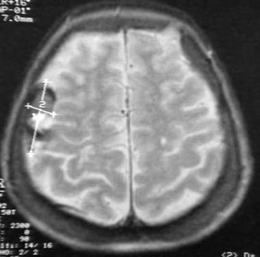
Magnetic Resonance Imaging (MRI) is a non-invasive diagnostic imaging technique that uses a strong magnetic field and radio waves to create detailed cross-sectional or three-dimensional images of the internal structures of the body. The patient lies within a large, cylindrical magnet, and the scanner detects changes in the direction of the magnetic field caused by protons in the body. These changes are then converted into detailed images that help medical professionals to diagnose and monitor various medical conditions, such as tumors, injuries, or diseases affecting the brain, spinal cord, heart, blood vessels, joints, and other internal organs. MRI does not use radiation like computed tomography (CT) scans.
The temporal lobe is one of the four main lobes of the cerebral cortex in the brain, located on each side of the head roughly level with the ears. It plays a major role in auditory processing, memory, and emotion. The temporal lobe contains several key structures including the primary auditory cortex, which is responsible for analyzing sounds, and the hippocampus, which is crucial for forming new memories. Damage to the temporal lobe can result in various neurological symptoms such as hearing loss, memory impairment, and changes in emotional behavior.
Cortical blindness is a type of visual impairment that is caused by damage to the occipital cortex, which is the part of the brain responsible for processing visual information. This condition is also known as cerebral blindness or cerebral visual impairment.
In cortical blindness, the eyes are able to receive and transmit visual signals to the brain, but the brain is unable to interpret these signals correctly. As a result, the person may have difficulty recognizing objects, faces, or movements in their visual field. They may also experience hallucinations, such as seeing patterns or shapes that aren't really there.
Cortical blindness can be caused by a variety of factors, including stroke, trauma, brain tumors, infection, or hypoxia (lack of oxygen). In some cases, cortical blindness may be temporary and improve over time with treatment and rehabilitation. However, in other cases, the damage to the occipital cortex may be permanent, leading to a lifelong visual impairment.
It is important to note that cortical blindness is different from legal blindness, which is a term used to describe a severe visual impairment that cannot be corrected with glasses or contact lenses. In contrast, cortical blindness is a neurological condition that affects the brain's ability to process visual information, rather than a problem with the eyes themselves.
Hemianopsia is a medical term that refers to a loss of vision in half of the visual field in one or both eyes. It can be either homonymous (the same side in both eyes) or heteronymous (different sides in each eye). Hemianopsia usually results from damage to the optic radiations or occipital cortex in the brain, often due to stroke, trauma, tumor, or other neurological conditions. It can significantly impact a person's daily functioning and may require visual rehabilitation to help compensate for the vision loss.
Epilepsy, partial is a type of epilepsy characterized by recurrent, unprovoked seizures that originate in a specific, localized area of the brain. These seizures are also known as focal seizures and can vary in severity and symptoms depending on the location of the abnormal electrical activity in the brain.
Partial epilepsies can be further classified into two main categories: simple partial seizures and complex partial seizures. Simple partial seizures do not involve a loss of consciousness, while complex partial seizures are associated with impaired awareness or responsiveness during the seizure.
The causes of partial epilepsies can include brain injury, infection, stroke, tumors, genetic factors, or an unknown cause. Treatment typically involves anti-seizure medications, and in some cases, surgery may be recommended to remove the specific area of the brain responsible for the seizures.
The brain is the central organ of the nervous system, responsible for receiving and processing sensory information, regulating vital functions, and controlling behavior, movement, and cognition. It is divided into several distinct regions, each with specific functions:
1. Cerebrum: The largest part of the brain, responsible for higher cognitive functions such as thinking, learning, memory, language, and perception. It is divided into two hemispheres, each controlling the opposite side of the body.
2. Cerebellum: Located at the back of the brain, it is responsible for coordinating muscle movements, maintaining balance, and fine-tuning motor skills.
3. Brainstem: Connects the cerebrum and cerebellum to the spinal cord, controlling vital functions such as breathing, heart rate, and blood pressure. It also serves as a relay center for sensory information and motor commands between the brain and the rest of the body.
4. Diencephalon: A region that includes the thalamus (a major sensory relay station) and hypothalamus (regulates hormones, temperature, hunger, thirst, and sleep).
5. Limbic system: A group of structures involved in emotional processing, memory formation, and motivation, including the hippocampus, amygdala, and cingulate gyrus.
The brain is composed of billions of interconnected neurons that communicate through electrical and chemical signals. It is protected by the skull and surrounded by three layers of membranes called meninges, as well as cerebrospinal fluid that provides cushioning and nutrients.
Reflex epilepsy is a type of epilepsy in which seizures are consistently triggered by specific, recurring sensory stimuli. These triggers can vary widely and may include visual patterns, flashes of light, touch, sound, or even emotional experiences. When the brain receives input from these triggers, it responds with an abnormal electrical discharge that can lead to a seizure.
Reflex epilepsy is relatively rare, accounting for only about 5-10% of all epilepsy cases. It's important to note that not everyone who experiences seizures in response to these triggers has reflex epilepsy; the defining characteristic of this condition is the consistent and reproducible nature of the seizure response to a specific stimulus.
There are several different types of reflex epilepsy, each characterized by its own unique set of triggers. For example, some people with this condition may experience seizures in response to visual patterns or flashes of light (known as photosensitive epilepsy), while others may have seizures triggered by certain sounds or tactile sensations.
Treatment for reflex epilepsy typically involves identifying and avoiding triggers whenever possible, as well as using medication to control seizures. In some cases, surgery may be recommended to remove the specific area of the brain that is responsible for the abnormal electrical activity. With proper treatment and management, many people with reflex epilepsy are able to lead full and active lives.
Brain mapping is a broad term that refers to the techniques used to understand the structure and function of the brain. It involves creating maps of the various cognitive, emotional, and behavioral processes in the brain by correlating these processes with physical locations or activities within the nervous system. Brain mapping can be accomplished through a variety of methods, including functional magnetic resonance imaging (fMRI), positron emission tomography (PET) scans, electroencephalography (EEG), and others. These techniques allow researchers to observe which areas of the brain are active during different tasks or thoughts, helping to shed light on how the brain processes information and contributes to our experiences and behaviors. Brain mapping is an important area of research in neuroscience, with potential applications in the diagnosis and treatment of neurological and psychiatric disorders.
The frontal lobe is the largest lobes of the human brain, located at the front part of each cerebral hemisphere and situated in front of the parietal and temporal lobes. It plays a crucial role in higher cognitive functions such as decision making, problem solving, planning, parts of social behavior, emotional expressions, physical reactions, and motor function. The frontal lobe is also responsible for what's known as "executive functions," which include the ability to focus attention, understand rules, switch focus, plan actions, and inhibit inappropriate behaviors. It is divided into five areas, each with its own specific functions: the primary motor cortex, premotor cortex, Broca's area, prefrontal cortex, and orbitofrontal cortex. Damage to the frontal lobe can result in a wide range of impairments, depending on the location and extent of the injury.
The Posterior Cerebral Artery (PCA) is one of the major arteries that supplies blood to the brain. It is a branch of the basilar artery, which is formed by the union of the two vertebral arteries. The PCA supplies oxygenated blood to the occipital lobe (responsible for visual processing), the temporal lobe (involved in auditory and memory functions), and the thalamus and midbrain (relay station for sensory and motor signals).
The PCA has two segments: the precommunicating segment (P1) and the postcommunicating segment (P2). The P1 segment runs posteriorly along the cerebral peduncle, while the P2 segment courses around the midbrain to reach the occipital lobe.
Atherosclerosis, embolism, or other vascular conditions can affect the PCA and lead to a variety of neurological symptoms, including visual loss, memory impairment, and difficulty with language processing.
The parietal lobe is a region of the brain that is located in the posterior part of the cerebral cortex, covering the upper and rear portions of the brain. It is involved in processing sensory information from the body, such as touch, temperature, and pain, as well as spatial awareness and perception, visual-spatial cognition, and the integration of different senses.
The parietal lobe can be divided into several functional areas, including the primary somatosensory cortex (which receives tactile information from the body), the secondary somatosensory cortex (which processes more complex tactile information), and the posterior parietal cortex (which is involved in spatial attention, perception, and motor planning).
Damage to the parietal lobe can result in various neurological symptoms, such as neglect of one side of the body, difficulty with spatial orientation, problems with hand-eye coordination, and impaired mathematical and language abilities.
Temporal lobe epilepsy (TLE) is a type of focal (localized) epilepsy that originates from the temporal lobes of the brain. The temporal lobes are located on each side of the brain and are involved in processing sensory information, memory, and emotion. TLE is characterized by recurrent seizures that originate from one or both temporal lobes.
The symptoms of TLE can vary depending on the specific area of the temporal lobe that is affected. However, common symptoms include auras (sensory or emotional experiences that occur before a seizure), strange smells or tastes, lip-smacking or chewing movements, and memory problems. Some people with TLE may also experience automatisms (involuntary movements such as picking at clothes or fumbling with objects) during their seizures.
Treatment for TLE typically involves medication to control seizures, although surgery may be recommended in some cases. The goal of treatment is to reduce the frequency and severity of seizures and improve quality of life.
Computer-assisted image processing is a medical term that refers to the use of computer systems and specialized software to improve, analyze, and interpret medical images obtained through various imaging techniques such as X-ray, CT (computed tomography), MRI (magnetic resonance imaging), ultrasound, and others.
The process typically involves several steps, including image acquisition, enhancement, segmentation, restoration, and analysis. Image processing algorithms can be used to enhance the quality of medical images by adjusting contrast, brightness, and sharpness, as well as removing noise and artifacts that may interfere with accurate diagnosis. Segmentation techniques can be used to isolate specific regions or structures of interest within an image, allowing for more detailed analysis.
Computer-assisted image processing has numerous applications in medical imaging, including detection and characterization of lesions, tumors, and other abnormalities; assessment of organ function and morphology; and guidance of interventional procedures such as biopsies and surgeries. By automating and standardizing image analysis tasks, computer-assisted image processing can help to improve diagnostic accuracy, efficiency, and consistency, while reducing the potential for human error.
Cerebral dominance is a concept in neuropsychology that refers to the specialization of one hemisphere of the brain over the other for certain cognitive functions. In most people, the left hemisphere is dominant for language functions such as speaking and understanding spoken or written language, while the right hemisphere is dominant for non-verbal functions such as spatial ability, face recognition, and artistic ability.
Cerebral dominance does not mean that the non-dominant hemisphere is incapable of performing the functions of the dominant hemisphere, but rather that it is less efficient or specialized in those areas. The concept of cerebral dominance has been used to explain individual differences in cognitive abilities and learning styles, as well as the laterality of brain damage and its effects on cognition and behavior.
It's important to note that cerebral dominance is a complex phenomenon that can vary between individuals and can be influenced by various factors such as genetics, environment, and experience. Additionally, recent research has challenged the strict lateralization of functions and suggested that there is more functional overlap and interaction between the two hemispheres than previously thought.
The cerebral cortex is the outermost layer of the brain, characterized by its intricate folded structure and wrinkled appearance. It is a region of great importance as it plays a key role in higher cognitive functions such as perception, consciousness, thought, memory, language, and attention. The cerebral cortex is divided into two hemispheres, each containing four lobes: the frontal, parietal, temporal, and occipital lobes. These areas are responsible for different functions, with some regions specializing in sensory processing while others are involved in motor control or associative functions. The cerebral cortex is composed of gray matter, which contains neuronal cell bodies, and is covered by a layer of white matter that consists mainly of myelinated nerve fibers.
The visual cortex is the part of the brain that processes visual information. It is located in the occipital lobe, which is at the back of the brain. The visual cortex is responsible for receiving and interpreting signals from the retina, which are then transmitted through the optic nerve and optic tract.
The visual cortex contains several areas that are involved in different aspects of visual processing, such as identifying shapes, colors, and movements. These areas work together to help us recognize and understand what we see. Damage to the visual cortex can result in various visual impairments, such as blindness or difficulty with visual perception.
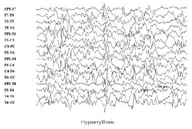
Electroencephalography (EEG) is a medical procedure that records electrical activity in the brain. It uses small, metal discs called electrodes, which are attached to the scalp with paste or a specialized cap. These electrodes detect tiny electrical charges that result from the activity of brain cells, and the EEG machine then amplifies and records these signals.
EEG is used to diagnose various conditions related to the brain, such as seizures, sleep disorders, head injuries, infections, and degenerative diseases like Alzheimer's or Parkinson's. It can also be used during surgery to monitor brain activity and ensure that surgical procedures do not interfere with vital functions.
EEG is a safe and non-invasive procedure that typically takes about 30 minutes to an hour to complete, although longer recordings may be necessary in some cases. Patients are usually asked to relax and remain still during the test, as movement can affect the quality of the recording.
Photic stimulation is a medical term that refers to the exposure of the eyes to light, specifically repetitive pulses of light, which is used as a method in various research and clinical settings. In neuroscience, it's often used in studies related to vision, circadian rhythms, and brain function.
In a clinical context, photic stimulation is sometimes used in the diagnosis of certain medical conditions such as seizure disorders (like epilepsy). By observing the response of the brain to this light stimulus, doctors can gain valuable insights into the functioning of the brain and the presence of any neurological disorders.
However, it's important to note that photic stimulation should be conducted under the supervision of a trained healthcare professional, as improper use can potentially trigger seizures in individuals who are susceptible to them.
Posterior Leukoencephalopathy Syndrome (PLS) is a neurological disorder characterized by the presence of vasogenic edema (swelling due to leakage of fluid from blood vessels) in the white matter (part of the brain that contains nerve fibers) of the posterior regions (occipital and parietal lobes) of the brain.
The symptoms of PLS can vary but often include headache, altered mental status, seizures, visual disturbances, and hypertension (high blood pressure). The exact cause of PLS is not fully understood, but it has been associated with certain conditions such as eclampsia, preeclampsia, kidney failure, autoimmune disorders, and the use of certain medications.
PLS is typically diagnosed based on clinical symptoms and imaging studies such as MRI or CT scans. Treatment usually involves addressing the underlying cause of PLS, controlling hypertension if present, and managing seizures if they occur. With prompt and appropriate treatment, most patients with PLS have a good prognosis and recover completely. However, in severe cases, PLS can lead to permanent neurological damage or even death.
Intracranial arteriovenous malformations (AVMs) are abnormal, tangled connections between the arteries and veins in the brain. These connections bypass the capillary system, which can lead to high-flow shunting and potential complications such as hemorrhage, stroke, or neurological deficits. AVMs are congenital conditions, meaning they are present at birth, although symptoms may not appear until later in life. They are relatively rare, affecting approximately 0.1% of the population. Treatment options for AVMs include surgery, radiation therapy, and endovascular embolization, depending on the size, location, and specific characteristics of the malformation.
Brain diseases, also known as neurological disorders, refer to a wide range of conditions that affect the brain and nervous system. These diseases can be caused by various factors such as genetics, infections, injuries, degeneration, or structural abnormalities. They can affect different parts of the brain, leading to a variety of symptoms and complications.
Some examples of brain diseases include:
1. Alzheimer's disease - a progressive degenerative disorder that affects memory and cognitive function.
2. Parkinson's disease - a movement disorder characterized by tremors, stiffness, and difficulty with coordination and balance.
3. Multiple sclerosis - a chronic autoimmune disease that affects the nervous system and can cause a range of symptoms such as vision loss, muscle weakness, and cognitive impairment.
4. Epilepsy - a neurological disorder characterized by recurrent seizures.
5. Brain tumors - abnormal growths in the brain that can be benign or malignant.
6. Stroke - a sudden interruption of blood flow to the brain, which can cause paralysis, speech difficulties, and other neurological symptoms.
7. Meningitis - an infection of the membranes surrounding the brain and spinal cord.
8. Encephalitis - an inflammation of the brain that can be caused by viruses, bacteria, or autoimmune disorders.
9. Huntington's disease - a genetic disorder that affects muscle coordination, cognitive function, and mental health.
10. Migraine - a neurological condition characterized by severe headaches, often accompanied by nausea, vomiting, and sensitivity to light and sound.
Brain diseases can range from mild to severe and may be treatable or incurable. They can affect people of all ages and backgrounds, and early diagnosis and treatment are essential for improving outcomes and quality of life.
Diffusion Magnetic Resonance Imaging (MRI) is a non-invasive medical imaging technique that uses magnetic fields and radio waves to produce detailed images of the body's internal structures, particularly the brain and nervous system. In diffusion MRI, the movement of water molecules in biological tissues is measured and analyzed to generate contrast in the images based on the microstructural properties of the tissue.
Diffusion MRI is unique because it allows for the measurement of water diffusion in various directions, which can reveal important information about the organization and integrity of nerve fibers in the brain. This technique has been widely used in research and clinical settings to study a variety of neurological conditions, including stroke, traumatic brain injury, multiple sclerosis, and neurodegenerative diseases such as Alzheimer's disease.
In summary, diffusion MRI is a specialized type of MRI that measures the movement of water molecules in biological tissues to generate detailed images of the body's internal structures, particularly the brain and nervous system. It provides valuable information about the microstructural properties of tissues and has important applications in both research and clinical settings.
Functional laterality, in a medical context, refers to the preferential use or performance of one side of the body over the other for specific functions. This is often demonstrated in hand dominance, where an individual may be right-handed or left-handed, meaning they primarily use their right or left hand for tasks such as writing, eating, or throwing.
However, functional laterality can also apply to other bodily functions and structures, including the eyes (ocular dominance), ears (auditory dominance), or legs. It's important to note that functional laterality is not a strict binary concept; some individuals may exhibit mixed dominance or no strong preference for one side over the other.
In clinical settings, assessing functional laterality can be useful in diagnosing and treating various neurological conditions, such as stroke or traumatic brain injury, where understanding any resulting lateralized impairments can inform rehabilitation strategies.
Cerebral infarction, also known as a "stroke" or "brain attack," is the sudden death of brain cells caused by the interruption of their blood supply. It is most commonly caused by a blockage in one of the blood vessels supplying the brain (an ischemic stroke), but can also result from a hemorrhage in or around the brain (a hemorrhagic stroke).
Ischemic strokes occur when a blood clot or other particle blocks a cerebral artery, cutting off blood flow to a part of the brain. The lack of oxygen and nutrients causes nearby brain cells to die. Hemorrhagic strokes occur when a weakened blood vessel ruptures, causing bleeding within or around the brain. This bleeding can put pressure on surrounding brain tissues, leading to cell death.
Symptoms of cerebral infarction depend on the location and extent of the affected brain tissue but may include sudden weakness or numbness in the face, arm, or leg; difficulty speaking or understanding speech; vision problems; loss of balance or coordination; and severe headache with no known cause. Immediate medical attention is crucial for proper diagnosis and treatment to minimize potential long-term damage or disability.
Myelinated nerve fibers are neuronal processes that are surrounded by a myelin sheath, a fatty insulating substance that is produced by Schwann cells in the peripheral nervous system and oligodendrocytes in the central nervous system. This myelin sheath helps to increase the speed of electrical impulse transmission, also known as action potentials, along the nerve fiber. The myelin sheath has gaps called nodes of Ranvier where the electrical impulses can jump from one node to the next, which also contributes to the rapid conduction of signals. Myelinated nerve fibers are typically found in the peripheral nerves and the optic nerve, but not in the central nervous system (CNS) tracts that are located within the brain and spinal cord.
Visual fields refer to the total area in which objects can be seen while keeping the eyes focused on a central point. It is the entire area that can be observed using peripheral (side) vision while the eye gazes at a fixed point. A visual field test is used to detect blind spots or gaps (scotomas) in a person's vision, which could indicate various medical conditions such as glaucoma, retinal damage, optic nerve disease, brain tumors, or strokes. The test measures both the central and peripheral vision and maps the entire area that can be seen when focusing on a single point.
Visual perception refers to the ability to interpret and organize information that comes from our eyes to recognize and understand what we are seeing. It involves several cognitive processes such as pattern recognition, size estimation, movement detection, and depth perception. Visual perception allows us to identify objects, navigate through space, and interact with our environment. Deficits in visual perception can lead to learning difficulties and disabilities.
The Circle of Willis is a circulatory arrangement in the brain where the major arteries that supply blood to the brain converge to form an almost circular structure. It is named after Thomas Willis, an English physician who first described it in 1664.
This circle is formed by the joining of the two internal carotid arteries, which divide into the anterior cerebral and middle cerebral arteries, with the basilar artery, which arises from the vertebral arteries. These vessels anastomose, or connect, to form a polygon-like structure at the base of the brain.
The Circle of Willis plays a crucial role in maintaining adequate blood flow to the brain, as it allows for collateral circulation. If one of the arteries that make up the circle becomes blocked or narrowed, blood can still reach the affected area through the other vessels in the circle. This helps to minimize the risk of stroke and other neurological disorders.
X-ray computed tomography (CT or CAT scan) is a medical imaging method that uses computer-processed combinations of many X-ray images taken from different angles to produce cross-sectional (tomographic) images (virtual "slices") of the body. These cross-sectional images can then be used to display detailed internal views of organs, bones, and soft tissues in the body.
The term "computed tomography" is used instead of "CT scan" or "CAT scan" because the machines take a series of X-ray measurements from different angles around the body and then use a computer to process these data to create detailed images of internal structures within the body.
CT scanning is a noninvasive, painless medical test that helps physicians diagnose and treat medical conditions. CT imaging provides detailed information about many types of tissue including lung, bone, soft tissue and blood vessels. CT examinations can be performed on every part of the body for a variety of reasons including diagnosis, surgical planning, and monitoring of therapeutic responses.
In computed tomography (CT), an X-ray source and detector rotate around the patient, measuring the X-ray attenuation at many different angles. A computer uses this data to construct a cross-sectional image by the process of reconstruction. This technique is called "tomography". The term "computed" refers to the use of a computer to reconstruct the images.
CT has become an important tool in medical imaging and diagnosis, allowing radiologists and other physicians to view detailed internal images of the body. It can help identify many different medical conditions including cancer, heart disease, lung nodules, liver tumors, and internal injuries from trauma. CT is also commonly used for guiding biopsies and other minimally invasive procedures.
In summary, X-ray computed tomography (CT or CAT scan) is a medical imaging technique that uses computer-processed combinations of many X-ray images taken from different angles to produce cross-sectional images of the body. It provides detailed internal views of organs, bones, and soft tissues in the body, allowing physicians to diagnose and treat medical conditions.
Evoked potentials, visual, also known as visually evoked potentials (VEPs), are electrical responses recorded from the brain following the presentation of a visual stimulus. These responses are typically measured using electroencephalography (EEG) and can provide information about the functioning of the visual pathways in the brain.
There are several types of VEPs, including pattern-reversal VEPs and flash VEPs. Pattern-reversal VEPs are elicited by presenting alternating checkerboard patterns, while flash VEPs are elicited by flashing a light. The responses are typically analyzed in terms of their latency (the time it takes for the response to occur) and amplitude (the size of the response).
VEPs are often used in clinical settings to help diagnose and monitor conditions that affect the visual system, such as multiple sclerosis, optic neuritis, and brainstem tumors. They can also be used in research to study the neural mechanisms underlying visual perception.
Blindness is a condition of complete or near-complete vision loss. It can be caused by various factors such as eye diseases, injuries, or birth defects. Total blindness means that a person cannot see anything at all, while near-complete blindness refers to having only light perception or the ability to perceive the direction of light, but not able to discern shapes or forms. Legal blindness is a term used to define a certain level of visual impairment that qualifies an individual for government assistance and benefits; it usually means best corrected visual acuity of 20/200 or worse in the better eye, or a visual field no greater than 20 degrees in diameter.
Visual pathways, also known as the visual system or the optic pathway, refer to the series of specialized neurons in the nervous system that transmit visual information from the eyes to the brain. This complex network includes the retina, optic nerve, optic chiasma, optic tract, lateral geniculate nucleus, pulvinar, and the primary and secondary visual cortices located in the occipital lobe of the brain.
The process begins when light enters the eye and strikes the photoreceptor cells (rods and cones) in the retina, converting the light energy into electrical signals. These signals are then transmitted to bipolar cells and subsequently to ganglion cells, whose axons form the optic nerve. The fibers from each eye's nasal hemiretina cross at the optic chiasma, while those from the temporal hemiretina continue without crossing. This results in the formation of the optic tract, which carries visual information from both eyes to the opposite side of the brain.
The majority of fibers in the optic tract synapse with neurons in the lateral geniculate nucleus (LGN), a part of the thalamus. The LGN sends this information to the primary visual cortex, also known as V1 or Brodmann area 17, located in the occipital lobe. Here, simple features like lines and edges are initially processed. Further processing occurs in secondary (V2) and tertiary (V3-V5) visual cortices, where more complex features such as shape, motion, and depth are analyzed. Ultimately, this information is integrated to form our perception of the visual world.
Cerebrovascular circulation refers to the network of blood vessels that supply oxygenated blood and nutrients to the brain tissue, and remove waste products. It includes the internal carotid arteries, vertebral arteries, circle of Willis, and the intracranial arteries that branch off from them.
The internal carotid arteries and vertebral arteries merge to form the circle of Willis, a polygonal network of vessels located at the base of the brain. The anterior cerebral artery, middle cerebral artery, posterior cerebral artery, and communicating arteries are the major vessels that branch off from the circle of Willis and supply blood to different regions of the brain.
Interruptions or abnormalities in the cerebrovascular circulation can lead to various neurological conditions such as stroke, transient ischemic attack (TIA), and vascular dementia.
Epilepsy is a chronic neurological disorder characterized by recurrent, unprovoked seizures. These seizures are caused by abnormal electrical activity in the brain, which can result in a wide range of symptoms, including convulsions, loss of consciousness, and altered sensations or behaviors. Epilepsy can have many different causes, including genetic factors, brain injury, infection, or stroke. In some cases, the cause may be unknown.
There are many different types of seizures that can occur in people with epilepsy, and the specific type of seizure will depend on the location and extent of the abnormal electrical activity in the brain. Some people may experience only one type of seizure, while others may have several different types. Seizures can vary in frequency, from a few per year to dozens or even hundreds per day.
Epilepsy is typically diagnosed based on the patient's history of recurrent seizures and the results of an electroencephalogram (EEG), which measures the electrical activity in the brain. Imaging tests such as MRI or CT scans may also be used to help identify any structural abnormalities in the brain that may be contributing to the seizures.
While there is no cure for epilepsy, it can often be effectively managed with medication. In some cases, surgery may be recommended to remove the area of the brain responsible for the seizures. With proper treatment and management, many people with epilepsy are able to lead normal, productive lives.
Atrophy is a medical term that refers to the decrease in size and wasting of an organ or tissue due to the disappearance of cells, shrinkage of cells, or decreased number of cells. This process can be caused by various factors such as disuse, aging, degeneration, injury, or disease.
For example, if a muscle is immobilized for an extended period, it may undergo atrophy due to lack of use. Similarly, certain medical conditions like diabetes, cancer, and heart failure can lead to the wasting away of various tissues and organs in the body.
Atrophy can also occur as a result of natural aging processes, leading to decreased muscle mass and strength in older adults. In general, atrophy is characterized by a decrease in the volume or weight of an organ or tissue, which can have significant impacts on its function and overall health.
The thalamus is a large, paired structure in the brain that serves as a relay station for sensory and motor signals to the cerebral cortex. It is located in the dorsal part of the diencephalon and is made up of two symmetrical halves, each connected to the corresponding cerebral hemisphere.
The thalamus receives inputs from almost all senses, except for the olfactory system, and processes them before sending them to specific areas in the cortex. It also plays a role in regulating consciousness, sleep, and alertness. Additionally, the thalamus is involved in motor control by relaying information between the cerebellum and the motor cortex.
The thalamus is divided into several nuclei, each with distinct connections and functions. Some of these nuclei are involved in sensory processing, while others are involved in motor function or regulation of emotions and cognition. Overall, the thalamus plays a critical role in integrating information from various brain regions and modulating cognitive and emotional processes.
The Atlanto-Occipital Joint, also known as the AO joint or the craniocervical joint, is the articulation between the occiput (the base of the skull) and the atlas (the first cervical vertebra). This joint allows for movements such as nodding your head "yes" and tilting your head from side to side. It is a crucial joint in maintaining the alignment and stability of the head and neck.
Neuropsychological tests are a type of psychological assessment that measures cognitive functions, such as attention, memory, language, problem-solving, and perception. These tests are used to help diagnose and understand the cognitive impact of neurological conditions, including dementia, traumatic brain injury, stroke, Parkinson's disease, and other disorders that affect the brain.
The tests are typically administered by a trained neuropsychologist and can take several hours to complete. They may involve paper-and-pencil tasks, computerized tasks, or interactive activities. The results of the tests are compared to normative data to help identify any areas of cognitive weakness or strength.
Neuropsychological testing can provide valuable information for treatment planning, rehabilitation, and assessing response to treatment. It can also be used in research to better understand the neural basis of cognition and the impact of neurological conditions on cognitive function.
Anisotropy is a medical term that refers to the property of being directionally dependent, meaning that its properties or characteristics vary depending on the direction in which they are measured. In the context of medicine and biology, anisotropy can refer to various biological structures, tissues, or materials that exhibit different physical or chemical properties along different axes.
For example, certain types of collagen fibers in tendons and ligaments exhibit anisotropic behavior because they are stronger and stiffer when loaded along their long axis compared to being loaded perpendicular to it. Similarly, some brain tissues may show anisotropy due to the presence of nerve fibers that are organized in specific directions, leading to differences in electrical conductivity or diffusion properties depending on the orientation of the measurement.
Anisotropy is an important concept in various medical fields, including radiology, neurology, and materials science, as it can provide valuable information about the structure and function of biological tissues and help guide diagnostic and therapeutic interventions.
The corpus callosum is the largest collection of white matter in the brain, consisting of approximately 200 million nerve fibers. It is a broad, flat band of tissue that connects the two hemispheres of the brain, allowing them to communicate and coordinate information processing. The corpus callosum plays a crucial role in integrating sensory, motor, and cognitive functions between the two sides of the brain. Damage to the corpus callosum can result in various neurological symptoms, including difficulties with movement, speech, memory, and social behavior.
Emission computed tomography (ECT) is a type of tomographic imaging technique in which an emission signal from within the body is detected to create cross-sectional images of that signal's distribution. In Emission-Computed Tomography (ECT), a radionuclide is introduced into the body, usually through injection, inhalation or ingestion. The radionuclide emits gamma rays that are then detected by external gamma cameras.
The data collected from these cameras is then used to create cross-sectional images of the distribution of the radiopharmaceutical within the body. This allows for the identification and quantification of functional information about specific organs or systems within the body, such as blood flow, metabolic activity, or receptor density.
One common type of Emission-Computed Tomography is Single Photon Emission Computed Tomography (SPECT), which uses a single gamma camera that rotates around the patient to collect data from multiple angles. Another type is Positron Emission Tomography (PET), which uses positron-emitting radionuclides and detects the coincident gamma rays emitted by the annihilation of positrons and electrons.
Overall, ECT is a valuable tool in medical imaging for diagnosing and monitoring various diseases, including cancer, heart disease, and neurological disorders.
Diffusion Tensor Imaging (DTI) is a type of magnetic resonance imaging (MRI) technique that allows for the measurement and visualization of water diffusion in biological tissues, particularly in the brain. DTI provides information about the microstructural organization and integrity of nerve fibers within the brain by measuring the directionality of water diffusion in the brain's white matter tracts.
In DTI, a tensor is used to describe the three-dimensional diffusion properties of water molecules in each voxel (three-dimensional pixel) of an MRI image. The tensor provides information about the magnitude and direction of water diffusion, which can be used to calculate various diffusion metrics such as fractional anisotropy (FA), mean diffusivity (MD), axial diffusivity (AD), and radial diffusivity (RD). These metrics provide insights into the structural properties of nerve fibers, including their orientation, density, and integrity.
DTI has numerous clinical applications, such as in the diagnosis and monitoring of neurological disorders like multiple sclerosis, traumatic brain injury, and neurodegenerative diseases. It can also be used for presurgical planning to identify critical white matter tracts that need to be preserved during surgery.
Vision disorders refer to a wide range of conditions that affect the visual system and result in various symptoms, such as blurry vision, double vision, distorted vision, impaired depth perception, and difficulty with visual tracking or focusing. These disorders can be categorized into several types, including:
1. Refractive errors: These occur when the shape of the eye prevents light from focusing directly on the retina, resulting in blurry vision. Examples include myopia (nearsightedness), hyperopia (farsightedness), astigmatism, and presbyopia (age-related loss of near vision).
2. Strabismus: Also known as crossed eyes or walleye, strabismus is a misalignment of the eyes where they point in different directions, which can lead to double vision or loss of depth perception.
3. Amblyopia: Often called lazy eye, amblyopia is a condition where one eye has reduced vision due to lack of proper visual development during childhood. It may be caused by strabismus, refractive errors, or other factors that interfere with normal visual development.
4. Accommodative disorders: These involve problems with the focusing ability of the eyes, such as convergence insufficiency (difficulty focusing on close objects) and accommodative dysfunction (inability to maintain clear vision at different distances).
5. Binocular vision disorders: These affect how the eyes work together as a team, leading to issues like poor depth perception, eye strain, and headaches. Examples include convergence insufficiency, divergence excess, and suppression.
6. Ocular motility disorders: These involve problems with eye movement, such as nystagmus (involuntary eye movements), strabismus, or restricted extraocular muscle function.
7. Visual processing disorders: These affect the brain's ability to interpret and make sense of visual information, even when the eyes themselves are healthy. Symptoms may include difficulty with reading, recognizing shapes and objects, and understanding spatial relationships.
8. Low vision: This term refers to significant visual impairment that cannot be fully corrected with glasses, contact lenses, medication, or surgery. It includes conditions like macular degeneration, diabetic retinopathy, glaucoma, and cataracts.
9. Blindness: Complete loss of sight in both eyes, which can be caused by various factors such as injury, disease, or genetic conditions.
Creatine is a organic acid that is produced naturally in the liver, kidneys and pancreas. It is also found in small amounts in certain foods such as meat and fish. The chemical formula for creatine is C4H9N3O2. In the body, creatine is converted into creatine phosphate, which is used to help produce energy during high-intensity exercise, such as weightlifting or sprinting.
Creatine can also be taken as a dietary supplement, in the form of creatine monohydrate, with the goal of increasing muscle creatine and phosphocreatine levels, which may improve athletic performance and help with muscle growth. However, it is important to note that while some studies have found that creatine supplementation can improve exercise performance and muscle mass in certain populations, others have not found significant benefits.
Creatine supplements are generally considered safe when used as directed, but they can cause side effects such as weight gain, stomach discomfort, and muscle cramps in some people. It is always recommended to consult a healthcare professional before starting any new supplement regimen.
Emission-Computed Tomography, Single-Photon (SPECT) is a type of nuclear medicine imaging procedure that generates detailed, three-dimensional images of the distribution of radioactive pharmaceuticals within the body. It uses gamma rays emitted by a radiopharmaceutical that is introduced into the patient's body, and a specialized gamma camera to detect these gamma rays and create tomographic images. The data obtained from the SPECT imaging can be used to diagnose various medical conditions, evaluate organ function, and guide treatment decisions. It is commonly used to image the heart, brain, and bones, among other organs and systems.
Vertebrobasilar insufficiency (VBI) is a medical condition characterized by inadequate blood flow to the vertebral and basilar arteries, which supply oxygenated blood to the brainstem and cerebellum. These arteries arise from the subclavian arteries and merge to form the basilar artery, which supplies critical structures in the posterior circulation of the brain.
VBI is often caused by atherosclerosis, or the buildup of plaque in the arterial walls, leading to narrowing (stenosis) or occlusion of these vessels. Other causes include embolism, arterial dissection, and vasculitis. The decreased blood flow can result in various neurological symptoms, such as dizziness, vertigo, imbalance, difficulty swallowing, slurred speech, visual disturbances, and even transient ischemic attacks (TIAs) or strokes.
Diagnosis of VBI typically involves a combination of clinical evaluation, imaging studies like MRA or CTA, and sometimes cerebral angiography to assess the extent and location of vascular narrowing or occlusion. Treatment options may include lifestyle modifications, medications to manage risk factors (such as hypertension, diabetes, or high cholesterol), antiplatelet therapy, or surgical interventions like endarterectomy or stenting in severe cases.
The optic lobe in non-mammals refers to a specific region of the brain that is responsible for processing visual information. It is a part of the protocerebrum in the insect brain and is analogous to the mammalian visual cortex. The optic lobes receive input directly from the eyes via the optic nerves and are involved in the interpretation and integration of visual stimuli, enabling non-mammals to perceive and respond to their environment. In some invertebrates, like insects, the optic lobe is further divided into subregions, including the lamina, medulla, and lobula, each with distinct functions in visual processing.
A seizure is an uncontrolled, abnormal firing of neurons (brain cells) that can cause various symptoms such as convulsions, loss of consciousness, altered awareness, or changes in behavior. Seizures can be caused by a variety of factors including epilepsy, brain injury, infection, toxic substances, or genetic disorders. They can also occur without any identifiable cause, known as idiopathic seizures. Seizures are a medical emergency and require immediate attention.
Brain neoplasms, also known as brain tumors, are abnormal growths of cells within the brain. These growths can be benign (non-cancerous) or malignant (cancerous). Benign brain tumors typically grow slowly and do not spread to other parts of the body. However, they can still cause serious problems if they press on sensitive areas of the brain. Malignant brain tumors, on the other hand, are cancerous and can grow quickly, invading surrounding brain tissue and spreading to other parts of the brain or spinal cord.
Brain neoplasms can arise from various types of cells within the brain, including glial cells (which provide support and insulation for nerve cells), neurons (nerve cells that transmit signals in the brain), and meninges (the membranes that cover the brain and spinal cord). They can also result from the spread of cancer cells from other parts of the body, known as metastatic brain tumors.
Symptoms of brain neoplasms may vary depending on their size, location, and growth rate. Common symptoms include headaches, seizures, weakness or paralysis in the limbs, difficulty with balance and coordination, changes in speech or vision, confusion, memory loss, and changes in behavior or personality.
Treatment for brain neoplasms depends on several factors, including the type, size, location, and grade of the tumor, as well as the patient's age and overall health. Treatment options may include surgery, radiation therapy, chemotherapy, targeted therapy, or a combination of these approaches. Regular follow-up care is essential to monitor for recurrence and manage any long-term effects of treatment.
Neural pathways, also known as nerve tracts or fasciculi, refer to the highly organized and specialized routes through which nerve impulses travel within the nervous system. These pathways are formed by groups of neurons (nerve cells) that are connected in a series, creating a continuous communication network for electrical signals to transmit information between different regions of the brain, spinal cord, and peripheral nerves.
Neural pathways can be classified into two main types: sensory (afferent) and motor (efferent). Sensory neural pathways carry sensory information from various receptors in the body (such as those for touch, temperature, pain, and vision) to the brain for processing. Motor neural pathways, on the other hand, transmit signals from the brain to the muscles and glands, controlling movements and other effector functions.
The formation of these neural pathways is crucial for normal nervous system function, as it enables efficient communication between different parts of the body and allows for complex behaviors, cognitive processes, and adaptive responses to internal and external stimuli.
Magnetic Resonance Spectroscopy (MRS) is a non-invasive diagnostic technique that provides information about the biochemical composition of tissues, including their metabolic state. It is often used in conjunction with Magnetic Resonance Imaging (MRI) to analyze various metabolites within body tissues, such as the brain, heart, liver, and muscles.
During MRS, a strong magnetic field, radio waves, and a computer are used to produce detailed images and data about the concentration of specific metabolites in the targeted tissue or organ. This technique can help detect abnormalities related to energy metabolism, neurotransmitter levels, pH balance, and other biochemical processes, which can be useful for diagnosing and monitoring various medical conditions, including cancer, neurological disorders, and metabolic diseases.
There are different types of MRS, such as Proton (^1^H) MRS, Phosphorus-31 (^31^P) MRS, and Carbon-13 (^13^C) MRS, each focusing on specific elements or metabolites within the body. The choice of MRS technique depends on the clinical question being addressed and the type of information needed for diagnosis or monitoring purposes.
Three-dimensional (3D) imaging in medicine refers to the use of technologies and techniques that generate a 3D representation of internal body structures, organs, or tissues. This is achieved by acquiring and processing data from various imaging modalities such as X-ray computed tomography (CT), magnetic resonance imaging (MRI), ultrasound, or confocal microscopy. The resulting 3D images offer a more detailed visualization of the anatomy and pathology compared to traditional 2D imaging techniques, allowing for improved diagnostic accuracy, surgical planning, and minimally invasive interventions.
In 3D imaging, specialized software is used to reconstruct the acquired data into a volumetric model, which can be manipulated and viewed from different angles and perspectives. This enables healthcare professionals to better understand complex anatomical relationships, detect abnormalities, assess disease progression, and monitor treatment response. Common applications of 3D imaging include neuroimaging, orthopedic surgery planning, cancer staging, dental and maxillofacial reconstruction, and interventional radiology procedures.
Cognitive disorders are a category of mental health disorders that primarily affect cognitive abilities including learning, memory, perception, and problem-solving. These disorders can be caused by various factors such as brain injury, degenerative diseases, infection, substance abuse, or developmental disabilities. Examples of cognitive disorders include dementia, amnesia, delirium, and intellectual disability. It's important to note that the specific definition and diagnostic criteria for cognitive disorders may vary depending on the medical source or classification system being used.
The cerebellum is a part of the brain that lies behind the brainstem and is involved in the regulation of motor movements, balance, and coordination. It contains two hemispheres and a central portion called the vermis. The cerebellum receives input from sensory systems and other areas of the brain and spinal cord and sends output to motor areas of the brain. Damage to the cerebellum can result in problems with movement, balance, and coordination.
Reference values, also known as reference ranges or reference intervals, are the set of values that are considered normal or typical for a particular population or group of people. These values are often used in laboratory tests to help interpret test results and determine whether a patient's value falls within the expected range.
The process of establishing reference values typically involves measuring a particular biomarker or parameter in a large, healthy population and then calculating the mean and standard deviation of the measurements. Based on these statistics, a range is established that includes a certain percentage of the population (often 95%) and excludes extreme outliers.
It's important to note that reference values can vary depending on factors such as age, sex, race, and other demographic characteristics. Therefore, it's essential to use reference values that are specific to the relevant population when interpreting laboratory test results. Additionally, reference values may change over time due to advances in measurement technology or changes in the population being studied.
Computer-assisted image interpretation is the use of computer algorithms and software to assist healthcare professionals in analyzing and interpreting medical images. These systems use various techniques such as pattern recognition, machine learning, and artificial intelligence to help identify and highlight abnormalities or patterns within imaging data, such as X-rays, CT scans, MRI, and ultrasound images. The goal is to increase the accuracy, consistency, and efficiency of image interpretation, while also reducing the potential for human error. It's important to note that these systems are intended to assist healthcare professionals in their decision making process and not to replace them.
An Encephalocele is a type of neural tube defect that occurs when the bones of the skull do not close completely during fetal development. This results in a sac-like protrusion of the brain and the membranes that cover it through an opening in the skull. The sac may be visible on the scalp, forehead, or back of the head, and can vary in size. Encephaloceles can cause a range of symptoms, including developmental delays, intellectual disabilities, vision problems, and seizures, depending on the severity and location of the defect. Treatment typically involves surgical repair of the encephalocele soon after birth to prevent further damage to the brain and improve outcomes.
Cerebral angiography is a medical procedure that involves taking X-ray images of the blood vessels in the brain after injecting a contrast dye into them. This procedure helps doctors to diagnose and treat various conditions affecting the blood vessels in the brain, such as aneurysms, arteriovenous malformations, and stenosis (narrowing of the blood vessels).
During the procedure, a catheter is inserted into an artery in the leg and threaded through the body to the blood vessels in the neck or brain. The contrast dye is then injected through the catheter, and X-ray images are taken to visualize the blood flow through the brain's blood vessels.
Cerebral angiography provides detailed images of the blood vessels in the brain, allowing doctors to identify any abnormalities or blockages that may be causing symptoms or increasing the risk of stroke. Based on the results of the cerebral angiography, doctors can develop a treatment plan to address these issues and prevent further complications.
Alzheimer's disease is a progressive disorder that causes brain cells to waste away (degenerate) and die. It's the most common cause of dementia — a continuous decline in thinking, behavioral and social skills that disrupts a person's ability to function independently.
The early signs of the disease include forgetting recent events or conversations. As the disease progresses, a person with Alzheimer's disease will develop severe memory impairment and lose the ability to carry out everyday tasks.
Currently, there's no cure for Alzheimer's disease. However, treatments can temporarily slow the worsening of dementia symptoms and improve quality of life.
Frontal lobe epilepsy is a type of focal epilepsy, which means that the seizures originate from a specific area in the brain called the frontal lobe. The frontal lobe is located at the front part of the brain and is responsible for various functions such as motor function, problem-solving, decision making, emotional expression, and social behavior.
In frontal lobe epilepsy, seizures can be quite varied in their presentation, but they often occur during sleep or wakefulness and may include symptoms such as:
* Brief staring spells or automatisms (such as lip smacking, chewing, or fumbling movements)
* Sudden and frequent falls or drops
* Vocalizations or sounds
* Complex behaviors, such as agitation, aggression, or sexual arousal
* Auras or warning sensations before the seizure
Frontal lobe epilepsy can be difficult to diagnose due to the varied nature of the seizures and their occurrence during sleep. Diagnostic tests such as electroencephalogram (EEG) and imaging studies like magnetic resonance imaging (MRI) may be used to help confirm the diagnosis. Treatment typically involves medication, but in some cases, surgery may be recommended if medications are not effective or cause significant side effects.
Spinal nerves are the bundles of nerve fibers that transmit signals between the spinal cord and the rest of the body. There are 31 pairs of spinal nerves in the human body, which can be divided into five regions: 8 cervical, 12 thoracic, 5 lumbar, 5 sacral, and 1 coccygeal. Each spinal nerve carries both sensory information (such as touch, temperature, and pain) from the periphery to the spinal cord, and motor information (such as muscle control) from the spinal cord to the muscles and other structures in the body. Spinal nerves also contain autonomic fibers that regulate involuntary functions such as heart rate, digestion, and blood pressure.
A cerebral hemorrhage, also known as an intracranial hemorrhage or intracerebral hemorrhage, is a type of stroke that results from bleeding within the brain tissue. It occurs when a weakened blood vessel bursts and causes localized bleeding in the brain. This bleeding can increase pressure in the skull, damage nearby brain cells, and release toxic substances that further harm brain tissues.
Cerebral hemorrhages are often caused by chronic conditions like hypertension (high blood pressure) or cerebral amyloid angiopathy, which weakens the walls of blood vessels over time. Other potential causes include trauma, aneurysms, arteriovenous malformations, illicit drug use, and brain tumors. Symptoms may include sudden headache, weakness, numbness, difficulty speaking or understanding speech, vision problems, loss of balance, and altered level of consciousness. Immediate medical attention is required to diagnose and manage cerebral hemorrhage through imaging techniques, supportive care, and possible surgical interventions.
The foramen magnum is the largest opening in the human skull, located at the base of the skull, through which the spinal cord connects to the brain. It is a crucial structure for the transmission of nerve impulses between the brain and the rest of the body. The foramen magnum also provides passage for blood vessels that supply the brainstem and upper spinal cord.
Visual pattern recognition is the ability to identify and interpret patterns in visual information. In a medical context, it often refers to the process by which healthcare professionals recognize and diagnose medical conditions based on visible signs or symptoms. This can involve recognizing the characteristic appearance of a rash, wound, or other physical feature associated with a particular disease or condition. It may also involve recognizing patterns in medical images such as X-rays, CT scans, or MRIs.
In the field of radiology, for example, visual pattern recognition is a critical skill. Radiologists are trained to recognize the typical appearances of various diseases and conditions in medical images. This allows them to make accurate diagnoses based on the patterns they see. Similarly, dermatologists use visual pattern recognition to identify skin abnormalities and diseases based on the appearance of rashes, lesions, or other skin changes.
Overall, visual pattern recognition is an essential skill in many areas of medicine, allowing healthcare professionals to quickly and accurately diagnose medical conditions based on visible signs and symptoms.
A skull fracture is a break in one or more of the bones that form the skull. It can occur from a direct blow to the head, penetrating injuries like gunshot wounds, or from strong rotational forces during an accident. There are several types of skull fractures, including:
1. Linear Skull Fracture: This is the most common type, where there's a simple break in the bone without any splintering, depression, or displacement. It often doesn't require treatment unless it's near a sensitive area like an eye or ear.
2. Depressed Skull Fracture: In this type, a piece of the skull is pushed inward toward the brain. Surgery may be needed to relieve pressure on the brain and repair the fracture.
3. Diastatic Skull Fracture: This occurs along the suture lines (the fibrous joints between the skull bones) that haven't fused yet, often seen in infants and young children.
4. Basilar Skull Fracture: This involves fractures at the base of the skull. It can be serious due to potential injury to the cranial nerves and blood vessels located in this area.
5. Comminuted Skull Fracture: In this severe type, the bone is shattered into many pieces. These fractures usually require extensive surgical repair.
Symptoms of a skull fracture can include pain, swelling, bruising, bleeding (if there's an open wound), and in some cases, clear fluid draining from the ears or nose (cerebrospinal fluid leak). Severe fractures may cause brain injury, leading to symptoms like confusion, loss of consciousness, seizures, or neurological deficits. Immediate medical attention is necessary for any suspected skull fracture.
Alpha rhythm is a type of brain wave that is typically observed in the electroencephalogram (EEG) of normal, awake individuals when they have their eyes closed. It is characterized by sinusoidal waves with a frequency range of 8-13 Hz and is most prominent over the occipital region of the head, which is located at the back of the skull above the brain's visual cortex.
Alpha rhythm is typically associated with relaxed wakefulness, and its presence may indicate that an individual is awake but not engaged in any mentally demanding tasks. It can be blocked or suppressed by various stimuli, such as opening one's eyes, hearing a loud noise, or engaging in mental activity.
Disruptions in alpha rhythm have been observed in various neurological and psychiatric conditions, including epilepsy, dementia, depression, and anxiety disorders. However, more research is needed to fully understand the clinical significance of these abnormalities.
A headache is defined as pain or discomfort in the head, scalp, or neck. It can be a symptom of various underlying conditions such as stress, sinus congestion, migraine, or more serious issues like meningitis or concussion. Headaches can vary in intensity, ranging from mild to severe, and may be accompanied by other symptoms such as nausea, vomiting, or sensitivity to light and sound. There are over 150 different types of headaches, including tension headaches, cluster headaches, and sinus headaches, each with their own specific characteristics and causes.
A meningocele is a type of neural tube defect that results in the herniation of the meninges (the protective membranes covering the brain and spinal cord) through a defect in the vertebral column. The meninges protrude as a sac-like structure, which may be covered by skin or a thin layer of tissue. Meningoceles usually do not contain neural tissue, but cerebrospinal fluid is present within the sac. They are typically asymptomatic unless there is compression of surrounding structures or infection. Treatment generally involves surgical repair to prevent potential complications such as meningitis or neurological damage.
Skull neoplasms refer to abnormal growths or tumors that develop within the skull. These growths can be benign (non-cancerous) or malignant (cancerous). They can originate from various types of cells, such as bone cells, nerve cells, or soft tissues. Skull neoplasms can cause various symptoms depending on their size and location, including headaches, seizures, vision problems, hearing loss, and neurological deficits. Treatment options include surgery, radiation therapy, and chemotherapy. It is important to note that a neoplasm in the skull can also refer to metastatic cancer, which has spread from another part of the body to the skull.
A case-control study is an observational research design used to identify risk factors or causes of a disease or health outcome. In this type of study, individuals with the disease or condition (cases) are compared with similar individuals who do not have the disease or condition (controls). The exposure history or other characteristics of interest are then compared between the two groups to determine if there is an association between the exposure and the disease.
Case-control studies are often used when it is not feasible or ethical to conduct a randomized controlled trial, as they can provide valuable insights into potential causes of diseases or health outcomes in a relatively short period of time and at a lower cost than other study designs. However, because case-control studies rely on retrospective data collection, they are subject to biases such as recall bias and selection bias, which can affect the validity of the results. Therefore, it is important to carefully design and conduct case-control studies to minimize these potential sources of bias.
Cranial sinuses are a part of the venous system in the human head. They are air-filled spaces located within the skull and are named according to their location. The cranial sinuses include:
1. Superior sagittal sinus: It runs along the top of the brain, inside the skull, and drains blood from the scalp and the veins of the brain.
2. Inferior sagittal sinus: It runs along the bottom of the brain and drains into the straight sinus.
3. Straight sinus: It is located at the back of the brain and receives blood from the inferior sagittal sinus and great cerebral vein.
4. Occipital sinuses: They are located at the back of the head and drain blood from the scalp and skull.
5. Cavernous sinuses: They are located on each side of the brain, near the temple, and receive blood from the eye and surrounding areas.
6. Sphenoparietal sinus: It is a small sinus that drains blood from the front part of the brain into the cavernous sinus.
7. Petrosquamosal sinuses: They are located near the ear and drain blood from the scalp and skull.
The cranial sinuses play an essential role in draining blood from the brain and protecting it from injury.
The Cervical Atlas, also known as C1 or the atlas vertebra, is the uppermost and most superior of the seven cervical vertebrae in the human spine. It plays a crucial role in supporting and facilitating the movement of the head, as it articulates with both the occipital bone (forming the joint called the atlanto-occipital joint) and the axis (or C2) vertebra (forming the atlantoaxial joint). The unique structure of the cervical atlas lacks a body, instead having an anterior and posterior arch with two lateral masses that form the facet joints for articulation with the axis. This arrangement allows for a wide range of motion in the neck, including flexion, extension, lateral bending, and rotation.
In medical terms, the face refers to the front part of the head that is distinguished by the presence of the eyes, nose, and mouth. It includes the bones of the skull (frontal bone, maxilla, zygoma, nasal bones, lacrimal bones, palatine bones, inferior nasal conchae, and mandible), muscles, nerves, blood vessels, skin, and other soft tissues. The face plays a crucial role in various functions such as breathing, eating, drinking, speaking, seeing, smelling, and expressing emotions. It also serves as an important identifier for individuals, allowing them to be recognized by others.
Dandy-Walker Syndrome is a congenital brain malformation characterized by the absence or underdevelopment of the cerebellar vermis (the part of the brain that helps coordinate movement) and an enlarged fluid-filled space (fourth ventricle) surrounding it. This condition can also be associated with an upward bulging of the back of the skull (occipital bone), and in some cases, hydrocephalus (excessive accumulation of cerebrospinal fluid in the brain). The syndrome can vary in severity, and symptoms may include problems with balance, coordination, developmental delays, and increased intracranial pressure. It is usually diagnosed through imaging tests such as ultrasound, CT scan, or MRI. Treatment typically involves managing symptoms and addressing complications, which may include surgical procedures to relieve hydrocephalus if present.
A cluster headache is a type of primary headache disorder characterized by severe, one-sided headaches that occur in clusters, meaning they happen several times a day for several weeks or months and then go into remission for a period of time. The pain of a cluster headache is typically intense and often described as a sharp, stabbing, or burning sensation around the eye or temple on one side of the head.
Cluster headaches are relatively rare, affecting fewer than 1 in 1000 people. They tend to affect men more often than women and usually start between the ages of 20 and 50. The exact cause of cluster headaches is not fully understood, but they are thought to be related to abnormalities in the hypothalamus, a part of the brain that regulates various bodily functions, including hormone production and sleep-wake cycles.
Cluster headache attacks can last from 15 minutes to several hours and may be accompanied by other symptoms such as redness or tearing of the eye, runny nose, sweating, or swelling on the affected side of the face. During a cluster period, headaches typically occur at the same time each day, often at night or in the early morning.
Cluster headaches can be treated with various medications, including triptans, oxygen therapy, and local anesthetics. Preventive treatments such as verapamil, lithium, or corticosteroids may also be used to reduce the frequency and severity of cluster headache attacks during a cluster period.
Reaction time, in the context of medicine and physiology, refers to the time period between the presentation of a stimulus and the subsequent initiation of a response. This complex process involves the central nervous system, particularly the brain, which perceives the stimulus, processes it, and then sends signals to the appropriate muscles or glands to react.
There are different types of reaction times, including simple reaction time (responding to a single, expected stimulus) and choice reaction time (choosing an appropriate response from multiple possibilities). These measures can be used in clinical settings to assess various aspects of neurological function, such as cognitive processing speed, motor control, and alertness.
However, it is important to note that reaction times can be influenced by several factors, including age, fatigue, attention, and the use of certain medications or substances.
Agnosia is a medical term that refers to the inability to recognize or comprehend the meaning or significance of sensory stimuli, even though the specific senses themselves are intact. It is a higher-level cognitive disorder, caused by damage to certain areas of the brain that are responsible for processing and interpreting information from our senses.
There are different types of agnosia, depending on which sense is affected:
* Visual agnosia: The inability to recognize or identify objects, faces, or shapes based on visual input.
* Auditory agnosia: The inability to understand spoken language or recognize sounds, even though hearing is intact.
* Tactile agnosia: The inability to recognize objects by touch, despite normal tactile sensation.
* Olfactory and gustatory agnosia: The inability to identify smells or tastes, respectively, even though the senses of smell and taste are functioning normally.
Agnosia can result from various causes, including stroke, brain injury, infection, degenerative diseases, or tumors that damage specific areas of the brain involved in sensory processing and interpretation. Treatment for agnosia typically focuses on rehabilitation and compensation strategies to help individuals adapt to their deficits and improve their quality of life.
The skull is the bony structure that encloses and protects the brain, the eyes, and the ears. It is composed of two main parts: the cranium, which contains the brain, and the facial bones. The cranium is made up of several fused flat bones, while the facial bones include the upper jaw (maxilla), lower jaw (mandible), cheekbones, nose bones, and eye sockets (orbits).
The skull also provides attachment points for various muscles that control chewing, moving the head, and facial expressions. Additionally, it contains openings for blood vessels, nerves, and the spinal cord to pass through. The skull's primary function is to protect the delicate and vital structures within it from injury and trauma.
In a medical or psychological context, attention is the cognitive process of selectively concentrating on certain aspects of the environment while ignoring other things. It involves focusing mental resources on specific stimuli, sensory inputs, or internal thoughts while blocking out irrelevant distractions. Attention can be divided into different types, including:
1. Sustained attention: The ability to maintain focus on a task or stimulus over time.
2. Selective attention: The ability to concentrate on relevant stimuli while ignoring irrelevant ones.
3. Divided attention: The capacity to pay attention to multiple tasks or stimuli simultaneously.
4. Alternating attention: The skill of shifting focus between different tasks or stimuli as needed.
Deficits in attention are common symptoms of various neurological and psychiatric conditions, such as ADHD, dementia, depression, and anxiety disorders. Assessment of attention is an essential part of neuropsychological evaluations and can be measured using various tests and tasks.
A nerve net, also known as a neural net or neuronal network, is not a medical term per se, but rather a concept in neuroscience and artificial intelligence (AI). It refers to a complex network of interconnected neurons that process and transmit information. In the context of the human body, the nervous system can be thought of as a type of nerve net, with the brain and spinal cord serving as the central processing unit and peripheral nerves carrying signals to and from various parts of the body.
In the field of AI, artificial neural networks are computational models inspired by the structure and function of biological nerve nets. These models consist of interconnected nodes or "neurons" that process information and learn patterns through a process of training and adaptation. They have been used in a variety of applications, including image recognition, natural language processing, and machine learning.
Neurosurgical procedures are operations that are performed on the brain, spinal cord, and peripheral nerves. These procedures are typically carried out by neurosurgeons, who are medical doctors with specialized training in the diagnosis and treatment of disorders of the nervous system. Neurosurgical procedures can be used to treat a wide range of conditions, including traumatic injuries, tumors, aneurysms, vascular malformations, infections, degenerative diseases, and congenital abnormalities.
Some common types of neurosurgical procedures include:
* Craniotomy: A procedure in which a bone flap is temporarily removed from the skull to gain access to the brain. This type of procedure may be performed to remove a tumor, repair a blood vessel, or relieve pressure on the brain.
* Spinal fusion: A procedure in which two or more vertebrae in the spine are fused together using bone grafts and metal hardware. This is often done to stabilize the spine and alleviate pain caused by degenerative conditions or spinal deformities.
* Microvascular decompression: A procedure in which a blood vessel that is causing pressure on a nerve is repositioned or removed. This type of procedure is often used to treat trigeminal neuralgia, a condition that causes severe facial pain.
* Deep brain stimulation: A procedure in which electrodes are implanted in specific areas of the brain and connected to a battery-operated device called a neurostimulator. The neurostimulator sends electrical impulses to the brain to help alleviate symptoms of movement disorders such as Parkinson's disease or dystonia.
* Stereotactic radiosurgery: A non-invasive procedure that uses focused beams of radiation to treat tumors, vascular malformations, and other abnormalities in the brain or spine. This type of procedure is often used for patients who are not good candidates for traditional surgery due to age, health status, or location of the lesion.
Neurosurgical procedures can be complex and require a high degree of skill and expertise. Patients considering neurosurgical treatment should consult with a qualified neurosurgeon to discuss their options and determine the best course of action for their individual situation.
Phosphenes are described as the phenomenon of seeing light without light actually entering the eye. This can occur through various mechanisms such as applying pressure to the eyeball, due to rubbing or closing the eyes tightly, or after exposure to bright lights. Additionally, phosphenes can also be experienced during conditions like migraines or as a result of certain neurological disorders.
In simpler terms, phosphenes are the sensation of seeing flashes of light caused by internal stimuli rather than external light input.
The posterior cranial fossa is a term used in anatomy to refer to the portion of the skull that forms the lower, back part of the cranial cavity. It is located between the occipital bone and the temporal bones, and it contains several important structures including the cerebellum, pons, medulla oblongata, and the lower cranial nerves (IX-XII). The posterior fossa also contains the foramen magnum, which is a large opening through which the spinal cord connects to the brainstem. This region of the skull is protected by the occipital bone, which forms the base of the skull and provides attachment for several neck muscles.
Magnetoencephalography (MEG) is a non-invasive functional neuroimaging technique used to measure the magnetic fields produced by electrical activity in the brain. These magnetic fields are detected by very sensitive devices called superconducting quantum interference devices (SQUIDs), which are cooled to extremely low temperatures to enhance their sensitivity. MEG provides direct and real-time measurement of neural electrical activity with high temporal resolution, typically on the order of milliseconds, allowing for the investigation of brain function during various cognitive, sensory, and motor tasks. It is often used in conjunction with other neuroimaging techniques, such as fMRI, to provide complementary information about brain structure and function.
Psychomotor performance refers to the integration and coordination of mental processes (cognitive functions) with physical movements. It involves the ability to perform complex tasks that require both cognitive skills, such as thinking, remembering, and perceiving, and motor skills, such as gross and fine motor movements. Examples of psychomotor performances include driving a car, playing a musical instrument, or performing surgical procedures.
In a medical context, psychomotor performance is often used to assess an individual's ability to perform activities of daily living (ADLs) and instrumental activities of daily living (IADLs), such as bathing, dressing, cooking, cleaning, and managing medications. Deficits in psychomotor performance can be a sign of neurological or psychiatric disorders, such as dementia, Parkinson's disease, or depression.
Assessment of psychomotor performance may involve tests that measure reaction time, coordination, speed, precision, and accuracy of movements, as well as cognitive functions such as attention, memory, and problem-solving skills. These assessments can help healthcare professionals develop appropriate treatment plans and monitor the progression of diseases or the effectiveness of interventions.
The parahippocampal gyrus is a region within the brain's temporal lobe that plays a significant role in memory encoding and retrieval, as well as in the processing of spatial navigation and visual perception. It is located next to the hippocampus, which is another crucial structure for long-term memory formation. The parahippocampal gyrus contains several subregions, including the entorhinal cortex, perirhinal cortex, and the posterior cingulate cortex, all of which contribute to various aspects of learning and memory. Damage to this area can lead to memory impairments, particularly in the context of recognizing places or objects (source: Nieuwenhuis & De Dreu, 2016).
The scalp is the anatomical region located at the upper part of the human head, covering the skull except for the face and the ears. It is made up of several layers: the skin, the connective tissue, the galea aponeurotica (a strong, flat, tendinous sheet), loose areolar tissue, and the periosteum (the highly vascularized innermost layer that attaches directly to the skull bones). The scalp has a rich blood supply and is home to numerous sensory receptors, including those for touch, pain, and temperature. It also contains hair follicles, sebaceous glands, and sweat glands.
The "tectum mesencephali" is a term used in anatomy to refer to the roof or dorsal portion of the midbrain, which is a part of the brainstem. It plays a crucial role in visual and auditory processing, as well as motor coordination. The tectum mesencephali contains several important structures, including the superior colliculi and the inferior colliculi, which are involved in the reflexive responses to visual and auditory stimuli, respectively. Additionally, the tectum mesencephali is connected to various other regions of the brain, allowing for the integration of sensory information and the coordination of motor responses.
The parietal bone is one of the four flat bones that form the skull's cranial vault, which protects the brain. There are two parietal bones in the skull, one on each side, located posterior to the frontal bone and temporal bone, and anterior to the occipital bone. Each parietal bone has a squamous part, which forms the roof and sides of the skull, and a smaller, wing-like portion called the mastoid process. The parietal bones contribute to the formation of the coronal and lambdoid sutures, which are fibrous joints that connect the bones in the skull.
Space perception, in the context of neuroscience and psychology, refers to the ability to perceive and understand the spatial arrangement of objects and their relationship to oneself. It involves integrating various sensory inputs such as visual, auditory, tactile, and proprioceptive information to create a coherent three-dimensional representation of our environment.
This cognitive process enables us to judge distances, sizes, shapes, and movements of objects around us. It also helps us navigate through space, reach for objects, avoid obstacles, and maintain balance. Disorders in space perception can lead to difficulties in performing everyday activities and may be associated with neurological conditions such as stroke, brain injury, or neurodevelopmental disorders like autism.
In medical terms, "axis" is used to describe a line or lines along which a structure or body part can move or around which it is oriented. It is often used in anatomical context to refer to specific axes of movement or alignment for various parts of the body. For example:
* The axial skeleton, also known as the upright skeleton, includes the skull, vertebral column, and chest cage.
* In neurology, the term "axis" is used to describe the second cervical vertebra (C2), which is also called the axis because it serves as a pivot point for head movement.
* The term "longitudinal axis" is used to describe an imaginary line that runs from the head to the foot, passing through the center of the body.
* In imaging studies such as X-rays or MRIs, the term "axis" may be used to describe a specific orientation or alignment for the image.
Overall, the term "axis" is used in medicine to describe lines or planes that serve as reference points for movement, alignment, or orientation of various body structures and parts.
A hallucination is a perception in the absence of external stimuli. They are sensory experiences that feel real, but are generated from inside the mind rather than by external reality. Hallucinations can occur in any of the senses, causing individuals to hear sounds, see visions, or smell odors that aren't actually present. They can range from relatively simple experiences, such as seeing flashes of light, to complex experiences like seeing and interacting with people or objects that aren't there. Hallucinations are often associated with certain medical conditions, mental health disorders, or the use of certain substances.
 Occipital lobe
Occipital epilepsy
Ulegyria
Cuneus
Cortical visual impairment
Occipital gyri
Lobes of the brain
Glossary of medicine
Britain's Best Brain
Visual cortex
Lingual gyrus
Idiopathic childhood occipital epilepsy of Gastaut
Meninges
Dysmorphopsia
Meller's chameleon
Parietal lobe
Hallucinatory palinopsia
Palinopsia
Retrograde amnesia
Transverse occipital sulcus
Calumma
Cerebral cortex
Network neuroscience
Visual field
Photopsia
Cerebral polyopia
Colour centre
Role of serotonin in visual orientation processing
Line of Gennari
Brodmann area 18
Occipital lobe - Wikipedia
Underwear fetishism induced by bilaterally decreased cerebral blood flow in the temporo-occipital lobe | BMJ Case Reports
 Course Six: Water Signs and Their Diseases: Occipital Lobe of the Brain - International Academy of Astrology
Occipital Lobe - McMaster Experts
 Occipital Lobe Stroke: Effects, Rehabilitation, & Outlook
 Occipital lobe Archives - Brain Injury Canada
 parieto-occipital lobes - Hektoen International
 Brain Tumor Location, Lobes | Moffitt
 Interactive Brain - How Injury Can Affect the Brain | BrainLine
 Parietal Lobe and Occipital Lobe Ependymal Tumor (Concept Id: C4330936)
- MedGen - NCBI
Parietal Lobe and Occipital Lobe Ependymal Tumor (Concept Id: C4330936)
- MedGen - NCBI
 Epilepsy and the Autonomic Nervous System: Overview, Methodology, Ictal (Peri-ictal) Autonomic Changes
 Saccadic eye movement strategies in patients with homonymous hemianopia
 Frontal Lobe Function, Location in Brain, Damage, More
"Survival of retinal ganglion cells after damage to the occipital lobe " by Colleen L. Schneider, Emily K. Prentiss et al.
 Evidence for intact local connectivity but disrupted regional function in the occipital lobe in children and adolescents with...
 Brain and Nervous System (for Teens) - Norton Children's
 Stroke after COVID-19 vaccine: Is there an increased risk?
 Charcot-Bouchard aneurysms revisited: clinicopathologic correlations | Modern Pathology
 A Blind Spot in the Diagnostic Field: The Challenging Diagnosis of Tumefactive Multiple Sclerosis
EEG in Common Epilepsy Syndromes: Role of EEG in Epilepsy Syndromes, Neonatal Seizures, Infantile Spasms and West Syndrome
 Psyc 100 Exam Practice - ProProfs Quiz
 The Brain - Map Quiz Game - Seterra
 musculo/neuro quiz Flashcards
 In Freud's theory, the actual content of a dream is called
| Quizlet
In Freud's theory, the actual content of a dream is called
| Quizlet
 Brain components - Health Video: MedlinePlus Medical Encyclopedia
 Brain Development Curriculum - Fort Mill
KCC3 axonopathy: neuropathological features in the central and peripheral nervous system | Modern Pathology
 Medial Surface of the Brain: Lobes and Functional Areas
 Putting your intelligence to the ultimate test | New Scientist
Frontal lobes5
- Located behind the forehead, the frontal lobes are the largest lobes of the brain. (brainline.org)
- The temporal lobes are located on the sides of the brain under the parietal lobes and behind the frontal lobes at about the level of the ears. (brainline.org)
- Virus infec- and frontal lobes. (cdc.gov)
- MRI fluid-attenuated inversion recovery sequence showed hyperintensity of the subcortical U fibers, concentrated in the occipital, parietal lobes and frontal lobes. (lu.se)
- Generally, damage to the frontal lobes causes loss of the ability to solve problems and to plan and initiate actions, such as crossing the street or answering a complex question (sometimes called executive functions). (msdmanuals.com)
Temporal lobes2
- The occipital lobe is located at the back of the head behind the parietal and temporal lobes. (medlineplus.gov)
- The brain stem is located underneath the temporal lobes and extended down to the spinal cord. (medlineplus.gov)
Cerebellum2
- The lobes rest on the tentorium cerebelli, a process of dura mater that separates the cerebrum from the cerebellum. (wikipedia.org)
- The cerebellum sits below the occipital lobes and above the foramen magnum. (moffitt.org)
Cerebrum3
- Located in the rearmost portion of the skull, the occipital lobes are part of the posterior cerebrum. (wikipedia.org)
- Within the cerebrum and the two hemispheres, there are 4 lobes of the brain that control different functions. (braininjurycanada.ca)
- Each half (hemisphere) of the cerebrum is divided into lobes. (msdmanuals.com)
Parietal and occipital2
- It is common after both parietal and occipital lobe damage. (flintrehab.com)
- A finding indicating the presence of an ependymal tumor in the parietal and occipital lobes of the brain. (nih.gov)
Left frontal lobe2
- Damage to the left frontal lobe may cause problems with speech and moving the right arm or leg. (braininjurycanada.ca)
- The left frontal lobe plays a large role in speech and language. (brainline.org)
Gyri1
- At the front edge of the occipital lobe are several occipital gyri, which are separated by lateral occipital sulcus. (wikipedia.org)
Parieto-occipital3
- Computed tomography revealed acute ischemia involving the right parieto-occipital lobe with vasogenic edema. (hindawi.com)
- Viewed from the side, it is separated from the parietal lobe dorsal ly and from the temporal lobe ventral ly by an oblique plane through the parieto-occipital sulcus on the dorsal margin of the hemisphere and the preoccipital notch on the ventral margin. (washington.edu)
- In some cases the lower part of the boundary is marked by a anterior occipital sulcus ( Duvernoy-1992 ).On the medial surface it is separated from the parietal lobe by the parieto-occipital sulcus and from the temporal lobe by the collateral sulcus . (washington.edu)
Right frontal lobe3
- We are extremely lucky that hers is in the front of her skull impacting the right frontal lobe of her brain rather than the occipital area. (cdc.gov)
- Tatum's first surgery lasted approximately 5-6 hours and consisted of the removal of the protruding portion of her right frontal lobe and reconstruction of the bones that make up her face. (cdc.gov)
- During her second surgery, the doctors removed the small, remaining portion of her non-functional right frontal lobe and completed a bone reconstruction of her eye sockets, forehead, and nasal bridge. (cdc.gov)
Lateral2
- There is compression of the midportion of the occipital horn of the right lateral ventricle. (hindawi.com)
- There is enhancement of a presumed mass surrounding the occipital horn of the right lateral ventricle with adjacent vasogenic edema. (hindawi.com)
Cerebral cortex3
- The occipital lobe is one of the four major lobes of the cerebral cortex in the brain of mammals. (wikipedia.org)
- The occipital lobe, located in the rear portion of the cerebral cortex, is primarily responsible for visual functions. (flintrehab.com)
- We also wanted to explore as much of the brain's anatomy as possible, from the major structures of the cerebral cortex - the outer layer of the brain responsible for higher processes - such as the frontal, temporal and parietal lobes (see diagram), to deeper-lying structures. (newscientist.com)
Processes4
- The occipital lobe processes visual information like patterns and colours. (braininjurycanada.ca)
- The occipital lobe, in the rear of the brain, processes light and other visual information from the eyes, and allows us to know what we are seeing. (kidshealth.org)
- The parietal lobe processes information relating to touch, temperature, and pain. (kidsrkids.com)
- The occipital lobe analyzes visual information from the retina and then processes that information. (medlineplus.gov)
Encephalocele1
- Babies born with an occipital encephalocele only typically have about a 55% survival rate and can suffer lifelong complications. (cdc.gov)
Thalamus1
- As these fibers are traced posteriorly around the convex surface of the thalamus they pass beneath the stria terminalis to radiate into the temporal and occipital lobes. (stanford.edu)
Blindness3
- Bilateral lesions of the occipital lobe can lead to cortical blindness (see Anton's syndrome). (wikipedia.org)
- Damage to the primary visual areas of the occipital lobe can cause partial or complete blindness. (wikipedia.org)
- The most common symptom of occipital lobe damage is blindness and visual distortions, such as hallucinations. (flintrehab.com)
Dorsal2
- Primary visual cortex projects to the occipital areas of the ventral stream (visual area V2 and visual area V4), and the occipital areas of the dorsal stream-visual area V3, visual area MT (V5), and the dorsomedial area (DM). (wikipedia.org)
- After it is processed, visual information leaves the occipital lobe via two major pathways: the dorsal stream and the ventral stream. (flintrehab.com)
Ventral1
- The ventral stream is a pathway that leads to the temporal lobe . (flintrehab.com)
Cortical1
- Cortical spreading depression (CSD), recognized as the neuronal phenomenon underlying visual aura, is believed to begin in the occipital region and then gradually spread anteriorly. (medscape.com)
Damage13
- Damage to the left parietal lobe may lead to problems in reading and math. (braininjurycanada.ca)
- Damage to the right parietal lobe may lead to problems with spatial tasks, like making sense out of pictures, diagrams, and reading maps. (braininjurycanada.ca)
- Damage to the left temporal lobe may cause problems in understanding and remembering language. (braininjurycanada.ca)
- Damage to the right temporal lobe may cause problems in understanding and remembering non-verbal information such as pictures, diagrams, body language cues, and other visual messages. (braininjurycanada.ca)
- Damage to the left occipital lobe may cause problems seeing things on the right side of space. (braininjurycanada.ca)
- Damage to either side of the frontal lobe may lead to problems with emotional control, social skills, judgment, planning, and organization. (braininjurycanada.ca)
- Occipital lobe damage can cause a person to experience different vision problems. (flintrehab.com)
- While other types of TBI may result in a wide variety of side effects, damage to the occipital lobe is unique because it generally affects one thing: sight. (flintrehab.com)
- This makes it important to gain a better understanding of how occipital lobe damage can affect someone, and what rehabilitation methods are available. (flintrehab.com)
- This article will explain the various symptoms and treatments for damage to the occipital lobe. (flintrehab.com)
- Therefore, the symptoms of occipital lobe damage can overlap with those associated with parietal or temporal lobe damage. (flintrehab.com)
- The symptoms of occipital lobe damage involve vision and perception problems. (flintrehab.com)
- Occipital lobe damage also leads to an inability to read/recognize written words. (flintrehab.com)
Hippocampus2
- The hippocampus is located in the medial temporal lobe. (brainline.org)
- SIRT3-5 expression declined significantly in the hippocampus and frontal lobe, associated with increases in superoxide and fatty acid oxidation levels, and acetylated CPS-1 protein expression, and a reduction in MnSOD level. (frontiersin.org)
Bone1
- The lobes of the brain are named from the overlying bone and the occipital bone overlies the occipital lobes. (wikipedia.org)
Posterior2
- Cells on the posterior aspect of the occipital lobes' gray matter are arranged as a spatial map of the retinal field. (wikipedia.org)
- The occipital pole is a portion of the occipital lobe and can be found in the posterior part. (innerbody.com)
Smallest2
- The two occipital lobes are the smallest of four paired lobes in the human brain. (wikipedia.org)
- The smallest of the four upper brain lobes, the occipital lobe is at the back of the head. (moffitt.org)
Ears3
- Situated on the sides of the brain, next to the ears, the temporal lobe serves as the site for initiating new memories. (moffitt.org)
- The temporal lobe, found near the ears, lets us understand sounds and language, allows us to recognize objects and faces, and helps us create memories. (kidshealth.org)
- The parietal lobes are located behind the frontal lobe towards the back of the head and above the ears. (medlineplus.gov)
Boundary1
- There is no distinct boundary between the occipital lobe and the parietal lobe neither in front of it nor with the temporal lobe that lies under it. (innerbody.com)
Sensory2
- The parietal lobe is the brain's primary sensory processing area, which interprets and integrates multiple types of inputs from the body, particularly with respect to understanding the relationship of "self" with the outside world. (moffitt.org)
- This lobe also helps with the brain function such as perceptual modality, sensory processing, spatial recognition, visual and speech processing, and perception. (kidsrkids.com)
Auditory2
- The parietal lobe is responsible for sending and receiving visual, auditory, and touch information. (braininjurycanada.ca)
- The two main functions of the temporal lobe are auditory processing and memory. (kidsrkids.com)
Visual6
- The occipital lobe is the visual processing center of the mammalian brain containing most of the anatomical region of the visual cortex. (wikipedia.org)
- The occipital lobe is divided into several functional visual areas. (wikipedia.org)
- The remaining inputs are from multiple sources that have to do with any sort of visual processing A significant functional aspect of the occipital lobe is that it contains the primary visual cortex. (wikipedia.org)
- The right parietal lobe is responsible for visual and spatial recognition. (braininjurycanada.ca)
- It occurs when visual information from the occipital lobe cannot pass to the areas of the brain that process language. (flintrehab.com)
- The occipital lobe is the part of the brain responsible for visual processing. (kidsrkids.com)
Sides1
- There are left and right sides in the temporal lobe. (braininjurycanada.ca)
Pole1
- the full extent of V1 often continues onto the occipital pole. (wikipedia.org)
Cognitive2
- Located behind the forehead, the frontal lobe of the brain controls cognitive skills, such as communication, memory, judgment, problem solving and emotional expression. (moffitt.org)
- The main function of the frontal lobe is cognitive thinking, such as reasoning and problem solving. (kidsrkids.com)
Interact1
- It receives information from the other lobes and figures out the best way to respond to interact with the environment. (braininjurycanada.ca)
Hemisphere1
- The occipital aspects along the inside face of each hemisphere are divided by the calcarine sulcus. (wikipedia.org)
Distinct1
- The upper part of the brain is divided into four distinct sections, or lobes, paired on each side, which are responsible for controlling different functions, and there are two other deeper areas that have their own specific functions. (moffitt.org)
Gyrus2
- In most brain s it contains five basic convolutions: the superior occipital gyrus , the middle occipital gyrus , the inferior occipital gyrus , the lingual gyrus , and the cuneus . (washington.edu)
- and the macaque has a prominent annectant gyrus deep in an extension of the intraparietal sulcus into the occipital lobe . (washington.edu)
Patient1
- Occipital lobe meningioma in a patient with multiple chemical sensitivities. (cdc.gov)
Surface1
- The dura is opened, and the meningioma can be seen extending en plaque over the surface of the brain. (medscape.com)
Movements2
- Voluntary body movements are controlled by a region of the frontal lobe. (medlineplus.gov)
- If the back part of the frontal lobe (which controls voluntary movements) is damaged, weakness or paralysis can result. (msdmanuals.com)
Helps2
- The right temporal lobe helps you comprehend non-verbal sounds (like a car horn) and music. (braininjurycanada.ca)
- Essentially, it helps the temporal lobe determine "what" you see. (flintrehab.com)
Term1
- Therefore, by extension, the use of the term Arnold neuralgia, for occipital neuralgia , is also suspect and should be discarded 5 . (radiopaedia.org)
Sounds2
- The temporal lobe is responsible for distinguishing smells and sounds. (braininjurycanada.ca)
- All sounds are processed in the temporal lobe. (medlineplus.gov)