Myoglobin
Whales
Metmyoglobin
Myoglobinuria
Heme
Horses
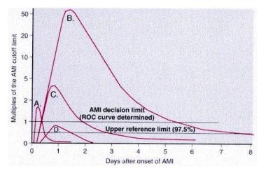
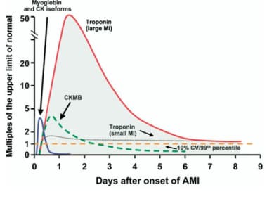
Cetacea
Sperm Whale
Carbon Monoxide
Spectrophotometry
Creatine Kinase
Hemeproteins
Hemoglobins
Spectrum Analysis, Raman
Oxygen
Leghemoglobin
Elephants
Protein Conformation
Bromotrichloromethane
Seals, Earless
Spectrum Analysis
Carboxyhemoglobin
Apoproteins
Clinical Enzyme Tests
Ligands
Spectrophotometry, Infrared
Hemin
Electron Spin Resonance Spectroscopy
Iron
Magnetic Resonance Spectroscopy
Oxidation-Reduction
The determination of hemoglobin and myoglogin residues as a parameter for testing heat exposure in back bacon. (1/1729)
The use of an extraction of the heme pigments hemoglobin and myoglobin as a test for the heat exposure of back bacon was investigated by treating back bacon at varying temperatures of 50-70 degrees C and times of two to 180 minutes and observing the effect on the absorbance of heme pigment residue after nitrite oxidation. Absorbance at 409 nm was used in place of the more usual 540 nm to provide greater sensitivity in the detection of heme. A decrease in residual heme pigments was time-dependent, particularly at lower temperatures. In view of this factor and the complex nature of the heat exposure of a large block of back bacon, the application of this test would require a calibration of each process. Alternatively, limits to the amounts of heme pigment residue could be set. The heme pigment test is useful in its simplicity and overcomes difficulties associated with the coagulation and enzyme tests. (+info)Myocardial oxygenation during high work states in hearts with postinfarction remodeling. (2/1729)
BACKGROUND: Postinfarction left ventricular remodeling (LVR) is associated with reductions in myocardial high-energy phosphate (HEP) levels, which are more severe in animals that develop overt congestive heart failure (CHF). During high work states, further HEP loss occurs, which suggests demand-induced ischemia. This study tested the hypothesis that inadequate myocyte oxygen availability is the basis for these HEP abnormalities. METHODS AND RESULTS: Myocardial infarction was produced by left circumflex coronary artery ligation in swine. Studies were performed in 20 normal animals, 14 animals with compensated LVR, and 9 animals with CHF. Phosphocreatine (PCr)/ATP was determined with 31P NMR and deoxymyoglobin (Mb-delta) with 1H NMR in myocardium remote from the infarct. Basal PCr/ATP tended to be decreased in postinfarct hearts, and this was significant in animals with CHF. Infusion of dobutamine (20 microg x kg-1 x min-1 IV) caused doubling of the rate-pressure product in both normal and LVR hearts and resulted in comparable significant decreases of PCr/ATP in both groups. This decrease in PCr/ATP was not associated with detectable Mb-delta. In CHF hearts, rate-pressure product increased only 40% in response to dobutamine; this attenuated response also was not associated with detectable Mb-delta. CONCLUSIONS: Thus, the decrease of PCr/ATP during dobutamine infusion is not the result of insufficient myocardial oxygen availability. Furthermore, in CHF hearts, the low basal PCr/ATP and the attenuated response to dobutamine occurred in the absence of myocardial hypoxia, indicating that the HEP and contractile abnormalities were not the result of insufficient oxygen availability. (+info)Structural dynamics of ligand diffusion in the protein matrix: A study on a new myoglobin mutant Y(B10) Q(E7) R(E10). (3/1729)
A triple mutant of sperm whale myoglobin (Mb) [Leu(B10) --> Tyr, His(E7) --> Gln, and Thr(E10) --> Arg, called Mb-YQR], investigated by stopped-flow, laser photolysis, crystallography, and molecular dynamics (MD) simulations, proved to be quite unusual. Rebinding of photodissociated NO, O2, and CO from within the protein (in a "geminate" mode) allows us to reach general conclusions about dynamics and cavities in proteins. The 3D structure of oxy Mb-YQR shows that bound O2 makes two H-bonds with Tyr(B10)29 and Gln(E7)64; on deoxygenation, these two residues move toward the space occupied by O2. The bimolecular rate constant for NO binding is the same as for wild-type, but those for CO and O2 binding are reduced 10-fold. While there is no geminate recombination with O2 and CO, geminate rebinding of NO displays an unusually large and very slow component, which is pretty much abolished in the presence of xenon. These results and MD simulations suggest that the ligand migrates in the protein matrix to a major "secondary site," located beneath Tyr(B10)29 and accessible via the motion of Ile(G8)107; this site is different from the "primary site" identified by others who investigated the photolyzed state of wild-type Mb by crystallography. Our hypothesis may rationalize the O2 binding properties of Mb-YQR, and more generally to propose a mechanism of control of ligand binding and dissociation in hemeproteins based on the dynamics of side chains that may (or may not) allow access to and direct temporary sequestration of the dissociated ligand in a docking site within the protein. This interpretation suggests that very fast (picosecond) fluctuations of amino acid side chains may play a crucial role in controlling O2 delivery to tissue at a rate compatible with physiology. (+info)Specificity of native-like interhelical hydrophobic contacts in the apomyoglobin intermediate. (4/1729)
On exposure to mildly acidic conditions, apomyoglobin forms a partially folded intermediate, I. The A, B, G, and H helices are significantly structured in this equilibrium intermediate, whereas the remainder of the protein is largely unfolded. We report here the effects of mutations at helix pairing sites on the stability of I in three classes of mutants that: (i) truncate hydrophobic side chains in native helix packing sites, (ii) truncate hydrophobic side chains not involved in interhelical contacts, and (iii) extend hydrophobic side chains at residues not involved in interhelical contacts. Class I mutants significantly decrease the stability and cooperativity of folding of the intermediate. Class II and III mutants show smaller effects on stability and have little effect on cooperativity. Qualitatively similar results to those found in I were obtained for all three classes of mutants in native myoglobin (N), demonstrating that hydrophobic burial is fairly specific to native helix packing sites in I as well as in N. These results suggest that hydrophobic burial along native-like interhelical contacts is important for the formation of the cooperatively folded intermediate. (+info)Development of diving capacity in emperor penguins. (5/1729)
To compare the diving capacities of juvenile and adult emperor penguins Aptenodytes forsteri, and to determine the physiological variables underlying the diving ability of juveniles, we monitored diving activity in juvenile penguins fitted with satellite-linked time/depth recorders and examined developmental changes in body mass (Mb), hemoglobin concentration, myoglobin (Mb) content and muscle citrate synthase and lactate dehydrogenase activities. Diving depth, diving duration and time-at-depth histograms were obtained from two fledged juveniles during the first 2.5 months after their depature from the Cape Washingon colony in the Ross Sea, Antarctica. During this period, values of all three diving variables increased progressively. After 8-10 weeks at sea, 24-41 % of transmitted maximum diving depths were between 80 and 200 m. Although most dives lasted less than 2 min during the 2 month period, 8-25 % of transmitted dives in the last 2 weeks lasted 2-4 min. These values are lower than those previously recorded in adults during foraging trips. Of the physiological variables examined during chick and juvenile development, only Mb and Mb content did not approach adult values. In both near-fledge chicks and juveniles, Mb was 50-60 % of adult values and Mb content was 24-31 % of adult values. This suggests that the increase in diving capacity of juveniles at sea will be most dependent on changes in these factors. (+info)Low molecular weight heparin (dalteparin) as adjuvant treatment of thrombolysis in acute myocardial infarction--a pilot study: biochemical markers in acute coronary syndromes (BIOMACS II). (6/1729)
OBJECTIVES: This randomized, double blind, placebo-controlled pilot trial evaluated the effect of dalteparin as an adjuvant to thrombolysis in patients with acute myocardial infarction regarding early reperfusion, recurrent ischemia and patency at 24 h. BACKGROUND: Low-molecular-weight heparin, given subcutaneously twice daily without monitoring, might be an attractive alternative to conventional intravenous heparin in the treatment of acute myocardial infarction. METHODS: In 101 patients dalteparin/placebo 100 IU/kg was given just before streptokinase and a second injection 120 IU/kg after 12 h. Monitoring with continuous vector-ECG was done to obtain signs of early reperfusion and later ischemic episodes. Blood samples for myoglobin were obtained at start and after 90 min to evaluate signs of reperfusion. Coronary angiography was performed after 20-28 h to evaluate TIMI-flow in the infarct-related artery. RESULTS: Dalteparin added to streptokinase tended to provide a higher rate of TIMI grade 3 flow in infarct-related artery compared to placebo, 68% versus 51% (p = 0.10). Dalteparin had no effects on noninvasive signs of early reperfusion. In patients with signs of early reperfusion, there seemed to be a higher rate of TIMI grade 3 flow, 74% versus 46% (myoglobin) (p = 0.04) and 73% versus 52% (vector-ECG) (p = 0.11). Ischemic episodes 6-24 h. after start of treatment were fewer in the dalteparin group, 16% versus 38% (p = 0.04). CONCLUSIONS: When dalteparin was added as an adjuvant to streptokinase and aspirin, there were tendencies for less ECG monitoring evidence of recurrent ischemia and better patency at 24 h, warranting further study. (+info)Direct sedimentation analysis of interference optical data in analytical ultracentrifugation. (7/1729)
Sedimentation data acquired with the interference optical scanning system of the Optima XL-I analytical ultracentrifuge can exhibit time-invariant noise components, as well as small radial-invariant baseline offsets, both superimposed onto the radial fringe shift data resulting from the macromolecular solute distribution. A well-established method for the interpretation of such ultracentrifugation data is based on the analysis of time-differences of the measured fringe profiles, such as employed in the g(s*) method. We demonstrate how the technique of separation of linear and nonlinear parameters can be used in the modeling of interference data by unraveling the time-invariant and radial-invariant noise components. This allows the direct application of the recently developed approximate analytical and numerical solutions of the Lamm equation to the analysis of interference optical fringe profiles. The presented method is statistically advantageous since it does not require the differentiation of the data and the model functions. The method is demonstrated on experimental data and compared with the results of a g(s*) analysis. It is also demonstrated that the calculation of time-invariant noise components can be useful in the analysis of absorbance optical data. They can be extracted from data acquired during the approach to equilibrium, and can be used to increase the reliability of the results obtained from a sedimentation equilibrium analysis. (+info)Convective oxygen transport and tissue oxygen consumption in Weddell seals during aerobic dives. (8/1729)
Unlike their terrestrial counterparts, marine mammals stop breathing and reduce their convective oxygen transport while performing activities (e.g. foraging, courtship, aggressive interactions, predator avoidance and migration) that require sustained power output during submergence. Since most voluntary dives are believed to remain aerobic, the goal of this study was to examine the potential importance of the dive response in optimizing the use of blood and muscle oxygen stores during dives involving different levels of muscular exertion. To accomplish this, we designed a numerical model based on Fick's principle that integrated cardiac output (Vb), regional blood flow, convective oxygen transport (Q(O2)), muscle oxymyoglobin desaturation and regional rates of oxygen consumption (VO2). The model quantified how the optimal matching or mismatching of QO2 to VO2 affected the aerobic dive limit (ADL). We chose an adult Weddell seal Leptonycotes weddellii on which to base our model because of available data on the diving physiology and metabolism of this species. The results show that the use of blood and muscle oxygen stores must be completed at the same time to maximize the ADL for each level of VO2. This is achieved by adjusting Vb (range 19-94 % of resting levels) and muscle QO2 according to the rate of muscle oxygen consumption (VMO2). At higher values of VMO2, Vb and muscle perfusion must increase to maintain an appropriate QO2/VO2 ratio so that available blood and muscle oxygen stores are depleted at the same time. Although the dive response does not sequester blood oxygen exclusively for brain and heart metabolism during aerobic dives, as it does during forced submersion, a reduction in Vb and muscle perfusion below resting levels is necessary to maximize the ADL over the range of diving VO2 (approximately 2-9 ml O2 min-1 kg-1). Despite the reduction in Vb, convective oxygen transport is adequate to maintain aerobic metabolism and normal function in the splanchnic organs, kidneys and other peripheral tissues. As a result, physiological homeostasis is maintained throughout the dive. The model shows that the cardiovascular adjustments known as the dive response enable the diving seal to balance the conflicting metabolic demands of (1) optimizing the distribution and use of blood and muscle oxygen stores to maximize the ADL over the normal range of diving VO2 and (2) ensuring that active muscle receives adequate oxygen as VMO2 increases. (+info)Myoglobin is a protein found in the muscle tissue, particularly in red or skeletal muscles. It belongs to the globin family and has a similar structure to hemoglobin, another oxygen-binding protein found in red blood cells. Myoglobin's primary function is to store oxygen within the muscle cells, making it readily available for use during periods of increased oxygen demand, such as during physical exertion.
Myoglobin contains heme groups that bind to and release oxygen molecules. The protein has a higher affinity for oxygen than hemoglobin, allowing it to maintain its bound oxygen even in low-oxygen environments. When muscle cells are damaged or undergo necrosis (cell death), myoglobin is released into the bloodstream and can be detected in serum or urine samples. Elevated levels of myoglobin in the blood or urine may indicate muscle injury, trauma, or diseases affecting muscle integrity, such as rhabdomyolysis or muscular dystrophies.
I believe there may be some confusion in your question. Whales are not a medical term but rather large marine mammals. They belong to the Cetacean family, which includes dolphins and porpoises. If you're asking about a medical condition or something similar that might be associated with the word "whales," I would need more information to provide an accurate response.
Metmyoglobin is the oxidized form of myoglobin, a protein found in muscle tissue that binds and stores oxygen. When myoglobin is exposed to oxidizing agents or when muscle tissue is damaged (such as during exercise or after death), it can become oxidized and transform into metmyoglobin. This form of the protein cannot bind or store oxygen, and its presence in food (particularly in meats) can lead to off-flavors, discoloration, and reduced shelf life. In medical contexts, metmyoglobin may be used as a marker for muscle damage or hypoxia (lack of oxygen).
Myoglobinuria is a medical condition characterized by the presence of myoglobin in the urine. Myoglobin is a protein found in muscle cells that is released into the bloodstream when muscle tissue is damaged or broken down, such as during intense exercise, trauma, or muscle diseases like muscular dystrophy and rhabdomyolysis.
When myoglobin is present in high concentrations in the blood, it can damage the kidneys by causing direct tubular injury, cast formation, and obstruction, which can lead to acute kidney injury (AKI) or even renal failure if left untreated. Symptoms of myoglobinuria may include dark-colored urine, muscle pain, weakness, and swelling, as well as symptoms related to AKI such as nausea, vomiting, and decreased urine output.
Diagnosis of myoglobinuria is typically made by detecting myoglobin in the urine using a dipstick test or more specific tests like immunoassays or mass spectrometry. Treatment may involve aggressive fluid resuscitation, alkalization of the urine to prevent myoglobin precipitation, and management of any underlying conditions causing muscle damage.
Heme is not a medical term per se, but it is a term used in the field of medicine and biology. Heme is a prosthetic group found in hemoproteins, which are proteins that contain a heme iron complex. This complex plays a crucial role in various biological processes, including oxygen transport (in hemoglobin), electron transfer (in cytochromes), and chemical catalysis (in peroxidases and catalases).
The heme group consists of an organic component called a porphyrin ring, which binds to a central iron atom. The iron atom can bind or release electrons, making it essential for redox reactions in the body. Heme is also vital for the formation of hemoglobin and myoglobin, proteins responsible for oxygen transport and storage in the blood and muscles, respectively.
In summary, heme is a complex organic-inorganic structure that plays a critical role in several biological processes, particularly in electron transfer and oxygen transport.
I'm sorry for any confusion, but "horses" is not a medical term. It is the common name given to the species *Equus ferus caballus*, which are large domesticated mammals used for transportation, work, and recreation. If you have any questions about horses or a related topic that you would like a medical perspective on, please let me know and I'd be happy to help!
Cetacea is a taxonomic order that includes whales, dolphins, and porpoises. This group of marine mammals is characterized by their fully aquatic lifestyle, torpedo-shaped bodies, modified limbs that serve as flippers, and the absence of external hindlimbs. Cetaceans have streamlined bodies that minimize drag while swimming, and their tail flukes enable powerful propulsion through vertical movement in the water column.
Their respiratory system features a pair of blowholes on the top of their heads, which they use to breathe air at the surface. Cetaceans exhibit complex social behaviors, advanced communication skills, and sophisticated echolocation abilities for navigation and hunting. They primarily feed on fish and invertebrates, with some larger species preying on marine mammals.
Cetaceans have a global distribution, occupying various habitats such as open oceans, coastal areas, and rivers. Unfortunately, many cetacean populations face threats from human activities like pollution, habitat degradation, climate change, and direct hunting or bycatch in fishing gear. Conservation efforts are crucial to protect these remarkable creatures and their vital roles in marine ecosystems.
I'm sorry for the confusion, but "Sperm Whale" is not a medical term. It is a species of whale, scientifically known as Physeter macrocephalus. Sperm whales are the largest toothed whales and have distinctive square-shaped heads that can make up to one-third of their body length. They are named for the waxy substance called spermaceti found in their heads, which was once mistakenly thought to be sperm.
If you're looking for a medical definition or information related to human health, please provide more details so I can assist you better.
Carbon monoxide (CO) is a colorless, odorless, and tasteless gas that is slightly less dense than air. It is toxic to hemoglobic animals when encountered in concentrations above about 35 ppm. This compound is a product of incomplete combustion of organic matter, and is a major component of automobile exhaust.
Carbon monoxide is poisonous because it binds to hemoglobin in red blood cells much more strongly than oxygen does, forming carboxyhemoglobin. This prevents the transport of oxygen throughout the body, which can lead to suffocation and death. Symptoms of carbon monoxide poisoning include headache, dizziness, weakness, nausea, vomiting, confusion, and disorientation. Prolonged exposure can lead to unconsciousness and death.
Carbon monoxide detectors are commonly used in homes and other buildings to alert occupants to the presence of this dangerous gas. It is important to ensure that these devices are functioning properly and that they are placed in appropriate locations throughout the building. Additionally, it is essential to maintain appliances and heating systems to prevent the release of carbon monoxide into living spaces.
Rhabdomyolysis is a medical condition characterized by the breakdown and degeneration of skeletal muscle fibers, leading to the release of their intracellular contents into the bloodstream. This can result in various complications, including electrolyte imbalances, kidney injury or failure, and potentially life-threatening conditions if not promptly diagnosed and treated.
The process of rhabdomyolysis typically involves three key components:
1. Muscle injury: Direct trauma, excessive exertion, prolonged immobilization, infections, metabolic disorders, toxins, or medications can cause muscle damage, leading to the release of intracellular components into the bloodstream.
2. Release of muscle contents: When muscle fibers break down, they release various substances, such as myoglobin, creatine kinase (CK), lactate dehydrogenase (LDH), aldolase, and potassium ions. Myoglobin is a protein that can cause kidney damage when present in high concentrations in the bloodstream, particularly when it is filtered through the kidneys and deposits in the renal tubules.
3. Systemic effects: The release of muscle contents into the bloodstream can lead to various systemic complications, such as electrolyte imbalances (particularly hyperkalemia), acidosis, hypocalcemia, and kidney injury or failure due to myoglobin-induced tubular damage.
Symptoms of rhabdomyolysis can vary widely depending on the severity and extent of muscle damage but may include muscle pain, weakness, swelling, stiffness, dark urine, and tea-colored or cola-colored urine due to myoglobinuria. In severe cases, patients may experience symptoms related to kidney failure, such as nausea, vomiting, fatigue, and decreased urine output.
Diagnosis of rhabdomyolysis typically involves measuring blood levels of muscle enzymes (such as CK and LDH) and evaluating renal function through blood tests and urinalysis. Treatment generally focuses on addressing the underlying cause of muscle damage, maintaining fluid balance, correcting electrolyte imbalances, and preventing or managing kidney injury.
Photolysis is a term used in medical and scientific contexts to describe a chemical reaction that is initiated by the absorption of light or photons. In this process, a molecule absorbs a photon, which provides sufficient energy to break a bond within the molecule, leading to the formation of two or more smaller molecules or radicals. This phenomenon is particularly relevant in fields such as pharmacology and toxicology, where photolysis can alter the chemical structure and biological activity of drugs and other substances upon exposure to light.
Spectrophotometry is a technical analytical method used in the field of medicine and science to measure the amount of light absorbed or transmitted by a substance at specific wavelengths. This technique involves the use of a spectrophotometer, an instrument that measures the intensity of light as it passes through a sample.
In medical applications, spectrophotometry is often used in laboratory settings to analyze various biological samples such as blood, urine, and tissues. For example, it can be used to measure the concentration of specific chemicals or compounds in a sample by measuring the amount of light that is absorbed or transmitted at specific wavelengths.
In addition, spectrophotometry can also be used to assess the properties of biological tissues, such as their optical density and thickness. This information can be useful in the diagnosis and treatment of various medical conditions, including skin disorders, eye diseases, and cancer.
Overall, spectrophotometry is a valuable tool for medical professionals and researchers seeking to understand the composition and properties of various biological samples and tissues.
Creatine kinase (CK) is a muscle enzyme that is normally present in small amounts in the blood. It is primarily found in tissues that require a lot of energy, such as the heart, brain, and skeletal muscles. When these tissues are damaged or injured, CK is released into the bloodstream, causing the levels to rise.
Creatine kinase exists in several forms, known as isoenzymes, which can be measured in the blood to help identify the location of tissue damage. The three main isoenzymes are:
1. CK-MM: Found primarily in skeletal muscle
2. CK-MB: Found primarily in heart muscle
3. CK-BB: Found primarily in the brain
Elevated levels of creatine kinase, particularly CK-MB, can indicate damage to the heart muscle, such as occurs with a heart attack. Similarly, elevated levels of CK-BB may suggest brain injury or disease. Overall, measuring creatine kinase levels is a useful diagnostic tool for assessing tissue damage and determining the severity of injuries or illnesses.
Heme proteins are a type of protein that contain a heme group, which is a prosthetic group composed of an iron atom contained in the center of a large organic ring called a porphyrin. The heme group gives these proteins their characteristic red color. Hemeproteins have various important functions in biological systems, including oxygen transport (e.g., hemoglobin), electron transfer (e.g., cytochromes), and enzymatic catalysis (e.g., peroxidases and catalases). The heme group can bind and release gases, such as oxygen and carbon monoxide, and can participate in redox reactions due to the ease with which iron can change its oxidation state.
Hemoglobin (Hb or Hgb) is the main oxygen-carrying protein in the red blood cells, which are responsible for delivering oxygen throughout the body. It is a complex molecule made up of four globin proteins and four heme groups. Each heme group contains an iron atom that binds to one molecule of oxygen. Hemoglobin plays a crucial role in the transport of oxygen from the lungs to the body's tissues, and also helps to carry carbon dioxide back to the lungs for exhalation.
There are several types of hemoglobin present in the human body, including:
* Hemoglobin A (HbA): This is the most common type of hemoglobin, making up about 95-98% of total hemoglobin in adults. It consists of two alpha and two beta globin chains.
* Hemoglobin A2 (HbA2): This makes up about 1.5-3.5% of total hemoglobin in adults. It consists of two alpha and two delta globin chains.
* Hemoglobin F (HbF): This is the main type of hemoglobin present in fetal life, but it persists at low levels in adults. It consists of two alpha and two gamma globin chains.
* Hemoglobin S (HbS): This is an abnormal form of hemoglobin that can cause sickle cell disease when it occurs in the homozygous state (i.e., both copies of the gene are affected). It results from a single amino acid substitution in the beta globin chain.
* Hemoglobin C (HbC): This is another abnormal form of hemoglobin that can cause mild to moderate hemolytic anemia when it occurs in the homozygous state. It results from a different single amino acid substitution in the beta globin chain than HbS.
Abnormal forms of hemoglobin, such as HbS and HbC, can lead to various clinical disorders, including sickle cell disease, thalassemia, and other hemoglobinopathies.
Spectrum analysis in the context of Raman spectroscopy refers to the measurement and interpretation of the Raman scattering spectrum of a material or sample. Raman spectroscopy is a non-destructive analytical technique that uses the inelastic scattering of light to examine the vibrational modes of molecules.
When a monochromatic light source, typically a laser, illuminates a sample, a small fraction of the scattered light undergoes a shift in frequency due to interactions with the molecular vibrations of the sample. This shift in frequency is known as the Raman shift and is unique to each chemical bond or functional group within a molecule.
In a Raman spectrum, the intensity of the scattered light is plotted against the Raman shift, which is expressed in wavenumbers (cm-1). The resulting spectrum provides a "fingerprint" of the sample's molecular structure and composition, allowing for the identification and characterization of various chemical components within the sample.
Spectrum analysis in Raman spectroscopy can reveal valuable information about the sample's crystallinity, phase transitions, polymorphism, molecular orientation, and other properties. This technique is widely used across various fields, including materials science, chemistry, biology, pharmaceuticals, and forensics, to analyze a diverse range of samples, from simple liquids and solids to complex biological tissues and nanomaterials.
Oxygen is a colorless, odorless, tasteless gas that constitutes about 21% of the earth's atmosphere. It is a crucial element for human and most living organisms as it is vital for respiration. Inhaled oxygen enters the lungs and binds to hemoglobin in red blood cells, which carries it to tissues throughout the body where it is used to convert nutrients into energy and carbon dioxide, a waste product that is exhaled.
Medically, supplemental oxygen therapy may be provided to patients with conditions such as chronic obstructive pulmonary disease (COPD), pneumonia, heart failure, or other medical conditions that impair the body's ability to extract sufficient oxygen from the air. Oxygen can be administered through various devices, including nasal cannulas, face masks, and ventilators.
Leghemoglobin is a type of protein known as a hemeprotein, found in the root nodules of leguminous plants (plants belonging to the family Fabaceae or Leguminosae). These root nodules are formed through a symbiotic relationship with nitrogen-fixing bacteria called Rhizobia.
The primary function of leghemoglobin is to facilitate the process of nitrogen fixation by maintaining an optimal oxygen concentration within the root nodule cells, where the Rhizobia reside. By binding and releasing oxygen reversibly, leghemoglobin protects the nitrogen-fixing enzyme, nitrogenase, from being inactivated by excess oxygen. This ensures that the Rhizobia can effectively convert atmospheric nitrogen gas (N2) into ammonia (NH3), which is then utilized by the plant for its growth and development.
In summary, leghemoglobin is a crucial protein in the process of biological nitrogen fixation, allowing leguminous plants to grow without the need for added nitrogen fertilizers.
I believe you are looking for a medical or scientific term that is related to elephants, as there is no medical definition for the word "elephants" itself. Elephants are large mammals of the family Elephantidae and the order Proboscidea. They are native to Africa and Asia and are known for their long trunks, large ears, and tusks.
One possible connection between elephants and medicine is the use of elephant ivory in medical equipment. In the past, elephant ivory was used to make a variety of medical instruments, such as dental tools and surgical instruments. However, due to concerns about animal welfare and the illegal trade in elephant ivory, the use of elephant ivory in medical equipment has become increasingly rare.
Another possible connection between elephants and medicine is the study of their social behavior and communication, which may provide insights into human social behavior and mental health. For example, research has shown that elephants have complex social structures and exhibit behaviors such as empathy, cooperation, and mourning, which are also important aspects of human social and emotional functioning.
Overall, while there is no specific medical definition for "elephants," these fascinating animals have contributed to our understanding of biology, medicine, and human behavior in various ways.
Protein conformation refers to the specific three-dimensional shape that a protein molecule assumes due to the spatial arrangement of its constituent amino acid residues and their associated chemical groups. This complex structure is determined by several factors, including covalent bonds (disulfide bridges), hydrogen bonds, van der Waals forces, and ionic bonds, which help stabilize the protein's unique conformation.
Protein conformations can be broadly classified into two categories: primary, secondary, tertiary, and quaternary structures. The primary structure represents the linear sequence of amino acids in a polypeptide chain. The secondary structure arises from local interactions between adjacent amino acid residues, leading to the formation of recurring motifs such as α-helices and β-sheets. Tertiary structure refers to the overall three-dimensional folding pattern of a single polypeptide chain, while quaternary structure describes the spatial arrangement of multiple folded polypeptide chains (subunits) that interact to form a functional protein complex.
Understanding protein conformation is crucial for elucidating protein function, as the specific three-dimensional shape of a protein directly influences its ability to interact with other molecules, such as ligands, nucleic acids, or other proteins. Any alterations in protein conformation due to genetic mutations, environmental factors, or chemical modifications can lead to loss of function, misfolding, aggregation, and disease states like neurodegenerative disorders and cancer.
Histidine is an essential amino acid, meaning it cannot be synthesized by the human body and must be obtained through dietary sources. Its chemical formula is C6H9N3O2. Histidine plays a crucial role in several physiological processes, including:
1. Protein synthesis: As an essential amino acid, histidine is required for the production of proteins, which are vital components of various tissues and organs in the body.
2. Hemoglobin synthesis: Histidine is a key component of hemoglobin, the protein in red blood cells responsible for carrying oxygen throughout the body. The imidazole side chain of histidine acts as a proton acceptor/donor, facilitating the release and uptake of oxygen by hemoglobin.
3. Acid-base balance: Histidine is involved in maintaining acid-base homeostasis through its role in the biosynthesis of histamine, which is a critical mediator of inflammatory responses and allergies. The decarboxylation of histidine results in the formation of histamine, which can increase vascular permeability and modulate immune responses.
4. Metal ion binding: Histidine has a high affinity for metal ions such as zinc, copper, and iron. This property allows histidine to participate in various enzymatic reactions and maintain the structural integrity of proteins.
5. Antioxidant defense: Histidine-containing dipeptides, like carnosine and anserine, have been shown to exhibit antioxidant properties by scavenging reactive oxygen species (ROS) and chelating metal ions. These compounds may contribute to the protection of proteins and DNA from oxidative damage.
Dietary sources of histidine include meat, poultry, fish, dairy products, and wheat germ. Histidine deficiency is rare but can lead to growth retardation, anemia, and impaired immune function.
Bromotrichloromethane is a type of halomethane, which is a class of chemicals containing carbon and halogen atoms. Specifically, bromotrichloromethane is a colorless liquid with the chemical formula CBrCl3. It has been used as a fire extinguishing agent, a refrigerant, and an intermediate in the production of other chemicals.
In medical terms, bromotrichloromethane may be encountered in the context of occupational health and safety or environmental exposure assessment. Exposure to high levels of this chemical can cause irritation to the eyes, skin, and respiratory tract, as well as potential neurological effects such as headache, dizziness, and loss of consciousness. Long-term exposure has been linked to liver and kidney damage in animal studies.
It is important to note that bromotrichloromethane is not used in medical treatments or procedures. Its use in industrial applications has been largely phased out due to its ozone-depleting properties and potential health hazards.
Earless seals, also known as true seals or Phocidae, are a family of marine mammals that lack external ears. They have a streamlined body adapted for fast swimming, and their hind limbs are modified into flippers, which they use to move through the water. Earless seals have small ear holes on the sides of their heads, but they do not have an outer ear flap like other mammals. Instead, their middle and inner ears are well-developed for hearing underwater. They are found in both the Northern and Southern Hemispheres, and there are 18 species of earless seals, including the harbor seal, gray seal, and leopard seal.
I'm sorry for any confusion, but "spectrum analysis" is not a commonly used medical term. Spectrum analysis is a term that is more frequently used in the fields of physics, mathematics, and engineering to describe the process of breaking down a signal or a wave into its different frequencies and amplitudes, creating a visual representation called a spectrum.
If you have any concerns about a medical issue, I would recommend consulting with a healthcare professional for accurate information and guidance.
Carboxyhemoglobin (COHb) is a form of hemoglobin that has bonded with carbon monoxide (CO), a colorless, odorless gas. Normally, hemoglobin in red blood cells binds with oxygen (O2) to carry it throughout the body. However, when exposed to CO, hemoglobin preferentially binds with it, forming carboxyhemoglobin, which reduces the amount of oxygen that can be carried by the blood. This can lead to hypoxia (lack of oxygen in tissues) and potentially serious medical consequences, including death. Carbon monoxide exposure can occur from sources such as smoke inhalation, vehicle exhaust, or faulty heating systems.
Apoproteins are the protein components of lipoprotein complexes, which are responsible for transporting fat molecules, such as cholesterol and triglycerides, throughout the body. Apoproteins play a crucial role in the metabolism of lipids by acting as recognition signals that allow lipoproteins to interact with specific receptors on cell surfaces.
There are several different types of apoproteins, each with distinct functions. For example, apolipoprotein A-1 (apoA-1) is the major protein component of high-density lipoproteins (HDL), which are responsible for transporting excess cholesterol from tissues to the liver for excretion. Apolipoprotein B (apoB) is a large apoprotein found in low-density lipoproteins (LDL), very low-density lipoproteins (VLDL), and lipoprotein(a). ApoB plays a critical role in the assembly and secretion of VLDL from the liver, and it also mediates the uptake of LDL by cells.
Abnormalities in apoprotein levels or function can contribute to the development of various diseases, including cardiovascular disease, diabetes, and Alzheimer's disease. Therefore, measuring apoprotein levels in the blood can provide valuable information for diagnosing and monitoring these conditions.
Clinical enzyme tests are laboratory tests that measure the amount or activity of certain enzymes in biological samples, such as blood or bodily fluids. These tests are used to help diagnose and monitor various medical conditions, including organ damage, infection, inflammation, and genetic disorders.
Enzymes are proteins that catalyze chemical reactions in the body. Some enzymes are found primarily within specific organs or tissues, so elevated levels of these enzymes in the blood can indicate damage to those organs or tissues. For example, high levels of creatine kinase (CK) may suggest muscle damage, while increased levels of aspartate aminotransferase (AST) and alanine aminotransferase (ALT) can indicate liver damage.
There are several types of clinical enzyme tests, including:
1. Serum enzyme tests: These measure the level of enzymes in the blood serum, which is the liquid portion of the blood after clotting. Examples include CK, AST, ALT, alkaline phosphatase (ALP), and lactate dehydrogenase (LDH).
2. Urine enzyme tests: These measure the level of enzymes in the urine. An example is N-acetyl-β-D-glucosaminidase (NAG), which can indicate kidney damage.
3. Enzyme immunoassays (EIAs): These use antibodies to detect and quantify specific enzymes or proteins in a sample. They are often used for the diagnosis of infectious diseases, such as HIV or hepatitis.
4. Genetic enzyme tests: These can identify genetic mutations that cause deficiencies in specific enzymes, leading to inherited metabolic disorders like phenylketonuria (PKU) or Gaucher's disease.
It is important to note that the interpretation of clinical enzyme test results should be done by a healthcare professional, taking into account the patient's medical history, symptoms, and other diagnostic tests.
A ligand, in the context of biochemistry and medicine, is a molecule that binds to a specific site on a protein or a larger biomolecule, such as an enzyme or a receptor. This binding interaction can modify the function or activity of the target protein, either activating it or inhibiting it. Ligands can be small molecules, like hormones or neurotransmitters, or larger structures, like antibodies. The study of ligand-protein interactions is crucial for understanding cellular processes and developing drugs, as many therapeutic compounds function by binding to specific targets within the body.
In the context of medicine and pharmacology, "kinetics" refers to the study of how a drug moves throughout the body, including its absorption, distribution, metabolism, and excretion (often abbreviated as ADME). This field is called "pharmacokinetics."
1. Absorption: This is the process of a drug moving from its site of administration into the bloodstream. Factors such as the route of administration (e.g., oral, intravenous, etc.), formulation, and individual physiological differences can affect absorption.
2. Distribution: Once a drug is in the bloodstream, it gets distributed throughout the body to various tissues and organs. This process is influenced by factors like blood flow, protein binding, and lipid solubility of the drug.
3. Metabolism: Drugs are often chemically modified in the body, typically in the liver, through processes known as metabolism. These changes can lead to the formation of active or inactive metabolites, which may then be further distributed, excreted, or undergo additional metabolic transformations.
4. Excretion: This is the process by which drugs and their metabolites are eliminated from the body, primarily through the kidneys (urine) and the liver (bile).
Understanding the kinetics of a drug is crucial for determining its optimal dosing regimen, potential interactions with other medications or foods, and any necessary adjustments for special populations like pediatric or geriatric patients, or those with impaired renal or hepatic function.
Spectrophotometry, Infrared is a scientific analytical technique used to measure the absorption or transmission of infrared light by a sample. It involves the use of an infrared spectrophotometer, which directs infrared radiation through a sample and measures the intensity of the radiation that is transmitted or absorbed by the sample at different wavelengths within the infrared region of the electromagnetic spectrum.
Infrared spectroscopy can be used to identify and quantify functional groups and chemical bonds present in a sample, as well as to study the molecular structure and composition of materials. The resulting infrared spectrum provides a unique "fingerprint" of the sample, which can be compared with reference spectra to aid in identification and characterization.
Infrared spectrophotometry is widely used in various fields such as chemistry, biology, pharmaceuticals, forensics, and materials science for qualitative and quantitative analysis of samples.
Hemin is defined as the iron(III) complex of protoporphyrin IX, which is a porphyrin derivative. It is a naturally occurring substance that is involved in various biological processes, most notably in the form of heme, which is a component of hemoglobin and other hemoproteins. Hemin is also used in medical research and therapy, such as in the treatment of methemoglobinemia and lead poisoning.
Electron Spin Resonance (ESR) Spectroscopy, also known as Electron Paramagnetic Resonance (EPR) Spectroscopy, is a technique used to investigate materials with unpaired electrons. It is based on the principle of absorption of energy by the unpaired electrons when they are exposed to an external magnetic field and microwave radiation.
In this technique, a sample is placed in a magnetic field and microwave radiation is applied. The unpaired electrons in the sample absorb energy and change their spin state when the energy of the microwaves matches the energy difference between the spin states. This absorption of energy is recorded as a function of the magnetic field strength, producing an ESR spectrum.
ESR spectroscopy can provide information about the number, type, and behavior of unpaired electrons in a sample, as well as the local environment around the electron. It is widely used in physics, chemistry, and biology to study materials such as free radicals, transition metal ions, and defects in solids.
In the context of medicine, iron is an essential micromineral and key component of various proteins and enzymes. It plays a crucial role in oxygen transport, DNA synthesis, and energy production within the body. Iron exists in two main forms: heme and non-heme. Heme iron is derived from hemoglobin and myoglobin in animal products, while non-heme iron comes from plant sources and supplements.
The recommended daily allowance (RDA) for iron varies depending on age, sex, and life stage:
* For men aged 19-50 years, the RDA is 8 mg/day
* For women aged 19-50 years, the RDA is 18 mg/day
* During pregnancy, the RDA increases to 27 mg/day
* During lactation, the RDA for breastfeeding mothers is 9 mg/day
Iron deficiency can lead to anemia, characterized by fatigue, weakness, and shortness of breath. Excessive iron intake may result in iron overload, causing damage to organs such as the liver and heart. Balanced iron levels are essential for maintaining optimal health.
Magnetic Resonance Spectroscopy (MRS) is a non-invasive diagnostic technique that provides information about the biochemical composition of tissues, including their metabolic state. It is often used in conjunction with Magnetic Resonance Imaging (MRI) to analyze various metabolites within body tissues, such as the brain, heart, liver, and muscles.
During MRS, a strong magnetic field, radio waves, and a computer are used to produce detailed images and data about the concentration of specific metabolites in the targeted tissue or organ. This technique can help detect abnormalities related to energy metabolism, neurotransmitter levels, pH balance, and other biochemical processes, which can be useful for diagnosing and monitoring various medical conditions, including cancer, neurological disorders, and metabolic diseases.
There are different types of MRS, such as Proton (^1^H) MRS, Phosphorus-31 (^31^P) MRS, and Carbon-13 (^13^C) MRS, each focusing on specific elements or metabolites within the body. The choice of MRS technique depends on the clinical question being addressed and the type of information needed for diagnosis or monitoring purposes.
Oxidation-Reduction (redox) reactions are a type of chemical reaction involving a transfer of electrons between two species. The substance that loses electrons in the reaction is oxidized, and the substance that gains electrons is reduced. Oxidation and reduction always occur together in a redox reaction, hence the term "oxidation-reduction."
In biological systems, redox reactions play a crucial role in many cellular processes, including energy production, metabolism, and signaling. The transfer of electrons in these reactions is often facilitated by specialized molecules called electron carriers, such as nicotinamide adenine dinucleotide (NAD+/NADH) and flavin adenine dinucleotide (FAD/FADH2).
The oxidation state of an element in a compound is a measure of the number of electrons that have been gained or lost relative to its neutral state. In redox reactions, the oxidation state of one or more elements changes as they gain or lose electrons. The substance that is oxidized has a higher oxidation state, while the substance that is reduced has a lower oxidation state.
Overall, oxidation-reduction reactions are fundamental to the functioning of living organisms and are involved in many important biological processes.
 Myoglobin