Myelitis
Neuromyelitis Optica
Neuroschistosomiasis
Aquaporin 4
Spinal Cord
Magnetic Resonance Imaging
Paraplegia
Muscle Hypotonia
Central Nervous System Helminthiasis
Tabes Dorsalis
Meningitis, Aseptic
Demyelinating Autoimmune Diseases, CNS
Influence of sex on clinical features, laboratory findings, and complications of typhoid fever. (1/166)
Clinical features, laboratory findings, and complications of typhoid fever were correlated with sex through a retrospective case note review of 102 hospitalized culture-positive patients in Durban, South Africa. Intestinal perforation (P = 0.04), occult blood losses in stools (P = 0.04), and a mild reticulocytosis in the absence of hemolysis (P = 0.02) occurred more frequently in males than in females. A single pretreatment Widal O antibody titer > or = 1:640 was also a statistically significant occurrence in males (P = 0. 006). Female patients were significantly more severely ill (P = 0.0004) on admission and had chest signs consistent with bronchopneumonia (P = 0.04), transverse myelitis (P = 0.04), abnormal liver function test results (P = 0.0003), and abnormal findings in urinalyses (P = 0.02). Typhoid hepatitis (P = 0.04) and glomerulonephritis (P = 0.02) were present significantly more frequently in females. Whether these differences were due to differences in host's immune response to acute infection need to be determined in a prospective study. (+info)Unusual cervical spinal cord toxicity associated with intra-arterial carboplatin, intra-arterial or intravenous etoposide phosphate, and intravenous cyclophosphamide in conjunction with osmotic blood brain-barrier disruption in the vertebral artery. (2/166)
BACKGROUND AND PURPOSE: When the clinical and radiologic characteristics of an unusual cervical spinal cord complication of intra-arterial (IA) chemotherapy with blood brain-barrier (BBB) disruption in the vertebral circulation are documented. Seven cases are reported and analyzed in search of a pathophysiologic explanation. METHODS: We retrospectively identified 94 patients who received a total of 380 standardized regimens of IA carboplatin, IA or IV etoposide phosphate, and IV cyclophosphamide infusion in conjunction with osmotic BBB disruption of the vertebral artery. We describe seven of those patients in whom unexpected neck pain developed followed by neurologic symptoms primarily in the upper extremities. RESULTS: The symptoms correlated with MR abnormalities (T1 hypointensity, T2 hyperintensity, and unusual contrast enhancement) in the cervical spinal cord, usually involving the gray matter. The neurologic deficits and MR changes were generally transient. One patient who received a flu vaccination 48 hours before the chemotherapy incurred progressive myelitis and expired. CONCLUSION: The pathophysiology of this complication is probably multifactorial but may be related to vascular streaming and an atypical inflammatory toxic reaction to carboplatin and etoposide. The complication has not recurred during a 6-month period following modification of the protocol. (+info)Acute myelitis after asthma attacks with onset after puberty. (3/166)
A poliomyelitis-like illness after asthma attacks has been found and is called asthmatic amyotrophy (Hopkins' syndrome). All of the previously reported cases were under 13 years of age. Three patients are described who developed acute myelitis after asthma attacks at 15, 22, and 73 years of age. All of them showed acute flaccid monoparesis, and needle EMG disclosed denervation potentials in the relevant muscles. In addition, in the two adult patients the sensory or pyramidal tracts were involved, and evoked potential studies confirmed an involvement of the pyramidal tracts in one of them. This 22 year old patient showed a second episode of monoparesis in the other limb after another asthma attack. All three patients had no significant changes in their antiviral antibody titres, whereas every patient had hyperIgEaemia and allergen specific IgE. These findings suggest that asthmatic amyotrophy can develop after puberty and that patients who develop this disease in adulthood seem to show both a widespread involvement of the spinal cord and a more varied course. (+info)Tuberculous radiculomyelitis complicating tuberculous meningitis: case report and review. (4/166)
Tuberculous radiculomyelitis (TBRM) is a complication of tuberculous meningitis (TBM), which has been reported rarely in the modern medical literature. We describe a case of TBRM that developed in an human immunodeficiency virus (HIV)-infected patient, despite prompt antituberculous treatment. To our knowledge, this is the second case of TBRM reported in an HIV-infected patient. We also review 74 previously reported cases of TBRM. TBRM develops at various periods after TBM, even in adequately treated patients after sterilization of the cerebrospinal fluid (CSF). The most common symptoms are subacute paraparesis, radicular pain, bladder disturbance, and subsequent paralysis. CSF evaluation usually shows an active inflammatory response with a very high protein level. MRI and CT scan are critical for diagnosis, revealing loculation and obliteration of the subarachnoid space along with linear intradural enhancement. As in other forms of paradoxical reactions to antituberculous treatment, there is evidence that steroid treatment might have a beneficial effect. (+info)Nationwide survey of the annual prevalence of viral and other neurological infections in Japanese inpatients. (5/166)
OBJECTIVE: To estimate the annual prevalence of viral and other neurological infections at large hospitals in Japan during the period from 1989 to 1991. METHODS: A nationwide questionnaire survey on the numbers of inpatients with viral and other neurological infections was sent for completion to the chiefs of Departments of Internal Medicine, Neurology and Pediatrics at all hospitals with more than 200 beds. RESULTS: The average annual number of inpatients (and the number per 10(6) population) with encephalitis in large hospitals was estimated to be 2,200+/-400 (17.7+/-3.2), while it was 32,000+/-16,000 (258+/-129) for meningitis, and 650+/-50 (5.2+/-0.4) for myelitis. Among the inpatients with encephalitis, meningitis, and myelitis, an unknown etiology was the most common (51.2% in encephalitis, 73.2% in meningitis, and 36.3% in myelitis), followed by a viral etiology for all three diseases. CONCLUSION: The first estimate was made of the annual prevalence of viral and other neurological infections and their etiology in Japan. (+info)Toxoplasma gondii myelitis in a patient with adult T-cell leukemia-lymphoma. (6/166)
Adult T cell leukemia-lymphoma (ATL) caused by HTLV-I may be associated with severe immunosupression and several opportunistic infections. Toxoplasmic encephalitis is a common central nervous system opportunistic infection in severely immunosupressed patients, however spinal cord involvement by this parasite is rare. In this paper, we report a case of toxoplasmic myelitis in a patient with ATL. (+info)An immunological syndrome featuring transverse myelitis, Evans syndrome and pulmonary infiltrates after unrelated bone marrow transplant in a patient with severe aplastic anemia. (7/166)
A patient with severe aplastic anemia underwent a matched unrelated bone marrow transplant, following which he developed a complex autoimmune syndrome. This featured transverse myelitis, immune mediated Coombs positive hemolytic anemia and immune thrombocytopenia (Evans syndrome), pulmonary infiltrates, eosinophilia, muscle pains and cramps and lichenoid dermatitis all of which may represent manifestations of graft-versus-host disease as they showed response to immunosuppression. Thus, although immune-mediated cytopenias after an allogeneic bone marrow transplant are rare, they should be considered as a possible cause of cytopenia in post-transplant patients. (+info)Acute flaccid paralysis in infants and young children with enterovirus 71 infection: MR imaging findings and clinical correlates. (8/166)
BACKGROUND AND PURPOSE: Enterovirus 71 (EV71) infection is now considered an important cause of childhood acute flaccid paralysis. The purpose of our study was to determine whether EV71-infection-related acute flaccid paralysis in infants and young children has characteristic MR imaging patterns. METHODS: Seven infants and young children with acute paralysis of the upper or lower extremities and positive EV71 cultures underwent spinal MR studies during an outbreak of hand-foot-and-mouth disease in Taiwan in 1998. RESULTS: Acute paralysis was observed in one upper extremity in two patients, in one lower extremity in three patients, and in both lower extremities in two patients. None of the patients had sensory impairment or bulbar palsy. MR studies showed unilateral or bilateral hyperintense lesions in the anterior horn regions of the cord on T2-weighted images in six patients. No abnormal signal was present in one patient. Two of three patients who received intravenous injections of contrast material had ventral root enhancement on T1-weighted images. One of them also had enhancement of the unilateral anterior horn cells. At clinical follow-up, both patients with bilateral anterior horn abnormalities had residual motor weakness, whereas only one of the five patients with unilateral involvement had residual weakness. CONCLUSION: EV71 radiculomyelitis tends to be unilateral and to specifically involve both the anterior horn cells of the cord and the ventral roots. MR imaging allows early detection of spinal cord and root lesions. (+info)Myelitis is a medical term that refers to inflammation of the spinal cord. This inflammation can cause damage to the myelin sheath, which is the protective covering of nerve fibers in the spinal cord. As a result, the transmission of nerve impulses along the spinal cord may be disrupted, leading to various neurological symptoms.
Myelitis can affect any part of the spinal cord and can have many different causes, including infections (such as viral or bacterial infections), autoimmune disorders (such as multiple sclerosis), and other conditions (such as spinal cord injuries or tumors). The specific symptoms of myelitis depend on the location and severity of the inflammation. They may include muscle weakness, numbness or tingling sensations, pain, bladder or bowel dysfunction, and difficulty with coordination and balance.
Myelitis can be a serious condition that requires prompt medical attention and treatment. Treatment typically focuses on addressing the underlying cause of the inflammation, as well as managing symptoms and supporting recovery.
Neuromyelitis optica (NMO), also known as Devic's disease, is an autoimmune disorder that affects the central nervous system (CNS). It primarily causes inflammation and damage to the optic nerves (which transmit visual signals from the eye to the brain) and the spinal cord. This results in optic neuritis (inflammation of the optic nerve, causing vision loss) and myelitis (inflammation of the spinal cord, leading to motor, sensory, and autonomic dysfunction).
A key feature of NMO is the presence of autoantibodies against aquaporin-4 (AQP4-IgG), a water channel protein found in astrocytes (a type of glial cell) in the CNS. These antibodies play a crucial role in the development of the disease, as they target and damage the AQP4 proteins, leading to inflammation, demyelination (loss of the protective myelin sheath around nerve fibers), and subsequent neurological dysfunction.
NMO is distinct from multiple sclerosis (MS), another autoimmune disorder affecting the CNS, as it has different clinical features, radiological findings, and treatment responses. However, NMO can sometimes be misdiagnosed as MS due to overlapping symptoms in some cases. Accurate diagnosis of NMO is essential for appropriate management and treatment, which often includes immunosuppressive therapies to control the autoimmune response and prevent further damage to the nervous system.
Neuroschistosomiasis is a form of schistosomiasis, which is a parasitic infection caused by Schistosoma species. It is characterized by the invasion and inflammation of the central nervous system (CNS) by the parasite's eggs or larvae. This can lead to various neurological symptoms such as seizures, headaches, visual disturbances, and motor or sensory deficits. Neuroschistosomiasis is a serious and potentially life-threatening condition that requires prompt diagnosis and treatment.
The two Schistosoma species most commonly associated with neuroschistosomiasis are S. japonicum and S. mansoni. The parasites typically enter the human body through skin contact with contaminated water, where they mature into adult worms in the bloodstream. Female worms then lay eggs, some of which may be carried to the CNS by the circulatory system.
Neuroschistosomiasis can occur in both acute and chronic forms. Acute neuroschistosomiasis is characterized by an inflammatory response to the parasite's eggs or larvae, which can cause eosinophilic meningitis or encephalitis. Chronic neuroschistosomiasis may result in the formation of granulomas around the eggs, leading to various neurological symptoms depending on the location and extent of the damage.
Diagnosis of neuroschistosomiasis typically involves a combination of clinical evaluation, imaging studies (such as MRI or CT scans), and laboratory tests (such as serology or CSF analysis). Treatment usually consists of anti-parasitic drugs such as praziquantel, combined with corticosteroids to manage the inflammatory response. In severe cases, surgical intervention may be necessary to alleviate symptoms or prevent further damage.
Aquaporin 4 (AQP4) is a water channel protein that is primarily found in the membranes of astrocytes, which are a type of glial cell in the central nervous system. AQP4 plays a crucial role in the regulation of water homeostasis and the clearance of excess fluid from the brain and spinal cord. It also facilitates the rapid movement of water across the blood-brain barrier and between astrocytes, which is important for maintaining proper neuronal function and protecting the brain from edema or swelling.
Mutations in the AQP4 gene can lead to various neurological disorders, such as neurodegenerative diseases and neuromyelitis optica spectrum disorder (NMOSD), a severe autoimmune condition that affects the optic nerves and spinal cord. In NMOSD, the immune system mistakenly attacks AQP4 proteins, causing inflammation, demyelination, and damage to the nervous tissue.
Methylprednisolone is a synthetic glucocorticoid drug, which is a class of hormones that naturally occur in the body and are produced by the adrenal gland. It is often used to treat various medical conditions such as inflammation, allergies, and autoimmune disorders. Methylprednisolone works by reducing the activity of the immune system, which helps to reduce symptoms such as swelling, pain, and redness.
Methylprednisolone is available in several forms, including tablets, oral suspension, and injectable solutions. It may be used for short-term or long-term treatment, depending on the condition being treated. Common side effects of methylprednisolone include increased appetite, weight gain, insomnia, mood changes, and increased susceptibility to infections. Long-term use of methylprednisolone can lead to more serious side effects such as osteoporosis, cataracts, and adrenal suppression.
It is important to note that methylprednisolone should be used under the close supervision of a healthcare provider, as it can cause serious side effects if not used properly. The dosage and duration of treatment will depend on various factors such as the patient's age, weight, medical history, and the condition being treated.
The spinal cord is a major part of the nervous system, extending from the brainstem and continuing down to the lower back. It is a slender, tubular bundle of nerve fibers (axons) and support cells (glial cells) that carries signals between the brain and the rest of the body. The spinal cord primarily serves as a conduit for motor information, which travels from the brain to the muscles, and sensory information, which travels from the body to the brain. It also contains neurons that can independently process and respond to information within the spinal cord without direct input from the brain.
The spinal cord is protected by the bony vertebral column (spine) and is divided into 31 segments: 8 cervical, 12 thoracic, 5 lumbar, 5 sacral, and 1 coccygeal. Each segment corresponds to a specific region of the body and gives rise to pairs of spinal nerves that exit through the intervertebral foramina at each level.
The spinal cord is responsible for several vital functions, including:
1. Reflexes: Simple reflex actions, such as the withdrawal reflex when touching a hot surface, are mediated by the spinal cord without involving the brain.
2. Muscle control: The spinal cord carries motor signals from the brain to the muscles, enabling voluntary movement and muscle tone regulation.
3. Sensory perception: The spinal cord transmits sensory information, such as touch, temperature, pain, and vibration, from the body to the brain for processing and awareness.
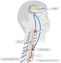
4. Autonomic functions: The sympathetic and parasympathetic divisions of the autonomic nervous system originate in the thoracolumbar and sacral regions of the spinal cord, respectively, controlling involuntary physiological responses like heart rate, blood pressure, digestion, and respiration.
Damage to the spinal cord can result in various degrees of paralysis or loss of sensation below the level of injury, depending on the severity and location of the damage.
Medical Definition:
Magnetic Resonance Imaging (MRI) is a non-invasive diagnostic imaging technique that uses a strong magnetic field and radio waves to create detailed cross-sectional or three-dimensional images of the internal structures of the body. The patient lies within a large, cylindrical magnet, and the scanner detects changes in the direction of the magnetic field caused by protons in the body. These changes are then converted into detailed images that help medical professionals to diagnose and monitor various medical conditions, such as tumors, injuries, or diseases affecting the brain, spinal cord, heart, blood vessels, joints, and other internal organs. MRI does not use radiation like computed tomography (CT) scans.
Paraplegia is a medical condition characterized by partial or complete loss of motor function and sensation in the lower extremities, typically affecting both legs. This results from damage to the spinal cord, often due to trauma such as accidents, falls, or gunshot wounds, or from diseases like spina bifida, polio, or tumors. The specific area and extent of the injury on the spinal cord determine the severity and location of paralysis. Individuals with paraplegia may require assistive devices for mobility, such as wheelchairs, and may face various health challenges, including pressure sores, urinary tract infections, and chronic pain.
Muscle hypotonia, also known as decreased muscle tone, refers to a condition where the muscles appear to be flaccid or lacking in tension and stiffness. This results in reduced resistance to passive movements, making the limbs feel "floppy" or "like a rag doll." It can affect any muscle group in the body and can be caused by various medical conditions, including neurological disorders, genetic diseases, and injuries to the nervous system. Hypotonia should not be confused with muscle weakness, which refers to the inability to generate normal muscle strength.
Central nervous system helminthiasis is a medical condition that refers to the invasion and infection of the central nervous system (CNS), specifically the brain and spinal cord, by parasitic worms, also known as helminths. This rare but serious condition can occur when helminth larvae or eggs accidentally migrate from their usual location in the body to the CNS through the bloodstream or cerebrospinal fluid.
The most common types of helminths that can cause CNS helminthiasis include:
1. Neurocysticercosis: This is caused by the larval stage of the tapeworm Taenia solium, which typically infects the muscles and brain. However, when the larvae invade the CNS, they can form cysts that cause inflammation, swelling, and damage to brain tissue.
2. Echinococcosis: This is caused by the larval stage of the tapeworm Echinococcus granulosus or Echinococcus multilocularis. The larvae can form hydatid cysts in various organs, including the brain, leading to neurological symptoms.
3. Gnathostomiasis: This is caused by the larval stage of the nematode Gnathostoma spinigerum or Gnathostoma hispidum. The larvae can migrate to various organs, including the CNS, causing inflammation and damage to brain tissue.
4. Angiostrongyliasis: This is caused by the nematode Angiostrongylus cantonensis, which typically infects rats but can accidentally infect humans through contaminated food or water. The larvae can migrate to the CNS and cause eosinophilic meningitis, an inflammation of the membranes surrounding the brain and spinal cord.
Symptoms of CNS helminthiasis depend on the type of parasite involved, the location and extent of the infection, and the host's immune response. They can range from mild to severe and may include headache, seizures, weakness, numbness, vision changes, confusion, and cognitive impairment. Diagnosis is usually based on clinical presentation, imaging studies, and laboratory tests, such as serology or CSF analysis. Treatment depends on the type of parasite involved and may include antiparasitic drugs, corticosteroids, and supportive care. Prevention measures include avoiding contaminated food and water, practicing good hygiene, and using insect repellents to prevent mosquito-borne infections.
Tabes dorsalis is a late-stage complication of untreated neurosyphilis, a sexually transmitted infection caused by the bacterium Treponema pallidum. It is characterized by degeneration of the posterior columns and dorsal roots of the spinal cord, leading to various neurological symptoms.
The medical definition of Tabes Dorsalis is:
A chronic progressive degenerative disease of the spinal cord, specifically affecting the dorsal root ganglia and posterior columns, caused by the tertiary stage of syphilis. The condition is characterized by a combination of motor, sensory, and autonomic disturbances, including ataxia, Romberg's sign, lightning pains, hypo- or areflexia, impaired proprioception, dissociated sensations, and Argyll Robertson pupils. If left untreated, Tabes Dorsalis can lead to significant disability and even death.
Aseptic meningitis is a type of meningitis (inflammation of the membranes covering the brain and spinal cord) that is not caused by bacterial infection. Instead, it can be due to viral infections, fungal infections, or non-infectious causes such as certain medications, chemical irritants, or underlying medical conditions. In aseptic meningitis, the cerebrospinal fluid (CSF) analysis may show increased white blood cells, typically lymphocytes, but no bacterial growth on culture. Common viral causes include enteroviruses, herpes simplex virus, and varicella-zoster virus. Treatment depends on the underlying cause and may include supportive care, antiviral medications, or immunosuppressive therapy in some cases.
Demyelinating autoimmune diseases of the central nervous system (CNS) are a group of disorders characterized by inflammation and damage to the myelin sheath, which is the protective covering that surrounds nerve fibers in the brain and spinal cord. This damage can result in various neurological symptoms, including muscle weakness, sensory loss, vision problems, and cognitive impairment.
The most common demyelinating autoimmune disease of the CNS is multiple sclerosis (MS), which affects approximately 2.3 million people worldwide. Other examples include neuromyelitis optica spectrum disorder (NMOSD), acute disseminated encephalomyelitis (ADEM), and transverse myelitis.
These conditions are thought to arise when the immune system mistakenly attacks the myelin sheath, leading to inflammation, damage, and scarring (sclerosis) in the CNS. The exact cause of this autoimmune response is not fully understood, but it is believed to involve a complex interplay between genetic, environmental, and immunological factors.
Treatment for demyelinating autoimmune diseases of the CNS typically involves a combination of medications to manage symptoms, reduce inflammation, and modify the course of the disease. These may include corticosteroids, immunosuppressive drugs, and disease-modifying therapies (DMTs) that target specific components of the immune system.
 Myelitis
 Acute Flaccid Myelitis (AFM) | CDC
 Acute flaccid myelitis: MedlinePlus Medical Encyclopedia
 Transverse Myelitis and MS | National MS Society
 Letter to Clinicians: Acute Flaccid Myelitis | Health
 transverse myelitis | Virology Blog
 Acute myelitis and ChAdOx1 nCoV-19 vaccine: Casual or causal association?
 Information for "Myelitis" - wikidoc
 What to Know About the Acute Flaccid Myelitis Investigations - Keep Kids Healthy
 Nerve Transfer Promising for Acute Flaccid Myelitis Patients - Physician's Weekly
 Acute Flaccid Myelitis (AFM) Information for Health Care Providers | Florida Department of Health
 Acute transverse myelitis of the cervical spine secondary to psoas abscess | BMC Infectious Diseases | Full Text
 Acute Flaccid Myelitis (AFM) | Vermont Department of Health
 Acute Transverse Myelitis - Neurologic Disorders - MSD Manual Professional Edition
 Probable Spinal Neuroschistosomiasis Manifesting as Transverse Myelitis. - MORU Tropical Health Network
 BrainWaves #38 Acute flaccid myelitis | MedLink Neurology
 myelitis - greencitizens
 Transverse Myelitis
 transverse myelitis
 transverse myelitis - The Curbsiders
 What is Transverse Myelitis?
 Transverse Myelitis | Northwestern Medicine
 Myelitis Archives - JSN Herbals
 Diagnosing Transverse Myelitis | PainScale