Moles
Hydatidiform Mole
Mole Rats
Hydatidiform Mole, Invasive
Spalax
Trophoblastic Neoplasms
Gestational Trophoblastic Disease
Nevus
Pregnancy
Pregnancy Complications, Neoplastic
Nevus, Pigmented
Clitoris
Shrews
Temperature
Thermodynamics
Wide distribution of short interspersed elements among eukaryotic genomes. (1/60)
Most short interspersed elements (SINEs) in eukaryotic genomes originate from tRNA and have internal promoters for RNA polymerase III. The promoter contains two boxes (A and B) spaced by approximately 33 bp. We used oligonucleotide primers specific to these boxes to detect SINEs in the genomic DNA by polymerase chain reaction (PCR). Appropriate DNA fragments were revealed by PCR in 30 out of 35 eukaryotic species suggesting the wide distribution of SINEs. The PCR products were used for hybridization screening of genomic libraries which resulted in identification of four novel SINE families. The application of this approach is illustrated by discovery of a SINE family in the genome of the bat Myotis daubentoni. Members of this SINE family termed VES have an additional B-like box, a putative polyadenylation signal and RNA polymerase III terminator. (+info)The development of a biological novelty: a different way to make appendages as revealed in the snout of the star-nosed mole Condylura cristata. (2/60)
The nose of the star-nosed mole Condylura cristata is a complex biological novelty consisting of 22 epidermal appendages. How did this new set of facial appendages arise? Recent studies find remarkable conservation of the genes expressed during appendage formation across phyla, suggesting that the basic mechanisms for appendage development are ancient. In the nose of these moles, however, we find a unique pattern of appendage morphogenesis, showing that evolution is capable of constructing appendages in different ways. During development, the nasal appendages of the mole begin as a series of waves in the epidermis. A second deep layer of epidermis then grows under these superficial epidermal waves to produce 22 separate, elongated epidermal cylinders embedded in the side of the mole's face. The caudal end of each cylinder later erupts from the face and rotates forward to project rostrally, remaining attached only at the tip of the snout. As a result of this unique 'unfolding' formation, the rostral end of each adult appendage is derived from caudal embryonic facial tissue, while the caudal end of each appendage is derived from rostral facial tissue. This developmental process has essentially no outgrowth phase and results in the reversal of the original embryonic orientation of each appendage. This differs from the development of other known appendages, which originate either as outgrowths of the body wall or from subdivisions of outgrowths (e.g. tetrapod digits). Adults of a different mole species (Scapanus townsendii) exhibit a star-like pattern that resembles an embryonic stage of the star-nosed mole, suggesting that the development of the star recapitulates stages of its evolution. (+info)Skull morphology and mitochondrial DNA sequence analysis in the lesser Japanese mole (Mogera imaizumii) from the Imperial Palace (Tokyo, Japan). (3/60)
Since about 1630, the Imperial Palace has been biologically isolated from other habitats by the development and urbanization of Tokyo. We morphologically examined the skulls of the lesser Japanese mole (Mogera imaizumii) from the Imperial Palace and compared them with those from Kanto District, while the sequences of the cytochrome b and 12S rRNA genes were also analyzed to clarify the genetic status of this isolated population. The skulls from the Imperial Palace were much larger than those from Kanto District in the length items. We suggest that the Imperial Palace skulls morphologically may compose a cluster as a large body-sized type in Kanto District within the dots of Mogera imaizumii in charts of principal component analysis. The mitochondrial DNA sequences of the Imperial Palace population were highly homologous to those of other Tokyo population at the level of 98.5% in cytochrome b and 98.7% in 12S rRNA genes. (+info)Postprandial heat increment does not substitute for active thermogenesis in cold-challenged star-nosed moles (Condylura cristata). (4/60)
The postprandial increase in metabolic rate associated with consuming, assimilating and excreting a meal is often termed the heat increment of feeding (HIF). The metabolic heat production of star-nosed moles, Condylura cristata, held at thermoneutrality was monitored for 4 h following a single 10 min session of feeding on a ration consisting of 0 g (controls), 3.5 g or 10 g of earthworms. Coefficients for metabolizable energy digestibility and digesta passage rate of earthworms fed to C. cristata were also determined. We then tested whether feeding-induced thermogenesis substitutes partially or completely for thermoregulatory heat production in these animals exposed to sub-thermoneutral air temperatures (9-24 degrees C). A single feeding on earthworms had both short- and long-term effects on the metabolic rate and respiratory exchange ratio of C. cristata. The observed short-term (0-65 min) rise in metabolic rate, assumed to be associated primarily with the physical costs of nutrient digestion, absorption and excretion, was similar to the calculated mean retention time (66.7+/-7.8 min; mean +/- s.e. m., N=5) of this species. This component of the HIF represented 2.9 % of the food energy ingested by moles fed a single 3.5 g (13.21 kJ) meal of earthworms and 1.4 % of the food energy ingested by moles fed a single 7.5 g (28.09 kJ) meal of earthworms. At all test temperatures, resting metabolic rate typically remained above fasting levels for 1-4 h following ingestion of the high-protein earthworm diet. This protracted rise in metabolic rate, presumably associated with the biochemical costs of amino acid oxidation/gluconeogenesis and ureagenesis, averaged 12.8 % of the metabolizable energy and 8.7 % of the gross energy intake. Despite the potential thermoregulatory benefit, we found no evidence that biochemical HIF substitutes for facultative thermogenesis in star-nosed moles exposed to low air temperatures. (+info)Reproductive features of the eastern mole (Scalopus aquaticus) and star-nose mole (Condylura cristata). (5/60)
Since moles are closely related to shrews, the gametes and reproductive tracts of the star-nose mole (Condylura cristata) and the eastern mole (Scalopus aquaticus) were examined to gain further insight into unusual reproductive traits of the Soricidae. Moles display many of these soricid traits, but with some important differences. The cumulus oophorus of Scalopus, ovulated about 16 h after hCG injection, was largely dispersed by hyaluronidase and, though quite dense, was nevertheless more similar to that of higher mammals than to the compact 'ball of the soricid cumulus. Within the female tract in these moles, approximately 85% of the length of the oviduct comprises a narrow ampulla with numerous differentiated crypts that, in shrews, house spermatozoa. However, in contrast to shrews, moles produce considerably larger numbers of spermatozoa, which challenges the proposal that, in shrews, oviductal sperm crypts specifically permit lower sperm production by the males. In the sperm head of these two moles, the acrosome displays the long rostrum that is typical of other Insectivora, and the perforatorium has the barbs by which soricid spermatozoa probably bind to the zona pellucida. Perhaps allied to this, immunoblots indicated that the immunoreactive acrosomal matrix of Scalopus spermatozoa is simpler than the polypeptide complex of the bovine and hamster acrosomal matrix. (+info)Molecular phylogeny of East Asian moles inferred from the sequence variation of the mitochondrial cytochrome b gene. (6/60)
Taxonomic analysis has previously revealed that the species of moles that inhabit Japan are characterized by exceptional species richness and a high level of endemism. Here, we focused on the evolutionary history of the four Japanese mole species of the genera Euroscapter and Mogera, examining mitochondrial cytochrome b (cyt b) gene sequences and comparing them with those of continental Mogera wogura (Korean and Russian populations), M. insularis from Taiwan, and Talpa europaea and T. altaica from the western and central Eurasian continent, respectively. Our data support the idea that in a radiation center somewhere on the Eurasian continent, a parental stock evolved to modern mole-like morph and radiated several times intermittently during the course of the evolution, spreading its branches to other peripheral geographic domains at each stage of the radiation. Under this hypothesis, the four lineages of Japanese mole species, E. mizura, M. tokudae, M. imaizumii, and M. wogura, could be explained to have immigrated to Japan in this order. Mogera wogura and M. imaizumii showed substantial amounts of geographic variation and somewhat complicated distributions of the cyt b gene types. These intraspecific variations are likely to be associated with the expansion processes of moles in the Japanese Islands during the Pleistocene glacial ages. (+info)The cycle of the seminiferous epithelium in the greater Japanese shrew mole, Urotrichus talpoides. (7/60)
Spermatogenesis and acrosomal formation in the greater Japanese shrew mole, Urotrichus talpoides, were studied by light microscopy. On the basis of acrosomal changes, morphology of spermatid head, nuclear shape, appearance of meiotic figures, location of spermatid and period of spermiation, the cycle of the seminiferous epithelium was classified into 12 stages, and developing spermatids could be divided into 15 steps. The mean relative frequencies of stages from I to XII were 10.9, 8.7, 9.8, 7.3, 8.5, 10.3, 12.5, 8.7, 5.8, 5.4, 5.1 and 7.1%, respectively. Similar to the case in the musk shrew, the spermatid nucleus of the greater Japanese shrew mole remained in the middle region of the seminiferous epithelium and only the acrosome extended towards the basement membrane. The elongation of the acrosome, however, was not prominent. The proacrosomal vesicle first appeared in stage II and then one large and round granule was seen in stage III. The acrosomal vesicle became flattened on the surface of the nucleus in stage IV. Spreading of the acrosomic system has been recognized from stage VII. In stage VII, spermiation occurred. In stage IX, the spermatid nucleus began to elongate. Elongation and condensation of the nucleus were clearly observed in stage X. In stage XII, pachytene spermatocytes divided into diplotene spermatocytes. In stage XII, meiotic figures and secondary spermatocytes were observed. (+info)Body oxygen stores, aerobic dive limits and diving behaviour of the star-nosed mole (Condylura cristata) and comparisons with non-aquatic talpids. (8/60)
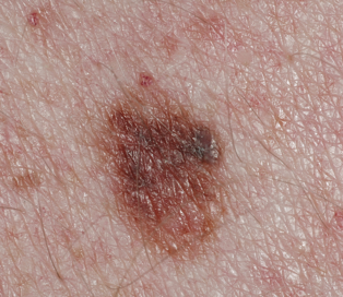
The dive performance, oxygen storage capacity and partitioning of body oxygen reserves of one of the world's smallest mammalian divers, the star-nosed mole Condylura cristata, were investigated. On the basis of 722 voluntary dives recorded from 18 captive star-nosed moles, the mean dive duration (9.2+/-0.2 s; mean +/- S.E.M.) and maximum recorded dive time (47 s) of this insectivore were comparable with those of several substantially larger semi-aquatic endotherms. Total body O(2) stores of adult star-nosed moles (34.0 ml kg(-1)) were 16.4 % higher than for similarly sized, strictly fossorial coast moles Scapanus orarius (29.2 ml kg(-1)), with the greatest differences observed in lung and muscle O(2) storage capacity. The mean lung volume of C. cristata (8.09 ml 100 g(-1)) was 1.81 times the predicted allometric value and exceeded that of coast moles by 65.4 % (P=0.0001). The overall mean myoglobin (Mb) concentration of skeletal muscles of adult star-nosed moles (13.57+/-0.40 mg g(-1) wet tissue, N=7) was 19.5 % higher than for coast moles (11.36+/-0.34 mg g(-1) wet tissue, N=10; P=0.0008) and 54.2 % higher than for American shrew-moles Neurotrichus gibbsii (8.8 mg g(-1) wet tissue; N=2). The mean skeletal muscle Mb content of adult star-nosed moles was 91.1 % higher than for juveniles of this species (P<0.0001). On the basis of an average diving metabolic rate of 5.38+/-0.35 ml O(2) g(-1) h(-1) (N=11), the calculated aerobic dive limit (ADL) of star-nosed moles was 22.8 s for adults and 20.7 s for juveniles. Only 2.9 % of voluntary dives by adult and juvenile star-nosed moles exceeded their respective calculated ADLs, suggesting that star-nosed moles rarely exploit anaerobic metabolism while diving, a conclusion supported by the low buffering capacity of their skeletal muscles. We suggest that a high mass-specific O(2) storage capacity and relatively low metabolic cost of submergence are key contributors to the impressive dive performance of these diminutive insectivores. (+info)A mole (nevus) is a benign growth on the skin that is usually brown or black. Moles can appear anywhere on the body, alone or in groups. Most adults have between 10 and 40 moles. They typically appear during childhood and adolescence. Some moles may change over time, possibly becoming raised and/or changing color. It's important to keep an eye on moles and see a healthcare provider if any changes are noticed, as melanoma, a type of skin cancer, can develop from moles.
It is also worth noting that there are different types of moles including congenital nevi (moles present at birth), dysplastic nevi (atypical moles) and acquired nevi (moles that appear after birth). Dysplastic nevi are larger than average and irregular in shape, with color variations. They are more likely to develop into melanoma than regular moles.
A hydatidiform mole, also known as a molar pregnancy, is a type of gestational trophoblastic disease (GTD), which is a group of rare disorders that involve abnormal growth of the placental tissue.
In a hydatidiform mole, there is an abnormal fertilization event leading to the growth of a mass of grapelike cysts in the uterus instead of a normal pregnancy. The chromosomes from the sperm and egg do not combine properly, resulting in an extra set of chromosomes, which leads to the development of the mole.
Hydatidiform moles can be complete or partial:
* Complete hydatidiform mole (CHM): This type arises when an egg without a nucleus is fertilized by one or two sperm, leading to the growth of abnormal placental tissue with no embryo. The chromosomes come from the father only, and there are typically 46 chromosomes, all of paternal origin.
* Partial hydatidiform mole (PHM): This type occurs when an egg is fertilized by two sperm or a single sperm that duplicates itself, resulting in an abnormal placenta with some fetal tissue. The chromosomes are of both maternal and paternal origin, and the placental tissue has a mix of normal and abnormal cells.
Hydatidiform moles can cause vaginal bleeding, rapid uterine enlargement, and high levels of human chorionic gonadotropin (hCG) hormone in the blood. They are usually detected during an ultrasound exam and require medical treatment to prevent complications such as gestational trophoblastic neoplasia, a malignant form of GTD that can spread to other organs.
A mole rat is not a medical term, but a common name for a burrowing rodent that belongs to the family Bathyergidae. There are about 20 species of mole rats, also known as "blind mole rats" or "naked mole rats," depending on the region and scientific classification.
Mole rats are fascinating creatures with several unique biological features. They are primarily subterranean animals, living in complex tunnel systems that they dig with their powerful incisors and sharp claws. Mole rats have reduced eyes or are completely blind, relying instead on their highly developed senses of touch and smell to navigate their environment.
One species, the naked mole rat (Heterocephalus glaber), is particularly well-known for its unusual biology and social behavior. Naked mole rats live in large colonies with a single breeding female (the queen) and multiple males. The queen is the only reproductively active female, while the other members of the colony function as workers, caring for the young and maintaining the burrow system.
Naked mole rats have several remarkable biological traits, including an extraordinarily long lifespan for a rodent (up to 30 years or more) and resistance to cancer. They are also able to survive in low-oxygen environments and exhibit a unique form of social behavior called eusociality, similar to that seen in bees and ants.
While mole rats may not have a direct medical definition, their unique biology has attracted significant scientific interest, leading to important discoveries in fields such as aging, cancer research, and neurobiology.
An invasive hydatidiform mole (IHM) is a rare and aggressive complication of a gestational trophoblastic disease (GTD), which itself originates from the abnormal proliferation of trophoblastic cells, the tissue that normally develops into the placenta during pregnancy. IHMs are characterized by the invasion of molar villi into the myometrium (the muscular layer of the uterus) and can potentially spread to other organs through the bloodstream, leading to distant metastases.
IHMs usually arise from a complete hydatidiform mole (CHM), which is an abnormal conceptus with no embryonic or fetal development. CHMs are typically diploid and originate from the fertilization of an egg without genetic material (an empty egg or an egg with two sets of paternal chromosomes) by one or two sperm cells. This results in a conceptus with only paternal chromosomes, which leads to uncontrolled proliferation of trophoblastic tissue and the formation of grapelike vesicles filled with fluid (hydatidiform moles).
Invasive hydatidiform moles can cause various symptoms, such as vaginal bleeding, pelvic pain, or the presence of an enlarged uterus. They also pose a risk for developing choriocarcinoma, another type of gestational trophoblastic neoplasia (GTN), which is a malignant tumor that can metastasize and spread to other organs. Proper diagnosis and timely treatment are crucial to prevent severe complications and improve the prognosis for patients with IHMs. Treatment usually involves surgical removal of the mole, followed by chemotherapy to eliminate any residual disease and reduce the risk of GTN development.
Uterine neoplasms refer to abnormal growths in the uterus, which can be benign (non-cancerous) or malignant (cancerous). These growths can originate from different types of cells within the uterus, leading to various types of uterine neoplasms. The two main categories of uterine neoplasms are endometrial neoplasms and uterine sarcomas.
Endometrial neoplasms develop from the endometrium, which is the inner lining of the uterus. Most endometrial neoplasms are classified as endometrioid adenocarcinomas, arising from glandular cells in the endometrium. Other types include serous carcinoma, clear cell carcinoma, and mucinous carcinoma.
Uterine sarcomas, on the other hand, are less common and originate from the connective tissue (stroma) or muscle (myometrium) of the uterus. Uterine sarcomas can be further divided into several subtypes, such as leiomyosarcoma, endometrial stromal sarcoma, and undifferentiated uterine sarcoma.
Uterine neoplasms can cause various symptoms, including abnormal vaginal bleeding or discharge, pelvic pain, and difficulty urinating or having bowel movements. The diagnosis typically involves a combination of imaging tests (such as ultrasound, CT, or MRI scans) and tissue biopsies to determine the type and extent of the neoplasm. Treatment options depend on the type, stage, and patient's overall health but may include surgery, radiation therapy, chemotherapy, or hormone therapy.
"Spalax" is a genus of subterranean rodents, also known as mole rats, that are found primarily in Eastern and Southeastern Europe and Western Asia. They are characterized by their blindness, lack of external ears, and highly specialized digging abilities. While "Spalax" is not a medical term per se, it is sometimes used in scientific research to refer to this specific genus of animals. Some studies have investigated the unique physiology and genetics of Spalax species, including their resistance to cancer and hypoxia, which may have implications for human medicine.
Trophoblastic neoplasms are a group of rare tumors that originate from the trophoblast, which is the outer layer of cells that surrounds a developing embryo and helps to form the placenta during pregnancy. These tumors can be benign or malignant and are characterized by their ability to produce human chorionic gonadotropin (hCG), a hormone that is normally produced during pregnancy.
There are several types of trophoblastic neoplasms, including:
1. Hydatidiform mole: A benign growth that forms in the uterus when a fertilized egg implants but does not develop into a normal embryo. There are two types of hydatidiform moles: complete and partial. Complete moles have no fetal tissue, while partial moles have some fetal tissue.
2. Invasive mole: A malignant form of hydatidiform mole that invades the uterine wall and may spread to other parts of the body.
3. Choriocarcinoma: A rapidly growing and highly invasive malignant tumor that can arise from a hydatidiform mole, a normal pregnancy, or an ectopic pregnancy. It can spread quickly to other parts of the body, such as the lungs, liver, and brain.
4. Placental site trophoblastic tumor (PSTT): A rare type of trophoblastic neoplasm that arises from the cells that attach the placenta to the uterine wall. It is usually slow-growing but can be aggressive in some cases.
5. Epithelioid trophoblastic tumor (ETT): Another rare type of trophoblastic neoplasm that arises from the cells that form the placental villi. It is typically low-grade and has a good prognosis, but it can recur in some cases.
The treatment for trophoblastic neoplasms depends on the type and stage of the tumor. Treatment options may include surgery, chemotherapy, radiation therapy, or a combination of these approaches. Regular monitoring of hCG levels is also important to ensure that the tumor has been completely removed and to detect any recurrence early.
'Insectivora' is an outdated taxonomic grouping that was once used to classify small, insect-eating mammals. This order included shrews, moles, hedgehogs, and several other related species. However, modern molecular evidence has revealed that this grouping is not monophyletic, meaning it does not include all descendants of a common ancestor. As a result, the order Insectivora is no longer recognized in current taxonomy. Instead, these animals are now classified into several different orders based on their evolutionary relationships.
Gestational Trophoblastic Disease (GTD) is a group of rare pregnancy-related disorders that involve abnormal growth of cells inside a woman's uterus. These cells are part of the placenta, which provides nutrients to the developing fetus. GTD occurs when some of these cells grow in an uncontrolled way, forming tumors or tumor-like growths.
There are several types of GTD:
1. Hydatidiform Mole (HM): Also known as a molar pregnancy, this is the most common type of GTD. It occurs when an egg that has no genetic information is fertilized by a sperm and then divides into multiple copies. This results in a growth that resembles a cluster of grapes, rather than a developing fetus. There are two types of HMs: complete and partial. A complete HM forms when an empty egg is fertilized by two sperms, resulting in no fetal tissue. A partial HM forms when a normal egg is fertilized by two sperm or an abnormal egg with two sets of genetic material, resulting in some fetal tissue.
2. Invasive Mole: This type of GTD occurs when cells from a molar pregnancy invade the uterine wall and surrounding tissues. It can also spread to other parts of the body, such as the lungs or brain.
3. Choriocarcinoma: This is a rare form of GTD that develops from trophoblastic cells and forms a malignant tumor. It can grow rapidly and spread quickly to other organs.
4. Placental Site Trophoblastic Tumor (PSTT): This is an even rarer type of GTD that forms in the tissue where the placenta attaches to the uterus. PSTTs are usually slow-growing but can sometimes spread to other parts of the body.
5. Epithelioid Trophoblastic Tumor (ETT): This is a very rare type of GTD that forms in the tissue where the placenta attaches to the uterus. ETTs are usually slow-growing and have a good prognosis.
It's important to note that most molar pregnancies do not develop into more serious forms of GTD, but regular follow-up care is necessary to monitor for any signs of progression. Treatment options depend on the type and stage of GTD and may include surgery, chemotherapy, or radiation therapy.
A nevus, also known as a mole, is a benign growth or mark on the skin that is usually brown or black. It can be raised or flat and can appear anywhere on the body. Nevi are made up of cells called melanocytes, which produce the pigment melanin. Most nevi develop in childhood or adolescence, but they can also appear later in life. Some people have many nevi, while others have few or none.
There are several types of nevi, including:
* Common nevi: These are the most common type of mole and are usually small, round, and brown or black. They can be flat or raised and can appear anywhere on the body.
* Atypical nevi: These moles are larger than common nevi and have irregular borders and color. They may be flat or raised and can appear anywhere on the body, but are most commonly found on the trunk and extremities. Atypical nevi are more likely to develop into melanoma, a type of skin cancer, than common nevi.
* Congenital nevi: These moles are present at birth and can vary in size from small to large. They are more likely to develop into melanoma than moles that develop later in life.
* Spitz nevi: These are rare, benign growths that typically appear in children and adolescents. They are usually pink or red and dome-shaped.
It is important to monitor nevi for changes in size, shape, color, and texture, as these can be signs of melanoma. If you notice any changes in a mole, or if you have a new mole that is unusual or bleeding, it is important to see a healthcare provider for further evaluation.
Pregnancy is a physiological state or condition where a fertilized egg (zygote) successfully implants and grows in the uterus of a woman, leading to the development of an embryo and finally a fetus. This process typically spans approximately 40 weeks, divided into three trimesters, and culminates in childbirth. Throughout this period, numerous hormonal and physical changes occur to support the growing offspring, including uterine enlargement, breast development, and various maternal adaptations to ensure the fetus's optimal growth and well-being.
Neoplastic pregnancy complications refer to the abnormal growth of cells (neoplasia) that can occur during pregnancy. These growths can be benign or malignant and can arise from any type of tissue in the body. However, when they occur in pregnant women, they can pose unique challenges due to the potential effects on the developing fetus and the changes in the mother's body.
Some common neoplastic pregnancy complications include:
1. Gestational trophoblastic disease (GTD): This is a group of rare tumors that occur in the uterus during pregnancy. GTD can range from benign conditions like hydatidiform mole to malignant forms like choriocarcinoma.
2. Breast cancer: Pregnancy-associated breast cancer (PABC) is a type of breast cancer that occurs during pregnancy or within one year after delivery. It can be aggressive and challenging to diagnose due to the changes in the breast tissue during pregnancy.
3. Cervical cancer: Cervical cancer can occur during pregnancy, and its management depends on the stage of the disease and the gestational age. In some cases, treatment may need to be delayed until after delivery.
4. Lung cancer: Pregnancy does not increase the risk of lung cancer, but it can make diagnosis and treatment more challenging.
5. Melanoma: Melanoma is the most common malignant skin cancer during pregnancy. It can spread quickly and requires prompt treatment.
The management of neoplastic pregnancy complications depends on several factors, including the type and stage of the tumor, gestational age, and the patient's wishes. In some cases, surgery, chemotherapy, or radiation therapy may be necessary. However, these treatments can have potential risks to the developing fetus, so a multidisciplinary team of healthcare providers is often involved in the care of pregnant women with neoplastic complications.
In the context of medicine and pharmacology, "kinetics" refers to the study of how a drug moves throughout the body, including its absorption, distribution, metabolism, and excretion (often abbreviated as ADME). This field is called "pharmacokinetics."
1. Absorption: This is the process of a drug moving from its site of administration into the bloodstream. Factors such as the route of administration (e.g., oral, intravenous, etc.), formulation, and individual physiological differences can affect absorption.
2. Distribution: Once a drug is in the bloodstream, it gets distributed throughout the body to various tissues and organs. This process is influenced by factors like blood flow, protein binding, and lipid solubility of the drug.
3. Metabolism: Drugs are often chemically modified in the body, typically in the liver, through processes known as metabolism. These changes can lead to the formation of active or inactive metabolites, which may then be further distributed, excreted, or undergo additional metabolic transformations.
4. Excretion: This is the process by which drugs and their metabolites are eliminated from the body, primarily through the kidneys (urine) and the liver (bile).
Understanding the kinetics of a drug is crucial for determining its optimal dosing regimen, potential interactions with other medications or foods, and any necessary adjustments for special populations like pediatric or geriatric patients, or those with impaired renal or hepatic function.
A nevus pigmentosus, also known as a pigmented mole or melanocytic nevus, is a benign proliferation of melanocytes, the pigment-producing cells in the skin. These lesions typically appear as well-circumscribed, brown to black macules or papules. They can vary in size and shape and may be flat or raised. Most nevi are harmless and do not require treatment; however, some may undergo malignant transformation into melanoma, a potentially life-threatening skin cancer. Regular self-skin examinations and professional skin checks are recommended to monitor for changes in nevi that may indicate malignancy.
The clitoris is an important female sex organ that is primarily responsible for sexual arousal and pleasure. It is a small, highly sensitive piece of tissue located at the front of the vulva, where the labia minora meet. The clitoris is made up of two parts: the visible part, known as the glans clitoris, and the hidden part, called the corpora cavernosa and crura.
The glans clitoris is a small knob-like structure that is covered by a hood, or prepuce, and is located at the top of the vulva. It contains a high concentration of nerve endings, making it highly sensitive to touch and stimulation. The corpora cavernosa and crura are the internal parts of the clitoris, which are made up of sponge-like erectile tissue that becomes engorged with blood during sexual arousal, leading to clitoral erection.
The clitoris plays a crucial role in female sexual response and pleasure. During sexual arousal, the clitoris swells and becomes more sensitive to touch, which can lead to orgasm. The clitoris is also an important source of sexual pleasure during masturbation and partnered sexual activity. Despite its importance in female sexuality, the clitoris has historically been overlooked or stigmatized in many cultures, leading to a lack of understanding and education about this vital organ.
I'm sorry for any confusion, but the term "shrews" is not a medical term. It is a common name used in zoology to refer to small mammals that are part of the family Soricidae and the suborder Soricomorpha. Shrews are insectivores, distantly related to moles and hedgehogs. If you have any questions about medical terminology or concepts, I would be happy to help!
Temperature, in a medical context, is a measure of the degree of hotness or coldness of a body or environment. It is usually measured using a thermometer and reported in degrees Celsius (°C), degrees Fahrenheit (°F), or kelvin (K). In the human body, normal core temperature ranges from about 36.5-37.5°C (97.7-99.5°F) when measured rectally, and can vary slightly depending on factors such as time of day, physical activity, and menstrual cycle. Elevated body temperature is a common sign of infection or inflammation, while abnormally low body temperature can indicate hypothermia or other medical conditions.
I'm sorry for any confusion, but "thermodynamics" is not a term that has a specific medical definition. It is a branch of physics that deals with the relationships between heat and other forms of energy. However, the principles of thermodynamics can be applied to biological systems, including those in the human body, such as in the study of metabolism or muscle function. But in a medical context, "thermodynamics" would not be a term used independently as a diagnosis, treatment, or any medical condition.
Phosphatidylcholines (PtdCho) are a type of phospholipids that are essential components of cell membranes in living organisms. They are composed of a hydrophilic head group, which contains a choline moiety, and two hydrophobic fatty acid chains. Phosphatidylcholines are crucial for maintaining the structural integrity and function of cell membranes, and they also serve as important precursors for the synthesis of signaling molecules such as acetylcholine. They can be found in various tissues and biological fluids, including blood, and are abundant in foods such as soybeans, eggs, and meat. Phosphatidylcholines have been studied for their potential health benefits, including their role in maintaining healthy lipid metabolism and reducing the risk of cardiovascular disease.
 Moles
Moles
Edwin Moles
Brodie Moles
Margot Moles
Osvaldo Moles
Ovos moles
Maria Moles
Abraham Moles
Andy Moles
David Moles
John Moles
Moles Brewery
Gene Moles
James Moles
Giuseppe Moles
Thomas Moles
Francesco Maria Moles
Robert N Moles
Pasqual Pere Moles
Enrique Moles Ormella
Maria Reig Moles
Moles de Xert
The Moles (Australian band)
Mole
No moles left in Irevan
Marsupial mole
Mole people
Caucasian mole
Mole-Richardson
Charles Mole
Edwin Moles - Wikipedia
 Moles | Nevus | MedlinePlus
Mole (architecture) - Wikipedia
 Moles: Types, causes, treatment, and diagnosis
 Moles - Knowledgebase Question - Garden.org
 Why Don't All Moles Progress To Melanoma? | ScienceDaily
 Meet the Moles - American Chemical Society
 Moles - Symptoms and causes - Mayo Clinic
 Movie Moles: Matewan | KBOO
 mole
mole
 PLATINUM-MOLES
 Many Melanoma Patients May Have Few Moles | Live Science
 Painless ways to remove moles at home
 Louise used to be a drama teacher. Now she kills moles
 Mole Archives - Universe Today
 Mole Architects | Tag
| ArchDaily
Mole Architects | Tag
| ArchDaily
 Wine, mescal and mole
 Naked mole-rats don't feel the burn | Nature
 Moles, voles and gophers | Metro
 WEC66/UW080: Moles
 Hydatidiform Mole: Practice Essentials, Background, Pathophysiology
 Naked mole rats may help cure cancer | New Scientist
 Freckles vs Moles: A Dermatologist Explains
 Bleep - The Mole
 Peter Harrap wants new Monty Mole | Eurogamer.net
 Common Moles, Dysplastic Nevi, and Risk of Melanoma - NCI
 FELTED WOOL JACKET - MOLE BROWN - COS
 The Growing Pains of Adrian Mole
 mole crickets maps
-
Encyclopedia of Life
mole crickets maps
-
Encyclopedia of Life
 Hairy Moles News, Articles | The Scientist Magazine®
Melanoma34
- They may be more likely than ordinary moles to develop into melanoma , a type of skin cancer. (medlineplus.gov)
- The mole is a suspected melanoma. (medicalnewstoday.com)
- If the melanoma is detected in the very early stages, when the mole is thin and has not grown downwards from the surface of the skin and spread to other parts of the body, it is removed using a simple surgical technique. (medicalnewstoday.com)
- Many moles - people with numerous moles run a greater risk of developing malignant melanoma. (medicalnewstoday.com)
- Why Don't All Moles Progress To Melanoma? (sciencedaily.com)
- But sometimes pigment-producing cells in moles called melanocytes start dividing abnormally to form a deadly form of skin cancer called melanoma. (sciencedaily.com)
- That's why so many people have moles, but few have melanoma. (sciencedaily.com)
- Being aware of changes in your moles and other pigmented patches is important to detecting skin cancer, especially malignant melanoma. (mayoclinic.org)
- Flesh moles don't carry a risk of melanoma, but they can be treated if you consider them a cosmetic concern. (mayoclinic.org)
- Checking out the moles on your skin is a common way to look for the deadly skin cancer melanoma, but a new study shows that many people with melanoma may have few moles. (livescience.com)
- In the study, researchers looked at about 560 people with melanoma and found that 66 percent of them had 20 or fewer moles. (livescience.com)
- Among patients younger than 60, those who had more than 50 moles tended to have melanoma tumors that were thinner (less than 2 millimeters thick, or about 0.08 of an inch), compared with those who had fewer than 50 moles. (livescience.com)
- The thickness of the melanoma indicates how deeply the cancer has gone into the skin, and so this finding shows that the people with a lot of moles did not necessarily have the most severe cases of melanoma . (livescience.com)
- The people who had five or more moles that looked different from ordinary, non-melanoma moles had a higher risk of thicker melanoma (more than 2 mm thick), compared with those who had no such moles , the researchers found. (livescience.com)
- Previous studies have linked having a greater number of moles to an increased risk of melanoma, the researchers said. (livescience.com)
- The new study serves as a reminder that even people who don't have many moles or other known risk factors for melanoma may still get the skin cancer, the researchers said. (livescience.com)
- Additionally, "having many moles or atypical moles is associated with an increased risk of melanoma. (dermstore.com)
- Melanoma can develop in moles or as a new spot that can look like a mole. (dermstore.com)
- Can a common mole turn into melanoma? (cancer.gov)
- Only rarely does a common mole turn into melanoma , the most serious type of skin cancer . (cancer.gov)
- Although common moles are not cancerous, people who have many small moles or several large ones have an increased risk of developing melanoma ( 1 ). (cancer.gov)
- Certain changes in a mole may indicate that it is turning into a melanoma ( 2 ). (cancer.gov)
- Though benign, they are worth more of your attention because individuals with atypical moles are at increased risk for melanoma , a dangerous skin cancer. (skincancer.org)
- An atypical mole is not a skin cancer but having these moles is a risk factor for developing melanoma. (skincancer.org)
- Although rare, melanoma can arise in association with atypical moles. (skincancer.org)
- If you have atypical moles plus a family history of melanoma, you have an increased risk of developing melanoma. (skincancer.org)
- It's important to note that even without a family history of melanoma, if you have atypical moles, you have an elevated risk of developing melanoma. (skincancer.org)
- The first five letters of the alphabet can be used as a guide to the warning signs for atypical moles and melanoma . (skincancer.org)
- While benign moles are usually a single shade of brown, a melanoma may have different shades of brown, tan or black. (skincancer.org)
- Your dermatologist will determine whether your pigmented lesion is an atypical mole or a melanoma, and provide you with detailed information about next steps. (skincancer.org)
- People with atypical mole syndrome are at especially high risk of developing melanoma. (skincancer.org)
- If you have hereditary risk factors for melanoma as well as many atypical moles, you may be classified as having Familial Atypical Multiple Mole Melanoma Syndrome (FAMMM), putting you at even greater risk for developing melanoma. (skincancer.org)
- Moles develop on nearly everybody, and are significant primarily because they can become dysplastic or malignant and need to be differentiated from melanoma. (msdmanuals.com)
- The transformation rate of moles (melanocytic nevi) into cutaneous melanoma: A population-based estimate. (msdmanuals.com)
Sarcomas de partes moles1
- Os sarcomas de partes moles (SPM) de alto grau são neoplasias heterogêneas, de prognóstico ruim e que apresentam poucas alternativas de tratamento . (bvsalud.org)
Atypical mole9
- About one out of every ten people has at least one unusual (or atypical) mole that looks different from an ordinary mole. (medlineplus.gov)
- Some doctors use the term "atypical mole" to refer to a dysplastic nevus. (cancer.gov)
- An atypical mole can occur anywhere on the body. (skincancer.org)
- At first glance, it can be tricky to see how an atypical mole differs from a normal mole. (skincancer.org)
- Atypical mole with asymmetry, border irregularity and multiple shades of brown. (skincancer.org)
- Atypical mole on lower back. (skincancer.org)
- Swedish CDKN2A mutation carriers do not present the atypical mole syndrome phenotype. (lu.se)
- No participant fulfilled the atypical mole syndrome phenotype criteria. (lu.se)
- The atypical mole syndrome phenotype was, however, not verified in the studied families and total naevus counts were low. (lu.se)
Damage caused by moles1
- The damage caused by moles is almost entirely cosmetic. (ufl.edu)
Malignant5
- Results of the U-M study -- involving melanocytes from normal human skin and biopsies of non-malignant human moles -- are being published in the October issue of Nature Cell Biology. (sciencedaily.com)
- Cancerous (malignant) moles vary greatly in appearance. (mayoclinic.org)
- Moles can be benign (harmless) or malignant (cancerous), and it's not always easy to tell (more on that in the next section). (dermstore.com)
- Moles are another chapter completely, as some growths can be malignant (cancerous). (dermstore.com)
- Just as the differences between a flat mole and a freckle can be unclear, the differences between a benign and malignant mole can also be subtle. (dermstore.com)
Cancerous2
- Most moles do not lead to complications, but some can become cancerous. (medicalnewstoday.com)
- Not all moles are cancerous. (selfgrowth.com)
Color of the mole changes1
- Geller recommended that people watch out for any of the following changes in moles, which dermatologists use the abbreviation "ABCD" to remember: asymmetry (one side of the mole starts to look different than the other side), border (the border around the mole begins to change), color (the color of the mole changes, for example becomes darker) and diameter (the mole becomes wider than the diameter of a pencil head eraser). (livescience.com)
Whack-a-Mo4
- An English couple created a human Whack-a-Mole game for their kids. (yahoo.com)
- While the future of arcades around the world remains uncertain due to the global lockdowns, life-size Whack-a-Mole looks like a worthy substitute. (yahoo.com)
- The post These parents created a life-size Whack-a-Mole game for their daughters appeared first on In The Know . (yahoo.com)
- Today, we'll take that line of thought a step further and build a simple game of Whack-A-Mole, where the player needs to react quickly to win … all without a touch of JavaScript. (css-tricks.com)
Develop new moles2
- A person may develop new moles from time to time, usually until about age 40. (medlineplus.gov)
- Most people continue to develop new moles until about age 40. (cancer.gov)
Complete hydatidiform7
- Magnified transverse sonogram shows a complete hydatidiform mole (CHM) at 7 weeks of menstrual age with a small anembryonic gestational sac. (medscape.com)
- Sagittal endovaginal sonogram of a complete hydatidiform mole (CHM) at 12 weeks of menstrual age demonstrates an enlarged endometrium containing an anembryonic gestational sac with adjacent hyperechoic material containing tiny anechoic spaces. (medscape.com)
- Transverse endovaginal sonogram of a second-trimester complete hydatidiform mole (CHM) demonstrates a distended endometrial cavity containing innumerable, variably sized anechoic cysts with intervening hyperechoic material. (medscape.com)
- Transverse endovaginal sonogram of a second-trimester complete hydatidiform mole (CHM). (medscape.com)
- The major imaging feature distinguishing PHM from complete hydatidiform mole (CHM) is the presence of fetal tissue on the left side of the image (mother's right side). (medscape.com)
- Second-trimester complete hydatidiform mole (CHM) can be confused with retained products of conception. (medscape.com)
- The World Health Organization (WHO) prognostic scoring system is important in the medical management of patients with complete hydatidiform moles, partial hydatidiform moles, and choriocarcinomas. (medscape.com)
Biopsy3
- Excisional surgery (excision biopsy) - the mole plus a surrounding margin of healthy skin is cut out using a scalpel or a punch device. (medicalnewstoday.com)
- Your dermatologist will know whether or not any moles need a biopsy to ensure they are not melanomas. (skincancer.org)
- I think that in general, when I think about moles and when to biopsy, if a patient is reliable and seems to be telling you very pointedly that something is changing, then I always think that you are safer by going ahead and doing the biopsy. (medscape.com)
Darker3
- Moles are usually brownish, but some may be much darker, while others are skin-colored. (medicalnewstoday.com)
- People who have dark skin or hair tend to have darker moles than people with fair skin or blonde hair. (cancer.gov)
- Dickens says the color can vary as well-moles tend to have darker gray fur, whereas voles tend to be shades of brown. (yahoo.com)
Nevi10
- Congenital melanocytic nevi are present at birth, any moles appearing after birth are melanocytic nevi. (medicalnewstoday.com)
- Congenital nevi - these are large moles that people are born with. (medicalnewstoday.com)
- Moles that appear in families - atypical (dysplastic) nevi are larger than normal and are usually hereditary. (medicalnewstoday.com)
- Moles (nevi) are a common type of skin growth. (mayoclinic.org)
- Clusters of brown spots around the eyes, cheeks and nose are sometimes called flesh moles, but they are actually dermatoses papulosa nigra - a type of seborrheic keratosis, not clusters of pigment-forming cells (nevi). (mayoclinic.org)
- People who have dysplastic nevi usually also have an increased number of common moles. (cancer.gov)
- The photos below show the difference between common moles and dysplastic nevi. (cancer.gov)
- Atypical moles , also known as dysplastic nevi, are unusual-looking moles that have irregular features under the microscope. (skincancer.org)
- Blue nevi are benign moles that appear as bluish gray macules or thin papules. (msdmanuals.com)
- The other factor to consider in someone who has traditional risk factors-fair-skinned, a variety of nevi-is to look for the mole that looks different . (medscape.com)
Placental site1
- These include complete and partial moles, placental site trophoblastic tumors, choriocarcinomas, and invasive moles. (medscape.com)
Nevus5
- Another name for a mole is a nevus. (cancer.gov)
- A dysplastic nevus is a type of mole that looks different from a common mole. (cancer.gov)
- A dysplastic nevus may be bigger than a common mole, and its color, surface, and border may be different. (cancer.gov)
- A common mole ( nevus ) is a small growth on the skin that is usually pink, tan, or brown and has a distinct edge. (cancer.gov)
- Moles are flesh- to brown-colored macules, papules, or nodules composed of nests of melanocytes or nevus cells. (msdmanuals.com)
Small moles1
- The second photo shows small moles on a person's back. (cancer.gov)
Fewer moles1
- Dark skinned people generally have fewer moles than those with fair skin. (medicalnewstoday.com)
Hydatidiform mole12
- A hydatidiform mole (molar pregnancy) is a gestational trophoblastic disease. (medscape.com)
- p.W195X) in a patient that appears to be associated with recurrent hydatidiform mole. (medscape.com)
- Ultrasonography is the imaging investigation of choice (see the images below) to confirm the diagnosis of hydatidiform mole . (medscape.com)
- Note that retained products of conception may mimic a hydatidiform mole. (medscape.com)
- Transverse transpelvic sonogram of a partial hydatidiform mole (PHM) at 16 weeks of menstrual age. (medscape.com)
- No reliable plain radiographic findings are reported for hydatidiform mole. (medscape.com)
- No angiographic studies are clinically useful for assessing hydatidiform mole. (medscape.com)
- Clinical features of hydatidiform mole in patients 40 years or older may differ from those in younger patients. (medscape.com)
- The sonographic appearance of a first-trimester hydatidiform mole can be indistinguishable from that of an anembryonic gestation, specifically blighted ovum. (medscape.com)
- In a multicenter study, many proven cases of hydatidiform mole were not clinically or sonographically evident. (medscape.com)
- Of 155 cases of hydatidiform mole, only 53 (34%) were correctly diagnosed as hydatidiform mole with ultrasonography. (medscape.com)
- CT presents a risk of ionizing radiation, and it has no role in the diagnosis of hydatidiform mole. (medscape.com)
Harmless4
- The vast majority of moles are harmless. (medicalnewstoday.com)
- The majority of moles are harmless and require no treatment. (medicalnewstoday.com)
- Moles are usually harmless. (mayoclinic.org)
- Moles are harmless in nature. (selfgrowth.com)
Changes in a mole1
- People who begin to see any changes in a mole, "should watch it and make sure their doctor - their primary- care physician or dermatologist - takes a good look at it," he added. (livescience.com)
Growths1
- Moles are growths on the skin. (medlineplus.gov)
Freckles3
- Freckles are not moles. (medicalnewstoday.com)
- Freckles vs. Moles: What's the Difference? (dermstore.com)
- Because moles develop from deeper in the skin than freckles they are harder to lighten or remove," says Dr. Rogers. (dermstore.com)
Fade away2
- Moles may change or fade away over time. (mayoclinic.org)
- In older people, common moles tend to fade away. (cancer.gov)
Melanocytes2
- In the case of moles, melanocytes can stay this way for 20 to 40 years or even your whole life. (sciencedaily.com)
- A common mole is a growth on the skin that develops when pigment cells (melanocytes) grow in clusters. (cancer.gov)
Talpa3
- There are fewer than 100 traditional specialists in Britain who make a living purely from Talpa europaea, to give the British mole its Latin name, without deploying other forms of pest control. (telegraph.co.uk)
- Blind but ferocious: The Iberian mole (Talpa occidentalis) that is common in Spain and Portugal has a peculiar property - the females develop strong muscles and testicular tissues due to increased levels of male sex hormones. (charite.de)
- To pursue this question, the researchers have completely sequenced the genome of the Iberian mole ( Talpa occidentalis ) for the first time. (charite.de)
Doctor if a mole1
- Make an appointment with your doctor if a mole looks unusual, grows or otherwise changes. (mayoclinic.org)
Voles5
- Voles versus moles: It sounds like the premise of a bad animated kids' film, but alas, it's not! (yahoo.com)
- Voles and moles are exclusively grown-up problems, and both creatures are kind of a nuisance. (yahoo.com)
- According to Roger Dickens, Terminix 's technical services manager of bird and wildlife control, one of the biggest differences is literally how big they are: Moles are larger than voles. (yahoo.com)
- Makes sense-voles are in the rodent family, while moles aren't! (yahoo.com)
- Additionally, moles have a longer snout compared to voles, which have a snout that is short and stubbed, similar to a hamster. (yahoo.com)
Diagnosis1
- MRI has no established role in the initial diagnosis of hydatidiform moles. (medscape.com)
Shrews3
- 3 Chinese mole shrews ( Anourosorex squamipes ), and 12 long-nosed moles ( Euroscaptor longirostris ), captured in northern, central, and southern Vietnam during November and December 2006. (cdc.gov)
- Hantavirus sequences were not detected in tissues of the white-toothed shrews and long-nosed moles. (cdc.gov)
- By contrast, the full-length 3,637-nt (1,139-aa) medium (M) segment was amplified from lung tissues of 3 Chinese mole shrews, captured in Thanh Cong commune, Nguyen Binh District, Cao Bang Province, along the southern border of the People's Republic of China. (cdc.gov)
Star-nose4
- The star-nosed mole, Condylura cristata , has been collected in the Okefenokee Swamp in Georgia and has been reported in Florida. (ufl.edu)
- Because of its rarity in Florida, the star-nosed mole will not be discussed further. (ufl.edu)
- The star-nosed mole, Condylura cristata . (ufl.edu)
- Star-nosed Mole has pink, fleshy appendages on the nose. (tn.gov)
Fetal tissue2
- A complete mole contains no fetal tissue. (medscape.com)
- With a partial mole, fetal tissue is often present. (medscape.com)
Rarely4
- This mole is normally found in wet soils, in marshes, and along streams, so it rarely causes problems in yards and turf. (ufl.edu)
- Flooding the tunnels with water may force moles to the surface, but this method rarely works in deep, sandy soils like those common in Florida. (ufl.edu)
- Partial moles with a 69,XYY complement are rarely seen, and 69,YYY does not occur. (medscape.com)
- Keep in mind though that since moles are fairly subterranean, you'll rarely see them, Dickens notes. (yahoo.com)
Earthworms3
- These tunnels are created as the mole searches among the plant roots for the earthworms and insects on which it feeds. (ufl.edu)
- There earthworms don't die, but can't move, and the moles store them in deep chambers called larders. (ufl.edu)
- Moles are insectivorous, meaning they eat insects like earthworms, slugs, and other invertebrates in the ground. (yahoo.com)
Crickets1
- Known occurrences, collected specimens and observations of mole crickets. (eol.org)
Mole's1
- In this second instalment of teenager Adrian Mole's diaries, the Mole family is in crisis and the country is beating the drum of war. (penguin.co.uk)
Female mole3
- There are only six registered female mole catchers in Britain. (telegraph.co.uk)
- Louise is one of a handful of female mole catchers in Britain . (telegraph.co.uk)
- The testicular tissue of the female mole does not produce sperm, but large amounts of the sex hormone testosterone, meaning the females have similarly high levels as the males. (charite.de)
Normal mole1
- An example of what was considered a normal mole. (medicalnewstoday.com)
Eastern mole5
- The eastern mole, Scalopus aquaticus , occurs throughout Florida. (ufl.edu)
- The eastern mole has an average total length of 5½-6 in (14-15 cm) and a short, sparsely haired tail 1-1½ in (2.5-3.8 cm) long. (ufl.edu)
- Eastern mole, Scalopus aquaticus . (ufl.edu)
- The eastern mole prefers loose, well-drained soils. (ufl.edu)
- Eastern Mole has a longer tail, which only has scattered hairs. (tn.gov)
Irregular3
- A mole may be a sign of skin cancer if it has irregular borders or an asymmetrical shape or if it changes in color, shape, size or height. (mayoclinic.org)
- Look for moles with irregular, notched or scalloped borders. (mayoclinic.org)
- Look for these warning signs: A new or existing mole that has an irregular border (ragged, notched, or blurred edges). (cdc.gov)
Hairs1
- Skin moles that sprout thick, long hairs produce signaling molecules that stimulate hair follicle stem cells to initiate new hair growth. (the-scientist.com)
Pregnancy3
- Some moles respond to changes in hormone levels, as may occur during pregnancy, adolescence, and older age. (medicalnewstoday.com)
- By studying elective pregnancy terminations, hydatidiform moles were determined to occur in approximately 1 in 1200 pregnancies. (medscape.com)
- Moles may darken during pregnancy. (msdmanuals.com)
Litter3
Common15
- Moles are very common. (medlineplus.gov)
- What is a common mole? (cancer.gov)
- Most adults have between 10 and 40 common moles. (cancer.gov)
- Although common moles may be present at birth, they usually appear later in childhood. (cancer.gov)
- What does a common mole look like? (cancer.gov)
- A common mole is usually smaller than about 5 millimeters wide (about 1/4 inch, the width of a pencil eraser). (cancer.gov)
- A common mole usually has an even color of pink, tan, or brown. (cancer.gov)
- This common mole is 1 millimeter in diameter (the width of the tip of a sharpened pencil). (cancer.gov)
- This common mole is 2 millimeters in diameter (the width of the tip of a new crayon). (cancer.gov)
- This common mole is about 5 millimeters in diameter (the width of a new pencil eraser). (cancer.gov)
- A common mole is usually small. (cancer.gov)
- The first photo shows a common mole that is less than 5 millimeters (about 1/4 inch) wide. (cancer.gov)
- If you draw a line through the middle of the lesion, the two halves don't match, so it looks different from a round to oval and symmetrical common mole. (skincancer.org)
- Borders tend to be uneven and may have scalloped or notched edges, while common moles tend to have more smoother, more even borders. (skincancer.org)
- Because moles are extremely common and melanomas are uncommon, prophylactic removal is not justifiable. (msdmanuals.com)
Tunnels5
- Moles dig seldom-used tunnels off the main run, so you may have to place the trap several times before you discover their main run. (garden.org)
- With their fur pointing up, moles can move forward or backward within their tunnels without rubbing their fur the wrong way and trapping soil in their coats. (ufl.edu)
- The characteristic mole ridges that lie just below the surface are foraging tunnels (Figure 3). (ufl.edu)
- When mole tunnels become an intolerable nuisance, moles may be captured and removed without a permit by homeowners, renters, or employees of the property owner. (ufl.edu)
- So if you see tunnels in your yard, you've got a mole! (yahoo.com)
Subterranean1
- Moles are the most subterranean of all mammals. (tn.gov)
Diameter2
- Moles are usually less than 1/4 inch (about 6 millimeters) in diameter - the size of a pencil eraser. (mayoclinic.org)
- How many moles {do you/does SP} have that are at least 1/4 inch in diameter? (cdc.gov)
Nuisance1
- If a professional is hired to trap nuisance animals, they should have a Florida Department of Agriculture and Consumer Services (DACS) wildlife certification for commercial mole trapping. (ufl.edu)
Greatly2
- I'm happy to report over the years my mole has greatly improved and I find myself storing the extra batch in the freezer for my mom to take home to enjoy. (kj.com)
- The appearance of these moles can vary greatly. (skincancer.org)
Sauce8
- Merlot is a great match for the cocoa and light spice in the mole sauce with mellow tannins and big dark berry fruits," he described. (pressdemocrat.com)
- Like many Americans, I think of mole as the dark chocolate chile sauce served over chicken - the sauce so complex and rich that I sometimes eat it with a spoon trying to catch all of the subtle flavors. (johnnyjet.com)
- This mole, mole poblano, is but one of many moles made in Mexico, the word "mole" having been derived from a pre-Hispanic term for sauce. (johnnyjet.com)
- Today in Mexico, travelers will find many types of mole, the only unifying factor the fact that each sauce contains chile peppers. (johnnyjet.com)
- Vianney from sweetlifebake.com here to share with you a comforting mole sauce made in the slow cooker. (kj.com)
- Mole (pronounced MOH-lay) is a rich, thick sauce typically made with a mixture of chiles, Mexican chocolate, seeds, spices and nuts. (kj.com)
- Shred chicken, return to mole sauce and serve in warm tortillas topped with pickled onion and cilantro. (kj.com)
- Mole sauce, a sauce unique to Mexico. (medscape.com)
Typically1
- Moles typically change consistency, becoming softer and boggy, or firmer, and less pigmented over the decades. (msdmanuals.com)
Melanomas1
- Scientists know that 30 percent of all melanomas begin in a mole. (sciencedaily.com)
Texture2
- If you have moles, you should check them regularly for changes in texture and appearance. (medicalnewstoday.com)
- My first attempt was a mess the mole lacked seasoning, the texture was too thick and the chicken was overcooked. (kj.com)
Partial3
- Patients with partial mole do not have the same clinical features as those with complete mole. (medscape.com)
- The following discussion is limited to hydatidiform moles (complete and partial). (medscape.com)
- Sequence analysis of the full-length medium segment and the partial small and large segments of a hantavirus, detected by reverse transcription-PCR in lung tissues of the Chinese mole shrew ( Anourosorex squamipes ) captured in Cao Bang Province, Vietnam, in December 2006, indicated that it is genetically distinct from rodentborne hantaviruses. (cdc.gov)
Symmetrical1
- A new or existing mole that is not symmetrical (one half doesn't match the other), or whose color is not the same throughout. (cdc.gov)
Trophoblastic1
- As in a complete mole, hyperplastic trophoblastic tissue and swelling of the chorionic villi occur. (medscape.com)
Tend1
- People brought up in sunny places tend to have more moles than others with the same type of skin who were raised in areas with comparatively little sun exposure. (medicalnewstoday.com)
Velvety1
- The mole is a small, black animal with soft, velvety fur. (daviddarling.info)
Asymmetry1
- Close-up of mole (on right) shows asymmetry, color variegation and border irregularity. (skincancer.org)
Dermatology2
- According to the search conducted by the American Academy of Dermatology, the majority of moles appear during the first two decades of a person's life, while about one in every 100 babies is born with moles. (selfgrowth.com)
- In the current landscape, where the wait time for many subspecialties, dermatology included, is sometimes longer than desirable, I think it's very acceptable and probably beneficial to the patient to have concerning moles biopsied in the primary care office. (medscape.com)
Creatures1
- Moles are special creatures that roam in an extreme habitat. (charite.de)
Oval1
- Most moles are oval or round. (mayoclinic.org)
Unlike2
- The defining feature of a mole, however, is that water cannot freely flow underneath it, unlike a true pier. (wikipedia.org)
- However, unlike in the real game, one of the moles gets her revenge. (yahoo.com)
Soil6
- If it's any consolation, moles like moist, organic soil full of grubs and other delicacies. (garden.org)
- To trap moles (scissors-type traps are most effective), find the main run by probing the soil between mounds and placing your trap there. (garden.org)
- The most notable aspect of the mole is its large, powerful front feet, designed for pushing soil out of its way (Figure 1). (ufl.edu)
- Moles also help to loosen and aerate the soil. (ufl.edu)
- In loose soil, moles can tunnel up to 18 ft (5.4 m) per hour. (ufl.edu)
- Dickens also points out that people will sometimes notice a mound of freshly-excavated soil where moles have been active. (yahoo.com)