Microsporida
Enterocytozoon
Microsporidia
Encephalitozoonosis
Encephalitozoon
Intestinal Diseases, Parasitic
Encephalitozoon cuniculi
Apansporoblastina
AIDS-Related Opportunistic Infections
Protozoan Infections
Central Nervous System Parasitic Infections
Feces
Diarrhea
Albendazole
Immunocompromised Host
Spores
Duodenum
Cryptosporidiosis
Microscopy
DNA, Ribosomal Spacer
Sesquiterpenes
Polymerase Chain Reaction
Persistent damage to Enterocytozoon bieneusi, with persistent symptomatic relief, after combined furazolidone and albendazole in AIDS patients. (1/217)
AIM: To investigate morphological changes in Enterocytozoon bieneusi and the duration of symptomatic relief after combination treatment with furazolidone and albendazole in AIDS patients. METHODS: Four severely immunocompromised AIDS patients with symptomatic E bieneusi infection of the gut received an 18 day course of combined furazolidone and albendazole (500 + 800 mg daily). All patients were monitored for parasite shedding in stool by light microscopy at the end of treatment and monthly during follow up. At the end of treatment, duodenal biopsy specimens obtained from three patients were studied by transmission electron microscopy by two pathologists blind to the patients' treatment or clinical outcome. Duodenal biopsy specimens obtained from one of the patients two months after completion of treatment were also studied electronmicroscopically. RESULTS: All patients had long lasting symptomatic relief, with a major decrease--or transient absence--of spore shedding in stools from completion of treatment. After treatment, changes in faecal spores were persistently found by light microscopy in all cases, and there was evidence of both a substantial decrease in the parasite load and ultrastructural damage in the parasite in all biopsy specimens. The treatment was well tolerated, and no patient had clinical or parasitological relapse during follow up (up to 15 months). CONCLUSIONS: The long lasting symptomatic relief observed in all four treated patients correlated with the persistent decrease in parasite load both in tissue and in stool, and with the morphological changes observed in the life cycle of the protozoan. These data suggest that combined treatment with furazolidone and albendazole is active against E bieneusi and may result in lasting remission even in severely immunocompromised patients. (+info)Cryptosporidium, enterocytozoon, and cyclospora infections in pediatric and adult patients with diarrhea in Tanzania. (2/217)
Cryptosporidiosis, microsporidiosis, and cyclosporiasis were studied in four groups of Tanzanian inpatients: adults with AIDS-associated diarrhea, children with chronic diarrhea (of whom 23 of 59 were positive [+] for human immunodeficiency virus [HIV]), children with acute diarrhea (of whom 15 of 55 were HIV+), and HIV control children without diarrhea. Cryptosporidium was identified in specimens from 6/86 adults, 5/59 children with chronic diarrhea (3/5, HIV+), 7/55 children with acute diarrhea (0/7, HIV+), and 0/20 control children. Among children with acute diarrhea, 7/7 with cryptosporidiosis were malnourished, compared with 10/48 without cryptosporidiosis (P < .01). Enterocytozoon was identified in specimens from 3/86 adults, 2/59 children with chronic diarrhea (1 HIV+), 0/55 children with acute diarrhea, and 4/20 control children. All four controls were underweight (P < .01). Cyclospora was identified in specimens from one adult and one child with acute diarrhea (HIV-). Thus, Cryptosporidium was the most frequent and Cyclospora the least frequent pathogen identified. Cryptosporidium and Enterocytozoon were associated with malnutrition. Asymptomatic fecal shedding of Enterocytozoon in otherwise healthy, HIV children has not been described previously. (+info)A powerful DNA extraction method and PCR for detection of microsporidia in clinical stool specimens. (3/217)
The diagnosis of intestinal microsporidiosis has traditionally depended on direct visualization of the parasite in stool specimens or intestinal biopsy samples by light and/or electron microscopy. Limited information about the specificity and sensitivity of PCR for the detection microsporidia in clinical stool specimens is available. To establish a sensitive and specific method for the detection of microsporidia in clinical samples, we studied clinical stool specimens of 104 randomly selected human immunodeficiency virus-infected patients with diarrhea to compare light microscopy and PCR. Fluorochrome Uvitex 2B staining was used for light microscopy. To raise the sensitivity of PCR, we used a powerful and fast DNA extraction method including stool sedimentation, glass bead disruption, and proteinase K and chitinase digestion. PCR was performed with primer pairs V1-PMP2, V1-EB450, and V1-SI500, and the nature of the PCR products was confirmed by Southern blot hybridization. Microsporidiosis was diagnosed by light microscopy in eight patients. Ten patients tested positive for microsporidiosis by PCR. Enterocytozoon bieneusi was found in seven cases, and Encephalitozoon intestinalis was found in four cases. In one case a double infection with E. bieneusi and E. intestinalis was diagnosed by PCR, whereas light microscopy showed only E. bieneusi infection. PCR testing of stool specimens is useful for diagnosis and species differentiation of intestinal microsporidiosis in HIV patients. (+info)Nosema notabilis (Microsporidia), its ultrastructure and effect on the myxosporean host Ortholinea polymorpha. (4/217)
Nosema notabilis Kudo, 1939 produces chain-forming meronts with a dense cell coat in direct contact with the host cell cytoplasm. Cytoplasmic microtubules and membranaceous whorls could be observed in meront cytoplasm. Sporonts differ in that they have a thicker cell wall and more conspicuous endoplasmic reticulum (ER) cisternae. Sporoblasts have an externally ridged cell wall. Spores have an apically located anchoring disc, an isofilar polar tube with 6 to 9 turns and polyribosomal strands in the sporoplasm. Diplokarya occur in all stages. Heavily infected plasmodia of Ortholinea polymorpha (Davis, 1917) reveal marked pathological signs. The most prominent are reduction of surface projections and/or pinocytosis, inflated mitochondria with altered inner structures, affected vegetative nuclei, damage to generative cells and occurrence of various anomalous formations in the plasmodium cytoplasm. The damage may result in complete disintegration of the plasmodium. However, the development of the microsporidian is affected by a remarkably high percentage of teratological stages revealing membranaceous and tubular structures. (+info)Molecular techniques for detection, species differentiation, and phylogenetic analysis of microsporidia. (5/217)
Microsporidia are obligate intracellular protozoan parasites that infect a broad range of vertebrates and invertebrates. These parasites are now recognized as one of the most common pathogens in human immunodeficiency virus-infected patients. For most patients with infectious diseases, microbiological isolation and identification techniques offer the most rapid and specific determination of the etiologic agent. This is not a suitable procedure for microsporidia, which are obligate intracellular parasites requiring cell culture systems for growth. Therefore, the diagnosis of microsporidiosis currently depends on morphological demonstration of the organisms themselves. Although the diagnosis of microsporidiosis and identification of microsporidia by light microscopy have greatly improved during the last few years, species differentiation by these techniques is usually impossible and transmission electron microscopy may be necessary. Immunfluorescent-staining techniques have been developed for species differentiation of microsporidia, but the antibodies used in these procedures are available only at research laboratories at present. During the last 10 years, the detection of infectious disease agents has begun to include the use of nucleic acid-based technologies. Diagnosis of infection caused by parasitic organisms is the last field of clinical microbiology to incorporate these techniques and molecular techniques (e.g., PCR and hybridization assays) have recently been developed for the detection, species differentiation, and phylogenetic analysis of microsporidia. In this paper we review human microsporidial infections and describe and discuss these newly developed molecular techniques. (+info)Small intestinal transit, absorption, and permeability in patients with AIDS with and without diarrhoea. (6/217)
BACKGROUND: Diarrhoea in AIDS is associated with anorexia and weight loss. The importance of gastrointestinal transit in such symptoms has not been addressed. AIMS: To assess jejunal to caecal transit times in subjects with AIDS related diarrhoea and weight loss and correlate these with measures of absorptive capacity and intestinal permeability. METHODS: Jejunal to caecal transit times were assessed in 20 seronegative controls and 60 HIV seropositive subjects from serum analysis of 3-O-methyl-D-glucose and sulphapyridine after ingestion of the monosaccharide and sulphasalazine in aqueous solution. The method also allows an estimation of gastric emptying times for liquids. Intestinal absorptive capacity and permeability were assessed by a combined test using 3-O-methyl-D-glucose, D-xylose, L-rhamnose, and lactulose. RESULTS: Gastric emptying was significantly delayed in all groups of patients with AIDS. Mean jejunal to caecal transit times were not significantly different between controls (246 (62) minutes) and patients without diarrhoea (AIDS, well: 278 (103) minutes; AIDS, wasting: 236 (68) minutes), cytomegalovirus colitis (289 (83) minutes), pathogen negative diarrhoea (192 (100) minutes), or microsporidiosis (190 (113) minutes), although 30% of patients had values below the control range. Patients with cryptosporidiosis differed significantly from controls (135 (35) minutes, p<0.0001), seven of 10 having rapid transit times. Absorptive capacity was reduced and intestinal permeability significantly increased in AIDS, but did not correlate significantly with transit times. CONCLUSION: Small bowel transit is accelerated in many patients with AIDS, particularily in protozoal diarrhoea, but is not the sole explanation for malabsorption of monosaccharides. (+info)Natural history of intestinal microsporidiosis among patients infected with human immunodeficiency virus. (7/217)
A chart review of 73 human immunodeficiency virus (HIV)-infected patients with enteric microsporidiosis was conducted to define the natural history of microsporidiosis. A substantial proportion of patients remained symptomatic after 6 months (54.8% with persistent diarrhea and 51.2% with weight loss). Predictors for persistent diarrhea included high HIV RNA viral load and no initiation of protease inhibitor therapy. (+info)Single and nested polymerase chain reaction assays for the detection of Microsporidium seriolae (Microspora), the causative agent of 'Beko' disease in yellowtail Seriola quinqueradiata. (8/217)
Single and nested polymerase chain reaction (PCR) assays were developed for the detection of the microsporidian parasite Microsporidium seriolae, which is responsible for emaciation and even death in farmed Japanese yellowtail. Extremely high rDNA identities exist between this parasite and other members of the as yet unclassified genus, necessitating the design of generic, rather than species-specific primer sets. The nested PCR was several orders of magnitude more sensitive than the standard single PCRs, with visible target product amplified from as little as 0.01 pg of parasite DNA (equivalent to that extracted from a single spore). The specificity of the assays was tested against a range of potential host fishes and 6 other microsporidians infecting either fish or the musculature of their hosts. Single PCRs were found to be specific to the target genus, but the nested PCR replicated rDNA from several different microsporidian genera, limiting its utility. This study highlights problems associated with the use of the rRNA gene for PCR assays of certain microsporidians, but nevertheless provides a rapid and sensitive means for the detection of pre-spore forms not possible by current staining methods. Consequently, these assays may be employed for further studies on the portals of entry, migration to the musculature and transmission of this economically important pathogen. (+info)Microsporidiosis is an infection caused by microscopic, single-celled parasites belonging to the phylum Microspora. These parasites are primarily intracellular and can infect various organisms, including humans. Infection typically occurs through ingestion of spores present in contaminated food, water, or soil, or through inhalation of spores. Once inside a host, the spores germinate, releasing the infective sporoplasm that invades host cells and multiplies within them.
In humans, microsporidiosis can cause various symptoms depending on the species involved and the immune status of the host. In immunocompetent individuals, it may present as self-limiting diarrhea or mild gastrointestinal disturbances. However, in immunocompromised patients (e.g., those with HIV/AIDS, organ transplants, or using immunosuppressive medications), microsporidiosis can lead to severe and chronic diarrhea, wasting, and potentially life-threatening complications affecting various organs such as the eyes, kidneys, and respiratory system.
Diagnosis of microsporidiosis typically involves detecting the parasites in stool or tissue samples using specialized staining techniques (e.g., chromotrope stains) or molecular methods (e.g., PCR). Treatment usually includes antiparasitic drugs such as albendazole, which has activity against many microsporidian species. In severe cases or when the infection involves multiple organs, additional supportive care and management of underlying immunodeficiencies may be necessary.
Microsporidia are a group of small, spore-forming, obligate intracellular parasites that were once considered to be primitive protozoans but are now classified within the fungi. They are characterized by a unique infection mechanism called "polysporous invasion," where a single spore can infect multiple host cells and produce numerous progeny spores.
Microsporidia infect a wide range of hosts, including insects, fish, birds, and mammals, including humans. In humans, microsporidiosis is an opportunistic infection that primarily affects immunocompromised individuals, such as those with HIV/AIDS, organ transplant recipients, and those undergoing chemotherapy.
The most common Microsporidia species that infect humans are Enterocytozoon bieneusi and Encephalitozoon intestinalis, which can cause gastrointestinal symptoms such as diarrhea, abdominal pain, and weight loss. Other species can infect various organs, including the eyes, muscles, and respiratory system, causing a range of clinical manifestations.
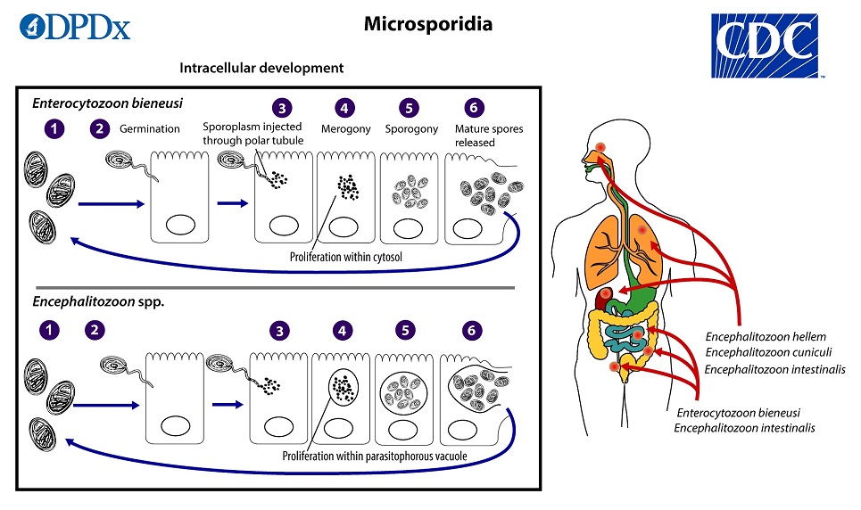
Microsporidia have a complex life cycle that involves several developmental stages, including spores, meronts, and sporonts. The spores are highly resistant to environmental stresses and can survive for long periods outside the host, facilitating their transmission. Once inside the host cell, the spore releases its infectious contents, including a coiled tubular structure called the polar filament, which penetrates the host cell membrane and injects the parasite's genetic material into the host cytoplasm. The parasite then undergoes rapid multiplication, eventually producing numerous progeny spores that are released into the environment upon host cell lysis.
Microsporidia have been identified as potential bioterrorism agents due to their high infectivity, environmental resistance, and ability to cause severe disease in immunocompromised hosts. However, there are currently no effective vaccines or specific antimicrobial therapies available for microsporidiosis, and treatment is mainly supportive, focusing on managing symptoms and improving immune function.
Enterocytozoon is a genus of microsporidian parasites that are known to infect a variety of animals, including humans. The most well-known species in this genus is Enterocytozoon bieneusi, which is a common cause of diarrhea and other gastrointestinal symptoms in immunocompromised individuals, such as those with HIV/AIDS.
Enterocytozoon species infect the host by invading intestinal epithelial cells, specifically enterocytes, hence the name "enterocytozoon." Once inside the host cell, they replicate and can cause damage to the cell, leading to symptoms such as diarrhea, abdominal cramps, nausea, and vomiting.
Transmission of Enterocytozoon species typically occurs through ingestion of contaminated food or water, although sexual contact and mother-to-child transmission have also been reported. Diagnosis is usually made by detecting the parasite's DNA in stool samples using molecular techniques such as PCR. Treatment options for Enterocytozoon infections are limited, but antimicrobial drugs such as albendazole and fumagillin have shown some efficacy in reducing symptoms and clearing the infection.
Microsporidia are a group of small, obligate intracellular parasites that belong to the kingdom Fungi. They are characterized by their spore stage, which contains a unique infection apparatus called the polar tube or coiled filament. These spores can infect a wide range of hosts, including humans, animals, and insects.
In humans, Microsporidia can cause chronic diarrhea and other gastrointestinal symptoms, particularly in individuals with weakened immune systems, such as those with HIV/AIDS. They can also infect various other tissues, including the eye, muscle, and kidney, leading to a variety of clinical manifestations.
Microsporidia were once considered to be protozoa but are now classified as fungi based on genetic and biochemical evidence. There are over 1,300 species of Microsporidia, with at least 14 species known to infect humans.
Encephalitozoonosis is a medical condition caused by infection with microsporidian parasites of the genus Encephalitozoon. The two most common species that cause disease in humans are Encephalitozoon cuniculi and Encephalitozoon intestinalis.
The infection typically occurs through the ingestion of spores present in contaminated food, water, or soil. Once inside the body, the spores can infect various organs, including the brain, lungs, eyes, and kidneys. The resulting disease can manifest as a wide range of symptoms, depending on the organ systems involved.
In the central nervous system, encephalitozoonosis can cause inflammation and damage to the brain and surrounding tissues, leading to symptoms such as headache, confusion, memory loss, and difficulty with coordination or balance. In the eyes, the infection can cause inflammation and scarring of the cornea, leading to vision loss. In the kidneys, encephalitozoonosis can cause interstitial nephritis, which can lead to kidney failure in severe cases.
Encephalitozoonosis is most commonly seen in immunocompromised individuals, such as those with HIV/AIDS or organ transplant recipients. However, it has also been reported in otherwise healthy individuals. Treatment typically involves the use of antimicrobial agents, such as albendazole or fumagillin, to eliminate the parasites from the body.
Encephalitozoon is a genus of intracellular parasites belonging to the phylum Microspora. The two species that are most relevant to human health are Encephalitozoon cuniculi and Encephalitozoon intestinalis (previously known as Septata intestinalis). These microscopic organisms are capable of infecting a wide range of hosts, including humans, and are often associated with opportunistic infections in immunocompromised individuals.
E. cuniculi is well-known for causing encephalitozoonosis, a disease that can lead to various symptoms depending on the infected organ. In humans, it primarily affects the central nervous system (CNS), leading to neurological issues such as seizures, cognitive impairment, and motor function loss. E. intestinalis, on the other hand, tends to infect the gastrointestinal tract, causing diarrhea and wasting syndrome.
Transmission of these parasites typically occurs through the ingestion of spores present in contaminated food, water, or soil. Once inside a host, the spores germinate and invade various cells, including intestinal epithelial cells, hepatocytes, and endothelial cells. The subsequent infection can lead to a range of clinical manifestations, from asymptomatic to severe, life-threatening disease.
Effective treatment for encephalitozoonosis involves the administration of antiparasitic drugs such as albendazole or nitazoxanide. In immunocompromised patients, improving immune function through appropriate therapy is also crucial to prevent recurrence and manage the infection effectively.
Parasitic intestinal diseases are disorders caused by microscopic parasites that invade the gastrointestinal tract, specifically the small intestine. These parasites include protozoa (single-celled organisms) and helminths (parasitic worms). The most common protozoan parasites that cause intestinal disease are Giardia lamblia, Cryptosporidium parvum, and Entamoeba histolytica. Common helminthic parasites include roundworms (Ascaris lumbricoides), tapeworms (Taenia saginata and Taenia solium), hookworms (Ancylostoma duodenale and Necator americanus), and pinworms (Enterobius vermicularis).
Parasitic intestinal diseases can cause a variety of symptoms, including diarrhea, abdominal pain, bloating, nausea, vomiting, fatigue, and weight loss. The severity and duration of the symptoms depend on the type of parasite, the number of organisms present, and the immune status of the host.
Transmission of these parasites can occur through various routes, including contaminated food and water, person-to-person contact, and contact with contaminated soil or feces. Preventive measures include practicing good hygiene, washing hands thoroughly after using the toilet and before handling food, cooking food thoroughly, and avoiding consumption of raw or undercooked meat, poultry, or seafood.
Treatment of parasitic intestinal diseases typically involves the use of antiparasitic medications that target the specific parasite causing the infection. In some cases, supportive care such as fluid replacement and symptom management may also be necessary.
'Encephalitozoon cuniculi' is a small, intracellular parasitic protozoan that belongs to the phylum Microspora. It is the causative agent of encephalitozoonosis, a disease that primarily affects rabbits but can also infect other animals including humans, particularly those with weakened immune systems.
In rabbits, E. cuniculi can cause a range of clinical signs, including neurological symptoms such as tremors, torticollis (wry neck), and hind limb paresis or paralysis. It can also lead to kidney disease and eye lesions. The parasite is typically transmitted through the ingestion of spores shed in the urine of infected animals.
In humans, E. cuniculi infection is usually asymptomatic but can cause serious complications in immunocompromised individuals, including encephalitis (inflammation of the brain), pneumonitis (inflammation of the lungs), and disseminated disease. It is typically transmitted through contact with infected animals or their feces, contaminated soil, or water.
Prevention measures include good hygiene practices, avoiding contact with infected animals, and proper handling and disposal of animal waste. In rabbits, vaccination and treatment with antiparasitic drugs may help reduce the risk of infection and transmission.
I'm sorry for any confusion, but "Apansporoblastina" is not a recognized medical term or concept in the field of medicine or biology. It is possible that this term may be used in another context outside of these fields, or it could be a term that is not widely accepted or used.
If you have any further questions or if there's something else I can help you with, please let me know!
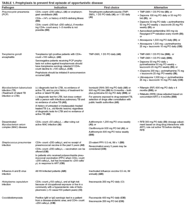
AIDS-related opportunistic infections (AROIs) are infections that occur more frequently or are more severe in people with weakened immune systems, such as those with advanced HIV infection or AIDS. These infections take advantage of a weakened immune system and can affect various organs and systems in the body.
Common examples of AROIs include:
1. Pneumocystis pneumonia (PCP), caused by the fungus Pneumocystis jirovecii
2. Mycobacterium avium complex (MAC) infection, caused by a type of bacteria called mycobacteria
3. Candidiasis, a fungal infection that can affect various parts of the body, including the mouth, esophagus, and genitals
4. Toxoplasmosis, caused by the parasite Toxoplasma gondii
5. Cryptococcosis, a fungal infection that affects the lungs and central nervous system
6. Cytomegalovirus (CMV) infection, caused by a type of herpes virus
7. Tuberculosis (TB), caused by the bacterium Mycobacterium tuberculosis
8. Cryptosporidiosis, a parasitic infection that affects the intestines
9. Progressive multifocal leukoencephalopathy (PML), a viral infection that affects the brain
Preventing and treating AROIs is an important part of managing HIV/AIDS, as they can cause significant illness and even death in people with weakened immune systems. Antiretroviral therapy (ART) is used to treat HIV infection and prevent the progression of HIV to AIDS, which can help reduce the risk of opportunistic infections. In addition, medications to prevent specific opportunistic infections may be prescribed for people with advanced HIV or AIDS.
Protozoan infections are diseases caused by microscopic, single-celled organisms known as protozoa. These parasites can enter the human body through contaminated food, water, or contact with an infected person or animal. Once inside the body, they can multiply and cause a range of symptoms depending on the type of protozoan and where it infects in the body. Some common protozoan infections include malaria, giardiasis, amoebiasis, and toxoplasmosis. Symptoms can vary widely but may include diarrhea, abdominal pain, fever, fatigue, and skin rashes. Treatment typically involves the use of antiprotozoal medications to kill the parasites and alleviate symptoms.
Intestinal diseases refer to a wide range of conditions that affect the function or structure of the small intestine, large intestine (colon), or both. These diseases can cause various symptoms such as abdominal pain, diarrhea, constipation, bloating, nausea, vomiting, and weight loss. They can be caused by infections, inflammation, genetic disorders, or other factors. Some examples of intestinal diseases include inflammatory bowel disease (IBD), irritable bowel syndrome (IBS), celiac disease, Crohn's disease, ulcerative colitis, and intestinal infections. The specific medical definition may vary depending on the context and the specific condition being referred to.
Central nervous system (CNS) parasitic infections refer to the invasion and infection of the brain and/or spinal cord by parasites. These infections can cause a range of symptoms depending on the type of parasite, the location of the infection within the CNS, and the severity of the infection.
Parasites that can infect the CNS include protozoa (such as Toxoplasma gondii, Naegleria fowleri, and Plasmodium falciparum), helminths (such as cysticercosis caused by Taenia solium tapeworm larvae), and arthropods (such as ticks that can transmit Lyme disease).
Symptoms of CNS parasitic infections can include headache, fever, seizures, confusion, weakness, numbness, loss of coordination, and changes in behavior or personality. Diagnosis typically involves a combination of clinical evaluation, imaging studies (such as MRI or CT scans), and laboratory tests (such as CSF analysis or PCR).
Treatment for CNS parasitic infections depends on the specific type of parasite involved and may include medications such as antiparasitics, antibiotics, or corticosteroids. In some cases, surgery may be necessary to remove parasites or cysts from the CNS. Prevention measures include avoiding contaminated food and water, practicing good hygiene, using insect repellent, and seeking prompt medical attention for any suspected infectious symptoms.
Feces are the solid or semisolid remains of food that could not be digested or absorbed in the small intestine, along with bacteria and other waste products. After being stored in the colon, feces are eliminated from the body through the rectum and anus during defecation. Feces can vary in color, consistency, and odor depending on a person's diet, health status, and other factors.
Diarrhea is a condition in which an individual experiences loose, watery stools frequently, often exceeding three times a day. It can be acute, lasting for several days, or chronic, persisting for weeks or even months. Diarrhea can result from various factors, including viral, bacterial, or parasitic infections, food intolerances, medications, and underlying medical conditions such as inflammatory bowel disease or irritable bowel syndrome. Dehydration is a potential complication of diarrhea, particularly in severe cases or in vulnerable populations like young children and the elderly.
Albendazole is an antiparasitic medication used to treat a variety of parasitic infections, including neurocysticercosis (a tapeworm infection that affects the brain), hydatid disease (a parasitic infection that can affect various organs), and other types of worm infestations such as pinworm, roundworm, hookworm, and whipworm infections.
Albendazole works by inhibiting the polymerization of beta-tubulin, a protein found in the microtubules of parasitic cells, which disrupts the parasite's ability to maintain its shape and move. This leads to the death of the parasite and elimination of the infection.
Albendazole is available in oral form and is typically taken two to three times a day with meals for several days or weeks, depending on the type and severity of the infection being treated. Common side effects of albendazole include nausea, vomiting, diarrhea, abdominal pain, and headache. Rare but serious side effects may include liver damage, bone marrow suppression, and neurological problems.
It is important to note that albendazole should only be used under the supervision of a healthcare provider, as it can have serious side effects and interactions with other medications. Additionally, it is not effective against all types of parasitic infections, so proper diagnosis is essential before starting treatment.
Cyclohexanes are organic compounds that consist of a six-carbon ring arranged in a cyclic structure, with each carbon atom joined to two other carbon atoms by single bonds. This gives the molecule a shape that resembles a hexagonal ring. The carbons in the ring can be saturated, meaning that they are bonded to hydrogen atoms, or they can contain double bonds between some of the carbon atoms.
Cyclohexanes are important intermediates in the production of many industrial and consumer products, including plastics, fibers, dyes, and pharmaceuticals. They are also used as solvents and starting materials for the synthesis of other organic compounds.
One of the most well-known properties of cyclohexane is its ability to exist in two different conformations: a "chair" conformation and a "boat" conformation. In the chair conformation, the carbon atoms are arranged in such a way that they form a puckered ring, with each carbon atom bonded to two other carbons and two hydrogens. This conformation is more stable than the boat conformation, in which the carbon atoms form a flattened, saddle-shaped ring.
Cyclohexanes are relatively nonpolar and have low water solubility, making them useful as solvents for nonpolar substances. They also have a relatively high boiling point compared to other hydrocarbons of similar molecular weight, due to the fact that they can form weak intermolecular forces called London dispersion forces.
Cyclohexane is a flammable liquid with a mild, sweet odor. It is classified as a hazardous substance and should be handled with care. Exposure to cyclohexane can cause irritation of the eyes, skin, and respiratory tract, and prolonged exposure can lead to more serious health effects, including neurological damage.
An immunocompromised host refers to an individual who has a weakened or impaired immune system, making them more susceptible to infections and decreased ability to fight off pathogens. This condition can be congenital (present at birth) or acquired (developed during one's lifetime).
Acquired immunocompromised states may result from various factors such as medical treatments (e.g., chemotherapy, radiation therapy, immunosuppressive drugs), infections (e.g., HIV/AIDS), chronic diseases (e.g., diabetes, malnutrition, liver disease), or aging.
Immunocompromised hosts are at a higher risk for developing severe and life-threatening infections due to their reduced immune response. Therefore, they require special consideration when it comes to prevention, diagnosis, and treatment of infectious diseases.
In the context of medicine, spores are typically discussed in relation to certain types of infections and diseases caused by microorganisms such as bacteria or fungi. Spores are a dormant, resistant form of these microorganisms that can survive under harsh environmental conditions, such as extreme temperatures, lack of nutrients, and exposure to chemicals.
Spores can be highly resistant to heat, radiation, and disinfectants, making them difficult to eliminate from contaminated surfaces or medical equipment. When the conditions are favorable, spores can germinate and grow into mature microorganisms that can cause infection.
Some examples of medically relevant spores include those produced by Clostridioides difficile (C. diff), a bacterium that can cause severe diarrhea and colitis in hospitalized patients, and Aspergillus fumigatus, a fungus that can cause invasive pulmonary aspergillosis in immunocompromised individuals.
It's worth noting that spores are not unique to medical contexts and have broader relevance in fields such as botany, mycology, and biology.
Antiprotozoal agents are a type of medication used to treat protozoal infections, which are infections caused by microscopic single-celled organisms called protozoa. These agents work by either killing the protozoa or inhibiting their growth and reproduction. They can be administered through various routes, including oral, topical, and intravenous, depending on the type of infection and the severity of the illness.
Examples of antiprotozoal agents include:
* Metronidazole, tinidazole, and nitazoxanide for treating infections caused by Giardia lamblia and Entamoeba histolytica.
* Atovaquone, clindamycin, and pyrimethamine-sulfadoxine for treating malaria caused by Plasmodium falciparum or other Plasmodium species.
* Pentamidine and suramin for treating African trypanosomiasis (sleeping sickness) caused by Trypanosoma brucei gambiense or T. b. rhodesiense.
* Nitroimidazoles, such as benznidazole and nifurtimox, for treating Chagas disease caused by Trypanosoma cruzi.
* Sodium stibogluconate and paromomycin for treating leishmaniasis caused by Leishmania species.
Antiprotozoal agents can have side effects, ranging from mild to severe, depending on the drug and the individual patient's response. It is essential to follow the prescribing physician's instructions carefully when taking these medications and report any adverse reactions promptly.
There doesn't seem to be a specific medical definition for "DNA, protozoan" as it is simply a reference to the DNA found in protozoa. Protozoa are single-celled eukaryotic organisms that can be found in various environments such as soil, water, and the digestive tracts of animals.
Protozoan DNA refers to the genetic material present in these organisms. It is composed of nucleic acids, including deoxyribonucleic acid (DNA) and ribonucleic acid (RNA), which contain the instructions for the development, growth, and reproduction of the protozoan.
The DNA in protozoa, like in other organisms, is made up of two strands of nucleotides that coil together to form a double helix. The four nucleotide bases that make up protozoan DNA are adenine (A), thymine (T), guanine (G), and cytosine (C). These bases pair with each other to form the rungs of the DNA ladder, with A always pairing with T and G always pairing with C.
The genetic information stored in protozoan DNA is encoded in the sequence of these nucleotide bases. This information is used to synthesize proteins, which are essential for the structure and function of the organism's cells. Protozoan DNA also contains other types of genetic material, such as regulatory sequences that control gene expression and repetitive elements with no known function.
Understanding the DNA of protozoa is important for studying their biology, evolution, and pathogenicity. It can help researchers develop new treatments for protozoan diseases and gain insights into the fundamental principles of genetics and cellular function.
The duodenum is the first part of the small intestine, immediately following the stomach. It is a C-shaped structure that is about 10-12 inches long and is responsible for continuing the digestion process that begins in the stomach. The duodenum receives partially digested food from the stomach through the pyloric valve and mixes it with digestive enzymes and bile produced by the pancreas and liver, respectively. These enzymes help break down proteins, fats, and carbohydrates into smaller molecules, allowing for efficient absorption in the remaining sections of the small intestine.
Cryptosporidiosis is a diarrheal disease caused by microscopic parasites called Cryptosporidium. The parasites are found in the feces of infected animals and humans. People can become infected with Cryptosporidium by ingesting contaminated water or food, or by coming into contact with infected persons or animals.
The infection can cause a wide range of symptoms, including watery diarrhea, stomach cramps, nausea, vomiting, fever, and dehydration. In people with weakened immune systems, such as those with HIV/AIDS, the infection can be severe and even life-threatening.
Cryptosporidiosis is typically treated with increased fluid intake to prevent dehydration, and in some cases, medication may be prescribed to help manage symptoms. Good hygiene practices, such as washing hands thoroughly after using the bathroom or changing diapers, can help prevent the spread of Cryptosporidium.
Microscopy is a technical field in medicine that involves the use of microscopes to observe structures and phenomena that are too small to be seen by the naked eye. It allows for the examination of samples such as tissues, cells, and microorganisms at high magnifications, enabling the detection and analysis of various medical conditions, including infections, diseases, and cellular abnormalities.
There are several types of microscopy used in medicine, including:
1. Light Microscopy: This is the most common type of microscopy, which uses visible light to illuminate and magnify samples. It can be used to examine a wide range of biological specimens, such as tissue sections, blood smears, and bacteria.
2. Electron Microscopy: This type of microscopy uses a beam of electrons instead of light to produce highly detailed images of samples. It is often used in research settings to study the ultrastructure of cells and tissues.
3. Fluorescence Microscopy: This technique involves labeling specific molecules within a sample with fluorescent dyes, allowing for their visualization under a microscope. It can be used to study protein interactions, gene expression, and cell signaling pathways.
4. Confocal Microscopy: This type of microscopy uses a laser beam to scan a sample point by point, producing high-resolution images with reduced background noise. It is often used in medical research to study the structure and function of cells and tissues.
5. Scanning Probe Microscopy: This technique involves scanning a sample with a physical probe, allowing for the measurement of topography, mechanical properties, and other characteristics at the nanoscale. It can be used in medical research to study the structure and function of individual molecules and cells.
The ribosomal spacer in DNA refers to the non-coding sequences of DNA that are located between the genes for ribosomal RNA (rRNA). These spacer regions are present in the DNA of organisms that have a nuclear genome, including humans and other animals, plants, and fungi.
In prokaryotic cells, such as bacteria, there are two ribosomal RNA genes, 16S and 23S, separated by a spacer region known as the intergenic spacer (IGS). In eukaryotic cells, there are multiple copies of ribosomal RNA genes arranged in clusters called nucleolar organizer regions (NORs), which are located on the short arms of several acrocentric chromosomes. Each cluster contains hundreds to thousands of copies of the 18S, 5.8S, and 28S rRNA genes, separated by non-transcribed spacer regions known as internal transcribed spacers (ITS) and external transcribed spacers (ETS).
The ribosomal spacer regions in DNA are often used as molecular markers for studying evolutionary relationships among organisms because they evolve more rapidly than the rRNA genes themselves. The sequences of these spacer regions can be compared among different species to infer their phylogenetic relationships and to estimate the time since they diverged from a common ancestor. Additionally, the length and composition of ribosomal spacers can vary between individuals within a species, making them useful for studying genetic diversity and population structure.
Sesquiterpenes are a class of terpenes that consist of three isoprene units, hence the name "sesqui-" meaning "one and a half" in Latin. They are composed of 15 carbon atoms and have a wide range of chemical structures and biological activities. Sesquiterpenes can be found in various plants, fungi, and insects, and they play important roles in the defense mechanisms of these organisms. Some sesquiterpenes are also used in traditional medicine and have been studied for their potential therapeutic benefits.
Polymerase Chain Reaction (PCR) is a laboratory technique used to amplify specific regions of DNA. It enables the production of thousands to millions of copies of a particular DNA sequence in a rapid and efficient manner, making it an essential tool in various fields such as molecular biology, medical diagnostics, forensic science, and research.
The PCR process involves repeated cycles of heating and cooling to separate the DNA strands, allow primers (short sequences of single-stranded DNA) to attach to the target regions, and extend these primers using an enzyme called Taq polymerase, resulting in the exponential amplification of the desired DNA segment.
In a medical context, PCR is often used for detecting and quantifying specific pathogens (viruses, bacteria, fungi, or parasites) in clinical samples, identifying genetic mutations or polymorphisms associated with diseases, monitoring disease progression, and evaluating treatment effectiveness.
'Staining and labeling' are techniques commonly used in pathology, histology, cytology, and molecular biology to highlight or identify specific components or structures within tissues, cells, or molecules. These methods enable researchers and medical professionals to visualize and analyze the distribution, localization, and interaction of biological entities, contributing to a better understanding of diseases, cellular processes, and potential therapeutic targets.
Medical definitions for 'staining' and 'labeling' are as follows:
1. Staining: A process that involves applying dyes or stains to tissues, cells, or molecules to enhance their contrast and reveal specific structures or components. Stains can be categorized into basic stains (which highlight acidic structures) and acidic stains (which highlight basic structures). Common staining techniques include Hematoxylin and Eosin (H&E), which differentiates cell nuclei from the surrounding cytoplasm and extracellular matrix; special stains, such as PAS (Periodic Acid-Schiff) for carbohydrates or Masson's trichrome for collagen fibers; and immunostains, which use antibodies to target specific proteins.
2. Labeling: A process that involves attaching a detectable marker or tag to a molecule of interest, allowing its identification, quantification, or tracking within a biological system. Labels can be direct, where the marker is directly conjugated to the targeting molecule, or indirect, where an intermediate linker molecule is used to attach the label to the target. Common labeling techniques include fluorescent labels (such as FITC, TRITC, or Alexa Fluor), enzymatic labels (such as horseradish peroxidase or alkaline phosphatase), and radioactive labels (such as ³²P or ¹⁴C). Labeling is often used in conjunction with staining techniques to enhance the specificity and sensitivity of detection.
Together, staining and labeling provide valuable tools for medical research, diagnostics, and therapeutic development, offering insights into cellular and molecular processes that underlie health and disease.
 Microsporidiosis
Gilt-head bream
Encephalitozoon intestinalis
Fumagillin
Antiparasitic
Thalidomide
Warthin-Starry stain
Parasitism
Encephalitozoon cuniculi
Fiji parrotfinch
Microsporidia
Pink-billed parrotfinch
Enterocytozoon bieneusi
Cyclospora cayetanensis
Antiprotozoal
Opportunistic infection
Enterospora nucleophila
List of diseases (M)
Albendazole
Microsporidiosis - Wikipedia
 CDC - DPDx - Microsporidiosis
 Donor Organs May Transmit Microsporidiosis
Microsporidiosis Differential Diagnoses
 Mammalian microsporidiosis - PubMed
 Microsporidiosis - causes, side effects and treatments at NaturalPedia.com
Disseminated Microsporidiosis in an Immunosuppressed Patient - Volume 18, Number 7-July 2012 - Emerging Infectious Diseases...
 Microsporidiosis - Infections - MSD Manual Consumer Version
 Anncaliia algerae Microsporidiosis Diagnosed by Metagenomic Next-Generation Sequencing, China
 Albendazole for treatment and prophylaxis of microsporidiosis due to Encephalitozoon intestinalis in patients with AIDS: a...
Microsporidiosis | Palmetto Profiles
 1997 USPHS/IDSA Guidelines for the Prevention of Opportunistic Infections in Persons Infected with Human Immunodeficiency Virus
 Chapter 302. Microsporidiosis | Rudolph's Pediatrics, 22e | AccessPediatrics | McGraw Hill Medical
Encephalitozoon cuniculi and Extraintestinal Microsporidiosis in Bird Owners - Volume 28, Number 3-March 2022 - Emerging...
Systemic microsporidiosis in inland bearded dragons (Pogona vitticeps)<...
McGowan I[au] - Search Results - PubMed
Albenza, (albendazole) dosing, indications, interactions, adverse effects, and more
 Publications at this Location : USDA ARS
What's Eating You: 12 Common Intestinal Parasites
 NIH Guide: INFECTIOUS CAUSES OF DIARRHEA OR WASTING SYNDROME IN PEOPLE WITH AIDS
 Microbiology Laboratory Tests: Malaria to Mosquitoes | Texas DSHS
 ZFIN Publication: Sanders et al., 2012
 Taj Pharmaceuticals Ltd Company
 Dr Thomas C Quinn Interview - Office of NIH History and Stetten Museum
Parasitic Fungus and Honeybee Decline
 RMU - Author index - COMBOL, A
CDC Science Clips
 Stat1 Mouse Gene Details | signal transducer and activator of transcription 1 | International Mouse Phenotyping Consortium
 Fighting shrimp diseases by making better use of data - IDH - the Sustainable Trade Initiative
Ocular Manifestations of HIV Infection: Overview, Adnexal Manifestations, Anterior Segment Manifestations
Encephalitozoon5
- The most common causes of microsporidiosis is Enterocytozoon bieneusi and Encephalitozoon intestinalis. (wikipedia.org)
- Cases of donor-derived microsporidiosis ( Encephalitozoon cuniculi ) following bone marrow, kidney, liver, and heart transplantation have been confirmed. (cdc.gov)
- Kidney biopsy in one recipient confirmed the diagnosis as microsporidiosis with Encephalitozoon cuniculi. (medscape.com)
- A patient with acquired immunodeficiency syndrome and untreated Encephalitozoon (Septata) intestinalis microsporidiosis leading to small bowel perforation. (nih.gov)
- We identified Encephalitozoon cuniculi genotype II parasites as a cause of extraintestinal microsporidiosis in 2 owners of birds also infected with E. cuniculi . (cdc.gov)
Infections2
- Given the diagnostic challenges and potential severity of illness with disseminated microsporidiosis, there have probably been additional undiagnosed infections and deaths," writes Dr. Camille Nelson Kotton from Massachusetts General Hospital in an editorial. (medscape.com)
- Microsporidiosis can also cause sinus and eye infections. (naturalpedia.com)
Enterocytozoon3
- Enterocytozoon bieneusi Microsporidiosis in Stem Cell Transplant Recipients Treated with Fumagillin. (medscape.com)
- Most cases of intestinal microsporidiosis in AIDS patients are caused by Enterocytozoon bieneusi . (naturalpedia.com)
- Cryptosporidium parvum and Isospora belli) and microsporidia (particularly Enterocytozoon bieneusi, causing intestinal microsporidiosis). (nih.gov)
Intestinalis1
- Disseminated microsporidiosis due to Septata intestinalis in nine patients infected with the human immunodeficiency virus: response to therapy with albendazole. (medscape.com)
Symptoms of microsporidiosis1
- Lung symptoms of microsporidiosis may include a cough and difficult/labored breathing. (naturalpedia.com)
Cryptosporidiosis2
- Treatment of HIV-1-associated microsporidiosis and cryptosporidiosis with combination antiretroviral therapy. (medscape.com)
- Approximately 30% of patients with cryptosporidiosis also have microsporidiosis. (med-chem.com)
Transmitted through solid organ transplantation1
- NEW YORK (Reuters Health) - Microsporidiosis can rarely be transmitted through solid organ transplantation, according to a report from the Microsporidia Transplant Transmission Investigation Team. (medscape.com)
Cuniculi1
- We describe the case of 2 bird owners in Poland who acquired E. cuniculi -caused microsporidiosis from their infected pet birds. (cdc.gov)
Organ transplant recipients1
- The side effects of microsporidiosis often manifest in patients with an immune system deficiency like HIV and organ-transplant recipients. (naturalpedia.com)
Albendazole1
- The medications for microsporidiosis often includes albendazole (Albenza) and fumagillin. (naturalpedia.com)
Fumagillin1
- Fumagillin is a selective and potent irreversible inhibitor of Methionine aminopeptidase 2 (MetAP2), used as an antibiotic to treat microsporidiosis. (targetmol.jp)
Microsporidia3
- Microsporidiosis refers to an intestinal infection caused by small parasites called Microsporidia . (naturalpedia.com)
- Microsporidiosis is infection caused by Microsporidia, which are parasitic fungi. (msdmanuals.com)
- Microsporidia were first discovered in 1857, but it was not until 1973 that a human case of microsporidiosis was confirmed from a case described in 1959. (antiinfectivemeds.com)
Kidney2
- The side effects of microsporidiosis may include bladder inflammation, bowel perforation, infection in the urinary tract, and kidney failure. (naturalpedia.com)
- Epidemiological and clinical study of microsporidiosis in French kidney transplant recipients from 2005 to 2019: TRANS-SPORE registry. (unistra.fr)
Intestinal infection1
- Microsporidiosis is an opportunistic intestinal infection that causes diarrhea and wasting in immunocompromised individuals (HIV, for example). (wikipedia.org)
Recipients1
- Microsporidiosis should be considered in the differential diagnosis especially when other, more commonly encountered illnesses have been ruled out or when recipients are poorly responsive to therapy. (medscape.com)
Solid organ1
- Microsporidiosis acquired through solid organ transplantation: a public health investigation. (medscape.com)
Opportunistic1
- Microsporidiosis: an emerging and opportunistic infection in humans and animals. (medscape.com)
AIDS1
- Microsporidiosis: not just in AIDS patients. (medscape.com)
Descriptor1
- Microsporidiosis" is a descriptor in the National Library of Medicine's controlled vocabulary thesaurus, MeSH (Medical Subject Headings) . (musc.edu)
Diagnosis2
- The diagnosis of microsporidiosis requires a high index of suspicion and can be difficult to make," Dr. Susan N. Hocevar from the Centers for Disease Control and Prevention in Atlanta, Georgia told Reuters Health. (medscape.com)
- Diagnosis of intestinal microsporidiosis by examination of stool and duodenal aspirate with Weber's modified trichrome and Uvitex 2B strains. (medscape.com)
Clinical2
- Anane S, Attouchi H. Microsporidiosis: epidemiology, clinical data and therapy. (medscape.com)
- Since June 2012, samples of wild caught white shrimp, Litopenaeus setiferus, from the Gulf of Mexico, Plaquemines and Jefferson Parishes (Louisiana, USA) with clinical signs of microsporidiosis have been delivered to the Louisiana Aquatic Diagnostic Laboratory for identification. (lsu.edu)
Diseases1
- Some of the types of shrimp diseases in Indonesia include White Spot Disease, White Feces Diseases, and Hepatopancreatic Microsporidiosis (HPM). (idhsustainabletrade.com)
Fungi1
- Although it is classified as a protozoal disease in ICD-10, their phylogenetic placement has been resolved to be within the Fungi, and some sources classify microsporidiosis as a mycosis, however, they are highly divergent and rapidly evolving. (wikipedia.org)
Kidneys2
- Microsporidiosis may affect organs like the brain , kidneys, and liver and cause complications like blindness and severe damage to infected organ systems (due to delayed treatment). (naturalpedia.com)
- Microsporidiosis may cause complications in the brain, kidneys, and liver. (naturalpedia.com)
Liver1
- Microsporidiosis after liver transplantation: A French nationwide retrospective study. (unistra.fr)
Patients3
- Microsporidiosis in pediatric renal transplant patients in Cape Town, South Africa: Two case reports. (medscape.com)
- Intestinal microsporidiosis: a hidden risk in rheumatic disease patients undergoing anti-tumor necrosis factor therapy combined with disease-modifying anti-rheumatic drugs? (medscape.com)
- Microsporidiosis develops in patients whose immune response has been weakened by diabetes or malignant disease treated with chemotherapy ( 2 , 4 ). (cdc.gov)
Progressive1
- Loignon M, Labrecque LG, Bard C, Robitaille Y, Toma E. Cerebral microsporidiosis manifesting as progressive multifocal leukoencephalopathy in an HIV-infected individual - a case report. (medscape.com)
Patient1
- We report a case of disseminated microsporidiosis in a patient with multiple myeloma who had received an allogeneic stem cell transplant requiring substantial immunosuppression. (cdc.gov)
Humans1
- Taken together, strong evidence exists for an increasing prevalence of microsporidiosis in animals and humans, and for sharing of pathogens across hosts and biomes. (oregonstate.edu)
Treatment1
- Treatment for microsporidiosis includes medications and supportive care. (naturalpedia.com)
Human2
- Therapy for human gastrointestinal microsporidiosis. (medscape.com)
- Further, human microsporidiosis appears to be adventitious and primarily associated with an increasing community of immune-deficient individuals. (oregonstate.edu)
Represents1
- This represents the first recognized cluster of transplant-transmitted microsporidiosis linked to a common organ donor, the authors say. (medscape.com)
Case1
- We describe a second case of disseminated microsporidiosis caused by a Tubulinosema sp. (cdc.gov)
People2
- This graph shows the total number of publications written about "Microsporidiosis" by people in this website by year, and whether "Microsporidiosis" was a major or minor topic of these publications. (musc.edu)
- Below are the most recent publications written about "Microsporidiosis" by people in Profiles. (musc.edu)
Major1
- The major risk factor for microsporidiosis is immunodeficiency. (naturalpedia.com)
Report1
- This report provides the first description of microsporidiosis in bearded dragons and is only the second report of this infection in a lizard. (johnshopkins.edu)