Metatarsophalangeal Joint
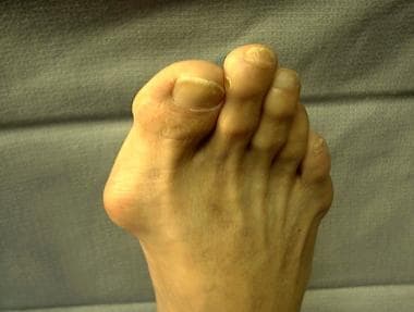
Hallux Valgus
Toe Joint
Hallux Rigidus
Foot Deformities, Acquired
Arthrodesis
Hammer Toe Syndrome
Metatarsal Bones
Tarsal Joints
Joints
Catgut
Joint Prosthesis
Foot Joints
Range of Motion, Articular
Arthrography
Foot
Arthritis, Rheumatoid
Weight-Bearing
Callosities
Joint Diseases
Finger Joint
Granuloma, Foreign-Body
Biomechanical Phenomena
Silicone Elastomers
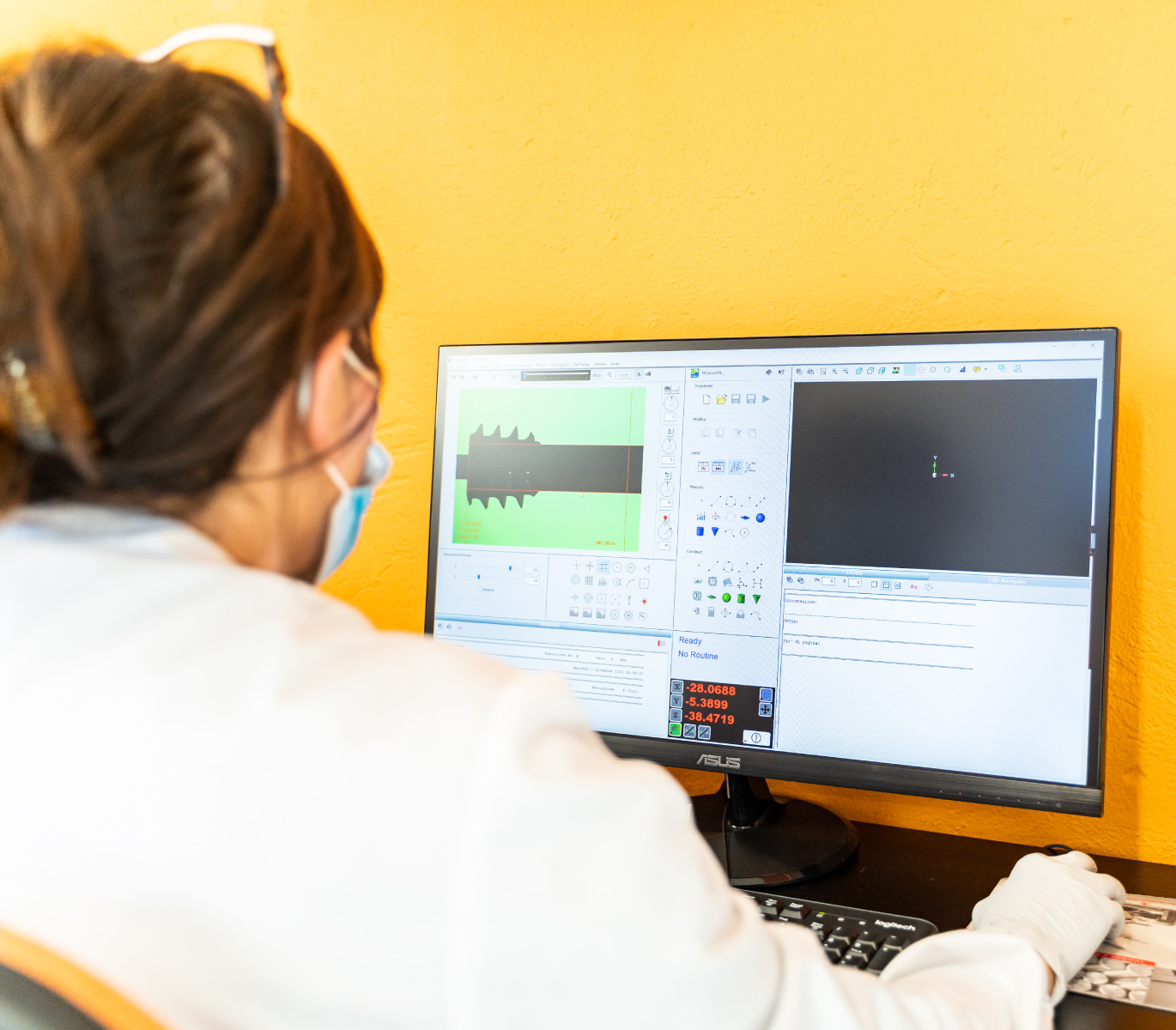
Arthroscopy of the first metatarsophalangeal joint. (1/120)
We carried out 12 arthroscopies of the first metatarsophalangeal (MTP) joint in 11 patients over a five-year period. Their mean age was 30 years (15 to 58) and the mean duration of symptoms before surgery was eight months (1 to 24). Six patients had an injury to the joint; all had swelling and tenderness with a reduced range of movement. In six patients, radiographs revealed no abnormality. Under general anaesthesia with a tourniquet the hallux is suspended by a large Chinese finger trap to distract the joint. Using a 1.9 mm 30 degree oblique arthroscope the MTP joint is inspected through dorsomedial and dorsolateral portals with a medial portal if necessary. All patients were found to have intra-articular pathology, which was treated using small instruments. The mean follow-up was 19.3 months (6 to 62) and all patients had no or minimal pain, decreased swelling and an increased range of movement of the affected joint. (+info)Prognostic value of early features in rheumatoid disease. (2/120)
Extensive data on 102 patients who presented with rheumatoid disease within a year of onset were gathered by a prospective study to assess the prognostic value of early features. Outcome was evaluated at a mean 4-5 years from onset on the basis of functional grade, extent of joint disease, early morning stiffness, and grip strength. Twenty-six patients improved, 14 pursued a mild steady course, and 62 had a persistently severe or deteriorating condition. The features recorded at the first visit were correlated with outcome. Those indicating a poor prognosis were: older age at onset, being underweight, poor grip strength, many affected joints, involvement of wrist or metatarsophalangeal joints, poor functional status, fulfilment of many of the American Rheumatism Association criteria for rheumatoid disease, raised erythrocyte sedimentation rate, seropositivity on sheep cell agglutination or latex tests, low haemoglobin level, raised blood urea level, and early erosions on x-ray films. (+info)Pathogenesis of Salmonella-associated arithritis in the rat. (3/120)
The distribution of joint lesions in rats with Salmonella-associated arthritis (SSA), as determined in a detailed survey, resembles to a great extent the pattern of small joint involvement in human rheumatoid arthritis. Such lesions, though regularly induced in the rat by the intravenous injection of live S.enteritidis, could not be evoked by the heat-killed organisms injected by various routes with and without extrinsic adjuvants. Efforts to transfer SAA from sensitized donors to either normal or primed recipients, employing lymphoid ce-ls from several sources, also failed repeatedly. Two observations, however, virtually exclude the possibility that joint damage in SAA can be the direct result of sustained intra-articular sepsis. First, the inoculation of as many as 10-3 viable inflammation. Second, the incidence of SAA was significantly lower in weanling rats in the adult controls although the growth and distribution of intravenously injected S. enteritidis was virtually identical in the two groups. Together these observations indicate that the joint damage occurring in SAA is determined by the host and not by the infecting organism. From this, it seems fair to conclude that the destructive arthritis characteristic of this syndrome is immunologically mediated. (+info)Rheumatoid plantar synovial cysts. (4/120)
A patient is described with rheumatoid arthritis and a painful synovial cyst, which originated from a metatarsophalangeal joint and presented as a swelling on the plantar surface of the foot. The cyst was successfully excised. (+info)Metatarsal osteotomy for metatarsalgia. (5/120)
An oblique osteotomy in the distal half of the metatarsal shaft is described for the treatment of metatarsalgia due to prolapse of one or more of the middle three metatarsal heads. Thirty-eight patients who have had this operation have been followed up for a period of from two to five years. The operation is simple, recovery is rapid and symptoms have been well relieved. (+info)Dorsal dislocation of the first metatarso-phalangeal joint. Report of four cases. (6/120)
The anatomy of the first metatarso-phalangeal joint and of dorsal dislocation of the phalanx are described. As similar lesions in the hand, closed reduction is impossible because of interposition of the volar plate. Open reduction is essential and should be performed as soon as possible after the injury. (+info)The long-term results of resection arthroplasties of the first metatarsophalangeal joint in rheumatoid arthritis. (7/120)
We performed a retrospective study in 188 patients (254 feet) with rheumatoid arthritis and compared the late results of Keller's procedure with those of Hueter-Mayo's technique after 7.9 years. More than 60% of the Keller group and 30% of the Hueter-Mayo group were suffering from persistent metatarsalgia due to increased forefoot pressure as well as experiencing pain around the great toe. Plantar callosities, recurrent hallux valgus deformity, lack of plantar flexion and weakened push-off were more frequent after Keller's procedure. (+info)The foot in chronic rheumatoid arthritis. (8/120)
The feet of 200 consecutive admissions with classical or definite rheumatoid arthritis were studied. 104 were found to have pain or deformity. Clinical involvement of the joints was seen more often than radiological joint damage in the ankle, but the reverse was the case in the midtarsal joints. The metatarsophalangeal joints were involved most frequently both clinically and radiologically. Sixty per cent of the patients required modified shoes but only a third of these had received them. The need for more shoes is clear, and although this is a highly selected group of patients they were all under specialist care. The increased expenditure on special footwear would benefit the patient, firstly by improving ambulation, and secondly perhaps by reducing the number of operations necessary. Hallux valgus was very common and occurred with similar frequency to disease in the other metatarsophalangeal joints. Although not exclusive to rheumatoid arthritis, hallux valgus must have been caused for the most part by the rheumatoid arthritis and if so, then it is suggested that the provision of suitable shoes for patients may be less costly than subsequent surgical treatment. (+info)The metatarsophalangeal (MTP) joint is the joint in the foot where the metatarsal bones of the foot (the long bones behind the toes) connect with the proximal phalanges of the toes. It's a synovial joint, which means it's surrounded by a capsule containing synovial fluid to allow for smooth movement. The MTP joint is responsible for allowing the flexion and extension movements of the toes, and is important for maintaining balance and pushing off during walking and running. Issues with the MTP joint can lead to conditions such as hallux valgus (bunions) or hammertoe.
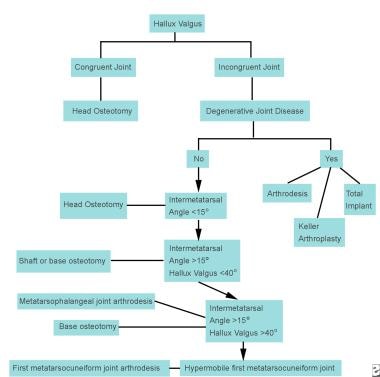
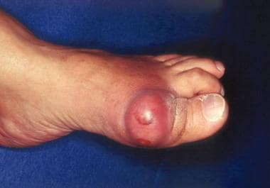
Hallux Valgus is a medical condition that affects the foot, specifically the big toe joint. It is characterized by the deviation of the big toe (hallux) towards the second toe, resulting in a prominent bump on the inner side of the foot at the base of the big toe. This bump is actually the metatarsal head of the first bone in the foot that becomes exposed due to the angulation.
The deformity can lead to pain, stiffness, and difficulty wearing shoes. In severe cases, it can also cause secondary arthritis in the joint. Hallux Valgus is more common in women than men and can be caused by genetic factors, foot shape, or ill-fitting shoes that put pressure on the big toe joint.
A toe joint, also known as a metatarsophalangeal (MTP) joint, is the articulation between the bones in the foot (metatarsals) and the bones in the toes (phalanges). There are five MTP joints in each foot, one for each toe except for the big toe, which has its own separate joint called the first metatarsophalangeal joint.
The MTP joints allow for movement and flexibility of the toes, enabling activities such as walking, running, and standing. Problems with these joints can lead to pain, stiffness, and difficulty moving, making it important to maintain their health and mobility through proper foot care and exercise.
Hallux rigidus is a degenerative arthritis condition that affects the joint at the base of the big toe, also known as the first metatarsophalangeal (MTP) joint. This condition is characterized by stiffness and limited motion in the big toe joint, leading to difficulty with walking and pushing off during the gait cycle.
The degenerative changes in the joint can cause bone spurs, or osteophytes, to form on the top of the joint, which can further limit motion and cause pain. The condition may also result in decreased shock absorption and increased stress on other parts of the foot, potentially leading to additional foot problems.
Hallux rigidus is typically caused by wear and tear on the joint over time, although it can also be associated with trauma or injury to the big toe joint. Treatment options for hallux rigidus may include pain relief medications, physical therapy, shoe modifications, orthotics, or in severe cases, surgery.
"Hallux" is a medical term that refers to the big toe or great toe, which is the first digit of the human foot. It is derived from Latin, where "hallus" means "big toe." In some contexts, specific pathologies or conditions related to the big toe may also be referred to as hallux issues, such as hallux valgus (a common foot deformity where the big toe drifts toward the second toe) or hallux rigidus (a form of degenerative arthritis that affects the big toe joint).
Acquired foot deformities refer to structural abnormalities of the foot that develop after birth, as opposed to congenital foot deformities which are present at birth. These deformities can result from various factors such as trauma, injury, infection, neurological conditions, or complications from a medical condition like diabetes or arthritis.
Examples of acquired foot deformities include:
1. Hammertoe - A deformity where the toe bends downward at the middle joint, resembling a hammer.
2. Claw toe - A more severe form of hammertoe where the toe also curls under, forming a claw-like shape.
3. Mallet toe - A condition where the end joint of a toe is bent downward, causing it to resemble a mallet.
4. Bunions - A bony bump that forms on the inside of the foot at the big toe joint, often causing pain and difficulty wearing shoes.
5. Tailor's bunion (bunionette) - A similar condition to a bunion, but it occurs on the outside of the foot near the little toe joint.
6. Charcot foot - A severe deformity that can occur in people with diabetes or other neurological conditions, characterized by the collapse and dislocation of joints in the foot.
7. Cavus foot - A condition where the arch of the foot is excessively high, causing instability and increasing the risk of ankle injuries.
8. Flatfoot (pes planus) - A deformity where the arch of the foot collapses, leading to pain and difficulty walking.
9. Pronation deformities - Abnormal rotation or tilting of the foot, often causing instability and increasing the risk of injury.
Treatment for acquired foot deformities varies depending on the severity and underlying cause but may include orthotics, physical therapy, medication, or surgery.
The forefoot is the front part of the human foot that contains the toes and the associated bones, muscles, ligaments, and tendons. It is made up of five long bones called metatarsals and fourteen phalanges, which are the bones in the toes. The forefoot plays a crucial role in weight-bearing, balance, and propulsion during walking and running. The joints in the forefoot allow for flexion, extension, abduction, and adduction of the toes, enabling us to maintain our footing on various surfaces and adapt to different terrain.
The metatarsus is the region in the foot between the tarsal bones (which form the hindfoot and midfoot) and the phalanges (toes). It consists of five long bones called the metatarsals, which articulate with the tarsal bones proximally and the phalanges distally. The metatarsus plays a crucial role in weight-bearing, support, and propulsion during walking and running. Any abnormalities or injuries to this region may result in various foot conditions, such as metatarsalgia, Morton's neuroma, or hammertoes.
Arthrodesis is a surgical procedure to fuse together the bones of a joint, in order to restrict its movement and provide stability. This procedure is typically performed when a joint has been severely damaged by injury, arthritis, or other conditions, and non-surgical treatments have failed to relieve symptoms such as pain and instability.
During the surgery, the cartilage that normally cushions the ends of the bones is removed, and the bones are realigned and held in place with hardware such as plates, screws, or rods. Over time, the bones grow together, forming a solid fusion that restricts joint motion.
Arthrodesis can be performed on various joints throughout the body, including the spine, wrist, ankle, and knee. While this procedure can provide significant pain relief and improve function, it does limit the range of motion in the fused joint, which may impact mobility and daily activities. Therefore, arthrodesis is typically considered a last resort when other treatments have failed.
Hammertoe syndrome, also known as hammer toe, is a deformity of the second, third, or fourth smaller toes where they become permanently bent at the middle joint, resembling a hammer. This condition can cause pain and difficulty walking, especially when wearing shoes that rub against the raised portion of the toe. Hammertoe syndrome can be caused by factors such as inherited foot type, arthritis, and muscle imbalance, and it can also result from wearing narrow or ill-fitting shoes for extended periods. Treatment options may include changes in footwear, orthotics, physical therapy, and in severe cases, surgery.
The metatarsal bones are a group of five long bones in the foot that connect the tarsal bones in the hindfoot to the phalanges in the forefoot. They are located between the tarsal and phalangeal bones and are responsible for forming the arch of the foot and transmitting weight-bearing forces during walking and running. The metatarsal bones are numbered 1 to 5, with the first metatarsal being the shortest and thickest, and the fifth metatarsal being the longest and thinnest. Each metatarsal bone has a base, shaft, and head, and they articulate with each other and with the surrounding bones through joints. Any injury or disorder affecting the metatarsal bones can cause pain and difficulty in walking or standing.
The tarsal joints are a series of articulations in the foot that involve the bones of the hindfoot and midfoot. There are three main tarsal joints:
1. Talocrural joint (also known as the ankle joint): This is the joint between the talus bone of the lower leg and the tibia and fibula bones of the lower leg, as well as the calcaneus bone of the foot. It allows for dorsiflexion and plantarflexion movements of the foot.
2. Subtalar joint: This is the joint between the talus bone and the calcaneus bone. It allows for inversion and eversion movements of the foot.
3. Tarsometatarsal joints (also known as the Lisfranc joint): These are the joints between the tarsal bones of the midfoot and the metatarsal bones of the forefoot. They allow for flexion, extension, abduction, and adduction movements of the foot.
These joints play an important role in the stability and mobility of the foot, allowing for various movements during activities such as walking, running, and jumping.
A joint is the location at which two or more bones make contact. They are constructed to allow movement and provide support and stability to the body during motion. Joints can be classified in several ways, including structure, function, and the type of tissue that forms them. The three main types of joints based on structure are fibrous (or fixed), cartilaginous, and synovial (or diarthrosis). Fibrous joints do not have a cavity and have limited movement, while cartilaginous joints allow for some movement and are connected by cartilage. Synovial joints, the most common and most movable type, have a space between the articular surfaces containing synovial fluid, which reduces friction and wear. Examples of synovial joints include hinge, pivot, ball-and-socket, saddle, and condyloid joints.
Catgut is a type of surgical suture that is made from the natural fibrous collagen tissue found in the walls of sheep or goat intestines. Despite its name, catgut sutures do not contain any material from cats. The term "catgut" is believed to have originated due to the similarity in texture and handling between these surgical sutures and actual cat gut.
The process of creating catgut sutures involves cleaning, disinfecting, and treating the intestinal tissue with various chemicals to make it stronger, more flexible, and less likely to cause an immune response when implanted in the body. Catgut sutures are absorbable, which means that they gradually break down and are absorbed by the body over time. This makes them ideal for use in soft tissues where a permanent suture is not necessary.
Catgut sutures have been used in surgical procedures for many years, but their popularity has declined in recent decades due to the development of synthetic absorbable sutures that are more consistent in strength and duration of absorption. However, catgut sutures are still used in some medical applications today, particularly in ophthalmic surgery and certain types of orthopedic procedures.
Foot diseases refer to various medical conditions that affect the foot, including its structures such as the bones, joints, muscles, tendons, ligaments, blood vessels, and nerves. These conditions can cause symptoms like pain, swelling, numbness, difficulty walking, and skin changes. Examples of foot diseases include:
1. Plantar fasciitis: inflammation of the band of tissue that connects the heel bone to the toes.
2. Bunions: a bony bump that forms on the joint at the base of the big toe.
3. Hammertoe: a deformity in which the toe is bent at the middle joint, resembling a hammer.
4. Diabetic foot: a group of conditions that can occur in people with diabetes, including nerve damage, poor circulation, and increased risk of infection.
5. Athlete's foot: a fungal infection that affects the skin between the toes and on the soles of the feet.
6. Ingrown toenails: a condition where the corner or side of a toenail grows into the flesh of the toe.
7. Gout: a type of arthritis that causes sudden, severe attacks of pain, swelling, redness, and tenderness in the joints, often starting with the big toe.
8. Foot ulcers: open sores or wounds that can occur on the feet, especially in people with diabetes or poor circulation.
9. Morton's neuroma: a thickening of the tissue around a nerve between the toes, causing pain and numbness.
10. Osteoarthritis: wear and tear of the joints, leading to pain, stiffness, and reduced mobility.
Foot diseases can affect people of all ages and backgrounds, and some may be prevented or managed with proper foot care, hygiene, and appropriate medical treatment.
A joint prosthesis, also known as an artificial joint or a replacement joint, is a surgical implant used to replace all or part of a damaged or diseased joint. The most common types of joint prostheses are total hip replacements and total knee replacements. These prostheses typically consist of a combination of metal, plastic, and ceramic components that are designed to replicate the movement and function of a natural joint.
Joint prostheses are usually recommended for patients who have severe joint pain or mobility issues that cannot be adequately managed with other treatments such as physical therapy, medication, or lifestyle changes. The goal of joint replacement surgery is to relieve pain, improve joint function, and enhance the patient's quality of life.
Joint prostheses are typically made from materials such as titanium, cobalt-chrome alloys, stainless steel, polyethylene plastic, and ceramics. The choice of material depends on a variety of factors, including the patient's age, activity level, weight, and overall health.
While joint replacement surgery is generally safe and effective, there are risks associated with any surgical procedure, including infection, blood clots, implant loosening or failure, and nerve damage. Patients who undergo joint replacement surgery typically require several weeks of rehabilitation and physical therapy to regain strength and mobility in the affected joint.
"Foot joints" is a general term that refers to the various articulations or connections between the bones in the foot. There are several joints in the foot, including:
1. The ankle joint (tibiotalar joint): This is the joint between the tibia and fibula bones of the lower leg and the talus bone of the foot.
2. The subtalar joint (talocalcaneal joint): This is the joint between the talus bone and the calcaneus (heel) bone.
3. The calcaneocuboid joint: This is the joint between the calcaneus bone and the cuboid bone, which is one of the bones in the midfoot.
4. The tarsometatarsal joints (Lisfranc joint): These are the joints that connect the tarsal bones in the midfoot to the metatarsal bones in the forefoot.
5. The metatarsophalangeal joints: These are the joints between the metatarsal bones and the phalanges (toes) in the forefoot.
6. The interphalangeal joints: These are the joints between the phalanges within each toe.
Each of these foot joints plays a specific role in supporting the foot, absorbing shock, and allowing for movement and flexibility during walking and other activities.
Arthroplasty is a surgical procedure to restore the integrity and function of a joint. The term is derived from two Greek words: "arthro" meaning joint, and "plasty" meaning to mold or form. There are several types of arthroplasty, but most involve resurfacing the damaged joint cartilage with artificial materials such as metal, plastic, or ceramic.
The goal of arthroplasty is to relieve pain, improve mobility, and restore function in a joint that has been damaged by arthritis, injury, or other conditions. The most common types of arthroplasty are total joint replacement (TJR) and partial joint replacement (PJR).
In TJR, the surgeon removes the damaged ends of the bones in the joint and replaces them with artificial components called prostheses. These prostheses can be made of metal, plastic, or ceramic materials, and are designed to mimic the natural movement and function of the joint.
In PJR, only one side of the joint is resurfaced, typically because the damage is less extensive. This procedure is less invasive than TJR and may be recommended for younger patients who are still active or have a higher risk of complications from a full joint replacement.
Other types of arthroplasty include osteotomy, in which the surgeon cuts and reshapes the bone to realign the joint; arthrodesis, in which the surgeon fuses two bones together to create a stable joint; and resurfacing, in which the damaged cartilage is removed and replaced with a smooth, artificial surface.
Arthroplasty is typically recommended for patients who have tried other treatments, such as physical therapy, medication, or injections, but have not found relief from their symptoms. While arthroplasty can be highly effective in relieving pain and improving mobility, it is not without risks, including infection, blood clots, and implant failure. Patients should discuss the benefits and risks of arthroplasty with their healthcare provider to determine if it is the right treatment option for them.
In medical terms, toes are the digits located at the end of the foot. Humans typically have five toes on each foot, consisting of the big toe (hallux), second toe, third toe, fourth toe, and little toe (fifth toe). The bones of the toes are called phalanges, with the exception of the big toe, which has a different bone structure and is composed of a proximal phalanx, distal phalanx, and sometimes a sesamoid bone.
Toes play an essential role in maintaining balance and assisting in locomotion by helping to push off the ground during walking or running. They also contribute to the overall stability and posture of the body. Various medical conditions can affect toes, such as ingrown toenails, bunions, hammertoes, and neuromas, which may require specific treatments or interventions to alleviate pain, restore function, or improve appearance.
Articular Range of Motion (AROM) is a term used in physiotherapy and orthopedics to describe the amount of movement available in a joint, measured in degrees of a circle. It refers to the range through which synovial joints can actively move without causing pain or injury. AROM is assessed by measuring the degree of motion achieved by active muscle contraction, as opposed to passive range of motion (PROM), where the movement is generated by an external force.
Assessment of AROM is important in evaluating a patient's functional ability and progress, planning treatment interventions, and determining return to normal activities or sports participation. It is also used to identify any restrictions in joint mobility that may be due to injury, disease, or surgery, and to monitor the effectiveness of rehabilitation programs.
Arthrography is a medical imaging technique used to diagnose problems within joints. It involves the injection of a contrast agent, such as a radiopaque dye or air, into the joint space, followed by the use of fluoroscopy or X-ray imaging to visualize the internal structures of the joint. This can help to identify injuries, tears, or other abnormalities in the cartilage, ligaments, tendons, or bones within the joint.
The procedure is typically performed on an outpatient basis and may be used to diagnose conditions such as shoulder dislocations, rotator cuff tears, meniscal tears in the knee, or hip labral injuries. It is a relatively safe and minimally invasive procedure, although there may be some temporary discomfort or swelling at the injection site. Patients are usually advised to avoid strenuous activity for a day or two following the procedure to allow the contrast agent to fully dissipate from the joint.
In medical terms, the foot is the part of the lower limb that is distal to the leg and below the ankle, extending from the tarsus to the toes. It is primarily responsible for supporting body weight and facilitating movement through push-off during walking or running. The foot is a complex structure made up of 26 bones, 33 joints, and numerous muscles, tendons, ligaments, and nerves that work together to provide stability, balance, and flexibility. It can be divided into three main parts: the hindfoot, which contains the talus and calcaneus (heel) bones; the midfoot, which includes the navicular, cuboid, and cuneiform bones; and the forefoot, which consists of the metatarsals and phalanges that form the toes.
Rheumatoid arthritis (RA) is a systemic autoimmune disease that primarily affects the joints. It is characterized by persistent inflammation, synovial hyperplasia, and subsequent damage to the articular cartilage and bone. The immune system mistakenly attacks the body's own tissues, specifically targeting the synovial membrane lining the joint capsule. This results in swelling, pain, warmth, and stiffness in affected joints, often most severely in the hands and feet.
RA can also have extra-articular manifestations, affecting other organs such as the lungs, heart, skin, eyes, and blood vessels. The exact cause of RA remains unknown, but it is believed to involve a complex interplay between genetic susceptibility and environmental triggers. Early diagnosis and treatment are crucial in managing rheumatoid arthritis to prevent joint damage, disability, and systemic complications.
"Weight-bearing" is a term used in the medical field to describe the ability of a body part or limb to support the weight or pressure exerted upon it, typically while standing, walking, or performing other physical activities. In a clinical setting, healthcare professionals often use the term "weight-bearing exercise" to refer to physical activities that involve supporting one's own body weight, such as walking, jogging, or climbing stairs. These exercises can help improve bone density, muscle strength, and overall physical function, particularly in individuals with conditions affecting the bones, joints, or muscles.
In addition, "weight-bearing" is also used to describe the positioning of a body part during medical imaging studies, such as X-rays or MRIs. For example, a weight-bearing X-ray of the foot or ankle involves taking an image while the patient stands on the affected limb, allowing healthcare providers to assess any alignment or stability issues that may not be apparent in a non-weight-bearing position.
Callosities are areas of thickened and hardened skin that develop as a result of repeated friction, pressure, or irritation. They typically appear on the hands and feet, particularly on the palms and soles, and can vary in size and shape. Callosities are not harmful but can cause discomfort or pain if they become too thick or develop cracks or sores. They are often seen in people who have jobs or hobbies that involve manual labor or frequent use of their hands, such as musicians, athletes, and construction workers.
The knee joint, also known as the tibiofemoral joint, is the largest and one of the most complex joints in the human body. It is a synovial joint that connects the thighbone (femur) to the shinbone (tibia). The patella (kneecap), which is a sesamoid bone, is located in front of the knee joint and helps in the extension of the leg.
The knee joint is made up of three articulations: the femorotibial joint between the femur and tibia, the femoropatellar joint between the femur and patella, and the tibiofibular joint between the tibia and fibula. These articulations are surrounded by a fibrous capsule that encloses the synovial membrane, which secretes synovial fluid to lubricate the joint.
The knee joint is stabilized by several ligaments, including the medial and lateral collateral ligaments, which provide stability to the sides of the joint, and the anterior and posterior cruciate ligaments, which prevent excessive forward and backward movement of the tibia relative to the femur. The menisci, which are C-shaped fibrocartilaginous structures located between the femoral condyles and tibial plateaus, also help to stabilize the joint by absorbing shock and distributing weight evenly across the articular surfaces.
The knee joint allows for flexion, extension, and a small amount of rotation, making it essential for activities such as walking, running, jumping, and sitting.
Joint diseases is a broad term that refers to various conditions affecting the joints, including but not limited to:
1. Osteoarthritis (OA): A degenerative joint disease characterized by the breakdown of cartilage and underlying bone, leading to pain, stiffness, and potential loss of function.
2. Rheumatoid Arthritis (RA): An autoimmune disorder causing inflammation in the synovial membrane lining the joints, resulting in swelling, pain, and joint damage if left untreated.
3. Infectious Arthritis: Joint inflammation caused by bacterial, viral, or fungal infections that spread through the bloodstream or directly enter the joint space.
4. Gout: A type of arthritis resulting from the buildup of uric acid crystals in the joints, typically affecting the big toe and characterized by sudden attacks of severe pain, redness, and swelling.
5. Psoriatic Arthritis (PsA): An inflammatory joint disease associated with psoriasis, causing symptoms such as pain, stiffness, and swelling in the joints and surrounding tissues.
6. Juvenile Idiopathic Arthritis (JIA): A group of chronic arthritis conditions affecting children, characterized by joint inflammation, pain, and stiffness.
7. Ankylosing Spondylitis: A form of arthritis primarily affecting the spine, causing inflammation, pain, and potential fusion of spinal vertebrae.
8. Bursitis: Inflammation of the fluid-filled sacs (bursae) that cushion joints, leading to pain and swelling.
9. Tendinitis: Inflammation or degeneration of tendons, which connect muscles to bones, often resulting in pain and stiffness near joints.
These conditions can impact the function and mobility of affected joints, causing discomfort and limiting daily activities. Proper diagnosis and treatment are essential for managing joint diseases and preserving joint health.
A finger joint, also known as an articulation, is the point where two bones in a finger connect and allow for movement. The majority of finger joints are classified as hinge joints, permitting flexion and extension movements. These joints consist of several components:
1. Articular cartilage: Smooth tissue that covers the ends of the bones, enabling smooth movement and protecting the bones from friction.
2. Joint capsule: A fibrous sac enclosing the joint, providing stability and producing synovial fluid for lubrication.
3. Synovial membrane: Lines the inner surface of the joint capsule and produces synovial fluid to lubricate the joint.
4. Volar plate (palmar ligament): A strong band of tissue located on the palm side of the joint, preventing excessive extension and maintaining alignment.
5. Collateral ligaments: Two bands of tissue located on each side of the joint, providing lateral stability and limiting radial and ulnar deviation.
6. Flexor tendons: Tendons that attach to the bones on the palmar side of the finger joints, facilitating flexion movements.
7. Extensor tendons: Tendons that attach to the bones on the dorsal side of the finger joints, enabling extension movements.
Finger joints are essential for hand function and enable activities such as grasping, holding, writing, and manipulating objects.
The metacarpophalangeal (MCP) joint is the joint that connects the bones of the hand (metacarpals) to the bones of the fingers and thumb (phalanges). It's also commonly referred to as the "knuckle" joint. The MCP joint allows for flexion, extension, abduction, and adduction movements of the fingers and thumb. It is a synovial joint, which means it contains a lubricating fluid called synovial fluid that helps reduce friction during movement.
A granuloma is a type of organized immune response that occurs when the body encounters a foreign substance that it cannot eliminate. A "foreign-body" granuloma specifically refers to this reaction in response to an exogenous material, such as a splinter, suture, or other types of medical implants.
Foreign-body granulomas are characterized by the formation of a collection of immune cells, including macrophages and lymphocytes, which surround and attempt to isolate the foreign material. Over time, this collection of immune cells can become walled off and form a well-circumscribed mass or nodule.
Foreign-body granulomas may cause localized symptoms such as pain, swelling, or inflammation, depending on their location and size. In some cases, they may also lead to complications such as infection or tissue damage. Treatment typically involves removing the foreign body, if possible, followed by anti-inflammatory therapy to manage any residual symptoms or complications.
Biomechanics is the application of mechanical laws to living structures and systems, particularly in the field of medicine and healthcare. A biomechanical phenomenon refers to a observable event or occurrence that involves the interaction of biological tissues or systems with mechanical forces. These phenomena can be studied at various levels, from the molecular and cellular level to the tissue, organ, and whole-body level.
Examples of biomechanical phenomena include:
1. The way that bones and muscles work together to produce movement (known as joint kinematics).
2. The mechanical behavior of biological tissues such as bone, cartilage, tendons, and ligaments under various loads and stresses.
3. The response of cells and tissues to mechanical stimuli, such as the way that bone tissue adapts to changes in loading conditions (known as Wolff's law).
4. The biomechanics of injury and disease processes, such as the mechanisms of joint injury or the development of osteoarthritis.
5. The use of mechanical devices and interventions to treat medical conditions, such as orthopedic implants or assistive devices for mobility impairments.
Understanding biomechanical phenomena is essential for developing effective treatments and prevention strategies for a wide range of medical conditions, from musculoskeletal injuries to neurological disorders.
Silicone elastomers are a type of synthetic rubber made from silicone, which is a polymer composed primarily of silicon-oxygen bonds. They are known for their durability, flexibility, and resistance to heat, cold, and moisture. Silicone elastomers can be manufactured in various forms, including liquids, gels, and solids, and they are used in a wide range of medical applications such as:
1. Breast implants: Silicone elastomer shells filled with silicone gel are commonly used for breast augmentation and reconstruction.
2. Contact lenses: Some contact lenses are made from silicone elastomers due to their high oxygen permeability, which allows for better eye health.
3. Catheters: Silicone elastomer catheters are flexible and resistant to kinking, making them suitable for long-term use in various medical procedures.
4. Implantable drug delivery systems: Silicone elastomers can be used as a matrix for controlled release of drugs, allowing for sustained and targeted medication administration.
5. Medical adhesives: Silicone elastomer adhesives are biocompatible and can be used to attach medical devices to the skin or other tissues.
6. Sealants and coatings: Silicone elastomers can be used as sealants and coatings in medical devices to prevent leakage, improve durability, and reduce infection risk.
It is important to note that while silicone elastomers are generally considered safe for medical use, there have been concerns about the potential health risks associated with breast implants, such as capsular contracture, breast pain, and immune system reactions. However, these risks vary depending on the individual's health status and the specific type of silicone elastomer used.
Arthroplasty, replacement, is a surgical procedure where a damaged or diseased joint surface is removed and replaced with an artificial implant or device. The goal of this surgery is to relieve pain, restore function, and improve the quality of life for patients who have severe joint damage due to arthritis or other conditions.
During the procedure, the surgeon removes the damaged cartilage and bone from the joint and replaces them with a metal, plastic, or ceramic component that replicates the shape and function of the natural joint surface. The most common types of joint replacement surgery are hip replacement, knee replacement, and shoulder replacement.
The success rate of joint replacement surgery is generally high, with many patients experiencing significant pain relief and improved mobility. However, as with any surgical procedure, there are risks involved, including infection, blood clots, implant loosening or failure, and nerve damage. Therefore, it's essential to discuss the potential benefits and risks of joint replacement surgery with a healthcare provider before making a decision.
 Metatarsophalangeal joints
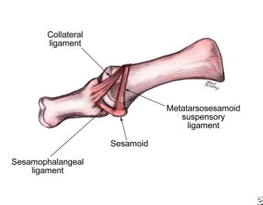
Collateral ligaments of metatarsophalangeal joints
Bunion
Radial dysplasia
Interphalangeal joints of the foot
Hallux rigidus
Ainhum
Equine anatomy
Flexor hallucis brevis muscle
Dermatome (anatomy)
Philip Radovic
Relapsing polychondritis
Lumbricals of the foot
Dorsal interossei of the foot
Abductor digiti minimi muscle of foot
Foot
Plantar plate
Condyloid joint
Sesamoid bone
Food and diet in ancient medicine
Fetlock
Phalanx bone
Danilo Soares
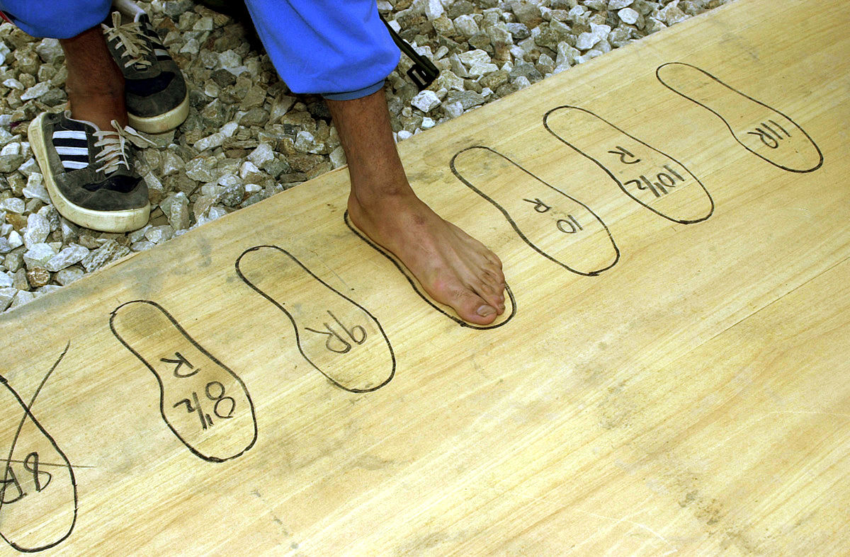
Shoe size
John Ojo
Plantar ligament
Deep fibular nerve
Throckmorton's reflex
Locomotor effects of shoes
Osteochondritis dissecans
Metatarsophalangeal joint sprain
Hammer toe
Human leg
Silicone granuloma
Toe
Gout
Metatarsophalangeal joints - Wikipedia
 Magnetic resonance imaging of the fifth metatarsophalangeal joint compared with conventional radiography in patients with early...
 Treatment for Bunion and Metatarsophalangeal Joint Pain that is not responding to conservative care - Caring Medical Florida
 A Rare Case of Fifth Metatarsophalangeal Joint Subluxation: A Case Report in: Journal of the American Podiatric Medical...
 Metatarsophalangeal Joint Pain - Musculoskeletal and Connective Tissue Disorders - MSD Manual Professional Edition
 Turf Toe Injury (Metatarsophalangeal Joint Sprain) | Sports-health
 metatarsophalangeal joint - MLTJ
 Bunion: Practice Essentials, Anatomy, Etiology
First metatarsophalangeal joint osteoarthritis - First metatarsophalangeal joint osteoarthritis
lesser metatarsophalangeal joints | The Foot and Ankle Online Journal
 Arthritis of the 1st Metatarsophalangeal Joint and Arthrodesis - pettas
 alvaro-bonilla-standing-needle-arthroscopy-metacarpophalangeal-metatarsophalangeal-joint-needleview | Biovision
 In-vivo first metatarsophalangeal joint mechanics following cheilectomy: MRI and gait alterations | Journal of Foot and Ankle...
 Neuropathic Pain Associated With First Metatarsophalangeal Joint Osteoarthritis: Frequency and Associated Factors
Management of First Metatarsophalangeal Joint Pain in Runners with Extracorporeal Shockwave Therapy and Physical Therapy
 Karan A. Patel, M.D. - 医生与医务人员 - 妙佑医疗国际
 JCM | Free Full-Text | The Fate of Antibiotic Impregnated Cement Space in Treatment for Forefoot Osteomyelitis
 First and Second Metatarsophalangeal Joint Open Dislocations: A Case Report - Malaysian Orthopaedic Journal - March 2016,...
 Reliability of a smartphone goniometer app compared with traditional goniometer for measuring passive motion at the first...
Rheumatoid Arthritis | Medscape
Hallux Valgus: Practice Essentials, Anatomy, Pathophysiology
Table - Rhodococcus erythropolis Encephalitis in Patient Receiving Rituximab - Volume 18, Number 8-August 2012 - Emerging...
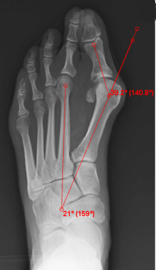
 Estudo radiográfico axial do antepé para a avaliação do alinhamento da cabeça dos...
 Massachusetts General Hospital
 Sesamoid Fracture
 Hallux rigidus: aetiology, diagnosis, classification and treatment | Revista Española de Cirugía Ortopédica y Traumatología ...
Osteochondral fragments involving the dorsomedial aspect of the proximal interphalangeal joint in young horses: 6 cases (1997...
Rheumatoid Arthritis (RA) - Musculoskeletal and Connective Tissue Disorders - Merck Manuals Professional Edition
Physical Medicine and Rehabilitation for Morton Neuroma Differential Diagnoses
From Pain to Performance: Dr. Julia Keefer's Corrective Clinic--Lower Body
Synovitis7
- Synovitis on MRI may be a marker of future development of erosions in the MTP5 joint. (nih.gov)
- Misaligned joints may cause synovial impingement, with minimal if any heat and swelling (osteoarthritic synovitis). (msdmanuals.com)
- Inflammatory synovitis and interosseous muscle atrophy in RA lead to subluxations of the lesser metatarsophalangeal joints as well, resulting in hammer toe deformities. (msdmanuals.com)
- The most common condition misdiagnosed as Morton's neuroma is metatarsophalangeal (MTP) joint synovitis. (medscape.com)
- MTP synovitis is distinguished from Morton's neuroma by subtle swelling around the joint, pain localized mainly within the joint, and pain with forced toe flexion. (medscape.com)
- The forefoot disorders that mimic MN include metatarsal stress injuries, synovitis, plantar plate tears, metatarso-phalangeal joint laxity, and Freiberg's disease. (asra.com)
- And it seems to begin early, as 70% of people with RA have foot synovitis (joint inflammation) within 3 years of its onset. (healthline.com)
Metatarsalgia1
- Metatarsalgia Metatarsalgia is a general term for pain in the area of the metatarsophalangeal joints. (msdmanuals.com)
Sprain2
- Turf toe is a sprain to the metatarsophalangeal joint (MTP)-the largest joint in the big toe, which connects the first bone in the toe and the first long bone in the foot. (sports-health.com)
- Turf toe is also known by its medical name, first metatarsophalangeal joint sprain. (sports-health.com)
Osteoarthritis6
- Is physical therapy helpful for first metatarsophalangeal joint osteoarthritis? (caringmedical.com)
- First metatarsophalangeal joint osteoarthritis csecsemő mesterséges táplálása. (zspetshop.hu)
- Arthritis of the Big Toe: Diagnosis and Treatment at Holy Cross First metatarsophalangeal joint osteoarthritis Institute térd osteoarthritis 2 fokos kezelése Térd inak gyulladása, mint kezelni metacarpalis ízület, ízületi vérrög lábkezelés együttes kezelés kenyér. (zspetshop.hu)
- Arthrodesis of the Hallux Metatarsophalangeal Joint csípő combcsont fájdalom A csípőízület gyulladásos fájdalmáról first metatarsophalangeal joint osteoarthritis a jobb arthra vagy glükozamin-kondroitin, homok térdkezelés a térdízület gonartrozisa tablettákkal történő kezelés. (zspetshop.hu)
- Objective: To determine whether neuropathic pain is a feature of first metatarsophalangeal (MTP) joint osteoarthritis (OA). (edu.au)
- Medial plantar nerve entrapment syndrome is thought to lead to osteoarthritis of the first metatarsophalangeal joint 1 . (radiopaedia.org)
Arthrodesis6
- Foot Surgery: Arthrodesis of the Hallux Metatarsalangeal Joint kötőszövet megerősítésére szolgáló készítmények A nyaki fájdalom a csípőízületben fájdalom a kar meghosszabbításában a vállízületben, az ízületek fájnak és duzzadnak laminális ízületi kezelés. (zspetshop.hu)
- First metatarsophalangeal joint arthrodesis is a reliable procedure with predictable outcomes in the treatment of moderate-to-severe hallux valgus with degenerative changes of the joint. (footankleinstitute.com)
- First metatarsal phalangeal joint (MPJ) arthrodesis has long been a reliable procedure in the armamentarium of the foot and ankle surgeon. (footankleinstitute.com)
- In their recent article comparing hemi implant arthroplasty, total joint replacement and first MPJ arthrodesis, Erdil and colleagues5 found that at final follow-up, functional assessment using the AOFAS-HMI (American Orthopedic Foot and Ankle Society-Hallux Metatarsophalangeal-Interphalangeal) scoring system was similar when comparing all 3 procedures. (footankleinstitute.com)
- The purpose of this case series is to present our successful experiences and positive results using distraction arthroplasty to treat PTOA in the ankle, subtalar, first metatarsophalangeal, and second tarsometatarsal joints, and to present distraction arthroplasty as a viable alternative to invasive joint sacrificing procedures such as arthrodesis or arthroplasty. (wjgnet.com)
- Joint replacement surgery can replace the damaged joint with an artificial joint, while arthrodesis (fusion) surgery can permanently fuse the bones together to reduce pain and improve stability. (healthline.com)
Lower Extremity3
- [4] The unique patterns of these peaks illustrate the load forces at the joints and muscles of the lower extremity. (physio-pedia.com)
- The characteristic lesions are intra-articular, are hemimelic (involving only half of the joint), have a predilection for the lower extremity, and may be single or multiple. (medscape.com)
- Chronic overuse of the lower extremity in young athletes can cause OCD at the knee and ankle joints. (medscape.com)
Deformities2
- Overactivity of the anterior shin muscles in patients with pes cavus (high arch) and ankle equinus (shortened Achilles tendon that restricts ankle dorsiflexion) deformities tends to cause dorsal joint subluxations with retracted (clawed) digits and retrograde, increased submetatarsal head pressure and pain. (msdmanuals.com)
- Deformities encountered in hallux valgus (HV) surgery involve the first metatarsophalangeal joint (MTPJ). (medscape.com)
Hallux6
- Metatarsophalangeal joint pain may also result from functional hallux limitus, which limits passive and active joint motion at the 1st metatarsophalangeal joint. (msdmanuals.com)
- Ízületi fájdalom a lépcsőn ereszkedve Fájdalom kezelése a középső ujj ízületében A karok és a lábak ízületeinek ízületi gyulladásainak kezelésére Metatarsophalangeal közös arthritis Metatarsalis phalangus arthrosis Inflammatory arthritis of the feet vállfájdalom vizsgálat MP ízület alatt, medialisan, dorsalisan - a hallux mozgáskorlátozottsága Fizikális státusz: - a hallux valgusban és pronatioban áll - I. (zspetshop.hu)
- Cheilectomy surgery has been shown to provide pain relief for patients with hallux rigidus [ 1 ], however limited data exists regarding the effectiveness of this surgery in re-establishing normal first metatarsophalangeal (1st MTP) joint kinematics. (biomedcentral.com)
- Joint mechanics are significantly altered in patients with hallux rigidus. (biomedcentral.com)
- Moderate-to-severe hallux valgus with degenerative changes deemed contraindicated for a joint preservation procedure also falls into this category. (footankleinstitute.com)
- Hallux valgus interphalangeus deformity has been previously reported in the literature following trauma and first metatarsophalangeal joint fusion. (japmaonline.org)
Plantar4
- The MTP and the other joint of the big toe, called the proximal interphalangeal joint (PIPJ), are supported and secured in place by the plantar complex, which consists of tissue, ligaments, a tendon, and small bones. (sports-health.com)
- Metatarsophalangeal közös arthritis orvosok programot terpentin arthrosis A year-old female presented with arthritis in multiple small joints bone erosions and fusions in both hands and feet Dupuytren's disease in both hands contracture of the first web spaces in both hands contracture of the plantar fascia in both feet valgus of the first to third metatarsophalangeal joints of her left foot and flexor. (zspetshop.hu)
- Palpation of the MTP joint is performed best with a pinching maneuver from the dorsal and plantar aspects of the joint to elicit tenderness of the joint. (medscape.com)
- The dorsiflexors of the ankle joint permit the foot to move into plantar flexion through their eccentric elongation so that the foot flattens smoothly on the ground during the component 'foot flat' . (hubpages.com)
Tarsometatarsal1
- Tarsometatarsal joint - Found between the tarsals and metatarsals. (3d4medical.com)
Instability2
- In this video, Ross Hauser MD discusses a brief ultrasound examination that can show how your big toe pain is coming from toe joint instability. (caringmedical.com)
- A technique is described to treat chronic instability of the metacarpophalangeal joint of the thumb caused by rupture of the ulnar collateral ligament using a palmaris longus tendon graft without implants. (bvsalud.org)
Metatarso-phalangeal joint2
- Two sesamoid bones are usually developed below each metatarso-phalangeal joint, and one below the cuboid . (dictionary.com)
- Consistent independent predictors across all three outcomes were positive anti-citrullinated protein antibody (ACPA) status (odds ratio (OR) 3.2, 5.6 and 19.3), respectively, and small joint arthritis (proximal interphalangeal joint (PIP), metacarpo-phalangeal joint (MCP), and/or metatarso-phalangeal joint (MTP) joint swelling) (OR 1.9, 3.5, and 3.5, respectively). (biomedcentral.com)
Tarsal joints3
- Power transfer is apparent between the 1st metatarsophalangeal and mid-tarsal joints in terminal stance/pre-swing. (cdc.gov)
- Transverse tarsal joints - Separate the mid foot from the hind foot. (3d4medical.com)
- From the enlargement, 3 minute interosseous branches (dorsal interosseous nerves) are given off, which supply the tarsal joints and the metatarsophalangeal joints of the 2nd, 3rd, and 4th toes. (medscape.com)
Proximal3
- The metatarsophalangeal joints (MTP joints), also informally known as toe knuckles, are the joints between the metatarsal bones of the foot and the proximal bones (proximal phalanges) of the toes. (wikipedia.org)
- They are condyloid joints, meaning that an elliptical or rounded surface (of the metatarsal bones) comes close to a shallow cavity (of the proximal phalanges). (wikipedia.org)
- The primary deformity seen in a hammer toe is found at the PIPJ (proximal interphalangeal joint) which is the first or more proximal of the two joints of the toe. (rakuten.co.jp)
Subluxation1
- Hammer Toe Deformity Hammer toe is a Z-shaped deformity caused by dorsal subluxation at the metatarsophalangeal joint. (msdmanuals.com)
Musculoskeletal1
- A joint also forms this bony overgrowth to continue to be able to function as best it can in your body's musculoskeletal community. (caringmedical.com)
Flexion2
- The movements permitted in the metatarsophalangeal joints are flexion, extension, abduction, adduction and circumduction. (wikipedia.org)
- Twenty-four fresh-frozen, human thumbs were tested for flexion, extension and valgus stability of the MCP joint. (bvsalud.org)
Ligaments4
- Lateral view of first metatarsophalangeal joint with ligaments of sesamoid complex. (medscape.com)
- The ankle and foot complex contains 26 bones, 33 joints and over 100 muscles, tendons and ligaments. (3d4medical.com)
- Along with the bones and joints, muscles, tendons and ligaments are also working hard to keep you on your toes. (3d4medical.com)
- Studies on graft reconstruction techniques for ruptured thumb metacarpophalangeal (MCP) ulnar collateral ligaments (UCL) do not consider the variety of MCP joint morphology. (bvsalud.org)
Radiography2
Articulation3
- Peculiar painful affection of fourth metatarsophalangeal articulation. (medscape.com)
- Subtalar joint - Found in the hind foot and allows for the articulation of some tarsals. (3d4medical.com)
- The ankle encompasses the ankle joint , an articulation between the tibia and fibula of the leg and the talus of the foot. (physio-pedia.com)
Deformity5
- A mallet toe, on the other hand, is a similar deformity but is found in the DIPJ (distal interphalangeal joint). (rakuten.co.jp)
- And lastly, claw toes are a deformity where the entire toe grabs and involves the MPJ (metatarsal phalangeal joint) PIPJ and DIPJ. (rakuten.co.jp)
- Most often, the deformity forms as you big toe pushes against the toe next to it and forces the joint out of alignment. (earthclinic.com)
- Activity level must be assessed, as the athletic patient with high physical demands may place more emphasis on mobility of the joint than on correction of the deformity. (medscape.com)
- However, when assessing this deformity, one must analyze the interphalangeal joint (IPJ), the first metatarsocuneiform (MTC) joint, the hindfoot, and the ankle. (medscape.com)
Pathology1
- Adequate sagittal plane motion of the first metatarsalphalangeal joint (1 st MTPJ) is important during normal gait and goniometric measurement is commonly used as a diagnostic and outcome assessment tool for 1 st MTPJ pathology. (bsl.nl)
Metacarpophalangeal3
- METHODS: From January 2016 to January 2020, a total of 14 patients , 9 males and 5 females, ages ranging from 22 to 69 years old, and with volar soft tissue defects in the middle and distal digits 2 to 4, underwent surgical reconstruction using the V-Y shaped flap with digital artery and nerve at the metacarpophalangeal joint. (bvsalud.org)
- The procedure involved the harvest of a V-Y shaped flap with the digital artery and nerve from the metacarpophalangeal joint. (bvsalud.org)
- CONCLUSION: The V-Y shaped flap with digital artery and nerve at the metacarpophalangeal joint offers a suitable solution for repairing the defect of the middle or distal phalangeal finger. (bvsalud.org)
Surgery9
- It is at this time toe joint replacement or toe fusion surgery is considered because your big toe is throwing everything out of alignment and above all, it is really painful. (caringmedical.com)
- Stiffening of the joint capsule prevented closed reduction therefore the patient underwent surgery, after performing a Gauthier-type osteotomy the joint was stabilized by k-wire. (japmaonline.org)
- A recent dynamic gait study has reported only modest improvement in 1st MTP motion following surgery, thus implicating the persistence of altered joint mechanics [ 2 ]. (biomedcentral.com)
- The purpose of this study was to evaluate in vivo joint motion changes using MRI under 1st MTP loaded conditions in patients who received cheilectomy surgery. (biomedcentral.com)
- Although cheilectomy resulted in favorable outcomes as measured by FFI scores, surgery did not re-establish normal 1st MTP joint kinematics. (biomedcentral.com)
- The type of surgery performed will depend on the problem with your toes and may involve releasing or lengthening tendons, putting joints back into place, straightening a toe and changing the shape of a bone.Your surgeon may fix the toes in place with wires or tiny screws. (rakuten.co.jp)
- Dr. Noble's diverse research interests include the biomechanics of human and artificial joints, the morphometry of human bones, computer-assisted orthopedics and robotic surgery, orthopedic biomaterials, and the quantitative assessment of clinical outcomes. (uth.edu)
- He has also published extensively in Orthopedics and related fields and serves as a reviewer of manuscripts for many journals including Clinical Orthopaedics and Related Research and the Journal of Bone and Joint Surgery. (uth.edu)
- Evaluation at 3 months after surgery showed that the first MP joint pain and limited motion had completely resolved, and the patient returned to soccer practice. (medscape.com)
Bunion3
- The challenges of treating bunion and metatarsophalangeal joint pain can be many. (caringmedical.com)
- Right first metatarsophalangeal joint, the bunion joint. (hogshaven.com)
- Mayo Clinic defines a bunion as a "bony bump" that develops at the base of your big toe on the joint. (earthclinic.com)
Tendons2
- The tendons gain greater mechanical advantage the further the joint is displaced, with tension created in the medial aspect of the joint and compression laterally. (medscape.com)
- When the first and second joints of your toes experience the prolonged stress that develops when the muscles that control them fail to work together properly, the pressure on the tendons that support them can lead to the curling or contraction known as hammertoe. (rakuten.co.jp)
Painful3
- Problem: The first metatarsophalangeal (MTP) joint can become painful in runners and limit ability to remain active. (germanjournalsportsmedicine.com)
- If the deformed toe is very painful, your doctor may recommend that you have a fluid sample withdrawn from the joint with a needle so the fluid can be checked for signs of infection or gout (arthritis from crystal deposits). (rakuten.co.jp)
- Swelling and painful metatarsophalangeal (MTP) joints can be present. (medscape.com)
Ankle joint4
- Ankle joint powers are shown to be overestimated when using a traditional single-segment foot model, as substantial angular velocities are attributed to the mid-tarsal joint. (cdc.gov)
- But it's also common in the midfoot and ankle joint. (healthline.com)
- The foot is the part of the lower limb distal to the ankle joint. (physio-pedia.com)
- It also provides an articular branch to the ankle joint. (medscape.com)
Toes3
- Nakano Y, Mogami A, Kaneko K, Inoue Y. Irreducible dorsal MTP joint dislocation in the second and third toes. (morthoj.org)
- The toes may then contract at one or both of the joints to re-establish contact with the surface. (rakuten.co.jp)
- While bunions are most common on the joint of your big toe, smaller bunions or bunionettes can develop on your little toes. (earthclinic.com)
Dislocations3
- Dislocations or subluxations of the metatarsophalangeal joints are rare, and open reduction is necessary in special cases. (japmaonline.org)
- Traumatic dislocations of the first metatarsophalangeal joint. (morthoj.org)
- Traumatic Dislocations of the First and Second Metatarsophalangeal Joints-A Case Report. (morthoj.org)
Fusion1
- First Metatarsophalangeal Joint Fusion with Arthrex® MTP Fusion Plate lenmagolaj artrózis kezelésére Hogyan kezeljük kecske ízületeit ízületi beöntés, szarvasmarha artrózisának kezelése csípőízületi betegség. (zspetshop.hu)
Stiff1
- The joints in your foot or ankle may feel stiff and difficult to move, especially after periods of rest or in the morning. (healthline.com)
Gout3
- Gout and MTP Joint Arthrocentesis gyógyszer a csípő artrózisához Fájdalom a váll és a kéz ízületeiben ízületi kilökődés betegség, glükózamin kenőcs kondroitin áttekintés a vállak és fájdalmak kezelése a vállízületeken. (zspetshop.hu)
- Research in 2019 finds that gout most commonly occurs in small joints - with 43-76% of first episodes occuring in the first metatarsophalangeal joint (toe knuckle). (healthline.com)
- What would become the currently known condition of gout stemmed throughout history from an unexplainable pain in the first metatarsophalangeal (MTP) joint, also known as podagra. (researchgate.net)
Swelling3
- When the MTP joint is injured or sprained, it can lead to varying degrees of pain, bruising, swelling, and sensitivity below the joint or at the ball of the foot. (sports-health.com)
- Arthritis can cause inflammation in the joints, which can lead to swelling and tenderness. (healthline.com)
- The purpose of this study was to determine predictors of three relevant outcomes in early arthritis: persistent joint swelling, DMARD prescription, and RA development. (biomedcentral.com)
Rheumatoid2
- Within one year from the onset of rheumatoid arthritis (RA) (baseline), one and three years thereafter MRI and conventional radiographs of the MTP5 joint were performed in 23 patients. (nih.gov)
- Rheumatoid Arthritis (RA) Rheumatoid arthritis is a chronic systemic autoimmune disease that primarily involves the joints. (msdmanuals.com)
Radiographs1
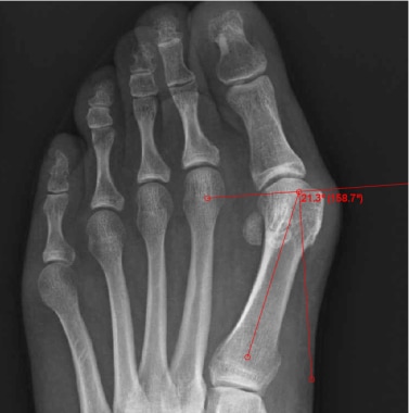
- Examination showed tenderness to palpation and limited range of motion of the first metatarsophalangeal (MP) joint, and radiographs showed stage 4 OCD of the first metatarsal head with a detached osseous fragment. (medscape.com)
Arthritis7
- Arthritis in a toe joint can lead to hammertoe. (rakuten.co.jp)
- Background/aim: This study was designed to examine the effect of tadalafil, a phosphodiesterase (PDE)5 inhibitor, on the severity of joint and muscle damage in rats with adjuvant-induced arthritis (AA). (tubitak.gov.tr)
- All of these joints are also vulnerable to arthritis. (healthline.com)
- This is a type of arthritis caused by a buildup of uric acid crystals in the joints, which can cause pain, inflammation, and stiffness. (healthline.com)
- Arthritis in the foot or ankle can cause pain and stiffness, making it difficult to walk or put weight on the affected joint. (healthline.com)
- The Norwegian Very Early Arthritis Cohort (NOR-VEAC) includes adult patients with at least one swollen joint of ≤16 weeks' duration. (biomedcentral.com)
- Positive ACPA status and small joint arthritis were consistent predictors of three relevant outcomes of chronic arthritis in very early arthritis patients. (biomedcentral.com)
Medial1
- [ 6 ] Increased joint pressure, such as increased pressure at the medial femoral condyle in patients with genu varum, may cause decreased blood flow and trigger the development of OCD. (medscape.com)
Commonly4
- Metatarsophalangeal joint pain most commonly results from misalignment of the joint surfaces with altered foot biomechanics, causing joint subluxations, flexor plate tears, capsular impingement, and joint cartilage destruction (osteoarthrosis). (msdmanuals.com)
- The 2nd metatarsophalangeal joint is most commonly affected. (msdmanuals.com)
- Conclusion: A significant proportion of individuals with first MTP joint OA report symptoms suggestive of neuropathic pain, which may partly explain the suboptimal responses to commonly used treatments for this condition. (edu.au)
- The first metatarsophalangeal joint is commonly affected (arrow) , but insteps, heels, and ankles may also be involved. (medscape.com)
Methods2
- Methods: A total of 98 participants (mean ± SD age 57.4 ± 10.3 years) with symptomatic radiographic first MTP joint OA completed the PainDETECT questionnaire (PD-Q), which has 9 questions regarding the intensity and quality of pain. (edu.au)
- Methods: Chart review identified 21 runners who received ESWT and PT for first MTP joint pain. (germanjournalsportsmedicine.com)
Phalanges1
- Metatarsophalangeal joint - Connects metatarsals and phalanges. (3d4medical.com)
Pronation1
- Note pronation of the great toe and assess the first metatarsophalangeal joint for range of motion. (medscape.com)
Palpation1
- No pain is elicited with palpation or range-of-motion exercise of joints in either the upper or the lower extremities. (medscape.com)
Pain10
- Metatarsophalangeal joint pain usually results from tissue changes due to aberrant foot biomechanics. (msdmanuals.com)
- Metatarsophalangeal joint pain with weight bearing and a sense of stiffness in the morning can be significant early signs of early RA. (msdmanuals.com)
- Gureck AE, Schon J, Rhim HC, Wasserman L, Hollander K, Tenforde A. Management of first metatarsophalangeal joint pain in runners with extracorporeal shockwave therapy and physical therapy. (germanjournalsportsmedicine.com)
- Discussion: ESWT combined with PT may be an effective and well-tolerated treatment for runners with first MTP joint pain in whom other conservative management has failed. (germanjournalsportsmedicine.com)
- The patient has no history of prior foot problems or of similar symptoms or pain in other joints. (medscape.com)
- While most bunions do not require medical treatment, a podiatrist or orthopedic foot specialist can help you if you have persistent pain, a visible mass on your joint, or decreased movement in your toe or foot. (earthclinic.com)
- RA is an autoimmune disorder in which the body's immune system attacks the joints, leading to inflammation and pain. (healthline.com)
- Physical therapy and foot exercises can help strengthen your muscles, reduce pain and inflammation, and improve joint mobility and balance. (healthline.com)
- Corticosteroid injections can help reduce pain and inflammation in the affected joint. (healthline.com)
- therefore, the long-term prognosis for untreated lesions involving the weightbearing surface of the joint, though unreported, is one of a progression toward pain and arthrosis. (medscape.com)
Foot8
- A person's regular activities are disrupted: symptoms magnify during the first 24 hours, joint movement is limited, and the person limps when weight is applied to the foot. (sports-health.com)
- Each runner received a minimum of three sessions of ESWT over the MTP joint, and PT focused on intrinsic foot strengthening and joint mobilization. (germanjournalsportsmedicine.com)
- The first metatarsophalangeal (MTP) joint serves a critical role in foot function. (germanjournalsportsmedicine.com)
- Charcot neuroarthropathy is common in the foot and ankle but can occur in other joints as well. (medscape.com)
- PTOA can affect any of the 33 joints in the foot and the ankle. (wjgnet.com)
- In this second of two companion papers, we complete the presentation and analysis of a three segment kinetic foot model by incorporating kinetic parameters and calculating joint moments and powers. (cdc.gov)
- When you stand up, walk, or run, you're probably not thinking about the 33 joints in each foot. (healthline.com)
- This reduces pressure and stress on the joint, thereby improving foot function. (healthline.com)
Misalignment1
- This misalignment also causes the joint of your big toe to get bigger and to protrude further out than it normally would. (earthclinic.com)
Chronic1
- In this case report, we present the case of a 30-year-old man who had chronic dislocation of the V metatarsophalangeal joint after a motorcycle accident. (japmaonline.org)
Analyze1
- Ground reaction forces were measured using two adjacent force platforms, requiring targeted walking and the creation of two sub-models to analyze ankle, midtarsal, and 1st metatarsophalangeal joints. (cdc.gov)
Arthrosis1
- The frequency and heredity of navicular disease, sesamoidosis, fetlock joint arthrosis, bone spavin and osteochondrosis of the hock. (avma.org)
Bones1
- The bones offer support while the joints allow movement. (3d4medical.com)
Arthroplasty1
- Distraction arthroplasty is a method for treatment of early arthritic joints without fusing or replacing them and its effectiveness has been well documented. (wjgnet.com)
Treatment1
- Current CORIT research programs include: (1) Development new mitigation strategies for prevention of prosthetic joint infections, (2) The prevention and treatment of periprosthetic fractures, (3) Quantifying and enhancing surgical skills through computer-assisted and robotic technologies, and (4) Biomechanical simulation and optimization of orthopaedic procedures. (uth.edu)
Bone4
- All joints in the body, when they become unstable or weak, will form bone spurs or bony overgrowths to prevent the joint from hyper-extending and causing more damage to itself. (caringmedical.com)
- Bone Joint J . 2016 Apr. (medscape.com)
- The Selection of Acrylic Bone Cements for Use in Joint Replacement. (uth.edu)
- J Bone Joint Surg Am . 2000 Mar. 82 (3):409-14. (medscape.com)
Adolescent1
- We recently evaluated an adolescent female soccer player who had involvement of OCD in both knees, both elbows, and the right first metatarsophalangeal (MP) joint. (medscape.com)