Melanocytes
Melanins
Hermanski-Pudlak Syndrome
Monophenol Monooxygenase
Melanophores
Hypopigmentation
gp100 Melanoma Antigen
Myosin Type V
rab GTP-Binding Proteins
Albinism, Ocular
Albinism, Oculocutaneous
Pigment Epithelium of Eye
Lipofuscin
Feathers
Melanocyte-Stimulating Hormones
Adaptor Protein Complex beta Subunits
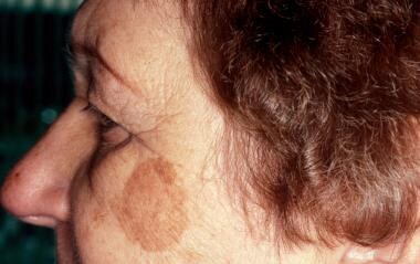
Melanosis
Retinal Pigment Epithelium
Lysosomes
Melanoma, Amelanotic
Adaptor Protein Complex 1
Pigmentation Disorders
Catechol Oxidase
Dequalinium
Microscopy, Electron
Protein Transport
Albinism
Dyneins
Melanoma
Mesopic Vision
Encyclopedias as Topic
Retina
Photoreceptor Cells, Vertebrate
Vision, Ocular
Retinal Cone Photoreceptor Cells
Altered trafficking of lysosomal proteins in Hermansky-Pudlak syndrome due to mutations in the beta 3A subunit of the AP-3 adaptor. (1/317)
Hermansky-Pudlak syndrome (HPS) is a genetic disorder characterized by defective lysosome-related organelles. Here, we report the identification of two HPS patients with mutations in the beta 3A subunit of the heterotetrameric AP-3 complex. The patients' fibroblasts exhibit drastically reduced levels of AP-3 due to enhanced degradation of mutant beta 3A. The AP-3 deficiency results in increased surface expression of the lysosomal membrane proteins CD63, lamp-1, and lamp-2, but not of nonlysosomal proteins. These differential effects are consistent with the preferential interaction of the AP-3 mu 3A subunit with tyrosine-based signals involved in lysosomal targeting. Our results suggest that AP-3 functions in protein sorting to lysosomes and provide an example of a human disease in which altered trafficking of integral membrane proteins is due to mutations in a component of the sorting machinery. (+info)Melanosomes of retinal pigment epithelium--distribution, shape, and acid phosphatase activity. (2/317)
The distribution and shape of melanosomes of the retinal pigment epithelium (RPE), and acid phosphatase activity in melanosomes were studied in rabbits. The rabbit eyes were observed using electron microscopy and enzyme cytochemical electron microscopy. The majority of melanosomes were located near the apical region of the RPE. Melanosomes in the RPE were classified as two shapes, elliptical and spherical or oval. Elliptical melanosomes were located parallel to the apical process and spherical or oval melanosomes were arranged vertically or obliquely to the apical process. We think that the distribution and shape of melanosomes contributes to the effective absorption and blocking of light coming from all directions. Almost all of the mature and immature melanosomes we identified showed positive in acid phosphatase reaction, indicating that melanosomes are commonly incorporated into the lysosomal system of the RPE. However, a few melanosomes showed negative in acid phosphatase reaction, suggesting that some melanosomes are stable and inert. The observed premelanosome showed negative reaction. Two types of melanosome-related complex granules were identified; melanosomes with a cortex of enzyme-reactive material (melanolysosome) and melanosomes with a cortex of lipofuscin (melanolipofuscin). These findings indicate tha a relationship between melanosomes and the lysosomal system of the RPE exists, and suggest that melanosomes may undergo modification or degradation in the cytoplasm. Also, the observation of a premelanosome and the positive acid phosphatase activity in mature and immature melanosomes indicates that melanosomes of the RPE may continue to be synthesized at a low rate in adult eyes. (+info)Protein kinase C-beta activates tyrosinase by phosphorylating serine residues in its cytoplasmic domain. (3/317)
We have previously shown that protein kinase C-beta (PKC-beta) is required for activation of tyrosinase (Park, H. Y., Russakovsky, V., Ohno, S., and Gilchrest, B. A. (1993) J. Biol. Chem. 268, 11742-11749), the rate-limiting enzyme in melanogenesis. We now examine its mechanism of activation in human melanocytes. In vivo phosphorylation experiments revealed that tyrosinase is phosphorylated through the PKC-dependent pathway and that introduction of PKC-beta into nonpigmented human melanoma cells lacking PKC-beta lead to the phosphorylation and activation of tyrosinase. Preincubation of intact melanosomes with purified active PKC-beta in vitro increased tyrosinase activity 3-fold. By immunoelectron microscopy, PKC-beta but not PKC-alpha was closely associated with tyrosinase on the outer surface of melanosomes. Western blot analysis confirmed the association of PKC-beta with melanosomes. Only the cytoplasmic (extra-melanosomal) domain of tyrosinase, which contains two serines but no threonines, was phosphorylated by the serine/threonine kinase PKC-beta. These two serines at positions 505 and 509 both are present in the C-terminal peptide generated by trypsin digestion of tyrosinase. Co-migration experiments comparing synthetic peptide standards of all three possible phosphorylated tryptic peptides, a diphosphopeptide and two monophosphopeptides, to tyrosinase-phosphorylated in intact melanocytes by PKC-beta and then subjected to trypsin digestion revealed that both serine residues are phosphorylated by PKC-beta. We conclude that PKC-beta activates tyrosinase directly by phosphorylating serine residues at positions 505 and 509 in the cytoplasmic domain of this melanosome-associated protein. (+info)Deposition of [3H]cocaine, [3H]nicotine, and [3H]flunitrazepam in mouse hair melanosomes after systemic administration. (4/317)
Microautoradiography was employed to show that association of drugs from the serum directly with forming hair pigment is a primary pathway of deposition into the hair. After systemic administration of [3H]flunitrazepam, [3H]nicotine, and [3H]cocaine, association of all three drugs with melanin in the forming hair was observed within minutes of dosage. Sebum was determined to be an insignificant deposition route for all three drugs. Pigmented mice had significantly higher concentrations of all three drugs than did nonpigmented mice. The results provide a better basis for ultimately using hair for reliable analysis of drug and environmental toxin exposure. (+info)Rescue effects of IPE transplants in RCS rats: short-term results. (5/317)
PURPOSE: The aim of this study was to investigate the possible rescue effect of subretinal iris pigment epithelial (IPE) cell transplantation in Royal College of Surgeons (RCS) rats by light and electron microscopic histology. METHODS: IPE cells were harvested from 20- to 26-day-old Long-Evans rats and were directly trans planted transsclerally into the subretinal space of 32 16- to 20-day-old RCS rats using a 32-gauge Hamilton syringe. Specimens of transplanted eyes were embedded for electron microscopy after 8 weeks. Specimens from the iris and retinal pigment epithelium (RPE) of Long-Evans rats and RPE from RCS rats without surgical treatment were also embedded. Sham surgery was also performed in 8 eyes. RESULTS: The IPE cells transplanted into the subretinal space were localized between host RPE and retina, had round cell shapes without polar organization, and contained phagosomes resulting from rod outer segment (ROS) uptake. The underlying host RPE cells were heavily pigmented. RPE cells from RCS rats revealed fragmentation of endoplasmic reticulum, which distinguishes them ultrastructurally from pigment epithelial cells of Long-Evans rats. Ultrastructural alterations were observed in the cytoplasm of transplanted cells. Melanin granules in the IPE cells were found in large vacuoles, which also contained phagosomes originating from ROS uptake. In 13 eyes, 1 to 4 rows and 5 to 8 rows of saved photoreceptors were detected facing transplanted IPE cells in 6 (46%) and 4 (31%) eyes, respectively, 2 months after surgery. However, in 10 (53%) and 7 (37%) of 19 eyes, 1 to 4 rows and 5 to 8 rows, respectively, were also found at sites without IPE cells in the plane of section. ROS directed toward transplanted IPE cells were seen in one case, but these rods were shortened and disorganized. At most sites between transplanted cells and inner segments of photoreceptors, outer segments and cellular debris were absent. In eyes without transplanted cells no photoreceptor cells were alive at the age of 2 months. After sham surgery 6 (75%) eyes had 1 to 4 rows and 2 (25%) 5 to 8 rows of photoreceptors. CONCLUSIONS: Transplanted IPE cells can take up and degrade ROS in vivo in RCS rats. Uptake of ROS alters the morphology of pigment granules in transplanted IPE cells. Pigmentation is an uncertain marker for identifying transplanted pigment cells. IPE transplants are not as good as RPE transplants in rescuing photoreceptors. However, there is a significant difference between transplanted eyes and nontreated eyes. The rescue effect of IPE cells was not significantly different from that of sham surgery. (+info)Tawny: a novel light coat color mutation found in a wild population of Mus musculus molossinus, a new allele at the melanocortin 1 receptor (Mc1r) locus. (6/317)
We found a new coat color mutant in a population of Japanese wild mice (Mus musculus molossinus) and called the trait tawny. The tawny mutant is characterized by a light yellowish brown coat color. The tawny hair has a so-called agouti pattern, but the yellow band is greatly lengthened. There are no differences between the tawny and wildtype hairs in size and the number of melanosomes. Genetic analyses revealed that the tawny trait is an autosomal recessive and its gene is located in the distal region on Chromosome 8 between the microsatellite markers D8Mit87 and D8Mit122. An allelism test indicated the tawny mutant gene to be a new allele at the Mc1r locus and dominant to the recessive yellow (Mc1re). The proposed gene symbol for the tawny is Mc1rtaw. (+info)Regulation of melanosome movement in the cell cycle by reversible association with myosin V. (7/317)
Previously, we have shown that melanosomes of Xenopus laevis melanophores are transported along both microtubules and actin filaments in a coordinated manner, and that myosin V is bound to purified melanosomes (Rogers, S., and V.I. Gelfand. 1998. Curr. Biol. 8:161-164). In the present study, we have demonstrated that myosin V is the actin-based motor responsible for melanosome transport. To examine whether myosin V was regulated in a cell cycle-dependent manner, purified melanosomes were treated with interphase- or metaphase-arrested Xenopus egg extracts and assayed for in vitro motility along Nitella actin filaments. Motility of organelles treated with mitotic extract was found to decrease dramatically, as compared with untreated or interphase extract-treated melanosomes. This mitotic inhibition of motility correlated with the dissociation of myosin V from melanosomes, but the activity of soluble motor remained unaffected. Furthermore, we find that myosin V heavy chain is highly phosphorylated in metaphase extracts versus interphase extracts. We conclude that organelle transport by myosin V is controlled by a cell cycle-regulated association of this motor to organelles, and that this binding is likely regulated by phosphorylation of myosin V during mitosis. (+info)A role for a melanosome transport signal in accessing the MHC class II presentation pathway and in eliciting CD4+ T cell responses. (8/317)
Melanosomal membrane proteins are frequently recognized by the immune system of patients with melanoma and vitiligo. Melanosomal glycoproteins are transported to melanosomes by a dileucine-based melanosomal transport signal (MTS). To investigate whether this sorting signal could be involved in presentation of melanosome membrane proteins to the immune system, we devised a fusion construct containing the MTS from the mouse brown locus product gp75/tyrosinase-related protein-1 and full-length OVA as a reporter Ag. The fusion protein was expressed as an intracellular membrane protein, sorted to the endocytic pathway, processed, and presented by class II MHC molecules. DNA immunization with this construct elicited CD4+ T cell proliferative responses in vivo. Ag presentation and T cell responses in vitro and in vivo required a functional MTS. Mutations of either the upstream leucine in MTS or elimination of the entire MTS negated in vitro Ag presentation and in vivo T cell responses. In a mouse melanoma model, DNA immunization with MTS constructs protected mice from tumor challenge in a CD4+ T cell-dependent manner, but complete deletion of MTS decreased tumor rejection. Therefore, MTS can target epitopes to the endocytic pathway leading to presentation by class II MHC molecules to helper T cells. (+info)Melanosomes are membrane-bound organelles found in melanocytes, the pigment-producing cells in the skin, hair, and eyes. They contain the pigment melanin, which is responsible for giving color to these tissues. Melanosomes are produced in the melanocyte and then transferred to surrounding keratinocytes in the epidermis via a process called cytocrinesis. There are four stages of melanosome development: stage I (immature), stage II (developing), stage III (mature), and stage IV (degrading). The amount and type of melanin in the melanosomes determine the color of an individual's skin, hair, and eyes. Mutations in genes involved in melanosome biogenesis or function can lead to various pigmentation disorders, such as albinism.
Melanocytes are specialized cells that produce, store, and transport melanin, the pigment responsible for coloring of the skin, hair, and eyes. They are located in the bottom layer of the epidermis (the outermost layer of the skin) and can also be found in the inner ear and the eye's retina. Melanocytes contain organelles called melanosomes, which produce and store melanin.
Melanin comes in two types: eumelanin (black or brown) and pheomelanin (red or yellow). The amount and type of melanin produced by melanocytes determine the color of a person's skin, hair, and eyes. Exposure to UV radiation from sunlight increases melanin production as a protective response, leading to skin tanning.
Melanocyte dysfunction or abnormalities can lead to various medical conditions, such as albinism (lack of melanin production), melasma (excessive pigmentation), and melanoma (cancerous growth of melanocytes).
Melanin is a pigment that determines the color of skin, hair, and eyes in humans and animals. It is produced by melanocytes, which are specialized cells found in the epidermis (the outer layer of the skin) and the choroid (the vascular coat of the eye). There are two main types of melanin: eumelanin and pheomelanin. Eumelanin is a black or brown pigment, while pheomelanin is a red or yellow pigment. The amount and type of melanin produced by an individual can affect their skin and hair color, as well as their susceptibility to certain diseases, such as skin cancer.
Hermanski-Pudlak Syndrome (HPS) is a rare genetic disorder characterized by the triad of albinism, bleeding disorders, and lysosomal storage disease. It is caused by mutations in any one of several genes involved in biogenesis of lysosome-related organelles (LROs), such as melanosomes in melanocytes, platelet dense granules, and lung lamellar bodies.
The albinism in HPS results from abnormal melanosome biogenesis, leading to decreased pigmentation in the skin, hair, and eyes. The bleeding disorder is due to defective platelet dense granules, which are necessary for normal clotting function. This can result in prolonged bleeding times and easy bruising.
The lysosomal storage disease component of HPS is characterized by the accumulation of ceroid lipofuscin within LROs, leading to progressive damage to affected tissues. The most common form of HPS (HPS-1) also involves pulmonary fibrosis, which can lead to respiratory failure and death in the third or fourth decade of life.
There are currently seven known subtypes of HPS, each caused by mutations in different genes involved in LRO biogenesis. The clinical features and severity of HPS can vary widely between subtypes and even within families with the same genetic mutation.
Tyrosinase, also known as monophenol monooxygenase, is an enzyme (EC 1.14.18.1) that catalyzes the ortho-hydroxylation of monophenols (like tyrosine) to o-diphenols (like L-DOPA) and the oxidation of o-diphenols to o-quinones. This enzyme plays a crucial role in melanin synthesis, which is responsible for the color of skin, hair, and eyes in humans and animals. Tyrosinase is found in various organisms, including plants, fungi, and animals. In humans, tyrosinase is primarily located in melanocytes, the cells that produce melanin. The enzyme's activity is regulated by several factors, such as pH, temperature, and metal ions like copper, which are essential for its catalytic function.
Melanophores are specialized pigment-containing cells found in various organisms, including vertebrates and some invertebrates. In humans and other mammals, melanophores are primarily located within the skin's dermal layer and are part of the larger group of chromatophores.
Melanophores contain melanosomes, which are organelles that store and transport the pigment melanin. These cells play a crucial role in determining the coloration of an individual's skin, hair, and eyes by producing, storing, and distributing melanin granules within their cytoplasm.
In response to hormonal signals or neural stimulation, melanophores can undergo changes in the distribution of melanosomes, leading to variations in color intensity. This process is known as melanin dispersion or aggregation and is responsible for various physiological responses, such as skin tanning upon exposure to sunlight or the color-changing abilities observed in some animals like chameleons and cuttlefish.
It's important to note that while humans do not have the ability to change their skin color rapidly like some other animals, melanophores still play a significant role in protecting our skin from harmful ultraviolet radiation by producing melanin, which helps absorb and dissipate this energy, reducing damage to skin cells.
Hypopigmentation is a medical term that refers to a condition where there is a decrease in the amount of pigment (melanin) in the skin, resulting in lighter patches or spots on the skin. This can occur due to various reasons such as skin injuries, certain skin disorders like vitiligo, fungal infections, burns, or as a side effect of some medical treatments like chemotherapy or radiation therapy. It is different from albinism, which is a genetic condition where the body is unable to produce melanin at all.
The gp100 melanoma antigen, also known as Pmel17 or gp100, is a protein found on the surface of melanocytes, which are the pigment-producing cells in the skin. It is overexpressed in melanoma cells and can be recognized by the immune system as a foreign target, making it an attractive candidate for cancer immunotherapy. The gp100 protein plays a role in the formation and transport of melanosomes, which are organelles involved in the production and distribution of melanin. In melanoma, mutations or abnormal regulation of gp100 can contribute to uncontrolled cell growth and survival, leading to the development of cancer. The gp100 protein is used as a target for various immunotherapeutic approaches, such as vaccines and monoclonal antibodies, to stimulate an immune response against melanoma cells.
Myosin Type V is an molecular motor protein involved in the intracellular transport of various cargoes, including vesicles and organelles. It belongs to the family of myosins, which are actin-based motors that convert chemical energy into mechanical work through the hydrolysis of ATP.
Myosin V is characterized by its long tail domain, which allows it to form dimers or higher-order oligomers, and its head domain, which binds to actin filaments and hydrolyzes ATP to generate force and movement. The protein moves in a hand-over-hand manner along the actin filament, allowing it to transport cargoes over long distances within the cell.
Myosin V has been implicated in various cellular processes, including exocytosis, endocytosis, and organelle positioning. Mutations in the MYO5A gene, which encodes Myosin Type V, have been associated with several human genetic disorders, such as Griscelli syndrome type 1 and familial progressive arthro-ophthalmopathy.
Rab GTP-binding proteins, also known as Rab GTPases or simply Rabs, are a large family of small GTP-binding proteins that play a crucial role in regulating intracellular vesicle trafficking. They function as molecular switches that cycle between an active GTP-bound state and an inactive GDP-bound state.
In the active state, Rab proteins interact with various effector molecules to mediate specific membrane trafficking events such as vesicle budding, transport, tethering, and fusion. Each Rab protein is thought to have a unique function and localize to specific intracellular compartments or membranes, where they regulate the transport of vesicles and organelles within the cell.
Rab proteins are involved in several important cellular processes, including endocytosis, exocytosis, Golgi apparatus function, autophagy, and intracellular signaling. Dysregulation of Rab GTP-binding proteins has been implicated in various human diseases, such as cancer, neurodegenerative disorders, and infectious diseases.
Skin pigmentation is the coloration of the skin that is primarily determined by two types of melanin pigments, eumelanin and pheomelanin. These pigments are produced by melanocytes, which are specialized cells located in the epidermis. Eumelanin is responsible for brown or black coloration, while pheomelanin produces a red or yellow hue.
The amount and distribution of melanin in the skin can vary depending on genetic factors, age, sun exposure, and various other influences. Increased production of melanin in response to UV radiation from the sun helps protect the skin from damage, leading to darkening or tanning of the skin. However, excessive sun exposure can also cause irregular pigmentation, such as sunspots or freckles.
Abnormalities in skin pigmentation can result from various medical conditions, including albinism (lack of melanin production), vitiligo (loss of melanocytes leading to white patches), and melasma (excessive pigmentation often caused by hormonal changes). These conditions may require medical treatment to manage or improve the pigmentation issues.
Pigmentation, in a medical context, refers to the coloring of the skin, hair, or eyes due to the presence of pigment-producing cells called melanocytes. These cells produce a pigment called melanin, which determines the color of our skin, hair, and eyes.
There are two main types of melanin: eumelanin and pheomelanin. Eumelanin is responsible for brown or black coloration, while pheomelanin produces a red or yellow hue. The amount and type of melanin produced by melanocytes can vary from person to person, leading to differences in skin color and hair color.
Changes in pigmentation can occur due to various factors such as genetics, exposure to sunlight, hormonal changes, inflammation, or certain medical conditions. For example, hyperpigmentation refers to an excess production of melanin that results in darkened patches on the skin, while hypopigmentation is a condition where there is a decreased production of melanin leading to lighter or white patches on the skin.
Ocular albinism is a type of albinism that primarily affects the eyes. It is a genetic disorder characterized by the reduction or absence of melanin, the pigment responsible for coloring the skin, hair, and eyes. In ocular albinism, melanin production is deficient in the eyes, leading to various eye abnormalities.
The main features of ocular albinism include:
1. Nystagmus: Rapid, involuntary back-and-forth movement of the eyes.
2. Iris transillumination: The iris appears translucent due to the lack of pigment, allowing light to pass through easily. This can be observed using a light source shone into the eye.
3. Foveal hypoplasia: Underdevelopment or absence of the fovea, a small pit in the retina responsible for sharp, central vision.
4. Photophobia: Increased sensitivity to light due to the lack of pigment in the eyes.
5. Strabismus: Misalignment of the eyes, which can result in double vision or lazy eye.
6. Reduced visual acuity: Decreased ability to see clearly, even with corrective lenses.
Ocular albinism is typically inherited as an X-linked recessive trait, meaning it primarily affects males, while females can be carriers of the condition. However, there are also autosomal recessive forms of ocular albinism that can affect both males and females equally. Treatment for ocular albinism usually involves managing symptoms with corrective lenses, low-vision aids, and vision therapy to improve visual skills.
Oculocutaneous albinism (OCA) is a group of genetic disorders characterized by reduced or complete absence of melanin pigment in the eyes, skin, and hair. Melanin is the pigment responsible for giving color to our skin, hair, and eyes. OCA affects both the eyes (oculo-) and the skin (cutaneous), hence the name oculocutaneous albinism.
There are several types of OCA, each caused by different genetic mutations affecting melanin production. The most common forms include:
1. OCA1: This type is further divided into two subtypes - OCA1A and OCA1B. OCA1A is characterized by complete absence of melanin in the eyes, skin, and hair from birth. Individuals with this condition have white hair, very light skin, and pale blue or gray irises. OCA1B, on the other hand, presents with reduced melanin production, leading to lighter-than-average skin, hair, and eye color at birth. Over time, some melanin may be produced, resulting in milder pigmentation changes compared to OCA1A.
2. OCA2: This form of albinism is caused by mutations in the tyrosinase-related protein 1 (TYRP1) gene, which plays a role in melanin production. Individuals with OCA2 typically have light brown or yellowish skin, golden or straw-colored hair, and lighter irises compared to their family members without albinism.
3. OCA3: Also known as Rufous oculocutaneous albinism (ROCA), this type is caused by mutations in the tyrosinase gene (TYR). It primarily affects people of African descent, leading to reddish-brown hair, light brown skin, and normal or near-normal eye color.
4. OCA4: This form of albinism results from mutations in the membrane-associated transporter protein (MATP) gene, which is involved in melanin transport within cells. Individuals with OCA4 usually have light brown skin, yellowish or blond hair, and lighter irises compared to their family members without albinism.
Regardless of the type, all individuals with oculocutaneous albinism face similar challenges, including reduced vision due to abnormal eye development (nystagmus, strabismus, and farsightedness) and increased sensitivity to sunlight (photophobia). Proper management, such as wearing UV-protective sunglasses, hats, and sunscreen, can help protect their skin and eyes from damage.
Adaptor Protein Complex 3 (APC3), also known as AP-3, is a type of adaptor protein complex that plays a crucial role in the sorting and trafficking of proteins within cells. It is composed of four subunits: delta, beta3A, mu3, and sigma3A. APC3 is primarily involved in the transport of proteins from the early endosomes to the lysosomes or to the plasma membrane. It also plays a role in the biogenesis of lysosome-related organelles such as melanosomes and platelet-dense granules. Mutations in the genes encoding for APC3 subunits have been associated with several genetic disorders, including Hermansky-Pudlak syndrome and Chediak-Higashi syndrome.
The pigment epithelium of the eye, also known as the retinal pigment epithelium (RPE), is a layer of cells located between the photoreceptor cells of the retina and the choroid, which is the vascular layer of the eye. The RPE plays a crucial role in maintaining the health and function of the photoreceptors by providing them with nutrients, removing waste products, and helping to regulate the light that enters the eye.
The RPE cells contain pigment granules that absorb excess light, preventing it from scattering within the eye and improving visual acuity. They also help to create a barrier between the retina and the choroid, which is important for maintaining the proper functioning of the photoreceptors. Additionally, the RPE plays a role in the regeneration of visual pigments in the photoreceptor cells, allowing us to see in different light conditions.
Damage to the RPE can lead to various eye diseases and conditions, including age-related macular degeneration (AMD), which is a leading cause of vision loss in older adults.
Lipofuscin is a type of pigment that accumulates in the lysosomes (membrane-bound organelles found inside cells) of various tissues, particularly in nerve cells and heart muscle cells. It consists of cross-linked proteins and lipids that are resistant to degradation by enzymes. The accumulation of lipofuscin is a normal part of aging but can also be associated with certain diseases such as neurodegenerative disorders.
It's often referred to as "age pigment" because it tends to increase in amount with age, and its presence in tissues has been linked to oxidative stress and cellular damage caused by free radicals. Lipofuscin is autofluorescent, meaning that it emits light when excited by certain wavelengths of light, which can be useful for its detection and quantification in research and diagnostic settings.
Feathers are not a medical term, but they are a feature found in birds and some extinct theropod dinosaurs. Feathers are keratinous structures that grow from the skin and are used for various functions such as insulation, flight, waterproofing, and display. They have a complex structure consisting of a central shaft with barbs branching off on either side, which further divide into smaller barbules. The arrangement and modification of these feather structures vary widely among bird species to serve different purposes.
I'm sorry for any confusion, but "hair color" is not a term that has a medical definition in the same way that a term like "myocardial infarction" (heart attack) does. Hair color can be described in various ways, such as being a natural hair color (like blonde, brunette, red, or black) or describing a change in hair color due to factors like aging (gray or white hairs) or hair dye usage.
However, it's worth noting that changes in hair color can sometimes be associated with certain medical conditions. For example, premature graying of the hair before the age of 30 can be a feature of certain genetic disorders or vitamin B12 deficiency. Similarly, some skin conditions like alopecia areata or vitiligo can cause patchy changes in hair color. But these associations don't provide a medical definition for 'hair color'.
Melanocyte-stimulating hormones (MSH) are a group of peptide hormones that originate from the precursor protein proopiomelanocortin (POMC). They play crucial roles in various physiological processes, including pigmentation, energy balance, and appetite regulation.
There are several types of MSH, but the most well-known ones include α-MSH, β-MSH, and γ-MSH. These hormones bind to melanocortin receptors (MCRs), which are found in various tissues throughout the body. The binding of MSH to MCRs triggers a series of intracellular signaling events that ultimately lead to changes in cell behavior.
In the context of skin physiology, α-MSH and β-MSH bind to melanocortin 1 receptor (MC1R) on melanocytes, which are the cells responsible for producing pigment (melanin). This binding stimulates the production and release of eumelanin, a type of melanin that is brown or black in color. As a result, increased levels of MSH can lead to darkening of the skin, also known as hyperpigmentation.
Apart from their role in pigmentation, MSH hormones have been implicated in several other physiological processes. For instance, α-MSH has been shown to suppress appetite and promote weight loss by binding to melanocortin 4 receptor (MC4R) in the hypothalamus, a region of the brain that regulates energy balance. Additionally, MSH hormones have been implicated in inflammation, immune response, and sexual function.
Overall, melanocyte-stimulating hormones are a diverse group of peptide hormones that play important roles in various physiological processes, including pigmentation, energy balance, and appetite regulation.
Adaptor Protein Complex (AP) beta subunits are structural proteins that play a crucial role in intracellular vesicle trafficking. They are part of the heterotetrameric AP complex, which is responsible for recognizing and binding to specific sorting signals on membrane cargo proteins, allowing for their packaging into transport vesicles.
There are four different types of AP complexes (AP-1, AP-2, AP-3, and AP-4), each with a unique set of subunits that confer specific functions. The beta subunit is a common component of all four complexes and is essential for their stability and function.
The beta subunit interacts with other subunits within the AP complex as well as with accessory proteins, such as clathrin, to form a coat around the transport vesicle. This coat helps to shape the vesicle and facilitate its movement between different cellular compartments.
Mutations in genes encoding AP beta subunits have been linked to various human diseases, including forms of hemolytic anemia, neurological disorders, and immunodeficiency.
Melanosis is a general term that refers to an increased deposit of melanin, the pigment responsible for coloring our skin, in the skin or other organs. It can occur in response to various factors such as sun exposure, aging, or certain medical conditions. There are several types of melanosis, including:
1. Epidermal melanosis: This type of melanosis is characterized by an increase in melanin within the epidermis, the outermost layer of the skin. It can result from sun exposure, hormonal changes, or inflammation.
2. Dermal melanosis: In this type of melanosis, there is an accumulation of melanin within the dermis, the middle layer of the skin. It can be caused by various conditions such as nevus of Ota, nevus of Ito, or melanoma metastasis.
3. Mucosal melanosis: This type of melanosis involves an increase in melanin within the mucous membranes, such as those lining the mouth, nose, and genitals. It can be a sign of systemic disorders like Addison's disease or Peutz-Jeghers syndrome.
4. Lentigo simplex: Also known as simple lentigines, these are small, benign spots that appear on sun-exposed skin. They result from an increase in melanocytes, the cells responsible for producing melanin.
5. Labial melanotic macule: This is a pigmented lesion found on the lips, typically the lower lip. It is more common in darker-skinned individuals and is usually benign but should be monitored for changes that may indicate malignancy.
6. Ocular melanosis: An increase in melanin within the eye can lead to various conditions such as ocular melanocytosis, oculodermal melanocytosis, or choroidal melanoma.
It is important to note that while some forms of melanosis are benign and harmless, others may indicate an underlying medical condition or even malignancy. Therefore, any new or changing pigmented lesions should be evaluated by a healthcare professional.
The retinal pigment epithelium (RPE) is a single layer of cells located between the photoreceptor cells of the retina and the choroid, which is a part of the eye containing blood vessels. The RPE plays a crucial role in maintaining the health and function of the photoreceptors by providing them with nutrients, removing waste products, and helping to regulate the light-sensitive visual pigments within the photoreceptors.
The RPE cells contain pigment granules that absorb excess light to prevent scattering within the eye and improve visual acuity. They also help to form the blood-retina barrier, which restricts the movement of certain molecules between the retina and the choroid, providing an important protective function for the retina.
Damage to the RPE can lead to a variety of eye conditions, including age-related macular degeneration (AMD), which is a leading cause of vision loss in older adults.
Lysosomes are membrane-bound organelles found in the cytoplasm of eukaryotic cells. They are responsible for breaking down and recycling various materials, such as waste products, foreign substances, and damaged cellular components, through a process called autophagy or phagocytosis. Lysosomes contain hydrolytic enzymes that can break down biomolecules like proteins, nucleic acids, lipids, and carbohydrates into their basic building blocks, which can then be reused by the cell. They play a crucial role in maintaining cellular homeostasis and are often referred to as the "garbage disposal system" of the cell.
Amelanotic melanoma is a type of melanoma, which is the most serious and deadly form of skin cancer. While most melanomas contain dark pigments called melanin, amelanotic melanomas lack melanin, giving them a pink, red, or white color. This absence of color can make amelanotic melanomas harder to detect and diagnose at an early stage compared to other types of melanoma.
Amelanotic melanomas may arise from existing moles or develop on their own in normal skin. They can occur anywhere on the body, but they are more common in sun-exposed areas such as the head, neck, and trunk.
Like other forms of melanoma, amelanotic melanoma can spread quickly to other parts of the body if left untreated. Therefore, it is essential to recognize any changes in the skin and consult a healthcare professional for proper evaluation and diagnosis. Treatment typically involves surgical excision, with additional therapies such as radiation therapy, immunotherapy, or targeted therapy recommended depending on the stage and specific features of the cancer.
Adaptor Protein Complex 1 (AP-1) is a group of proteins that function as a complex to play a crucial role in the intracellular transport of various molecules, particularly in the formation of vesicles that transport cargo from one compartment of the cell to another. The AP-1 complex is composed of four subunits: γ, β1, μ1, and σ1. It is primarily associated with the trans-Golgi network and early endosomes, where it facilitates the sorting and packaging of cargo into vesicles for transport to various destinations within the cell. The AP-1 complex recognizes specific sorting signals on the membrane proteins and adaptor proteins, thereby ensuring the accurate delivery of cargo to the correct location. Defects in the AP-1 complex have been implicated in several human diseases, including neurological disorders and cancer.
Pigmentation disorders are conditions that affect the production or distribution of melanin, the pigment responsible for the color of skin, hair, and eyes. These disorders can cause changes in the color of the skin, resulting in areas that are darker (hyperpigmentation) or lighter (hypopigmentation) than normal. Examples of pigmentation disorders include melasma, age spots, albinism, and vitiligo. The causes, symptoms, and treatments for these conditions can vary widely, so it is important to consult a healthcare provider for an accurate diagnosis and treatment plan.
Catechol oxidase, also known as polyphenol oxidase, is an enzyme that catalyzes the oxidation of catechols and other phenolic compounds to quinones. These quinones can then undergo further reactions to form various pigmented compounds, such as melanins. Catechol oxidase is widely distributed in nature and is found in plants, fungi, and some bacteria. In humans, catechol oxidase is involved in the metabolism of neurotransmitters such as dopamine and epinephrine.
I'm sorry for any confusion, but "Organelle Size" is not a recognized medical term or concept with a specific definition. Organelles are specialized structures within cells that have specific functions, such as mitochondria, ribosomes, and endoplasmic reticulum. The size of these organelles can vary depending on the type of cell, its function, and various other factors. However, there is no standardized medical definition for the term "Organelle Size." If you have more specific questions about the size or function of certain organelles, I'd be happy to help further!
Dequalinium is an antimicrobial agent with both antibacterial and antifungal properties. It is commonly used in the form of a salt, such as dequalinium chloride, in various pharmaceutical and medical applications. Dequalinium works by disrupting the bacterial or fungal cell membrane, leading to their death. It is often found in topical creams, ointments, and oral suspensions for treating infections of the skin, mouth, and throat.
The medical definition of 'dequalinium' is:
A quaternary ammonium compound with antimicrobial properties, used as a topical antiseptic and in the treatment of oral candidiasis and other fungal infections. It works by disrupting the bacterial or fungal cell membrane, leading to their death. Dequalinium is available in various forms, including creams, ointments, and oral suspensions.
Biological pigments are substances produced by living organisms that absorb certain wavelengths of light and reflect others, resulting in the perception of color. These pigments play crucial roles in various biological processes such as photosynthesis, vision, and protection against harmful radiation. Some examples of biological pigments include melanin, hemoglobin, chlorophyll, carotenoids, and flavonoids.
Melanin is a pigment responsible for the color of skin, hair, and eyes in animals, including humans. Hemoglobin is a protein found in red blood cells that contains a porphyrin ring with an iron atom at its center, which gives blood its red color and facilitates oxygen transport. Chlorophyll is a green pigment found in plants, algae, and some bacteria that absorbs light during photosynthesis to convert carbon dioxide and water into glucose and oxygen. Carotenoids are orange, yellow, or red pigments found in fruits, vegetables, and some animals that protect against oxidative stress and help maintain membrane fluidity. Flavonoids are a class of plant pigments with antioxidant properties that have been linked to various health benefits.
Membrane glycoproteins are proteins that contain oligosaccharide chains (glycans) covalently attached to their polypeptide backbone. They are integral components of biological membranes, spanning the lipid bilayer and playing crucial roles in various cellular processes.
The glycosylation of these proteins occurs in the endoplasmic reticulum (ER) and Golgi apparatus during protein folding and trafficking. The attached glycans can vary in structure, length, and composition, which contributes to the diversity of membrane glycoproteins.
Membrane glycoproteins can be classified into two main types based on their orientation within the lipid bilayer:
1. Type I (N-linked): These glycoproteins have a single transmembrane domain and an extracellular N-terminus, where the oligosaccharides are predominantly attached via asparagine residues (Asn-X-Ser/Thr sequon).
2. Type II (C-linked): These glycoproteins possess two transmembrane domains and an intracellular C-terminus, with the oligosaccharides linked to tryptophan residues via a mannose moiety.
Membrane glycoproteins are involved in various cellular functions, such as:
* Cell adhesion and recognition
* Receptor-mediated signal transduction
* Enzymatic catalysis
* Transport of molecules across membranes
* Cell-cell communication
* Immunological responses
Some examples of membrane glycoproteins include cell surface receptors (e.g., growth factor receptors, cytokine receptors), adhesion molecules (e.g., integrins, cadherins), and transporters (e.g., ion channels, ABC transporters).
Electron microscopy (EM) is a type of microscopy that uses a beam of electrons to create an image of the sample being examined, resulting in much higher magnification and resolution than light microscopy. There are several types of electron microscopy, including transmission electron microscopy (TEM), scanning electron microscopy (SEM), and reflection electron microscopy (REM).
In TEM, a beam of electrons is transmitted through a thin slice of the sample, and the electrons that pass through the sample are focused to form an image. This technique can provide detailed information about the internal structure of cells, viruses, and other biological specimens, as well as the composition and structure of materials at the atomic level.
In SEM, a beam of electrons is scanned across the surface of the sample, and the electrons that are scattered back from the surface are detected to create an image. This technique can provide information about the topography and composition of surfaces, as well as the structure of materials at the microscopic level.
REM is a variation of SEM in which the beam of electrons is reflected off the surface of the sample, rather than scattered back from it. This technique can provide information about the surface chemistry and composition of materials.
Electron microscopy has a wide range of applications in biology, medicine, and materials science, including the study of cellular structure and function, disease diagnosis, and the development of new materials and technologies.
Protein transport, in the context of cellular biology, refers to the process by which proteins are actively moved from one location to another within or between cells. This is a crucial mechanism for maintaining proper cell function and regulation.
Intracellular protein transport involves the movement of proteins within a single cell. Proteins can be transported across membranes (such as the nuclear envelope, endoplasmic reticulum, Golgi apparatus, or plasma membrane) via specialized transport systems like vesicles and transport channels.
Intercellular protein transport refers to the movement of proteins from one cell to another, often facilitated by exocytosis (release of proteins in vesicles) and endocytosis (uptake of extracellular substances via membrane-bound vesicles). This is essential for communication between cells, immune response, and other physiological processes.
It's important to note that any disruption in protein transport can lead to various diseases, including neurological disorders, cancer, and metabolic conditions.
Albinism is a group of genetic disorders that result in little or no production of melanin, the pigment responsible for coloring skin, hair, and eyes. It is caused by mutations in genes involved in the production of melanin. There are several types of albinism, including oculocutaneous albinism (OCA) and ocular albinism (OA). OCA affects the skin, hair, and eyes, while OA primarily affects the eyes.
People with albinism typically have very pale skin, white or light-colored hair, and light-colored eyes. They may also have vision problems, such as sensitivity to light (photophobia), rapid eye movements (nystagmus), and decreased visual acuity. The severity of these symptoms can vary depending on the type and extent of albinism.
Albinism is inherited in an autosomal recessive manner, which means that an individual must inherit two copies of the mutated gene, one from each parent, in order to have the condition. If both parents are carriers of a mutated gene for albinism, they have a 25% chance with each pregnancy of having a child with albinism.
There is no cure for albinism, but individuals with the condition can take steps to protect their skin and eyes from the sun and use visual aids to help with vision problems. It is important for people with albinism to undergo regular eye examinations and to use sun protection, such as sunscreen, hats, and sunglasses, to prevent skin damage and skin cancer.
Dyneins are a type of motor protein that play an essential role in the movement of cellular components and structures within eukaryotic cells. They are responsible for generating force and motion along microtubules, which are critical components of the cell's cytoskeleton. Dyneins are involved in various cellular processes, including intracellular transport, organelle positioning, and cell division.
There are several types of dyneins, but the two main categories are cytoplasmic dyneins and axonemal dyneins. Cytoplasmic dyneins are responsible for moving various cargoes, such as vesicles, organelles, and mRNA complexes, toward the minus-end of microtubules, which is usually located near the cell center. Axonemal dyneins, on the other hand, are found in cilia and flagella and are responsible for their movement by sliding adjacent microtubules past each other.
Dyneins consist of multiple subunits, including heavy chains, intermediate chains, light-intermediate chains, and light chains. The heavy chains contain the motor domain that binds to microtubules and hydrolyzes ATP to generate force. Dysfunction in dynein proteins has been linked to various human diseases, such as neurodevelopmental disorders, ciliopathies, and cancer.
A "mutant strain of mice" in a medical context refers to genetically engineered mice that have specific genetic mutations introduced into their DNA. These mutations can be designed to mimic certain human diseases or conditions, allowing researchers to study the underlying biological mechanisms and test potential therapies in a controlled laboratory setting.
Mutant strains of mice are created through various techniques, including embryonic stem cell manipulation, gene editing technologies such as CRISPR-Cas9, and radiation-induced mutagenesis. These methods allow scientists to introduce specific genetic changes into the mouse genome, resulting in mice that exhibit altered physiological or behavioral traits.
These strains of mice are widely used in biomedical research because their short lifespan, small size, and high reproductive rate make them an ideal model organism for studying human diseases. Additionally, the mouse genome has been well-characterized, and many genetic tools and resources are available to researchers working with these animals.
Examples of mutant strains of mice include those that carry mutations in genes associated with cancer, neurodegenerative disorders, metabolic diseases, and immunological conditions. These mice provide valuable insights into the pathophysiology of human diseases and help advance our understanding of potential therapeutic interventions.
Melanoma is defined as a type of cancer that develops from the pigment-containing cells known as melanocytes. It typically occurs in the skin but can rarely occur in other parts of the body, including the eyes and internal organs. Melanoma is characterized by the uncontrolled growth and multiplication of melanocytes, which can form malignant tumors that invade and destroy surrounding tissue.
Melanoma is often caused by exposure to ultraviolet (UV) radiation from the sun or tanning beds, but it can also occur in areas of the body not exposed to the sun. It is more likely to develop in people with fair skin, light hair, and blue or green eyes, but it can affect anyone, regardless of their skin type.
Melanoma can be treated effectively if detected early, but if left untreated, it can spread to other parts of the body and become life-threatening. Treatment options for melanoma include surgery, radiation therapy, chemotherapy, immunotherapy, and targeted therapy, depending on the stage and location of the cancer. Regular skin examinations and self-checks are recommended to detect any changes or abnormalities in moles or other pigmented lesions that may indicate melanoma.
Mesopic vision is a term used to describe the intermediate level of vision that occurs in conditions of decreased illumination, specifically between 0.02 and 3 candelas per square meter (cd/m²). This range falls between photopic vision, which is vision in bright light (>3 cd/m²), and scotopic vision, which is vision in very low light (
An encyclopedia is a comprehensive reference work containing articles on various topics, usually arranged in alphabetical order. In the context of medicine, a medical encyclopedia is a collection of articles that provide information about a wide range of medical topics, including diseases and conditions, treatments, tests, procedures, and anatomy and physiology. Medical encyclopedias may be published in print or electronic formats and are often used as a starting point for researching medical topics. They can provide reliable and accurate information on medical subjects, making them useful resources for healthcare professionals, students, and patients alike. Some well-known examples of medical encyclopedias include the Merck Manual and the Stedman's Medical Dictionary.
The retina is the innermost, light-sensitive layer of tissue in the eye of many vertebrates and some cephalopods. It receives light that has been focused by the cornea and lens, converts it into neural signals, and sends these to the brain via the optic nerve. The retina contains several types of photoreceptor cells including rods (which handle vision in low light) and cones (which are active in bright light and are capable of color vision).
In medical terms, any pathological changes or diseases affecting the retinal structure and function can lead to visual impairment or blindness. Examples include age-related macular degeneration, diabetic retinopathy, retinal detachment, and retinitis pigmentosa among others.
Photoreceptor cells in vertebrates are specialized types of neurons located in the retina of the eye that are responsible for converting light stimuli into electrical signals. These cells are primarily responsible for the initial process of vision and have two main types: rods and cones.
Rods are more numerous and are responsible for low-light vision or scotopic vision, enabling us to see in dimly lit conditions. They do not contribute to color vision but provide information about the shape and movement of objects.
Cones, on the other hand, are less numerous and are responsible for color vision and high-acuity vision or photopic vision. There are three types of cones, each sensitive to different wavelengths of light: short (S), medium (M), and long (L) wavelengths, which correspond to blue, green, and red, respectively. The combination of signals from these three types of cones allows us to perceive a wide range of colors.
Both rods and cones contain photopigments that consist of a protein called opsin and a light-sensitive chromophore called retinal. When light hits the photopigment, it triggers a series of chemical reactions that ultimately lead to the generation of an electrical signal that is transmitted to the brain via the optic nerve. This process enables us to see and perceive our visual world.
Ocular vision refers to the ability to process and interpret visual information that is received by the eyes. This includes the ability to see clearly and make sense of the shapes, colors, and movements of objects in the environment. The ocular system, which includes the eye and related structures such as the optic nerve and visual cortex of the brain, works together to enable vision.
There are several components of ocular vision, including:
* Visual acuity: the clarity or sharpness of vision
* Field of vision: the extent of the visual world that is visible at any given moment
* Color vision: the ability to distinguish different colors
* Depth perception: the ability to judge the distance of objects in three-dimensional space
* Contrast sensitivity: the ability to distinguish an object from its background based on differences in contrast
Disorders of ocular vision can include refractive errors such as nearsightedness or farsightedness, as well as more serious conditions such as cataracts, glaucoma, and macular degeneration. These conditions can affect one or more aspects of ocular vision and may require medical treatment to prevent further vision loss.
In the context of medical terminology, "light" doesn't have a specific or standardized definition on its own. However, it can be used in various medical terms and phrases. For example, it could refer to:
1. Visible light: The range of electromagnetic radiation that can be detected by the human eye, typically between wavelengths of 400-700 nanometers. This is relevant in fields such as ophthalmology and optometry.
2. Therapeutic use of light: In some therapies, light is used to treat certain conditions. An example is phototherapy, which uses various wavelengths of ultraviolet (UV) or visible light for conditions like newborn jaundice, skin disorders, or seasonal affective disorder.
3. Light anesthesia: A state of reduced consciousness in which the patient remains responsive to verbal commands and physical stimulation. This is different from general anesthesia where the patient is completely unconscious.
4. Pain relief using light: Certain devices like transcutaneous electrical nerve stimulation (TENS) units have a 'light' setting, indicating lower intensity or frequency of electrical impulses used for pain management.
Without more context, it's hard to provide a precise medical definition of 'light'.
Retinal cone photoreceptor cells are specialized neurons located in the retina of the eye, responsible for visual phototransduction and color vision. They are one of the two types of photoreceptors, with the other being rods, which are more sensitive to low light levels. Cones are primarily responsible for high-acuity, color vision during daylight or bright-light conditions.
There are three types of cone cells, each containing different photopigments that absorb light at distinct wavelengths: short (S), medium (M), and long (L) wavelengths, which correspond to blue, green, and red light, respectively. The combination of signals from these three types of cones allows the human visual system to perceive a wide range of colors and discriminate between them. Cones are densely packed in the central region of the retina, known as the fovea, which provides the highest visual acuity.
 Melanosome
Feathered dinosaur
Maria McNamara
BBSome
Eocypselus rowei
Skin whitening
2016 in archosaur paleontology
Tupandactylus
Feather
2022 in archosaur paleontology
Oral pigmentation
Amelanism
CTNS (gene)
Pterosaur
RAB27
2016 in paleoichthyology
Caihong
Melanocyte
Human skin color
Dark skin
GPNMB
Paracytophagy
Light skin
ERP29
Chromatophore
Neutral amino acid transporter B(0)
Paleobiota of the Yixian Formation
Sinosauropteryx
RAB35
ERC1
Melanosome - Wikipedia
 Frontiers | The Inhibitory Effect of Curcumin Derivative J147 on Melanogenesis and Melanosome Transport by Facilitating ERK...
Plus it
 Cooperation of endothelin-1 signaling with melanosomes plays a role in developing and/or maintaining human skin...
 Negative Staining Electron Microscope Protocol for Rash Illness | Smallpox | CDC
 A pre-Archaeopteryx troodontid theropod from China with long feathers on the metatarsus | Nature
 Melanosome - mouse HMB45 - Diagnostic Technology
 I was wondering how do you get from a gene to a blue eye? - The Tech Interactive
 Revolutionary new sunscreen features melanin-mimicking nanoparticles - UPI.com
Retina - Wikipedia
 Buy gp100 / Melanosome / PMEL17 / SILV (Melanoma Marker) (PMEL/2039), CF647 conjugate, 0.1mg/mL | ChemDirect
 Laser Treatment of Benign Pigmented Lesions: Introduction, Pathophysiology of Melanosomal Destruction, Laser Types
 Malignant clear-cell myomelanocytic tumor of broad ligament--a case report
![RAB29 RAB29, member RAS oncogene family [Homo sapiens (human)] - Gene - NCBI](data:image/png;base64,iVBORw0KGgoAAAANSUhEUgAAABAAAAAQCAYAAAAf8/9hAAAB1ElEQVQ4jaWSPWgTcRjGf/ehuWhobO2JxGJRY3taTTRV2yoqSpW6iIWO4iAoUsRBioNDKUWKLU7i4KA4OfhVREQnETRia03k7IdiS0LaQYKJQg3mLtfc30GySNUDn/V5nx/vy/vAf0pqad3db2xquiBJku93s2Tb2eEHdw1rTcsxol23sObTjN7oIp9KVmaU9kMdTxcLAyiqGtA0bfms+XKQULSdQG2EmnUx0q9ughAA8p/CFW0IN3Sv0vUI5p2zIMpUrd5JeP/Jii//80ZJUlrb9lyV8qn3zI5dB8A4MoBWtcITAKBmZe3eRmPzccYf9uIUsyzx6zQd7fMMAIjFdgxpkuPy4clFANbu6qa6fouybXtznxeAoqoBn0/zz5kvBqVQ5DBasJ5gXaPnDQAWFpwCkiwLZekyAMp2wTPAsqy5d8nEZcIHThPQo7jlIua9854BibdvekqKX8PouARAOn6F+c8pT4Bc7svz6U8f77O1cwDVV439PcPU4yHw8AUhhDPyOn4OfWOMuuZfBZp41INTLACorhC2/Jc2zsxMX8vl8lMcPBUHFL5mnpEZGa748sS42esKYS8WLtl2NjE22s/6fScIhtr48W2S5O0zIFwvp3vST6Z+myCvkaonAAAAAElFTkSuQmCC) RAB29 RAB29, member RAS oncogene family [Homo sapiens (human)] - Gene - NCBI
RAB29 RAB29, member RAS oncogene family [Homo sapiens (human)] - Gene - NCBI
 Rab27a and MyoVa are the primary MIph interactors regulating melanosome transport in melanocytes - Fingerprint
-...
Rab27a and MyoVa are the primary MIph interactors regulating melanosome transport in melanocytes - Fingerprint
-...
 Researchers Identify 135 New Melanin Genes Responsible for Pigmentation
 Flagellum - New World Encyclopedia
 Geology | Postgraduate research | University of Leicester
BBS7 Bardet-Biedl syndrome 7 [Homo sapiens (human)] - Gene - NCBI
 Chinese Dinosaur Had Bat-Like Wings and Feathers
 Paleontology - Ask Me Help Desk
![Anti-Hsp90 alpha antibody [2G5.G3] KO Tested (ab79849) | Abcam](data:image/png;base64,iVBORw0KGgoAAAANSUhEUgAAABAAAAAQCAYAAAAf8/9hAAABm0lEQVQ4jaWTv0tbURTHP/cl75lqTIiNRFyEJIiUxNB2qf+D6NIuDg7WwcXFxU2yOznYte2klFIqpXVqoXQqgTYZKhURUURTlajJy++Xdx1eeJrmTc/vcuHc8/3cc869V8ileBZkClcSOcW9GUCmFPdmS16n4PfjKp/2KxQbJsmwxnwywOs/RUoNCcB0rI+xAbUb0DQls9tnbO7qHcC3OyWOigbn1RYA0aDXGbD8o8Dmro6qCOYS/Tx6qPHztMbGXx3Zzll5FuJppKe7hULNZD17jQA+TA0xGe21Nh4HmRj2sfjtAoDno36iQdUG2EPM5Gs0WpJ4SL01t7UwHqBPdZ63HTVMaxWOaWBK6Ri3AU8iPSgC9i6bfDmodCS9yhWpGs4AT3piIA3Qrykc6y1+ndV5v1cmX25xWGqy9vua1cyVbRh84GEk4CXk81gVy6WYja4YkpnP/9jaL3eckghrnOgGhZrV57vJCC9G/cB/19jrFXycHuLrUZXtgwpXdZPUoMbLZIA3dx5SMnx7jR0VuNG9/4ICIufaLWT2BlLHjkWr+SchAAAAAElFTkSuQmCC) Anti-Hsp90 alpha antibody [2G5.G3] KO Tested (ab79849) | Abcam
Anti-Hsp90 alpha antibody [2G5.G3] KO Tested (ab79849) | Abcam
JCI - Functional redundancy of Rab27 proteins and the pathogenesis of Griscelli syndrome
 Griscelli syndrome: MedlinePlus Genetics
 The science of shimmer: iridescence in bird feathers - The Daily Evergreen
Sexual selection in humans - Wikipedia
 10 Reasons Chickens Are Dinosaurs - Listverse
 Did Some Dinosaurs Really Have Feathers? | The Institute for Creation Research
 Neatorama
Extraordinary Mosasaur Fossil Reveals Original Soft Tissues | The Institute for Creation Research
Melanocytes21
- Melanosomes are synthesised in the skin in melanocyte cells, as well as the eye in choroidal melanocytes and retinal pigment epithelial (RPE) cells. (wikipedia.org)
- In some melanocytes, the melanosomes remain static within the cell. (wikipedia.org)
- The pseudopodial process (aka the tanning process) happens slowly in dermal melanocytes in response to ultraviolet light and to production of new melanosomes and increased donation of melanosomes to adjacent keratinocytes, which are typical skin surface cells. (wikipedia.org)
- The pigments that create skin, hair, and eye color are produced in organelles called melanosomes, which are located within skin cells called melanocytes and several types of eye pigment cells. (phys.org)
- Melanosomes are unique organelles in melanocytes that produce melanin, the pigment for skin, hair, and eye color. (nih.gov)
- Melanosomes Melanocytes in stratum basale synthesize melanin pigment in vesicles called melanosomes. (web.app)
- It works by decreasing the production and increasing the breakdown of melanosomes (melanin pigment granules) in the skin's pigment cells (melanocytes). (web.app)
- Once mature and in place, melanocytes produce melanin, the pigment responsible for skin colour, which is exported to the surrounding keratinocytes (each melanocyte is connected to roughly 40 keratinocytes) in grain-like structures known as melanosomes. (web.app)
- Melanin pigments are synthesized in the melanosomes, which are specific organelles produced by melanocytes in the basal layer. (web.app)
- Melanosomes are tissue-specific lysosome-related organelles that synthesize and store melanin pigments in melanocytes (1). (web.app)
- Either eumelanin or pheomelanin is produced, the transfer of melanin-containing melanosomes into keratinocytes having the the dysfunctional cycle of melanin overproduction in pigment-producing cells Melanosomes are synthesised in the skin in melanocyte cells, as well as the eye in choroidal melanocytes and retinal pigment epithelial (RPE) cells. (web.app)
- Lysosomes of leukocytes and fibroblasts, dense bodies of platelets, azurophilic granules of neutrophils, and melanosomes of melanocytes are generally larger in size and irregular in morphology, indicating that a common pathway in the synthesis of organelles responsible for storage is affected in patients with CHS. (medscape.com)
- Melanin is produced in melanocytes and stored in melanosomes, after which it is transferred to keratinocytes and, thus, determines skin color. (mdpi.com)
- In melanocytes, the Rab27a protein helps transport structures called melanosomes. (medlineplus.gov)
- Rab27a interacts with proteins produced from the MLPH and MYO5A genes to form a complex that transports melanosomes to the outer edges of melanocytes. (medlineplus.gov)
- A shortage of functional Rab27a protein impairs the normal transport of melanosomes to the edges of melanocytes. (medlineplus.gov)
- Sequence #12a: Melanosome dynamics in microtubule-depleted wild type melanocytes. (nih.gov)
- Sequence #12b: Melanosome dynamics in mirotubule-depleted dilute melanocytes. (nih.gov)
- Melanin is produced by organelles called melanosomes, contained in special cells called melanocytes. (creation.com)
- Within melanocytes, the P protein may transport molecules into and out of structures called melanosomes (where melanin is produced). (nih.gov)
- Regarding the coat color defect, myosin Va is required to generate the correct intracellular distribution of pigment granules (melanosomes) inside melanocytes, which in turn is required for the intercellular transfer of these organelles to keratinocytes, the principal cell type in hair and skin. (nih.gov)
Pigmentation1
- Differences in pigmentation, melanosome stages, melanosome number, and cellular structures in different cell lines in response to various treatments were examined by electron microscopy. (nih.gov)
Biogenesis4
- The book is entitled " Melanins and Melanosomes: Biosynthesis, Biogenesis, Physiological, and Pathological Functions ", has been edited by Jan Borovansky and Patrick A. Riley , and published by Wiley (2011). (espcr.org)
- Chapter 9: Biogenesis of Melanosomes ( Cedric Delevoye, Francesca Giordano, Michael S. Marks, and Graca Raposo ). (espcr.org)
- 6] "Proteomic and bioinformatic characterization of the biogenesis and function of melanosomes. (tcdb.org)
- Nystagmus is also one of the symptoms in oculocutaneus albinism (OCA), a heterogeneous disease mainly caused by defects in melanin synthesis or melanosome biogenesis. (uni-koeln.de)
Structures2
- Melanosomes are the organelles, or structures, inside our cells, that produce melanin, the molecule that gives our skin, hair and eyes their color. (phys.org)
- Melanin absorbs and localizes the high-intensity irradiation from Q-switched lasers, thereby creating a sharp temperature gradient between the melanosome and its surrounding other structures. (medscape.com)
Melanin synthesis3
Melanogenesis3
- When ultraviolet rays penetrate the skin and damage DNA, thymidine dinucleotide (pTpT) fragments from damaged DNA will trigger melanogenesis [26] and cause the melanocyte to produce melanosomes, which are then transferred by dendrites to the top layer of keratinocytes. (web.app)
- In this study, we used a multidisciplinary approach to examine the processing and sorting of TYR through the endoplasmic reticulum (ER), Golgi apparatus, coated vesicles, endosomes and early melanosomes because those organelles hold the key to understanding the trafficking of TYR to melanosomes and thus the regulation of melanogenesis. (nih.gov)
- This may be due to either a sublethal change in the melanosome (interfering with the normal feedback inhibition of melanogenesis) or simply postinflammatory hyperpigmentation. (medscape.com)
Keratinocytes3
- Donation occurs when some keratinocytes engulf the end of the melanocyte pseudopodia, which contain many melanosomes. (wikipedia.org)
- Melanosomes containing melanin pigments are transported to the neighboring keratinocytes. (web.app)
- However, the behavior of melanosomes after being transported to the keratinocytes has been poorly understood. (web.app)
Pigments3
- Melanosomes found in certain fish species contain pigments that control the color of the fish's scales. (wikipedia.org)
- Molecular motors, when signaled, will either carry melanosomes containing pigments out to the periphery of the cell, or concentrate them at the center. (wikipedia.org)
- The key, Hone explains, are "packages of pigments" called melanosomes found in cells. (npr.org)
Vesicles1
- Cytoplasmic dynein will carry the vesicles containing the melanin to the center of the cell, which causes melanosomes to sequester the keratinocyte's nucleus, providing optimal protection from UV rays. (wikipedia.org)
Fossil6
- Melanosomes were used to discover the true colors of fossil Anchiornis huxleyi by a collaborative team including members from the Beijing Museum of Natural History, Peking University, Yale University, the Peabody Museum of Natural History, the University of Akron, and the University of Texas at Austin. (wikipedia.org)
- Such signals preserve in fossil melanosomes, informing on the anatomy and phylogenetic affinities of fossil vertebrates. (palass.org)
- in particular, melanosomes from fossil vertebrate eyes are depleted in Zn and enriched in Cu relative to their extant counterparts. (palass.org)
- In particular, maturation of melanosomes in Cu‐rich solutions results in significant depletion of Zn, probably due to low pH and competition effects with Cu. These results confirm fossil melanosome chemistry is susceptible to alteration due to variations in local chemical conditions during diagenesis. (palass.org)
- J shows fossil melanosomes. (creation.com)
- It's extraordinarily fortunate that a melanosome's shape reflects exactly its color type: "So while the fossil melanosomes have no color now, we know what they should have held and from that we can work out the colors. (npr.org)
Cells9
- A melanosome is an organelle found in animal cells and is the site for synthesis, storage and transport of melanin, the most common light-absorbing pigment found in the animal kingdom. (wikipedia.org)
- Melanosomes are responsible for color and photoprotection in animal cells and tissues. (wikipedia.org)
- The growth and sensitivity to cisplatin of MNT-1 cells, which are melanotic and enriched with mature stage III and IV melanosomes, and SK-MEL-28 cells, which have only immature stage I and II melanosomes, were compared using clonogenic assays. (nih.gov)
- Endogenous melanogenic cytotoxicity, produced by damaged melanosomes, resulted in pronounced cell growth inhibition in MNT-1 cells compared with amelanotic SK-MEL-28 cells. (nih.gov)
- Melanin is synthesized in a specialized organelle, the melanosome, which is produced in melanocyte cells in skin and hair follicles and in the eye retinal and iris pigmented epithelial cells (2 ⇓ - 4). (web.app)
- Recent Examples on the Web Niacinamide, a form of vitamin B3, inhibits melanosome transfer (with melanin) to skin cells. (web.app)
- Studies of acoustic waves generated by pulsed irradiation of melanosomes and pigmented cells support this possibility. (medscape.com)
- This gradient leads to thermal expansion and the generation and propagation of acoustic waves, which can mechanically damage the melanosome-laden cells. (medscape.com)
- From there, the melanosomes are transferred to other types of cells, where they provide the pigment needed for normal hair, skin, and eye coloring. (medlineplus.gov)
Microscopy2
- They have a characteristic ultrastructure on electron microscopy, which varies according to the maturity of the melanosome, and for research purposes a numeric staging system is sometimes used. (wikipedia.org)
- Before it generates sufficient pigment to be seen on light microscopy it is known as a pre-melanosome. (wikipedia.org)
Ultrastructure1
- Tests revealed melanosomes with a pheomelanogenesis like ultrastructure. (cdc.gov)
Photoprotection2
- citation needed] In many species of fish, amphibians, crustaceans, and reptiles, melanosomes can be highly mobile within the cell in response to hormonal (or sometimes neural) control, which leads to visible changes in colour that are used for behavioural signaling or photoprotection. (wikipedia.org)
- melanosome A large (500-nm) lysosome-related pigment-containing organelle, which provides tissues with colour and photoprotection. (web.app)
Immature1
- The serum prevents dark spots and freckles by preventing melanin production, so the source of spots-black melanosomes-cannot be formed and the ratio of nearly colorless immature melanosomes increases. (guardian.com.sg)
Mice1
- In recent work, Dr. Hammer's lab has shown that melanoregulin, the product of the dilute suppressor locus , an extragenic suppressor of the coat color defect exhibited by dilute mice, is a negative regulator of intercellular melanosome transfer. (nih.gov)
Protein3
- The motor protein dynein is responsible for concentrating the melanosomes toward the center of the cell, or the "minus end" of microtubules. (wikipedia.org)
- Conversely, the protein kinesin is responsible for dispersing the melanosomes to the periphery of the cell, and are plus end directed motors. (wikipedia.org)
- Researchers believe that this protein may also help regulate the relative acidity (pH) of melanosomes. (nih.gov)
Abnormal1
- They result either from abnormal metabolism of melanosomes or due to melanosome degeneration. (webpathology.com)
Melanins2
Endosomal2
- Melanosomes are produced around the nucleus mainly by the endosomal transport systems. (web.app)
- We now show that TYR can be released from the ER in the presence of protonophore or proton pump inhibitors which increase the pH of intracellular organelles, after which TYR is transported correctly to the Golgi, and then to melanosomes via the endosomal sorting system. (nih.gov)
Vertebrates2
- In modern vertebrates trace metals associated with melanosomes, melanin‐rich organelles, can show tissue‐specific and taxon‐specific distribution patterns. (palass.org)
- Here, we use maturation experiments on eye melanosomes from extant vertebrates and synchrotron rapid scan‐x‐ray fluorescence analysis to show that thermal maturation can dramatically alter melanosome trace element chemistry. (palass.org)
Darker3
- People with lighter-coloured skin have smaller, fewer and less dense melanosomes than darker-skinned people. (creation.com)
- Darker skin has larger, more numerous melanosomes , which manufacture, store and transport melanin, in turn giving the skin pigment. (cosmosmagazine.com)
- People with darker skin have more melanosomes, the storage and transport of melanin. (thezoereport.com)
Characterization1
- 9. Fine structural characterization of melanosomes in dysplastic nevi. (nih.gov)
Dilute4
- Sequence #1: Melanosome movements in a heavily melanized dilute melanocyte. (nih.gov)
- Sequence #2: Melanosome movements in a lightly melanized dilute melanocyte. (nih.gov)
- Sequence #3: Melanosome movements within the thin dendrite of a dilute melanocyte. (nih.gov)
- Sequence #4: Melanosome movements within a large dendritic extension of a dilute melanocyte. (nih.gov)
Microtubules2
- Because the plus ends of microtubules are oriented towards the periphery, kinesin will carry melanosomes to the periphery. (wikipedia.org)
- Mel-c melanocyte treated with cytochalasin and stained for F-actin (blue), microtubules (red), and the melanosome marker TRP-1 (green). (nih.gov)
Genetics1
- Chapter 11: Genetics of Melanosome Structure and Function ( Vincent J. Hearing ). (espcr.org)
Enzymes2
- Melanosomes are dependent for their pigment on certain enzymes, especially tyrosinase, that synthesise the large polymers of melanin within the cell. (wikipedia.org)
- But for some dinosaurs, we can actually figure out their with production of melanin through a chemical process completed by tyrosinase enzymes in the melanosomes. (web.app)
Dynamics5
- Here we propose and test a model that explains drug resistance or sensitivity in terms of melanosome dynamics. (nih.gov)
- Sequence #8: Melanosome dynamics within a well-spread wild type melanocyte exhibiting an even distribution of melanosomes. (nih.gov)
- Sequence #9a: Low mag image of melanosome dynamics within a wild type melanocyte exhibiting a prominent accumulation of melanosomes at the dendritic tip. (nih.gov)
- Sequence #9b: High mag image of melanosome dynamics within the dendrite of the cell shown in seq. (nih.gov)
- Sequence #10a: Low mag image of melanosome dynamics within a wild type melanocyte exhibiting a prominent accumulation of melanosomes along the edges of its dendritic extension. (nih.gov)
Proteins2
- In this review, we summarize the up-to-date knowledge of the ion transport system, such as transport function, structure, and the physiological roles and mechanisms of the ion transport proteins in melanosomes. (nih.gov)
- Those proteins comprise the structural and enzymatic components of melanosomes, the membrane-bound organelles in which melanin is synthesized and deposited. (nih.gov)
Irregular1
- After 2 weeks of treatment many melanosomes demonstrated irregular deposition of pigment which was ultrastructurally interpreted as the start of pheomelanogenesis. (cdc.gov)
Transport1
- Chapter 10: Transport and Distribution of Melanosomes ( Mireille Van Gele and Jo Lambert ). (espcr.org)
Fewer1
- Those with naturally paler skin have smaller and fewer melanosomes that contain varying amounts and kinds of eumelanin and the lighter-colored pheomelanin. (web.app)
Definition1
- Melanosome definition is - a melanin-producing granule in a melanocyte. (web.app)
Results2
- Denervation of scale slips in 6-OH dopamine followed by incubation in a high concentration of isoproterenol and propranolol results in complete melanosome aggregation. (mun.ca)
- These results on the solubalized synthetic pigment are consistent with the FEL-PEEM results on the human melanosomes. (bioone.org)
Produce1
- Melanosomes are so-named because they produce melanin, the pigment that gives us our skin and hair color - and that gives color to feathers as well. (web.app)
People3
- This graph shows the total number of publications written about "Melanosomes" by people in this website by year, and whether "Melanosomes" was a major or minor topic of these publications. (rush.edu)
- Below are the most recent publications written about "Melanosomes" by people in Profiles. (rush.edu)
- People with naturally darkly pigmented skin have melanosomes that are large and filled with eumelanin. (web.app)
Large1
- Melanosomes are relatively large organelles, measuring up to 500 nm in diameter. (wikipedia.org)
Types1
- Varying the types of melanin and melanosomes present can create a dizzying array of coloration. (web.app)
Size2
- of melanin produced, and the number and size of melanosomes and how they are distributed. (web.app)
- Insets show the lower number, size and pigment density of melanosomes in the mutant form. (creation.com)
Black1
- We focused on how black, matured melanosomes create dark spots. (guardian.com.sg)
Studies1
- Studies examining the effects of increasing concentrations of prazosin and yohimbine on melanosome aggregation evoked by electrical stimulation indicate that both α₁- and α₂-adrenoceptors play a role in melanosome aggregation, but there is a predominance of the latter. (mun.ca)
Color1
- concentrating melanosomes towards the center will cause the cell to appear lighter color. (wikipedia.org)
Effects1
- More likely explanations are shockwave and/or cavitation damage, the photomechanical physical effects produced from thermal expansion, and/or the extreme temperature gradients created within the melanosome. (medscape.com)