Erythroid Precursor Cells
Erythropoiesis
Erythropoietin
Receptors, Erythropoietin
Erythroid Cells
Colony-Forming Units Assay
Erythroblasts
Parvovirus B19, Human
Cell Differentiation
Stem Cells
Bone Marrow Cells
Hematopoiesis
Stem Cell Factor
Thrombopoietin
Megakaryocyte Progenitor Cells
Leukemia, Erythroblastic, Acute
GATA1 Transcription Factor
Cells, Cultured
Antigens, CD34
Erythrocytes
Bone Marrow
Polycythemia Vera
Thrombopoiesis
Interleukin-3
Erythroid-Specific DNA-Binding Factors
Cell Division
Cell Lineage
Fetal Hemoglobin
K562 Cells
Erythema Infectiosum
Globins
Fetal Blood
Cell Separation
Granulocytes
Glycophorin
Proto-Oncogene Proteins c-kit
Hematopoietic Cell Growth Factors
Flow Cytometry
Friend murine leukemia virus
Myeloid Progenitor Cells
Receptors, Thrombopoietin
Mice, Inbred C57BL
Signal Transduction
Blood Platelets
Spleen Focus-Forming Viruses
RNA, Messenger
Gene Expression Regulation
Proto-Oncogene Proteins
Transcription Factors
Janus Kinase 2
Colony-Stimulating Factors
Receptors, Transferrin
STAT5 Transcription Factor
Base Sequence
Hemin
gamma-Globins
Molecular Sequence Data
DNA-Binding Proteins
Apoptosis
Yolk Sac
Mice, Transgenic
Parvoviridae
Leukemia, Experimental
Platelet Membrane Glycoprotein IIb
Gene Expression Regulation, Developmental
Antigens, CD
Cell Survival
Neural Stem Cells
Cell Count
Granulocyte Colony-Stimulating Factor
Hemoglobins
Mice, Knockout
Granulocyte-Macrophage Colony-Stimulating Factor
Reverse Transcriptase Polymerase Chain Reaction
GATA2 Transcription Factor
Megakaryocyte-Erythroid Progenitor Cells
Gene Expression
Multipotent Stem Cells
Blood Cell Count
Antigens, CD36
Endothelial Cells
Liver
Leukemia, Megakaryoblastic, Acute
Tumor Cells, Cultured
NF-E2 Transcription Factor, p45 Subunit
NF-E2 Transcription Factor
Mice, Inbred BALB C
Growth Substances
Stem Cell Transplantation
Transcription, Genetic
Basic Helix-Loop-Helix Transcription Factors
Hematopoiesis, Extramedullary
Platelet Factor 4
Transfection
Clone Cells
Culture Media, Serum-Free
Retroviridae
Hematopoietic Stem Cell Mobilization
Neurogenesis
Lymphoid Progenitor Cells
Mutation
Dose-Response Relationship, Drug
Embryo, Mammalian
beta-Globins
Receptors, Cytokine
Cell Cycle
Hematopoietic Stem Cell Transplantation
Myeloproliferative Disorders
Reticulocytes
Nestin
Protein-Tyrosine Kinases
Receptors, Cell Surface
Phenylhydrazines
Amino Acid Sequence
beta-Thalassemia
Phenotype
Phosphorylation
Papio
Bone Marrow Transplantation
Green Fluorescent Proteins
Interleukin-11
Chemokine CXCL12
Thrombocythemia, Essential
Hemoglobin A
Oligodendroglia
Cell Movement
Furylfuramide
Proto-Oncogene Proteins c-myb
Fetus
Promoter Regions, Genetic
Anemia, Aplastic
DNA Primers
Stromal Cells
Polyploidy
Anemia, Diamond-Blackfan
Neurons
Trans-Activators
DNA
Embryonic Stem Cells
Genetic Vectors
Neovascularization, Physiologic
Gene Expression Profiling
Homeodomain Proteins
Biological Markers
Culture Media
Granulocyte-Macrophage Progenitor Cells
Models, Biological
Polymerase Chain Reaction
Mice, Inbred Strains
Recombinant Fusion Proteins
Immunohistochemistry
Fetal Stem Cells
Nerve Tissue Proteins
Neoplasm Proteins
Platelet Glycoprotein GPIb-IX Complex
Mesenchymal Stromal Cells
Blotting, Western
Antigens, Differentiation
Leukemia
Macrophages
Bromodeoxyuridine
Virus Replication
Neoplastic Stem Cells
Leukemia, Myeloid
Mice, SCID
Oncogene Proteins v-erbA
Primary Myelofibrosis
Membrane Proteins
Fluorescent Antibody Technique
Bone Morphogenetic Protein 4
Myelopoiesis
Cytokines
Milk Proteins
Hematopoietic System
Nuclear Proteins
Leukapheresis
Down-Regulation
Effects of weiganli on the hemopoietic function of the myelosuppressed anemic mice. (1/13)
OBJECTIVE: To study the effect of weiganli ([Chinese characters: see text]) on bone marrow hemopoiesis. METHODS: The effects of weiganli on the peripheral blood picture and the number of bone marrow nucleated cells (BMCs) were observed in myelosuppressed anemic model mice, and the effects of weiganli on the growth of colony forming unit-granulocyte macrophage (CFU-GM), colony forming unit-erythroid (CFU-E), burst forming unit-erythroid (BFU-E), colony forming unit-megkaryocyte (CFU-Meg) were investigated by in vitro cell culture technique. The hemopoietic stem cells (HSCs, c-kit+) in bone marrow were double stained with fluorescent antibody PE-C-Kit and FITC-CD45, and the HSCs (c-kit+) were counted by flow cytometer with CD45/SSC (side scatter) gating. RESULTS: Peripheral blood cell counts and the number of BMCs were significantly improved after weiganli administration; and bone marrow hemopoietic stem/progenitor cells were significantly increased. CONCLUSION: Weiganli can effectively promote the recovery of hemopoietic function in the myelosuppressed anemic mice. (+info)Identification of the human eosinophil lineage-committed progenitor: revision of phenotypic definition of the human common myeloid progenitor. (2/13)
(+info)A common bipotent progenitor generates the erythroid and megakaryocyte lineages in embryonic stem cell-derived primitive hematopoiesis. (3/13)
(+info)Platelet factor 4 regulates megakaryopoiesis through low-density lipoprotein receptor-related protein 1 (LRP1) on megakaryocytes. (4/13)
(+info)Megakaryocyte-erythroid lineage promiscuity in EKLF null mouse blood. (5/13)
(+info)FOG1 requires NuRD to promote hematopoiesis and maintain lineage fidelity within the megakaryocytic-erythroid compartment. (6/13)
(+info)Gfi-1B controls human erythroid and megakaryocytic differentiation by regulating TGF-beta signaling at the bipotent erythro-megakaryocytic progenitor stage. (7/13)
(+info)Physiological Jak2V617F expression causes a lethal myeloproliferative neoplasm with differential effects on hematopoietic stem and progenitor cells. (8/13)
(+info)Erythroid precursor cells, also known as erythroblasts or normoblasts, are early stage cells in the process of producing mature red blood cells (erythrocytes) in the bone marrow. These cells are derived from hematopoietic stem cells and undergo a series of maturation stages, including proerythroblast, basophilic erythroblast, polychromatophilic erythroblast, and orthochromatic erythroblast, before becoming reticulocytes and then mature red blood cells. During this maturation process, the cells lose their nuclei and become enucleated, taking on the biconcave shape and flexible membrane that allows them to move through small blood vessels and deliver oxygen to tissues throughout the body.
Erythropoiesis is the process of forming and developing red blood cells (erythrocytes) in the body. It occurs in the bone marrow and is regulated by the hormone erythropoietin (EPO), which is produced by the kidneys. Erythropoiesis involves the differentiation and maturation of immature red blood cell precursors called erythroblasts into mature red blood cells, which are responsible for carrying oxygen to the body's tissues. Disorders that affect erythropoiesis can lead to anemia or other blood-related conditions.
Megakaryocytes are large, specialized bone marrow cells that are responsible for the production and release of platelets (also known as thrombocytes) into the bloodstream. Platelets play an essential role in blood clotting and hemostasis, helping to prevent excessive bleeding during injuries or trauma.
Megakaryocytes have a unique structure with multilobed nuclei and abundant cytoplasm rich in organelles called alpha-granules and dense granules, which store various proteins, growth factors, and enzymes necessary for platelet function. As megakaryocytes mature, they extend long cytoplasmic processes called proplatelets into the bone marrow sinuses, where these extensions fragment into individual platelets that are released into circulation.
Abnormalities in megakaryocyte number, size, or function can lead to various hematological disorders, such as thrombocytopenia (low platelet count), thrombocytosis (high platelet count), and certain types of leukemia.
Erythropoietin (EPO) is a hormone that is primarily produced by the kidneys and plays a crucial role in the production of red blood cells in the body. It works by stimulating the bone marrow to produce more red blood cells, which are essential for carrying oxygen to various tissues and organs.
EPO is a glycoprotein that is released into the bloodstream in response to low oxygen levels in the body. When the kidneys detect low oxygen levels, they release EPO, which then travels to the bone marrow and binds to specific receptors on immature red blood cells called erythroblasts. This binding triggers a series of events that promote the maturation and proliferation of erythroblasts, leading to an increase in the production of red blood cells.
In addition to its role in regulating red blood cell production, EPO has also been shown to have neuroprotective effects and may play a role in modulating the immune system. Abnormal levels of EPO have been associated with various medical conditions, including anemia, kidney disease, and certain types of cancer.
EPO is also used as a therapeutic agent for the treatment of anemia caused by chronic kidney disease, chemotherapy, or other conditions that affect red blood cell production. Recombinant human EPO (rhEPO) is a synthetic form of the hormone that is produced using genetic engineering techniques and is commonly used in clinical practice to treat anemia. However, misuse of rhEPO for performance enhancement in sports has been a subject of concern due to its potential to enhance oxygen-carrying capacity and improve endurance.
Hematopoietic stem cells (HSCs) are immature, self-renewing cells that give rise to all the mature blood and immune cells in the body. They are capable of both producing more hematopoietic stem cells (self-renewal) and differentiating into early progenitor cells that eventually develop into red blood cells, white blood cells, and platelets. HSCs are found in the bone marrow, umbilical cord blood, and peripheral blood. They have the ability to repair damaged tissues and offer significant therapeutic potential for treating various diseases, including hematological disorders, genetic diseases, and cancer.
Erythropoietin receptors are cell surface proteins found on immature red blood cell precursors in the bone marrow. They bind to the hormone erythropoietin (EPO), which is produced by the kidneys in response to low oxygen levels in the blood. When EPO binds to its receptor, it activates a signaling pathway that promotes the survival, proliferation, and differentiation of red blood cell precursors, leading to increased production of red blood cells. This process is critical for maintaining adequate oxygen delivery to tissues in the body. Mutations in the erythropoietin receptor gene can lead to various blood disorders, including anemia and polycythemia.
Erythroid cells are a type of blood cell that develops in the bone marrow and mature into red blood cells (RBCs), also known as erythrocytes. These cells play a crucial role in the body's oxygen-carrying capacity by transporting oxygen from the lungs to the body's tissues and carbon dioxide from the tissues to the lungs.
The development of erythroid cells begins with hematopoietic stem cells, which can differentiate into various types of blood cells. Through a series of maturation stages, including proerythroblasts, basophilic erythroblasts, polychromatophilic erythroblasts, and orthochromatic erythroblasts, these cells gradually lose their nuclei and organelles to become reticulocytes. Reticulocytes are immature RBCs that still contain some residual ribosomes and are released into the bloodstream. Over time, they mature into fully functional RBCs, which have a biconcave shape and a flexible membrane that allows them to navigate through small blood vessels.
Erythroid cells are essential for maintaining adequate oxygenation of body tissues, and their production is tightly regulated by various hormones and growth factors, such as erythropoietin (EPO), which stimulates the proliferation and differentiation of erythroid progenitor cells. Abnormalities in erythroid cell development or function can lead to various blood disorders, including anemia, polycythemia, and myelodysplastic syndromes.
A Colony-Forming Units (CFU) assay is a type of laboratory test used to measure the number of viable, or living, cells in a sample. It is commonly used to enumerate bacteria, yeast, and other microorganisms. The test involves placing a known volume of the sample onto a nutrient-agar plate, which provides a solid growth surface for the cells. The plate is then incubated under conditions that allow the cells to grow and form colonies. Each colony that forms on the plate represents a single viable cell from the original sample. By counting the number of colonies and multiplying by the known volume of the sample, the total number of viable cells in the sample can be calculated. This information is useful in a variety of applications, including monitoring microbial populations, assessing the effectiveness of disinfection procedures, and studying microbial growth and survival.
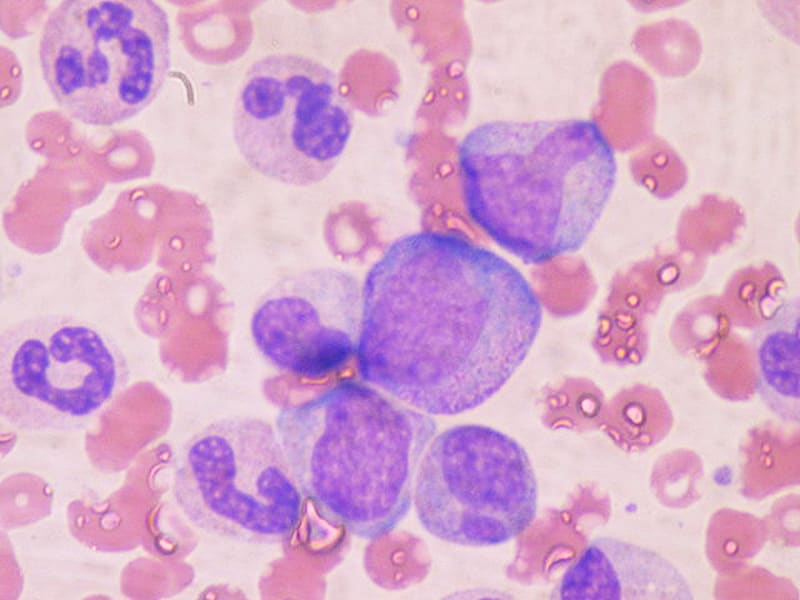
Erythroblasts are immature red blood cells that are produced in the bone marrow. They are also known as normoblasts and are a stage in the development of red blood cells, or erythrocytes. Erythroblasts are larger than mature red blood cells and have a nucleus, which is lost during the maturation process. These cells are responsible for producing hemoglobin, the protein that carries oxygen in the blood. Abnormal increases or decreases in the number of erythroblasts can be indicative of certain medical conditions, such as anemia or leukemia.
Parvovirus B19, Human is a single-stranded DNA virus that primarily infects humans. It belongs to the Parvoviridae family and Erbovirus genus. This virus is the causative agent of erythema infectiosum, also known as fifth disease, a mild, self-limiting illness characterized by a facial rash and occasionally joint pain or inflammation.
Parvovirus B19 has a strong tropism for erythroid progenitor cells in the bone marrow, where it replicates and causes temporary suppression of red blood cell production (aplastic crisis) in individuals with underlying hemolytic disorders such as sickle cell disease or spherocytosis.
Additionally, Parvovirus B19 can cause more severe complications in immunocompromised individuals, pregnant women, and fetuses. Infection during pregnancy may lead to hydrops fetalis, anemia, or even fetal death, particularly in the first and second trimesters. Transmission of the virus occurs primarily through respiratory droplets and occasionally via blood transfusions or vertical transmission from mother to fetus.
Cell differentiation is the process by which a less specialized cell, or stem cell, becomes a more specialized cell type with specific functions and structures. This process involves changes in gene expression, which are regulated by various intracellular signaling pathways and transcription factors. Differentiation results in the development of distinct cell types that make up tissues and organs in multicellular organisms. It is a crucial aspect of embryonic development, tissue repair, and maintenance of homeostasis in the body.
According to the National Institutes of Health (NIH), stem cells are "initial cells" or "precursor cells" that have the ability to differentiate into many different cell types in the body. They can also divide without limit to replenish other cells for as long as the person or animal is still alive.
There are two main types of stem cells: embryonic stem cells, which come from human embryos, and adult stem cells, which are found in various tissues throughout the body. Embryonic stem cells have the ability to differentiate into all cell types in the body, while adult stem cells have more limited differentiation potential.
Stem cells play an essential role in the development and repair of various tissues and organs in the body. They are currently being studied for their potential use in the treatment of a wide range of diseases and conditions, including cancer, diabetes, heart disease, and neurological disorders. However, more research is needed to fully understand the properties and capabilities of these cells before they can be used safely and effectively in clinical settings.
Bone marrow cells are the types of cells found within the bone marrow, which is the spongy tissue inside certain bones in the body. The main function of bone marrow is to produce blood cells. There are two types of bone marrow: red and yellow. Red bone marrow is where most blood cell production takes place, while yellow bone marrow serves as a fat storage site.
The three main types of bone marrow cells are:
1. Hematopoietic stem cells (HSCs): These are immature cells that can differentiate into any type of blood cell, including red blood cells, white blood cells, and platelets. They have the ability to self-renew, meaning they can divide and create more hematopoietic stem cells.
2. Red blood cell progenitors: These are immature cells that will develop into mature red blood cells, also known as erythrocytes. Red blood cells carry oxygen from the lungs to the body's tissues and carbon dioxide back to the lungs.
3. Myeloid and lymphoid white blood cell progenitors: These are immature cells that will develop into various types of white blood cells, which play a crucial role in the body's immune system by fighting infections and diseases. Myeloid progenitors give rise to granulocytes (neutrophils, eosinophils, and basophils), monocytes, and megakaryocytes (which eventually become platelets). Lymphoid progenitors differentiate into B cells, T cells, and natural killer (NK) cells.
Bone marrow cells are essential for maintaining a healthy blood cell count and immune system function. Abnormalities in bone marrow cells can lead to various medical conditions, such as anemia, leukopenia, leukocytosis, thrombocytopenia, or thrombocytosis, depending on the specific type of blood cell affected. Additionally, bone marrow cells are often used in transplantation procedures to treat patients with certain types of cancer, such as leukemia and lymphoma, or other hematologic disorders.
Hematopoiesis is the process of forming and developing blood cells. It occurs in the bone marrow and includes the production of red blood cells (erythropoiesis), white blood cells (leukopoiesis), and platelets (thrombopoiesis). This process is regulated by various growth factors, hormones, and cytokines. Hematopoiesis begins early in fetal development and continues throughout a person's life. Disorders of hematopoiesis can result in conditions such as anemia, leukopenia, leukocytosis, thrombocytopenia, or thrombocytosis.
Stem Cell Factor (SCF), also known as Kit Ligand or Steel Factor, is a growth factor that plays a crucial role in the regulation of hematopoiesis, which is the process of producing various blood cells. It is a glycoprotein that binds to the c-Kit receptor found on hematopoietic stem cells and progenitor cells, promoting their survival, proliferation, and differentiation into mature blood cells.
SCF is involved in the development and function of several types of blood cells, including red blood cells, white blood cells, and platelets. It also plays a role in the maintenance and self-renewal of hematopoietic stem cells, which are essential for the continuous production of new blood cells throughout an individual's lifetime.
In addition to its role in hematopoiesis, SCF has been implicated in various other biological processes, such as melanogenesis, gametogenesis, and tissue repair and regeneration. Dysregulation of SCF signaling has been associated with several diseases, including certain types of cancer, bone marrow failure disorders, and autoimmune diseases.
Thrombopoietin (TPO) is a glycoprotein hormone that plays a crucial role in the regulation of platelet production, also known as thrombopoiesis. It is primarily produced by the liver and to some extent by megakaryocytes, which are the cells responsible for producing platelets.
TPO binds to its receptor, c-Mpl, on the surface of megakaryocytes and their precursor cells, stimulating their proliferation, differentiation, and maturation into platelets. By regulating the number of platelets in circulation, TPO helps maintain hemostasis, the process that prevents excessive bleeding after injury.
In addition to its role in thrombopoiesis, TPO has been shown to have potential effects on other cell types, including hematopoietic stem cells and certain immune cells. However, its primary function remains the regulation of platelet production.
Megakaryocyte progenitor cells are a type of hematopoietic (blood-forming) stem or progenitor cell that give rise to megakaryocytes, which are large cells found in the bone marrow. Megakaryocytes are responsible for producing platelets, also known as thrombocytes, which are small cell fragments that play a crucial role in blood clotting and hemostasis.
Megakaryocyte progenitor cells are characterized by their ability to differentiate into megakaryocytes and express specific surface markers, such as CD34, CD41, and CD61. They can be found in the bone marrow and peripheral blood and can be expanded and differentiated in vitro for therapeutic purposes, such as in platelet production for transfusion therapy.
Abnormalities in megakaryocyte progenitor cells can lead to various hematological disorders, including thrombocytopenia (low platelet count) and myeloproliferative neoplasms (abnormal blood cell growth). Therefore, understanding the biology and regulation of megakaryocyte progenitor cells is essential for developing new diagnostic and therapeutic strategies for these conditions.
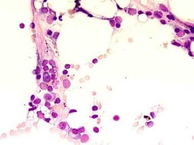
Erythroblastic Leukemia, Acute (also known as Acute Erythroid Leukemia or AEL) is a subtype of acute myeloid leukemia (AML), which is a type of cancer affecting the blood and bone marrow. In this condition, there is an overproduction of erythroblasts (immature red blood cells) in the bone marrow, leading to their accumulation and interference with normal blood cell production. This results in a decrease in the number of functional red blood cells, white blood cells, and platelets in the body. Symptoms may include fatigue, weakness, frequent infections, and easy bruising or bleeding. AEL is typically treated with chemotherapy and sometimes requires stem cell transplantation.
GATA1 (Global Architecture of Tissue/stage-specific Transcription Factors 1) is a transcription factor that belongs to the GATA family, which recognizes and binds to the (A/T)GATA(A/G) motif in the DNA. It plays a crucial role in the development and differentiation of hematopoietic cells, particularly erythroid, megakaryocytic, eosinophilic, and mast cell lineages.
GATA1 regulates gene expression by binding to specific DNA sequences and recruiting other co-factors that modulate chromatin structure and transcriptional activity. Mutations in the GATA1 gene can lead to various blood disorders such as congenital dyserythropoietic anemia type II, Diamond-Blackfan anemia, acute megakaryoblastic leukemia (AMKL), and myelodysplastic syndrome.
In summary, GATA1 Transcription Factor is a protein that binds to specific DNA sequences in the genome and regulates gene expression, playing a critical role in hematopoietic cell development and differentiation.
"Cells, cultured" is a medical term that refers to cells that have been removed from an organism and grown in controlled laboratory conditions outside of the body. This process is called cell culture and it allows scientists to study cells in a more controlled and accessible environment than they would have inside the body. Cultured cells can be derived from a variety of sources, including tissues, organs, or fluids from humans, animals, or cell lines that have been previously established in the laboratory.
Cell culture involves several steps, including isolation of the cells from the tissue, purification and characterization of the cells, and maintenance of the cells in appropriate growth conditions. The cells are typically grown in specialized media that contain nutrients, growth factors, and other components necessary for their survival and proliferation. Cultured cells can be used for a variety of purposes, including basic research, drug development and testing, and production of biological products such as vaccines and gene therapies.
It is important to note that cultured cells may behave differently than they do in the body, and results obtained from cell culture studies may not always translate directly to human physiology or disease. Therefore, it is essential to validate findings from cell culture experiments using additional models and ultimately in clinical trials involving human subjects.
CD34 is a type of antigen that is found on the surface of certain cells in the human body. Specifically, CD34 antigens are present on hematopoietic stem cells, which are immature cells that can develop into different types of blood cells. These stem cells are found in the bone marrow and are responsible for producing red blood cells, white blood cells, and platelets.
CD34 antigens are a type of cell surface marker that is used in medical research and clinical settings to identify and isolate hematopoietic stem cells. They are also used in the development of stem cell therapies and transplantation procedures. CD34 antigens can be detected using various laboratory techniques, such as flow cytometry or immunohistochemistry.
It's important to note that while CD34 is a useful marker for identifying hematopoietic stem cells, it is not exclusive to these cells and can also be found on other cell types, such as endothelial cells that line blood vessels. Therefore, additional markers are often used in combination with CD34 to more specifically identify and isolate hematopoietic stem cells.
Erythrocytes, also known as red blood cells (RBCs), are the most common type of blood cell in circulating blood in mammals. They are responsible for transporting oxygen from the lungs to the body's tissues and carbon dioxide from the tissues to the lungs.
Erythrocytes are formed in the bone marrow and have a biconcave shape, which allows them to fold and bend easily as they pass through narrow blood vessels. They do not have a nucleus or mitochondria, which makes them more flexible but also limits their ability to reproduce or repair themselves.
In humans, erythrocytes are typically disc-shaped and measure about 7 micrometers in diameter. They contain the protein hemoglobin, which binds to oxygen and gives blood its red color. The lifespan of an erythrocyte is approximately 120 days, after which it is broken down in the liver and spleen.
Abnormalities in erythrocyte count or function can lead to various medical conditions, such as anemia, polycythemia, and sickle cell disease.
Bone marrow is the spongy tissue found inside certain bones in the body, such as the hips, thighs, and vertebrae. It is responsible for producing blood-forming cells, including red blood cells, white blood cells, and platelets. There are two types of bone marrow: red marrow, which is involved in blood cell production, and yellow marrow, which contains fatty tissue.
Red bone marrow contains hematopoietic stem cells, which can differentiate into various types of blood cells. These stem cells continuously divide and mature to produce new blood cells that are released into the circulation. Red blood cells carry oxygen throughout the body, white blood cells help fight infections, and platelets play a crucial role in blood clotting.
Bone marrow also serves as a site for immune cell development and maturation. It contains various types of immune cells, such as lymphocytes, macrophages, and dendritic cells, which help protect the body against infections and diseases.
Abnormalities in bone marrow function can lead to several medical conditions, including anemia, leukopenia, thrombocytopenia, and various types of cancer, such as leukemia and multiple myeloma. Bone marrow aspiration and biopsy are common diagnostic procedures used to evaluate bone marrow health and function.
Polycythemia Vera is a type of myeloproliferative neoplasm, a group of rare blood cancers. In Polycythemia Vera, the body produces too many red blood cells, leading to an increased risk of blood clots and thickening of the blood, which can cause various symptoms such as fatigue, headache, dizziness, and itching. It can also lead to enlargement of the spleen. The exact cause of Polycythemia Vera is not known, but it is associated with genetic mutations in the JAK2 gene in most cases. It is a progressive disease that can lead to complications such as bleeding, thrombosis, and transformation into acute leukemia if left untreated.
Thrombopoiesis is the process of formation and development of thrombocytes or platelets, which are small, colorless cell fragments in our blood that play an essential role in clotting. Thrombopoiesis occurs inside the bone marrow, where stem cells differentiate into megakaryoblasts, then progressively develop into promegakaryocytes and megakaryocytes. These megakaryocytes subsequently undergo a process called cytoplasmic fragmentation to produce platelets.
The regulation of thrombopoiesis is primarily controlled by the hormone thrombopoietin (TPO), which is produced mainly in the liver and binds to the thrombopoietin receptor (c-Mpl) on megakaryocytes and their precursors. This binding stimulates the proliferation, differentiation, and maturation of megakaryocytes, leading to an increase in platelet production.
Abnormalities in thrombopoiesis can result in conditions such as thrombocytopenia (low platelet count) or thrombocytosis (high platelet count), which may be associated with bleeding disorders or increased risk of thrombosis, respectively.
Interleukin-3 (IL-3) is a type of cytokine, which is a small signaling protein that modulates the immune response, cell growth, and differentiation. IL-3 is primarily produced by activated T cells and mast cells. It plays an essential role in the survival, proliferation, and differentiation of hematopoietic stem cells, which give rise to all blood cell types. Specifically, IL-3 supports the development of myeloid lineage cells, including basophils, eosinophils, mast cells, megakaryocytes, and erythroid progenitors.
IL-3 binds to its receptor, the interleukin-3 receptor (IL-3R), which consists of two subunits: CD123 (the alpha chain) and CD131 (the beta chain). The binding of IL-3 to its receptor triggers a signaling cascade within the cell that ultimately leads to changes in gene expression, promoting cell growth and differentiation. Dysregulation of IL-3 production or signaling has been implicated in several hematological disorders, such as leukemia and myelodysplastic syndromes.
Erythroid-specific DNA-binding factors are transcription factors that bind to specific sequences of DNA and help regulate the expression of genes that are involved in the development and differentiation of erythroid cells, which are cells that mature to become red blood cells. These transcription factors play a crucial role in the production of hemoglobin, the protein in red blood cells that carries oxygen throughout the body. Examples of erythroid-specific DNA-binding factors include GATA-1 and KLF1.
Cell division is the process by which a single eukaryotic cell (a cell with a true nucleus) divides into two identical daughter cells. This complex process involves several stages, including replication of DNA, separation of chromosomes, and division of the cytoplasm. There are two main types of cell division: mitosis and meiosis.
Mitosis is the type of cell division that results in two genetically identical daughter cells. It is a fundamental process for growth, development, and tissue repair in multicellular organisms. The stages of mitosis include prophase, prometaphase, metaphase, anaphase, and telophase, followed by cytokinesis, which divides the cytoplasm.
Meiosis, on the other hand, is a type of cell division that occurs in the gonads (ovaries and testes) during the production of gametes (sex cells). Meiosis results in four genetically unique daughter cells, each with half the number of chromosomes as the parent cell. This process is essential for sexual reproduction and genetic diversity. The stages of meiosis include meiosis I and meiosis II, which are further divided into prophase, prometaphase, metaphase, anaphase, and telophase.
In summary, cell division is the process by which a single cell divides into two daughter cells, either through mitosis or meiosis. This process is critical for growth, development, tissue repair, and sexual reproduction in multicellular organisms.
'Cell lineage' is a term used in biology and medicine to describe the developmental history or relationship of a cell or group of cells to other cells, tracing back to the original progenitor or stem cell. It refers to the series of cell divisions and differentiation events that give rise to specific types of cells in an organism over time.
In simpler terms, cell lineage is like a family tree for cells, showing how they are related to each other through a chain of cell division and specialization events. This concept is important in understanding the development, growth, and maintenance of tissues and organs in living beings.
Fetal hemoglobin (HbF) is a type of hemoglobin that is produced in the fetus and newborn babies. It is composed of two alpha-like globin chains and two gamma-globin chains, designated as α2γ2. HbF is the primary form of hemoglobin during fetal development, replacing the embryonic hemoglobin (HbG) around the eighth week of gestation.
The unique property of HbF is its higher affinity for oxygen compared to adult hemoglobin (HbA), which helps ensure adequate oxygen supply from the mother to the developing fetus. After birth, as the newborn starts breathing on its own and begins to receive oxygen directly, the production of HbF gradually decreases and is usually replaced by HbA within the first year of life.
In some genetic disorders like sickle cell disease and beta-thalassemia, persistence of HbF into adulthood can be beneficial as it reduces the severity of symptoms due to its higher oxygen-carrying capacity and less polymerization tendency compared to HbS (in sickle cell disease) or unpaired alpha chains (in beta-thalassemia). Treatments like hydroxyurea are used to induce HbF production in these patients as a therapeutic approach.
Polycythemia is a medical condition characterized by an abnormal increase in the total red blood cell (RBC) mass or hematocrit (the percentage of RBCs in the blood). This results in a higher-than-normal viscosity of the blood, which can lead to various complications such as impaired circulation, increased risk of blood clots, and reduced oxygen supply to the tissues.
There are two main types of polycythemia: primary and secondary. Primary polycythemia, also known as polycythemia vera, is a rare myeloproliferative neoplasm caused by genetic mutations that lead to excessive production of RBCs in the bone marrow. Secondary polycythemia, on the other hand, is a reactive condition triggered by various factors such as chronic hypoxia (low oxygen levels), high altitude, smoking, or certain medical conditions like sleep apnea, heart disease, or kidney tumors.
Symptoms of polycythemia may include fatigue, headaches, dizziness, shortness of breath, itching, and a bluish or reddish tint to the skin (cyanosis). Treatment depends on the underlying cause and severity of the condition and may involve phlebotomy, medications to reduce RBC production, and management of associated complications.
K562 cells are a type of human cancer cell that are commonly used in scientific research. They are derived from a patient with chronic myelogenous leukemia (CML), a type of cancer that affects the blood and bone marrow.
K562 cells are often used as a model system to study various biological processes, including cell signaling, gene expression, differentiation, and apoptosis (programmed cell death). They are also commonly used in drug discovery and development, as they can be used to test the effectiveness of potential new therapies against cancer.
K562 cells have several characteristics that make them useful for research purposes. They are easy to grow and maintain in culture, and they can be manipulated genetically to express or knock down specific genes. Additionally, K562 cells are capable of differentiating into various cell types, such as red blood cells and megakaryocytes, which allows researchers to study the mechanisms of cell differentiation.
It's important to note that while K562 cells are a valuable tool for research, they do not fully recapitulate the complexity of human CML or other cancers. Therefore, findings from studies using K562 cells should be validated in more complex model systems or in clinical trials before they can be translated into treatments for patients.
Erythema infectiosum is a viral infection commonly known as "fifth disease." It is caused by the human parvovirus B19 and primarily affects children. The characteristic symptom of erythema infectiosum is a distinctive red rash on the cheeks, which gives the appearance of having been slapped, hence one of its other names, "slapped cheek syndrome." After a few days, the rash may spread to the arms, legs, and trunk, often in a lacy or net-like pattern. The rash is usually not itchy or painful.
In addition to the rash, people with erythema infectiosum may experience mild flu-like symptoms such as fever, headache, and fatigue. Some individuals may also develop joint pain and swelling, particularly adolescents and adults. In most cases, erythema infectiosum is a self-limiting illness that resolves within one to three weeks without specific treatment. However, the rash may come and go for several weeks, especially when exposed to sunlight, heat, or emotional stress.
Erythema infectiosum is usually spread through respiratory droplets when an infected person coughs or sneezes. It can also be transmitted through blood transfusions and from mother to fetus during pregnancy. While most cases of erythema infectiosum are mild, the infection can cause more severe complications in people with weakened immune systems, sickle cell disease, or chronic hemolytic anemia. Pregnant women who contract erythema infectiosum may have a higher risk of miscarriage, stillbirth, or premature delivery, especially during the first half of pregnancy.
Globins are a group of proteins that contain a heme prosthetic group, which binds and transports oxygen in the blood. The most well-known globin is hemoglobin, which is found in red blood cells and is responsible for carrying oxygen from the lungs to the body's tissues. Other members of the globin family include myoglobin, which is found in muscle tissue and stores oxygen, and neuroglobin and cytoglobin, which are found in the brain and other organs and may have roles in protecting against oxidative stress and hypoxia (low oxygen levels). Globins share a similar structure, with a folded protein surrounding a central heme group. Mutations in globin genes can lead to various diseases, such as sickle cell anemia and thalassemia.
Fetal blood refers to the blood circulating in a fetus during pregnancy. It is essential for the growth and development of the fetus, as it carries oxygen and nutrients from the placenta to the developing tissues and organs. Fetal blood also removes waste products, such as carbon dioxide, from the fetal tissues and transports them to the placenta for elimination.
Fetal blood has several unique characteristics that distinguish it from adult blood. For example, fetal hemoglobin (HbF) is the primary type of hemoglobin found in fetal blood, whereas adults primarily have adult hemoglobin (HbA). Fetal hemoglobin has a higher affinity for oxygen than adult hemoglobin, which allows it to more efficiently extract oxygen from the maternal blood in the placenta.
Additionally, fetal blood contains a higher proportion of reticulocytes (immature red blood cells) and nucleated red blood cells compared to adult blood. These differences reflect the high turnover rate of red blood cells in the developing fetus and the need for rapid growth and development.
Examination of fetal blood can provide important information about the health and well-being of the fetus during pregnancy. For example, fetal blood sampling (also known as cordocentesis or percutaneous umbilical blood sampling) can be used to diagnose genetic disorders, infections, and other conditions that may affect fetal development. However, this procedure carries risks, including preterm labor, infection, and fetal loss, and is typically only performed when there is a significant risk of fetal compromise or when other diagnostic tests have been inconclusive.
Cell separation is a process used to separate and isolate specific cell types from a heterogeneous mixture of cells. This can be accomplished through various physical or biological methods, depending on the characteristics of the cells of interest. Some common techniques for cell separation include:
1. Density gradient centrifugation: In this method, a sample containing a mixture of cells is layered onto a density gradient medium and then centrifuged. The cells are separated based on their size, density, and sedimentation rate, with denser cells settling closer to the bottom of the tube and less dense cells remaining near the top.
2. Magnetic-activated cell sorting (MACS): This technique uses magnetic beads coated with antibodies that bind to specific cell surface markers. The labeled cells are then passed through a column placed in a magnetic field, which retains the magnetically labeled cells while allowing unlabeled cells to flow through.
3. Fluorescence-activated cell sorting (FACS): In this method, cells are stained with fluorochrome-conjugated antibodies that recognize specific cell surface or intracellular markers. The stained cells are then passed through a laser beam, which excites the fluorophores and allows for the detection and sorting of individual cells based on their fluorescence profile.
4. Filtration: This simple method relies on the physical size differences between cells to separate them. Cells can be passed through filters with pore sizes that allow smaller cells to pass through while retaining larger cells.
5. Enzymatic digestion: In some cases, cells can be separated by enzymatically dissociating tissues into single-cell suspensions and then using various separation techniques to isolate specific cell types.
These methods are widely used in research and clinical settings for applications such as isolating immune cells, stem cells, or tumor cells from biological samples.
Granulocytes are a type of white blood cell that plays a crucial role in the body's immune system. They are called granulocytes because they contain small granules in their cytoplasm, which are filled with various enzymes and proteins that help them fight off infections and destroy foreign substances.
There are three types of granulocytes: neutrophils, eosinophils, and basophils. Neutrophils are the most abundant type and are primarily responsible for fighting bacterial infections. Eosinophils play a role in defending against parasitic infections and regulating immune responses. Basophils are involved in inflammatory reactions and allergic responses.
Granulocytes are produced in the bone marrow and released into the bloodstream, where they circulate and patrol for any signs of infection or foreign substances. When they encounter a threat, they quickly move to the site of infection or injury and release their granules to destroy the invading organisms or substances.
Abnormal levels of granulocytes in the blood can indicate an underlying medical condition, such as an infection, inflammation, or a bone marrow disorder.
Anemia is a medical condition characterized by a lower than normal number of red blood cells or lower than normal levels of hemoglobin in the blood. Hemoglobin is an important protein in red blood cells that carries oxygen from the lungs to the rest of the body. Anemia can cause fatigue, weakness, shortness of breath, and a pale complexion because the body's tissues are not getting enough oxygen.
Anemia can be caused by various factors, including nutritional deficiencies (such as iron, vitamin B12, or folate deficiency), blood loss, chronic diseases (such as kidney disease or rheumatoid arthritis), inherited genetic disorders (such as sickle cell anemia or thalassemia), and certain medications.
There are different types of anemia, classified based on the underlying cause, size and shape of red blood cells, and the level of hemoglobin in the blood. Treatment for anemia depends on the underlying cause and may include dietary changes, supplements, medication, or blood transfusions.
Glycophorin is a type of protein found on the surface of red blood cells, also known as erythrocytes. These proteins are heavily glycosylated, meaning they have many carbohydrate chains attached to them. Glycophorins play a crucial role in maintaining the structure and flexibility of the red blood cell membrane, and they also help to mediate interactions between the red blood cells and other cells or molecules in the body.
There are several different types of glycophorin proteins, including glycophorin A, B, C, and D. Glycophorin A is the most abundant type and is often used as a marker for identifying the ABO blood group. Mutations in the genes that encode glycophorin proteins can lead to various blood disorders, such as hereditary spherocytosis and hemolytic anemia.
Proto-oncogene proteins c-kit, also known as CD117 or stem cell factor receptor, are transmembrane receptor tyrosine kinases that play crucial roles in various biological processes, including cell survival, proliferation, differentiation, and migration. They are encoded by the c-KIT gene located on human chromosome 4q12.
These proteins consist of an extracellular ligand-binding domain, a transmembrane domain, and an intracellular tyrosine kinase domain. The binding of their ligand, stem cell factor (SCF), leads to receptor dimerization, autophosphorylation, and activation of several downstream signaling pathways such as PI3K/AKT, MAPK/ERK, and JAK/STAT.
Abnormal activation or mutation of c-kit proto-oncogene proteins has been implicated in the development and progression of various malignancies, including gastrointestinal stromal tumors (GISTs), acute myeloid leukemia (AML), mast cell diseases, and melanoma. Targeted therapies against c-kit, such as imatinib mesylate (Gleevec), have shown promising results in the treatment of these malignancies.
Hematopoietic cell growth factors are a group of glycoproteins that stimulate the proliferation, differentiation, and survival of hematopoietic cells, which are the precursor cells that give rise to all blood cells. These growth factors include colony-stimulating factors (CSFs) such as granulocyte-colony stimulating factor (G-CSF), granulocyte-macrophage colony-stimulating factor (GM-CSF), and macrophage colony-stimulating factor (M-CSF), as well as erythropoietin (EPO) and thrombopoietin (TPO).
G-CSF primarily stimulates the production of neutrophils, a type of white blood cell that plays a crucial role in the immune response to bacterial infections. GM-CSF stimulates the production of both granulocytes and monocytes/macrophages, while M-CSF specifically stimulates the production of monocytes/macrophages. EPO stimulates the production of red blood cells, while TPO stimulates the production of platelets.
Hematopoietic cell growth factors are used clinically to treat a variety of conditions associated with impaired hematopoiesis, such as chemotherapy-induced neutropenia, aplastic anemia, and congenital disorders of hematopoiesis. They can also be used to mobilize hematopoietic stem cells from the bone marrow into the peripheral blood for collection and transplantation.
Flow cytometry is a medical and research technique used to measure physical and chemical characteristics of cells or particles, one cell at a time, as they flow in a fluid stream through a beam of light. The properties measured include:
* Cell size (light scatter)
* Cell internal complexity (granularity, also light scatter)
* Presence or absence of specific proteins or other molecules on the cell surface or inside the cell (using fluorescent antibodies or other fluorescent probes)
The technique is widely used in cell counting, cell sorting, protein engineering, biomarker discovery and monitoring disease progression, particularly in hematology, immunology, and cancer research.
Friend murine leukemia virus (F-MuLV) is a type of retrovirus that specifically infects mice. It was first discovered by Charlotte Friend in the 1950s and has since been widely used as a model system to study retroviral pathogenesis, oncogenesis, and immune responses.
F-MuLV is a complex retrovirus that contains several accessory genes, including gag, pol, env, and others. The virus can cause leukemia and other malignancies in susceptible mice, particularly when it is transmitted from mother to offspring through the milk.
The virus is also known to induce immunosuppression, which makes infected mice more susceptible to other infections and diseases. F-MuLV has been used extensively in laboratory research to investigate various aspects of retroviral biology, including viral entry, replication, gene expression, and host immune responses.
It is important to note that Friend murine leukemia virus only infects mice and is not known to cause any disease in humans or other animals.
Recombinant proteins are artificially created proteins produced through the use of recombinant DNA technology. This process involves combining DNA molecules from different sources to create a new set of genes that encode for a specific protein. The resulting recombinant protein can then be expressed, purified, and used for various applications in research, medicine, and industry.
Recombinant proteins are widely used in biomedical research to study protein function, structure, and interactions. They are also used in the development of diagnostic tests, vaccines, and therapeutic drugs. For example, recombinant insulin is a common treatment for diabetes, while recombinant human growth hormone is used to treat growth disorders.
The production of recombinant proteins typically involves the use of host cells, such as bacteria, yeast, or mammalian cells, which are engineered to express the desired protein. The host cells are transformed with a plasmid vector containing the gene of interest, along with regulatory elements that control its expression. Once the host cells are cultured and the protein is expressed, it can be purified using various chromatography techniques.
Overall, recombinant proteins have revolutionized many areas of biology and medicine, enabling researchers to study and manipulate proteins in ways that were previously impossible.
Myeloid progenitor cells are a type of precursor cells that originate from hematopoietic stem cells (HSCs) in the bone marrow. These cells have the ability to differentiate into various types of blood cells, including red blood cells, platelets, and different kinds of white blood cells, specifically granulocytes (neutrophils, eosinophils, and basophils), monocytes, and megakaryocytes. Myeloid progenitor cells are crucial for the maintenance of normal hematopoiesis and immune function. Abnormalities in myeloid progenitor cell differentiation or function can lead to various hematological disorders such as leukemia, myelodysplastic syndromes, and myeloproliferative neoplasms.
Thrombopoietin receptors are a type of cell surface receptor found on megakaryocytes and platelets. They are also known as MPL (myeloproliferative leukemia virus) receptors. Thrombopoietin is a hormone that regulates the production of platelets in the body, and it binds to these receptors to stimulate the proliferation and differentiation of megakaryocytes, which are large bone marrow cells that produce platelets.
The thrombopoietin receptor is a type I transmembrane protein with an extracellular domain that contains the thrombopoietin-binding site, a single transmembrane domain, and an intracellular domain that contains several tyrosine residues that become phosphorylated upon thrombopoietin binding. This triggers a signaling cascade that leads to the activation of various downstream pathways involved in cell proliferation, differentiation, and survival.
Mutations in the thrombopoietin receptor gene have been associated with certain myeloproliferative neoplasms, such as essential thrombocythemia and primary myelofibrosis, which are characterized by excessive platelet production and bone marrow fibrosis.
C57BL/6 (C57 Black 6) is an inbred strain of laboratory mouse that is widely used in biomedical research. The term "inbred" refers to a strain of animals where matings have been carried out between siblings or other closely related individuals for many generations, resulting in a population that is highly homozygous at most genetic loci.
The C57BL/6 strain was established in 1920 by crossing a female mouse from the dilute brown (DBA) strain with a male mouse from the black strain. The resulting offspring were then interbred for many generations to create the inbred C57BL/6 strain.
C57BL/6 mice are known for their robust health, longevity, and ease of handling, making them a popular choice for researchers. They have been used in a wide range of biomedical research areas, including studies of cancer, immunology, neuroscience, cardiovascular disease, and metabolism.
One of the most notable features of the C57BL/6 strain is its sensitivity to certain genetic modifications, such as the introduction of mutations that lead to obesity or impaired glucose tolerance. This has made it a valuable tool for studying the genetic basis of complex diseases and traits.
Overall, the C57BL/6 inbred mouse strain is an important model organism in biomedical research, providing a valuable resource for understanding the genetic and molecular mechanisms underlying human health and disease.
Cell proliferation is the process by which cells increase in number, typically through the process of cell division. In the context of biology and medicine, it refers to the reproduction of cells that makes up living tissue, allowing growth, maintenance, and repair. It involves several stages including the transition from a phase of quiescence (G0 phase) to an active phase (G1 phase), DNA replication in the S phase, and mitosis or M phase, where the cell divides into two daughter cells.
Abnormal or uncontrolled cell proliferation is a characteristic feature of many diseases, including cancer, where deregulated cell cycle control leads to excessive and unregulated growth of cells, forming tumors that can invade surrounding tissues and metastasize to distant sites in the body.
Signal transduction is the process by which a cell converts an extracellular signal, such as a hormone or neurotransmitter, into an intracellular response. This involves a series of molecular events that transmit the signal from the cell surface to the interior of the cell, ultimately resulting in changes in gene expression, protein activity, or metabolism.
The process typically begins with the binding of the extracellular signal to a receptor located on the cell membrane. This binding event activates the receptor, which then triggers a cascade of intracellular signaling molecules, such as second messengers, protein kinases, and ion channels. These molecules amplify and propagate the signal, ultimately leading to the activation or inhibition of specific cellular responses.
Signal transduction pathways are highly regulated and can be modulated by various factors, including other signaling molecules, post-translational modifications, and feedback mechanisms. Dysregulation of these pathways has been implicated in a variety of diseases, including cancer, diabetes, and neurological disorders.
Thrombocytosis is a medical condition characterized by an abnormally high platelet count (also known as thrombocytes) in the blood. Platelets are small cell fragments that play a crucial role in blood clotting. A normal platelet count ranges from 150,000 to 450,000 platelets per microliter of blood. Thrombocytosis is typically defined as a platelet count exceeding 450,000-500,000 platelets/µL.
Thrombocytosis can be classified into two types: reactive (or secondary) thrombocytosis and primary (or essential) thrombocytosis. Reactive thrombocytosis is more common and occurs as a response to an underlying condition, such as infection, inflammation, surgery, or certain types of cancer. Primary thrombocytosis, on the other hand, is caused by intrinsic abnormalities in the bone marrow cells responsible for platelet production (megakaryocytes), and it is often associated with myeloproliferative neoplasms like essential thrombocythemia.
While mild thrombocytosis may not cause any symptoms, higher platelet counts can increase the risk of blood clots (thrombosis) and bleeding disorders due to excessive platelet aggregation. Symptoms of thrombocytosis may include headaches, dizziness, visual disturbances, or chest pain if a blood clot forms in the brain or heart. Bleeding symptoms can manifest as easy bruising, nosebleeds, or gastrointestinal bleeding.
Treatment for thrombocytosis depends on the underlying cause and the severity of the condition. In cases of reactive thrombocytosis, treating the underlying disorder often resolves the high platelet count. For primary thrombocytosis, medications like aspirin or cytoreductive therapy (such as hydroxyurea) may be used to reduce the risk of blood clots and control platelet production. Regular monitoring of platelet counts is essential for managing this condition and preventing potential complications.
A cell line is a culture of cells that are grown in a laboratory for use in research. These cells are usually taken from a single cell or group of cells, and they are able to divide and grow continuously in the lab. Cell lines can come from many different sources, including animals, plants, and humans. They are often used in scientific research to study cellular processes, disease mechanisms, and to test new drugs or treatments. Some common types of human cell lines include HeLa cells (which come from a cancer patient named Henrietta Lacks), HEK293 cells (which come from embryonic kidney cells), and HUVEC cells (which come from umbilical vein endothelial cells). It is important to note that cell lines are not the same as primary cells, which are cells that are taken directly from a living organism and have not been grown in the lab.
Ploidy is a term used in genetics to describe the number of sets of chromosomes in a cell or an organism. The ploidy level can have important implications for genetic inheritance and expression, as well as for evolutionary processes such as speciation and hybridization.
In most animals, including humans, the normal ploidy level is diploid, meaning that each cell contains two sets of chromosomes - one set inherited from each parent. However, there are also many examples of polyploidy, in which an organism has more than two sets of chromosomes.
Polyploidy can arise through various mechanisms, such as genome duplication or hybridization between different species. In some cases, polyploidy may confer evolutionary advantages, such as increased genetic diversity and adaptability to new environments. However, it can also lead to reproductive isolation and the formation of new species.
In plants, polyploidy is relatively common and has played a significant role in their evolution and diversification. Many crop plants are polyploids, including wheat, cotton, and tobacco. In some cases, artificial induction of polyploidy has been used to create new varieties with desirable traits for agriculture and horticulture.
Overall, ploidy is an important concept in genetics and evolution, with implications for a wide range of biological processes and phenomena.
Thrombocytopenia is a medical condition characterized by an abnormally low platelet count (thrombocytes) in the blood. Platelets are small cell fragments that play a crucial role in blood clotting, helping to stop bleeding when a blood vessel is damaged. A healthy adult typically has a platelet count between 150,000 and 450,000 platelets per microliter of blood. Thrombocytopenia is usually diagnosed when the platelet count falls below 150,000 platelets/µL.
Thrombocytopenia can be classified into three main categories based on its underlying cause:
1. Immune thrombocytopenia (ITP): An autoimmune disorder where the immune system mistakenly attacks and destroys its own platelets, leading to a decreased platelet count. ITP can be further divided into primary or secondary forms, depending on whether it occurs alone or as a result of another medical condition or medication.
2. Decreased production: Thrombocytopenia can occur when there is insufficient production of platelets in the bone marrow due to various causes, such as viral infections, chemotherapy, radiation therapy, leukemia, aplastic anemia, or vitamin B12 or folate deficiency.
3. Increased destruction or consumption: Thrombocytopenia can also result from increased platelet destruction or consumption due to conditions like disseminated intravascular coagulation (DIC), thrombotic thrombocytopenic purpura (TTP), hemolytic uremic syndrome (HUS), or severe bacterial infections.
Symptoms of thrombocytopenia may include easy bruising, prolonged bleeding from cuts, spontaneous nosebleeds, bleeding gums, blood in urine or stools, and skin rashes like petechiae (small red or purple spots) or purpura (larger patches). The severity of symptoms can vary depending on the degree of thrombocytopenia and the presence of any underlying conditions. Treatment for thrombocytopenia depends on the cause and may include medications, transfusions, or addressing the underlying condition.
Blood platelets, also known as thrombocytes, are small, colorless cell fragments in our blood that play an essential role in normal blood clotting. They are formed in the bone marrow from large cells called megakaryocytes and circulate in the blood in an inactive state until they are needed to help stop bleeding. When a blood vessel is damaged, platelets become activated and change shape, releasing chemicals that attract more platelets to the site of injury. These activated platelets then stick together to form a plug, or clot, that seals the wound and prevents further blood loss. In addition to their role in clotting, platelets also help to promote healing by releasing growth factors that stimulate the growth of new tissue.
"Spleen Focus-Forming Virus" (SFFV) is not a widely used medical term, but it is a term from the field of virology. SFFV is a type of retrovirus that primarily infects mice and causes erythroleukemia, a cancer of the blood-forming organs. The virus is called "Spleen Focus-Forming" because when it infects mice, it initially replicates in the spleen and forms distinct foci or clusters of infected cells.
The virus contains an oncogene called v-abl, which is a cancer-causing gene that contributes to the development of leukemia in infected animals. SFFV is closely related to another retrovirus called Friend Virus (FV), and together they are referred to as the FV complex. These viruses have been extensively studied as models for retroviral-induced leukemogenesis and have provided valuable insights into the mechanisms of cancer development.
Messenger RNA (mRNA) is a type of RNA (ribonucleic acid) that carries genetic information copied from DNA in the form of a series of three-base code "words," each of which specifies a particular amino acid. This information is used by the cell's machinery to construct proteins, a process known as translation. After being transcribed from DNA, mRNA travels out of the nucleus to the ribosomes in the cytoplasm where protein synthesis occurs. Once the protein has been synthesized, the mRNA may be degraded and recycled. Post-transcriptional modifications can also occur to mRNA, such as alternative splicing and addition of a 5' cap and a poly(A) tail, which can affect its stability, localization, and translation efficiency.
A platelet count is a laboratory test that measures the number of platelets, also known as thrombocytes, in a sample of blood. Platelets are small, colorless cell fragments that circulate in the blood and play a crucial role in blood clotting. They help to stop bleeding by sticking together to form a plug at the site of an injured blood vessel.
A normal platelet count ranges from 150,000 to 450,000 platelets per microliter (µL) of blood. A lower than normal platelet count is called thrombocytopenia, while a higher than normal platelet count is known as thrombocytosis.
Abnormal platelet counts can be a sign of various medical conditions, including bleeding disorders, infections, certain medications, and some types of cancer. It is important to consult with a healthcare provider if you have any concerns about your platelet count or if you experience symptoms such as easy bruising, prolonged bleeding, or excessive menstrual flow.
'Gene expression regulation' refers to the processes that control whether, when, and where a particular gene is expressed, meaning the production of a specific protein or functional RNA encoded by that gene. This complex mechanism can be influenced by various factors such as transcription factors, chromatin remodeling, DNA methylation, non-coding RNAs, and post-transcriptional modifications, among others. Proper regulation of gene expression is crucial for normal cellular function, development, and maintaining homeostasis in living organisms. Dysregulation of gene expression can lead to various diseases, including cancer and genetic disorders.
The spleen is an organ in the upper left side of the abdomen, next to the stomach and behind the ribs. It plays multiple supporting roles in the body:
1. It fights infection by acting as a filter for the blood. Old red blood cells are recycled in the spleen, and platelets and white blood cells are stored there.
2. The spleen also helps to control the amount of blood in the body by removing excess red blood cells and storing platelets.
3. It has an important role in immune function, producing antibodies and removing microorganisms and damaged red blood cells from the bloodstream.
The spleen can be removed without causing any significant problems, as other organs take over its functions. This is known as a splenectomy and may be necessary if the spleen is damaged or diseased.
Proto-oncogene proteins are normal cellular proteins that play crucial roles in various cellular processes, such as signal transduction, cell cycle regulation, and apoptosis (programmed cell death). They are involved in the regulation of cell growth, differentiation, and survival under physiological conditions.
When proto-oncogene proteins undergo mutations or aberrations in their expression levels, they can transform into oncogenic forms, leading to uncontrolled cell growth and division. These altered proteins are then referred to as oncogene products or oncoproteins. Oncogenic mutations can occur due to various factors, including genetic predisposition, environmental exposures, and aging.
Examples of proto-oncogene proteins include:
1. Ras proteins: Involved in signal transduction pathways that regulate cell growth and differentiation. Activating mutations in Ras genes are found in various human cancers.
2. Myc proteins: Regulate gene expression related to cell cycle progression, apoptosis, and metabolism. Overexpression of Myc proteins is associated with several types of cancer.
3. EGFR (Epidermal Growth Factor Receptor): A transmembrane receptor tyrosine kinase that regulates cell proliferation, survival, and differentiation. Mutations or overexpression of EGFR are linked to various malignancies, such as lung cancer and glioblastoma.
4. Src family kinases: Intracellular tyrosine kinases that regulate signal transduction pathways involved in cell proliferation, survival, and migration. Dysregulation of Src family kinases is implicated in several types of cancer.
5. Abl kinases: Cytoplasmic tyrosine kinases that regulate various cellular processes, including cell growth, differentiation, and stress responses. Aberrant activation of Abl kinases, as seen in chronic myelogenous leukemia (CML), leads to uncontrolled cell proliferation.
Understanding the roles of proto-oncogene proteins and their dysregulation in cancer development is essential for developing targeted cancer therapies that aim to inhibit or modulate these aberrant signaling pathways.
Transcription factors are proteins that play a crucial role in regulating gene expression by controlling the transcription of DNA to messenger RNA (mRNA). They function by binding to specific DNA sequences, known as response elements, located in the promoter region or enhancer regions of target genes. This binding can either activate or repress the initiation of transcription, depending on the properties and interactions of the particular transcription factor. Transcription factors often act as part of a complex network of regulatory proteins that determine the precise spatiotemporal patterns of gene expression during development, differentiation, and homeostasis in an organism.
Parvoviridae infections refer to diseases caused by viruses belonging to the Parvoviridae family. These viruses are known to infect a wide range of hosts, including humans, animals, and insects. The most well-known member of this family is the human parvovirus B19, which is responsible for a variety of clinical manifestations such as:
1. Erythema infectiosum (Fifth disease): A common childhood exanthem characterized by a "slapped cheek" rash and a lace-like rash on the extremities.
2. Transient aplastic crisis: A sudden and temporary halt in red blood cell production, which can lead to severe anemia in individuals with underlying hematologic disorders.
3. Hydrops fetalis: Intrauterine death due to severe anemia caused by parvovirus B19 infection in pregnant women, leading to heart failure and widespread fluid accumulation in the fetus.
Parvoviruses are small, non-enveloped viruses with a single-stranded DNA genome. They primarily infect and replicate within actively dividing cells, making them particularly harmful to rapidly proliferating tissues such as bone marrow and fetal tissues. In addition to parvovirus B19, other Parvoviridae family members can cause significant diseases in animals, including cats, dogs, and livestock.
Janus Kinase 2 (JAK2) is a tyrosine kinase enzyme that plays a crucial role in intracellular signal transduction. It is named after the Roman god Janus, who is depicted with two faces, as JAK2 has two similar phosphate-transferring domains. JAK2 is involved in various cytokine receptor-mediated signaling pathways and contributes to hematopoiesis, immune function, and cell growth.
Mutations in the JAK2 gene have been associated with several myeloproliferative neoplasms (MPNs), including polycythemia vera, essential thrombocythemia, and primary myelofibrosis. The most common mutation is JAK2 V617F, which results in a constitutively active enzyme that promotes uncontrolled cell proliferation and survival, contributing to the development of these MPNs.
Colony-stimulating factors (CSFs) are a group of growth factors that stimulate the production of blood cells in the bone marrow. They include granulocyte colony-stimulating factor (G-CSF), granulocyte-macrophage colony-stimulating factor (GM-CSF), and macrophage colony-stimulating factor (M-CSF). These factors play an important role in the regulation of hematopoiesis, which is the process of producing different types of blood cells.
G-CSF stimulates the production of neutrophils, a type of white blood cell that helps fight against bacterial and fungal infections. GM-CSF stimulates the production of both neutrophils and monocytes/macrophages, which are important in the immune response to infection and tissue injury. M-CSF stimulates the production and activation of macrophages, which play a role in the immune response, wound healing, and the regulation of hematopoiesis.
Colony-stimulating factors are used clinically to stimulate the production of white blood cells in patients undergoing chemotherapy or radiation therapy, which can suppress bone marrow function and lead to low white blood cell counts. They are also used to mobilize stem cells from the bone marrow into the peripheral blood for collection and transplantation.
Transferrin receptors are membrane-bound proteins found on the surface of many cell types, including red and white blood cells, as well as various tissues such as the liver, brain, and placenta. These receptors play a crucial role in iron homeostasis by regulating the uptake of transferrin, an iron-binding protein, into the cells.
Transferrin binds to two ferric ions (Fe3+) in the bloodstream, forming a complex known as holo-transferrin. This complex then interacts with the transferrin receptors on the cell surface, leading to endocytosis of the transferrin-receptor complex into the cell. Once inside the cell, the acidic environment within the endosome causes the release of iron ions from the transferrin molecule, which can then be transported into the cytoplasm for use in various metabolic processes.
After releasing the iron, the apo-transferrin (iron-free transferrin) is recycled back to the cell surface and released back into the bloodstream, where it can bind to more ferric ions and repeat the cycle. This process helps maintain appropriate iron levels within the body and ensures that cells have access to the iron they need for essential functions such as DNA synthesis, energy production, and oxygen transport.
In summary, transferrin receptors are membrane-bound proteins responsible for recognizing and facilitating the uptake of transferrin-bound iron into cells, playing a critical role in maintaining iron homeostasis within the body.
Stat5 (Signal Transducer and Activator of Transcription 5) is a transcription factor that plays a crucial role in various cellular processes, including growth, survival, and differentiation. It exists in two closely related isoforms, Stat5a and Stat5b, which are encoded by separate genes but share significant sequence homology and functional similarity.
When activated through phosphorylation by receptor or non-receptor tyrosine kinases, Stat5 forms homodimers or heterodimers that translocate to the nucleus. Once in the nucleus, these dimers bind to specific DNA sequences called Stat-binding elements (SBEs) in the promoter regions of target genes, leading to their transcriptional activation or repression.
Stat5 is involved in various physiological and pathological conditions, such as hematopoiesis, lactation, immune response, and cancer progression. Dysregulation of Stat5 signaling has been implicated in several malignancies, including leukemias, lymphomas, and breast cancer, making it an attractive therapeutic target for these diseases.
A base sequence in the context of molecular biology refers to the specific order of nucleotides in a DNA or RNA molecule. In DNA, these nucleotides are adenine (A), guanine (G), cytosine (C), and thymine (T). In RNA, uracil (U) takes the place of thymine. The base sequence contains genetic information that is transcribed into RNA and ultimately translated into proteins. It is the exact order of these bases that determines the genetic code and thus the function of the DNA or RNA molecule.
Hemin is defined as the iron(III) complex of protoporphyrin IX, which is a porphyrin derivative. It is a naturally occurring substance that is involved in various biological processes, most notably in the form of heme, which is a component of hemoglobin and other hemoproteins. Hemin is also used in medical research and therapy, such as in the treatment of methemoglobinemia and lead poisoning.
Gamma-globulins are a type of globulin, which are proteins found in the blood plasma. More specifically, gamma-globulins are a class of immunoglobulins, also known as antibodies, that play a crucial role in the immune system's response to foreign substances and infectious agents.
Immunoglobulins are divided into several classes based on their structure and function. Gamma-globulins include IgG, IgA, and IgD isotypes of immunoglobulins. Among these, IgG is the most abundant type found in the blood and other body fluids, responsible for providing protection against bacterial and viral infections.
Gamma-globulins are produced by B cells, a type of white blood cell involved in the immune response. They can be measured in the blood as part of a complete blood count (CBC) or specific protein electrophoresis tests to assess immune system function or diagnose various medical conditions such as infections, inflammation, and autoimmune disorders.
Molecular sequence data refers to the specific arrangement of molecules, most commonly nucleotides in DNA or RNA, or amino acids in proteins, that make up a biological macromolecule. This data is generated through laboratory techniques such as sequencing, and provides information about the exact order of the constituent molecules. This data is crucial in various fields of biology, including genetics, evolution, and molecular biology, allowing for comparisons between different organisms, identification of genetic variations, and studies of gene function and regulation.
DNA-binding proteins are a type of protein that have the ability to bind to DNA (deoxyribonucleic acid), the genetic material of organisms. These proteins play crucial roles in various biological processes, such as regulation of gene expression, DNA replication, repair and recombination.
The binding of DNA-binding proteins to specific DNA sequences is mediated by non-covalent interactions, including electrostatic, hydrogen bonding, and van der Waals forces. The specificity of binding is determined by the recognition of particular nucleotide sequences or structural features of the DNA molecule.
DNA-binding proteins can be classified into several categories based on their structure and function, such as transcription factors, histones, and restriction enzymes. Transcription factors are a major class of DNA-binding proteins that regulate gene expression by binding to specific DNA sequences in the promoter region of genes and recruiting other proteins to modulate transcription. Histones are DNA-binding proteins that package DNA into nucleosomes, the basic unit of chromatin structure. Restriction enzymes are DNA-binding proteins that recognize and cleave specific DNA sequences, and are widely used in molecular biology research and biotechnology applications.
Apoptosis is a programmed and controlled cell death process that occurs in multicellular organisms. It is a natural process that helps maintain tissue homeostasis by eliminating damaged, infected, or unwanted cells. During apoptosis, the cell undergoes a series of morphological changes, including cell shrinkage, chromatin condensation, and fragmentation into membrane-bound vesicles called apoptotic bodies. These bodies are then recognized and engulfed by neighboring cells or phagocytic cells, preventing an inflammatory response. Apoptosis is regulated by a complex network of intracellular signaling pathways that involve proteins such as caspases, Bcl-2 family members, and inhibitors of apoptosis (IAPs).
The yolk sac is a structure that forms in the early stages of an embryo's development. It is a extra-embryonic membrane, which means it exists outside of the developing embryo, and it plays a critical role in providing nutrients to the growing embryo during the initial stages of development.
In more detail, the yolk sac is responsible for producing blood cells, contributing to the formation of the early circulatory system, and storing nutrients that are absorbed from the yolk material inside the egg or uterus. The yolk sac also has a role in the development of the gut and the immune system.
As the embryo grows and the placenta develops, the yolk sac's function becomes less critical, and it eventually degenerates. However, remnants of the yolk sac can sometimes persist and may be found in the developing fetus or newborn baby. In some cases, abnormalities in the development or regression of the yolk sac can lead to developmental problems or congenital disorders.
Transgenic mice are genetically modified rodents that have incorporated foreign DNA (exogenous DNA) into their own genome. This is typically done through the use of recombinant DNA technology, where a specific gene or genetic sequence of interest is isolated and then introduced into the mouse embryo. The resulting transgenic mice can then express the protein encoded by the foreign gene, allowing researchers to study its function in a living organism.
The process of creating transgenic mice usually involves microinjecting the exogenous DNA into the pronucleus of a fertilized egg, which is then implanted into a surrogate mother. The offspring that result from this procedure are screened for the presence of the foreign DNA, and those that carry the desired genetic modification are used to establish a transgenic mouse line.
Transgenic mice have been widely used in biomedical research to model human diseases, study gene function, and test new therapies. They provide a valuable tool for understanding complex biological processes and developing new treatments for a variety of medical conditions.
Parvoviridae is a family of small, non-enveloped viruses that infect a wide range of hosts, including humans, animals, and birds. These viruses have a single-stranded DNA genome and replicate in the nucleus of infected cells. They are resistant to heat, acid, and organic solvents, making them difficult to inactivate.
The family Parvoviridae is divided into two subfamilies: Parvovirinae and Densovirinae. Parvovirinae infect vertebrates, while Densovirinae infect invertebrates. The subfamily Parvovirinae includes several genera that infect various hosts, such as humans, dogs, cats, and primates.
Parvovirus B19 is a well-known member of this family that causes a variety of clinical manifestations in humans, including fifth disease (slapped cheek syndrome), arthralgia, and occasionally more severe diseases in immunocompromised individuals or those with certain hematological disorders.
In animals, parvoviruses can cause serious diseases such as canine parvovirus infection in dogs and feline panleukopenia in cats, which can be fatal if left untreated.
Experimental leukemia refers to the stage of research or clinical trials where new therapies, treatments, or diagnostic methods are being studied for leukemia. Leukemia is a type of cancer that affects the blood and bone marrow, leading to an overproduction of abnormal white blood cells.
In the experimental stage, researchers investigate various aspects of leukemia, such as its causes, progression, and potential treatments. They may conduct laboratory studies using cell cultures or animal models to understand the disease better and test new therapeutic approaches. Additionally, clinical trials may be conducted to evaluate the safety and efficacy of novel treatments in human patients with leukemia.
Experimental research in leukemia is crucial for advancing our understanding of the disease and developing more effective treatment strategies. It involves a rigorous and systematic process that adheres to ethical guidelines and scientific standards to ensure the validity and reliability of the findings.
Glycoprotein IIb (also known as integrin αIIbβ3 or CD41/CD61) is a type of protein found on the surface of platelets, which are small cell fragments involved in blood clotting. This glycoprotein plays a crucial role in the final pathway of platelet activation and aggregation, which ultimately leads to the formation of a clot to stop bleeding.
More specifically, Glycoprotein IIb is responsible for binding fibrinogen, von Willebrand factor, and other adhesive proteins in the blood, allowing platelets to bind together and form a clot. Mutations or defects in this glycoprotein can lead to bleeding disorders such as Glanzmann thrombasthenia, which is characterized by abnormal platelet function and excessive bleeding.
Developmental gene expression regulation refers to the processes that control the activation or repression of specific genes during embryonic and fetal development. These regulatory mechanisms ensure that genes are expressed at the right time, in the right cells, and at appropriate levels to guide proper growth, differentiation, and morphogenesis of an organism.
Developmental gene expression regulation is a complex and dynamic process involving various molecular players, such as transcription factors, chromatin modifiers, non-coding RNAs, and signaling molecules. These regulators can interact with cis-regulatory elements, like enhancers and promoters, to fine-tune the spatiotemporal patterns of gene expression during development.
Dysregulation of developmental gene expression can lead to various congenital disorders and developmental abnormalities. Therefore, understanding the principles and mechanisms governing developmental gene expression regulation is crucial for uncovering the etiology of developmental diseases and devising potential therapeutic strategies.
CD (cluster of differentiation) antigens are cell-surface proteins that are expressed on leukocytes (white blood cells) and can be used to identify and distinguish different subsets of these cells. They are important markers in the field of immunology and hematology, and are commonly used to diagnose and monitor various diseases, including cancer, autoimmune disorders, and infectious diseases.
CD antigens are designated by numbers, such as CD4, CD8, CD19, etc., which refer to specific proteins found on the surface of different types of leukocytes. For example, CD4 is a protein found on the surface of helper T cells, while CD8 is found on cytotoxic T cells.
CD antigens can be used as targets for immunotherapy, such as monoclonal antibody therapy, in which antibodies are designed to bind to specific CD antigens and trigger an immune response against cancer cells or infected cells. They can also be used as markers to monitor the effectiveness of treatments and to detect minimal residual disease (MRD) after treatment.
It's important to note that not all CD antigens are exclusive to leukocytes, some can be found on other cell types as well, and their expression can vary depending on the activation state or differentiation stage of the cells.
Cell survival refers to the ability of a cell to continue living and functioning normally, despite being exposed to potentially harmful conditions or treatments. This can include exposure to toxins, radiation, chemotherapeutic drugs, or other stressors that can damage cells or interfere with their normal processes.
In scientific research, measures of cell survival are often used to evaluate the effectiveness of various therapies or treatments. For example, researchers may expose cells to a particular drug or treatment and then measure the percentage of cells that survive to assess its potential therapeutic value. Similarly, in toxicology studies, measures of cell survival can help to determine the safety of various chemicals or substances.
It's important to note that cell survival is not the same as cell proliferation, which refers to the ability of cells to divide and multiply. While some treatments may promote cell survival, they may also inhibit cell proliferation, making them useful for treating diseases such as cancer. Conversely, other treatments may be designed to specifically target and kill cancer cells, even if it means sacrificing some healthy cells in the process.
Neural stem cells (NSCs) are a type of undifferentiated cells found in the central nervous system, including the brain and spinal cord. They have the ability to self-renew and generate the main types of cells found in the nervous system, such as neurons, astrocytes, and oligodendrocytes. NSCs are capable of dividing symmetrically to increase their own population or asymmetrically to produce one stem cell and one differentiated cell. They play a crucial role in the development and maintenance of the nervous system, and have the potential to be used in regenerative medicine and therapies for neurological disorders and injuries.
"Cell count" is a medical term that refers to the process of determining the number of cells present in a given volume or sample of fluid or tissue. This can be done through various laboratory methods, such as counting individual cells under a microscope using a specialized grid called a hemocytometer, or using automated cell counters that use light scattering and electrical impedance techniques to count and classify different types of cells.
Cell counts are used in a variety of medical contexts, including hematology (the study of blood and blood-forming tissues), microbiology (the study of microscopic organisms), and pathology (the study of diseases and their causes). For example, a complete blood count (CBC) is a routine laboratory test that includes a white blood cell (WBC) count, red blood cell (RBC) count, hemoglobin level, hematocrit value, and platelet count. Abnormal cell counts can indicate the presence of various medical conditions, such as infections, anemia, or leukemia.
Granulocyte Colony-Stimulating Factor (G-CSF) is a type of growth factor that specifically stimulates the production and survival of granulocytes, a type of white blood cell crucial for fighting off infections. G-CSF works by promoting the proliferation and differentiation of hematopoietic stem cells into mature granulocytes, primarily neutrophils, in the bone marrow.
Recombinant forms of G-CSF are used clinically as a medication to boost white blood cell production in patients undergoing chemotherapy or radiation therapy for cancer, those with congenital neutropenia, and those who have had a bone marrow transplant. By increasing the number of circulating neutrophils, G-CSF helps reduce the risk of severe infections during periods of intense immune suppression.
Examples of recombinant G-CSF medications include filgrastim (Neupogen), pegfilgrastim (Neulasta), and lipegfilgrastim (Lonquex).
In the field of medicine, "time factors" refer to the duration of symptoms or time elapsed since the onset of a medical condition, which can have significant implications for diagnosis and treatment. Understanding time factors is crucial in determining the progression of a disease, evaluating the effectiveness of treatments, and making critical decisions regarding patient care.
For example, in stroke management, "time is brain," meaning that rapid intervention within a specific time frame (usually within 4.5 hours) is essential to administering tissue plasminogen activator (tPA), a clot-busting drug that can minimize brain damage and improve patient outcomes. Similarly, in trauma care, the "golden hour" concept emphasizes the importance of providing definitive care within the first 60 minutes after injury to increase survival rates and reduce morbidity.
Time factors also play a role in monitoring the progression of chronic conditions like diabetes or heart disease, where regular follow-ups and assessments help determine appropriate treatment adjustments and prevent complications. In infectious diseases, time factors are crucial for initiating antibiotic therapy and identifying potential outbreaks to control their spread.
Overall, "time factors" encompass the significance of recognizing and acting promptly in various medical scenarios to optimize patient outcomes and provide effective care.
Hemoglobin (Hb or Hgb) is the main oxygen-carrying protein in the red blood cells, which are responsible for delivering oxygen throughout the body. It is a complex molecule made up of four globin proteins and four heme groups. Each heme group contains an iron atom that binds to one molecule of oxygen. Hemoglobin plays a crucial role in the transport of oxygen from the lungs to the body's tissues, and also helps to carry carbon dioxide back to the lungs for exhalation.
There are several types of hemoglobin present in the human body, including:
* Hemoglobin A (HbA): This is the most common type of hemoglobin, making up about 95-98% of total hemoglobin in adults. It consists of two alpha and two beta globin chains.
* Hemoglobin A2 (HbA2): This makes up about 1.5-3.5% of total hemoglobin in adults. It consists of two alpha and two delta globin chains.
* Hemoglobin F (HbF): This is the main type of hemoglobin present in fetal life, but it persists at low levels in adults. It consists of two alpha and two gamma globin chains.
* Hemoglobin S (HbS): This is an abnormal form of hemoglobin that can cause sickle cell disease when it occurs in the homozygous state (i.e., both copies of the gene are affected). It results from a single amino acid substitution in the beta globin chain.
* Hemoglobin C (HbC): This is another abnormal form of hemoglobin that can cause mild to moderate hemolytic anemia when it occurs in the homozygous state. It results from a different single amino acid substitution in the beta globin chain than HbS.
Abnormal forms of hemoglobin, such as HbS and HbC, can lead to various clinical disorders, including sickle cell disease, thalassemia, and other hemoglobinopathies.
A "knockout" mouse is a genetically engineered mouse in which one or more genes have been deleted or "knocked out" using molecular biology techniques. This allows researchers to study the function of specific genes and their role in various biological processes, as well as potential associations with human diseases. The mice are generated by introducing targeted DNA modifications into embryonic stem cells, which are then used to create a live animal. Knockout mice have been widely used in biomedical research to investigate gene function, disease mechanisms, and potential therapeutic targets.
Blood cells are the formed elements in the blood, including red blood cells (erythrocytes), white blood cells (leukocytes), and platelets (thrombocytes). These cells are produced in the bone marrow and play crucial roles in the body's functions. Red blood cells are responsible for carrying oxygen to tissues and carbon dioxide away from them, while white blood cells are part of the immune system and help defend against infection and disease. Platelets are cell fragments that are essential for normal blood clotting.
Granulocyte-Macrophage Colony-Stimulating Factor (GM-CSF) is a type of cytokine, which is a small signaling protein involved in immune response and hematopoiesis (the formation of blood cells). GM-CSF's specific role is to stimulate the production, proliferation, and activation of granulocytes (a type of white blood cell that fights against infection) and macrophages (large white blood cells that eat foreign substances, bacteria, and dead or dying cells).
In medical terms, GM-CSF is often used in therapeutic settings to boost the production of white blood cells in patients undergoing chemotherapy or radiation treatment for cancer. This can help to reduce the risk of infection during these treatments. It can also be used to promote the growth and differentiation of stem cells in bone marrow transplant procedures.
Reverse Transcriptase Polymerase Chain Reaction (RT-PCR) is a laboratory technique used in molecular biology to amplify and detect specific DNA sequences. This technique is particularly useful for the detection and quantification of RNA viruses, as well as for the analysis of gene expression.
The process involves two main steps: reverse transcription and polymerase chain reaction (PCR). In the first step, reverse transcriptase enzyme is used to convert RNA into complementary DNA (cDNA) by reading the template provided by the RNA molecule. This cDNA then serves as a template for the PCR amplification step.
In the second step, the PCR reaction uses two primers that flank the target DNA sequence and a thermostable polymerase enzyme to repeatedly copy the targeted cDNA sequence. The reaction mixture is heated and cooled in cycles, allowing the primers to anneal to the template, and the polymerase to extend the new strand. This results in exponential amplification of the target DNA sequence, making it possible to detect even small amounts of RNA or cDNA.
RT-PCR is a sensitive and specific technique that has many applications in medical research and diagnostics, including the detection of viruses such as HIV, hepatitis C virus, and SARS-CoV-2 (the virus that causes COVID-19). It can also be used to study gene expression, identify genetic mutations, and diagnose genetic disorders.
GATA2 transcription factor is a protein that plays a crucial role in the development and function of blood cells. It belongs to the family of GATA transcription factors, which are characterized by their ability to bind to specific DNA sequences called GATA motifs, through a zinc finger domain. The GATA2 transcription factor, in particular, is essential for the development of hematopoietic stem and progenitor cells (HSPCs), which give rise to all blood cell types.
GATA2 binds to the regulatory regions of genes involved in hematopoiesis and modulates their transcription, thereby controlling the differentiation, proliferation, and survival of HSPCs. Mutations in the GATA2 gene have been associated with various hematological disorders, such as acute myeloid leukemia (AML), myelodysplastic syndrome (MDS), and severe congenital neutropenia. These genetic alterations can lead to impaired hematopoiesis, dysfunctional immune cells, and an increased risk of developing blood cancers.
In summary, GATA2 transcription factor is a protein that regulates the development and function of blood cells by controlling the expression of genes involved in hematopoiesis. Genetic defects in this transcription factor can result in various hematological disorders and predispose individuals to blood cancers.
Megakaryocyte-Erythroid Progenitor (MEP) cells are a type of hematopoietic progenitor cell found in the bone marrow. These cells are responsible for giving rise to two types of blood cells: megakaryocytes and erythrocytes.
Megakaryocytes are large cells that produce platelets, which are essential for blood clotting. Erythrocytes, also known as red blood cells, are responsible for carrying oxygen throughout the body.
MEP cells are a type of common myeloid progenitor (CMP) cell and are downstream of hematopoietic stem cells (HSCs). They are characterized by their ability to differentiate into both megakaryocytes and erythrocytes, but not into other types of blood cells such as lymphocytes or granulocytes.
MEP cells express specific surface markers that can be used to identify them, including CD34, CD38, CD41, and CD42b. They are an important focus of research in the field of hematopoiesis and blood disorders such as thrombocytopenia (low platelet count) and anemia (low red blood cell count).
Gene expression is the process by which the information encoded in a gene is used to synthesize a functional gene product, such as a protein or RNA molecule. This process involves several steps: transcription, RNA processing, and translation. During transcription, the genetic information in DNA is copied into a complementary RNA molecule, known as messenger RNA (mRNA). The mRNA then undergoes RNA processing, which includes adding a cap and tail to the mRNA and splicing out non-coding regions called introns. The resulting mature mRNA is then translated into a protein on ribosomes in the cytoplasm through the process of translation.
The regulation of gene expression is a complex and highly controlled process that allows cells to respond to changes in their environment, such as growth factors, hormones, and stress signals. This regulation can occur at various stages of gene expression, including transcriptional activation or repression, RNA processing, mRNA stability, and translation. Dysregulation of gene expression has been implicated in many diseases, including cancer, genetic disorders, and neurological conditions.
Multipotent stem cells are a type of stem cell that have the ability to differentiate into multiple cell types, but are more limited than pluripotent stem cells. These stem cells are found in various tissues and organs throughout the body, including bone marrow, adipose tissue, and dental pulp. They can give rise to a number of different cell types within their own germ layer (endoderm, mesoderm, or ectoderm), but cannot cross germ layer boundaries. For example, multipotent stem cells found in bone marrow can differentiate into various blood cells such as red and white blood cells, but they cannot differentiate into nerve cells or liver cells. These stem cells play important roles in tissue repair and regeneration, and have potential therapeutic applications in regenerative medicine.
A "Blood Cell Count" is a medical laboratory test that measures the number of red blood cells (RBCs), white blood cells (WBCs), and platelets in a sample of blood. This test is often used as a part of a routine check-up or to help diagnose various medical conditions, such as anemia, infection, inflammation, and many others.
The RBC count measures the number of oxygen-carrying cells in the blood, while the WBC count measures the number of immune cells that help fight infections. The platelet count measures the number of cells involved in clotting. Abnormal results in any of these counts may indicate an underlying medical condition and further testing may be required for diagnosis and treatment.
CD36 is a type of protein found on the surface of certain cells in the human body, including platelets, white blood cells (monocytes and macrophages), and fat (adipose) cells. It is a type of scavenger receptor that plays a role in various biological processes, such as:
1. Fatty acid uptake and metabolism: CD36 helps facilitate the transport of long-chain fatty acids into cells for energy production and storage.
2. Inflammation and immune response: CD36 is involved in the recognition and clearance of foreign substances (pathogens) and damaged or dying cells, which can trigger an immune response.
3. Angiogenesis: CD36 has been implicated in the regulation of blood vessel formation (angiogenesis), particularly during wound healing and tumor growth.
4. Atherosclerosis: CD36 has been associated with the development and progression of atherosclerosis, a condition characterized by the buildup of fats, cholesterol, and other substances in and on the artery walls. This is due to its role in the uptake of oxidized low-density lipoprotein (oxLDL) by macrophages, leading to the formation of foam cells and the development of fatty streaks in the arterial wall.
5. Infectious diseases: CD36 has been identified as a receptor for various pathogens, including malaria parasites, HIV, and some bacteria, which can use this protein to gain entry into host cells.
As an antigen, CD36 is a molecule that can be targeted by the immune system to produce an immune response. Antibodies against CD36 have been found in various diseases, such as autoimmune disorders and certain infections. Modulation of CD36 activity has been suggested as a potential therapeutic strategy for several conditions, including atherosclerosis, diabetes, and infectious diseases.
Endothelial cells are the type of cells that line the inner surface of blood vessels, lymphatic vessels, and heart chambers. They play a crucial role in maintaining vascular homeostasis by controlling vasomotor tone, coagulation, platelet activation, and inflammation. Endothelial cells also regulate the transport of molecules between the blood and surrounding tissues, and contribute to the maintenance of the structural integrity of the vasculature. They are flat, elongated cells with a unique morphology that allows them to form a continuous, nonthrombogenic lining inside the vessels. Endothelial cells can be isolated from various tissues and cultured in vitro for research purposes.
The liver is a large, solid organ located in the upper right portion of the abdomen, beneath the diaphragm and above the stomach. It plays a vital role in several bodily functions, including:
1. Metabolism: The liver helps to metabolize carbohydrates, fats, and proteins from the food we eat into energy and nutrients that our bodies can use.
2. Detoxification: The liver detoxifies harmful substances in the body by breaking them down into less toxic forms or excreting them through bile.
3. Synthesis: The liver synthesizes important proteins, such as albumin and clotting factors, that are necessary for proper bodily function.
4. Storage: The liver stores glucose, vitamins, and minerals that can be released when the body needs them.
5. Bile production: The liver produces bile, a digestive juice that helps to break down fats in the small intestine.
6. Immune function: The liver plays a role in the immune system by filtering out bacteria and other harmful substances from the blood.
Overall, the liver is an essential organ that plays a critical role in maintaining overall health and well-being.
Acute Megakaryoblastic Leukemia (AMKL) is a type of cancer that affects the blood and bone marrow. Specifically, it is a subtype of acute myeloid leukemia (AML), which is characterized by the rapid growth of abnormal cells in the bone marrow that interfere with the production of normal blood cells.
In AMKL, the abnormal cells are megakaryoblasts, which are immature cells that should develop into platelet-producing cells called megakaryocytes. However, in AMKL, these cells do not mature properly and instead accumulate in the bone marrow and bloodstream, leading to a shortage of healthy blood cells.
Symptoms of AMKL may include fatigue, weakness, frequent infections, easy bruising or bleeding, and the appearance of small red spots on the skin (petechiae). Diagnosis typically involves a combination of physical exam, medical history, blood tests, bone marrow aspiration and biopsy, and sometimes imaging studies.
Treatment for AMKL usually involves a combination of chemotherapy, radiation therapy, and/or stem cell transplantation. The specific treatment plan will depend on several factors, including the patient's age, overall health, and the extent of the disease.
'Tumor cells, cultured' refers to the process of removing cancerous cells from a tumor and growing them in controlled laboratory conditions. This is typically done by isolating the tumor cells from a patient's tissue sample, then placing them in a nutrient-rich environment that promotes their growth and multiplication.
The resulting cultured tumor cells can be used for various research purposes, including the study of cancer biology, drug development, and toxicity testing. They provide a valuable tool for researchers to better understand the behavior and characteristics of cancer cells outside of the human body, which can lead to the development of more effective cancer treatments.
It is important to note that cultured tumor cells may not always behave exactly the same way as they do in the human body, so findings from cell culture studies must be validated through further research, such as animal models or clinical trials.
The NF-E2 (Nuclear Factor, Erythroid-derived 2) transcription factor is a heterodimeric protein that plays a crucial role in the regulation of gene expression. It is composed of two subunits: p18 and p45. The p45 subunit, also known as NFE2L2 or GABPalpha, is a member of the basic region-leucine zipper (bZIP) family of transcription factors.
The p45 subunit forms a complex with the p18 subunit, and this complex binds to specific DNA sequences called antioxidant response elements (AREs) or electrophile response elements (EpREs), which are present in the promoter regions of various genes involved in cellular defense against oxidative stress and xenobiotic metabolism.
The p45 subunit is responsible for recognizing and binding to the DNA sequence, while the p18 subunit stabilizes the complex and enhances its DNA-binding affinity. Together, they regulate the expression of genes involved in heme biosynthesis, cytochrome P450 activity, antioxidant defense, and other cellular processes.
Mutations in the NFE2L2 gene, which encodes the p45 subunit, have been associated with various diseases, including chronic obstructive pulmonary disease (COPD), neurodegenerative disorders, and cancer.
Nuclear factor, erythroid-derived 2 (NFE2), also known as NF-E2 transcription factor, is a protein that plays a crucial role in the regulation of gene expression. It belongs to the cap'n'collar (CNC) subfamily of basic region-leucine zipper (bZIP) transcription factors.
NFE2 forms a heterodimer with small Maf proteins and binds to antioxidant response elements (AREs) in the promoter regions of target genes. These target genes are often involved in cellular defense against oxidative stress, electrophiles, and inflammation. NFE2 regulates the expression of various enzymes and proteins that protect cells from damage caused by reactive oxygen species (ROS) and other harmful substances.
Mutations in the NFE2 gene have been associated with several diseases, including chronic obstructive pulmonary disease (COPD), acute respiratory distress syndrome (ARDS), and certain types of cancer. Proper regulation of NFE2 is essential for maintaining cellular homeostasis and preventing the development of various pathological conditions.
Cell culture is a technique used in scientific research to grow and maintain cells from plants, animals, or humans in a controlled environment outside of their original organism. This environment typically consists of a sterile container called a cell culture flask or plate, and a nutrient-rich liquid medium that provides the necessary components for the cells' growth and survival, such as amino acids, vitamins, minerals, and hormones.
There are several different types of cell culture techniques used in research, including:
1. Adherent cell culture: In this technique, cells are grown on a flat surface, such as the bottom of a tissue culture dish or flask. The cells attach to the surface and spread out, forming a monolayer that can be observed and manipulated under a microscope.
2. Suspension cell culture: In suspension culture, cells are grown in liquid medium without any attachment to a solid surface. These cells remain suspended in the medium and can be agitated or mixed to ensure even distribution of nutrients.
3. Organoid culture: Organoids are three-dimensional structures that resemble miniature organs and are grown from stem cells or other progenitor cells. They can be used to study organ development, disease processes, and drug responses.
4. Co-culture: In co-culture, two or more different types of cells are grown together in the same culture dish or flask. This technique is used to study cell-cell interactions and communication.
5. Conditioned medium culture: In this technique, cells are grown in a medium that has been conditioned by previous cultures of other cells. The conditioned medium contains factors secreted by the previous cells that can influence the growth and behavior of the new cells.
Cell culture techniques are widely used in biomedical research to study cellular processes, develop drugs, test toxicity, and investigate disease mechanisms. However, it is important to note that cell cultures may not always accurately represent the behavior of cells in a living organism, and results from cell culture experiments should be validated using other methods.
BALB/c is an inbred strain of laboratory mouse that is widely used in biomedical research. The strain was developed at the Institute of Cancer Research in London by Henry Baldwin and his colleagues in the 1920s, and it has since become one of the most commonly used inbred strains in the world.
BALB/c mice are characterized by their black coat color, which is determined by a recessive allele at the tyrosinase locus. They are also known for their docile and friendly temperament, making them easy to handle and work with in the laboratory.
One of the key features of BALB/c mice that makes them useful for research is their susceptibility to certain types of tumors and immune responses. For example, they are highly susceptible to developing mammary tumors, which can be induced by chemical carcinogens or viral infection. They also have a strong Th2-biased immune response, which makes them useful models for studying allergic diseases and asthma.
BALB/c mice are also commonly used in studies of genetics, neuroscience, behavior, and infectious diseases. Because they are an inbred strain, they have a uniform genetic background, which makes it easier to control for genetic factors in experiments. Additionally, because they have been bred in the laboratory for many generations, they are highly standardized and reproducible, making them ideal subjects for scientific research.
Growth substances, in the context of medical terminology, typically refer to natural hormones or chemically synthesized agents that play crucial roles in controlling and regulating cell growth, differentiation, and division. They are also known as "growth factors" or "mitogens." These substances include:
1. Proteins: Examples include insulin-like growth factors (IGFs), transforming growth factor-beta (TGF-β), platelet-derived growth factor (PDGF), and fibroblast growth factors (FGFs). They bind to specific receptors on the cell surface, activating intracellular signaling pathways that promote cell proliferation, differentiation, and survival.
2. Steroids: Certain steroid hormones, such as androgens and estrogens, can also act as growth substances by binding to nuclear receptors and influencing gene expression related to cell growth and division.
3. Cytokines: Some cytokines, like interleukins (ILs) and hematopoietic growth factors (HGFs), contribute to the regulation of hematopoiesis, immune responses, and inflammation, thus indirectly affecting cell growth and differentiation.
These growth substances have essential roles in various physiological processes, such as embryonic development, tissue repair, and wound healing. However, abnormal or excessive production or response to these growth substances can lead to pathological conditions, including cancer, benign tumors, and other proliferative disorders.
Stem cell transplantation is a medical procedure where stem cells, which are immature and unspecialized cells with the ability to differentiate into various specialized cell types, are introduced into a patient. The main purpose of this procedure is to restore the function of damaged or destroyed tissues or organs, particularly in conditions that affect the blood and immune systems, such as leukemia, lymphoma, aplastic anemia, and inherited metabolic disorders.
There are two primary types of stem cell transplantation: autologous and allogeneic. In autologous transplantation, the patient's own stem cells are collected, stored, and then reinfused back into their body after high-dose chemotherapy or radiation therapy to destroy the diseased cells. In allogeneic transplantation, stem cells are obtained from a donor (related or unrelated) whose human leukocyte antigen (HLA) type closely matches that of the recipient.
The process involves several steps: first, the patient undergoes conditioning therapy to suppress their immune system and make space for the new stem cells. Then, the harvested stem cells are infused into the patient's bloodstream, where they migrate to the bone marrow and begin to differentiate and produce new blood cells. This procedure requires close monitoring and supportive care to manage potential complications such as infections, graft-versus-host disease, and organ damage.
Genetic transcription is the process by which the information in a strand of DNA is used to create a complementary RNA molecule. This process is the first step in gene expression, where the genetic code in DNA is converted into a form that can be used to produce proteins or functional RNAs.
During transcription, an enzyme called RNA polymerase binds to the DNA template strand and reads the sequence of nucleotide bases. As it moves along the template, it adds complementary RNA nucleotides to the growing RNA chain, creating a single-stranded RNA molecule that is complementary to the DNA template strand. Once transcription is complete, the RNA molecule may undergo further processing before it can be translated into protein or perform its functional role in the cell.
Transcription can be either "constitutive" or "regulated." Constitutive transcription occurs at a relatively constant rate and produces essential proteins that are required for basic cellular functions. Regulated transcription, on the other hand, is subject to control by various intracellular and extracellular signals, allowing cells to respond to changing environmental conditions or developmental cues.
Basic Helix-Loop-Helix (bHLH) transcription factors are a type of proteins that regulate gene expression through binding to specific DNA sequences. They play crucial roles in various biological processes, including cell growth, differentiation, and apoptosis. The bHLH domain is composed of two amphipathic α-helices separated by a loop region. This structure allows the formation of homodimers or heterodimers, which then bind to the E-box DNA motif (5'-CANNTG-3') to regulate transcription.
The bHLH family can be further divided into several subfamilies based on their sequence similarities and functional characteristics. Some members of this family are involved in the development and function of the nervous system, while others play critical roles in the development of muscle and bone. Dysregulation of bHLH transcription factors has been implicated in various human diseases, including cancer and neurodevelopmental disorders.
Extramedullary hematopoiesis (EMH) is defined as the production of blood cells outside of the bone marrow in adults. In normal physiological conditions, hematopoiesis occurs within the bone marrow cavities of flat bones such as the pelvis, ribs, skull, and vertebrae. However, certain disease states or conditions can cause EMH to occur in various organs such as the liver, spleen, lymph nodes, and peripheral blood.
EMH can be seen in several pathological conditions, including hematologic disorders such as myeloproliferative neoplasms (e.g., polycythemia vera, essential thrombocytopenia), myelodysplastic syndromes, and leukemias. It can also occur in response to bone marrow failure or infiltration by malignant cells, as well as in some non-hematologic disorders such as fibrocystic disease of the breast and congenital hemolytic anemias.
EMH may lead to organ enlargement, dysfunction, and clinical symptoms depending on the site and extent of involvement. Treatment of EMH is generally directed at managing the underlying condition causing it.
Platelet Factor 4 (PF4), also known as CXCL4, is a chemokine that is primarily secreted by activated platelets and involved in hemostasis and inflammation. It is a small protein with a molecular weight of approximately 8 kDa and is stored in the alpha granules of resting platelets. Upon activation, platelets release PF4 into the bloodstream, where it plays a role in attracting immune cells to sites of injury or infection.
PF4 can bind to various negatively charged molecules, including heparin, DNA, and RNA, which can lead to the formation of immune complexes. In some cases, these immune complexes can trigger an abnormal immune response, resulting in conditions such as heparin-induced thrombocytopenia (HIT) or vaccine-induced immune thrombotic thrombocytopenia (VITT).
In summary, Platelet Factor 4 is a chemokine released by activated platelets that plays a role in hemostasis and inflammation but can also contribute to the development of certain immune-related disorders.
Transfection is a term used in molecular biology that refers to the process of deliberately introducing foreign genetic material (DNA, RNA or artificial gene constructs) into cells. This is typically done using chemical or physical methods, such as lipofection or electroporation. Transfection is widely used in research and medical settings for various purposes, including studying gene function, producing proteins, developing gene therapies, and creating genetically modified organisms. It's important to note that transfection is different from transduction, which is the process of introducing genetic material into cells using viruses as vectors.
A clone is a group of cells that are genetically identical to each other because they are derived from a common ancestor cell through processes such as mitosis or asexual reproduction. Therefore, the term "clone cells" refers to a population of cells that are genetic copies of a single parent cell.
In the context of laboratory research, cells can be cloned by isolating a single cell and allowing it to divide in culture, creating a population of genetically identical cells. This is useful for studying the behavior and characteristics of individual cell types, as well as for generating large quantities of cells for use in experiments.
It's important to note that while clone cells are genetically identical, they may still exhibit differences in their phenotype (physical traits) due to epigenetic factors or environmental influences.
"Serum-free culture media" refers to a type of nutrient medium used in cell culture and tissue engineering that does not contain fetal bovine serum (FBS) or other animal serums. Instead, it is supplemented with defined, chemically-defined components such as hormones, growth factors, vitamins, and amino acids.
The use of serum-free media offers several advantages over traditional media formulations that contain serum. For example, it reduces the risk of contamination with adventitious agents, such as viruses and prions, that may be present in animal serums. Additionally, it allows for greater control over the culture environment, as the concentration and composition of individual components can be carefully regulated. This is particularly important in applications where precise control over cell behavior is required, such as in the production of therapeutic proteins or in stem cell research.
However, serum-free media may not be suitable for all cell types, as some cells require the complex mixture of growth factors and other components found in animal serums to survive and proliferate. Therefore, it is important to carefully evaluate the needs of each specific cell type when selecting a culture medium.
Retroviridae is a family of viruses that includes human immunodeficiency virus (HIV) and other viruses that primarily use RNA as their genetic material. The name "retrovirus" comes from the fact that these viruses reverse transcribe their RNA genome into DNA, which then becomes integrated into the host cell's genome. This is a unique characteristic of retroviruses, as most other viruses use DNA as their genetic material.
Retroviruses can cause a variety of diseases in animals and humans, including cancer, neurological disorders, and immunodeficiency syndromes like AIDS. They have a lipid membrane envelope that contains glycoprotein spikes, which allow them to attach to and enter host cells. Once inside the host cell, the viral RNA is reverse transcribed into DNA by the enzyme reverse transcriptase, which is then integrated into the host genome by the enzyme integrase.
Retroviruses can remain dormant in the host genome for extended periods of time, and may be reactivated under certain conditions to produce new viral particles. This ability to integrate into the host genome has also made retroviruses useful tools in molecular biology, where they are used as vectors for gene therapy and other genetic manipulations.
Hematopoietic Stem Cell Mobilization is the process of mobilizing hematopoietic stem cells (HSCs) from the bone marrow into the peripheral blood. HSCs are immature cells that have the ability to differentiate into all types of blood cells, including red and white blood cells and platelets.
Mobilization is often achieved through the use of medications such as granulocyte-colony stimulating factor (G-CSF) or plerixafor, which stimulate the release of HSCs from the bone marrow into the peripheral blood. This allows for the collection of HSCs from the peripheral blood through a procedure called apheresis.
Mobilized HSCs can be used in stem cell transplantation procedures to reconstitute a patient's hematopoietic system after high-dose chemotherapy or radiation therapy. It is an important process in the field of regenerative medicine and has been used to treat various diseases such as leukemia, lymphoma, and sickle cell disease.
Neurogenesis is the process by which new neurons (nerve cells) are generated in the brain. It occurs throughout life in certain areas of the brain, such as the hippocampus and subventricular zone, although the rate of neurogenesis decreases with age. Neurogenesis involves the proliferation, differentiation, and integration of new neurons into existing neural circuits. This process plays a crucial role in learning, memory, and recovery from brain injury or disease.
Lymphoid progenitor cells are a type of hematopoietic (blood-forming) stem cells that give rise to lymphocytes, which are the white blood cells responsible for immune responses. These progenitor cells differentiate into precursors of B cells, T cells, and natural killer (NK) cells in the bone marrow and thymus. They have the ability to self-renew and generate multiple cell lineages, playing a crucial role in the development and maintenance of the immune system.
A mutation is a permanent change in the DNA sequence of an organism's genome. Mutations can occur spontaneously or be caused by environmental factors such as exposure to radiation, chemicals, or viruses. They may have various effects on the organism, ranging from benign to harmful, depending on where they occur and whether they alter the function of essential proteins. In some cases, mutations can increase an individual's susceptibility to certain diseases or disorders, while in others, they may confer a survival advantage. Mutations are the driving force behind evolution, as they introduce new genetic variability into populations, which can then be acted upon by natural selection.
A dose-response relationship in the context of drugs refers to the changes in the effects or symptoms that occur as the dose of a drug is increased or decreased. Generally, as the dose of a drug is increased, the severity or intensity of its effects also increases. Conversely, as the dose is decreased, the effects of the drug become less severe or may disappear altogether.
The dose-response relationship is an important concept in pharmacology and toxicology because it helps to establish the safe and effective dosage range for a drug. By understanding how changes in the dose of a drug affect its therapeutic and adverse effects, healthcare providers can optimize treatment plans for their patients while minimizing the risk of harm.
The dose-response relationship is typically depicted as a curve that shows the relationship between the dose of a drug and its effect. The shape of the curve may vary depending on the drug and the specific effect being measured. Some drugs may have a steep dose-response curve, meaning that small changes in the dose can result in large differences in the effect. Other drugs may have a more gradual dose-response curve, where larger changes in the dose are needed to produce significant effects.
In addition to helping establish safe and effective dosages, the dose-response relationship is also used to evaluate the potential therapeutic benefits and risks of new drugs during clinical trials. By systematically testing different doses of a drug in controlled studies, researchers can identify the optimal dosage range for the drug and assess its safety and efficacy.
Erythrocyte count, also known as red blood cell (RBC) count, is a laboratory test that measures the number of red blood cells in a sample of blood. Red blood cells are important because they carry oxygen from the lungs to the rest of the body. A low erythrocyte count may indicate anemia, while a high count may be a sign of certain medical conditions such as polycythemia. The normal range for erythrocyte count varies depending on a person's age, sex, and other factors.
A mammalian embryo is the developing offspring of a mammal, from the time of implantation of the fertilized egg (blastocyst) in the uterus until the end of the eighth week of gestation. During this period, the embryo undergoes rapid cell division and organ differentiation to form a complex structure with all the major organs and systems in place. This stage is followed by fetal development, which continues until birth. The study of mammalian embryos is important for understanding human development, evolution, and reproductive biology.
Adult stem cells, also known as somatic stem cells, are undifferentiated cells found in specialized tissues or organs throughout the body of a developed organism. Unlike embryonic stem cells, which are derived from blastocysts and have the ability to differentiate into any cell type in the body (pluripotency), adult stem cells are typically more limited in their differentiation potential, meaning they can only give rise to specific types of cells within the tissue or organ where they reside.
Adult stem cells serve to maintain and repair tissues by replenishing dying or damaged cells. They can divide and self-renew over time, producing one daughter cell that remains a stem cell and another that differentiates into a mature, functional cell type. The most well-known adult stem cells are hematopoietic stem cells, which give rise to all types of blood cells, and mesenchymal stem cells, which can differentiate into various connective tissue cells such as bone, cartilage, fat, and muscle.
The potential therapeutic use of adult stem cells has been explored in various medical fields, including regenerative medicine and cancer therapy. However, their limited differentiation capacity and the challenges associated with isolating and expanding them in culture have hindered their widespread application. Recent advances in stem cell research, such as the development of techniques to reprogram adult cells into induced pluripotent stem cells (iPSCs), have opened new avenues for studying and harnessing the therapeutic potential of these cells.
Beta-globins are the type of globin proteins that make up the beta-chain of hemoglobin, the oxygen-carrying protein in red blood cells. Hemoglobin is composed of four polypeptide chains, two alpha-globin and two beta-globin chains, arranged in a specific structure. The beta-globin gene is located on chromosome 11, and mutations in this gene can lead to various forms of hemoglobin disorders such as sickle cell anemia and beta-thalassemia.
Cytokine receptors are specialized protein molecules found on the surface of cells that selectively bind to specific cytokines. Cytokines are signaling molecules used for communication between cells, and they play crucial roles in regulating immune responses, inflammation, hematopoiesis, and cell survival.
Cytokine receptors have specific binding sites that recognize and interact with the corresponding cytokines. This interaction triggers a series of intracellular signaling events that ultimately lead to changes in gene expression and various cellular responses. Cytokine receptors can be found on many different types of cells, including immune cells, endothelial cells, and structural cells like fibroblasts.
Cytokine receptors are typically composed of multiple subunits, which may include both extracellular and intracellular domains. The extracellular domain is responsible for cytokine binding, while the intracellular domain is involved in signal transduction. Cytokine receptors can be classified into several families based on their structural features and signaling mechanisms, such as the hematopoietic cytokine receptor family, the interferon receptor family, the tumor necrosis factor receptor family, and the interleukin-1 receptor family.
Dysregulation of cytokine receptors and their signaling pathways has been implicated in various diseases, including autoimmune disorders, chronic inflammation, and cancer. Therefore, understanding the biology of cytokine receptors is essential for developing targeted therapies to treat these conditions.
The cell cycle is a series of events that take place in a cell leading to its division and duplication. It consists of four main phases: G1 phase, S phase, G2 phase, and M phase.
During the G1 phase, the cell grows in size and synthesizes mRNA and proteins in preparation for DNA replication. In the S phase, the cell's DNA is copied, resulting in two complete sets of chromosomes. During the G2 phase, the cell continues to grow and produces more proteins and organelles necessary for cell division.
The M phase is the final stage of the cell cycle and consists of mitosis (nuclear division) and cytokinesis (cytoplasmic division). Mitosis results in two genetically identical daughter nuclei, while cytokinesis divides the cytoplasm and creates two separate daughter cells.
The cell cycle is regulated by various checkpoints that ensure the proper completion of each phase before progressing to the next. These checkpoints help prevent errors in DNA replication and division, which can lead to mutations and cancer.
Hematopoietic Stem Cell Transplantation (HSCT) is a medical procedure where hematopoietic stem cells (immature cells that give rise to all blood cell types) are transplanted into a patient. This procedure is often used to treat various malignant and non-malignant disorders affecting the hematopoietic system, such as leukemias, lymphomas, multiple myeloma, aplastic anemia, inherited immune deficiency diseases, and certain genetic metabolic disorders.
The transplantation can be autologous (using the patient's own stem cells), allogeneic (using stem cells from a genetically matched donor, usually a sibling or unrelated volunteer), or syngeneic (using stem cells from an identical twin).
The process involves collecting hematopoietic stem cells, most commonly from the peripheral blood or bone marrow. The collected cells are then infused into the patient after the recipient's own hematopoietic system has been ablated (or destroyed) using high-dose chemotherapy and/or radiation therapy. This allows the donor's stem cells to engraft, reconstitute, and restore the patient's hematopoietic system.
HSCT is a complex and potentially risky procedure with various complications, including graft-versus-host disease, infections, and organ damage. However, it offers the potential for cure or long-term remission in many patients with otherwise fatal diseases.
Myeloproliferative disorders (MPDs) are a group of rare, chronic blood cancers that originate from the abnormal proliferation or growth of one or more types of blood-forming cells in the bone marrow. These disorders result in an overproduction of mature but dysfunctional blood cells, which can lead to serious complications such as blood clots, bleeding, and organ damage.
There are several subtypes of MPDs, including:
1. Chronic Myeloid Leukemia (CML): A disorder characterized by the overproduction of mature granulocytes (a type of white blood cell) in the bone marrow, leading to an increased number of these cells in the blood. CML is caused by a genetic mutation that results in the formation of the BCR-ABL fusion protein, which drives uncontrolled cell growth and division.
2. Polycythemia Vera (PV): A disorder characterized by the overproduction of all three types of blood cells - red blood cells, white blood cells, and platelets - in the bone marrow. This can lead to an increased risk of blood clots, bleeding, and enlargement of the spleen.
3. Essential Thrombocythemia (ET): A disorder characterized by the overproduction of platelets in the bone marrow, leading to an increased risk of blood clots and bleeding.
4. Primary Myelofibrosis (PMF): A disorder characterized by the replacement of normal bone marrow tissue with scar tissue, leading to impaired blood cell production and anemia, enlargement of the spleen, and increased risk of infections and bleeding.
5. Chronic Neutrophilic Leukemia (CNL): A rare disorder characterized by the overproduction of neutrophils (a type of white blood cell) in the bone marrow, leading to an increased number of these cells in the blood. CNL can lead to an increased risk of infections and organ damage.
MPDs are typically treated with a combination of therapies, including chemotherapy, targeted therapy, immunotherapy, and stem cell transplantation. The choice of treatment depends on several factors, including the subtype of MPD, the patient's age and overall health, and the presence of any comorbidities.
Reticulocytes are immature red blood cells that still contain remnants of organelles, such as ribosomes and mitochondria, which are typically found in developing cells. These organelles are involved in the process of protein synthesis and energy production, respectively. Reticulocytes are released from the bone marrow into the bloodstream, where they continue to mature into fully developed red blood cells called erythrocytes.
Reticulocytes can be identified under a microscope by their staining characteristics, which reveal a network of fine filaments or granules known as the reticular apparatus. This apparatus is composed of residual ribosomal RNA and other proteins that have not yet been completely eliminated during the maturation process.
The percentage of reticulocytes in the blood can be used as a measure of bone marrow function and erythropoiesis, or red blood cell production. An increased reticulocyte count may indicate an appropriate response to blood loss, hemolysis, or other conditions that cause anemia, while a decreased count may suggest impaired bone marrow function or a deficiency in erythropoietin, the hormone responsible for stimulating red blood cell production.
Nestin is a type of class VI intermediate filament protein that is primarily expressed in various types of undifferentiated or progenitor cells in the nervous system, including neural stem cells and progenitor cells. It is often used as a marker for these cells due to its expression during stages of active cell division and migration. Nestin is also expressed in some other tissues undergoing regeneration or injury.
Protein-Tyrosine Kinases (PTKs) are a type of enzyme that plays a crucial role in various cellular functions, including signal transduction, cell growth, differentiation, and metabolism. They catalyze the transfer of a phosphate group from ATP to the tyrosine residues of proteins, thereby modifying their activity, localization, or interaction with other molecules.
PTKs can be divided into two main categories: receptor tyrosine kinases (RTKs) and non-receptor tyrosine kinases (NRTKs). RTKs are transmembrane proteins that become activated upon binding to specific ligands, such as growth factors or hormones. NRTKs, on the other hand, are intracellular enzymes that can be activated by various signals, including receptor-mediated signaling and intracellular messengers.
Dysregulation of PTK activity has been implicated in several diseases, such as cancer, diabetes, and inflammatory disorders. Therefore, PTKs are important targets for drug development and therapy.
Cell surface receptors, also known as membrane receptors, are proteins located on the cell membrane that bind to specific molecules outside the cell, known as ligands. These receptors play a crucial role in signal transduction, which is the process of converting an extracellular signal into an intracellular response.
Cell surface receptors can be classified into several categories based on their structure and mechanism of action, including:
1. Ion channel receptors: These receptors contain a pore that opens to allow ions to flow across the cell membrane when they bind to their ligands. This ion flux can directly activate or inhibit various cellular processes.
2. G protein-coupled receptors (GPCRs): These receptors consist of seven transmembrane domains and are associated with heterotrimeric G proteins that modulate intracellular signaling pathways upon ligand binding.
3. Enzyme-linked receptors: These receptors possess an intrinsic enzymatic activity or are linked to an enzyme, which becomes activated when the receptor binds to its ligand. This activation can lead to the initiation of various signaling cascades within the cell.
4. Receptor tyrosine kinases (RTKs): These receptors contain intracellular tyrosine kinase domains that become activated upon ligand binding, leading to the phosphorylation and activation of downstream signaling molecules.
5. Integrins: These receptors are transmembrane proteins that mediate cell-cell or cell-matrix interactions by binding to extracellular matrix proteins or counter-receptors on adjacent cells. They play essential roles in cell adhesion, migration, and survival.
Cell surface receptors are involved in various physiological processes, including neurotransmission, hormone signaling, immune response, and cell growth and differentiation. Dysregulation of these receptors can contribute to the development of numerous diseases, such as cancer, diabetes, and neurological disorders.
Phenylhydrazines are organic compounds that contain a phenyl group (a benzene ring with a hydrogen atom substituted by a hydroxy group) and a hydrazine group (-NH-NH2). They are aromatic amines that have been used in various chemical reactions, including the formation of azos and hydrazones. In medicine, phenylhydrazines were once used as vasodilators to treat angina pectoris, but their use has largely been discontinued due to their toxicity and potential carcinogenicity.
An amino acid sequence is the specific order of amino acids in a protein or peptide molecule, formed by the linking of the amino group (-NH2) of one amino acid to the carboxyl group (-COOH) of another amino acid through a peptide bond. The sequence is determined by the genetic code and is unique to each type of protein or peptide. It plays a crucial role in determining the three-dimensional structure and function of proteins.
Beta-thalassemia is a genetic blood disorder that affects the production of hemoglobin, a protein in red blood cells that carries oxygen throughout the body. Specifically, beta-thalassemia is caused by mutations in the beta-globin gene, which leads to reduced or absent production of the beta-globin component of hemoglobin.
There are two main types of beta-thalassemia:
1. Beta-thalassemia major (also known as Cooley's anemia): This is a severe form of the disorder that typically becomes apparent in early childhood. It is characterized by a significant reduction or absence of beta-globin production, leading to anemia, enlarged spleen and liver, jaundice, and growth retardation.
2. Beta-thalassemia intermedia: This is a milder form of the disorder that may not become apparent until later in childhood or even adulthood. It is characterized by a variable reduction in beta-globin production, leading to mild to moderate anemia and other symptoms that can range from nonexistent to severe.
Treatment for beta-thalassemia depends on the severity of the disorder and may include blood transfusions, iron chelation therapy, and/or bone marrow transplantation. In some cases, genetic counseling and prenatal diagnosis may also be recommended for families with a history of the disorder.
A phenotype is the physical or biochemical expression of an organism's genes, or the observable traits and characteristics resulting from the interaction of its genetic constitution (genotype) with environmental factors. These characteristics can include appearance, development, behavior, and resistance to disease, among others. Phenotypes can vary widely, even among individuals with identical genotypes, due to differences in environmental influences, gene expression, and genetic interactions.
Phosphorylation is the process of adding a phosphate group (a molecule consisting of one phosphorus atom and four oxygen atoms) to a protein or other organic molecule, which is usually done by enzymes called kinases. This post-translational modification can change the function, localization, or activity of the target molecule, playing a crucial role in various cellular processes such as signal transduction, metabolism, and regulation of gene expression. Phosphorylation is reversible, and the removal of the phosphate group is facilitated by enzymes called phosphatases.
"Papio" is a term used in the field of primatology, specifically for a genus of Old World monkeys known as baboons. It's not typically used in human or medical contexts. Baboons are large monkeys with robust bodies and distinctive dog-like faces. They are native to various parts of Africa and are known for their complex social structures and behaviors.
Bone marrow transplantation (BMT) is a medical procedure in which damaged or destroyed bone marrow is replaced with healthy bone marrow from a donor. Bone marrow is the spongy tissue inside bones that produces blood cells. The main types of BMT are autologous, allogeneic, and umbilical cord blood transplantation.
In autologous BMT, the patient's own bone marrow is used for the transplant. This type of BMT is often used in patients with lymphoma or multiple myeloma who have undergone high-dose chemotherapy or radiation therapy to destroy their cancerous bone marrow.
In allogeneic BMT, bone marrow from a genetically matched donor is used for the transplant. This type of BMT is often used in patients with leukemia, lymphoma, or other blood disorders who have failed other treatments.
Umbilical cord blood transplantation involves using stem cells from umbilical cord blood as a source of healthy bone marrow. This type of BMT is often used in children and adults who do not have a matched donor for allogeneic BMT.
The process of BMT typically involves several steps, including harvesting the bone marrow or stem cells from the donor, conditioning the patient's body to receive the new bone marrow or stem cells, transplanting the new bone marrow or stem cells into the patient's body, and monitoring the patient for signs of engraftment and complications.
BMT is a complex and potentially risky procedure that requires careful planning, preparation, and follow-up care. However, it can be a life-saving treatment for many patients with blood disorders or cancer.
Green Fluorescent Protein (GFP) is not a medical term per se, but a scientific term used in the field of molecular biology. GFP is a protein that exhibits bright green fluorescence when exposed to light, particularly blue or ultraviolet light. It was originally discovered in the jellyfish Aequorea victoria.
In medical and biological research, scientists often use recombinant DNA technology to introduce the gene for GFP into other organisms, including bacteria, plants, and animals, including humans. This allows them to track the expression and localization of specific genes or proteins of interest in living cells, tissues, or even whole organisms.
The ability to visualize specific cellular structures or processes in real-time has proven invaluable for a wide range of research areas, from studying the development and function of organs and organ systems to understanding the mechanisms of diseases and the effects of therapeutic interventions.
Interleukin-11 (IL-11) is a type of cytokine, which is a small signaling protein involved in the immune response and hematopoiesis (the formation of blood cells). IL-11 is primarily produced by bone marrow stromal cells and is involved in regulating the production and function of platelets, which are cell fragments necessary for blood clotting.
IL-11 has a number of biological activities, including promoting the growth and differentiation of megakaryocytes (the precursor cells to platelets), stimulating the production of acute phase proteins during inflammation, and regulating the function of certain immune cells. In addition, IL-11 has been shown to have effects on other tissues, including promoting the growth and survival of some cancer cells.
Dysregulation of IL-11 signaling has been implicated in a number of diseases, including thrombocytopenia (low platelet count), certain types of anemia, and various cancers.
Chemokine (C-X-C motif) ligand 12 (CXCL12), also known as stromal cell-derived factor 1 (SDF-1), is a small signaling protein belonging to the chemokine family. Chemokines are a group of cytokines, or signaling molecules, that play important roles in immune responses and inflammation by recruiting and activating various immune cells.
CXCL12 is produced by several types of cells, including stromal cells, endothelial cells, and certain immune cells. It exerts its effects by binding to a specific receptor called C-X-C chemokine receptor type 4 (CXCR4), which is found on the surface of various cell types, including immune cells, stem cells, and some cancer cells.
The CXCL12-CXCR4 axis plays crucial roles in various physiological processes, such as embryonic development, tissue homeostasis, hematopoiesis (the formation of blood cells), and neurogenesis (the formation of neurons). Additionally, this signaling pathway has been implicated in several pathological conditions, including cancer metastasis, inflammatory diseases, and HIV infection.
In summary, Chemokine CXCL12 is a small signaling protein that binds to the CXCR4 receptor and plays essential roles in various physiological processes and pathological conditions.
Essential thrombocythemia (ET) is a myeloproliferative neoplasm (MPN), a type of blood cancer characterized by the overproduction of platelets (thrombocytosis) in the bone marrow. In ET, there is an excessive proliferation of megakaryocytes, the precursor cells that produce platelets. This leads to increased platelet counts in the peripheral blood, which can increase the risk of blood clots (thrombosis) and bleeding episodes (hemorrhage).
The term "essential" is used to indicate that the cause of this condition is not known or idiopathic. ET is primarily a disease of older adults, but it can also occur in younger individuals. The diagnosis of essential thrombocythemia requires careful evaluation and exclusion of secondary causes of thrombocytosis, such as reactive conditions, inflammation, or other myeloproliferative neoplasms.
The clinical presentation of ET can vary widely among patients. Some individuals may be asymptomatic and discovered only during routine blood tests, while others may experience symptoms related to thrombosis or bleeding. Common symptoms include headaches, visual disturbances, dizziness, weakness, numbness, or tingling in the extremities, if there are complications due to blood clots in the brain or other parts of the body. Excessive bruising, nosebleeds, or blood in the stool can indicate bleeding complications.
Treatment for essential thrombocythemia is aimed at reducing the risk of thrombosis and managing symptoms. Hydroxyurea is a commonly used medication to lower platelet counts, while aspirin may be prescribed to decrease the risk of blood clots. In some cases, interferon-alpha or ruxolitinib might be considered as treatment options. Regular follow-up with a hematologist and monitoring of blood counts are essential for managing this condition and detecting potential complications early.
In the context of medicine and pharmacology, "kinetics" refers to the study of how a drug moves throughout the body, including its absorption, distribution, metabolism, and excretion (often abbreviated as ADME). This field is called "pharmacokinetics."
1. Absorption: This is the process of a drug moving from its site of administration into the bloodstream. Factors such as the route of administration (e.g., oral, intravenous, etc.), formulation, and individual physiological differences can affect absorption.
2. Distribution: Once a drug is in the bloodstream, it gets distributed throughout the body to various tissues and organs. This process is influenced by factors like blood flow, protein binding, and lipid solubility of the drug.
3. Metabolism: Drugs are often chemically modified in the body, typically in the liver, through processes known as metabolism. These changes can lead to the formation of active or inactive metabolites, which may then be further distributed, excreted, or undergo additional metabolic transformations.
4. Excretion: This is the process by which drugs and their metabolites are eliminated from the body, primarily through the kidneys (urine) and the liver (bile).
Understanding the kinetics of a drug is crucial for determining its optimal dosing regimen, potential interactions with other medications or foods, and any necessary adjustments for special populations like pediatric or geriatric patients, or those with impaired renal or hepatic function.
Hemoglobin A is the most common form of hemoglobin, which is the oxygen-carrying protein in red blood cells. Hemoglobin A is a tetramer composed of two alpha and two beta globin chains, each containing a heme group that binds to oxygen. It is typically measured in laboratory tests to assess for various medical conditions such as anemia or diabetes. In the context of diabetes, the measurement of hemoglobin A1c (a form of hemoglobin A that is glycated or bound to glucose) is used to monitor long-term blood sugar control.
Oligodendroglia are a type of neuroglial cell found in the central nervous system (CNS) of vertebrates, including humans. These cells play a crucial role in providing support and insulation to nerve fibers (axons) in the CNS, which includes the brain and spinal cord.
More specifically, oligodendroglia produce a fatty substance called myelin that wraps around axons, forming myelin sheaths. This myelination process helps to increase the speed of electrical impulse transmission (nerve impulses) along the axons, allowing for efficient communication between different neurons.
In addition to their role in myelination, oligodendroglia also contribute to the overall health and maintenance of the CNS by providing essential nutrients and supporting factors to neurons. Dysfunction or damage to oligodendroglia has been implicated in various neurological disorders, such as multiple sclerosis (MS), where demyelination of axons leads to impaired nerve function and neurodegeneration.
Cell movement, also known as cell motility, refers to the ability of cells to move independently and change their location within tissue or inside the body. This process is essential for various biological functions, including embryonic development, wound healing, immune responses, and cancer metastasis.
There are several types of cell movement, including:
1. **Crawling or mesenchymal migration:** Cells move by extending and retracting protrusions called pseudopodia or filopodia, which contain actin filaments. This type of movement is common in fibroblasts, immune cells, and cancer cells during tissue invasion and metastasis.
2. **Amoeboid migration:** Cells move by changing their shape and squeezing through tight spaces without forming protrusions. This type of movement is often observed in white blood cells (leukocytes) as they migrate through the body to fight infections.
3. **Pseudopodial extension:** Cells extend pseudopodia, which are temporary cytoplasmic projections containing actin filaments. These protrusions help the cell explore its environment and move forward.
4. **Bacterial flagellar motion:** Bacteria use a whip-like structure called a flagellum to propel themselves through their environment. The rotation of the flagellum is driven by a molecular motor in the bacterial cell membrane.
5. **Ciliary and ependymal movement:** Ciliated cells, such as those lining the respiratory tract and fallopian tubes, have hair-like structures called cilia that beat in coordinated waves to move fluids or mucus across the cell surface.
Cell movement is regulated by a complex interplay of signaling pathways, cytoskeletal rearrangements, and adhesion molecules, which enable cells to respond to environmental cues and navigate through tissues.
Furylfuramide is not typically considered a medical term, but it is a chemical compound that has been used in research and industry. It's a type of antimicrobial agent known as an nitrofuran derivative. However, it is not commonly used in clinical medicine due to concerns about its potential toxicity and the development of resistance in bacteria.
In a medical context, Furylfuramide might be mentioned in relation to laboratory research or in discussions of historical uses of antimicrobial agents. It's important to note that the use of this compound in medicine is not widespread and has largely been replaced by other more effective and safer treatments.
Regeneration in a medical context refers to the process of renewal, restoration, and growth that replaces damaged or missing cells, tissues, organs, or even whole limbs in some organisms. This complex biological process involves various cellular and molecular mechanisms, such as cell proliferation, differentiation, and migration, which work together to restore the structural and functional integrity of the affected area.
In human medicine, regeneration has attracted significant interest due to its potential therapeutic applications in treating various conditions, including degenerative diseases, trauma, and congenital disorders. Researchers are actively studying the underlying mechanisms of regeneration in various model organisms to develop novel strategies for promoting tissue repair and regeneration in humans.
Examples of regeneration in human medicine include liver regeneration after partial hepatectomy, where the remaining liver lobes can grow back to their original size within weeks, and skin wound healing, where keratinocytes migrate and proliferate to close the wound and restore the epidermal layer. However, the regenerative capacity of humans is limited compared to some other organisms, such as planarians and axolotls, which can regenerate entire body parts or even their central nervous system.
Proto-oncogene proteins c-Myb, also known as MYB proteins, are transcription factors that play crucial roles in the regulation of gene expression during normal cell growth, differentiation, and development. They are named after the avian myeloblastosis virus, which contains an oncogenic version of the c-myb gene.
The human c-Myb protein is encoded by the MYB gene located on chromosome 6 (6q22-q23). This protein contains a highly conserved N-terminal DNA-binding domain, followed by a transcription activation domain and a C-terminal negative regulatory domain. The DNA-binding domain recognizes specific DNA sequences in the promoter regions of target genes, allowing c-Myb to regulate their expression.
Inappropriate activation or overexpression of c-Myb can contribute to oncogenesis, leading to the development of various types of cancer, such as leukemia and lymphoma. This occurs due to uncontrolled cell growth and proliferation, impaired differentiation, and increased resistance to apoptosis (programmed cell death).
Regulation of c-Myb activity is tightly controlled in normal cells through various mechanisms, including post-translational modifications, protein-protein interactions, and degradation. Dysregulation of these control mechanisms can result in the aberrant activation of c-Myb, contributing to oncogenesis.
A fetus is the developing offspring in a mammal, from the end of the embryonic period (approximately 8 weeks after fertilization in humans) until birth. In humans, the fetal stage of development starts from the eleventh week of pregnancy and continues until childbirth, which is termed as full-term pregnancy at around 37 to 40 weeks of gestation. During this time, the organ systems become fully developed and the body grows in size. The fetus is surrounded by the amniotic fluid within the amniotic sac and is connected to the placenta via the umbilical cord, through which it receives nutrients and oxygen from the mother. Regular prenatal care is essential during this period to monitor the growth and development of the fetus and ensure a healthy pregnancy and delivery.
Promoter regions in genetics refer to specific DNA sequences located near the transcription start site of a gene. They serve as binding sites for RNA polymerase and various transcription factors that regulate the initiation of gene transcription. These regulatory elements help control the rate of transcription and, therefore, the level of gene expression. Promoter regions can be composed of different types of sequences, such as the TATA box and CAAT box, and their organization and composition can vary between different genes and species.
Aplastic anemia is a medical condition characterized by pancytopenia (a decrease in all three types of blood cells: red blood cells, white blood cells, and platelets) due to the failure of bone marrow to produce new cells. It is called "aplastic" because the bone marrow becomes hypocellular or "aplastic," meaning it contains few or no blood-forming stem cells.
The condition can be acquired or inherited, with acquired aplastic anemia being more common. Acquired aplastic anemia can result from exposure to toxic chemicals, radiation, drugs, viral infections, or autoimmune disorders. Inherited forms of the disease include Fanconi anemia and dyskeratosis congenita.
Symptoms of aplastic anemia may include fatigue, weakness, shortness of breath, pale skin, easy bruising or bleeding, frequent infections, and fever. Treatment options for aplastic anemia depend on the severity of the condition and its underlying cause. They may include blood transfusions, immunosuppressive therapy, and stem cell transplantation.
DNA primers are short single-stranded DNA molecules that serve as a starting point for DNA synthesis. They are typically used in laboratory techniques such as the polymerase chain reaction (PCR) and DNA sequencing. The primer binds to a complementary sequence on the DNA template through base pairing, providing a free 3'-hydroxyl group for the DNA polymerase enzyme to add nucleotides and synthesize a new strand of DNA. This allows for specific and targeted amplification or analysis of a particular region of interest within a larger DNA molecule.
Stromal cells, also known as stromal/stroma cells, are a type of cell found in various tissues and organs throughout the body. They are often referred to as the "connective tissue" or "supporting framework" of an organ because they play a crucial role in maintaining the structure and function of the tissue. Stromal cells include fibroblasts, adipocytes (fat cells), and various types of progenitor/stem cells. They produce and maintain the extracellular matrix, which is the non-cellular component of tissues that provides structural support and biochemical cues for other cells. Stromal cells also interact with immune cells and participate in the regulation of the immune response. In some contexts, "stromal cells" can also refer to cells found in the microenvironment of tumors, which can influence cancer growth and progression.
Polyploidy is a condition in which a cell or an organism has more than two sets of chromosomes, unlike the typical diploid state where there are only two sets (one from each parent). Polyploidy can occur through various mechanisms such as errors during cell division, fusion of egg and sperm cells that have an abnormal number of chromosomes, or through the reproduction process in plants.
Polyploidy is common in the plant kingdom, where it often leads to larger size, increased biomass, and sometimes hybrid vigor. However, in animals, polyploidy is less common and usually occurs in only certain types of cells or tissues, as most animals require a specific number of chromosomes for normal development and reproduction. In humans, polyploidy is typically not compatible with life and can lead to developmental abnormalities and miscarriage.
Diamond-Blackfan anemia is a rare, congenital bone marrow failure disorder characterized by a decreased production of red blood cells (erythroblasts) in the bone marrow. This results in a reduced number of circulating red blood cells, leading to anemia and related symptoms such as fatigue, weakness, and pallor. The disorder is typically diagnosed in infancy or early childhood and can also be associated with physical abnormalities.
The exact cause of Diamond-Blackfan anemia is not fully understood, but it is believed to involve genetic mutations that affect the development and function of the bone marrow. In many cases, the disorder is inherited in an autosomal dominant manner, meaning that a child has a 50% chance of inheriting the mutated gene from an affected parent. However, some cases may arise spontaneously due to new genetic mutations.
Treatment for Diamond-Blackfan anemia typically involves regular blood transfusions to maintain adequate red blood cell levels and alleviate symptoms. Corticosteroid therapy may also be used to stimulate red blood cell production in some cases. In severe or refractory cases, stem cell transplantation may be considered as a curative treatment option.
Neurons, also known as nerve cells or neurocytes, are specialized cells that constitute the basic unit of the nervous system. They are responsible for receiving, processing, and transmitting information and signals within the body. Neurons have three main parts: the dendrites, the cell body (soma), and the axon. The dendrites receive signals from other neurons or sensory receptors, while the axon transmits these signals to other neurons, muscles, or glands. The junction between two neurons is called a synapse, where neurotransmitters are released to transmit the signal across the gap (synaptic cleft) to the next neuron. Neurons vary in size, shape, and structure depending on their function and location within the nervous system.
Trans-activators are proteins that increase the transcriptional activity of a gene or a set of genes. They do this by binding to specific DNA sequences and interacting with the transcription machinery, thereby enhancing the recruitment and assembly of the complexes needed for transcription. In some cases, trans-activators can also modulate the chromatin structure to make the template more accessible to the transcription machinery.
In the context of HIV (Human Immunodeficiency Virus) infection, the term "trans-activator" is often used specifically to refer to the Tat protein. The Tat protein is a viral regulatory protein that plays a critical role in the replication of HIV by activating the transcription of the viral genome. It does this by binding to a specific RNA structure called the Trans-Activation Response Element (TAR) located at the 5' end of all nascent HIV transcripts, and recruiting cellular cofactors that enhance the processivity and efficiency of RNA polymerase II, leading to increased viral gene expression.
Deoxyribonucleic acid (DNA) is the genetic material present in the cells of organisms where it is responsible for the storage and transmission of hereditary information. DNA is a long molecule that consists of two strands coiled together to form a double helix. Each strand is made up of a series of four nucleotide bases - adenine (A), guanine (G), cytosine (C), and thymine (T) - that are linked together by phosphate and sugar groups. The sequence of these bases along the length of the molecule encodes genetic information, with A always pairing with T and C always pairing with G. This base-pairing allows for the replication and transcription of DNA, which are essential processes in the functioning and reproduction of all living organisms.
A "mutant strain of mice" in a medical context refers to genetically engineered mice that have specific genetic mutations introduced into their DNA. These mutations can be designed to mimic certain human diseases or conditions, allowing researchers to study the underlying biological mechanisms and test potential therapies in a controlled laboratory setting.
Mutant strains of mice are created through various techniques, including embryonic stem cell manipulation, gene editing technologies such as CRISPR-Cas9, and radiation-induced mutagenesis. These methods allow scientists to introduce specific genetic changes into the mouse genome, resulting in mice that exhibit altered physiological or behavioral traits.
These strains of mice are widely used in biomedical research because their short lifespan, small size, and high reproductive rate make them an ideal model organism for studying human diseases. Additionally, the mouse genome has been well-characterized, and many genetic tools and resources are available to researchers working with these animals.
Examples of mutant strains of mice include those that carry mutations in genes associated with cancer, neurodegenerative disorders, metabolic diseases, and immunological conditions. These mice provide valuable insights into the pathophysiology of human diseases and help advance our understanding of potential therapeutic interventions.
Embryonic stem cells are a type of pluripotent stem cell that are derived from the inner cell mass of a blastocyst, which is a very early-stage embryo. These cells have the ability to differentiate into any cell type in the body, making them a promising area of research for regenerative medicine and the study of human development and disease. Embryonic stem cells are typically obtained from surplus embryos created during in vitro fertilization (IVF) procedures, with the consent of the donors. The use of embryonic stem cells is a controversial issue due to ethical concerns surrounding the destruction of human embryos.
A genetic vector is a vehicle, often a plasmid or a virus, that is used to introduce foreign DNA into a host cell as part of genetic engineering or gene therapy techniques. The vector contains the desired gene or genes, along with regulatory elements such as promoters and enhancers, which are needed for the expression of the gene in the target cells.
The choice of vector depends on several factors, including the size of the DNA to be inserted, the type of cell to be targeted, and the efficiency of uptake and expression required. Commonly used vectors include plasmids, adenoviruses, retroviruses, and lentiviruses.
Plasmids are small circular DNA molecules that can replicate independently in bacteria. They are often used as cloning vectors to amplify and manipulate DNA fragments. Adenoviruses are double-stranded DNA viruses that infect a wide range of host cells, including human cells. They are commonly used as gene therapy vectors because they can efficiently transfer genes into both dividing and non-dividing cells.
Retroviruses and lentiviruses are RNA viruses that integrate their genetic material into the host cell's genome. This allows for stable expression of the transgene over time. Lentiviruses, a subclass of retroviruses, have the advantage of being able to infect non-dividing cells, making them useful for gene therapy applications in post-mitotic tissues such as neurons and muscle cells.
Overall, genetic vectors play a crucial role in modern molecular biology and medicine, enabling researchers to study gene function, develop new therapies, and modify organisms for various purposes.
Physiologic neovascularization is the natural and controlled formation of new blood vessels in the body, which occurs as a part of normal growth and development, as well as in response to tissue repair and wound healing. This process involves the activation of endothelial cells, which line the interior surface of blood vessels, and their migration, proliferation, and tube formation to create new capillaries. Physiologic neovascularization is tightly regulated by a balance of pro-angiogenic and anti-angiogenic factors, ensuring that it occurs only when and where it is needed. It plays crucial roles in various physiological processes, such as embryonic development, tissue regeneration, and wound healing.
Gene expression profiling is a laboratory technique used to measure the activity (expression) of thousands of genes at once. This technique allows researchers and clinicians to identify which genes are turned on or off in a particular cell, tissue, or organism under specific conditions, such as during health, disease, development, or in response to various treatments.
The process typically involves isolating RNA from the cells or tissues of interest, converting it into complementary DNA (cDNA), and then using microarray or high-throughput sequencing technologies to determine which genes are expressed and at what levels. The resulting data can be used to identify patterns of gene expression that are associated with specific biological states or processes, providing valuable insights into the underlying molecular mechanisms of diseases and potential targets for therapeutic intervention.
In recent years, gene expression profiling has become an essential tool in various fields, including cancer research, drug discovery, and personalized medicine, where it is used to identify biomarkers of disease, predict patient outcomes, and guide treatment decisions.
Homeodomain proteins are a group of transcription factors that play crucial roles in the development and differentiation of cells in animals and plants. They are characterized by the presence of a highly conserved DNA-binding domain called the homeodomain, which is typically about 60 amino acids long. The homeodomain consists of three helices, with the third helix responsible for recognizing and binding to specific DNA sequences.
Homeodomain proteins are involved in regulating gene expression during embryonic development, tissue maintenance, and organismal growth. They can act as activators or repressors of transcription, depending on the context and the presence of cofactors. Mutations in homeodomain proteins have been associated with various human diseases, including cancer, congenital abnormalities, and neurological disorders.
Some examples of homeodomain proteins include PAX6, which is essential for eye development, HOX genes, which are involved in body patterning, and NANOG, which plays a role in maintaining pluripotency in stem cells.
A biological marker, often referred to as a biomarker, is a measurable indicator that reflects the presence or severity of a disease state, or a response to a therapeutic intervention. Biomarkers can be found in various materials such as blood, tissues, or bodily fluids, and they can take many forms, including molecular, histologic, radiographic, or physiological measurements.
In the context of medical research and clinical practice, biomarkers are used for a variety of purposes, such as:
1. Diagnosis: Biomarkers can help diagnose a disease by indicating the presence or absence of a particular condition. For example, prostate-specific antigen (PSA) is a biomarker used to detect prostate cancer.
2. Monitoring: Biomarkers can be used to monitor the progression or regression of a disease over time. For instance, hemoglobin A1c (HbA1c) levels are monitored in diabetes patients to assess long-term blood glucose control.
3. Predicting: Biomarkers can help predict the likelihood of developing a particular disease or the risk of a negative outcome. For example, the presence of certain genetic mutations can indicate an increased risk for breast cancer.
4. Response to treatment: Biomarkers can be used to evaluate the effectiveness of a specific treatment by measuring changes in the biomarker levels before and after the intervention. This is particularly useful in personalized medicine, where treatments are tailored to individual patients based on their unique biomarker profiles.
It's important to note that for a biomarker to be considered clinically valid and useful, it must undergo rigorous validation through well-designed studies, including demonstrating sensitivity, specificity, reproducibility, and clinical relevance.
Culture media is a substance that is used to support the growth of microorganisms or cells in an artificial environment, such as a petri dish or test tube. It typically contains nutrients and other factors that are necessary for the growth and survival of the organisms being cultured. There are many different types of culture media, each with its own specific formulation and intended use. Some common examples include blood agar, which is used to culture bacteria; Sabouraud dextrose agar, which is used to culture fungi; and Eagle's minimum essential medium, which is used to culture animal cells.
Granulocyte-macrophage progenitor cells (GMPs) are a type of hematopoietic progenitor cell that is capable of giving rise to two major types of white blood cells: granulocytes and macrophages. These cells play crucial roles in the immune system, with granulocytes being primarily involved in the defense against bacterial and fungal infections, while macrophages are responsible for phagocytosing (ingesting) and destroying foreign particles, microorganisms, and cancer cells.
GMPs are found in the bone marrow and are produced from more immature hematopoietic stem cells through a process called differentiation. GMPs can further differentiate into more mature progenitor cells, such as granulocyte progenitors and macrophage-dendritic cell progenitors, which then give rise to the final differentiated cells of the granulocyte and macrophage lineages.
Abnormalities in GMPs can lead to various hematological disorders, including leukemias and myelodysplastic syndromes. Therefore, understanding the biology and regulation of GMPs is important for developing new therapies for these diseases.
Biological models, also known as physiological models or organismal models, are simplified representations of biological systems, processes, or mechanisms that are used to understand and explain the underlying principles and relationships. These models can be theoretical (conceptual or mathematical) or physical (such as anatomical models, cell cultures, or animal models). They are widely used in biomedical research to study various phenomena, including disease pathophysiology, drug action, and therapeutic interventions.
Examples of biological models include:
1. Mathematical models: These use mathematical equations and formulas to describe complex biological systems or processes, such as population dynamics, metabolic pathways, or gene regulation networks. They can help predict the behavior of these systems under different conditions and test hypotheses about their underlying mechanisms.
2. Cell cultures: These are collections of cells grown in a controlled environment, typically in a laboratory dish or flask. They can be used to study cellular processes, such as signal transduction, gene expression, or metabolism, and to test the effects of drugs or other treatments on these processes.
3. Animal models: These are living organisms, usually vertebrates like mice, rats, or non-human primates, that are used to study various aspects of human biology and disease. They can provide valuable insights into the pathophysiology of diseases, the mechanisms of drug action, and the safety and efficacy of new therapies.
4. Anatomical models: These are physical representations of biological structures or systems, such as plastic models of organs or tissues, that can be used for educational purposes or to plan surgical procedures. They can also serve as a basis for developing more sophisticated models, such as computer simulations or 3D-printed replicas.
Overall, biological models play a crucial role in advancing our understanding of biology and medicine, helping to identify new targets for therapeutic intervention, develop novel drugs and treatments, and improve human health.
Polymerase Chain Reaction (PCR) is a laboratory technique used to amplify specific regions of DNA. It enables the production of thousands to millions of copies of a particular DNA sequence in a rapid and efficient manner, making it an essential tool in various fields such as molecular biology, medical diagnostics, forensic science, and research.
The PCR process involves repeated cycles of heating and cooling to separate the DNA strands, allow primers (short sequences of single-stranded DNA) to attach to the target regions, and extend these primers using an enzyme called Taq polymerase, resulting in the exponential amplification of the desired DNA segment.
In a medical context, PCR is often used for detecting and quantifying specific pathogens (viruses, bacteria, fungi, or parasites) in clinical samples, identifying genetic mutations or polymorphisms associated with diseases, monitoring disease progression, and evaluating treatment effectiveness.
Inbred strains of mice are defined as lines of mice that have been brother-sister mated for at least 20 consecutive generations. This results in a high degree of homozygosity, where the mice of an inbred strain are genetically identical to one another, with the exception of spontaneous mutations.
Inbred strains of mice are widely used in biomedical research due to their genetic uniformity and stability, which makes them useful for studying the genetic basis of various traits, diseases, and biological processes. They also provide a consistent and reproducible experimental system, as compared to outbred or genetically heterogeneous populations.
Some commonly used inbred strains of mice include C57BL/6J, BALB/cByJ, DBA/2J, and 129SvEv. Each strain has its own unique genetic background and phenotypic characteristics, which can influence the results of experiments. Therefore, it is important to choose the appropriate inbred strain for a given research question.
Recombinant fusion proteins are artificially created biomolecules that combine the functional domains or properties of two or more different proteins into a single protein entity. They are generated through recombinant DNA technology, where the genes encoding the desired protein domains are linked together and expressed as a single, chimeric gene in a host organism, such as bacteria, yeast, or mammalian cells.
The resulting fusion protein retains the functional properties of its individual constituent proteins, allowing for novel applications in research, diagnostics, and therapeutics. For instance, recombinant fusion proteins can be designed to enhance protein stability, solubility, or immunogenicity, making them valuable tools for studying protein-protein interactions, developing targeted therapies, or generating vaccines against infectious diseases or cancer.
Examples of recombinant fusion proteins include:
1. Etaglunatide (ABT-523): A soluble Fc fusion protein that combines the heavy chain fragment crystallizable region (Fc) of an immunoglobulin with the extracellular domain of the human interleukin-6 receptor (IL-6R). This fusion protein functions as a decoy receptor, neutralizing IL-6 and its downstream signaling pathways in rheumatoid arthritis.
2. Etanercept (Enbrel): A soluble TNF receptor p75 Fc fusion protein that binds to tumor necrosis factor-alpha (TNF-α) and inhibits its proinflammatory activity, making it a valuable therapeutic option for treating autoimmune diseases like rheumatoid arthritis, ankylosing spondylitis, and psoriasis.
3. Abatacept (Orencia): A fusion protein consisting of the extracellular domain of cytotoxic T-lymphocyte antigen 4 (CTLA-4) linked to the Fc region of an immunoglobulin, which downregulates T-cell activation and proliferation in autoimmune diseases like rheumatoid arthritis.
4. Belimumab (Benlysta): A monoclonal antibody that targets B-lymphocyte stimulator (BLyS) protein, preventing its interaction with the B-cell surface receptor and inhibiting B-cell activation in systemic lupus erythematosus (SLE).
5. Romiplostim (Nplate): A fusion protein consisting of a thrombopoietin receptor agonist peptide linked to an immunoglobulin Fc region, which stimulates platelet production in patients with chronic immune thrombocytopenia (ITP).
6. Darbepoetin alfa (Aranesp): A hyperglycosylated erythropoiesis-stimulating protein that functions as a longer-acting form of recombinant human erythropoietin, used to treat anemia in patients with chronic kidney disease or cancer.
7. Palivizumab (Synagis): A monoclonal antibody directed against the F protein of respiratory syncytial virus (RSV), which prevents RSV infection and is administered prophylactically to high-risk infants during the RSV season.
8. Ranibizumab (Lucentis): A recombinant humanized monoclonal antibody fragment that binds and inhibits vascular endothelial growth factor A (VEGF-A), used in the treatment of age-related macular degeneration, diabetic retinopathy, and other ocular disorders.
9. Cetuximab (Erbitux): A chimeric monoclonal antibody that binds to epidermal growth factor receptor (EGFR), used in the treatment of colorectal cancer and head and neck squamous cell carcinoma.
10. Adalimumab (Humira): A fully humanized monoclonal antibody that targets tumor necrosis factor-alpha (TNF-α), used in the treatment of various inflammatory diseases, including rheumatoid arthritis, psoriasis, and Crohn's disease.
11. Bevacizumab (Avastin): A recombinant humanized monoclonal antibody that binds to VEGF-A, used in the treatment of various cancers, including colorectal, lung, breast, and kidney cancer.
12. Trastuzumab (Herceptin): A humanized monoclonal antibody that targets HER2/neu receptor, used in the treatment of breast cancer.
13. Rituximab (Rituxan): A chimeric monoclonal antibody that binds to CD20 antigen on B cells, used in the treatment of non-Hodgkin's lymphoma and rheumatoid arthritis.
14. Palivizumab (Synagis): A humanized monoclonal antibody that binds to the F protein of respiratory syncytial virus, used in the prevention of respiratory syncytial virus infection in high-risk infants.
15. Infliximab (Remicade): A chimeric monoclonal antibody that targets TNF-α, used in the treatment of various inflammatory diseases, including Crohn's disease, ulcerative colitis, rheumatoid arthritis, and ankylosing spondylitis.
16. Natalizumab (Tysabri): A humanized monoclonal antibody that binds to α4β1 integrin, used in the treatment of multiple sclerosis and Crohn's disease.
17. Adalimumab (Humira): A fully human monoclonal antibody that targets TNF-α, used in the treatment of various inflammatory diseases, including rheumatoid arthritis, psoriatic arthritis, ankylosing spondylitis, Crohn's disease, and ulcerative colitis.
18. Golimumab (Simponi): A fully human monoclonal antibody that targets TNF-α, used in the treatment of rheumatoid arthritis, psoriatic arthritis, ankylosing spondylitis, and ulcerative colitis.
19. Certolizumab pegol (Cimzia): A PEGylated Fab' fragment of a humanized monoclonal antibody that targets TNF-α, used in the treatment of rheumatoid arthritis, psoriatic arthritis, ankylosing spondylitis, and Crohn's disease.
20. Ustekinumab (Stelara): A fully human monoclonal antibody that targets IL-12 and IL-23, used in the treatment of psoriasis, psoriatic arthritis, and Crohn's disease.
21. Secukinumab (Cosentyx): A fully human monoclonal antibody that targets IL-17A, used in the treatment of psoriasis, psoriatic arthritis, and ankylosing spondylitis.
22. Ixekizumab (Taltz): A fully human monoclonal antibody that targets IL-17A, used in the treatment of psoriasis and psoriatic arthritis.
23. Brodalumab (Siliq): A fully human monoclonal antibody that targets IL-17 receptor A, used in the treatment of psoriasis.
24. Sarilumab (Kevzara): A fully human monoclonal antibody that targets the IL-6 receptor, used in the treatment of rheumatoid arthritis.
25. Tocilizumab (Actemra): A humanized monoclonal antibody that targets the IL-6 receptor, used in the treatment of rheumatoid arthritis, systemic juvenile idiopathic arthritis, polyarticular juvenile idiopathic arthritis, giant cell arteritis, and chimeric antigen receptor T-cell-induced cytokine release syndrome.
26. Siltuximab (Sylvant): A chimeric monoclonal antibody that targets IL-6, used in the treatment of multicentric Castleman disease.
27. Satralizumab (Enspryng): A humanized monoclonal antibody that targets IL-6 receptor alpha, used in the treatment of neuromyelitis optica spectrum disorder.
28. Sirukumab (Plivensia): A human monoclonal antibody that targets IL-6, used in the treatment
Immunohistochemistry (IHC) is a technique used in pathology and laboratory medicine to identify specific proteins or antigens in tissue sections. It combines the principles of immunology and histology to detect the presence and location of these target molecules within cells and tissues. This technique utilizes antibodies that are specific to the protein or antigen of interest, which are then tagged with a detection system such as a chromogen or fluorophore. The stained tissue sections can be examined under a microscope, allowing for the visualization and analysis of the distribution and expression patterns of the target molecule in the context of the tissue architecture. Immunohistochemistry is widely used in diagnostic pathology to help identify various diseases, including cancer, infectious diseases, and immune-mediated disorders.
Cell adhesion refers to the binding of cells to extracellular matrices or to other cells, a process that is fundamental to the development, function, and maintenance of multicellular organisms. Cell adhesion is mediated by various cell surface receptors, such as integrins, cadherins, and immunoglobulin-like cell adhesion molecules (Ig-CAMs), which interact with specific ligands in the extracellular environment. These interactions lead to the formation of specialized junctions, such as tight junctions, adherens junctions, and desmosomes, that help to maintain tissue architecture and regulate various cellular processes, including proliferation, differentiation, migration, and survival. Disruptions in cell adhesion can contribute to a variety of diseases, including cancer, inflammation, and degenerative disorders.
Fetal stem cells are a type of stem cell that are derived from fetal tissue, which is tissue obtained from an elective abortion or a spontaneous miscarriage. These stem cells have the ability to differentiate into various cell types, including neurons, cardiac muscle cells, and hepatocytes (liver cells). Fetal stem cells are unique in that they have a greater capacity for self-renewal and can generate a larger number of differentiated cells compared to adult stem cells. They also have the potential to be less immunogenic than other types of stem cells, making them a promising candidate for cell-based therapies and regenerative medicine. However, the use of fetal stem cells is a subject of ethical debate due to their source.
Nerve tissue proteins are specialized proteins found in the nervous system that provide structural and functional support to nerve cells, also known as neurons. These proteins include:
1. Neurofilaments: These are type IV intermediate filaments that provide structural support to neurons and help maintain their shape and size. They are composed of three subunits - NFL (light), NFM (medium), and NFH (heavy).
2. Neuronal Cytoskeletal Proteins: These include tubulins, actins, and spectrins that provide structural support to the neuronal cytoskeleton and help maintain its integrity.
3. Neurotransmitter Receptors: These are specialized proteins located on the postsynaptic membrane of neurons that bind neurotransmitters released by presynaptic neurons, triggering a response in the target cell.
4. Ion Channels: These are transmembrane proteins that regulate the flow of ions across the neuronal membrane and play a crucial role in generating and transmitting electrical signals in neurons.
5. Signaling Proteins: These include enzymes, receptors, and adaptor proteins that mediate intracellular signaling pathways involved in neuronal development, differentiation, survival, and death.
6. Adhesion Proteins: These are cell surface proteins that mediate cell-cell and cell-matrix interactions, playing a crucial role in the formation and maintenance of neural circuits.
7. Extracellular Matrix Proteins: These include proteoglycans, laminins, and collagens that provide structural support to nerve tissue and regulate neuronal migration, differentiation, and survival.
A neoplasm is a tumor or growth that is formed by an abnormal and excessive proliferation of cells, which can be benign or malignant. Neoplasm proteins are therefore any proteins that are expressed or produced in these neoplastic cells. These proteins can play various roles in the development, progression, and maintenance of neoplasms.
Some neoplasm proteins may contribute to the uncontrolled cell growth and division seen in cancer, such as oncogenic proteins that promote cell cycle progression or inhibit apoptosis (programmed cell death). Others may help the neoplastic cells evade the immune system, allowing them to proliferate undetected. Still others may be involved in angiogenesis, the formation of new blood vessels that supply the tumor with nutrients and oxygen.
Neoplasm proteins can also serve as biomarkers for cancer diagnosis, prognosis, or treatment response. For example, the presence or level of certain neoplasm proteins in biological samples such as blood or tissue may indicate the presence of a specific type of cancer, help predict the likelihood of cancer recurrence, or suggest whether a particular therapy will be effective.
Overall, understanding the roles and behaviors of neoplasm proteins can provide valuable insights into the biology of cancer and inform the development of new diagnostic and therapeutic strategies.
The platelet glycoprotein GPIb-IX complex is a crucial receptor on the surface of platelets that plays a vital role in hemostasis and thrombosis. It is a heterotetrameric transmembrane protein complex composed of two disulfide-linked glycoprotein subunits, GPIbα, GPIbβ, GPV (Glycoprotein V), and GPIX (Glycoprotein IX).
The GPIb-IX complex is responsible for the initial interaction between platelets and von Willebrand factor (vWF) in the circulation. When blood vessels are damaged, exposed collagen recruits vWF to the site of injury, where it binds to the GPIbα subunit of the GPIb-IX complex, leading to platelet adhesion and activation. This interaction is critical for primary hemostasis, which helps prevent excessive blood loss from injured vessels.
Genetic mutations or deficiencies in the genes encoding these glycoproteins can lead to bleeding disorders such as Bernard-Soulier syndrome, a rare autosomal recessive disorder characterized by thrombocytopenia and large platelets with impaired vWF binding and platelet adhesion.
Mesenchymal Stromal Cells (MSCs) are a type of adult stem cells found in various tissues, including bone marrow, adipose tissue, and umbilical cord blood. They have the ability to differentiate into multiple cell types, such as osteoblasts, chondrocytes, and adipocytes, under specific conditions. MSCs also possess immunomodulatory properties, making them a promising tool in regenerative medicine and therapeutic strategies for various diseases, including autoimmune disorders and tissue injuries. It is important to note that the term "Mesenchymal Stem Cells" has been replaced by "Mesenchymal Stromal Cells" in the scientific community to better reflect their biological characteristics and potential functions.
Western blotting is a laboratory technique used in molecular biology to detect and quantify specific proteins in a mixture of many different proteins. This technique is commonly used to confirm the expression of a protein of interest, determine its size, and investigate its post-translational modifications. The name "Western" blotting distinguishes this technique from Southern blotting (for DNA) and Northern blotting (for RNA).
The Western blotting procedure involves several steps:
1. Protein extraction: The sample containing the proteins of interest is first extracted, often by breaking open cells or tissues and using a buffer to extract the proteins.
2. Separation of proteins by electrophoresis: The extracted proteins are then separated based on their size by loading them onto a polyacrylamide gel and running an electric current through the gel (a process called sodium dodecyl sulfate-polyacrylamide gel electrophoresis or SDS-PAGE). This separates the proteins according to their molecular weight, with smaller proteins migrating faster than larger ones.
3. Transfer of proteins to a membrane: After separation, the proteins are transferred from the gel onto a nitrocellulose or polyvinylidene fluoride (PVDF) membrane using an electric current in a process called blotting. This creates a replica of the protein pattern on the gel but now immobilized on the membrane for further analysis.
4. Blocking: The membrane is then blocked with a blocking agent, such as non-fat dry milk or bovine serum albumin (BSA), to prevent non-specific binding of antibodies in subsequent steps.
5. Primary antibody incubation: A primary antibody that specifically recognizes the protein of interest is added and allowed to bind to its target protein on the membrane. This step may be performed at room temperature or 4°C overnight, depending on the antibody's properties.
6. Washing: The membrane is washed with a buffer to remove unbound primary antibodies.
7. Secondary antibody incubation: A secondary antibody that recognizes the primary antibody (often coupled to an enzyme or fluorophore) is added and allowed to bind to the primary antibody. This step may involve using a horseradish peroxidase (HRP)-conjugated or alkaline phosphatase (AP)-conjugated secondary antibody, depending on the detection method used later.
8. Washing: The membrane is washed again to remove unbound secondary antibodies.
9. Detection: A detection reagent is added to visualize the protein of interest by detecting the signal generated from the enzyme-conjugated or fluorophore-conjugated secondary antibody. This can be done using chemiluminescent, colorimetric, or fluorescent methods.
10. Analysis: The resulting image is analyzed to determine the presence and quantity of the protein of interest in the sample.
Western blotting is a powerful technique for identifying and quantifying specific proteins within complex mixtures. It can be used to study protein expression, post-translational modifications, protein-protein interactions, and more. However, it requires careful optimization and validation to ensure accurate and reproducible results.
Antigens are substances (usually proteins) on the surface of cells, viruses, fungi, or bacteria that can be recognized by the immune system and provoke an immune response. In the context of differentiation, antigens refer to specific markers that identify the developmental stage or lineage of a cell.
Differentiation antigens are proteins or carbohydrates expressed on the surface of cells during various stages of differentiation, which can be used to distinguish between cells at different maturation stages or of different cell types. These antigens play an essential role in the immune system's ability to recognize and respond to abnormal or infected cells while sparing healthy cells.
Examples of differentiation antigens include:
1. CD (cluster of differentiation) molecules: A group of membrane proteins used to identify and define various cell types, such as T cells, B cells, natural killer cells, monocytes, and granulocytes.
2. Lineage-specific antigens: Antigens that are specific to certain cell lineages, such as CD3 for T cells or CD19 for B cells.
3. Maturation markers: Antigens that indicate the maturation stage of a cell, like CD34 and CD38 on hematopoietic stem cells.
Understanding differentiation antigens is crucial in immunology, cancer research, transplantation medicine, and vaccine development.
Leukemia is a type of cancer that originates from the bone marrow - the soft, inner part of certain bones where new blood cells are made. It is characterized by an abnormal production of white blood cells, known as leukocytes or blasts. These abnormal cells accumulate in the bone marrow and interfere with the production of normal blood cells, leading to a decrease in red blood cells (anemia), platelets (thrombocytopenia), and healthy white blood cells (leukopenia).
There are several types of leukemia, classified based on the specific type of white blood cell affected and the speed at which the disease progresses:
1. Acute Leukemias - These types of leukemia progress rapidly, with symptoms developing over a few weeks or months. They involve the rapid growth and accumulation of immature, nonfunctional white blood cells (blasts) in the bone marrow and peripheral blood. The two main categories are:
- Acute Lymphoblastic Leukemia (ALL) - Originates from lymphoid progenitor cells, primarily affecting children but can also occur in adults.
- Acute Myeloid Leukemia (AML) - Develops from myeloid progenitor cells and is more common in older adults.
2. Chronic Leukemias - These types of leukemia progress slowly, with symptoms developing over a period of months to years. They involve the production of relatively mature, but still abnormal, white blood cells that can accumulate in large numbers in the bone marrow and peripheral blood. The two main categories are:
- Chronic Lymphocytic Leukemia (CLL) - Affects B-lymphocytes and is more common in older adults.
- Chronic Myeloid Leukemia (CML) - Originates from myeloid progenitor cells, characterized by the presence of a specific genetic abnormality called the Philadelphia chromosome. It can occur at any age but is more common in middle-aged and older adults.
Treatment options for leukemia depend on the type, stage, and individual patient factors. Treatments may include chemotherapy, targeted therapy, immunotherapy, stem cell transplantation, or a combination of these approaches.
Macrophages are a type of white blood cell that are an essential part of the immune system. They are large, specialized cells that engulf and destroy foreign substances, such as bacteria, viruses, parasites, and fungi, as well as damaged or dead cells. Macrophages are found throughout the body, including in the bloodstream, lymph nodes, spleen, liver, lungs, and connective tissues. They play a critical role in inflammation, immune response, and tissue repair and remodeling.
Macrophages originate from monocytes, which are a type of white blood cell produced in the bone marrow. When monocytes enter the tissues, they differentiate into macrophages, which have a larger size and more specialized functions than monocytes. Macrophages can change their shape and move through tissues to reach sites of infection or injury. They also produce cytokines, chemokines, and other signaling molecules that help coordinate the immune response and recruit other immune cells to the site of infection or injury.
Macrophages have a variety of surface receptors that allow them to recognize and respond to different types of foreign substances and signals from other cells. They can engulf and digest foreign particles, bacteria, and viruses through a process called phagocytosis. Macrophages also play a role in presenting antigens to T cells, which are another type of immune cell that helps coordinate the immune response.
Overall, macrophages are crucial for maintaining tissue homeostasis, defending against infection, and promoting wound healing and tissue repair. Dysregulation of macrophage function has been implicated in a variety of diseases, including cancer, autoimmune disorders, and chronic inflammatory conditions.
Bromodeoxyuridine (BrdU) is a synthetic thymidine analog that can be incorporated into DNA during cell replication. It is often used in research and medical settings as a marker for cell proliferation or as a tool to investigate DNA synthesis and repair. When cells are labeled with BrdU and then examined using immunofluorescence or other detection techniques, the presence of BrdU can indicate which cells have recently divided or are actively synthesizing DNA.
In medical contexts, BrdU has been used in cancer research to study tumor growth and response to treatment. It has also been explored as a potential therapeutic agent for certain conditions, such as neurodegenerative diseases, where promoting cell proliferation and replacement of damaged cells may be beneficial. However, its use as a therapeutic agent is still experimental and requires further investigation.
Monoclonal antibodies are a type of antibody that are identical because they are produced by a single clone of cells. They are laboratory-produced molecules that act like human antibodies in the immune system. They can be designed to attach to specific proteins found on the surface of cancer cells, making them useful for targeting and treating cancer. Monoclonal antibodies can also be used as a therapy for other diseases, such as autoimmune disorders and inflammatory conditions.
Monoclonal antibodies are produced by fusing a single type of immune cell, called a B cell, with a tumor cell to create a hybrid cell, or hybridoma. This hybrid cell is then able to replicate indefinitely, producing a large number of identical copies of the original antibody. These antibodies can be further modified and engineered to enhance their ability to bind to specific targets, increase their stability, and improve their effectiveness as therapeutic agents.
Monoclonal antibodies have several mechanisms of action in cancer therapy. They can directly kill cancer cells by binding to them and triggering an immune response. They can also block the signals that promote cancer growth and survival. Additionally, monoclonal antibodies can be used to deliver drugs or radiation directly to cancer cells, increasing the effectiveness of these treatments while minimizing their side effects on healthy tissues.
Monoclonal antibodies have become an important tool in modern medicine, with several approved for use in cancer therapy and other diseases. They are continuing to be studied and developed as a promising approach to treating a wide range of medical conditions.
Virus replication is the process by which a virus produces copies or reproduces itself inside a host cell. This involves several steps:
1. Attachment: The virus attaches to a specific receptor on the surface of the host cell.
2. Penetration: The viral genetic material enters the host cell, either by invagination of the cell membrane or endocytosis.
3. Uncoating: The viral genetic material is released from its protective coat (capsid) inside the host cell.
4. Replication: The viral genetic material uses the host cell's machinery to produce new viral components, such as proteins and nucleic acids.
5. Assembly: The newly synthesized viral components are assembled into new virus particles.
6. Release: The newly formed viruses are released from the host cell, often through lysis (breaking) of the cell membrane or by budding off the cell membrane.
The specific mechanisms and details of virus replication can vary depending on the type of virus. Some viruses, such as DNA viruses, use the host cell's DNA polymerase to replicate their genetic material, while others, such as RNA viruses, use their own RNA-dependent RNA polymerase or reverse transcriptase enzymes. Understanding the process of virus replication is important for developing antiviral therapies and vaccines.
Neoplastic stem cells, also known as cancer stem cells (CSCs), are a subpopulation of cells within a tumor that are capable of self-renewal and generating the heterogeneous lineages of cells that comprise the tumor. These cells are believed to be responsible for the initiation, maintenance, and progression of cancer, as well as its recurrence and resistance to therapy.
CSCs share some similarities with normal stem cells, such as their ability to divide asymmetrically and give rise to differentiated progeny. However, they also have distinct characteristics that distinguish them from their normal counterparts, including aberrant gene expression, altered signaling pathways, and increased resistance to apoptosis (programmed cell death).
The existence of CSCs has important implications for cancer diagnosis, treatment, and prevention. Targeting these cells specifically may be necessary to achieve durable remissions and prevent relapse, as they are thought to survive conventional therapies that target the bulk of the tumor. Further research is needed to better understand the biology of CSCs and develop effective strategies for their elimination.
Leukemia, myeloid is a type of cancer that originates in the bone marrow, where blood cells are produced. Myeloid leukemia affects the myeloid cells, which include red blood cells, platelets, and most types of white blood cells. In this condition, the bone marrow produces abnormal myeloid cells that do not mature properly and accumulate in the bone marrow and blood. These abnormal cells hinder the production of normal blood cells, leading to various symptoms such as anemia, fatigue, increased risk of infections, and easy bruising or bleeding.
There are several types of myeloid leukemias, including acute myeloid leukemia (AML) and chronic myeloid leukemia (CML). AML progresses rapidly and requires immediate treatment, while CML tends to progress more slowly. The exact causes of myeloid leukemia are not fully understood, but risk factors include exposure to radiation or certain chemicals, smoking, genetic disorders, and a history of chemotherapy or other cancer treatments.
SCID mice is an acronym for Severe Combined Immunodeficiency mice. These are genetically modified mice that lack a functional immune system due to the mutation or knockout of several key genes required for immunity. This makes them ideal for studying the human immune system, infectious diseases, and cancer, as well as testing new therapies and treatments in a controlled environment without the risk of interference from the mouse's own immune system. SCID mice are often used in xenotransplantation studies, where human cells or tissues are transplanted into the mouse to study their behavior and interactions with the human immune system.
The oncogene proteins v-erbA are a subset of oncogenes that were initially discovered in retroviruses, specifically the avian erythroblastosis virus (AEV). These oncogenes are derived from normal cellular genes called proto-oncogenes, which play crucial roles in various cellular processes such as growth, differentiation, and survival.
The v-erbA oncogene protein is a truncated and mutated version of the thyroid hormone receptor alpha (THRA) gene, which is a nuclear receptor that regulates gene expression in response to thyroid hormones. The v-erbA protein can bind to DNA but cannot interact with thyroid hormones, leading to aberrant regulation of gene expression and uncontrolled cell growth, ultimately resulting in cancer.
In particular, the v-erbA oncogene has been implicated in the development of erythroblastosis, a disease characterized by the proliferation of immature red blood cells, leading to anemia and other symptoms. The activation of the v-erbA oncogene can also contribute to the development of other types of cancer, such as leukemia and lymphoma.
Drug synergism is a pharmacological concept that refers to the interaction between two or more drugs, where the combined effect of the drugs is greater than the sum of their individual effects. This means that when these drugs are administered together, they produce an enhanced therapeutic response compared to when they are given separately.
Drug synergism can occur through various mechanisms, such as:
1. Pharmacodynamic synergism - When two or more drugs interact with the same target site in the body and enhance each other's effects.
2. Pharmacokinetic synergism - When one drug affects the metabolism, absorption, distribution, or excretion of another drug, leading to an increased concentration of the second drug in the body and enhanced therapeutic effect.
3. Physiochemical synergism - When two drugs interact physically, such as when one drug enhances the solubility or permeability of another drug, leading to improved absorption and bioavailability.
It is important to note that while drug synergism can result in enhanced therapeutic effects, it can also increase the risk of adverse reactions and toxicity. Therefore, healthcare providers must carefully consider the potential benefits and risks when prescribing combinations of drugs with known or potential synergistic effects.
Primary myelofibrosis (PMF) is a rare, chronic bone marrow disorder characterized by the replacement of normal bone marrow tissue with fibrous scar tissue, leading to impaired production of blood cells. This results in cytopenias (anemia, leukopenia, thrombocytopenia), which can cause fatigue, infection susceptibility, and bleeding tendencies. Additionally, PMF is often accompanied by the proliferation of abnormal megakaryocytes (large, atypical bone marrow cells that produce platelets) and extramedullary hematopoiesis (blood cell formation outside the bone marrow, typically in the spleen and liver).
PMF is a type of myeloproliferative neoplasm (MPN), which is a group of clonal stem cell disorders characterized by excessive proliferation of one or more types of blood cells. PMF can present with various symptoms such as fatigue, weight loss, night sweats, abdominal discomfort due to splenomegaly (enlarged spleen), and bone pain. In some cases, PMF may progress to acute myeloid leukemia (AML).
The exact cause of PMF remains unclear; however, genetic mutations are known to play a significant role in its development. The Janus kinase 2 (JAK2), calreticulin (CALR), and MPL genes have been identified as commonly mutated in PMF patients. These genetic alterations contribute to the dysregulated production of blood cells and the activation of signaling pathways that promote fibrosis.
Diagnosis of PMF typically involves a combination of clinical evaluation, complete blood count (CBC), bone marrow aspiration and biopsy, cytogenetic analysis, and molecular testing to identify genetic mutations. Treatment options depend on the individual patient's symptoms, risk stratification, and disease progression. They may include observation, supportive care, medications to manage symptoms and control the disease (such as JAK inhibitors), and stem cell transplantation for eligible patients.
Membrane proteins are a type of protein that are embedded in the lipid bilayer of biological membranes, such as the plasma membrane of cells or the inner membrane of mitochondria. These proteins play crucial roles in various cellular processes, including:
1. Cell-cell recognition and signaling
2. Transport of molecules across the membrane (selective permeability)
3. Enzymatic reactions at the membrane surface
4. Energy transduction and conversion
5. Mechanosensation and signal transduction
Membrane proteins can be classified into two main categories: integral membrane proteins, which are permanently associated with the lipid bilayer, and peripheral membrane proteins, which are temporarily or loosely attached to the membrane surface. Integral membrane proteins can further be divided into three subcategories based on their topology:
1. Transmembrane proteins, which span the entire width of the lipid bilayer with one or more alpha-helices or beta-barrels.
2. Lipid-anchored proteins, which are covalently attached to lipids in the membrane via a glycosylphosphatidylinositol (GPI) anchor or other lipid modifications.
3. Monotopic proteins, which are partially embedded in the membrane and have one or more domains exposed to either side of the bilayer.
Membrane proteins are essential for maintaining cellular homeostasis and are targets for various therapeutic interventions, including drug development and gene therapy. However, their structural complexity and hydrophobicity make them challenging to study using traditional biochemical methods, requiring specialized techniques such as X-ray crystallography, nuclear magnetic resonance (NMR) spectroscopy, and single-particle cryo-electron microscopy (cryo-EM).
The Fluorescent Antibody Technique (FAT) is a type of immunofluorescence assay used in laboratory medicine and pathology for the detection and localization of specific antigens or antibodies in tissues, cells, or microorganisms. In this technique, a fluorescein-labeled antibody is used to selectively bind to the target antigen or antibody, forming an immune complex. When excited by light of a specific wavelength, the fluorescein label emits light at a longer wavelength, typically visualized as green fluorescence under a fluorescence microscope.
The FAT is widely used in diagnostic microbiology for the identification and characterization of various bacteria, viruses, fungi, and parasites. It has also been applied in the diagnosis of autoimmune diseases and certain cancers by detecting specific antibodies or antigens in patient samples. The main advantage of FAT is its high sensitivity and specificity, allowing for accurate detection and differentiation of various pathogens and disease markers. However, it requires specialized equipment and trained personnel to perform and interpret the results.
Bone Morphogenetic Protein 4 (BMP-4) is a growth factor that belongs to the transforming growth factor-beta (TGF-β) superfamily. It plays crucial roles in various biological processes, including embryonic development, cell growth, and differentiation. In the skeletal system, BMP-4 stimulates the formation of bone and cartilage by inducing the differentiation of mesenchymal stem cells into chondrocytes and osteoblasts. It also regulates the maintenance and repair of bones throughout life. An imbalance in BMP-4 signaling has been associated with several skeletal disorders, such as heterotopic ossification and osteoarthritis.
Myelopoiesis is the process of formation and development of myeloid cells (a type of blood cell) within the bone marrow. This includes the production of red blood cells (erythropoiesis), platelets (thrombopoiesis), and white blood cells such as granulocytes (neutrophils, eosinophils, basophils), monocytes, and mast cells. Myelopoiesis is a continuous process that is regulated by various growth factors and hormones to maintain the normal levels of these cells in the body. Abnormalities in myelopoiesis can lead to several hematological disorders like anemia, leukopenia, leukocytosis, thrombocytopenia, or thrombocytosis.
Cytokines are a broad and diverse category of small signaling proteins that are secreted by various cells, including immune cells, in response to different stimuli. They play crucial roles in regulating the immune response, inflammation, hematopoiesis, and cellular communication.
Cytokines mediate their effects by binding to specific receptors on the surface of target cells, which triggers intracellular signaling pathways that ultimately result in changes in gene expression, cell behavior, and function. Some key functions of cytokines include:
1. Regulating the activation, differentiation, and proliferation of immune cells such as T cells, B cells, natural killer (NK) cells, and macrophages.
2. Coordinating the inflammatory response by recruiting immune cells to sites of infection or tissue damage and modulating their effector functions.
3. Regulating hematopoiesis, the process of blood cell formation in the bone marrow, by controlling the proliferation, differentiation, and survival of hematopoietic stem and progenitor cells.
4. Modulating the development and function of the nervous system, including neuroinflammation, neuroprotection, and neuroregeneration.
Cytokines can be classified into several categories based on their structure, function, or cellular origin. Some common types of cytokines include interleukins (ILs), interferons (IFNs), tumor necrosis factors (TNFs), chemokines, colony-stimulating factors (CSFs), and transforming growth factors (TGFs). Dysregulation of cytokine production and signaling has been implicated in various pathological conditions, such as autoimmune diseases, chronic inflammation, cancer, and neurodegenerative disorders.
Milk proteins are a complex mixture of proteins that are naturally present in milk, consisting of casein and whey proteins. Casein makes up about 80% of the total milk protein and is divided into several types including alpha-, beta-, gamma- and kappa-casein. Whey proteins account for the remaining 20% and include beta-lactoglobulin, alpha-lactalbumin, bovine serum albumin, and immunoglobulins. These proteins are important sources of essential amino acids and play a crucial role in the nutrition of infants and young children. Additionally, milk proteins have various functional properties that are widely used in the food industry for their gelling, emulsifying, and foaming abilities.
The hematopoietic system is the group of tissues and organs in the body that are responsible for the production and maturation of blood cells. These include:
1. Bone marrow: The spongy tissue inside some bones, like the hips and thighs, where most blood cells are produced.
2. Spleen: An organ located in the upper left part of the abdomen that filters the blood, stores red and white blood cells, and removes waste products.
3. Liver: A large organ in the upper right part of the abdomen that filters blood, detoxifies harmful substances, produces bile to aid in digestion, and stores some nutrients like glucose and iron.
4. Lymph nodes: Small glands found throughout the body, especially in the neck, armpits, and groin, that filter lymph fluid and help fight infection.
5. Thymus: A small organ located in the chest, between the lungs, that helps develop T-cells, a type of white blood cell that fights infection.
The hematopoietic system produces three main types of cells:
1. Red blood cells (erythrocytes): Carry oxygen from the lungs to the body's tissues and carbon dioxide from the tissues to the lungs.
2. White blood cells (leukocytes): Help fight infection and are part of the body's immune system.
3. Platelets (thrombocytes): Small cell fragments that help form blood clots to stop bleeding.
Disorders of the hematopoietic system can lead to conditions such as anemia, leukemia, and lymphoma.
Nuclear proteins are a category of proteins that are primarily found in the nucleus of a eukaryotic cell. They play crucial roles in various nuclear functions, such as DNA replication, transcription, repair, and RNA processing. This group includes structural proteins like lamins, which form the nuclear lamina, and regulatory proteins, such as histones and transcription factors, that are involved in gene expression. Nuclear localization signals (NLS) often help target these proteins to the nucleus by interacting with importin proteins during active transport across the nuclear membrane.
Leukapheresis is a medical procedure that involves the separation and removal of white blood cells (leukocytes) from the blood. It is performed using a specialized machine called an apheresis instrument, which removes the desired component (in this case, leukocytes) and returns the remaining components (red blood cells, platelets, and plasma) back to the donor or patient. This procedure is often used in the treatment of certain blood disorders, such as leukemia and lymphoma, where high white blood cell counts can cause complications. It may also be used to collect stem cells for transplantation purposes. Leukapheresis is generally a safe procedure with minimal side effects, although it may cause temporary discomfort or bruising at the site of needle insertion.
Down-regulation is a process that occurs in response to various stimuli, where the number or sensitivity of cell surface receptors or the expression of specific genes is decreased. This process helps maintain homeostasis within cells and tissues by reducing the ability of cells to respond to certain signals or molecules.
In the context of cell surface receptors, down-regulation can occur through several mechanisms:
1. Receptor internalization: After binding to their ligands, receptors can be internalized into the cell through endocytosis. Once inside the cell, these receptors may be degraded or recycled back to the cell surface in smaller numbers.
2. Reduced receptor synthesis: Down-regulation can also occur at the transcriptional level, where the expression of genes encoding for specific receptors is decreased, leading to fewer receptors being produced.
3. Receptor desensitization: Prolonged exposure to a ligand can lead to a decrease in receptor sensitivity or affinity, making it more difficult for the cell to respond to the signal.
In the context of gene expression, down-regulation refers to the decreased transcription and/or stability of specific mRNAs, leading to reduced protein levels. This process can be induced by various factors, including microRNA (miRNA)-mediated regulation, histone modification, or DNA methylation.
Down-regulation is an essential mechanism in many physiological processes and can also contribute to the development of several diseases, such as cancer and neurodegenerative disorders.
Immunophenotyping is a medical laboratory technique used to identify and classify cells, usually in the context of hematologic (blood) disorders and malignancies (cancers), based on their surface or intracellular expression of various proteins and antigens. This technique utilizes specific antibodies tagged with fluorochromes, which bind to the target antigens on the cell surface or within the cells. The labeled cells are then analyzed using flow cytometry, allowing for the detection and quantification of multiple antigenic markers simultaneously.
Immunophenotyping helps in understanding the distribution of different cell types, their subsets, and activation status, which can be crucial in diagnosing various hematological disorders, immunodeficiencies, and distinguishing between different types of leukemias, lymphomas, and other malignancies. Additionally, it can also be used to monitor the progression of diseases, evaluate the effectiveness of treatments, and detect minimal residual disease (MRD) during follow-up care.
Surface antigens are molecules found on the surface of cells that can be recognized by the immune system as being foreign or different from the host's own cells. Antigens are typically proteins or polysaccharides that are capable of stimulating an immune response, leading to the production of antibodies and activation of immune cells such as T-cells.
Surface antigens are important in the context of infectious diseases because they allow the immune system to identify and target infected cells for destruction. For example, viruses and bacteria often display surface antigens that are distinct from those found on host cells, allowing the immune system to recognize and attack them. In some cases, these surface antigens can also be used as targets for vaccines or other immunotherapies.
In addition to their role in infectious diseases, surface antigens are also important in the context of cancer. Tumor cells often display abnormal surface antigens that differ from those found on normal cells, allowing the immune system to potentially recognize and attack them. However, tumors can also develop mechanisms to evade the immune system, making it difficult to mount an effective response.
Overall, understanding the properties and behavior of surface antigens is crucial for developing effective immunotherapies and vaccines against infectious diseases and cancer.
Notch receptors are a type of transmembrane receptor proteins that play crucial roles in cell-cell communication and regulation of various biological processes, including cell fate determination, differentiation, proliferation, and apoptosis. These receptors are highly conserved across species and are essential for normal development and tissue homeostasis.
The Notch signaling pathway is initiated when the extracellular domain of a Notch receptor on one cell interacts with its ligand (such as Delta or Jagged) on an adjacent cell. This interaction triggers a series of proteolytic cleavage events that release the intracellular domain of the Notch receptor, which then translocates to the nucleus and regulates gene expression by interacting with transcription factors like CSL (CBF1/RBP-Jκ/Su(H)/Lag-1).
There are four known Notch receptors in humans (Notch1-4) that share a similar structure, consisting of an extracellular domain containing multiple epidermal growth factor (EGF)-like repeats, a transmembrane domain, and an intracellular domain. Mutations or dysregulation of the Notch signaling pathway have been implicated in various human diseases, including cancer, cardiovascular disorders, and developmental abnormalities.
Intermediate filament proteins (IFPs) are a type of cytoskeletal protein that form the intermediate filaments (IFs), which are one of the three major components of the cytoskeleton in eukaryotic cells, along with microtubules and microfilaments. These proteins have a unique structure, characterized by an alpha-helical rod domain flanked by non-helical head and tail domains.
Intermediate filament proteins are classified into six major types based on their amino acid sequence: Type I (acidic) and Type II (basic) keratins, Type III (desmin, vimentin, glial fibrillary acidic protein, and peripherin), Type IV (neurofilaments), Type V (lamins), and Type VI (nestin). Each type of IFP has a distinct pattern of expression in different tissues and cell types.
Intermediate filament proteins play important roles in maintaining the structural integrity and mechanical strength of cells, providing resilience to mechanical stress, and regulating various cellular processes such as cell division, migration, and signal transduction. Mutations in IFP genes have been associated with several human diseases, including cancer, neurodegenerative disorders, and genetic skin fragility disorders.
Polyethylene glycols (PEGs) are a family of synthetic, water-soluble polymers with a wide range of molecular weights. They are commonly used in the medical field as excipients in pharmaceutical formulations due to their ability to improve drug solubility, stability, and bioavailability. PEGs can also be used as laxatives to treat constipation or as bowel cleansing agents prior to colonoscopy examinations. Additionally, some PEG-conjugated drugs have been developed for use in targeted cancer therapies.
In a medical context, PEGs are often referred to by their average molecular weight, such as PEG 300, PEG 400, PEG 1500, and so on. Higher molecular weight PEGs tend to be more viscous and have longer-lasting effects in the body.
It's worth noting that while PEGs are generally considered safe for use in medical applications, some people may experience allergic reactions or hypersensitivity to these compounds. Prolonged exposure to high molecular weight PEGs has also been linked to potential adverse effects, such as decreased fertility and developmental toxicity in animal studies. However, more research is needed to fully understand the long-term safety of PEGs in humans.
Myeloid cells are a type of immune cell that originate from the bone marrow. They develop from hematopoietic stem cells, which can differentiate into various types of blood cells. Myeloid cells include monocytes, macrophages, granulocytes (such as neutrophils, eosinophils, and basophils), dendritic cells, and mast cells. These cells play important roles in the immune system, such as defending against pathogens, modulating inflammation, and participating in tissue repair and remodeling.
Myeloid cell development is a tightly regulated process that involves several stages of differentiation, including the commitment to the myeloid lineage, proliferation, and maturation into specific subtypes. Dysregulation of myeloid cell development or function can contribute to various diseases, such as infections, cancer, and autoimmune disorders.
Hematocrit is a medical term that refers to the percentage of total blood volume that is made up of red blood cells. It is typically measured as part of a complete blood count (CBC) test. A high hematocrit may indicate conditions such as dehydration, polycythemia, or living at high altitudes, while a low hematocrit may be a sign of anemia, bleeding, or overhydration. It is important to note that hematocrit values can vary depending on factors such as age, gender, and pregnancy status.
Hemolytic anemia is a type of anemia that occurs when red blood cells are destroyed (hemolysis) faster than they can be produced. Red blood cells are essential for carrying oxygen throughout the body. When they are destroyed, hemoglobin and other cellular components are released into the bloodstream, which can lead to complications such as kidney damage and gallstones.
Hemolytic anemia can be inherited or acquired. Inherited forms of the condition may result from genetic defects that affect the structure or function of red blood cells. Acquired forms of hemolytic anemia can be caused by various factors, including infections, medications, autoimmune disorders, and certain medical conditions such as cancer or blood disorders.
Symptoms of hemolytic anemia may include fatigue, weakness, shortness of breath, pale skin, jaundice (yellowing of the skin and eyes), dark urine, and a rapid heartbeat. Treatment for hemolytic anemia depends on the underlying cause and may include medications, blood transfusions, or surgery.
Organ specificity, in the context of immunology and toxicology, refers to the phenomenon where a substance (such as a drug or toxin) or an immune response primarily affects certain organs or tissues in the body. This can occur due to various reasons such as:
1. The presence of specific targets (like antigens in the case of an immune response or receptors in the case of drugs) that are more abundant in these organs.
2. The unique properties of certain cells or tissues that make them more susceptible to damage.
3. The way a substance is metabolized or cleared from the body, which can concentrate it in specific organs.
For example, in autoimmune diseases, organ specificity describes immune responses that are directed against antigens found only in certain organs, such as the thyroid gland in Hashimoto's disease. Similarly, some toxins or drugs may have a particular affinity for liver cells, leading to liver damage or specific drug interactions.
A stem cell niche is a specific microenvironment in which stem cells reside, interact with surrounding cells and receive molecular signals that regulate their self-renewal, proliferation, differentiation, and survival. This specialized niche provides the necessary conditions for maintaining the undifferentiated state of stem cells and controlling their fate decisions. The components of a stem cell niche typically include various cell types (such as supporting cells, immune cells, and blood vessels), extracellular matrix proteins, signaling molecules, and physical factors like oxygen tension and mechanical stress. Together, these elements create a unique microenvironment that helps to preserve the functional integrity and potential of stem cells for tissue repair, regeneration, and homeostasis.
Platelet membrane glycoproteins are specialized proteins found on the surface of platelets, which are small blood cells responsible for clotting. These glycoproteins play crucial roles in various processes related to hemostasis and thrombosis, including platelet adhesion, activation, and aggregation.
There are several key platelet membrane glycoproteins, such as:
1. Glycoprotein (GP) Ia/IIa (also known as integrin α2β1): This glycoprotein mediates the binding of platelets to collagen fibers in the extracellular matrix, facilitating platelet adhesion and activation.
2. GP IIb/IIIa (also known as integrin αIIbβ3): This is the most abundant glycoprotein on the platelet surface and functions as a receptor for fibrinogen, von Willebrand factor, and other adhesive proteins. Upon activation, GP IIb/IIIa undergoes conformational changes that enable it to bind these ligands, leading to platelet aggregation and clot formation.
3. GPIb-IX-V: This glycoprotein complex is involved in the initial tethering and adhesion of platelets to von Willebrand factor (vWF) in damaged blood vessels. It consists of four subunits: GPIbα, GPIbβ, GPIX, and GPV.
4. GPVI: This glycoprotein is essential for platelet activation upon contact with collagen. It associates with the Fc receptor γ-chain (FcRγ) to form a signaling complex that triggers intracellular signaling pathways, leading to platelet activation and aggregation.
Abnormalities in these platelet membrane glycoproteins can lead to bleeding disorders or thrombotic conditions. For example, mutations in GPIIb/IIIa can result in Glanzmann's thrombasthenia, a severe bleeding disorder characterized by impaired platelet aggregation. On the other hand, increased expression or activation of these glycoproteins may contribute to the development of arterial thrombosis and cardiovascular diseases.
Neuroglia, also known as glial cells or simply glia, are non-neuronal cells that provide support and protection for neurons in the nervous system. They maintain homeostasis, form myelin sheaths around nerve fibers, and provide structural support. They also play a role in the immune response of the central nervous system. Some types of neuroglia include astrocytes, oligodendrocytes, microglia, and ependymal cells.
Pure red cell aplasia (PRCA) is a rare hematologic disorder characterized by selective absence or severe reduction in the production of mature red blood cells (erythropoiesis) in the bone marrow, while the production of other blood cell lines such as white blood cells and platelets remains normal or near normal. This condition leads to anemia, which can be severe and require transfusions.
In PRCA, there is a specific absence or reduction of erythroblasts (immature red blood cells) in the bone marrow. The cause of this disorder can be congenital or acquired. Acquired forms are more common and can be idiopathic or associated with various conditions such as viral infections, immunological disorders, drugs, malignancies, or autoimmune diseases.
In pure red cell aplasia, the immune system often produces antibodies against erythroid progenitor cells, leading to their destruction and impaired red blood cell production. This results in anemia, which can be severe and require regular transfusions to maintain adequate hemoglobin levels.
The diagnosis of PRCA is confirmed through bone marrow aspiration and biopsy, which reveal a marked decrease or absence of erythroid precursors. Additional tests, such as immunological studies and viral serologies, may be performed to identify potential causes or associated conditions. Treatment options depend on the underlying cause and can include corticosteroids, immunosuppressive therapy, intravenous immunoglobulins, and occasionally, targeted therapies or stem cell transplantation.
Animal disease models are specialized animals, typically rodents such as mice or rats, that have been genetically engineered or exposed to certain conditions to develop symptoms and physiological changes similar to those seen in human diseases. These models are used in medical research to study the pathophysiology of diseases, identify potential therapeutic targets, test drug efficacy and safety, and understand disease mechanisms.
The genetic modifications can include knockout or knock-in mutations, transgenic expression of specific genes, or RNA interference techniques. The animals may also be exposed to environmental factors such as chemicals, radiation, or infectious agents to induce the disease state.
Examples of animal disease models include:
1. Mouse models of cancer: Genetically engineered mice that develop various types of tumors, allowing researchers to study cancer initiation, progression, and metastasis.
2. Alzheimer's disease models: Transgenic mice expressing mutant human genes associated with Alzheimer's disease, which exhibit amyloid plaque formation and cognitive decline.
3. Diabetes models: Obese and diabetic mouse strains like the NOD (non-obese diabetic) or db/db mice, used to study the development of type 1 and type 2 diabetes, respectively.
4. Cardiovascular disease models: Atherosclerosis-prone mice, such as ApoE-deficient or LDLR-deficient mice, that develop plaque buildup in their arteries when fed a high-fat diet.
5. Inflammatory bowel disease models: Mice with genetic mutations affecting intestinal barrier function and immune response, such as IL-10 knockout or SAMP1/YitFc mice, which develop colitis.
Animal disease models are essential tools in preclinical research, but it is important to recognize their limitations. Differences between species can affect the translatability of results from animal studies to human patients. Therefore, researchers must carefully consider the choice of model and interpret findings cautiously when applying them to human diseases.
Kruppel-like transcription factors (KLFs) are a family of transcription factors that are characterized by their highly conserved DNA-binding domain, known as the Kruppel-like zinc finger domain. This domain consists of approximately 30 amino acids and is responsible for binding to specific DNA sequences, thereby regulating gene expression.
KLFs play important roles in various biological processes, including cell proliferation, differentiation, apoptosis, and inflammation. They are involved in the development and function of many tissues and organs, such as the hematopoietic system, cardiovascular system, nervous system, and gastrointestinal tract.
There are 17 known members of the KLF family in humans, each with distinct functions and expression patterns. Some KLFs act as transcriptional activators, while others function as repressors. Dysregulation of KLFs has been implicated in various diseases, including cancer, cardiovascular disease, and diabetes.
Overall, Kruppel-like transcription factors are crucial regulators of gene expression that play important roles in normal development and physiology, as well as in the pathogenesis of various diseases.
Complementary DNA (cDNA) is a type of DNA that is synthesized from a single-stranded RNA molecule through the process of reverse transcription. In this process, the enzyme reverse transcriptase uses an RNA molecule as a template to synthesize a complementary DNA strand. The resulting cDNA is therefore complementary to the original RNA molecule and is a copy of its coding sequence, but it does not contain non-coding regions such as introns that are present in genomic DNA.
Complementary DNA is often used in molecular biology research to study gene expression, protein function, and other genetic phenomena. For example, cDNA can be used to create cDNA libraries, which are collections of cloned cDNA fragments that represent the expressed genes in a particular cell type or tissue. These libraries can then be screened for specific genes or gene products of interest. Additionally, cDNA can be used to produce recombinant proteins in heterologous expression systems, allowing researchers to study the structure and function of proteins that may be difficult to express or purify from their native sources.
Proto-oncogene protein c-Fli-1 is a transcription factor that belongs to the ETS family and plays crucial roles in hematopoiesis, vascular development, and cell proliferation. The gene encoding this protein, called c-Fli-1, can be mutated or its expression can be dysregulated, leading to the formation of a proto-oncogene. When this happens, the protein can contribute to the development of various types of cancer, such as Ewing's sarcoma and acute myeloid leukemia. In these cases, the protein promotes cell growth and division, inhibits apoptosis (programmed cell death), and increases angiogenesis (the formation of new blood vessels). Overall, c-Fli-1 is an important regulator of normal cellular processes, but when its activity is deregulated, it can contribute to the development of cancer.
Myelodysplastic syndromes (MDS) are a group of diverse bone marrow disorders characterized by dysplasia (abnormal development or maturation) of one or more types of blood cells or by ineffective hematopoiesis, resulting in cytopenias (lower than normal levels of one or more types of blood cells). MDS can be classified into various subtypes based on the number and type of cytopenias, the degree of dysplasia, the presence of ring sideroblasts, and cytogenetic abnormalities.
The condition primarily affects older adults, with a median age at diagnosis of around 70 years. MDS can evolve into acute myeloid leukemia (AML) in approximately 30-40% of cases. The pathophysiology of MDS involves genetic mutations and chromosomal abnormalities that lead to impaired differentiation and increased apoptosis of hematopoietic stem and progenitor cells, ultimately resulting in cytopenias and an increased risk of developing AML.
The diagnosis of MDS typically requires a bone marrow aspiration and biopsy, along with cytogenetic and molecular analyses to identify specific genetic mutations and chromosomal abnormalities. Treatment options for MDS depend on the subtype, severity of cytopenias, and individual patient factors. These may include supportive care measures, such as transfusions and growth factor therapy, or more aggressive treatments, such as chemotherapy and stem cell transplantation.
GPI-linked proteins are a type of cell surface protein that are attached to the plasma membrane via a glycosylphosphatidylinositol (GPI) anchor. The GPI anchor is a complex glycolipid molecule that acts as a molecular tether, connecting the protein to the outer leaflet of the lipid bilayer of the cell membrane.
The GPI anchor is synthesized in the endoplasmic reticulum (ER) and added to proteins in the ER or Golgi apparatus during protein trafficking. The addition of the GPI anchor to a protein occurs in a post-translational modification process called GPI anchoring, which involves the transfer of the GPI moiety from a lipid carrier to the carboxyl terminus of the protein.
GPI-linked proteins are found on the surface of many different types of cells, including red blood cells, immune cells, and nerve cells. They play important roles in various cellular processes, such as cell signaling, cell adhesion, and enzyme function. Some GPI-linked proteins also serve as receptors for bacterial toxins and viruses, making them potential targets for therapeutic intervention.
Oligonucleotide Array Sequence Analysis is a type of microarray analysis that allows for the simultaneous measurement of the expression levels of thousands of genes in a single sample. In this technique, oligonucleotides (short DNA sequences) are attached to a solid support, such as a glass slide, in a specific pattern. These oligonucleotides are designed to be complementary to specific target mRNA sequences from the sample being analyzed.
During the analysis, labeled RNA or cDNA from the sample is hybridized to the oligonucleotide array. The level of hybridization is then measured and used to determine the relative abundance of each target sequence in the sample. This information can be used to identify differences in gene expression between samples, which can help researchers understand the underlying biological processes involved in various diseases or developmental stages.
It's important to note that this technique requires specialized equipment and bioinformatics tools for data analysis, as well as careful experimental design and validation to ensure accurate and reproducible results.
Interleukin-6 (IL-6) is a cytokine, a type of protein that plays a crucial role in communication between cells, especially in the immune system. It is produced by various cells including T-cells, B-cells, fibroblasts, and endothelial cells in response to infection, injury, or inflammation.
IL-6 has diverse effects on different cell types. In the immune system, it stimulates the growth and differentiation of B-cells into plasma cells that produce antibodies. It also promotes the activation and survival of T-cells. Moreover, IL-6 plays a role in fever induction by acting on the hypothalamus to raise body temperature during an immune response.
In addition to its functions in the immune system, IL-6 has been implicated in various physiological processes such as hematopoiesis (the formation of blood cells), bone metabolism, and neural development. However, abnormal levels of IL-6 have also been associated with several diseases, including autoimmune disorders, chronic inflammation, and cancer.
The Rh-Hr blood group system is a complex system of antigens found on the surface of red blood cells (RBCs), which is separate from the more well-known ABO blood group system. The term "Rh" refers to the Rhesus monkey, as these antigens were first discovered in rhesus macaques.
The Rh system consists of several antigens, but the most important ones are the D antigen (also known as the Rh factor) and the hr/Hr antigens. The D antigen is the one that determines whether a person's blood is Rh-positive or Rh-negative. If the D antigen is present, the blood is Rh-positive; if it is absent, the blood is Rh-negative.
The hr/Hr antigens are less well known but can still cause problems in blood transfusions and pregnancy. The Hr antigen is relatively rare, found in only about 1% of the population, while the hr antigen is more common.
When a person with Rh-negative blood is exposed to Rh-positive blood (for example, through a transfusion or during pregnancy), their immune system may produce antibodies against the D antigen. This can cause problems if they later receive a transfusion with Rh-positive blood or if they become pregnant with an Rh-positive fetus.
The Rh-Hr blood group system is important in blood transfusions and obstetrics, as it can help ensure that patients receive compatible blood and prevent complications during pregnancy.
Lymphopoiesis is the process of formation and development of lymphocytes, which are a type of white blood cell that plays a crucial role in the immune system. Lymphocytes include B cells, T cells, and natural killer (NK) cells, which are responsible for defending the body against infectious diseases and cancer.
Lymphopoiesis occurs in the bone marrow and lymphoid organs such as the spleen, lymph nodes, and tonsils. In the bone marrow, hematopoietic stem cells differentiate into common lymphoid progenitors (CLPs), which then give rise to B cells, T cells, and NK cells through a series of intermediate stages.
B cells mature in the bone marrow, while T cells mature in the thymus gland. Once matured, these lymphocytes migrate to the peripheral lymphoid organs where they can encounter foreign antigens and mount an immune response. The process of lymphopoiesis is tightly regulated by various growth factors, cytokines, and transcription factors that control the differentiation, proliferation, and survival of lymphocytes.
A radiation chimera is not a widely used or recognized medical term. However, in the field of genetics and radiation biology, a "chimera" refers to an individual that contains cells with different genetic backgrounds. A radiation chimera, therefore, could refer to an organism that has become a chimera as a result of exposure to radiation, which can cause mutations and changes in the genetic makeup of cells.
Ionizing radiation, such as that used in cancer treatments or nuclear accidents, can cause DNA damage and mutations in cells. If an organism is exposed to radiation and some of its cells undergo mutations while others do not, this could result in a chimera with genetically distinct populations of cells.
However, it's important to note that the term "radiation chimera" is not commonly used in medical literature or clinical settings. If you encounter this term in a different context, I would recommend seeking clarification from the source to ensure a proper understanding.
Receptor Protein-Tyrosine Kinases (RTKs) are a type of transmembrane receptors found on the cell surface that play a crucial role in signal transduction and regulation of various cellular processes, including cell growth, differentiation, metabolism, and survival. They are called "tyrosine kinases" because they possess an intrinsic enzymatic activity that catalyzes the transfer of a phosphate group from ATP to tyrosine residues on target proteins, thereby modulating their function.
RTKs are composed of three main domains: an extracellular domain that binds to specific ligands (growth factors, hormones, or cytokines), a transmembrane domain that spans the cell membrane, and an intracellular domain with tyrosine kinase activity. Upon ligand binding, RTKs undergo conformational changes that lead to their dimerization or oligomerization, which in turn activates their tyrosine kinase activity. Activated RTKs then phosphorylate specific tyrosine residues on downstream signaling proteins, initiating a cascade of intracellular signaling events that ultimately result in the appropriate cellular response.
Dysregulation of RTK signaling has been implicated in various human diseases, including cancer, diabetes, and developmental disorders. As such, RTKs are important targets for therapeutic intervention in these conditions.
Thrombocytopenic purpura (TTP) is a rare blood disorder characterized by the abnormal breakdown of platelets, leading to a low platelet count (thrombocytopenia). Platelets are small blood cells that help your body form clots to stop bleeding. A low platelet count can cause purple spots on the skin (purpura) and easy or excessive bruising or bleeding.
TTP is caused by the formation of blood clots in small blood vessels throughout the body, which can lead to serious complications such as damage to the heart, brain, and kidneys if left untreated. The condition can be acute (sudden onset) or chronic (long-term).
TTP is often caused by an autoimmune response where the body's immune system produces antibodies that attack and destroy a protein called ADAMTS13, which is necessary for breaking down large von Willebrand factor proteins in the blood. Without enough ADAMTS13, these proteins can form clots and deplete platelets, leading to thrombocytopenia and purpura.
Treatment typically involves plasma exchange therapy to replace the missing or nonfunctional ADAMTS13 protein and suppress the immune system's production of antibodies. Corticosteroids, immunosuppressive drugs, and rituximab may also be used in treatment.
"Chickens" is a common term used to refer to the domesticated bird, Gallus gallus domesticus, which is widely raised for its eggs and meat. However, in medical terms, "chickens" is not a standard term with a specific definition. If you have any specific medical concern or question related to chickens, such as food safety or allergies, please provide more details so I can give a more accurate answer.
The platelet glycoprotein GPIIb-IIIa complex, also known as integrin αIIbβ3 or CD41/CD61, is a heterodimeric transmembrane receptor found on the surface of platelets and megakaryocytes. It plays a crucial role in platelet aggregation and thrombus formation during hemostasis and pathological conditions such as arterial thrombosis.
The GPIIb-IIIa complex is composed of two non-covalently associated subunits, GPIIb (αIIb or CD41) and IIIa (β3 or CD61). Upon platelet activation by various agonists like ADP, thrombin, or collagen, the GPIIb-IIIa complex undergoes a conformational change that allows it to bind fibrinogen, von Willebrand factor, and other adhesive proteins. This binding event leads to platelet aggregation and the formation of a hemostatic plug or pathological thrombus.
Inhibition of the GPIIb-IIIa complex has been a target for antiplatelet therapy in the prevention and treatment of arterial thrombosis, such as myocardial infarction and stroke. Several pharmacological agents, including monoclonal antibodies and small molecule antagonists, have been developed to block this complex and reduce platelet aggregation.
The lateral ventricles are a pair of fluid-filled cavities located within the brain. They are part of the ventricular system, which is a series of interconnected spaces filled with cerebrospinal fluid (CSF). The lateral ventricles are situated in the left and right hemispheres of the brain and are among the largest of the ventricles.
Each lateral ventricle has a complex structure and can be divided into several parts:
1. Anterior horn: This is the front part of the lateral ventricle, located in the frontal lobe of the brain.
2. Body: The central part of the lateral ventricle, which is continuous with the anterior horn and posterior horn.
3. Posterior horn: The back part of the lateral ventricle, located in the occipital lobe of the brain.
4. Temporal horn: An extension that projects into the temporal lobe of the brain.
The lateral ventricles are lined with ependymal cells, which produce cerebrospinal fluid. CSF circulates through the ventricular system, providing buoyancy and protection to the brain, and is eventually absorbed into the bloodstream. Abnormalities in the size or shape of the lateral ventricles can be associated with various neurological conditions, such as hydrocephalus, brain tumors, or neurodegenerative diseases.
Luminescent proteins are a type of protein that emit light through a chemical reaction, rather than by absorbing and re-emitting light like fluorescent proteins. This process is called bioluminescence. The light emitted by luminescent proteins is often used in scientific research as a way to visualize and track biological processes within cells and organisms.
One of the most well-known luminescent proteins is Green Fluorescent Protein (GFP), which was originally isolated from jellyfish. However, GFP is actually a fluorescent protein, not a luminescent one. A true example of a luminescent protein is the enzyme luciferase, which is found in fireflies and other bioluminescent organisms. When luciferase reacts with its substrate, luciferin, it produces light through a process called oxidation.
Luminescent proteins have many applications in research, including as reporters for gene expression, as markers for protein-protein interactions, and as tools for studying the dynamics of cellular processes. They are also used in medical imaging and diagnostics, as well as in the development of new therapies.
A reticulocyte count is a laboratory test that measures the percentage of reticulocytes in the peripheral blood. Reticulocytes are immature red blood cells produced in the bone marrow and released into the bloodstream. They contain residual ribosomal RNA, which gives them a reticular or net-like appearance under a microscope when stained with certain dyes.
The reticulocyte count is often used as an indicator of the rate of red blood cell production in the bone marrow. A higher than normal reticulocyte count may indicate an increased production of red blood cells, which can be seen in conditions such as hemolysis, blood loss, or response to treatment of anemia. A lower than normal reticulocyte count may suggest a decreased production of red blood cells, which can be seen in conditions such as bone marrow suppression, aplastic anemia, or vitamin deficiencies.
The reticulocyte count is usually expressed as a percentage of the total number of red blood cells, but it can also be reported as an absolute reticulocyte count (the actual number of reticulocytes per microliter of blood). The normal range for the reticulocyte count varies depending on the laboratory and the population studied.
Membrane glycoproteins are proteins that contain oligosaccharide chains (glycans) covalently attached to their polypeptide backbone. They are integral components of biological membranes, spanning the lipid bilayer and playing crucial roles in various cellular processes.
The glycosylation of these proteins occurs in the endoplasmic reticulum (ER) and Golgi apparatus during protein folding and trafficking. The attached glycans can vary in structure, length, and composition, which contributes to the diversity of membrane glycoproteins.
Membrane glycoproteins can be classified into two main types based on their orientation within the lipid bilayer:
1. Type I (N-linked): These glycoproteins have a single transmembrane domain and an extracellular N-terminus, where the oligosaccharides are predominantly attached via asparagine residues (Asn-X-Ser/Thr sequon).
2. Type II (C-linked): These glycoproteins possess two transmembrane domains and an intracellular C-terminus, with the oligosaccharides linked to tryptophan residues via a mannose moiety.
Membrane glycoproteins are involved in various cellular functions, such as:
* Cell adhesion and recognition
* Receptor-mediated signal transduction
* Enzymatic catalysis
* Transport of molecules across membranes
* Cell-cell communication
* Immunological responses
Some examples of membrane glycoproteins include cell surface receptors (e.g., growth factor receptors, cytokine receptors), adhesion molecules (e.g., integrins, cadherins), and transporters (e.g., ion channels, ABC transporters).
Coculture techniques refer to a type of experimental setup in which two or more different types of cells or organisms are grown and studied together in a shared culture medium. This method allows researchers to examine the interactions between different cell types or species under controlled conditions, and to study how these interactions may influence various biological processes such as growth, gene expression, metabolism, and signal transduction.
Coculture techniques can be used to investigate a wide range of biological phenomena, including the effects of host-microbe interactions on human health and disease, the impact of different cell types on tissue development and homeostasis, and the role of microbial communities in shaping ecosystems. These techniques can also be used to test the efficacy and safety of new drugs or therapies by examining their effects on cells grown in coculture with other relevant cell types.
There are several different ways to establish cocultures, depending on the specific research question and experimental goals. Some common methods include:
1. Mixed cultures: In this approach, two or more cell types are simply mixed together in a culture dish or flask and allowed to grow and interact freely.
2. Cell-layer cultures: Here, one cell type is grown on a porous membrane or other support structure, while the second cell type is grown on top of it, forming a layered coculture.
3. Conditioned media cultures: In this case, one cell type is grown to confluence and its culture medium is collected and then used to grow a second cell type. This allows the second cell type to be exposed to any factors secreted by the first cell type into the medium.
4. Microfluidic cocultures: These involve growing cells in microfabricated channels or chambers, which allow for precise control over the spatial arrangement and flow of nutrients, waste products, and signaling molecules between different cell types.
Overall, coculture techniques provide a powerful tool for studying complex biological systems and gaining insights into the mechanisms that underlie various physiological and pathological processes.
Thalassemia is a group of inherited genetic disorders that affect the production of hemoglobin, a protein in red blood cells responsible for carrying oxygen throughout the body. The disorder results in less efficient or abnormal hemoglobin, which can lead to anemia, an insufficient supply of oxygen-rich red blood cells.
There are two main types of Thalassemia: alpha and beta. Alpha thalassemia occurs when there is a problem with the alpha globin chain production, while beta thalassemia results from issues in beta globin chain synthesis. These disorders can range from mild to severe, depending on the number of genes affected and their specific mutations.
Severe forms of Thalassemia may require regular blood transfusions, iron chelation therapy, or even a bone marrow transplant to manage symptoms and prevent complications.
Blood platelet disorders are conditions that affect the number and/or function of platelets, which are small blood cells that help your body form clots to stop bleeding. Normal platelet count ranges from 150,000 to 450,000 platelets per microliter of blood. A lower-than-normal platelet count is called thrombocytopenia, while a higher-than-normal platelet count is called thrombocytosis.
There are several types of platelet disorders, including:
1. Immune thrombocytopenia (ITP): A condition in which the immune system mistakenly attacks and destroys platelets, leading to a low platelet count. ITP can be acute (lasting less than six months) or chronic (lasting longer than six months).
2. Thrombotic thrombocytopenic purpura (TTP): A rare but serious condition that causes blood clots to form in small blood vessels throughout the body, leading to a low platelet count, anemia, and other symptoms.
3. Hemolytic uremic syndrome (HUS): A condition that is often caused by a bacterial infection, which can lead to the formation of blood clots in the small blood vessels of the kidneys, resulting in kidney damage and a low platelet count.
4. Hereditary platelet disorders: Some people inherit genetic mutations that can affect the number or function of their platelets, leading to bleeding disorders such as von Willebrand disease or Bernard-Soulier syndrome.
5. Medication-induced thrombocytopenia: Certain medications can cause a decrease in platelet count as a side effect.
6. Platelet dysfunction disorders: Some conditions can affect the ability of platelets to function properly, leading to bleeding disorders such as von Willebrand disease or storage pool deficiency.
Symptoms of platelet disorders may include easy bruising, prolonged bleeding from cuts or injuries, nosebleeds, blood in urine or stools, and in severe cases, internal bleeding. Treatment for platelet disorders depends on the underlying cause and may include medications, surgery, or other therapies.
Chronic myelogenous leukemia (CML), BCR-ABL positive is a specific subtype of leukemia that originates in the bone marrow and involves the excessive production of mature granulocytes, a type of white blood cell. It is characterized by the presence of the Philadelphia chromosome, which is formed by a genetic translocation between chromosomes 9 and 22, resulting in the formation of the BCR-ABL fusion gene. This gene encodes for an abnormal protein with increased tyrosine kinase activity, leading to uncontrolled cell growth and division. The presence of this genetic abnormality is used to confirm the diagnosis and guide treatment decisions.
A platelet transfusion is the process of medically administering platelets, which are small blood cells that help your body form clots to stop bleeding. Platelet transfusions are often given to patients with low platelet counts or dysfunctional platelets due to various reasons such as chemotherapy, bone marrow transplantation, disseminated intravascular coagulation (DIC), and other medical conditions leading to increased consumption or destruction of platelets. This procedure helps to prevent or treat bleeding complications in these patients. It's important to note that platelet transfusions should be given under the supervision of a healthcare professional, taking into account the patient's clinical condition, platelet count, and potential risks associated with transfusion reactions.
The telencephalon is the most anterior (front) region of the embryonic brain, which eventually develops into the largest portion of the adult human brain, including the cerebral cortex, basal ganglia, and olfactory bulbs. It is derived from the prosencephalon (forebrain) during embryonic development and is responsible for higher cognitive functions such as thinking, perception, and language. The telencephalon can be further divided into two hemispheres, each containing regions associated with different functions.
Vascular Endothelial Growth Factor Receptor-2 (VEGFR-2) is a tyrosine kinase receptor that is primarily expressed on vascular endothelial cells. It is a crucial regulator of angiogenesis, the process of new blood vessel formation from pre-existing vessels. VEGFR-2 is activated by binding to its ligand, Vascular Endothelial Growth Factor-A (VEGF-A), leading to receptor dimerization and autophosphorylation. This activation triggers a cascade of intracellular signaling events that promote endothelial cell proliferation, migration, survival, and vascular permeability, all essential steps in the angiogenic process.
VEGFR-2 plays a significant role in physiological and pathological conditions associated with angiogenesis, such as embryonic development, wound healing, tumor growth, and retinopathies. Inhibition of VEGFR-2 signaling has been an attractive target for anti-angiogenic therapies in various diseases, including cancer and age-related macular degeneration.
Hydroxyurea is an antimetabolite drug that is primarily used in the treatment of myeloproliferative disorders such as chronic myelogenous leukemia (CML), essential thrombocythemia, and polycythemia vera. It works by interfering with the synthesis of DNA, which inhibits the growth of cancer cells.
In addition to its use in cancer therapy, hydroxyurea is also used off-label for the management of sickle cell disease. In this context, it helps to reduce the frequency and severity of painful vaso-occlusive crises by increasing the production of fetal hemoglobin (HbF), which decreases the formation of sickled red blood cells.
The medical definition of hydroxyurea is:
A hydantoin derivative and antimetabolite that inhibits ribonucleoside diphosphate reductase, thereby interfering with DNA synthesis. It has been used as an antineoplastic agent, particularly in the treatment of myeloproliferative disorders, and more recently for the management of sickle cell disease to reduce the frequency and severity of painful vaso-occlusive crises by increasing fetal hemoglobin production.
Glycoproteins are complex proteins that contain oligosaccharide chains (glycans) covalently attached to their polypeptide backbone. These glycans are linked to the protein through asparagine residues (N-linked) or serine/threonine residues (O-linked). Glycoproteins play crucial roles in various biological processes, including cell recognition, cell-cell interactions, cell adhesion, and signal transduction. They are widely distributed in nature and can be found on the outer surface of cell membranes, in extracellular fluids, and as components of the extracellular matrix. The structure and composition of glycoproteins can vary significantly depending on their function and location within an organism.
Pregnancy is a physiological state or condition where a fertilized egg (zygote) successfully implants and grows in the uterus of a woman, leading to the development of an embryo and finally a fetus. This process typically spans approximately 40 weeks, divided into three trimesters, and culminates in childbirth. Throughout this period, numerous hormonal and physical changes occur to support the growing offspring, including uterine enlargement, breast development, and various maternal adaptations to ensure the fetus's optimal growth and well-being.
Molecular cloning is a laboratory technique used to create multiple copies of a specific DNA sequence. This process involves several steps:
1. Isolation: The first step in molecular cloning is to isolate the DNA sequence of interest from the rest of the genomic DNA. This can be done using various methods such as PCR (polymerase chain reaction), restriction enzymes, or hybridization.
2. Vector construction: Once the DNA sequence of interest has been isolated, it must be inserted into a vector, which is a small circular DNA molecule that can replicate independently in a host cell. Common vectors used in molecular cloning include plasmids and phages.
3. Transformation: The constructed vector is then introduced into a host cell, usually a bacterial or yeast cell, through a process called transformation. This can be done using various methods such as electroporation or chemical transformation.
4. Selection: After transformation, the host cells are grown in selective media that allow only those cells containing the vector to grow. This ensures that the DNA sequence of interest has been successfully cloned into the vector.
5. Amplification: Once the host cells have been selected, they can be grown in large quantities to amplify the number of copies of the cloned DNA sequence.
Molecular cloning is a powerful tool in molecular biology and has numerous applications, including the production of recombinant proteins, gene therapy, functional analysis of genes, and genetic engineering.
C-X-C chemokine receptor type 4 (CXCR4) is a type of protein found on the surface of some cells, including white blood cells, and is a type of G protein-coupled receptor (GPCR). CXCR4 binds specifically to the chemokine ligand CXCL12 (also known as stromal cell-derived factor 1, or SDF-1), which plays a crucial role in the trafficking and homing of immune cells, particularly hematopoietic stem cells and lymphocytes. The binding of CXCL12 to CXCR4 triggers various intracellular signaling pathways that regulate cell migration, proliferation, survival, and differentiation.
In addition to its role in the immune system, CXCR4 has been implicated in several physiological and pathological processes, such as embryonic development, neurogenesis, angiogenesis, cancer metastasis, and HIV infection. In cancer, the overexpression of CXCR4 or increased levels of its ligand CXCL12 have been associated with poor prognosis, tumor growth, and metastasis in various types of malignancies, including breast, lung, prostate, colon, and ovarian cancers. In HIV infection, the CXCR4 coreceptor, together with CD4, facilitates viral entry into host cells, particularly during the later stages of the disease when the virus shifts its preference from CCR5 to CXCR4 as a coreceptor.
In summary, CXCR4 is a cell-surface receptor that binds specifically to the chemokine ligand CXCL12 and plays essential roles in immune cell trafficking, hematopoiesis, cancer metastasis, and HIV infection.
Bcl-x is a protein that belongs to the Bcl-2 family, which regulates programmed cell death (apoptosis). Specifically, Bcl-x has both pro-survival and pro-apoptotic functions, depending on its splice variants. The long form of Bcl-x (Bcl-xL) is a potent inhibitor of apoptosis, while the short form (Bcl-xS) promotes cell death. Bcl-x plays critical roles in various cellular processes, including development, homeostasis, and stress responses, by controlling the mitochondrial outer membrane permeabilization and the release of cytochrome c, which eventually leads to caspase activation and apoptosis. Dysregulation of Bcl-x has been implicated in several diseases, such as cancer and neurodegenerative disorders.
Core Binding Factor Alpha 2 Subunit, also known as CBF-A2 or CEBP-α, is a protein that forms a complex with other proteins to act as a transcription factor. Transcription factors are proteins that help regulate the expression of genes by binding to specific DNA sequences and controlling the rate of transcription of genetic information from DNA to RNA.
CBF-A2 is a member of the CCAAT/enhancer-binding protein (C/EBP) family of transcription factors, which are important in regulating various biological processes such as cell growth, development, and inflammation. CBF-A2 forms a heterodimer with Core Binding Factor Beta (CBF-β) to form the active transcription factor complex known as the core binding factor (CBF).
The CBF complex binds to the CCAAT box, a specific DNA sequence found in the promoter regions of many genes. By binding to this sequence, the CBF complex can either activate or repress the transcription of target genes, depending on the context and the presence of other regulatory factors.
Mutations in the gene encoding CBF-A2 have been associated with several human diseases, including acute myeloid leukemia (AML) and multiple myeloma. In AML, mutations in the CBF-A2 gene can lead to the formation of abnormal CBF complexes that disrupt normal gene expression patterns and contribute to the development of leukemia.
Autologous transplantation is a medical procedure where cells, tissues, or organs are removed from a person, stored and then returned back to the same individual at a later time. This is different from allogeneic transplantation where the tissue or organ is obtained from another donor. The term "autologous" is derived from the Greek words "auto" meaning self and "logos" meaning study.
In autologous transplantation, the patient's own cells or tissues are used to replace or repair damaged or diseased ones. This reduces the risk of rejection and eliminates the need for immunosuppressive drugs, which are required in allogeneic transplants to prevent the body from attacking the foreign tissue.
Examples of autologous transplantation include:
* Autologous bone marrow or stem cell transplantation, where stem cells are removed from the patient's blood or bone marrow, stored and then reinfused back into the same individual after high-dose chemotherapy or radiation therapy to treat cancer.
* Autologous skin grafting, where a piece of skin is taken from one part of the body and transplanted to another area on the same person.
* Autologous chondrocyte implantation, where cartilage cells are harvested from the patient's own knee, cultured in a laboratory and then implanted back into the knee to repair damaged cartilage.
Monocytes are a type of white blood cell that are part of the immune system. They are large cells with a round or oval shape and a nucleus that is typically indented or horseshoe-shaped. Monocytes are produced in the bone marrow and then circulate in the bloodstream, where they can differentiate into other types of immune cells such as macrophages and dendritic cells.
Monocytes play an important role in the body's defense against infection and tissue damage. They are able to engulf and digest foreign particles, microorganisms, and dead or damaged cells, which helps to clear them from the body. Monocytes also produce cytokines, which are signaling molecules that help to coordinate the immune response.
Elevated levels of monocytes in the bloodstream can be a sign of an ongoing infection, inflammation, or other medical conditions such as cancer or autoimmune disorders.
Protein binding, in the context of medical and biological sciences, refers to the interaction between a protein and another molecule (known as the ligand) that results in a stable complex. This process is often reversible and can be influenced by various factors such as pH, temperature, and concentration of the involved molecules.
In clinical chemistry, protein binding is particularly important when it comes to drugs, as many of them bind to proteins (especially albumin) in the bloodstream. The degree of protein binding can affect a drug's distribution, metabolism, and excretion, which in turn influence its therapeutic effectiveness and potential side effects.
Protein-bound drugs may be less available for interaction with their target tissues, as only the unbound or "free" fraction of the drug is active. Therefore, understanding protein binding can help optimize dosing regimens and minimize adverse reactions.
Genetic transduction is a process in molecular biology that describes the transfer of genetic material from one bacterium to another by a viral vector called a bacteriophage (or phage). In this process, the phage infects one bacterium and incorporates a portion of the bacterial DNA into its own genetic material. When the phage then infects a second bacterium, it can transfer the incorporated bacterial DNA to the new host. This can result in the horizontal gene transfer (HGT) of traits such as antibiotic resistance or virulence factors between bacteria.
There are two main types of transduction: generalized and specialized. In generalized transduction, any portion of the bacterial genome can be packaged into the phage particle, leading to a random assortment of genetic material being transferred. In specialized transduction, only specific genes near the site where the phage integrates into the bacterial chromosome are consistently transferred.
It's important to note that genetic transduction is not to be confused with transformation or conjugation, which are other mechanisms of HGT in bacteria.
Cell size refers to the volume or spatial dimensions of a cell, which can vary widely depending on the type and function of the cell. In general, eukaryotic cells (cells with a true nucleus) tend to be larger than prokaryotic cells (cells without a true nucleus). The size of a cell is determined by various factors such as genetic makeup, the cell's role in the organism, and its environment.
The study of cell size and its relationship to cell function is an active area of research in biology, with implications for our understanding of cellular processes, evolution, and disease. For example, changes in cell size have been linked to various pathological conditions, including cancer and neurodegenerative disorders. Therefore, measuring and analyzing cell size can provide valuable insights into the health and function of cells and tissues.
Whole-Body Irradiation (WBI) is a medical procedure that involves the exposure of the entire body to a controlled dose of ionizing radiation, typically used in the context of radiation therapy for cancer treatment. The purpose of WBI is to destroy cancer cells or suppress the immune system prior to a bone marrow transplant. It can be delivered using various sources of radiation, such as X-rays, gamma rays, or electrons, and is carefully planned and monitored to minimize harm to healthy tissues while maximizing the therapeutic effect on cancer cells. Potential side effects include nausea, vomiting, fatigue, and an increased risk of infection due to decreased white blood cell counts.
Platelet activation is the process by which platelets (also known as thrombocytes) become biologically active and change from their inactive discoid shape to a spherical shape with pseudopodia, resulting in the release of chemical mediators that are involved in hemostasis and thrombosis. This process is initiated by various stimuli such as exposure to subendothelial collagen, von Willebrand factor, or thrombin during vascular injury, leading to platelet aggregation and the formation of a platelet plug to stop bleeding. Platelet activation also plays a role in inflammation, immune response, and wound healing.
A zebrafish is a freshwater fish species belonging to the family Cyprinidae and the genus Danio. Its name is derived from its distinctive striped pattern that resembles a zebra's. Zebrafish are often used as model organisms in scientific research, particularly in developmental biology, genetics, and toxicology studies. They have a high fecundity rate, transparent embryos, and a rapid development process, making them an ideal choice for researchers. However, it is important to note that providing a medical definition for zebrafish may not be entirely accurate or relevant since they are primarily used in biological research rather than clinical medicine.
Mitosis is a type of cell division in which the genetic material of a single cell, called the mother cell, is equally distributed into two identical daughter cells. It's a fundamental process that occurs in multicellular organisms for growth, maintenance, and repair, as well as in unicellular organisms for reproduction.
The process of mitosis can be broken down into several stages: prophase, prometaphase, metaphase, anaphase, and telophase. During prophase, the chromosomes condense and become visible, and the nuclear envelope breaks down. In prometaphase, the nuclear membrane is completely disassembled, and the mitotic spindle fibers attach to the chromosomes at their centromeres.
During metaphase, the chromosomes align at the metaphase plate, an imaginary line equidistant from the two spindle poles. In anaphase, sister chromatids are pulled apart by the spindle fibers and move toward opposite poles of the cell. Finally, in telophase, new nuclear envelopes form around each set of chromosomes, and the chromosomes decondense and become less visible.
Mitosis is followed by cytokinesis, a process that divides the cytoplasm of the mother cell into two separate daughter cells. The result of mitosis and cytokinesis is two genetically identical cells, each with the same number and kind of chromosomes as the original parent cell.
Sickle cell anemia is a genetic disorder that affects the hemoglobin in red blood cells. Hemoglobin is responsible for carrying oxygen throughout the body. In sickle cell anemia, the hemoglobin is abnormal and causes the red blood cells to take on a sickle shape, rather than the normal disc shape. These sickled cells are stiff and sticky, and they can block blood vessels, causing tissue damage and pain. They also die more quickly than normal red blood cells, leading to anemia.
People with sickle cell anemia often experience fatigue, chronic pain, and jaundice. They may also have a higher risk of infections and complications such as stroke, acute chest syndrome, and priapism. The disease is inherited from both parents, who must both be carriers of the sickle cell gene. It primarily affects people of African descent, but it can also affect people from other ethnic backgrounds.
There is no cure for sickle cell anemia, but treatments such as blood transfusions, medications to manage pain and prevent complications, and bone marrow transplantation can help improve quality of life for affected individuals. Regular medical care and monitoring are essential for managing the disease effectively.
T-lymphocytes, also known as T-cells, are a type of white blood cell that plays a key role in the adaptive immune system's response to infection. They are produced in the bone marrow and mature in the thymus gland. There are several different types of T-cells, including CD4+ helper T-cells, CD8+ cytotoxic T-cells, and regulatory T-cells (Tregs).
CD4+ helper T-cells assist in activating other immune cells, such as B-lymphocytes and macrophages. They also produce cytokines, which are signaling molecules that help coordinate the immune response. CD8+ cytotoxic T-cells directly kill infected cells by releasing toxic substances. Regulatory T-cells help maintain immune tolerance and prevent autoimmune diseases by suppressing the activity of other immune cells.
T-lymphocytes are important in the immune response to viral infections, cancer, and other diseases. Dysfunction or depletion of T-cells can lead to immunodeficiency and increased susceptibility to infections. On the other hand, an overactive T-cell response can contribute to autoimmune diseases and chronic inflammation.
Zinc fingers are a type of protein structural motif involved in specific DNA binding and, by extension, in the regulation of gene expression. They are so named because of their characteristic "finger-like" shape that is formed when a zinc ion binds to the amino acids within the protein. This structure allows the protein to interact with and recognize specific DNA sequences, thereby playing a crucial role in various biological processes such as transcription, repair, and recombination of genetic material.
CD45 is a protein that is found on the surface of many types of white blood cells, including T-cells, B-cells, and natural killer (NK) cells. It is also known as leukocyte common antigen because it is present on almost all leukocytes. CD45 is a tyrosine phosphatase that plays a role in regulating the activity of various proteins involved in cell signaling pathways.
As an antigen, CD45 is used as a marker to identify and distinguish different types of white blood cells. It has several isoforms that are generated by alternative splicing of its mRNA, resulting in different molecular weights. The size of the CD45 isoform can be used to distinguish between different subsets of T-cells and B-cells.
CD45 is an important molecule in the immune system, and abnormalities in its expression or function have been implicated in various diseases, including autoimmune disorders and cancer.
Pancytopenia is a medical condition characterized by a reduction in the number of all three types of blood cells in the peripheral blood: red blood cells (anemia), white blood cells (leukopenia), and platelets (thrombocytopenia). This condition can be caused by various underlying diseases, including bone marrow disorders, viral infections, exposure to toxic substances or radiation, vitamin deficiencies, and certain medications. Symptoms of pancytopenia may include fatigue, weakness, increased susceptibility to infections, and easy bruising or bleeding.
In the context of medicine, iron is an essential micromineral and key component of various proteins and enzymes. It plays a crucial role in oxygen transport, DNA synthesis, and energy production within the body. Iron exists in two main forms: heme and non-heme. Heme iron is derived from hemoglobin and myoglobin in animal products, while non-heme iron comes from plant sources and supplements.
The recommended daily allowance (RDA) for iron varies depending on age, sex, and life stage:
* For men aged 19-50 years, the RDA is 8 mg/day
* For women aged 19-50 years, the RDA is 18 mg/day
* During pregnancy, the RDA increases to 27 mg/day
* During lactation, the RDA for breastfeeding mothers is 9 mg/day
Iron deficiency can lead to anemia, characterized by fatigue, weakness, and shortness of breath. Excessive iron intake may result in iron overload, causing damage to organs such as the liver and heart. Balanced iron levels are essential for maintaining optimal health.
SOXB1 transcription factors are a subgroup of the SOX (SRY-related HMG box) family of transcription factors, which are characterized by a conserved high mobility group (HMG) box DNA-binding domain. The SOXB1 subfamily includes SOX1, SOX2, and SOX3, which play crucial roles during embryonic development and in the maintenance of stem cells. They regulate gene expression by binding to specific DNA sequences and interacting with other transcription factors and cofactors. SOXB1 proteins have been implicated in various biological processes, such as neurogenesis, eye development, and sex determination. Dysregulation of SOXB1 transcription factors has been associated with several human diseases, including cancer.
Up-regulation is a term used in molecular biology and medicine to describe an increase in the expression or activity of a gene, protein, or receptor in response to a stimulus. This can occur through various mechanisms such as increased transcription, translation, or reduced degradation of the molecule. Up-regulation can have important functional consequences, for example, enhancing the sensitivity or response of a cell to a hormone, neurotransmitter, or drug. It is a normal physiological process that can also be induced by disease or pharmacological interventions.
Enzyme activation refers to the process by which an enzyme becomes biologically active and capable of carrying out its specific chemical or biological reaction. This is often achieved through various post-translational modifications, such as proteolytic cleavage, phosphorylation, or addition of cofactors or prosthetic groups to the enzyme molecule. These modifications can change the conformation or structure of the enzyme, exposing or creating a binding site for the substrate and allowing the enzymatic reaction to occur.
For example, in the case of proteolytic cleavage, an inactive precursor enzyme, known as a zymogen, is cleaved into its active form by a specific protease. This is seen in enzymes such as trypsin and chymotrypsin, which are initially produced in the pancreas as inactive precursors called trypsinogen and chymotrypsinogen, respectively. Once they reach the small intestine, they are activated by enteropeptidase, a protease that cleaves a specific peptide bond, releasing the active enzyme.
Phosphorylation is another common mechanism of enzyme activation, where a phosphate group is added to a specific serine, threonine, or tyrosine residue on the enzyme by a protein kinase. This modification can alter the conformation of the enzyme and create a binding site for the substrate, allowing the enzymatic reaction to occur.
Enzyme activation is a crucial process in many biological pathways, as it allows for precise control over when and where specific reactions take place. It also provides a mechanism for regulating enzyme activity in response to various signals and stimuli, such as hormones, neurotransmitters, or changes in the intracellular environment.
Myoblasts are immature cells that later develop into muscle cells (also known as myocytes). Cardiac myoblasts, therefore, are the immature cells that will specialize and develop into cardiac muscle cells. These cells play a crucial role in the growth, repair, and regeneration of heart muscles. In adults, however, the ability of these cells to regenerate damaged heart muscle tissue is limited. Recent research has focused on the potential use of cardiac myoblasts in cell-based therapies for various heart conditions, such as heart failure and myocardial infarction (heart attack).
"Newborn animals" refers to the very young offspring of animals that have recently been born. In medical terminology, newborns are often referred to as "neonates," and they are classified as such from birth until about 28 days of age. During this time period, newborn animals are particularly vulnerable and require close monitoring and care to ensure their survival and healthy development.
The specific needs of newborn animals can vary widely depending on the species, but generally, they require warmth, nutrition, hydration, and protection from harm. In many cases, newborns are unable to regulate their own body temperature or feed themselves, so they rely heavily on their mothers for care and support.
In medical settings, newborn animals may be examined and treated by veterinarians to ensure that they are healthy and receiving the care they need. This can include providing medical interventions such as feeding tubes, antibiotics, or other treatments as needed to address any health issues that arise. Overall, the care and support of newborn animals is an important aspect of animal medicine and conservation efforts.
Organogenesis is the process of formation and development of organs during embryonic growth. It involves the complex interactions of cells, tissues, and signaling molecules that lead to the creation of specialized structures in the body. This process begins in the early stages of embryonic development, around week 4-8, and continues until birth. During organogenesis, the three primary germ layers (ectoderm, mesoderm, and endoderm) differentiate into various cell types and organize themselves into specific structures that will eventually form the functional organs of the body. Abnormalities in organogenesis can result in congenital disorders or birth defects.
Cell-and tissue-based therapy is a type of medical treatment that involves the use of living cells or tissues to repair, replace, or regenerate damaged or diseased cells or tissues in the body. This can include the transplantation of stem cells, which are immature cells that have the ability to develop into different types of cells, as well as the use of fully differentiated cells or tissues that have specific functions in the body.
Cell-and tissue-based therapies may be used to treat a wide variety of medical conditions, including degenerative diseases, injuries, and congenital defects. Some examples of cell-and tissue-based therapies include:
* Bone marrow transplantation: This involves the transplantation of blood-forming stem cells from the bone marrow of a healthy donor to a patient in need of new blood cells due to disease or treatment with chemotherapy or radiation.
* Corneal transplantation: This involves the transplantation of healthy corneal tissue from a deceased donor to a patient with damaged or diseased corneas.
* Articular cartilage repair: This involves the use of cells or tissues to repair damaged articular cartilage, which is the smooth, white tissue that covers the ends of bones where they come together to form joints.
Cell-and tissue-based therapies are a rapidly evolving field of medicine, and researchers are continually exploring new ways to use these treatments to improve patient outcomes. However, it is important to note that cell-and tissue-based therapies also carry some risks, including the possibility of rejection or infection, and they should only be performed by qualified medical professionals in appropriate settings.
In the context of medical and biological sciences, a "binding site" refers to a specific location on a protein, molecule, or cell where another molecule can attach or bind. This binding interaction can lead to various functional changes in the original protein or molecule. The other molecule that binds to the binding site is often referred to as a ligand, which can be a small molecule, ion, or even another protein.
The binding between a ligand and its target binding site can be specific and selective, meaning that only certain ligands can bind to particular binding sites with high affinity. This specificity plays a crucial role in various biological processes, such as signal transduction, enzyme catalysis, or drug action.
In the case of drug development, understanding the location and properties of binding sites on target proteins is essential for designing drugs that can selectively bind to these sites and modulate protein function. This knowledge can help create more effective and safer therapeutic options for various diseases.
The endothelium is a thin layer of simple squamous epithelial cells that lines the interior surface of blood vessels, lymphatic vessels, and heart chambers. The vascular endothelium, specifically, refers to the endothelial cells that line the blood vessels. These cells play a crucial role in maintaining vascular homeostasis by regulating vasomotor tone, coagulation, platelet activation, inflammation, and permeability of the vessel wall. They also contribute to the growth and repair of the vascular system and are involved in various pathological processes such as atherosclerosis, hypertension, and diabetes.
Growth inhibitors, in a medical context, refer to substances or agents that reduce or prevent the growth and proliferation of cells. They play an essential role in regulating normal cellular growth and can be used in medical treatments to control the excessive growth of unwanted cells, such as cancer cells.
There are two main types of growth inhibitors:
1. Endogenous growth inhibitors: These are naturally occurring molecules within the body that help regulate cell growth and division. Examples include retinoids, which are vitamin A derivatives, and interferons, which are signaling proteins released by host cells in response to viruses.
2. Exogenous growth inhibitors: These are synthetic or natural substances from outside the body that can be used to inhibit cell growth. Many chemotherapeutic agents and targeted therapies for cancer treatment fall into this category. They work by interfering with specific pathways involved in cell division, such as DNA replication or mitosis, or by inducing apoptosis (programmed cell death) in cancer cells.
It is important to note that growth inhibitors may also affect normal cells, which can lead to side effects during treatment. The challenge for medical researchers is to develop targeted therapies that specifically inhibit the growth of abnormal cells while minimizing harm to healthy cells.
In situ hybridization (ISH) is a molecular biology technique used to detect and localize specific nucleic acid sequences, such as DNA or RNA, within cells or tissues. This technique involves the use of a labeled probe that is complementary to the target nucleic acid sequence. The probe can be labeled with various types of markers, including radioisotopes, fluorescent dyes, or enzymes.
During the ISH procedure, the labeled probe is hybridized to the target nucleic acid sequence in situ, meaning that the hybridization occurs within the intact cells or tissues. After washing away unbound probe, the location of the labeled probe can be visualized using various methods depending on the type of label used.
In situ hybridization has a wide range of applications in both research and diagnostic settings, including the detection of gene expression patterns, identification of viral infections, and diagnosis of genetic disorders.
Notch 1 is a type of receptor that belongs to the family of single-transmembrane receptors known as Notch receptors. It is a heterodimeric transmembrane protein composed of an extracellular domain and an intracellular domain, which play crucial roles in cell fate determination, proliferation, differentiation, and apoptosis during embryonic development and adult tissue homeostasis.
The Notch 1 receptor is activated through a conserved mechanism of ligand-receptor interaction, where the extracellular domain of the receptor interacts with the membrane-bound ligands Jagged 1 or 2 and Delta-like 1, 3, or 4 expressed on adjacent cells. This interaction triggers a series of proteolytic cleavages that release the intracellular domain of Notch 1 (NICD) from the membrane. NICD then translocates to the nucleus and interacts with the DNA-binding protein CSL (CBF1/RBPJκ in mammals) and coactivators Mastermind-like proteins to regulate the expression of target genes, including members of the HES and HEY families.
Mutations in NOTCH1 have been associated with various human diseases, such as T-cell acute lymphoblastic leukemia (T-ALL), a type of cancer that affects the immune system's T cells, and vascular diseases, including arterial calcification, atherosclerosis, and aneurysms.
A chick embryo refers to the developing organism that arises from a fertilized chicken egg. It is often used as a model system in biological research, particularly during the stages of development when many of its organs and systems are forming and can be easily observed and manipulated. The study of chick embryos has contributed significantly to our understanding of various aspects of developmental biology, including gastrulation, neurulation, organogenesis, and pattern formation. Researchers may use various techniques to observe and manipulate the chick embryo, such as surgical alterations, cell labeling, and exposure to drugs or other agents.
A splenectomy is a surgical procedure in which the spleen is removed from the body. The spleen is an organ located in the upper left quadrant of the abdomen, near the stomach and behind the ribs. It plays several important roles in the body, including fighting certain types of infections, removing old or damaged red blood cells from the circulation, and storing platelets and white blood cells.
There are several reasons why a splenectomy may be necessary, including:
* Trauma to the spleen that cannot be repaired
* Certain types of cancer, such as Hodgkin's lymphoma or non-Hodgkin's lymphoma
* Sickle cell disease, which can cause the spleen to enlarge and become damaged
* A ruptured spleen, which can be life-threatening if not treated promptly
* Certain blood disorders, such as idiopathic thrombocytopenic purpura (ITP) or hemolytic anemia
A splenectomy is typically performed under general anesthesia and may be done using open surgery or laparoscopically. After the spleen is removed, the incision(s) are closed with sutures or staples. Recovery time varies depending on the individual and the type of surgery performed, but most people are able to return to their normal activities within a few weeks.
It's important to note that following a splenectomy, individuals may be at increased risk for certain types of infections, so it's recommended that they receive vaccinations to help protect against these infections. They should also seek medical attention promptly if they develop fever, chills, or other signs of infection.
Gene transfer techniques, also known as gene therapy, refer to medical procedures where genetic material is introduced into an individual's cells or tissues to treat or prevent diseases. This can be achieved through various methods:
1. **Viral Vectors**: The most common method uses modified viruses, such as adenoviruses, retroviruses, or lentiviruses, to carry the therapeutic gene into the target cells. The virus infects the cell and inserts the new gene into the cell's DNA.
2. **Non-Viral Vectors**: These include methods like electroporation (using electric fields to create pores in the cell membrane), gene guns (shooting gold particles coated with DNA into cells), or liposomes (tiny fatty bubbles that can enclose DNA).
3. **Direct Injection**: In some cases, the therapeutic gene can be directly injected into a specific tissue or organ.
The goal of gene transfer techniques is to supplement or replace a faulty gene with a healthy one, thereby correcting the genetic disorder. However, these techniques are still largely experimental and have their own set of challenges, including potential immune responses, issues with accurate targeting, and risks of mutations or cancer development.
I'm assuming you are asking for information about "Ly" antigens in the context of human immune system and immunology.
Ly (Lymphocyte) antigens are a group of cell surface markers found on human leukocytes, including T cells, NK cells, and some B cells. These antigens were originally identified through serological analysis and were historically used to distinguish different subsets of lymphocytes based on their surface phenotype.
The "Ly" nomenclature has been largely replaced by the CD (Cluster of Differentiation) system, which is a more standardized and internationally recognized classification system for cell surface markers. However, some Ly antigens are still commonly referred to by their historical names, such as:
* Ly-1 or CD5: A marker found on mature T cells, including both CD4+ and CD8+ subsets.
* Ly-2 or CD8: A marker found on cytotoxic T cells, which are a subset of CD8+ T cells that can directly kill infected or damaged cells.
* Ly-3 or CD56: A marker found on natural killer (NK) cells, which are a type of immune cell that can recognize and destroy virus-infected or cancerous cells without the need for prior activation.
It's worth noting that while these antigens were originally identified through serological analysis, they are now more commonly detected using flow cytometry, which allows for the simultaneous measurement of multiple surface markers on individual cells. This has greatly expanded our ability to identify and characterize different subsets of immune cells and has led to a better understanding of their roles in health and disease.
5-Aminolevulinate synthase (ALAS) is an enzyme that catalyzes the first step in heme biosynthesis, a metabolic pathway that produces heme, a porphyrin ring with an iron atom at its center. Heme is a crucial component of hemoglobin, cytochromes, and other important molecules in the body.
ALAS exists in two forms: ALAS1 and ALAS2. ALAS1 is expressed in all tissues, while ALAS2 is primarily expressed in erythroid cells (precursors to red blood cells). The reaction catalyzed by ALAS involves the condensation of glycine and succinyl-CoA to form 5-aminolevulinate.
Deficiencies or mutations in the ALAS2 gene can lead to a rare genetic disorder called X-linked sideroblastic anemia, which is characterized by abnormal red blood cell maturation and iron overload in mitochondria.
CD38 is a type of antigen that is found on the surface of many different types of cells in the human body, including immune cells such as T-cells and B-cells. Antigens are substances (usually proteins) on the surface of cells that can be recognized by the immune system, triggering an immune response.
CD38 plays a role in several different cellular processes, including the regulation of calcium levels within cells, the production of energy in the form of ATP, and the modulation of immune responses. It is also involved in the activation and proliferation of T-cells and B-cells, which are critical components of the adaptive immune system.
CD38 can be targeted by certain types of immunotherapy, such as monoclonal antibodies, to help stimulate an immune response against cancer cells that express this antigen on their surface.
Repressor proteins are a type of regulatory protein in molecular biology that suppress the transcription of specific genes into messenger RNA (mRNA) by binding to DNA. They function as part of gene regulation processes, often working in conjunction with an operator region and a promoter region within the DNA molecule. Repressor proteins can be activated or deactivated by various signals, allowing for precise control over gene expression in response to changing cellular conditions.
There are two main types of repressor proteins:
1. DNA-binding repressors: These directly bind to specific DNA sequences (operator regions) near the target gene and prevent RNA polymerase from transcribing the gene into mRNA.
2. Allosteric repressors: These bind to effector molecules, which then cause a conformational change in the repressor protein, enabling it to bind to DNA and inhibit transcription.
Repressor proteins play crucial roles in various biological processes, such as development, metabolism, and stress response, by controlling gene expression patterns in cells.
In medical and embryological terms, the mesoderm is one of the three primary germ layers in the very early stages of embryonic development. It forms between the ectoderm and endoderm during gastrulation, and it gives rise to a wide variety of cell types, tissues, and organs in the developing embryo.
The mesoderm contributes to the formation of structures such as:
1. The connective tissues (including tendons, ligaments, and most of the bones)
2. Muscular system (skeletal, smooth, and cardiac muscles)
3. Circulatory system (heart, blood vessels, and blood cells)
4. Excretory system (kidneys and associated structures)
5. Reproductive system (gonads, including ovaries and testes)
6. Dermis of the skin
7. Parts of the eye and inner ear
8. Several organs in the urogenital system
Dysfunctions or abnormalities in mesoderm development can lead to various congenital disorders and birth defects, highlighting its importance during embryogenesis.
Cell transplantation is the process of transferring living cells from one part of the body to another or from one individual to another. In medicine, cell transplantation is often used as a treatment for various diseases and conditions, including neurodegenerative disorders, diabetes, and certain types of cancer. The goal of cell transplantation is to replace damaged or dysfunctional cells with healthy ones, thereby restoring normal function to the affected area.
In the context of medical research, cell transplantation may involve the use of stem cells, which are immature cells that have the ability to develop into many different types of specialized cells. Stem cell transplantation has shown promise in the treatment of a variety of conditions, including spinal cord injuries, stroke, and heart disease.
It is important to note that cell transplantation carries certain risks, such as immune rejection and infection. As such, it is typically reserved for cases where other treatments have failed or are unlikely to be effective.
Dimethyl Sulfoxide (DMSO) is an organosulfur compound with the formula (CH3)2SO. It is a polar aprotic solvent, which means it can dissolve both polar and nonpolar compounds. DMSO has a wide range of uses in industry and in laboratory research, including as a cryoprotectant, a solvent for pharmaceuticals, and a penetration enhancer in topical formulations.
In medicine, DMSO is used as a topical analgesic and anti-inflammatory agent. It works by increasing the flow of blood and other fluids to the site of application, which can help to reduce pain and inflammation. DMSO is also believed to have antioxidant properties, which may contribute to its therapeutic effects.
It's important to note that while DMSO has been studied for various medical uses, its effectiveness for many conditions is not well established, and it can have side effects, including skin irritation and a garlic-like taste or odor in the mouth after application. It should be used under the supervision of a healthcare provider.
Small interfering RNA (siRNA) is a type of short, double-stranded RNA molecule that plays a role in the RNA interference (RNAi) pathway. The RNAi pathway is a natural cellular process that regulates gene expression by targeting and destroying specific messenger RNA (mRNA) molecules, thereby preventing the translation of those mRNAs into proteins.
SiRNAs are typically 20-25 base pairs in length and are generated from longer double-stranded RNA precursors called hairpin RNAs or dsRNAs by an enzyme called Dicer. Once generated, siRNAs associate with a protein complex called the RNA-induced silencing complex (RISC), which uses one strand of the siRNA (the guide strand) to recognize and bind to complementary sequences in the target mRNA. The RISC then cleaves the target mRNA, leading to its degradation and the inhibition of protein synthesis.
SiRNAs have emerged as a powerful tool for studying gene function and have shown promise as therapeutic agents for a variety of diseases, including viral infections, cancer, and genetic disorders. However, their use as therapeutics is still in the early stages of development, and there are challenges associated with delivering siRNAs to specific cells and tissues in the body.
ADP-ribosyl cyclase is an enzyme that catalyzes the conversion of nicotinamide adenine dinucleotide (NAD+) to cyclic ADP-ribose (cADPR). This enzyme plays a role in intracellular signaling, particularly in calcium mobilization in various cell types including immune cells and neurons. The regulation of this enzyme has been implicated in several physiological processes as well as in the pathophysiology of some diseases such as cancer and neurodegenerative disorders.
The cerebral ventricles are a system of interconnected fluid-filled cavities within the brain. They are located in the center of the brain and are filled with cerebrospinal fluid (CSF), which provides protection to the brain by cushioning it from impacts and helping to maintain its stability within the skull.
There are four ventricles in total: two lateral ventricles, one third ventricle, and one fourth ventricle. The lateral ventricles are located in each cerebral hemisphere, while the third ventricle is located between the thalami of the two hemispheres. The fourth ventricle is located at the base of the brain, above the spinal cord.
CSF flows from the lateral ventricles into the third ventricle through narrow passageways called the interventricular foramen. From there, it flows into the fourth ventricle through another narrow passageway called the cerebral aqueduct. CSF then leaves the fourth ventricle and enters the subarachnoid space surrounding the brain and spinal cord, where it can be absorbed into the bloodstream.
Abnormalities in the size or shape of the cerebral ventricles can indicate underlying neurological conditions, such as hydrocephalus (excessive accumulation of CSF) or atrophy (shrinkage) of brain tissue. Imaging techniques, such as computed tomography (CT) or magnetic resonance imaging (MRI), are often used to assess the size and shape of the cerebral ventricles in clinical settings.
Inbred NOD (Nonobese Diabetic) mice are a strain of laboratory mice that are genetically predisposed to develop autoimmune diabetes. This strain was originally developed in Japan and has been widely used as an animal model for studying type 1 diabetes and its complications.
NOD mice typically develop diabetes spontaneously at around 12-14 weeks of age, although the onset and severity of the disease can vary between individual mice. The disease is caused by a breakdown in immune tolerance, leading to an autoimmune attack on the insulin-producing beta cells of the pancreas.
Inbred NOD mice are highly valuable for research purposes because they exhibit many of the same genetic and immunological features as human patients with type 1 diabetes. By studying these mice, researchers can gain insights into the underlying mechanisms of the disease and develop new treatments and therapies.
The thymus gland is an essential organ of the immune system, located in the upper chest, behind the sternum and surrounding the heart. It's primarily active until puberty and begins to shrink in size and activity thereafter. The main function of the thymus gland is the production and maturation of T-lymphocytes (T-cells), which are crucial for cell-mediated immunity, helping to protect the body from infection and cancer.
The thymus gland provides a protected environment where immune cells called pre-T cells develop into mature T cells. During this process, they learn to recognize and respond appropriately to foreign substances while remaining tolerant to self-tissues, which is crucial for preventing autoimmune diseases.
Additionally, the thymus gland produces hormones like thymosin that regulate immune cell activities and contribute to the overall immune response.
Antibodies are proteins produced by the immune system in response to the presence of a foreign substance, such as a bacterium or virus. They are capable of identifying and binding to specific antigens (foreign substances) on the surface of these invaders, marking them for destruction by other immune cells. Antibodies are also known as immunoglobulins and come in several different types, including IgA, IgD, IgE, IgG, and IgM, each with a unique function in the immune response. They are composed of four polypeptide chains, two heavy chains and two light chains, that are held together by disulfide bonds. The variable regions of the heavy and light chains form the antigen-binding site, which is specific to a particular antigen.
Dexamethasone is a type of corticosteroid medication, which is a synthetic version of a natural hormone produced by the adrenal glands. It is often used to reduce inflammation and suppress the immune system in a variety of medical conditions, including allergies, asthma, rheumatoid arthritis, and certain skin conditions.
Dexamethasone works by binding to specific receptors in cells, which triggers a range of anti-inflammatory effects. These include reducing the production of chemicals that cause inflammation, suppressing the activity of immune cells, and stabilizing cell membranes.
In addition to its anti-inflammatory effects, dexamethasone can also be used to treat other medical conditions, such as certain types of cancer, brain swelling, and adrenal insufficiency. It is available in a variety of forms, including tablets, liquids, creams, and injectable solutions.
Like all medications, dexamethasone can have side effects, particularly if used for long periods of time or at high doses. These may include mood changes, increased appetite, weight gain, acne, thinning skin, easy bruising, and an increased risk of infections. It is important to follow the instructions of a healthcare provider when taking dexamethasone to minimize the risk of side effects.
A lentivirus is a type of slow-acting retrovirus that can cause chronic diseases and cancers. The term "lentivirus" comes from the Latin word "lentus," which means slow. Lentiviruses are characterized by their ability to establish a persistent infection, during which they continuously produce new viral particles.
Lentiviruses have a complex genome that includes several accessory genes, in addition to the typical gag, pol, and env genes found in all retroviruses. These accessory genes play important roles in regulating the virus's replication cycle and evading the host's immune response.
One of the most well-known lentiviruses is the human immunodeficiency virus (HIV), which causes AIDS. Other examples include the feline immunodeficiency virus (FIV) and the simian immunodeficiency virus (SIV). Lentiviruses have also been used as vectors for gene therapy, as they can efficiently introduce new genes into both dividing and non-dividing cells.
Cytoplasmic granules are small, membrane-bound organelles or inclusions found within the cytoplasm of cells. They contain various substances such as proteins, lipids, carbohydrates, and genetic material. Cytoplasmic granules have diverse functions depending on their specific composition and cellular location. Some examples include:
1. Secretory granules: These are found in secretory cells and store hormones, neurotransmitters, or enzymes before they are released by exocytosis.
2. Lysosomes: These are membrane-bound organelles that contain hydrolytic enzymes for intracellular digestion of waste materials, foreign substances, and damaged organelles.
3. Melanosomes: Found in melanocytes, these granules produce and store the pigment melanin, which is responsible for skin, hair, and eye color.
4. Weibel-Palade bodies: These are found in endothelial cells and store von Willebrand factor and P-selectin, which play roles in hemostasis and inflammation.
5. Peroxisomes: These are single-membrane organelles that contain enzymes for various metabolic processes, such as β-oxidation of fatty acids and detoxification of harmful substances.
6. Lipid bodies (also called lipid droplets): These are cytoplasmic granules that store neutral lipids, such as triglycerides and cholesteryl esters. They play a role in energy metabolism and intracellular signaling.
7. Glycogen granules: These are cytoplasmic inclusions that store glycogen, a polysaccharide used for energy storage in animals.
8. Protein bodies: Found in plants, these granules store excess proteins and help regulate protein homeostasis within the cell.
9. Electron-dense granules: These are found in certain immune cells, such as mast cells and basophils, and release mediators like histamine during an allergic response.
10. Granules of unknown composition or function may also be present in various cell types.
Paired box (PAX) transcription factors are a group of proteins that regulate gene expression during embryonic development and in some adult tissues. They are characterized by the presence of a paired box domain, a conserved DNA-binding motif that recognizes specific DNA sequences. PAX proteins play crucial roles in various developmental processes, such as the formation of the nervous system, eyes, and pancreas. Dysregulation of PAX genes has been implicated in several human diseases, including cancer.
Ischemia is the medical term used to describe a lack of blood flow to a part of the body, often due to blocked or narrowed blood vessels. This can lead to a shortage of oxygen and nutrients in the tissues, which can cause them to become damaged or die. Ischemia can affect many different parts of the body, including the heart, brain, legs, and intestines. Symptoms of ischemia depend on the location and severity of the blockage, but they may include pain, cramping, numbness, weakness, or coldness in the affected area. In severe cases, ischemia can lead to tissue death (gangrene) or organ failure. Treatment for ischemia typically involves addressing the underlying cause of the blocked blood flow, such as through medication, surgery, or lifestyle changes.
Spectrin is a type of cytoskeletal protein that is responsible for providing structural support and maintaining the shape of red blood cells (erythrocytes). It is a key component of the erythrocyte membrane skeleton, which provides flexibility and resilience to these cells, allowing them to deform and change shape as they pass through narrow capillaries. Spectrin forms a network of fibers just beneath the cell membrane, along with other proteins such as actin, band 4.1, and band 3. Mutations in spectrin genes can lead to various blood disorders, including hereditary spherocytosis and hemolytic anemia.
Epithelial cells are types of cells that cover the outer surfaces of the body, line the inner surfaces of organs and glands, and form the lining of blood vessels and body cavities. They provide a protective barrier against the external environment, regulate the movement of materials between the internal and external environments, and are involved in the sense of touch, temperature, and pain. Epithelial cells can be squamous (flat and thin), cuboidal (square-shaped and of equal height), or columnar (tall and narrow) in shape and are classified based on their location and function.
Leukopoiesis is the process of formation and development of leukocytes or white blood cells in the body. It occurs in the bone marrow, where immature cells known as hematopoietic stem cells differentiate and mature into various types of white blood cells, including neutrophils, lymphocytes, monocytes, eosinophils, and basophils. These cells play a crucial role in the body's immune system by helping to fight infections and diseases. Leukopoiesis is regulated by various growth factors and hormones that stimulate the production and differentiation of hematopoietic stem cells into mature white blood cells.
Mitogen-activated protein kinase (MAPK) signaling system is a crucial pathway for the transmission and regulation of various cellular responses in eukaryotic cells. It plays a significant role in several biological processes, including proliferation, differentiation, apoptosis, inflammation, and stress response. The MAPK cascade consists of three main components: MAP kinase kinase kinase (MAP3K or MEKK), MAP kinase kinase (MAP2K or MEK), and MAP kinase (MAPK).
The signaling system is activated by various extracellular stimuli, such as growth factors, cytokines, hormones, and stress signals. These stimuli initiate a phosphorylation cascade that ultimately leads to the activation of MAPKs. The activated MAPKs then translocate into the nucleus and regulate gene expression by phosphorylating various transcription factors and other regulatory proteins.
There are four major MAPK families: extracellular signal-regulated kinases (ERK1/2), c-Jun N-terminal kinases (JNK1/2/3), p38 MAPKs (p38α/β/γ/δ), and ERK5. Each family has distinct functions, substrates, and upstream activators. Dysregulation of the MAPK signaling system can lead to various diseases, including cancer, diabetes, cardiovascular diseases, and neurological disorders. Therefore, understanding the molecular mechanisms underlying this pathway is crucial for developing novel therapeutic strategies.
A "reporter gene" is a type of gene that is linked to a gene of interest in order to make the expression or activity of that gene detectable. The reporter gene encodes for a protein that can be easily measured and serves as an indicator of the presence and activity of the gene of interest. Commonly used reporter genes include those that encode for fluorescent proteins, enzymes that catalyze colorimetric reactions, or proteins that bind to specific molecules.
In the context of genetics and genomics research, a reporter gene is often used in studies involving gene expression, regulation, and function. By introducing the reporter gene into an organism or cell, researchers can monitor the activity of the gene of interest in real-time or after various experimental treatments. The information obtained from these studies can help elucidate the role of specific genes in biological processes and diseases, providing valuable insights for basic research and therapeutic development.
Protein-kinase B, also known as AKT, is a group of intracellular proteins that play a crucial role in various cellular processes such as glucose metabolism, apoptosis, cell proliferation, transcription, and cell migration. The AKT family includes three isoforms: AKT1, AKT2, and AKT3, which are encoded by the genes PKBalpha, PKBbeta, and PKBgamma, respectively.
Proto-oncogene proteins c-AKT refer to the normal, non-mutated forms of these proteins that are involved in the regulation of cell growth and survival under physiological conditions. However, when these genes are mutated or overexpressed, they can become oncogenes, leading to uncontrolled cell growth and cancer development.
Activation of c-AKT occurs through a signaling cascade that begins with the binding of extracellular ligands such as insulin-like growth factor 1 (IGF-1) or epidermal growth factor (EGF) to their respective receptors on the cell surface. This triggers a series of phosphorylation events that ultimately lead to the activation of c-AKT, which then phosphorylates downstream targets involved in various cellular processes.
In summary, proto-oncogene proteins c-AKT are normal intracellular proteins that play essential roles in regulating cell growth and survival under physiological conditions. However, their dysregulation can contribute to cancer development and progression.
Hedgehog proteins are a group of signaling molecules that play crucial roles in the development and regulation of various biological processes in animals. They are named after the hedgehog mutant fruit flies, which have spiky bristles due to defects in this pathway. These proteins are involved in cell growth, differentiation, and tissue regeneration. They exert their effects by binding to specific receptors on the surface of target cells, leading to a cascade of intracellular signaling events that ultimately influence gene expression and cell behavior.
There are three main types of Hedgehog proteins in mammals: Sonic hedgehog (Shh), Indian hedgehog (Ihh), and Desert hedgehog (Dhh). These protecules undergo post-translational modifications, including cleavage and lipid modification, which are essential for their activity. Dysregulation of Hedgehog signaling has been implicated in various diseases, including cancer, developmental abnormalities, and degenerative disorders.
Proto-oncogene proteins c-bcl-2 are a group of proteins that play a role in regulating cell death (apoptosis). The c-bcl-2 gene produces one of these proteins, which helps to prevent cells from undergoing apoptosis. This protein is located on the membrane of mitochondria and endoplasmic reticulum and it can inhibit the release of cytochrome c, a key player in the activation of caspases, which are enzymes that trigger apoptosis.
In normal cells, the regulation of c-bcl-2 protein helps to maintain a balance between cell proliferation and cell death, ensuring proper tissue homeostasis. However, when the c-bcl-2 gene is mutated or its expression is dysregulated, it can contribute to cancer development by allowing cancer cells to survive and proliferate. High levels of c-bcl-2 protein have been found in many types of cancer, including leukemia, lymphoma, and carcinomas, and are often associated with a poor prognosis.
Wnt proteins are a family of secreted signaling molecules that play crucial roles in the regulation of fundamental biological processes, including cell proliferation, differentiation, migration, and survival. They were first discovered in 1982 through genetic studies in Drosophila melanogaster (fruit flies) and have since been found to be highly conserved across various species, from invertebrates to humans.
Wnt proteins exert their effects by binding to specific receptors on the target cell surface, leading to the activation of several intracellular signaling pathways:
1. Canonical Wnt/β-catenin pathway: In the absence of Wnt ligands, β-catenin is continuously degraded by a destruction complex consisting of Axin, APC (Adenomatous polyposis coli), and GSK3β (Glycogen synthase kinase 3 beta). When Wnt proteins bind to their receptors Frizzled and LRP5/6, the formation of a "signalosome" complex leads to the inhibition of the destruction complex, allowing β-catenin to accumulate in the cytoplasm and translocate into the nucleus. Here, it interacts with TCF/LEF (T-cell factor/lymphoid enhancer-binding factor) transcription factors to regulate the expression of target genes involved in cell proliferation, differentiation, and survival.
2. Non-canonical Wnt pathways: These include the Wnt/Ca^2+^ pathway and the planar cell polarity (PCP) pathway. In the Wnt/Ca^2+^ pathway, Wnt ligands bind to Frizzled receptors and activate heterotrimeric G proteins, leading to an increase in intracellular Ca^2+^ levels and activation of downstream targets such as protein kinase C (PKC) and calcium/calmodulin-dependent protein kinase II (CAMKII). These signaling events ultimately regulate cell movement, adhesion, and gene expression. In the PCP pathway, Wnt ligands bind to Frizzled receptors and coreceptor complexes containing Ror2 or Ryk, leading to activation of small GTPases such as RhoA and Rac1, which control cytoskeletal organization and cell polarity.
Dysregulation of Wnt signaling has been implicated in various human diseases, including cancer, developmental disorders, and degenerative conditions. In cancer, aberrant activation of the canonical Wnt/β-catenin pathway contributes to tumor initiation, progression, and metastasis by promoting cell proliferation, survival, and epithelial-mesenchymal transition (EMT). Inhibitors targeting different components of the Wnt signaling pathway are currently being developed as potential therapeutic strategies for cancer treatment.
LIM-homeodomain proteins are a family of transcription factors that contain both LIM domains and homeodomains. LIM domains are cysteine-rich motifs that function in protein-protein interactions, often mediating the formation of multimeric complexes. Homeodomains are DNA-binding domains that recognize and bind to specific DNA sequences, thereby regulating gene transcription.
LIM-homeodomain proteins play important roles in various developmental processes, including cell fate determination, differentiation, and migration. They have been implicated in the regulation of muscle, nerve, and cardiovascular development, as well as in cancer and other diseases. Some examples of LIM-homeodomain proteins include LMX1A, LHX2, and ISL1.
These proteins are characterized by the presence of two LIM domains at the N-terminus and a homeodomain at the C-terminus. The LIM domains are involved in protein-protein interactions, while the homeodomain is responsible for DNA binding and transcriptional regulation. Some LIM-homeodomain proteins also contain other functional domains, such as zinc fingers or leucine zippers, which contribute to their diverse functions.
Overall, LIM-homeodomain proteins are important regulators of gene expression and play critical roles in various developmental and disease processes.
Astrocytes are a type of star-shaped glial cell found in the central nervous system (CNS), including the brain and spinal cord. They play crucial roles in supporting and maintaining the health and function of neurons, which are the primary cells responsible for transmitting information in the CNS.
Some of the essential functions of astrocytes include:
1. Supporting neuronal structure and function: Astrocytes provide structural support to neurons by ensheathing them and maintaining the integrity of the blood-brain barrier, which helps regulate the entry and exit of substances into the CNS.
2. Regulating neurotransmitter levels: Astrocytes help control the levels of neurotransmitters in the synaptic cleft (the space between two neurons) by taking up excess neurotransmitters and breaking them down, thus preventing excessive or prolonged activation of neuronal receptors.
3. Providing nutrients to neurons: Astrocytes help supply energy metabolites, such as lactate, to neurons, which are essential for their survival and function.
4. Modulating synaptic activity: Through the release of various signaling molecules, astrocytes can modulate synaptic strength and plasticity, contributing to learning and memory processes.
5. Participating in immune responses: Astrocytes can respond to CNS injuries or infections by releasing pro-inflammatory cytokines and chemokines, which help recruit immune cells to the site of injury or infection.
6. Promoting neuronal survival and repair: In response to injury or disease, astrocytes can become reactive and undergo morphological changes that aid in forming a glial scar, which helps contain damage and promote tissue repair. Additionally, they release growth factors and other molecules that support the survival and regeneration of injured neurons.
Dysfunction or damage to astrocytes has been implicated in several neurological disorders, including Alzheimer's disease, Parkinson's disease, amyotrophic lateral sclerosis (ALS), and multiple sclerosis (MS).
Chromones are a type of chemical compound that contain a benzopyran ring, which is a structural component made up of a benzene ring fused to a pyran ring. They can be found in various plants and have been used in medicine for their anti-inflammatory, antimicrobial, and antitussive (cough suppressant) properties. Some chromones are also known to have estrogenic activity and have been studied for their potential use in hormone replacement therapy. Additionally, some synthetic chromones have been developed as drugs for the treatment of asthma and other respiratory disorders.
Proteins are complex, large molecules that play critical roles in the body's functions. They are made up of amino acids, which are organic compounds that are the building blocks of proteins. Proteins are required for the structure, function, and regulation of the body's tissues and organs. They are essential for the growth, repair, and maintenance of body tissues, and they play a crucial role in many biological processes, including metabolism, immune response, and cellular signaling. Proteins can be classified into different types based on their structure and function, such as enzymes, hormones, antibodies, and structural proteins. They are found in various foods, especially animal-derived products like meat, dairy, and eggs, as well as plant-based sources like beans, nuts, and grains.
Caspase-10 is a type of protease enzyme that plays a crucial role in programmed cell death, also known as apoptosis. It is a member of the cysteine-aspartic acid protease (caspase) family, which are proteases that specifically cleave their substrates after an aspartic acid residue. Caspase-10 is activated in response to various cellular signals, such as those triggered by immune responses or DNA damage, and it contributes to the execution of apoptosis by cleaving and activating other downstream effector caspases. Additionally, caspase-10 has been implicated in the regulation of inflammatory responses.
'Radiation injuries, experimental' is not a widely recognized medical term. However, in the field of radiation biology and medicine, it may refer to the study and understanding of radiation-induced damage using various experimental models (e.g., cell cultures, animal models) before applying this knowledge to human health situations. These experiments aim to investigate the effects of ionizing radiation on living organisms' biological processes, tissue responses, and potential therapeutic interventions. The findings from these studies contribute to the development of medical countermeasures, diagnostic tools, and treatment strategies for accidental or intentional radiation exposures in humans.
Granulocyte precursor cells, also known as myeloid precursors or myeloblasts, are early-stage cells found in the bone marrow. These cells are part of the production process for granulocytes, a type of white blood cell that plays a crucial role in fighting off infections.
Granulocyte precursor cells differentiate and mature into three main types of granulocytes: neutrophils, eosinophils, and basophils. These cells have distinct functions in the immune response, such as neutralizing and destroying invading pathogens (neutrophils), regulating inflammation and fighting parasitic infections (eosinophils), and mediating allergic reactions and inflammation (basophils).
Abnormalities in granulocyte precursor cells can lead to various medical conditions, such as leukemia, where these cells become cancerous and multiply uncontrollably. Monitoring granulocyte precursor cells is essential for diagnosing and managing hematological disorders.
Fluorescence microscopy is a type of microscopy that uses fluorescent dyes or proteins to highlight and visualize specific components within a sample. In this technique, the sample is illuminated with high-energy light, typically ultraviolet (UV) or blue light, which excites the fluorescent molecules causing them to emit lower-energy, longer-wavelength light, usually visible light in the form of various colors. This emitted light is then collected by the microscope and detected to produce an image.
Fluorescence microscopy has several advantages over traditional brightfield microscopy, including the ability to visualize specific structures or molecules within a complex sample, increased sensitivity, and the potential for quantitative analysis. It is widely used in various fields of biology and medicine, such as cell biology, neuroscience, and pathology, to study the structure, function, and interactions of cells and proteins.
There are several types of fluorescence microscopy techniques, including widefield fluorescence microscopy, confocal microscopy, two-photon microscopy, and total internal reflection fluorescence (TIRF) microscopy, each with its own strengths and limitations. These techniques can provide valuable insights into the behavior of cells and proteins in health and disease.
Zebrafish proteins refer to the diverse range of protein molecules that are produced by the organism Danio rerio, commonly known as the zebrafish. These proteins play crucial roles in various biological processes such as growth, development, reproduction, and response to environmental stimuli. They are involved in cellular functions like enzymatic reactions, signal transduction, structural support, and regulation of gene expression.
Zebrafish is a popular model organism in biomedical research due to its genetic similarity with humans, rapid development, and transparent embryos that allow for easy observation of biological processes. As a result, the study of zebrafish proteins has contributed significantly to our understanding of protein function, structure, and interaction in both zebrafish and human systems.
Some examples of zebrafish proteins include:
* Transcription factors that regulate gene expression during development
* Enzymes involved in metabolic pathways
* Structural proteins that provide support to cells and tissues
* Receptors and signaling molecules that mediate communication between cells
* Heat shock proteins that assist in protein folding and protect against stress
The analysis of zebrafish proteins can be performed using various techniques, including biochemical assays, mass spectrometry, protein crystallography, and computational modeling. These methods help researchers to identify, characterize, and understand the functions of individual proteins and their interactions within complex networks.
The retina is the innermost, light-sensitive layer of tissue in the eye of many vertebrates and some cephalopods. It receives light that has been focused by the cornea and lens, converts it into neural signals, and sends these to the brain via the optic nerve. The retina contains several types of photoreceptor cells including rods (which handle vision in low light) and cones (which are active in bright light and are capable of color vision).
In medical terms, any pathological changes or diseases affecting the retinal structure and function can lead to visual impairment or blindness. Examples include age-related macular degeneration, diabetic retinopathy, retinal detachment, and retinitis pigmentosa among others.
'DBA' is an abbreviation for 'Database of Genotypes and Phenotypes,' but in the context of "Inbred DBA mice," it refers to a specific strain of laboratory mice that have been inbred for many generations. The DBA strain is one of the oldest inbred strains, and it was established in 1909 by C.C. Little at the Bussey Institute of Harvard University.
The "Inbred DBA" mice are genetically identical mice that have been produced by brother-sister matings for more than 20 generations. This extensive inbreeding results in a homozygous population, where all members of the strain have the same genetic makeup. The DBA strain is further divided into several sub-strains, including DBA/1, DBA/2, and DBA/J, among others.
DBA mice are known for their black coat color, which can fade to gray with age, and they exhibit a range of phenotypic traits that make them useful for research purposes. For example, DBA mice have a high incidence of retinal degeneration, making them a valuable model for studying eye diseases. They also show differences in behavior, immune response, and susceptibility to various diseases compared to other inbred strains.
In summary, "Inbred DBA" mice are a specific strain of laboratory mice that have been inbred for many generations, resulting in a genetically identical population with distinct phenotypic traits. They are widely used in biomedical research to study various diseases and biological processes.
Acute myeloid leukemia (AML) is a type of cancer that originates in the bone marrow, the soft inner part of certain bones where new blood cells are made. In AML, the immature cells, called blasts, in the bone marrow fail to mature into normal blood cells. Instead, these blasts accumulate and interfere with the production of normal blood cells, leading to a shortage of red blood cells (anemia), platelets (thrombocytopenia), and normal white blood cells (leukopenia).
AML is called "acute" because it can progress quickly and become severe within days or weeks without treatment. It is a type of myeloid leukemia, which means that it affects the myeloid cells in the bone marrow. Myeloid cells are a type of white blood cell that includes monocytes and granulocytes, which help fight infection and defend the body against foreign invaders.
In AML, the blasts can build up in the bone marrow and spread to other parts of the body, including the blood, lymph nodes, liver, spleen, and brain. This can cause a variety of symptoms, such as fatigue, fever, frequent infections, easy bruising or bleeding, and weight loss.
AML is typically treated with a combination of chemotherapy, radiation therapy, and/or stem cell transplantation. The specific treatment plan will depend on several factors, including the patient's age, overall health, and the type and stage of the leukemia.
I'm sorry for any confusion, but "Morpholines" is not a medical term. It is a chemical term that refers to a class of heterocyclic organic compounds containing one nitrogen atom and one oxygen atom in the ring. They are widely used as intermediates in the synthesis of various pharmaceuticals, agrochemicals, and dyes. If you have any questions about a medical issue or term, I'd be happy to try to help answer those for you!
Integrases are enzymes that are responsible for the integration of genetic material into a host's DNA. In particular, integrases play a crucial role in the life cycle of retroviruses, such as HIV (Human Immunodeficiency Virus). These viruses have an RNA genome, which must be reverse-transcribed into DNA before it can be integrated into the host's chromosomal DNA.
The integrase enzyme, encoded by the virus's pol gene, is responsible for this critical step in the retroviral replication cycle. It mediates the cutting and pasting of the viral cDNA into a specific site within the host cell's genome, leading to the formation of a provirus. This provirus can then be transcribed and translated by the host cell's machinery, resulting in the production of new virus particles.
Integrase inhibitors are an important class of antiretroviral drugs used in the treatment of HIV infection. They work by blocking the activity of the integrase enzyme, thereby preventing the integration of viral DNA into the host genome and halting the replication of the virus.
Graft survival, in medical terms, refers to the success of a transplanted tissue or organ in continuing to function and integrate with the recipient's body over time. It is the opposite of graft rejection, which occurs when the recipient's immune system recognizes the transplanted tissue as foreign and attacks it, leading to its failure.
Graft survival depends on various factors, including the compatibility between the donor and recipient, the type and location of the graft, the use of immunosuppressive drugs to prevent rejection, and the overall health of the recipient. A successful graft survival implies that the transplanted tissue or organ has been accepted by the recipient's body and is functioning properly, providing the necessary physiological support for the recipient's survival and improved quality of life.
Fibronectin is a high molecular weight glycoprotein that is found in many tissues and body fluids, including plasma, connective tissue, and the extracellular matrix. It is composed of two similar subunits that are held together by disulfide bonds. Fibronectin plays an important role in cell adhesion, migration, and differentiation by binding to various cell surface receptors, such as integrins, and other extracellular matrix components, such as collagen and heparan sulfate proteoglycans.
Fibronectin has several isoforms that are produced by alternative splicing of a single gene transcript. These isoforms differ in their biological activities and can be found in different tissues and developmental stages. Fibronectin is involved in various physiological processes, such as wound healing, tissue repair, and embryonic development, and has been implicated in several pathological conditions, including fibrosis, tumor metastasis, and thrombosis.
Osteogenesis is the process of bone formation or development. It involves the differentiation and maturation of osteoblasts, which are bone-forming cells that synthesize and deposit the organic matrix of bone tissue, composed mainly of type I collagen. This organic matrix later mineralizes to form the inorganic crystalline component of bone, primarily hydroxyapatite.
There are two primary types of osteogenesis: intramembranous and endochondral. Intramembranous osteogenesis occurs directly within connective tissue, where mesenchymal stem cells differentiate into osteoblasts and form bone tissue without an intervening cartilage template. This process is responsible for the formation of flat bones like the skull and clavicles.
Endochondral osteogenesis, on the other hand, involves the initial development of a cartilaginous model or template, which is later replaced by bone tissue. This process forms long bones, such as those in the limbs, and occurs through several stages involving chondrocyte proliferation, hypertrophy, and calcification, followed by invasion of blood vessels and osteoblasts to replace the cartilage with bone tissue.
Abnormalities in osteogenesis can lead to various skeletal disorders and diseases, such as osteogenesis imperfecta (brittle bone disease), achondroplasia (a form of dwarfism), and cleidocranial dysplasia (a disorder affecting skull and collarbone development).
Thy-1, also known as Thy-1 antigen or CD90, is a glycosylphosphatidylinositol (GPI)-anchored protein found on the surface of various cells in the body. It was first discovered as a cell surface antigen on thymocytes, hence the name Thy-1.
Thy-1 is a member of the immunoglobulin superfamily and is widely expressed in different tissues, including the brain, where it is found on the surface of neurons and glial cells. In the immune system, Thy-1 is expressed on the surface of T lymphocytes, natural killer (NK) cells, and some subsets of dendritic cells.
The function of Thy-1 is not fully understood, but it has been implicated in various biological processes, including cell adhesion, signal transduction, and regulation of immune responses. Thy-1 has also been shown to play a role in the development and maintenance of the nervous system, as well as in the pathogenesis of certain neurological disorders.
As an antigen, Thy-1 can be recognized by specific antibodies, which can be used in various research and clinical applications, such as immunohistochemistry, flow cytometry, and cell sorting.
Electron microscopy (EM) is a type of microscopy that uses a beam of electrons to create an image of the sample being examined, resulting in much higher magnification and resolution than light microscopy. There are several types of electron microscopy, including transmission electron microscopy (TEM), scanning electron microscopy (SEM), and reflection electron microscopy (REM).
In TEM, a beam of electrons is transmitted through a thin slice of the sample, and the electrons that pass through the sample are focused to form an image. This technique can provide detailed information about the internal structure of cells, viruses, and other biological specimens, as well as the composition and structure of materials at the atomic level.
In SEM, a beam of electrons is scanned across the surface of the sample, and the electrons that are scattered back from the surface are detected to create an image. This technique can provide information about the topography and composition of surfaces, as well as the structure of materials at the microscopic level.
REM is a variation of SEM in which the beam of electrons is reflected off the surface of the sample, rather than scattered back from it. This technique can provide information about the surface chemistry and composition of materials.
Electron microscopy has a wide range of applications in biology, medicine, and materials science, including the study of cellular structure and function, disease diagnosis, and the development of new materials and technologies.
A leukocyte count, also known as a white blood cell (WBC) count, is a laboratory test that measures the number of leukocytes in a sample of blood. Leukocytes are a vital part of the body's immune system and help fight infection and inflammation. A high or low leukocyte count may indicate an underlying medical condition, such as an infection, inflammation, or a bone marrow disorder. The normal range for a leukocyte count in adults is typically between 4,500 and 11,000 cells per microliter (mcL) of blood. However, the normal range can vary slightly depending on the laboratory and the individual's age and sex.
Peptides are short chains of amino acid residues linked by covalent bonds, known as peptide bonds. They are formed when two or more amino acids are joined together through a condensation reaction, which results in the elimination of a water molecule and the formation of an amide bond between the carboxyl group of one amino acid and the amino group of another.
Peptides can vary in length from two to about fifty amino acids, and they are often classified based on their size. For example, dipeptides contain two amino acids, tripeptides contain three, and so on. Oligopeptides typically contain up to ten amino acids, while polypeptides can contain dozens or even hundreds of amino acids.
Peptides play many important roles in the body, including serving as hormones, neurotransmitters, enzymes, and antibiotics. They are also used in medical research and therapeutic applications, such as drug delivery and tissue engineering.
Hemangioblasts are stem cells that are believed to give rise to the endothelial cells that line blood vessels and the blood cells themselves. They are found in the embryonic yolk sac and fetal liver, and they express both endothelial and hematopoietic markers. In adults, hemangioblasts are thought to be involved in the process of vasculogenesis, or the formation of new blood vessels from pre-existing ones.
It's important to note that the existence of true hemangioblasts in adult organisms is still a topic of ongoing research and debate. Some studies suggest that cells with hemangioblastic potential may exist in certain adult tissues, but more research is needed to confirm this and to fully understand their role in vasculogenesis and other processes.
Vascular Endothelial Growth Factor A (VEGFA) is a specific isoform of the vascular endothelial growth factor (VEGF) family. It is a well-characterized signaling protein that plays a crucial role in angiogenesis, the process of new blood vessel formation from pre-existing vessels. VEGFA stimulates the proliferation and migration of endothelial cells, which line the interior surface of blood vessels, thereby contributing to the growth and development of new vasculature. This protein is essential for physiological processes such as embryonic development and wound healing, but it has also been implicated in various pathological conditions, including cancer, age-related macular degeneration, and diabetic retinopathy. The regulation of VEGFA expression and activity is critical to maintaining proper vascular function and homeostasis.
Neoplastic cell transformation is a process in which a normal cell undergoes genetic alterations that cause it to become cancerous or malignant. This process involves changes in the cell's DNA that result in uncontrolled cell growth and division, loss of contact inhibition, and the ability to invade surrounding tissues and metastasize (spread) to other parts of the body.
Neoplastic transformation can occur as a result of various factors, including genetic mutations, exposure to carcinogens, viral infections, chronic inflammation, and aging. These changes can lead to the activation of oncogenes or the inactivation of tumor suppressor genes, which regulate cell growth and division.
The transformation of normal cells into cancerous cells is a complex and multi-step process that involves multiple genetic and epigenetic alterations. It is characterized by several hallmarks, including sustained proliferative signaling, evasion of growth suppressors, resistance to cell death, enabling replicative immortality, induction of angiogenesis, activation of invasion and metastasis, reprogramming of energy metabolism, and evading immune destruction.
Neoplastic cell transformation is a fundamental concept in cancer biology and is critical for understanding the molecular mechanisms underlying cancer development and progression. It also has important implications for cancer diagnosis, prognosis, and treatment, as identifying the specific genetic alterations that underlie neoplastic transformation can help guide targeted therapies and personalized medicine approaches.
Neuroepithelial cells are stem cells that line the developing central nervous system (CNS) in embryos. These cells have the ability to differentiate into various cell types, including neurons and glial cells, which make up the brain and spinal cord. Neuroepithelial cells form a pseudostratified epithelium, meaning that the nuclei of the cells are at varying heights within the cell layer, giving it a striped appearance. These cells play a crucial role in the development and growth of the CNS.
A transgene is a segment of DNA that has been artificially transferred from one organism to another, typically between different species, to introduce a new trait or characteristic. The term "transgene" specifically refers to the genetic material that has been transferred and has become integrated into the host organism's genome. This technology is often used in genetic engineering and biomedical research, including the development of genetically modified organisms (GMOs) for agricultural purposes or the creation of animal models for studying human diseases.
Transgenes can be created using various techniques, such as molecular cloning, where a desired gene is isolated, manipulated, and then inserted into a vector (a small DNA molecule, such as a plasmid) that can efficiently enter the host organism's cells. Once inside the cell, the transgene can integrate into the host genome, allowing for the expression of the new trait in the resulting transgenic organism.
It is important to note that while transgenes can provide valuable insights and benefits in research and agriculture, their use and release into the environment are subjects of ongoing debate due to concerns about potential ecological impacts and human health risks.
ICR (Institute of Cancer Research) is a strain of albino Swiss mice that are widely used in scientific research. They are an outbred strain, which means that they have been bred to maintain maximum genetic heterogeneity. However, it is also possible to find inbred strains of ICR mice, which are genetically identical individuals produced by many generations of brother-sister mating.
Inbred ICR mice are a specific type of ICR mouse that has been inbred for at least 20 generations. This means that they have a high degree of genetic uniformity and are essentially genetically identical to one another. Inbred strains of mice are often used in research because their genetic consistency makes them more reliable models for studying biological phenomena and testing new therapies or treatments.
It is important to note that while inbred ICR mice may be useful for certain types of research, they do not necessarily represent the genetic diversity found in human populations. Therefore, it is important to consider the limitations of using any animal model when interpreting research findings and applying them to human health.
Beta-catenin is a protein that plays a crucial role in gene transcription and cell-cell adhesion. It is a key component of the Wnt signaling pathway, which regulates various processes such as cell proliferation, differentiation, and migration during embryonic development and tissue homeostasis in adults.
In the absence of Wnt signals, beta-catenin forms a complex with other proteins, including adenomatous polyposis coli (APC) and axin, which targets it for degradation by the proteasome. When Wnt ligands bind to their receptors, this complex is disrupted, allowing beta-catenin to accumulate in the cytoplasm and translocate to the nucleus. In the nucleus, beta-catenin interacts with T cell factor/lymphoid enhancer-binding factor (TCF/LEF) transcription factors to activate the transcription of target genes involved in cell fate determination, survival, and proliferation.
Mutations in the genes encoding components of the Wnt signaling pathway, including beta-catenin, have been implicated in various human diseases, such as cancer, developmental disorders, and degenerative conditions.
Integrin β3 is a subunit of certain integrin heterodimers, which are transmembrane receptors that mediate cell-cell and cell-extracellular matrix (ECM) adhesion. Integrin β3 combines with either integrin αv (to form the integrin αvβ3) or integrin αIIb (to form the integrin αIIbβ3). These integrins are involved in various cellular processes, including platelet aggregation, angiogenesis, and tumor metastasis.
Integrin αIIbβ3 is primarily expressed on platelets and mediates platelet aggregation by binding to fibrinogen, von Willebrand factor, and other adhesive proteins in the ECM. Integrin αvβ3 is widely expressed in various cell types and participates in diverse functions such as cell migration, proliferation, differentiation, and survival. It binds to a variety of ECM proteins, including fibronectin, vitronectin, and osteopontin, as well as to soluble ligands like vascular endothelial growth factor (VEGF) and transforming growth factor-β (TGF-β).
Dysregulation of integrin β3 has been implicated in several pathological conditions, such as thrombosis, atherosclerosis, tumor metastasis, and inflammatory diseases.
The dentate gyrus is a region of the brain that is located in the hippocampal formation, which is a part of the limbic system and plays a crucial role in learning, memory, and spatial navigation. It is characterized by the presence of densely packed granule cells, which are a type of neuron. The dentate gyrus is involved in the formation of new memories and the integration of information from different brain regions. It is also one of the few areas of the adult brain where new neurons can be generated throughout life, a process known as neurogenesis. Damage to the dentate gyrus has been linked to memory impairments, cognitive decline, and neurological disorders such as Alzheimer's disease and epilepsy.
A newborn infant is a baby who is within the first 28 days of life. This period is also referred to as the neonatal period. Newborns require specialized care and attention due to their immature bodily systems and increased vulnerability to various health issues. They are closely monitored for signs of well-being, growth, and development during this critical time.
Immunoblotting, also known as western blotting, is a laboratory technique used in molecular biology and immunogenetics to detect and quantify specific proteins in a complex mixture. This technique combines the electrophoretic separation of proteins by gel electrophoresis with their detection using antibodies that recognize specific epitopes (protein fragments) on the target protein.
The process involves several steps: first, the protein sample is separated based on size through sodium dodecyl sulfate-polyacrylamide gel electrophoresis (SDS-PAGE). Next, the separated proteins are transferred onto a nitrocellulose or polyvinylidene fluoride (PVDF) membrane using an electric field. The membrane is then blocked with a blocking agent to prevent non-specific binding of antibodies.
After blocking, the membrane is incubated with a primary antibody that specifically recognizes the target protein. Following this, the membrane is washed to remove unbound primary antibodies and then incubated with a secondary antibody conjugated to an enzyme such as horseradish peroxidase (HRP) or alkaline phosphatase (AP). The enzyme catalyzes a colorimetric or chemiluminescent reaction that allows for the detection of the target protein.
Immunoblotting is widely used in research and clinical settings to study protein expression, post-translational modifications, protein-protein interactions, and disease biomarkers. It provides high specificity and sensitivity, making it a valuable tool for identifying and quantifying proteins in various biological samples.
Lymphocytes are a type of white blood cell that is an essential part of the immune system. They are responsible for recognizing and responding to potentially harmful substances such as viruses, bacteria, and other foreign invaders. There are two main types of lymphocytes: B-lymphocytes (B-cells) and T-lymphocytes (T-cells).
B-lymphocytes produce antibodies, which are proteins that help to neutralize or destroy foreign substances. When a B-cell encounters a foreign substance, it becomes activated and begins to divide and differentiate into plasma cells, which produce and secrete large amounts of antibodies. These antibodies bind to the foreign substance, marking it for destruction by other immune cells.
T-lymphocytes, on the other hand, are involved in cell-mediated immunity. They directly attack and destroy infected cells or cancerous cells. T-cells can also help to regulate the immune response by producing chemical signals that activate or inhibit other immune cells.
Lymphocytes are produced in the bone marrow and mature in either the bone marrow (B-cells) or the thymus gland (T-cells). They circulate throughout the body in the blood and lymphatic system, where they can be found in high concentrations in lymph nodes, the spleen, and other lymphoid organs.
Abnormalities in the number or function of lymphocytes can lead to a variety of immune-related disorders, including immunodeficiency diseases, autoimmune disorders, and cancer.
A hindlimb, also known as a posterior limb, is one of the pair of extremities that are located distally to the trunk in tetrapods (four-legged vertebrates) and include mammals, birds, reptiles, and amphibians. In humans and other primates, hindlimbs are equivalent to the lower limbs, which consist of the thigh, leg, foot, and toes.
The primary function of hindlimbs is locomotion, allowing animals to move from one place to another. However, they also play a role in other activities such as balance, support, and communication. In humans, the hindlimbs are responsible for weight-bearing, standing, walking, running, and jumping.
In medical terminology, the term "hindlimb" is not commonly used to describe human anatomy. Instead, healthcare professionals use terms like lower limbs or lower extremities to refer to the same region of the body. However, in comparative anatomy and veterinary medicine, the term hindlimb is still widely used to describe the corresponding structures in non-human animals.
Tumor suppressor proteins are a type of regulatory protein that helps control the cell cycle and prevent cells from dividing and growing in an uncontrolled manner. They work to inhibit tumor growth by preventing the formation of tumors or slowing down their progression. These proteins can repair damaged DNA, regulate gene expression, and initiate programmed cell death (apoptosis) if the damage is too severe for repair.
Mutations in tumor suppressor genes, which provide the code for these proteins, can lead to a decrease or loss of function in the resulting protein. This can result in uncontrolled cell growth and division, leading to the formation of tumors and cancer. Examples of tumor suppressor proteins include p53, Rb (retinoblastoma), and BRCA1/2.
Cyclin D3 is a type of cyclin protein that regulates the cell cycle, particularly during the G1 phase. It forms a complex with and acts as a regulatory subunit of CDK4 or CDK6, which are cyclin-dependent kinases. This complex plays a crucial role in phosphorylating and inactivating the retinoblastoma protein (pRb), leading to the release of E2F transcription factors that promote the expression of genes required for DNA replication and cell cycle progression into the S phase.
Cyclin D3 is primarily expressed in activated lymphocytes and is essential for normal immune function, as well as in certain tissues during development. Alterations in CYCLIN D3 gene expression or function have been implicated in several types of cancer, such as leukemias and lymphomas, due to their role in uncontrolled cell proliferation.
The brain is the central organ of the nervous system, responsible for receiving and processing sensory information, regulating vital functions, and controlling behavior, movement, and cognition. It is divided into several distinct regions, each with specific functions:
1. Cerebrum: The largest part of the brain, responsible for higher cognitive functions such as thinking, learning, memory, language, and perception. It is divided into two hemispheres, each controlling the opposite side of the body.
2. Cerebellum: Located at the back of the brain, it is responsible for coordinating muscle movements, maintaining balance, and fine-tuning motor skills.
3. Brainstem: Connects the cerebrum and cerebellum to the spinal cord, controlling vital functions such as breathing, heart rate, and blood pressure. It also serves as a relay center for sensory information and motor commands between the brain and the rest of the body.
4. Diencephalon: A region that includes the thalamus (a major sensory relay station) and hypothalamus (regulates hormones, temperature, hunger, thirst, and sleep).
5. Limbic system: A group of structures involved in emotional processing, memory formation, and motivation, including the hippocampus, amygdala, and cingulate gyrus.
The brain is composed of billions of interconnected neurons that communicate through electrical and chemical signals. It is protected by the skull and surrounded by three layers of membranes called meninges, as well as cerebrospinal fluid that provides cushioning and nutrients.
FMS-like tyrosine kinase 3 (FLT3) is a type of receptor tyrosine kinase, which is a type of enzyme that plays a role in signal transduction within cells. FLT3 is found on the surface of certain types of blood cells, including hematopoietic stem cells and some types of leukemia cells.
FLT3 is activated when it binds to its ligand, FLT3L, leading to activation of various signaling pathways that are involved in cell survival, proliferation, and differentiation. Mutations in the FLT3 gene can lead to constitutive activation of the receptor, even in the absence of its ligand, resulting in uncontrolled cell growth and division. Such mutations are commonly found in certain types of leukemia, including acute myeloid leukemia (AML) and acute lymphoblastic leukemia (ALL), and are associated with a poor prognosis.
FLT3 inhibitors are a class of drugs that are being developed to target FLT3 mutations in leukemia cells, with the goal of blocking the abnormal signaling pathways that contribute to the growth and survival of these cancer cells.
Transforming Growth Factor-beta (TGF-β) is a type of cytokine, which is a cell signaling protein involved in the regulation of various cellular processes, including cell growth, differentiation, and apoptosis (programmed cell death). TGF-β plays a critical role in embryonic development, tissue homeostasis, and wound healing. It also has been implicated in several pathological conditions such as fibrosis, cancer, and autoimmune diseases.
TGF-β exists in multiple isoforms (TGF-β1, TGF-β2, and TGF-β3) that are produced by many different cell types, including immune cells, epithelial cells, and fibroblasts. The protein is synthesized as a precursor molecule, which is cleaved to release the active TGF-β peptide. Once activated, TGF-β binds to its receptors on the cell surface, leading to the activation of intracellular signaling pathways that regulate gene expression and cell behavior.
In summary, Transforming Growth Factor-beta (TGF-β) is a multifunctional cytokine involved in various cellular processes, including cell growth, differentiation, apoptosis, embryonic development, tissue homeostasis, and wound healing. It has been implicated in several pathological conditions such as fibrosis, cancer, and autoimmune diseases.
Phosphatidylinositol 3-Kinases (PI3Ks) are a family of enzymes that play a crucial role in intracellular signal transduction. They phosphorylate the 3-hydroxyl group of the inositol ring in phosphatidylinositol and its derivatives, which results in the production of second messengers that regulate various cellular processes such as cell growth, proliferation, differentiation, motility, and survival.
PI3Ks are divided into three classes based on their structure and substrate specificity. Class I PI3Ks are further subdivided into two categories: class IA and class IB. Class IA PI3Ks are heterodimers consisting of a catalytic subunit (p110α, p110β, or p110δ) and a regulatory subunit (p85α, p85β, p55γ, or p50γ). They are primarily activated by receptor tyrosine kinases and G protein-coupled receptors. Class IB PI3Ks consist of a catalytic subunit (p110γ) and a regulatory subunit (p101 or p84/87). They are mainly activated by G protein-coupled receptors.
Dysregulation of PI3K signaling has been implicated in various human diseases, including cancer, diabetes, and autoimmune disorders. Therefore, PI3Ks have emerged as important targets for drug development in these areas.
I'm happy to help! However, I believe there may be a slight error in your question. "WF" is not a recognized strain identifier for inbred rats used in scientific research. Instead, "WI" or "Wistar-Imamichi" is sometimes used to refer to an inbred strain of rat developed in Japan.
Assuming you meant to ask about "Rats, Inbred WI," here's a definition:
Inbred WI rats are a strain of laboratory rats that have been selectively bred for research purposes. "Inbred" means that the rats have been brother-sister mated for at least 20 generations, resulting in a high degree of genetic uniformity within the strain. This uniformity makes it easier to control variables and repeat experiments.
WI rats were developed in Japan by crossing outbred Wistar rats with an inbred strain called F344. They have since been maintained as an independent inbred strain.
These rats are often used in biomedical research due to their well-characterized genetic background and consistent phenotypic traits, such as their size, behavior, and susceptibility to certain diseases. However, like all animal models, they have limitations and may not always accurately reflect human physiology or disease processes.
'Cellular spheroids' refer to three-dimensional (3D) aggregates of cells that come together to form spherical structures. These spheroids can be formed by various cell types, including cancer cells, stem cells, and primary cells, and they are often used as models to study cell-cell interactions, cell signaling, drug development, and tumor biology in a more physiologically relevant context compared to traditional two-dimensional (2D) cell cultures.
Cellular spheroids can form spontaneously under certain conditions or be induced through various methods such as hanging drop, spinner flask, or microfluidic devices. The formation of spheroids allows cells to interact with each other and the extracellular matrix in a more natural way, leading to the creation of complex structures that mimic the organization and behavior of tissues in vivo.
Studying cellular spheroids has several advantages over traditional 2D cultures, including better preservation of cell-cell interactions, improved modeling of drug penetration and resistance, and enhanced ability to recapitulate the complexity of tumor microenvironments. As a result, cellular spheroids have become an important tool in various areas of biomedical research, including cancer biology, tissue engineering, and regenerative medicine.
Bone Morphogenetic Proteins (BMPs) are a group of growth factors that play crucial roles in the development, growth, and repair of bones and other tissues. They belong to the Transforming Growth Factor-β (TGF-β) superfamily and were first discovered when researchers found that certain proteins extracted from demineralized bone matrix had the ability to induce new bone formation.
BMPs stimulate the differentiation of mesenchymal stem cells into osteoblasts, which are the cells responsible for bone formation. They also promote the recruitment and proliferation of these cells, enhancing the overall process of bone regeneration. In addition to their role in bone biology, BMPs have been implicated in various other biological processes, including embryonic development, wound healing, and the regulation of fat metabolism.
There are several types of BMPs (BMP-2, BMP-4, BMP-7, etc.) that exhibit distinct functions and expression patterns. Due to their ability to stimulate bone formation, recombinant human BMPs have been used in clinical applications, such as spinal fusion surgery and non-healing fracture treatment. However, the use of BMPs in medicine has been associated with certain risks and complications, including uncontrolled bone growth, inflammation, and cancer development, which necessitates further research to optimize their therapeutic potential.
Idiopathic Thrombocytopenic Purpura (ITP) is a medical condition characterized by a low platelet count (thrombocytopenia) in the blood without an identifiable cause. Platelets are small blood cells that help your body form clots to stop bleeding. When you don't have enough platelets, you may bleed excessively or spontaneously, causing purpura, which refers to purple-colored spots on the skin that result from bleeding under the skin.
In ITP, the immune system mistakenly attacks and destroys platelets, leading to their decreased levels in the blood. This condition can occur at any age but is more common in children following a viral infection, and in adults after the age of 30-40 years. Symptoms may include easy or excessive bruising, prolonged bleeding from cuts, spontaneous bleeding from the gums or nose, blood blisters, and small red or purple spots on the skin (petechiae).
Depending on the severity of thrombocytopenia and the presence of bleeding symptoms, ITP treatment may include observation, corticosteroids, intravenous immunoglobulin (IVIG), or other medications that modify the immune system's response. In severe cases or when other treatments are ineffective, surgical removal of the spleen (splenectomy) might be considered.
Siglec-3, also known as CD33, is a type of Siglec (Sialic acid-binding immunoglobulin-like lectin) that is primarily expressed on the surface of myeloid cells, including monocytes, macrophages, and some dendritic cell subsets. It is a transmembrane protein with an extracellular domain containing an N-terminal V-set immunoglobulin-like domain, followed by one to three C2-set immunoglobulin-like domains, a transmembrane region, and a cytoplasmic tail. Siglec-3 selectively binds to sialic acid residues on glycoproteins and gangliosides, and its function is thought to regulate immune cell activation and inflammation. It has been implicated in the pathogenesis of several diseases, including cancer, Alzheimer's disease, and HIV infection.
Conditioned culture media refers to a type of growth medium that has been previously used to culture and maintain the cells of an organism. The conditioned media contains factors secreted by those cells, such as hormones, nutrients, and signaling molecules, which can affect the behavior and growth of other cells that are introduced into the media later on.
When the conditioned media is used for culturing a new set of cells, it can provide a more physiologically relevant environment than traditional culture media, as it contains factors that are specific to the original cell type. This can be particularly useful in studies that aim to understand cell-cell interactions and communication, or to mimic the natural microenvironment of cells in the body.
It's important to note that conditioned media should be handled carefully and used promptly after preparation, as the factors it contains can degrade over time and affect the quality of the results.
Physiological stress is a response of the body to a demand or threat that disrupts homeostasis and activates the autonomic nervous system and hypothalamic-pituitary-adrenal (HPA) axis. This results in the release of stress hormones such as adrenaline, cortisol, and noradrenaline, which prepare the body for a "fight or flight" response. Increased heart rate, rapid breathing, heightened sensory perception, and increased alertness are some of the physiological changes that occur during this response. Chronic stress can have negative effects on various bodily functions, including the immune, cardiovascular, and nervous systems.
CD31 (also known as PECAM-1 or Platelet Endothelial Cell Adhesion Molecule-1) is a type of protein that is found on the surface of certain cells in the body, including platelets, endothelial cells (which line the blood vessels), and some immune cells.
CD31 functions as a cell adhesion molecule, meaning it helps cells stick together and interact with each other. It plays important roles in various physiological processes, such as the regulation of leukocyte migration, angiogenesis (the formation of new blood vessels), hemostasis (the process that stops bleeding), and thrombosis (the formation of a blood clot inside a blood vessel).
As an antigen, CD31 is used in immunological techniques to identify and characterize cells expressing this protein. Antigens are substances that can be recognized by the immune system and stimulate an immune response. In the case of CD31, antibodies specific to this protein can be used to detect its presence on the surface of cells, providing valuable information for research and diagnostic purposes.
The cerebral cortex is the outermost layer of the brain, characterized by its intricate folded structure and wrinkled appearance. It is a region of great importance as it plays a key role in higher cognitive functions such as perception, consciousness, thought, memory, language, and attention. The cerebral cortex is divided into two hemispheres, each containing four lobes: the frontal, parietal, temporal, and occipital lobes. These areas are responsible for different functions, with some regions specializing in sensory processing while others are involved in motor control or associative functions. The cerebral cortex is composed of gray matter, which contains neuronal cell bodies, and is covered by a layer of white matter that consists mainly of myelinated nerve fibers.
Blood component removal, also known as blood component therapy or apheresis, is a medical procedure that involves separating and removing specific components of the blood, such as red blood cells, white blood cells, platelets, or plasma, while returning the remaining components back to the donor or patient. This process can be used for therapeutic purposes, such as in the treatment of certain diseases and conditions, or for donation, such as in the collection of blood products for transfusion. The specific method and equipment used to perform blood component removal may vary depending on the intended application and the particular component being removed.
Enzyme inhibitors are substances that bind to an enzyme and decrease its activity, preventing it from catalyzing a chemical reaction in the body. They can work by several mechanisms, including blocking the active site where the substrate binds, or binding to another site on the enzyme to change its shape and prevent substrate binding. Enzyme inhibitors are often used as drugs to treat various medical conditions, such as high blood pressure, abnormal heart rhythms, and bacterial infections. They can also be found naturally in some foods and plants, and can be used in research to understand enzyme function and regulation.
Beta-thromboglobulin is a type of protein that is released from platelets (a component of blood) when they are activated. It is often used as a marker for platelet activation, which can occur in various physiological and pathological conditions such as hemostasis, thrombosis, inflammation, and atherosclerosis.
Beta-thromboglobulin is a member of the thromboglobulin family, which also includes platelet factor 4 (PF4) and other proteins that are involved in hemostasis and thrombosis. These proteins play important roles in the regulation of blood clotting and wound healing, but their excessive release or activation can contribute to the development of various cardiovascular diseases, such as myocardial infarction (heart attack) and stroke.
Elevated levels of beta-thromboglobulin have been found in patients with thromboembolic disorders, inflammatory bowel disease, cancer, and other conditions associated with platelet activation. Therefore, the measurement of beta-thromboglobulin can be useful in the diagnosis and monitoring of these diseases.
Intercellular signaling peptides and proteins are molecules that mediate communication and interaction between different cells in living organisms. They play crucial roles in various biological processes, including cell growth, differentiation, migration, and apoptosis (programmed cell death). These signals can be released into the extracellular space, where they bind to specific receptors on the target cell's surface, triggering intracellular signaling cascades that ultimately lead to a response.
Peptides are short chains of amino acids, while proteins are larger molecules made up of one or more polypeptide chains. Both can function as intercellular signaling molecules by acting as ligands for cell surface receptors or by being cleaved from larger precursor proteins and released into the extracellular space. Examples of intercellular signaling peptides and proteins include growth factors, cytokines, chemokines, hormones, neurotransmitters, and their respective receptors.
These molecules contribute to maintaining homeostasis within an organism by coordinating cellular activities across tissues and organs. Dysregulation of intercellular signaling pathways has been implicated in various diseases, such as cancer, autoimmune disorders, and neurodegenerative conditions. Therefore, understanding the mechanisms underlying intercellular signaling is essential for developing targeted therapies to treat these disorders.
Heterologous transplantation is a type of transplantation where an organ or tissue is transferred from one species to another. This is in contrast to allogeneic transplantation, where the donor and recipient are of the same species, or autologous transplantation, where the donor and recipient are the same individual.
In heterologous transplantation, the immune systems of the donor and recipient are significantly different, which can lead to a strong immune response against the transplanted organ or tissue. This is known as a graft-versus-host disease (GVHD), where the immune cells in the transplanted tissue attack the recipient's body.
Heterologous transplantation is not commonly performed in clinical medicine due to the high risk of rejection and GVHD. However, it may be used in research settings to study the biology of transplantation and to develop new therapies for transplant rejection.
'C3H' is the name of an inbred strain of laboratory mice that was developed at the Jackson Laboratory in Bar Harbor, Maine. The mice are characterized by their uniform genetic background and have been widely used in biomedical research for many decades.
The C3H strain is particularly notable for its susceptibility to certain types of cancer, including mammary tumors and lymphomas. It also has a high incidence of age-related macular degeneration and other eye diseases. The strain is often used in studies of immunology, genetics, and carcinogenesis.
Like all inbred strains, the C3H mice are the result of many generations of brother-sister matings, which leads to a high degree of genetic uniformity within the strain. This makes them useful for studying the effects of specific genes or environmental factors on disease susceptibility and other traits. However, it also means that they may not always be representative of the genetic diversity found in outbred populations, including humans.
Cellular aging, also known as cellular senescence, is a natural process that occurs as cells divide and grow older. Over time, cells accumulate damage to their DNA, proteins, and lipids due to various factors such as genetic mutations, oxidative stress, and epigenetic changes. This damage can impair the cell's ability to function properly and can lead to changes associated with aging, such as decreased tissue repair and regeneration, increased inflammation, and increased risk of age-related diseases.
Cellular aging is characterized by several features, including:
1. Shortened telomeres: Telomeres are the protective caps on the ends of chromosomes that shorten each time a cell divides. When telomeres become too short, the cell can no longer divide and becomes senescent or dies.
2. Epigenetic changes: Epigenetic modifications refer to chemical changes to DNA and histone proteins that affect gene expression without changing the underlying genetic code. As cells age, they accumulate epigenetic changes that can alter gene expression and contribute to cellular aging.
3. Oxidative stress: Reactive oxygen species (ROS) are byproducts of cellular metabolism that can damage DNA, proteins, and lipids. Accumulated ROS over time can lead to oxidative stress, which is associated with cellular aging.
4. Inflammation: Senescent cells produce pro-inflammatory cytokines, chemokines, and matrix metalloproteinases that contribute to a low-grade inflammation known as inflammaging. This chronic inflammation can lead to tissue damage and increase the risk of age-related diseases.
5. Genomic instability: DNA damage accumulates with age, leading to genomic instability and an increased risk of mutations and cancer.
Understanding cellular aging is crucial for developing interventions that can delay or prevent age-related diseases and improve healthy lifespan.
B-lymphocytes, also known as B-cells, are a type of white blood cell that plays a key role in the immune system's response to infection. They are responsible for producing antibodies, which are proteins that help to neutralize or destroy pathogens such as bacteria and viruses.
When a B-lymphocyte encounters a pathogen, it becomes activated and begins to divide and differentiate into plasma cells, which produce and secrete large amounts of antibodies specific to the antigens on the surface of the pathogen. These antibodies bind to the pathogen, marking it for destruction by other immune cells such as neutrophils and macrophages.
B-lymphocytes also have a role in presenting antigens to T-lymphocytes, another type of white blood cell involved in the immune response. This helps to stimulate the activation and proliferation of T-lymphocytes, which can then go on to destroy infected cells or help to coordinate the overall immune response.
Overall, B-lymphocytes are an essential part of the adaptive immune system, providing long-lasting immunity to previously encountered pathogens and helping to protect against future infections.
Chemokines are a family of small signaling proteins that are involved in immune regulation and inflammation. They mediate their effects by interacting with specific cell surface receptors, leading to the activation and migration of various types of immune cells. Chemokines can be divided into four subfamilies based on the arrangement of conserved cysteine residues near the N-terminus: CXC, CC, C, and CX3C.
CXC chemokines are characterized by the presence of a single amino acid (X) between the first two conserved cysteine residues. They play important roles in the recruitment and activation of neutrophils, which are critical effector cells in the early stages of inflammation. CXC chemokines can be further divided into two subgroups based on the presence or absence of a specific amino acid sequence (ELR motif) near the N-terminus: ELR+ and ELR-.
ELR+ CXC chemokines, such as IL-8, are potent chemoattractants for neutrophils and play important roles in the recruitment of these cells to sites of infection or injury. They bind to and activate the CXCR1 and CXCR2 receptors on the surface of neutrophils, leading to their migration towards the source of the chemokine.
ELR- CXC chemokines, such as IP-10 and MIG, are involved in the recruitment of T cells and other immune cells to sites of inflammation. They bind to and activate different receptors, such as CXCR3, on the surface of these cells, leading to their migration towards the source of the chemokine.
Overall, CXC chemokines play important roles in the regulation of immune responses and inflammation, and dysregulation of their expression or activity has been implicated in a variety of diseases, including cancer, autoimmune disorders, and infectious diseases.
Tissue engineering is a branch of biomedical engineering that combines the principles of engineering, materials science, and biological sciences to develop functional substitutes for damaged or diseased tissues and organs. It involves the creation of living, three-dimensional structures that can restore, maintain, or improve tissue function. This is typically accomplished through the use of cells, scaffolds (biodegradable matrices), and biologically active molecules. The goal of tissue engineering is to develop biological substitutes that can ultimately restore normal function and structure in damaged tissues or organs.
Glial Fibrillary Acidic Protein (GFAP) is a type of intermediate filament protein that is primarily found in astrocytes, which are a type of star-shaped glial cells in the central nervous system (CNS). These proteins play an essential role in maintaining the structural integrity and stability of astrocytes. They also participate in various cellular processes such as responding to injury, providing support to neurons, and regulating the extracellular environment.
GFAP is often used as a marker for astrocytic activation or reactivity, which can occur in response to CNS injuries, neuroinflammation, or neurodegenerative diseases. Elevated GFAP levels in cerebrospinal fluid (CSF) or blood can indicate astrocyte damage or dysfunction and are associated with several neurological conditions, including traumatic brain injury, stroke, multiple sclerosis, Alzheimer's disease, and Alexander's disease.
Molecular weight, also known as molecular mass, is the mass of a molecule. It is expressed in units of atomic mass units (amu) or daltons (Da). Molecular weight is calculated by adding up the atomic weights of each atom in a molecule. It is a useful property in chemistry and biology, as it can be used to determine the concentration of a substance in a solution, or to calculate the amount of a substance that will react with another in a chemical reaction.
Tetradecanoylphorbol acetate (TPA) is defined as a pharmacological agent that is a derivative of the phorbol ester family. It is a potent tumor promoter and activator of protein kinase C (PKC), a group of enzymes that play a role in various cellular processes such as signal transduction, proliferation, and differentiation. TPA has been widely used in research to study PKC-mediated signaling pathways and its role in cancer development and progression. It is also used in topical treatments for skin conditions such as psoriasis.
Bleeding time is a medical test that measures the time it takes for a small blood vessel to stop bleeding after being cut. It's used to evaluate platelet function and the effectiveness of blood clotting. The most common method used to measure bleeding time is the Ivy method, which involves making a standardized incision on the forearm and measuring the time it takes for the bleeding to stop. A normal bleeding time ranges from 2 to 9 minutes, but this can vary depending on the specific method used. Prolonged bleeding time may indicate an impairment in platelet function or clotting factor deficiency.
Genetic enhancer elements are DNA sequences that increase the transcription of specific genes. They work by binding to regulatory proteins called transcription factors, which in turn recruit RNA polymerase II, the enzyme responsible for transcribing DNA into messenger RNA (mRNA). This results in the activation of gene transcription and increased production of the protein encoded by that gene.
Enhancer elements can be located upstream, downstream, or even within introns of the genes they regulate, and they can act over long distances along the DNA molecule. They are an important mechanism for controlling gene expression in a tissue-specific and developmental stage-specific manner, allowing for the precise regulation of gene activity during embryonic development and throughout adult life.
It's worth noting that genetic enhancer elements are often referred to simply as "enhancers," and they are distinct from other types of regulatory DNA sequences such as promoters, silencers, and insulators.
Methylcellulose is a semisynthetic, inert, viscous, and tasteless white powder that is soluble in cold water but not in hot water. It is derived from cellulose through the process of methylation. In medical contexts, it is commonly used as a bulk-forming laxative to treat constipation, as well as a lubricant in ophthalmic solutions and a suspending agent in pharmaceuticals.
When mixed with water, methylcellulose forms a gel-like substance that can increase stool volume and promote bowel movements. It is generally considered safe for most individuals, but like any medication or supplement, it should be used under the guidance of a healthcare provider.
Carrier proteins, also known as transport proteins, are a type of protein that facilitates the movement of molecules across cell membranes. They are responsible for the selective and active transport of ions, sugars, amino acids, and other molecules from one side of the membrane to the other, against their concentration gradient. This process requires energy, usually in the form of ATP (adenosine triphosphate).
Carrier proteins have a specific binding site for the molecule they transport, and undergo conformational changes upon binding, which allows them to move the molecule across the membrane. Once the molecule has been transported, the carrier protein returns to its original conformation, ready to bind and transport another molecule.
Carrier proteins play a crucial role in maintaining the balance of ions and other molecules inside and outside of cells, and are essential for many physiological processes, including nerve impulse transmission, muscle contraction, and nutrient uptake.
The prosencephalon is a term used in the field of neuroembryology, which refers to the developmental stage of the forebrain in the embryonic nervous system. It is one of the three primary vesicles that form during the initial stages of neurulation, along with the mesencephalon (midbrain) and rhombencephalon (hindbrain).
The prosencephalon further differentiates into two secondary vesicles: the telencephalon and diencephalon. The telencephalon gives rise to structures such as the cerebral cortex, basal ganglia, and olfactory bulbs, while the diencephalon develops into structures like the thalamus, hypothalamus, and epithalamus.
It is important to note that 'prosencephalon' itself is not used as a medical term in adult neuroanatomy, but it is crucial for understanding the development of the human brain during embryogenesis.
Immunomagnetic separation (IMS) is a medical diagnostic technique that combines the specificity of antibodies with the magnetic properties of nanoparticles to isolate and concentrate target cells or molecules from a sample. This method is widely used in research and clinical laboratories for the detection and analysis of various biological components, including bacteria, viruses, parasites, and tumor cells.
The process involves the use of magnetic beads coated with specific antibodies that bind to the target cells or molecules. Once bound, an external magnetic field is applied to separate the labeled cells or molecules from the unbound components in the sample. The isolated targets can then be washed, concentrated, and further analyzed using various methods such as polymerase chain reaction (PCR), flow cytometry, or microscopy.
IMS offers several advantages over traditional separation techniques, including high specificity, gentle handling of cells, minimal sample manipulation, and the ability to process large volumes of samples. These features make IMS a valuable tool in various fields, such as immunology, microbiology, hematology, oncology, and molecular biology.
Genetic therapy, also known as gene therapy, is a medical intervention that involves the use of genetic material, such as DNA or RNA, to treat or prevent diseases. It works by introducing functional genes into cells to replace missing or faulty ones caused by genetic disorders or mutations. The introduced gene is incorporated into the recipient's genome, allowing for the production of a therapeutic protein that can help manage the disease symptoms or even cure the condition.
There are several approaches to genetic therapy, including:
1. Replacing a faulty gene with a healthy one
2. Inactivating or "silencing" a dysfunctional gene causing a disease
3. Introducing a new gene into the body to help fight off a disease, such as cancer
Genetic therapy holds great promise for treating various genetic disorders, including cystic fibrosis, muscular dystrophy, hemophilia, and certain types of cancer. However, it is still an evolving field with many challenges, such as efficient gene delivery, potential immune responses, and ensuring the safety and long-term effectiveness of the therapy.
Heterocyclic compounds are organic compounds that contain at least one atom within the ring structure, other than carbon, such as nitrogen, oxygen, sulfur or phosphorus. These compounds make up a large class of naturally occurring and synthetic materials, including many drugs, pigments, vitamins, and antibiotics. The presence of the heteroatom in the ring can have significant effects on the physical and chemical properties of the compound, such as its reactivity, stability, and bonding characteristics. Examples of heterocyclic compounds include pyridine, pyrimidine, and furan.
Phosphoproteins are proteins that have been post-translationally modified by the addition of a phosphate group (-PO3H2) onto specific amino acid residues, most commonly serine, threonine, or tyrosine. This process is known as phosphorylation and is mediated by enzymes called kinases. Phosphoproteins play crucial roles in various cellular processes such as signal transduction, cell cycle regulation, metabolism, and gene expression. The addition or removal of a phosphate group can activate or inhibit the function of a protein, thereby serving as a switch to control its activity. Phosphoproteins can be detected and quantified using techniques such as Western blotting, mass spectrometry, and immunofluorescence.
Gene knockdown techniques are methods used to reduce the expression or function of specific genes in order to study their role in biological processes. These techniques typically involve the use of small RNA molecules, such as siRNAs (small interfering RNAs) or shRNAs (short hairpin RNAs), which bind to and promote the degradation of complementary mRNA transcripts. This results in a decrease in the production of the protein encoded by the targeted gene.
Gene knockdown techniques are often used as an alternative to traditional gene knockout methods, which involve completely removing or disrupting the function of a gene. Knockdown techniques allow for more subtle and reversible manipulation of gene expression, making them useful for studying genes that are essential for cell survival or have redundant functions.
These techniques are widely used in molecular biology research to investigate gene function, genetic interactions, and disease mechanisms. However, it is important to note that gene knockdown can have off-target effects and may not completely eliminate the expression of the targeted gene, so results should be interpreted with caution.
Genetically modified animals (GMAs) are those whose genetic makeup has been altered using biotechnological techniques. This is typically done by introducing one or more genes from another species into the animal's genome, resulting in a new trait or characteristic that does not naturally occur in that species. The introduced gene is often referred to as a transgene.
The process of creating GMAs involves several steps:
1. Isolation: The desired gene is isolated from the DNA of another organism.
2. Transfer: The isolated gene is transferred into the target animal's cells, usually using a vector such as a virus or bacterium.
3. Integration: The transgene integrates into the animal's chromosome, becoming a permanent part of its genetic makeup.
4. Selection: The modified cells are allowed to multiply, and those that contain the transgene are selected for further growth and development.
5. Breeding: The genetically modified individuals are bred to produce offspring that carry the desired trait.
GMAs have various applications in research, agriculture, and medicine. In research, they can serve as models for studying human diseases or testing new therapies. In agriculture, GMAs can be developed to exhibit enhanced growth rates, improved disease resistance, or increased nutritional value. In medicine, GMAs may be used to produce pharmaceuticals or other therapeutic agents within their bodies.
Examples of genetically modified animals include mice with added genes for specific proteins that make them useful models for studying human diseases, goats that produce a human protein in their milk to treat hemophilia, and pigs with enhanced resistance to certain viruses that could potentially be used as organ donors for humans.
It is important to note that the use of genetically modified animals raises ethical concerns related to animal welfare, environmental impact, and potential risks to human health. These issues must be carefully considered and addressed when developing and implementing GMA technologies.
Protein-Serine-Threonine Kinases (PSTKs) are a type of protein kinase that catalyzes the transfer of a phosphate group from ATP to the hydroxyl side chains of serine or threonine residues on target proteins. This phosphorylation process plays a crucial role in various cellular signaling pathways, including regulation of metabolism, gene expression, cell cycle progression, and apoptosis. PSTKs are involved in many physiological and pathological processes, and their dysregulation has been implicated in several diseases, such as cancer, diabetes, and neurodegenerative disorders.
Interleukin-11 receptor alpha subunit (IL-11Rα) is a protein that forms part of the interleukin-11 (IL-11) receptor complex. IL-11 is a cytokine, a type of signaling molecule used for communication between cells. The IL-11 receptor complex consists of two subunits: IL-11Rα and glycoprotein 130 (gp130).
IL-11Rα is primarily expressed on the surface of hematopoietic stem cells, megakaryocytes, osteoblasts, and some epithelial cells. When IL-11 binds to the IL-11Rα subunit, it induces a conformational change that allows the gp130 subunit to be recruited, forming a high-affinity receptor complex. This interaction triggers a series of intracellular signaling events, primarily through the JAK/STAT (Janus kinase/signal transducer and activator of transcription) pathway, which ultimately regulates various cellular responses such as proliferation, differentiation, and survival.
IL-11 and its receptor complex play essential roles in several biological processes, including hematopoiesis (the formation of blood cells), osteogenesis (bone formation), and mucosal protection in the gastrointestinal tract. Dysregulation of this pathway has been implicated in various diseases, such as thrombocytopenia, bone disorders, and cancer.
Mononuclear leukocytes are a type of white blood cells (leukocytes) that have a single, large nucleus. They include lymphocytes (B-cells, T-cells, and natural killer cells), monocytes, and dendritic cells. These cells play important roles in the body's immune system, including defending against infection and disease, and participating in immune responses and surveillance. Mononuclear leukocytes can be found in the bloodstream as well as in tissues throughout the body. They are involved in both innate and adaptive immunity, providing specific and nonspecific defense mechanisms to protect the body from harmful pathogens and other threats.
A nonmammalian embryo refers to the developing organism in animals other than mammals, from the fertilized egg (zygote) stage until hatching or birth. In nonmammalian species, the developmental stages and terminology differ from those used in mammals. The term "embryo" is generally applied to the developing organism up until a specific stage of development that is characterized by the formation of major organs and structures. After this point, the developing organism is referred to as a "larva," "juvenile," or other species-specific terminology.
The study of nonmammalian embryos has played an important role in our understanding of developmental biology and evolutionary developmental biology (evo-devo). By comparing the developmental processes across different animal groups, researchers can gain insights into the evolutionary origins and diversification of body plans and structures. Additionally, nonmammalian embryos are often used as model systems for studying basic biological processes, such as cell division, gene regulation, and pattern formation.
Lymphokines are a type of cytokines that are produced and released by activated lymphocytes, a type of white blood cell, in response to an antigenic stimulation. They play a crucial role in the regulation of immune responses and inflammation. Lymphokines can mediate various biological activities such as chemotaxis, activation, proliferation, and differentiation of different immune cells including lymphocytes, monocytes, macrophages, and eosinophils. Examples of lymphokines include interleukins (ILs), interferons (IFNs), tumor necrosis factor (TNF), and colony-stimulating factors (CSFs).
A fusion protein known as "BCR-ABL" is formed due to a genetic abnormality called the Philadelphia chromosome (derived from a reciprocal translocation between chromosomes 9 and 22). This results in the formation of the oncogenic BCR-ABL tyrosine kinase, which contributes to unregulated cell growth and division, leading to chronic myeloid leukemia (CML) and some types of acute lymphoblastic leukemia (ALL). The BCR-ABL fusion protein has constitutively active tyrosine kinase activity, which results in the activation of various signaling pathways promoting cell proliferation, survival, and inhibition of apoptosis. This genetic alteration is crucial in the development and progression of CML and some types of ALL, making BCR-ABL an important therapeutic target for these malignancies.
NAD+ nucleosidase, also known as NMN hydrolase or nicotinamide mononucleotide hydrolase, is an enzyme that catalyzes the hydrolysis of nicotinamide mononucleotide (NMN) to produce nicotinamide and 5-phosphoribosyl-1-pyrophosphate (PRPP). NAD+ (nicotinamide adenine dinucleotide) is a crucial coenzyme involved in various redox reactions in the body, and its biosynthesis involves several steps, one of which is the conversion of nicotinamide to NMN by the enzyme nicotinamide phosphoribosyltransferase (NAMPT).
The hydrolysis of NMN to nicotinamide and PRPP by NAD+ nucleosidase is a rate-limiting step in the salvage pathway of NAD+ biosynthesis, which recycles nicotinamide back to NMN and then to NAD+. Therefore, NAD+ nucleosidase plays an essential role in maintaining NAD+ homeostasis in the body.
Deficiencies or mutations in NAD+ nucleosidase can lead to various metabolic disorders, including neurological and cardiovascular diseases, as well as aging-related conditions associated with decreased NAD+ levels.
Isoenzymes, also known as isoforms, are multiple forms of an enzyme that catalyze the same chemical reaction but differ in their amino acid sequence, structure, and/or kinetic properties. They are encoded by different genes or alternative splicing of the same gene. Isoenzymes can be found in various tissues and organs, and they play a crucial role in biological processes such as metabolism, detoxification, and cell signaling. Measurement of isoenzyme levels in body fluids (such as blood) can provide valuable diagnostic information for certain medical conditions, including tissue damage, inflammation, and various diseases.
Cell shape refers to the physical form or configuration of a cell, which is determined by the cytoskeleton (the internal framework of the cell) and the extracellular matrix (the external environment surrounding the cell). The shape of a cell can vary widely depending on its type and function. For example, some cells are spherical, such as red blood cells, while others are elongated or irregularly shaped. Changes in cell shape can be indicative of various physiological or pathological processes, including development, differentiation, migration, and disease.
Glucocorticoid receptors (GRs) are a type of nuclear receptor proteins found inside cells that bind to glucocorticoids, a class of steroid hormones. These receptors play an essential role in the regulation of various physiological processes, including metabolism, immune response, and stress response.
When a glucocorticoid hormone such as cortisol binds to the GR, it undergoes a conformational change that allows it to translocate into the nucleus of the cell. Once inside the nucleus, the GR acts as a transcription factor, binding to specific DNA sequences called glucocorticoid response elements (GREs) located in the promoter regions of target genes. The binding of the GR to the GRE can either activate or repress gene transcription, depending on the context and the presence of co-regulatory proteins.
Glucocorticoids have diverse effects on the body, including anti-inflammatory and immunosuppressive actions. They are commonly used in clinical settings to treat a variety of conditions such as asthma, rheumatoid arthritis, and inflammatory bowel disease. However, long-term use of glucocorticoids can lead to several side effects, including osteoporosis, weight gain, and increased risk of infections, due to the widespread effects of these hormones on multiple organ systems.
Morphogenesis is a term used in developmental biology and refers to the process by which cells give rise to tissues and organs with specific shapes, structures, and patterns during embryonic development. This process involves complex interactions between genes, cells, and the extracellular environment that result in the coordinated movement and differentiation of cells into specialized functional units.
Morphogenesis is a dynamic and highly regulated process that involves several mechanisms, including cell proliferation, death, migration, adhesion, and differentiation. These processes are controlled by genetic programs and signaling pathways that respond to environmental cues and regulate the behavior of individual cells within a developing tissue or organ.
The study of morphogenesis is important for understanding how complex biological structures form during development and how these processes can go awry in disease states such as cancer, birth defects, and degenerative disorders.
A Locus Control Region (LCR) is a term used in molecular biology to describe a specific type of cis-acting DNA regulatory element that controls the expression of genes located within a genetic locus. These regions are characterized by their ability to enhance or increase the transcription of genes, particularly when they are located at a distance from the gene itself.
LCRs typically contain multiple binding sites for various transcription factors and other regulatory proteins, which work together to modulate the expression of the associated genes. They are often found in clusters near the genes they regulate, and can have a profound impact on the level, timing, and specificity of gene expression.
In the context of human genetics, LCRs have been identified as important regulators of gene expression in a number of different contexts, including development, differentiation, and disease. For example, mutations or variations in LCRs have been linked to several genetic disorders, including certain forms of cancer and hemoglobinopathies such as sickle cell anemia.
 Megakaryocyte-erythroid progenitor cell
MEP
Hematopoietic stem cell niche
List of MeSH codes (A11)
MECOM
Induced stem cells
KDM1A
Kenneth Kaushansky
Haematopoiesis
Techniques to isolate haematopoietic stem cells
List of MeSH codes (A15)
Outline of immunology
Erythropoietin receptor
PTPN9
Extramedullary hematopoiesis
Erythropoiesis
Myelodysplastic syndrome
MAFB (gene)
Lymphopoiesis
William Vainchenker
Megakaryocyte-erythroid progenitor cell - Wikipedia
 abnormal megakaryocyte-erythroid progenitor cell morphology - Ontology Report - Rat Genome Database
 Mitch Weiss, MD, PhD | | Center for Sickle Cell Disease | UTHSC
 Biotin anti-mouse IL-3 Antibody anti-IL-3 - MP2-43D11
 Multiple faces of succinate beyond metabolism in blood
| Haematologica
Multiple faces of succinate beyond metabolism in blood
| Haematologica
 Hematopoietic Stem/Progenitor Cell Fate Specification in Health and Disease | Cooperative Center of Excellence in Hematology
 Tabula Muris Cell expression Track Settings
 Frontiers | Advancement of single-cell sequencing for clinical diagnosis and treatment of pancreatic cancer
 Hyperhomocysteinemia potentiates megakaryocyte differentiation and thrombopoiesis via GH-PI3K-Akt axis | Journal of Hematology ...
 FFCP PHASE1:Mm9::chr10:42547657..42547670,+ - resource browser
Tropomyosin 1 genetically constrains in vitro hematopoiesis | BMC Biology | Full Text
 Parvovirus B19 Infection: Background, Pathophysiology, Epidemiology
Bethan Psaila - MRC Weatherall Institute of Molecular Medicine
 Recombinant Mouse FLT3 Ligand (FLT3LG) | Z200055 | ABMGood
 Sources of Hematopoietic Stem & Progenitor Cells | STEMCELL Technologies
 GEO Accession viewer
Genome Editing of Human CD34+ Hematopoietic Stem and Progenitor Cells Using the ArciTect™ CRISPR-Cas9 System and StemSpan™ Media
 Parvovirus B19 Infection and its Association to Autoimmune Disease<...
 What is erythroid precursors? - Assemblymade.com
 Stanley Nelson | UCLA Profiles
Acute Myeloid Leukemia | Basicmedical Key
 Table 5 Reveal the Heterogeneity in the Tumor Microenvironment of Pancreatic Cancer and Analyze the Differences in Prognosis...
In silico analysis suggests disruption of interactions between HAMP from hepatocytes and SLC40A1 from macrophages in...
Perturbation of fetal liver hematopoietic stem and progenitor cell development by trisomy 21. - Oxford Stem Cell Institute
 S3norm: simultaneous normalization of sequencing depth and signal-to-noise ratio in epigenomic data | bioRxiv
 Pesquisa | Prevenção e Controle de Câncer
Myeloid Progenitor Cells | Profiles RNS
 Supplementary MaterialsDocument S1. all emerge from the earliest specified hemogenic endothelium. | bioskinrevive.com
 Anemia and Polycythemia | Harrison's Principles of Internal Medicine, 20e | AccessPharmacy | McGraw Hill Medical
Differentiation26
- M, Peterson C. 2018 Haematopoietic stem suggests that during differentiation a stem cell approaches a stable equili- cells: entropic landscapes of differentiation. (lu.se)
- Single cells in these populations exhibit different com- stem cell differentiation, Shannon information binations of regulator activity that suggest the presence of multiple theory, entropy configurations of a potential differentiation network as a result of multiple entry points into the committed state. (lu.se)
- Together with in vitro and in vivo analysis, we demonstrated that Hcy promoted megakaryocytes (MKs) differentiation via growth hormone (GH)-PI3K-Akt axis. (biomedcentral.com)
- Single-cell profiling of human megakaryocyte-erythroid progenitors identifies distinct megakaryocyte and erythroid differentiation pathways. (ox.ac.uk)
- FLT-3 ligand promotes long-term expansion and differentiation of human pro-B cells in the presence of IL-7 or in combination of IL-7 and IL-3. (topsan.org)
- Z100225Known to bind to cells that express the tyrosine kinase receptor, FLT3LG promotes long-term expansion and differentiation of human pro-B cells. (topsan.org)
- The colony-forming unit (CFU) assay is a clonal, in vitro culture assay that measures the growth and frequency of functionally viable HSPCs by assessing the proliferation and differentiation of individual progenitor cells, resulting in the formation of discrete colonies in a semi-solid methylcellulose medium (such as MethoCult™ ) when supplemented with appropriate cytokines. (stemcell.com)
- Cell markers, or surface antigens, are molecules located on a cell's membrane used to identify specific cell types, their lineage, and their stage in the differentiation process according to the presence or absence of the expression of defined markers. (stemcell.com)
- MYB is an essential regulator of primitive human hematopoiesis in human pluripotent stem cell differentiation cultures. (nih.gov)
- MYB is well recognized to be a key regulator of definitive hematopoiesis that plays an important role in the maintenance and multilineage differentiation of hematopoietic stem cells (HSCs). (nih.gov)
- To explore the role of MYB in human hematopoietic development we have inactivated the gene by bi-allelic TALEN-supported gene targeting in several lines of human embryonic stem cells (hESCs) and induced pluripotent stem cells (iPSCs), and subjected these cells to hematopoietic differentiation in well-defined cell culture conditions. (nih.gov)
- Additionally, edited CD34 + cells can be further cultured in conditions stimulating lineage-specific differentiation by combining StemSpan™ media with expansion supplements designed for the expansion and differentiation of progenitors to erythroid, myeloid, megakaryocyte, natural killer (NK), or T cell lineages. (stemcell.com)
- Erythropoiesis starts in the bone marrow with lineage commitment of pluripotent myeloid progenitor cells and differentiation of these cells into immature erythroid progenitors that retain a certain proliferative capacity. (assemblymade.com)
- Subsequently, these progenitor cells undergo further differentiation and maturation. (assemblymade.com)
- Here we demonstrate that SATB1 expression is required for the upregulation of the critical erythroid factors heat shock protein 70 (HSP70) and GATA1 which accompanies MEP differentiation. (bvsalud.org)
- Following hemogenic potential of endothelium cell populations frequently during the period of hESC differentiation would address a few of these problems but to time this has hardly ever been reported. (bioskinrevive.com)
- AQP0 was successfully demonstrated to be a marker of erythroid differentiation. (biomedcentral.com)
- To further study the role of the AHR in human hematopoiesis, we developed a unique platform for the directed differentiation of pluripotent stem cells in chemically-defined, serum- and feeder cell-free culture conditions. (grantome.com)
- Using human iPSCs genetically engineered to express an AHR-driven reporter or inducible AHR-specific shRNA, we will construct a temporal map of AHR expression, activation, and function during nominal human hematopoietic cell differentiation, establishing a foundation for global analysis of the AHR- regulated transcriptome (Aim 2) and for validating a humanized mouse model (Aim 3). (grantome.com)
- Determine the role of the AHR in human blood cell expansion and differentiation in an adoptive transfer animal model. (grantome.com)
- Differentiation of human pluripotent stem cells (hPSCs) into functional cell types is a crucial step in cell therapy. (ashpublications.org)
- The production of terminally differentiated cells from such cells, therefore, requires successive changes of cell fates, meaning that genes relevant for the next stage of differentiation must be switched on and genes not relevant anymore must be switched off. (amegroups.org)
- The development of the hematopoietic system has long served as model to understand how changes in gene regulatory networks drive the differentiation of the various blood cell types. (amegroups.org)
- In recent years, remarkable progress has been made in the obtention of induced pluripotent stem cells and their differentiation into several cellular populations, tissues and organs using state-of-art techniques. (youbookinc.com)
- There is clonal proliferation of malignant hematopoietic stem cells, dysregulated cellular differentiation, and compromised tissue function. (standardofcare.com)
- Subsequently, EPO continues to stimulate the development and terminal differentiation of these progenitors. (medscape.com)
Proliferation12
- The ED(50) was determined by the dose-dependent proliferation of human AML5 cells was found to be in the range of 10.0 ng/mL. (topsan.org)
- Z500147FLT3 is a receptor tyrosine kinase that has been shown to play a role in proliferation and survival of hematopoietic progenitor cells as well as. (topsan.org)
- However, inactivation of MYB severely abrogated proliferation of the primitive erythroid and mixed erythroid-macrophage-megakaryocyte progenitors. (nih.gov)
- Proliferation occurs as a result of several successive cell divisions. (assemblymade.com)
- Based on previous reports, we found that HAMP modulates cell proliferation through interaction with its receptor SLC40A1 . (biomedcentral.com)
- PMF is characterized by proliferation predominantly of bone marrow megakaryocytes and granulocytes, resulting in deposition of fibrous connective tissue, and often associated with peripheral blood leukoerythroblastosis, cytopenias, and constitutional symptoms. (oncohemakey.com)
- CML is a hematologic cancer that results from an abnormal proliferation of an early myeloid progenitor cell. (mhmedical.com)
- Cell proliferation was decided using MTT assay, while apoptosis was analyzed with circulation cytometry using Annexin V-fluorescein isothiocyanate/propidium iodide assay. (forgetmenotinitiative.org)
- or gene silencing significantly inhibited cell proliferation and induced apoptosis in RPMI 8226 cells ( (forgetmenotinitiative.org)
- Гострий мієлоїдний лейкоз (ГМЛ) In acute myeloid leukemia (AML), malignant transformation and uncontrolled proliferation of an abnormally differentiated, long-lived myeloid progenitor cell results in high circulating numbers. (msdmanuals.com)
- Polycythemia vera, also known as polycythemia rubra vera, is a myeloproliferative disorder characterized by clonal proliferation of myeloid cells. (medscape.com)
- Chronic myelogenous leukemia (CML), also known as chronic myeloid leukemia, is a myeloproliferative disorder characterized by increased proliferation of the granulocytic cell line without the loss of their capacity to differentiate. (medscape.com)
Precursors10
- Alone, flt-3 ligand supports the survival of precursors in the lineage of blood-forming cells such as CFU-GM, CFU-GEMM, and the very primitive high proliferative potential colony-forming cells, HPP-CFC. (topsan.org)
- What is erythroid precursors? (assemblymade.com)
- Where are erythroid precursors found in the bone marrow? (assemblymade.com)
- Erythroid precursors at various stages of maturation. (assemblymade.com)
- After cell sorting, the gene expressions of membrane proteins in early and late erythropoiesis precursors and in nonerythroid cells were characterized using microarray analysis. (biomedcentral.com)
- Among characterized gene candidates, aquaporin 0 (AQP0) expressed as a surface protein in early- and late-stage erythropoiesis precursors and was not expressed on nonerythroid cells. (biomedcentral.com)
- To produce RBCs, HSCs differentiate into megakaryocyte-erythroid progenitors and then burst forming unit-erythroid (BFU-E) and colony-forming unit-erythroid (CFU-E). Upon erythropoietin (EPO) stimulation, CFU-E further develops into various erythroid precursors (proerythroblasts, basophilic erythroblasts, polychromatic erythroblasts, and orthochromatic erythroblasts). (biomedcentral.com)
- Moreover, in human studies, JAK2V617F has been identified in the stem cell-enriched CD34 + CD38 − CD90 + Lin − compartment, common myeloid progenitors, granulocyte-monocyte precursors, and megakaryocyte-erythroid precursors as well as natural killer cells, B cells, and T cells in some patients, confirming its origin in an early hematopoietic progenitor. (oncohemakey.com)
- The paradox is that the mutation provides advantage at the stem cell level and progenitor cells, with a disadvantage at the level of hematopoietic precursors. (standardofcare.com)
- Primary polycythemias are due to factors intrinsic to red cell precursors caused by acquired or inherited mutations. (medscape.com)
Embryonic7
- Before that, Dr. Weiss was the Associate Director for The Human Embryonic Stem Cell Core Facility at The Children's Hospital of Philadelphia. (uthsc.edu)
- Inducible Gata1 suppression expands megakaryocyte-erythroid progenitors from embryonic stem cells. (uthsc.edu)
- We report that a broadly used rabbit monoclonal antibody raised against human EZH2, a catalytic subunit of PRC2, cross-reacts with an RNA-binding protein called Scaffold Attachment Factor B (SAFB) in mouse embryonic stem cells (ESCs) under buffer conditions that are commonly used for chromatin immunoprecipitation (ChIP). (bvsalud.org)
- derivation of this specialized endothelium Ezetimibe enzyme inhibitor from human being embryonic stem cells (hESCs) provides an priceless platform to study and dissect blood specification and the emergence of hematopoietic stem and progenitor cells. (bioskinrevive.com)
- Recent work using primary human foetal liver and bone marrow cells, human embryonic stem cells and iPS cells shows that prior to acquisition of GATA1 mutations, trisomy 21 itself alters human foetal haematopoietic stem cell and progenitor cell biology causing multiple abnormalities in myelopoiesis and B-lymphopoiesis. (ox.ac.uk)
- In the present study, we demonstrate that functional CD34 + progenitor cells can be efficiently produced from human embryonic stem cells (hESCs) and induced pluripotent stem cells (hiPSCs) by combined modulation of 2 signaling pathways. (ashpublications.org)
- Human embryonic stem cells (hESCs) derived from an early embryo can self-renew indefinitely and differentiate into a variety of cell types. (ashpublications.org)
Endothelial cells2
- This platform relies on the ability of non-toxic AHR agonists to efficiently produce virtually unlimited numbers of hemogenic endothelial cells (HECs), bi-potential hematopoietic progenitor cells (HPCs), hemoglobin- producing erythroid (Ery) cells, and polyploid megakaryocytes. (grantome.com)
- It is produced by a number of different cell types (including activated T cells, B cells, macrophages, mast cells, endothelial cells and fibroblasts) in response to cytokine or immune and inflammatory stimuli. (ixcellsbiotech.com)
Hematopoiesis11
- Megakaryocyte-erythroid progenitor cells, among other blood cells, are generated as a result of hematopoiesis, which occurs in the bone marrow. (wikipedia.org)
- Most of the blood cells produced in the bone marrow during hematopoiesis come from megakaryocyte/erythrocyte progenitor cells. (wikipedia.org)
- Single-Cell Analyses Reveal Megakaryocyte-Biased Hematopoiesis in Myelofibrosis and Identify Mutant Clone-Specific Targets. (ox.ac.uk)
- The ability to genetically manipulate hematopoietic stem and progenitor cells (HSPCs) in particular has significantly advanced our understanding of the mechanisms that regulate hematopoiesis and is contributing to the development of novel cellular therapies. (stemcell.com)
- In approximately 15% of patients, however, myelofibrosis is accompanied by significant extramedullary hematopoiesis, hepatosplenomegaly, and transfusion-dependent anemia, which are manifestations of stem cell failure. (basicmedicalkey.com)
- The 40-fold increase in childhood megakaryocyte-erythroid and B-cell leukemia in Down syndrome implicates trisomy 21 (T21) in perturbing fetal hematopoiesis. (ox.ac.uk)
- Hematopoiesis occurs in the bone marrow, producing a complete spectrum of blood cells to maintain homeostasis. (biomedcentral.com)
- Our computational analyses of primary human cells indicate Ahr upregulation during hematopoiesis and Ahr co- regulation with several genes critical to stem cells, erythroid cells, and megakaryocyte development. (grantome.com)
- These studies are highly significant in that they use unique strategies to: 1) compare the effects of disparate AHR ligands on hematopoiesis, 2) reveal the basic biology behind AHR control of blood cell development, and 3) advance the technology towards the goal of generating clinical grade, transferable RBCs and platelets. (grantome.com)
- Here, we will use a unique system for growing human erythroid- and megakaryocyte-lineage cells to investigate the molecular mechanisms through which the AHR contributes to hematopoiesis. (grantome.com)
- Clonal hematopoietic stem cell disorders characterized by ineffective hematopoiesis and peripheral cytopenias. (standardofcare.com)
CD3411
- We used single-cell RNA sequencing (scRNA-seq) to characterize the impact of Hcy on transcriptome, cellular heterogeneity, and developmental trajectories of megakaryopoiesis from human umbilical cord blood (hUCB) CD34 + cells. (biomedcentral.com)
- CD34 is a transmembrane phosphoglycoprotein that was first identified on HSPCs, but its expression is also present on other cell types, such as vascular tissue. (stemcell.com)
- CD34 is the most commonly used cell surface marker to identify human HSPCs as it is expressed on HSCs, in addition to both multipotent and more differentiated progenitor cells of individual blood cell lineages. (stemcell.com)
- The cells were harvested at different time points for cell-sorting based on the expression of MYB-YFP and CD34 cell surface marker on day 6 and day 12. (nih.gov)
- This Technical Bulletin provides instructions for CRISPR-Cas9 genome editing and maintenance of human CD34 + cells, including optimized pre- and post-editing culture conditions and methods to evaluate genome editing efficiency. (stemcell.com)
- While CRISPR-Cas9 technology has been successfully applied in numerous cell lines, its application in primary human CD34 + cells has been hampered by challenges in efficient expression and delivery of CRISPR-Cas9 components. (stemcell.com)
- The ArciTect™ CRISPR-Cas9 RNP is then prepared and delivered into CD34 + cells using electroporation, and cells are plated in StemSpan™ SFEM II supplemented with StemSpan™ CD34 + Expansion Supplement post-electroporation. (stemcell.com)
- A higher proportion of CD34 + cells (∼ 20%) could be derived from hPSCs by inhibition of mitogen-activated protein kinase (MAPK) extracellular signal-regulated protein kinase (MEK)/extracellular signal-regulated kinase (ERK) signaling and activation of bone morphogenic protein-4 (BMP4) signaling. (ashpublications.org)
- hPSC-derived CD34 + progenitor cells further developed to endothelial and smooth muscle cells with functionality. (ashpublications.org)
- Our results suggest that combined modulation of signaling pathways may be an efficient means of differentiating hPSCs into functional CD34 + progenitor cells. (ashpublications.org)
- CD34 + cells were isolated from UCB specimens using a high magnetic gradient MiniMACS purification system (Miltenyi, Sunnyvale, CA). (biomedcentral.com)
Granulocytes4
- In addition, MYB-negative hESC/iPSC lines demonstrated major defects in myeloid cell development and completely failed to generate mature granulocytes. (nih.gov)
- Stem cells are capable of producing red cells, all classes of granulocytes, monocytes, platelets, and the cells of the immune system. (mhmedical.com)
- Mononuclear cells and granulocytes were isolated using Ficoll-Paque (Lymphoprep™, Fresenius Kabi Norge AS, Norway) density gradient separation. (biomedcentral.com)
- On mature hematopoietic cells, GM-CSF is a survival factor for and activates the effectors functions of granulocytes, monocytes/macrophages and eosinophils. (ixcellsbiotech.com)
Differentiate2
- Hematopoietic stem cells can differentiate into one of two progenitor cells: the common lymphoid progenitor and the common myeloid progenitor. (wikipedia.org)
- 2014). Using OP9 stromal cells to differentiate hESCs, both Rafii et?al. (bioskinrevive.com)
Precursor cell3
- What will be precursor cell of erythrocytes? (assemblymade.com)
- The leukemic cell also has features of erythroid progenitors and therefore appears to be a precursor cell with biphenotypic properties. (nih.gov)
- However, all developmental processes originate from pluripotent precursor cell types. (amegroups.org)
Lineage11
- MEPs derive from the common myeloid progenitor lineage. (wikipedia.org)
- Transitions in lineage specification and gene regulatory networks in hematopoietic stem/progenitor cells over human development. (ox.ac.uk)
- Also making up a part of the hematopoietic system are short-term repopulating progenitor cells, which give rise to lineage-specific cell types. (stemcell.com)
- What is erythroid lineage? (assemblymade.com)
- Definition: A immature or mature cell in the lineage leading to and including erythrocytes. (assemblymade.com)
- Myeloid neoplasms always concern bone marrow cell lineage and are related to hematopoietic cells. (assemblymade.com)
- Single-cell analysis of mixed-lineage states leading to a binary cell fate choice. (uchicago.edu)
- The precise molecular mechanism by which the stem cell becomes committed to a given lineage is not fully defined. (mhmedical.com)
- Following lineage commitment, hematopoietic progenitor and precursor cells come increasingly under the regulatory influence of growth factors and hormones. (mhmedical.com)
- In all three aims, the effects of putative endogenous and environmental AHR ligands on human erythroid- and megakaryocyte-lineage development will be compared. (grantome.com)
- Criteria for diagnosis of MDS consist of anemia, thrombocytopenia, or neutropenia that persist for six months or longer, dysplasia greater than 10% in at least one bone marrow cell lineage, and MDS associated clonal cytogenetic abnormalities or molecular markers. (standardofcare.com)
Lymphoid2
- Similarly, myelogenous usually refers to nonlymphocytic white blood cells, and erythroid can often be used to distinguish "erythrocyte-related" from that sense of myeloid and from lymphoid. (assemblymade.com)
- In lymphopoiesis, T21 FL lymphoid-primed multipotential progenitors and early lymphoid progenitor numbers are maintained, but there was a 10-fold reduction in committed PreproB-lymphoid progenitors and the functional B-cell potential of HSC and early lymphoid progenitor is severely impaired, in tandem with reduced early lymphoid gene expression. (ox.ac.uk)
Lineages1
- Conditions associated with bone marrow failure such as aplastic anemia with hypocellular marrow, typical bone marrow findings of MDS are a hypercellular marrow for a persons age, dysplasia in 1 to 3 lineages manifested by pseudo Pelger-Huet nuclei, hypogranular neutrophils, micro megakaryocytes, and/or ringed sideroblasts, glass Siri glass, and increased myeloblasts in a subset of patients. (standardofcare.com)
Bone13
- IL-3 is a highly species-specific pleiotropic factor produced primarily by activated T cells though also by mast cells keratinocytes, and astrocytes, which stimulates colony formation of megakaryocytes, neutrophils, and macrophages from bone marrow cultures. (biolegend.com)
- I study megakaryocytes - large, rare cells found in the bone marrow that release blood platelets into the circulation and also produce many growth factors and other proteins that regulate blood cell development and the bone marrow microenvironment. (ox.ac.uk)
- This is important as in certain malignancies, such as erythro-megakaryocytic leukaemias and myeloproliferative neoplasms, megakaryocytes develop abnormally and contribute to key pathological features of the disease, including the harmful scarring that destroys the bone marrow. (ox.ac.uk)
- NOD-SCID-Gamma (NSG) mice) and measuring the presence of human blood cells in the blood or bone marrow (BM) after extended periods of engraftment, e.g., 20 weeks or longer. (stemcell.com)
- What is erythroid precursor bone marrow? (assemblymade.com)
- Within the bone marrow the red cell is derived from a primitive precursor, or erythroblast, a nucleated cell in which there is no hemoglobin. (assemblymade.com)
- Red bone marrow contains blood stem cells that can become red blood cells, white blood cells, or platelets. (assemblymade.com)
- Through this process red blood cells are continuously produced in the red bone marrow of large bones. (assemblymade.com)
- Erythrocyte precursor cells reside in the bone marrow, and are part of erythropoesis, the formation of circulating erythrocytes (i.e., red blood cells). (assemblymade.com)
- In the bone marrow, the first morphologically recognizable erythroid precursor is the pronormoblast. (mhmedical.com)
- Here we show that retroviral overexpression of Jak2 V617F mutant into wild-type p53 murine bone marrow cells induced polycythemia vera (PV) in the recipient mice, whereas Jak2 V617F-transduced p53-null mice developed lethal leukemia after the preceding PV phase. (elsevierpure.com)
- In PV, bone marrow panmyelosis is associated with increased numbers of red blood cells and, in some patients, neutrophilia and/or thrombocytosis. (oncohemakey.com)
- When the bone marrow hematopoietic cells are mostly clonally derived cells, the disease is clinically manifested as cytopenia and morphologic dysplasia. (standardofcare.com)
Colonies3
- Colonies derived from different types of progenitor cells are classified and counted based on morphological and phenotypic criteria. (stemcell.com)
- Commensurately, HSC and megakaryocyte-erythroid progenitors show higher clonogenicity, with increased megakaryocyte, megakaryocyte-erythroid, and replatable blast colonies. (ox.ac.uk)
- STAT5 activation seems particularly important for JAK2V617F -induced cytokine independence in vitro and erythrocytosis in vivo and is in itself sufficient to support formation of endogenous erythroid colonies, a cardinal feature of PV. (oncohemakey.com)
Abnormal2
- The high frequency of a unique neonatal preleukaemic syndrome, transient abnormal myelopoiesis (TAM), and subsequent acute myeloid leukaemia in early childhood in patients with trisomy 21 (Down syndrome) points to a specific role for trisomy 21 in transforming foetal haematopoietic cells. (ox.ac.uk)
- The clonal outgrowth of mutant stem cells favors and abnormal hematopoietic stem cell niche. (standardofcare.com)
Vitro8
- 02:47 and red blood cells in vitro. (yale.edu)
- Identifying causal variants and genes from human genetic studies of hematopoietic traits is important to enumerate basic regulatory mechanisms underlying these traits, and could ultimately augment translational efforts to generate platelets and/or red blood cells in vitro. (biomedcentral.com)
- Cost-effective blood cell generation will require novel strategies based on better knowledge of underlying mechanisms driving in vitro development. (biomedcentral.com)
- however, in vitro, parvovirus B19 proteins have a cytotoxic effect on megakaryocytes. (medscape.com)
- Other uses of the CFU assay include studying the effects of stimulatory and inhibitory growth factors, screening novel compounds to predict potential toxicity to the hematopoietic system, and testing the effects of various in vitro manipulations (e.g. cell processing, cryopreservation, gene transduction, and transmission) on cellular products used in hematopoietic cell transplantation. (stemcell.com)
- Progenitor cell function may be measured post-modification by in vitro CFU assays. (stemcell.com)
- Granulocyte Macrophage Colony Stimulating Factor (GM-CSF) was initially characterized as a growth factor that can support the in vitro colony formation of granulocyte-macrophage progenitors. (ixcellsbiotech.com)
- This book describes the use of induced pluripotent stem cells to form different cell types which can be used in cell therapy as well as to model several diseases in vitro, enabling us to study the cellular and molecular mechanisms involved in different pathologies. (youbookinc.com)
Stem cell11
- Here, we further discuss current suggestions of the possible contribution of succinate to blood stem cell function and blood formation. (haematologica.org)
- 02:32 what makes us Ella stem cell? (yale.edu)
- The process is regulated through a series of steps beginning with the hematopoietic stem cell. (mhmedical.com)
- They also exploit the combined expertise of the co-PIs in AHR signaling (Dr. Sherr) and stem cell biology (Dr. Murphy) and of Dr. Monti, a co-investigator, in computational biology. (grantome.com)
- Mutations in TET2, other epigenetic regulators, and other regulators of cytokine signaling are not specific to the classic myeloproliferative neoplasms (MPNs) but may influence prognosis and play roles in hematopoietic stem cell (HSC) dysregulation and progression to accelerated or blast-phase disease. (oncohemakey.com)
- Recommend the appropriate treatment for patients with MM, recognizing the importance of combination therapy and the role of autologous hematopoietic stem cell transplant. (mhmedical.com)
- The series Advances in Stem Cell Biology is a timely and expansive collection of comprehensive information and new discoveries in the field of stem cell biology. (youbookinc.com)
- The MDS cell of origin is a hematopoetic stem cell that proliferates and escapes apoptosis. (standardofcare.com)
- Myelodysplastic syndromes are a group of clonal hematopoietic stem cell disorders unified by the presence of distinct mutations of hematopoietic stem cells, most frequently in genes involved in RNA splicing. (msdmanuals.com)
- Risk increases with age due to the acquisition of somatic mutations that can promote clonal expansion and dominance of a particular hematopoietic stem cell, and possibly due to exposure to environmental toxins such as benzene, radiation, and chemotherapeutic agents (particularly long or intense regimens and those involving alkylating agents, hydroxyurea, and/or topoisomerase inhibitors). (msdmanuals.com)
- Red cell development is initially regulated by stem cell factor (SCF), which commits hematopoietic stem cells to develop into erythroid progenitors. (medscape.com)
Erythropoiesis4
- Megakaryocyte/erythrocyte progenitor cells must commit to becoming either platelet-producing megakaryocytes via megakaryopoiesis or erythrocyte-producing erythroblasts via erythropoiesis. (wikipedia.org)
- The regulated process of red cell production is erythropoiesis , and its key elements are illustrated in Fig. 59-1 . (mhmedical.com)
- In addition, because patients with dyserythropoiesis generally exhibited a reduced population of APQ0 high cells relative to healthy participants, the analysis results also suggested that the levels of APQ0 high cells in early erythropoiesis serve as a novel biomarker that distinguishes normal from dysregulated erythropoiesis. (biomedcentral.com)
- Based on the differential expression levels of cell-surface molecules during erythropoiesis, flow cytometry analysis is a useful tool for analyzing the erythropoiesis process [ 8 ]. (biomedcentral.com)
Early myeloid1
- This demonstrates that SATB1, although gradually downregulated during myelopoiesis, maintains a biological function in early myeloid progenitors. (bvsalud.org)
Pluripotent8
- Patient-derived induced pluripotent stem cells recapitulate hematopoietic abnormalities of juvenile myelomonocytic leukemia. (uthsc.edu)
- CRISPR/Cas9-mediated TPM1 knockout in human induced pluripotent stem cells (iPSCs) enhanced hematopoietic progenitor development, increasing total megakaryocyte and erythroid cell yields. (biomedcentral.com)
- To validate this putative candidate gene and to explore its function, we used CRISPR/Cas9 genome editing to discover that cultured TPM1 -deficient induced pluripotent stem cells enhanced hematopoietic progenitor cell formation. (biomedcentral.com)
- Current Progress in iPSC-derived Cell Types, Volume 10 addresses how induced pluripotent stem cells can be differentiated into different cell types. (youbookinc.com)
- Somatic cells can be reprogrammed into induced pluripotent stem cells by the expression of specific transcription factors. (youbookinc.com)
- This volume will address the advances in research of how research of induced pluripotent stem cells can be reprogrammed to develop new treatment technologies in regenerative medicine. (youbookinc.com)
- Recent Advances in iPSC-derived Cell Types, Volume 4 addresses how different cell types can be derived from induced pluripotent stem cells. (youbookinc.com)
- The book addresses the importance of induced pluripotent stems cells and how can they be differentiated into different progenitors. (youbookinc.com)
Epithelial1
- The results of the correlation analysis between the risk index and immune cells show that the patient's disease risk is significantly related to epithelial cells, megakaryocyte-erythroid progenitor (MEP), and Th2 cells. (figshare.com)
MEPs1
- The chromatin-binding special AT-rich sequence-binding protein-1 (SATB1) is downregulated in megakaryocyte/erythroid progenitors (MEPs) in patients and cell models of DBA, leading to a reduction in MEP expansion. (bvsalud.org)
Gene expression10
- In this context, in the undifferentiated state, the entropy would be large since fewer constraints exist on the gene expression programmes of the cell. (lu.se)
- The activities of transcription factors and other genes involved in cell fate decisions can be measured by a number of different gene expression quantification experiments. (lu.se)
- Until recently, and due to technical limitations, for a given cell type such experiments had to be done on an ensemble of many cells and, hence, gene expression quantifi- cations represented the average over a given population. (lu.se)
- The data allow for direct and controlled comparison of gene expression in cell types shared between tissues, such as immune cells from distinct anatomical locations. (ucsc.edu)
- Gene expression profiles of individual cells in HCC and para-carcinoma tissues can be effectively obtained using the single-cell RNA sequencing (scRNA-Seq) technique. (biomedcentral.com)
- The ability to distinguish proliferative hepatocytes from hepatocellular carcinoma (HCC) and normal liver tissues and comparison of their gene expression profiles will aid us in understanding the mechanisms underlying aberrant proliferative signaling in malignant cells. (biomedcentral.com)
- Biased megakaryocyte-erythroid-primed gene expression was detected as early as the HSC compartment. (ox.ac.uk)
- This complex process is regulated by the interplay of transcription factors with a chromatin environment, both of which provide the epigenetic information maintaining cell-type specific gene expression patterns. (amegroups.org)
- The stimulus for the change of cell fate originates from extrinsic signals which set a cascade of intracellular processes in motion that eventually terminate at the genome leading to changes in gene expression and the development of alternate gene regulatory networks. (amegroups.org)
- In the developing embryo, TFs regulating the assembly/disassembly of transcriptional complexes and ultimately gene expression, are directed by complex extrinsic signalling processes which connect all cells within a multi-cellular organism to their environment. (amegroups.org)
Hematopoietic cell2
- Using this system, we generated compelling data supporting the central hypothesis that the AHR plays a critical role at several key decision points throughout normal hematopoietic cell development. (grantome.com)
- Map AHR-regulated fate decision points in human hematopoietic cell development. (grantome.com)
RBCs2
- To form mature RBCs, the terminal maturation of erythroid cells involves two steps: (1) the condensation and expulsion of the nucleus to form reticulocytes and (2) the organelle clearance and remodeling of the membrane and proteome [ 1 , 7 ]. (biomedcentral.com)
- Polycythemia is categorized as primary or secondary due to intrinsic or extrinsic changes, respectively, to erythroid progenitors and RBCs. (medscape.com)
Pathways5
- Nonetheless, when cells rely on anaerobic glycolysis, like cancer cells and certain innate immune cells upon activation, other metabolic pathways sustain succinate levels, including glutamine-dependent anerplerosis to α-ketoglutarate, and eventually citrate by reductive carboxylation. (haematologica.org)
- It unlocks novel dimensions by uncovering elusive cell populations and tumor heterogeneities for early detection, unraveling mechanisms of treatment resistance, tailoring therapies to individuals, and illuminating metastasis and cachexia pathways, thereby opening fresh avenues for preventive strategies. (frontiersin.org)
- My group applies state-of-the-art single-cell approaches to clarify the cellular pathways by which megakaryocytes arise from haematopoietic stem cells. (ox.ac.uk)
- Objectives Seeking to elucidate the origin of these vascular and BBB abnormalities, we studied platelets that are known to play a role in maintaining the integrity of the vasculature and thrombotic pathways linked to this, given they surprisingly contain the highest concentration of mHtt of all blood cells. (bmj.com)
- The activating effects of the mutation are thought to reflect disruption of the normal autoinhibitory function of the JH2 domain, within which JAK2V617F is found, and have been shown to affect downstream pathways, including STATs (especially STAT5), phosphatidylinositol 3-kinase, and MAP kinase pathways in cell lines and animal models. (oncohemakey.com)
Hemoglobin7
- Endothelial cell α-globin and its molecular chaperone α-hemoglobin-stabilizing protein regulate arteriolar contractility. (uthsc.edu)
- During maturation, hemoglobin appears in the cell, and the nucleus becomes progressively smaller. (assemblymade.com)
- Other laboratory studies that may aid in diagnosis include the red cell count, mean corpuscular volume, and red cell distribution width (RDW), particularly when the hematocrit or hemoglobin levels are less than 60% or 20 g/dL, respectively. (basicmedicalkey.com)
- Patients with massive splenomegaly are particularly prone to thrombotic events because the associated increase in plasma volume masks the true extent of the red cell mass elevation measured by the hematocrit or hemoglobin level. (basicmedicalkey.com)
- A "normal" hematocrit or hemoglobin level in a PV patient with massive splenomegaly should be considered indicative of an elevated red cell mass until proven otherwise. (basicmedicalkey.com)
- In mammals, O 2 is transported to tissues bound to the hemoglobin contained within circulating red cells. (mhmedical.com)
- Polycythemia is characterized by an increase in the red blood cell (RBC) compartment in the peripheral blood and is measured by an increase in the RBC count, the hemoglobin content, and the hematocrit level above reference ranges adjusted for age, sex, race, and altitude. (medscape.com)
HSCs2
- HSCs make up a very small population of the hematopoietic system, however, these cells are invaluable as they have the potential to give rise to all mature blood and immune cell types and sustain life-long blood production. (stemcell.com)
- All types of blood cells are derived from hematopoietic stem cells (HSCs). (biomedcentral.com)
Gata11
- SATB1 Chromatin Loops Regulate Megakaryocyte/Erythroid Progenitor Expansion by Facilitating HSP70 and GATA1 Induction. (ucla.edu)
Primitive3
- The gene reporter system showed that MYB is specifically expressed during hematopoietic commitment in the earliest primitive blood cells. (nih.gov)
- We found that MYB was not required for initial hematopoietic commitment of nascent mesoderm and emergence of primitive, yolk sac-type human hematopoietic progenitors. (nih.gov)
- Transposon-mediated rescue of MYB expression in MYB-null cells efficiently restored both the primitive hematopoietic progenitors and immature myeloid cells. (nih.gov)
Eosinophil1
- Besides granulocyte-macrophage progenitors, GM-CSF is also a growth factor for erythroid, megakaryocyte and eosinophil progenitors. (ixcellsbiotech.com)
Dysplastic1
- The leukemic mice had severe anemia, hepatosplenomegaly, pulmonary hemorrhage and expansion of dysplastic erythroid progenitors. (elsevierpure.com)
Biology4
- Further study will be required in the future to better understand succinate biology in blood cells. (haematologica.org)
- Emerging single-cell sequencing technologies will undoubtedly enhance our understanding of the complex biology of pancreatic cancer and pave the way for new directions in its clinical diagnosis and treatment. (frontiersin.org)
- CRISPR-Cas9, an RNA-guided genome editing technology, is revolutionizing cell biology due to the ease and efficiency by which it enables genetic manipulation of mammalian cells. (stemcell.com)
- Although these findings have been confirmed using cell lines, there is a growing evidence to suggest that cell lines do not fully recapitulate the biology of human disease. (biomedcentral.com)
Erythrocytes1
- With increased EPO production, or the administration of EPO as a drug, early progenitor cell numbers are amplified and, in turn, give rise to increased numbers of erythrocytes. (mhmedical.com)
Myeloid cell1
- The MPNs comprise a set of clonal HSC disorders, characterized by the overproduction of 1 or more mature myeloid cell types. (oncohemakey.com)
Platelets1
- Many of the clinical complications of PV relate directly to the increase in blood viscosity associated with red cell mass elevation and indirectly to the increased turnover of red cells, leukocytes, and platelets with the attendant increase in uric acid and cytokine production. (basicmedicalkey.com)
Mast cell1
- Peptic ulcer disease can also be due to Helicobacter pylori infection, the incidence of which is increased in PV, while the pruritus associated with this disorder may be a consequence of mast cell activation by JAK2 V617F. (basicmedicalkey.com)
Fetal4
- Fetal myocardial cells are known to express P antigen and may become infected with parvovirus B19. (medscape.com)
- Perturbation of fetal liver hematopoietic stem and progenitor cell development by trisomy 21. (ox.ac.uk)
- Here, we show that compared with primary disomic controls, primary T21 fetal liver (FL) hematopoietic stem cells (HSC) and megakaryocyte-erythroid progenitors are markedly increased, whereas granulocyte-macrophage progenitors are reduced. (ox.ac.uk)
- Cells were cultured in RPMI-1640 medium (ATCC) supplemented with 10% of fetal bovine serum (Lonza, Switzerland). (forgetmenotinitiative.org)
Erythropoietin1
- For red cell production, erythropoietin (EPO) is the primary regulatory hormone. (mhmedical.com)
Cellular1
- Single-cell sequencing is a high-throughput technique that enables detection of genomic, transcriptomic, and epigenomic information at the individual cell level, offering significant advantages in detecting cellular heterogeneity, precise cell classification, and identifying rare subpopulations. (frontiersin.org)
Proliferative2
- Two respective gene signatures for proliferative cells and hepatocytes were established and used to identify proliferative hepatocytes from HCC and para-carcinoma tissues based on scRNA-Seq data. (biomedcentral.com)
- Cancer cells emit sustained proliferative signals that activate progression of the cell cycle as well as support the formation and growth of tumor tissue. (biomedcentral.com)
Antigen2
- Evidence of recent infection (viral antigen, immunoglobulin M [IgM]-specific antibodies to the virus) was first found in the blood of Jamaican children living in London, England, all of whom presented with transient aplastic crisis (TAC) of sickle cell disease . (medscape.com)
- The virus requires the P blood antigen receptor (also known as globoside) to enter the cell. (medscape.com)
Human3
- Parvovirus B19 has a unique tropism for human erythroid progenitor cells. (medscape.com)
- To date, a great deal of data describing the emergence of blood cells from human being HE have been acquired using stromal co-culture protocols Ezetimibe enzyme inhibitor (Choi et?al. (bioskinrevive.com)
- The manifestation of is usually shown to impact lysosomal size, granule size, and autophagy in human cells [12]. (forgetmenotinitiative.org)
Distinct1
- Chromatin states have initially been annotated in a spatial manner genome-wide, by segmenting the genome into distinct states based on histone modification ChIP-seq data from, for instance, one cell line, which represents an unsupervised learning problem. (biomedcentral.com)