Maximal Voluntary Ventilation
Respiratory Function Tests
Breathing Exercises
Pulmonary Ventilation
Isometric Contraction
Muscle Fatigue
Electromyography
Lung Diseases, Obstructive
Lung
Total Lung Capacity
Vital Capacity
Lung Diseases, Interstitial
Noninvasive measurement of respiratory muscle performance after exhaustive endurance exercise. (1/55)
The use of noninvasive techniques to measure respiratory muscle performance after different types of endurance exercise has not been entirely successful, as the results have not consistently indicated diminished performance for similar types of exercise. The aim of the present study was 1) to compare different, noninvasive methods to assess respiratory muscle performance before and after an exhaustive cycling endurance test (which has previously been shown to induce diaphragmatic fatigue) and 2) to determine which of the tests best reflect published results of measurements of diaphragmatic fatigue. Twelve healthy subjects participated in the study and performed three different test series in a random order on three different days. These tests were performed before, and 5, 40 and 75 min after an exhausting task (a cycling endurance run at 85% of maximal oxygen uptake (V'O2,max)). The tests of the three test series were 1) breathing against a constant inspiratory resistance to task failure, 2) determination of 12-min sustained ventilatory capacity, and 3) spirometric and maximal inspiratory and expiratory mouth pressure measurements. The only measurement that was affected by exhaustive cycling was the time to task failure breathing against inspiratory resistance. It was significantly reduced from (mean+/-sD) 364+/-88 s before exercise to 219+/-122 s at 5 min after cessation of exercise. It is concluded that the constant-load resistive breathing test to task failure is the only noninvasive respiratory muscle performance test evaluated in this study which shows a decrease in respiratory muscle performance after exhaustive endurance exercise. (+info)Chest mechanics in morbidly obese non-hypoventilated patients. (2/55)
Seventy-seven patients with morbid obesity, body mass index (BMI) 40-69.9 kg m(-2), who were candidates for gastroplasty, were studied in our laboratory as part of a pre-operative survey. They had no complaints other than obesity and were not cyanotic. A group of 28 lean subjects (BMI 20-29.8 kg m(-2)) who were candidates for abdominal surgery, without any respiratory complaint, were included as controls. For each patient a pulmonary function test was performed, measuring slow vital capacity with expiratory residual volume (ERV), forced vital capacity (flow/volume) and maximal voluntary ventilation (MVV). In obese patients the MVV is reduced as BMI increases. This results in the reduction of expiratory flows and volumes. Forced expiratory volume in 1 sec (FEV1) is reduced in proportion to the FVC reduction and is related to MVV. It is suggested that the main consequence of the burden of the chest wall by increased adipose mass is a reduction in its compliance, making inspiration increasingly difficult, and resulting in lower static volumes and flows. (+info)Spirometric performance in Belgian Blue calves: II. Analysis of environmental factors and estimation of genetic parameters. (3/55)
Genetic parameters and environmental effects for spirometric variables (SV) in calves were estimated using 734 Belgian Blue calves (15 to 297 d of age), sired by 20 AI bulls. For each calf, the following SV were measured: 1) the average ventilation (l/min) recorded during the 15 s of maximal ventilatory changes induced by lobeline administration (0.25 mg/kg, i.v.) (15-s MV(L)); 2) the vital capacity, and the maximal peak expiratory and inspiratory flows recorded after lobeline administration; and 3) the ventilatory reserve (15-s MV(L) - ventilation at rest). Analysis of environmental factors showed age of calf, herd, sex, and vaccination status had significant effects on SV. A sire model and a multiple-trait derivative-free REML procedure were used to estimate genetic parameters for SV, body weight, and muscling score. Heritabilities for SV ranged from 0.28 +/- 0.11 to 0.44 +/- 0.16. Genetic correlations among SV varied from 0.76 to 0.98 and environmental correlations from 0.69 to 0.80. Genetic correlations of SV with body weight (0.25 to 0.56) and with muscling score (0.21 to 0.76) were positive, as were environmental correlations of SV with body weight (0.44 to 0.70) and muscling score (0.09 to 0.25). These results suggest that selection may improve SV without impairing other traits of economic importance. (+info)Dynamic changes in human diaphragm length: maximal inspiratory and expulsive efforts studied with sequential radiography. (4/55)
1. The maximal voluntary pressure generated by the diaphragm (transdiaphragmatic pressure, Pdi) is about 50% greater during maximal expulsive efforts than during maximal inspiratory efforts against a closed airway. However, these pressures cannot be increased by interpolated phrenic stimuli in trained subjects. This suggests that variable neural drive is not responsible for the difference in voluntary pressure. To investigate whether dynamic changes in diaphragm length during inspiratory and expulsive efforts could account for this difference, we used digital sequential radiography at 6 frames per second. 2. During the development of peak Pdi in inspiratory efforts, total diaphragm length decreased by about 20% in the antero-posterior and lateral projections. During maximal expulsive efforts (with glottis open), the diaphragm shortened slightly in the early stage of pressure development but then lengthened due to contraction of abdominal muscles before peak pressure was achieved. 3. Given that force increases when a contracting muscle is lengthened (expulsive effort) and decreases during shortening (inspiratory effort), this study provides a definitive explanation for the difference in maximal voluntary pressure between pure inspiratory and expulsive efforts. (+info)Repeated measurements of pulmonary function following spinal cord injury. (5/55)
Twelve subjects (11 males, 1 female) with complete spinal cord lesion (level of lesion ranging from C4 to T10), with a mean age of 23.5 years participated in pulmonary function testing (PFT). Inspiratory and expiratory flow measurements were made at 2-month intervals from 25 to 351 days post injury. The values were interpolated and extrapolated to common dates to facilitate comparison. The effect of time on pulmonary function was determined by a repeated measures ANOVA. Forced expiratory volume in 1 second (FEV1.0) significantly increased during the course of the study (40%, p < 0.05), but increases in forced vital capacity (FVC) (32.5%) and maximal voluntary ventilation (MVV) (16%) were not statistically significant. Lesion level was found to be correlated (Spearman Product Moment Correlation) with pulmonary function if a single measure was made (r = 0.55 to 0.73), but emerged as a stronger predictor if the average of several repeated PFTs was correlated with lesion level (r = 0.74 to 0.84). In addition, lesion level was not correlated with the amount of improvement attained during the time period studied. We conclude that the time course of recovery of pulmonary function is variable between individuals with spinal cord injuries and can only be weakly predicted by knowledge of the initial value and the lesion level. (+info)Peak or plateau maximal inspiratory mouth pressure: which is best? (6/55)
There is no clear evidence as to how maximal inspiratory mouth pressure (PI,max) should be measured, although plateau pressures sustained for 1 s and measured at residual volume (RV) are usually recommended. Peak and plateau PI,max were measured at RV and at functional residual capacity (FRC) in 533 healthy subjects (aged 10-90 yrs) in order to comparably test all PI,max measurements for their predictors, reproducibility and normal values. Plateau pressures accounted for 82.0-86.3%, of peak pressures. Peak and plateau pressures measured at FRC accounted for 84.3-90.5% of pressures at RV, and were highly correlated. Age was negatively predictive and weight and body mass index positively predictive of PI,max, but regression parameters were low. All PI,max measurements were comparable when calculating regression parameters, between-subject variability and reproducibility. In conclusion, peak and plateau maximal inspiratory mouth pressure are comparably useful for the assessment of inspiratory muscle strength and can be reliably measured at functional residual capacity and at residual volume. Regression equations are of low impact in predicting normal values due to the weak influence of demographic and anthropometric factors and to the high unexplained between-subject-variability. Age-related 5th percentiles can indicate the lower limit of the normal range. (+info)Respiratory muscle strength and the risk of incident cardiovascular events. (7/55)
BACKGROUND: Maximal inspiratory pressure (MIP) is a measure of inspiratory muscle strength. The prognostic importance of MIP for cardiovascular events among elderly community dwelling individuals is unknown. Diminished forced vital capacity (FVC) is a risk factor for cardiovascular events which remains largely unexplained. METHODS: MIP was measured at the baseline examination of the Cardiovascular Health Study. Participants had to be free of prevalent congestive heart failure (CHF), myocardial infarction (MI), and stroke. RESULTS: Subjects in the lowest quintile of MIP had a 1.5-fold increased risk of MI (HR 1.48, 95% CI 1.07 to 2.06) and cardiovascular disease (CVD) death (HR 1.54, 95% CI 1.09 to 2.15) after adjustment for non-pulmonary function covariates. There was a potential inverse relationship with stroke (HR 1.36, 95% CI 0.97 to 1.90), but there was little evidence of an association between MIP and CHF (HR 1.22, 95% CI 0.93 to 1.60). The addition of FVC to models attenuated the HR associated with MIP only modestly; similarly, addition of MIP attenuated the HR associated with FVC only modestly. CONCLUSIONS: A reduced MIP is an independent risk factor for MI and CVD death, and a suggestion of an increased risk for stroke. This association with MIP appeared to be mediated through mechanisms other than inflammation. (+info)Relationship between maximal oxygen uptake and oxygenation level in inactive muscle at exhaustion in incremental exercise in humans. (8/55)
The aim of the present study was to determine whether the oxygenation level in an inactive muscle during an incremental exercise test, determined by near-infrared spectroscopy, influences the maximal oxygen uptake (Vo2max). The oxygenation level at the onset of incremental exercise was higher than that at rest and started to decrease at a high power output. A minimal level was observed at exhaustion during incremental exercise. Vo2 increased linearly after some delay, and the rate of increase in Vo2 was greater at a higher power output. Heart rate increased linearly after the time delay, and the rate of increase in heart rate did not change. There was a significant correlation between Vo2max and oxygenation level in inactive muscle at exhaustion (r=-0.89). We therefore concluded that the oxygenation level in inactive muscle at exhaustion during incremental exercise is associated with an individual difference in Vo2max. (+info)Maximal Voluntary Ventilation (MVV) is a measure of the maximum amount of air that can be voluntarily breathed in and out of the lungs in one minute. It is often used as a clinical assessment to evaluate respiratory function and lung capacity. The test involves breathing as deeply and quickly as possible for a period of time, usually 12-15 breaths, and the total volume of air exhaled during that time is measured. This value is then extrapolated to one minute to determine the MVV. It is typically expressed in liters per minute (L/min).
MVV provides information about a person's overall respiratory muscle strength and endurance, as well as their ability to ventilate their lungs effectively. Reduced MVV values may indicate restrictive or obstructive lung diseases, such as COPD or pulmonary fibrosis, or neuromuscular disorders that affect the respiratory muscles. However, MVV should be interpreted in conjunction with other clinical data and tests to make a definitive diagnosis.
Respiratory Function Tests (RFTs) are a group of medical tests that measure how well your lungs take in and exhale air, and how well they transfer oxygen and carbon dioxide into and out of your blood. They can help diagnose certain lung disorders, measure the severity of lung disease, and monitor response to treatment.
RFTs include several types of tests, such as:
1. Spirometry: This test measures how much air you can exhale and how quickly you can do it. It's often used to diagnose and monitor conditions like asthma, chronic obstructive pulmonary disease (COPD), and other lung diseases.
2. Lung volume testing: This test measures the total amount of air in your lungs. It can help diagnose restrictive lung diseases, such as pulmonary fibrosis or sarcoidosis.
3. Diffusion capacity testing: This test measures how well oxygen moves from your lungs into your bloodstream. It's often used to diagnose and monitor conditions like pulmonary fibrosis, interstitial lung disease, and other lung diseases that affect the ability of the lungs to transfer oxygen to the blood.
4. Bronchoprovocation testing: This test involves inhaling a substance that can cause your airways to narrow, such as methacholine or histamine. It's often used to diagnose and monitor asthma.
5. Exercise stress testing: This test measures how well your lungs and heart work together during exercise. It's often used to diagnose lung or heart disease.
Overall, Respiratory Function Tests are an important tool for diagnosing and managing a wide range of lung conditions.
Respiratory muscles are a group of muscles involved in the process of breathing. They include the diaphragm, intercostal muscles (located between the ribs), scalene muscles (located in the neck), and abdominal muscles. These muscles work together to allow the chest cavity to expand or contract, which draws air into or pushes it out of the lungs. The diaphragm is the primary muscle responsible for breathing, contracting to increase the volume of the chest cavity and draw air into the lungs during inhalation. The intercostal muscles help to further expand the ribcage, while the abdominal muscles assist in exhaling by compressing the abdomen and pushing up on the diaphragm.
Breathing exercises are a series of deliberate breathing techniques that aim to improve respiratory function, reduce stress and anxiety, and promote relaxation. These exercises can involve various methods such as deep, slow, or rhythmic breathing, often combined with other practices like pursed-lips breathing, diaphragmatic breathing, or alternate nostril breathing. By focusing on the breath and controlling its pace and depth, individuals can experience numerous health benefits, including improved lung capacity, reduced heart rate, increased oxygenation of the blood, and a greater sense of calm and well-being. Breathing exercises are often used as a complementary therapy in various medical and holistic practices, such as yoga, meditation, and stress management programs.
Pulmonary ventilation, also known as pulmonary respiration or simply ventilation, is the process of moving air into and out of the lungs to facilitate gas exchange. It involves two main phases: inhalation (or inspiration) and exhalation (or expiration). During inhalation, the diaphragm and external intercostal muscles contract, causing the chest volume to increase and the pressure inside the chest to decrease, which then draws air into the lungs. Conversely, during exhalation, these muscles relax, causing the chest volume to decrease and the pressure inside the chest to increase, which pushes air out of the lungs. This process ensures that oxygen-rich air from the atmosphere enters the alveoli (air sacs in the lungs), where it can diffuse into the bloodstream, while carbon dioxide-rich air from the bloodstream in the capillaries surrounding the alveoli is expelled out of the body.
Isometric contraction is a type of muscle activation where the muscle contracts without any change in the length of the muscle or movement at the joint. This occurs when the force generated by the muscle matches the external force opposing it, resulting in a balanced state with no visible movement. It is commonly experienced during activities such as holding a heavy object in static position or trying to push against an immovable object. Isometric contractions are important in maintaining posture and providing stability to joints.
Muscle fatigue is a condition characterized by a reduction in the ability of a muscle to generate force or power, typically after prolonged or strenuous exercise. It is often accompanied by sensations of tiredness, weakness, and discomfort in the affected muscle(s). The underlying mechanisms of muscle fatigue are complex and involve both peripheral factors (such as changes in muscle metabolism, ion handling, and neuromuscular transmission) and central factors (such as changes in the nervous system's ability to activate muscles). Muscle fatigue can also occur as a result of various medical conditions or medications that impair muscle function.
Electromyography (EMG) is a medical diagnostic procedure that measures the electrical activity of skeletal muscles during contraction and at rest. It involves inserting a thin needle electrode into the muscle to record the electrical signals generated by the muscle fibers. These signals are then displayed on an oscilloscope and may be heard through a speaker.
EMG can help diagnose various neuromuscular disorders, such as muscle weakness, numbness, or pain, and can distinguish between muscle and nerve disorders. It is often used in conjunction with other diagnostic tests, such as nerve conduction studies, to provide a comprehensive evaluation of the nervous system.
EMG is typically performed by a neurologist or a physiatrist, and the procedure may cause some discomfort or pain, although this is usually minimal. The results of an EMG can help guide treatment decisions and monitor the progression of neuromuscular conditions over time.
In medical terms, "volition" refers to the conscious and deliberate process of making decisions and initiating actions based on personal choice. It is the ability to choose or decide on a course of action and then carry it out willfully. Volition involves the integration of cognitive, emotional, and motor functions to achieve a specific goal-oriented behavior.
Volitional processes are often impaired in certain neurological and psychiatric conditions, such as dementia, Parkinson's disease, schizophrenia, and depression, among others. Assessing volition is important for evaluating an individual's capacity to make informed decisions and take responsibility for their actions.
Lung diseases refer to a broad category of disorders that affect the lungs and other structures within the respiratory system. These diseases can impair lung function, leading to symptoms such as coughing, shortness of breath, chest pain, and wheezing. They can be categorized into several types based on the underlying cause and nature of the disease process. Some common examples include:
1. Obstructive lung diseases: These are characterized by narrowing or blockage of the airways, making it difficult to breathe out. Examples include chronic obstructive pulmonary disease (COPD), asthma, bronchiectasis, and cystic fibrosis.
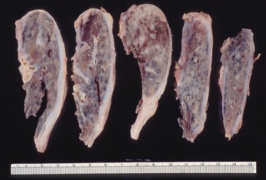
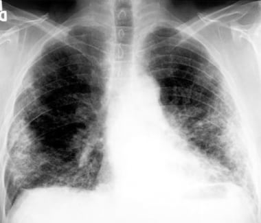
2. Restrictive lung diseases: These involve stiffening or scarring of the lungs, which reduces their ability to expand and take in air. Examples include idiopathic pulmonary fibrosis, sarcoidosis, and asbestosis.
3. Infectious lung diseases: These are caused by bacteria, viruses, fungi, or parasites that infect the lungs. Examples include pneumonia, tuberculosis, and influenza.
4. Vascular lung diseases: These affect the blood vessels in the lungs, impairing oxygen exchange. Examples include pulmonary embolism, pulmonary hypertension, and chronic thromboembolic pulmonary hypertension (CTEPH).
5. Neoplastic lung diseases: These involve abnormal growth of cells within the lungs, leading to cancer. Examples include small cell lung cancer, non-small cell lung cancer, and mesothelioma.
6. Other lung diseases: These include interstitial lung diseases, pleural effusions, and rare disorders such as pulmonary alveolar proteinosis and lymphangioleiomyomatosis (LAM).
It is important to note that this list is not exhaustive, and there are many other conditions that can affect the lungs. Proper diagnosis and treatment of lung diseases require consultation with a healthcare professional, such as a pulmonologist or respiratory therapist.
Obstructive lung disease is a category of respiratory diseases characterized by airflow limitation that causes difficulty in completely emptying the alveoli (tiny air sacs) of the lungs during exhaling. This results in the trapping of stale air and prevents fresh air from entering the alveoli, leading to various symptoms such as coughing, wheezing, shortness of breath, and decreased exercise tolerance.
The most common obstructive lung diseases include:
1. Chronic Obstructive Pulmonary Disease (COPD): A progressive disease that includes chronic bronchitis and emphysema, often caused by smoking or exposure to harmful pollutants.
2. Asthma: A chronic inflammatory disorder of the airways characterized by variable airflow obstruction, bronchial hyperresponsiveness, and an underlying inflammation. Symptoms can be triggered by various factors such as allergens, irritants, or physical activity.
3. Bronchiectasis: A condition in which the airways become abnormally widened, scarred, and thickened due to chronic inflammation or infection, leading to mucus buildup and impaired clearance.
4. Cystic Fibrosis: An inherited genetic disorder that affects the exocrine glands, resulting in thick and sticky mucus production in various organs, including the lungs. This can lead to chronic lung infections, inflammation, and airway obstruction.
5. Alpha-1 Antitrypsin Deficiency: A genetic condition characterized by low levels of alpha-1 antitrypsin protein, which leads to uncontrolled protease enzyme activity that damages the lung tissue, causing emphysema-like symptoms.
Treatment for obstructive lung diseases typically involves bronchodilators (to relax and widen the airways), corticosteroids (to reduce inflammation), and lifestyle modifications such as smoking cessation and pulmonary rehabilitation programs. In severe cases, oxygen therapy or even lung transplantation may be considered.
A lung is a pair of spongy, elastic organs in the chest that work together to enable breathing. They are responsible for taking in oxygen and expelling carbon dioxide through the process of respiration. The left lung has two lobes, while the right lung has three lobes. The lungs are protected by the ribcage and are covered by a double-layered membrane called the pleura. The trachea divides into two bronchi, which further divide into smaller bronchioles, leading to millions of tiny air sacs called alveoli, where the exchange of gases occurs.
Total Lung Capacity (TLC) is the maximum volume of air that can be contained within the lungs at the end of a maximal inspiration. It includes all of the following lung volumes: tidal volume, inspiratory reserve volume, expiratory reserve volume, and residual volume. TLC can be measured directly using gas dilution techniques or indirectly by adding residual volume to vital capacity. Factors that affect TLC include age, sex, height, and lung health status.
Vital capacity (VC) is a term used in pulmonary function tests to describe the maximum volume of air that can be exhaled after taking a deep breath. It is the sum of inspiratory reserve volume, tidal volume, and expiratory reserve volume. In other words, it's the total amount of air you can forcibly exhale after inhaling as deeply as possible. Vital capacity is an important measurement in assessing lung function and can be reduced in conditions such as chronic obstructive pulmonary disease (COPD), asthma, and other respiratory disorders.
Interstitial lung diseases (ILDs) are a group of disorders characterized by inflammation and scarring (fibrosis) in the interstitium, the tissue and space around the air sacs (alveoli) of the lungs. The interstitium is where the blood vessels that deliver oxygen to the lungs are located. ILDs can be caused by a variety of factors, including environmental exposures, medications, connective tissue diseases, and autoimmune disorders.
The scarring and inflammation in ILDs can make it difficult for the lungs to expand and contract normally, leading to symptoms such as shortness of breath, cough, and fatigue. The scarring can also make it harder for oxygen to move from the air sacs into the bloodstream.
There are many different types of ILDs, including:
* Idiopathic pulmonary fibrosis (IPF): a type of ILD that is caused by unknown factors and tends to progress rapidly
* Hypersensitivity pneumonitis: an ILD that is caused by an allergic reaction to inhaled substances, such as mold or bird droppings
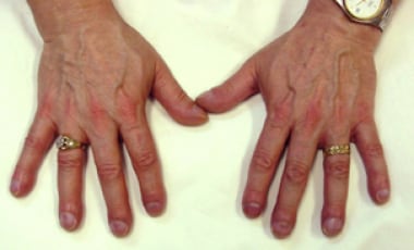
* Connective tissue diseases: ILDs can be a complication of conditions such as rheumatoid arthritis and scleroderma
* Sarcoidosis: an inflammatory disorder that can affect multiple organs, including the lungs
* Asbestosis: an ILD caused by exposure to asbestos fibers
Treatment for ILDs depends on the specific type of disease and its underlying cause. Some treatments may include corticosteroids, immunosuppressive medications, and oxygen therapy. In some cases, a lung transplant may be necessary.
 List of MeSH codes (G09)
 Restrictive Lung Disease: Background, Pathophysiology, Etiology
 Frontiers | Acute Effects of Inspiratory Loads and Interfaces on Breathing Pattern and Activity of Respiratory Muscles in...
 Advanced Search Results - Public Health Image Library(PHIL)
 Exercise induced bronchoconstriction in adults: evidence based diagnosis and management | The BMJ
 Pulmonary Function Testing (PFT) - Lung and Airway Disorders - MSD Manual Consumer Version
 Rodrigo Torres-Castro | Hilaris SRL
 Liver and Other Neoplasms - Treatment Approaches - Medical Clinical Policy Bulletins | Aetna
 The Impact of Early Pulmonary Rehabilitation on the Multidimensional Aspects of Dyspnea and Exercise Performance Following...
 Journal of Back and Musculoskeletal Rehabilitation - Volume Pre-press, issue Pre-press - Journals - IOS Press
 "You can leave your mask on": effects on cardiopulmonary parameters of different airway protective masks at rest and during...
 How do I bill CPT 96160? - KOOLOADER.COM
 Nervous, Respiratory Systems | Transfection Reagents | Cell Lines, In Vivo | Altogen Biosystems
 Scoliosis. Medical search. Definitions
 Obtained and predicted values for maximal respiratory pressures of Brazilian children
 Proof of Concept for Effect of Respiratory Muscle Therapy on Heart Failure - PN Medical
 Lung function six months after severe COVID-19: Does time, in fact, heal all wounds? | The Brazilian Journal of Infectious...
 Braz J Cardiovasc Surg - Randomized and comparative study between two intra-hospital exercise programs for heart transplant...
 Pesquisa | Portal Regional da BVS
 Ultrasound evaluation of diaphragm functionality: relationship with standard and imaging approaches
 Vocal cord dysfunction in patients with exertional dyspnea