Maxillary Sinus
Maxillary Sinus Neoplasms
Paranasal Sinus Diseases
Maxillary Sinusitis
Maxillary Artery
Maxilla
Paranasal Sinuses
Maxillary Diseases
Sinus Floor Augmentation
Dentigerous Cyst
Jaw, Edentulous
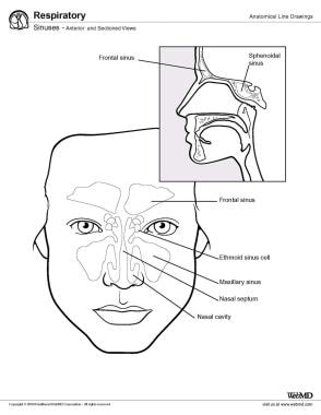
Frontal Sinus
Maxillary Nerve
Otorhinolaryngologic Surgical Procedures
Cranial Sinuses
Nasal Cavity
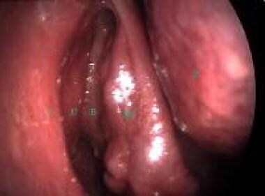
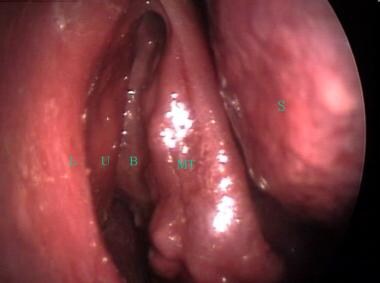
Endoscopy
Dental Implants
Mucocele
Alveolar Process
Accreditation
Oral Surgical Procedures, Preprosthetic
Ethmoid Sinus
Incisor
Molar
Cavernous Sinus
Cone-Beam Computed Tomography
Zygoma
Periapical Diseases
Tooth, Impacted
Tomography, X-Ray Computed
Carotid Sinus
Cuspid
Palatal Expansion Technique
Palate
Sphenoid Sinus
Bicuspid
Radiography, Panoramic
Dimensional Measurement Accuracy
Dental Arch
Anatomic Variation
Coronary Sinus
Osteoma
Nasal Polyps
Oral Surgical Procedures
Dental Implantation
Sinus Thrombosis, Intracranial
Bone Substitutes
Models, Anatomic
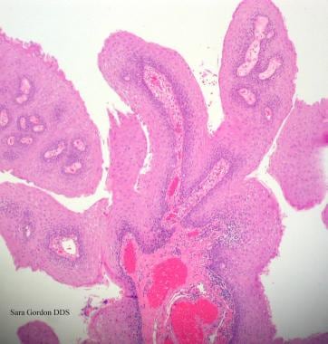
Papilloma, Inverted
Leontopithecus
Sick Sinus Syndrome
Dental Prosthesis Design
Age Determination by Teeth
Anatomic Landmarks
Mandible
Nasal Obstruction
Tooth Crown
Nasal Mucosa
Saguinus
Yttrium Isotopes
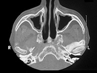
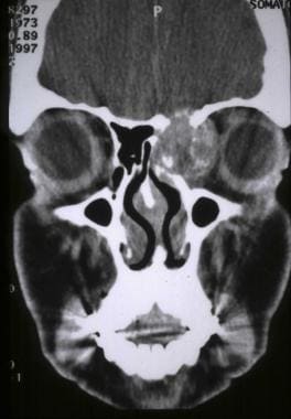
Calcification in chronic maxillary sinusitis: comparison of CT findings with histopathologic results. (1/220)
BACKGROUND AND PURPOSE: It is important to differentiate fungal from nonfungal sinusitis in order to determine the optimal treatment for chronic sinusitis. The purpose of this study was to describe the CT findings of calcifications in chronic fungal and nonfungal maxillary sinusitis. METHODS: Five hundred ten patients with pathologically proved chronic maxillary sinusitis were studied with unenhanced CT before undergoing sinonasal surgery. In 36 patients, the CT scans were reviewed retrospectively to ascertain the shape and location of intrasinus calcifications. RESULTS: Calcifications were found in 20 (51%) of 39 patients with fungal sinusitis and in 16 (3%) of 471 patients with nonfungal sinusitis. Direct histopathologic correlation was performed in two of 16 patients with nonfungal sinusitis who had intrasinus calcification. The location of intrasinus calcification was central in 95% of the patients with fungal sinusitis and peripheral in 81% of those with nonfungal sinusitis. Although calcifications with a nodular or linear shape were seen in both fungal and nonfungal sinusitis, fine punctate type calcifications were seen only in those with fungal sinusitis (50%) and round or eggshell type calcifications only in those with nonfungal sinusitis (19%). CONCLUSION: Intrasinus calcifications are different in location and shape between fungal and nonfungal maxillary sinusitis. Although intrasinus calcification is uncommon in nonfungal sinusitis, the CT finding of intrasinus calcification may be helpful for differentiating fungal from nonfungal maxillary sinusitis. (+info)Comparison of the response to histamine challenge of the nose and the maxillary sinus: effect of loratadine. (2/220)
To study the response of the maxillary sinus to histamine provocation, we performed a double-blind, randomized, crossover trial during which nonallergic subjects without symptoms of rhinitis (n = 25) received either 10 mg loratadine or placebo once daily for a week and then underwent nasal challenge with histamine (3, 10, and 30 mg/ml) followed, 24 h later, by a maxillary sinus challenge while still receiving the medication. Nasal challenge with histamine led to significant increases in vascular permeability, reflex nasal secretions, sneezing, and other nasal symptoms. Sinus challenge resulted in significant increases in vascular permeability within the sinus cavity (P < 0.01) and some nasal symptoms but no significant change in reflex nasal secretions. The response of the sinus mucosa to histamine was lower in magnitude than that of the nose. Treatment with loratadine resulted in a significant inhibition of the histamine-induced changes in both nasal and sinus cavities. Our data suggest the lack of a sinonasal reflex response to histamine provocation of the maxillary sinus of nonallergic individuals. (+info)The pterygopalatine fossa: postoperative MR imaging appearance. (3/220)
BACKGROUND AND PURPOSE: The pterygopalatine fossa (PPF) is an important anatomic location of the deep portion of the face. It is essential to review this area on both pre- and posttreatment studies of head and neck malignancies to assess local extent of disease or recurrence and perineural tumor spread. The purpose of this study was to review the postoperative appearance of the PPF on MR images. METHODS: Imaging and clinical data of 10 patients who underwent surgical resection of tumor in which the PPF was violated at surgery were reviewed. Patients were included in the study if there was no imaging or clinical evidence of tumor in the PPF pre- or postoperatively. Postoperative MR studies were examined to assess the appearance of the PPF. RESULTS: The PPF is consistently and persistently abnormal after surgical violation. There is loss of the normal T1 signal hyperintensity and abnormal, increased contrast enhancement, as seen on fat-suppressed T1-weighted images. These postoperative changes are strikingly similar to those of tumor involvement. CONCLUSION: After surgical violation, the PPF will always appear abnormal on MR images, and the expected imaging findings must be recognized to avoid the misdiagnosis of tumor recurrence. (+info)Ventilator-associated sinusitis: microbiological results of sinus aspirates in patients on antibiotics. (4/220)
BACKGROUND: The efficacy of systemic antibiotics on the treatment of ventilator-associated infectious maxillary sinusitis (VAIMS) is debated. The objective of this study was to determine the etiologic diagnosis of VAIMS in patients receiving antibiotics. METHODS: Patients mechanically ventilated for more than or equal to 72 h, who had persistent fever while on antibiotics for more than or equal to 48 h, underwent computed tomography scan followed by transnasal puncture of involved maxillary sinuses. VAIMS was defined as follows: fever greater than or equal to 38 degrees C, radiographic signs (air fluid level or opacification of maxillary sinuses on computed tomography scan), and a quantitative culture of sinus aspirate yielding more than or equal to 103 colony-forming units/ml. RESULTS: Twenty-four patients had radiographic signs of sinusitis. The mean +/- SD prior durations of mechanical ventilation and antibiotic exposure were 9.5 +/- 4.7 days and 6 +/- 4 days, respectively. Six unilateral and nine bilateral VAIMS were diagnosed in 15 patients. The median number of etiologic organisms per patient was two (range, one to four). The bacteriologic cultures yielded gram-positive bacteria (n = 21), gram-negative bacteria (n = 22), and yeasts (n = 5). Forty percent of causative agents were susceptible to the antibiotics prescribed. Seven patients with VAIMS developed 10 concomitant infections: ventilator-associated pneumonia (n = 5), urinary tract infection (n = 3), catheter infections (n = 2). In all cases of ventilator-associated pneumonia, the implicated agents were the causative agents of VAIMS. CONCLUSION: In VAIMS patients on antibiotics, quantitative cultures of sinus aspirates may contribute to establish the diagnosis. The frequent recovery of microorganisms susceptible to the antimicrobial treatment administered suggests that therapy of VAIMS with systemic antibiotics may not be sufficient. (+info)Sinusitis demonstrated by brain scanning. (5/220)
Increased concentration of technietum was noted in the region of the frontal, ethmoidal, and maxillary sinuses of two patients. Radiographs of the sinuses revealed extensive sinusitis involving the sinuses in the area of increased uptake. The increased uptake was attributed to the sinusitis. (+info)Maxillary sinusitis caused by Actinomucor elegans. (6/220)
We report the first case of maxillary sinusitis caused by Actinomucor elegans in an 11-year-old patient. Histopathological and mycological examinations of surgical maxillary sinuses samples showed coenocytic hyphae characteristic of mucoraceous fungi. The fungi recovered had stolons and rhizoids, nonapophyseal and globose sporangia, and whorled branched sporangiophores and was identified as A. elegans. After surgical cleaning and chemotherapy with amphotericin B administered intravenously and by irrigation, the patient became asymptomatic and the mycological study results were negative. (+info)Contrast-enhanced conventional CT in patients after surgery for malignant tumors: evaluation of the optimal method of the administration of the contrast medium. (7/220)
Patients after ablative surgery for malignant tumors require computed tomography (CT) examination of a wide area on the head and neck to follow-up for recurrence and lymph metastasis. The aim of this study was to determine a more effective method for the infusion of the contrast medium into post-operative patients undergoing conventional CT, based on the relationship between the method of administering the contrast medium and the contrast-enhancing effect in the internal jugular vein. First eleven images were selected from the existing contrast-enhanced and plain CT images in a manner such that the CT values of the internal jugular vein were distributed evenly in a range of 50-180. Seven experienced observers evaluated the contrast-enhancing effect of each image set at a window value of 40 and window widths of 120, 200, and 280. Secondly, the CT values of the right internal jugular vein were measured in a total of 10 CT images from the thyroid to maxillary sinus level from each of 60 post-operative patients. The injection needles and contrast-enhancing techniques used in the 60 patients were drip infusion using an 18G injection needle in 20, drip infusion using a 21G injection needle with bolus intravenous injection immediately before scanning in 20, and drip infusion using a 23G injection needle with bolus intravenous injection immediately before scanning in 20. A CT value of 100 or above, preferably 120 or above, in the internal jugular vein was needed for the contrast-enhancing effect of a CT image to be judged as clinically significant. Our results found that, when a conventional CT was used in patients after surgery for malignant tumors, drip infusion using a 21G or 23G injection needle should be combined with bolus injections immediately before the beginning of scanning, and at the glottis or submandibular gland level during the scanning. A sufficient contrast-enhancing effect can also be obtained by drip infusion using an 18G injection needle without bolus injection. (+info)A combined frontal and maxillary sinus approach for repulsion of the third maxillary molar in a horse. (8/220)
The 3rd maxillary molar is a difficult tooth to remove by extraction or repulsion. A combined frontal and maxillary approach provides good exposure for repulsion of this tooth, debridement of the sinuses, and placement of an alveolar seal. The improved exposure should minimize operative difficulties and postoperative complications. (+info)The maxillary sinuses, also known as the antrums of Highmore, are the largest of the four pairs of paranasal sinuses located in the maxilla bones. They are air-filled cavities that surround the nasolacrimal duct and are situated superior to the upper teeth and lateral to the nasal cavity. Each maxillary sinus is lined with a mucous membrane, which helps to warm, humidify, and filter the air we breathe. Inflammation or infection of the maxillary sinuses can result in conditions such as sinusitis, leading to symptoms like facial pain, headaches, and nasal congestion.
Maxillary sinus neoplasms refer to abnormal growths or tumors that develop in the maxillary sinuses, which are located in the upper part of your cheekbones, below your eyes. These growths can be benign (non-cancerous) or malignant (cancerous).
Benign neoplasms may include conditions such as an osteoma (a benign bone tumor), a papilloma (a benign growth of the lining of the sinus), or a fibrous dysplasia (a condition where bone is replaced by fibrous tissue).
Malignant neoplasms, on the other hand, can be primary (originating in the maxillary sinuses) or secondary (spreading to the maxillary sinuses from another site in the body). Common types of malignant tumors that arise in the maxillary sinus include squamous cell carcinoma, adenocarcinoma, and mucoepidermoid carcinoma.
Symptoms of maxillary sinus neoplasms may include nasal congestion, nosebleeds, facial pain or numbness, vision changes, and difficulty swallowing or speaking. Treatment options depend on the type, size, and location of the tumor but may include surgery, radiation therapy, chemotherapy, or a combination of these approaches.
Paranasal sinus diseases refer to a group of medical conditions that affect the paranasal sinuses, which are air-filled cavities located within the skull near the nasal cavity. These sinuses include the maxillary, frontal, ethmoid, and sphenoid sinuses.
Paranasal sinus diseases can be caused by a variety of factors, including viral, bacterial, or fungal infections, allergies, structural abnormalities, or autoimmune disorders. Some common paranasal sinus diseases include:
1. Sinusitis: Inflammation or infection of the sinuses, which can cause symptoms such as nasal congestion, thick nasal discharge, facial pain or pressure, and reduced sense of smell.
2. Nasal polyps: Soft, benign growths that develop in the lining of the nasal passages or sinuses, which can obstruct airflow and cause difficulty breathing through the nose.
3. Sinonasal tumors: Abnormal growths that can be benign or malignant, which can cause symptoms such as nasal congestion, facial pain, and bleeding from the nose.
4. Sinus cysts: Fluid-filled sacs that form in the sinuses, which can cause symptoms similar to those of sinusitis.
5. Fungal sinusitis: Infection of the sinuses with fungi, which can cause symptoms such as nasal congestion, facial pain, and thick, discolored mucus.
Treatment for paranasal sinus diseases depends on the underlying cause and severity of the condition. Treatment options may include medications, such as antibiotics, antihistamines, or corticosteroids, as well as surgical intervention in more severe cases.
Maxillary sinusitis is a medical condition characterized by inflammation or infection of the maxillary sinuses, which are air-filled cavities located in the upper part of the cheekbones. These sinuses are lined with mucous membranes that produce mucus to help filter and humidify the air we breathe.
When the maxillary sinuses become inflamed or infected, they can fill with fluid and pus, leading to symptoms such as:
* Pain or pressure in the cheeks, upper teeth, or behind the eyes
* Nasal congestion or stuffiness
* Runny nose or postnasal drip
* Reduced sense of smell or taste
* Headache or facial pain
* Fatigue or fever (in cases of bacterial infection)
Maxillary sinusitis can be caused by viruses, bacteria, or fungi, and may also result from allergies, structural abnormalities, or exposure to environmental irritants such as smoke or pollution. Treatment typically involves managing symptoms with over-the-counter remedies or prescription medications, such as decongestants, antihistamines, or antibiotics. In some cases, more invasive treatments such as sinus surgery may be necessary.
The maxillary artery is a branch of the external carotid artery that supplies the deep structures of the face and head. It originates from the external carotid artery just below the neck of the mandible and passes laterally to enter the parotid gland. Within the gland, it gives off several branches, including the deep auricular, anterior tympanic, and middle meningeal arteries.
After leaving the parotid gland, the maxillary artery travels through the infratemporal fossa, where it gives off several more branches, including the inferior alveolar, buccinator, and masseteric arteries. These vessels supply blood to the teeth, gums, and muscles of mastication.
The maxillary artery also gives off the sphenopalatine artery, which supplies the nasal cavity, nasopharynx, and palate. Additionally, it provides branches that supply the meninges, dura mater, and brain. Overall, the maxillary artery plays a critical role in providing blood flow to many structures in the head and neck region.
The maxilla is a paired bone that forms the upper jaw in vertebrates. In humans, it is a major bone in the face and plays several important roles in the craniofacial complex. Each maxilla consists of a body and four processes: frontal process, zygomatic process, alveolar process, and palatine process.
The maxillae contribute to the formation of the eye sockets (orbits), nasal cavity, and the hard palate of the mouth. They also contain the upper teeth sockets (alveoli) and help form the lower part of the orbit and the cheekbones (zygomatic arches).
Here's a quick rundown of its key functions:
1. Supports the upper teeth and forms the upper jaw.
2. Contributes to the formation of the eye sockets, nasal cavity, and hard palate.
3. Helps shape the lower part of the orbit and cheekbones.
4. Partakes in the creation of important sinuses, such as the maxillary sinus, which is located within the body of the maxilla.
Paranasal sinuses are air-filled cavities in the skull that surround the nasal cavity. There are four pairs of paranasal sinuses, including the maxillary, frontal, ethmoid, and sphenoid sinuses. These sinuses help to warm, humidify, and filter the air we breathe. They also contribute to our voice resonance and provide a slight cushioning effect for the skull. The openings of the paranasal sinuses lead directly into the nasal cavity, allowing mucus produced in the sinuses to drain into the nose. Infections or inflammation of the paranasal sinuses can result in conditions such as sinusitis.
Paranasal sinus neoplasms refer to abnormal growths or tumors that develop within the paranasal sinuses, which are air-filled cavities located inside the skull near the nasal cavity. These tumors can be benign (noncancerous) or malignant (cancerous), and they can arise from various types of tissue within the sinuses, such as the lining of the sinuses (mucosa), bone, or other soft tissues.
Paranasal sinus neoplasms can cause a variety of symptoms, including nasal congestion, nosebleeds, facial pain or numbness, and visual disturbances. The diagnosis of these tumors typically involves a combination of imaging studies (such as CT or MRI scans) and biopsy to determine the type and extent of the tumor. Treatment options may include surgery, radiation therapy, chemotherapy, or a combination of these approaches, depending on the specific type and stage of the neoplasm.
Maxillary diseases refer to conditions that affect the maxilla, which is the upper bone of the jaw. This bone plays an essential role in functions such as biting, chewing, and speaking, and also forms the upper part of the oral cavity, houses the upper teeth, and supports the nose and the eyes.
Maxillary diseases can be caused by various factors, including infections, trauma, tumors, congenital abnormalities, or systemic conditions. Some common maxillary diseases include:
1. Maxillary sinusitis: Inflammation of the maxillary sinuses, which are air-filled cavities located within the maxilla, can cause symptoms such as nasal congestion, facial pain, and headaches.
2. Periodontal disease: Infection and inflammation of the tissues surrounding the teeth, including the gums and the alveolar bone (which is part of the maxilla), can lead to tooth loss and other complications.
3. Maxillary fractures: Trauma to the face can result in fractures of the maxilla, which can cause pain, swelling, and difficulty breathing or speaking.
4. Maxillary cysts and tumors: Abnormal growths in the maxilla can be benign or malignant and may require surgical intervention.
5. Oral cancer: Cancerous lesions in the oral cavity, including the maxilla, can cause pain, swelling, and difficulty swallowing or speaking.
Treatment for maxillary diseases depends on the specific condition and its severity. Treatment options may include antibiotics, surgery, radiation therapy, or chemotherapy. Regular dental check-ups and good oral hygiene practices can help prevent many maxillary diseases.
Maxillary neoplasms refer to abnormal growths or tumors in the maxilla, which is the upper jaw bone. These growths can be benign (non-cancerous) or malignant (cancerous). Benign neoplasms are slow-growing and do not spread to other parts of the body, while malignant neoplasms can invade surrounding tissues and spread to distant sites.
Maxillary neoplasms can cause various symptoms such as swelling, pain, numbness, loose teeth, or difficulty in chewing or swallowing. They may also cause nasal congestion, nosebleeds, or visual changes if they affect the eye or orbit. The diagnosis of maxillary neoplasms usually involves a combination of clinical examination, imaging studies such as CT or MRI scans, and biopsy to determine the type and extent of the tumor.
Treatment options for maxillary neoplasms depend on several factors, including the type, size, location, and stage of the tumor, as well as the patient's overall health and preferences. Treatment may include surgery, radiation therapy, chemotherapy, or a combination of these modalities. Regular follow-up care is essential to monitor for recurrence or metastasis and ensure optimal outcomes.
Sinus floor augmentation, also known as sinus lift or maxillary sinus floor elevation, is a surgical procedure in dental medicine that aims to increase the amount of bone in the upper jaw (maxilla) in the area of the premolars and molars. This procedure is typically performed in preparation for dental implant placement in cases where there is insufficient bone height or density due to alveolar bone resorption, anatomical variations such as pneumatization of the maxillary sinus, or complications from previous oral surgery.
During the sinus floor augmentation procedure, a small opening is made in the upper jawbone, usually through the side of the mouth (buccal approach) or from inside the mouth (crestal approach). The membrane lining the sinus cavity (sinus membrane or Schneiderian membrane) is carefully lifted and detached from the underlying bone, creating a space between the sinus floor and the jawbone. Bone graft material, which can be autogenous (patient's own), allogeneic (donor-derived), xenogeneic (animal-derived), or synthetic, is then placed into this space to stimulate new bone growth. The opening in the jawbone is then closed with sutures, and a healing period follows before dental implant placement can be considered.
The primary goal of sinus floor augmentation is to provide adequate bone volume and quality for successful dental implant integration and long-term stability.
A dentigerous cyst is a type of odontogenic cyst that forms around the crown of an unerupted tooth. It is typically slow-growing and often asymptomatic, but it can cause displacement or resorption of adjacent teeth if it becomes large enough. Dentigerous cysts are more common in permanent teeth than primary teeth, and they are more likely to occur in the mandible (lower jaw) than the maxilla (upper jaw). They are usually diagnosed through radiographic examination and can be treated by surgical removal of the cyst along with the affected tooth. If left untreated, dentigerous cysts can continue to grow and may eventually develop into a tumor or cancer.
"Edentulous jaw" is a medical term used to describe a jaw that is missing all of its natural teeth. The term "edentulous" is derived from the Latin word "edentulus," which means "without teeth." This condition can affect either the upper jaw (maxilla) or the lower jaw (mandible), or both, resulting in a significant impact on an individual's ability to eat, speak, and maintain proper facial structure.
Edentulism is often associated with aging, as tooth loss becomes more common in older adults due to factors like gum disease, tooth decay, and injury. However, it can also affect younger individuals who have lost their teeth due to various reasons. Dental professionals typically recommend the use of dentures or dental implants to restore oral function and aesthetics for patients with edentulous jaws.
A frontal sinus is a paired, air-filled paranasal sinus located in the frontal bone of the skull, above the eyes and behind the forehead. It is one of the four pairs of sinuses found in the human head. The frontal sinuses are lined with mucous membrane and are interconnected with the nasal cavity through small openings called ostia. They help to warm, humidify, and filter the air we breathe, and contribute to the resonance of our voice. Variations in size, shape, and asymmetry of frontal sinuses are common among individuals.
The maxillary nerve, also known as the second division of the trigeminal nerve (cranial nerve V2), is a primary sensory nerve that provides innervation to the skin of the lower eyelid, side of the nose, part of the cheek, upper lip, and roof of the mouth. It also supplies sensory fibers to the mucous membranes of the nasal cavity, maxillary sinus, palate, and upper teeth. Furthermore, it contributes motor innervation to the muscles involved in chewing (muscles of mastication), specifically the tensor veli palatini and tensor tympani. The maxillary nerve originates from the trigeminal ganglion and passes through the foramen rotundum in the skull before reaching its target areas.
Otorhinolaryngologic surgical procedures are surgeries that are performed on the head and neck region, specifically involving the ear, nose, and throat (ENT) regions. This field is also known as otolaryngology-head and neck surgery. The procedures can range from relatively minor ones, such as removing a small nasal polyp or inserting ear tubes, to more complex surgeries like cochlear implantation, endoscopic sinus surgery, or removal of tumors in the head and neck region. These surgical procedures are typically performed by specialized physicians called otorhinolaryngologists (also known as ENT surgeons) who have completed extensive training in this area.
Cranial sinuses are a part of the venous system in the human head. They are air-filled spaces located within the skull and are named according to their location. The cranial sinuses include:
1. Superior sagittal sinus: It runs along the top of the brain, inside the skull, and drains blood from the scalp and the veins of the brain.
2. Inferior sagittal sinus: It runs along the bottom of the brain and drains into the straight sinus.
3. Straight sinus: It is located at the back of the brain and receives blood from the inferior sagittal sinus and great cerebral vein.
4. Occipital sinuses: They are located at the back of the head and drain blood from the scalp and skull.
5. Cavernous sinuses: They are located on each side of the brain, near the temple, and receive blood from the eye and surrounding areas.
6. Sphenoparietal sinus: It is a small sinus that drains blood from the front part of the brain into the cavernous sinus.
7. Petrosquamosal sinuses: They are located near the ear and drain blood from the scalp and skull.
The cranial sinuses play an essential role in draining blood from the brain and protecting it from injury.
An oroantral fistula is an abnormal communication or connection between the oral cavity (mouth) and the maxillary sinus, which is one of the air-filled cavities in the upper jaw. This condition typically arises as a complication following dental procedures, such as tooth extractions, particularly in the upper molars, where the roots are close to or even within the maxillary sinus.
An oroantral fistula may also result from other factors, including trauma, infection, tumors, or cysts that erode the thin bony wall separating the oral cavity and the maxillary sinus. The presence of an oroantral fistula can lead to various symptoms, such as nasal discharge, pain, difficulty swallowing, and communication between the mouth and nose.
Treatment for an oroantral fistula usually involves surgical closure of the communication, often with the use of a flap of tissue from another part of the mouth. Proper diagnosis and management are essential to prevent further complications and restore normal function.
The nasal cavity is the air-filled space located behind the nose, which is divided into two halves by the nasal septum. It is lined with mucous membrane and is responsible for several functions including respiration, filtration, humidification, and olfaction (smell). The nasal cavity serves as an important part of the upper respiratory tract, extending from the nares (nostrils) to the choanae (posterior openings of the nasal cavity that lead into the pharynx). It contains specialized structures such as turbinate bones, which help to warm, humidify and filter incoming air.
Endoscopy is a medical procedure that involves the use of an endoscope, which is a flexible tube with a light and camera at the end, to examine the interior of a body cavity or organ. The endoscope is inserted through a natural opening in the body, such as the mouth or anus, or through a small incision. The images captured by the camera are transmitted to a monitor, allowing the physician to visualize the internal structures and detect any abnormalities, such as inflammation, ulcers, or tumors. Endoscopy can also be used for diagnostic purposes, such as taking tissue samples for biopsy, or for therapeutic purposes, such as removing polyps or performing minimally invasive surgeries.
Ectopic tooth eruption is a condition where a tooth fails to erupt into its normal position in the dental arch. Instead, it emerupts in an abnormal location, such as in the wrong direction or through another tissue like the gums, palate, or jawbone. This can occur due to various reasons, including genetics, crowding of teeth, or trauma. Ectopic tooth eruption may cause problems with oral function and dental health, and treatment options depend on the severity and location of the ectopic tooth.
Dental implants are artificial tooth roots that are surgically placed into the jawbone to replace missing or extracted teeth. They are typically made of titanium, a biocompatible material that can fuse with the bone over time in a process called osseointegration. Once the implant has integrated with the bone, a dental crown, bridge, or denture can be attached to it to restore function and aesthetics to the mouth.
Dental implants are a popular choice for tooth replacement because they offer several advantages over traditional options like dentures or bridges. They are more stable and comfortable, as they do not rely on adjacent teeth for support and do not slip or move around in the mouth. Additionally, dental implants can help to preserve jawbone density and prevent facial sagging that can occur when teeth are missing.
The process of getting dental implants typically involves several appointments with a dental specialist called a prosthodontist or an oral surgeon. During the first appointment, the implant is placed into the jawbone, and the gum tissue is stitched closed. Over the next few months, the implant will fuse with the bone. Once this process is complete, a second surgery may be necessary to expose the implant and attach an abutment, which connects the implant to the dental restoration. Finally, the crown, bridge, or denture is attached to the implant, providing a natural-looking and functional replacement for the missing tooth.
Sinusitis, also known as rhinosinusitis, is a medical condition characterized by inflammation of the paranasal sinuses, which are air-filled cavities located within the skull near the nose. The inflammation can be caused by viral, bacterial, or fungal infections, as well as allergies, structural issues, or autoimmune disorders.
In sinusitis, the mucous membranes lining the sinuses become swollen and may produce excess mucus, leading to symptoms such as nasal congestion, thick green or yellow nasal discharge, facial pain or pressure, reduced sense of smell, cough, fatigue, and fever.
Sinusitis can be classified into acute (lasting less than 4 weeks), subacute (lasting 4-12 weeks), chronic (lasting more than 12 weeks), or recurrent (multiple episodes within a year). Treatment options depend on the underlying cause and severity of symptoms, and may include antibiotics, nasal corticosteroids, decongestants, saline irrigation, and in some cases, surgery.
A mucocele is a mucus-containing cystic lesion that results from the accumulation of mucin within a damaged minor salivary gland duct or mucous gland. It is typically caused by trauma, injury, or blockage of the duct. Mucocele appears as a round, dome-shaped, fluid-filled swelling, which may be bluish or clear in color. They are most commonly found on the lower lip but can also occur on other areas of the oral cavity. Mucocele is generally painless unless it becomes secondarily infected; however, it can cause discomfort during speaking, chewing, or swallowing, and may affect aesthetics. Treatment usually involves surgical excision of the mucocele to prevent recurrence.
The alveolar process is the curved part of the jawbone (mandible or maxilla) that contains sockets or hollow spaces (alveoli) for the teeth to be embedded. These processes are covered with a specialized mucous membrane called the gingiva, which forms a tight seal around the teeth to help protect the periodontal tissues and maintain oral health.
The alveolar process is composed of both compact and spongy bone tissue. The compact bone forms the outer layer, while the spongy bone is found inside the alveoli and provides support for the teeth. When a tooth is lost or extracted, the alveolar process begins to resorb over time due to the lack of mechanical stimulation from the tooth's chewing forces. This can lead to changes in the shape and size of the jawbone, which may require bone grafting procedures before dental implant placement.
Accreditation is a process in which a healthcare organization, facility, or program is evaluated and certified as meeting certain standards and criteria established by a recognized accrediting body. The purpose of accreditation is to ensure that the organization, facility, or program provides safe, high-quality care and services to its patients or clients.
Accreditation typically involves a thorough review of an organization's policies, procedures, practices, and outcomes, as well as an on-site survey by a team of experts from the accrediting body. The evaluation focuses on various aspects of the organization's operations, such as leadership and management, patient safety, infection control, clinical services, quality improvement, and staff competence.
Accreditation is voluntary, but many healthcare organizations seek it as a way to demonstrate their commitment to excellence and continuous improvement. Accreditation can also be a requirement for licensure, reimbursement, or participation in certain programs or initiatives.
Examples of accrediting bodies in the healthcare field include The Joint Commission, the Accreditation Council for Graduate Medical Education (ACGME), the Commission on Accreditation of Rehabilitation Facilities (CARF), and the National Committee for Quality Assurance (NCQA).
Preprosthetic oral surgical procedures are dental surgeries performed to prepare the mouth for the placement of dental prostheses such as dentures. These procedures aim to create a smooth, stable, and suitable foundation in the mouth to support the prosthesis and ensure its proper functioning, retention, and comfort.
Common preprosthetic oral surgical procedures include:
1. Alveoloplasty: This procedure involves reshaping the alveolar ridge (the bony ridge that supports the teeth) to create a more uniform and even surface. It helps to eliminate any sharp or irregular bony edges that may interfere with the fit or comfort of the denture.
2. Gingivectomy/Gingivoplasty: These procedures involve removing or reshaping excess gum tissue to improve the fit and appearance of the dental prosthesis. A gingivectomy removes a portion of the gum tissue, while a gingivoplasty sculpts and reshapes the existing gum tissue.
3. Frenectomy: This procedure involves removing or repositioning the frenum, a small fold of tissue that connects the lips, cheeks, or tongue to the jawbone. A lingual frenectomy may be necessary when the frenum restricts tongue movement and interferes with proper denture placement or speech.
4. Maxillary tori reduction: This procedure involves removing or reducing the size of tori, which are bony growths found on the roof of the mouth (maxilla). Large tori can make it difficult to wear a denture, so their removal or reduction can improve the fit and comfort of the prosthesis.
5. Ridge augmentation: This procedure involves adding bone grafting material to the jaw ridge to increase its height, width, or volume. This is often done when there is significant bone loss due to tooth extraction, periodontal disease, or other factors, making it difficult to achieve a secure and comfortable denture fit.
6. Exostectomy: This procedure involves removing small, benign bony growths (exostoses) that may develop on the hard palate or along the jaw ridge. These growths can interfere with the fit and comfort of a denture, so their removal can improve the prosthesis' functionality.
These procedures are typically performed by oral surgeons, periodontists, or prosthodontists who specialize in dental implants, oral surgery, and complex restorative treatments. The specific treatment plan will depend on each patient's individual needs and preferences.
The ethmoid sinuses are a pair of air-filled spaces located in the ethmoid bone, which is a part of the skull that forms the upper portion of the nasal cavity and the inner eye socket. These sinuses are divided into anterior and posterior groups and are present in adults, but not at birth. They continue to grow and develop until early adulthood.
The ethmoid sinuses are lined with mucous membrane, which helps to warm, humidify, and filter the air we breathe. They are surrounded by a network of blood vessels and nerves, making them susceptible to inflammation and infection. Inflammation of the ethmoid sinuses can lead to conditions such as sinusitis, which can cause symptoms such as nasal congestion, headache, and facial pain.
An incisor is a type of tooth that is primarily designed for biting off food pieces rather than chewing or grinding. They are typically chisel-shaped, flat, and have a sharp cutting edge. In humans, there are eight incisors - four on the upper jaw and four on the lower jaw, located at the front of the mouth. Other animals such as dogs, cats, and rodents also have incisors that they use for different purposes like tearing or gnawing.
In the context of dentistry, a molar is a type of tooth found in the back of the mouth. They are larger and wider than other types of teeth, such as incisors or canines, and have a flat biting surface with multiple cusps. Molars are primarily used for grinding and chewing food into smaller pieces that are easier to swallow. Humans typically have twelve molars in total, including the four wisdom teeth.
In medical terminology outside of dentistry, "molar" can also refer to a unit of mass in the apothecaries' system of measurement, which is equivalent to 4.08 grams. However, this usage is less common and not related to dental or medical anatomy.
The cavernous sinus is a venous structure located in the middle cranial fossa, which is a depression in the skull that houses several important nerves and blood vessels. The cavernous sinus is situated on either side of the sphenoid bone, near the base of the skull, and it contains several important structures:
* The internal carotid artery, which supplies oxygenated blood to the brain
* The abducens nerve (cranial nerve VI), which controls lateral movement of the eye
* The oculomotor nerve (cranial nerve III), which controls most of the muscles that move the eye
* The trochlear nerve (cranial nerve IV), which controls one of the muscles that moves the eye
* The ophthalmic and maxillary divisions of the trigeminal nerve (cranial nerve V), which transmit sensory information from the face and head
The cavernous sinus is an important structure because it serves as a conduit for several critical nerves and blood vessels. However, it is also vulnerable to various pathological conditions such as thrombosis (blood clots), infection, tumors, or aneurysms, which can lead to serious neurological deficits or even death.
The Sinus of Valsalva are three pouch-like dilations or outpouchings located at the upper part (root) of the aorta, just above the aortic valve. They are named after Antonio Maria Valsalva, an Italian anatomist and physician. These sinuses are divided into three parts:
1. Right Sinus of Valsalva: It is located to the right of the ascending aorta and usually gives rise to the right coronary artery.
2. Left Sinus of Valsalva: It is situated to the left of the ascending aorta and typically gives rise to the left coronary artery.
3. Non-coronary Sinus of Valsalva: This sinus is located in between the right and left coronary sinuses, and it does not give rise to any coronary arteries.
These sinuses play a crucial role during the cardiac cycle, particularly during ventricular contraction (systole). The pressure difference between the aorta and the ventricles causes the aortic valve cusps to be pushed into these sinuses, preventing the backflow of blood from the aorta into the ventricles.
Anatomical variations in the size and shape of the Sinuses of Valsalva can occur, and certain conditions like congenital heart diseases (e.g., aortic valve stenosis or bicuspid aortic valve) may affect their structure and function. Additionally, aneurysms or ruptures of the sinuses can lead to severe complications, such as cardiac tamponade, endocarditis, or stroke.
Cone-beam computed tomography (CBCT) is a medical imaging technique that uses a cone-shaped X-ray beam to create detailed, cross-sectional images of the body. In dental and maxillofacial radiology, CBCT is used to produce three-dimensional images of the teeth, jaws, and surrounding bones.
CBCT differs from traditional computed tomography (CT) in that it uses a cone-shaped X-ray beam instead of a fan-shaped beam, which allows for a faster scan time and lower radiation dose. The X-ray beam is rotated around the patient's head, capturing data from multiple angles, which is then reconstructed into a three-dimensional image using specialized software.
CBCT is commonly used in dental implant planning, orthodontic treatment planning, airway analysis, and the diagnosis and management of jaw pathologies such as tumors and fractures. It provides detailed information about the anatomy of the teeth, jaws, and surrounding structures, which can help clinicians make more informed decisions about patient care.
However, it is important to note that CBCT should only be used when necessary, as it still involves exposure to ionizing radiation. The benefits of using CBCT must be weighed against the potential risks associated with radiation exposure.
The zygoma is the scientific name for the cheekbone. It is a part of the facial skeleton that forms the prominence of the cheek and houses the maxillary sinus, one of the pairs of paranasal sinuses. The zygomatic bone, also known as the malar bone, contributes to the formation of the zygoma.
Periapical diseases are a group of conditions that affect the periapical tissue, which is the tissue located at the tip of the tooth roots. These diseases are primarily caused by bacterial infections that originate from the dental pulp, the soft tissue inside the tooth. The most common types of periapical diseases include:
1. Periapical periodontitis: This is an inflammatory reaction of the periapical tissues due to the spread of infection from the dental pulp. It can cause symptoms such as pain, swelling, and tenderness in the affected area.
2. Periapical abscess: An abscess is a collection of pus that forms in response to an infection. A periapical abscess occurs when the infection from the dental pulp spreads to the periapical tissue, causing pus to accumulate in the area. This can cause severe pain, swelling, and redness in the affected area.
3. Periapical granuloma: A granuloma is a mass of inflammatory cells that forms in response to an infection. A periapical granuloma is a small, benign tumor-like growth that develops in the periapical tissue due to chronic inflammation caused by a bacterial infection.
Periapical diseases are typically treated with root canal therapy, which involves removing the infected dental pulp and cleaning and sealing the root canals to prevent further infection. In some cases, extraction of the affected tooth may be necessary if the infection is too severe or if the tooth is not salvageable.
An impacted tooth is a condition where a tooth fails to erupt into the oral cavity within its expected time frame, resulting in its partial or complete entrapment within the jawbone or soft tissues. This commonly occurs with wisdom teeth (third molars) but can affect any tooth. Impacted teeth may cause problems such as infection, decay of adjacent teeth, gum disease, or cyst formation, and they may require surgical removal.
X-ray computed tomography (CT or CAT scan) is a medical imaging method that uses computer-processed combinations of many X-ray images taken from different angles to produce cross-sectional (tomographic) images (virtual "slices") of the body. These cross-sectional images can then be used to display detailed internal views of organs, bones, and soft tissues in the body.
The term "computed tomography" is used instead of "CT scan" or "CAT scan" because the machines take a series of X-ray measurements from different angles around the body and then use a computer to process these data to create detailed images of internal structures within the body.
CT scanning is a noninvasive, painless medical test that helps physicians diagnose and treat medical conditions. CT imaging provides detailed information about many types of tissue including lung, bone, soft tissue and blood vessels. CT examinations can be performed on every part of the body for a variety of reasons including diagnosis, surgical planning, and monitoring of therapeutic responses.
In computed tomography (CT), an X-ray source and detector rotate around the patient, measuring the X-ray attenuation at many different angles. A computer uses this data to construct a cross-sectional image by the process of reconstruction. This technique is called "tomography". The term "computed" refers to the use of a computer to reconstruct the images.
CT has become an important tool in medical imaging and diagnosis, allowing radiologists and other physicians to view detailed internal images of the body. It can help identify many different medical conditions including cancer, heart disease, lung nodules, liver tumors, and internal injuries from trauma. CT is also commonly used for guiding biopsies and other minimally invasive procedures.
In summary, X-ray computed tomography (CT or CAT scan) is a medical imaging technique that uses computer-processed combinations of many X-ray images taken from different angles to produce cross-sectional images of the body. It provides detailed internal views of organs, bones, and soft tissues in the body, allowing physicians to diagnose and treat medical conditions.
The carotid sinus is a small, dilated area located at the bifurcation (or fork) of the common carotid artery into the internal and external carotid arteries. It is a baroreceptor region, which means it contains specialized sensory nerve endings that can detect changes in blood pressure. When the blood pressure increases, the walls of the carotid sinus stretch, activating these nerve endings and sending signals to the brain. The brain then responds by reducing the heart rate and relaxing the blood vessels, which helps to lower the blood pressure back to normal.
The carotid sinus is an important part of the body's autonomic nervous system, which regulates various involuntary functions such as heart rate, blood pressure, and digestion. It plays a crucial role in maintaining cardiovascular homeostasis and preventing excessive increases in blood pressure that could potentially damage vital organs.
Nose neoplasms refer to abnormal growths or tumors in the nasal cavity or paranasal sinuses. These growths can be benign (non-cancerous) or malignant (cancerous). Benign neoplasms are typically slow-growing and do not spread to other parts of the body, while malignant neoplasms can invade surrounding tissues and have the potential to metastasize.
Nose neoplasms can cause various symptoms such as nasal congestion, nosebleeds, difficulty breathing through the nose, loss of smell, facial pain or numbness, and visual changes if they affect the eye. The diagnosis of nose neoplasms usually involves a combination of physical examination, imaging studies (such as CT or MRI scans), and biopsy to determine the type and extent of the growth. Treatment options depend on the type, size, location, and stage of the neoplasm and may include surgery, radiation therapy, chemotherapy, or a combination of these approaches.
A cuspid, also known as a canine tooth or cuspid tooth, is a type of tooth in mammals. It is the pointiest tooth in the dental arch and is located between the incisors and bicuspids (or premolars). Cuspids have a single cusp or pointed tip that is used for tearing and grasping food. In humans, there are four cuspids, two on the upper jaw and two on the lower jaw, one on each side of the dental arch.
Palatal expansion technique is a dental or orthodontic treatment procedure that aims to widen the upper jaw (maxilla) by expanding the palate. This is typically done using a device called a palatal expander, which is attached to the upper molars and applies pressure to gradually separate the two bones that form the palate (the maxillary bones). As the appliance is activated (usually through turning a screw or key), it gently expands the palatal suture, allowing for an increase in the width of the upper dental arch. This procedure can help correct crossbites, crowding, and other jaw alignment issues. It's commonly used in children and adolescents but may also be employed in adults with certain conditions.
The palate is the roof of the mouth in humans and other mammals, separating the oral cavity from the nasal cavity. It consists of two portions: the anterior hard palate, which is composed of bone, and the posterior soft palate, which is composed of muscle and connective tissue. The palate plays a crucial role in speech, swallowing, and breathing, as it helps to direct food and air to their appropriate locations during these activities.
In medical terms, the orbit refers to the bony cavity or socket in the skull that contains and protects the eye (eyeball) and its associated structures, including muscles, nerves, blood vessels, fat, and the lacrimal gland. The orbit is made up of several bones: the frontal bone, sphenoid bone, zygomatic bone, maxilla bone, and palatine bone. These bones form a pyramid-like shape that provides protection for the eye while also allowing for a range of movements.
The sphenoid sinuses are air-filled spaces located within the sphenoid bone, which is one of the bones that make up the skull base. These sinuses are located deep inside the skull, behind the eyes and nasal cavity. They are paired and separated by a thin bony septum, and each one opens into the corresponding nasal cavity through a small opening called the sphenoethmoidal recess. The sphenoid sinuses vary greatly in size and shape between individuals. They develop during childhood and continue to grow until early adulthood. The function of the sphenoid sinuses, like other paranasal sinuses, is not entirely clear, but they may contribute to reducing the weight of the skull, resonating voice during speech, and insulating the brain from trauma.
A bicuspid valve, also known as a mitral valve in the heart, is a heart valve that has two leaflets or cusps. It lies between the left atrium and the left ventricle and helps to regulate blood flow between these two chambers of the heart. In a healthy heart, the bicuspid valve opens to allow blood to flow from the left atrium into the left ventricle and closes tightly to prevent blood from flowing back into the left atrium during contraction of the ventricle.
A congenital heart defect known as a bicuspid aortic valve occurs when the aortic valve, which normally has three leaflets or cusps, only has two. This can lead to narrowing of the valve (aortic stenosis) or leakage of the valve (aortic regurgitation), which can cause symptoms and may require medical treatment.
Panoramic radiography is a specialized type of dental X-ray imaging that captures a panoramic view of the entire mouth, including the teeth, upper and lower jaws, and surrounding structures. It uses a special machine that rotates around the head, capturing images as it moves. This technique provides a two-dimensional image that is helpful in diagnosing and planning treatment for various dental conditions such as impacted teeth, bone abnormalities, and jaw disorders.
The panoramic radiograph can also be used to assess the development and positioning of wisdom teeth, detect cysts or tumors in the jaws, and evaluate the effects of trauma or injury to the mouth. It is a valuable tool for dental professionals as it allows them to see a comprehensive view of the oral structures, which may not be visible with traditional X-ray techniques.
It's important to note that while panoramic radiography provides valuable information, it should be used in conjunction with other diagnostic tools and clinical examinations to ensure accurate diagnosis and treatment planning.
Dimensional measurement accuracy refers to the degree of closeness with which the measured dimension of a object or feature corresponds to its true value. It is usually expressed as a tolerance, which indicates the maximum allowable deviation from the true value. This measurement accuracy can be affected by various factors such as the precision and calibration of the measuring instrument, the skill and experience of the person taking the measurement, and environmental conditions such as temperature and humidity. High dimensional measurement accuracy is essential in many fields, including manufacturing, engineering, and scientific research, to ensure that parts and products meet specified dimensions and function properly.
The dental arch refers to the curved shape formed by the upper or lower teeth when they come together. The dental arch follows the curve of the jaw and is important for proper bite alignment and overall oral health. The dental arches are typically described as having a U-shaped appearance, with the front teeth forming a narrower section and the back teeth forming a wider section. The shape and size of the dental arch can vary from person to person, and any significant deviations from the typical shape or size may indicate an underlying orthodontic issue that requires treatment.
An anatomic variation refers to a deviation from the typical or normal anatomical structure, position, or configuration of organs, tissues, or bodily parts. These variations can occur in any part of the body and can be congenital (present at birth) or acquired (develop later in life).
Anatomic variations are relatively common and usually do not cause any symptoms or problems. However, in some cases, they may affect the function of adjacent structures, predispose to injury or disease, or complicate medical procedures or surgeries. Therefore, it is essential for healthcare professionals to be aware of these variations during diagnoses, treatment planning, and surgical interventions.
Examples of anatomic variations include:
* Variations in the course or number of blood vessels, such as a persistent left superior vena cava or an accessory renal artery.
* Variations in the position or shape of organs, such as a mobile cecum or a horseshoe kidney.
* Variations in the number or configuration of bones, such as an extra rib or a bifid uvula.
* Variations in the innervation or sensory distribution of nerves, such as a variant course of the brachial plexus or a cross-innervated hand.
Anatomic variations can be detected through various imaging techniques, such as X-rays, CT scans, MRI scans, and ultrasound examinations. Sometimes, they are discovered during surgical procedures or autopsies. Understanding anatomic variations is crucial for accurate diagnosis, effective treatment, and optimal patient outcomes.
The coronary sinus is a large vein that receives blood from the heart's muscle tissue. It is located on the posterior side of the heart and is a part of the cardiovascular system. The coronary sinus collects oxygen-depleted blood from the myocardium (the heart muscle) and drains it into the right atrium, where it will then be pumped to the lungs for oxygenation.
The coronary sinus is an essential structure in medical procedures such as cardiac catheterization and electrophysiological studies. It is also a common site for the implantation of pacemakers and other cardiac devices.
Osteoma is a benign (noncancerous) tumor that is made up of mature bone tissue. It usually grows slowly over a period of years and is most commonly found in the skull or jaw, although it can occur in other bones of the body as well. Osteomas are typically small, but they can grow to be several centimeters in size. They may cause symptoms if they press on nearby tissues or structures, such as nerves or blood vessels. In some cases, osteomas may not cause any symptoms and may only be discovered during routine imaging studies. Treatment for osteoma is typically not necessary unless it is causing problems or growing rapidly. If treatment is needed, it may involve surgical removal of the tumor.
Enophthalmos is a medical term that refers to the abnormal positioning of the eyeball within its socket, resulting in a posterior or backward displacement of the eye. This condition can occur due to various reasons such as trauma, surgical procedures, or diseases that affect the orbital tissues, including cancer, inflammation, or infection. Enophthalmos may lead to cosmetic concerns and visual disturbances, depending on its severity. A thorough examination by an ophthalmologist or an oculoplastic surgeon is necessary for accurate diagnosis and management of this condition.
Rhinitis is a medical condition characterized by inflammation and irritation of the nasal passages, leading to symptoms such as sneezing, runny nose, congestion, and postnasal drip. It can be caused by various factors, including allergies (such as pollen, dust mites, or pet dander), infections (viral or bacterial), environmental irritants (such as smoke or pollution), and hormonal changes. Depending on the cause, rhinitis can be classified as allergic rhinitis, non-allergic rhinitis, infectious rhinitis, or hormonal rhinitis. Treatment options vary depending on the underlying cause but may include medications such as antihistamines, decongestants, nasal sprays, and immunotherapy (allergy shots).
Nasal polyps are benign (noncancerous) growths that originate from the lining of your nasal passages or sinuses. They most often occur in the area where the sinuses open into the nasal cavity. Small nasal polyps may not cause any problems. But if they grow large enough, they can block your nasal passages and lead to breathing issues, frequent infections and loss of smell.
Nasal polyps are associated with chronic inflammation due to conditions such as asthma, allergic rhinitis or chronic sinusitis. Treatment typically includes medication to reduce the size of the polyps or surgery to remove them. Even after successful treatment, nasal polyps often return.
Oral surgical procedures refer to various types of surgeries performed in the oral cavity and maxillofacial region, which includes the mouth, jaws, face, and skull. These procedures are typically performed by oral and maxillofacial surgeons, who are dental specialists with extensive training in surgical procedures involving the mouth, jaws, and face.
Some common examples of oral surgical procedures include:
1. Tooth extractions: This involves removing a tooth that is damaged beyond repair or causing problems for the surrounding teeth. Wisdom tooth removal is a common type of tooth extraction.
2. Dental implant placement: This procedure involves placing a small titanium post in the jawbone to serve as a replacement root for a missing tooth. A dental crown is then attached to the implant, creating a natural-looking and functional replacement tooth.
3. Jaw surgery: Also known as orthognathic surgery, this procedure involves repositioning the jaws to correct bite problems or facial asymmetry.
4. Biopsy: This procedure involves removing a small sample of tissue from the oral cavity for laboratory analysis, often to diagnose suspicious lesions or growths.
5. Lesion removal: This procedure involves removing benign or malignant growths from the oral cavity, such as tumors or cysts.
6. Temporomandibular joint (TMJ) surgery: This procedure involves treating disorders of the TMJ, which connects the jawbone to the skull and allows for movement when eating, speaking, and yawning.
7. Facial reconstruction: This procedure involves rebuilding or reshaping the facial bones after trauma, cancer surgery, or other conditions that affect the face.
Overall, oral surgical procedures are an important part of dental and medical care, helping to diagnose and treat a wide range of conditions affecting the mouth, jaws, and face.
Bone transplantation, also known as bone grafting, is a surgical procedure in which bone or bone-like material is transferred from one part of the body to another or from one person to another. The graft may be composed of cortical (hard outer portion) bone, cancellous (spongy inner portion) bone, or a combination of both. It can be taken from different sites in the same individual (autograft), from another individual of the same species (allograft), or from an animal source (xenograft). The purpose of bone transplantation is to replace missing bone, provide structural support, and stimulate new bone growth. This procedure is commonly used in orthopedic, dental, and maxillofacial surgeries to repair bone defects caused by trauma, tumors, or congenital conditions.
Dental implantation is a surgical procedure in which a titanium post or frame is inserted into the jawbone beneath the gum line to replace the root of a missing tooth. Once the implant has integrated with the bone, a replacement tooth (crown) is attached to the top of the implant, providing a stable and durable restoration that looks, feels, and functions like a natural tooth. Dental implants can also be used to support dental bridges or dentures, providing added stability and comfort for patients who are missing multiple teeth.
Intracranial sinus thrombosis is a medical condition characterized by the formation of a blood clot (thrombus) within the intracranial venous sinuses, which are responsible for draining blood from the brain. The condition can lead to various neurological symptoms and complications, such as increased intracranial pressure, headaches, seizures, visual disturbances, and altered consciousness. Intracranial sinus thrombosis may result from various factors, including hypercoagulable states, infections, trauma, and malignancies. Immediate medical attention is necessary for proper diagnosis and treatment to prevent potential long-term neurological damage or even death.
Cephalometry is a medical term that refers to the measurement and analysis of the skull, particularly the head face relations. It is commonly used in orthodontics and maxillofacial surgery to assess and plan treatment for abnormalities related to the teeth, jaws, and facial structures. The process typically involves taking X-ray images called cephalograms, which provide a lateral view of the head, and then using various landmarks and reference lines to make measurements and evaluate skeletal and dental relationships. This information can help clinicians diagnose problems, plan treatment, and assess treatment outcomes.
Bone substitutes are materials that are used to replace missing or damaged bone in the body. They can be made from a variety of materials, including natural bone from other parts of the body or from animals, synthetic materials, or a combination of both. The goal of using bone substitutes is to provide structural support and promote the growth of new bone tissue.
Bone substitutes are often used in dental, orthopedic, and craniofacial surgery to help repair defects caused by trauma, tumors, or congenital abnormalities. They can also be used to augment bone volume in procedures such as spinal fusion or joint replacement.
There are several types of bone substitutes available, including:
1. Autografts: Bone taken from another part of the patient's body, such as the hip or pelvis.
2. Allografts: Bone taken from a deceased donor and processed to remove any cells and infectious materials.
3. Xenografts: Bone from an animal source, typically bovine or porcine, that has been processed to remove any cells and infectious materials.
4. Synthetic bone substitutes: Materials such as calcium phosphate ceramics, bioactive glass, and polymer-based materials that are designed to mimic the properties of natural bone.
The choice of bone substitute material depends on several factors, including the size and location of the defect, the patient's medical history, and the surgeon's preference. It is important to note that while bone substitutes can provide structural support and promote new bone growth, they may not have the same strength or durability as natural bone. Therefore, they may not be suitable for all applications, particularly those that require high load-bearing capacity.
Nose diseases, also known as rhinologic disorders, refer to a wide range of conditions that affect the nose and its surrounding structures. These may include:
1. Nasal Allergies (Allergic Rhinitis): An inflammation of the inner lining of the nose caused by an allergic reaction to substances such as pollen, dust mites, or mold.
2. Sinusitis: Inflammation or infection of the sinuses, which are air-filled cavities in the skull that surround the nasal cavity.
3. Nasal Polyps: Soft, fleshy growths that develop on the lining of the nasal passages or sinuses.
4. Deviated Septum: A condition where the thin wall (septum) between the two nostrils is displaced to one side, causing difficulty breathing through the nose.
5. Rhinitis Medicamentosa: Nasal congestion caused by overuse of decongestant nasal sprays.
6. Nosebleeds (Epistaxis): Bleeding from the nostrils, which can be caused by a variety of factors including dryness, trauma, or underlying medical conditions.
7. Nasal Fractures: Breaks in the bone structure of the nose, often caused by trauma.
8. Tumors: Abnormal growths that can occur in the nasal passages or sinuses. These can be benign or malignant.
9. Choanal Atresia: A congenital condition where the back of the nasal passage is blocked, often by a thin membrane or bony partition.
10. Nasal Valve Collapse: A condition where the side walls of the nose collapse inward during breathing, causing difficulty breathing through the nose.
These are just a few examples of the many diseases that can affect the nose.
"Foreign bodies" refer to any object or substance that is not normally present in a particular location within the body. These can range from relatively harmless items such as splinters or pieces of food in the skin or gastrointestinal tract, to more serious objects like bullets or sharp instruments that can cause significant damage and infection.
Foreign bodies can enter the body through various routes, including ingestion, inhalation, injection, or penetrating trauma. The location of the foreign body will determine the potential for harm and the necessary treatment. Some foreign bodies may pass through the body without causing harm, while others may require medical intervention such as removal or surgical extraction.
It is important to seek medical attention if a foreign body is suspected, as untreated foreign bodies can lead to complications such as infection, inflammation, and tissue damage.
Facial asymmetry refers to a condition in which the facial features are not identical or proportionate on both sides of a vertical line drawn down the middle of the face. This can include differences in the size, shape, or positioning of facial features such as the eyes, ears, nose, cheeks, and jaw. Facial asymmetry can be mild and barely noticeable, or it can be more severe and affect a person's appearance and/or functionality of the mouth and jaw.
Facial asymmetry can be present at birth (congenital) or can develop later in life due to various factors such as injury, surgery, growth disorders, nerve damage, or tumors. In some cases, facial asymmetry may not cause any medical problems and may only be of cosmetic concern. However, in other cases, it may indicate an underlying medical condition that requires treatment.
Depending on the severity and cause of the facial asymmetry, treatment options may include cosmetic procedures such as fillers or surgery, orthodontic treatment, physical therapy, or medication to address any underlying conditions.
A third molar is the most posterior of the three molars present in an adult human dental arch. They are also commonly known as wisdom teeth, due to their late eruption period which usually occurs between the ages of 17-25, a time traditionally associated with gaining maturity and wisdom.
Anatomically, third molars have four cusps, making them the largest of all the teeth. However, not everyone develops third molars; some people may have one, two, three or no third molars at all. In many cases, third molars do not have enough space to fully erupt and align properly with the rest of the teeth, leading to impaction, infection, or other dental health issues. As a result, third molars are often extracted if they cause problems or if there is a risk they will cause problems in the future.
Anatomic models are three-dimensional representations of body structures used for educational, training, or demonstration purposes. They can be made from various materials such as plastic, wax, or rubber and may depict the entire body or specific regions, organs, or systems. These models can be used to provide a visual aid for understanding anatomy, physiology, and pathology, and can be particularly useful in situations where actual human specimens are not available or practical to use. They may also be used for surgical planning and rehearsal, as well as in medical research and product development.
Inverted papilloma is a specific type of benign (non-cancerous) growth that occurs in the mucosal lining of the nasal cavity or paranasal sinuses. It is also known as schneiderian papilloma or cylindrical cell papilloma.
This condition is characterized by the growth of finger-like projections (papillae) that invert or grow inward into the underlying tissue, hence the name "inverted." The lesions are usually composed of an outer layer of stratified squamous epithelium and an inner core of connective tissue.
Inverted papillomas can cause symptoms such as nasal congestion, nosebleeds, sinus pressure, and difficulty breathing through the nose. In some cases, they may also lead to more serious complications, including recurrence after removal and a small risk of malignant transformation into squamous cell carcinoma.
It is important to note that while inverted papillomas are benign, they can still cause significant problems due to their location and tendency to recur. Therefore, they typically require surgical removal and close follow-up with an otolaryngologist (ear, nose, and throat specialist).
"Leontopithecus" is not a medical term, but a taxonomic genus name in the field of zoology. It refers to a group of small New World monkeys known as lion tamarins, which are native to the Atlantic coastal forests of Brazil. These primates are characterized by their thick manes that resemble those of lions, hence their common name.
The medical community may be interested in Leontopithecus species due to their potential use in biomedical research or conservation efforts. However, it is not a term commonly used in medical practice or literature.
Sick Sinus Syndrome (SSS) is a term used to describe a group of abnormal heart rhythm disturbances that originates in the sinoatrial node (the natural pacemaker of the heart). This syndrome is characterized by impaired functioning of the sinoatrial node, resulting in various abnormalities such as sinus bradycardia (abnormally slow heart rate), sinus arrest (complete cessation of sinus node activity), and/or sinoatrial exit block (failure of the electrical impulse to leave the sinus node and spread to the atria).
People with SSS may experience symptoms such as palpitations, dizziness, fatigue, shortness of breath, or syncope (fainting) due to inadequate blood supply to the brain caused by slow heart rate. The diagnosis of SSS is typically made based on the patient's symptoms and the results of an electrocardiogram (ECG), Holter monitoring, or event recorder that shows evidence of abnormal sinus node function. Treatment options for SSS may include lifestyle modifications, medications, or implantation of a pacemaker to regulate the heart rate.
A dental prosthesis is a device that replaces missing teeth or parts of teeth and restores their function and appearance. The design of a dental prosthesis refers to the plan and specifications used to create it, including the materials, shape, size, and arrangement of the artificial teeth and any supporting structures.
The design of a dental prosthesis is typically based on a variety of factors, including:
* The number and location of missing teeth
* The condition of the remaining teeth and gums
* The patient's bite and jaw alignment
* The patient's aesthetic preferences
* The patient's ability to chew and speak properly
There are several types of dental prostheses, including:
* Dentures: A removable appliance that replaces all or most of the upper or lower teeth.
* Fixed partial denture (FPD): Also known as a bridge, this is a fixed (non-removable) appliance that replaces one or more missing teeth by attaching artificial teeth to the remaining natural teeth on either side of the gap.
* Removable partial denture (RPD): A removable appliance that replaces some but not all of the upper or lower teeth.
* Implant-supported prosthesis: An artificial tooth or set of teeth that is supported by dental implants, which are surgically placed in the jawbone.
The design of a dental prosthesis must be carefully planned and executed to ensure a good fit, proper function, and natural appearance. It may involve several appointments with a dentist or dental specialist, such as a prosthodontist, to take impressions, make measurements, and try in the finished prosthesis.
I'm sorry for any confusion, but "Peplomycin" is not a widely recognized or established medical term. It appears that it might be a term related to a specific type of antibiotic drug called "Pleuromutilin." Pleuromutilins are a class of antibiotics derived from certain types of fungi. Peplomycin could potentially be a specific formulation, brand name, or experimental version of a pleuromutilin antibiotic, but without more context, it is difficult to provide a precise definition. I would recommend consulting the original source or seeking additional information for clarification.
"Age determination by teeth" is a method used in forensic dentistry to estimate the age of an individual based on the development and wear of their teeth. This process involves examining various features such as tooth eruption, crown and root formation, and dental attrition or wear.
The developmental stages of teeth can provide a rough estimate of age during childhood and adolescence, while dental wear patterns can offer insights into an individual's age during adulthood. However, it is important to note that there can be significant variation in tooth development and wear between individuals, making this method somewhat imprecise.
In addition to forensic applications, age determination by teeth can also be useful in archaeology and anthropology for studying past populations and their lifestyles.
The skull is the bony structure that encloses and protects the brain, the eyes, and the ears. It is composed of two main parts: the cranium, which contains the brain, and the facial bones. The cranium is made up of several fused flat bones, while the facial bones include the upper jaw (maxilla), lower jaw (mandible), cheekbones, nose bones, and eye sockets (orbits).
The skull also provides attachment points for various muscles that control chewing, moving the head, and facial expressions. Additionally, it contains openings for blood vessels, nerves, and the spinal cord to pass through. The skull's primary function is to protect the delicate and vital structures within it from injury and trauma.
A nasal spray is a medication delivery device that delivers a liquid formulation directly into the nostrils, where it can then be absorbed through the nasal mucosa and into the bloodstream. Nasal sprays are commonly used to administer medications for local effects in the nose, such as decongestants, corticosteroids, and antihistamines, as well as for systemic absorption of drugs like vaccines and pain relievers.
The medication is typically contained in a small bottle or container that is pressurized or uses a pump mechanism to create a fine mist or spray. This allows the medication to be easily and precisely administered in a controlled dose, reducing the risk of overdose or incorrect dosing. Nasal sprays are generally easy to use, non-invasive, and can provide rapid onset of action for certain medications.
Anatomic landmarks are specific, identifiable structures or features on the body that are used as references in medicine and surgery. These landmarks can include bones, muscles, joints, or other visible or palpable features that help healthcare professionals identify specific locations, orient themselves during procedures, or measure changes in the body.
Examples of anatomic landmarks include:
* The anterior iliac spine, a bony prominence on the front of the pelvis that can be used to locate the hip joint.
* The cubital fossa, a depression at the elbow where the median nerve and brachial artery can be palpated.
* The navel (umbilicus), which serves as a reference point for measuring distances in the abdomen.
* The xiphoid process, a small piece of cartilage at the bottom of the breastbone that can be used to locate the heart and other structures in the chest.
Anatomic landmarks are important for accurate diagnosis, treatment planning, and surgical procedures, as they provide reliable and consistent reference points that can help ensure safe and effective care.
A cadaver is a deceased body that is used for medical research or education. In the field of medicine, cadavers are often used in anatomy lessons, surgical training, and other forms of medical research. The use of cadavers allows medical professionals to gain a deeper understanding of the human body and its various systems without causing harm to living subjects. Cadavers may be donated to medical schools or obtained through other means, such as through consent of the deceased or their next of kin. It is important to handle and treat cadavers with respect and dignity, as they were once living individuals who deserve to be treated with care even in death.
The mandible, also known as the lower jaw, is the largest and strongest bone in the human face. It forms the lower portion of the oral cavity and plays a crucial role in various functions such as mastication (chewing), speaking, and swallowing. The mandible is a U-shaped bone that consists of a horizontal part called the body and two vertical parts called rami.
The mandible articulates with the skull at the temporomandibular joints (TMJs) located in front of each ear, allowing for movements like opening and closing the mouth, protrusion, retraction, and side-to-side movement. The mandible contains the lower teeth sockets called alveolar processes, which hold the lower teeth in place.
In medical terminology, the term "mandible" refers specifically to this bone and its associated structures.
Tooth movement, in a dental and orthodontic context, refers to the physical change in position or alignment of one or more teeth within the jaw bone as a result of controlled forces applied through various orthodontic appliances such as braces, aligners, or other orthodontic devices. The purposeful manipulation of these forces encourages the periodontal ligament (the tissue that connects the tooth to the bone) to remodel, allowing the tooth to move gradually over time into the desired position. This process is crucial in achieving proper bite alignment, correcting malocclusions, and enhancing overall oral function and aesthetics.
Nasal obstruction is a medical condition that refers to any blockage or restriction in the normal flow of air through the nasal passages. This can be caused by various factors such as inflammation, swelling, or physical abnormalities in the nasal cavity. Common causes of nasal obstruction include allergies, sinusitis, deviated septum, enlarged turbinates, and nasal polyps. Symptoms may include difficulty breathing through the nose, nasal congestion, and nasal discharge. Treatment options depend on the underlying cause and may include medications, surgery, or lifestyle changes.
Facial injuries refer to any damage or trauma caused to the face, which may include the bones of the skull that form the face, teeth, salivary glands, muscles, nerves, and skin. Facial injuries can range from minor cuts and bruises to severe fractures and disfigurement. They can be caused by a variety of factors such as accidents, falls, sports-related injuries, physical assaults, or animal attacks.
Facial injuries can affect one or more areas of the face, including the forehead, eyes, nose, cheeks, ears, mouth, and jaw. Common types of facial injuries include lacerations (cuts), contusions (bruises), abrasions (scrapes), fractures (broken bones), and burns.
Facial injuries can have significant psychological and emotional impacts on individuals, in addition to physical effects. Treatment for facial injuries may involve simple first aid, suturing of wounds, splinting or wiring of broken bones, reconstructive surgery, or other medical interventions. It is essential to seek prompt medical attention for any facial injury to ensure proper healing and minimize the risk of complications.
A tooth crown is a type of dental restoration that covers the entire visible portion of a tooth, restoring its shape, size, and strength. It is typically made of materials like porcelain, ceramic, or metal alloys and is custom-made to fit over the prepared tooth. The tooth crown is cemented in place and becomes the new outer surface of the tooth, protecting it from further damage or decay.
The process of getting a tooth crown usually involves two dental appointments. During the first appointment, the dentist prepares the tooth by removing any decay or damaged tissue and shaping the tooth to accommodate the crown. An impression is then taken of the prepared tooth and sent to a dental laboratory where the crown is fabricated. In the meantime, a temporary crown is placed over the prepared tooth to protect it until the permanent crown is ready. At the second appointment, the temporary crown is removed, and the permanent crown is cemented in place.
Tooth crowns are often recommended for several reasons, including:
* To restore a broken or fractured tooth
* To protect a weakened tooth from further damage or decay
* To support a large filling when there isn't enough natural tooth structure left
* To cover a dental implant
* To improve the appearance of a discolored or misshapen tooth
Overall, a tooth crown is an effective and long-lasting solution for restoring damaged or decayed teeth and improving oral health.
Nasal mucosa refers to the mucous membrane that lines the nasal cavity. It is a delicate, moist, and specialized tissue that contains various types of cells including epithelial cells, goblet cells, and glands. The primary function of the nasal mucosa is to warm, humidify, and filter incoming air before it reaches the lungs.
The nasal mucosa produces mucus, which traps dust, allergens, and microorganisms, preventing them from entering the respiratory system. The cilia, tiny hair-like structures on the surface of the epithelial cells, help move the mucus towards the back of the throat, where it can be swallowed or expelled.
The nasal mucosa also contains a rich supply of blood vessels and immune cells that help protect against infections and inflammation. It plays an essential role in the body's defense system by producing antibodies, secreting antimicrobial substances, and initiating local immune responses.
"Saguinus" is a genus of small, New World monkeys that are commonly known as tamarins. They are native to the forests of Central and South America. Tamarins have a slender body with long limbs, a specialized claw-like nail on their second digit of the foot, and a distinct coat coloration that varies between species. They primarily feed on fruits, insects, and exudates from trees. Tamarins are also known for their social structure, typically living in family groups consisting of a mated pair and their offspring.
Yttrium isotopes are different forms of the element yttrium, which have the same number of protons in their atomic nuclei (which is 39 and defines the element), but different numbers of neutrons. This results in a difference in the mass number of the isotopes. For example, the most common yttrium isotope is Y-89, which has 39 protons and 50 neutrons, giving it a mass number of 89. However, there are also other yttrium isotopes such as Y-88, Y-90, Y-91 etc., with different numbers of neutrons and thus different mass numbers. Some yttrium isotopes are stable, while others are unstable and radioactive, meaning they will decay into other elements over time.
An adenomatoid tumor is a benign (non-cancerous) neoplasm that typically arises in the serosal surfaces of the reproductive organs, such as the epididymis in men and the fallopian tube or uterus in women. These tumors are composed of epithelioid cells arranged in tubules, glands, or cysts, and they can sometimes be mistaken for malignant tumors due to their gross appearance. However, adenomatoid tumors are generally slow-growing and do not spread to other parts of the body. They are usually treated with surgical excision and have an excellent prognosis.
 Maxillary sinus
Maxilla
Proconsul africanus
Paecilomyces variotii
Monostotic fibrous dysplasia
Waters' view
Schneiderian membrane
Odontogenic sinusitis
Ectopic tooth
Antral lavage
Plasmacytoma
Peripheral neuropathy
Paranasal sinuses
Fungal sinusitis
Chapin A. Harris
Endodontics
Ethmoid bulla
Delino DeShields Jr.
Aspergillus
Zygoma implant
Sinus lift
Dental extraction
Beta defensin 1
Beta-defensin 2
Tarsier
Joseph Gensoul
Ramesh C. Deka
Photodynamic therapy
Sinus implant
Dentigerous cyst
Maxillary sinus - Wikipedia
 Surgical Treatment of Chronic Maxillary Sinusitis: Surgical Overview, Confirmatory CT Scans, Anatomy of the Maxillary Sinus
Flow Cytometry and Squamous Cell Carcinoma of the Maxillary Sinus: A Possible Prognostic Indicator for Multimodality...
 Maxillary sinus augmentation using growth factors and collagen membranes
 Various Advanced Techniques for Maxillary Sinus Lifts - AB Dental
Maxillary Sinus Neoplasms | Colorado PROFILES
 Maxillary Sinus
 Maxillary Sinus - Earth's Lab
 Large radicular cyst involving the maxillary sinus
Paranasal Sinus Anatomy: Overview, Gross Anatomy, Microscopic Anatomy
 Maxillary sinus septa : A systematic review
 A maxillary sinus floor augmentation proceduce
 Tooth Extraction Risks
 maxillary sinus disease Archives - TUCSON CENTRAL PEDIATRICS
 Management of Maxillary Sinus Complications
 Gender determination of maxillary sinus using CBCT
 Lateral Displacement of Maxillary Sinus Palatal Wall: A 1-Year Retrospective Computerized Tomography Study | Quintessenz...
 Malignant Melanoma of Maxillary Sinus | TNM Data SEER*RSA
Maxillary Sinus (excluding Malignant Melanoma) | TNM Data SEER*RSA
 Case of a Horse with a Maxillary Sinus Cyst | HorseDVM
 Concomitant inverted papilloma and fungus ball in unilateral maxillary sinus
 Autologous Tooth Graft for Maxillary Sinus Augmentation - Tooth Transformer system
 View of Maxillary Sinus Carcinoma Presenting as Trochlear Nerve Palsy
 Atraumatic Removal of an Asymptomatic Migrated Dental Implant Into the Maxillary Sinus: A Case Report | Journal of Oral...
 Volumetric evaluations of the maxillary sinus before and post regenerative surgery
Prevalence and variation of haller's cells on cbct images and its correlation with maxillary sinus pathologies | International...
 Extensive Maxillary Sinus Pneumatization: A Clinico- Radiological Case Report - Decisions in Dentistry
Indirect Osteotome Maxillary Sinus Floor Elevation: An Update | Journal of Oral Implantology
Ectopic tooth in the roof of the maxillary sinus<...
 Sinus MRI scan: MedlinePlus Medical Encyclopedia
Sinusitis23
- Maxillary sinusitis is inflammation of the maxillary sinuses. (wikipedia.org)
- In adults, the maxillary sinuses are most commonly affected with acute and chronic sinusitis. (medscape.com)
- When medical management fails, surgery may be needed to treat chronic maxillary sinusitis. (medscape.com)
- [ 1 , 2 ] FESS has become the standard surgical treatment for chronic maxillary sinusitis, with external approaches being used as an adjunct in more complicated cases or in tumor management. (medscape.com)
- No absolute contraindications to endoscopic surgical treatment of chronic maxillary sinusitis exist. (medscape.com)
- A study by Joshua et al suggested that the severity of chronic maxillary sinusitis can be determined by CT-scan evaluation of sinus wall thickness. (medscape.com)
- The study, which included 38 patients with unilateral chronic maxillary sinusitis, found that the degree of inflammation corresponded to wall thickness, although not to wall density. (medscape.com)
- The most common disease of the Maxillary Sinus is the Maxillary Sinusitis due to inflammation . (anatomic.us)
- Due to common innervation by the maxillary nerve, pain of maxillary sinusitis is transmitted towards the upper teeth and infraorbital skin. (earthslab.com)
- It is common for the sinus doctor to see patients who let their allergy change into sinusitis that requires surgery. (tucsoncentralpediatrics.com)
- These horses generally have radiographic changes as well, with acute sinusitis of the paranasal sinuses and occasional remodeling of the bone. (horsedvm.com)
- This is because without treatment, horses are more at risk of developing secondary sinusitis (which can be from something as simple as a tooth root abscess), and more sinister growths within the same area of the paranasal sinuses. (horsedvm.com)
- Displacement of dental implants into the maxillary sinus can violate the anatomic integrity and interfere with the physiologic mechanisms of the maxillary sinus, creating potential complications, like a foreign-body reaction of conventional sinusitis. (allenpress.com)
- 8 Moreover, inadequate implant preparation, drilling or installation errors, and excessive tapping during sinus osteotomy procedure 4 can lead to sinus complications related to several maxillary reactions, chronic maxillary sinusitis of "dental" origin being the more frequent one. (allenpress.com)
- 0.05) Conclusion: We conclude that Haller's cells can be considered as an important contributing factor for maxillary sinusitis. (journalcra.com)
- Thirty-six systemically healthy patients , presenting with oral implants displaced into the maxillary sinus , but with no signs of acute or chronic sinusitis , were consecutively treated between 2002 and 2012 via an intraoral approach with the bony window technique . (bvsalud.org)
- Imaging shows sinusitis of the maxillary sinus and the right meatus. (forbes.com)
- Sinusitis is characterized by inflammation of the lining of the paranasal sinuses. (medscape.com)
- Air-fluid level (arrow) in the maxillary sinus suggests sinusitis. (medscape.com)
- Treatment of acute sinusitis consists of providing adequate drainage of the involved sinus and appropriate systemic treatment of the likely bacterial pathogens. (medscape.com)
- Endodontic sealer-induced maxillary sinusitis: intraoral surgical approach and implant placement at a site of failed nasal endoscopic surgeries. (nih.gov)
- Sinusitis Sinusitis is inflammation of the paranasal sinuses due to viral, bacterial, or fungal infections or allergic reactions. (msdmanuals.com)
- There are reports of high frequency of chronic sinusitis in patients with cleft lip and palate, especially children, that may be related to hypoplasia of the maxilla and lower volume of the maxillary sinus related to the cleft. (bvsalud.org)
Ethmoid9
- The main functional component of the maxillary sinus outflow tract is the ostiomeatal complex, which is collectively constituted by the uncinate process, the maxillary ostium, the infundibulum, and the ethmoid bulla. (medscape.com)
- The ethmoid sinuses arise in the ethmoid bone, forming several distinct air cells between the eyes. (medscape.com)
- The paired maxillary sinuses, which are a part of the facial skeleton, form an integral anatomical component of the paranasal sinuses, along with the frontal, ethmoid and sphenoid sinuses, and are always imaged in CBCT scans. (decisionsindentistry.com)
- These recesses extend centrifugally (i.e., inferomedially) into the hard palate, laterally into zygomatic bone, posteriorly into the ethmoid bone, and inferiorly up to the alveolar processes that lodge the maxillary teeth. (decisionsindentistry.com)
- They can also develop in the sphenoid sinuses that are in the hollow spaces in the bones behind your nose, as well as in the ethmoid sinuses on either side of your nose between the eyes. (healthline.com)
- Only rarely do tumors form behind your ethmoid sinuses or in your frontal sinuses located in the forehead. (healthline.com)
- Air containing mucosal lined sinuses surround the nasal cavity, which includes the frontal, paired maxillary, sphenoid, and ethmoid sinuses. (nih.gov)
- Surrounding the nasal cavities are air-containing mucosal lined sinuses, which include the frontal sinuses (superior anterior), ethmoid sinuses (superior), paired maxillary sinuses (lateral), and sphenoid sinuses (posterior). (nih.gov)
- It usually is squamous cell carcinoma but can also be adenocarcinoma, and it occurs most often in the maxillary and ethmoid sinuses. (msdmanuals.com)
Pneumatization8
- The earliest phase of pneumatization is directed horizontally and posteriorly, whereas the later phase proceeds inferiorly toward the maxillary teeth. (medscape.com)
- or they may be acquired structures resulting from the pneumatization of maxillary sinus after tooth loss, in which case they are called secondary septa. (uv.es)
- An investigation of the developmental entity extensive maxillary sinus pneumatization, with the goal of helping clinicians identify this rare condition during radiographic diagnosis. (decisionsindentistry.com)
- 4 This case report details the use of CBCT in diagnosing extensive maxillary sinus pneumatization (EMSP) in a patient being considered for implant treatment. (decisionsindentistry.com)
- Panoramic image showing bilateral extensive maxillary sinus pneumatization. (decisionsindentistry.com)
- A trophy of the alveolar crest and pneumatization of the maxillary sinus limits the quality and quantity of residual bone, therefore complicating the placement of implants in the posterior maxillary area. (allenpress.com)
- Augmentation of the maxillary sinus when severe atrophy and/or pneumatization of the sinus has occurred results in minimal crestal height that will not allow simultaneous implant placement has been a clinical challenge. (augmabio.com)
- Pneumatization of a maxillary sinus with a residual ridge = 5 mm in height (according to baseline conebeam tomograph). (who.int)
Bone28
- in such cases, the roots of the teeth are typically surrounded by a thin layer of bone, but may sometimes lie directly underneath the mucous membrane of the sinus. (wikipedia.org)
- Projection of the roots into the maxillary sinus is more common in advanced age due to bone resorption. (wikipedia.org)
- It will include closed and open sinus lift, use of various bone substitutes, use of various membranes, complications resulting from this procedure, and ways of coping with them. (ab-dent.com)
- Maxillary Sinus plays a key role by reducing the weight of the Cranium , performing functions of the Resonate Bone and controls the inhaled air temperatures. (anatomic.us)
- The maxillary sinus is the largest paranasal sinus and lies inferior to the eyes in the maxillary bone. (medscape.com)
- The frontal sinus is housed in the frontal bone superior to the eyes in the forehead. (medscape.com)
- The sphenoid sinus originates in the sphenoid bone at the center of the head. (medscape.com)
- These septa are barriers of cortical bone that arise from the floor or from the walls of the sinus and may even divide the sinus into two or more cavities. (uv.es)
- Maxillary sinus floor augmentation (also known as sinus lift, sinus graft, sinus augmentation, or sinus procedure) is a surgical procedure, which increases the amount of bone in the posterior maxilla by the elevation of the sinus (Schneiderian) membrane from the underlying sinus wall and by placing a bone graft under it. (nhantamdental.com)
- A maxillary sinus floor augmentation procedure (sometimes known informally as a sinus-lift or sinus procedure) is a surgical procedure performed by an appropriately trained dentist or dental specialist, to augment bone mass in the top jaw (maxilla), which increases the likelihood of successful placement of dental implants. (nhantamdental.com)
- Bone from another part of the body, such as the iliac crest, a human or animal donor (from an accredited tissue bank), or artificial bone grafting material is grafted into the bone (endosseous) above the floor of the maxillary sinus. (nhantamdental.com)
- In the upper jaw the amount of bone is reduced by the presence of the sinus. (nhantamdental.com)
- A sinus lift is done when there is not enough bone in the upper jaw, or the sinuses are too close to the jaw, for dental implants to be placed. (nhantamdental.com)
- Sinus floor and alveolar bone augmentation surgery led to a significant increase in the bone volume (P = .0002) from 0.134 ± 0.060 cm3 to 0.639 ± 0.166 cm3, with an overall gain of +0.504 ± 0.139 cm3. (quintessence-publishing.com)
- 6 The failure rate of osseointegration of dental implants is more significant to the maxilla than the mandible because of the anatomy related to the surrounding structures (such as the maxillary sinus), bone quality Class IV (thin cortical bone with spongy or low density), rapid alveolar bone resorption after tooth extraction, maxillary sinus pneumatization, and so on. (allenpress.com)
- Patients and methods: This study included 32 sinuses corresponding to 16 patients referred for bilateral maxillary sinus augmentation procedures needing to increase the volume of the surgical site with the porcine cortical bone for dental implant placement in the posterior maxilla. (unich.it)
- Conclusions: Our findings suggest that: (1) in the procedure of sinus floor augmentation by a lateral approach with deproteinized porcine bone there are no relations between the initial dimensions of the sinus and the long-term dimensional changes of the graft, and (2) that, among the main recesses of the sinus, the anterior one is generally the less filled. (unich.it)
- 3 Cone beam imaging is indicated for implant therapy to assess alveolar bone volume and density, and also evaluate the proximity of the implant site to important anatomic structures, such as the maxillary sinuses, incisive canal, inferior alveolar canal and mental foramen. (decisionsindentistry.com)
- Osteotome sinus membrane elevation is a predictable and effective procedure for placing implants in areas of the posterior maxilla with low bone height. (allenpress.com)
- 1 , 2 Various classifications have been established to determine the most appropriate treatment based on the pattern of alveolar resorption: Misch 3 established 4 groups in function of the bone height existing between the free margin of the alveolar process and the floor of the maxillary sinus. (allenpress.com)
- The need to increase bone volume has led to the development of maxillary sinus augmentation procedures, 5 although other therapeutic options are available to resolve this anatomic limitation, such as the use of angulated, zygomatic, or pterygoid implants. (allenpress.com)
- Twelve of the 36 treated patients were treated with a sinus grafting procedure 12-18 months after in the same areas previously treated with the bone lid technique . (bvsalud.org)
- This article aims to propose a new technique utilizing specific surgical instruments, making it possible to increase the height of the crestal bone under the maxillary sinus in cases of significant atrophy, without complications using a two-stage crestal approach with biphasic calcium sulphate as the graft material. (augmabio.com)
- The article will discuss the technique, present a case example and review 51 cases representing initially 1-3 mm sub sinus crestal bone heights that the technique was utilized with. (augmabio.com)
- The mean gain in the height of grafted sinus bone was 4.0 ± 1.4 mm at the last follow-up. (nycu.edu.tw)
- The presence of vital bone after maxillary sinus augmentation is crucial to enhance the quality of bone-implant interface, ensuring predictable long-term results. (unich.it)
- According to the participants, osseointegrated implants protruding radiographically into the maxillary sinus or nasal fossae require as much moni toring and maintenance as implants fully covered by bone. (tau.ac.il)
- Dental sockets provide a pathway of invasion to the alveolar process of the maxillary bone and into the maxillary sinus. (medscape.com)
Mucosa2
- 4 It is widely recognized that prompt and conservative surgical intervention is desirable to remove these foreign bodies from paranasal sinuses but also to treat a possible hyperplastic or infected sinus mucosa. (allenpress.com)
- Embryologically, the maxillary sinuses begin to develop as lateral out-pouchings of the ethmo-maxillary recess mucosa during the 10th to 12th week of gestation. (decisionsindentistry.com)
Cavities5
- [ 3 ] The frontal sinuses are funnel-shaped structures with their ostia located in the most dependent portion of the cavities. (medscape.com)
- Horses have complex, air-filled paranasal sinus cavities with seven paired paranasal sinuses. (horsedvm.com)
- Five of these sinus cavities drain through a communal opening into the nasal cavity. (horsedvm.com)
- Sinus tumors develop in your nasal cavity and in the open spaces around your nose, which are called the paranasal sinus cavities. (healthline.com)
- Adenocarcinomas grow in the lining of sinus cavities. (healthline.com)
Wall of the maxillary sinus5
- The lateral wall of the maxillary sinus goes beneath the medial orbital wall during the first year of life, extends laterally pass the infraorbital groove by the age of four years, and reach the maxilla by the age of nine years. (wikipedia.org)
- Medial Wall of the Maxillary sinus is formed of Cartilage . (anatomic.us)
- The posterior wall of the Maxillary sinus transmits posterior superior alveolar nerves and vessels to the Molar Teeth . (anatomic.us)
- Behind the posteromedial wall of the maxillary sinus lies the pterygopalatine fossa, a small inverted space that houses several important neurovascular structures and communicates with several skull base foramina. (medscape.com)
- The infratemporal fossa lies behind the posterolateral wall of the maxillary sinus. (medscape.com)
Frontal air s1
- It functions like a tributary container for pus from frontal air sinus via frontonasal duct along with hiatus semilunaris. (earthslab.com)
Bilateral2
- Bilateral maxillary sinus images (left and right) were acquired for 30 patients (15 females and 15 males) and different parameters (width, length, height, area, perimeter and volume) were measured and evaluated. (oraljournal.com)
- Twenty-eight patients (10 females, 18 males) were treated with bilateral sinus floor elevation with lateral approach. (unich.it)
Squamous Cell Carc5
- Yunaiyama D, Tsukahara K, Okubo M, Saito K . Clinical Outcomes of Intra-arterial Chemoradiotherapy and Neoadjuvant Chemoradiotherapy Followed by Surgery for Maxillary Sinus Squamous Cell Carcinoma. (brieflands.com)
- Although intra-arterial chemoradiotherapy (CRT) followed by surgery has been the standard of care for patients with advanced maxillary sinus squamous cell carcinoma (MSSCC), concurrent intra-arterial chemotherapy and high-dose radiotherapy without surgery has emerged as a promising alternative. (brieflands.com)
- Maxillary sinus carcinoma is the most common type of paranasal sinus cancer ( 1 ), and squamous cell carcinoma is the primary histological type of maxillary sinus tumors. (brieflands.com)
- Therefore, chemotherapy followed by surgical resection, with or without radiotherapy, is the treatment of choice for patients with locally advanced maxillary sinus squamous cell carcinoma (MSSCC) ( 7 - 10 ). (brieflands.com)
- Squamous cell carcinoma is the most common type of cancer in the respiratory tract and sinuses. (healthline.com)
Carcinoma1
- Carcinoma of the Sinuses may invade the Palate and cause dental pain . (anatomic.us)
Graft2
- Objective: The purpose of this study was to investigate possible differences in the filling of the various recesses of the maxillary sinus after the procedure of sinus floor elevation in relation with the initial volume of the same and if the sinus volume can influence long-term graft dimensions, by using cone beam computed tomography (CBCT) as diagnostic analysis device. (unich.it)
- Results: No statistically significant relations were observed between initial sinus dimensions and the entity of the contraction of the graft between T1 (15 days after surgery) and T2 (180 days after surgery). (unich.it)
Middle meatus2
- Subsequent advances in the understanding of the physiologic drainage pattern of the maxillary sinus led to intranasal middle meatus antrostomy in the late 1960s and the early 1970s. (medscape.com)
- Maxillary sinuses open nearby the roof of the sinus in the semilunar hiatus of middle meatus. (earthslab.com)
Cone beam computed tomography2
- The sinuses were analyzed by preoperative and postoperative (15 days and 6 months after sinus surgery) cone beam computed tomography. (unich.it)
- Cone-beam computed tomography (CBCT) was used to measure antrolith size and membrane thickness at sites of osteotome sinus floor elevation. (nycu.edu.tw)
Endoscopic sinus surgery4
- Functional endoscopic sinus surgery ( FESS ) is based on the surgical approach performed by Messerklinger and Wigand in Europe via the ostiomeatal complex. (medscape.com)
- Go to Functional Endoscopic Sinus Surgery for more complete information on this topic. (medscape.com)
- An understanding of the lateral nasal wall anatomy is critical to performing safe and effective endoscopic sinus surgery. (medscape.com)
- We herein describe the case of a 32-year-old male patient with recurrent epistaxis, nasal obstruction, and facial deformity due to a giant cavernous hemangioma successfully treated by endoscopic sinus surgery. (amjcaserep.com)
Computerized tomography1
- Thus, the aim of this work is to report an extensive radicular cyst involving the maxillary sinus and nasal cavity which cone bean computerized tomography was essential for the surgical planning. (bvsalud.org)
Ostium3
- The ostium of the Maxillary sinus is of 2.4 mm in diameter with a volume of 10ml located high up on the Medial Wall. (anatomic.us)
- The natural ostium of the maxillary sinus is located in the superior portion of the medial wall. (medscape.com)
- The sphenoid sinus ostium is located on the anterosuperior surface of the sphenoid face, usually medial to the superior turbinate. (medscape.com)
Implant7
- No part of the implant apices appeared to protrude into the maxillary sinus at the 1-year follow-up. (quintessence-publishing.com)
- 9 Also, the displacement of an implant into the maxillary sinus can result in a foreign-body reaction or a sinus infection secondary to peri-implantitis, 10 or it can remain uneventful for a long period. (allenpress.com)
- 8 In dentistry, the paired maxillary sinuses are evaluated with CBCT as part of implant imaging protocol, particularly when implants are planned in the region of missing maxillary posterior teeth. (decisionsindentistry.com)
- 2 , 11 , 12 The indirect osteotome technique offers a number of advantages: the surgery is more conservative, sinus augmentation is localized, there is a low rate of postoperative morbidity, a shorter time to implant loading is possible than with the direct technique, and high survival rates of around 90% are obtained. (allenpress.com)
- Finally, this procedure allows a second-stage sinus grafting procedure via a lateral approach as in a previously untreated maxillary sinus , thus allowing an implant-supported prosthetic restoration. (bvsalud.org)
- Background: Large antroliths and those located adjacent to the sinus floor can affect clinical interventions and increase the difficulty of implant placement performed simultaneously with osteotome sinus floor elevation surgery. (nycu.edu.tw)
- The aim of the present study was to generate an international and multidisciplinary consensus on the clinical management of implant protrusion into the maxillary sinuses and nasal fossae. (tau.ac.il)
Tumors17
- 5 Extension of infection intracranially, aspergillosis connected with zinc endodontic obstruction materials, or malignant tumors are rare complications of sinus foreign bodies and pathologic conditions. (allenpress.com)
- Maxillary sinus tumors often advance locally without lymph node metastasis due to limited lymphatic drainage ( 6 ). (brieflands.com)
- A complete sinus CT scan with frontal and coronal planes is used if an alternative diagnosis (eg, tumors) must be excluded. (medscape.com)
- Less than one-half percent of all diagnosed cancers are cancerous sinus tumors, and not all sinus tumors are cancerous. (healthline.com)
- However, treatment is usually needed because even benign, or noncancerous, tumors can damage your sinuses and nasal cavity. (healthline.com)
- The majority of sinus tumors develop in the maxillary sinus cavity . (healthline.com)
- The exact cause of the more severe malignant sinus tumors is often unknown, but there are a few proven risk factors. (healthline.com)
- This is one of the most common risk factors for all cancers of the respiratory tract, including sinus tumors. (healthline.com)
- Breathing in dust from wood, leather, and textiles can increase the risk of cancerous sinus tumors. (healthline.com)
- There's a link between HPV and cancers in your head and neck, including cancerous sinus tumors. (healthline.com)
- Most people who receive a diagnosis of sinus tumors are in their 50s and 60s. (healthline.com)
- People assigned male at birth are more likely to receive a diagnosis of sinus tumors. (healthline.com)
- There are multiple types of noncancerous tumors that form in your sinus. (healthline.com)
- Even if they remain benign, these tumors do need to be treated because they can cause damage to your nasal passages and sinuses, as well as damage to the eyes and base of the skull. (healthline.com)
- Other tumors that form in your sinuses are cancerous. (healthline.com)
- Melanomas are aggressive tumors that form in the sinus lining. (healthline.com)
- Defects can form after tumors in the maxillary region are resected surgically. (agd.org)
Paranasal sinus cancer2
- Although rare in the US, paranasal sinus cancer is more common in Japan and among the Bantu people of South Africa. (msdmanuals.com)
- The earlier paranasal sinus cancer is treated, the better the prognosis. (msdmanuals.com)
Teeth7
- This sinus can acquire infection from infected nose (viral rhinitis), carious upper premolar as well as molar teeth, especially molars, along with infected frontal and anterior ethmoidal sinuses. (earthslab.com)
- Sinus lifts have become common during the last 15 years as more people get dental implants to replace missing teeth. (nhantamdental.com)
- If these teeth are being removed, a dental X-ray can be utilized to estimate the risk of sinus exposure and direct the treatment decisions. (news-medical.net)
- In younger horses, the maxillary sinus accommodates the "reserve" crowns of the last four cheek teeth and as the horse ages, so the sinus becomes progressively larger. (horsedvm.com)
- 2 Most common foreign bodies associated with sinus perforation are displaced fractured roots, whole teeth, dental impression materials, dental burs, gutta-percha 3 and silver points, amalgam fillings, and dental implants. (allenpress.com)
- Close proximity between the maxillary teeth roots and the maxillary sinus can lead to an unintentional deposition of endodontic obturation materials into the latter. (authorea.com)
- This is the sinus area below your cheeks and above your teeth. (healthline.com)
Medial1
- The maxillary sinus is housed in the body of the maxilla, with the inferior orbital wall as the superior border, the lateral nasal wall as the medial boundary, the alveolar process of the maxilla as its inferior border, and the canine fossa as the anterior border. (medscape.com)
Membrane5
- Maxillary Sinus is lined with Mucoperiosteum also known as Schneiderian Membrane . (anatomic.us)
- AIM OF THE STUDY To present the authors' experience concerning the removal of dental implants displaced in the maxillary sinus via an intraoral approach consisting of the creation of a bony window pedicled to the maxillary sinus membrane . (bvsalud.org)
- The lateral sinus augmentation approach has been traditionally utilized in these clinical situations, but there are known complications such as membrane tears and increased post-operative issues that follow use of this technique. (augmabio.com)
- The mean thickness of the sinus membrane at osteotome sites was 5.4 ± 3.3 mm. (nycu.edu.tw)
- None of the cases presented sinus membrane perforation or sinus symptoms following osteotome intervention. (nycu.edu.tw)
Anterior7
- citation needed] The superior alveolar (anterior, middle, and posterior) nerves, branches of the maxillary nerve provide sensory innervation. (wikipedia.org)
- It is from maxillary and infraorbital arteries by the anterior, middle, and posterior superior alveolar arteries. (earthslab.com)
- The anterior, middle, and posterior superior alveolar nerves from the maxillary and infraorbital nerves supply the maxillary sinuses. (earthslab.com)
- Sinuses, anterior and sectioned views. (medscape.com)
- The anterior maxillary sinus wall houses the infraorbital nerve, which runs through the infraorbital canal along the roof of the sinus and sends branches to the soft tissues of the cheek. (medscape.com)
- The thinnest portion of the anterior wall is above the canine tooth, called the canine fossa, which is an ideal entry site for addressing various disease processes of the maxillary sinus. (medscape.com)
- The posterior wall of the frontal sinus, which separates the sinus from the anterior cranial fossa, is much thinner than its anterior wall. (medscape.com)
Materials and Met1
- Materials and Methods: The maxillary sinus floor was fractured in its palatal aspect by allowing a displacement in the buccal and apical direction with a magnetoelectric system. (quintessence-publishing.com)
CBCT1
- This retrospective study aimed at assessing the reliability of morphometric measurements performed on the maxillary sinus for sex determination on cranio-facial and sinuses CBCT scans. (oraljournal.com)
Anatomy2
- Pertinent sinus anatomy is seen in the image below. (medscape.com)
- 1. Anatomy and imaging in sinus floor lift procedures. (ab-dent.com)
Elevation7
- The objective was to review publications on indirect osteotome maxillary sinus floor elevation (OMSFE) procedures. (allenpress.com)
- in cases with higher resorption, the direct sinus elevation technique is used. (allenpress.com)
- Clinical studies in which the maxillary sinus elevation was made immediately after dental extraction were excluded. (allenpress.com)
- indirect sinus lift , osteotome sinus floor elevation , and osteotome technique . (allenpress.com)
- Purpose: This retrospective study investigated the clinical outcomes of implants placed simultaneously with osteotome sinus floor elevation subjacent to maxillary antroliths. (nycu.edu.tw)
- Material and methods: Twenty implants inserted subjacent to or intruding into the antrolith after sinus floor elevation were evaluated in 18 patients. (nycu.edu.tw)
- The aims of this RCT with split-mouth design were the histologic and histomorphometric comparison of two different biomaterials in sinus elevation after 6 months of healing and the evaluation of the clinical outcomes of implants inserted in the augmented areas after 12 months of prosthetic loading. (unich.it)
Implants9
- 2. Augmentation of the sinus floor by opening a side window and placing implants step by step. (ab-dent.com)
- 4. Options for raising the floor of the sinus: short implants and zygomatic implants, sinus floor lifting procedures using the closed method: indications and contraindications, treatment options. (ab-dent.com)
- The maxillary sinus may be too close to the upper jaw for implants to be placed. (nhantamdental.com)
- 11 Nevertheless, cases of complete migration of dental implants into the maxillary sinus 7 , 9 , 12 or even into the sphenoid sinus 13 are rarely mentioned in the literature. (allenpress.com)
- A 43-year-old white man came to our private practice office with a chief complaint of a mucosal trauma on the left posterior maxillary region caused by the prosthetic rehabilitation of movable overstructure, placed and loaded on dental implants 8 years ago. (allenpress.com)
- An easy access to retrieve dental implants displaced into the maxillary sinus: the bony window technique. (bvsalud.org)
- Removal of oral implants from the maxillary sinus was achieved in all patients , and postoperative recovery was uneventful in all of them. (bvsalud.org)
- The survival rate of implants was 100%, and no complications related to the sinuses and implants were recorded. (bvsalud.org)
- Results from this study seem to demonstrate that the bony window technique is a safe and easy way to remove oral implants from the maxillary sinus under local anesthesia . (bvsalud.org)
Mucosal2
- Maxillary sinus pathologies were categorised as Mucosal thickening, Poypoidal thickening, Opacification. (journalcra.com)
- The lateral walls are spiral shaped mucosal folds that overlie the turbinates and sinus ducts draining into the ostia. (nih.gov)
Zygomatic1
- It is pyramid-shaped, with the apex at the maxillary zygomatic process, and the base represented by the lateral nasal wall. (wikipedia.org)
Surgical6
- Fourways Equine Clinic performed a specific type of surgical procedure that is typical for horses with a maxillary sinus cyst. (horsedvm.com)
- The surgical procedure involves a sinus trephination (drilling a round trephination hole) into the frontal sinus of the horse, using surgical tools. (horsedvm.com)
- The surgical access is hardly visible 6-12 months after surgery , and maxillary sinuses appeared free from residual pathology in all treated patients . (bvsalud.org)
- Treatment for most early-stage paranasal sinus cancers is complete surgical excision. (msdmanuals.com)
- This article describes a clinical case of a 24-year-old patient presenting with a maxillary fourth molar, who was diagnosed by dental x-ray, and surgical resolution of the case. (bvsalud.org)
- The aim of this study was to understand the influence of surgical repair of cleft lip and palate on the volume of the maxillary sinus of these individuals. (bvsalud.org)
Infratemporal fossa1
- Posterior to the sinus and its wall are the pterygopalatine fossa and the infratemporal fossa. (wikipedia.org)
Nasal passages1
- Here, we inoculated ferrets with IAV by either intranasal or ocular routes and performed histopathologic assessments of the URT (nasal passages, sinuses, and soft and hard palates) and periocular tissues (conjunctiva, lacrimal glands, and nasolacrimal ducts) through the acute phase of infection, to more clearly identify the dynamic processes involved in virus spread throughout these tissues. (cdc.gov)
Inferiorly2
- Later in adult life, the sinus continues to pneumatize inferiorly and may expand to contain tooth roots. (medscape.com)
- it is pointed inferiorly and is bounded by the Maxillary Alveolar process. (anatomic.us)
Drainage3
- The position of Osita prevents the drainage of Maxillary sinus contents in the head when it is erect. (anatomic.us)
- Drainage can be achieved surgically with sinus puncture and irrigation techniques. (medscape.com)
- The nasal cavity also functions to facilitate drainage for the secretions from the adjacent paranasal sinuses. (nih.gov)
Dental2
- Migration of foreign bodies into the maxillary sinuses is a relatively frequent complication in dental clinical practice. (allenpress.com)
- He was sent to the Maxillofacial Surgery Department to undergo a clinical assessment and receive treatment for a supernumerary maxillary tooth found on dental x-rays. (bvsalud.org)
Trigeminal nerve1
- Innervation of the sphenoid sinus comes from branches of the first and second divisions of the trigeminal nerve. (medscape.com)
Nose2
- Normally the cilia beat in harmony to move bacteria out of the nose and sinuses. (tucsoncentralpediatrics.com)
- Regular use of the Hydro Pulse™ will move bacteria and pollen out of the nose and sinuses. (tucsoncentralpediatrics.com)
Infection6
- Don't Get a Sinus Infection! (tucsoncentralpediatrics.com)
- Computed tomographies performed after surgery showed no signs of residual sinus infection in all patients and a complete ossification of the bony window margins. (bvsalud.org)
- Yes, a sinus infection can occasionally spread to these areas and thus have more serious complications. (forbes.com)
- And, yes, on rare occasions, these complications can be life threatening, like what reportedly happened in Michigan to a 13-year-old boy, who died after his sinus infection had apparently spread to his brain, according to Jason Duaine Hahn, writing for People . (forbes.com)
- However, this does not mean that you should freak out just because you have a sinus infection and begin popping antibiotics like they are chocolate-covered potato chips. (forbes.com)
- Instead, when you have a sinus infection, pay close attention to your symptoms. (forbes.com)
Symptoms3
- The symptoms of it are Headache near Sinus, pharyngeal discharge, fever and weakness. (anatomic.us)
- Unlike other head and neck malignancies, maxillary sinus carcinomas are often diagnosed in locally advanced stages ( 2 ), owing to their localization and lack of symptoms in early stages. (brieflands.com)
- Because the sinuses provide room for the cancer to grow, symptoms usually do not develop until the cancer is well advanced. (msdmanuals.com)
Roof5
- The roof of the sinus is also the floor or the orbit. (wikipedia.org)
- The roof of the maxillary sinus is the floor of the orbit. (medscape.com)
- The thickness of the walls of the sphenoid sinus is variable, with the anterosuperior wall and the roof of the sphenoid sinus (the planum sphenoidale) being the thinnest bones. (medscape.com)
- This is a report of an ectopic third molar tooth in the roof of the maxillary sinus, which has not, to our knowledge, been reported before. (uaeu.ac.ae)
- The sphenoid sinus empties into the posterior roof. (nih.gov)
Orbit1
- A less recognized developmental anatomic variant of the maxillary sinus, EMSP is a rare condition in which the largest horizontal and/or vertical dimension of the maxillary sinus equals or exceeds 90% of the corresponding diameter of the orbit. (decisionsindentistry.com)
Tooth3
- In such cases, tooth extraction can create a fistula between the oral cavity and the sinus that nevertheless usually resolves spontaneously. (wikipedia.org)
- After the first permanent tooth erupted at the age of six to seven, aeration of maxillary sinus is the main growth feature. (wikipedia.org)
- 6. When the sinus met the tooth. (ab-dent.com)
Clinical1
- The clinical signs that the horse was experiencing were typical of horses with a maxillary sinus cyst---with the most common being nasal airway obstruction, facial swelling, and nasal discharge. (horsedvm.com)
Lateral wall2
- The opening of the maxillary sinus is located in the center of the semilunar hiatus which grooves the lateral wall of the middle nasal meatus, near the top part of the base. (earthslab.com)
- All of these paranasal sinuses, except the sphenoid, communicate with the nasal cavity via ducts that drain through ostia, which empty into spaces located on the lateral wall. (nih.gov)
Cilia3
- citation needed] The sinus is lined with mucoperiosteum, with cilia that beat toward the ostia. (wikipedia.org)
- When the sinus opening has been milked to open, then the doctor will prescribe Hydro Pulse Nasal/Sinus irrigation to encourage cilia function and to keep the milked opening patent. (tucsoncentralpediatrics.com)
- Best of all, once normal nasal/sinus cilia function has been restored, no further irrigation may be needed. (tucsoncentralpediatrics.com)
Cavity7
- At the final phase of aeration, the floor of maxillary sinus is four to five milimetres below the floor of nasal cavity. (wikipedia.org)
- This development places the floor of the sinus well below the floor of the nasal cavity. (medscape.com)
- C310 Maxillary sinus **Note 1:** This schema is based on the UICC chapter *Nasal Cavity and Paranasal Sinuses,* pages 46-50. (cancer.gov)
- The sinus cavity was flushed for a couple of days to remove all the residual mucoid (mucus) material and an endoscopy of the paranasal sinuses revealed no other abnormalities. (horsedvm.com)
- According to a 2004 publication, this is a rare form of cancer in the nasal cavity or paranasal sinuses. (healthline.com)
- The secretions from these sinuses drain into the nasal cavity via the thin-walled ostia. (nih.gov)
- Like the nasal cavity, the wall lining of the sinuses also secretes mucus. (nih.gov)
Maxilla3
- The pyramid-shaped maxillary sinus (or antrum of Highmore) is the largest of the paranasal sinuses, located in the maxilla. (wikipedia.org)
- Maxillary sinus is found in the Body of Maxilla and has three recesses which are as follows. (anatomic.us)
- The floor of Maxillary Sinus is formed by the Alveolar process of the Maxilla. (anatomic.us)
Structures1
- These structures form a functional complex through which the maxillary sinus contents egress. (medscape.com)
Head and neck1
- Hemangiomas are commonly located in the head and neck and rarely in the paranasal sinuses. (amjcaserep.com)
Complication1
- Thus, the patient with cleft lip and palate may present maxillary underdevelopment, which may result in hypoplasia of the maxillary sinus, and reparative surgeries can aggravate this complication. (bvsalud.org)
Right maxillary4
- Hypoplastic right maxillary sinus. (medscape.com)
- Before surgery, the patient underwent angiographic evaluation, with evidence of main irrigation of the lesion by the right maxillary artery, which was then embolized. (amjcaserep.com)
- She also complained of heaviness in the right maxillary sinus area. (com.pk)
- mild tenderness on palpation in the right maxillary sinus area was noted. (com.pk)
Cyst4
- The radiographs revealed that the horse had developed a cyst in his caudal maxillary sinus. (horsedvm.com)
- The growth of this cyst had resulted in a blockage which was preventing his interrelated paranasal sinuses (air-filled spaces located within the bones of the skull) from draining. (horsedvm.com)
- If a horse is suspected of having a maxillary sinus cyst, it is really important for the horse to be examined by a veterinarian. (horsedvm.com)
- Maxillary premolar, retention cyst, endoantral syndrome. (com.pk)
Hypoplasia1
- These entities should be differentiated from other conditions, such as maxillary sinus hyperplasia or maxillary sinus hypoplasia. (decisionsindentistry.com)