Luteinizing Hormone, beta Subunit
Follicle Stimulating Hormone, beta Subunit
Thyrotropin, beta Subunit
Luteinizing Hormone
Glycoprotein Hormones, alpha Subunit
Gonadotropin-Releasing Hormone
Gonadotrophs
Fushi Tarazu Transcription Factors
Steroidogenic Factor 1
Pituitary Gland
Early Growth Response Protein 1
Follicle Stimulating Hormone
Receptors, LH
Promoter Regions, Genetic
Gene Expression Regulation
Chorionic Gonadotropin
Testosterone
Estradiol
Progesterone
Chorionic Gonadotropin, beta Subunit, Human
Hormones
Thyroid Hormone Resistance Syndrome
Molecular Sequence Data
Ovary
Thyroid Hormone Receptors beta
Pituitary Hormone-Releasing Hormones
Pituitary Gland, Anterior
Prolactin
Estrus
Base Sequence
Receptors, Thyroid Hormone
Macromolecular Substances
Leydig Cells
RNA, Messenger
Amino Acid Sequence
Receptors, LHRH
Corpus Luteum
Gonadal Steroid Hormones
Testis
Radioimmunoassay
Gonadotropins
Thyroid Hormones
Proestrus
Sheep
Cattle
Gonadotropins, Pituitary
Ovarian Follicle
Follicular Phase
Pituitary Hormones, Anterior
Protein Subunits
Granulosa Cells
Transcription, Genetic
Hypothalamus
Pregnancy
Luteal Phase
Diestrus
Activins
Receptors, FSH
Pituitary Hormones
Protein Binding
Triptorelin Pamoate
Inhibins
Rats, Inbred Strains
Anestrus
Periodicity
Adrenocorticotropic Hormone
Hypogonadism
Drug Implants
Parathyroid Hormone
Interleukin-1beta
Binding Sites
Transcription Factors
Hypothalamus, Middle
Cells, Cultured
Proton-Translocating ATPases
Cloning, Molecular
Hypothalamo-Hypophyseal System
Swine
Hypophysectomy
Feedback
Androstenedione
Steroids
Thyrotropin
Gonadotropins, Equine
Leuprolide
Puberty, Precocious
Dose-Response Relationship, Drug
Mutation
Menstrual Cycle
Hormone Antagonists
Cyclic AMP
Estrogens
Estrous Cycle
Gonadal Hormones
Median Eminence
Menstruation
Ovulation Detection
Human Growth Hormone
Kisspeptins
Ovulation Induction
Luteal Cells
Protein Conformation
Theca Cells
Peptide Fragments
Signal Transduction
Receptors, Gonadotropin
Electrophoresis, Polyacrylamide Gel
Oocytes
Mice, Transgenic
beta 2-Microglobulin
Anovulation
Rats, Sprague-Dawley
Androgens
Transfection
Sequence Homology, Amino Acid
Dihydrotestosterone
Biological Assay
Hydrocortisone
Integrin beta3
Secretory Rate
Gene Expression
Receptors, Cell Surface
Horses
Escherichia coli
Buserelin
Naloxone
Endorphins
Mutagenesis, Site-Directed
Phosphorylation
Fertility
DNA
Cricetinae
Triiodothyronine
Recombinant Fusion Proteins
Follicle Stimulating Hormone, Human
Structure-Activity Relationship
Cell Membrane
DNA, Complementary
Seasons
Receptors, Adrenergic, beta
Lactation
An Otx-related homeodomain protein binds an LHbeta promoter element important for activation during gonadotrope maturation. (1/123)
The hormone-secreting cell types of the anterior pituitary differentiate in a specific spatial and temporal manner. The alpha-subunit of the glycoprotein hormones appears at embryonic d 11.5 in the mouse, followed by steroidogenic factor-1, which distinguishes the gonadotrope progenitor cells, around embryonic d 14. Gonadotrope maturation is marked by the onset of LHbeta-gene expression 2 d later. The alphaT3-1 and LbetaT2 immortalized mouse pituitary cell lines correspond to these later sequential stages of gonadotrope differentiation. In addition to the early markers of the gonadotrope lineage present in alphaT3-1 cells, LbetaT2 cells also express markers of a mature gonadotrope, including LHbeta and FSHbeta. Using transient transfections to compare expression among gonadotrope and nongonadotrope-derived cell types, we show that the rat 1.8-kb LHbeta promoter directs reporter gene expression specifically to the mature gonadotrope LbetaT2 cell line. Promoter truncation and mutagenesis analyses indicate that the homeodomain (HD) element located at approximately -100 bp relative to the transcriptional start site is essential for this selectivity to LbetaT2 cells when compared with alphaT3-1 cells. In EMSAs, this HD site binds a protein present in LbetaT2 but not other gonadotrope-derived cells. Antibody supershift and competition experiments indicate that this LbetaT2 nuclear protein is a K50 HD protein related to the Otx family, though it is not a known pituitary homeobox transcription factor protein. These studies indicate a role for a novel Otx-related HD protein in gonadotrope maturation during development. (+info)Lack of association of the common immunologically anomalous LH with endometriosis. (2/123)
BACKGROUND: Subfertile women with endometriosis have been reported to demonstrate impaired follicular growth, ovulatory dysfunction and disturbed LH patterns. In addition, abnormal LH and/or LH receptors have been linked with endometriosis-associated infertility. Carriers of a variant of the beta-subunit of luteinizing hormone (V-LH) are largely healthy; however, differences in their gonadal function such as alterations in gonadal steroidogenesis, ovarian reserve, pubertal development and predisposition to diseases such as infertility and polycystic ovarian disease have been found. METHODS AND RESULTS: To explore the possible relationship between endometriosis and V-LH, we examined its frequency in 230 women undergoing laparoscopic surgery for the investigation of infertility. For the entire study population, 185 (80.4%) were wild type; 42 (18.3%) were heterozygous; and three (1.3%) were homozygous for V-LH. No difference was found between women with (n = 85) and without (n = 145) endometriosis concerning the frequency of the type of LH. CONCLUSION: Our results do not support the hypothesis that the variant form of LH is associated with an altered risk of endometriosis in the population tested. (+info)Infant feeding with soy formula milk: effects on the testis and on blood testosterone levels in marmoset monkeys during the period of neonatal testicular activity. (3/123)
BACKGROUND: This study has addressed concerns about possible effects of feeding human infants soy formula milk (SFM). METHODS: This is a feeding study in marmosets, using a mainly co-twin design. From 4-5 until 35-45 days of age, co-twin males were fed by hand with either standard (cow) formula milk (SMA = controls) or with SFM for approximately 8 h each day (2 h at weekends) and intake related to bodyweight. Blood samples were collected at 18-20 and at 35-45 days of age in 13 sets of co-twins plus two non-twin males per group and, at the later age, seven sets of co-twins were killed and the testes and pituitary gland fixed for cell counts. RESULTS: Weight gain and formula intake were similar in both feeding groups. SMA-fed males had mean testosterone levels of 2.8-3.1 ng/ml, typical of the "neonatal testosterone rise", whereas SFM-fed males exhibited consistently lower mean levels (1.2-2.6 ng/ml); paired comparison in SMA-and SFM-fed co-twins at day 35-45 revealed 53-70% lower levels in 11 of 13 co-twins fed with SFM (P = 0.004). Further evidence for suppression of testosterone levels in SFM-fed males came from comparison of the frequency of low testosterone levels (<0.5 ng/ml). In historical controls aged 35-45 days, two out of 22 values were <0.5 ng/ml, a similar frequency as found in control SMA-fed males (one out of 15 values <0.5 ng/ml). In contrast, 12 out of 15 values for SFM-fed males were <0.5 ng/ml (P < 0.001). There was no consistent relationship between SFM intake/g and testosterone levels. Paradoxically, the mean number of Leydig cells per testis was increased by 74% (P < 0.001) in co-twins fed SFM, when compared with their SMA-fed brothers, whereas no significant changes were found in numbers of Sertoli and germ cells. Because of the lack of gonadotrophin assays, the number of immunopositive LHbeta and FSHbeta cells in the pituitary gland, and their ratio, were determined but no consistent difference was found between SMA- and SFM-fed twins. CONCLUSIONS: Based on the average isoflavone content of the SFM brand used, intake of isoflavones was estimated at 1.6-3.5 mg/kg/day in the SFM-fed marmosets which is 40-87% of that reported in 4 month human infants fed on a 100% SFM diet. It is therefore considered likely that similar, or larger, effects to those shown here in marmosets may occur in human male infants fed with SFM. Whether the changes described result in longer-term effects is under investigation. (+info)Influence of steroids and GnRH on biosynthesis and secretion of secretogranin II and chromogranin A in relation to LH release in LbetaT2 gonadotroph cells. (4/123)
The granin proteins secretogranin II (SgII) and chromogranin A (CgA) are commonly found associated with LH and/or FSH within specialised secretory granules in gonadotroph cells, and it is possible that they play an important role in the differential secretion of the gonadotrophins. In this study we have examined the regulation of the biosynthesis and secretion of SgII and CgA, in relation to LH secretion, in the LbetaT2 mouse pituitary gonadotroph cell line. Three experiments were carried out to investigate the effects of oestradiol (E2) and dexamethasone (Dex) in the presence and absence of GnRH (experiment 1), differing GnRH concentrations (experiment 2) and alterations in GnRH pulse frequency (experiment 3). In experiment 1, exposure to E2, Dex or E2+Dex, either with or without GnRH treatment, resulted in increased LH secretion. Steroids alone had no effect on LHbeta mRNA levels, but in the presence of GnRH LHbeta mRNA levels were increased in Dex- and E2+Dex-treated cells. GnRH receptor (GnRH-R) mRNA levels were up-regulated by Dex and E2+Dex, but were unaffected by GnRH. There were no steroid-induced changes in SgII or CgA mRNA, but increased levels of CgA mRNA were observed after GnRH treatment in cells cultured in the presence of Dex. In experiment 2, increasing concentrations of GnRH resulted in increases in LH secretion that were inversely dose-dependent. No changes in LHbeta, GnRH-R or SgII mRNA levels were observed, but there were dose-dependent increases in CgA mRNA levels. In experiment 3, GnRH was given as either 1 pulse/day or 4 pulses/day for 3 days. Both pulse regimes resulted in increased LH, SgII and CgA secretion compared with controls during the first 15 min pulse on day 3. Exposure to GnRH at 4 pulses/day increased LH and SgII secretion compared with controls during all 4 pulses, but secretion of both proteins was reduced during pulses 2-4 compared with pulse 1. CgA secretion also increased due to GnRH in pulse 1, but was decreased by GnRH treatment during pulse 2, and unchanged by GnRH during pulses 3 and 4. Total daily secretion of LH and SgII from cells given 1 pulse/day of GnRH increased compared with controls on all three treatment days, while total CgA secretion increased in response to GnRH on days 2 and 3 only. Intracellular levels of SgII, but not LH, decreased after GnRH treatment. In contrast, intracellular CgA was increased, but only after 4 pulses/day of GnRH. Levels of LHbeta, but not SgII, mRNA were increased by both pulse regimes, while CgA mRNA levels increased after 1 pulse/day of GnRH. These results indicate that there is a close correlation between the GnRH-stimulated release of LH and SgII from LbetaT2 cells, suggesting that SgII may have an influential role in the regulated secretion of LH, possibly by inducing LH aggregation to facilitate trafficking into secretory granules. CgA secretion does not appear to be closely associated with that of LH, but CgA expression does appear to be regulated by GnRH, which may indicate involvement in the control of LH secretion, possibly by influencing the proportion of LH in the different types of secretory granules. (+info)Functional study of a recombinant form of human LHbeta-subunit variant carrying the Gly(102)Ser mutation found in Asian populations. (5/123)
Genetic variants of human LH caused by amino acid replacements in the beta-subunit have been demonstrated to affect reproductive function. Occurrence of a G(1502)A substitution in the LHbeta gene leading to Gly(102)Ser replacement of the LHbeta protein has been found to be associated with infertility in the Singapore Chinese population. In the present study, a search for this LHbeta allele from 383 DNA samples from different continents, using a PCR-based strategy, demonstrated its total absence in these populations. Functional properties of the variant (V) (Gly(102)Ser substitution) LHbeta subunit were assessed using a recombinant (r) form of V-LH produced in HEK293 cells, in comparison with wild-type (WT) LH or hCG. The synthesized V-LH was purified by a single step of immunoaffinity chromatography, and it had a molecular weight of 30 kDa as determined by SDS-PAGE. The affinities of the WT-hCG and rV-LH in mouse Leydig tumour (mLT-1) cell LH receptor binding were similar, with K(d) values of 0.140 +/- 0.03 and 0.156 +/- 0.01 nmol/l respectively. Likewise, the effects of WT- and V-rLH preparations on mLT-1 cell cAMP and progesterone production were concentration-dependent and with similar biopotencies. In addition, HEK293 cells expressing the human LH receptor documented similar dose-dependent increases in inositol phosphate production by the two rLH forms. In conclusion, these findings demonstrate that Gly(102)Ser mutation of the LHbeta gene does not affect receptor binding and bioactivity of the hormone, when tested in vitro. (+info)Embryonic expression of the luteinizing hormone beta gene appears to be coupled to the transient appearance of p8, a high mobility group-related transcription factor. (6/123)
A comparison between two pituitary-derived cell lines (alpha T3-1 and L beta T2) that represent gonadotropes at early and late stages of development, respectively, was performed to further elucidate the genomic repertoire required for gonadotrope specification and luteinizing hormone beta (LH beta) gene expression. One isolated clone that displayed higher expression levels in L beta T2 cells encodes p8, a high mobility group-like protein with mitogenic potential that is up-regulated in response to proapoptotic stimuli and in some developing tissues. To test the functional significance of this factor in developing gonadotropes, a knockdown of p8 in L beta T2 cells was generated. The loss of p8 mRNA correlated with loss of endogenous LH beta mRNA and the loss of activity of a transfected LH beta promoter-driven reporter, even upon treatment with gonadotropin-releasing hormone. In addition, expression of p8 mRNA in developing mouse pituitary glands mirrored its expression in the gonadotrope-derived cell lines and coincided with the first detectable appearance of LH beta mRNA. In contrast, p8 mRNA was undetectable in the pituitary glands of normal adults. Taken together, our data indicate that p8 is a stage-specific component of the gonadotrope transcriptome that may play a functional role in the initiation of LH beta gene expression during embryonic cellular differentiation. (+info)The genetic basis of polycystic ovary syndrome. (7/123)
Polycystic ovary syndrome (PCOS) is a common endocrine disorder in women of reproductive age. The disorder is characterized by clinical features of hyperandrogenism, menstrual irregularities and often central obesity and hyperinsulinaemia. PCOS may increase the risk for infertility, type 2 diabetes mellitus, dyslipidaemia, cardiovascular disease and endometrial cancer, emphasizing the need for early diagnosis of the syndrome. The genetic basis of PCOS is unknown. There is a strong familial component but the mode of inheritance is uncertain and several candidate genes have been proposed to contribute to susceptibility. Not only genes involved in steroid hormone biosynthesis have been studied but also genes associated with the regulation of insulin secretion and action since hyperinsulinaemia is a characteristic of PCOS. So far there is evidence that INS VNTR (insulin variable number of tandem repeats) or CYP11alpha (cholesterol side chain cleavage) genes are associated with this syndrome. PCOS appears, however, to be an oligogenic disorder and more studies are necessary to define the genetic basis. (+info)A new subclass of the luteinizing hormone/chorionic gonadotropin receptor lacking exon 10 messenger RNA in the New World monkey (Platyrrhini) lineage. (8/123)
The luteinizing hormone receptor (LHR) plays an essential role as a mediator of LH and CG action during embryonic sexual differentiation and in gametogenesis. In a hypogonadal male patient, we recently demonstrated that a genomic deletion of exon 10, located in the hinge region of the extracellular domain, results in discrimination of LH and hCG action. In the common marmoset (Calltithrix jacchus), exon 10 of the LHR is naturally missing at the mRNA level. In order to investigate whether this is an isolated species-specific phenomenon, we performed a phylogenetic screening, searching for the presence of LHR exon 10 mRNA in a number of primate species representative for the major lineages of primate evolution. The expressed LHR region encompassing exon 10 was amplified from testicular tissue by RT-PCR, cloned, and sequenced. In addition, we performed Southern blot analysis of the LHR of selected New World and Old World primates. The results revealed that exon 10 mRNA is lacking in the complete New World monkey (Platyrrhini) lineage but is present in both more primitive and more advanced primates. However, exon 10 seems to be present at the genomic level, arguing for a splicing failure possibly due to a genomic mutation or the lack of appropriate splicing factors. Considering that, in the human, LH is far less active than hCG on the LHR lacking exon 10, we addressed the question whether the existence of such a receptor has any consequences on the dual hormone LH/CG system present in Platyrrhini. Using primers specific for the known marmoset CG beta cDNA, we amplified the CG beta subunit cDNA from male common marmoset pituitaries by RT-PCR, while LH beta could not be amplified, suggesting a possible physiological role of pituitary CG in this species. In conclusion, we demonstrated for the first time that the LH mRNA without exon10 is the natural wild-type LHR in the Platyrrhini lineage. We propose that this LHR represents a new subclass of receptors that should be named LHR type II. In addition, the high expression of CG beta in the marmoset pituitary suggests a physiological role of CG in the reproductive function of these primates beyond pregnancy. (+info)Luteinizing Hormone (LH) is a glycoprotein hormone secreted by the anterior pituitary gland. It plays a crucial role in regulating the reproductive system. The beta subunit of LH is one of the two non-identical polypeptide chains that make up the LH molecule (the other being the alpha subunit, which is common to several hormones).
The beta subunit of LH is unique to LH and is often used in assays to measure and determine the concentration of LH in blood or urine. It's responsible for the biological specificity and activity of the LH hormone. Any changes in the structure of this subunit can affect the function of LH, which in turn can have implications for reproductive processes such as ovulation and testosterone production.
Follicle-stimulating hormone (FSH) is a glycoprotein hormone produced and released by the anterior pituitary gland. It plays crucial roles in the reproductive system, primarily by promoting the growth and development of follicles in the ovaries or sperm production in the testes.
The FSH molecule consists of two subunits: α (alpha) and β (beta). The α-subunit is common to several glycoprotein hormones, including thyroid-stimulating hormone (TSH), luteinizing hormone (LH), and human chorionic gonadotropin (hCG). In contrast, the β-subunit is unique to each hormone and determines its specific biological activity.
A medical definition of 'Follicle Stimulating Hormone, beta Subunit' refers to the distinct portion of the FSH molecule that is responsible for its particular functions in the body. The β-subunit of FSH enables the hormone to bind to its specific receptors in the gonads and initiate downstream signaling pathways leading to follicular development and spermatogenesis. Any alterations or mutations in the FSH beta subunit can lead to disruptions in reproductive processes, potentially causing infertility or other related disorders.
Thyrotropin, also known as thyroid-stimulating hormone (TSH), is a hormone produced and released by the anterior pituitary gland. It plays a crucial role in regulating the function of the thyroid gland by stimulating the production and release of thyroid hormones, triiodothyronine (T3) and thyroxine (T4).
The TSH molecule is composed of two subunits: alpha and beta. The alpha subunit is common to several pituitary hormones, including TSH, follicle-stimulating hormone (FSH), luteinizing hormone (LH), and human chorionic gonadotropin (hCG). In contrast, the beta subunit is unique to each hormone, determining its specific biological activity.
Therefore, 'Thyrotropin, beta Subunit' refers to the distinct portion of the TSH molecule that confers its thyroid-stimulating properties and allows it to be identified and measured separately from other pituitary hormones sharing the common alpha subunit. Beta-subunit assays are sometimes used in clinical settings to evaluate thyroid function, as they can provide information about TSH levels independent of the common alpha subunit.
Luteinizing Hormone (LH) is a glycoprotein hormone, which is primarily produced and released by the anterior pituitary gland. In women, a surge of LH triggers ovulation, the release of an egg from the ovaries during the menstrual cycle. During pregnancy, LH stimulates the corpus luteum to produce progesterone. In men, LH stimulates the testes to produce testosterone. It plays a crucial role in sexual development, reproduction, and maintaining the reproductive system.
Glycoprotein hormones are a group of hormones that share a similar structure and are made up of four subunits: two identical alpha subunits and two distinct beta subunits. The alpha subunit is common to all glycoprotein hormones, including thyroid-stimulating hormone (TSH), follicle-stimulating hormone (FSH), luteinizing hormone (LH), and human chorionic gonadotropin (hCG).
The alpha subunit of glycoprotein hormones is a 92 amino acid polypeptide chain that contains several disulfide bonds, which help to stabilize its structure. It is heavily glycosylated, meaning that it contains many carbohydrate groups attached to the protein backbone. The alpha subunit plays an important role in the biological activity of the hormone by interacting with a specific receptor on the target cell surface.
The alpha subunit contains several regions that are important for its function, including a signal peptide, a variable region, and a conserved region. The signal peptide is a short sequence of amino acids at the N-terminus of the protein that directs it to the endoplasmic reticulum for processing and secretion. The variable region contains several amino acid residues that differ between different glycoprotein hormones, while the conserved region contains amino acids that are identical or very similar in all glycoprotein hormones.
Together with the beta subunit, the alpha subunit forms the functional hormone molecule. The beta subunit determines the specificity of the hormone for its target cells and regulates its biological activity.
Gonadotropin-Releasing Hormone (GnRH), also known as Luteinizing Hormone-Releasing Hormone (LHRH), is a hormonal peptide consisting of 10 amino acids. It is produced and released by the hypothalamus, an area in the brain that links the nervous system to the endocrine system via the pituitary gland.
GnRH plays a crucial role in regulating reproduction and sexual development through its control of two gonadotropins: follicle-stimulating hormone (FSH) and luteinizing hormone (LH). These gonadotropins, in turn, stimulate the gonads (ovaries or testes) to produce sex steroids and eggs or sperm.
GnRH acts on the anterior pituitary gland by binding to its specific receptors, leading to the release of FSH and LH. The hypothalamic-pituitary-gonadal axis is under negative feedback control, meaning that when sex steroid levels are high, they inhibit the release of GnRH, which subsequently decreases FSH and LH secretion.
GnRH agonists and antagonists have clinical applications in various medical conditions, such as infertility treatments, precocious puberty, endometriosis, uterine fibroids, prostate cancer, and hormone-responsive breast cancer.
Gonadotrophs are a type of hormone-secreting cells located in the anterior pituitary gland, a small endocrine gland at the base of the brain. These cells produce and release two important gonadotropin hormones: follicle-stimulating hormone (FSH) and luteinizing hormone (LH).
Follicle-stimulating hormone (FSH) plays a crucial role in the reproductive system by stimulating the growth and development of ovarian follicles in females and sperm production in males. In females, FSH also promotes the production of estrogen during the menstrual cycle.
Luteinizing hormone (LH) is responsible for triggering ovulation in females, releasing a mature egg from the ovary into the fallopian tube. In addition, LH stimulates the production of progesterone by the remaining cells of the ruptured follicle, which forms the corpus luteum. In males, LH helps regulate testosterone production in the testes.
Gonadotrophs are essential for maintaining reproductive function and hormonal balance in both sexes. Their activity is controlled by the hypothalamus, another part of the brain that releases gonadotropin-releasing hormone (GnRH) to regulate FSH and LH secretion.
Fushi Tarazu (FTZ) transcription factors are a family of proteins that regulate gene expression during development in various organisms, including insects and mammals. The name "Fushi Tarazu" comes from the phenotype observed in Drosophila melanogaster (fruit fly) mutants, which have segmentation defects resembling a "broken rosary bead" or "incomplete abdomen."
FTZ transcription factors contain a zinc finger DNA-binding domain and are involved in the regulation of homeotic genes, which control body pattern formation during development. They play crucial roles in establishing and maintaining proper segmentation and regional identity along the anterior-posterior axis of the organism. In mammals, FTZ transcription factors have been implicated in various processes, including neurogenesis, adipogenesis, and energy metabolism.
Steroidogenic Factor 1 (SF-1 or NR5A1) is a nuclear receptor protein that functions as a transcription factor, playing a crucial role in the development and regulation of the endocrine system. It is involved in the differentiation and maintenance of steroidogenic tissues such as the adrenal glands, gonads (ovaries and testes), and the hypothalamus and pituitary glands in the brain.
SF-1 regulates the expression of genes that are essential for steroid hormone biosynthesis, including enzymes involved in the production of cortisol, aldosterone, and sex steroids (androgens, estrogens). Mutations in the SF-1 gene can lead to various disorders related to sexual development, adrenal function, and fertility.
In summary, Steroidogenic Factor 1 is a critical transcription factor that regulates the development and function of steroidogenic tissues and the biosynthesis of steroid hormones.
The pituitary gland is a small, endocrine gland located at the base of the brain, in the sella turcica of the sphenoid bone. It is often called the "master gland" because it controls other glands and makes the hormones that trigger many body functions. The pituitary gland measures about 0.5 cm in height and 1 cm in width, and it weighs approximately 0.5 grams.
The pituitary gland is divided into two main parts: the anterior lobe (adenohypophysis) and the posterior lobe (neurohypophysis). The anterior lobe is further divided into three zones: the pars distalis, pars intermedia, and pars tuberalis. Each part of the pituitary gland has distinct functions and produces different hormones.
The anterior pituitary gland produces and releases several important hormones, including:
* Growth hormone (GH), which regulates growth and development in children and helps maintain muscle mass and bone strength in adults.
* Thyroid-stimulating hormone (TSH), which controls the production of thyroid hormones by the thyroid gland.
* Adrenocorticotropic hormone (ACTH), which stimulates the adrenal glands to produce cortisol and other steroid hormones.
* Follicle-stimulating hormone (FSH) and luteinizing hormone (LH), which regulate reproductive function in both males and females.
* Prolactin, which stimulates milk production in pregnant and lactating women.
The posterior pituitary gland stores and releases two hormones that are produced by the hypothalamus:
* Antidiuretic hormone (ADH), which helps regulate water balance in the body by controlling urine production.
* Oxytocin, which stimulates uterine contractions during childbirth and milk release during breastfeeding.
Overall, the pituitary gland plays a critical role in maintaining homeostasis and regulating various bodily functions, including growth, development, metabolism, and reproductive function.
Early Growth Response Protein 1 (EGR1) is a transcription factor that belongs to the EGR family of proteins, which are also known as zinc finger transcription factors. EGR1 plays crucial roles in various biological processes, including cell proliferation, differentiation, and apoptosis. It regulates gene expression by binding to specific DNA sequences in the promoter regions of target genes.
EGR1 is rapidly induced in response to a variety of stimuli, such as growth factors, neurotransmitters, and stress signals. Once induced, EGR1 modulates the transcription of downstream target genes involved in different signaling pathways, such as mitogen-activated protein kinase (MAPK), phosphatidylinositol 3-kinase (PI3K), and nuclear factor kappa B (NF-κB) pathways.
EGR1 has been implicated in several physiological and pathological processes, including development, learning and memory, neurodegeneration, and cancer. In the context of cancer, EGR1 can act as a tumor suppressor or an oncogene, depending on the cellular context and the specific target genes it regulates.
Follicle-Stimulating Hormone (FSH) is a glycoprotein hormone secreted and released by the anterior pituitary gland. In females, it promotes the growth and development of ovarian follicles in the ovary, which ultimately leads to the maturation and release of an egg (ovulation). In males, FSH stimulates the testes to produce sperm. It works in conjunction with luteinizing hormone (LH) to regulate reproductive processes. The secretion of FSH is controlled by the hypothalamic-pituitary-gonadal axis and its release is influenced by the levels of gonadotropin-releasing hormone (GnRH), estrogen, inhibin, and androgens.
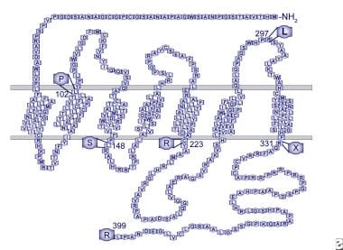
Luteinizing Hormone (LH) receptors are specialized protein structures found on the surface of certain cells in the body. They play a crucial role in the endocrine system by binding to specific hormones, such as Luteinizing Hormone, and triggering a series of intracellular events that ultimately lead to changes in cell function.
In particular, LH receptors are found on the cells of the ovaries and testes. In females, when LH binds to its receptor in the ovary, it stimulates ovulation and the development of the corpus luteum, which produces progesterone. In males, LH (also known as Interstitial Cell-Stimulating Hormone in this context) binding to its receptor on testicular Leydig cells triggers the production of testosterone.
Therefore, LH receptors are essential for reproductive processes and the maintenance of secondary sexual characteristics.
Promoter regions in genetics refer to specific DNA sequences located near the transcription start site of a gene. They serve as binding sites for RNA polymerase and various transcription factors that regulate the initiation of gene transcription. These regulatory elements help control the rate of transcription and, therefore, the level of gene expression. Promoter regions can be composed of different types of sequences, such as the TATA box and CAAT box, and their organization and composition can vary between different genes and species.
'Gene expression regulation' refers to the processes that control whether, when, and where a particular gene is expressed, meaning the production of a specific protein or functional RNA encoded by that gene. This complex mechanism can be influenced by various factors such as transcription factors, chromatin remodeling, DNA methylation, non-coding RNAs, and post-transcriptional modifications, among others. Proper regulation of gene expression is crucial for normal cellular function, development, and maintaining homeostasis in living organisms. Dysregulation of gene expression can lead to various diseases, including cancer and genetic disorders.
Chorionic Gonadotropin (hCG) is a hormone that is produced during pregnancy. It is produced by the placenta after implantation of the fertilized egg in the uterus. The main function of hCG is to prevent the disintegration of the corpus luteum, which is a temporary endocrine structure that forms in the ovary after ovulation and produces progesterone during early pregnancy. Progesterone is essential for maintaining the lining of the uterus and supporting the pregnancy.
hCG can be detected in the blood or urine as early as 10 days after conception, and its levels continue to rise throughout the first trimester of pregnancy. In addition to its role in maintaining pregnancy, hCG is also used as a clinical marker for pregnancy and to monitor certain medical conditions such as gestational trophoblastic diseases.
Testosterone is a steroid hormone that belongs to androsten class of hormones. It is primarily secreted by the Leydig cells in the testes of males and, to a lesser extent, by the ovaries and adrenal glands in females. Testosterone is the main male sex hormone and anabolic steroid. It plays a key role in the development of masculine characteristics, such as body hair and muscle mass, and contributes to bone density, fat distribution, red cell production, and sex drive. In females, testosterone contributes to sexual desire and bone health. Testosterone is synthesized from cholesterol and its production is regulated by luteinizing hormone (LH) and follicle-stimulating hormone (FSH).
Estradiol is a type of estrogen, which is a female sex hormone. It is the most potent and dominant form of estrogen in humans. Estradiol plays a crucial role in the development and maintenance of secondary sexual characteristics in women, such as breast development and regulation of the menstrual cycle. It also helps maintain bone density, protect the lining of the uterus, and is involved in cognition and mood regulation.
Estradiol is produced primarily by the ovaries, but it can also be synthesized in smaller amounts by the adrenal glands and fat cells. In men, estradiol is produced from testosterone through a process called aromatization. Abnormal levels of estradiol can contribute to various health issues, such as hormonal imbalances, infertility, osteoporosis, and certain types of cancer.
Progesterone is a steroid hormone that is primarily produced in the ovaries during the menstrual cycle and in pregnancy. It plays an essential role in preparing the uterus for implantation of a fertilized egg and maintaining the early stages of pregnancy. Progesterone works to thicken the lining of the uterus, creating a nurturing environment for the developing embryo.
During the menstrual cycle, progesterone is produced by the corpus luteum, a temporary structure formed in the ovary after an egg has been released from a follicle during ovulation. If pregnancy does not occur, the levels of progesterone will decrease, leading to the shedding of the uterine lining and menstruation.
In addition to its reproductive functions, progesterone also has various other effects on the body, such as helping to regulate the immune system, supporting bone health, and potentially influencing mood and cognition. Progesterone can be administered medically in the form of oral pills, intramuscular injections, or vaginal suppositories for various purposes, including hormone replacement therapy, contraception, and managing certain gynecological conditions.
Chorionic Gonadotropin, beta Subunit, Human (β-hCG) is a protein that is produced by the placenta during pregnancy. It is a component of human chorionic gonadotropin (hCG), which is a hormone that is composed of two subunits: alpha and beta. The β-hCG subunit is specific to hCG and is not found in other hormones, making it a useful marker for pregnancy and certain medical conditions.
During early pregnancy, the levels of β-hCG increase rapidly and can be detected in the blood and urine. This has led to the development of pregnancy tests that detect the presence of β-hCG to confirm pregnancy. In addition to its role in pregnancy, β-hCG is also used as a tumor marker for certain types of cancer, such as germ cell tumors and choriocarcinoma.
Elevated levels of β-hCG may indicate the presence of a molar pregnancy, a condition in which a fertilized egg implants in the uterus but does not develop properly. In some cases, a molar pregnancy can become cancerous and require treatment. Therefore, monitoring β-hCG levels during pregnancy is important for detecting any potential complications.
Hormones are defined as chemical messengers that are produced by endocrine glands or specialized cells and are transported through the bloodstream to tissues and organs, where they elicit specific responses. They play crucial roles in regulating various physiological processes such as growth, development, metabolism, reproduction, and mood. Examples of hormones include insulin, estrogen, testosterone, adrenaline, and thyroxine.
Thyroid Hormone Resistance Syndrome, also known as Refractory Thyroid Disease or Generalized T3 Resistance, is a rare genetic disorder characterized by reduced sensitivity and impaired response of the body's tissues to thyroid hormones, despite having normal or elevated levels of these hormones in the blood. This condition is caused by mutations in the THRB gene, which encodes the thyroid hormone receptor beta.
In this syndrome, the target cells and tissues do not respond properly to thyroid hormones, leading to a wide range of symptoms similar to those seen in hypothyroidism (underactive thyroid), such as fatigue, weight gain, cold intolerance, constipation, dry skin, and depression. However, unlike hypothyroidism, patients with Thyroid Hormone Resistance Syndrome usually have normal or increased levels of thyroid-stimulating hormone (TSH) and free thyroxine (FT4) in their blood.
The diagnosis of Thyroid Hormone Resistance Syndrome is often challenging, as it requires the exclusion of other causes of hypothyroidism and the confirmation of normal or elevated thyroid hormone levels with impaired tissue response. Treatment typically involves careful monitoring and management of symptoms, as the use of additional thyroid hormones may not improve the condition and can even worsen symptoms in some cases.
Molecular sequence data refers to the specific arrangement of molecules, most commonly nucleotides in DNA or RNA, or amino acids in proteins, that make up a biological macromolecule. This data is generated through laboratory techniques such as sequencing, and provides information about the exact order of the constituent molecules. This data is crucial in various fields of biology, including genetics, evolution, and molecular biology, allowing for comparisons between different organisms, identification of genetic variations, and studies of gene function and regulation.
An ovary is a part of the female reproductive system in which ova or eggs are produced through the process of oogenesis. They are a pair of solid, almond-shaped structures located one on each side of the uterus within the pelvic cavity. Each ovary measures about 3 to 5 centimeters in length and weighs around 14 grams.
The ovaries have two main functions: endocrine (hormonal) function and reproductive function. They produce and release eggs (ovulation) responsible for potential fertilization and development of an embryo/fetus during pregnancy. Additionally, they are essential in the production of female sex hormones, primarily estrogen and progesterone, which regulate menstrual cycles, sexual development, and reproduction.
During each menstrual cycle, a mature egg is released from one of the ovaries into the fallopian tube, where it may be fertilized by sperm. If not fertilized, the egg, along with the uterine lining, will be shed, leading to menstruation.
Thyroid hormone receptors (THRs) are nuclear receptor proteins that bind to thyroid hormones and mediate their effects in target cells. There are two main types of THRs, referred to as THR alpha and THR beta. THR beta is further divided into two subtypes, THR beta1 and THR beta2.
THR beta is a type of nuclear receptor that is primarily expressed in the liver, kidney, and heart, as well as in the central nervous system. It plays an important role in regulating the metabolism of carbohydrates, lipids, and proteins, as well as in the development and function of the heart. THR beta is also involved in the regulation of body weight and energy expenditure.
THR beta1 is the predominant subtype expressed in the liver and is responsible for many of the metabolic effects of thyroid hormones in this organ. THR beta2, on the other hand, is primarily expressed in the heart and plays a role in regulating cardiac function.
Abnormalities in THR beta function can lead to various diseases, including thyroid hormone resistance, a condition in which the body's cells are unable to respond properly to thyroid hormones. This can result in symptoms such as weight gain, fatigue, and cold intolerance.
Ovulation is the medical term for the release of a mature egg from an ovary during a woman's menstrual cycle. The released egg travels through the fallopian tube where it may be fertilized by sperm if sexual intercourse has occurred recently. If the egg is not fertilized, it will break down and leave the body along with the uterine lining during menstruation. Ovulation typically occurs around day 14 of a 28-day menstrual cycle, but the timing can vary widely from woman to woman and even from cycle to cycle in the same woman.
During ovulation, there are several physical changes that may occur in a woman's body, such as an increase in basal body temperature, changes in cervical mucus, and mild cramping or discomfort on one side of the lower abdomen (known as mittelschmerz). These symptoms can be used to help predict ovulation and improve the chances of conception.
It's worth noting that some medical conditions, such as polycystic ovary syndrome (PCOS) or premature ovarian failure, may affect ovulation and make it difficult for a woman to become pregnant. In these cases, medical intervention may be necessary to help promote ovulation and increase the chances of conception.
Pituitary hormone-releasing hormones (PRHs), also known as hypothalamic releasing hormones or hypothalamic hormones, are small neuropeptides produced and released by the hypothalamus - a small region of the brain. These hormones play crucial roles in regulating the secretion and release of various pituitary hormones, which in turn control several essential bodily functions, including growth, development, metabolism, stress response, reproduction, and lactation.
There are several PRHs, each with a specific target pituitary hormone:
1. Thyrotropin-releasing hormone (TRH): Stimulates the release of thyroid-stimulating hormone (TSH) from the anterior pituitary gland, which then promotes the production and release of thyroid hormones.
2. Gonadotropin-releasing hormone (GnRH): Regulates the secretion of follicle-stimulating hormone (FSH) and luteinizing hormone (LH) from the anterior pituitary gland, which are essential for reproductive functions.
3. Corticotropin-releasing hormone (CRH): Stimulates the release of adrenocorticotropic hormone (ACTH) from the anterior pituitary gland, which then promotes the production and release of cortisol and other glucocorticoids from the adrenal glands.
4. Growth hormone-releasing hormone (GHRH): Stimulates the release of growth hormone (GH) from the anterior pituitary gland, which is essential for growth, development, and metabolism regulation.
5. Somatostatin or growth hormone-inhibiting hormone (GHIH): Inhibits the release of GH from the anterior pituitary gland and also suppresses the secretion of thyroid hormones.
6. Prolactin-releasing hormone (PRH) or prolactin-releasing factor (PRF): Stimulates the release of prolactin from the anterior pituitary gland, which is essential for lactation and reproductive functions.
7. Prolactin-inhibiting hormone (PIH) or dopamine: Inhibits the release of prolactin from the anterior pituitary gland.
These releasing hormones and inhibitory hormones work together to maintain a delicate balance in various physiological processes, including growth, development, metabolism, stress response, and reproductive functions. Dysregulation of these hormonal systems can lead to various endocrine disorders and diseases.
The anterior pituitary, also known as the adenohypophysis, is the front portion of the pituitary gland. It is responsible for producing and secreting several important hormones that regulate various bodily functions. These hormones include:
* Growth hormone (GH), which stimulates growth and cell reproduction in bones and other tissues.
* Thyroid-stimulating hormone (TSH), which regulates the production of thyroid hormones by the thyroid gland.
* Adrenocorticotropic hormone (ACTH), which stimulates the adrenal glands to produce cortisol and other steroid hormones.
* Follicle-stimulating hormone (FSH) and luteinizing hormone (LH), which regulate reproductive function in both males and females by controlling the development and release of eggs or sperm.
* Prolactin, which stimulates milk production in pregnant and nursing women.
* Melanocyte-stimulating hormone (MSH), which regulates skin pigmentation and appetite.
The anterior pituitary gland is controlled by the hypothalamus, a small region of the brain located just above it. The hypothalamus produces releasing and inhibiting hormones that regulate the secretion of hormones from the anterior pituitary. These hormones are released into a network of blood vessels called the portal system, which carries them directly to the anterior pituitary gland.
Damage or disease of the anterior pituitary can lead to hormonal imbalances and various medical conditions, such as growth disorders, thyroid dysfunction, adrenal insufficiency, reproductive problems, and diabetes insipidus.
Prolactin is a hormone produced by the pituitary gland, a small gland located at the base of the brain. Its primary function is to stimulate milk production in women after childbirth, a process known as lactation. However, prolactin also plays other roles in the body, including regulating immune responses, metabolism, and behavior. In men, prolactin helps maintain the sexual glands and contributes to paternal behaviors.
Prolactin levels are usually low in both men and non-pregnant women but increase significantly during pregnancy and after childbirth. Various factors can affect prolactin levels, including stress, sleep, exercise, and certain medications. High prolactin levels can lead to medical conditions such as amenorrhea (absence of menstruation), galactorrhea (spontaneous milk production not related to childbirth), infertility, and reduced sexual desire in both men and women.
Estrus is a term used in veterinary medicine to describe the physiological and behavioral state of female mammals that are ready to mate and conceive. It refers to the period of time when the female's reproductive system is most receptive to fertilization.
During estrus, the female's ovaries release one or more mature eggs (ovulation) into the fallopian tubes, where they can be fertilized by sperm from a male. This phase of the estrous cycle is often accompanied by changes in behavior and physical appearance, such as increased vocalization, restlessness, and swelling of the genital area.
The duration and frequency of estrus vary widely among different species of mammals. In some animals, such as dogs and cats, estrus occurs regularly at intervals of several weeks or months, while in others, such as cows and mares, it may only occur once or twice a year.
It's important to note that the term "estrus" is not used to describe human reproductive physiology. In humans, the equivalent phase of the menstrual cycle is called ovulation.
A base sequence in the context of molecular biology refers to the specific order of nucleotides in a DNA or RNA molecule. In DNA, these nucleotides are adenine (A), guanine (G), cytosine (C), and thymine (T). In RNA, uracil (U) takes the place of thymine. The base sequence contains genetic information that is transcribed into RNA and ultimately translated into proteins. It is the exact order of these bases that determines the genetic code and thus the function of the DNA or RNA molecule.
Thyroid hormone receptors (THRs) are nuclear receptor proteins that bind to thyroid hormones, triiodothyronine (T3) and thyroxine (T4), and regulate gene transcription in target cells. These receptors play a crucial role in the development, growth, and metabolism of an organism by mediating the actions of thyroid hormones. THRs are encoded by genes THRA and THRB, which give rise to two major isoforms: TRα1 and TRβ1. Additionally, alternative splicing results in other isoforms with distinct tissue distributions and functions. THRs function as heterodimers with retinoid X receptors (RXRs) and bind to thyroid hormone response elements (TREs) in the regulatory regions of target genes. The binding of T3 or T4 to THRs triggers a conformational change, which leads to recruitment of coactivators or corepressors, ultimately resulting in activation or repression of gene transcription.
Ovariectomy is a surgical procedure in which one or both ovaries are removed. It is also known as "ovary removal" or "oophorectomy." This procedure is often performed as a treatment for various medical conditions, including ovarian cancer, endometriosis, uterine fibroids, and pelvic pain. Ovariectomy can also be part of a larger surgical procedure called an hysterectomy, in which the uterus is also removed.
In some cases, an ovariectomy may be performed as a preventative measure for individuals at high risk of developing ovarian cancer. This is known as a prophylactic ovariectomy. After an ovariectomy, a person will no longer have menstrual periods and will be unable to become pregnant naturally. Hormone replacement therapy may be recommended in some cases to help manage symptoms associated with the loss of hormones produced by the ovaries.
Castration is a surgical procedure to remove the testicles in males or ovaries in females. In males, it is also known as orchiectomy. This procedure results in the inability to produce sex hormones and gametes (sperm in men and eggs in women), and can be done for various reasons such as medical treatment for certain types of cancer, to reduce sexual urges in individuals with criminal tendencies, or as a form of birth control in animals.
Macromolecular substances, also known as macromolecules, are large, complex molecules made up of repeating subunits called monomers. These substances are formed through polymerization, a process in which many small molecules combine to form a larger one. Macromolecular substances can be naturally occurring, such as proteins, DNA, and carbohydrates, or synthetic, such as plastics and synthetic fibers.
In the context of medicine, macromolecular substances are often used in the development of drugs and medical devices. For example, some drugs are designed to bind to specific macromolecules in the body, such as proteins or DNA, in order to alter their function and produce a therapeutic effect. Additionally, macromolecular substances may be used in the creation of medical implants, such as artificial joints and heart valves, due to their strength and durability.
It is important for healthcare professionals to have an understanding of macromolecular substances and how they function in the body, as this knowledge can inform the development and use of medical treatments.
Sexual maturation is the process of physical development during puberty that leads to the ability to reproduce. This process involves the development of primary and secondary sexual characteristics, changes in hormone levels, and the acquisition of reproductive capabilities. In females, this includes the onset of menstruation and the development of breasts and hips. In males, this includes the deepening of the voice, growth of facial hair, and the production of sperm. Achieving sexual maturation is an important milestone in human development and typically occurs during adolescence.
Leydig cells, also known as interstitial cells of Leydig or interstitial cell-stroma, are cells in the testes that produce and release testosterone and other androgens into the bloodstream. They are located in the seminiferous tubules of the testis, near the blood vessels, and are named after Franz Leydig, the German physiologist who discovered them in 1850.
Leydig cells contain cholesterol esters, which serve as precursors for the synthesis of testosterone. They respond to luteinizing hormone (LH) released by the anterior pituitary gland, which stimulates the production and release of testosterone. Testosterone is essential for the development and maintenance of male secondary sexual characteristics, such as facial hair, deep voice, and muscle mass. It also plays a role in sperm production and bone density.
In addition to their endocrine function, Leydig cells have been shown to have non-hormonal functions, including phagocytosis, antigen presentation, and immune regulation. However, these functions are not as well understood as their hormonal roles.
Messenger RNA (mRNA) is a type of RNA (ribonucleic acid) that carries genetic information copied from DNA in the form of a series of three-base code "words," each of which specifies a particular amino acid. This information is used by the cell's machinery to construct proteins, a process known as translation. After being transcribed from DNA, mRNA travels out of the nucleus to the ribosomes in the cytoplasm where protein synthesis occurs. Once the protein has been synthesized, the mRNA may be degraded and recycled. Post-transcriptional modifications can also occur to mRNA, such as alternative splicing and addition of a 5' cap and a poly(A) tail, which can affect its stability, localization, and translation efficiency.
An amino acid sequence is the specific order of amino acids in a protein or peptide molecule, formed by the linking of the amino group (-NH2) of one amino acid to the carboxyl group (-COOH) of another amino acid through a peptide bond. The sequence is determined by the genetic code and is unique to each type of protein or peptide. It plays a crucial role in determining the three-dimensional structure and function of proteins.
LHRH (Luteinizing Hormone-Releasing Hormone) receptors are a type of G protein-coupled receptor found on the surface of certain cells in the body, most notably in the anterior pituitary gland. These receptors bind to LHRH, a hormone that is produced and released by the hypothalamus in the brain.
When LHRH binds to its receptor, it triggers a series of intracellular signaling events that ultimately lead to the release of two other hormones from the anterior pituitary gland: luteinizing hormone (LH) and follicle-stimulating hormone (FSH). These hormones play critical roles in regulating reproductive function, including the development and maturation of sex cells (sperm and eggs), the production of sex steroid hormones (such as testosterone and estrogen), and the regulation of the menstrual cycle in females.
Disorders of the LHRH receptor or its signaling pathway can lead to a variety of reproductive disorders, including precocious puberty, delayed puberty, and infertility.
The corpus luteum is a temporary endocrine structure that forms in the ovary after an oocyte (egg) has been released from a follicle during ovulation. It's formed by the remaining cells of the ruptured follicle, which transform into large, hormone-secreting cells.
The primary function of the corpus luteum is to produce progesterone and, to a lesser extent, estrogen during the menstrual cycle or pregnancy. Progesterone plays a crucial role in preparing the uterus for potential implantation of a fertilized egg and maintaining the early stages of pregnancy. If pregnancy does not occur, the corpus luteum will typically degenerate and stop producing hormones after approximately 10-14 days, leading to menstruation.
However, if pregnancy occurs, the developing embryo starts to produce human chorionic gonadotropin (hCG), which signals the corpus luteum to continue secreting progesterone and estrogen until the placenta takes over hormonal production, usually around the end of the first trimester.
Gonadal steroid hormones, also known as gonadal sex steroids, are hormones that are produced and released by the gonads (i.e., ovaries in women and testes in men). These hormones play a critical role in the development and maintenance of secondary sexual characteristics, reproductive function, and overall health.
The three main classes of gonadal steroid hormones are:
1. Androgens: These are male sex hormones that are primarily produced by the testes but also produced in smaller amounts by the ovaries and adrenal glands. The most well-known androgen is testosterone, which plays a key role in the development of male secondary sexual characteristics such as facial hair, deepening of the voice, and increased muscle mass.
2. Estrogens: These are female sex hormones that are primarily produced by the ovaries but also produced in smaller amounts by the adrenal glands. The most well-known estrogen is estradiol, which plays a key role in the development of female secondary sexual characteristics such as breast development and the menstrual cycle.
3. Progestogens: These are hormones that are produced by the ovaries during the second half of the menstrual cycle and play a key role in preparing the uterus for pregnancy. The most well-known progestogen is progesterone, which also plays a role in maintaining pregnancy and regulating the menstrual cycle.
Gonadal steroid hormones can have significant effects on various physiological processes, including bone density, cognitive function, mood, and sexual behavior. Disorders of gonadal steroid hormone production or action can lead to a range of health problems, including infertility, osteoporosis, and sexual dysfunction.
The testis, also known as the testicle, is a male reproductive organ that is part of the endocrine system. It is located in the scrotum, outside of the abdominal cavity. The main function of the testis is to produce sperm and testosterone, the primary male sex hormone.
The testis is composed of many tiny tubules called seminiferous tubules, where sperm are produced. These tubules are surrounded by a network of blood vessels, nerves, and supportive tissues. The sperm then travel through a series of ducts to the epididymis, where they mature and become capable of fertilization.
Testosterone is produced in the Leydig cells, which are located in the interstitial tissue between the seminiferous tubules. Testosterone plays a crucial role in the development and maintenance of male secondary sexual characteristics, such as facial hair, deep voice, and muscle mass. It also supports sperm production and sexual function.
Abnormalities in testicular function can lead to infertility, hormonal imbalances, and other health problems. Regular self-examinations and medical check-ups are recommended for early detection and treatment of any potential issues.
Radioimmunoassay (RIA) is a highly sensitive analytical technique used in clinical and research laboratories to measure concentrations of various substances, such as hormones, vitamins, drugs, or tumor markers, in biological samples like blood, urine, or tissues. The method relies on the specific interaction between an antibody and its corresponding antigen, combined with the use of radioisotopes to quantify the amount of bound antigen.
In a typical RIA procedure, a known quantity of a radiolabeled antigen (also called tracer) is added to a sample containing an unknown concentration of the same unlabeled antigen. The mixture is then incubated with a specific antibody that binds to the antigen. During the incubation period, the antibody forms complexes with both the radiolabeled and unlabeled antigens.
After the incubation, the unbound (free) radiolabeled antigen is separated from the antibody-antigen complexes, usually through a precipitation or separation step involving centrifugation, filtration, or chromatography. The amount of radioactivity in the pellet (containing the antibody-antigen complexes) is then measured using a gamma counter or other suitable radiation detection device.
The concentration of the unlabeled antigen in the sample can be determined by comparing the ratio of bound to free radiolabeled antigen in the sample to a standard curve generated from known concentrations of unlabeled antigen and their corresponding bound/free ratios. The higher the concentration of unlabeled antigen in the sample, the lower the amount of radiolabeled antigen that will bind to the antibody, resulting in a lower bound/free ratio.
Radioimmunoassays offer high sensitivity, specificity, and accuracy, making them valuable tools for detecting and quantifying low levels of various substances in biological samples. However, due to concerns about radiation safety and waste disposal, alternative non-isotopic immunoassay techniques like enzyme-linked immunosorbent assays (ELISAs) have become more popular in recent years.
Gonadotropins are hormones that stimulate the gonads (sex glands) to produce sex steroids and gametes (sex cells). In humans, there are two main types of gonadotropins: follicle-stimulating hormone (FSH) and luteinizing hormone (LH), which are produced and released by the anterior pituitary gland.
FSH plays a crucial role in the development and maturation of ovarian follicles in females and sperm production in males. LH triggers ovulation in females, causing the release of a mature egg from the ovary, and stimulates testosterone production in males.
Gonadotropins are often used in medical treatments to stimulate the gonads, such as in infertility therapies where FSH and LH are administered to induce ovulation or increase sperm production.
Thyroid hormones are hormones produced and released by the thyroid gland, a small endocrine gland located in the neck that helps regulate metabolism, growth, and development in the human body. The two main thyroid hormones are triiodothyronine (T3) and thyroxine (T4), which contain iodine atoms. These hormones play a crucial role in various bodily functions, including heart rate, body temperature, digestion, and brain development. They help regulate the rate at which your body uses energy, affects how sensitive your body is to other hormones, and plays a vital role in the development and differentiation of all cells of the human body. Thyroid hormone levels are regulated by the hypothalamus and pituitary gland through a feedback mechanism that helps maintain proper balance.
Proestrus is a stage in the estrous cycle of animals, specifically referring to the phase preceding estrus (heat) during which follicle development and estrogen production occur. It is characterized by the swelling of the vulva and the onset of behaviors indicating readiness to mate, although the animal is not yet receptive to males. This stage typically lasts around 2-13 days, depending on the species. In humans, this equivalent phase does not exist due to menstrual cycles rather than estrous cycles.
I apologize, but "sheep" is not a term that has a medical definition. It is a common animal with the scientific name Ovis aries. If you have any medical or health-related questions, I would be happy to try and help answer those for you.
"Cattle" is a term used in the agricultural and veterinary fields to refer to domesticated animals of the genus *Bos*, primarily *Bos taurus* (European cattle) and *Bos indicus* (Zebu). These animals are often raised for meat, milk, leather, and labor. They are also known as bovines or cows (for females), bulls (intact males), and steers/bullocks (castrated males). However, in a strict medical definition, "cattle" does not apply to humans or other animals.
Gonadotropins are hormones produced and released by the anterior pituitary gland, a small endocrine gland located at the base of the brain. These hormones play crucial roles in regulating reproduction and sexual development. There are two main types of gonadotropins:
1. Follicle-Stimulating Hormone (FSH): FSH is essential for the growth and development of follicles in the ovaries (in females) or sperm production in the testes (in males). In females, FSH stimulates the maturation of eggs within the follicles.
2. Luteinizing Hormone (LH): LH triggers ovulation in females, causing the release of a mature egg from the dominant follicle. In males, LH stimulates the production and secretion of testosterone in the testes.
Together, FSH and LH work synergistically to regulate various aspects of reproductive function and sexual development. Their secretion is controlled by the hypothalamus, which releases gonadotropin-releasing hormone (GnRH) to stimulate the production and release of FSH and LH from the anterior pituitary gland.
Abnormal levels of gonadotropins can lead to various reproductive disorders, such as infertility or menstrual irregularities in females and issues related to sexual development or function in both sexes. In some cases, synthetic forms of gonadotropins may be used clinically to treat these conditions or for assisted reproductive technologies (ART).
A cell line is a culture of cells that are grown in a laboratory for use in research. These cells are usually taken from a single cell or group of cells, and they are able to divide and grow continuously in the lab. Cell lines can come from many different sources, including animals, plants, and humans. They are often used in scientific research to study cellular processes, disease mechanisms, and to test new drugs or treatments. Some common types of human cell lines include HeLa cells (which come from a cancer patient named Henrietta Lacks), HEK293 cells (which come from embryonic kidney cells), and HUVEC cells (which come from umbilical vein endothelial cells). It is important to note that cell lines are not the same as primary cells, which are cells that are taken directly from a living organism and have not been grown in the lab.
An ovarian follicle is a fluid-filled sac in the ovary that contains an immature egg or ovum (oocyte). It's a part of the female reproductive system and plays a crucial role in the process of ovulation.
Ovarian follicles start developing in the ovaries during fetal development, but only a small number of them will mature and release an egg during a woman's reproductive years. The maturation process is stimulated by hormones like follicle-stimulating hormone (FSH) and luteinizing hormone (LH).
There are different types of ovarian follicles, including primordial, primary, secondary, and tertiary or Graafian follicles. The Graafian follicle is the mature follicle that ruptures during ovulation to release the egg into the fallopian tube, where it may be fertilized by sperm.
It's important to note that abnormal growth or development of ovarian follicles can lead to conditions like polycystic ovary syndrome (PCOS) and ovarian cancer.
The follicular phase is a term used in reproductive endocrinology, which refers to the first part of the menstrual cycle. This phase begins on the first day of menstruation and lasts until ovulation. During this phase, several follicles in the ovaries begin to mature under the influence of follicle-stimulating hormone (FSH) released by the pituitary gland.
Typically, one follicle becomes dominant and continues to mature, while the others regress. The dominant follicle produces increasing amounts of estrogen, which causes the lining of the uterus to thicken in preparation for a possible pregnancy. The follicular phase can vary in length, but on average it lasts about 14 days.
It's important to note that the length and characteristics of the follicular phase can provide valuable information in diagnosing various reproductive disorders, such as polycystic ovary syndrome (PCOS) or thyroid dysfunction.
Anterior pituitary hormones are a group of six major hormones that are produced and released by the anterior portion (lobe) of the pituitary gland, a small endocrine gland located at the base of the brain. These hormones play crucial roles in regulating various bodily functions and activities. The six main anterior pituitary hormones are:
1. Growth Hormone (GH): Also known as somatotropin, GH is essential for normal growth and development in children and adolescents. It helps regulate body composition, metabolism, and bone density in adults.
2. Prolactin (PRL): A hormone that stimulates milk production in females after childbirth and is also involved in various reproductive and immune functions in both sexes.
3. Follicle-Stimulating Hormone (FSH): FSH regulates the development, growth, and maturation of follicles in the ovaries (in females) and sperm production in the testes (in males).
4. Luteinizing Hormone (LH): LH plays a key role in triggering ovulation in females and stimulating testosterone production in males.
5. Thyroid-Stimulating Hormone (TSH): TSH regulates the function of the thyroid gland, which is responsible for producing and releasing thyroid hormones that control metabolism and growth.
6. Adrenocorticotropic Hormone (ACTH): ACTH stimulates the adrenal glands to produce cortisol, a steroid hormone involved in stress response, metabolism, and immune function.
These anterior pituitary hormones are regulated by the hypothalamus, which is located above the pituitary gland. The hypothalamus releases releasing and inhibiting factors that control the synthesis and secretion of anterior pituitary hormones, creating a complex feedback system to maintain homeostasis in the body.
A protein subunit refers to a distinct and independently folding polypeptide chain that makes up a larger protein complex. Proteins are often composed of multiple subunits, which can be identical or different, that come together to form the functional unit of the protein. These subunits can interact with each other through non-covalent interactions such as hydrogen bonds, ionic bonds, and van der Waals forces, as well as covalent bonds like disulfide bridges. The arrangement and interaction of these subunits contribute to the overall structure and function of the protein.
Orchiectomy is a surgical procedure where one or both of the testicles are removed. It is also known as castration. This procedure can be performed for various reasons, including the treatment of testicular cancer, prostate cancer, or other conditions that may affect the testicles. It can also be done to reduce levels of male hormones in the body, such as in the case of transgender women undergoing gender affirming surgery. The specific medical definition may vary slightly depending on the context and the extent of the procedure.
Granulosa cells are specialized cells that surround and enclose the developing egg cells (oocytes) in the ovaries. They play a crucial role in the growth, development, and maturation of the follicles (the fluid-filled sacs containing the oocytes) by providing essential nutrients and hormones.
Granulosa cells are responsible for producing estrogen, which supports the development of the endometrium during the menstrual cycle in preparation for a potential pregnancy. They also produce inhibin and activin, two hormones that regulate the function of the pituitary gland and its secretion of follicle-stimulating hormone (FSH) and luteinizing hormone (LH).
These cells are critical for female reproductive health and fertility. Abnormalities in granulosa cell function can lead to various reproductive disorders, such as polycystic ovary syndrome (PCOS), premature ovarian failure, and infertility.
In the context of medicine and pharmacology, "kinetics" refers to the study of how a drug moves throughout the body, including its absorption, distribution, metabolism, and excretion (often abbreviated as ADME). This field is called "pharmacokinetics."
1. Absorption: This is the process of a drug moving from its site of administration into the bloodstream. Factors such as the route of administration (e.g., oral, intravenous, etc.), formulation, and individual physiological differences can affect absorption.
2. Distribution: Once a drug is in the bloodstream, it gets distributed throughout the body to various tissues and organs. This process is influenced by factors like blood flow, protein binding, and lipid solubility of the drug.
3. Metabolism: Drugs are often chemically modified in the body, typically in the liver, through processes known as metabolism. These changes can lead to the formation of active or inactive metabolites, which may then be further distributed, excreted, or undergo additional metabolic transformations.
4. Excretion: This is the process by which drugs and their metabolites are eliminated from the body, primarily through the kidneys (urine) and the liver (bile).
Understanding the kinetics of a drug is crucial for determining its optimal dosing regimen, potential interactions with other medications or foods, and any necessary adjustments for special populations like pediatric or geriatric patients, or those with impaired renal or hepatic function.
Genetic transcription is the process by which the information in a strand of DNA is used to create a complementary RNA molecule. This process is the first step in gene expression, where the genetic code in DNA is converted into a form that can be used to produce proteins or functional RNAs.
During transcription, an enzyme called RNA polymerase binds to the DNA template strand and reads the sequence of nucleotide bases. As it moves along the template, it adds complementary RNA nucleotides to the growing RNA chain, creating a single-stranded RNA molecule that is complementary to the DNA template strand. Once transcription is complete, the RNA molecule may undergo further processing before it can be translated into protein or perform its functional role in the cell.
Transcription can be either "constitutive" or "regulated." Constitutive transcription occurs at a relatively constant rate and produces essential proteins that are required for basic cellular functions. Regulated transcription, on the other hand, is subject to control by various intracellular and extracellular signals, allowing cells to respond to changing environmental conditions or developmental cues.
The hypothalamus is a small, vital region of the brain that lies just below the thalamus and forms part of the limbic system. It plays a crucial role in many important functions including:
1. Regulation of body temperature, hunger, thirst, fatigue, sleep, and circadian rhythms.
2. Production and regulation of hormones through its connection with the pituitary gland (the hypophysis). It controls the release of various hormones by producing releasing and inhibiting factors that regulate the anterior pituitary's function.
3. Emotional responses, behavior, and memory formation through its connections with the limbic system structures like the amygdala and hippocampus.
4. Autonomic nervous system regulation, which controls involuntary physiological functions such as heart rate, blood pressure, and digestion.
5. Regulation of the immune system by interacting with the autonomic nervous system.
Damage to the hypothalamus can lead to various disorders like diabetes insipidus, growth hormone deficiency, altered temperature regulation, sleep disturbances, and emotional or behavioral changes.
Pregnancy is a physiological state or condition where a fertilized egg (zygote) successfully implants and grows in the uterus of a woman, leading to the development of an embryo and finally a fetus. This process typically spans approximately 40 weeks, divided into three trimesters, and culminates in childbirth. Throughout this period, numerous hormonal and physical changes occur to support the growing offspring, including uterine enlargement, breast development, and various maternal adaptations to ensure the fetus's optimal growth and well-being.
The luteal phase is the second half of the menstrual cycle, starting from ovulation (release of an egg from the ovaries) and lasting until the start of the next menstruation. This phase typically lasts around 12-14 days in a regular 28-day menstrual cycle. During this phase, the remains of the dominant follicle that released the egg transform into the corpus luteum, which produces progesterone and some estrogen to support the implantation of a fertilized egg and maintain the early stages of pregnancy. If pregnancy does not occur, the corpus luteum degenerates, leading to a drop in hormone levels and the start of a new menstrual cycle.
Diestrus is a stage in the estrous cycle of animals, which is similar to the menstrual cycle in humans. It follows the phase of estrus (or heat), during which the animal is receptive to mating. Diestrus is the period of relative sexual quiescence and hormonal stability between cycles. In this phase, the corpus luteum in the ovary produces progesterone, preparing the uterus for potential pregnancy. If fertilization does not occur, the corpus luteum will degenerate, leading to a drop in progesterone levels and the onset of the next estrous cycle. The duration of diestrus varies among species.
In humans, this phase is analogous to the luteal phase of the menstrual cycle. However, since humans do not exhibit estrous behavior, the term 'diestrus' is typically not used in human reproductive physiology discussions.
Activins are a type of protein that belongs to the transforming growth factor-beta (TGF-β) superfamily. They are produced and released by various cells in the body, including those in the ovaries, testes, pituitary gland, and other tissues. Activins play important roles in regulating several biological processes, such as cell growth, differentiation, and apoptosis (programmed cell death).
Activins bind to specific receptors on the surface of cells, leading to the activation of intracellular signaling pathways that control gene expression. They are particularly well-known for their role in reproductive biology, where they help regulate follicle stimulation and hormone production in the ovaries and testes. Activins also have been implicated in various disease processes, including cancer, fibrosis, and inflammation.
There are three main isoforms of activin in humans: activin A, activin B, and inhibin A. While activins and inhibins share similar structures and functions, they have opposite effects on the activity of the pituitary gland. Activins stimulate the production of follicle-stimulating hormone (FSH), while inhibins suppress it. This delicate balance between activins and inhibins helps regulate reproductive function and other physiological processes in the body.
Follicle-stimulating hormone (FSH) receptors are specialized protein structures found on the surface of specific cells in the body. They play a crucial role in the endocrine system, particularly in the regulation of reproduction and development.
FSH receptors are primarily located on the granulosa cells that surround and support the developing eggs (oocytes) within the ovarian follicles in females. In males, these receptors can be found on the Sertoli cells in the seminiferous tubules of the testes.
When FSH, a glycoprotein hormone secreted by the anterior pituitary gland, binds to its specific receptor, it triggers a series of intracellular signaling events that ultimately lead to various physiological responses. In females, FSH receptor activation stimulates follicle growth, estrogen production, and oocyte maturation. In males, FSH receptor signaling supports spermatogenesis, the process of sperm cell development within the testes.
In summary, FSH receptors are essential components in the hormonal regulation of reproduction and development, mediating the actions of follicle-stimulating hormone on target cells in both females and males.
Pituitary hormones are chemical messengers produced and released by the pituitary gland, a small endocrine gland located at the base of the brain. The pituitary gland is often referred to as the "master gland" because it controls several other endocrine glands and regulates various bodily functions.
There are two main types of pituitary hormones: anterior pituitary hormones and posterior pituitary hormones, which are produced in different parts of the pituitary gland and have distinct functions.
Anterior pituitary hormones include:
1. Growth hormone (GH): regulates growth and metabolism.
2. Thyroid-stimulating hormone (TSH): stimulates the thyroid gland to produce thyroid hormones.
3. Adrenocorticotropic hormone (ACTH): stimulates the adrenal glands to produce cortisol and other steroid hormones.
4. Follicle-stimulating hormone (FSH) and luteinizing hormone (LH): regulate reproductive function in both males and females.
5. Prolactin: stimulates milk production in lactating women.
6. Melanocyte-stimulating hormone (MSH): regulates skin pigmentation and appetite.
Posterior pituitary hormones include:
1. Oxytocin: stimulates uterine contractions during childbirth and milk ejection during lactation.
2. Vasopressin (antidiuretic hormone, ADH): regulates water balance in the body by controlling urine production in the kidneys.
Overall, pituitary hormones play crucial roles in regulating growth, development, metabolism, reproductive function, and various other bodily functions. Abnormalities in pituitary hormone levels can lead to a range of medical conditions, such as dwarfism, acromegaly, Cushing's disease, infertility, and diabetes insipidus.
Protein binding, in the context of medical and biological sciences, refers to the interaction between a protein and another molecule (known as the ligand) that results in a stable complex. This process is often reversible and can be influenced by various factors such as pH, temperature, and concentration of the involved molecules.
In clinical chemistry, protein binding is particularly important when it comes to drugs, as many of them bind to proteins (especially albumin) in the bloodstream. The degree of protein binding can affect a drug's distribution, metabolism, and excretion, which in turn influence its therapeutic effectiveness and potential side effects.
Protein-bound drugs may be less available for interaction with their target tissues, as only the unbound or "free" fraction of the drug is active. Therefore, understanding protein binding can help optimize dosing regimens and minimize adverse reactions.
Triptorelin pamoate is a synthetic analogue of the natural hormone gonadotropin-releasing hormone (GnRH). It is used in the treatment of various conditions such as endometriosis, uterine fibroids, precocious puberty, and prostate cancer.
Triptorelin pamoate works by stimulating the release of follicle-stimulating hormone (FSH) and luteinizing hormone (LH) from the pituitary gland, which in turn stimulate the production of sex hormones such as estrogen and testosterone. However, with continued use, it causes downregulation of the pituitary gland, leading to a decrease in the production of FSH and LH, and therefore a reduction in the levels of sex hormones.
The pamoate salt is used to slow down the release of triptorelin, allowing for longer-acting formulations that can be administered monthly or quarterly. The medication is usually given as an injection into a muscle (intramuscularly).
Inhibins are a group of protein hormones that play a crucial role in regulating the function of the reproductive system, specifically by inhibiting the production of follicle-stimulating hormone (FSH) in the pituitary gland. They are produced and secreted primarily by the granulosa cells in the ovaries of females and Sertoli cells in the testes of males.
Inhibins consist of two subunits, an alpha subunit, and a beta subunit, which can be further divided into two types: inhibin A and inhibin B. Inhibin A is primarily produced by the granulosa cells of developing follicles in the ovary, while inhibin B is mainly produced by the Sertoli cells in the testes.
By regulating FSH production, inhibins help control the development and maturation of ovarian follicles in females and spermatogenesis in males. Abnormal levels of inhibins have been associated with various reproductive disorders, including polycystic ovary syndrome (PCOS) and certain types of cancer.
"Inbred strains of rats" are genetically identical rodents that have been produced through many generations of brother-sister mating. This results in a high degree of homozygosity, where the genes at any particular locus in the genome are identical in all members of the strain.
Inbred strains of rats are widely used in biomedical research because they provide a consistent and reproducible genetic background for studying various biological phenomena, including the effects of drugs, environmental factors, and genetic mutations on health and disease. Additionally, inbred strains can be used to create genetically modified models of human diseases by introducing specific mutations into their genomes.
Some commonly used inbred strains of rats include the Wistar Kyoto (WKY), Sprague-Dawley (SD), and Fischer 344 (F344) rat strains. Each strain has its own unique genetic characteristics, making them suitable for different types of research.
Anestrus is a term used in veterinary medicine to describe the period of sexual quiescence in female animals, during which they do not exhibit estrous cycles. This phase is characterized by low levels of reproductive hormones and is seen in some species as a part of their natural reproductive cycle, while in others it may indicate an abnormality or underlying health issue.
For example, in dogs, anestrus is the period between heat cycles when the reproductive system is relatively inactive. In contrast, in domestic cats, continuous estrous cycling is the norm, and they do not typically exhibit an anestrus phase.
In some cases, anestrus may be induced by factors such as poor nutrition, stress, or illness, and it can have negative consequences for an animal's reproductive health if it persists for too long. If an animal is experiencing prolonged anestrus or other reproductive issues, it is important to consult with a veterinarian for proper diagnosis and treatment.
In the context of medicine, "periodicity" refers to the occurrence of events or phenomena at regular intervals or cycles. This term is often used in reference to recurring symptoms or diseases that have a pattern of appearing and disappearing over time. For example, some medical conditions like menstrual cycles, sleep-wake disorders, and certain infectious diseases exhibit periodicity. It's important to note that the duration and frequency of these cycles can vary depending on the specific condition or individual.
Adrenocorticotropic Hormone (ACTH) is a hormone produced and released by the anterior pituitary gland, a small endocrine gland located at the base of the brain. ACTH plays a crucial role in the regulation of the body's stress response and has significant effects on various physiological processes.
The primary function of ACTH is to stimulate the adrenal glands, which are triangular-shaped glands situated on top of the kidneys. The adrenal glands consist of two parts: the outer cortex and the inner medulla. ACTH specifically targets the adrenal cortex, where it binds to specific receptors and initiates a series of biochemical reactions leading to the production and release of steroid hormones, primarily cortisol (a glucocorticoid) and aldosterone (a mineralocorticoid).
Cortisol is involved in various metabolic processes, such as regulating blood sugar levels, modulating the immune response, and helping the body respond to stress. Aldosterone plays a vital role in maintaining electrolyte and fluid balance by promoting sodium reabsorption and potassium excretion in the kidneys.
ACTH release is controlled by the hypothalamus, another part of the brain, which produces corticotropin-releasing hormone (CRH). CRH stimulates the anterior pituitary gland to secrete ACTH, which in turn triggers cortisol production in the adrenal glands. This complex feedback system helps maintain homeostasis and ensures that appropriate amounts of cortisol are released in response to various physiological and psychological stressors.
Disorders related to ACTH can lead to hormonal imbalances, resulting in conditions such as Cushing's syndrome (excessive cortisol production) or Addison's disease (insufficient cortisol production). Proper diagnosis and management of these disorders typically involve assessing the function of the hypothalamic-pituitary-adrenal axis and addressing any underlying issues affecting ACTH secretion.
Hypogonadism is a medical condition characterized by the inability of the gonads (testes in males and ovaries in females) to produce sufficient amounts of sex hormones, such as testosterone and estrogen. This can lead to various symptoms including decreased libido, erectile dysfunction in men, irregular menstrual periods in women, and reduced fertility in both sexes. Hypogonadism may be caused by genetic factors, aging, injury to the gonads, or certain medical conditions such as pituitary disorders. It can be treated with hormone replacement therapy.
A drug implant is a medical device that is specially designed to provide controlled release of a medication into the body over an extended period of time. Drug implants can be placed under the skin or in various body cavities, depending on the specific medical condition being treated. They are often used when other methods of administering medication, such as oral pills or injections, are not effective or practical.
Drug implants come in various forms, including rods, pellets, and small capsules. The medication is contained within the device and is released slowly over time, either through diffusion or erosion of the implant material. This allows for a steady concentration of the drug to be maintained in the body, which can help to improve treatment outcomes and reduce side effects.
Some common examples of drug implants include:
1. Hormonal implants: These are small rods that are inserted under the skin of the upper arm and release hormones such as progestin or estrogen over a period of several years. They are often used for birth control or to treat conditions such as endometriosis or uterine fibroids.
2. Intraocular implants: These are small devices that are placed in the eye during surgery to release medication directly into the eye. They are often used to treat conditions such as age-related macular degeneration or diabetic retinopathy.
3. Bone cement implants: These are specially formulated cements that contain antibiotics and are used to fill bone defects or joint spaces during surgery. The antibiotics are released slowly over time, helping to prevent infection.
4. Implantable pumps: These are small devices that are placed under the skin and deliver medication directly into a specific body cavity, such as the spinal cord or the peritoneal cavity. They are often used to treat chronic pain or cancer.
Overall, drug implants offer several advantages over other methods of administering medication, including improved compliance, reduced side effects, and more consistent drug levels in the body. However, they may also have some disadvantages, such as the need for surgical placement and the potential for infection or other complications. As with any medical treatment, it is important to discuss the risks and benefits of drug implants with a healthcare provider.
Recombinant proteins are artificially created proteins produced through the use of recombinant DNA technology. This process involves combining DNA molecules from different sources to create a new set of genes that encode for a specific protein. The resulting recombinant protein can then be expressed, purified, and used for various applications in research, medicine, and industry.
Recombinant proteins are widely used in biomedical research to study protein function, structure, and interactions. They are also used in the development of diagnostic tests, vaccines, and therapeutic drugs. For example, recombinant insulin is a common treatment for diabetes, while recombinant human growth hormone is used to treat growth disorders.
The production of recombinant proteins typically involves the use of host cells, such as bacteria, yeast, or mammalian cells, which are engineered to express the desired protein. The host cells are transformed with a plasmid vector containing the gene of interest, along with regulatory elements that control its expression. Once the host cells are cultured and the protein is expressed, it can be purified using various chromatography techniques.
Overall, recombinant proteins have revolutionized many areas of biology and medicine, enabling researchers to study and manipulate proteins in ways that were previously impossible.
Parathyroid hormone (PTH) is a polypeptide hormone that plays a crucial role in the regulation of calcium and phosphate levels in the body. It is produced and secreted by the parathyroid glands, which are four small endocrine glands located on the back surface of the thyroid gland.
The primary function of PTH is to maintain normal calcium levels in the blood by increasing calcium absorption from the gut, mobilizing calcium from bones, and decreasing calcium excretion by the kidneys. PTH also increases phosphate excretion by the kidneys, which helps to lower serum phosphate levels.
In addition to its role in calcium and phosphate homeostasis, PTH has been shown to have anabolic effects on bone tissue, stimulating bone formation and preventing bone loss. However, chronic elevations in PTH levels can lead to excessive bone resorption and osteoporosis.
Overall, Parathyroid Hormone is a critical hormone that helps maintain mineral homeostasis and supports healthy bone metabolism.
Interleukin-1 beta (IL-1β) is a member of the interleukin-1 cytokine family and is primarily produced by activated macrophages in response to inflammatory stimuli. It is a crucial mediator of the innate immune response and plays a key role in the regulation of various biological processes, including cell proliferation, differentiation, and apoptosis. IL-1β is involved in the pathogenesis of several inflammatory diseases, such as rheumatoid arthritis, inflammatory bowel disease, and atherosclerosis. It exerts its effects by binding to the interleukin-1 receptor, which triggers a signaling cascade that leads to the activation of various transcription factors and the expression of target genes.
In the context of medical and biological sciences, a "binding site" refers to a specific location on a protein, molecule, or cell where another molecule can attach or bind. This binding interaction can lead to various functional changes in the original protein or molecule. The other molecule that binds to the binding site is often referred to as a ligand, which can be a small molecule, ion, or even another protein.
The binding between a ligand and its target binding site can be specific and selective, meaning that only certain ligands can bind to particular binding sites with high affinity. This specificity plays a crucial role in various biological processes, such as signal transduction, enzyme catalysis, or drug action.
In the case of drug development, understanding the location and properties of binding sites on target proteins is essential for designing drugs that can selectively bind to these sites and modulate protein function. This knowledge can help create more effective and safer therapeutic options for various diseases.
Transcription factors are proteins that play a crucial role in regulating gene expression by controlling the transcription of DNA to messenger RNA (mRNA). They function by binding to specific DNA sequences, known as response elements, located in the promoter region or enhancer regions of target genes. This binding can either activate or repress the initiation of transcription, depending on the properties and interactions of the particular transcription factor. Transcription factors often act as part of a complex network of regulatory proteins that determine the precise spatiotemporal patterns of gene expression during development, differentiation, and homeostasis in an organism.
I believe you may be referring to the "ventral" part of the hypothalamus, as there isn't a widely recognized anatomical division called the "middle" hypothalamus. The ventral hypothalamus is a region that contains several critical structures, including:
1. The infundibular stem: This is a funnel-shaped structure that extends downward from the hypothalamus and forms the beginning of the pituitary stalk. It contains tuber cinereum and the median eminence.
2. Tuber cinereum: A region with several nuclei, including the arcuate nucleus, which plays a role in regulating feeding behavior, growth hormone release, and sexual function.
3. Median eminence: A crucial area where the hypothalamus interacts with the pituitary gland. It contains nerve terminals that release neurohormones into the portal capillaries, which then carry these substances to the anterior pituitary to regulate hormone secretion.
The ventral hypothalamus is essential for various functions, such as releasing and inhibiting hormones, regulating body temperature, hunger, thirst, sleep, emotional behavior, and parental behaviors.
"Cells, cultured" is a medical term that refers to cells that have been removed from an organism and grown in controlled laboratory conditions outside of the body. This process is called cell culture and it allows scientists to study cells in a more controlled and accessible environment than they would have inside the body. Cultured cells can be derived from a variety of sources, including tissues, organs, or fluids from humans, animals, or cell lines that have been previously established in the laboratory.
Cell culture involves several steps, including isolation of the cells from the tissue, purification and characterization of the cells, and maintenance of the cells in appropriate growth conditions. The cells are typically grown in specialized media that contain nutrients, growth factors, and other components necessary for their survival and proliferation. Cultured cells can be used for a variety of purposes, including basic research, drug development and testing, and production of biological products such as vaccines and gene therapies.
It is important to note that cultured cells may behave differently than they do in the body, and results obtained from cell culture studies may not always translate directly to human physiology or disease. Therefore, it is essential to validate findings from cell culture experiments using additional models and ultimately in clinical trials involving human subjects.
In the field of medicine, "time factors" refer to the duration of symptoms or time elapsed since the onset of a medical condition, which can have significant implications for diagnosis and treatment. Understanding time factors is crucial in determining the progression of a disease, evaluating the effectiveness of treatments, and making critical decisions regarding patient care.
For example, in stroke management, "time is brain," meaning that rapid intervention within a specific time frame (usually within 4.5 hours) is essential to administering tissue plasminogen activator (tPA), a clot-busting drug that can minimize brain damage and improve patient outcomes. Similarly, in trauma care, the "golden hour" concept emphasizes the importance of providing definitive care within the first 60 minutes after injury to increase survival rates and reduce morbidity.
Time factors also play a role in monitoring the progression of chronic conditions like diabetes or heart disease, where regular follow-ups and assessments help determine appropriate treatment adjustments and prevent complications. In infectious diseases, time factors are crucial for initiating antibiotic therapy and identifying potential outbreaks to control their spread.
Overall, "time factors" encompass the significance of recognizing and acting promptly in various medical scenarios to optimize patient outcomes and provide effective care.
Proton-translocating ATPases are complex, multi-subunit enzymes found in the membranes of many organisms, from bacteria to humans. They play a crucial role in energy transduction processes within cells.
In simpler terms, these enzymes help convert chemical energy into a form that can be used to perform mechanical work, such as moving molecules across membranes against their concentration gradients. This is achieved through a process called chemiosmosis, where the movement of ions (in this case, protons or hydrogen ions) down their electrochemical gradient drives the synthesis of ATP, an essential energy currency for cellular functions.
Proton-translocating ATPases consist of two main domains: a catalytic domain responsible for ATP binding and hydrolysis, and a membrane domain that contains the ion transport channel. The enzyme operates in either direction depending on the energy status of the cell: it can use ATP to pump protons out of the cell when there's an excess of chemical energy or utilize the proton gradient to generate ATP during times of energy deficit.
These enzymes are essential for various biological processes, including nutrient uptake, pH regulation, and maintaining ion homeostasis across membranes. In humans, they are primarily located in the inner mitochondrial membrane (forming the F0F1-ATP synthase) and plasma membranes of certain cells (as V-type ATPases). Dysfunction of these enzymes has been linked to several diseases, including neurological disorders and cancer.
Molecular cloning is a laboratory technique used to create multiple copies of a specific DNA sequence. This process involves several steps:
1. Isolation: The first step in molecular cloning is to isolate the DNA sequence of interest from the rest of the genomic DNA. This can be done using various methods such as PCR (polymerase chain reaction), restriction enzymes, or hybridization.
2. Vector construction: Once the DNA sequence of interest has been isolated, it must be inserted into a vector, which is a small circular DNA molecule that can replicate independently in a host cell. Common vectors used in molecular cloning include plasmids and phages.
3. Transformation: The constructed vector is then introduced into a host cell, usually a bacterial or yeast cell, through a process called transformation. This can be done using various methods such as electroporation or chemical transformation.
4. Selection: After transformation, the host cells are grown in selective media that allow only those cells containing the vector to grow. This ensures that the DNA sequence of interest has been successfully cloned into the vector.
5. Amplification: Once the host cells have been selected, they can be grown in large quantities to amplify the number of copies of the cloned DNA sequence.
Molecular cloning is a powerful tool in molecular biology and has numerous applications, including the production of recombinant proteins, gene therapy, functional analysis of genes, and genetic engineering.
The Hypothalamo-Hypophyseal system, also known as the hypothalamic-pituitary system, is a crucial part of the endocrine system that regulates many bodily functions. It consists of two main components: the hypothalamus and the pituitary gland.
The hypothalamus is a region in the brain that receives information from various parts of the body and integrates them to regulate vital functions such as body temperature, hunger, thirst, sleep, and emotional behavior. It also produces and releases neurohormones that control the secretion of hormones from the pituitary gland.
The pituitary gland is a small gland located at the base of the brain, just below the hypothalamus. It consists of two parts: the anterior pituitary (also called adenohypophysis) and the posterior pituitary (also called neurohypophysis). The anterior pituitary produces and releases several hormones that regulate various bodily functions such as growth, metabolism, reproduction, and stress response. The posterior pituitary stores and releases hormones produced by the hypothalamus, including antidiuretic hormone (ADH) and oxytocin.
The hypothalamo-hypophyseal system works together to maintain homeostasis in the body by regulating various physiological processes through hormonal signaling. Dysfunction of this system can lead to several endocrine disorders, such as diabetes insipidus, pituitary tumors, and hypothalamic-pituitary axis disorders.
"Swine" is a common term used to refer to even-toed ungulates of the family Suidae, including domestic pigs and wild boars. However, in a medical context, "swine" often appears in the phrase "swine flu," which is a strain of influenza virus that typically infects pigs but can also cause illness in humans. The 2009 H1N1 pandemic was caused by a new strain of swine-origin influenza A virus, which was commonly referred to as "swine flu." It's important to note that this virus is not transmitted through eating cooked pork products; it spreads from person to person, mainly through respiratory droplets produced when an infected person coughs or sneezes.
Hypophysectomy is a surgical procedure that involves the removal or partial removal of the pituitary gland, also known as the hypophysis. The pituitary gland is a small endocrine gland located at the base of the brain, just above the nasal cavity, and is responsible for producing and secreting several important hormones that regulate various bodily functions.
Hypophysectomy may be performed for therapeutic or diagnostic purposes. In some cases, it may be used to treat pituitary tumors or other conditions that affect the function of the pituitary gland. It may also be performed as a research procedure in animal models to study the effects of pituitary hormone deficiency on various physiological processes.
The surgical approach for hypophysectomy may vary depending on the specific indication and the patient's individual anatomy. In general, however, the procedure involves making an incision in the skull and exposing the pituitary gland through a small opening in the bone. The gland is then carefully dissected and removed or partially removed as necessary.
Potential complications of hypophysectomy include damage to surrounding structures such as the optic nerves, which can lead to vision loss, and cerebrospinal fluid leaks. Additionally, removal of the pituitary gland can result in hormonal imbalances that may require long-term management with hormone replacement therapy.
In a medical context, feedback refers to the information or data about the results of a process, procedure, or treatment that is used to evaluate and improve its effectiveness. This can include both quantitative data (such as vital signs or laboratory test results) and qualitative data (such as patient-reported symptoms or satisfaction). Feedback can come from various sources, including patients, healthcare providers, medical equipment, and electronic health records. It is an essential component of quality improvement efforts, allowing healthcare professionals to make informed decisions about changes to care processes and treatments to improve patient outcomes.
Androstenedione is a steroid hormone produced by the adrenal glands, ovaries, and testes. It is a precursor to both male and female sex hormones, including testosterone and estrogen. In the adrenal glands, it is produced from cholesterol through a series of biochemical reactions involving several enzymes. Androstenedione can also be converted into other steroid hormones, such as dehydroepiandrosterone (DHEA) and estrone.
In the body, androstenedione plays an important role in the development and maintenance of secondary sexual characteristics, such as facial hair and a deep voice in men, and breast development and menstrual cycles in women. It also contributes to bone density, muscle mass, and overall physical strength.
Androstenedione is available as a dietary supplement and has been marketed as a way to boost athletic performance and increase muscle mass. However, its effectiveness for these purposes is not supported by scientific evidence, and it may have harmful side effects when taken in high doses or for extended periods of time. Additionally, the use of androstenedione as a dietary supplement is banned by many sports organizations, including the International Olympic Committee and the National Collegiate Athletic Association.
Steroids, also known as corticosteroids, are a type of hormone that the adrenal gland produces in your body. They have many functions, such as controlling the balance of salt and water in your body and helping to reduce inflammation. Steroids can also be synthetically produced and used as medications to treat a variety of conditions, including allergies, asthma, skin conditions, and autoimmune disorders.
Steroid medications are available in various forms, such as oral pills, injections, creams, and inhalers. They work by mimicking the effects of natural hormones produced by your body, reducing inflammation and suppressing the immune system's response to prevent or reduce symptoms. However, long-term use of steroids can have significant side effects, including weight gain, high blood pressure, osteoporosis, and increased risk of infections.
It is important to note that anabolic steroids are a different class of drugs that are sometimes abused for their muscle-building properties. These steroids are synthetic versions of the male hormone testosterone and can have serious health consequences when taken in large doses or without medical supervision.
Thyrotropin, also known as thyroid-stimulating hormone (TSH), is a hormone secreted by the anterior pituitary gland. Its primary function is to regulate the production and release of thyroxine (T4) and triiodothyronine (T3) hormones from the thyroid gland. Thyrotropin binds to receptors on the surface of thyroid follicular cells, stimulating the uptake of iodide and the synthesis and release of T4 and T3. The secretion of thyrotropin is controlled by the hypothalamic-pituitary-thyroid axis: thyrotropin-releasing hormone (TRH) from the hypothalamus stimulates the release of thyrotropin, while T3 and T4 inhibit its release through a negative feedback mechanism.
Equine Gonadotropins are glycoprotein hormones derived from the pituitary gland of horses. They consist of two subunits: a common alpha subunit and a unique beta subunit that determines the biological activity of each hormone. There are two main types of equine gonadotropins: Equine Follicle Stimulating Hormone (eFSH) and Equine Luteinizing Hormone (eLH).
eFSH plays a crucial role in the growth and development of ovarian follicles in females, while eLH stimulates ovulation and the production of sex steroids in both males and females. These hormones are often used in veterinary medicine to induce ovulation and improve fertility in horses, as well as in research to study the physiology and biochemistry of gonadotropins and reproduction. It's important to note that equine gonadotropins have limited application in human reproductive medicine due to potential immunogenic reactions and other safety concerns.
Leuprolide is a synthetic hormonal analog of gonadotropin-releasing hormone (GnRH or LHRH). It acts as a potent agonist of GnRH receptors, leading to the suppression of pituitary gland's secretion of follicle-stimulating hormone (FSH) and luteinizing hormone (LH). This, in turn, results in decreased levels of sex hormones such as testosterone and estrogen.
Leuprolide is used clinically for the treatment of various conditions related to hormonal imbalances, including:
- Prostate cancer: Leuprolide can help slow down the growth of prostate cancer cells by reducing testosterone levels in the body.
- Endometriosis: By lowering estrogen levels, leuprolide can alleviate symptoms associated with endometriosis such as pelvic pain and menstrual irregularities.
- Central precocious puberty: Leuprolide is used to delay the onset of puberty in children who experience it prematurely by inhibiting the release of gonadotropins.
- Uterine fibroids: Lowering estrogen levels with leuprolide can help shrink uterine fibroids and reduce symptoms like heavy menstrual bleeding and pelvic pain.
Leuprolide is available in various formulations, such as injectable depots or implants, for long-term hormonal suppression. Common side effects include hot flashes, mood changes, and potential loss of bone density due to prolonged hormone suppression.
Precocious puberty is a medical condition where the onset of sexual maturation occurs at an unusually early age, typically before the age of 8 in girls and before the age of 9 in boys. It is characterized by the development of secondary sexual characteristics such as breast development or growth of facial hair, as well as the start of menstruation in girls. This condition can be caused by various factors including central nervous system abnormalities, genetic disorders, or exposure to certain hormones. Early diagnosis and treatment are important to prevent potential negative effects on growth, bone health, and psychosocial development.
The preoptic area (POA) is a region within the anterior hypothalamus of the brain. It is named for its location near the optic chiasm, where the optic nerves cross. The preoptic area is involved in various functions, including body temperature regulation, sexual behavior, and sleep-wake regulation.
The preoptic area contains several groups of neurons that are sensitive to changes in temperature and are responsible for generating heat through shivering or non-shivering thermogenesis. It also contains neurons that release inhibitory neurotransmitters such as GABA and galanin, which help regulate arousal and sleep.
Additionally, the preoptic area has been implicated in the regulation of sexual behavior, particularly in males. Certain populations of neurons within the preoptic area are involved in the expression of male sexual behavior, such as mounting and intromission.
Overall, the preoptic area is a critical region for the regulation of various physiological and behavioral functions, making it an important area of study in neuroscience research.
A dose-response relationship in the context of drugs refers to the changes in the effects or symptoms that occur as the dose of a drug is increased or decreased. Generally, as the dose of a drug is increased, the severity or intensity of its effects also increases. Conversely, as the dose is decreased, the effects of the drug become less severe or may disappear altogether.
The dose-response relationship is an important concept in pharmacology and toxicology because it helps to establish the safe and effective dosage range for a drug. By understanding how changes in the dose of a drug affect its therapeutic and adverse effects, healthcare providers can optimize treatment plans for their patients while minimizing the risk of harm.
The dose-response relationship is typically depicted as a curve that shows the relationship between the dose of a drug and its effect. The shape of the curve may vary depending on the drug and the specific effect being measured. Some drugs may have a steep dose-response curve, meaning that small changes in the dose can result in large differences in the effect. Other drugs may have a more gradual dose-response curve, where larger changes in the dose are needed to produce significant effects.
In addition to helping establish safe and effective dosages, the dose-response relationship is also used to evaluate the potential therapeutic benefits and risks of new drugs during clinical trials. By systematically testing different doses of a drug in controlled studies, researchers can identify the optimal dosage range for the drug and assess its safety and efficacy.
A mutation is a permanent change in the DNA sequence of an organism's genome. Mutations can occur spontaneously or be caused by environmental factors such as exposure to radiation, chemicals, or viruses. They may have various effects on the organism, ranging from benign to harmful, depending on where they occur and whether they alter the function of essential proteins. In some cases, mutations can increase an individual's susceptibility to certain diseases or disorders, while in others, they may confer a survival advantage. Mutations are the driving force behind evolution, as they introduce new genetic variability into populations, which can then be acted upon by natural selection.
The menstrual cycle is a series of natural changes that occur in the female reproductive system over an approximate 28-day interval, marking the body's preparation for potential pregnancy. It involves the interplay of hormones that regulate the growth and disintegration of the uterine lining (endometrium) and the release of an egg (ovulation) from the ovaries.
The menstrual cycle can be divided into three main phases:
1. Menstrual phase: The cycle begins with the onset of menstruation, where the thickened uterine lining is shed through the vagina, lasting typically for 3-7 days. This shedding occurs due to a decrease in estrogen and progesterone levels, which are hormones essential for maintaining the endometrium during the previous cycle.
2. Follicular phase: After menstruation, the follicular phase commences with the pituitary gland releasing follicle-stimulating hormone (FSH). FSH stimulates the growth of several ovarian follicles, each containing an immature egg. One dominant follicle usually becomes selected to mature and release an egg during ovulation. Estrogen levels rise as the dominant follicle grows, causing the endometrium to thicken in preparation for a potential pregnancy.
3. Luteal phase: Following ovulation, the ruptured follicle transforms into the corpus luteum, which produces progesterone and estrogen to further support the endometrial thickening. If fertilization does not occur within approximately 24 hours after ovulation, the corpus luteum will degenerate, leading to a decline in hormone levels. This drop triggers the onset of menstruation, initiating a new menstrual cycle.
Understanding the menstrual cycle is crucial for monitoring reproductive health and planning or preventing pregnancies. Variations in cycle length and symptoms are common among women, but persistent irregularities may indicate underlying medical conditions requiring further evaluation by a healthcare professional.
Hormone antagonists are substances or drugs that block the action of hormones by binding to their receptors without activating them, thereby preventing the hormones from exerting their effects. They can be classified into two types: receptor antagonists and enzyme inhibitors. Receptor antagonists bind directly to hormone receptors and prevent the hormone from binding, while enzyme inhibitors block the production or breakdown of hormones by inhibiting specific enzymes involved in their metabolism. Hormone antagonists are used in the treatment of various medical conditions, such as cancer, hormonal disorders, and cardiovascular diseases.
Cyclic adenosine monophosphate (cAMP) is a key secondary messenger in many biological processes, including the regulation of metabolism, gene expression, and cellular excitability. It is synthesized from adenosine triphosphate (ATP) by the enzyme adenylyl cyclase and is degraded by the enzyme phosphodiesterase.
In the body, cAMP plays a crucial role in mediating the effects of hormones and neurotransmitters on target cells. For example, when a hormone binds to its receptor on the surface of a cell, it can activate a G protein, which in turn activates adenylyl cyclase to produce cAMP. The increased levels of cAMP then activate various effector proteins, such as protein kinases, which go on to regulate various cellular processes.
Overall, the regulation of cAMP levels is critical for maintaining proper cellular function and homeostasis, and abnormalities in cAMP signaling have been implicated in a variety of diseases, including cancer, diabetes, and neurological disorders.
Estrogens are a group of steroid hormones that are primarily responsible for the development and regulation of female sexual characteristics and reproductive functions. They are also present in lower levels in males. The main estrogen hormone is estradiol, which plays a key role in promoting the growth and development of the female reproductive system, including the uterus, fallopian tubes, and breasts. Estrogens also help regulate the menstrual cycle, maintain bone density, and have important effects on the cardiovascular system, skin, hair, and cognitive function.
Estrogens are produced primarily by the ovaries in women, but they can also be produced in smaller amounts by the adrenal glands and fat cells. In men, estrogens are produced from the conversion of testosterone, the primary male sex hormone, through a process called aromatization.
Estrogen levels vary throughout a woman's life, with higher levels during reproductive years and lower levels after menopause. Estrogen therapy is sometimes used to treat symptoms of menopause, such as hot flashes and vaginal dryness, or to prevent osteoporosis in postmenopausal women. However, estrogen therapy also carries risks, including an increased risk of certain cancers, blood clots, and stroke, so it is typically recommended only for women who have a high risk of these conditions.
The estrous cycle is the reproductive cycle in certain mammals, characterized by regular changes in the reproductive tract and behavior, which are regulated by hormonal fluctuations. It is most commonly observed in non-primate mammals such as dogs, cats, cows, pigs, and horses.
The estrous cycle consists of several stages:
1. Proestrus: This stage lasts for a few days and is characterized by the development of follicles in the ovaries and an increase in estrogen levels. During this time, the female may show signs of sexual receptivity, but will not allow mating to occur.
2. Estrus: This is the period of sexual receptivity, during which the female allows mating to take place. It typically lasts for a few days and is marked by a surge in luteinizing hormone (LH) and follicle-stimulating hormone (FSH), which triggers ovulation.
3. Metestrus: This stage follows ovulation and is characterized by the formation of a corpus luteum, a structure that produces progesterone to support pregnancy. If fertilization does not occur, the corpus luteum will eventually regress, leading to the next phase.
4. Diestrus: This is the final stage of the estrous cycle and can last for several weeks or months. During this time, the female's reproductive tract returns to its resting state, and she is not sexually receptive. If pregnancy has occurred, the corpus luteum will continue to produce progesterone until the placenta takes over this function later in pregnancy.
It's important to note that the human menstrual cycle is different from the estrous cycle. While both cycles involve hormonal fluctuations and changes in the reproductive tract, the menstrual cycle includes a shedding of the uterine lining (menstruation) if fertilization does not occur, which is not a feature of the estrous cycle.
Gonadal hormones, also known as sex hormones, are steroid hormones that are primarily produced by the gonads (ovaries in females and testes in males). They play crucial roles in the development and regulation of sexual characteristics and reproductive functions. The three main types of gonadal hormones are:
1. Estrogens - predominantly produced by ovaries, they are essential for female sexual development and reproduction. The most common estrogen is estradiol, which supports the growth and maintenance of secondary sexual characteristics in women, such as breast development and wider hips. Estrogens also play a role in regulating the menstrual cycle and maintaining bone health.
2. Progesterone - primarily produced by ovaries during the menstrual cycle and pregnancy, progesterone prepares the uterus for implantation of a fertilized egg and supports the growth and development of the fetus during pregnancy. It also plays a role in regulating the menstrual cycle.
3. Androgens - produced by both ovaries and testes, but primarily by testes in males. The most common androgen is testosterone, which is essential for male sexual development and reproduction. Testosterone supports the growth and maintenance of secondary sexual characteristics in men, such as facial hair, a deeper voice, and increased muscle mass. It also plays a role in regulating sex drive (libido) and bone health in both males and females.
In summary, gonadal hormones are steroid hormones produced by the gonads that play essential roles in sexual development, reproduction, and maintaining secondary sexual characteristics.
The median eminence is a small, elevated region located at the base of the hypothalamus in the brain. It plays a crucial role in the regulation of the endocrine system by controlling the release of hormones from the pituitary gland. The median eminence contains numerous specialized blood vessels called portal capillaries that carry hormones and neurotransmitters from the hypothalamus to the anterior pituitary gland.
The median eminence is also the site where several releasing and inhibiting hormones produced in the hypothalamus are secreted into the portal blood vessels, which then transport them to the anterior pituitary gland. These hormones include thyroid-stimulating hormone (TSH) releasing hormone, growth hormone-releasing hormone, prolactin-inhibiting hormone, and gonadotropin-releasing hormone, among others.
Once these hormones reach the anterior pituitary gland, they bind to specific receptors on the surface of target cells, triggering a cascade of intracellular signals that ultimately lead to the synthesis and release of various pituitary hormones. In this way, the median eminence serves as an essential link between the nervous system and the endocrine system, allowing for precise regulation of hormone secretion and overall homeostasis in the body.
Menstruation is the regular, cyclical shedding of the uterine lining (endometrium) in women and female individuals of reproductive age, accompanied by the discharge of blood and other materials from the vagina. It typically occurs every 21 to 35 days and lasts for approximately 2-7 days. This process is a part of the menstrual cycle, which is under the control of hormonal fluctuations involving follicle-stimulating hormone (FSH), luteinizing hormone (LH), estrogen, and progesterone.
The menstrual cycle can be divided into three main phases:
1. Menstruation phase: The beginning of the cycle is marked by the start of menstrual bleeding, which signals the breakdown and shedding of the endometrium due to the absence of pregnancy and low levels of estrogen and progesterone. This phase typically lasts for 2-7 days.
2. Proliferative phase: After menstruation, under the influence of rising estrogen levels, the endometrium starts to thicken and regenerate. The uterine lining becomes rich in blood vessels and glands, preparing for a potential pregnancy. This phase lasts from day 5 until around day 14 of an average 28-day cycle.
3. Secretory phase: Following ovulation (release of an egg from the ovaries), which usually occurs around day 14, increased levels of progesterone cause further thickening and maturation of the endometrium. The glands in the lining produce nutrients to support a fertilized egg. If pregnancy does not occur, both estrogen and progesterone levels will drop, leading to menstruation and the start of a new cycle.
Understanding menstruation is essential for monitoring reproductive health, identifying potential issues such as irregular periods or menstrual disorders, and planning family planning strategies.
Ovulation detection refers to the process of identifying the time period during which an ovary releases an oocyte (mature egg) from its follicle, ready for fertilization. This is a crucial aspect of reproductive health and assisted reproduction technologies (ART), such as in vitro fertilization (IVF).
There are several methods to detect ovulation, including:
1. Ovulation Predictor Kits (OPKs): These are home-use test kits that detect the surge of luteinizing hormone (LH) in urine, which occurs 24-36 hours prior to ovulation.
2. Basal Body Temperature (BBT) Charting: This involves tracking and recording daily basal body temperature (the lowest temperature attained by the body during rest), as it tends to rise slightly after ovulation due to increased progesterone levels.
3. Hormonal Monitoring: Blood tests can be used to measure hormone levels, such as estrogen and progesterone, throughout a menstrual cycle to detect ovulation.
4. Transvaginal Ultrasound: This imaging technique is often used in clinical settings to monitor follicular development and determine the exact time of ovulation by observing changes in the ovarian follicle and endometrial lining.
5. Saliva Ferning Tests: A microscope is used to examine the patterns formed by dried saliva, which can indicate increased estrogen levels prior to ovulation.
Accurate ovulation detection helps individuals or couples trying to conceive optimize their chances of success and provides valuable information for healthcare providers in managing reproductive health issues.
Human Growth Hormone (HGH), also known as somatotropin, is a peptide hormone produced in the pituitary gland. It plays a crucial role in human development and growth by stimulating the production of another hormone called insulin-like growth factor 1 (IGF-1). IGF-1 promotes the growth and reproduction of cells throughout the body, particularly in bones and other tissues. HGH also helps regulate body composition, body fluids, muscle and bone growth, sugar and fat metabolism, and possibly heart function. It is essential for human development and continues to have important effects throughout life. The secretion of HGH decreases with age, which is thought to contribute to the aging process.
Kisspeptins are a family of peptides that are derived from the preproprotein kisspeptin. The most well-known member of this family is kisspeptin-54, which is also known as metastin. Kisspeptins play important roles in several physiological processes, including the regulation of growth, inflammation, and energy homeostasis. However, they are perhaps best known for their role in the reproductive system.
In the reproductive system, kisspeptins act as key regulators of the hypothalamic-pituitary-gonadal (HPG) axis, which is responsible for controlling reproductive function. Kisspeptins are produced by neurons in the hypothalamus and bind to receptors on other neurons that release gonadotropin-releasing hormone (GnRH). GnRH then stimulates the pituitary gland to release follicle-stimulating hormone (FSH) and luteinizing hormone (LH), which act on the gonads to promote the production of sex steroids and eggs or sperm.
Dysregulation of the HPG axis, including abnormal kisspeptin signaling, has been implicated in a number of reproductive disorders, such as precocious puberty, delayed puberty, and infertility. As such, there is significant interest in understanding the role of kisspeptins in reproductive function and developing therapies that target this pathway.
Reproduction, in the context of biology and medicine, refers to the process by which organisms produce offspring. It is a complex process that involves the creation, development, and growth of new individuals from parent organisms. In sexual reproduction, this process typically involves the combination of genetic material from two parents through the fusion of gametes (sex cells) such as sperm and egg cells. This results in the formation of a zygote, which then develops into a new individual with a unique genetic makeup.
In contrast, asexual reproduction does not involve the fusion of gametes and can occur through various mechanisms such as budding, fragmentation, or parthenogenesis. Asexual reproduction results in offspring that are genetically identical to the parent organism.
Reproduction is a fundamental process that ensures the survival and continuation of species over time. It is also an area of active research in fields such as reproductive medicine, where scientists and clinicians work to understand and address issues related to human fertility, contraception, and genetic disorders.
Ovulation induction is a medical procedure that involves the stimulation of ovulation (the release of an egg from the ovaries) in women who have difficulties conceiving due to ovulatory disorders. This is typically achieved through the use of medications such as clomiphene citrate or gonadotropins, which promote the development and maturation of follicles in the ovaries containing eggs. The process is closely monitored through regular ultrasounds and hormone tests to ensure appropriate response and minimize the risk of complications like multiple pregnancies. Ovulation induction may be used as a standalone treatment or in conjunction with other assisted reproductive technologies (ART), such as intrauterine insemination (IUI) or in vitro fertilization (IVF).
Organ size refers to the volume or physical measurement of an organ in the body of an individual. It can be described in terms of length, width, and height or by using specialized techniques such as imaging studies (like CT scans or MRIs) to determine the volume. The size of an organ can vary depending on factors such as age, sex, body size, and overall health status. Changes in organ size may indicate various medical conditions, including growths, inflammation, or atrophy.
Luteal cells, also known as granulosa-lutein cells, are specialized cells found in the ovary that play a crucial role in the menstrual cycle and pregnancy. They are formed from the granulosa cells of the ovarian follicle after ovulation, during which the follicle ruptures and releases the egg (oocyte). The remaining cells then transform into luteal cells, forming a structure called the corpus luteum.
The primary function of luteal cells is to produce and secrete progesterone and estrogen, two hormones that are essential for preparing the uterus for implantation of a fertilized egg and maintaining early pregnancy. Progesterone stimulates the growth of blood vessels in the endometrium (the lining of the uterus), making it thicker and more receptive to the implantation of a fertilized egg. It also suppresses further development of ovarian follicles, preventing the release of additional eggs during pregnancy.
If pregnancy does not occur, the corpus luteum will degenerate, and the levels of progesterone and estrogen will decrease, leading to menstruation. However, if pregnancy occurs, the developing embryo will produce human chorionic gonadotropin (hCG), which stimulates the luteal cells to continue producing progesterone and estrogen until the placenta takes over these functions around the 10th week of gestation.
In summary, luteal cells are specialized ovarian cells that produce and secrete progesterone and estrogen during the menstrual cycle and early pregnancy to prepare the uterus for implantation and maintain pregnancy.
Protein conformation refers to the specific three-dimensional shape that a protein molecule assumes due to the spatial arrangement of its constituent amino acid residues and their associated chemical groups. This complex structure is determined by several factors, including covalent bonds (disulfide bridges), hydrogen bonds, van der Waals forces, and ionic bonds, which help stabilize the protein's unique conformation.
Protein conformations can be broadly classified into two categories: primary, secondary, tertiary, and quaternary structures. The primary structure represents the linear sequence of amino acids in a polypeptide chain. The secondary structure arises from local interactions between adjacent amino acid residues, leading to the formation of recurring motifs such as α-helices and β-sheets. Tertiary structure refers to the overall three-dimensional folding pattern of a single polypeptide chain, while quaternary structure describes the spatial arrangement of multiple folded polypeptide chains (subunits) that interact to form a functional protein complex.
Understanding protein conformation is crucial for elucidating protein function, as the specific three-dimensional shape of a protein directly influences its ability to interact with other molecules, such as ligands, nucleic acids, or other proteins. Any alterations in protein conformation due to genetic mutations, environmental factors, or chemical modifications can lead to loss of function, misfolding, aggregation, and disease states like neurodegenerative disorders and cancer.
Theca cells are specialized cells that are part of the follicle where the egg matures in the ovary. They are located in the outer layer of the follicle and play an important role in producing hormones necessary for the growth and development of the follicle and the egg within it. Specifically, they produce androgens, such as testosterone, which are then converted into estrogens by another type of cells in the follicle called granulosa cells. These hormones help to thicken the lining of the uterus in preparation for a possible pregnancy. In some cases, theca cells can become overactive and produce too much testosterone, leading to conditions such as polycystic ovary syndrome (PCOS).
A peptide fragment is a short chain of amino acids that is derived from a larger peptide or protein through various biological or chemical processes. These fragments can result from the natural breakdown of proteins in the body during regular physiological processes, such as digestion, or they can be produced experimentally in a laboratory setting for research or therapeutic purposes.
Peptide fragments are often used in research to map the structure and function of larger peptides and proteins, as well as to study their interactions with other molecules. In some cases, peptide fragments may also have biological activity of their own and can be developed into drugs or diagnostic tools. For example, certain peptide fragments derived from hormones or neurotransmitters may bind to receptors in the body and mimic or block the effects of the full-length molecule.
Signal transduction is the process by which a cell converts an extracellular signal, such as a hormone or neurotransmitter, into an intracellular response. This involves a series of molecular events that transmit the signal from the cell surface to the interior of the cell, ultimately resulting in changes in gene expression, protein activity, or metabolism.
The process typically begins with the binding of the extracellular signal to a receptor located on the cell membrane. This binding event activates the receptor, which then triggers a cascade of intracellular signaling molecules, such as second messengers, protein kinases, and ion channels. These molecules amplify and propagate the signal, ultimately leading to the activation or inhibition of specific cellular responses.
Signal transduction pathways are highly regulated and can be modulated by various factors, including other signaling molecules, post-translational modifications, and feedback mechanisms. Dysregulation of these pathways has been implicated in a variety of diseases, including cancer, diabetes, and neurological disorders.
Gonadotropin receptors are specialized protein molecules found on the surface of certain cells in the body. They play a crucial role in regulating the functions of the reproductive system.
More specifically, gonadotropin receptors are found on the surface of cells in the gonads (ovaries and testes) and respond to two hormones produced by the pituitary gland: follicle-stimulating hormone (FSH) and luteinizing hormone (LH). These hormones are called gonadotropins because they stimulate the gonads.
When FSH or LH binds to its specific receptor on the target cell, it triggers a series of intracellular signals that ultimately lead to various physiological responses. For example, in the ovaries, FSH binds to its receptor on granulosa cells and stimulates the production of estrogen, which is essential for follicle development and ovulation. LH, on the other hand, binds to its receptor on theca cells and granulosa cells, triggering the final stages of follicle development and ovulation, as well as the production of progesterone, which supports pregnancy.
In the testes, FSH binds to its receptor on Sertoli cells and stimulates spermatogenesis (the production of sperm), while LH binds to its receptor on Leydig cells and stimulates the production of testosterone, which is necessary for male sexual development and function.
Abnormalities in gonadotropin receptors have been linked to various reproductive disorders, such as polycystic ovary syndrome (PCOS) and precocious puberty.
Electrophoresis, polyacrylamide gel (EPG) is a laboratory technique used to separate and analyze complex mixtures of proteins or nucleic acids (DNA or RNA) based on their size and electrical charge. This technique utilizes a matrix made of cross-linked polyacrylamide, a type of gel, which provides a stable and uniform environment for the separation of molecules.
In this process:
1. The polyacrylamide gel is prepared by mixing acrylamide monomers with a cross-linking agent (bis-acrylamide) and a catalyst (ammonium persulfate) in the presence of a buffer solution.
2. The gel is then poured into a mold and allowed to polymerize, forming a solid matrix with uniform pore sizes that depend on the concentration of acrylamide used. Higher concentrations result in smaller pores, providing better resolution for separating smaller molecules.
3. Once the gel has set, it is placed in an electrophoresis apparatus containing a buffer solution. Samples containing the mixture of proteins or nucleic acids are loaded into wells on the top of the gel.
4. An electric field is applied across the gel, causing the negatively charged molecules to migrate towards the positive electrode (anode) while positively charged molecules move toward the negative electrode (cathode). The rate of migration depends on the size, charge, and shape of the molecules.
5. Smaller molecules move faster through the gel matrix and will migrate farther from the origin compared to larger molecules, resulting in separation based on size. Proteins and nucleic acids can be selectively stained after electrophoresis to visualize the separated bands.
EPG is widely used in various research fields, including molecular biology, genetics, proteomics, and forensic science, for applications such as protein characterization, DNA fragment analysis, cloning, mutation detection, and quality control of nucleic acid or protein samples.
The postpartum period refers to the time frame immediately following childbirth, typically defined as the first 6-12 weeks. During this time, significant physical and emotional changes occur as the body recovers from pregnancy and delivery. Hormone levels fluctuate dramatically, leading to various symptoms such as mood swings, fatigue, and breast engorgement. The reproductive system also undergoes significant changes, with the uterus returning to its pre-pregnancy size and shape, and the cervix closing.
It is essential to monitor physical and emotional health during this period, as complications such as postpartum depression, infection, or difficulty breastfeeding may arise. Regular check-ups with healthcare providers are recommended to ensure a healthy recovery and address any concerns. Additionally, proper rest, nutrition, and support from family and friends can help facilitate a smooth transition into this new phase of life.
An oocyte, also known as an egg cell or female gamete, is a large specialized cell found in the ovary of female organisms. It contains half the number of chromosomes as a normal diploid cell, as it is the product of meiotic division. Oocytes are surrounded by follicle cells and are responsible for the production of female offspring upon fertilization with sperm. The term "oocyte" specifically refers to the immature egg cell before it reaches full maturity and is ready for fertilization, at which point it is referred to as an ovum or egg.
Transgenic mice are genetically modified rodents that have incorporated foreign DNA (exogenous DNA) into their own genome. This is typically done through the use of recombinant DNA technology, where a specific gene or genetic sequence of interest is isolated and then introduced into the mouse embryo. The resulting transgenic mice can then express the protein encoded by the foreign gene, allowing researchers to study its function in a living organism.
The process of creating transgenic mice usually involves microinjecting the exogenous DNA into the pronucleus of a fertilized egg, which is then implanted into a surrogate mother. The offspring that result from this procedure are screened for the presence of the foreign DNA, and those that carry the desired genetic modification are used to establish a transgenic mouse line.
Transgenic mice have been widely used in biomedical research to model human diseases, study gene function, and test new therapies. They provide a valuable tool for understanding complex biological processes and developing new treatments for a variety of medical conditions.
Beta-2 microglobulin (β2M) is a small protein that is a component of the major histocompatibility complex class I molecule, which plays a crucial role in the immune system. It is found on the surface of almost all nucleated cells in the body and is involved in presenting intracellular peptides to T-cells for immune surveillance.
β2M is produced at a relatively constant rate by cells throughout the body and is freely filtered by the glomeruli in the kidneys. Under normal circumstances, most of the filtrated β2M is reabsorbed and catabolized in the proximal tubules of the nephrons. However, when the glomerular filtration rate (GFR) is decreased, as in chronic kidney disease (CKD), the reabsorption capacity of the proximal tubules becomes overwhelmed, leading to increased levels of β2M in the blood and its subsequent appearance in the urine.
Elevated serum and urinary β2M levels have been associated with various clinical conditions, such as CKD, multiple myeloma, autoimmune disorders, and certain infectious diseases. Measuring β2M concentrations can provide valuable information for diagnostic, prognostic, and monitoring purposes in these contexts.
Anovulation is a medical condition in which there is a failure to ovulate, or release a mature egg from the ovaries, during a menstrual cycle. This can occur due to various reasons such as hormonal imbalances, polycystic ovary syndrome (PCOS), premature ovarian failure, excessive exercise, stress, low body weight, or certain medications. Anovulation is common in women with irregular menstrual cycles and can cause infertility if left untreated. In some cases, anovulation may be treated with medication to stimulate ovulation.
Sprague-Dawley rats are a strain of albino laboratory rats that are widely used in scientific research. They were first developed by researchers H.H. Sprague and R.C. Dawley in the early 20th century, and have since become one of the most commonly used rat strains in biomedical research due to their relatively large size, ease of handling, and consistent genetic background.
Sprague-Dawley rats are outbred, which means that they are genetically diverse and do not suffer from the same limitations as inbred strains, which can have reduced fertility and increased susceptibility to certain diseases. They are also characterized by their docile nature and low levels of aggression, making them easier to handle and study than some other rat strains.
These rats are used in a wide variety of research areas, including toxicology, pharmacology, nutrition, cancer, and behavioral studies. Because they are genetically diverse, Sprague-Dawley rats can be used to model a range of human diseases and conditions, making them an important tool in the development of new drugs and therapies.
Androgens are a class of hormones that are primarily responsible for the development and maintenance of male sexual characteristics and reproductive function. Testosterone is the most well-known androgen, but other androgens include dehydroepiandrosterone (DHEA), androstenedione, and dihydrotestosterone (DHT).
Androgens are produced primarily by the testes in men and the ovaries in women, although small amounts are also produced by the adrenal glands in both sexes. They play a critical role in the development of male secondary sexual characteristics during puberty, such as the growth of facial hair, deepening of the voice, and increased muscle mass.
In addition to their role in sexual development and function, androgens also have important effects on bone density, mood, and cognitive function. Abnormal levels of androgens can contribute to a variety of medical conditions, including infertility, erectile dysfunction, acne, hirsutism (excessive hair growth), and prostate cancer.
Transfection is a term used in molecular biology that refers to the process of deliberately introducing foreign genetic material (DNA, RNA or artificial gene constructs) into cells. This is typically done using chemical or physical methods, such as lipofection or electroporation. Transfection is widely used in research and medical settings for various purposes, including studying gene function, producing proteins, developing gene therapies, and creating genetically modified organisms. It's important to note that transfection is different from transduction, which is the process of introducing genetic material into cells using viruses as vectors.
Sequence homology, amino acid, refers to the similarity in the order of amino acids in a protein or a portion of a protein between two or more species. This similarity can be used to infer evolutionary relationships and functional similarities between proteins. The higher the degree of sequence homology, the more likely it is that the proteins are related and have similar functions. Sequence homology can be determined through various methods such as pairwise alignment or multiple sequence alignment, which compare the sequences and calculate a score based on the number and type of matching amino acids.
Dihydrotestosterone (DHT) is a sex hormone and androgen that plays a critical role in the development and maintenance of male characteristics, such as facial hair, deep voice, and muscle mass. It is synthesized from testosterone through the action of the enzyme 5-alpha reductase. DHT is essential for the normal development of the male genitalia during fetal development and for the maturation of the sexual organs at puberty.
In addition to its role in sexual development, DHT also contributes to the growth of hair follicles, the health of the prostate gland, and the maintenance of bone density. However, an excess of DHT has been linked to certain medical conditions, such as benign prostatic hyperplasia (BPH) and androgenetic alopecia (male pattern baldness).
DHT exerts its effects by binding to androgen receptors in various tissues throughout the body. Once bound, DHT triggers a series of cellular responses that regulate gene expression and influence the growth and differentiation of cells. In some cases, these responses can lead to unwanted side effects, such as hair loss or prostate enlargement.
Medications that block the action of 5-alpha reductase, such as finasteride and dutasteride, are sometimes used to treat conditions associated with excess DHT production. These drugs work by reducing the amount of DHT available to bind to androgen receptors, thereby alleviating symptoms and slowing disease progression.
In summary, dihydrotestosterone is a potent sex hormone that plays a critical role in male sexual development and function. While it is essential for normal growth and development, an excess of DHT has been linked to certain medical conditions, such as BPH and androgenetic alopecia. Medications that block the action of 5-alpha reductase are sometimes used to treat these conditions by reducing the amount of DHT available to bind to androgen receptors.
Molecular weight, also known as molecular mass, is the mass of a molecule. It is expressed in units of atomic mass units (amu) or daltons (Da). Molecular weight is calculated by adding up the atomic weights of each atom in a molecule. It is a useful property in chemistry and biology, as it can be used to determine the concentration of a substance in a solution, or to calculate the amount of a substance that will react with another in a chemical reaction.
A biological assay is a method used in biology and biochemistry to measure the concentration or potency of a substance (like a drug, hormone, or enzyme) by observing its effect on living cells or tissues. This type of assay can be performed using various techniques such as:
1. Cell-based assays: These involve measuring changes in cell behavior, growth, or viability after exposure to the substance being tested. Examples include proliferation assays, apoptosis assays, and cytotoxicity assays.
2. Protein-based assays: These focus on measuring the interaction between the substance and specific proteins, such as enzymes or receptors. Examples include enzyme-linked immunosorbent assays (ELISAs), radioimmunoassays (RIAs), and pull-down assays.
3. Genetic-based assays: These involve analyzing the effects of the substance on gene expression, DNA structure, or protein synthesis. Examples include quantitative polymerase chain reaction (qPCR) assays, reporter gene assays, and northern blotting.
Biological assays are essential tools in research, drug development, and diagnostic applications to understand biological processes and evaluate the potential therapeutic efficacy or toxicity of various substances.
Hydrocortisone is a synthetic glucocorticoid, which is a class of steroid hormones. It is identical to the naturally occurring cortisol, a hormone produced by the adrenal gland that helps regulate metabolism and helps your body respond to stress. Hydrocortisone has anti-inflammatory effects and is used to treat various inflammatory conditions such as allergies, skin disorders, and autoimmune diseases. It works by suppressing the immune system's response to reduce swelling, redness, itching, and other symptoms caused by inflammation.
Hydrocortisone is available in different forms, including oral tablets, topical creams, lotions, gels, and ointments, as well as injectable solutions. The specific use and dosage depend on the condition being treated and the individual patient's medical history and current health status.
As with any medication, hydrocortisone can have side effects, especially when used in high doses or for extended periods. Common side effects include increased appetite, weight gain, mood changes, insomnia, and skin thinning. Long-term use of hydrocortisone may also increase the risk of developing osteoporosis, diabetes, cataracts, and other health problems. Therefore, it is essential to follow your healthcare provider's instructions carefully when using this medication.
Integrin β3 is a subunit of certain integrin heterodimers, which are transmembrane receptors that mediate cell-cell and cell-extracellular matrix (ECM) adhesion. Integrin β3 combines with either integrin αv (to form the integrin αvβ3) or integrin αIIb (to form the integrin αIIbβ3). These integrins are involved in various cellular processes, including platelet aggregation, angiogenesis, and tumor metastasis.
Integrin αIIbβ3 is primarily expressed on platelets and mediates platelet aggregation by binding to fibrinogen, von Willebrand factor, and other adhesive proteins in the ECM. Integrin αvβ3 is widely expressed in various cell types and participates in diverse functions such as cell migration, proliferation, differentiation, and survival. It binds to a variety of ECM proteins, including fibronectin, vitronectin, and osteopontin, as well as to soluble ligands like vascular endothelial growth factor (VEGF) and transforming growth factor-β (TGF-β).
Dysregulation of integrin β3 has been implicated in several pathological conditions, such as thrombosis, atherosclerosis, tumor metastasis, and inflammatory diseases.
Secretory rate refers to the amount or volume of a secretion produced by a gland or an organ over a given period of time. It is a measure of the productivity or activity level of the secreting structure. The secretory rate can be quantified for various bodily fluids, such as saliva, sweat, digestive enzymes, hormones, or milk, depending on the context and the specific gland or organ being studied.
In clinical settings, measuring the secretory rate might involve collecting and analyzing samples over a certain duration to estimate the production rate of the substance in question. This information can be helpful in diagnosing conditions related to impaired secretion, monitoring treatment responses, or understanding the physiological adaptations of the body under different circumstances.
Gene expression is the process by which the information encoded in a gene is used to synthesize a functional gene product, such as a protein or RNA molecule. This process involves several steps: transcription, RNA processing, and translation. During transcription, the genetic information in DNA is copied into a complementary RNA molecule, known as messenger RNA (mRNA). The mRNA then undergoes RNA processing, which includes adding a cap and tail to the mRNA and splicing out non-coding regions called introns. The resulting mature mRNA is then translated into a protein on ribosomes in the cytoplasm through the process of translation.
The regulation of gene expression is a complex and highly controlled process that allows cells to respond to changes in their environment, such as growth factors, hormones, and stress signals. This regulation can occur at various stages of gene expression, including transcriptional activation or repression, RNA processing, mRNA stability, and translation. Dysregulation of gene expression has been implicated in many diseases, including cancer, genetic disorders, and neurological conditions.
Cell surface receptors, also known as membrane receptors, are proteins located on the cell membrane that bind to specific molecules outside the cell, known as ligands. These receptors play a crucial role in signal transduction, which is the process of converting an extracellular signal into an intracellular response.
Cell surface receptors can be classified into several categories based on their structure and mechanism of action, including:
1. Ion channel receptors: These receptors contain a pore that opens to allow ions to flow across the cell membrane when they bind to their ligands. This ion flux can directly activate or inhibit various cellular processes.
2. G protein-coupled receptors (GPCRs): These receptors consist of seven transmembrane domains and are associated with heterotrimeric G proteins that modulate intracellular signaling pathways upon ligand binding.
3. Enzyme-linked receptors: These receptors possess an intrinsic enzymatic activity or are linked to an enzyme, which becomes activated when the receptor binds to its ligand. This activation can lead to the initiation of various signaling cascades within the cell.
4. Receptor tyrosine kinases (RTKs): These receptors contain intracellular tyrosine kinase domains that become activated upon ligand binding, leading to the phosphorylation and activation of downstream signaling molecules.
5. Integrins: These receptors are transmembrane proteins that mediate cell-cell or cell-matrix interactions by binding to extracellular matrix proteins or counter-receptors on adjacent cells. They play essential roles in cell adhesion, migration, and survival.
Cell surface receptors are involved in various physiological processes, including neurotransmission, hormone signaling, immune response, and cell growth and differentiation. Dysregulation of these receptors can contribute to the development of numerous diseases, such as cancer, diabetes, and neurological disorders.
I'm sorry for any confusion, but "horses" is not a medical term. It is the common name given to the species *Equus ferus caballus*, which are large domesticated mammals used for transportation, work, and recreation. If you have any questions about horses or a related topic that you would like a medical perspective on, please let me know and I'd be happy to help!
'Escherichia coli' (E. coli) is a type of gram-negative, facultatively anaerobic, rod-shaped bacterium that commonly inhabits the intestinal tract of humans and warm-blooded animals. It is a member of the family Enterobacteriaceae and one of the most well-studied prokaryotic model organisms in molecular biology.
While most E. coli strains are harmless and even beneficial to their hosts, some serotypes can cause various forms of gastrointestinal and extraintestinal illnesses in humans and animals. These pathogenic strains possess virulence factors that enable them to colonize and damage host tissues, leading to diseases such as diarrhea, urinary tract infections, pneumonia, and sepsis.
E. coli is a versatile organism with remarkable genetic diversity, which allows it to adapt to various environmental niches. It can be found in water, soil, food, and various man-made environments, making it an essential indicator of fecal contamination and a common cause of foodborne illnesses. The study of E. coli has contributed significantly to our understanding of fundamental biological processes, including DNA replication, gene regulation, and protein synthesis.
Buserelin is a synthetic analogue of gonadotropin-releasing hormone (GnRH or LHRH), which is a hormonal drug used in the treatment of various conditions such as endometriosis, uterine fibroids, prostate cancer, and central precocious puberty.
By mimicking the action of natural GnRH, buserelin stimulates the release of follicle-stimulating hormone (FSH) and luteinizing hormone (LH) from the pituitary gland, which in turn regulates the production of sex hormones such as estrogen and testosterone.
However, prolonged use of buserelin leads to downregulation of GnRH receptors and a decrease in FSH and LH secretion, resulting in reduced levels of sex hormones. This property is exploited in the treatment of hormone-dependent cancers such as prostate cancer, where reducing testosterone levels can help slow tumor growth.
Buserelin is available in various forms, including nasal sprays, implants, and injectable solutions, and its use should be under the supervision of a healthcare professional due to potential side effects and the need for careful monitoring of hormone levels during treatment.
Naloxone is a medication used to reverse the effects of opioids, both illicit and prescription. It works by blocking the action of opioids on the brain and restoring breathing in cases where opioids have caused depressed respirations. Common brand names for naloxone include Narcan and Evzio.
Naloxone is an opioid antagonist, meaning that it binds to opioid receptors in the body without activating them, effectively blocking the effects of opioids already present at these sites. It has no effect in people who have not taken opioids and does not reverse the effects of other sedatives or substances.
Naloxone can be administered via intranasal, intramuscular, intravenous, or subcutaneous routes. The onset of action varies depending on the route of administration but generally ranges from 1 to 5 minutes when given intravenously and up to 10-15 minutes with other methods.
The duration of naloxone's effects is usually shorter than that of most opioids, so multiple doses or a continuous infusion may be necessary in severe cases to maintain reversal of opioid toxicity. Naloxone has been used successfully in emergency situations to treat opioid overdoses and has saved many lives.
It is important to note that naloxone does not reverse the effects of other substances or address the underlying causes of addiction, so it should be used as part of a comprehensive treatment plan for individuals struggling with opioid use disorders.
Endorphins are a type of neurotransmitter, which are chemicals that transmit signals in the nervous system and brain. The term "endorphin" comes from "endogenous morphine," reflecting the fact that these substances are produced naturally within the body and have effects similar to opiate drugs like morphine.
Endorphins are released in response to stress or pain, but they also occur naturally during exercise, excitement, laughter, love, and orgasm. They work by interacting with the opiate receptors in the brain to reduce the perception of pain and promote feelings of pleasure and well-being. Endorphins also play a role in regulating various physiological processes, including appetite, mood, and sleep.
In summary, endorphins are natural painkillers and mood elevators produced by the body in response to stress, pain, or enjoyable activities.
Site-directed mutagenesis is a molecular biology technique used to introduce specific and targeted changes to a specific DNA sequence. This process involves creating a new variant of a gene or a specific region of interest within a DNA molecule by introducing a planned, deliberate change, or mutation, at a predetermined site within the DNA sequence.
The methodology typically involves the use of molecular tools such as PCR (polymerase chain reaction), restriction enzymes, and/or ligases to introduce the desired mutation(s) into a plasmid or other vector containing the target DNA sequence. The resulting modified DNA molecule can then be used to transform host cells, allowing for the production of large quantities of the mutated gene or protein for further study.
Site-directed mutagenesis is a valuable tool in basic research, drug discovery, and biotechnology applications where specific changes to a DNA sequence are required to understand gene function, investigate protein structure/function relationships, or engineer novel biological properties into existing genes or proteins.
Phosphorylation is the process of adding a phosphate group (a molecule consisting of one phosphorus atom and four oxygen atoms) to a protein or other organic molecule, which is usually done by enzymes called kinases. This post-translational modification can change the function, localization, or activity of the target molecule, playing a crucial role in various cellular processes such as signal transduction, metabolism, and regulation of gene expression. Phosphorylation is reversible, and the removal of the phosphate group is facilitated by enzymes called phosphatases.
Fertility is the natural ability to conceive or to cause conception of offspring. In humans, it is the capacity of a woman and a man to reproduce through sexual reproduction. For women, fertility usually takes place during their reproductive years, which is from adolescence until menopause. A woman's fertility depends on various factors including her age, overall health, and the health of her reproductive system.
For men, fertility can be affected by a variety of factors such as age, genetics, general health, sexual function, and environmental factors that may affect sperm production or quality. Factors that can negatively impact male fertility include exposure to certain chemicals, radiation, smoking, alcohol consumption, drug use, and sexually transmitted infections (STIs).
Infertility is a common medical condition affecting about 10-15% of couples trying to conceive. Infertility can be primary or secondary. Primary infertility refers to the inability to conceive after one year of unprotected sexual intercourse, while secondary infertility refers to the inability to conceive following a previous pregnancy.
Infertility can be treated with various medical and surgical interventions depending on the underlying cause. These may include medications to stimulate ovulation, intrauterine insemination (IUI), in vitro fertilization (IVF), or surgery to correct anatomical abnormalities.
Deoxyribonucleic acid (DNA) is the genetic material present in the cells of organisms where it is responsible for the storage and transmission of hereditary information. DNA is a long molecule that consists of two strands coiled together to form a double helix. Each strand is made up of a series of four nucleotide bases - adenine (A), guanine (G), cytosine (C), and thymine (T) - that are linked together by phosphate and sugar groups. The sequence of these bases along the length of the molecule encodes genetic information, with A always pairing with T and C always pairing with G. This base-pairing allows for the replication and transcription of DNA, which are essential processes in the functioning and reproduction of all living organisms.
Amenorrhea is a medical condition characterized by the absence or cessation of menstrual periods in women of reproductive age. It can be categorized as primary amenorrhea, when a woman who has not yet had her first period at the expected age (usually around 16 years old), or secondary amenorrhea, when a woman who has previously had regular periods stops getting them for six months or more.
There are various causes of amenorrhea, including hormonal imbalances, pregnancy, breastfeeding, menopause, extreme weight loss or gain, eating disorders, intense exercise, stress, chronic illness, tumors, and certain medications or medical treatments. In some cases, amenorrhea may indicate an underlying medical condition that requires further evaluation and treatment.
Amenorrhea can have significant impacts on a woman's health and quality of life, including infertility, bone loss, and emotional distress. Therefore, it is essential to consult with a healthcare provider if you experience amenorrhea or missed periods to determine the underlying cause and develop an appropriate treatment plan.
Cricetinae is a subfamily of rodents that includes hamsters, gerbils, and relatives. These small mammals are characterized by having short limbs, compact bodies, and cheek pouches for storing food. They are native to various parts of the world, particularly in Europe, Asia, and Africa. Some species are popular pets due to their small size, easy care, and friendly nature. In a medical context, understanding the biology and behavior of Cricetinae species can be important for individuals who keep them as pets or for researchers studying their physiology.
Triiodothyronine (T3) is a thyroid hormone, specifically the active form of thyroid hormone, that plays a critical role in the regulation of metabolism, growth, and development in the human body. It is produced by the thyroid gland through the iodination and coupling of the amino acid tyrosine with three atoms of iodine. T3 is more potent than its precursor, thyroxine (T4), which has four iodine atoms, as T3 binds more strongly to thyroid hormone receptors and accelerates metabolic processes at the cellular level.
In circulation, about 80% of T3 is bound to plasma proteins, while the remaining 20% is unbound or free, allowing it to enter cells and exert its biological effects. The primary functions of T3 include increasing the rate of metabolic reactions, promoting protein synthesis, enhancing sensitivity to catecholamines (e.g., adrenaline), and supporting normal brain development during fetal growth and early infancy. Imbalances in T3 levels can lead to various medical conditions, such as hypothyroidism or hyperthyroidism, which may require clinical intervention and management.
Recombinant fusion proteins are artificially created biomolecules that combine the functional domains or properties of two or more different proteins into a single protein entity. They are generated through recombinant DNA technology, where the genes encoding the desired protein domains are linked together and expressed as a single, chimeric gene in a host organism, such as bacteria, yeast, or mammalian cells.
The resulting fusion protein retains the functional properties of its individual constituent proteins, allowing for novel applications in research, diagnostics, and therapeutics. For instance, recombinant fusion proteins can be designed to enhance protein stability, solubility, or immunogenicity, making them valuable tools for studying protein-protein interactions, developing targeted therapies, or generating vaccines against infectious diseases or cancer.
Examples of recombinant fusion proteins include:
1. Etaglunatide (ABT-523): A soluble Fc fusion protein that combines the heavy chain fragment crystallizable region (Fc) of an immunoglobulin with the extracellular domain of the human interleukin-6 receptor (IL-6R). This fusion protein functions as a decoy receptor, neutralizing IL-6 and its downstream signaling pathways in rheumatoid arthritis.
2. Etanercept (Enbrel): A soluble TNF receptor p75 Fc fusion protein that binds to tumor necrosis factor-alpha (TNF-α) and inhibits its proinflammatory activity, making it a valuable therapeutic option for treating autoimmune diseases like rheumatoid arthritis, ankylosing spondylitis, and psoriasis.
3. Abatacept (Orencia): A fusion protein consisting of the extracellular domain of cytotoxic T-lymphocyte antigen 4 (CTLA-4) linked to the Fc region of an immunoglobulin, which downregulates T-cell activation and proliferation in autoimmune diseases like rheumatoid arthritis.
4. Belimumab (Benlysta): A monoclonal antibody that targets B-lymphocyte stimulator (BLyS) protein, preventing its interaction with the B-cell surface receptor and inhibiting B-cell activation in systemic lupus erythematosus (SLE).
5. Romiplostim (Nplate): A fusion protein consisting of a thrombopoietin receptor agonist peptide linked to an immunoglobulin Fc region, which stimulates platelet production in patients with chronic immune thrombocytopenia (ITP).
6. Darbepoetin alfa (Aranesp): A hyperglycosylated erythropoiesis-stimulating protein that functions as a longer-acting form of recombinant human erythropoietin, used to treat anemia in patients with chronic kidney disease or cancer.
7. Palivizumab (Synagis): A monoclonal antibody directed against the F protein of respiratory syncytial virus (RSV), which prevents RSV infection and is administered prophylactically to high-risk infants during the RSV season.
8. Ranibizumab (Lucentis): A recombinant humanized monoclonal antibody fragment that binds and inhibits vascular endothelial growth factor A (VEGF-A), used in the treatment of age-related macular degeneration, diabetic retinopathy, and other ocular disorders.
9. Cetuximab (Erbitux): A chimeric monoclonal antibody that binds to epidermal growth factor receptor (EGFR), used in the treatment of colorectal cancer and head and neck squamous cell carcinoma.
10. Adalimumab (Humira): A fully humanized monoclonal antibody that targets tumor necrosis factor-alpha (TNF-α), used in the treatment of various inflammatory diseases, including rheumatoid arthritis, psoriasis, and Crohn's disease.
11. Bevacizumab (Avastin): A recombinant humanized monoclonal antibody that binds to VEGF-A, used in the treatment of various cancers, including colorectal, lung, breast, and kidney cancer.
12. Trastuzumab (Herceptin): A humanized monoclonal antibody that targets HER2/neu receptor, used in the treatment of breast cancer.
13. Rituximab (Rituxan): A chimeric monoclonal antibody that binds to CD20 antigen on B cells, used in the treatment of non-Hodgkin's lymphoma and rheumatoid arthritis.
14. Palivizumab (Synagis): A humanized monoclonal antibody that binds to the F protein of respiratory syncytial virus, used in the prevention of respiratory syncytial virus infection in high-risk infants.
15. Infliximab (Remicade): A chimeric monoclonal antibody that targets TNF-α, used in the treatment of various inflammatory diseases, including Crohn's disease, ulcerative colitis, rheumatoid arthritis, and ankylosing spondylitis.
16. Natalizumab (Tysabri): A humanized monoclonal antibody that binds to α4β1 integrin, used in the treatment of multiple sclerosis and Crohn's disease.
17. Adalimumab (Humira): A fully human monoclonal antibody that targets TNF-α, used in the treatment of various inflammatory diseases, including rheumatoid arthritis, psoriatic arthritis, ankylosing spondylitis, Crohn's disease, and ulcerative colitis.
18. Golimumab (Simponi): A fully human monoclonal antibody that targets TNF-α, used in the treatment of rheumatoid arthritis, psoriatic arthritis, ankylosing spondylitis, and ulcerative colitis.
19. Certolizumab pegol (Cimzia): A PEGylated Fab' fragment of a humanized monoclonal antibody that targets TNF-α, used in the treatment of rheumatoid arthritis, psoriatic arthritis, ankylosing spondylitis, and Crohn's disease.
20. Ustekinumab (Stelara): A fully human monoclonal antibody that targets IL-12 and IL-23, used in the treatment of psoriasis, psoriatic arthritis, and Crohn's disease.
21. Secukinumab (Cosentyx): A fully human monoclonal antibody that targets IL-17A, used in the treatment of psoriasis, psoriatic arthritis, and ankylosing spondylitis.
22. Ixekizumab (Taltz): A fully human monoclonal antibody that targets IL-17A, used in the treatment of psoriasis and psoriatic arthritis.
23. Brodalumab (Siliq): A fully human monoclonal antibody that targets IL-17 receptor A, used in the treatment of psoriasis.
24. Sarilumab (Kevzara): A fully human monoclonal antibody that targets the IL-6 receptor, used in the treatment of rheumatoid arthritis.
25. Tocilizumab (Actemra): A humanized monoclonal antibody that targets the IL-6 receptor, used in the treatment of rheumatoid arthritis, systemic juvenile idiopathic arthritis, polyarticular juvenile idiopathic arthritis, giant cell arteritis, and chimeric antigen receptor T-cell-induced cytokine release syndrome.
26. Siltuximab (Sylvant): A chimeric monoclonal antibody that targets IL-6, used in the treatment of multicentric Castleman disease.
27. Satralizumab (Enspryng): A humanized monoclonal antibody that targets IL-6 receptor alpha, used in the treatment of neuromyelitis optica spectrum disorder.
28. Sirukumab (Plivensia): A human monoclonal antibody that targets IL-6, used in the treatment
Follicle-Stimulating Hormone (FSH) is a glycoprotein hormone secreted by the anterior pituitary gland. In humans, FSH plays a crucial role in the reproductive system. Specifically, in females, it stimulates the growth of ovarian follicles in the ovary and the production of estrogen. In males, FSH promotes the formation of sperm within the testes' seminiferous tubules. The human FSH is a heterodimer, consisting of two noncovalently associated subunits: α (alpha) and β (beta). The alpha subunit is common to several pituitary hormones, including thyroid-stimulating hormone (TSH), luteinizing hormone (LH), and human chorionic gonadotropin (hCG). In contrast, the beta subunit is unique to FSH and determines its biological specificity. The regulation of FSH secretion is primarily controlled by the hypothalamic-pituitary axis, involving complex feedback mechanisms with gonadal steroid hormones and inhibins.
A Structure-Activity Relationship (SAR) in the context of medicinal chemistry and pharmacology refers to the relationship between the chemical structure of a drug or molecule and its biological activity or effect on a target protein, cell, or organism. SAR studies aim to identify patterns and correlations between structural features of a compound and its ability to interact with a specific biological target, leading to a desired therapeutic response or undesired side effects.
By analyzing the SAR, researchers can optimize the chemical structure of lead compounds to enhance their potency, selectivity, safety, and pharmacokinetic properties, ultimately guiding the design and development of novel drugs with improved efficacy and reduced toxicity.
A cell membrane, also known as the plasma membrane, is a thin semi-permeable phospholipid bilayer that surrounds all cells in animals, plants, and microorganisms. It functions as a barrier to control the movement of substances in and out of the cell, allowing necessary molecules such as nutrients, oxygen, and signaling molecules to enter while keeping out harmful substances and waste products. The cell membrane is composed mainly of phospholipids, which have hydrophilic (water-loving) heads and hydrophobic (water-fearing) tails. This unique structure allows the membrane to be flexible and fluid, yet selectively permeable. Additionally, various proteins are embedded in the membrane that serve as channels, pumps, receptors, and enzymes, contributing to the cell's overall functionality and communication with its environment.
Complementary DNA (cDNA) is a type of DNA that is synthesized from a single-stranded RNA molecule through the process of reverse transcription. In this process, the enzyme reverse transcriptase uses an RNA molecule as a template to synthesize a complementary DNA strand. The resulting cDNA is therefore complementary to the original RNA molecule and is a copy of its coding sequence, but it does not contain non-coding regions such as introns that are present in genomic DNA.
Complementary DNA is often used in molecular biology research to study gene expression, protein function, and other genetic phenomena. For example, cDNA can be used to create cDNA libraries, which are collections of cloned cDNA fragments that represent the expressed genes in a particular cell type or tissue. These libraries can then be screened for specific genes or gene products of interest. Additionally, cDNA can be used to produce recombinant proteins in heterologous expression systems, allowing researchers to study the structure and function of proteins that may be difficult to express or purify from their native sources.
I'm sorry for any confusion, but "seasons" is not a term that has a medical definition. Seasons refer to the four divisions of the year (spring, summer, autumn or fall, and winter) based on the position of the earth in its orbit around the sun. If you have any questions related to health or medicine, I'd be happy to try to help answer those!
Adrenergic receptors are a type of G protein-coupled receptor that binds and responds to catecholamines, such as epinephrine (adrenaline) and norepinephrine (noradrenaline). Beta adrenergic receptors (β-adrenergic receptors) are a subtype of adrenergic receptors that include three distinct subclasses: β1, β2, and β3. These receptors are widely distributed throughout the body and play important roles in various physiological functions, including cardiovascular regulation, bronchodilation, lipolysis, and glucose metabolism.
β1-adrenergic receptors are primarily located in the heart and regulate cardiac contractility, chronotropy (heart rate), and relaxation. β2-adrenergic receptors are found in various tissues, including the lungs, vascular smooth muscle, liver, and skeletal muscle. They mediate bronchodilation, vasodilation, glycogenolysis, and lipolysis. β3-adrenergic receptors are mainly expressed in adipose tissue, where they stimulate lipolysis and thermogenesis.
Agonists of β-adrenergic receptors include catecholamines like epinephrine and norepinephrine, as well as synthetic drugs such as dobutamine (a β1-selective agonist) and albuterol (a non-selective β2-agonist). Antagonists of β-adrenergic receptors are commonly used in the treatment of various conditions, including hypertension, angina pectoris, heart failure, and asthma. Examples of β-blockers include metoprolol (a β1-selective antagonist) and carvedilol (a non-selective β-blocker with additional α1-adrenergic receptor blocking activity).
Lactation is the process by which milk is produced and secreted from the mammary glands of female mammals, including humans, for the nourishment of their young. This physiological function is initiated during pregnancy and continues until it is deliberately stopped or weaned off. The primary purpose of lactation is to provide essential nutrients, antibodies, and other bioactive components that support the growth, development, and immune system of newborns and infants.
The process of lactation involves several hormonal and physiological changes in a woman's body. During pregnancy, the hormones estrogen and progesterone stimulate the growth and development of the mammary glands. After childbirth, the levels of these hormones drop significantly, allowing another hormone called prolactin to take over. Prolactin is responsible for triggering the production of milk in the alveoli, which are tiny sacs within the breast tissue.
Another hormone, oxytocin, plays a crucial role in the release or "let-down" of milk from the alveoli to the nipple during lactation. This reflex is initiated by suckling or thinking about the baby, which sends signals to the brain to release oxytocin. The released oxytocin then binds to receptors in the mammary glands, causing the smooth muscles around the alveoli to contract and push out the milk through the ducts and into the nipple.
Lactation is a complex and highly regulated process that ensures the optimal growth and development of newborns and infants. It provides not only essential nutrients but also various bioactive components, such as immunoglobulins, enzymes, and growth factors, which protect the infant from infections and support their immune system.
In summary, lactation is the physiological process by which milk is produced and secreted from the mammary glands of female mammals for the nourishment of their young. It involves hormonal changes, including the actions of prolactin, oxytocin, estrogen, and progesterone, to regulate the production, storage, and release of milk.
 Fertile eunuch syndrome
Luteinizing hormone beta polypeptide
FSHB
CGB2 (gene)
CGB1
Equine chorionic gonadotropin
Thyroid-stimulating hormone
Luteinizing hormone
CGB5
Chorionic gonadotropin beta
Immunocontraception
Follicle-stimulating hormone
Glycoprotein hormones, alpha polypeptide
Nuggehalli Raghuveer Moudgal
CGB7
Gonadotropin
List of MeSH codes (D12.644)
List of MeSH codes (D06)
Human chorionic gonadotropin
Macroorchidism
Sp4 transcription factor
SEDLP
Hormone receptor
Transcription factor Sp1
Pulsatile secretion
HDAC1
Amyloid-beta precursor protein
Histone deacetylase 2
Cortisone reductase deficiency
Insulin-like growth factor 2
 Passive immunization with an antibody to the beta-subunit of ovine luteinizing hormone as a method of early abortion--a...
Fertile eunuch syndrome - Wikipedia
NHANES 2001-2002: Standard Biochemistry Profile, Follicle Stimulating Hormone & Luteinizing Hormone, Second Exam Data...
 Pregnancy Diagnosis: Overview, History and Physical Examination, Laboratory Evaluation
 Fertility Drugs & Medications in Metro Vancouver | PCRM
 WikiGenes - Lhb - luteinizing hormone beta polypeptide
Amenorrhea Treatment & Management: Approach Considerations, Treatment of Common Causes, Diet and Activity
 Pregnyl: Package Insert - Drugs.com
 Cold seawater induces early sexual developmental stages in the BPG axis of European eel males | BMC Genomics | Full Text
 Effects on Cell Viability and on Apoptosis in Tumoral (MCF-7) and in Normal (MCF10A) Epithelial Breast Cells after Human...
 Steroidogenesis and androgen/estrogen signaling pathways are altered in in vitro matured testicular tissues of prepubertal mice
 Serum Tumor Markers | AAFP
 The history of human chorionic gonadotropin - Institute of Biomedical Science
 Tumor Markers: Alpha-fetoprotein (AFP) and human Chorionic Gonadotropin (hCG) | myADLM.org
CGB7 Antibody - Cat. No. 23-636 | ProSci
Kallmann Syndrome and Idiopathic Hypogonadotropic Hypogonadism: Practice Essentials, Pathophysiology, Etiology
 Búsqueda | BVS Bolivia
Silent corticotroph adenoma with multiple cysts: Pars intermedia tumor? - Fingerprint - Albert Einstein College of Medicine
 GSE17721 LPS VS CPG 4H BMDC UP
 Glycoprotein Hormones, alpha Subunit | Profiles RNS
Follicle-Stimulating Hormone Abnormalities: Background, Pathophysiology, Epidemiology
 Ovidac 10000 IU Injection - Uses, Dosage | Mymedistore.
 Search: cell type category rna:Melanocytes;Cell type enriched,Group enriched,Cell type enhanced AND sort by:tissue specific...
A common polymorphic allele of the LH beta-subunit gene is associated with higher exogenous FSH consumption during controlled...
 Cloprostenol. Medical search
 The FOXO1 transcription factor is very important to multiple aspects of | immune-source.com
Gonadotropins, Equine | Profiles RNS
A luteinizing hormone receptor intronic variant is significantly associated with decreased risk of Alzheimer's disease in males...
Human Chorionic Gonadotropin (hCG): Reference Range, Interpretation, Collection and Panels
Gonadotropin-releasi7
- Gonadotropin therapy or the use of pulsatile gonadotropin-releasing hormone (GnRH) therapy may be required to induce ovulation in patients with infertility whose underlying pathology cannot be reversed. (medscape.com)
- Most patients have gonadotropin-releasing hormone (GnRH) deficiency, as suggested by their response to pulsatile GnRH therapy. (medscape.com)
- Follicle-stimulating hormone (FSH) is a glycoprotein gonadotropin secreted by the anterior pituitary in response to gonadotropin-releasing hormone (GnRH) released by the hypothalamus. (medscape.com)
- A potent synthetic analog of GONADOTROPIN-RELEASING HORMONE with D-serine substitution at residue 6, glycine10 deletion, and other modifications. (lookformedical.com)
- Gonadotropin synthesis and secretion are primarily regulated by gonadotropin releasing hormone (GnRH) but other hormones such as steroids activin follistatin and inhibin also modulate gonadotropin production (26). (immune-source.com)
- 2012. Immunization of beef heifers against gonadotropin-releasing hormone prevents luteal activity and pregnancy: Effect of conjugation to different proteins and effectiveness of adjuvants. (aamu.edu)
- The production of LH is regulated by gonadotropin-releasing hormone (GnRH) from the hypothalamus. (aivdbiotech.com)
Glycoprotein hormone1
- This gene is a member of the glycoprotein hormone beta chain family and encodes the beta 7 subunit of chorionic gonadotropin (CG). (prosci-inc.com)
Chorionic19
- Antiserum to the beta-subunit of ovine luteinizing hormone (oLH-beta) raised in monkeys (Macaca radiata) has been tested by a variety of criteria both in vivo and in vitro to establish its ability to neutralize oLH, hLH, and human chorionic gonadotropin (hCG). (iisc.ac.in)
- Testicular effects of isolated luteinizing hormone deficiency and reversal by long-term human chorionic gonadotropin treatment. (wikipedia.org)
- The most commonly used assays are for the beta subunit of human chorionic gonadotropin (hCG). (medscape.com)
- Human chorionic gonadotropin (hCG) is known as the pregnancy hormone because its levels increase once a pregnancy is established. (pacificfertility.ca)
- Chorionic gonadotropin, a gonadotropin, is a polypeptide hormone produced by the human placenta and obtained from the urine of pregnant persons. (drugs.com)
- Chorionic gonadotropin is a purified preparation composed of an alpha and a beta subunit. (drugs.com)
- Testing for the beta subunit of human chorionic gonadotropin (β-hCG) is an integral part of the diagnosis and management of gestational trophoblastic disease. (aafp.org)
- Buy Ovidac 10000 iu online, Human chorionic gonadotropin also known as HCG which is a polypeptide hormone that is generated by the syncytiotrophoblast, a component of the fertilized egg after conception.HCG has a wide role in treating infertility in women and increases sperm count in men. (mymedistore.com)
- Ovidac 10000 iu includes an active ingredient Human chorionic gonadotropin (HCG) which is a polypeptide hormone that is generated by the syncytiotrophoblast, a component of the fertilized egg after conception. (mymedistore.com)
- Human chorionic gonadotropin is a hormone that is prescribed in the inability to conceive, enhances sperm count, and in pituitary gland condition. (mymedistore.com)
- The four human glycoprotein hormones chorionic gonadotropin (CG), luteinizing hormone (LH), follicle stimulating hormone (FSH), and thyroid stimulating hormone (TSH) are dimers consisting of alpha and beta subunits that are associated noncovalently. (nih.gov)
- Human chorionic gonadotropin (HCG), a polypeptide hormone produced by the human placenta, is composed of an alpha and a beta sub-unit. (druglib.com)
- The 3 human pituitary glycoprotein hormones: luteinizing hormone (LH), follicle-stimulating hormone (FSH), thyrotropin, and the placenta-derived chorionic gonadotropin, are closely related tropic hormones. (mayocliniclabs.com)
- Chorionic gonadotropin is a polypeptide hormone normally found in the female body during the early months of pregnancy. (secretsofbodybuilding.org)
- Although it possesses minor FSH-like (Follicle Stimulating Hormone) activity, the physiological actions of chorionic gonadotropin mainly mimic those of the gonadotropin luteinizing hormone (LH). (secretsofbodybuilding.org)
- Chorionic gonadotropin was first discovered in 1920, 767 and was identified as a pregnancy hormone approximately 8 years later. (secretsofbodybuilding.org)
- Role of carbohydrate of human chorionic gonadotropin in the mechanism of hormone action. (wikipedia.org)
- Isolation, cloning and sequence analysis of the cDNA for the alpha-subunit of human chorionic gonadotropin. (wikipedia.org)
- Its structure is similar to that of the other glycoprotein hormones, follicle-stimulating hormone (FSH), thyroid-stimulating hormone (TSH), and human chorionic gonadotropin (hCG). (aivdbiotech.com)
Gonadotropins2
- The alpha subunit is essentially identical to the alpha subunits of the human pituitary gonadotropins, luteinizing hormone (LH) and follicle-stimulating hormone (FSH), as well as to the alpha subunit of human thyroid-stimulating hormone (TSH). (drugs.com)
- the alpha sub unit structure is common to other gonadotropins, for example luteinising hormone, but the beta sub unit of 145 amino acids is unique to hCG. (ibms.org)
Common alpha subunit2
- Glycoprotein hormones are heterodimers consisting of a common alpha subunit and an unique beta subunit which confers biological specificity. (prosci-inc.com)
- LH and FSH are heterodimers composed of a common alpha subunit (CGA) and a unique beta subunit (LHB or FSHB) that provides hormone specificity (25). (immune-source.com)
Composed of alpha and beta subunits2
- hCG is composed of alpha and beta subunits. (medscape.com)
- FSH and LH are composed of alpha and beta subunits. (medscape.com)
Follicle18
- hCG is a glycoprotein similar in structure to follicle-stimulating hormone (FSH), luteinizing hormone (LH), and thyrotropin. (medscape.com)
- Via feedback to the hypothalamus in the brain, it causes the pituitary gland in the brain to release more FSH (follicle stimulating hormone), and LH (luteinizing hormone), and provides more egg recruitment from the ovary. (pacificfertility.ca)
- Letrozole is administered orally and results in an increase in follicle-stimulating hormone (FSH), intended to increase ovarian folliculogenesis. (pacificfertility.ca)
- Model simulations for FSHR antagonism were indicative of increased testosterone levels, increased ratio of luteinizing hormone/follicle stimulating hormone, and stockpiling of secondary follicles, which are typical characteristics of PCOS. (bvsalud.org)
- Follicle-stimulating hormone (FSH) abnormalities are divided into 2 major groups (low and high), depending on FSH levels. (medscape.com)
- Luteinizing hormone in addition to Follicle-stimulating hormone has a duty in advancement and also maturation of typical ovarian hair follicle and in mid-cycle LH rise launches ovulation. (mymedistore.com)
- More than 80% of patients receiving stimulation with exogenous follicle-stimulating hormone (FSH), devoid of any luteinizing hormone (LH) activity during IVF/ICSI, respond adequately to the stimulation in terms of follicular development and steroid synthesis. (biomedcentral.com)
- In the pituitary the gonadotropin hormones luteinizing hormone (LH) and follicle stimulating hormone (FSH) are produced exclusively in gonadotrope cells (19-21). (immune-source.com)
- The alpha-subunit is identical to that for luteinizing hormone (LH), follicle-stimulating hormone (FSH), and thyroid-stimulating hormone (TSH). (medscape.com)
- Shared alpha chain of the active heterodimeric glycoprotein hormones thyrotropin/thyroid stimulating hormone/TSH, lutropin/luteinizing hormone/LH, follitropin/follicle stimulating hormone/FSH and choriogonadotropin/CG. (nih.gov)
- 2010. Lamprey GnRH-III releases luteinizing hormone but not follicle stimulating hormone in pigs. (aamu.edu)
- Human FSH ELISA kit is a reliable quantitative procedure for measuring follicle stimulating hormone (FSH) in human serum samples. (elisakits.co.uk)
- Follicle stimulating hormone (FSH) is a glycoprotein which is secreted by basophil cells, located in the anterior pituitary. (elisakits.co.uk)
- The minimum sensitivity detection limit of follicle stimulating hormone (FSH) using current FSH ELISA kit was approximately 0.856 mIU/ml. (elisakits.co.uk)
- Follicle Stimulating Hormone: A Review of Form and Function in the Treatment of Infertility. (elisakits.co.uk)
- Primary amino acid sequence of follicle-stimulating hormone from human pituitary glands. (wikipedia.org)
- Human follicle stimulating hormone (hFSH): first proposal for the amino acid sequence of the alpha-subunit (hFSHa) and first demonstration of its identity with the alpha-subunit of human luteinizing hormone (hLHa). (wikipedia.org)
- It acts synergistically with follicle-stimulating hormone (FSH). (aivdbiotech.com)
GnRH1
- FSH and LH enter peripheral blood circulation to ultimately promote the secretion of sex hormones, and the sex hormones exert feedback inhibition to affect the synthesis of GnRH, FSH, and LH. (scielo.br)
Thyrotropin3
- The alpha subunit of hCG is similar to the alpha subunit of FSH, LH, and thyrotropin. (medscape.com)
- The subunits of pituitary thyrotropin--their relationship to other glycoprotein hormones. (wikipedia.org)
- Molecular cloning of the human thyrotropin-beta subunit gene. (wikipedia.org)
Free beta subunit5
- The free beta subunit of hCG differs from the others in that it has a 30-amino acid tailpiece at the COOH terminus. (medscape.com)
- Intact and free beta subunit are initially the predominant forms of hCG, with the beta core fragment emerging as the predominant form in the fifth week after conception. (medscape.com)
- Additionally, intact and free beta subunit have the most day-to-day variability and are transiently undetectable even 10 days after detection of pregnancy. (medscape.com)
- [ 3 ] Optimally, tests used for early pregnancy detection should be able to recognize all forms of intact hCG, including the free beta subunit and the beta core fragment. (medscape.com)
- U-LH-ir is composed of intact luteinizing hormone (LH), its free beta-subunit (LHβ), and the core fragment of LHβ (LHβcf), the latter two establishing the non-intact portion of LH-ir. (oulu.fi)
Lutropin1
- Luteinizing hormone (LH, also known as luteinising hormone, lutropin and sometimes lutrophin) is a hormone produced by gonadotropic cells in the anterior pituitary gland. (aivdbiotech.com)
Identical2
- Unlike that in humans, the equine LUTEINIZING HORMONE, BETA SUBUNIT is identical to the equine choronic gonadotropin, beta. (lookformedical.com)
- The alpha subunits of these hormones are identical, however, their beta chains are unique and confer biological specificity. (nih.gov)
Gene4
- Hypogonadism in a patient with a mutation in the luteinizing hormone beta-subunit gene. (wikipedia.org)
- The beta subunit of CG is encoded by 6 genes which are arranged in tandem and inverted pairs on chromosome 19q13.3 and contiguous with the luteinizing hormone beta subunit gene. (prosci-inc.com)
- V-betaLH is a common genetic variant of LH caused by two polymorphic base changes in the beta subunit gene, altering the amino acid sequence (Trp8Arg and Ile15Thr). (biomedcentral.com)
- The protein encoded by this gene is the alpha subunit and belongs to the glycoprotein hormones alpha chain family. (nih.gov)
Serum4
- This is a frequently sampled serum luteinizing hormone (LH) profile in a male patient with Kallmann syndrome (KS), compared with that in a healthy individual. (medscape.com)
- Newborn serum beta hCG concentrations are approximately 1/400th of the corresponding maternal serum concentrations, resulting in neonate beta hCG levels of 10-50 IU/L at birth. (medscape.com)
- Description: A sandwich ELISA kit for detection of Calpain 1, Large Subunit from Rat in samples from blood, serum, plasma, cell culture fluid and other biological fluids. (clinical-trial-logistics.com)
- Description: A sandwich quantitative ELISA assay kit for detection of Human Calpain 1, Large Subunit (CAPN1) in samples from serum, plasma, tissue homogenates, cell lysates, cell culture supernates or other biological fluids. (clinical-trial-logistics.com)
Specificity1
- By contrast, all the different beta-subunits are transcribed from separate genes, show less homology, and convey the receptor specificity of the dimeric hormones. (mayocliniclabs.com)
Receptor3
- Menon KM, Nair AK, Wang L, Peegel H. Regulation of luteinizing hormone receptor mRNA expression by a specific RNA binding protein in the ovary. (jefferson.edu)
- Since polymorphisms of LH beta-subunit ( LHB ) and its receptor ( LHCGR ) have not been tested for their association with AD, we scored AD and age-matched control samples for APOE genotype and 14 polymorphisms of LHB and LHCGR . (biomedcentral.com)
- The alpha-subunits are essential for receptor transactivation. (mayocliniclabs.com)
Polypeptide hormone1
- It is a polypeptide hormone that has a function in the normal advancement of an egg in a female's ovary and also boosts the egg released during ovulation. (mymedistore.com)
Gonadal steroid2
- It stimulates production of gonadal steroid hormones by stimulating the interstitial cells (Leydig cells) of the testis to produce androgens and the corpus luteum of the ovary to produce progesterone. (drugs.com)
- Progesterone, converted from PREGNENOLONE, also serves as an intermediate in the biosynthesis of GONADAL STEROID HORMONES and adrenal CORTICOSTEROIDS. (lookformedical.com)
Deficiency3
- The fertile eunuch syndrome or Pasqualini syndrome is a cause of hypogonadotropic hypogonadism caused by a luteinizing hormone deficiency. (wikipedia.org)
- Valdes-Socin H, Daly AF and Beckers A. Luteinizing Hormone Deficiency: Historical Views and Future Perspectives. (wikipedia.org)
- In conditions leading to estrogen deficiency, hormone replacement therapy is required to maintain bone density, and it may have other possible health benefits in patients whose underlying pathology cannot be reversed to restore normal endocrine function. (medscape.com)
Placental1
- The syncytiotrophoblast covers the villous tree and has several functions, such as transport of gases, nutrients, and waste products and synthesis of peptide and steroid hormones that regulate placental, fetal, and maternal systems. (medscape.com)
Physiological1
- Under physiological conditions, alpha- and beta-chain synthesis and secretions are tightly coupled, and only small amounts of monomeric subunits are secreted. (mayocliniclabs.com)
Thyroid1
- The regulation and organization of thyroid stimulating hormone genes. (wikipedia.org)
Heterodimers1
- Both subunits, the non-covalently bound heterodimers, are required for full biologic activity. (wakehealth.edu)
Genes1
- The alpha and beta-subunits have separate genes on separate chromosomes (chromosomes 6 and 19, respectively). (medscape.com)
Confers1
- The specific beta subunit confers the unique biologic activity. (medscape.com)
Recombinant1
- A recombinant giant grouper Luteinizing Hormone (LH) consisting of tethered beta and alpha subunits was produced in a yeast expression system. (seafdec.org)
Testosterone2
- HCG is used by male athletes for its ability to increase endogenous testosterone production, generally during, or at the conclusion of, a steroid cycle , when natural hormone production has been interrupted. (secretsofbodybuilding.org)
- In males, where LH had also been called interstitial cell-stimulating hormone (ICSH), it stimulates Leydig cell production of testosterone. (aivdbiotech.com)
Amino acid4
- Hypogonadism caused by a single amino acid substitution in the beta subunit of luteinizing hormone N Engl J Med. (wikipedia.org)
- The beta subunits of these hormones differ in amino acid sequence. (drugs.com)
- hCG is a dimer consisting of a 145 amino acid beta-subunit that is unique to hCG and a 92 amino acid alpha-subunit. (medscape.com)
- Linear amino acid sequence of the alpha subunit. (wikipedia.org)
Synthesis2
- After synthesis, the alpha and beta-subunits are bonded with a noncovalent bond before being released into the circulation. (medscape.com)
- Alongside luteinizing hormone (LH), FSH is able to regulate the synthesis and secretion of female and male sex hormones via a negative feedback relationship, together they are able to control the growth and reproductive activities of gonadal tissues. (elisakits.co.uk)
Hypothalamus1
- In this study, we evaluated the effect of heat stress (HS) on the development of cells secreting major reproductive hormones of the HPG axis (i.e., hypothalamus, pituitary gland, ovary, and testis) of Wenchang chicks. (scielo.br)
Pregnancy4
- Several hormones can be measured and monitored to aid in the diagnosis of pregnancy. (medscape.com)
- Other hormones that have been used include progesterone and early pregnancy factor. (medscape.com)
- In the absence of pregnancy and diminishing trophic hormones, the corpus luteum undergoes luteolysis which is characterized by the involution and cessation of its endocrine function. (lookformedical.com)
- It is synthesized in syncytiotrophoblast cells of the placenta, and is responsible for increasing the production of progesterone, a pregnancy-sustaining hormone. (secretsofbodybuilding.org)
Core fragment2
- Free beta subunits are degraded by macrophage enzymes in the kidney to make a beta subunit core fragment, which is primarily detected in urine samples. (medscape.com)
- hCG is present in the maternal circulation as either an intact dimer, alpha or beta subunit, and degraded form, or beta core fragment. (medscape.com)
Noncovalent1
- Each is composed of an alpha- and a beta-subunit, coupled by strong noncovalent bonds. (mayocliniclabs.com)
Injections1
- According to the study, patients were able to effectively stave off hunger with severely low-calorie diets provided they took the hormone injections. (secretsofbodybuilding.org)
Human1
- Description: Enzyme-linked immunosorbent assay based on the Double-antibody Sandwich method for detection of Human Calpain 1, Large Subunit (CAPN1) in samples from tissue homogenates, cell lysates and other biological fluids with no significant corss-reactivity with analogues from other species. (clinical-trial-logistics.com)
Urine2
- citation needed] A first step in the understanding of the physiopathology of Pasqualini syndrome was the absence of Lutheinizing Hormone (LH) in plasma and urine of patients. (wikipedia.org)
- Manufacturing techniques were introduced in 1940 that allowed the hormone to be obtained by filtering and purifying the urine of pregnant women, and by the late 1960's were adopted by all manufacturers formerly using animal extracts. (secretsofbodybuilding.org)
Steroid2
- Steroid hormones being essential for the progression of spermatogenesis, the aim of this study was to investigate steroidogenesis and steroid signaling in organotypic cultures. (elifesciences.org)
- Histological, RT-qPCR, western blot analyses and steroid hormone measurements were performed on in vitro cultured mouse prepubertal testicular tissues and age-matched in vivo controls. (elifesciences.org)
Receptors1
- These hormones bind specific receptors on target cells that in turn activate downstream signaling pathways. (nih.gov)
Pituitary adenomas1
- Vascular anomalies in patients with growth hormone-secreting pituitary adenomas: illustrative case report and systematic review of the literature. (uliege.be)
Reproductive hormone1
- Lupron, Cetrotide, and Orgalutran down-regulate the reproductive hormone system, resulting in profoundly low levels of FSH and LH. (pacificfertility.ca)
Alpha and beta2
- It is just like to other glycoproteins being made up of an alpha and beta subunit. (elisakits.co.uk)
- The primary structure of the alpha and beta subunits. (wikipedia.org)
Endocrine3
- Hormone replacement therapy is required to achieve peak bone density in patients whose underlying pathology cannot be reversed to restore normal endocrine function. (medscape.com)
- In the early years of the last century, the isolation of adrenalin and secretin and the concept of hormones as chemical messengers, as proposed by the British physiologist Ernest Starling in 1905, encouraged a surge of active endocrine research (most notably in Germany during the next three decades). (ibms.org)
- aminobutyric acid on laying performance, egg quality, immune activity and endocrine hormone in heat-stressed roman hens. (scielo.br)
Biological2
- The immunological and biological properties of these glycoproteins is determined solely by the uniqueness of the beta subunits whilst the alpha subunits remain very similar structurally. (elisakits.co.uk)
- Description: Enzyme-linked immunosorbent assay based on the Double-antibody Sandwich method for detection of Rat Calpain 1, Large Subunit (CAPN1) in samples from Tissue homogenates, cell lysates and other biological fluids. (clinical-trial-logistics.com)
20221
- Hormones 21, 413-420 (2022). (oulu.fi)