Pituitary Gland, Anterior
Prolactin
Thyrotropin-Releasing Hormone
Pituitary Gland
Hyperprolactinemia
Transcription Factor Pit-1
Growth Hormone
Molting
Sulpiride
Receptors, Dopamine D2
Cadmium Chloride
Dopamine
Exocytosis
Estrous Cycle
Cells, Cultured
Membrane Potentials
Calcium
Radioimmunoassay
Rats, Sprague-Dawley
Rats, Wistar
Potassium Channels
Low dose of dopamine may stimulate prolactin secretion by increasing fast potassium currents. (1/53)
Dopamine (DA) released from the hypothalamus tonically inhibits pituitary lactotrophs. DA (at micromolar concentration) opens potassium channels, hyperpolarizing the lactotrophs and thus preventing the calcium influx that triggers prolactin hormone release. Surprisingly, at concentrations approximately 1000 lower, DA can stimulate prolactin secretion. Here, we investigated whether an increase in a K+ current could mediate this stimulatory effect. We considered the fast K+ currents flowing through large-conductance BK channels and through A-type channels. We developed a minimal lactotroph model to investigate the effects of these two currents. Both IBK and IA could transform the electrical pattern of activity from spiking to bursting, but through distinct mechanisms. IBK always increased the intracellular Ca2+ concentration, while IA could either increase or decrease it. Thus, the stimulatory effects of DA could be mediated by a fast K+ conductance which converts tonically spiking cells to bursters. In addition, the study illustrates that (+info)Increased apoptosis of lactotrophs in streptozotocin-induced diabetic rats is followed by increased proliferation. (2/53)
Poorly controlled diabetes mellitus can result in decreased prolactin production and thus problems with lactation, reproduction, and other physiological processes. This may be due to a loss of lactotrophs, as we have previously shown that long-term (8 weeks) poorly controlled streptozotocin-induced diabetes results in increased death of lactotrophs and that this most likely occurs through the activation of caspase-8 and the extrinsic cell death cascade. However, cell proliferation is also increased in the anterior pituitary at this time, although the cell type undergoing this proliferation and whether it is a response to the increased cell death remains unknown. In order to determine the time-course of increased cell death and proliferation in the anterior pituitary and if this is related to changes in tumor necrosis factor (TNF)-alpha, a cytokine involved in the activation of the extrinsic cell death pathway, rats were killed at 1, 4, 6, and 8 weeks after the induction of diabetes. Cell death was significantly increased after 4 weeks, as was caspase-8 activation, although circulating levels of TNF-alpha were increased as early as 1 week. Pituitary levels of TNF-alpha did not change significantly until 8 weeks after diabetes onset. Similarly, Western-blot analysis of proliferating cell nuclear antigen showed that anterior pituitary cell proliferation increased significantly 8 weeks after diabetes onset, with the majority of proliferating cells, as detected by BrdU incorporation, corresponding to lactotrophs. These results suggest that the increased death of lactotrophs in poorly controlled diabetic rats is followed by increased proliferation of this cell type, even when no treatment is given. (+info)Characterizing a mammalian circannual pacemaker. (3/53)
Many species express endogenous cycles in physiology and behavior that allow anticipation of the seasons. The anatomical and cellular bases of these circannual rhythms have not been defined. Here, we provide strong evidence using an in vivo Soay sheep model that the circannual regulation of prolactin secretion, and its associated biology, derive from a pituitary-based timing mechanism. Circannual rhythm generation is seen as the product of the interaction between melatonin-regulated timer cells and adjacent prolactin-secreting cells, which together function as an intrapituitary "pacemaker-slave" timer system. These new insights open the way for a molecular analysis of long-term timing mechanisms. (+info)Cell-specific expression of X-linked inhibitor of apoptosis in the anterior pituitary of streptozotocin-induced diabetic rats. (4/53)
Cell death is increased in the anterior pituitary of poorly controlled diabetic rats, but anti-apoptotic mechanisms are also activated. We hypothesized that specific cell types are selectively protected against diabetes-induced cell death. To determine when anti-apoptotic mechanisms are activated, streptozotocin-induced diabetic rats were killed after 1, 4, 6 and 8 weeks of evolution. Anterior pituitaries were processed for western blot analysis to determine changes in the intrinsic cell death pathway and upstream kinases involved in cell protection mechanisms. An increase in cell death was detected by ELISA at 4 weeks of diabetes. TUNEL labelling demonstrated that this corresponded to death of primarily lactotrophs, a few somatotrophs, and no thyrotrophs, corticotrophs or gonadotrophs. Levels of phosphorylated (p) Akt were increased at 1 week of diabetes, while pERK1/2 levels increased at 4 weeks and pJNK at 6 weeks. Activation of caspase 3 decreased and anti-apoptotic members of the Bcl-2 protein family increased as early as 1 week after diabetes onset. These changes were coincident with increased IGF-I receptor levels. Levels of X-linked inhibitor of apoptosis protein (XIAP) increased significantly after 6 weeks of diabetes, as did activation of nuclear factor (NF)kappaB. Double immunohistochemistry indicated that XIAP was expressed in less than 1% of lactotrophs and gonadotrophs, approximately 50% of somatotrophs and more than 90% of corticotrophs and thyrotrophs. These results suggest that some cell survival mechanisms are rapidly activated in the anterior pituitary, even before increased cell death can be detected, while others are more delayed. Furthermore, both pituitary cell death and expression of protective mechanisms such as XIAP are cell-type specific. (+info)PTTG expression in different experimental and human prolactinomas in relation to dopaminergic control of lactotropes. (5/53)
BACKGROUND: Pituitary tumor transforming gene (pttg) is a novel oncogene that is expressed at higher level in most of the tumors analyzed to date compared to normal tissues. Nevertheless, its expression in prolactinomas and its relation with the pituitary dopamine receptor 2 (D2R) are not well defined. We sought to determine the pituitary level of pttg in three different experimental models of prolactinomas with altered dopaminergic control of the pituitary: the dopaminergic D2R knockout female mouse, the estrogen-treated rat, and the senescent female rat. These three models shared the characteristics of increased pituitary weight, hyperprolactinemia, lactotrope hyperplasia and reduced or absent dopaminergic action at the pituitary level. We also studied samples from human macroprolactinomas, which were characterized as responsive or resistant to dopamine agonist therapy. RESULTS: When compared to female wild-type mice, pituitaries from female D2R knockout mice had decreased PTTG concentration, while no difference in pttg mRNA level was found. In senescent rats no difference in pituitary PTTG protein expression was found when compared to young rats. But, in young female rats treated with a synthetic estrogen (Diethylstylbestrol, 20 mg) PTTG protein expression was enhanced (P = 0.029). Therefore, in the three experimental models of prolactinomas, pituitary size was increased and there was hyperprolactinemia, but PTTG levels followed different patterns.Patients with macroprolactinomas were divided in those in which dopaminergic therapy normalized or failed to normalize prolactin levels (responsive and resistant, respectively). When pituitary pttg mRNA level was analyzed in these macroprolactinomas, no differences were found. We next analyzed estrogen action at the pituitary by measuring pituitary estrogen receptor alpha levels. The D2R knockout female mice have low estrogen levels and in accordance, pituitary estrogen receptors were increased (P = 0.047). On the other hand, in senescent rats estrogen levels were slightly though not significantly higher, and estrogen receptors were similar between groups. The estrogen-treated rats had high pharmacological levels of the synthetic estrogen, and estrogen receptors were markedly lower than in controls (P < 0.0001). Finally, in patients with dopamine resistant or responsive prolactinomas no significant differences in estrogen receptor alpha levels were found. Therefore, pituitary PTTG was increased only if estrogen action was increased, which correlated with a decrease in pituitary estrogen receptor level. CONCLUSION: We conclude that PTTG does not correlate with prolactin levels or tumor size in animal models of prolactinoma, and its pituitary content is not related to a decrease in dopaminergic control of the lactotrope, but may be influenced by estrogen action at the pituitary level. Therefore it is increased only in prolactinomas generated by estrogen treatment, and not in prolactinomas arising from deficient dopamine control, or in dopamine resistant compared with dopamine responsive human prolactinomas. These results are important in the search for reliable prognostic indicators for patients with pituitary adenomas which will make tumor-specific therapy possible, and help to elucidate the poorly understood phenomenon of pituitary tumorigenesis. (+info)Subnanometer fusion pores in spontaneous exocytosis of peptidergic vesicles. (6/53)
Kiss-and-run exocytosis, consisting of reversible fusion between the vesicle membrane and the plasma membrane, is considered to lead to full fusion after stimulation of vesicles containing classical transmitters. However, whether this is also the case in the fusion of peptidergic vesicles is unknown. Previously, we have observed that spontaneous neuropeptide discharge from a single vesicle is slower than stimulated release, because of the kinetic constraints of fusion pore opening. To explore whether slow spontaneous release also reflects a relatively narrow fusion pore, we analyzed the permeation of FM 4-64 dye and HEPES molecules through spontaneously forming fusion pores in lactotroph vesicles expressing synaptopHluorin, a pH-dependent fluorescent fusion marker. Confocal imaging showed that half of the spontaneous exocytotic events exhibited fusion pore openings associated with a change in synaptopHluorin fluorescence but were impermeable to FM 4-64 and HEPES. Together with membrane capacitance measurements, these findings indicate an open fusion pore diameter <0.5 nm, much smaller than the neuropeptides. In stimulated cells, >70% of exocytotic events exhibited a larger, FM 4-64-permeable pore (>1 nm). Interestingly, capacitance measurements showed that the majority of exocytotic events in spontaneous and stimulated conditions were transient. Stimulation increased the frequency of transient events and the fusion pore dwell time but decreased the fraction of events with lowest measurable fusion pore. Kiss-and-run is the predominant mode of exocytosis in resting and in stimulated peptidergic vesicles. Stimulation prolongs the effective opening of the fusion pore and expands its primary subnanometer diameter to enable hormone secretion without full fusion. (+info)Oxytocin action at the lactotroph is required for prolactin surges in cervically stimulated ovariectomized rats. (7/53)
Cervical stimulation induces two daily rhythmic prolactin surges, nocturnal and diurnal, which persist for several days. We have shown that a bolus injection of oxytocin initiates a similar prolactin rhythm, which persists despite low levels of oxytocin after injection. This suggests that oxytocin may trigger the cervical stimulation-induced rhythmic prolactin surges. To investigate this hypothesis, we infused an oxytocin antagonist that does not cross the blood-brain barrier for 24 h before and after cervical stimulation and measured serum prolactin. We also measured dopaminergic neuronal activity because mathematical modeling predicted that this activity would be low in the presence of the oxytocin antagonist. We thus tested this hypothesis by measuring dopaminergic neuronal activity in the tuberoinfundibular, periventricular hypophyseal, and tuberohypophyseal dopaminergic neurons. Infusion of oxytocin antagonist before cervical stimulation abolished prolactin surges, and infusion of oxytocin antagonist after cervical stimulation abolished the diurnal and significantly decreased the nocturnal surges of prolactin. The rhythmic prolactin surges returned after the clearance of the oxytocin antagonist. Hypothalamic dopaminergic activity was elevated in antiphase with prolactin surges, and the antiphase elevation was abolished by the oxytocin antagonist in the tuberoinfundibular and tuberohypophyseal dopaminergic neurons, consistent with the mathematical model. These findings suggest that oxytocin is a physiologically relevant prolactin-releasing factor. However, the cervical stimulation-induced prolactin surges are maintained even in the absence of oxytocin actions at the lactotroph, which strongly suggests the maintenance of prolactin surges are not dependent upon oxytocin actions at the pituitary gland. (+info)Male gonadal function, prolactin secretion and lactotroph population in an experimental model of cirrhosis. (8/53)
Liver cirrhosis, a highly prevalent chronic disease, is frequently associated with endocrine dysfunctions, notably in the gonadal axis. We evaluated lactotroph population by immunohistochemistry, gonadotropins and prolactin by immunoradiometric assay and testosterone and estradiol by radioimmunoassay in adult male Wistar rats with cirrhosis induced by carbon tetrachloride. No significant difference in mean +/- SEM percentages of lactotrophs was found between cirrhotic animals and controls (N = 12, mean 18.95 +/- 1.29%). Although there was no significant difference between groups in mean serum levels of prolactin (control: 19.2 +/- 4 ng/mL), luteinizing hormone (control: 1.58 +/- 0.43 ng/mL), follicle-stimulating hormone (control: 19.11 +/- 2.28 ng/mL), estradiol (control: 14.65 +/- 3.22 pg/mL), and total testosterone (control: 138.41 +/- 20.07 ng/dL), 5 of the cirrhotic animals presented a hormonal profile consistent with hypogonadism, all of them pointing to a central origin of this dysfunction. Four of these animals presented high levels of estradiol and/or prolactin, with a significant correlation between these two hormones in both groups (r = 0.54; P = 0.013). It was possible to detect the presence of central hypogonadism in this model of cirrhotic animals. The hyperestrogenemia and hyperprolactinemia found in some hypogonadal animals suggest a role in the genesis of hypogonadism, and in the present study they were not associated with lactotroph hyperplasia. (+info)Lactotrophs, also known as mammotrophs or prolactin cells, are a type of hormone-producing cell found in the anterior pituitary gland. They are responsible for producing and secreting the hormone prolactin, which plays a crucial role in lactation (milk production) in females after childbirth. Prolactin also has other functions in the body, such as regulating immune responses, metabolism, and behavior. Lactotrophs can be stimulated by factors like estrogen, thyroid-stimulating hormone (TSH), and stress, leading to increased prolactin secretion.
The anterior pituitary, also known as the adenohypophysis, is the front portion of the pituitary gland. It is responsible for producing and secreting several important hormones that regulate various bodily functions. These hormones include:
* Growth hormone (GH), which stimulates growth and cell reproduction in bones and other tissues.
* Thyroid-stimulating hormone (TSH), which regulates the production of thyroid hormones by the thyroid gland.
* Adrenocorticotropic hormone (ACTH), which stimulates the adrenal glands to produce cortisol and other steroid hormones.
* Follicle-stimulating hormone (FSH) and luteinizing hormone (LH), which regulate reproductive function in both males and females by controlling the development and release of eggs or sperm.
* Prolactin, which stimulates milk production in pregnant and nursing women.
* Melanocyte-stimulating hormone (MSH), which regulates skin pigmentation and appetite.
The anterior pituitary gland is controlled by the hypothalamus, a small region of the brain located just above it. The hypothalamus produces releasing and inhibiting hormones that regulate the secretion of hormones from the anterior pituitary. These hormones are released into a network of blood vessels called the portal system, which carries them directly to the anterior pituitary gland.
Damage or disease of the anterior pituitary can lead to hormonal imbalances and various medical conditions, such as growth disorders, thyroid dysfunction, adrenal insufficiency, reproductive problems, and diabetes insipidus.
Prolactin is a hormone produced by the pituitary gland, a small gland located at the base of the brain. Its primary function is to stimulate milk production in women after childbirth, a process known as lactation. However, prolactin also plays other roles in the body, including regulating immune responses, metabolism, and behavior. In men, prolactin helps maintain the sexual glands and contributes to paternal behaviors.
Prolactin levels are usually low in both men and non-pregnant women but increase significantly during pregnancy and after childbirth. Various factors can affect prolactin levels, including stress, sleep, exercise, and certain medications. High prolactin levels can lead to medical conditions such as amenorrhea (absence of menstruation), galactorrhea (spontaneous milk production not related to childbirth), infertility, and reduced sexual desire in both men and women.
Somatotrophs are a type of cell found within the anterior pituitary gland, a small endocrine gland located at the base of the brain. These cells are responsible for producing and secreting the hormone known as somatotropin or growth hormone (GH). This hormone plays a crucial role in regulating growth, cell reproduction, and regeneration. It also helps to regulate the body's metabolism and maintain proper body composition by promoting the breakdown of fats and the synthesis of proteins. Disorders related to somatotrophs can lead to conditions such as gigantism or dwarfism, depending on whether there is an overproduction or underproduction of growth hormone.
Thyrotropin-Releasing Hormone (TRH) is a tripeptide hormone that is produced and released by the hypothalamus in the brain. Its main function is to regulate the release of thyroid-stimulating hormone (TSH) from the anterior pituitary gland. TRH acts on the pituitary gland to stimulate the synthesis and secretion of TSH, which then stimulates the thyroid gland to produce and release thyroid hormones (triiodothyronine (T3) and thyroxine (T4)) into the bloodstream.
TRH is a tripeptide amino acid sequence with the structure of pGlu-His-Pro-NH2, and it is synthesized as a larger precursor molecule called preprothyrotropin-releasing hormone (preproTRH) in the hypothalamus. PreproTRH undergoes post-translational processing to produce TRH, which is then stored in secretory vesicles and released into the hypophyseal portal system, where it travels to the anterior pituitary gland and binds to TRH receptors on thyrotroph cells.
In addition to its role in regulating TSH release, TRH has been shown to have other physiological functions, including modulation of feeding behavior, body temperature, and neurotransmitter release. Dysregulation of the TRH-TSH axis can lead to various thyroid disorders, such as hypothyroidism or hyperthyroidism.
Thyrotrophs, also known as thyroid-stimulating hormone (TSH) producing cells, are a type of endocrine cell located in the anterior pituitary gland. They synthesize and secrete TSH, which is a hormone that regulates the function of the thyroid gland by stimulating the production and release of thyroxine (T4) and triiodothyronine (T3), two important thyroid hormones. Thyrotrophs respond to the levels of thyroid hormones in the blood through a negative feedback mechanism, increasing or decreasing TSH secretion as needed to maintain proper levels of T4 and T3.
The pituitary gland is a small, endocrine gland located at the base of the brain, in the sella turcica of the sphenoid bone. It is often called the "master gland" because it controls other glands and makes the hormones that trigger many body functions. The pituitary gland measures about 0.5 cm in height and 1 cm in width, and it weighs approximately 0.5 grams.
The pituitary gland is divided into two main parts: the anterior lobe (adenohypophysis) and the posterior lobe (neurohypophysis). The anterior lobe is further divided into three zones: the pars distalis, pars intermedia, and pars tuberalis. Each part of the pituitary gland has distinct functions and produces different hormones.
The anterior pituitary gland produces and releases several important hormones, including:
* Growth hormone (GH), which regulates growth and development in children and helps maintain muscle mass and bone strength in adults.
* Thyroid-stimulating hormone (TSH), which controls the production of thyroid hormones by the thyroid gland.
* Adrenocorticotropic hormone (ACTH), which stimulates the adrenal glands to produce cortisol and other steroid hormones.
* Follicle-stimulating hormone (FSH) and luteinizing hormone (LH), which regulate reproductive function in both males and females.
* Prolactin, which stimulates milk production in pregnant and lactating women.
The posterior pituitary gland stores and releases two hormones that are produced by the hypothalamus:
* Antidiuretic hormone (ADH), which helps regulate water balance in the body by controlling urine production.
* Oxytocin, which stimulates uterine contractions during childbirth and milk release during breastfeeding.
Overall, the pituitary gland plays a critical role in maintaining homeostasis and regulating various bodily functions, including growth, development, metabolism, and reproductive function.
Hyperprolactinemia is a medical condition characterized by abnormally high levels of prolactin, a hormone produced by the pituitary gland. In women, this can lead to menstrual irregularities, milk production outside of pregnancy (galactorrhea), and infertility. In men, it can cause decreased libido, erectile dysfunction, breast enlargement (gynecomastia), and infertility. The condition can be caused by various factors, including pituitary tumors, certain medications, and hypothyroidism. Treatment typically involves addressing the underlying cause and may include medication to lower prolactin levels.
Transcription Factor Pit-1, also known as POU1F1 or pituitary-specific transcription factor 1, is a protein that plays a crucial role in the development and function of the anterior pituitary gland. It is a member of the POU domain family of transcription factors, which are characterized by a conserved DNA-binding domain.
Pit-1 is essential for the differentiation and proliferation of certain types of pituitary cells, including those that produce growth hormone (GH), prolactin (PRL), and thyroid-stimulating hormone (TSH). Pit-1 binds to specific DNA sequences in the promoter regions of these hormone genes, thereby activating their transcription and promoting hormone production.
Mutations in the gene encoding Pit-1 can lead to a variety of pituitary disorders, such as dwarfism due to GH deficiency, delayed puberty, and hypothyroidism due to TSH deficiency. Additionally, some studies have suggested that Pit-1 may also play a role in regulating energy balance and body weight, although the exact mechanisms are not fully understood.
Growth Hormone (GH), also known as somatotropin, is a peptide hormone secreted by the somatotroph cells in the anterior pituitary gland. It plays a crucial role in regulating growth, cell reproduction, and regeneration by stimulating the production of another hormone called insulin-like growth factor 1 (IGF-1) in the liver and other tissues. GH also has important metabolic functions, such as increasing glucose levels, enhancing protein synthesis, and reducing fat storage. Its secretion is regulated by two hypothalamic hormones: growth hormone-releasing hormone (GHRH), which stimulates its release, and somatostatin (SRIF), which inhibits its release. Abnormal levels of GH can lead to various medical conditions, such as dwarfism or gigantism if there are deficiencies or excesses, respectively.
"Molting" is not a term typically used in medical contexts. It is primarily used to describe the shedding and replacement of feathers, hair, or skin in animals, including birds, reptiles, insects, and other invertebrates. In humans and other mammals, this process is more commonly referred to as "shedding" or "growing new hair/skin."
However, if you are referring to the medical term "molt," it is a rare genetic disorder that affects the skin's pigmentation and causes it to shed in patches. It is also known as "congenital ichthyosiform erythroderma" or "non-bullous congenital ichthyosiform erythroderma." The condition is present at birth, and affected individuals have red, scaly skin that sheds in a pattern similar to snake skin. Molting is not contagious and has no known cure, but various treatments can help manage its symptoms.
Sulpiride is an antipsychotic drug that belongs to the chemical class of benzamides. It primarily acts as a selective dopamine D2 and D3 receptor antagonist. Sulpiride is used in the treatment of various psychiatric disorders such as schizophrenia, psychosis, anxiety, and depression. In addition, it has been found to be effective in managing gastrointestinal disorders like gastroparesis due to its prokinetic effects on the gastrointestinal tract.
The medical definition of Sulpiride is as follows:
Sulpiride (INN, BAN), also known as Sultopride (USAN) or SP, is a selective dopamine D2 and D3 receptor antagonist used in the treatment of various psychiatric disorders such as schizophrenia, psychosis, anxiety, and depression. It has been found to be effective in managing gastrointestinal disorders like gastroparesis due to its prokinetic effects on the gastrointestinal tract. Sulpiride is available under various brand names worldwide, including Dogmatil, Sulpitac, and Espirid."
Please note that this definition includes information about the drug's therapeutic uses, which are essential aspects of understanding a medication in its entirety.
Dopamine D2 receptor is a type of metabotropic G protein-coupled receptor that binds to the neurotransmitter dopamine. It is one of five subtypes of dopamine receptors (D1-D5) and is encoded by the gene DRD2. The activation of D2 receptors leads to a decrease in the activity of adenylyl cyclase, which results in reduced levels of cAMP and modulation of ion channels.
D2 receptors are widely distributed throughout the central nervous system (CNS) and play important roles in various physiological functions, including motor control, reward processing, emotion regulation, and cognition. They are also involved in several neurological and psychiatric disorders, such as Parkinson's disease, schizophrenia, drug addiction, and Tourette syndrome.
D2 receptors have two main subtypes: D2 short (D2S) and D2 long (D2L). The D2S subtype is primarily located in the presynaptic terminals and functions as an autoreceptor that regulates dopamine release, while the D2L subtype is mainly found in the postsynaptic neurons and modulates intracellular signaling pathways.
Antipsychotic drugs, which are used to treat schizophrenia and other psychiatric disorders, work by blocking D2 receptors. However, excessive blockade of these receptors can lead to side effects such as extrapyramidal symptoms (EPS), tardive dyskinesia, and hyperprolactinemia. Therefore, the development of drugs that selectively target specific subtypes of dopamine receptors is an active area of research in the field of neuropsychopharmacology.
Cadmium chloride is an inorganic compound with the chemical formula CdCl2. It is a white crystalline solid that is highly soluble in water and has a bitter, metallic taste. Cadmium chloride is a toxic compound that can cause serious health effects, including kidney damage, respiratory problems, and bone degeneration. It is classified as a hazardous substance and should be handled with care.
Cadmium chloride is used in various industrial applications, such as electroplating, soldering, and as a stabilizer in plastics. It is also used in some research settings as a reagent in chemical reactions.
It's important to note that exposure to cadmium chloride should be avoided, and appropriate safety measures should be taken when handling this compound. This includes wearing protective clothing, such as gloves and lab coats, and working in a well-ventilated area or under a fume hood. In case of accidental ingestion or inhalation, seek medical attention immediately.
Dopamine is a type of neurotransmitter, which is a chemical messenger that transmits signals in the brain and nervous system. It plays several important roles in the body, including:
* Regulation of movement and coordination
* Modulation of mood and motivation
* Control of the reward and pleasure centers of the brain
* Regulation of muscle tone
* Involvement in memory and attention
Dopamine is produced in several areas of the brain, including the substantia nigra and the ventral tegmental area. It is released by neurons (nerve cells) and binds to specific receptors on other neurons, where it can either excite or inhibit their activity.
Abnormalities in dopamine signaling have been implicated in several neurological and psychiatric conditions, including Parkinson's disease, schizophrenia, and addiction.
Exocytosis is the process by which cells release molecules, such as hormones or neurotransmitters, to the extracellular space. This process involves the transport of these molecules inside vesicles (membrane-bound sacs) to the cell membrane, where they fuse and release their contents to the outside of the cell. It is a crucial mechanism for intercellular communication and the regulation of various physiological processes in the body.
The estrous cycle is the reproductive cycle in certain mammals, characterized by regular changes in the reproductive tract and behavior, which are regulated by hormonal fluctuations. It is most commonly observed in non-primate mammals such as dogs, cats, cows, pigs, and horses.
The estrous cycle consists of several stages:
1. Proestrus: This stage lasts for a few days and is characterized by the development of follicles in the ovaries and an increase in estrogen levels. During this time, the female may show signs of sexual receptivity, but will not allow mating to occur.
2. Estrus: This is the period of sexual receptivity, during which the female allows mating to take place. It typically lasts for a few days and is marked by a surge in luteinizing hormone (LH) and follicle-stimulating hormone (FSH), which triggers ovulation.
3. Metestrus: This stage follows ovulation and is characterized by the formation of a corpus luteum, a structure that produces progesterone to support pregnancy. If fertilization does not occur, the corpus luteum will eventually regress, leading to the next phase.
4. Diestrus: This is the final stage of the estrous cycle and can last for several weeks or months. During this time, the female's reproductive tract returns to its resting state, and she is not sexually receptive. If pregnancy has occurred, the corpus luteum will continue to produce progesterone until the placenta takes over this function later in pregnancy.
It's important to note that the human menstrual cycle is different from the estrous cycle. While both cycles involve hormonal fluctuations and changes in the reproductive tract, the menstrual cycle includes a shedding of the uterine lining (menstruation) if fertilization does not occur, which is not a feature of the estrous cycle.
"Cells, cultured" is a medical term that refers to cells that have been removed from an organism and grown in controlled laboratory conditions outside of the body. This process is called cell culture and it allows scientists to study cells in a more controlled and accessible environment than they would have inside the body. Cultured cells can be derived from a variety of sources, including tissues, organs, or fluids from humans, animals, or cell lines that have been previously established in the laboratory.
Cell culture involves several steps, including isolation of the cells from the tissue, purification and characterization of the cells, and maintenance of the cells in appropriate growth conditions. The cells are typically grown in specialized media that contain nutrients, growth factors, and other components necessary for their survival and proliferation. Cultured cells can be used for a variety of purposes, including basic research, drug development and testing, and production of biological products such as vaccines and gene therapies.
It is important to note that cultured cells may behave differently than they do in the body, and results obtained from cell culture studies may not always translate directly to human physiology or disease. Therefore, it is essential to validate findings from cell culture experiments using additional models and ultimately in clinical trials involving human subjects.
Membrane potential is the electrical potential difference across a cell membrane, typically for excitable cells such as nerve and muscle cells. It is the difference in electric charge between the inside and outside of a cell, created by the selective permeability of the cell membrane to different ions. The resting membrane potential of a typical animal cell is around -70 mV, with the interior being negative relative to the exterior. This potential is generated and maintained by the active transport of ions across the membrane, primarily through the action of the sodium-potassium pump. Membrane potentials play a crucial role in many physiological processes, including the transmission of nerve impulses and the contraction of muscle cells.
Calcium is an essential mineral that is vital for various physiological processes in the human body. The medical definition of calcium is as follows:
Calcium (Ca2+) is a crucial cation and the most abundant mineral in the human body, with approximately 99% of it found in bones and teeth. It plays a vital role in maintaining structural integrity, nerve impulse transmission, muscle contraction, hormonal secretion, blood coagulation, and enzyme activation.
Calcium homeostasis is tightly regulated through the interplay of several hormones, including parathyroid hormone (PTH), calcitonin, and vitamin D. Dietary calcium intake, absorption, and excretion are also critical factors in maintaining optimal calcium levels in the body.
Hypocalcemia refers to low serum calcium levels, while hypercalcemia indicates high serum calcium levels. Both conditions can have detrimental effects on various organ systems and require medical intervention to correct.
Radioimmunoassay (RIA) is a highly sensitive analytical technique used in clinical and research laboratories to measure concentrations of various substances, such as hormones, vitamins, drugs, or tumor markers, in biological samples like blood, urine, or tissues. The method relies on the specific interaction between an antibody and its corresponding antigen, combined with the use of radioisotopes to quantify the amount of bound antigen.
In a typical RIA procedure, a known quantity of a radiolabeled antigen (also called tracer) is added to a sample containing an unknown concentration of the same unlabeled antigen. The mixture is then incubated with a specific antibody that binds to the antigen. During the incubation period, the antibody forms complexes with both the radiolabeled and unlabeled antigens.
After the incubation, the unbound (free) radiolabeled antigen is separated from the antibody-antigen complexes, usually through a precipitation or separation step involving centrifugation, filtration, or chromatography. The amount of radioactivity in the pellet (containing the antibody-antigen complexes) is then measured using a gamma counter or other suitable radiation detection device.
The concentration of the unlabeled antigen in the sample can be determined by comparing the ratio of bound to free radiolabeled antigen in the sample to a standard curve generated from known concentrations of unlabeled antigen and their corresponding bound/free ratios. The higher the concentration of unlabeled antigen in the sample, the lower the amount of radiolabeled antigen that will bind to the antibody, resulting in a lower bound/free ratio.
Radioimmunoassays offer high sensitivity, specificity, and accuracy, making them valuable tools for detecting and quantifying low levels of various substances in biological samples. However, due to concerns about radiation safety and waste disposal, alternative non-isotopic immunoassay techniques like enzyme-linked immunosorbent assays (ELISAs) have become more popular in recent years.
Sprague-Dawley rats are a strain of albino laboratory rats that are widely used in scientific research. They were first developed by researchers H.H. Sprague and R.C. Dawley in the early 20th century, and have since become one of the most commonly used rat strains in biomedical research due to their relatively large size, ease of handling, and consistent genetic background.
Sprague-Dawley rats are outbred, which means that they are genetically diverse and do not suffer from the same limitations as inbred strains, which can have reduced fertility and increased susceptibility to certain diseases. They are also characterized by their docile nature and low levels of aggression, making them easier to handle and study than some other rat strains.
These rats are used in a wide variety of research areas, including toxicology, pharmacology, nutrition, cancer, and behavioral studies. Because they are genetically diverse, Sprague-Dawley rats can be used to model a range of human diseases and conditions, making them an important tool in the development of new drugs and therapies.
"Wistar rats" are a strain of albino rats that are widely used in laboratory research. They were developed at the Wistar Institute in Philadelphia, USA, and were first introduced in 1906. Wistar rats are outbred, which means that they are genetically diverse and do not have a fixed set of genetic characteristics like inbred strains.
Wistar rats are commonly used as animal models in biomedical research because of their size, ease of handling, and relatively low cost. They are used in a wide range of research areas, including toxicology, pharmacology, nutrition, cancer, cardiovascular disease, and behavioral studies. Wistar rats are also used in safety testing of drugs, medical devices, and other products.
Wistar rats are typically larger than many other rat strains, with males weighing between 500-700 grams and females weighing between 250-350 grams. They have a lifespan of approximately 2-3 years. Wistar rats are also known for their docile and friendly nature, making them easy to handle and work with in the laboratory setting.
Potassium channels are membrane proteins that play a crucial role in regulating the electrical excitability of cells, including cardiac, neuronal, and muscle cells. These channels facilitate the selective passage of potassium ions (K+) across the cell membrane, maintaining the resting membrane potential and shaping action potentials. They are composed of four or six subunits that assemble to form a central pore through which potassium ions move down their electrochemical gradient. Potassium channels can be modulated by various factors such as voltage, ligands, mechanical stimuli, or temperature, allowing cells to fine-tune their electrical properties and respond to different physiological demands. Dysfunction of potassium channels has been implicated in several diseases, including cardiac arrhythmias, epilepsy, and neurodegenerative disorders.
Estradiol is a type of estrogen, which is a female sex hormone. It is the most potent and dominant form of estrogen in humans. Estradiol plays a crucial role in the development and maintenance of secondary sexual characteristics in women, such as breast development and regulation of the menstrual cycle. It also helps maintain bone density, protect the lining of the uterus, and is involved in cognition and mood regulation.
Estradiol is produced primarily by the ovaries, but it can also be synthesized in smaller amounts by the adrenal glands and fat cells. In men, estradiol is produced from testosterone through a process called aromatization. Abnormal levels of estradiol can contribute to various health issues, such as hormonal imbalances, infertility, osteoporosis, and certain types of cancer.
 Hypothalamic-pituitary hormone
Development of the endocrine system
Endocrine system
Hypogonadotropic hypogonadism
Dihydrexidine
Dopamine receptor D1
Side effects of cyproterone acetate
Eduardo Arzt
Prolactin
Estrogen receptor
PITX2
Hyperprolactinaemia
Tuberoinfundibular pathway
Domperidone
Dopamine receptor D2
Dopamine agonist
Sheehan's syndrome
Hypothalamic-pituitary-prolactin axis
Acidophil cell
Prochlorperazine
List of human cell types derived from the germ layers
Cabergoline
Lactotropic cell
 Characterization of the calcium response to thyrotropin-releasing hormone in lactotrophs and GH cells - PubMed
 Differential Regulation of Granule-to-Granule and Granule-to-Plasma Membrane Fusion during Secretion from Rat Pituitary...
 Calcium-Prolactin Secretion Coupling in Rat Pituitary Lactotrophs Is Controlled by PI4-Kinase Alpha - PubMed
Lactotrophs | Profiles RNS
 Pituitary Tumors Pathology: Overview, Anterior Pituitary Gland Tumors - Pituitary Adenomas, Pituitary Adenoma Subtypes
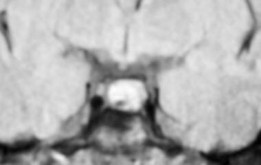
 Pituitary Tumor Diagnosis | Tests for Pituitary Tumors | American Cancer Society
When to Discontinue Treatment of Prolactinoma?
Hypothalamic-pituitary hormone - Wikipedia
 2018 NPA Minisymposium | College of American Pathologists
 Table: Adenohypophysis Endocrine Cells and Hormones - Merck Veterinary Manual
 Neural & Neuroendocrine Mathematical Models - Endocrine & Neural Dynamics Section - NIDDK
Publication Detail
 Androgen Receptors Expression in Pituitary of Male Viscacha in relation to Growth and Reproductive Cycle
MESH TREE NUMBER CHANGES - 2008 MeSH
Biomarkers Search
Abstract Search
 Overactive Pituitary Gland (Hyperpituitarism), Pituitary Adenomas | Healthhype.com
 Frontiers | Grass Carp Follisatin: Molecular Cloning, Functional Characterization, Dopamine D1 Regulation at Pituitary Level,...
 FSU Math Department e-Print Archive 1994 - 2022
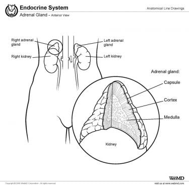
Endocrine System Anatomy: Overview, Gross Anatomy
 Publications - Microscopy and Imaging Core | NICHD - Eunice Kennedy Shriver National Institute of Child Health and Human...
 Epidemiology and Management Challenges in Prolactinomas | Neuroendocrinology | Karger Publishers
Prolactinoma: Practice Essentials, Pathophysiology, Epidemiology
 DailyMed - CABERGOLINE tablet
 Texas Pediatric Society Obesity Toolkit | Texas Pediatric Society
 Medical Pharmacology: Adrenergic, Autonomic pharmacology
Medical Pharmacology: Clinical Presentation, Etiology and Treatment of
Parkinson's Disease
 Somatotrophs | Harvard Catalyst Profiles | Harvard Catalyst
TREE NUMBER DESCRIPTOR
Prolactin16
- Here, we analyzed the contribution of phosphatidylinositol kinases (PIKs) to calcium-driven prolactin (PRL) release in pituitary lactotrophs: PI4Ks - which control PI4P production, PIP5Ks - which synthesize PI(4, 5)P2 by phosphorylating the D-5 position of the inositol ring of PI4P, and PI3KCs - which phosphorylate PI(4, 5)P 2 to generate PI(3, 4, 5)P 3 . (nih.gov)
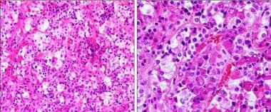
- Overall, there is a reduction in the size of the LACTOTROPH adenoma cells because of regression of the rough endoplasmic reticulum and Golgi apparatus, which is associated with cessation of intracellular prolactin synthesis. (medscape.com)
- 10-15% lactotroph, produce prolactin. (wikipedia.org)
- Lactotroph adenoma (prolactinoma) typically exhibits strong cytoplasmic prolactin immunoreactivity. (cap.org)
- 10. Leukemia inhibitory factor regulates prolactin secretion in prolactinoma and lactotroph cells. (nih.gov)
- We have previously identified pulsatile prolactin transcription patterns in living lactotroph cells in fetal tissue, that became stabilised. (endocrine-abstracts.org)
- Tumor formation is due to neoplastic transformation of anterior pituitary lactotrophs, resulting in excess synthesis and secretion of prolactin (PRL). (medscape.com)
- Results of in vitro studies demonstrate that cabergoline exerts a direct inhibitory effect on the secretion of prolactin by rat pituitary lactotrophs. (nih.gov)
- The effect of this connection is to inhibit prolactin release by pituitary lactotrophs. (pharmacology2000.com)
- D 2 receptors are notably expressed on anterior pituitary gland lactotrophs, regulating prolactin secretion. (pharmacology2000.com)
- There are specialized cells in the pituitary responsible for prolactin hormone production - these are called lactotrophs. (ada.com)
- In pregnancy, when more prolactin is needed, the number of lactotrophs in the pituitary increases. (ada.com)
- Prolactinomas are noncancerous tumors composed of lactotrophs, which are prolactin-secreting adenomas. (msdmanuals.com)
- Prolactin is produced in cells called lactotrophs that constitute about 30% of the cells of the anterior pituitary. (msdmanuals.com)
- Lactotrophs, somatotrophs, thyrotrophs, corticotrophs and gonadotrophs in the anterior pituitary gland secrete the polypeptide hormones prolactin (PRL), growth hormone (GH), thyroid stimulating hormone (TSH), adrenocorticotropic hormone (ACTH) and luteinizing (LH). (endocrine-abstracts.org)
- There are studies proving that Cabergoline is directly inhibiting pituitary lactotroph (prolactin) cells. (body-gear.to)
Somatotrophs4
- Intracellular calcium concentration and hormone secretion are controlled differently by TRH in rat neonatal lactotrophs and somatotrophs. (nih.gov)
- The model behavior resembles that seen in somatotrophs, lactotrophs, and corticotrophs. (nih.gov)
- At the pituitary level, follistatin signals could be located in carp somatotrophs, gonadotrophs, and lactotrophs. (frontiersin.org)
- Somatotrophs and lactotrophs are acidophils that secrete GH and PRL, respectively, whereas mammosomatotrophs secrete both GH and PRL. (oncohemakey.com)
Somatotroph2
- For the purpose of this review, only the more common lactotroph and somatotroph adenomas will be addressed. (cap.org)
- Post-operative surveillance for somatotroph, lactotroph and non-functional pituitary adenomas after curative resection: a systematic review. (harvard.edu)
Adenomas1
- 19. Refractory lactotroph adenomas. (nih.gov)
Pituitary Gland2
- Prolactinomas are noncancerous tumors made up from lactotrophs in the pituitary gland. (msdmanuals.com)
- Hormone secreted from lactotroph cells of the anterior pituitary gland with an essential role in lactation. (citizendium.com)
Lactation1
- Pituitary haemorrhage causing death of the lactotrophs results in failure of lactation (Sheehan's syndrome). (brainkart.com)
SF3B11
- Prevalence and clinical correlations of SF3B1 variants in lactotroph tumours. (cdc.gov)
Hormone2
Receptor3
- Selective estrogen receptor down-regulator and selective estrogen receptor modulators differentially regulate lactotroph proliferation. (nih.gov)
- We recently reported that estrogen receptor alpha (ERalpha), even in absence of estrogen (E2), plays a critical role in lactotroph homeostasis. (nih.gov)
- However, all three ER antagonists suppressed PRL release, suggesting that receptor occupation is sufficient to inhibit prl gene expression whereas receptor degradation is required to suppress lactotroph proliferation. (nih.gov)
Outcome2
- In this study our objective was to determine whether ERalpha degradation versus occupation, differentially modulates the biological outcome of anti-estrogens.Using the rat lactotroph cell line, GH3 cells, we report that ICI induced proteosome mediated degradation of ERalpha. (nih.gov)
- This really is likely mediated by an immediate outcome on the lactotroph. (llclinic.com)
Previously1
- These results provide a cellular mechanism that can account for the previously demonstrated potentiation of secretion from lactotrophs by cAMP- and PKC-dependent pathways. (rupress.org)