Bursa, Synovial
Olecranon Process
Osteoarthritis, Knee
Ischium
Tendinopathy
Calcaneus
Popliteal Cyst
Buffaloes
Shoulder Joint
Bursitis
Hip Joint
Pain
Magnetic Resonance Imaging
Range of Motion, Articular
Arthritis
Anterior Cruciate Ligament
Tibia
Treatment Outcome
Medial Collateral Ligament, Knee
Cartilage, Articular
Biomechanical Phenomena
Weight-Bearing
Joint Instability
Follow-Up Studies
Osteoarthritis
Bone Malalignment
Posterior Cruciate Ligament
Ligaments, Articular
Pain Measurement
Quadriceps Muscle
Joint Diseases
Recovery of Function
Muscle Strength
Joint Deformities, Acquired
Prosthesis Failure
Reoperation
Arthrometry, Articular
Walking
Osteophyte
Synovitis
Torque
Patellofemoral Joint
Contracture
Severity of Illness Index
Synovial Fluid
Hemarthrosis
Patellar Ligament
Ankle Joint
Prosthesis-Related Infections
Movement
Arthrography
Anterior Cruciate Ligament Reconstruction
Prospective Studies
Synovial Membrane
Hip
Muscle, Skeletal
Postoperative Complications
Shoes
A bursa is a small fluid-filled sac that provides a cushion between bones and other moving parts, such as muscles, tendons, or skin. A synovial bursa is a type of bursa that contains synovial fluid, which is produced by the synovial membrane that lines the inside of the bursa. Synovial bursae are found in various locations throughout the body, particularly near joints that experience a lot of movement or friction. They help to reduce wear and tear on the bones and other tissues, and can become inflamed or irritated due to overuse, injury, or infection, leading to a condition called bursitis.
The knee joint, also known as the tibiofemoral joint, is the largest and one of the most complex joints in the human body. It is a synovial joint that connects the thighbone (femur) to the shinbone (tibia). The patella (kneecap), which is a sesamoid bone, is located in front of the knee joint and helps in the extension of the leg.
The knee joint is made up of three articulations: the femorotibial joint between the femur and tibia, the femoropatellar joint between the femur and patella, and the tibiofibular joint between the tibia and fibula. These articulations are surrounded by a fibrous capsule that encloses the synovial membrane, which secretes synovial fluid to lubricate the joint.
The knee joint is stabilized by several ligaments, including the medial and lateral collateral ligaments, which provide stability to the sides of the joint, and the anterior and posterior cruciate ligaments, which prevent excessive forward and backward movement of the tibia relative to the femur. The menisci, which are C-shaped fibrocartilaginous structures located between the femoral condyles and tibial plateaus, also help to stabilize the joint by absorbing shock and distributing weight evenly across the articular surfaces.
The knee joint allows for flexion, extension, and a small amount of rotation, making it essential for activities such as walking, running, jumping, and sitting.
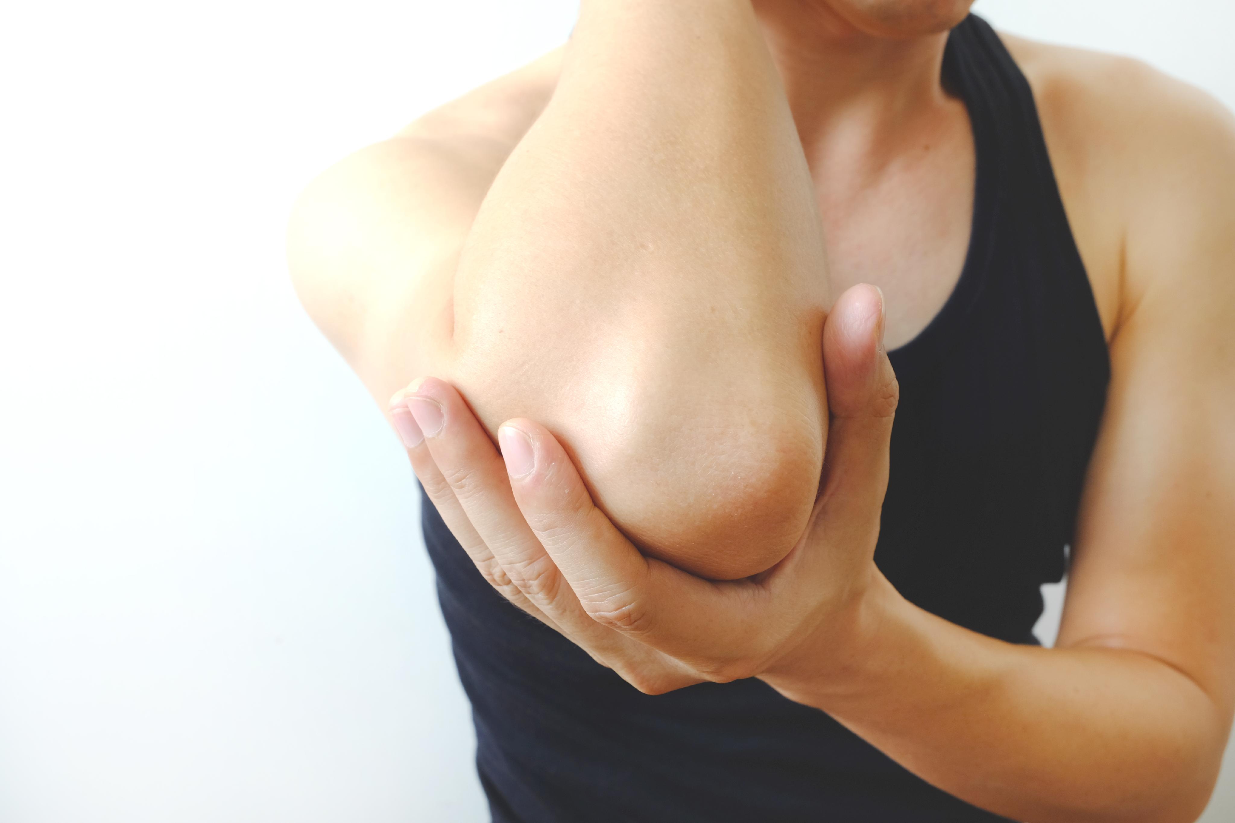
The olecranon process is a bony prominence and the tip of the ulna bone, which forms the point of the elbow. It serves as an attachment site for several muscles and tendons, including the triceps brachii muscle, and provides structure to the back of the elbow joint. The olecranon process also articulates with the humerus bone to form the hinge joint that allows for extension and flexion of the forearm.
In medical terms, the knee is referred to as the largest and one of the most complex joints in the human body. It is a hinge joint that connects the thigh bone (femur) to the shin bones (tibia and fibula), enabling movements like flexion, extension, and a small amount of rotation. The knee also contains several other components such as menisci, ligaments, tendons, and bursae, which provide stability, cushioning, and protection during movement.
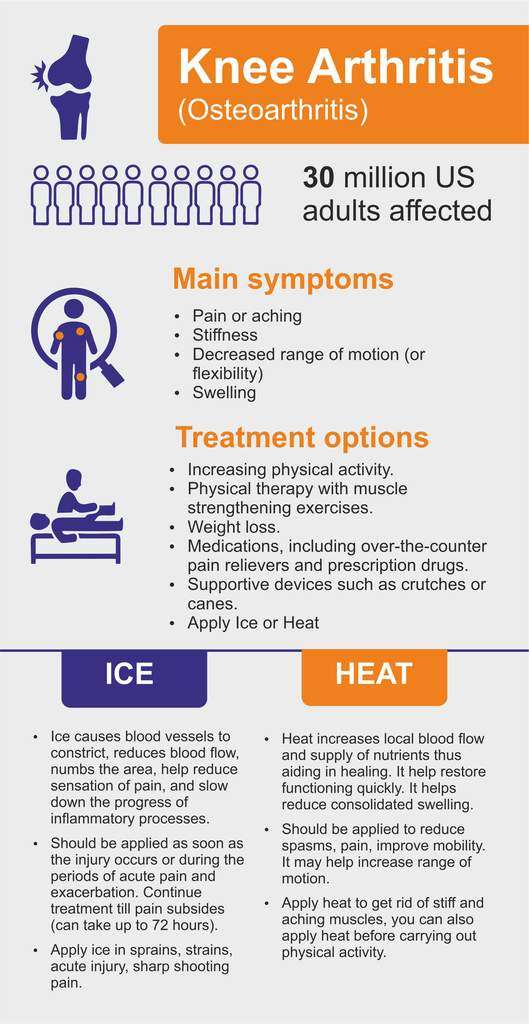
Osteoarthritis (OA) of the knee is a degenerative joint disease that affects the articular cartilage and subchondral bone in the knee joint. It is characterized by the breakdown and eventual loss of the smooth, cushioning cartilage that covers the ends of bones and allows for easy movement within joints. As the cartilage wears away, the bones rub against each other, causing pain, stiffness, and limited mobility. Osteoarthritis of the knee can also lead to the formation of bone spurs (osteophytes) and cysts in the joint. This condition is most commonly found in older adults, but it can also occur in younger people as a result of injury or overuse. Risk factors include obesity, family history, previous joint injuries, and repetitive stress on the knee joint. Treatment options typically include pain management, physical therapy, and in some cases, surgery.
Arthroplasty, replacement, knee is a surgical procedure where the damaged or diseased joint surface of the knee is removed and replaced with an artificial joint or prosthesis. The procedure involves resurfacing the worn-out ends of the femur (thigh bone) and tibia (shin bone) with metal components, and the back of the kneecap with a plastic button. This surgery is usually performed to relieve pain and restore function in patients with severe knee osteoarthritis, rheumatoid arthritis, or traumatic injuries that have damaged the joint beyond repair. The goal of knee replacement surgery is to improve mobility, reduce pain, and enhance the quality of life for the patient.
The ischium is a part of the pelvic bone, specifically the lower and posterior portion. It is one of the three bones that fuse together to form each half of the pelvis, along with the ilium (the upper and largest portion) and the pubis (anteriorly).
The ischium has a thick, robust structure because it supports our body weight when we sit. Its main parts include:
1. The ischial tuberosity (sitting bone): This is the roughened, weight-bearing portion where you typically feel discomfort after sitting for long periods.
2. The ischial spine: A thin bony projection that serves as an attachment point for various muscles and ligaments.
3. The ramus of the ischium: The slender, curved part that extends downwards and joins with the pubis to form the inferior (lower) portion of the pelvic ring called the obturator foramen.
Together with the other components of the pelvis, the ischium plays a crucial role in providing stability, supporting the lower limbs, and protecting internal organs.
A knee prosthesis, also known as a knee replacement or artificial knee joint, is a medical device used to replace the damaged or diseased weight-bearing surfaces of the knee joint. It typically consists of three components: the femoral component (made of metal) that fits over the end of the thighbone (femur), the tibial component (often made of metal and plastic) that fits into the top of the shinbone (tibia), and a patellar component (usually made of plastic) that replaces the damaged surface of the kneecap.
The primary goal of knee prosthesis is to relieve pain, restore function, and improve quality of life for individuals with advanced knee joint damage due to conditions such as osteoarthritis, rheumatoid arthritis, or traumatic injuries. The procedure to implant a knee prosthesis is called knee replacement surgery or total knee arthroplasty (TKA).
Knee injuries refer to damages or harm caused to the structures surrounding or within the knee joint, which may include the bones (femur, tibia, and patella), cartilage (meniscus and articular cartilage), ligaments (ACL, PCL, MCL, and LCL), tendons (patellar and quadriceps), muscles, bursae, and other soft tissues. These injuries can result from various causes, such as trauma, overuse, degeneration, or sports-related activities. Symptoms may include pain, swelling, stiffness, instability, reduced range of motion, and difficulty walking or bearing weight on the affected knee. Common knee injuries include fractures, dislocations, meniscal tears, ligament sprains or ruptures, and tendonitis. Proper diagnosis and treatment are crucial to ensure optimal recovery and prevent long-term complications.
The patella, also known as the kneecap, is a sesamoid bone located at the front of the knee joint. It is embedded in the tendon of the quadriceps muscle and serves to protect the knee joint and increase the leverage of the extensor mechanism, allowing for greater extension force of the lower leg. The patella moves within a groove on the femur called the trochlea during flexion and extension of the knee.
Tendinopathy is a general term referring to the degeneration or dysrepair of a tendon, which can result in pain and impaired function. It was previously referred to as tendinitis or tendinosis, but tendinopathy is now preferred because it describes various pathological conditions within the tendon, rather than a specific diagnosis.
Tendinopathy often develops due to overuse, repetitive strain, or age-related wear and tear. The condition typically involves collagen breakdown in the tendon, along with an increase in disorganized tenocytes (tendon cells) and vascular changes. This process can lead to thickening of the tendon, loss of elasticity, and the formation of calcium deposits or nodules.
Commonly affected tendons include the Achilles tendon, patellar tendon, rotator cuff tendons in the shoulder, and the extensor carpi radialis brevis tendon in the elbow (also known as tennis elbow). Treatment for tendinopathy often includes rest, physical therapy, exercise, pain management, and occasionally, surgical intervention.
The calcaneus is the largest tarsal bone in the human foot, and it is commonly known as the heel bone. It articulates with the cuboid bone anteriorly, the talus bone superiorly, and several tendons and ligaments that help to form the posterior portion of the foot's skeletal structure. The calcaneus plays a crucial role in weight-bearing and movement, as it forms the lower part of the leg's ankle joint and helps to absorb shock during walking or running.
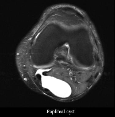
A Popliteal cyst, also known as Baker's cyst, is a fluid-filled sac that develops behind the knee, in the popliteal fossa. It forms when synovial fluid from the knee joint extends through a tear in the joint capsule, creating a visible bulge. The cyst may cause discomfort, swelling, or pain, especially when fully extended or flexed. In some cases, it can rupture and cause further complications, such as increased pain and inflammation in the calf region. Treatment options for Popliteal cysts include physical therapy, corticosteroid injections, and, in severe cases, surgical intervention to repair the underlying joint issue and remove the cyst.
The elbow is a joint formed by the articulation between the humerus bone of the upper arm and the radius and ulna bones of the forearm. It allows for flexion, extension, and rotation of the forearm. The medical definition of "elbow" refers to this specific anatomical structure and its associated functions in human anatomy.
The elbow joint, also known as the cubitus joint, is a hinge joint that connects the humerus bone of the upper arm to the radius and ulna bones of the forearm. It allows for flexion and extension movements of the forearm, as well as some degree of rotation. The main articulation occurs between the trochlea of the humerus and the trochlear notch of the ulna, while the radial head of the radius also contributes to the joint's stability and motion. Ligaments, muscles, and tendons surround and support the elbow joint, providing strength and protection during movement.
I am not aware of a medical definition for the term "buffaloes." The term generally refers to large, hoofed mammals that are native to Africa and Asia. In English language slang, the term "buffalo" is sometimes used to describe a lie or exaggeration, but this usage is not related to the medical field. If you have more context about where you encountered this term, I may be able to provide a more specific answer.
The Achilles tendon, also known as the calcaneal tendon, is a strong band of tissue that connects the calf muscles to the heel bone (calcaneus). It plays a crucial role in enabling activities such as walking, running, and jumping by facilitating the movement of the foot downward, which is called plantar flexion. Injuries to the Achilles tendon, such as tendinitis or ruptures, can be quite painful and impact mobility.
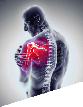
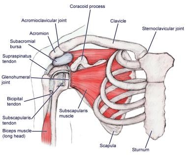
The shoulder joint, also known as the glenohumeral joint, is the most mobile joint in the human body. It is a ball and socket synovial joint that connects the head of the humerus (upper arm bone) to the glenoid cavity of the scapula (shoulder blade). The shoulder joint allows for a wide range of movements including flexion, extension, abduction, adduction, internal rotation, and external rotation. It is surrounded by a group of muscles and tendons known as the rotator cuff that provide stability and enable smooth movement of the joint.
Arthralgia is a medical term that refers to pain in the joints. It does not involve inflammation, which would be referred to as arthritis. The pain can range from mild to severe and may occur in one or multiple joints. Arthralgia can have various causes, including injuries, infections, degenerative conditions, or systemic diseases. In some cases, the underlying cause of arthralgia remains unknown. Treatment typically focuses on managing the pain and addressing the underlying condition if it can be identified.
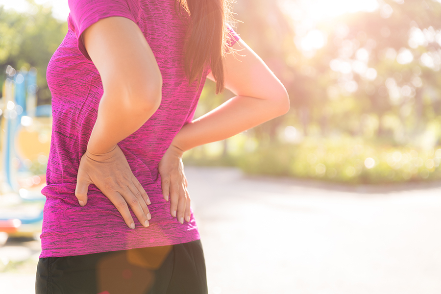
Bursitis is the inflammation or irritation of the bursa, a small fluid-filled sac that provides a cushion between bones and muscles, tendons, or skin around a joint. The bursae help to reduce friction and provide smooth movement of the joints. Bursitis can occur in any joint but is most common in the shoulder, elbow, hip, knee, and heel.
The inflammation of the bursa can result from various factors, including repetitive motions, injury or trauma to the joint, bacterial infection, or underlying health conditions such as rheumatoid arthritis or gout. The symptoms of bursitis include pain and tenderness in the affected area, swelling, warmth, and redness. Treatment for bursitis typically involves resting and immobilizing the affected joint, applying ice to reduce swelling, taking anti-inflammatory medications, and undergoing physical therapy exercises to improve strength and flexibility. In severe cases, corticosteroid injections or surgery may be necessary to alleviate symptoms and promote healing.
The hip joint, also known as the coxal joint, is a ball-and-socket type synovial joint that connects the femur (thigh bone) to the pelvis. The "ball" is the head of the femur, while the "socket" is the acetabulum, a concave surface on the pelvic bone.
The hip joint is surrounded by a strong fibrous capsule and is reinforced by several ligaments, including the iliofemoral, ischiofemoral, and pubofemoral ligaments. The joint allows for flexion, extension, abduction, adduction, medial and lateral rotation, and circumduction movements, making it one of the most mobile joints in the body.
The hip joint is also supported by various muscles, including the gluteus maximus, gluteus medius, gluteus minimus, iliopsoas, and other hip flexors and extensors. These muscles provide stability and strength to the joint, allowing for weight-bearing activities such as walking, running, and jumping.
Intra-articular injections refer to the administration of medication directly into a joint space. This route of administration is used for treating various joint conditions such as inflammation, pain, and arthritis. Commonly injected medications include corticosteroids, local anesthetics, and viscosupplementation agents. The procedure is usually performed using imaging guidance, like ultrasound or fluoroscopy, to ensure accurate placement of the medication within the joint.
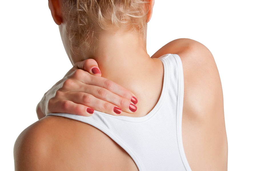
Pain is an unpleasant sensory and emotional experience associated with actual or potential tissue damage, or described in terms of such damage. It is a complex phenomenon that can result from various stimuli, such as thermal, mechanical, or chemical irritation, and it can be acute or chronic. The perception of pain involves the activation of specialized nerve cells called nociceptors, which transmit signals to the brain via the spinal cord. These signals are then processed in different regions of the brain, leading to the conscious experience of pain. It's important to note that pain is a highly individual and subjective experience, and its perception can vary widely among individuals.
Medical Definition:
Magnetic Resonance Imaging (MRI) is a non-invasive diagnostic imaging technique that uses a strong magnetic field and radio waves to create detailed cross-sectional or three-dimensional images of the internal structures of the body. The patient lies within a large, cylindrical magnet, and the scanner detects changes in the direction of the magnetic field caused by protons in the body. These changes are then converted into detailed images that help medical professionals to diagnose and monitor various medical conditions, such as tumors, injuries, or diseases affecting the brain, spinal cord, heart, blood vessels, joints, and other internal organs. MRI does not use radiation like computed tomography (CT) scans.
Articular Range of Motion (AROM) is a term used in physiotherapy and orthopedics to describe the amount of movement available in a joint, measured in degrees of a circle. It refers to the range through which synovial joints can actively move without causing pain or injury. AROM is assessed by measuring the degree of motion achieved by active muscle contraction, as opposed to passive range of motion (PROM), where the movement is generated by an external force.
Assessment of AROM is important in evaluating a patient's functional ability and progress, planning treatment interventions, and determining return to normal activities or sports participation. It is also used to identify any restrictions in joint mobility that may be due to injury, disease, or surgery, and to monitor the effectiveness of rehabilitation programs.
Arthritis is a medical condition characterized by inflammation in one or more joints, leading to symptoms such as pain, stiffness, swelling, and reduced range of motion. There are many different types of arthritis, including osteoarthritis, rheumatoid arthritis, psoriatic arthritis, gout, and lupus, among others.
Osteoarthritis is the most common form of arthritis and is caused by wear and tear on the joints over time. Rheumatoid arthritis, on the other hand, is an autoimmune disorder in which the body's immune system mistakenly attacks the joint lining, causing inflammation and damage.
Arthritis can affect people of all ages, including children, although it is more common in older adults. Treatment for arthritis may include medications to manage pain and reduce inflammation, physical therapy, exercise, and in some cases, surgery.
Knee dislocation is a serious and uncommon orthopedic injury that occurs when the bones that form the knee joint (femur, tibia, and patella) are forced out of their normal position due to extreme trauma or force. This injury often requires immediate medical attention and reduction (repositioning) by a healthcare professional. If left untreated, it can lead to serious complications such as compartment syndrome, nerve damage, and long-term joint instability. It's important to note that knee dislocation is different from a kneecap (patellar) dislocation, which involves the patella sliding out of its groove in the femur.
The Anterior Cruciate Ligament (ACL) is a major stabilizing ligament in the knee. It is one of the four strong bands of tissue that connect the bones of the knee joint together. The ACL runs diagonally through the middle of the knee and helps to control the back and forth motion of the knee, as well as provide stability to the knee joint. Injuries to the ACL often occur during sports or physical activities that involve sudden stops, changes in direction, or awkward landings.
The tibia, also known as the shin bone, is the larger of the two bones in the lower leg and part of the knee joint. It supports most of the body's weight and is a major insertion point for muscles that flex the foot and bend the leg. The tibia articulates with the femur at the knee joint and with the fibula and talus bone at the ankle joint. Injuries to the tibia, such as fractures, are common in sports and other activities that put stress on the lower leg.
Treatment outcome is a term used to describe the result or effect of medical treatment on a patient's health status. It can be measured in various ways, such as through symptoms improvement, disease remission, reduced disability, improved quality of life, or survival rates. The treatment outcome helps healthcare providers evaluate the effectiveness of a particular treatment plan and make informed decisions about future care. It is also used in clinical research to compare the efficacy of different treatments and improve patient care.
The menisci are crescent-shaped fibrocartilaginous structures located in the knee joint. There are two menisci in each knee: the medial meniscus and the lateral meniscus. The tibial menisci, also known as the medial and lateral menisci, are named according to their location in the knee joint. They lie on the top surface of the tibia (shin bone) and provide shock absorption, stability, and lubrication to the knee joint.
The tibial menisci have a complex shape, with a wider outer portion called the peripheral rim and a narrower inner portion called the central portion or root attachment. The menisci are attached to the bones of the knee joint by ligaments and have a rich blood supply in their outer portions, which helps in healing after injury. However, the inner two-thirds of the menisci have a poor blood supply, making them more prone to degeneration and less likely to heal after injury.
Damage to the tibial menisci can occur due to trauma or degenerative changes, leading to symptoms such as pain, swelling, stiffness, and limited mobility of the knee joint. Treatment for meniscal injuries may include physical therapy, bracing, or surgery, depending on the severity and location of the injury.
The medial collateral ligament (MCL) of the knee is a band-like structure located on the inner side of the knee joint. It connects the end of the femur (thighbone) to the top of the tibia (shinbone) and helps stabilize the knee by controlling side-to-side movement and preventing excessive separation of the bones. The MCL provides resistance to valgus force, which is a pushing or pulling force that attempts to push the bones apart in a direction away from the midline of the body. MCL injuries often occur due to direct impact to the outer knee or sudden changes in direction that strain the ligament.
Articular cartilage is the smooth, white tissue that covers the ends of bones where they come together to form joints. It provides a cushion between bones and allows for smooth movement by reducing friction. Articular cartilage also absorbs shock and distributes loads evenly across the joint, protecting the bones from damage. It is avascular, meaning it does not have its own blood supply, and relies on the surrounding synovial fluid for nutrients. Over time, articular cartilage can wear down or become damaged due to injury or disease, leading to conditions such as osteoarthritis.
Biomechanics is the application of mechanical laws to living structures and systems, particularly in the field of medicine and healthcare. A biomechanical phenomenon refers to a observable event or occurrence that involves the interaction of biological tissues or systems with mechanical forces. These phenomena can be studied at various levels, from the molecular and cellular level to the tissue, organ, and whole-body level.
Examples of biomechanical phenomena include:
1. The way that bones and muscles work together to produce movement (known as joint kinematics).
2. The mechanical behavior of biological tissues such as bone, cartilage, tendons, and ligaments under various loads and stresses.
3. The response of cells and tissues to mechanical stimuli, such as the way that bone tissue adapts to changes in loading conditions (known as Wolff's law).
4. The biomechanics of injury and disease processes, such as the mechanisms of joint injury or the development of osteoarthritis.
5. The use of mechanical devices and interventions to treat medical conditions, such as orthopedic implants or assistive devices for mobility impairments.
Understanding biomechanical phenomena is essential for developing effective treatments and prevention strategies for a wide range of medical conditions, from musculoskeletal injuries to neurological disorders.
"Weight-bearing" is a term used in the medical field to describe the ability of a body part or limb to support the weight or pressure exerted upon it, typically while standing, walking, or performing other physical activities. In a clinical setting, healthcare professionals often use the term "weight-bearing exercise" to refer to physical activities that involve supporting one's own body weight, such as walking, jogging, or climbing stairs. These exercises can help improve bone density, muscle strength, and overall physical function, particularly in individuals with conditions affecting the bones, joints, or muscles.
In addition, "weight-bearing" is also used to describe the positioning of a body part during medical imaging studies, such as X-rays or MRIs. For example, a weight-bearing X-ray of the foot or ankle involves taking an image while the patient stands on the affected limb, allowing healthcare providers to assess any alignment or stability issues that may not be apparent in a non-weight-bearing position.
Joint instability is a condition characterized by the loss of normal joint function and increased risk of joint injury due to impaired integrity of the supporting structures, such as ligaments, muscles, or cartilage. This can result in excessive movement or laxity within the joint, leading to decreased stability and increased susceptibility to dislocations or subluxations. Joint instability may cause pain, swelling, and limited range of motion, and it can significantly impact a person's mobility and quality of life. It is often caused by trauma, degenerative conditions, or congenital abnormalities and may require medical intervention, such as physical therapy, bracing, or surgery, to restore joint stability.
Follow-up studies are a type of longitudinal research that involve repeated observations or measurements of the same variables over a period of time, in order to understand their long-term effects or outcomes. In medical context, follow-up studies are often used to evaluate the safety and efficacy of medical treatments, interventions, or procedures.
In a typical follow-up study, a group of individuals (called a cohort) who have received a particular treatment or intervention are identified and then followed over time through periodic assessments or data collection. The data collected may include information on clinical outcomes, adverse events, changes in symptoms or functional status, and other relevant measures.
The results of follow-up studies can provide important insights into the long-term benefits and risks of medical interventions, as well as help to identify factors that may influence treatment effectiveness or patient outcomes. However, it is important to note that follow-up studies can be subject to various biases and limitations, such as loss to follow-up, recall bias, and changes in clinical practice over time, which must be carefully considered when interpreting the results.
Arthroscopy is a minimally invasive surgical procedure where an orthopedic surgeon uses an arthroscope (a thin tube with a light and camera on the end) to diagnose and treat problems inside a joint. The surgeon makes a small incision, inserts the arthroscope into the joint, and then uses the attached camera to view the inside of the joint on a monitor. They can then insert other small instruments through additional incisions to repair or remove damaged tissue.
Arthroscopy is most commonly used for joints such as the knee, shoulder, hip, ankle, and wrist. It offers several advantages over traditional open surgery, including smaller incisions, less pain and bleeding, faster recovery time, and reduced risk of infection. The procedure can be used to diagnose and treat a wide range of conditions, including torn ligaments or cartilage, inflamed synovial tissue, loose bone or cartilage fragments, and joint damage caused by arthritis.
Anti-bacterial agents, also known as antibiotics, are a type of medication used to treat infections caused by bacteria. These agents work by either killing the bacteria or inhibiting their growth and reproduction. There are several different classes of anti-bacterial agents, including penicillins, cephalosporins, fluoroquinolones, macrolides, and tetracyclines, among others. Each class of antibiotic has a specific mechanism of action and is used to treat certain types of bacterial infections. It's important to note that anti-bacterial agents are not effective against viral infections, such as the common cold or flu. Misuse and overuse of antibiotics can lead to antibiotic resistance, which is a significant global health concern.
Osteoarthritis (OA) is a type of joint disease that is characterized by the breakdown and eventual loss of cartilage - the tissue that cushions the ends of bones where they meet in the joints. This breakdown can cause the bones to rub against each other, causing pain, stiffness, and loss of mobility. OA can occur in any joint, but it most commonly affects the hands, knees, hips, and spine. It is often associated with aging and can be caused or worsened by obesity, injury, or overuse.
The medical definition of osteoarthritis is: "a degenerative, non-inflammatory joint disease characterized by the loss of articular cartilage, bone remodeling, and the formation of osteophytes (bone spurs). It is often associated with pain, stiffness, and decreased range of motion in the affected joint."
Bone malalignment is a term used to describe the abnormal alignment or positioning of bones in relation to each other. This condition can occur as a result of injury, deformity, surgery, or disease processes that affect the bones and joints. Bone malalignment can cause pain, stiffness, limited mobility, and an increased risk of further injury. In some cases, bone malalignment may require treatment such as bracing, physical therapy, or surgery to correct the alignment and improve function.
The Posterior Cruciate Ligament (PCL) is one of the major ligaments in the knee, providing stability to the joint. It is a strong band of tissue located in the back of the knee, connecting the thighbone (femur) to the shinbone (tibia). The PCL limits the backward motion of the tibia relative to the femur and provides resistance to forces that tend to push the tibia backwards. It also assists in maintaining the overall alignment and function of the knee joint during various movements and activities. Injuries to the PCL are less common compared to injuries to the Anterior Cruciate Ligament (ACL) but can still occur due to high-energy trauma, such as motor vehicle accidents or sports incidents involving direct impact to the front of the knee.
Articular ligaments, also known as fibrous ligaments, are bands of dense, fibrous connective tissue that connect and stabilize bones to each other at joints. They help to limit the range of motion of a joint and provide support, preventing excessive movement that could cause injury. Articular ligaments are composed mainly of collagen fibers arranged in a parallel pattern, making them strong and flexible. They have limited blood supply and few nerve endings, which makes them less prone to injury but also slower to heal if damaged. Examples of articular ligaments include the anterior cruciate ligament (ACL) and posterior cruciate ligament (PCL) in the knee joint, and the medial collateral ligament (MCL) and lateral collateral ligament (LCL) in the elbow joint.
Pain measurement, in a medical context, refers to the quantification or evaluation of the intensity and/or unpleasantness of a patient's subjective pain experience. This is typically accomplished through the use of standardized self-report measures such as numerical rating scales (NRS), visual analog scales (VAS), or categorical scales (mild, moderate, severe). In some cases, physiological measures like heart rate, blood pressure, and facial expressions may also be used to supplement self-reported pain ratings. The goal of pain measurement is to help healthcare providers better understand the nature and severity of a patient's pain in order to develop an effective treatment plan.
Gait is a medical term used to describe the pattern of movement of the limbs during walking or running. It includes the manner or style of walking, including factors such as rhythm, speed, and step length. A person's gait can provide important clues about their physical health and neurological function, and abnormalities in gait may indicate the presence of underlying medical conditions, such as neuromuscular disorders, orthopedic problems, or injuries.
A typical human gait cycle involves two main phases: the stance phase, during which the foot is in contact with the ground, and the swing phase, during which the foot is lifted and moved forward in preparation for the next step. The gait cycle can be further broken down into several sub-phases, including heel strike, foot flat, midstance, heel off, and toe off.
Gait analysis is a specialized field of study that involves observing and measuring a person's gait pattern using various techniques, such as video recordings, force plates, and motion capture systems. This information can be used to diagnose and treat gait abnormalities, improve mobility and function, and prevent injuries.
The Quadriceps muscle, also known as the Quadriceps Femoris, is a large muscle group located in the front of the thigh. It consists of four individual muscles - the Rectus Femoris, Vastus Lateralis, Vastus Intermedius, and Vastus Medialis. These muscles work together to extend the leg at the knee joint and flex the thigh at the hip joint. The Quadriceps muscle is crucial for activities such as walking, running, jumping, and kicking.
Joint diseases is a broad term that refers to various conditions affecting the joints, including but not limited to:
1. Osteoarthritis (OA): A degenerative joint disease characterized by the breakdown of cartilage and underlying bone, leading to pain, stiffness, and potential loss of function.
2. Rheumatoid Arthritis (RA): An autoimmune disorder causing inflammation in the synovial membrane lining the joints, resulting in swelling, pain, and joint damage if left untreated.
3. Infectious Arthritis: Joint inflammation caused by bacterial, viral, or fungal infections that spread through the bloodstream or directly enter the joint space.
4. Gout: A type of arthritis resulting from the buildup of uric acid crystals in the joints, typically affecting the big toe and characterized by sudden attacks of severe pain, redness, and swelling.
5. Psoriatic Arthritis (PsA): An inflammatory joint disease associated with psoriasis, causing symptoms such as pain, stiffness, and swelling in the joints and surrounding tissues.
6. Juvenile Idiopathic Arthritis (JIA): A group of chronic arthritis conditions affecting children, characterized by joint inflammation, pain, and stiffness.
7. Ankylosing Spondylitis: A form of arthritis primarily affecting the spine, causing inflammation, pain, and potential fusion of spinal vertebrae.
8. Bursitis: Inflammation of the fluid-filled sacs (bursae) that cushion joints, leading to pain and swelling.
9. Tendinitis: Inflammation or degeneration of tendons, which connect muscles to bones, often resulting in pain and stiffness near joints.
These conditions can impact the function and mobility of affected joints, causing discomfort and limiting daily activities. Proper diagnosis and treatment are essential for managing joint diseases and preserving joint health.
Prosthesis design is a specialized field in medical device technology that involves creating and developing artificial substitutes to replace a missing body part, such as a limb, tooth, eye, or internal organ. The design process typically includes several stages: assessment of the patient's needs, selection of appropriate materials, creation of a prototype, testing and refinement, and final fabrication and fitting of the prosthesis.
The goal of prosthesis design is to create a device that functions as closely as possible to the natural body part it replaces, while also being comfortable, durable, and aesthetically pleasing for the patient. The design process may involve collaboration between medical professionals, engineers, and designers, and may take into account factors such as the patient's age, lifestyle, occupation, and overall health.
Prosthesis design can be highly complex, particularly for advanced devices such as robotic limbs or implantable organs. These devices often require sophisticated sensors, actuators, and control systems to mimic the natural functions of the body part they replace. As a result, prosthesis design is an active area of research and development in the medical field, with ongoing efforts to improve the functionality, comfort, and affordability of these devices for patients.
"Recovery of function" is a term used in medical rehabilitation to describe the process in which an individual regains the ability to perform activities or tasks that were previously difficult or impossible due to injury, illness, or disability. This can involve both physical and cognitive functions. The goal of recovery of function is to help the person return to their prior level of independence and participation in daily activities, work, and social roles as much as possible.
Recovery of function may be achieved through various interventions such as physical therapy, occupational therapy, speech-language therapy, and other rehabilitation strategies. The specific approach used will depend on the individual's needs and the nature of their impairment. Recovery of function can occur spontaneously as the body heals, or it may require targeted interventions to help facilitate the process.
It is important to note that recovery of function does not always mean a full return to pre-injury or pre-illness levels of ability. Instead, it often refers to the person's ability to adapt and compensate for any remaining impairments, allowing them to achieve their maximum level of functional independence and quality of life.
Muscle strength, in a medical context, refers to the amount of force a muscle or group of muscles can produce during contraction. It is the maximum amount of force that a muscle can generate through its full range of motion and is often measured in units of force such as pounds or newtons. Muscle strength is an important component of physical function and mobility, and it can be assessed through various tests, including manual muscle testing, dynamometry, and isokinetic testing. Factors that can affect muscle strength include age, sex, body composition, injury, disease, and physical activity level.
Acquired joint deformities refer to structural changes in the alignment and shape of a joint that develop after birth, due to various causes such as injury, disease, or wear and tear. These deformities can affect the function and mobility of the joint, causing pain, stiffness, and limited range of motion. Examples of conditions that can lead to acquired joint deformities include arthritis, infection, trauma, and nerve damage. Treatment may involve medication, physical therapy, or surgery to correct the deformity and alleviate symptoms.
Prosthesis failure is a term used to describe a situation where a prosthetic device, such as an artificial joint or limb, has stopped functioning or failed to meet its intended purpose. This can be due to various reasons, including mechanical failure, infection, loosening of the device, or a reaction to the materials used in the prosthesis.
Mechanical failure can occur due to wear and tear, manufacturing defects, or improper use of the prosthetic device. Infection can also lead to prosthesis failure, particularly in cases where the prosthesis is implanted inside the body. The immune system may react to the presence of the foreign material, leading to inflammation and infection.
Loosening of the prosthesis can also cause it to fail over time, as the device becomes less stable and eventually stops working properly. Additionally, some people may have a reaction to the materials used in the prosthesis, leading to tissue damage or other complications that can result in prosthesis failure.
In general, prosthesis failure can lead to decreased mobility, pain, and the need for additional surgeries or treatments to correct the problem. It is important for individuals with prosthetic devices to follow their healthcare provider's instructions carefully to minimize the risk of prosthesis failure and ensure that the device continues to function properly over time.
A reoperation is a surgical procedure that is performed again on a patient who has already undergone a previous operation for the same or related condition. Reoperations may be required due to various reasons, such as inadequate initial treatment, disease recurrence, infection, or complications from the first surgery. The nature and complexity of a reoperation can vary widely depending on the specific circumstances, but it often carries higher risks and potential complications compared to the original operation.
Arthrometry is a measurement technique used in the field of orthopedics and rheumatology to assess the integrity and mobility of joints. When qualified with the term "articular," it specifically refers to the measurement of articular motion or range of motion (ROM) within a synovial joint.
Articular arthrometry involves using specialized instruments, such as goniometers, inclinometers, or digital devices like smartphone applications and wearable sensors, to quantify the degree of flexion, extension, abduction, adduction, rotation, or other movements in a joint. This information can help medical professionals evaluate joint function, diagnose injuries or conditions affecting joint mobility, monitor disease progression, and assess treatment outcomes.
In summary, articular arthrometry is the measurement of articular motion within synovial joints to evaluate joint health and function.
Medical science often defines and describes "walking" as a form of locomotion or mobility where an individual repeatedly lifts and sets down each foot to move forward, usually bearing weight on both legs. It is a complex motor activity that requires the integration and coordination of various systems in the human body, including the musculoskeletal, neurological, and cardiovascular systems.
Walking involves several components such as balance, coordination, strength, and endurance. The ability to walk independently is often used as a measure of functional mobility and overall health status. However, it's important to note that the specific definition of walking may vary depending on the context and the medical or scientific field in question.
An osteophyte, also known as a bone spur, is a bony projection that forms along the margins of joints, often as a result of degenerative changes in the cartilage and underlying bone. These changes are most commonly seen in conditions such as osteoarthritis, where the protective cartilage that cushions the ends of bones breaks down, leading to inflammation, pain, and reduced mobility.
Osteophytes can develop in any joint in the body, but they are most commonly found in the spine, hips, knees, and hands. They may vary in size from small bumps to large, irregular growths that can restrict joint movement and cause discomfort or pain. In some cases, osteophytes may also compress nearby nerves, leading to symptoms such as numbness, tingling, or weakness in the affected limb.
While osteophytes are often considered a sign of aging or joint degeneration, they can also be caused by other conditions that put excessive stress on the joints, such as injury, infection, or inflammatory arthritis. Treatment for osteophytes typically involves addressing the underlying cause of joint damage, along with pain management strategies such as physical therapy, medication, or in some cases, surgery.
Synovitis is a medical condition characterized by inflammation of the synovial membrane, which is the soft tissue that lines the inner surface of joint capsules and tendon sheaths. The synovial membrane produces synovial fluid, which lubricates the joint and allows for smooth movement.
Inflammation of the synovial membrane can cause it to thicken, redden, and become painful and swollen. This can lead to stiffness, limited mobility, and discomfort in the affected joint or tendon sheath. Synovitis may occur as a result of injury, overuse, infection, or autoimmune diseases such as rheumatoid arthritis.
If left untreated, synovitis can cause irreversible damage to the joint and surrounding tissues, including cartilage loss and bone erosion. Treatment typically involves a combination of medications, physical therapy, and lifestyle modifications to reduce inflammation and manage pain.
"Torque" is not a term that has a specific medical definition. It is a physical concept used in the fields of physics and engineering, referring to a twisting force that causes rotation around an axis. However, in certain medical contexts, such as in discussions of spinal or joint biomechanics, the term "torque" may be used to describe a rotational force applied to a body part. But generally speaking, "torque" is not a term commonly used in medical terminology.
The patellofemoral joint is the articulation between the patella (kneecap) and the femur (thigh bone). It is a synovial joint, which means it is surrounded by a joint capsule containing synovial fluid to lubricate the joint. This joint is responsible for providing stability to the knee extensor mechanism and allows for smooth movement of the patella during activities like walking, running, and jumping. Pain or dysfunction in this joint can result in various conditions such as patellofemoral pain syndrome, chondromalacia patella, or patellar dislocation.
A contracture, in a medical context, refers to the abnormal shortening and hardening of muscles, tendons, or other tissue, which can result in limited mobility and deformity of joints. This condition can occur due to various reasons such as injury, prolonged immobilization, scarring, neurological disorders, or genetic conditions.
Contractures can cause significant impairment in daily activities and quality of life, making it difficult for individuals to perform routine tasks like dressing, bathing, or walking. Treatment options may include physical therapy, splinting, casting, medications, surgery, or a combination of these approaches, depending on the severity and underlying cause of the contracture.
A Severity of Illness Index is a measurement tool used in healthcare to assess the severity of a patient's condition and the risk of mortality or other adverse outcomes. These indices typically take into account various physiological and clinical variables, such as vital signs, laboratory values, and co-morbidities, to generate a score that reflects the patient's overall illness severity.
Examples of Severity of Illness Indices include the Acute Physiology and Chronic Health Evaluation (APACHE) system, the Simplified Acute Physiology Score (SAPS), and the Mortality Probability Model (MPM). These indices are often used in critical care settings to guide clinical decision-making, inform prognosis, and compare outcomes across different patient populations.
It is important to note that while these indices can provide valuable information about a patient's condition, they should not be used as the sole basis for clinical decision-making. Rather, they should be considered in conjunction with other factors, such as the patient's overall clinical presentation, treatment preferences, and goals of care.
Synovial fluid is a viscous, clear, and straw-colored fluid found in the cavities of synovial joints, bursae, and tendon sheaths. It is produced by the synovial membrane, which lines the inner surface of the capsule surrounding these structures.
The primary function of synovial fluid is to reduce friction between articulating surfaces, providing lubrication for smooth and painless movement. It also acts as a shock absorber, protecting the joints from external forces during physical activities. Synovial fluid contains nutrients that nourish the articular cartilage, hyaluronic acid, which provides its viscoelastic properties, and lubricin, a protein responsible for boundary lubrication.
Abnormalities in synovial fluid composition or volume can indicate joint-related disorders, such as osteoarthritis, rheumatoid arthritis, gout, infection, or trauma. Analysis of synovial fluid is often used diagnostically to determine the underlying cause of joint pain, inflammation, or dysfunction.
Hemarthrosis is a medical term that refers to the presence of blood in a joint space. This condition usually occurs as a result of trauma or injury that causes bleeding into the joint, such as a fracture or dislocation. Certain medical conditions like hemophilia and other bleeding disorders can also make a person more prone to hemarthrosis.
The accumulation of blood in the joint space can cause pain, swelling, warmth, and stiffness, making it difficult for the individual to move the affected joint. In some cases, hemarthrosis may require medical intervention, such as draining the excess blood from the joint or administering clotting factors to help stop the bleeding. If left untreated, hemarthrosis can lead to complications like joint damage and chronic pain.
The patellar ligament, also known as the patellar tendon, is a strong band of tissue that connects the bottom part of the kneecap (patella) to the top part of the shinbone (tibia). This ligament plays a crucial role in enabling the extension and straightening of the leg during activities such as walking, running, and jumping. Injuries to the patellar ligament, such as tendonitis or tears, can cause pain and difficulty with mobility.
The ankle joint, also known as the talocrural joint, is the articulation between the bones of the lower leg (tibia and fibula) and the talus bone in the foot. It is a synovial hinge joint that allows for dorsiflexion and plantarflexion movements, which are essential for walking, running, and jumping. The ankle joint is reinforced by strong ligaments on both sides to provide stability during these movements.
Hip arthroplasty, also known as hip replacement surgery, is a medical procedure where the damaged or diseased joint surfaces of the hip are removed and replaced with artificial components. These components typically include a metal or ceramic ball that replaces the head of the femur (thigh bone), and a polyethylene or ceramic socket that replaces the acetabulum (hip socket) in the pelvis.
The goal of hip arthroplasty is to relieve pain, improve joint mobility, and restore function to the hip joint. This procedure is commonly performed in patients with advanced osteoarthritis, rheumatoid arthritis, hip fractures, or other conditions that cause significant damage to the hip joint.
There are several types of hip replacement surgeries, including traditional total hip arthroplasty, partial (hemi) hip arthroplasty, and resurfacing hip arthroplasty. The choice of procedure depends on various factors, such as the patient's age, activity level, overall health, and the extent of joint damage.
After surgery, patients typically require rehabilitation to regain strength, mobility, and function in the affected hip. With proper care and follow-up, most patients can expect significant pain relief and improved quality of life following hip arthroplasty.
Prosthesis-related infections, also known as prosthetic joint infections (PJIs), are infections that occur around or within a prosthetic device, such as an artificial joint. These infections can be caused by bacteria, fungi, or other microorganisms and can lead to serious complications if not treated promptly and effectively.
Prosthesis-related infections can occur soon after the implantation of the prosthetic device (early infection) or months or even years later (late infection). Early infections are often caused by bacteria that enter the surgical site during the procedure, while late infections may be caused by hematogenous seeding (i.e., when bacteria from another source spread through the bloodstream and settle in the prosthetic device) or by contamination during a subsequent medical procedure.
Symptoms of prosthesis-related infections can include pain, swelling, redness, warmth, and drainage around the affected area. In some cases, patients may also experience fever, chills, or fatigue. Diagnosis typically involves a combination of clinical evaluation, laboratory tests (such as blood cultures, joint fluid analysis, and tissue biopsy), and imaging studies (such as X-rays, CT scans, or MRI).
Treatment of prosthesis-related infections usually involves a combination of antibiotics and surgical intervention. The specific treatment approach will depend on the type and severity of the infection, as well as the patient's overall health status. In some cases, it may be necessary to remove or replace the affected prosthetic device.
In the context of medicine and healthcare, "movement" refers to the act or process of changing physical location or position. It involves the contraction and relaxation of muscles, which allows for the joints to move and the body to be in motion. Movement can also refer to the ability of a patient to move a specific body part or limb, which is assessed during physical examinations. Additionally, "movement" can describe the progression or spread of a disease within the body.
Arthrography is a medical imaging technique used to diagnose problems within joints. It involves the injection of a contrast agent, such as a radiopaque dye or air, into the joint space, followed by the use of fluoroscopy or X-ray imaging to visualize the internal structures of the joint. This can help to identify injuries, tears, or other abnormalities in the cartilage, ligaments, tendons, or bones within the joint.
The procedure is typically performed on an outpatient basis and may be used to diagnose conditions such as shoulder dislocations, rotator cuff tears, meniscal tears in the knee, or hip labral injuries. It is a relatively safe and minimally invasive procedure, although there may be some temporary discomfort or swelling at the injection site. Patients are usually advised to avoid strenuous activity for a day or two following the procedure to allow the contrast agent to fully dissipate from the joint.
Anterior cruciate ligament (ACL) reconstruction is a surgical procedure in which the damaged or torn ACL, a major stabilizing ligament in the knee, is replaced with a graft. The ACL is responsible for preventing excessive motion of the knee joint, and when it is injured, the knee may become unstable and prone to further damage.
During the procedure, the surgeon makes an incision in the knee to access the damaged ligament. The torn ends of the ACL are then removed, and a graft is taken from another part of the body (such as the patellar tendon or hamstring tendons) or from a donor. This graft is then positioned in the same location as the original ACL and fixed in place with screws or other devices.
The goal of ACL reconstruction is to restore stability and function to the knee joint, allowing the patient to return to their normal activities, including sports and exercise. Physical therapy is typically required after surgery to help strengthen the knee and improve range of motion.
Prospective studies, also known as longitudinal studies, are a type of cohort study in which data is collected forward in time, following a group of individuals who share a common characteristic or exposure over a period of time. The researchers clearly define the study population and exposure of interest at the beginning of the study and follow up with the participants to determine the outcomes that develop over time. This type of study design allows for the investigation of causal relationships between exposures and outcomes, as well as the identification of risk factors and the estimation of disease incidence rates. Prospective studies are particularly useful in epidemiology and medical research when studying diseases with long latency periods or rare outcomes.
The synovial membrane, also known as the synovium, is the soft tissue that lines the inner surface of the capsule of a synovial joint, which is a type of joint that allows for smooth movement between bones. This membrane secretes synovial fluid, a viscous substance that lubricates and nourishes the cartilage and helps to reduce friction within the joint during movement.
The synovial membrane has a highly specialized structure, consisting of two layers: the intima and the subintima. The intima is a thin layer of cells that are in direct contact with the synovial fluid, while the subintima is a more fibrous layer that contains blood vessels and nerves.
The main function of the synovial membrane is to produce and regulate the production of synovial fluid, as well as to provide nutrients to the articular cartilage. It also plays a role in the immune response within the joint, helping to protect against infection and inflammation. However, abnormalities in the synovial membrane can lead to conditions such as rheumatoid arthritis, where the membrane becomes inflamed and produces excess synovial fluid, leading to pain, swelling, and joint damage.
In medical terms, the hip is a ball-and-socket joint where the rounded head of the femur (thigh bone) fits into the cup-shaped socket, also known as the acetabulum, of the pelvis. This joint allows for a wide range of movement in the lower extremities and supports the weight of the upper body during activities such as walking, running, and jumping. The hip joint is surrounded by strong ligaments, muscles, and tendons that provide stability and enable proper functioning.
Skeletal muscle, also known as striated or voluntary muscle, is a type of muscle that is attached to bones by tendons or aponeuroses and functions to produce movements and support the posture of the body. It is composed of long, multinucleated fibers that are arranged in parallel bundles and are characterized by alternating light and dark bands, giving them a striped appearance under a microscope. Skeletal muscle is under voluntary control, meaning that it is consciously activated through signals from the nervous system. It is responsible for activities such as walking, running, jumping, and lifting objects.
Athletic injuries are damages or injuries to the body that occur while participating in sports, physical activities, or exercise. These injuries can be caused by a variety of factors, including:
1. Trauma: Direct blows, falls, collisions, or crushing injuries can cause fractures, dislocations, contusions, lacerations, or concussions.
2. Overuse: Repetitive motions or stress on a particular body part can lead to injuries such as tendonitis, stress fractures, or muscle strains.
3. Poor technique: Using incorrect form or technique during exercise or sports can put additional stress on muscles, joints, and ligaments, leading to injury.
4. Inadequate warm-up or cool-down: Failing to properly prepare the body for physical activity or neglecting to cool down afterwards can increase the risk of injury.
5. Lack of fitness or flexibility: Insufficient strength, endurance, or flexibility can make individuals more susceptible to injuries during sports and exercise.
6. Environmental factors: Extreme weather conditions, poor field or court surfaces, or inadequate equipment can contribute to the risk of athletic injuries.
Common athletic injuries include ankle sprains, knee injuries, shoulder dislocations, tennis elbow, shin splints, and concussions. Proper training, warm-up and cool-down routines, use of appropriate protective gear, and attention to technique can help prevent many athletic injuries.
Postoperative complications refer to any unfavorable condition or event that occurs during the recovery period after a surgical procedure. These complications can vary in severity and may include, but are not limited to:
1. Infection: This can occur at the site of the incision or inside the body, such as pneumonia or urinary tract infection.
2. Bleeding: Excessive bleeding (hemorrhage) can lead to a drop in blood pressure and may require further surgical intervention.
3. Blood clots: These can form in the deep veins of the legs (deep vein thrombosis) and can potentially travel to the lungs (pulmonary embolism).
4. Wound dehiscence: This is when the surgical wound opens up, which can lead to infection and further complications.
5. Pulmonary issues: These include atelectasis (collapsed lung), pneumonia, or respiratory failure.
6. Cardiovascular problems: These include abnormal heart rhythms (arrhythmias), heart attack, or stroke.
7. Renal failure: This can occur due to various reasons such as dehydration, blood loss, or the use of certain medications.
8. Pain management issues: Inadequate pain control can lead to increased stress, anxiety, and decreased mobility.
9. Nausea and vomiting: These can be caused by anesthesia, opioid pain medication, or other factors.
10. Delirium: This is a state of confusion and disorientation that can occur in the elderly or those with certain medical conditions.
Prompt identification and management of these complications are crucial to ensure the best possible outcome for the patient.
I'm sorry for any confusion, but "shoes" are not a medical term. Shoes are items of footwear intended to protect and comfort the feet. They typically have a durable sole that provides protection from sharp objects, hot surfaces, and the effects of moisture. The upper part of a shoe can be made from various materials such as leather, plastic, or textiles, and is designed to provide coverage and support for the foot.
If you have any questions related to medical terminology or health-related topics, I'd be happy to help!
In the context of human anatomy, the thigh is the part of the lower limb that extends from the hip to the knee. It is the upper and largest portion of the leg and is primarily composed of the femur bone, which is the longest and strongest bone in the human body, as well as several muscles including the quadriceps femoris (front thigh), hamstrings (back thigh), and adductors (inner thigh). The major blood vessels and nerves that supply the lower limb also pass through the thigh.
 Pes anserinus (leg)
Black tar heroin
Prepatellar bursitis
Bursitis
Collegiate wrestling
Knee effusion
Sartorius muscle
Soft tissue injury
Therapeutic ultrasound
Nintendo thumb
Ergonomic hazard
Chikungunya
Snapping hip syndrome
Intermittent hydrarthrosis
Hypermobility (joints)
Achilles tendon
Patellar tendinitis
Septic arthritis
Steven Wright (baseball)
Knee examination
Osteochondroma
Neurogenic claudication
Achilles tendon rupture
Patellofemoral pain syndrome
Dianetics
Pointe technique
Achilles tendinitis
Night of the Living Dead
Life of L. Ron Hubbard from 1967 to 1975
Muhammad Ali vs. Sonny Liston
 Bursitis | MedlinePlus
 Knee pain when kneeling: Causes and more
 Bursitis: Practice Essentials, Anatomy, Pathophysiology
Pes anserinus (leg) - Wikipedia
 What is Student's Elbow or Olecranon Bursitis? Causes and Symptoms
 Knee Pain | Lehigh Valley Health Network
 Learn About Bursitis - Sports Injury Info
 Knee Pain When Bending? Here's What You Can Do About It | Guthrie
 How a Paleo Diet helped Meagan battle rheumatoid arthritis
 PDF) Knee Implants Market Key Findings, Growth Rate Comparison, Consumption, Revenue, Structure Analysis, Process Analysis By...
 Barton Health | Health Library | Staywell Health Library | Injectable Corticosteroids
Pes Anserine Bursitis Workup: Approach Considerations, Laboratory Studies, Radiography
 Workers' Compensation for Repetitive Stress Injuries in LA? ODG Law
 Go to Knee Injuries.
 Pediatric Knee Pain and Knee Injuries | Children's Healthcare of Atlanta
 Bursa Injections - Clermont, FL: South Lake Pain Institute
 Does Knee Bursitis Go Away? | Recovery, Treatment, And More
 Repetitive Motion Disorders
 knee pain Archives - Midland Hip and Knee Clinic
 Knee pain - causes and treatment - Juzo
 What are the Most Common Bursitis Symptoms? (with pictures)
 5 Hip Bursitis Exercises to Avoid: Moves Which Won't Cause Pain
 PPT - What is Knee pain? PowerPoint presentation | free to download - id: 92f2ef-Y2YwN
 Hip Pain and Athletes | Atrium Health Wake Forest Baptist
 Musculoskeletal disorders | Procure Physio Center
 Bursitis | Magic Massage Therapy
Bursitis FAQs» Northwest Orthopaedic Specialists, Spokane, WA
 Sports Injury Physiotherapy & Rehabilitation Mississauga | Curezone Physiotherapy
 Arthroscopic Knee Surgery: What You Should Know | CARE Hospitals
 Gait Training - Stance Phase - An Analysis - Variations in Normal Gait | HubPages
Bursa42
- Bursitis occurs when a bursa becomes inflamed. (medlineplus.gov)
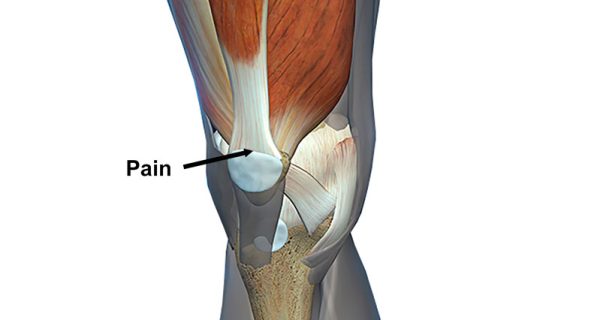
- The prepatellar bursa sits on top of the patella, or knee cap. (medicalnewstoday.com)
- Bursitis is defined as inflammation of a bursa. (medscape.com)
- Bursitis is inflammation of the bursa and the bursa at the back of the elbow over the olecranon is the most common bursa to become inflamed, causing swelling, fluid build-up and pain. (iowalum.com)
- Prepatellar bursitis occurs when the bursa, a fluid-filled sac on the front of the knee, becomes swollen. (lvhn.org)
- Bursitis is an inflammation of your bursa. (sports-injury-info.com)
- Common bursa include the subacromial bursa in your shoulder, the trochanteric bursa in your hip, and the pes anserine bursa in your knee. (sports-injury-info.com)
- The bursa becomes irritated, and an inflammatory response occurs. (sports-injury-info.com)
- A diagnostic or therapeutic lidocaine or lidocaine-corticosteroid injection into the area of the pes anserine bursa may help the clinician to determine the contribution of pes anserine bursitis to a patient's overall knee pathology, as well as possibly alleviate the patient's symptoms. (medscape.com)
- Infections of the pes anserine bursa are very rare and occur primarily in immunocompromised patients. (medscape.com)
- The appearance of pes anserine bursitis on MRI is characterized by increased signal intensity and fluid formation around the area of the pes anserinus bursa. (medscape.com)
- The key is to avoid activities that increase your knee pain while your bursa heals. (kneeforce.com)
- Trochanteric bursitis is a medical term used to describe the irritation or inflammation of a bursa found around the hip joint. (mainhealthfitness.com)
- Additionally, when the bursa located in the groin area (iliopsoas) of the hip becomes irritated or inflamed, the condition can also be referred to as hip bursitis. (mainhealthfitness.com)
- Bursitis- A bursa, a small fluid filled sac present under the skin covering the knee joint. (powershow.com)
- Continuous friction of the knee caused by bruising, abrasion or trauma to the knee may cause inflammation of bursa. (powershow.com)
- Inflammation of the cushion (bursa) between the muscle and the hip bone can cause bursitis. (wakehealth.edu)
- Bursitis is the swelling and irritation of a bursa. (magicmassagetherapy.com)
- Bursitis is an inflammation or irritation of a bursa, which is a fluid-filled sac located around joints. (nworthopaedicspecialists.com)
- Bursitis is a condition in which one of these bursa sacs becomes inflamed, typically occurring in conjunction with an injury or overuse. (nworthopaedicspecialists.com)
- Inflammation of the bursa (fluid-filled saclike cavity) over the kneecap or below the knee joint. (carehospitals.com)
- In the course of time the bursa may get inflamed due to repeated trauma resulting in bursitis, causing severe pain. (hubpages.com)
- Each of your knees has a bursa sack that is filled with fluid. (pinnaclehealthgroup.com.au)
- Knee bursitis can cause pain above, below or on your kneecap depending on which bursa is inflamed. (pinnaclehealthgroup.com.au)
- Your bursa essentially prevents different tissues and structures from catching on one another during a smooth movement of the knee joint. (pinnaclehealthgroup.com.au)
- Pes anserinus bursa - Located in the lower inside part of the knee in close to the upper part of the tibia. (pinnaclehealthgroup.com.au)
- Bursitis is the inflammation of a bursa. (rockyfootandankle.com)
- Rapid movements and sudden twisting can cause bursitis by overstressing the bursa. (rockyfootandankle.com)
- If you arrive at Rocky Mountain Foot & Ankle displaying signs of bursitis, after a thorough examination, we may prescribe anti-inflammatory medicines such as cortisone or steroids to help shrink the bursa. (rockyfootandankle.com)
- Bursa inflammation most often occurs when the bursa is located near a joint that performs repetitive motions. (orthopaedic-surgery-md.com)
- Calcium deposits or calcification of the joint can occur over time in response to ongoing bursa inflammation. (orthopaedic-surgery-md.com)
- It occurs when the fluid-filled sac, or bursa, located above the kneecap becomes inflamed. (granicusideas.com)
- It occurs when the bursa sac, which is located above the kneecap (patella), becomes inflamed due to excessive use of the joint or an injury. (granicusideas.com)
- Suprapatellar bursitis is an inflammation of the bursa located above the kneecap, or patella. (granicusideas.com)
- Bursitis of the knee occurs when the bursa that surround the knee joint become inflamed. (alignedmodernhealth.com)
- The bursa is important for proper knee function and movement because they provide a cushion for the bones that make up the joint. (alignedmodernhealth.com)
- Inflamed bursa can result in swelling and pain, limiting your ability to bend and move your knee and making walking difficult. (alignedmodernhealth.com)
- Knee bursitis is a condition in which the protective and supportive bursa, which are fluid-filled sacs located between the bone of a joint and the surrounding soft tissue, develop inflammation. (alignedmodernhealth.com)
- When this inflammation occurs, the synovial membrane of the bursa becomes thicker and produces more fluid than normal, causing swelling. (alignedmodernhealth.com)
- The calves, the hip flexors, the gluteus maximus, and the abdominal muscles of the core are all used in the act of running, and if these muscles are weak or out of alignment, it is easy to put excessive pressure on the bursa of the knee, setting yourself up for bursitis and other injuries. (alignedmodernhealth.com)
- bursitis: It is inflammatory condition of bursa characterized by pain and swelling. (surgicalshoppe.co.in)
- If bursitis fails to pass on its own and joint mobility is limited too much, your doctor may suggest keyhole surgery to remove a small part of the bursa. (movingwithoutpain.com)
Bursae28
- Learn about bursae in the knee. (medicalnewstoday.com)
- Surgical excision of bursae may be required as a last resort for chronic or frequently recurrent bursitis. (medscape.com)
- The three upper-extremity bursae that are most commonly affected by bursitis are the subacromial, subscapular, and olecranon bursae. (medscape.com)
- Bursitis is an inflammation of the bursae sac. (robbwolf.com)
- We have bursae sacs in joints like our knees, shoulders, hips…it allows for smooth rotation and movement. (robbwolf.com)
- When these bursae become inflamed bursitis occurs, causing movement to create intense pressure or even pain. (southlakepaininstitute.com)
- This risk increases for superficial bursae, as in prepatellar bursitis. (kneeforce.com)
- The severity of the bursitis symptoms is typically determined by how many of the bursae are damaged and to what degree. (wisegeek.net)
- Allowing the knee to take the total impact frequently is more than sufficient to burst bursae around the knee joint . (wisegeek.net)
- Your knee consists of up to 11 bursae. (pinnaclehealthgroup.com.au)
- Over 150 individual bursae are located throughout your body, and bursitis can erupt when any one of them becomes inflamed. (orthopaedic-surgery-md.com)
- The bursae near your elbows, shoulders, knees and hips most commonly cause bursitis inflammation and pain, but bursitis is also often seen in the foot, especially in the heel or at the base of the big toe. (orthopaedic-surgery-md.com)
- Bursitis is a condition that occurs when the small fluid-filled sacs (bursae) in the knee become inflamed. (accordhospitals.co.in)
- Different structures including bursae ensure friction-free movement at the knee joint. (advancedsofttissuerelease.com)
- Inflammation of bursae at the knee gives rise to pain and compromises the range of movement. (advancedsofttissuerelease.com)
- Bursae are proximal to large joints of the body, such as the knee, hip, and shoulder joint, which experience greater movement and friction. (advancedsofttissuerelease.com)
- Following bursae are associated with the knee joint. (advancedsofttissuerelease.com)
- Knee bursitis is characterized by inflammation of bursae around the knee joint. (advancedsofttissuerelease.com)
- Bursae are located in many joints in the body, such as the elbows, shoulders, and knees. (bone-joint.com)
- Knee bursitis occurs when one of the bursae - which are closed, fluid-filled sacs that allow for frictionless gliding between tissues -.in the knee becomes inflamed. (bone-joint.com)
- Bursitis occurs when the bursae get inflamed. (fyzical.com)
- Bursitis - or inflammation of the bursae - can have a major impact on your daily life. (movingwithoutpain.com)
- Since bursae are all throughout our bodies, this condition can occur in many joints. (movingwithoutpain.com)
- The bursae that are most susceptible to inflammation are those in the shoulder , knee, hip, elbow, and ankle. (movingwithoutpain.com)
- Long-term, mild straining of the bursae, for example due to one-sided straining of a joint during daily activities or sports causes chronic bursitis. (movingwithoutpain.com)
- Similar to tendonitis, bursitis is when the bursae, or fluid-filled pads that cushion joints, become inflamed. (permianbasinpainmanagement.com)
- Bursitis occurs when the fluid-filled sacs in the joints (bursae) become inflamed, achy or stiff. (torranceasc.com)
- Tendons near the hip bone (trochanter): Because bursae may also be affected, the term trochanteric bursitis is often used to include inflammation of these tendons. (msdmanuals.com)
Tendons9
- Patellar tendonitis - also known as jumper's knee - occurs when the tendons connecting the kneecap to the shinbone become inflamed and result in pain. (medicalnewstoday.com)
- RSI occurs due to tendons and muscle overuse. (odglawgroup.com)
- Some of the most common injuries to children and teens that cause knee pain include fractures, dislocations, and sprains and tears of soft tissues like ligaments and tendons. (choa.org)
- Tendonitis can occur in any of the tendons that surround the hip joint. (wakehealth.edu)
- The surrounding muscles and tendons enable the knee to bend back and forth and provide dynamic stability when you are active. (carehospitals.com)
- Occurs when tendons and cartilage cannot properly support the knee joint. (vetsdisabilityclaims.com)
- Swelling of tendons in the knees, including the patellar tendons. (vetsdisabilityclaims.com)
- This condition occurs when the tendons that connect the muscles to the bone become inflamed. (accordhospitals.co.in)
- Tendinitis usually occurs during middle or older age, as the tendons weaken and become more susceptible to injury and inflammation. (msdmanuals.com)
Joints16
- Long term stress may cause a build up of inflammation in the body that may lead to painful hip and knee joints. (birminghamhipsurgeon.com)
- The knee is one of the body's joints most prone to injuries. (juzo.com)
- Overuse of specific joints is considered the most common cause of bursitis. (wisegeek.net)
- Two of the most commonly affected joints are the knee and shoulder. (wisegeek.net)
- Hip joints are also commonly plagued by bursitis. (wisegeek.net)
- Sharp pain- It is the type of pain sudden on onset, cause stiffness to the knee joints. (powershow.com)
- The knee is one of the largest joints in the body, and one of the most easily injured. (carehospitals.com)
- If you have pain and inflammation in your joints, it is possible that you may have bursitis. (rockyfootandankle.com)
- The tell-tale symptoms of bursitis are swollen, tender areas near joints. (orthopaedic-surgery-md.com)
- Bursitis occurs when small fluid sacs that protect joints become inflamed, causing pain and stiffness. (vetsdisabilityclaims.com)
- Often the joints become deformed (usually occurring in the fingers and wrists). (palmbeachpainconsultants.com)
- Bursitis occurs most often at joints that perform frequent repetitive motion. (jatkos11.com)
- The knee joint is the largest and one of the most complex weight-bearing joints in the human body. (advancedsofttissuerelease.com)
- The knees and hips are the body's largest joints. (fyzical.com)
- The knee is one of the commonly injured joints where ligaments are injured. (fightingartshealthlab.com)
- This condition most often occurs near joints that perform repetitive motion, such as the shoulder, elbow and hip. (permianbasinpainmanagement.com)
Injuries24
- Knee pain can be the result of many different injuries or illnesses. (lvhn.org)
- While bursitis is not as common as tendonitis or other acute sports injuries, it can be frustrating and limit your sports performance. (sports-injury-info.com)
- The orthopedic and sports medicine specialists at Children's are trained to diagnose, treat and prevent knee pain and injuries in children, teens and young adults, from birth to age 18. (choa.org)
- Knee pain in kids and teens can be a result of traumatic knee injuries or repetitive overuse injuries from physical activity, such as competitive sports. (choa.org)
- In many cases, injuries involve more than one structure in the knee. (choa.org)
- Injuries to the knee, especially for kids and teens who are still growing, can lead to short-term and long-term damage. (choa.org)
- It's also important for kids and teens with knee pain and injuries to see an orthopedic or sports medicine specialist specifically trained to treat kids and teens. (choa.org)
- Knee injuries in rugby are very common due to nature of the sport which can be gradual wear and tear or damage from impact and trauma. (birminghamhipsurgeon.com)
- The structure of the knee joint is highly complex, which means it is very vulnerable and prone to injuries. (juzo.com)
- Sports-related injuries may lead to bursitis. (wisegeek.net)
- Hip overuse injuries such as bursitis and tendonitis occur commonly in active individuals. (wakehealth.edu)
- Knee pain can be caused by sudden or overuse injuries, mechanical problems, or underlying conditions like arthritis. (carehospitals.com)
- Knee injuries are extremely common. (pinnaclehealthgroup.com.au)
- It can be injured due to acute trauma and repeated microtrauma, for example, falls onto the knee, overuse injuries such as running on soft or uneven surfaces or from jobs that require being on your knees such as carpet laying. (pinnaclehealthgroup.com.au)
- Sports injuries or other forms of trauma can lead to bursitis. (rockyfootandankle.com)
- Suprapatellar Bursitis is a painful and disabling condition that can affect those who are active or have knee injuries. (granicusideas.com)
- Overuse injuries occur when repetitive activities put excessive stress on the knee joint, leading to inflammation and eventually pain in the area above the kneecap. (granicusideas.com)
- Knee injuries can result from a range of causes, such as sports injuries, falls, or accidents. (accordhospitals.co.in)
- Common knee injuries include sprains, strains, tears, dislocations and fractures in & around the knee. (accordhospitals.co.in)
- The most common types of repetitive motion injuries are tendinitis and bursitis. (physicalguru.com)
- Traumatic bursitis is the type involved with repetitive motion injuries. (physicalguru.com)
- Tendinitis and bursitis are also conditions that can occur from injuries. (ladrugcrimesattorney.com)
- These injuries typically result from sudden, one-time incidents that occur in the workplace. (solovteitell.com)
- With any season & sport, injuries can occur! (dryprousa.com)
Stiffness5
- The main symptom of jumper's knee is knee pain at the front of the knee, though sometimes there may also be some swelling and joint stiffness. (choa.org)
- Hip pain and stiffness are unarguably the most noticeable symptoms of hip bursitis. (mainhealthfitness.com)
- It occurs when the cartilage that cushions the joint breaks down, leading to pain, stiffness, and swelling. (accordhospitals.co.in)
- You may first experience stiffness in the knee. (ladrugcrimesattorney.com)
- There may be swelling, stiffness, instability, popping noises or the inability to fully straighten the knee. (fyzical.com)
Shoulder9
- Bursitis can also occur in the heel, hip, shoulder and knee. (iowalum.com)
- It can be found at the shoulder, hip, or knee, and if left untreated, can curb sports performance. (sports-injury-info.com)
- When my bursitis flare up occurs, I cannot move my shoulder. (robbwolf.com)
- Dr. Sunil Rajan is the best known Knee, hip, shoulder and Orthopaedic surgeon in Indore. (powershow.com)
- He provides joint replacement surgery of knee, hip and and shoulder at very low cost. (powershow.com)
- Knee, hip, shoulder and Orthopaedic surgeon in Indore. (powershow.com)
- A few examples: painting a whole day (bursitis of the shoulder), weeding the garden for hours (bursitis of the knee), or practicing intense athletics (bursitis of the hip). (movingwithoutpain.com)
- Sometimes X-rays are taken to better assess the condition of the joint (cartilage and bone), because if the shoulder is affected, for example, the culprit could also be osteoarthritis , a calcified tendon, 'frozen shoulder' etc., which all require a different treatment to bursitis. (movingwithoutpain.com)
- Rotator Cuff Injury/Subacromial Bursitis The muscles that help hold the upper arm in the shoulder joint (the rotator cuff muscles) can get pinched (shoulder impingement syndrome), become inflamed (tendinitis), or can tear partially. (msdmanuals.com)
Sacs1
- Another common summer injury is Bursitis, which is inflammation or injury to the fluid filled sacs that surround the knee. (dryprousa.com)
Muscles8
- Additionally, strengthening the muscles that support your knee can help prevent injury by increasing overall stability and providing extra cushioning for the joint. (guthrie.org)
- Injury Injury directly to the knee cause damage to the ligaments, muscles or the surrounding tissues which may lead to the inflammation and cause the knee pain. (powershow.com)
- It occurs when the muscles or ligaments are stretched or torn. (procurephysio.ca)
- The extension of the knee is essential for the gait to proceed further and it is done by the contraction of the Quadriceps Muscles. (hubpages.com)
- In the case of weak Quadriceps Muscles, the knee extension does not occur. (hubpages.com)
- Additionally, physical therapy can be used to strengthen muscles around the knee joint and improve range of motion. (granicusideas.com)
- Strengthening the muscles that are used in running goes a long way in keeping the knee joint healthy. (alignedmodernhealth.com)
- Tendinitis and bursitis are inflammation or degeneration (breakdown) of the soft tissue around muscles and bones. (joint-docs.com)
Lead to bursitis2
- A condition called gout can also lead to bursitis. (wisegeek.net)
- Acute, sudden injury to the joint can also lead to bursitis, but this is seen less often. (orthopaedic-surgery-md.com)
Becomes inflamed2
- In rheumatoid arthritis, the joint becomes inflamed and the knee cartilage is damaged. (lvhn.org)
- Bursitis is a common condition that occurs when the fluid sac that normally cushions your joint spaces becomes inflamed and painful. (bartonhealth.org)
Patella9
- In addition to the chondromalacia patella revealed during the arthroscopic exam, Sean's knee also had patellofemoral mailtracking. (medleague.com)
- Also known as patellar tendonitis, jumper's knee is an overuse injury of the patella tendon (tendon that connects the thigh muscle and kneecap to the shinbone) that can cause a child or teen to experience pain during activity. (choa.org)
- Three bones join in the knee: the two longest bones in the human body, the thigh bone (femur) and the shin bone (tibia), as well as the kneecap (patella). (juzo.com)
- The knee is a hinge joint formed by three bones, thighbone (femur), shinbone (tibia, the front bone of the leg, between the knee and the foot), and kneecap (patella). (carehospitals.com)
- The patients who have the problem of dislocation of patella will have an unstable knee resulting in frequent sudden falls due to unexpected knee flexion. (hubpages.com)
- The most common symptom of suprapatellar bursitis is pain and swelling in the area above the patella. (granicusideas.com)
- It involves replacing the entire knee joint, including the end of the femur (thigh bone), the top of the tibia (shin bone), and the patella (kneecap), with an artificial joint made of metal and plastic components. (accordhospitals.co.in)
- The knee joint is the junction between the tibia, femur, and patella. (advancedsofttissuerelease.com)
- Chondromalacia patella is abnormal softening of the cartilage of the under the knee cap (patella) characterized by knee pain. (surgicalshoppe.co.in)
Prevent bursitis1
- If you can't avoid this due to your job, try wearing knee pads to prevent bursitis from getting worse. (kneeforce.com)
Osteoarthritis10
- Osteoarthritis (OA) of the knee is a common cause of knee pain. (medicalnewstoday.com)
- Osteoarthritis of the knee may be caused by excess stress on the joint caused by repeated injury or being overweight. (lvhn.org)
- Such a high prevalence of osteoarthritis has increased the demand for knee implants. (medgadget.com)
- Osteoarthritis It is mainly referred as the inflammation of the knee joint caused due to the wear and tear of the joint. (powershow.com)
- Rheumatoid Arthritis is very painful to the knee occurs due to deficiency of the immune system and it takes much more time to get healed than osteoarthritis. (powershow.com)
- One of the main reasons for undergoing TKR is to alleviate chronic knee pain caused by conditions such as osteoarthritis, rheumatoid arthritis, or injury. (accordhospitals.co.in)
- Pre-existing inflammatory conditions that include rheumatoid arthritis, osteoarthritis, and gout are predisposing factors for knee bursitis. (advancedsofttissuerelease.com)
- Knee pain can be caused by a sudden incident, overuse, or underlying diagnosis like osteoarthritis. (healthykneesclub.com)
- Osteoarthritis, or OA, is the most common chronic joint condition, occurring when tissue at the ends of bones wear away. (permianbasinpainmanagement.com)
- Osteonecrosis of the knee is commonly mistaken for osteochondritis dissecans, primary osteoarthritis, meniscal tears, bone bruises, transient osteopenia of the knee, and pes anserine bursitis. (medscape.com)
Trauma5
- Trauma to the knee can tear the menisci, the pads of cartilage between surfaces in the knee joint. (lvhn.org)
- But trauma can also occur from overuse, leading to tendinitis and bursitis - known as jumper's knee, runner's knee, and housemaid's knee, depending on the part of the knee involved. (medleague.com)
- Whiplash is most often caused by car accidents, but it can also occur during contact sports, falls, or other types of trauma. (procurephysio.ca)
- If your healthcare provider suspects bursitis may be responsible for your painful symptoms, a physical examination and review of your medical history-especially your history of infection, disease, trauma, or repetitive movement using a specific joint-is always the first step. (orthopaedic-surgery-md.com)
- This is a type of knee replacement surgery that uses smaller incisions and specialized surgical instruments to reduce the size of the surgical site and minimize trauma to the surrounding tissues. (accordhospitals.co.in)
Tendonitis4
- Knee pain when kneeling can be due to a number of conditions, such as bursitis, arthritis, and patellar tendonitis. (medicalnewstoday.com)
- Bursitis and tendonitis. (bartonhealth.org)
- The most frequently encountered hip tendonitis is iliotibial (IT) band tendonitis, which can cause pain in both the knee and hip. (wakehealth.edu)
- Hip tendonitis can affect both your knees and your groin, so you should consult a doctor if you have joint inflammation or a strong pain in your groin. (healthykneesclub.com)
Ligaments8
- Ligaments help to stabilize the knee. (medleague.com)
- The anterior cruciate ligament (ACL) is one of the four main knee ligaments, and it helps stabilize a child's knee. (choa.org)
- It is one of the most commonly injured knee ligaments. (choa.org)
- A sprain occurs when the ligaments that join the ends of one bone with another are stretched or torn. (curezonephysiotherapy.com)
- The patients with torn menisci and collateral ligaments have unstable knees which may buckle. (hubpages.com)
- Collateral ligament tears/sprains: These ligaments supply stability to your knee for side to side movement and even during normal walking. (surgicalshoppe.co.in)
- Knee pain can also be caused by damage to the ligaments and cartilage of the knee. (fyzical.com)
- This type of injury occurs to one of the four ligaments in the knee that provides stability. (dryprousa.com)
Treat bursitis1
- A podiatrist can treat bursitis in your feet and ankles. (rockyfootandankle.com)
Bone9
- Kneeling aggravates knee pain because of increased mechanical compression in the knee joint - which means the kneecap pushes forcefully against the thigh bone. (medicalnewstoday.com)
- Patellofemoral pain syndrome , or runner's knee , is a common knee condition that causes pain at the front of the knee around the patellofemoral joint - between the kneecap and the thigh bone (femur). (medicalnewstoday.com)
- Bone fractures or infections in the joint may also cause knee pain. (guthrie.org)
- Although rare, these serious complications can occur as a result of non-verifiable situations such as improper procedure, bone deterioration, bone spurs due to bursitis, and changes in body weight. (medgadget.com)
- Instead of gliding smoothly across the bone, the knee cap rubs against it, therefore roughening the cartilage under the kneecap. (medleague.com)
- Shin splints occur when there is pain in the front of the lower leg, known as the tibia or shin bone. (curezonephysiotherapy.com)
- The inflammation of the tendon usually occurs at the site of insertion into bone. (physicalguru.com)
- Other people experience the wearing down of cartilage or bone around the knee, which can cause intense pain. (ladrugcrimesattorney.com)
- Since these protective cushions lie close to the area where the tendon joins to the bone, it is sometimes difficult to distinguish bursitis from an inflamed tendon ( tendinitis ), because these two conditions cause similar symptoms. (movingwithoutpain.com)
Types of bursitis include1
- Different types of bursitis include traumatic, infectious, and gouty. (physicalguru.com)
Hips5
- Additionally, obesity or misalignment of the hips (such as hip aberration) can lead to increased pressure and tension on the knees that can cause pain while bending them. (guthrie.org)
- Joint discomfort is a very common complaint and usually felt in the hands, feet, hips, knees, or spine. (palmbeachpainconsultants.com)
- Common areas include the knees, shoulders, elbows and hips. (jatkos11.com)
- When it comes to your hips and knees, there are 5 symptoms you shouldnt ignore . (healthykneesclub.com)
- Whether its your hips or your knees that are hurting you, physical therapy can help get to the root of your problem safely and comfortably, without the need for harmful drugs or surgery. (healthykneesclub.com)
Treatments for knee bursitis1
- At Aligned Modern Health, we focus on treatments for knee bursitis that work along with your body's natural ability to heal. (alignedmodernhealth.com)
Relieve5
- Nonsteroidal anti-inflammatory drugs (NSAIDs) such as ibuprofen and naproxen are commonly used to reduce swelling and relieve pain associated with suprapatellar bursitis. (granicusideas.com)
- Knee replacement surgery can help relieve pain, improve mobility, and enhance the quality of life for individuals with severe knee problems. (accordhospitals.co.in)
- People may opt for several home remedies and clinical treatments to relieve knee bursitis pain. (advancedsofttissuerelease.com)
- Relieve pain in knee. (fyzical.com)
- Additionally, applying ice, using a compression dressing, and elevating the affected knee can also help relieve pain and swelling. (healthykneesclub.com)
Redness5
- Additionally, if you experience any redness or swelling around your knee, you should also make an appointment with your doctor right away. (guthrie.org)
- Two of the most common bursitis symptoms are redness and swelling. (wisegeek.net)
- There can be swelling of the area along with redness and heat, and sometimes, a lump may appear, usually behind the knee. (alignedmodernhealth.com)
- The signs could be visible, as swelling or redness could occur. (ladrugcrimesattorney.com)
- With Bursitis, you may experience swelling, pain, knee redness or difficulty bending your knee. (dryprousa.com)
Commonly affected by bursitis1
- The knee is commonly affected by bursitis. (wisegeek.net)
Anserine6
- Pes anserinus tendinitis/bursitis syndrome, or pes anserine bursitis, is a cause of chronic knee pain and weakness. (wikipedia.org)
- The diagnosis of pes anserine bursitis usually is made on clinical grounds, and further workup is not necessarily indicated. (medscape.com)
- Ultrasonography can facilitate the diagnosis of pes anserine bursitis. (medscape.com)
- [ 32 ] This lack of ultrasonographic findings has led some to question the frequency of pes anserine bursitis and its accepted pathophysiology. (medscape.com)
- Magnetic resonance imaging (MRI) is the preferred imaging technique for confirming the diagnosis of pes anserine bursitis and differentiating it from concurrent pathology of the medial compartment. (medscape.com)
- consequently, the diagnosis of pes anserine bursitis cannot be based solely on MRI findings. (medscape.com)
Tendon8
- Osgood-Schlatter disease causes knee pain due to inflammation of the area just below the knee, where the kneecap tendon attaches to the shinbone. (medicalnewstoday.com)
- A strain occurs when a muscle or tendon is pulled, torn or twisted. (curezonephysiotherapy.com)
- Children can develop inflammation of the area just below the knee where the tendon from the kneecap attaches to the shinbone. (carehospitals.com)
- Types of bursitis are identified according to the joint involved-for example, hip bursitis, knee bursitis, elbow bursitis, anterior or posterior Achilles tendon bursitis, etc. (orthopaedic-surgery-md.com)
- This condition occurs from inflammation below the knee from repeated stress to the patellar tendon. (vetsdisabilityclaims.com)
- Weakening of the tendon, called tendinopathy, usually results from many small tears that occur over time. (msdmanuals.com)
- The tendon above the biceps muscle in the upper arm (bicipital tendon): Pain can occur when the elbow is bent or the arm is elevated or rotated. (msdmanuals.com)
- A tendon that runs over the side of the knee (popliteus tendon): Pain occurs on the outer part of the knee. (msdmanuals.com)
Exercises17
- Incorporating exercises such as squats and lunges into your regular routine will increase muscular strength and reduce pressure on your knees when you are actively bending. (guthrie.org)
- Lunges, squats, hip extensions, knee circles, and side leg lifts are all excellent exercises for stretching the knee joint. (guthrie.org)
- Adding strength training exercises to your routine can also help build knee stability. (guthrie.org)
- Exercises like squats and leg curls involve knee movement against resistance from weights or bodyweight, allowing for improved range of motion and muscle control, which helps improve joint stability over time. (guthrie.org)
- Even simple exercises like ankle rotations can improve circulation around your knee joint and keep it healthy. (guthrie.org)
- Whether you do stretching or strengthening exercises for knee flexibility or range of motion, regular exercise can bring tremendous benefits. (guthrie.org)
- There are also steps that can be taken to prevent knee pain, such as exercises to improve flexibility and range of motion. (guthrie.org)
- 8 Best Cardio Exercises to do With Hip Bursitis: Low Impact Calorie Burners! (mainhealthfitness.com)
- Nevertheless, a lot of active people do not know the hip bursitis exercises to avoid. (mainhealthfitness.com)
- This article contains, amongst other things, some hip bursitis exercises to avoid. (mainhealthfitness.com)
- Before we look into the possible causes and symptoms of hip bursitis, let's first take a look at hip bursitis exercises to avoid. (mainhealthfitness.com)
- However, here are some hip bursitis exercises to avoid in order not to complicate things. (mainhealthfitness.com)
- Running is major when it comes to hip bursitis exercises to avoid as it can cause you pain and exert a considerable amount of force on the hip. (mainhealthfitness.com)
- This occurs because of repeating the same motion during exercises, like running and cycling. (carehospitals.com)
- You may also be given knee bursitis exercises to do to assist in your recovery. (pinnaclehealthgroup.com.au)
- Common examples of activities associated with overuse-related bursitis include running, cycling, weight lifting and squatting exercises. (granicusideas.com)
- One study compared patients with manual techniques and mobilizations to people who just did exercises at home to reduce hip and knee pain. (fyzical.com)
Acute6
- Both types can be involved in acute or chronic bursitis. (medscape.com)
- A direct blow to the knee can cause this kind of acute injury. (medleague.com)
- It is a common cause of knee pain and swelling that can be either acute or chronic in nature. (granicusideas.com)
- Acute suprapatellar bursitis typically results from a single traumatic event such as a fall, while chronic cases are often caused by repeated overuse activities. (granicusideas.com)
- Acute bursitis is a combination of all these forms, which cause inflammation due to excessive straining. (movingwithoutpain.com)
- Therefore, this treatment is less suitable in case of acute bursitis. (movingwithoutpain.com)
Swelling16
- Symptoms of bursitis include pain and swelling. (medlineplus.gov)
- It can cause swelling, pressure, and discomfort in the knee. (medicalnewstoday.com)
- Symptoms may include pain and swelling when bending or straightening knees due to repetitive motion. (medicalnewstoday.com)
- Bursitis occurs when the synovial lining becomes thickened and produces excessive fluid, leading to localized swelling and pain. (medscape.com)
- Usually caused by a blow to the knee or a sudden twist of the knee, this injury has symptoms that include pain, swelling and difficulty walking. (lvhn.org)
- Symptoms include painful or painless swelling in the area behind the knee. (lvhn.org)
- symptoms that suggest a joint effusion (collection of fluid) such as swelling and impaired bending, and symptoms that reflect the change in knee function, such as weakness, giving out, catching, and difficulty walking (limping, contusion and complaints of pain, catching, swelling, and fatigue and favoring one knee). (medleague.com)
- You might have less knee pain and swelling after a few days. (kneeforce.com)
- This is one of the most common home remedies for knee swelling. (kneeforce.com)
- Symptoms include difficulty bearing weight on the knee, swelling of the knee and pain and 'popping' in the knee. (birminghamhipsurgeon.com)
- Symptoms include sudden knee pain, swelling and difficulty bending or extending the knee. (birminghamhipsurgeon.com)
- Pain and swelling near the hip joint may be a sign of bursitis. (wisegeek.net)
- An autoimmune and inflammatory condition that can affect the knee or any joint in the body, causing swelling, severe pain, and disability. (carehospitals.com)
- Occurs because of the infection in the knee joint leading to pain, swelling, and fever. (carehospitals.com)
- Suprapatellar bursitis is a condition that affects the knee joint and can cause pain and swelling. (granicusideas.com)
- It can cause pain and swelling around the knee joint. (accordhospitals.co.in)
Symptom2
- Knee pain is the most common symptom mainly experienced by the old age person and with severe obesity. (powershow.com)
- The symptom of bursitis that usually presents itself first is pain. (alignedmodernhealth.com)
Arthritis9
- According to Versus Arthritis, OA of the knee can affect anyone at any age but is more widespread in females aged 50 years and older. (medicalnewstoday.com)
- Learn more about arthritis of the knee. (medicalnewstoday.com)
- For patient education resources, see the Arthritis Center , as well as Bursitis . (medscape.com)
- Many knee problems, such as arthritis, are a result of aging and continual wear and stress on the knee joint. (lvhn.org)
- The knee joint is also the most complex one in the body and has the greatest susceptibility to injury, age-related wear and tear, inflammatory arthritis, and septic arthritis. (medleague.com)
- Arthritis, commonly found in the big toe, can affect the knee and is painful during episodes of flare-ups. (carehospitals.com)
- This procedure involves replacing both knees at the same time, typically done when both knees are severely affected by arthritis or other conditions. (accordhospitals.co.in)
- Diseases such as arthritis can also cause knee pain. (ladrugcrimesattorney.com)
- It could be due to an injury, arthritis, bursitis, or even a pinched nerve. (healthykneesclub.com)
Fluid7
- Bursitis occurs due to extra fluid buildup and inflammation. (medicalnewstoday.com)
- This is a fluid collection in a cyst that bulges out from the knee joint. (lvhn.org)
- From there, treatment may include aspiration (draining the fluid from your knee), physical therapy or a corticosteroid injection. (pinnaclehealthgroup.com.au)
- Visco-supplementation (for knee pain): in this procedure, a gel-like fluid called hyaluronic acid will be injected into the knee joint. (palmbeachpainconsultants.com)
- Hyaluronic acid is a naturally occurring substance found in the synovial fluid surrounding the joint's cartilages. (palmbeachpainconsultants.com)
- In elbow bursitis, a fluid-filled saccushions your elbow joint gets irritated or inflamed. (bone-joint.com)
- One of those is knee bursitis - a condition in which the tiny, fluid-filled sac around the knee joint gets inflamed. (bone-joint.com)
Prepatellar bursitis1
- Prepatellar bursitis is common for carpet layers, gardeners, roofers, plumbers and other professions that require a lot of kneeling. (pinnaclehealthgroup.com.au)
Painful6
- That swollen lump on your knee can prevent you from doing your job and even make walking a painful task. (kneeforce.com)
- This is known as bursitis which is a very painful condition. (powershow.com)
- Bursitis, if not detected and treated early, can become abscessed and extremely painful, requiring surgery. (nworthopaedicspecialists.com)
- This leads to a very painful knee injury. (carehospitals.com)
- Bursitis of the elbow, also known as olecranon bursitis, is a painful condition that can make it very hard for you to use your elbow like you usually can. (bone-joint.com)
- Are your knees feel painful, or even feel as if they may buckle underneath you without warning? (healthykneesclub.com)
Jumper's knee4
- The best way to treat a jumper's knee is to stop any activity causing the pain until the injury is healed. (medicalnewstoday.com)
- What is jumper's knee? (choa.org)
- Jumper's knee usually affects children and teens who play sports that involve a lot of jumping, squatting, climbing and running, including track and field, basketball, volleyball, gymnastics, running and soccer. (choa.org)
- What are symptoms of jumper's knee? (choa.org)
Diagnosis5
- Routine laboratory blood work is generally not helpful in the diagnosis of noninfectious bursitis but is appropriate when septic bursitis or underlying autoimmune disease is suspected. (medscape.com)
- There are many possible causes of pain when bending your knees, so it's important to see a doctor to get an accurate diagnosis. (guthrie.org)
- At Children's Healthcare of Atlanta, our pediatric orthopedic and sports medicine specialists are specially trained to recognize the specific signs and symptoms that may be causing your child's or teen's knee pain in order to make an accurate diagnosis and treatment plan. (choa.org)
- If inflammation persists after a few days, you should get a proper diagnosis from a medical professional to determine the exact cause of your bursitis. (pinnaclehealthgroup.com.au)
- This condition is one of the most common causes of localized knee pain and necessitates a precise diagnosis in order to receive proper treatment. (granicusideas.com)
Common24
- Mild but repetitive injury and use is the most common cause of olecranon bursitis. (iowalum.com)
- Bursitis is a common sports injury that occurs with sports that require repetitive motions. (sports-injury-info.com)
- Knee pain when bending is a common problem for many people and can have numerous underlying causes. (guthrie.org)
- It is very common for children and teens to experience knee pain, especially for those who are playing sports. (choa.org)
- That said, knee bursitis is a common injury in sports medicine. (kneeforce.com)
- Surgeries for knee bursitis are not common. (kneeforce.com)
- The ACL connects the thighbone to the shinbone, stabilising the knee joint, and is a common injury in rugby players due to quick twists and turns of the body. (birminghamhipsurgeon.com)
- Due to the sudden twists and turns when playing football, as well as the repetitive nature of running it is common for footballers to experience hip and knee pain. (birminghamhipsurgeon.com)
- What are the Most Common Bursitis Symptoms? (wisegeek.net)
- It is very common for athletes to injure the Anterior cruciate ligament (ACL) or the posterior cruciate ligament (PCL), which are the tissues that hold your knee together and help it move in different directions. (curezonephysiotherapy.com)
- A meniscus tear is a common knee injury. (curezonephysiotherapy.com)
- While anyone can develop this condition, it is most common in athletes who engage in sports such as running and jumping that require frequent knee bending. (granicusideas.com)
- Rest and medication are two common treatment options used to reduce knee pain caused by this condition. (granicusideas.com)
- How Common is Knee Pain in Veterans? (vetsdisabilityclaims.com)
- The most common knee condition that veterans receive VA disability benefits for. (vetsdisabilityclaims.com)
- The most common option to obtain a VA rating for knee pain is by establishing a direct service connection. (vetsdisabilityclaims.com)
- Knee problems are a common issue that can affect people of all ages. (accordhospitals.co.in)
- This is the most common type of knee replacement surgery. (accordhospitals.co.in)
- Common areas where bursitis can occur include the elbow, knee, and hip. (physicalguru.com)
- Traumatic bursitis is most common in people younger than 35 years. (physicalguru.com)
- Whether through injury, wear and tear, or disease, knee pain can be common among children and adults. (ladrugcrimesattorney.com)
- Knee pain is a common complaint of many people. (fyzical.com)
- Bursitis is equally common among men and women. (movingwithoutpain.com)
- The knee is the second most common site for osteonecrosis, but it is affected much less often than the hip. (medscape.com)
Joint42
- People get bursitis by overusing a joint. (medlineplus.gov)
- If diagnostic tests show serious damage to the knee joint, surgery might be needed. (lvhn.org)
- Bending the knees deeply and keeping them in line with your feet helps alleviate stress on the joint. (guthrie.org)
- The knee joint is created when three bones meet: the femur, tibia, and fibula. (medleague.com)
- The knee joint is the largest human joint in terms of its volume and surface area of articulating (joining) cartilage. (medleague.com)
- The knee is the largest joint in the body, and it is made up of many important and complex structures. (choa.org)
- When an ACL is injured, it can be partially torn or completely torn, which could leave the knee unstable and at risk for worsening joint damage. (choa.org)
- A physical therapist will work with you for some weeks after the surgery to help you heal your knee joint. (kneeforce.com)
- However, it is difficult to conclude that anxiety and stress directly leads to joint pain and it is advise to get any hip and knee pain looked at by a professional to rule out any other underlying condition. (birminghamhipsurgeon.com)
- With every step, every leg movement and even when resting - the knee joint is subjected to varying strain every day that can often amount to several times a person's body weight. (juzo.com)
- Studies have shown that, when climbing the stairs, the knee joint is subjected to three times a person's body weight. (juzo.com)
- If they are ignored for a long time, treated too late or incorrectly, subsequent damage to the knee joint can be caused that can result in severe and ongoing problems. (juzo.com)
- The knee joint (articulatio genus) is the largest joint in the human body. (juzo.com)
- This hinge joint allows the knee to perform movements in two directions: flexion or extension of the leg, but also the option of turning the lower leg slightly outwards or inwards when the leg is bent. (juzo.com)
- These allow the low-friction rolling-gliding motion of the knee joint, and therefore a smooth gait. (juzo.com)
- The kneecap has a triangular shape, slightly convex at the front, and it protects the front of the knee joint. (juzo.com)
- The fibrous cartilage disks can cushion severe impact, compensate for heavy loads on the knee joint and evenly distribute the strain of weight. (juzo.com)
- Bursitis is caused by the rupture of tiny sacks that surround the joint. (wisegeek.net)
- Bursitis is often caused by a joint injury. (wisegeek.net)
- Reason for the occurrence of pain Knee pain is mainly occurs due to damage to the joint and the reason for this can vary with the condition of the patient. (powershow.com)
- When this occurs in the hip it is usually in the ball of the joint, called the femoral head. (wakehealth.edu)
- Bursitis causes a reduction in or a loss of motion at the affected joint. (nworthopaedicspecialists.com)
- The knee joint can also get dislocated affecting the blood flow to the leg and may cause many other problems. (carehospitals.com)
- Arthroscopic knee surgical procedure helps doctors to view the interior of the knee joint through small incisions (cuts) via the skin and other soft tissues to treat knee problems. (carehospitals.com)
- During the procedure, the surgeon inserts a thin tube called an arthroscope into the knee joint through a small incision. (carehospitals.com)
- However, bursitis can occur near almost any joint in your body. (rockyfootandankle.com)
- Bursitis usually develops over time through overuse or repetitive use of a specific joint. (orthopaedic-surgery-md.com)
- The initial treatment for bursitis is to rest the joint area that is experiencing bursitis symptoms. (orthopaedic-surgery-md.com)
- Immobilization of the joint and cold therapy may also be necessary for immediate bursitis pain relief. (orthopaedic-surgery-md.com)
- Suprapatellar bursitis is a condition that causes inflammation and pain in the knee joint. (granicusideas.com)
- Suprapatellar Bursitis is a condition that affects the knee joint, causing pain and discomfort in many individuals. (granicusideas.com)
- A VA rating for knee pain or joint pain is determined based on multiple factors such as instability, reduced range of motion, movement without pain, and knee replacements. (vetsdisabilityclaims.com)
- This is a degenerative condition that can affect the knee joint. (accordhospitals.co.in)
- Knee replacement, also known as knee arthroplasty, is a surgical procedure that involves replacing a damaged or worn-out knee joint with an artificial joint made of metal and plastic components. (accordhospitals.co.in)
- It involves replacing only the damaged or arthritic portion of the knee joint, typically the medial (inner) or lateral (outer) compartment, with an artificial joint. (accordhospitals.co.in)
- The integrity of the knee joint determines the ease and range of movement as well as the quality of life of an individual. (advancedsofttissuerelease.com)
- Occupational associations that involve repetitive movement at the knee joint. (advancedsofttissuerelease.com)
- Obesity imposes greater stress on the knee joint and leads to knee bursitis. (advancedsofttissuerelease.com)
- There are many possible causes for the pain and inflammation around the knee joint. (bone-joint.com)
- Knee pain can result in any of the bony structures that make up the knee joint, which are the fibula, tibia, and femur. (fyzical.com)
- If the knee joint becomes dislocated, this can also cause knee pain. (fyzical.com)
- Hip joint pain and knee pain can seriously interfere with your life, especially if you are dealing with both. (healthykneesclub.com)
Septic bursitis2
- Patients with suspected septic bursitis should be treated with antibiotics while awaiting culture results. (medscape.com)
- Also known as "septic bursitis", you'll probably have to take antibiotics for at least 10 days. (kneeforce.com)
Causes bursitis2
- What Causes Bursitis? (orthopaedic-surgery-md.com)
- Sometimes, what causes bursitis is nothing more than a minor impact or mild pressure that is prolonged (e.g., kneeling for long periods, excessive sitting, etc.) which occurs from everyday work or routine activity. (orthopaedic-surgery-md.com)
Chronic bursitis4
- Another form is chronic bursitis . (movingwithoutpain.com)
- Chronic bursitis is a rheumatic disease, but generally, bursitis is only temporary . (movingwithoutpain.com)
- However, if you suffer from chronic bursitis, you could therefore opt for a natural alternative, a liquid combination of Green-lipped mussel and Curcumin . (movingwithoutpain.com)
- In fact, some patients have reported positive results when using these natural anti-inflammatories to treat chronic bursitis. (movingwithoutpain.com)
Diagnose bursitis2
- Your doctor will diagnose bursitis with a physical exam and tests such as x-rays and MRIs. (medlineplus.gov)
- How do you diagnose bursitis? (movingwithoutpain.com)
Olecranon bursitis5
- What is Student's Elbow or Olecranon Bursitis? (iowalum.com)
- What Causes Olecranon Bursitis? (iowalum.com)
- Most cases of olecranon bursitis do not cause significant pain and do not affect the movement of the elbow. (iowalum.com)
- Most cases of olecranon bursitis disappear on their own, though a cold compress can decrease any discomfort. (iowalum.com)
- Olecranon bursitis can be prevented by wearing elbow pads when playing sports or working in jobs that involve putting pressure on this area. (iowalum.com)
Elbows2
- Kneeling or leaning your elbows on a hard surface for a long time can make bursitis start. (medlineplus.gov)
- This condition is also called "student's elbow" because it often occurs in students who rest their elbows on a desk or armchair while studying or typing. (iowalum.com)
Trochanteric3
- Hip bursitis is otherwise known as trochanteric bursitis. (mainhealthfitness.com)
- Bursitis of the Trochanteric hip has to do with the inflammation of that region. (mainhealthfitness.com)
- The iliopsoas bursitis is not as rampant as trochanteric bursitis. (mainhealthfitness.com)
Ligament3
- It lies superficial to the tibial insertion of the medial collateral ligament of the knee. (wikipedia.org)
- A sudden twisting motion while pivoting on a foot can injure the anterior cruciate ligament in the knee. (fightingartshealthlab.com)
- When the knee hyperextends when pivoting, the anterior cruciate ligament or ACL can tear. (dryprousa.com)
Baker's Cyst1
- For instance, semimembranosus bursitis is also called baker's cyst whereas clergyman's knee is referred to as superficial infrapatellar bursitis. (advancedsofttissuerelease.com)
Cartilage1
- The term was created to describe the appearance of a knee at the time of surgery after the cartilage had been traumatized. (medleague.com)
Infection3
- It's often caused by constant kneeling, a blow to the knee or an infection. (lvhn.org)
- These treatments may also cause an infection to your already inflamed bursitis. (kneeforce.com)
- This often occurs from sports, infection or even just standing for long periods of time. (dryprousa.com)
Mobility2
- Pain in the affected knee can also lead to lack of mobility. (alignedmodernhealth.com)
- Both hip and knee pain can be persistent and limit mobility and activities. (fyzical.com)
Tibia1
- It runs diagonally in the middle of the knee and connects the thighbone (femur) to the tibia (shinbone). (choa.org)
Cause knee pain2
- This article examines various causes and conditions that may cause knee pain when kneeling. (medicalnewstoday.com)
- Can hip problems cause knee pain/problems? (healthykneesclub.com)
Fractures2
- Fractures can also occur to the kneecap due to a hard fall or blow to the knee. (ladrugcrimesattorney.com)
- Fractures to the knee can cause pain. (fyzical.com)
Swollen1
- The goal is to restore your knee's range of motion and manage your swollen knee. (kneeforce.com)
Caused by overuse2
- Maltracking may also be caused by overuse from repetitive knee flexion, uphill running, hiking, kneeling, squatting, or prolonged sitting with knees flexed. (medleague.com)
- Bursitis is caused by overuse. (palmbeachpainconsultants.com)
Kneecap2
- Motor vehicle collisions or falls can cause bones in the knee, including the kneecap, to break. (carehospitals.com)
- Pain on the front of the knee and around the back of the kneecap. (vetsdisabilityclaims.com)
Heel1
- However, it is also possible to have bursitis by your heel, knee or big toe. (permianbasinpainmanagement.com)