Killer Cells, Natural
Killer Cells, Lymphokine-Activated
Cytotoxicity, Immunologic
Receptors, Natural Killer Cell
Killer Factors, Yeast
Receptors, KIR
Cytokine-Induced Killer Cells
Whale, Killer
Interleukin-2
Natural Killer T-Cells
Receptors, Immunologic
Lymphocyte Activation
NK Cell Lectin-Like Receptor Subfamily C
Interferon-gamma
NK Cell Lectin-Like Receptor Subfamily D
Receptors, KIR2DL3
Flow Cytometry
Receptors, KIR3DL1
HLA-C Antigens
NK Cell Lectin-Like Receptor Subfamily K
Antibody-Dependent Cell Cytotoxicity
Receptors, KIR2DL1
Receptors, KIR2DL4
Receptors, IgG
Interleukin-15
Granzymes
Perforin
T-Lymphocytes
Cytotoxicity Tests, Immunologic
Antigens, CD
Lectins, C-Type
Receptors, KIR2DL2
Lymphocyte Subsets
Antigens, CD56
Mice, Inbred C57BL
Antigens, Differentiation, T-Lymphocyte
Histocompatibility Antigens Class I
Immunity, Innate
Immunity, Cellular
Pore Forming Cytotoxic Proteins
Immunophenotyping
Lymphocytes
Receptors, NK Cell Lectin-Like
Natural Cytotoxicity Triggering Receptor 3
Cells, Cultured
NK Cell Lectin-Like Receptor Subfamily B
NK Cell Lectin-Like Receptor Subfamily A
K562 Cells
Antigens, Surface
Antigens, CD3
Mice, Inbred BALB C
T-Lymphocytes, Cytotoxic
Receptors, KIR3DL2
Monocytes, Activated Killer
Cytokines
Receptors, KIR3DS1
Antigens, CD57
Molecular Sequence Data
Immunotherapy, Adoptive
Natural Cytotoxicity Triggering Receptor 1
Galactosylceramides
Immunotherapy
Phenotype
Lysosomal-Associated Membrane Protein 1
Interferons
Receptors, Fc
Immunologic Surveillance
Antigens, CD1d
Biological Products
Cell Separation
Tumor Cells, Cultured
T-Lymphocyte Subsets
G(M1) Ganglioside
Dendritic Cells
Leukocytes, Mononuclear
Antigens, Ly
Ligands
CD8-Positive T-Lymphocytes
Receptors, Interleukin-2
Interleukin-18
Antigens, CD2
Picibanil
HLA-B Antigens
HLA Antigens
Decidua
Interferon Type I
Mice, Inbred Strains
Receptors, KIR2DL5
Lymphoma, T-Cell
B-Lymphocytes
Amino Acid Sequence
Leukocyte Count
Antigens, CD1
Mice, SCID
Neoplasms
Clone Cells
Interleukins
Cell Degranulation
Receptors, Interleukin-15
CD4-Positive T-Lymphocytes
Muromegalovirus
GPI-Linked Proteins
Signal Transduction
Cell Differentiation
Chromium Radioisotopes
Mice, Knockout
Base Sequence
Interleukin-12
Poly I-C
Monocytes
Antigens, Differentiation
Cell Communication
Lymphocyte Depletion
Glycosphingolipids
HLA-G Antigens
Receptors, Antigen, T-Cell
Antigens, CD8
Melanoma
Major Histocompatibility Complex
Macrophages
Cell Engineering
Membrane Proteins
Leukocytes
Uterus
Organotin Compounds
Mice, Inbred C3H
Leukocyte Reduction Procedures
Adjuvants, Immunologic
Immunologic Deficiency Syndromes
RNA, Messenger
Serine Endopeptidases
Natural Cytotoxicity Triggering Receptor 2
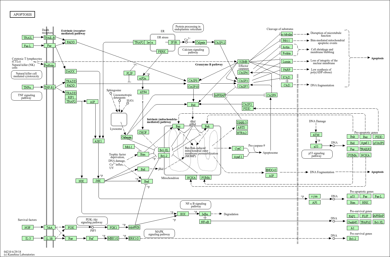
Apoptosis
Immunization, Passive
Pregnancy
Models, Immunological
Immune Tolerance
Neoplasm Transplantation
Leukemia
Immunological Synapses
Melanoma, Experimental
Thymus Gland
Gene Expression Regulation
Cell Division
Receptors, Interleukin-12
Receptors, Natural Cytotoxicity Triggering
Lymphocytes, Tumor-Infiltrating
Neoplasms, Experimental
Transfection
Gene Expression
Tumor Necrosis Factor-alpha
Interleukin-15 Receptor alpha Subunit
Mice, Inbred CBA
Coculture Techniques
Lymphoma, Extranodal NK-T-Cell
Genes, MHC Class I
Isoantigens
Interferon Inducers
T-Lymphocytes, Regulatory
Receptors, Antigen, T-Cell, gamma-delta
Dose-Response Relationship, Immunologic
Antigens, Neoplasm
Diethylamines
Lymphocyte Function-Associated Antigen-1
Natural Language Processing
Sarcoma, Experimental
Receptors, Antigen, T-Cell, alpha-beta
Reverse Transcriptase Polymerase Chain Reaction
Mutation
Cell Survival
Liver
Lymph Nodes
Genotype
Histocompatibility Antigens
Protein Binding
Transplantation, Homologous
Lymphocyte Culture Test, Mixed
Alleles
Species Specificity
Immunoglobulin G
Lymphokines
Polymerase Chain Reaction
Ziram
Hematopoietic stem-cell transplantation for the treatment of severe combined immunodeficiency. (1/9720)
BACKGROUND: Since 1968 it has been known that bone marrow transplantation can ameliorate severe combined immunodeficiency, but data on the long-term efficacy of this treatment are limited. We prospectively studied immunologic function in 89 consecutive infants with severe combined immunodeficiency who received hematopoietic stem-cell transplants at Duke University Medical Center between May 1982 and September 1998. METHODS: Serum immunoglobulin levels and lymphocyte phenotypes and function were assessed and genetic analyses performed according to standard methods. Bone marrow was depleted of T cells by agglutination with soybean lectin and by sheep-erythrocyte rosetting before transplantation. RESULTS: Seventy-seven of the infants received T-cell-depleted, HLA-haploidentical parental marrow, and 12 received HLA-identical marrow from a related donor; 3 of the recipients of haploidentical marrow also received placental-blood transplants from unrelated donors. Except for two patients who received placental blood, none of the recipients received chemotherapy before transplantation or prophylaxis against graft-versus-host disease. Of the 89 infants, 72 (81 percent) were still alive 3 months to 16.5 years after transplantation, including all of the 12 who received HLA-identical marrow, 60 of the 77 (78 percent) who were given haploidentical marrow, and 2 of the 3 (67 percent) who received both haploidentical marrow and placental blood. T-cell function became normal within two weeks after transplantation in the patients who received unfractionated HLA-identical marrow but usually not until three to four months after transplantation in those who received T-cell-depleted marrow. At the time of the most recent evaluation, all but 4 of the 72 survivors had normal T-cell function, and all the T cells in their blood were of donor origin. B-cell function remained abnormal in many of the recipients of haploidentical marrow. In 26 children (5 recipients of HLA-identical marrow and 21 recipients of haploidentical marrow) between 2 percent and 100 percent of B cells were of donor origin. Forty-five of the 72 children were receiving intravenous immune globulin. CONCLUSIONS: Transplantation of marrow from a related donor is a life-saving and life-sustaining treatment for patients with any type of severe combined immunodeficiency, even when there is no HLA-identical donor. (+info)Structure of CD94 reveals a novel C-type lectin fold: implications for the NK cell-associated CD94/NKG2 receptors. (2/9720)
The crystal structure of the extracellular domain of CD94, a component of the CD94/NKG2 NK cell receptor, has been determined to 2.6 A resolution, revealing a unique variation of the C-type lectin fold. In this variation, the second alpha helix, corresponding to residues 102-112, is replaced by a loop, the putative carbohydrate-binding site is significantly altered, and the Ca2+-binding site appears nonfunctional. This structure may serve as a prototype for other NK cell receptors such as Ly-49, NKR-P1, and CD69. The CD94 dimer observed in the crystal has an extensive hydrophobic interface that stabilizes the loop conformation of residues 102-112. The formation of this dimer reveals a putative ligand-binding region for HLA-E and suggests how NKG2 interacts with CD94. (+info)Daidzein and genistein glucuronides in vitro are weakly estrogenic and activate human natural killer cells at nutritionally relevant concentrations. (3/9720)
Daidzein and genistein glucuronides (DG and GG), major isoflavone metabolites, may be partly responsible for biological effects of isoflavones, such as estrogen receptor binding and natural killer cell (NK) activation or inhibition. DG and GG were synthesized using 3-methylcholanthrene-induced rat liver microsomes. The Km and Vmax for daidzein and genistein were 9.0 and 7.7 micromol/L, and 0.7 and 1.6 micromol/(mg protein. min), respectively. The absence of ultraviolet absorbance maxima shifts in the presence of sodium acetate confirmed that the synthesized products were 7-O-glucuronides. DG and GG were further purified by a Sephadex LH-20 column. DG and GG competed with the binding of 17beta-(3H) estradiol to estrogen receptors of B6D2F1 mouse uterine cytosol. The concentrations required for 50% displacement of 17beta-(3H) estradiol (CB50) were: 17beta-estradiol, 1.34 nmol/L; diethylstilbestrol, 1.46 nmol/L; daidzein, 1.6 micromol/L; DG, 14.7 micromol/L; genistein, 0.154 micromol/L; GG, 7.27 micromol/L. In human peripheral blood NK cells, genistein at <0.5 micromol/L and DG and GG at 0.1-10 micromol/L enhanced NK cell-mediated K562 cancer cell killing significantly (P < 0.05). At > 0.5 micromol/L, genistein inhibited NK cytotoxicity significantly (P < 0.05). The glucuronides only inhibited NK cytotoxicity at 50 micromol/L. Isoflavones, and especially the isoflavone glucuronides, enhanced activation of NK cells by interleukin-2 (IL-2), additively. At physiological concentrations, DG and GG were weakly estrogenic, and they activated human NK cells in nutritionally relevant concentrations in vitro, probably at a site different from IL-2 action. (+info)Enhanced tumor growth and invasiveness in vivo by a carboxyl-terminal fragment of alpha1-proteinase inhibitor generated by matrix metalloproteinases: a possible modulatory role in natural killer cytotoxicity. (4/9720)
Matrix metalloproteinases (MMPs) are believed to contribute to the complex process of cancer progression. They also exhibit an alpha1-proteinase inhibitor (alphaPI)-degrading activity generating a carboxyl-terminal fragment of approximately 5 kd (alphaPI-C). This study reports that overexpression of alphaPI-C in S2-020, a cloned subline derived from the human pancreas adenocarcinoma cell line SUIT-2, potentiates the growth capability of the cells in nude mice. After stable transfection of a vector containing a chimeric cDNA encoding a signal peptide sequence of tissue inhibitor of metalloproteinase-1 followed by cDNA for alphaPI-C into S2-020 cells, three clones that stably secrete alphaPI-C were obtained. The ectopic expression of alphaPI-C did not alter in vitro cellular growth. However, subcutaneous injection of the alphaPI-C-secreting clones resulted in tumors that were 1.5 to 3-fold larger than those of control clones with an increased tendency to invasiveness and lymph node metastasis. These effects could be a result of modulation of natural killer (NK) cell-mediated control of tumor growth in nude mice, as the growth advantage of alphaPI-C-secreting clones was not observed in NK-depleted mice, and alphaPI-C-secreting clones showed decreased NK sensitivity in vitro. In addition, production of alphaPI and generation of the cleaved form of alphaPI by MMP were observed in various human tumor cell lines and in a highly metastatic subline of SUIT-2 in vitro. These results provide experimental evidence that the alphaPI-degrading activity of MMPs may play a role in tumor progression not only via the inactivation of alphaPI but also via the generation of alphaPI-C. (+info)Human uterine lymphocytes. (5/9720)
During the luteal phase and the early months of pregnancy, there is a dense mucosal infiltration of CD56+ natural killer (NK) cells. These uterine NK cells have a phenotype (CD56bright, CD16-, mCD3-) which distinguishes them from peripheral blood NK cells (CD56dim, CD16bright, mCD3-). The uterine NK cells are in close association with extravillous trophoblast (EVT) cells which infiltrate into the decidua and maternal spiral arteries. This subpopulation of trophoblast expresses two human leukocyte antigen (HLA) class I molecules, HLA-G and HLA-C. Circulating NK cells express receptors for HLA class I molecules. We have recently found evidence that similar receptors are present on decidual NK cells belonging to both the Killer Inhibitory Receptor (KIR) and CD94 families. The repertoire of NK receptors expressed varies between different women. The findings indicate that decidual NK cells do have receptors for trophoblast HLA class I molecules. Experiments are underway to determine the effects of this interaction on NK cell function. (+info)Phenotypic and functional studies of leukocytes in human endometrium and endometriosis. (6/9720)
The aetiology of endometriosis, a common and disabling disorder, is presently unknown, although immune dysfunction could allow ectopic endometrial fragments to survive outside the uterine cavity. These studies investigate the relationship between leukocyte populations, steroid hormone receptor expression, proliferative activity, bcl-2 expression and apoptosis in eutopic and ectopic endometrium from women with endometriosis or adenomyosis at different phases of the menstrual cycle. Significantly increased oestrogen receptor expression, bcl-2 expression and numbers of CD8+ leukocytes were found in ectopic compared with eutopic endometrium in endometriosis, and CD56+ endometrial granulated lymphocytes (eGLs) were significantly reduced in ectopic endometrium. Apoptotic cells were rarely found in control and subject endometria. In contrast with endometriosis, adenomyotic lesions showed identical steroid hormone receptor expression, proliferative activity, bcl-2 expression and leukocyte subpopulations to eutopic endometrium, indicating different aetiologies for these disorders. The unusual CD56+ CD16- eGLs present in large numbers in late secretory phase eutopic endometrium were highly purified (>98%) by immunomagnetic separation. Except for a negligible cytotoxic activity of eGLs from early proliferative samples, cytotoxic activity of eGLs from non-pregnant endometrium during the menstrual cycle was comparable with those in peripheral blood, predominantly CD56+ CD16+ natural killer cells. eGLs from non-pregnant endometrium and early pregnancy showed a variable proliferative response to 5 and 100 U/ml interleukin-2 over 48-h and 120-h time courses. eGLs are evidently functionally important in the eutopic endometrium. Their absence in endometriotic lesions together with increased CD+8 T-cell numbers and increased oestrogen receptor and bcl-2 expression may have significant effects on the development and progression of endometriosis. (+info)Endometriotic disease: the role of peritoneal fluid. (7/9720)
Peritoneal fluid and the intraovarian milieu are a specific microenvironment. Peritoneal fluid originates mainly as an ovarian exudation product caused by increased vascular permeability, with cyclic variation in volume and steroid hormones which are always higher than in plasma. It contains large amounts of macrophages and their secretion products, and has a large exchange area with plasma through the peritoneum, which is highly permeable for small molecules. Diffusion becomes virtually zero for molecules with a molecular weight of >100000 Da. In women with the luteinized unruptured follicle (LUF) syndrome, concentrations of oestrogens and progesterone are much lower in the luteal phase. Endometriosis is associated with sterile low-grade inflammation, increased concentrations of activated macrophages and many of their secretions, such as cytokines, growth factors and angiogenic factors. Concentrations of CA-125 and of glycodelins are also increased, secreted locally by the endometrial cells. Natural killer (NK) cell function declines, possibly mediated by glycodelins or local intercellular adhesion molecule (ICAM) -1 shedding. The ovary is also a specific microenvironment, with steroid hormone concentrations 1000-fold higher in follicles than in plasma. Endometrial and superficially implanted cells are influenced by peritoneal fluid concentrations so that local environment, rather than inherent cellular differences could explain differences between superficial endometriosis and eutopic endometrium. Differences between superficial implants and endometriotic disease, deep infiltrating or cystic ovarian endometriosis, may thus arise via different endocrine environments. Superficial endometrial implants are regulated by peritoneal fluid factors, whereas deep endometriosis and cystic ovarian endometriosis are influenced by blood or ovarian factors. The endometriotic disease theory considers superficial endometriotic implants and their remodelling as a physiological process in most women, and concentrates on the causes of severe endometriosis such as differences in the eutopic endometrium from women with and without endometriosis (which may indicate hereditary differences), the invasiveness of some endometriotic cells in vitro, focal 'shielding' of endometriotic foci by adhesions, and inhibition of NK activity by ICAM-1 and glycodelins. Endometriotic disease is thus seen as a benign tumour. The type of cellular lesion, hereditary and immunological environments and local hormone concentrations in the ovary and in peritoneal fluid, will decide expression as cystic ovarian endometriosis, deep endometriosis or adenomyosis externa, and whether the latter is associated with adhesions. (+info)Suppression of angiogenesis, tumorigenicity, and metastasis by human prostate cancer cells engineered to produce interferon-beta. (8/9720)
We determined whether the IFN-beta gene can be used to suppress angiogenesis, tumor growth, and metastasis of human prostate cancer cells growing in the prostate of nude mice. Highly metastatic PC-3M human prostate cancer cells were engineered to constitutively produce murine IFN-beta subsequent to infection with a retroviral vector containing murine IFN-beta cDNA. Parental (PC-3M-P), control vector-transduced (PC-3M-Neo), and IFN-beta-transduced (PC-3M-IFN-beta) cells were injected into the prostate (orthotopic) or subcutis (ectopic) of nude mice. PC-3M-P and PC-3M-Neo cells produced rapidly growing tumors and regional lymph node metastases, whereas PC-3M-IFN-beta cells did not. PC-3M-IFN-beta cells also suppressed the tumorigenicity of bystander nontransduced prostate cancer cells. PC-3M-IFN-beta cells produced small tumors (3-5 mm in diameter) in nude mice treated with anti-asialo GM1 antibodies and in severe combined immunodeficient/Beige mice. Immunohistochemical staining revealed that PC-3M-IFN-beta tumors were homogeneously infiltrated by macrophages, whereas control tumors contained fewer macrophages at their periphery. Most tumor cells in the control tumors were stained positive by an antibody to proliferative cell nuclear antigen; very few were positively stained by terminal deoxynucleotidyl transferase-mediated dUTP-biotin nick-end labeling. In sharp contrast, PC-3M-IFN-beta tumors contained fewer proliferative cell nuclear antigen-positive cells and many terminal deoxynucleotidyl transferase-mediated dUTP-biotin nick-end labeling-positive cells. Staining with antibody against CD31 showed that control tumors contained more blood vessels than PC-3M-IFN-beta tumors. PC-3M-IFN-beta cells were more sensitive to lysis mediated by natural killer cells in vitro or to cytostasis mediated by macrophages than control transduced cells. Conditioned medium from PC-3M-IFN-beta cells augmented splenic cell-mediated cytolysis to control tumor cells, which could be neutralized by antibody against IFN-beta. Collectively, the data suggest that the suppression of tumorigenicity and metastasis of PC-3M-IFN-beta cells is due to inhibition of angiogenesis and activation of host effector cells. (+info)Natural Killer (NK) cells are a type of lymphocyte, which are large granular innate immune cells that play a crucial role in the host's defense against viral infections and malignant transformations. They do not require prior sensitization to target and destroy abnormal cells, such as virus-infected cells or tumor cells. NK cells recognize their targets through an array of germline-encoded activating and inhibitory receptors that detect the alterations in the cell surface molecules of potential targets. Upon activation, NK cells release cytotoxic granules containing perforins and granzymes to induce target cell apoptosis, and they also produce a variety of cytokines and chemokines to modulate immune responses. Overall, natural killer cells serve as a critical component of the innate immune system, providing rapid and effective responses against infected or malignant cells.
Lymphokine-activated killer (LAK) cells are a type of immune cell that has been activated to kill certain types of cells, including cancer cells and virus-infected cells. They are called "lymphokine-activated" because they are activated through the action of lymphokines, which are proteins secreted by other immune cells. LAK cells are a type of natural killer (NK) cell, which are a type of white blood cell that plays a role in the body's defense against viruses and cancer.
LAK cells are generated in the laboratory by incubating peripheral blood mononuclear cells (PBMCs), which include lymphocytes and monocytes, with high concentrations of interleukin-2 (IL-2) for several days. This process activates and expands the population of NK cells, resulting in the formation of LAK cells. These activated cells are then able to recognize and kill a wide range of tumor cells and virus-infected cells, regardless of whether they express specific antigens or not.
LAK cell therapy is an experimental form of cancer treatment that involves infusing patients with large numbers of LAK cells in order to enhance their immune response against cancer. While some studies have shown promising results, more research is needed to determine the safety and effectiveness of this approach.
Immunologic cytotoxicity refers to the damage or destruction of cells that occurs as a result of an immune response. This process involves the activation of immune cells, such as cytotoxic T cells and natural killer (NK) cells, which release toxic substances, such as perforins and granzymes, that can kill target cells.
In addition, antibodies produced by B cells can also contribute to immunologic cytotoxicity by binding to antigens on the surface of target cells and triggering complement-mediated lysis or antibody-dependent cellular cytotoxicity (ADCC) by activating immune effector cells.
Immunologic cytotoxicity plays an important role in the body's defense against viral infections, cancer cells, and other foreign substances. However, it can also contribute to tissue damage and autoimmune diseases if the immune system mistakenly targets healthy cells or tissues.
Natural Killer (NK) cell receptors are a type of cell surface receptors expressed by natural killer cells, which are a crucial component of the innate immune system. These receptors play an essential role in the recognition and elimination of abnormal cells, such as virus-infected or malignantly transformed cells.
There are two major types of NK cell receptors: activating receptors and inhibitory receptors. Activating receptors bind to ligands on the surface of target cells, triggering a signaling cascade that leads to the cytotoxic killing of the abnormal cell. In contrast, inhibitory receptors recognize major histocompatibility complex (MHC) class I molecules on healthy cells and transmit an inhibitory signal, preventing NK cells from attacking normal cells.
The balance between activating and inhibitory signals received by NK cells determines their response to target cells. When the activating signals outweigh the inhibitory ones, NK cells become activated and initiate cytotoxic responses or release cytokines to help coordinate the immune response. Dysregulation of NK cell receptors has been implicated in various diseases, including cancer and autoimmune disorders.
I cannot find a specific medical term called "killer factors" in the context of yeast. However, there is a phenomenon known as "killer yeasts," where certain strains of yeast produce proteins that can kill other strains of yeast. These proteins are referred to as "killer toxins."
Killer yeasts have been found in various species, including Saccharomyces cerevisiae and Saccharomyces bayanus. The killer toxin produced by these yeasts targets specific receptors on the cell membrane of sensitive yeast cells, leading to ion imbalance, disruption of cellular processes, and eventually cell death.
Therefore, "killer factors" in the context of yeast may refer to the genetic elements or proteins that enable certain strains of yeast to produce killer toxins and kill other sensitive yeast cells.
KIR (Killer-cell Immunoglobulin-like Receptors) are a group of receptors found on the surface of natural killer (NK) cells and some T-cells. These receptors play a crucial role in the regulation of the immune system's response to virally infected or cancerous cells.
KIR receptors can be further classified into two main groups: inhibitory receptors and activating receptors. Inhibitory KIR receptors recognize major histocompatibility complex (MHC) class I molecules on the surface of healthy cells, transmitting an inhibitory signal that prevents NK cells from attacking these cells. Activating KIR receptors, on the other hand, recognize viral or stress-induced ligands and transmit an activating signal, leading to the destruction of infected or abnormal cells.
The interaction between KIR receptors and their ligands is critical for maintaining immune tolerance and preventing autoimmune diseases. Variations in KIR genes and their MHC class I ligands can influence susceptibility to various diseases, including viral infections, cancer, and pregnancy-related complications.
Cytokine-Induced Killer (CIK) cells are a heterogeneous population of immune effector cells, primarily consisting of CD3+CD56+ T lymphocytes, generated through the ex vivo expansion of peripheral blood mononuclear cells in the presence of interferon-gamma, interleukin-2, and anti-CD3 antibody. These cells exhibit non-MHC-restricted cytotoxicity against various tumor cell types and have been investigated as a potential adoptive immunotherapy for cancer.
The term "Killer Whale" is used in medical literature to describe an unusual and very rare phenomenon where a live newborn calf becomes lodged in the birth canal of a female whale (usually a species of baleen whale), leading to potential serious complications such as infection, injury, or even death for the mother if not resolved. This condition is also known as "whale entrapment" or "cesarean delivery candidate." It is not to be confused with the common name of the species Orcinus orca, which are actually the largest species of dolphin and not whales, but are often called "killer whales" due to their size and predatory behavior.
Interleukin-2 (IL-2) is a type of cytokine, which are signaling molecules that mediate and regulate immunity, inflammation, and hematopoiesis. Specifically, IL-2 is a growth factor for T cells, a type of white blood cell that plays a central role in the immune response. It is primarily produced by CD4+ T cells (also known as T helper cells) and stimulates the proliferation and differentiation of activated T cells, including effector T cells and regulatory T cells. IL-2 also has roles in the activation and function of other immune cells, such as B cells, natural killer cells, and dendritic cells. Dysregulation of IL-2 production or signaling can contribute to various pathological conditions, including autoimmune diseases, chronic infections, and cancer.
Natural Killer T-cells (NKT cells) are a type of unconventional T-cell that express both T-cell receptors and natural killer cell receptors. They recognize lipid antigens presented by CD1d molecules, which are mainly expressed on the surface of antigen-presenting cells. NKT cells play a crucial role in the immune response against certain infections, cancer cells, and autoimmune diseases. They can quickly produce large amounts of cytokines, such as interferon-gamma and tumor necrosis factor-alpha, upon activation, thereby modulating the immune response and exerting cytotoxic effects on target cells.
Immunologic receptors are specialized proteins found on the surface of immune cells that recognize and bind to specific molecules, known as antigens, on the surface of pathogens or infected cells. This binding triggers a series of intracellular signaling events that activate the immune cell and initiate an immune response.
There are several types of immunologic receptors, including:
1. T-cell receptors (TCRs): These receptors are found on the surface of T cells and recognize antigens presented in the context of major histocompatibility complex (MHC) molecules.
2. B-cell receptors (BCRs): These receptors are found on the surface of B cells and recognize free antigens in solution.
3. Pattern recognition receptors (PRRs): These receptors are found inside immune cells and recognize conserved molecular patterns associated with pathogens, such as lipopolysaccharides and flagellin.
4. Fc receptors: These receptors are found on the surface of various immune cells and bind to the constant region of antibodies, mediating effector functions such as phagocytosis and antibody-dependent cellular cytotoxicity (ADCC).
Immunologic receptors play a critical role in the recognition and elimination of pathogens and infected cells, and dysregulation of these receptors can lead to immune disorders and diseases.
Lymphocyte activation is the process by which B-cells and T-cells (types of lymphocytes) become activated to perform effector functions in an immune response. This process involves the recognition of specific antigens presented on the surface of antigen-presenting cells, such as dendritic cells or macrophages.
The activation of B-cells leads to their differentiation into plasma cells that produce antibodies, while the activation of T-cells results in the production of cytotoxic T-cells (CD8+ T-cells) that can directly kill infected cells or helper T-cells (CD4+ T-cells) that assist other immune cells.
Lymphocyte activation involves a series of intracellular signaling events, including the binding of co-stimulatory molecules and the release of cytokines, which ultimately result in the expression of genes involved in cell proliferation, differentiation, and effector functions. The activation process is tightly regulated to prevent excessive or inappropriate immune responses that can lead to autoimmunity or chronic inflammation.
NK cell lectin-like receptor subfamily C, also known as NKG2C, is a type of activating receptor found on the surface of natural killer (NK) cells. These receptors are part of the larger family of C-type lectin receptors, which are characterized by their ability to bind carbohydrates in a calcium-dependent manner.
NKG2C is particularly interesting because it can recognize and bind to human leukocyte antigen-E (HLA-E) molecules that are present on the surface of infected or stressed cells. When NKG2C binds to HLA-E, it triggers a signaling pathway inside the NK cell that leads to its activation and the killing of the target cell.
NKG2C has been shown to play an important role in the immune response to viral infections, such as HIV and hCMV, by helping to control the spread of the virus and prevent infection. Additionally, variations in the NKG2C gene have been associated with differences in susceptibility to certain infectious diseases and autoimmune conditions.
Interferon-gamma (IFN-γ) is a soluble cytokine that is primarily produced by the activation of natural killer (NK) cells and T lymphocytes, especially CD4+ Th1 cells and CD8+ cytotoxic T cells. It plays a crucial role in the regulation of the immune response against viral and intracellular bacterial infections, as well as tumor cells. IFN-γ has several functions, including activating macrophages to enhance their microbicidal activity, increasing the presentation of major histocompatibility complex (MHC) class I and II molecules on antigen-presenting cells, stimulating the proliferation and differentiation of T cells and NK cells, and inducing the production of other cytokines and chemokines. Additionally, IFN-γ has direct antiproliferative effects on certain types of tumor cells and can enhance the cytotoxic activity of immune cells against infected or malignant cells.
NK cell lectin-like receptor subfamily D (also known as NKG2D) is a type II transmembrane protein found on the surface of natural killer (NK) cells, CD8+ T cells, and some γδ T cells. It functions as an activating receptor that recognizes stress-induced ligands expressed on the surface of infected or damaged cells. These ligands include MHC class I chain-related proteins A and B (MICA/B) and UL16-binding proteins (ULBPs). The interaction between NKG2D and its ligands triggers cytotoxic responses and cytokine production, leading to the elimination of target cells.
KIR2DL3 is a type of killer-cell immunoglobulin-like receptor (KIR) that is expressed on the surface of natural killer (NK) cells and some T cells. These receptors are involved in the regulation of the immune response, particularly in recognizing and responding to virally infected or cancerous cells.
KIR2DL3 is a inhibitory receptor, which means that it transmits a negative signal upon engagement with its ligand, helping to prevent NK cell activation and subsequent destruction of healthy cells. The ligand for KIR2DL3 is HLA-C2, a type of human leukocyte antigen (HLA) class I molecule.
It's important to note that the function of KIR2DL3 and other KIR receptors can be highly variable due to genetic differences in their expression and specificity for different HLA ligands. This variability can have implications for an individual's susceptibility to certain diseases, including viral infections and cancer.
Flow cytometry is a medical and research technique used to measure physical and chemical characteristics of cells or particles, one cell at a time, as they flow in a fluid stream through a beam of light. The properties measured include:
* Cell size (light scatter)
* Cell internal complexity (granularity, also light scatter)
* Presence or absence of specific proteins or other molecules on the cell surface or inside the cell (using fluorescent antibodies or other fluorescent probes)
The technique is widely used in cell counting, cell sorting, protein engineering, biomarker discovery and monitoring disease progression, particularly in hematology, immunology, and cancer research.
KIR3DL1 (Killer-cell Immunoglobulin-like Receptor 3DL1) is a type of receptor found on the surface of natural killer (NK) cells, which are a type of white blood cell in the human body's immune system. KIR3DL1 belongs to the family of KIR receptors that recognize and interact with Human Leukocyte Antigens (HLAs) expressed on the surface of other cells.
More specifically, KIR3DL1 recognizes HLA-A and HLA-B allotypes that have a specific motif called the Bw4 epitope. The interaction between KIR3DL1 and HLA-Bw4 can either inhibit or activate NK cell function, depending on the presence of other co-stimulatory signals.
The binding of KIR3DL1 to its ligands plays an essential role in regulating NK cell activity during immune responses against viral infections and cancer. The genetic variability in KIR3DL1 and its ligands has been associated with differences in susceptibility to various diseases, including HIV/AIDS, hepatitis C virus infection, and certain types of cancer.
HLA-C antigens are a type of human leukocyte antigen (HLA) found on the surface of cells in the human body. They are part of the major histocompatibility complex (MHC) class I molecules, which play a critical role in the immune system's ability to differentiate between "self" and "non-self" cells.
HLA-C antigens are responsible for presenting peptide fragments from inside the cell to CD8+ T cells, also known as cytotoxic T lymphocytes (CTLs). This presentation allows the CTLs to recognize and destroy infected or damaged cells, helping to prevent the spread of viruses and other pathogens.
Like other HLA antigens, HLA-C antigens are highly polymorphic, meaning that there are many different variations of these molecules in the human population. This diversity allows for a better match between an individual's immune system and the pathogens they encounter, increasing the chances of mounting an effective immune response. However, this same diversity can also make it more challenging to find compatible organ donors for transplantation.
'NK cell lectin-like receptor subfamily K' refers to a group of genes that encode for proteins found on natural killer (NK) cells, which are a type of immune cell. These proteins are known as lectin-like receptors because they bind to carbohydrates in a manner similar to lectins.
The NK cell lectin-like receptor subfamily K includes several different genes, including KLRK1 (which encodes for the protein NKG2D), KLRC1 (which encodes for the protein NKG2A), and KLRD1 (which encodes for the protein CD94). These proteins play important roles in regulating NK cell function, including activating or inhibiting NK cells in response to signals from other cells.
NKG2D, for example, binds to ligands expressed on stressed or infected cells, triggering NK cell activation and killing of those cells. NKG2A, on the other hand, binds to a different set of ligands that can inhibit NK cell activation and help prevent the destruction of healthy cells.
Overall, the NK cell lectin-like receptor subfamily K is an important component of the immune system, helping to regulate NK cell function and protect against infection and cancer.
Antibody-Dependent Cell Cytotoxicity (ADCC) is a type of immune response in which the effector cells of the immune system, such as natural killer (NK) cells, cytotoxic T-cells or macrophages, recognize and destroy virus-infected or cancer cells that are coated with antibodies.
In this process, an antibody produced by B-cells binds specifically to an antigen on the surface of a target cell. The other end of the antibody then interacts with Fc receptors found on the surface of effector cells. This interaction triggers the effector cells to release cytotoxic substances, such as perforins and granzymes, which create pores in the target cell membrane and induce apoptosis (programmed cell death).
ADCC plays an important role in the immune defense against viral infections and cancer. It is also a mechanism of action for some monoclonal antibody therapies used in cancer treatment.
KIR2DL1 (Killer-cell Immunoglobulin-like Receptor, Two Ig Domains and Long Cytoplasmic Tail 1) is a type of receptor found on the surface of natural killer (NK) cells, which are a type of white blood cell in the human body's immune system.
KIR2DL1 belongs to the KIR family of receptors, which recognize and interact with Human Leukocyte Antigen (HLA) class I molecules expressed on the surface of other cells. Specifically, KIR2DL1 recognizes HLA-C group 2 molecules, which have a specific motif at position 80 in their heavy chain (HLA-C2).
KIR2DL1 is an inhibitory receptor, meaning that its activation leads to the dampening of NK cell responses. When KIR2DL1 binds to its ligand HLA-C2 on target cells, it transmits a negative signal that helps prevent NK cell-mediated killing of healthy cells. However, if a cell lacks or has altered expression of HLA-C2 molecules, KIR2DL1 may not be able to transmit the inhibitory signal effectively, leading to NK cell activation and target cell destruction.
In summary, KIR2DL1 is an inhibitory receptor on NK cells that recognizes specific HLA class I molecules (HLA-C2) and helps regulate NK cell responses to maintain immune homeostasis.
KIR2DL4 is a type of killer-cell immunoglobulin-like receptor (KIR) that is primarily expressed on natural killer (NK) cells and some T-cell subsets. The "2D" designation indicates that it belongs to the second subgroup of KIRs, which have two extracellular immunoglobulin-like domains. The "L4" specifies its long cytoplasmic tail containing inhibitory signaling motifs, such as immunoreceptor tyrosine-based inhibition motifs (ITIMs).
KIR2DL4 is unique among KIRs because it can interact with both classical and nonclassical major histocompatibility complex class I molecules. Its primary ligand is the nonclassical HLA-G, which is involved in maternal-fetal tolerance during pregnancy. The activation of KIR2DL4 by HLA-G has been shown to induce the release of proinflammatory cytokines and chemokines, making it a potentially important player in immune responses and inflammation.
In summary, KIR2DL4 is a type of inhibitory receptor found on NK cells and some T-cells that can interact with HLA-G to modulate immune responses.
IgG receptors, also known as Fcγ receptors (Fc gamma receptors), are specialized protein molecules found on the surface of various immune cells, such as neutrophils, monocytes, macrophages, and some lymphocytes. These receptors recognize and bind to the Fc region of IgG antibodies, one of the five classes of immunoglobulins in the human body.
IgG receptors play a crucial role in immune responses by mediating different effector functions, including:
1. Antibody-dependent cellular cytotoxicity (ADCC): IgG receptors on natural killer (NK) cells and other immune cells bind to IgG antibodies coated on the surface of virus-infected or cancer cells, leading to their destruction.
2. Phagocytosis: When IgG antibodies tag pathogens or foreign particles, phagocytes like neutrophils and macrophages recognize and bind to these immune complexes via IgG receptors, facilitating the engulfment and removal of the targeted particles.
3. Antigen presentation: IgG receptors on antigen-presenting cells (APCs) can internalize immune complexes, process the antigens, and present them to T cells, thereby initiating adaptive immune responses.
4. Inflammatory response regulation: IgG receptors can modulate inflammation by activating or inhibiting downstream signaling pathways in immune cells, depending on the specific type of Fcγ receptor and its activation state.
There are several types of IgG receptors (FcγRI, FcγRII, FcγRIII, and FcγRIV) with varying affinities for different subclasses of IgG antibodies (IgG1, IgG2, IgG3, and IgG4). The distinct functions and expression patterns of these receptors contribute to the complexity and fine-tuning of immune responses in the human body.
Interleukin-15 (IL-15) is a small protein with a molecular weight of approximately 14 to 15 kilodaltons. It belongs to the class of cytokines known as the four-alpha-helix bundle family, which also includes IL-2, IL-4, and IL-7.
IL-15 is primarily produced by monocytes, macrophages, and dendritic cells, but it can also be produced by other cell types such as fibroblasts, epithelial cells, and endothelial cells. It plays a crucial role in the immune system by regulating the activation, proliferation, and survival of various immune cells, including T cells, natural killer (NK) cells, and dendritic cells.
IL-15 binds to its receptor complex, which consists of three components: IL-15Rα, IL-2/IL-15Rβ, and the common γ-chain (γc). The binding of IL-15 to this receptor complex leads to the activation of several signaling pathways, including the JAK-STAT, MAPK, and PI3K pathways.
IL-15 has a wide range of biological activities, including promoting the survival and proliferation of T cells and NK cells, enhancing their cytotoxic activity, and regulating their differentiation and maturation. It also plays a role in the development and maintenance of memory T cells, which are critical for long-term immunity to pathogens.
Dysregulation of IL-15 signaling has been implicated in various diseases, including autoimmune disorders, chronic inflammation, and cancer. Therefore, IL-15 is a potential target for therapeutic intervention in these conditions.
Granzymes are a group of proteases (enzymes that break down other proteins) that are stored in the granules of cytotoxic T cells and natural killer (NK) cells. They play an important role in the immune response by inducing apoptosis (programmed cell death) in target cells, such as virus-infected or cancer cells. Granzymes are released into the immunological synapse between the effector and target cells, where they can enter the target cell and cleave specific substrates, leading to the activation of caspases and ultimately apoptosis. There are several different types of granzymes, each with distinct substrate specificities and functions.
Perforin is a protein that plays a crucial role in the immune system's response to virally infected or cancerous cells. It is primarily produced and released by cytotoxic T-cells and natural killer (NK) cells, two types of white blood cells involved in defending the body against infection and disease.
Perforin functions by creating pores or holes in the membrane of target cells, leading to their lysis or destruction. This process allows for the release of cellular contents and the exposure of intracellular antigens, which can then be processed and presented to other immune cells, thereby enhancing the immune response against the pathogen or abnormal cells.
In summary, perforin is a vital component of the immune system's cytotoxic activity, contributing to the elimination of infected or malignant cells and maintaining overall health and homeostasis in the body.
T-lymphocytes, also known as T-cells, are a type of white blood cell that plays a key role in the adaptive immune system's response to infection. They are produced in the bone marrow and mature in the thymus gland. There are several different types of T-cells, including CD4+ helper T-cells, CD8+ cytotoxic T-cells, and regulatory T-cells (Tregs).
CD4+ helper T-cells assist in activating other immune cells, such as B-lymphocytes and macrophages. They also produce cytokines, which are signaling molecules that help coordinate the immune response. CD8+ cytotoxic T-cells directly kill infected cells by releasing toxic substances. Regulatory T-cells help maintain immune tolerance and prevent autoimmune diseases by suppressing the activity of other immune cells.
T-lymphocytes are important in the immune response to viral infections, cancer, and other diseases. Dysfunction or depletion of T-cells can lead to immunodeficiency and increased susceptibility to infections. On the other hand, an overactive T-cell response can contribute to autoimmune diseases and chronic inflammation.
Cytotoxicity tests, immunologic are a group of laboratory assays used to measure the immune-mediated damage or destruction (cytotoxicity) of cells. These tests are often used in medical research and clinical settings to evaluate the potential toxicity of drugs, biological agents, or environmental factors on specific types of cells.
Immunologic cytotoxicity tests typically involve the use of immune effector cells, such as cytotoxic T lymphocytes (CTLs) or natural killer (NK) cells, which can recognize and kill target cells that express specific antigens on their surface. The tests may also involve the use of antibodies or other immune molecules that can bind to target cells and trigger complement-mediated cytotoxicity.
There are several types of immunologic cytotoxicity tests, including:
1. Cytotoxic T lymphocyte (CTL) assays: These tests measure the ability of CTLs to recognize and kill target cells that express specific antigens. The test involves incubating target cells with CTLs and then measuring the amount of cell death or damage.
2. Natural killer (NK) cell assays: These tests measure the ability of NK cells to recognize and kill target cells that lack self-antigens or express stress-induced antigens. The test involves incubating target cells with NK cells and then measuring the amount of cell death or damage.
3. Antibody-dependent cellular cytotoxicity (ADCC) assays: These tests measure the ability of antibodies to bind to target cells and recruit immune effector cells, such as NK cells or macrophages, to mediate cell lysis. The test involves incubating target cells with antibodies and then measuring the amount of cell death or damage.
4. Complement-dependent cytotoxicity (CDC) assays: These tests measure the ability of complement proteins to bind to target cells and form a membrane attack complex that leads to cell lysis. The test involves incubating target cells with complement proteins and then measuring the amount of cell death or damage.
Immunologic cytotoxicity tests are important tools in immunology, cancer research, and drug development. They can help researchers understand how immune cells recognize and kill infected or damaged cells, as well as how to develop new therapies that enhance or inhibit these processes.
CD (cluster of differentiation) antigens are cell-surface proteins that are expressed on leukocytes (white blood cells) and can be used to identify and distinguish different subsets of these cells. They are important markers in the field of immunology and hematology, and are commonly used to diagnose and monitor various diseases, including cancer, autoimmune disorders, and infectious diseases.
CD antigens are designated by numbers, such as CD4, CD8, CD19, etc., which refer to specific proteins found on the surface of different types of leukocytes. For example, CD4 is a protein found on the surface of helper T cells, while CD8 is found on cytotoxic T cells.
CD antigens can be used as targets for immunotherapy, such as monoclonal antibody therapy, in which antibodies are designed to bind to specific CD antigens and trigger an immune response against cancer cells or infected cells. They can also be used as markers to monitor the effectiveness of treatments and to detect minimal residual disease (MRD) after treatment.
It's important to note that not all CD antigens are exclusive to leukocytes, some can be found on other cell types as well, and their expression can vary depending on the activation state or differentiation stage of the cells.
C-type lectins are a family of proteins that contain one or more carbohydrate recognition domains (CRDs) with a characteristic pattern of conserved sequence motifs. These proteins are capable of binding to specific carbohydrate structures in a calcium-dependent manner, making them important in various biological processes such as cell adhesion, immune recognition, and initiation of inflammatory responses.
C-type lectins can be further classified into several subfamilies based on their structure and function, including selectins, collectins, and immunoglobulin-like receptors. They play a crucial role in the immune system by recognizing and binding to carbohydrate structures on the surface of pathogens, facilitating their clearance by phagocytic cells. Additionally, C-type lectins are involved in various physiological processes such as cell development, tissue repair, and cancer progression.
It is important to note that some C-type lectins can also bind to self-antigens and contribute to autoimmune diseases. Therefore, understanding the structure and function of these proteins has important implications for developing new therapeutic strategies for various diseases.
The spleen is an organ in the upper left side of the abdomen, next to the stomach and behind the ribs. It plays multiple supporting roles in the body:
1. It fights infection by acting as a filter for the blood. Old red blood cells are recycled in the spleen, and platelets and white blood cells are stored there.
2. The spleen also helps to control the amount of blood in the body by removing excess red blood cells and storing platelets.
3. It has an important role in immune function, producing antibodies and removing microorganisms and damaged red blood cells from the bloodstream.
The spleen can be removed without causing any significant problems, as other organs take over its functions. This is known as a splenectomy and may be necessary if the spleen is damaged or diseased.
KIR2DL2 (Killer-cell Immunoglobulin-like Receptor 2DL2) is a type of receptor found on the surface of natural killer (NK) cells, which are a type of white blood cell in the human body's immune system. KIR2DL2 belongs to the family of KIR receptors that recognize and interact with Human Leukocyte Antigens (HLAs) expressed on the surface of other cells.
More specifically, KIR2DL2 is an inhibitory receptor that recognizes HLA-C group 2 molecules, which are a type of class I major histocompatibility complex (MHC) molecule. When KIR2DL2 binds to its ligand, it sends a negative signal that dampens the NK cell's activation and prevents it from attacking and killing the target cell.
Therefore, KIR2DL2 plays an essential role in regulating NK cell activity and maintaining immune tolerance by preventing the destruction of healthy cells. Variations in KIR genes, including KIR2DL2, have been associated with susceptibility to various diseases, including autoimmune disorders, viral infections, and cancer.
Lymphocyte subsets refer to distinct populations of white blood cells called lymphocytes, which are crucial components of the adaptive immune system. There are two main types of lymphocytes: T cells and B cells, and each type has several subsets based on their surface receptors, functions, and activation status.
1. T cell subsets: These include CD4+ T helper cells (Th cells), CD8+ cytotoxic T cells (Tc cells), regulatory T cells (Tregs), and memory T cells. Th cells are further divided into Th1, Th2, Th17, and Tfh cells based on their cytokine production profiles and functions.
* CD4+ T helper cells (Th cells) play a central role in orchestrating the immune response by producing various cytokines that activate other immune cells.
* CD8+ cytotoxic T cells (Tc cells) directly kill virus-infected or malignant cells upon recognition of specific antigens presented on their surface.
* Regulatory T cells (Tregs) suppress the activation and proliferation of other immune cells to maintain self-tolerance and prevent autoimmunity.
* Memory T cells are long-lived cells that remain in the body after an initial infection or immunization, providing rapid protection upon subsequent encounters with the same pathogen.
2. B cell subsets: These include naïve B cells, memory B cells, and plasma cells. Upon activation by antigens, B cells differentiate into antibody-secreting plasma cells that produce specific antibodies to neutralize or eliminate pathogens.
* Naïve B cells are resting cells that have not yet encountered their specific antigen.
* Memory B cells are long-lived cells generated after initial antigen exposure, which can quickly differentiate into antibody-secreting plasma cells upon re-exposure to the same antigen.
* Plasma cells are terminally differentiated B cells that secrete large amounts of specific antibodies.
Analyzing lymphocyte subsets is essential for understanding immune system function and dysfunction, as well as monitoring the effectiveness of immunotherapies and vaccinations.
CD56 is a type of antigen that is found on the surface of certain cells in the human body. It is also known as neural cell adhesion molecule 1 (NCAM-1) and is a member of the immunoglobulin superfamily. CD56 antigens are primarily expressed on natural killer (NK) cells, a type of immune cell that plays a role in the body's defense against viruses and cancer.
CD56 antigens help NK cells recognize and bind to other cells in the body, such as infected or abnormal cells. This binding can trigger the NK cells to release chemicals that can kill the target cells. CD56 antigens also play a role in the development and function of NK cells, including their ability to communicate with other immune cells and coordinate an effective response to threats.
In addition to NK cells, CD56 antigens are also found on some subsets of T cells, another type of immune cell. In these cells, CD56 antigens help regulate the activation and function of the T cells.
Abnormalities in the expression of CD56 antigens have been associated with various diseases, including certain types of cancer and autoimmune disorders.
C57BL/6 (C57 Black 6) is an inbred strain of laboratory mouse that is widely used in biomedical research. The term "inbred" refers to a strain of animals where matings have been carried out between siblings or other closely related individuals for many generations, resulting in a population that is highly homozygous at most genetic loci.
The C57BL/6 strain was established in 1920 by crossing a female mouse from the dilute brown (DBA) strain with a male mouse from the black strain. The resulting offspring were then interbred for many generations to create the inbred C57BL/6 strain.
C57BL/6 mice are known for their robust health, longevity, and ease of handling, making them a popular choice for researchers. They have been used in a wide range of biomedical research areas, including studies of cancer, immunology, neuroscience, cardiovascular disease, and metabolism.
One of the most notable features of the C57BL/6 strain is its sensitivity to certain genetic modifications, such as the introduction of mutations that lead to obesity or impaired glucose tolerance. This has made it a valuable tool for studying the genetic basis of complex diseases and traits.
Overall, the C57BL/6 inbred mouse strain is an important model organism in biomedical research, providing a valuable resource for understanding the genetic and molecular mechanisms underlying human health and disease.
Antigens are substances (usually proteins) on the surface of cells, viruses, fungi, or bacteria that the immune system recognizes as foreign and mounts a response against.
Differentiation in the context of T-lymphocytes refers to the process by which immature T-cells mature and develop into different types of T-cells with specific functions, such as CD4+ helper T-cells or CD8+ cytotoxic T-cells.
T-lymphocytes, also known as T-cells, are a type of white blood cell that plays a central role in cell-mediated immunity. They are produced in the bone marrow and mature in the thymus gland. Once mature, they circulate throughout the body in search of foreign antigens to attack and destroy.
Therefore, 'Antigens, Differentiation, T-Lymphocyte' refers to the process by which T-lymphocytes mature and develop the ability to recognize and respond to specific foreign antigens.
Histocompatibility antigens, class I are proteins found on the surface of most cells in the body. They play a critical role in the immune system's ability to differentiate between "self" and "non-self." These antigens are composed of three polypeptides - two heavy chains and one light chain - and are encoded by genes in the major histocompatibility complex (MHC) on chromosome 6 in humans.
Class I MHC molecules present peptide fragments from inside the cell to CD8+ T cells, also known as cytotoxic T cells. This presentation allows the immune system to detect and destroy cells that have been infected by viruses or other intracellular pathogens, or that have become cancerous.
There are three main types of class I MHC molecules in humans: HLA-A, HLA-B, and HLA-C. The term "HLA" stands for human leukocyte antigen, which reflects the original identification of these proteins on white blood cells (leukocytes). The genes encoding these molecules are highly polymorphic, meaning there are many different variants in the population, and matching HLA types is essential for successful organ transplantation to minimize the risk of rejection.
Innate immunity, also known as non-specific immunity or natural immunity, is the inherent defense mechanism that provides immediate protection against potentially harmful pathogens (like bacteria, viruses, fungi, and parasites) without the need for prior exposure. This type of immunity is present from birth and does not adapt to specific threats over time.
Innate immune responses involve various mechanisms such as:
1. Physical barriers: Skin and mucous membranes prevent pathogens from entering the body.
2. Chemical barriers: Enzymes, stomach acid, and lysozyme in tears, saliva, and sweat help to destroy or inhibit the growth of microorganisms.
3. Cellular responses: Phagocytic cells (neutrophils, monocytes, macrophages) recognize and engulf foreign particles and pathogens, while natural killer (NK) cells target and eliminate virus-infected or cancerous cells.
4. Inflammatory response: When an infection occurs, the innate immune system triggers inflammation to increase blood flow, recruit immune cells, and remove damaged tissue.
5. Complement system: A group of proteins that work together to recognize and destroy pathogens directly or enhance phagocytosis by coating them with complement components (opsonization).
Innate immunity plays a crucial role in initiating the adaptive immune response, which is specific to particular pathogens and provides long-term protection through memory cells. Both innate and adaptive immunity work together to maintain overall immune homeostasis and protect the body from infections and diseases.
Monoclonal antibodies are a type of antibody that are identical because they are produced by a single clone of cells. They are laboratory-produced molecules that act like human antibodies in the immune system. They can be designed to attach to specific proteins found on the surface of cancer cells, making them useful for targeting and treating cancer. Monoclonal antibodies can also be used as a therapy for other diseases, such as autoimmune disorders and inflammatory conditions.
Monoclonal antibodies are produced by fusing a single type of immune cell, called a B cell, with a tumor cell to create a hybrid cell, or hybridoma. This hybrid cell is then able to replicate indefinitely, producing a large number of identical copies of the original antibody. These antibodies can be further modified and engineered to enhance their ability to bind to specific targets, increase their stability, and improve their effectiveness as therapeutic agents.
Monoclonal antibodies have several mechanisms of action in cancer therapy. They can directly kill cancer cells by binding to them and triggering an immune response. They can also block the signals that promote cancer growth and survival. Additionally, monoclonal antibodies can be used to deliver drugs or radiation directly to cancer cells, increasing the effectiveness of these treatments while minimizing their side effects on healthy tissues.
Monoclonal antibodies have become an important tool in modern medicine, with several approved for use in cancer therapy and other diseases. They are continuing to be studied and developed as a promising approach to treating a wide range of medical conditions.
Cellular immunity, also known as cell-mediated immunity, is a type of immune response that involves the activation of immune cells, such as T lymphocytes (T cells), to protect the body against infected or damaged cells. This form of immunity is important for fighting off infections caused by viruses and intracellular bacteria, as well as for recognizing and destroying cancer cells.
Cellular immunity involves a complex series of interactions between various immune cells and molecules. When a pathogen infects a cell, the infected cell displays pieces of the pathogen on its surface in a process called antigen presentation. This attracts T cells, which recognize the antigens and become activated. Activated T cells then release cytokines, chemicals that help coordinate the immune response, and can directly attack and kill infected cells or help activate other immune cells to do so.
Cellular immunity is an important component of the adaptive immune system, which is able to learn and remember specific pathogens in order to mount a faster and more effective response upon subsequent exposure. This form of immunity is also critical for the rejection of transplanted organs, as the immune system recognizes the transplanted tissue as foreign and attacks it.
Pore-forming cytotoxic proteins are a group of toxins that can create pores or holes in the membranes of cells, leading to cell damage or death. These toxins are produced by various organisms, including bacteria, fungi, and plants, as a defense mechanism or to help establish an infection.
The pore-forming cytotoxic proteins can be divided into two main categories:
1. Membrane attack complex/perforin (MACPF) domain-containing proteins: These are found in many organisms, including humans. They form pores by oligomerizing, or clustering together, in the target cell membrane. An example of this type of toxin is the perforin protein, which is released by cytotoxic T cells and natural killer cells to destroy virus-infected or cancerous cells.
2. Cholesterol-dependent cytolysins (CDCs): These are mainly produced by gram-positive bacteria. They bind to cholesterol in the target cell membrane, forming a prepore structure that then undergoes conformational changes to create a pore. An example of a CDC is alpha-hemolysin from Staphylococcus aureus, which can lyse red blood cells and damage various other cell types.
These pore-forming cytotoxic proteins play a significant role in host-pathogen interactions and have implications for the development of novel therapeutic strategies.
Immunophenotyping is a medical laboratory technique used to identify and classify cells, usually in the context of hematologic (blood) disorders and malignancies (cancers), based on their surface or intracellular expression of various proteins and antigens. This technique utilizes specific antibodies tagged with fluorochromes, which bind to the target antigens on the cell surface or within the cells. The labeled cells are then analyzed using flow cytometry, allowing for the detection and quantification of multiple antigenic markers simultaneously.
Immunophenotyping helps in understanding the distribution of different cell types, their subsets, and activation status, which can be crucial in diagnosing various hematological disorders, immunodeficiencies, and distinguishing between different types of leukemias, lymphomas, and other malignancies. Additionally, it can also be used to monitor the progression of diseases, evaluate the effectiveness of treatments, and detect minimal residual disease (MRD) during follow-up care.
Lymphocytes are a type of white blood cell that is an essential part of the immune system. They are responsible for recognizing and responding to potentially harmful substances such as viruses, bacteria, and other foreign invaders. There are two main types of lymphocytes: B-lymphocytes (B-cells) and T-lymphocytes (T-cells).
B-lymphocytes produce antibodies, which are proteins that help to neutralize or destroy foreign substances. When a B-cell encounters a foreign substance, it becomes activated and begins to divide and differentiate into plasma cells, which produce and secrete large amounts of antibodies. These antibodies bind to the foreign substance, marking it for destruction by other immune cells.
T-lymphocytes, on the other hand, are involved in cell-mediated immunity. They directly attack and destroy infected cells or cancerous cells. T-cells can also help to regulate the immune response by producing chemical signals that activate or inhibit other immune cells.
Lymphocytes are produced in the bone marrow and mature in either the bone marrow (B-cells) or the thymus gland (T-cells). They circulate throughout the body in the blood and lymphatic system, where they can be found in high concentrations in lymph nodes, the spleen, and other lymphoid organs.
Abnormalities in the number or function of lymphocytes can lead to a variety of immune-related disorders, including immunodeficiency diseases, autoimmune disorders, and cancer.
NK cell lectin-like receptors are a type of receptor found on natural killer (NK) cells, which are a type of immune cell that plays a role in the body's defense against viruses and cancer. These receptors are characterized by their ability to bind to specific carbohydrate structures on the surface of infected or abnormal cells.
The lectin-like receptors include several different types, such as the natural cytotoxicity receptors (NCRs), the C-type lectin-like receptors (CLRs), and the immunoglobulin-like transcript (ILT) receptors. These receptors recognize and bind to specific ligands on the surface of target cells, which can trigger NK cell activation and the release of cytotoxic granules that kill the target cell.
The lectin-like receptors play an important role in NK cell function and regulation, and dysregulation of these receptors has been implicated in various diseases, including cancer and autoimmune disorders.
Natural Cytotoxicity Triggering Receptor 3 (NKp30 or NCR3) is a type II transmembrane protein that belongs to the natural cytotoxicity receptors (NCRs) family. It is primarily expressed on natural killer (NK) cells and some T-cell subsets, including CD8+ αβ T cells, CD4+ αβ T cells, and γδ T cells.
NKp30 plays a crucial role in the cytotoxic function of NK cells by mediating their natural cytotoxicity against virus-infected or malignantly transformed cells. It recognizes various ligands present on the surface of target cells, including B7-H6, BAG6, and viral hemagglutinins, leading to the activation of NK cells and subsequent killing of the target cells.
Additionally, NKp30 has been implicated in the regulation of adaptive immune responses by modulating dendritic cell maturation and cytokine production. Dysregulation of NKp30 expression or function has been associated with several pathological conditions, including cancer, viral infections, and autoimmune diseases.
"Cells, cultured" is a medical term that refers to cells that have been removed from an organism and grown in controlled laboratory conditions outside of the body. This process is called cell culture and it allows scientists to study cells in a more controlled and accessible environment than they would have inside the body. Cultured cells can be derived from a variety of sources, including tissues, organs, or fluids from humans, animals, or cell lines that have been previously established in the laboratory.
Cell culture involves several steps, including isolation of the cells from the tissue, purification and characterization of the cells, and maintenance of the cells in appropriate growth conditions. The cells are typically grown in specialized media that contain nutrients, growth factors, and other components necessary for their survival and proliferation. Cultured cells can be used for a variety of purposes, including basic research, drug development and testing, and production of biological products such as vaccines and gene therapies.
It is important to note that cultured cells may behave differently than they do in the body, and results obtained from cell culture studies may not always translate directly to human physiology or disease. Therefore, it is essential to validate findings from cell culture experiments using additional models and ultimately in clinical trials involving human subjects.
'NK Cell Lectin-Like Receptor Subfamily B' refers to a group of genes that encode proteins found on natural killer (NK) cells, which are a type of white blood cell in the human body. These proteins belong to a larger family called C-type lectin receptors (CLRs), which are involved in various immune functions such as pathogen recognition and immune cell activation.
The NK Cell Lectin-Like Receptor Subfamily B includes several genes, such as NKp80, NKp46, and NKp30, that encode proteins expressed on the surface of NK cells. These proteins function as activating receptors, meaning they can trigger NK cell activation and subsequent immune responses when they bind to specific ligands on the surface of infected or abnormal cells.
Overall, the NK Cell Lectin-Like Receptor Subfamily B plays an essential role in the innate immune response against viral infections and cancer by mediating NK cell cytotoxicity and cytokine production.
'NK Cell Lectin-Like Receptor Subfamily A' refers to a group of activating receptors expressed on natural killer (NK) cells, which are a type of immune cell. These receptors are named for their similarity in structure to lectins, which are proteins that bind to carbohydrates.
The NK Cell Lectin-Like Receptor Subfamily A includes several different receptors, such as NKp46, NKp44, and NKp30, that play important roles in the immune response. These receptors recognize specific molecules on the surface of infected or damaged cells, triggering the activation of NK cells and the release of cytotoxic substances that can kill the target cells.
The activation of NK cells through these receptors helps to control infections and prevent the development of cancer. However, abnormal activation of NK cells can also contribute to autoimmune diseases and other conditions. Therefore, understanding the function of NK Cell Lectin-Like Receptor Subfamily A members is important for developing new therapies and treatments for a variety of diseases.
K562 cells are a type of human cancer cell that are commonly used in scientific research. They are derived from a patient with chronic myelogenous leukemia (CML), a type of cancer that affects the blood and bone marrow.
K562 cells are often used as a model system to study various biological processes, including cell signaling, gene expression, differentiation, and apoptosis (programmed cell death). They are also commonly used in drug discovery and development, as they can be used to test the effectiveness of potential new therapies against cancer.
K562 cells have several characteristics that make them useful for research purposes. They are easy to grow and maintain in culture, and they can be manipulated genetically to express or knock down specific genes. Additionally, K562 cells are capable of differentiating into various cell types, such as red blood cells and megakaryocytes, which allows researchers to study the mechanisms of cell differentiation.
It's important to note that while K562 cells are a valuable tool for research, they do not fully recapitulate the complexity of human CML or other cancers. Therefore, findings from studies using K562 cells should be validated in more complex model systems or in clinical trials before they can be translated into treatments for patients.
Surface antigens are molecules found on the surface of cells that can be recognized by the immune system as being foreign or different from the host's own cells. Antigens are typically proteins or polysaccharides that are capable of stimulating an immune response, leading to the production of antibodies and activation of immune cells such as T-cells.
Surface antigens are important in the context of infectious diseases because they allow the immune system to identify and target infected cells for destruction. For example, viruses and bacteria often display surface antigens that are distinct from those found on host cells, allowing the immune system to recognize and attack them. In some cases, these surface antigens can also be used as targets for vaccines or other immunotherapies.
In addition to their role in infectious diseases, surface antigens are also important in the context of cancer. Tumor cells often display abnormal surface antigens that differ from those found on normal cells, allowing the immune system to potentially recognize and attack them. However, tumors can also develop mechanisms to evade the immune system, making it difficult to mount an effective response.
Overall, understanding the properties and behavior of surface antigens is crucial for developing effective immunotherapies and vaccines against infectious diseases and cancer.
CD3 antigens are a group of proteins found on the surface of T-cells, which are a type of white blood cell that plays a central role in the immune response. The CD3 antigens are composed of several different subunits (ε, δ, γ, and α) that associate to form the CD3 complex, which is involved in T-cell activation and signal transduction.
The CD3 complex is associated with the T-cell receptor (TCR), which recognizes and binds to specific antigens presented by antigen-presenting cells. When the TCR binds to an antigen, it triggers a series of intracellular signaling events that lead to T-cell activation and the initiation of an immune response.
CD3 antigens are important targets for immunotherapy in some diseases, such as certain types of cancer. For example, monoclonal antibodies that target CD3 have been developed to activate T-cells and enhance their ability to recognize and destroy tumor cells. However, CD3-targeted therapies can also cause side effects, such as cytokine release syndrome, which can be serious or life-threatening in some cases.
BALB/c is an inbred strain of laboratory mouse that is widely used in biomedical research. The strain was developed at the Institute of Cancer Research in London by Henry Baldwin and his colleagues in the 1920s, and it has since become one of the most commonly used inbred strains in the world.
BALB/c mice are characterized by their black coat color, which is determined by a recessive allele at the tyrosinase locus. They are also known for their docile and friendly temperament, making them easy to handle and work with in the laboratory.
One of the key features of BALB/c mice that makes them useful for research is their susceptibility to certain types of tumors and immune responses. For example, they are highly susceptible to developing mammary tumors, which can be induced by chemical carcinogens or viral infection. They also have a strong Th2-biased immune response, which makes them useful models for studying allergic diseases and asthma.
BALB/c mice are also commonly used in studies of genetics, neuroscience, behavior, and infectious diseases. Because they are an inbred strain, they have a uniform genetic background, which makes it easier to control for genetic factors in experiments. Additionally, because they have been bred in the laboratory for many generations, they are highly standardized and reproducible, making them ideal subjects for scientific research.
A cell line is a culture of cells that are grown in a laboratory for use in research. These cells are usually taken from a single cell or group of cells, and they are able to divide and grow continuously in the lab. Cell lines can come from many different sources, including animals, plants, and humans. They are often used in scientific research to study cellular processes, disease mechanisms, and to test new drugs or treatments. Some common types of human cell lines include HeLa cells (which come from a cancer patient named Henrietta Lacks), HEK293 cells (which come from embryonic kidney cells), and HUVEC cells (which come from umbilical vein endothelial cells). It is important to note that cell lines are not the same as primary cells, which are cells that are taken directly from a living organism and have not been grown in the lab.
Cytotoxic T-lymphocytes, also known as CD8+ T cells, are a type of white blood cell that plays a central role in the cell-mediated immune system. They are responsible for identifying and destroying virus-infected cells and cancer cells. When a cytotoxic T-lymphocyte recognizes a specific antigen presented on the surface of an infected or malignant cell, it becomes activated and releases toxic substances such as perforins and granzymes, which can create pores in the target cell's membrane and induce apoptosis (programmed cell death). This process helps to eliminate the infected or malignant cells and prevent the spread of infection or cancer.
KIR3DL2 (Killer-cell Immunoglobulin-like Receptor 3DL2) is a type of receptor found on the surface of natural killer (NK) cells, which are a type of white blood cell in the human body's immune system. KIR3DL2 belongs to a family of receptors called KIRs (Killer-cell Immunoglobulin-like Receptors) that help NK cells recognize and respond to infected or abnormal cells.
KIR3DL2 is a inhibitory receptor, which means it can transmit a negative signal that dampens the NK cell's activation and prevents it from attacking normal, healthy cells. Specifically, KIR3DL2 recognizes and binds to HLA-A3 and HLA-A11 molecules, which are found on the surface of many human cells. When KIR3DL2 binds to these HLA molecules, it sends a signal that inhibits NK cell activation and helps prevent an immune response against normal cells.
Abnormalities in KIR3DL2 have been associated with various diseases, including certain types of cancer and autoimmune disorders. For example, some studies suggest that changes in KIR3DL2 expression or function may contribute to the development of certain leukemias and lymphomas. Additionally, variations in KIR3DL2 genes have been linked to susceptibility to autoimmune diseases such as rheumatoid arthritis and multiple sclerosis.
I'm sorry for any confusion, but there seems to be a slight misunderstanding in your question. Monocytes and activated killer cells are two different types of immune cells.
Monocytes are a type of white blood cell that are part of the innate immune system. They play a crucial role in the nonspecific defense against infection and help initiate the adaptive immune response. Monocytes can differentiate into macrophages or dendritic cells under certain conditions, which then participate in the phagocytosis and presentation of antigens to T-cells.
Activated killer cells, also known as cytotoxic T-lymphocytes (CTLs) or CD8+ T-cells, are a type of white blood cell that are part of the adaptive immune system. They are responsible for identifying and eliminating infected or abnormal cells, such as cancer cells, by releasing cytotoxic substances like perforins and granzymes, which induce apoptosis (programmed cell death) in the target cells. Activated killer cells become activated when they recognize specific antigens presented on the surface of infected or abnormal cells via major histocompatibility complex class I molecules.
Therefore, there is no medical definition for 'Activated Killer Monocytes' as it combines two distinct types of immune cells with different functions and characteristics.
Cytokines are a broad and diverse category of small signaling proteins that are secreted by various cells, including immune cells, in response to different stimuli. They play crucial roles in regulating the immune response, inflammation, hematopoiesis, and cellular communication.
Cytokines mediate their effects by binding to specific receptors on the surface of target cells, which triggers intracellular signaling pathways that ultimately result in changes in gene expression, cell behavior, and function. Some key functions of cytokines include:
1. Regulating the activation, differentiation, and proliferation of immune cells such as T cells, B cells, natural killer (NK) cells, and macrophages.
2. Coordinating the inflammatory response by recruiting immune cells to sites of infection or tissue damage and modulating their effector functions.
3. Regulating hematopoiesis, the process of blood cell formation in the bone marrow, by controlling the proliferation, differentiation, and survival of hematopoietic stem and progenitor cells.
4. Modulating the development and function of the nervous system, including neuroinflammation, neuroprotection, and neuroregeneration.
Cytokines can be classified into several categories based on their structure, function, or cellular origin. Some common types of cytokines include interleukins (ILs), interferons (IFNs), tumor necrosis factors (TNFs), chemokines, colony-stimulating factors (CSFs), and transforming growth factors (TGFs). Dysregulation of cytokine production and signaling has been implicated in various pathological conditions, such as autoimmune diseases, chronic inflammation, cancer, and neurodegenerative disorders.
Membrane glycoproteins are proteins that contain oligosaccharide chains (glycans) covalently attached to their polypeptide backbone. They are integral components of biological membranes, spanning the lipid bilayer and playing crucial roles in various cellular processes.
The glycosylation of these proteins occurs in the endoplasmic reticulum (ER) and Golgi apparatus during protein folding and trafficking. The attached glycans can vary in structure, length, and composition, which contributes to the diversity of membrane glycoproteins.
Membrane glycoproteins can be classified into two main types based on their orientation within the lipid bilayer:
1. Type I (N-linked): These glycoproteins have a single transmembrane domain and an extracellular N-terminus, where the oligosaccharides are predominantly attached via asparagine residues (Asn-X-Ser/Thr sequon).
2. Type II (C-linked): These glycoproteins possess two transmembrane domains and an intracellular C-terminus, with the oligosaccharides linked to tryptophan residues via a mannose moiety.
Membrane glycoproteins are involved in various cellular functions, such as:
* Cell adhesion and recognition
* Receptor-mediated signal transduction
* Enzymatic catalysis
* Transport of molecules across membranes
* Cell-cell communication
* Immunological responses
Some examples of membrane glycoproteins include cell surface receptors (e.g., growth factor receptors, cytokine receptors), adhesion molecules (e.g., integrins, cadherins), and transporters (e.g., ion channels, ABC transporters).
Mycotoxins are toxic secondary metabolites produced by certain types of fungi (molds) that can contaminate food and feed crops, both during growth and storage. These toxins can cause a variety of adverse health effects in humans and animals, ranging from acute poisoning to long-term chronic exposure, which may lead to immune suppression, cancer, and other diseases. Mycotoxin-producing fungi mainly belong to the genera Aspergillus, Penicillium, Fusarium, and Alternaria. Common mycotoxins include aflatoxins, ochratoxins, fumonisins, zearalenone, patulin, and citrinin. The presence of mycotoxins in food and feed is a significant public health concern and requires stringent monitoring and control measures to ensure safety.
KIR3DS1 is a type of killer-cell immunoglobulin-like receptor (KIR) that is expressed on the surface of natural killer (NK) cells. These receptors are involved in the regulation of NK cell activation and function. KIR3DS1 is a stimulatory receptor, which means that it transmits an activating signal upon engagement with its ligand.
The ligand for KIR3DS1 is thought to be the human leukocyte antigen-F (HLA-F) molecule, which is a member of the HLA class I family. The interaction between KIR3DS1 and HLA-F is believed to play a role in the immune response against viral infections and tumors.
It's worth noting that the presence of KIR3DS1 has been associated with a reduced risk of HIV disease progression, as well as with a better clinical outcome in hematopoietic stem cell transplantation. However, more research is needed to fully understand the functional role and clinical relevance of this receptor.
CD57 is a protein found on the surface of some immune cells, specifically natural killer (NK) cells and certain T-cells. It is often used as a marker to identify these populations of cells. Antigens are substances that can stimulate an immune response, leading to the production of antibodies. In the context of CD57, antigens would refer to any substance that can bind to the CD57 protein on the surface of NK or T-cells.
It's worth noting that CD57 has been studied as a potential marker for certain diseases and conditions, such as HIV infection and some types of cancer. However, its use as a diagnostic or prognostic marker is still a subject of ongoing research and debate.
Molecular sequence data refers to the specific arrangement of molecules, most commonly nucleotides in DNA or RNA, or amino acids in proteins, that make up a biological macromolecule. This data is generated through laboratory techniques such as sequencing, and provides information about the exact order of the constituent molecules. This data is crucial in various fields of biology, including genetics, evolution, and molecular biology, allowing for comparisons between different organisms, identification of genetic variations, and studies of gene function and regulation.
Adoptive immunotherapy is a type of cancer treatment that involves the removal of immune cells from a patient, followed by their modification and expansion in the laboratory, and then reinfusion back into the patient to help boost their immune system's ability to fight cancer. This approach can be used to enhance the natural ability of T-cells (a type of white blood cell) to recognize and destroy cancer cells.
There are different types of adoptive immunotherapy, including:
1. T-cell transfer therapy: In this approach, T-cells are removed from the patient's tumor or blood, activated and expanded in the laboratory, and then reinfused back into the patient. Some forms of T-cell transfer therapy involve genetically modifying the T-cells to express chimeric antigen receptors (CARs) that recognize specific proteins on the surface of cancer cells.
2. Tumor-infiltrating lymphocyte (TIL) therapy: This type of adoptive immunotherapy involves removing T-cells directly from a patient's tumor, expanding them in the laboratory, and then reinfusing them back into the patient. The expanded T-cells are specifically targeted to recognize and destroy cancer cells.
3. Dendritic cell (DC) vaccine: DCs are specialized immune cells that help activate T-cells. In this approach, DCs are removed from the patient, exposed to tumor antigens in the laboratory, and then reinfused back into the patient to stimulate a stronger immune response against cancer cells.
Adoptive immunotherapy has shown promise in treating certain types of cancer, such as melanoma and leukemia, but more research is needed to determine its safety and efficacy in other types of cancer.
Natural Cytotoxicity Triggering Receptor 1 (NKp46, also known as NCR1) is a type of receptor found on the surface of natural killer (NK) cells and some T-cells. It is a member of the Natural Cytotoxicity Receptors (NCR) family which are involved in the recognition and elimination of target cells, such as virus-infected or tumor cells, by NK cells.
NKp46 recognizes specific structures on the surface of target cells, known as ligands, and when it binds to these ligands, it triggers a series of events that can lead to the killing of the target cell. The activation of NKp46 has been shown to play an important role in the immune response against viral infections and tumors.
It's worth noting that mutations in the NKp46 gene have been associated with certain immunodeficiency disorders, highlighting its importance in the immune system.
Galactosylceramides are a type of glycosphingolipids, which are lipid molecules that contain a sugar (glyco-) attached to a ceramide. Galactosylceramides have a galactose molecule attached to the ceramide. They are important components of cell membranes and play a role in cell recognition and signaling. In particular, they are abundant in the myelin sheath, which is the protective covering around nerve fibers in the brain and spinal cord. Abnormal accumulation of galactosylceramides can lead to certain genetic disorders, such as Krabbe disease and Gaucher disease.
Immunotherapy is a type of medical treatment that uses the body's own immune system to fight against diseases, such as cancer. It involves the use of substances (like vaccines, medications, or immune cells) that stimulate or suppress the immune system to help it recognize and destroy harmful disease-causing cells or agents, like tumor cells.
Immunotherapy can work in several ways:
1. Activating the immune system: Certain immunotherapies boost the body's natural immune responses, helping them recognize and attack cancer cells more effectively.
2. Suppressing immune system inhibitors: Some immunotherapies target and block proteins or molecules that can suppress the immune response, allowing the immune system to work more efficiently against diseases.
3. Replacing or enhancing specific immune cells: Immunotherapy can also involve administering immune cells (like T-cells) that have been genetically engineered or modified to recognize and destroy cancer cells.
Immunotherapies have shown promising results in treating various types of cancer, autoimmune diseases, and allergies. However, they can also cause side effects, as an overactive immune system may attack healthy tissues and organs. Therefore, careful monitoring is necessary during immunotherapy treatment.
Recombinant proteins are artificially created proteins produced through the use of recombinant DNA technology. This process involves combining DNA molecules from different sources to create a new set of genes that encode for a specific protein. The resulting recombinant protein can then be expressed, purified, and used for various applications in research, medicine, and industry.
Recombinant proteins are widely used in biomedical research to study protein function, structure, and interactions. They are also used in the development of diagnostic tests, vaccines, and therapeutic drugs. For example, recombinant insulin is a common treatment for diabetes, while recombinant human growth hormone is used to treat growth disorders.
The production of recombinant proteins typically involves the use of host cells, such as bacteria, yeast, or mammalian cells, which are engineered to express the desired protein. The host cells are transformed with a plasmid vector containing the gene of interest, along with regulatory elements that control its expression. Once the host cells are cultured and the protein is expressed, it can be purified using various chromatography techniques.
Overall, recombinant proteins have revolutionized many areas of biology and medicine, enabling researchers to study and manipulate proteins in ways that were previously impossible.
A phenotype is the physical or biochemical expression of an organism's genes, or the observable traits and characteristics resulting from the interaction of its genetic constitution (genotype) with environmental factors. These characteristics can include appearance, development, behavior, and resistance to disease, among others. Phenotypes can vary widely, even among individuals with identical genotypes, due to differences in environmental influences, gene expression, and genetic interactions.
Lysosome-Associated Membrane Protein 1 (LAMP-1) is a type I transmembrane protein that is heavily glycosylated and primarily localized to the limiting membrane of lysosomes. It is one of the most abundant proteins in the lysosomal membrane, making up approximately 50% of its total protein mass. LAMP-1 plays a crucial role in maintaining the integrity and stability of the lysosomal membrane by preventing lysosomal enzyme leakage into the cytosol. It also participates in various cellular processes, including autophagy, cell death, and antigen presentation.
LAMP-1 is often used as a marker for late endosomes and lysosomes due to its specific localization in these organelles. The protein contains several structural features that are important for its function, such as a large luminal domain with multiple glycosylation sites, a transmembrane domain, and a short cytoplasmic tail. The cytoplasmic tail interacts with various proteins involved in intracellular trafficking, membrane fusion, and cytoskeletal organization, which contributes to the proper functioning of lysosomes and other related organelles.
Interferons (IFNs) are a group of signaling proteins made and released by host cells in response to the presence of pathogens such as viruses, bacteria, parasites, or tumor cells. They belong to the larger family of cytokines and are crucial for the innate immune system's defense against infections. Interferons exist in multiple forms, classified into three types: type I (alpha and beta), type II (gamma), and type III (lambda). These proteins play a significant role in modulating the immune response, inhibiting viral replication, regulating cell growth, and promoting apoptosis of infected cells. Interferons are used as therapeutic agents for various medical conditions, including certain viral infections, cancers, and autoimmune diseases.
Fc receptors (FcRs) are specialized proteins found on the surface of various immune cells, including neutrophils, monocytes, macrophages, eosinophils, basophils, mast cells, and B lymphocytes. They play a crucial role in the immune response by recognizing and binding to the Fc region of antibodies (IgG, IgA, and IgE) after they have interacted with their specific antigens.
FcRs can be classified into several types based on the class of antibody they bind:
1. FcγRs - bind to the Fc region of IgG antibodies
2. FcαRs - bind to the Fc region of IgA antibodies
3. FcεRs - bind to the Fc region of IgE antibodies
The binding of antibodies to Fc receptors triggers various cellular responses, such as phagocytosis, degranulation, and antibody-dependent cellular cytotoxicity (ADCC), which contribute to the elimination of pathogens, immune complexes, and other foreign substances. Dysregulation of Fc receptor function has been implicated in several diseases, including autoimmune disorders and allergies.
Immunologic surveillance is the concept that the immune system plays a critical role in monitoring and defending the body against the development of malignancies or cancers. The immune cells, particularly T-cells and natural killer (NK) cells, are constantly scanning the body for any abnormal changes in cells, such as mutations or viral infections, that could lead to cancer.
Once these abnormal cells are detected, the immune system mounts an immune response to eliminate them, preventing their proliferation and progression into full-blown cancers. This process of immunologic surveillance is a critical component of the body's defense mechanisms against cancer and helps to maintain tissue homeostasis and prevent tumorigenesis.
However, in some cases, cancer cells may evade or suppress the immune system's surveillance mechanisms, leading to the development and progression of malignancies. Therefore, understanding the mechanisms of immunologic surveillance is crucial for developing novel cancer therapies that harness the power of the immune system to fight against cancer.
A lymphocyte count is a laboratory test that measures the number of white blood cells called lymphocytes in a sample of blood. Lymphocytes are a vital part of the immune system and help fight off infections and diseases. A normal lymphocyte count ranges from 1,000 to 4,800 cells per microliter (µL) of blood for adults.
An abnormal lymphocyte count can indicate an infection, immune disorder, or blood cancer. A low lymphocyte count is called lymphopenia, while a high lymphocyte count is called lymphocytosis. The cause of an abnormal lymphocyte count should be investigated through further testing and clinical evaluation.
CD1d is a type of antigen presenting molecule that is expressed on the surface of certain immune cells, including dendritic cells and B cells. Unlike classical MHC molecules, which present peptide antigens to T cells, CD1d presents lipid antigens to a specific subset of T cells called natural killer T (NKT) cells.
CD1d is composed of an alpha-helical heavy chain and a beta-2 microglobulin light chain, and it has a hydrophobic binding groove that can accommodate lipid antigens. CD1d-restricted NKT cells recognize and respond to these lipid antigens through their invariant T cell receptor (TCR), leading to the rapid production of cytokines and the activation of various immune responses.
CD1d-restricted NKT cells have been implicated in a variety of immunological functions, including the regulation of autoimmunity, antitumor immunity, and infectious disease.
According to the United States Food and Drug Administration (FDA), biological products are "products that are made from or contain a living organism or its derivatives, such as vaccines, blood and blood components, cells, genes, tissues, and proteins." These products can be composed of sugars, proteins, nucleic acids, or complex combinations of these substances, and they can come from many sources, including humans, animals, microorganisms, or plants.
Biological products are often used to diagnose, prevent, or treat a wide range of medical conditions, and they can be administered in various ways, such as through injection, inhalation, or topical application. Because biological products are derived from living organisms, their manufacturing processes can be complex and must be tightly controlled to ensure the safety, purity, and potency of the final product.
It's important to note that biological products are not the same as drugs, which are chemically synthesized compounds. While drugs are designed to interact with specific targets in the body, such as enzymes or receptors, biological products can have more complex and varied mechanisms of action, making them potentially more difficult to characterize and regulate.
Cell separation is a process used to separate and isolate specific cell types from a heterogeneous mixture of cells. This can be accomplished through various physical or biological methods, depending on the characteristics of the cells of interest. Some common techniques for cell separation include:
1. Density gradient centrifugation: In this method, a sample containing a mixture of cells is layered onto a density gradient medium and then centrifuged. The cells are separated based on their size, density, and sedimentation rate, with denser cells settling closer to the bottom of the tube and less dense cells remaining near the top.
2. Magnetic-activated cell sorting (MACS): This technique uses magnetic beads coated with antibodies that bind to specific cell surface markers. The labeled cells are then passed through a column placed in a magnetic field, which retains the magnetically labeled cells while allowing unlabeled cells to flow through.
3. Fluorescence-activated cell sorting (FACS): In this method, cells are stained with fluorochrome-conjugated antibodies that recognize specific cell surface or intracellular markers. The stained cells are then passed through a laser beam, which excites the fluorophores and allows for the detection and sorting of individual cells based on their fluorescence profile.
4. Filtration: This simple method relies on the physical size differences between cells to separate them. Cells can be passed through filters with pore sizes that allow smaller cells to pass through while retaining larger cells.
5. Enzymatic digestion: In some cases, cells can be separated by enzymatically dissociating tissues into single-cell suspensions and then using various separation techniques to isolate specific cell types.
These methods are widely used in research and clinical settings for applications such as isolating immune cells, stem cells, or tumor cells from biological samples.
'Tumor cells, cultured' refers to the process of removing cancerous cells from a tumor and growing them in controlled laboratory conditions. This is typically done by isolating the tumor cells from a patient's tissue sample, then placing them in a nutrient-rich environment that promotes their growth and multiplication.
The resulting cultured tumor cells can be used for various research purposes, including the study of cancer biology, drug development, and toxicity testing. They provide a valuable tool for researchers to better understand the behavior and characteristics of cancer cells outside of the human body, which can lead to the development of more effective cancer treatments.
It is important to note that cultured tumor cells may not always behave exactly the same way as they do in the human body, so findings from cell culture studies must be validated through further research, such as animal models or clinical trials.
T-lymphocyte subsets refer to distinct populations of T-cells, which are a type of white blood cell that plays a central role in cell-mediated immunity. The two main types of T-lymphocytes are CD4+ and CD8+ cells, which are defined by the presence or absence of specific proteins called cluster differentiation (CD) molecules on their surface.
CD4+ T-cells, also known as helper T-cells, play a crucial role in activating other immune cells, such as B-lymphocytes and macrophages, to mount an immune response against pathogens. They also produce cytokines that help regulate the immune response.
CD8+ T-cells, also known as cytotoxic T-cells, directly kill infected cells or tumor cells by releasing toxic substances such as perforins and granzymes.
The balance between these two subsets of T-cells is critical for maintaining immune homeostasis and mounting effective immune responses against pathogens while avoiding excessive inflammation and autoimmunity. Therefore, the measurement of T-lymphocyte subsets is essential in diagnosing and monitoring various immunological disorders, including HIV infection, cancer, and autoimmune diseases.
Dendritic cells (DCs) are a type of immune cell that play a critical role in the body's defense against infection and cancer. They are named for their dendrite-like projections, which they use to interact with and sample their environment. DCs are responsible for processing antigens (foreign substances that trigger an immune response) and presenting them to T cells, a type of white blood cell that plays a central role in the immune system's response to infection and cancer.
DCs can be found throughout the body, including in the skin, mucous membranes, and lymphoid organs. They are able to recognize and respond to a wide variety of antigens, including those from bacteria, viruses, fungi, and parasites. Once they have processed an antigen, DCs migrate to the lymph nodes, where they present the antigen to T cells. This interaction activates the T cells, which then go on to mount a targeted immune response against the invading pathogen or cancerous cells.
DCs are a diverse group of cells that can be divided into several subsets based on their surface markers and function. Some DCs, such as Langerhans cells and dermal DCs, are found in the skin and mucous membranes, where they serve as sentinels for invading pathogens. Other DCs, such as plasmacytoid DCs and conventional DCs, are found in the lymphoid organs, where they play a role in activating T cells and initiating an immune response.
Overall, dendritic cells are essential for the proper functioning of the immune system, and dysregulation of these cells has been implicated in a variety of diseases, including autoimmune disorders and cancer.
Mononuclear leukocytes are a type of white blood cells (leukocytes) that have a single, large nucleus. They include lymphocytes (B-cells, T-cells, and natural killer cells), monocytes, and dendritic cells. These cells play important roles in the body's immune system, including defending against infection and disease, and participating in immune responses and surveillance. Mononuclear leukocytes can be found in the bloodstream as well as in tissues throughout the body. They are involved in both innate and adaptive immunity, providing specific and nonspecific defense mechanisms to protect the body from harmful pathogens and other threats.
I'm assuming you are asking for information about "Ly" antigens in the context of human immune system and immunology.
Ly (Lymphocyte) antigens are a group of cell surface markers found on human leukocytes, including T cells, NK cells, and some B cells. These antigens were originally identified through serological analysis and were historically used to distinguish different subsets of lymphocytes based on their surface phenotype.
The "Ly" nomenclature has been largely replaced by the CD (Cluster of Differentiation) system, which is a more standardized and internationally recognized classification system for cell surface markers. However, some Ly antigens are still commonly referred to by their historical names, such as:
* Ly-1 or CD5: A marker found on mature T cells, including both CD4+ and CD8+ subsets.
* Ly-2 or CD8: A marker found on cytotoxic T cells, which are a subset of CD8+ T cells that can directly kill infected or damaged cells.
* Ly-3 or CD56: A marker found on natural killer (NK) cells, which are a type of immune cell that can recognize and destroy virus-infected or cancerous cells without the need for prior activation.
It's worth noting that while these antigens were originally identified through serological analysis, they are now more commonly detected using flow cytometry, which allows for the simultaneous measurement of multiple surface markers on individual cells. This has greatly expanded our ability to identify and characterize different subsets of immune cells and has led to a better understanding of their roles in health and disease.
A ligand, in the context of biochemistry and medicine, is a molecule that binds to a specific site on a protein or a larger biomolecule, such as an enzyme or a receptor. This binding interaction can modify the function or activity of the target protein, either activating it or inhibiting it. Ligands can be small molecules, like hormones or neurotransmitters, or larger structures, like antibodies. The study of ligand-protein interactions is crucial for understanding cellular processes and developing drugs, as many therapeutic compounds function by binding to specific targets within the body.
CD8-positive T-lymphocytes, also known as CD8+ T cells or cytotoxic T cells, are a type of white blood cell that plays a crucial role in the adaptive immune system. They are named after the CD8 molecule found on their surface, which is a protein involved in cell signaling and recognition.
CD8+ T cells are primarily responsible for identifying and destroying virus-infected cells or cancerous cells. When activated, they release cytotoxic granules that contain enzymes capable of inducing apoptosis (programmed cell death) in the target cells. They also produce cytokines such as interferon-gamma, which can help coordinate the immune response and activate other immune cells.
CD8+ T cells are generated in the thymus gland and are a type of T cell, which is a lymphocyte that matures in the thymus and plays a central role in cell-mediated immunity. They recognize and respond to specific antigens presented on the surface of infected or cancerous cells in conjunction with major histocompatibility complex (MHC) class I molecules.
Overall, CD8+ T cells are an essential component of the immune system's defense against viral infections and cancer.
Interleukin-2 (IL-2) receptors are a type of cell surface receptor that bind to and interact with the cytokine interleukin-2. IL-2 is a protein that plays an important role in the immune system, particularly in the activation and proliferation of T cells, a type of white blood cell that helps protect the body from infection and disease.
IL-2 receptors are composed of three subunits: alpha (CD25), beta (CD122), and gamma (CD132). These subunits can combine to form different types of IL-2 receptors, each with different functions. The high-affinity IL-2 receptor is made up of all three subunits and is found on the surface of activated T cells. This type of receptor has a strong binding affinity for IL-2 and plays a crucial role in T cell activation and proliferation.
The intermediate-affinity IL-2 receptor, which consists of the beta and gamma subunits, is found on the surface of resting T cells and natural killer (NK) cells. This type of receptor has a lower binding affinity for IL-2 and plays a role in activating and proliferating these cells.
IL-2 receptors are important targets for immunotherapy, as they play a key role in the regulation of the immune response. Drugs that target IL-2 receptors, such as aldesleukin (Proleukin), have been used to treat certain types of cancer and autoimmune diseases.
Interleukin-18 (IL-18) is a pro-inflammatory cytokine, a type of signaling molecule used in intercellular communication. It belongs to the interleukin-1 (IL-1) family and is primarily produced by macrophages, although other cells such as keratinocytes, osteoblasts, and Kupffer cells can also produce it.
IL-18 plays a crucial role in the innate and adaptive immune responses. It contributes to the differentiation of Th1 (T helper 1) cells, which are critical for fighting intracellular pathogens, and enhances the cytotoxic activity of natural killer (NK) cells and CD8+ T cells. IL-18 also has a role in the production of interferon-gamma (IFN-γ), a cytokine that activates immune cells and has antiviral properties.
Dysregulation of IL-18 has been implicated in several inflammatory diseases, such as rheumatoid arthritis, Crohn's disease, and psoriasis. It is also involved in the pathogenesis of some autoimmune disorders and has been investigated as a potential therapeutic target for these conditions.
CD2 is a type of cell surface protein known as a glycoprotein that is found on the surface of T cells, natural killer (NK) cells, and thymocytes in humans. It plays a role in the activation and regulation of the immune response. CD2 can also function as an adhesion molecule, helping to bind T cells to other cells during an immune response.
An antigen is any substance that can stimulate an immune response, leading to the production of antibodies or the activation of immune cells such as T cells. In the context of CD2, an "antigen" may refer to a specific molecule or structure that interacts with CD2 and triggers a response from T cells or other immune cells.
It's worth noting that while CD2 can interact with certain antigens, it is not itself an antigen in the traditional sense. However, the term "antigen" is sometimes used more broadly to refer to any molecule that interacts with the immune system and triggers a response, so it is possible for CD2 to be referred to as an "antigen" in this context.
Picibanil is not a commonly used medical term, and it may be more familiar as the brand name for a specific preparation of Group A Streptococcus OK-432. It is an immunotherapeutic agent that has been used in Japan for the treatment of certain types of cancer, such as nasopharyngeal carcinoma and soft tissue sarcoma.
Group A Streptococcus OK-432 is a weakened form of a bacterium that causes strep throat. When administered, it stimulates the immune system to produce cytokines, which are substances that help regulate the immune response. This can enhance the body's ability to fight off cancer cells and potentially slow or stop tumor growth.
It is important to note that Picibanil/OK-432 is not approved for use in the United States and its effectiveness as a cancer treatment has not been extensively studied outside of Japan.
HLA-B antigens are human leukocyte antigen (HLA) proteins found on the surface of cells that play an important role in the body's immune system. They are part of the major histocompatibility complex (MHC) class I molecules, which present pieces of proteins from inside the cell to T-cells, a type of white blood cell involved in immune responses.
HLA-B antigens are highly polymorphic, meaning that there are many different variations or alleles of this gene in the human population. This genetic diversity allows for a wide range of potential HLA-B proteins to be expressed, which can help recognize and respond to a variety of foreign substances, such as viruses and cancer cells.
The HLA-B antigens are inherited from both parents, and an individual may express one or two different HLA-B antigens depending on their genetic makeup. The specific combination of HLA-B antigens that a person expresses can have implications for their susceptibility to certain diseases, as well as their compatibility with organ transplants.
HLA (Human Leukocyte Antigen) antigens are a group of proteins found on the surface of cells in our body. They play a crucial role in the immune system's ability to differentiate between "self" and "non-self." HLA antigens are encoded by a group of genes located on chromosome 6, known as the major histocompatibility complex (MHC).
There are three types of HLA antigens: HLA class I, HLA class II, and HLA class III. HLA class I antigens are found on the surface of almost all cells in the body and help the immune system recognize and destroy virus-infected or cancerous cells. They consist of three components: HLA-A, HLA-B, and HLA-C.
HLA class II antigens are primarily found on the surface of immune cells, such as macrophages, B cells, and dendritic cells. They assist in the presentation of foreign particles (like bacteria and viruses) to CD4+ T cells, which then activate other parts of the immune system. HLA class II antigens include HLA-DP, HLA-DQ, and HLA-DR.
HLA class III antigens consist of various molecules involved in immune responses, such as cytokines and complement components. They are not directly related to antigen presentation.
The genetic diversity of HLA antigens is extensive, with thousands of variations or alleles. This diversity allows for a better ability to recognize and respond to a wide range of pathogens. However, this variation can also lead to compatibility issues in organ transplantation, as the recipient's immune system may recognize the donor's HLA antigens as foreign and attack the transplanted organ.
The decidua is a specialized type of tissue that lines the uterus during pregnancy. It forms after the implantation of a fertilized egg (embryo) into the uterine lining, and it plays an important role in supporting the growth and development of the embryo and fetus.
The decidua is composed of several layers, including the decidual capsularis, which surrounds the embryo, and the decidual parietalis, which lines the rest of the uterus. The tissue is rich in blood vessels and contains a variety of immune cells that help to protect the developing fetus from infection.
During pregnancy, the decidua produces various hormones and growth factors that support the growth of the placenta, which provides nutrients and oxygen to the fetus. After the birth of the baby, the decidua is shed along with the placenta in a process called childbirth or parturition.
It's worth noting that abnormalities in the decidua can contribute to pregnancy complications such as preeclampsia, preterm labor, and miscarriage.
Interferon type I is a class of signaling proteins, also known as cytokines, that are produced and released by cells in response to the presence of pathogens such as viruses, bacteria, and parasites. These interferons play a crucial role in the body's innate immune system and help to establish an antiviral state in surrounding cells to prevent the spread of infection.
Interferon type I includes several subtypes, such as interferon-alpha (IFN-α), interferon-beta (IFN-β), and interferon-omega (IFN-ω). When produced, these interferons bind to specific receptors on the surface of nearby cells, triggering a cascade of intracellular signaling events that lead to the activation of genes involved in the antiviral response.
The activation of these genes results in the production of enzymes that inhibit viral replication and promote the destruction of infected cells. Interferon type I also enhances the adaptive immune response by promoting the activation and proliferation of immune cells such as T-cells and natural killer (NK) cells, which can directly target and eliminate infected cells.
Overall, interferon type I plays a critical role in the body's defense against viral infections and is an important component of the immune response to many different types of pathogens.
Inbred strains of mice are defined as lines of mice that have been brother-sister mated for at least 20 consecutive generations. This results in a high degree of homozygosity, where the mice of an inbred strain are genetically identical to one another, with the exception of spontaneous mutations.
Inbred strains of mice are widely used in biomedical research due to their genetic uniformity and stability, which makes them useful for studying the genetic basis of various traits, diseases, and biological processes. They also provide a consistent and reproducible experimental system, as compared to outbred or genetically heterogeneous populations.
Some commonly used inbred strains of mice include C57BL/6J, BALB/cByJ, DBA/2J, and 129SvEv. Each strain has its own unique genetic background and phenotypic characteristics, which can influence the results of experiments. Therefore, it is important to choose the appropriate inbred strain for a given research question.
KIR2DL5 is a type of killer-cell immunoglobulin-like receptor (KIR) that is expressed on the surface of natural killer (NK) cells and some T cells. These receptors are involved in the regulation of the immune response, particularly in recognizing and responding to virally infected or cancerous cells.
KIR2DL5 is a subtype of KIR2D receptors, which have two immunoglobulin-like domains. Specifically, KIR2DL5 is a inhibitory receptor, meaning that it transmits a negative signal upon engagement with its ligand, which helps to prevent the destruction of healthy cells.
The ligands for KIR2DL5 are thought to be HLA-C molecules, which are found on the surface of many types of cells and play a critical role in the immune response by presenting pieces of proteins from viruses or cancer cells to T cells. However, the specificity and function of KIR2DL5 are still being studied, and more research is needed to fully understand its role in the immune system.
T-cell lymphoma is a type of cancer that affects the T-cells, which are a specific type of white blood cell responsible for immune function. These lymphomas develop from mature T-cells and can be classified into various subtypes based on their clinical and pathological features.
T-cell lymphomas can arise in many different organs, including the lymph nodes, skin, and other soft tissues. They often present with symptoms such as enlarged lymph nodes, fever, night sweats, and weight loss. The diagnosis of T-cell lymphoma typically involves a biopsy of the affected tissue, followed by immunophenotyping and genetic analysis to determine the specific subtype.
Treatment for T-cell lymphomas may include chemotherapy, radiation therapy, immunotherapy, or stem cell transplantation, depending on the stage and aggressiveness of the disease. The prognosis for T-cell lymphoma varies widely depending on the subtype and individual patient factors.
In the field of medicine, "time factors" refer to the duration of symptoms or time elapsed since the onset of a medical condition, which can have significant implications for diagnosis and treatment. Understanding time factors is crucial in determining the progression of a disease, evaluating the effectiveness of treatments, and making critical decisions regarding patient care.
For example, in stroke management, "time is brain," meaning that rapid intervention within a specific time frame (usually within 4.5 hours) is essential to administering tissue plasminogen activator (tPA), a clot-busting drug that can minimize brain damage and improve patient outcomes. Similarly, in trauma care, the "golden hour" concept emphasizes the importance of providing definitive care within the first 60 minutes after injury to increase survival rates and reduce morbidity.
Time factors also play a role in monitoring the progression of chronic conditions like diabetes or heart disease, where regular follow-ups and assessments help determine appropriate treatment adjustments and prevent complications. In infectious diseases, time factors are crucial for initiating antibiotic therapy and identifying potential outbreaks to control their spread.
Overall, "time factors" encompass the significance of recognizing and acting promptly in various medical scenarios to optimize patient outcomes and provide effective care.
B-lymphocytes, also known as B-cells, are a type of white blood cell that plays a key role in the immune system's response to infection. They are responsible for producing antibodies, which are proteins that help to neutralize or destroy pathogens such as bacteria and viruses.
When a B-lymphocyte encounters a pathogen, it becomes activated and begins to divide and differentiate into plasma cells, which produce and secrete large amounts of antibodies specific to the antigens on the surface of the pathogen. These antibodies bind to the pathogen, marking it for destruction by other immune cells such as neutrophils and macrophages.
B-lymphocytes also have a role in presenting antigens to T-lymphocytes, another type of white blood cell involved in the immune response. This helps to stimulate the activation and proliferation of T-lymphocytes, which can then go on to destroy infected cells or help to coordinate the overall immune response.
Overall, B-lymphocytes are an essential part of the adaptive immune system, providing long-lasting immunity to previously encountered pathogens and helping to protect against future infections.
An amino acid sequence is the specific order of amino acids in a protein or peptide molecule, formed by the linking of the amino group (-NH2) of one amino acid to the carboxyl group (-COOH) of another amino acid through a peptide bond. The sequence is determined by the genetic code and is unique to each type of protein or peptide. It plays a crucial role in determining the three-dimensional structure and function of proteins.
A leukocyte count, also known as a white blood cell (WBC) count, is a laboratory test that measures the number of leukocytes in a sample of blood. Leukocytes are a vital part of the body's immune system and help fight infection and inflammation. A high or low leukocyte count may indicate an underlying medical condition, such as an infection, inflammation, or a bone marrow disorder. The normal range for a leukocyte count in adults is typically between 4,500 and 11,000 cells per microliter (mcL) of blood. However, the normal range can vary slightly depending on the laboratory and the individual's age and sex.
CD1 antigens are a group of molecules found on the surface of certain immune cells, including dendritic cells and B cells. They play a role in the immune system by presenting lipid antigens to T cells, which helps initiate an immune response against foreign substances such as bacteria and viruses. CD1 molecules are distinct from other antigen-presenting molecules like HLA because they present lipids rather than peptides. There are five different types of CD1 molecules (CD1a, CD1b, CD1c, CD1d, and CD1e) that differ in their tissue distribution and the types of lipid antigens they present.
SCID mice is an acronym for Severe Combined Immunodeficiency mice. These are genetically modified mice that lack a functional immune system due to the mutation or knockout of several key genes required for immunity. This makes them ideal for studying the human immune system, infectious diseases, and cancer, as well as testing new therapies and treatments in a controlled environment without the risk of interference from the mouse's own immune system. SCID mice are often used in xenotransplantation studies, where human cells or tissues are transplanted into the mouse to study their behavior and interactions with the human immune system.
Neoplasms are abnormal growths of cells or tissues in the body that serve no physiological function. They can be benign (non-cancerous) or malignant (cancerous). Benign neoplasms are typically slow growing and do not spread to other parts of the body, while malignant neoplasms are aggressive, invasive, and can metastasize to distant sites.
Neoplasms occur when there is a dysregulation in the normal process of cell division and differentiation, leading to uncontrolled growth and accumulation of cells. This can result from genetic mutations or other factors such as viral infections, environmental exposures, or hormonal imbalances.
Neoplasms can develop in any organ or tissue of the body and can cause various symptoms depending on their size, location, and type. Treatment options for neoplasms include surgery, radiation therapy, chemotherapy, immunotherapy, and targeted therapy, among others.
A clone is a group of cells that are genetically identical to each other because they are derived from a common ancestor cell through processes such as mitosis or asexual reproduction. Therefore, the term "clone cells" refers to a population of cells that are genetic copies of a single parent cell.
In the context of laboratory research, cells can be cloned by isolating a single cell and allowing it to divide in culture, creating a population of genetically identical cells. This is useful for studying the behavior and characteristics of individual cell types, as well as for generating large quantities of cells for use in experiments.
It's important to note that while clone cells are genetically identical, they may still exhibit differences in their phenotype (physical traits) due to epigenetic factors or environmental influences.
Interleukins (ILs) are a group of naturally occurring proteins that are important in the immune system. They are produced by various cells, including immune cells like lymphocytes and macrophages, and they help regulate the immune response by facilitating communication between different types of cells. Interleukins can have both pro-inflammatory and anti-inflammatory effects, depending on the specific interleukin and the context in which it is produced. They play a role in various biological processes, including the development of immune responses, inflammation, and hematopoiesis (the formation of blood cells).
There are many different interleukins that have been identified, and they are numbered according to the order in which they were discovered. For example, IL-1, IL-2, IL-3, etc. Each interleukin has a specific set of functions and targets certain types of cells. Dysregulation of interleukins has been implicated in various diseases, including autoimmune disorders, infections, and cancer.
Cell degranulation is the process by which cells, particularly immune cells like mast cells and basophils, release granules containing inflammatory mediators in response to various stimuli. These mediators include histamine, leukotrienes, prostaglandins, and other chemicals that play a role in allergic reactions, inflammation, and immune responses. The activation of cell surface receptors triggers a signaling cascade that leads to the exocytosis of these granules, resulting in degranulation. This process is important for the immune system's response to foreign invaders and for the development of allergic reactions.
Interleukin-15 (IL-15) receptors are a complex of proteins found on the surface of certain cells that bind and respond to the cytokine IL-15. IL-15 is a small signaling protein involved in the regulation of the immune system, particularly the activation and proliferation of immune cells such as T cells and natural killer (NK) cells.
The IL-15 receptor complex consists of three subunits: IL-15Rα, IL-2/IL-15Rβ, and the common γ-chain (γc). The IL-15Rα subunit has a high affinity for IL-15 and is primarily responsible for capturing and presenting IL-15 to the other two subunits. The IL-2/IL-15Rβ and γc subunits are shared with the receptor for another cytokine, IL-2, and are necessary for signal transduction and activation of downstream signaling pathways.
The binding of IL-15 to its receptor leads to the activation of several signaling pathways, including the JAK/STAT, MAPK, and PI3K pathways, which ultimately result in the proliferation, differentiation, and survival of immune cells. Dysregulation of IL-15 and its receptors has been implicated in various diseases, including autoimmune disorders and cancer.
CD4-positive T-lymphocytes, also known as CD4+ T cells or helper T cells, are a type of white blood cell that plays a crucial role in the immune response. They express the CD4 receptor on their surface and help coordinate the immune system's response to infectious agents such as viruses and bacteria.
CD4+ T cells recognize and bind to specific antigens presented by antigen-presenting cells, such as dendritic cells or macrophages. Once activated, they can differentiate into various subsets of effector cells, including Th1, Th2, Th17, and Treg cells, each with distinct functions in the immune response.
CD4+ T cells are particularly important in the immune response to HIV (human immunodeficiency virus), which targets and destroys these cells, leading to a weakened immune system and increased susceptibility to opportunistic infections. The number of CD4+ T cells is often used as a marker of disease progression in HIV infection, with lower counts indicating more advanced disease.
I'm sorry for any confusion, but "Muromegalovirus" is not a recognized term in medical literature or in virology. It's possible there may be a misunderstanding or typo in your query. Murine gammaherpesvirus 68 (MHV-68) is a virus that infects rodents and is studied in laboratory settings to understand gammaherpesvirus biology and pathogenesis, including Kaposi's sarcoma-associated herpesvirus (KSHV) and Epstein-Barr virus (EBV), which are human pathogens. If you meant to ask about Murine cytomegalovirus (MCMV), that is a different virus and is a member of the betaherpesvirinae subfamily, which can cause serious disease in mice.
If you could provide more context or clarify your question, I would be happy to help further.
A cell line that is derived from tumor cells and has been adapted to grow in culture. These cell lines are often used in research to study the characteristics of cancer cells, including their growth patterns, genetic changes, and responses to various treatments. They can be established from many different types of tumors, such as carcinomas, sarcomas, and leukemias. Once established, these cell lines can be grown and maintained indefinitely in the laboratory, allowing researchers to conduct experiments and studies that would not be feasible using primary tumor cells. It is important to note that tumor cell lines may not always accurately represent the behavior of the original tumor, as they can undergo genetic changes during their time in culture.
GPI-linked proteins are a type of cell surface protein that are attached to the plasma membrane via a glycosylphosphatidylinositol (GPI) anchor. The GPI anchor is a complex glycolipid molecule that acts as a molecular tether, connecting the protein to the outer leaflet of the lipid bilayer of the cell membrane.
The GPI anchor is synthesized in the endoplasmic reticulum (ER) and added to proteins in the ER or Golgi apparatus during protein trafficking. The addition of the GPI anchor to a protein occurs in a post-translational modification process called GPI anchoring, which involves the transfer of the GPI moiety from a lipid carrier to the carboxyl terminus of the protein.
GPI-linked proteins are found on the surface of many different types of cells, including red blood cells, immune cells, and nerve cells. They play important roles in various cellular processes, such as cell signaling, cell adhesion, and enzyme function. Some GPI-linked proteins also serve as receptors for bacterial toxins and viruses, making them potential targets for therapeutic intervention.
Signal transduction is the process by which a cell converts an extracellular signal, such as a hormone or neurotransmitter, into an intracellular response. This involves a series of molecular events that transmit the signal from the cell surface to the interior of the cell, ultimately resulting in changes in gene expression, protein activity, or metabolism.
The process typically begins with the binding of the extracellular signal to a receptor located on the cell membrane. This binding event activates the receptor, which then triggers a cascade of intracellular signaling molecules, such as second messengers, protein kinases, and ion channels. These molecules amplify and propagate the signal, ultimately leading to the activation or inhibition of specific cellular responses.
Signal transduction pathways are highly regulated and can be modulated by various factors, including other signaling molecules, post-translational modifications, and feedback mechanisms. Dysregulation of these pathways has been implicated in a variety of diseases, including cancer, diabetes, and neurological disorders.
Lymphoma is a type of cancer that originates from the white blood cells called lymphocytes, which are part of the immune system. These cells are found in various parts of the body such as the lymph nodes, spleen, bone marrow, and other organs. Lymphoma can be classified into two main types: Hodgkin lymphoma (HL) and non-Hodgkin lymphoma (NHL).
HL is characterized by the presence of a specific type of abnormal lymphocyte called Reed-Sternberg cells, while NHL includes a diverse group of lymphomas that lack these cells. The symptoms of lymphoma may include swollen lymph nodes, fever, night sweats, weight loss, and fatigue.
The exact cause of lymphoma is not known, but it is believed to result from genetic mutations in the lymphocytes that lead to uncontrolled cell growth and division. Exposure to certain viruses, chemicals, and radiation may increase the risk of developing lymphoma. Treatment options for lymphoma depend on various factors such as the type and stage of the disease, age, and overall health of the patient. Common treatments include chemotherapy, radiation therapy, immunotherapy, and stem cell transplantation.
Cell differentiation is the process by which a less specialized cell, or stem cell, becomes a more specialized cell type with specific functions and structures. This process involves changes in gene expression, which are regulated by various intracellular signaling pathways and transcription factors. Differentiation results in the development of distinct cell types that make up tissues and organs in multicellular organisms. It is a crucial aspect of embryonic development, tissue repair, and maintenance of homeostasis in the body.
Chromium radioisotopes are unstable isotopes or variants of the chemical element chromium that emit radiation as they decay into more stable forms. These isotopes have an excess of energy and particles, making them unstable and capable of emitting ionizing radiation in the form of gamma rays or subatomic particles such as alpha or beta particles.
Chromium has several radioisotopes, including chromium-50, chromium-51, and chromium-53, among others. Chromium-51 is one of the most commonly used radioisotopes in medical applications, particularly in diagnostic procedures such as red blood cell labeling and imaging studies.
It's important to note that handling and using radioisotopes require proper training and safety measures due to their potential radiation hazards.
A "knockout" mouse is a genetically engineered mouse in which one or more genes have been deleted or "knocked out" using molecular biology techniques. This allows researchers to study the function of specific genes and their role in various biological processes, as well as potential associations with human diseases. The mice are generated by introducing targeted DNA modifications into embryonic stem cells, which are then used to create a live animal. Knockout mice have been widely used in biomedical research to investigate gene function, disease mechanisms, and potential therapeutic targets.
A base sequence in the context of molecular biology refers to the specific order of nucleotides in a DNA or RNA molecule. In DNA, these nucleotides are adenine (A), guanine (G), cytosine (C), and thymine (T). In RNA, uracil (U) takes the place of thymine. The base sequence contains genetic information that is transcribed into RNA and ultimately translated into proteins. It is the exact order of these bases that determines the genetic code and thus the function of the DNA or RNA molecule.
Interleukin-12 (IL-12) is a naturally occurring protein that is primarily produced by activated macrophages and dendritic cells, which are types of immune cells. It plays a crucial role in the regulation of the immune response, particularly in the development of cell-mediated immunity.
IL-12 is composed of two subunits, p35 and p40, which combine to form a heterodimer. This cytokine stimulates the differentiation and activation of naive T cells into Th1 cells, which are important for fighting intracellular pathogens such as viruses and bacteria. IL-12 also enhances the cytotoxic activity of natural killer (NK) cells and CD8+ T cells, which can directly kill infected or malignant cells.
In addition to its role in the immune response, IL-12 has been implicated in the pathogenesis of several autoimmune diseases, including multiple sclerosis, rheumatoid arthritis, and psoriasis. As a result, therapeutic strategies targeting IL-12 or its signaling pathways have been explored as potential treatments for these conditions.
Poly(I):C is a synthetic double-stranded RNA (dsRNA) molecule made up of polycytidylic acid (poly C) and polyinosinic acid (poly I), joined by a 1:1 ratio of their phosphodiester linkages. It is used in research as an immunostimulant, particularly to induce the production of interferons and other cytokines, and to activate immune cells such as natural killer (NK) cells, dendritic cells, and macrophages. Poly(I):C has been studied for its potential use in cancer immunotherapy and as a vaccine adjuvant. It can also induce innate antiviral responses and has been explored as an antiviral agent itself.
Monocytes are a type of white blood cell that are part of the immune system. They are large cells with a round or oval shape and a nucleus that is typically indented or horseshoe-shaped. Monocytes are produced in the bone marrow and then circulate in the bloodstream, where they can differentiate into other types of immune cells such as macrophages and dendritic cells.
Monocytes play an important role in the body's defense against infection and tissue damage. They are able to engulf and digest foreign particles, microorganisms, and dead or damaged cells, which helps to clear them from the body. Monocytes also produce cytokines, which are signaling molecules that help to coordinate the immune response.
Elevated levels of monocytes in the bloodstream can be a sign of an ongoing infection, inflammation, or other medical conditions such as cancer or autoimmune disorders.
Antigens are substances (usually proteins) on the surface of cells, viruses, fungi, or bacteria that can be recognized by the immune system and provoke an immune response. In the context of differentiation, antigens refer to specific markers that identify the developmental stage or lineage of a cell.
Differentiation antigens are proteins or carbohydrates expressed on the surface of cells during various stages of differentiation, which can be used to distinguish between cells at different maturation stages or of different cell types. These antigens play an essential role in the immune system's ability to recognize and respond to abnormal or infected cells while sparing healthy cells.
Examples of differentiation antigens include:
1. CD (cluster of differentiation) molecules: A group of membrane proteins used to identify and define various cell types, such as T cells, B cells, natural killer cells, monocytes, and granulocytes.
2. Lineage-specific antigens: Antigens that are specific to certain cell lineages, such as CD3 for T cells or CD19 for B cells.
3. Maturation markers: Antigens that indicate the maturation stage of a cell, like CD34 and CD38 on hematopoietic stem cells.
Understanding differentiation antigens is crucial in immunology, cancer research, transplantation medicine, and vaccine development.
Cell communication, also known as cell signaling, is the process by which cells exchange and transmit signals between each other and their environment. This complex system allows cells to coordinate their functions and maintain tissue homeostasis. Cell communication can occur through various mechanisms including:
1. Autocrine signaling: When a cell releases a signal that binds to receptors on the same cell, leading to changes in its behavior or function.
2. Paracrine signaling: When a cell releases a signal that binds to receptors on nearby cells, influencing their behavior or function.
3. Endocrine signaling: When a cell releases a hormone into the bloodstream, which then travels to distant target cells and binds to specific receptors, triggering a response.
4. Synaptic signaling: In neurons, communication occurs through the release of neurotransmitters that cross the synapse and bind to receptors on the postsynaptic cell, transmitting electrical or chemical signals.
5. Contact-dependent signaling: When cells physically interact with each other, allowing for the direct exchange of signals and information.
Cell communication is essential for various physiological processes such as growth, development, differentiation, metabolism, immune response, and tissue repair. Dysregulation in cell communication can contribute to diseases, including cancer, diabetes, and neurological disorders.
Lymphocyte depletion is a medical term that refers to the reduction in the number of lymphocytes (a type of white blood cell) in the body. Lymphocytes play a crucial role in the immune system, as they help to fight off infections and diseases.
Lymphocyte depletion can occur due to various reasons, including certain medical treatments such as chemotherapy or radiation therapy, immune disorders, viral infections, or bone marrow transplantation. This reduction in lymphocytes can make a person more susceptible to infections and diseases, as their immune system is weakened.
There are different types of lymphocytes, including T cells, B cells, and natural killer (NK) cells, and lymphocyte depletion can affect one or all of these types. In some cases, lymphocyte depletion may be temporary and resolve on its own or with treatment. However, in other cases, it may be more prolonged and require medical intervention to manage the associated risks and complications.
Glycosphingolipids are a type of complex lipid molecule found in animal cell membranes, particularly in the outer leaflet of the plasma membrane. They consist of a hydrophobic ceramide backbone, which is composed of sphingosine and fatty acids, linked to one or more hydrophilic sugar residues, such as glucose or galactose.
Glycosphingolipids can be further classified into two main groups: neutral glycosphingolipids (which include cerebrosides and gangliosides) and acidic glycosphingolipids (which are primarily gangliosides). Glycosphingolipids play important roles in various cellular processes, including cell recognition, signal transduction, and cell adhesion.
Abnormalities in the metabolism or structure of glycosphingolipids have been implicated in several diseases, such as lysosomal storage disorders (e.g., Gaucher's disease, Fabry's disease) and certain types of cancer (e.g., ganglioside-expressing neuroblastoma).
HLA-G antigens are a type of human leukocyte antigen (HLA) class Ib molecule that plays a crucial role in the immune system. HLA molecules are responsible for presenting pieces of proteins from inside the cell to the surface, where they can be recognized by the immune system's T-cells.
HLA-G antigens are primarily expressed in fetal tissues, including trophoblast cells that make up the placenta, and are involved in protecting the fetus from rejection by the mother's immune system during pregnancy. They have also been found to have immunosuppressive effects in other contexts, such as in cancer and transplantation.
HLA-G antigens are highly polymorphic, meaning that there are many different variations or "alleles" of the HLA-G gene that can be inherited from each parent. These genetic differences can affect the structure and function of the HLA-G molecule and may have implications for disease susceptibility and immune responses.
1. Receptors: In the context of physiology and medicine, receptors are specialized proteins found on the surface of cells or inside cells that detect and respond to specific molecules, known as ligands. These interactions can trigger a range of responses within the cell, such as starting a signaling pathway or changing the cell's behavior. There are various types of receptors, including ion channels, G protein-coupled receptors, and enzyme-linked receptors.
2. Antigen: An antigen is any substance (usually a protein) that can be recognized by the immune system, specifically by antibodies or T-cells, as foreign and potentially harmful. Antigens can be derived from various sources, such as bacteria, viruses, fungi, parasites, or even non-living substances like pollen, chemicals, or toxins. An antigen typically contains epitopes, which are the specific regions that antibodies or T-cell receptors recognize and bind to.
3. T-Cell: Also known as T lymphocytes, T-cells are a type of white blood cell that plays a crucial role in cell-mediated immunity, a part of the adaptive immune system. They are produced in the bone marrow and mature in the thymus gland. There are several types of T-cells, including CD4+ helper T-cells, CD8+ cytotoxic T-cells, and regulatory T-cells (Tregs). T-cells recognize antigens presented to them by antigen-presenting cells (APCs) via their surface receptors called the T-cell receptor (TCR). Once activated, T-cells can proliferate and differentiate into various effector cells that help eliminate infected or damaged cells.
CD8 antigens are a type of protein found on the surface of certain immune cells called cytotoxic T lymphocytes or cytotoxic T cells. These cells play a critical role in the adaptive immune response, which is the specific and targeted response of the immune system to foreign substances (antigens) that invade the body.
CD8 antigens help cytotoxic T cells recognize and respond to infected or abnormal cells, such as those that have been infected by a virus or have become cancerous. When a cytotoxic T cell encounters a cell displaying a specific antigen bound to a CD8 molecule, it becomes activated and releases toxic substances that can kill the target cell.
CD8 antigens are also known as cluster of differentiation 8 antigens or CD8 receptors. They belong to a larger family of proteins called major histocompatibility complex class I (MHC class I) molecules, which present antigens to T cells and play a crucial role in the immune system's ability to distinguish between self and non-self.
Melanoma is defined as a type of cancer that develops from the pigment-containing cells known as melanocytes. It typically occurs in the skin but can rarely occur in other parts of the body, including the eyes and internal organs. Melanoma is characterized by the uncontrolled growth and multiplication of melanocytes, which can form malignant tumors that invade and destroy surrounding tissue.
Melanoma is often caused by exposure to ultraviolet (UV) radiation from the sun or tanning beds, but it can also occur in areas of the body not exposed to the sun. It is more likely to develop in people with fair skin, light hair, and blue or green eyes, but it can affect anyone, regardless of their skin type.
Melanoma can be treated effectively if detected early, but if left untreated, it can spread to other parts of the body and become life-threatening. Treatment options for melanoma include surgery, radiation therapy, chemotherapy, immunotherapy, and targeted therapy, depending on the stage and location of the cancer. Regular skin examinations and self-checks are recommended to detect any changes or abnormalities in moles or other pigmented lesions that may indicate melanoma.
The Major Histocompatibility Complex (MHC) is a group of cell surface proteins in vertebrates that play a central role in the adaptive immune system. They are responsible for presenting peptide antigens to T-cells, which helps the immune system distinguish between self and non-self. The MHC is divided into two classes:
1. MHC Class I: These proteins present endogenous (intracellular) peptides to CD8+ T-cells (cytotoxic T-cells). The MHC class I molecule consists of a heavy chain and a light chain, together with an antigenic peptide.
2. MHC Class II: These proteins present exogenous (extracellular) peptides to CD4+ T-cells (helper T-cells). The MHC class II molecule is composed of two heavy chains and two light chains, together with an antigenic peptide.
MHC genes are highly polymorphic, meaning there are many different alleles within a population. This diversity allows for better recognition and presentation of various pathogens, leading to a more robust immune response. The term "histocompatibility" refers to the compatibility between donor and recipient MHC molecules in tissue transplantation. Incompatible MHC molecules can lead to rejection of the transplanted tissue due to an activated immune response against the foreign MHC antigens.
Macrophages are a type of white blood cell that are an essential part of the immune system. They are large, specialized cells that engulf and destroy foreign substances, such as bacteria, viruses, parasites, and fungi, as well as damaged or dead cells. Macrophages are found throughout the body, including in the bloodstream, lymph nodes, spleen, liver, lungs, and connective tissues. They play a critical role in inflammation, immune response, and tissue repair and remodeling.
Macrophages originate from monocytes, which are a type of white blood cell produced in the bone marrow. When monocytes enter the tissues, they differentiate into macrophages, which have a larger size and more specialized functions than monocytes. Macrophages can change their shape and move through tissues to reach sites of infection or injury. They also produce cytokines, chemokines, and other signaling molecules that help coordinate the immune response and recruit other immune cells to the site of infection or injury.
Macrophages have a variety of surface receptors that allow them to recognize and respond to different types of foreign substances and signals from other cells. They can engulf and digest foreign particles, bacteria, and viruses through a process called phagocytosis. Macrophages also play a role in presenting antigens to T cells, which are another type of immune cell that helps coordinate the immune response.
Overall, macrophages are crucial for maintaining tissue homeostasis, defending against infection, and promoting wound healing and tissue repair. Dysregulation of macrophage function has been implicated in a variety of diseases, including cancer, autoimmune disorders, and chronic inflammatory conditions.
Cell engineering is a branch of biotechnology that involves the manipulation and modification of cells to achieve desired functions or characteristics. This can be accomplished through various techniques, including genetic engineering, gene editing, cell culturing, and tissue engineering. The goal of cell engineering may be to develop new therapies for diseases, create cells or tissues that can replace damaged ones in the body, or to better understand how cells function.
In genetic engineering, genes are introduced into cells using vectors such as plasmids or viruses. These genes can encode for specific proteins or enzymes that can help the cell perform a particular function, such as producing a therapeutic protein or breaking down a toxic substance. Gene editing techniques, such as CRISPR-Cas9, allow for precise editing of an organism's genome, enabling the correction of genetic mutations or the introduction of new traits.
Cell culturing involves growing cells in controlled conditions outside of the body, allowing researchers to study their behavior and properties. Tissue engineering combines cell engineering with materials science to create functional tissues or organs that can be used for transplantation or other medical applications.
Overall, cell engineering has the potential to revolutionize medicine by enabling the development of personalized therapies, regenerative medicine, and new treatments for a wide range of diseases and conditions.
Membrane proteins are a type of protein that are embedded in the lipid bilayer of biological membranes, such as the plasma membrane of cells or the inner membrane of mitochondria. These proteins play crucial roles in various cellular processes, including:
1. Cell-cell recognition and signaling
2. Transport of molecules across the membrane (selective permeability)
3. Enzymatic reactions at the membrane surface
4. Energy transduction and conversion
5. Mechanosensation and signal transduction
Membrane proteins can be classified into two main categories: integral membrane proteins, which are permanently associated with the lipid bilayer, and peripheral membrane proteins, which are temporarily or loosely attached to the membrane surface. Integral membrane proteins can further be divided into three subcategories based on their topology:
1. Transmembrane proteins, which span the entire width of the lipid bilayer with one or more alpha-helices or beta-barrels.
2. Lipid-anchored proteins, which are covalently attached to lipids in the membrane via a glycosylphosphatidylinositol (GPI) anchor or other lipid modifications.
3. Monotopic proteins, which are partially embedded in the membrane and have one or more domains exposed to either side of the bilayer.
Membrane proteins are essential for maintaining cellular homeostasis and are targets for various therapeutic interventions, including drug development and gene therapy. However, their structural complexity and hydrophobicity make them challenging to study using traditional biochemical methods, requiring specialized techniques such as X-ray crystallography, nuclear magnetic resonance (NMR) spectroscopy, and single-particle cryo-electron microscopy (cryo-EM).
Leukocytes, also known as white blood cells (WBCs), are a crucial component of the human immune system. They are responsible for protecting the body against infections and foreign substances. Leukocytes are produced in the bone marrow and circulate throughout the body in the bloodstream and lymphatic system.
There are several types of leukocytes, including:
1. Neutrophils - These are the most abundant type of leukocyte and are primarily responsible for fighting bacterial infections. They contain enzymes that can destroy bacteria.
2. Lymphocytes - These are responsible for producing antibodies and destroying virus-infected cells, as well as cancer cells. There are two main types of lymphocytes: B-lymphocytes and T-lymphocytes.
3. Monocytes - These are the largest type of leukocyte and help to break down and remove dead or damaged tissues, as well as microorganisms.
4. Eosinophils - These play a role in fighting parasitic infections and are also involved in allergic reactions and inflammation.
5. Basophils - These release histamine and other chemicals that cause inflammation in response to allergens or irritants.
An abnormal increase or decrease in the number of leukocytes can indicate an underlying medical condition, such as an infection, inflammation, or a blood disorder.
The uterus, also known as the womb, is a hollow, muscular organ located in the female pelvic cavity, between the bladder and the rectum. It has a thick, middle layer called the myometrium, which is composed of smooth muscle tissue, and an inner lining called the endometrium, which provides a nurturing environment for the fertilized egg to develop into a fetus during pregnancy.
The uterus is where the baby grows and develops until it is ready for birth through the cervix, which is the lower, narrow part of the uterus that opens into the vagina. The uterus plays a critical role in the menstrual cycle as well, by shedding its lining each month if pregnancy does not occur.
Organotin compounds are a group of chemical compounds that contain carbon, hydrogen, and tin. They have the general formula RnSnX4-n, where R represents an organic group (such as a methyl or phenyl group), X represents a halogen or other substituent, and n can range from 1 to 3. These compounds are used in a variety of applications, including as biocides, PVC stabilizers, and catalysts. However, they have also been found to have toxic effects on the immune system, endocrine system, and nervous system, and some organotin compounds have been restricted or banned for use in certain products due to these concerns.
Cell proliferation is the process by which cells increase in number, typically through the process of cell division. In the context of biology and medicine, it refers to the reproduction of cells that makes up living tissue, allowing growth, maintenance, and repair. It involves several stages including the transition from a phase of quiescence (G0 phase) to an active phase (G1 phase), DNA replication in the S phase, and mitosis or M phase, where the cell divides into two daughter cells.
Abnormal or uncontrolled cell proliferation is a characteristic feature of many diseases, including cancer, where deregulated cell cycle control leads to excessive and unregulated growth of cells, forming tumors that can invade surrounding tissues and metastasize to distant sites in the body.
'C3H' is the name of an inbred strain of laboratory mice that was developed at the Jackson Laboratory in Bar Harbor, Maine. The mice are characterized by their uniform genetic background and have been widely used in biomedical research for many decades.
The C3H strain is particularly notable for its susceptibility to certain types of cancer, including mammary tumors and lymphomas. It also has a high incidence of age-related macular degeneration and other eye diseases. The strain is often used in studies of immunology, genetics, and carcinogenesis.
Like all inbred strains, the C3H mice are the result of many generations of brother-sister matings, which leads to a high degree of genetic uniformity within the strain. This makes them useful for studying the effects of specific genes or environmental factors on disease susceptibility and other traits. However, it also means that they may not always be representative of the genetic diversity found in outbred populations, including humans.
Leukocyte reduction procedures are medical processes that aim to decrease the number of white blood cells (leukocytes) in a unit of blood or blood component, such as red blood cells or platelets. These procedures are often used during transfusions for patients who have heightened reactions to leukocytes, or to lower the risk of complications like febrile non-hemolytic transfusion reactions, allergic reactions, and transmission of certain infectious agents.
The most common method for leukocyte reduction is filtration, where the blood component passes through a specialized filter that captures and removes the white blood cells. This process can reduce the leukocyte count to less than 1 x 10^6 per unit, which is significantly lower than the typical 5-10 x 10^6 leukocytes per unit found in unprocessed components.
Leukocyte reduction procedures are beneficial for specific patient populations, such as those undergoing chemotherapy or bone marrow transplantation, and help improve overall transfusion safety and efficacy.
H-2 antigens are a group of cell surface proteins found in mice that play a critical role in the immune system. They are similar to the human leukocyte antigen (HLA) complex in humans and are involved in the presentation of peptide antigens to T cells, which is a crucial step in the adaptive immune response.
The H-2 antigens are encoded by a cluster of genes located on chromosome 17 in mice. They are highly polymorphic, meaning that there are many different variations of these proteins circulating in the population. This genetic diversity allows for a wide range of potential peptide antigens to be presented to T cells, thereby enhancing the ability of the immune system to recognize and respond to a variety of pathogens.
The H-2 antigens are divided into two classes based on their function and structure. Class I H-2 antigens are found on almost all nucleated cells and consist of a heavy chain, a light chain, and a peptide fragment. They present endogenous peptides, such as those derived from viruses that infect the cell, to CD8+ T cells.
Class II H-2 antigens, on the other hand, are found primarily on professional antigen-presenting cells, such as dendritic cells and macrophages. They consist of an alpha chain and a beta chain and present exogenous peptides, such as those derived from bacteria that have been engulfed by the cell, to CD4+ T cells.
Overall, H-2 antigens are essential components of the mouse immune system, allowing for the recognition and elimination of pathogens and infected cells.
Immunologic adjuvants are substances that are added to a vaccine to enhance the body's immune response to the antigens contained in the vaccine. They work by stimulating the immune system and promoting the production of antibodies and activating immune cells, such as T-cells and macrophages, which help to provide a stronger and more sustained immune response to the vaccine.
Immunologic adjuvants can be derived from various sources, including bacteria, viruses, and chemicals. Some common examples include aluminum salts (alum), oil-in-water emulsions (such as MF59), and bacterial components (such as lipopolysaccharide or LPS).
The use of immunologic adjuvants in vaccines can help to improve the efficacy of the vaccine, particularly for vaccines that contain weak or poorly immunogenic antigens. They can also help to reduce the amount of antigen needed in a vaccine, which can be beneficial for vaccines that are difficult or expensive to produce.
It's important to note that while adjuvants can enhance the immune response to a vaccine, they can also increase the risk of adverse reactions, such as inflammation and pain at the injection site. Therefore, the use of immunologic adjuvants must be carefully balanced against their potential benefits and risks.
Immunologic deficiency syndromes refer to a group of disorders characterized by defective functioning of the immune system, leading to increased susceptibility to infections and malignancies. These deficiencies can be primary (genetic or congenital) or secondary (acquired due to environmental factors, medications, or diseases).
Primary immunodeficiency syndromes (PIDS) are caused by inherited genetic mutations that affect the development and function of immune cells, such as T cells, B cells, and phagocytes. Examples include severe combined immunodeficiency (SCID), common variable immunodeficiency (CVID), Wiskott-Aldrich syndrome, and X-linked agammaglobulinemia.
Secondary immunodeficiency syndromes can result from various factors, including:
1. HIV/AIDS: Human Immunodeficiency Virus infection leads to the depletion of CD4+ T cells, causing profound immune dysfunction and increased vulnerability to opportunistic infections and malignancies.
2. Medications: Certain medications, such as chemotherapy, immunosuppressive drugs, and long-term corticosteroid use, can impair immune function and increase infection risk.
3. Malnutrition: Deficiencies in essential nutrients like protein, vitamins, and minerals can weaken the immune system and make individuals more susceptible to infections.
4. Aging: The immune system naturally declines with age, leading to an increased incidence of infections and poorer vaccine responses in older adults.
5. Other medical conditions: Chronic diseases such as diabetes, cancer, and chronic kidney or liver disease can also compromise the immune system and contribute to immunodeficiency syndromes.
Immunologic deficiency syndromes require appropriate diagnosis and management strategies, which may include antimicrobial therapy, immunoglobulin replacement, hematopoietic stem cell transplantation, or targeted treatments for the underlying cause.
Messenger RNA (mRNA) is a type of RNA (ribonucleic acid) that carries genetic information copied from DNA in the form of a series of three-base code "words," each of which specifies a particular amino acid. This information is used by the cell's machinery to construct proteins, a process known as translation. After being transcribed from DNA, mRNA travels out of the nucleus to the ribosomes in the cytoplasm where protein synthesis occurs. Once the protein has been synthesized, the mRNA may be degraded and recycled. Post-transcriptional modifications can also occur to mRNA, such as alternative splicing and addition of a 5' cap and a poly(A) tail, which can affect its stability, localization, and translation efficiency.
Serine endopeptidases are a type of enzymes that cleave peptide bonds within proteins (endopeptidases) and utilize serine as the nucleophilic amino acid in their active site for catalysis. These enzymes play crucial roles in various biological processes, including digestion, blood coagulation, and programmed cell death (apoptosis). Examples of serine endopeptidases include trypsin, chymotrypsin, thrombin, and elastase.
Natural Cytotoxicity Triggering Receptor 2 (NKp44, also known as NCR2) is a type of receptor found on the surface of natural killer (NK) cells and some T-cells. It belongs to a group of activating receptors called Natural Cytotoxicity Triggering Receptors (NCRs). These receptors play a crucial role in the immune system's response against virally infected or malignantly transformed cells.
NKp44 recognizes and binds to specific ligands expressed on the surface of target cells, leading to the activation of NK cells and subsequent release of cytotoxic granules containing proteins like granzymes and perforin, which can induce apoptosis (programmed cell death) in those target cells.
It is important to note that NKp44 expression is generally limited to activated NK cells and certain subsets of T-cells, such as CD4+ T helper 2 (Th2) cells and some CD8+ cytotoxic T lymphocytes (CTLs). The regulation of NKp44 expression and its ligand interactions contribute significantly to the fine-tuning of immune responses against various pathological conditions.
Apoptosis is a programmed and controlled cell death process that occurs in multicellular organisms. It is a natural process that helps maintain tissue homeostasis by eliminating damaged, infected, or unwanted cells. During apoptosis, the cell undergoes a series of morphological changes, including cell shrinkage, chromatin condensation, and fragmentation into membrane-bound vesicles called apoptotic bodies. These bodies are then recognized and engulfed by neighboring cells or phagocytic cells, preventing an inflammatory response. Apoptosis is regulated by a complex network of intracellular signaling pathways that involve proteins such as caspases, Bcl-2 family members, and inhibitors of apoptosis (IAPs).
Passive immunization is a type of temporary immunity that is transferred to an individual through the injection of antibodies produced outside of the body, rather than through the active production of antibodies in the body in response to vaccination or infection. This can be done through the administration of preformed antibodies, such as immune globulins, which contain a mixture of antibodies that provide immediate protection against specific diseases.
Passive immunization is often used in situations where individuals have been exposed to a disease and do not have time to develop their own active immune response, or in cases where individuals are unable to produce an adequate immune response due to certain medical conditions. It can also be used as a short-term measure to provide protection until an individual can receive a vaccination that will confer long-term immunity.
Passive immunization provides immediate protection against disease, but the protection is typically short-lived, lasting only a few weeks or months. This is because the transferred antibodies are gradually broken down and eliminated by the body over time. In contrast, active immunization confers long-term immunity through the production of memory cells that can mount a rapid and effective immune response upon re-exposure to the same pathogen in the future.
Hematopoietic stem cells (HSCs) are immature, self-renewing cells that give rise to all the mature blood and immune cells in the body. They are capable of both producing more hematopoietic stem cells (self-renewal) and differentiating into early progenitor cells that eventually develop into red blood cells, white blood cells, and platelets. HSCs are found in the bone marrow, umbilical cord blood, and peripheral blood. They have the ability to repair damaged tissues and offer significant therapeutic potential for treating various diseases, including hematological disorders, genetic diseases, and cancer.
Pregnancy is a physiological state or condition where a fertilized egg (zygote) successfully implants and grows in the uterus of a woman, leading to the development of an embryo and finally a fetus. This process typically spans approximately 40 weeks, divided into three trimesters, and culminates in childbirth. Throughout this period, numerous hormonal and physical changes occur to support the growing offspring, including uterine enlargement, breast development, and various maternal adaptations to ensure the fetus's optimal growth and well-being.
Immunological models are simplified representations or simulations of the immune system's structure, function, and interactions with pathogens or other entities. These models can be theoretical (conceptual), mathematical, or computational and are used to understand, explain, and predict immunological phenomena. They help researchers study complex immune processes and responses that cannot be easily observed or manipulated in vivo.
Theoretical immunological models provide conceptual frameworks for understanding immune system behavior, often using diagrams or flowcharts to illustrate interactions between immune components. Mathematical models use mathematical equations to describe immune system dynamics, allowing researchers to simulate and analyze the outcomes of various scenarios. Computational models, also known as in silico models, are created using computer software and can incorporate both theoretical and mathematical concepts to create detailed simulations of immunological processes.
Immunological models are essential tools for advancing our understanding of the immune system and developing new therapies and vaccines. They enable researchers to test hypotheses, explore the implications of different assumptions, and identify areas requiring further investigation.
Immune tolerance, also known as immunological tolerance or specific immune tolerance, is a state of unresponsiveness or non-reactivity of the immune system towards a particular substance (antigen) that has the potential to elicit an immune response. This occurs when the immune system learns to distinguish "self" from "non-self" and does not attack the body's own cells, tissues, and organs.
In the context of transplantation, immune tolerance refers to the absence of a destructive immune response towards the transplanted organ or tissue, allowing for long-term graft survival without the need for immunosuppressive therapy. Immune tolerance can be achieved through various strategies, including hematopoietic stem cell transplantation, costimulation blockade, and regulatory T cell induction.
In summary, immune tolerance is a critical mechanism that prevents the immune system from attacking the body's own structures while maintaining the ability to respond appropriately to foreign pathogens and antigens.
Neoplasm transplantation is not a recognized or established medical procedure in the field of oncology. The term "neoplasm" refers to an abnormal growth of cells, which can be benign or malignant (cancerous). "Transplantation" typically refers to the surgical transfer of living cells, tissues, or organs from one part of the body to another or between individuals.
The concept of neoplasm transplantation may imply the transfer of cancerous cells or tissues from a donor to a recipient, which is not a standard practice due to ethical considerations and the potential harm it could cause to the recipient. In some rare instances, researchers might use laboratory animals to study the transmission and growth of human cancer cells, but this is done for scientific research purposes only and under strict regulatory guidelines.
In summary, there is no medical definition for 'Neoplasm Transplantation' as it does not represent a standard or ethical medical practice.
Leukemia is a type of cancer that originates from the bone marrow - the soft, inner part of certain bones where new blood cells are made. It is characterized by an abnormal production of white blood cells, known as leukocytes or blasts. These abnormal cells accumulate in the bone marrow and interfere with the production of normal blood cells, leading to a decrease in red blood cells (anemia), platelets (thrombocytopenia), and healthy white blood cells (leukopenia).
There are several types of leukemia, classified based on the specific type of white blood cell affected and the speed at which the disease progresses:
1. Acute Leukemias - These types of leukemia progress rapidly, with symptoms developing over a few weeks or months. They involve the rapid growth and accumulation of immature, nonfunctional white blood cells (blasts) in the bone marrow and peripheral blood. The two main categories are:
- Acute Lymphoblastic Leukemia (ALL) - Originates from lymphoid progenitor cells, primarily affecting children but can also occur in adults.
- Acute Myeloid Leukemia (AML) - Develops from myeloid progenitor cells and is more common in older adults.
2. Chronic Leukemias - These types of leukemia progress slowly, with symptoms developing over a period of months to years. They involve the production of relatively mature, but still abnormal, white blood cells that can accumulate in large numbers in the bone marrow and peripheral blood. The two main categories are:
- Chronic Lymphocytic Leukemia (CLL) - Affects B-lymphocytes and is more common in older adults.
- Chronic Myeloid Leukemia (CML) - Originates from myeloid progenitor cells, characterized by the presence of a specific genetic abnormality called the Philadelphia chromosome. It can occur at any age but is more common in middle-aged and older adults.
Treatment options for leukemia depend on the type, stage, and individual patient factors. Treatments may include chemotherapy, targeted therapy, immunotherapy, stem cell transplantation, or a combination of these approaches.
Trialkyltin compounds are a category of organotin (oceanic) chemicals, characterized by the presence of three alkyl groups bonded to a tin atom. The general formula for these compounds is (CnH2n+1)3Sn, where n represents the number of carbon atoms in each alkyl group.
These compounds have been used in various industrial applications such as biocides, heat stabilizers, and PVC plasticizers. However, due to their high toxicity, environmental persistence, and potential bioaccumulation, their use has been restricted or banned in many countries.
Examples of trialkyltin compounds include tributyltin (TBT) and triphenyltin (TPT). TBT was widely used as an antifouling agent in marine paints to prevent the growth of barnacles, algae, and other organisms on ship hulls. However, due to its detrimental effects on marine life, particularly on shellfish and mollusks, its use has been largely phased out.
Trialkyltin compounds can have toxic effects on both aquatic and terrestrial organisms, including humans. They can cause neurological damage, impaired immune function, reproductive issues, and developmental abnormalities in various species.
An immunological synapse is a specialized type of junction that forms between an antigen-presenting cell (APC) and a T lymphocyte (T cell), such as a cytotoxic T cell or a helper T cell. It is a highly organized and dynamic structure that plays a critical role in the activation and regulation of the immune response.
The immunological synapse forms when the T cell receptor (TCR) on the surface of the T cell recognizes and binds to a specific antigen presented on the major histocompatibility complex (MHC) molecule of the APC. This interaction leads to the recruitment and activation of various signaling molecules, adhesion molecules, and cytoskeletal proteins, which cluster together in a bull's-eye pattern at the center of the synapse.
The immunological synapse is divided into several distinct regions, including the central supramolecular activation cluster (cSMAC), the peripheral supramolecular activation cluster (pSMAC), and the distal supramolecular activation cluster (dSMAC). The cSMAC contains the TCR, CD3, and CD28 molecules, as well as various signaling proteins. The pSMAC contains adhesion molecules such as LFA-1 and ICAM-1, which help to stabilize the synapse. The dSMAC contains actin and other cytoskeletal proteins that help to maintain the structure of the synapse.
The immunological synapse is a highly dynamic structure that can undergo rapid changes in response to various signals. For example, the size and shape of the synapse can change depending on the strength of the TCR signal, and the composition of the cSMAC can shift as different signaling molecules are recruited or released. These dynamic properties allow the immunological synapse to function as a sophisticated communication hub that regulates the activation and differentiation of T cells during an immune response.
I cannot provide a specific medical definition for "Melanoma, Experimental," as it is not a standardized medical term. However, I can give you information about melanoma and experimental treatments related to this disease.
Melanoma is a type of cancer that develops from pigment-producing cells known as melanocytes. It usually occurs in the skin but can rarely occur in other parts of the body, such as the eyes or internal organs. Melanoma is characterized by the uncontrolled growth and multiplication of melanocytes, forming malignant tumors.
Experimental treatments for melanoma refer to novel therapeutic strategies that are currently being researched and tested in clinical trials. These experimental treatments may include:
1. Targeted therapies: Drugs that target specific genetic mutations or molecular pathways involved in melanoma growth and progression. Examples include BRAF and MEK inhibitors, such as vemurafenib, dabrafenib, and trametinib.
2. Immunotherapies: Treatments designed to enhance the immune system's ability to recognize and destroy cancer cells. These may include checkpoint inhibitors (e.g., ipilimumab, nivolumab, pembrolizumab), adoptive cell therapies (e.g., CAR T-cell therapy), and therapeutic vaccines.
3. Oncolytic viruses: Genetically modified viruses that can selectively infect and kill cancer cells while leaving healthy cells unharmed. Talimogene laherparepvec (T-VEC) is an example of an oncolytic virus approved for the treatment of advanced melanoma.
4. Combination therapies: The use of multiple experimental treatments in combination to improve efficacy and reduce the risk of resistance. For instance, combining targeted therapies with immunotherapies or different types of immunotherapies.
5. Personalized medicine approaches: Using genetic testing and biomarker analysis to identify the most effective treatment for an individual patient based on their specific tumor characteristics.
It is essential to consult with healthcare professionals and refer to clinical trial databases, such as ClinicalTrials.gov, for up-to-date information on experimental treatments for melanoma.
The thymus gland is an essential organ of the immune system, located in the upper chest, behind the sternum and surrounding the heart. It's primarily active until puberty and begins to shrink in size and activity thereafter. The main function of the thymus gland is the production and maturation of T-lymphocytes (T-cells), which are crucial for cell-mediated immunity, helping to protect the body from infection and cancer.
The thymus gland provides a protected environment where immune cells called pre-T cells develop into mature T cells. During this process, they learn to recognize and respond appropriately to foreign substances while remaining tolerant to self-tissues, which is crucial for preventing autoimmune diseases.
Additionally, the thymus gland produces hormones like thymosin that regulate immune cell activities and contribute to the overall immune response.
'Gene expression regulation' refers to the processes that control whether, when, and where a particular gene is expressed, meaning the production of a specific protein or functional RNA encoded by that gene. This complex mechanism can be influenced by various factors such as transcription factors, chromatin remodeling, DNA methylation, non-coding RNAs, and post-transcriptional modifications, among others. Proper regulation of gene expression is crucial for normal cellular function, development, and maintaining homeostasis in living organisms. Dysregulation of gene expression can lead to various diseases, including cancer and genetic disorders.
Cell division is the process by which a single eukaryotic cell (a cell with a true nucleus) divides into two identical daughter cells. This complex process involves several stages, including replication of DNA, separation of chromosomes, and division of the cytoplasm. There are two main types of cell division: mitosis and meiosis.
Mitosis is the type of cell division that results in two genetically identical daughter cells. It is a fundamental process for growth, development, and tissue repair in multicellular organisms. The stages of mitosis include prophase, prometaphase, metaphase, anaphase, and telophase, followed by cytokinesis, which divides the cytoplasm.
Meiosis, on the other hand, is a type of cell division that occurs in the gonads (ovaries and testes) during the production of gametes (sex cells). Meiosis results in four genetically unique daughter cells, each with half the number of chromosomes as the parent cell. This process is essential for sexual reproduction and genetic diversity. The stages of meiosis include meiosis I and meiosis II, which are further divided into prophase, prometaphase, metaphase, anaphase, and telophase.
In summary, cell division is the process by which a single cell divides into two daughter cells, either through mitosis or meiosis. This process is critical for growth, development, tissue repair, and sexual reproduction in multicellular organisms.
Interleukin-12 (IL-12) receptors are a type of cell surface receptor that play a crucial role in the immune response. IL-12 is a cytokine involved in the activation of immune cells, particularly T cells and natural killer (NK) cells. The IL-12 receptor is composed of two subunits, IL-12Rβ1 and IL-12Rβ2, which are expressed on the surface of T cells, NK cells, and other immune cells.
The binding of IL-12 to its receptor leads to the activation of several signaling pathways that result in the production of inflammatory cytokines, the proliferation and activation of T cells and NK cells, and the differentiation of naive T cells into Th1 cells. These responses are critical for the development of cell-mediated immunity and the clearance of intracellular pathogens such as bacteria and viruses.
Defects in IL-12 receptor signaling have been associated with various immune disorders, including certain types of primary immunodeficiency diseases and autoimmune diseases. Additionally, IL-12 receptors are a target for the development of therapeutic agents for the treatment of cancer and other diseases.
Natural Cytotoxicity Triggering Receptors (NCRs) are a group of activating receptors found on the surface of natural killer (NK) cells and some T-cells. They play a crucial role in the immune system's response to viral infections and cancer. NCRs recognize and bind to specific molecules expressed on the surface of infected or malignant cells, triggering a cytotoxic response that leads to the destruction of these target cells. There are three main types of NCRs: NKp46 (also known as NCR1), NKp30 (NCR2), and NKp44 (NCR3). These receptors contribute to the natural cytotoxicity of NK cells, which is the ability of these immune cells to recognize and eliminate target cells without prior sensitization or antibody-mediated enhancement.
Tumor-infiltrating lymphocytes (TILs) are a type of immune cell that have migrated from the bloodstream into a tumor. They are primarily composed of T cells, B cells, and natural killer (NK) cells. TILs can be found in various types of solid tumors, and their presence and composition have been shown to correlate with patient prognosis and response to certain therapies.
TILs play a crucial role in the immune response against cancer, as they are able to recognize and kill cancer cells. They can also release cytokines and chemokines that attract other immune cells to the tumor site, enhancing the anti-tumor immune response. However, tumors can develop mechanisms to evade or suppress the immune response, including the suppression of TILs.
TILs have emerged as a promising target for cancer immunotherapy, with adoptive cell transfer (ACT) being one of the most widely studied approaches. In ACT, TILs are isolated from a patient's tumor, expanded in the laboratory, and then reinfused back into the patient to enhance their anti-tumor immune response. This approach has shown promising results in clinical trials for several types of cancer, including melanoma and cervical cancer.
Experimental neoplasms refer to abnormal growths or tumors that are induced and studied in a controlled laboratory setting, typically in animals or cell cultures. These studies are conducted to understand the fundamental mechanisms of cancer development, progression, and potential treatment strategies. By manipulating various factors such as genetic mutations, environmental exposures, and pharmacological interventions, researchers can gain valuable insights into the complex processes underlying neoplasm formation and identify novel targets for cancer therapy. It is important to note that experimental neoplasms may not always accurately represent human cancers, and further research is needed to translate these findings into clinically relevant applications.
Transfection is a term used in molecular biology that refers to the process of deliberately introducing foreign genetic material (DNA, RNA or artificial gene constructs) into cells. This is typically done using chemical or physical methods, such as lipofection or electroporation. Transfection is widely used in research and medical settings for various purposes, including studying gene function, producing proteins, developing gene therapies, and creating genetically modified organisms. It's important to note that transfection is different from transduction, which is the process of introducing genetic material into cells using viruses as vectors.
Gene expression is the process by which the information encoded in a gene is used to synthesize a functional gene product, such as a protein or RNA molecule. This process involves several steps: transcription, RNA processing, and translation. During transcription, the genetic information in DNA is copied into a complementary RNA molecule, known as messenger RNA (mRNA). The mRNA then undergoes RNA processing, which includes adding a cap and tail to the mRNA and splicing out non-coding regions called introns. The resulting mature mRNA is then translated into a protein on ribosomes in the cytoplasm through the process of translation.
The regulation of gene expression is a complex and highly controlled process that allows cells to respond to changes in their environment, such as growth factors, hormones, and stress signals. This regulation can occur at various stages of gene expression, including transcriptional activation or repression, RNA processing, mRNA stability, and translation. Dysregulation of gene expression has been implicated in many diseases, including cancer, genetic disorders, and neurological conditions.
Tumor Necrosis Factor-alpha (TNF-α) is a cytokine, a type of small signaling protein involved in immune response and inflammation. It is primarily produced by activated macrophages, although other cell types such as T-cells, natural killer cells, and mast cells can also produce it.
TNF-α plays a crucial role in the body's defense against infection and tissue injury by mediating inflammatory responses, activating immune cells, and inducing apoptosis (programmed cell death) in certain types of cells. It does this by binding to its receptors, TNFR1 and TNFR2, which are found on the surface of many cell types.
In addition to its role in the immune response, TNF-α has been implicated in the pathogenesis of several diseases, including autoimmune disorders such as rheumatoid arthritis, inflammatory bowel disease, and psoriasis, as well as cancer, where it can promote tumor growth and metastasis.
Therapeutic agents that target TNF-α, such as infliximab, adalimumab, and etanercept, have been developed to treat these conditions. However, these drugs can also increase the risk of infections and other side effects, so their use must be carefully monitored.
The Interleukin-15 (IL-15) Receptor alpha Subunit, also known as IL-15Rα or CD215a, is a protein that plays a crucial role in the immune system. It is a subunit of the IL-15 receptor, which is a heterotrimeric complex composed of three distinct chains: IL-15Rα, IL-2Rβ (also known as CD122), and the common γ-chain (also known as CD132). This receptor complex is essential for the signal transduction of IL-15, a cytokine involved in the proliferation, activation, and survival of various immune cells, including T lymphocytes, natural killer (NK) cells, and innate lymphoid cells (ILCs).
IL-15Rα is primarily expressed on the surface of antigen-presenting cells, such as dendritic cells, macrophages, and B cells. It has a high affinity for IL-15 and can form a stable complex with it, which can then be presented to neighboring cells expressing the IL-2Rβ and common γ-chain subunits. This interaction leads to the activation of several signaling pathways, including the JAK/STAT (Janus kinase/signal transducer and activator of transcription) pathway, which promotes cell proliferation, differentiation, and survival.
In summary, the Interleukin-15 Receptor alpha Subunit (IL-15Rα) is a critical component of the IL-15 receptor complex, involved in the signaling and regulation of immune cell functions, particularly T lymphocytes, NK cells, and ILCs.
"CBA" is an abbreviation for a specific strain of inbred mice that were developed at the Cancer Research Institute in London. The "Inbred CBA" mice are genetically identical individuals within the same strain, due to many generations of brother-sister matings. This results in a homozygous population, making them valuable tools for research because they reduce variability and increase reproducibility in experimental outcomes.
The CBA strain is known for its susceptibility to certain diseases, such as autoimmune disorders and cancer, which makes it a popular choice for researchers studying those conditions. Additionally, the CBA strain has been widely used in studies related to transplantation immunology, infectious diseases, and genetic research.
It's important to note that while "Inbred CBA" mice are a well-established and useful tool in biomedical research, they represent only one of many inbred strains available for scientific investigation. Each strain has its own unique characteristics and advantages, depending on the specific research question being asked.
Coculture techniques refer to a type of experimental setup in which two or more different types of cells or organisms are grown and studied together in a shared culture medium. This method allows researchers to examine the interactions between different cell types or species under controlled conditions, and to study how these interactions may influence various biological processes such as growth, gene expression, metabolism, and signal transduction.
Coculture techniques can be used to investigate a wide range of biological phenomena, including the effects of host-microbe interactions on human health and disease, the impact of different cell types on tissue development and homeostasis, and the role of microbial communities in shaping ecosystems. These techniques can also be used to test the efficacy and safety of new drugs or therapies by examining their effects on cells grown in coculture with other relevant cell types.
There are several different ways to establish cocultures, depending on the specific research question and experimental goals. Some common methods include:
1. Mixed cultures: In this approach, two or more cell types are simply mixed together in a culture dish or flask and allowed to grow and interact freely.
2. Cell-layer cultures: Here, one cell type is grown on a porous membrane or other support structure, while the second cell type is grown on top of it, forming a layered coculture.
3. Conditioned media cultures: In this case, one cell type is grown to confluence and its culture medium is collected and then used to grow a second cell type. This allows the second cell type to be exposed to any factors secreted by the first cell type into the medium.
4. Microfluidic cocultures: These involve growing cells in microfabricated channels or chambers, which allow for precise control over the spatial arrangement and flow of nutrients, waste products, and signaling molecules between different cell types.
Overall, coculture techniques provide a powerful tool for studying complex biological systems and gaining insights into the mechanisms that underlie various physiological and pathological processes.
Nose neoplasms refer to abnormal growths or tumors in the nasal cavity or paranasal sinuses. These growths can be benign (non-cancerous) or malignant (cancerous). Benign neoplasms are typically slow-growing and do not spread to other parts of the body, while malignant neoplasms can invade surrounding tissues and have the potential to metastasize.
Nose neoplasms can cause various symptoms such as nasal congestion, nosebleeds, difficulty breathing through the nose, loss of smell, facial pain or numbness, and visual changes if they affect the eye. The diagnosis of nose neoplasms usually involves a combination of physical examination, imaging studies (such as CT or MRI scans), and biopsy to determine the type and extent of the growth. Treatment options depend on the type, size, location, and stage of the neoplasm and may include surgery, radiation therapy, chemotherapy, or a combination of these approaches.
Extranodal NK-T-cell lymphoma is a rare and aggressive type of lymphoma that typically involves the nasal area and other extranodal sites. It is characterized by the proliferation of natural killer (NK) cells or T-cells, specifically those that express the CD56 surface antigen and are positive for cytoplasmic CD3 epsilon.
The tumor cells in this type of lymphoma often produce large amounts of cytokines, leading to extensive tissue destruction and necrosis at the site of involvement. The disease can also involve the skin, gastrointestinal tract, lungs, and other organs.
Extranodal NK-T-cell lymphoma is more prevalent in Asians and Latin Americans than in other populations. It tends to affect middle-aged adults and has a poor prognosis, with a high rate of relapse and a low survival rate. Treatment typically involves a combination of chemotherapy, radiation therapy, and sometimes stem cell transplantation.
Cytoplasmic granules are small, membrane-bound organelles or inclusions found within the cytoplasm of cells. They contain various substances such as proteins, lipids, carbohydrates, and genetic material. Cytoplasmic granules have diverse functions depending on their specific composition and cellular location. Some examples include:
1. Secretory granules: These are found in secretory cells and store hormones, neurotransmitters, or enzymes before they are released by exocytosis.
2. Lysosomes: These are membrane-bound organelles that contain hydrolytic enzymes for intracellular digestion of waste materials, foreign substances, and damaged organelles.
3. Melanosomes: Found in melanocytes, these granules produce and store the pigment melanin, which is responsible for skin, hair, and eye color.
4. Weibel-Palade bodies: These are found in endothelial cells and store von Willebrand factor and P-selectin, which play roles in hemostasis and inflammation.
5. Peroxisomes: These are single-membrane organelles that contain enzymes for various metabolic processes, such as β-oxidation of fatty acids and detoxification of harmful substances.
6. Lipid bodies (also called lipid droplets): These are cytoplasmic granules that store neutral lipids, such as triglycerides and cholesteryl esters. They play a role in energy metabolism and intracellular signaling.
7. Glycogen granules: These are cytoplasmic inclusions that store glycogen, a polysaccharide used for energy storage in animals.
8. Protein bodies: Found in plants, these granules store excess proteins and help regulate protein homeostasis within the cell.
9. Electron-dense granules: These are found in certain immune cells, such as mast cells and basophils, and release mediators like histamine during an allergic response.
10. Granules of unknown composition or function may also be present in various cell types.
Major Histocompatibility Complex (MHC) class I genes are a group of genes that encode proteins found on the surface of most nucleated cells in the body. These proteins play a crucial role in the immune system by presenting pieces of protein from inside the cell to T-cells, which are a type of white blood cell. This process allows the immune system to detect and respond to cells that have been infected by viruses or become cancerous.
MHC class I genes are highly polymorphic, meaning there are many different variations of these genes in the population. This diversity is important for the immune system's ability to recognize and respond to a wide variety of pathogens. The MHC class I proteins are composed of three main regions: the heavy chain, which is encoded by the MHC class I gene; a short peptide, which is derived from inside the cell; and a light chain called beta-2 microglobulin, which is not encoded by an MHC gene.
There are three major types of MHC class I genes in humans, known as HLA-A, HLA-B, and HLA-C. These genes are located on chromosome 6 and are among the most polymorphic genes in the human genome. The products of these genes are critical for the immune system's ability to distinguish between self and non-self, and play a key role in organ transplant rejection.
Isoantigens are antigens that are present on the cells or tissues of one individual of a species, but are absent or different in another individual of the same species. They are also known as "alloantigens." Isoantigens are most commonly found on the surface of red blood cells and other tissues, and they can stimulate an immune response when transplanted into a different individual. This is because the recipient's immune system recognizes the isoantigens as foreign and mounts a defense against them. Isoantigens are important in the field of transplantation medicine, as they must be carefully matched between donor and recipient to reduce the risk of rejection.
Interferon inducers are substances or agents that stimulate the production of interferons, which are a type of signaling protein released by host cells in response to the presence of viruses, bacteria, parasites, or other pathogens. Interferons play a crucial role in the immune system's defense against infections by inhibiting viral replication and promoting the activation of immune cells.
Interferon inducers can be synthetic or natural compounds that activate specific signaling pathways in the cell leading to the production of interferons. Examples of interferon inducers include:
1. Double-stranded RNA (dsRNA) analogs, such as polyinosinic-polycytidylic acid (Poly I:C), which mimic viral RNA and activate Toll-like receptor 3 (TLR3) and retinoic acid-inducible gene I (RIG-I) pathways.
2. Small molecule activators of cyclic GMP-AMP synthase (cGAS)-stimulator of interferon genes (STING) pathway, such as DMXAA and c-di-GMP, which activate the production of type I interferons in response to cytosolic DNA.
3. Protein kinase R (PKR) activators, such as dsRNA and certain viral proteins, which induce interferon production through the activation of PKR and eukaryotic initiation factor 2α (eIF2α).
4. Interferon regulatory factors (IRFs) activators, such as amycin and resveratrol, which directly activate IRFs leading to the induction of interferons.
Interferon inducers have potential therapeutic applications in the treatment of various diseases, including viral infections, cancer, and autoimmune disorders. However, their use is limited by potential side effects, such as inflammation and immune activation, which may lead to tissue damage and other adverse events.
Regulatory T-lymphocytes (Tregs), also known as suppressor T cells, are a subpopulation of T-cells that play a critical role in maintaining immune tolerance and preventing autoimmune diseases. They function to suppress the activation and proliferation of other immune cells, thereby regulating the immune response and preventing it from attacking the body's own tissues.
Tregs constitutively express the surface markers CD4 and CD25, as well as the transcription factor Foxp3, which is essential for their development and function. They can be further divided into subsets based on their expression of other markers, such as CD127 and CD45RA.
Tregs are critical for maintaining self-tolerance by suppressing the activation of self-reactive T cells that have escaped negative selection in the thymus. They also play a role in regulating immune responses to foreign antigens, such as those encountered during infection or cancer, and can contribute to the immunosuppressive microenvironment found in tumors.
Dysregulation of Tregs has been implicated in various autoimmune diseases, including type 1 diabetes, rheumatoid arthritis, and multiple sclerosis, as well as in cancer and infectious diseases. Therefore, understanding the mechanisms that regulate Treg function is an important area of research with potential therapeutic implications.
1. Receptors: In the context of physiology and medicine, receptors are specialized proteins found on the surface of cells or inside cells that detect and respond to specific molecules, known as ligands. They play a crucial role in various biological processes, including signal transduction, cell communication, and regulation of physiological functions.
2. Antigen: An antigen is a foreign substance (usually a protein) that triggers an immune response when introduced into the body. Antigens can be derived from various sources, such as bacteria, viruses, fungi, or parasites. They are recognized by the immune system as non-self and stimulate the production of antibodies and activation of immune cells, like T-cells, to eliminate the threat.
3. T-Cell: T-cells, also known as T-lymphocytes, are a type of white blood cell that plays a central role in cell-mediated immunity. They are produced in the bone marrow and mature in the thymus gland. T-cells have receptors on their surface called T-cell receptors (TCRs) that enable them to recognize and respond to specific antigens presented by antigen-presenting cells (APCs). There are several types of T-cells, including CD4+ helper T-cells, CD8+ cytotoxic T-cells, and regulatory T-cells.
4. gamma-delta (γδ) T-Cell: Gamma-delta (γδ) T-cells are a subset of T-cells that possess a distinct T-cell receptor (TCR) composed of gamma and delta chains. Unlike conventional T-cells, which typically recognize peptide antigens presented by major histocompatibility complex (MHC) molecules, γδ T-cells can directly recognize various non-peptide antigens, such as lipids, glycolipids, and small metabolites. They are involved in the early stages of immune responses, tissue homeostasis, and cancer surveillance.
A dose-response relationship in immunology refers to the quantitative relationship between the dose or amount of an antigen (a substance that triggers an immune response) and the magnitude or strength of the resulting immune response. Generally, as the dose of an antigen increases, the intensity and/or duration of the immune response also increase, up to a certain point. This relationship helps in determining the optimal dosage for vaccines and immunotherapies, ensuring sufficient immune activation while minimizing potential adverse effects.
Neoplasm antigens, also known as tumor antigens, are substances that are produced by cancer cells (neoplasms) and can stimulate an immune response. These antigens can be proteins, carbohydrates, or other molecules that are either unique to the cancer cells or are overexpressed or mutated versions of normal cellular proteins.
Neoplasm antigens can be classified into two main categories: tumor-specific antigens (TSAs) and tumor-associated antigens (TAAs). TSAs are unique to cancer cells and are not expressed by normal cells, while TAAs are present at low levels in normal cells but are overexpressed or altered in cancer cells.
TSAs can be further divided into viral antigens and mutated antigens. Viral antigens are produced when cancer is caused by a virus, such as human papillomavirus (HPV) in cervical cancer. Mutated antigens are the result of genetic mutations that occur during cancer development and are unique to each patient's tumor.
Neoplasm antigens play an important role in the immune response against cancer. They can be recognized by the immune system, leading to the activation of immune cells such as T cells and natural killer (NK) cells, which can then attack and destroy cancer cells. However, cancer cells often develop mechanisms to evade the immune response, allowing them to continue growing and spreading.
Understanding neoplasm antigens is important for the development of cancer immunotherapies, which aim to enhance the body's natural immune response against cancer. These therapies include checkpoint inhibitors, which block proteins that inhibit T cell activation, and therapeutic vaccines, which stimulate an immune response against specific tumor antigens.
Diethylamines are organic compounds that consist of a nitrogen atom bonded to two ethyl groups and one hydrogen atom. The chemical formula for diethylamine is (C2H5)2NH, and it is a colorless liquid with an unpleasant fishy odor. It is used as a building block in the synthesis of various pharmaceuticals, agrochemicals, and other organic compounds. Diethylamines can also be found as byproducts in some industrial processes and are produced naturally by certain plants and animals.
Diethylamines can have stimulant effects on the central nervous system and can cause symptoms such as excitement, restlessness, and confusion. In high concentrations or with prolonged exposure, diethylamines can be toxic and may cause respiratory, cardiovascular, and neurological problems. Therefore, it is important to handle diethylamines with care and use appropriate safety measures when working with them.
Genetic variation refers to the differences in DNA sequences among individuals and populations. These variations can result from mutations, genetic recombination, or gene flow between populations. Genetic variation is essential for evolution by providing the raw material upon which natural selection acts. It can occur within a single gene, between different genes, or at larger scales, such as differences in the number of chromosomes or entire sets of chromosomes. The study of genetic variation is crucial in understanding the genetic basis of diseases and traits, as well as the evolutionary history and relationships among species.
Lymphocyte Function-Associated Antigen-1 (LFA-1) is a type of integrin, which is a family of cell surface proteins that are important for cell-cell adhesion and signal transduction. LFA-1 is composed of two subunits, called alpha-L (CD11a) and beta-2 (CD18), and it is widely expressed on various leukocytes, including T cells, B cells, and natural killer cells.
LFA-1 plays a crucial role in the immune system by mediating the adhesion of leukocytes to other cells, such as endothelial cells that line blood vessels, and extracellular matrix components. This adhesion is necessary for leukocyte migration from the bloodstream into tissues during inflammation or immune responses. LFA-1 also contributes to the activation of T cells and their interaction with antigen-presenting cells, such as dendritic cells and macrophages.
The binding of LFA-1 to its ligands, including intercellular adhesion molecule 1 (ICAM-1) and ICAM-2, triggers intracellular signaling pathways that regulate various cellular functions, such as cytoskeletal reorganization, gene expression, and cell survival. Dysregulation of LFA-1 function has been implicated in several immune-related diseases, including autoimmune disorders, inflammatory diseases, and cancer.
I'm sorry for any confusion, but "Natural Language Processing" (NLP) is actually a subfield of artificial intelligence that focuses on the interaction between computers and human language. It involves developing algorithms and software to understand, interpret, and generate human language in a valuable way.
In a medical context, NLP can be used to analyze electronic health records, clinical notes, and other forms of medical documentation to extract meaningful information, support clinical decision-making, and improve patient care. For example, NLP can help identify patients at risk for certain conditions, monitor treatment responses, and detect adverse drug events.
However, NLP is not a medical term or concept itself, so it doesn't have a specific medical definition.
I'm sorry for any confusion, but "Sarcoma, Experimental" is not a recognized medical term or definition. Sarcomas are a type of cancer that develop in the body's connective tissues, such as bones, muscles, tendons, cartilage, and fat. There are many different types of sarcomas, classified based on the specific type of tissue they originate from.
Experimental, on the other hand, refers to something that is being tested or tried out for the first time, typically as part of a scientific experiment or clinical trial. In the context of cancer treatment, an experimental therapy might refer to a new drug, procedure, or device that is still being studied in clinical trials to determine its safety and effectiveness.
Therefore, "Sarcoma, Experimental" could potentially refer to a clinical trial or research study involving a new treatment for sarcoma, but it would not be a medical definition in and of itself. If you have any specific questions about sarcomas or experimental treatments, I would recommend consulting with a healthcare professional or medical researcher for more accurate information.
1. Receptors: In the context of physiology and medicine, receptors are specialized proteins found on the surface of cells or inside cells that detect and respond to specific molecules, known as ligands. Receptors play a crucial role in signal transduction, enabling cells to communicate with each other and respond to changes in their environment.
2. Antigen: An antigen is any substance (usually a protein) that can be recognized by the immune system and stimulate an immune response. Antigens can be foreign substances such as bacteria, viruses, or pollen, or they can be components of our own cells, such as tumor antigens in cancer cells. Antigens are typically bound and presented to the immune system by specialized cells called antigen-presenting cells (APCs).
3. T-Cell: T-cells, also known as T lymphocytes, are a type of white blood cell that plays a central role in cell-mediated immunity. T-cells are produced in the bone marrow and mature in the thymus gland. There are two main types of T-cells: CD4+ helper T-cells and CD8+ cytotoxic T-cells. Helper T-cells assist other immune cells, such as B-cells and macrophages, in mounting an immune response, while cytotoxic T-cells directly kill infected or cancerous cells.
4. Alpha-Beta: Alpha-beta is a type of T-cell receptor (TCR) that is found on the surface of most mature T-cells. The alpha-beta TCR is composed of two polypeptide chains, an alpha chain and a beta chain, that are held together by disulfide bonds. The alpha-beta TCR recognizes and binds to specific antigens presented in the context of major histocompatibility complex (MHC) molecules on the surface of APCs. This interaction is critical for initiating an immune response against infected or cancerous cells.
Reverse Transcriptase Polymerase Chain Reaction (RT-PCR) is a laboratory technique used in molecular biology to amplify and detect specific DNA sequences. This technique is particularly useful for the detection and quantification of RNA viruses, as well as for the analysis of gene expression.
The process involves two main steps: reverse transcription and polymerase chain reaction (PCR). In the first step, reverse transcriptase enzyme is used to convert RNA into complementary DNA (cDNA) by reading the template provided by the RNA molecule. This cDNA then serves as a template for the PCR amplification step.
In the second step, the PCR reaction uses two primers that flank the target DNA sequence and a thermostable polymerase enzyme to repeatedly copy the targeted cDNA sequence. The reaction mixture is heated and cooled in cycles, allowing the primers to anneal to the template, and the polymerase to extend the new strand. This results in exponential amplification of the target DNA sequence, making it possible to detect even small amounts of RNA or cDNA.
RT-PCR is a sensitive and specific technique that has many applications in medical research and diagnostics, including the detection of viruses such as HIV, hepatitis C virus, and SARS-CoV-2 (the virus that causes COVID-19). It can also be used to study gene expression, identify genetic mutations, and diagnose genetic disorders.
In the context of medicine and pharmacology, "kinetics" refers to the study of how a drug moves throughout the body, including its absorption, distribution, metabolism, and excretion (often abbreviated as ADME). This field is called "pharmacokinetics."
1. Absorption: This is the process of a drug moving from its site of administration into the bloodstream. Factors such as the route of administration (e.g., oral, intravenous, etc.), formulation, and individual physiological differences can affect absorption.
2. Distribution: Once a drug is in the bloodstream, it gets distributed throughout the body to various tissues and organs. This process is influenced by factors like blood flow, protein binding, and lipid solubility of the drug.
3. Metabolism: Drugs are often chemically modified in the body, typically in the liver, through processes known as metabolism. These changes can lead to the formation of active or inactive metabolites, which may then be further distributed, excreted, or undergo additional metabolic transformations.
4. Excretion: This is the process by which drugs and their metabolites are eliminated from the body, primarily through the kidneys (urine) and the liver (bile).
Understanding the kinetics of a drug is crucial for determining its optimal dosing regimen, potential interactions with other medications or foods, and any necessary adjustments for special populations like pediatric or geriatric patients, or those with impaired renal or hepatic function.
A mutation is a permanent change in the DNA sequence of an organism's genome. Mutations can occur spontaneously or be caused by environmental factors such as exposure to radiation, chemicals, or viruses. They may have various effects on the organism, ranging from benign to harmful, depending on where they occur and whether they alter the function of essential proteins. In some cases, mutations can increase an individual's susceptibility to certain diseases or disorders, while in others, they may confer a survival advantage. Mutations are the driving force behind evolution, as they introduce new genetic variability into populations, which can then be acted upon by natural selection.
Cell survival refers to the ability of a cell to continue living and functioning normally, despite being exposed to potentially harmful conditions or treatments. This can include exposure to toxins, radiation, chemotherapeutic drugs, or other stressors that can damage cells or interfere with their normal processes.
In scientific research, measures of cell survival are often used to evaluate the effectiveness of various therapies or treatments. For example, researchers may expose cells to a particular drug or treatment and then measure the percentage of cells that survive to assess its potential therapeutic value. Similarly, in toxicology studies, measures of cell survival can help to determine the safety of various chemicals or substances.
It's important to note that cell survival is not the same as cell proliferation, which refers to the ability of cells to divide and multiply. While some treatments may promote cell survival, they may also inhibit cell proliferation, making them useful for treating diseases such as cancer. Conversely, other treatments may be designed to specifically target and kill cancer cells, even if it means sacrificing some healthy cells in the process.
The liver is a large, solid organ located in the upper right portion of the abdomen, beneath the diaphragm and above the stomach. It plays a vital role in several bodily functions, including:
1. Metabolism: The liver helps to metabolize carbohydrates, fats, and proteins from the food we eat into energy and nutrients that our bodies can use.
2. Detoxification: The liver detoxifies harmful substances in the body by breaking them down into less toxic forms or excreting them through bile.
3. Synthesis: The liver synthesizes important proteins, such as albumin and clotting factors, that are necessary for proper bodily function.
4. Storage: The liver stores glucose, vitamins, and minerals that can be released when the body needs them.
5. Bile production: The liver produces bile, a digestive juice that helps to break down fats in the small intestine.
6. Immune function: The liver plays a role in the immune system by filtering out bacteria and other harmful substances from the blood.
Overall, the liver is an essential organ that plays a critical role in maintaining overall health and well-being.
Lymph nodes are small, bean-shaped organs that are part of the immune system. They are found throughout the body, especially in the neck, armpits, groin, and abdomen. Lymph nodes filter lymph fluid, which carries waste and unwanted substances such as bacteria, viruses, and cancer cells. They contain white blood cells called lymphocytes that help fight infections and diseases by attacking and destroying the harmful substances found in the lymph fluid. When an infection or disease is present, lymph nodes may swell due to the increased number of immune cells and fluid accumulation as they work to fight off the invaders.
Genotype, in genetics, refers to the complete heritable genetic makeup of an individual organism, including all of its genes. It is the set of instructions contained in an organism's DNA for the development and function of that organism. The genotype is the basis for an individual's inherited traits, and it can be contrasted with an individual's phenotype, which refers to the observable physical or biochemical characteristics of an organism that result from the expression of its genes in combination with environmental influences.
It is important to note that an individual's genotype is not necessarily identical to their genetic sequence. Some genes have multiple forms called alleles, and an individual may inherit different alleles for a given gene from each parent. The combination of alleles that an individual inherits for a particular gene is known as their genotype for that gene.
Understanding an individual's genotype can provide important information about their susceptibility to certain diseases, their response to drugs and other treatments, and their risk of passing on inherited genetic disorders to their offspring.
Histocompatibility antigens, also known as human leukocyte antigens (HLAs), are proteins found on the surface of most cells in the body. They play a critical role in the immune system's ability to differentiate between "self" and "non-self" cells. Histocompatibility antigens are encoded by a group of genes called the major histocompatibility complex (MHC).
There are two main types of histocompatibility antigens: class I and class II. Class I antigens are found on almost all nucleated cells, while class II antigens are primarily expressed on immune cells such as B cells, macrophages, and dendritic cells. These antigens present pieces of proteins (peptides) from both inside and outside the cell to T-cells, a type of white blood cell that plays a central role in the immune response.
When foreign peptides are presented to T-cells by histocompatibility antigens, it triggers an immune response aimed at eliminating the threat. This is why histocompatibility antigens are so important in organ transplantation - if the donor's and recipient's antigens do not match closely enough, the recipient's immune system may recognize the transplanted organ as foreign and attack it.
Understanding the role of histocompatibility antigens has been crucial in developing techniques for matching donors and recipients in organ transplantation, as well as in diagnosing and treating various autoimmune diseases and cancers.
Protein binding, in the context of medical and biological sciences, refers to the interaction between a protein and another molecule (known as the ligand) that results in a stable complex. This process is often reversible and can be influenced by various factors such as pH, temperature, and concentration of the involved molecules.
In clinical chemistry, protein binding is particularly important when it comes to drugs, as many of them bind to proteins (especially albumin) in the bloodstream. The degree of protein binding can affect a drug's distribution, metabolism, and excretion, which in turn influence its therapeutic effectiveness and potential side effects.
Protein-bound drugs may be less available for interaction with their target tissues, as only the unbound or "free" fraction of the drug is active. Therefore, understanding protein binding can help optimize dosing regimens and minimize adverse reactions.
Homologous transplantation is a type of transplant surgery where organs or tissues are transferred between two genetically non-identical individuals of the same species. The term "homologous" refers to the similarity in structure and function of the donated organ or tissue to the recipient's own organ or tissue.
For example, a heart transplant from one human to another is an example of homologous transplantation because both organs are hearts and perform the same function. Similarly, a liver transplant, kidney transplant, lung transplant, and other types of organ transplants between individuals of the same species are also considered homologous transplantations.
Homologous transplantation is in contrast to heterologous or xenogeneic transplantation, where organs or tissues are transferred from one species to another, such as a pig heart transplanted into a human. Homologous transplantation is more commonly performed than heterologous transplantation due to the increased risk of rejection and other complications associated with xenogeneic transplants.
A Lymphocyte Culture Test, Mixed (LCTM) is not a standardized medical test with a universally accepted definition. However, in some contexts, it may refer to a laboratory procedure where both T-lymphocytes and B-lymphocytes are cultured together from a sample of peripheral blood or other tissues. This test is sometimes used in research or specialized diagnostic settings to evaluate the immune function or to study the interactions between T-cells and B-cells in response to various stimuli, such as antigens or mitogens.
The test typically involves isolating lymphocytes from a sample, adding them to a culture medium along with appropriate stimulants, and then incubating the mixture for a period of time. The resulting responses, such as proliferation, differentiation, or production of cytokines, can be measured and analyzed to gain insights into the immune function or dysfunction.
It's important to note that LCTM is not a routine diagnostic test and its use and interpretation may vary depending on the specific laboratory or research setting.
An allele is a variant form of a gene that is located at a specific position on a specific chromosome. Alleles are alternative forms of the same gene that arise by mutation and are found at the same locus or position on homologous chromosomes.
Each person typically inherits two copies of each gene, one from each parent. If the two alleles are identical, a person is said to be homozygous for that trait. If the alleles are different, the person is heterozygous.
For example, the ABO blood group system has three alleles, A, B, and O, which determine a person's blood type. If a person inherits two A alleles, they will have type A blood; if they inherit one A and one B allele, they will have type AB blood; if they inherit two B alleles, they will have type B blood; and if they inherit two O alleles, they will have type O blood.
Alleles can also influence traits such as eye color, hair color, height, and other physical characteristics. Some alleles are dominant, meaning that only one copy of the allele is needed to express the trait, while others are recessive, meaning that two copies of the allele are needed to express the trait.
Species specificity is a term used in the field of biology, including medicine, to refer to the characteristic of a biological entity (such as a virus, bacterium, or other microorganism) that allows it to interact exclusively or preferentially with a particular species. This means that the biological entity has a strong affinity for, or is only able to infect, a specific host species.
For example, HIV is specifically adapted to infect human cells and does not typically infect other animal species. Similarly, some bacterial toxins are species-specific and can only affect certain types of animals or humans. This concept is important in understanding the transmission dynamics and host range of various pathogens, as well as in developing targeted therapies and vaccines.
Immunoglobulin G (IgG) is a type of antibody, which is a protective protein produced by the immune system in response to foreign substances like bacteria or viruses. IgG is the most abundant type of antibody in human blood, making up about 75-80% of all antibodies. It is found in all body fluids and plays a crucial role in fighting infections caused by bacteria, viruses, and toxins.
IgG has several important functions:
1. Neutralization: IgG can bind to the surface of bacteria or viruses, preventing them from attaching to and infecting human cells.
2. Opsonization: IgG coats the surface of pathogens, making them more recognizable and easier for immune cells like neutrophils and macrophages to phagocytose (engulf and destroy) them.
3. Complement activation: IgG can activate the complement system, a group of proteins that work together to help eliminate pathogens from the body. Activation of the complement system leads to the formation of the membrane attack complex, which creates holes in the cell membranes of bacteria, leading to their lysis (destruction).
4. Antibody-dependent cellular cytotoxicity (ADCC): IgG can bind to immune cells like natural killer (NK) cells and trigger them to release substances that cause target cells (such as virus-infected or cancerous cells) to undergo apoptosis (programmed cell death).
5. Immune complex formation: IgG can form immune complexes with antigens, which can then be removed from the body through various mechanisms, such as phagocytosis by immune cells or excretion in urine.
IgG is a critical component of adaptive immunity and provides long-lasting protection against reinfection with many pathogens. It has four subclasses (IgG1, IgG2, IgG3, and IgG4) that differ in their structure, function, and distribution in the body.
Lymphokines are a type of cytokines that are produced and released by activated lymphocytes, a type of white blood cell, in response to an antigenic stimulation. They play a crucial role in the regulation of immune responses and inflammation. Lymphokines can mediate various biological activities such as chemotaxis, activation, proliferation, and differentiation of different immune cells including lymphocytes, monocytes, macrophages, and eosinophils. Examples of lymphokines include interleukins (ILs), interferons (IFNs), tumor necrosis factor (TNF), and colony-stimulating factors (CSFs).
Polymerase Chain Reaction (PCR) is a laboratory technique used to amplify specific regions of DNA. It enables the production of thousands to millions of copies of a particular DNA sequence in a rapid and efficient manner, making it an essential tool in various fields such as molecular biology, medical diagnostics, forensic science, and research.
The PCR process involves repeated cycles of heating and cooling to separate the DNA strands, allow primers (short sequences of single-stranded DNA) to attach to the target regions, and extend these primers using an enzyme called Taq polymerase, resulting in the exponential amplification of the desired DNA segment.
In a medical context, PCR is often used for detecting and quantifying specific pathogens (viruses, bacteria, fungi, or parasites) in clinical samples, identifying genetic mutations or polymorphisms associated with diseases, monitoring disease progression, and evaluating treatment effectiveness.
Ziram is not typically considered a medical term, but rather a chemical compound. It's an organic compound that contains zinc and is used primarily as a fungicide in agriculture to prevent the growth of fungi on crops. Ziram is classified as a dithiocarbamate and can be toxic if ingested or inhaled in large amounts, causing symptoms such as nausea, vomiting, diarrhea, and respiratory irritation. However, it's not commonly used in medical contexts, so there isn't a specific medical definition for it.
Cytomegalovirus (CMV) infections are caused by the human herpesvirus 5 (HHV-5), a type of herpesvirus. The infection can affect people of all ages, but it is more common in individuals with weakened immune systems, such as those with HIV/AIDS or who have undergone organ transplantation.
CMV can be spread through close contact with an infected person's saliva, urine, blood, tears, semen, or breast milk. It can also be spread through sexual contact or by sharing contaminated objects, such as toys, eating utensils, or drinking glasses. Once a person is infected with CMV, the virus remains in their body for life and can reactivate later, causing symptoms to recur.
Most people who are infected with CMV do not experience any symptoms, but some may develop a mononucleosis-like illness, characterized by fever, fatigue, swollen glands, and sore throat. In people with weakened immune systems, CMV infections can cause more severe symptoms, including pneumonia, gastrointestinal disease, retinitis, and encephalitis.
Congenital CMV infection occurs when a pregnant woman passes the virus to her fetus through the placenta. This can lead to serious complications, such as hearing loss, vision loss, developmental delays, and mental disability.
Diagnosis of CMV infections is typically made through blood tests or by detecting the virus in bodily fluids, such as urine or saliva. Treatment depends on the severity of the infection and the patient's overall health. Antiviral medications may be prescribed to help manage symptoms and prevent complications.
The Interleukin-2 Receptor beta Subunit, also known as CD122 or IL-2Rβ, is a protein that plays a crucial role in the immune response. It is a component of the interleukin-2 receptor, which is a heterodimer made up of three subunits: alpha (IL-2Rα or CD25), beta (IL-2Rβ or CD122), and gamma (IL-2Rγ or CD132).
The IL-2Rβ subunit is encoded by the IL2RB gene in humans. It is a transmembrane protein that helps to form high-affinity receptors for interleukin-2 (IL-2), a cytokine that is essential for the activation and proliferation of T cells, natural killer (NK) cells, and other immune cells.
The IL-2Rβ subunit is primarily expressed on the surface of activated T cells, NK cells, and some B cells. The binding of IL-2 to the IL-2 receptor complex triggers a signaling cascade that leads to the activation of various signaling pathways, including the JAK-STAT pathway, which promotes cell growth, differentiation, and survival.
Dysregulation of the IL-2/IL-2R pathway has been implicated in several immune-related disorders, such as autoimmune diseases and cancer. Therefore, targeting this pathway with therapeutic agents has emerged as a promising strategy for the treatment of these conditions.
Animal disease models are specialized animals, typically rodents such as mice or rats, that have been genetically engineered or exposed to certain conditions to develop symptoms and physiological changes similar to those seen in human diseases. These models are used in medical research to study the pathophysiology of diseases, identify potential therapeutic targets, test drug efficacy and safety, and understand disease mechanisms.
The genetic modifications can include knockout or knock-in mutations, transgenic expression of specific genes, or RNA interference techniques. The animals may also be exposed to environmental factors such as chemicals, radiation, or infectious agents to induce the disease state.
Examples of animal disease models include:
1. Mouse models of cancer: Genetically engineered mice that develop various types of tumors, allowing researchers to study cancer initiation, progression, and metastasis.
2. Alzheimer's disease models: Transgenic mice expressing mutant human genes associated with Alzheimer's disease, which exhibit amyloid plaque formation and cognitive decline.
3. Diabetes models: Obese and diabetic mouse strains like the NOD (non-obese diabetic) or db/db mice, used to study the development of type 1 and type 2 diabetes, respectively.
4. Cardiovascular disease models: Atherosclerosis-prone mice, such as ApoE-deficient or LDLR-deficient mice, that develop plaque buildup in their arteries when fed a high-fat diet.
5. Inflammatory bowel disease models: Mice with genetic mutations affecting intestinal barrier function and immune response, such as IL-10 knockout or SAMP1/YitFc mice, which develop colitis.
Animal disease models are essential tools in preclinical research, but it is important to recognize their limitations. Differences between species can affect the translatability of results from animal studies to human patients. Therefore, researchers must carefully consider the choice of model and interpret findings cautiously when applying them to human diseases.
'DBA' is an abbreviation for 'Database of Genotypes and Phenotypes,' but in the context of "Inbred DBA mice," it refers to a specific strain of laboratory mice that have been inbred for many generations. The DBA strain is one of the oldest inbred strains, and it was established in 1909 by C.C. Little at the Bussey Institute of Harvard University.
The "Inbred DBA" mice are genetically identical mice that have been produced by brother-sister matings for more than 20 generations. This extensive inbreeding results in a homozygous population, where all members of the strain have the same genetic makeup. The DBA strain is further divided into several sub-strains, including DBA/1, DBA/2, and DBA/J, among others.
DBA mice are known for their black coat color, which can fade to gray with age, and they exhibit a range of phenotypic traits that make them useful for research purposes. For example, DBA mice have a high incidence of retinal degeneration, making them a valuable model for studying eye diseases. They also show differences in behavior, immune response, and susceptibility to various diseases compared to other inbred strains.
In summary, "Inbred DBA" mice are a specific strain of laboratory mice that have been inbred for many generations, resulting in a genetically identical population with distinct phenotypic traits. They are widely used in biomedical research to study various diseases and biological processes.
The immune system is a complex network of cells, tissues, and organs that work together to defend the body against harmful invaders. It recognizes and responds to threats such as bacteria, viruses, parasites, fungi, and damaged or abnormal cells, including cancer cells. The immune system has two main components: the innate immune system, which provides a general defense against all types of threats, and the adaptive immune system, which mounts specific responses to particular threats.
The innate immune system includes physical barriers like the skin and mucous membranes, chemical barriers such as stomach acid and enzymes in tears and saliva, and cellular defenses like phagocytes (white blood cells that engulf and destroy invaders) and natural killer cells (which recognize and destroy virus-infected or cancerous cells).
The adaptive immune system is more specific and takes longer to develop a response but has the advantage of "remembering" previous encounters with specific threats. This allows it to mount a faster and stronger response upon subsequent exposures, providing immunity to certain diseases. The adaptive immune system includes T cells (which help coordinate the immune response) and B cells (which produce antibodies that neutralize or destroy invaders).
Overall, the immune system is essential for maintaining health and preventing disease. Dysfunction of the immune system can lead to a variety of disorders, including autoimmune diseases, immunodeficiencies, and allergies.
F344 is a strain code used to designate an outbred stock of rats that has been inbreeded for over 100 generations. The F344 rats, also known as Fischer 344 rats, were originally developed at the National Institutes of Health (NIH) and are now widely used in biomedical research due to their consistent and reliable genetic background.
Inbred strains, like the F344, are created by mating genetically identical individuals (siblings or parents and offspring) for many generations until a state of complete homozygosity is reached, meaning that all members of the strain have identical genomes. This genetic uniformity makes inbred strains ideal for use in studies where consistent and reproducible results are important.
F344 rats are known for their longevity, with a median lifespan of around 27-31 months, making them useful for aging research. They also have a relatively low incidence of spontaneous tumors compared to other rat strains. However, they may be more susceptible to certain types of cancer and other diseases due to their inbred status.
It's important to note that while F344 rats are often used as a standard laboratory rat strain, there can still be some genetic variation between individual animals within the same strain, particularly if they come from different suppliers or breeding colonies. Therefore, it's always important to consider the source and history of any animal model when designing experiments and interpreting results.
Adaptive immunity is a specific type of immune response that involves the activation of immune cells, such as T-lymphocytes and B-lymphocytes, to recognize and respond to specific antigens. This type of immunity is called "adaptive" because it can change over time to better recognize and respond to particular threats.
Adaptive immunity has several key features that distinguish it from innate immunity, which is the other main type of immune response. One of the most important features of adaptive immunity is its ability to specifically recognize and target individual antigens. This is made possible by the presence of special receptors on T-lymphocytes and B-lymphocytes that can bind to specific proteins or other molecules on the surface of invading pathogens.
Another key feature of adaptive immunity is its ability to "remember" previous encounters with antigens. This allows the immune system to mount a more rapid and effective response when it encounters the same antigen again in the future. This is known as immunological memory, and it is the basis for vaccination, which exposes the immune system to a harmless form of an antigen in order to stimulate the production of immunological memory and protect against future infection.
Overall, adaptive immunity plays a crucial role in protecting the body against infection and disease, and it is an essential component of the overall immune response.
Interleukin-4 (IL-4) is a type of cytokine, which is a cell signaling molecule that mediates communication between cells in the immune system. Specifically, IL-4 is produced by activated T cells and mast cells, among other cells, and plays an important role in the differentiation and activation of immune cells called Th2 cells.
Th2 cells are involved in the immune response to parasites, as well as in allergic reactions. IL-4 also promotes the growth and survival of B cells, which produce antibodies, and helps to regulate the production of certain types of antibodies. In addition, IL-4 has anti-inflammatory effects and can help to downregulate the immune response in some contexts.
Defects in IL-4 signaling have been implicated in a number of diseases, including asthma, allergies, and certain types of cancer.
Immunity, in medical terms, refers to the body's ability to resist or fight against harmful foreign substances or organisms such as bacteria, viruses, parasites, and fungi. This resistance is achieved through various mechanisms, including the production of antibodies, the activation of immune cells like T-cells and B-cells, and the release of cytokines and other chemical messengers that help coordinate the immune response.
There are two main types of immunity: innate immunity and adaptive immunity. Innate immunity is the body's first line of defense against infection and involves nonspecific mechanisms such as physical barriers (e.g., skin and mucous membranes), chemical barriers (e.g., stomach acid and enzymes), and inflammatory responses. Adaptive immunity, on the other hand, is specific to particular pathogens and involves the activation of T-cells and B-cells, which recognize and remember specific antigens (foreign substances that trigger an immune response). This allows the body to mount a more rapid and effective response to subsequent exposures to the same pathogen.
Immunity can be acquired through natural means, such as when a person recovers from an infection and develops immunity to that particular pathogen, or artificially, through vaccination. Vaccines contain weakened or inactivated forms of a pathogen or its components, which stimulate the immune system to produce a response without causing the disease. This response provides protection against future infections with that same pathogen.
STAT4 (Signal Transducer and Activator of Transcription 4) is a transcription factor protein that plays a crucial role in the immune response. When activated, STAT4 translocates to the nucleus and binds to specific DNA sequences, regulating the expression of target genes involved in various cellular processes such as differentiation, proliferation, and activation of immune cells like T-cells.
Activation of STAT4 occurs through phosphorylation by receptor associated kinases, following cytokine stimulation, particularly interleukin (IL)-12 and type I interferons. Once activated, STAT4 forms homodimers or heterodimers with other STAT proteins, which then translocate to the nucleus and bind to specific DNA sequences called gamma-activated sites (GAS) in the promoter regions of target genes.
Mutations in the STAT4 gene have been associated with various autoimmune diseases, including rheumatoid arthritis, systemic lupus erythematosus, and multiple sclerosis, highlighting its importance in maintaining immune homeostasis.
Transgenic mice are genetically modified rodents that have incorporated foreign DNA (exogenous DNA) into their own genome. This is typically done through the use of recombinant DNA technology, where a specific gene or genetic sequence of interest is isolated and then introduced into the mouse embryo. The resulting transgenic mice can then express the protein encoded by the foreign gene, allowing researchers to study its function in a living organism.
The process of creating transgenic mice usually involves microinjecting the exogenous DNA into the pronucleus of a fertilized egg, which is then implanted into a surrogate mother. The offspring that result from this procedure are screened for the presence of the foreign DNA, and those that carry the desired genetic modification are used to establish a transgenic mouse line.
Transgenic mice have been widely used in biomedical research to model human diseases, study gene function, and test new therapies. They provide a valuable tool for understanding complex biological processes and developing new treatments for a variety of medical conditions.
A dose-response relationship in the context of drugs refers to the changes in the effects or symptoms that occur as the dose of a drug is increased or decreased. Generally, as the dose of a drug is increased, the severity or intensity of its effects also increases. Conversely, as the dose is decreased, the effects of the drug become less severe or may disappear altogether.
The dose-response relationship is an important concept in pharmacology and toxicology because it helps to establish the safe and effective dosage range for a drug. By understanding how changes in the dose of a drug affect its therapeutic and adverse effects, healthcare providers can optimize treatment plans for their patients while minimizing the risk of harm.
The dose-response relationship is typically depicted as a curve that shows the relationship between the dose of a drug and its effect. The shape of the curve may vary depending on the drug and the specific effect being measured. Some drugs may have a steep dose-response curve, meaning that small changes in the dose can result in large differences in the effect. Other drugs may have a more gradual dose-response curve, where larger changes in the dose are needed to produce significant effects.
In addition to helping establish safe and effective dosages, the dose-response relationship is also used to evaluate the potential therapeutic benefits and risks of new drugs during clinical trials. By systematically testing different doses of a drug in controlled studies, researchers can identify the optimal dosage range for the drug and assess its safety and efficacy.
Bone marrow cells are the types of cells found within the bone marrow, which is the spongy tissue inside certain bones in the body. The main function of bone marrow is to produce blood cells. There are two types of bone marrow: red and yellow. Red bone marrow is where most blood cell production takes place, while yellow bone marrow serves as a fat storage site.
The three main types of bone marrow cells are:
1. Hematopoietic stem cells (HSCs): These are immature cells that can differentiate into any type of blood cell, including red blood cells, white blood cells, and platelets. They have the ability to self-renew, meaning they can divide and create more hematopoietic stem cells.
2. Red blood cell progenitors: These are immature cells that will develop into mature red blood cells, also known as erythrocytes. Red blood cells carry oxygen from the lungs to the body's tissues and carbon dioxide back to the lungs.
3. Myeloid and lymphoid white blood cell progenitors: These are immature cells that will develop into various types of white blood cells, which play a crucial role in the body's immune system by fighting infections and diseases. Myeloid progenitors give rise to granulocytes (neutrophils, eosinophils, and basophils), monocytes, and megakaryocytes (which eventually become platelets). Lymphoid progenitors differentiate into B cells, T cells, and natural killer (NK) cells.
Bone marrow cells are essential for maintaining a healthy blood cell count and immune system function. Abnormalities in bone marrow cells can lead to various medical conditions, such as anemia, leukopenia, leukocytosis, thrombocytopenia, or thrombocytosis, depending on the specific type of blood cell affected. Additionally, bone marrow cells are often used in transplantation procedures to treat patients with certain types of cancer, such as leukemia and lymphoma, or other hematologic disorders.
Viral diseases are illnesses caused by the infection and replication of viruses in host organisms. These infectious agents are obligate parasites, meaning they rely on the cells of other living organisms to survive and reproduce. Viruses can infect various types of hosts, including animals, plants, and microorganisms, causing a wide range of diseases with varying symptoms and severity.
Once a virus enters a host cell, it takes over the cell's machinery to produce new viral particles, often leading to cell damage or death. The immune system recognizes the viral components as foreign and mounts an immune response to eliminate the infection. This response can result in inflammation, fever, and other symptoms associated with viral diseases.
Examples of well-known viral diseases include:
1. Influenza (flu) - caused by influenza A, B, or C viruses
2. Common cold - usually caused by rhinoviruses or coronaviruses
3. HIV/AIDS - caused by human immunodeficiency virus (HIV)
4. Measles - caused by measles morbillivirus
5. Hepatitis B and C - caused by hepatitis B virus (HBV) and hepatitis C virus (HCV), respectively
6. Herpes simplex - caused by herpes simplex virus type 1 (HSV-1) or type 2 (HSV-2)
7. Chickenpox and shingles - both caused by varicella-zoster virus (VZV)
8. Rabies - caused by rabies lyssavirus
9. Ebola - caused by ebolaviruses
10. COVID-19 - caused by severe acute respiratory syndrome coronavirus 2 (SARS-CoV-2)
Prevention and treatment strategies for viral diseases may include vaccination, antiviral medications, and supportive care to manage symptoms while the immune system fights off the infection.
Self tolerance, also known as immunological tolerance or biological tolerance, is a critical concept in the field of immunology. It refers to the ability of the immune system to distinguish between "self" and "non-self" antigens and to refrain from mounting an immune response against its own cells, tissues, and organs.
In other words, self tolerance is the state of immune non-responsiveness to self antigens, which are molecules or structures that are normally present in an individual's own body. This ensures that the immune system does not attack the body's own cells and cause autoimmune diseases.
Self tolerance is established during the development and maturation of the immune system, particularly in the thymus gland for T cells and the bone marrow for B cells. During this process, immature immune cells that recognize self antigens are either eliminated or rendered tolerant to them, so that they do not mount an immune response against the body's own tissues.
Maintaining self tolerance is essential for the proper functioning of the immune system and for preventing the development of autoimmune diseases, in which the immune system mistakenly attacks the body's own cells and tissues.
Bone marrow transplantation (BMT) is a medical procedure in which damaged or destroyed bone marrow is replaced with healthy bone marrow from a donor. Bone marrow is the spongy tissue inside bones that produces blood cells. The main types of BMT are autologous, allogeneic, and umbilical cord blood transplantation.
In autologous BMT, the patient's own bone marrow is used for the transplant. This type of BMT is often used in patients with lymphoma or multiple myeloma who have undergone high-dose chemotherapy or radiation therapy to destroy their cancerous bone marrow.
In allogeneic BMT, bone marrow from a genetically matched donor is used for the transplant. This type of BMT is often used in patients with leukemia, lymphoma, or other blood disorders who have failed other treatments.
Umbilical cord blood transplantation involves using stem cells from umbilical cord blood as a source of healthy bone marrow. This type of BMT is often used in children and adults who do not have a matched donor for allogeneic BMT.
The process of BMT typically involves several steps, including harvesting the bone marrow or stem cells from the donor, conditioning the patient's body to receive the new bone marrow or stem cells, transplanting the new bone marrow or stem cells into the patient's body, and monitoring the patient for signs of engraftment and complications.
BMT is a complex and potentially risky procedure that requires careful planning, preparation, and follow-up care. However, it can be a life-saving treatment for many patients with blood disorders or cancer.
"Nude mice" is a term used in the field of laboratory research to describe a strain of mice that have been genetically engineered to lack a functional immune system. Specifically, nude mice lack a thymus gland and have a mutation in the FOXN1 gene, which results in a failure to develop a mature T-cell population. This means that they are unable to mount an effective immune response against foreign substances or organisms.
The name "nude" refers to the fact that these mice also have a lack of functional hair follicles, resulting in a hairless or partially hairless phenotype. This feature is actually a secondary consequence of the same genetic mutation that causes their immune deficiency.
Nude mice are commonly used in research because their weakened immune system makes them an ideal host for transplanted tumors, tissues, and cells from other species, including humans. This allows researchers to study the behavior of these foreign substances in a living organism without the complication of an immune response. However, it's important to note that because nude mice lack a functional immune system, they must be kept in sterile conditions and are more susceptible to infection than normal mice.
Cell adhesion refers to the binding of cells to extracellular matrices or to other cells, a process that is fundamental to the development, function, and maintenance of multicellular organisms. Cell adhesion is mediated by various cell surface receptors, such as integrins, cadherins, and immunoglobulin-like cell adhesion molecules (Ig-CAMs), which interact with specific ligands in the extracellular environment. These interactions lead to the formation of specialized junctions, such as tight junctions, adherens junctions, and desmosomes, that help to maintain tissue architecture and regulate various cellular processes, including proliferation, differentiation, migration, and survival. Disruptions in cell adhesion can contribute to a variety of diseases, including cancer, inflammation, and degenerative disorders.
Protein Tyrosine Phosphatase, Non-Receptor Type 6 (PTPN6) is a protein encoded by the PTPN6 gene in humans. It belongs to the family of protein tyrosine phosphatases (PTPs), which are enzymes that remove phosphate groups from phosphorylated tyrosine residues on proteins. This regulation of protein phosphorylation is critical for various cellular processes, including signal transduction, cell growth, and differentiation.
PTPN6, also known as SHP-1 (Src Homology 2 domain-containing Protein Tyrosine Phosphatase-1), is a non-receptor type PTP, meaning it does not have a transmembrane domain and is found in the cytosol. It contains two SH2 domains at its N-terminus, which allow it to bind to specific phosphotyrosine-containing motifs on target proteins, and a catalytic PTP domain at its C-terminus, responsible for its enzymatic activity.
PTPN6 plays essential roles in hematopoiesis, immune responses, and cancer. It negatively regulates various signaling pathways, including those downstream of cytokine receptors, growth factor receptors, and T-cell receptors. Dysregulation of PTPN6 has been implicated in several diseases, such as leukemia, lymphoma, and autoimmune disorders.
Fetal blood refers to the blood circulating in a fetus during pregnancy. It is essential for the growth and development of the fetus, as it carries oxygen and nutrients from the placenta to the developing tissues and organs. Fetal blood also removes waste products, such as carbon dioxide, from the fetal tissues and transports them to the placenta for elimination.
Fetal blood has several unique characteristics that distinguish it from adult blood. For example, fetal hemoglobin (HbF) is the primary type of hemoglobin found in fetal blood, whereas adults primarily have adult hemoglobin (HbA). Fetal hemoglobin has a higher affinity for oxygen than adult hemoglobin, which allows it to more efficiently extract oxygen from the maternal blood in the placenta.
Additionally, fetal blood contains a higher proportion of reticulocytes (immature red blood cells) and nucleated red blood cells compared to adult blood. These differences reflect the high turnover rate of red blood cells in the developing fetus and the need for rapid growth and development.
Examination of fetal blood can provide important information about the health and well-being of the fetus during pregnancy. For example, fetal blood sampling (also known as cordocentesis or percutaneous umbilical blood sampling) can be used to diagnose genetic disorders, infections, and other conditions that may affect fetal development. However, this procedure carries risks, including preterm labor, infection, and fetal loss, and is typically only performed when there is a significant risk of fetal compromise or when other diagnostic tests have been inconclusive.
Up-regulation is a term used in molecular biology and medicine to describe an increase in the expression or activity of a gene, protein, or receptor in response to a stimulus. This can occur through various mechanisms such as increased transcription, translation, or reduced degradation of the molecule. Up-regulation can have important functional consequences, for example, enhancing the sensitivity or response of a cell to a hormone, neurotransmitter, or drug. It is a normal physiological process that can also be induced by disease or pharmacological interventions.
Adoptive transfer is a medical procedure in which immune cells are transferred from a donor to a recipient with the aim of providing immunity or treating a disease, such as cancer. This technique is often used in the field of immunotherapy and involves isolating specific immune cells (like T-cells) from the donor, expanding their numbers in the laboratory, and then infusing them into the patient. The transferred cells are expected to recognize and attack the target cells, such as malignant or infected cells, leading to a therapeutic effect. This process requires careful matching of donor and recipient to minimize the risk of rejection and graft-versus-host disease.
Genetic selection, also known as natural selection, is a fundamental mechanism of evolution. It refers to the process by which certain heritable traits become more or less common in a population over successive generations due to differential reproduction of organisms with those traits.
In genetic selection, traits that increase an individual's fitness (its ability to survive and reproduce) are more likely to be passed on to the next generation, while traits that decrease fitness are less likely to be passed on. This results in a gradual change in the distribution of traits within a population over time, leading to adaptation to the environment and potentially speciation.
Genetic selection can occur through various mechanisms, including viability selection (differential survival), fecundity selection (differences in reproductive success), and sexual selection (choices made by individuals during mating). The process of genetic selection is driven by environmental pressures, such as predation, competition for resources, and changes in the availability of food or habitat.
CD7 is a type of protein found on the surface of certain cells in the human body, including some immune cells like T-cells and natural killer cells. It is a type of antigen that can be recognized by other immune cells and their receptors, and it plays a role in the regulation of the immune response.
CD7 antigens are often used as targets for immunotherapy in certain types of cancer, as they are overexpressed on the surface of some cancer cells. For example, anti-CD7 monoclonal antibodies have been developed to target and kill CD7-positive cancer cells, or to deliver drugs or radiation directly to those cells.
It's important to note that while CD7 is a well-established target for immunotherapy in certain types of cancer, it is not a specific disease or condition itself. Rather, it is a molecular marker that can be used to identify and target certain types of cells in the body.
The medical definition of "Habitual Abortion" refers to a woman who has three or more consecutive pregnancies that end in spontaneous miscarriages before 20 weeks of gestation. The cause of habitual abortions can be difficult to determine and may involve genetic, anatomical, hormonal, or immune system factors. Treatment is often aimed at addressing any underlying issues that may be contributing to the recurrent miscarriages. It's important to note that the terminology has changed over time and the term "recurrent pregnancy loss" is now more commonly used in place of "habitual abortion".
"Cell count" is a medical term that refers to the process of determining the number of cells present in a given volume or sample of fluid or tissue. This can be done through various laboratory methods, such as counting individual cells under a microscope using a specialized grid called a hemocytometer, or using automated cell counters that use light scattering and electrical impedance techniques to count and classify different types of cells.
Cell counts are used in a variety of medical contexts, including hematology (the study of blood and blood-forming tissues), microbiology (the study of microscopic organisms), and pathology (the study of diseases and their causes). For example, a complete blood count (CBC) is a routine laboratory test that includes a white blood cell (WBC) count, red blood cell (RBC) count, hemoglobin level, hematocrit value, and platelet count. Abnormal cell counts can indicate the presence of various medical conditions, such as infections, anemia, or leukemia.
Immunoglobulin Fc fragments are the crystallizable fragment of an antibody that is responsible for effector functions such as engagement with Fc receptors on immune cells, activation of the complement system, and neutralization of toxins. The Fc region is located at the tail end of the Y-shaped immunoglobulin molecule, and it is made up of constant regions of the heavy chains of the antibody.
When an antibody binds to its target antigen, the Fc region can interact with other proteins in the immune system, leading to a variety of responses such as phagocytosis, antibody-dependent cellular cytotoxicity (ADCC), and complement activation. These effector functions help to eliminate pathogens and infected cells from the body.
Immunoglobulin Fc fragments can be produced artificially through enzymatic digestion of intact antibodies, resulting in a fragment that retains the ability to interact with Fc receptors and other proteins involved in immune responses. These fragments have potential therapeutic applications in a variety of diseases, including autoimmune disorders, inflammatory conditions, and cancer.
Down-regulation is a process that occurs in response to various stimuli, where the number or sensitivity of cell surface receptors or the expression of specific genes is decreased. This process helps maintain homeostasis within cells and tissues by reducing the ability of cells to respond to certain signals or molecules.
In the context of cell surface receptors, down-regulation can occur through several mechanisms:
1. Receptor internalization: After binding to their ligands, receptors can be internalized into the cell through endocytosis. Once inside the cell, these receptors may be degraded or recycled back to the cell surface in smaller numbers.
2. Reduced receptor synthesis: Down-regulation can also occur at the transcriptional level, where the expression of genes encoding for specific receptors is decreased, leading to fewer receptors being produced.
3. Receptor desensitization: Prolonged exposure to a ligand can lead to a decrease in receptor sensitivity or affinity, making it more difficult for the cell to respond to the signal.
In the context of gene expression, down-regulation refers to the decreased transcription and/or stability of specific mRNAs, leading to reduced protein levels. This process can be induced by various factors, including microRNA (miRNA)-mediated regulation, histone modification, or DNA methylation.
Down-regulation is an essential mechanism in many physiological processes and can also contribute to the development of several diseases, such as cancer and neurodegenerative disorders.
Fas Ligand Protein (FasL or CD95L) is a type II transmembrane protein belonging to the tumor necrosis factor (TNF) superfamily. It plays a crucial role in programmed cell death, also known as apoptosis. The FasL protein binds to its receptor, Fas (CD95 or APO-1), which is found on the surface of various cells including immune cells. This binding triggers a signaling cascade that leads to apoptosis, helping to regulate the immune response and maintain homeostasis in tissues.
FasL can also be produced as a soluble protein (sFasL) through alternative splicing or proteolytic cleavage of the membrane-bound form. Soluble FasL may have different functions compared to its membrane-bound counterpart, and its role in physiology and disease is still under investigation.
Dysregulation of the Fas/FasL system has been implicated in various pathological conditions, including autoimmune diseases, neurodegenerative disorders, and cancer.
The conservation of natural resources refers to the responsible use and management of natural resources, such as water, soil, minerals, forests, and wildlife, in a way that preserves their availability for future generations. This may involve measures such as reducing waste and pollution, promoting sustainable practices, protecting habitats and ecosystems, and engaging in careful planning and decision-making to ensure the long-term sustainability of these resources. The goal of conservation is to balance the needs of the present with the needs of the future, so that current and future generations can continue to benefit from the many goods and services that natural resources provide.
Trophoblasts are specialized cells that make up the outer layer of a blastocyst, which is a hollow ball of cells that forms in the earliest stages of embryonic development. In humans, this process occurs about 5-6 days after fertilization. The blastocyst consists of an inner cell mass (which will eventually become the embryo) and an outer layer of trophoblasts.
Trophoblasts play a crucial role in implantation, which is the process by which the blastocyst attaches to and invades the lining of the uterus. Once implanted, the trophoblasts differentiate into two main layers: the cytotrophoblasts (which are closer to the inner cell mass) and the syncytiotrophoblasts (which form a multinucleated layer that is in direct contact with the maternal tissues).
The cytotrophoblasts proliferate and fuse to form the syncytiotrophoblasts, which have several important functions. They secrete enzymes that help to degrade and remodel the extracellular matrix of the uterine lining, allowing the blastocyst to implant more deeply. They also form a barrier between the maternal and fetal tissues, helping to protect the developing embryo from the mother's immune system.
Additionally, trophoblasts are responsible for the formation of the placenta, which provides nutrients and oxygen to the developing fetus and removes waste products. The syncytiotrophoblasts in particular play a key role in this process by secreting hormones such as human chorionic gonadotropin (hCG), which helps to maintain pregnancy, and by forming blood vessels that allow for the exchange of nutrients and waste between the mother and fetus.
Abnormalities in trophoblast development or function can lead to a variety of pregnancy-related complications, including preeclampsia, intrauterine growth restriction, and gestational trophoblastic diseases such as hydatidiform moles and choriocarcinomas.
Immunologic memory, also known as adaptive immunity, refers to the ability of the immune system to recognize and mount a more rapid and effective response upon subsequent exposure to a pathogen or antigen that it has encountered before. This is a key feature of the vertebrate immune system and allows for long-term protection against infectious diseases.
Immunologic memory is mediated by specialized cells called memory T cells and B cells, which are produced during the initial response to an infection or immunization. These cells persist in the body after the pathogen has been cleared and can quickly respond to future encounters with the same or similar antigens. This rapid response leads to a more effective and efficient elimination of the pathogen, resulting in fewer symptoms and reduced severity of disease.
Immunologic memory is the basis for vaccines, which work by exposing the immune system to a harmless form of a pathogen or its components, inducing an initial response and generating memory cells that provide long-term protection against future infections.
Recombinant fusion proteins are artificially created biomolecules that combine the functional domains or properties of two or more different proteins into a single protein entity. They are generated through recombinant DNA technology, where the genes encoding the desired protein domains are linked together and expressed as a single, chimeric gene in a host organism, such as bacteria, yeast, or mammalian cells.
The resulting fusion protein retains the functional properties of its individual constituent proteins, allowing for novel applications in research, diagnostics, and therapeutics. For instance, recombinant fusion proteins can be designed to enhance protein stability, solubility, or immunogenicity, making them valuable tools for studying protein-protein interactions, developing targeted therapies, or generating vaccines against infectious diseases or cancer.
Examples of recombinant fusion proteins include:
1. Etaglunatide (ABT-523): A soluble Fc fusion protein that combines the heavy chain fragment crystallizable region (Fc) of an immunoglobulin with the extracellular domain of the human interleukin-6 receptor (IL-6R). This fusion protein functions as a decoy receptor, neutralizing IL-6 and its downstream signaling pathways in rheumatoid arthritis.
2. Etanercept (Enbrel): A soluble TNF receptor p75 Fc fusion protein that binds to tumor necrosis factor-alpha (TNF-α) and inhibits its proinflammatory activity, making it a valuable therapeutic option for treating autoimmune diseases like rheumatoid arthritis, ankylosing spondylitis, and psoriasis.
3. Abatacept (Orencia): A fusion protein consisting of the extracellular domain of cytotoxic T-lymphocyte antigen 4 (CTLA-4) linked to the Fc region of an immunoglobulin, which downregulates T-cell activation and proliferation in autoimmune diseases like rheumatoid arthritis.
4. Belimumab (Benlysta): A monoclonal antibody that targets B-lymphocyte stimulator (BLyS) protein, preventing its interaction with the B-cell surface receptor and inhibiting B-cell activation in systemic lupus erythematosus (SLE).
5. Romiplostim (Nplate): A fusion protein consisting of a thrombopoietin receptor agonist peptide linked to an immunoglobulin Fc region, which stimulates platelet production in patients with chronic immune thrombocytopenia (ITP).
6. Darbepoetin alfa (Aranesp): A hyperglycosylated erythropoiesis-stimulating protein that functions as a longer-acting form of recombinant human erythropoietin, used to treat anemia in patients with chronic kidney disease or cancer.
7. Palivizumab (Synagis): A monoclonal antibody directed against the F protein of respiratory syncytial virus (RSV), which prevents RSV infection and is administered prophylactically to high-risk infants during the RSV season.
8. Ranibizumab (Lucentis): A recombinant humanized monoclonal antibody fragment that binds and inhibits vascular endothelial growth factor A (VEGF-A), used in the treatment of age-related macular degeneration, diabetic retinopathy, and other ocular disorders.
9. Cetuximab (Erbitux): A chimeric monoclonal antibody that binds to epidermal growth factor receptor (EGFR), used in the treatment of colorectal cancer and head and neck squamous cell carcinoma.
10. Adalimumab (Humira): A fully humanized monoclonal antibody that targets tumor necrosis factor-alpha (TNF-α), used in the treatment of various inflammatory diseases, including rheumatoid arthritis, psoriasis, and Crohn's disease.
11. Bevacizumab (Avastin): A recombinant humanized monoclonal antibody that binds to VEGF-A, used in the treatment of various cancers, including colorectal, lung, breast, and kidney cancer.
12. Trastuzumab (Herceptin): A humanized monoclonal antibody that targets HER2/neu receptor, used in the treatment of breast cancer.
13. Rituximab (Rituxan): A chimeric monoclonal antibody that binds to CD20 antigen on B cells, used in the treatment of non-Hodgkin's lymphoma and rheumatoid arthritis.
14. Palivizumab (Synagis): A humanized monoclonal antibody that binds to the F protein of respiratory syncytial virus, used in the prevention of respiratory syncytial virus infection in high-risk infants.
15. Infliximab (Remicade): A chimeric monoclonal antibody that targets TNF-α, used in the treatment of various inflammatory diseases, including Crohn's disease, ulcerative colitis, rheumatoid arthritis, and ankylosing spondylitis.
16. Natalizumab (Tysabri): A humanized monoclonal antibody that binds to α4β1 integrin, used in the treatment of multiple sclerosis and Crohn's disease.
17. Adalimumab (Humira): A fully human monoclonal antibody that targets TNF-α, used in the treatment of various inflammatory diseases, including rheumatoid arthritis, psoriatic arthritis, ankylosing spondylitis, Crohn's disease, and ulcerative colitis.
18. Golimumab (Simponi): A fully human monoclonal antibody that targets TNF-α, used in the treatment of rheumatoid arthritis, psoriatic arthritis, ankylosing spondylitis, and ulcerative colitis.
19. Certolizumab pegol (Cimzia): A PEGylated Fab' fragment of a humanized monoclonal antibody that targets TNF-α, used in the treatment of rheumatoid arthritis, psoriatic arthritis, ankylosing spondylitis, and Crohn's disease.
20. Ustekinumab (Stelara): A fully human monoclonal antibody that targets IL-12 and IL-23, used in the treatment of psoriasis, psoriatic arthritis, and Crohn's disease.
21. Secukinumab (Cosentyx): A fully human monoclonal antibody that targets IL-17A, used in the treatment of psoriasis, psoriatic arthritis, and ankylosing spondylitis.
22. Ixekizumab (Taltz): A fully human monoclonal antibody that targets IL-17A, used in the treatment of psoriasis and psoriatic arthritis.
23. Brodalumab (Siliq): A fully human monoclonal antibody that targets IL-17 receptor A, used in the treatment of psoriasis.
24. Sarilumab (Kevzara): A fully human monoclonal antibody that targets the IL-6 receptor, used in the treatment of rheumatoid arthritis.
25. Tocilizumab (Actemra): A humanized monoclonal antibody that targets the IL-6 receptor, used in the treatment of rheumatoid arthritis, systemic juvenile idiopathic arthritis, polyarticular juvenile idiopathic arthritis, giant cell arteritis, and chimeric antigen receptor T-cell-induced cytokine release syndrome.
26. Siltuximab (Sylvant): A chimeric monoclonal antibody that targets IL-6, used in the treatment of multicentric Castleman disease.
27. Satralizumab (Enspryng): A humanized monoclonal antibody that targets IL-6 receptor alpha, used in the treatment of neuromyelitis optica spectrum disorder.
28. Sirukumab (Plivensia): A human monoclonal antibody that targets IL-6, used in the treatment
Herpesviridae infections refer to diseases caused by the Herpesviridae family of double-stranded DNA viruses, which include herpes simplex virus type 1 (HSV-1), herpes simplex virus type 2 (HSV-2), varicella-zoster virus (VZV), cytomegalovirus (CMV), human herpesvirus 6 (HHV-6), human herpesvirus 7 (HHV-7), and human herpesvirus 8 (HHV-8). These viruses can cause a variety of clinical manifestations, ranging from mild skin lesions to severe systemic diseases.
After the initial infection, these viruses typically become latent in various tissues and may reactivate later in life, causing recurrent symptoms. The clinical presentation of Herpesviridae infections depends on the specific virus and the immune status of the host. Common manifestations include oral or genital ulcers (HSV-1 and HSV-2), chickenpox and shingles (VZV), mononucleosis (CMV), roseola (HHV-6), and Kaposi's sarcoma (HHV-8).
Preventive measures include avoiding close contact with infected individuals during the active phase of the infection, practicing safe sex, and avoiding sharing personal items that may come into contact with infectious lesions. Antiviral medications are available to treat Herpesviridae infections and reduce the severity and duration of symptoms.
Cell culture is a technique used in scientific research to grow and maintain cells from plants, animals, or humans in a controlled environment outside of their original organism. This environment typically consists of a sterile container called a cell culture flask or plate, and a nutrient-rich liquid medium that provides the necessary components for the cells' growth and survival, such as amino acids, vitamins, minerals, and hormones.
There are several different types of cell culture techniques used in research, including:
1. Adherent cell culture: In this technique, cells are grown on a flat surface, such as the bottom of a tissue culture dish or flask. The cells attach to the surface and spread out, forming a monolayer that can be observed and manipulated under a microscope.
2. Suspension cell culture: In suspension culture, cells are grown in liquid medium without any attachment to a solid surface. These cells remain suspended in the medium and can be agitated or mixed to ensure even distribution of nutrients.
3. Organoid culture: Organoids are three-dimensional structures that resemble miniature organs and are grown from stem cells or other progenitor cells. They can be used to study organ development, disease processes, and drug responses.
4. Co-culture: In co-culture, two or more different types of cells are grown together in the same culture dish or flask. This technique is used to study cell-cell interactions and communication.
5. Conditioned medium culture: In this technique, cells are grown in a medium that has been conditioned by previous cultures of other cells. The conditioned medium contains factors secreted by the previous cells that can influence the growth and behavior of the new cells.
Cell culture techniques are widely used in biomedical research to study cellular processes, develop drugs, test toxicity, and investigate disease mechanisms. However, it is important to note that cell cultures may not always accurately represent the behavior of cells in a living organism, and results from cell culture experiments should be validated using other methods.
Experimental leukemia refers to the stage of research or clinical trials where new therapies, treatments, or diagnostic methods are being studied for leukemia. Leukemia is a type of cancer that affects the blood and bone marrow, leading to an overproduction of abnormal white blood cells.
In the experimental stage, researchers investigate various aspects of leukemia, such as its causes, progression, and potential treatments. They may conduct laboratory studies using cell cultures or animal models to understand the disease better and test new therapeutic approaches. Additionally, clinical trials may be conducted to evaluate the safety and efficacy of novel treatments in human patients with leukemia.
Experimental research in leukemia is crucial for advancing our understanding of the disease and developing more effective treatment strategies. It involves a rigorous and systematic process that adheres to ethical guidelines and scientific standards to ensure the validity and reliability of the findings.
Interferon-alpha (IFN-α) is a type I interferon, which is a group of signaling proteins made and released by host cells in response to the presence of viruses, parasites, and tumor cells. It plays a crucial role in the immune response against viral infections. IFN-α has antiviral, immunomodulatory, and anti-proliferative effects.
IFN-α is produced naturally by various cell types, including leukocytes (white blood cells), fibroblasts, and epithelial cells, in response to viral or bacterial stimulation. It binds to specific receptors on the surface of nearby cells, triggering a signaling cascade that leads to the activation of genes involved in the antiviral response. This results in the production of proteins that inhibit viral replication and promote the presentation of viral antigens to the immune system, enhancing its ability to recognize and eliminate infected cells.
In addition to its role in the immune response, IFN-α has been used as a therapeutic agent for various medical conditions, including certain types of cancer, chronic hepatitis B and C, and multiple sclerosis. However, its use is often limited by side effects such as flu-like symptoms, depression, and neuropsychiatric disorders.
Immunohistochemistry (IHC) is a technique used in pathology and laboratory medicine to identify specific proteins or antigens in tissue sections. It combines the principles of immunology and histology to detect the presence and location of these target molecules within cells and tissues. This technique utilizes antibodies that are specific to the protein or antigen of interest, which are then tagged with a detection system such as a chromogen or fluorophore. The stained tissue sections can be examined under a microscope, allowing for the visualization and analysis of the distribution and expression patterns of the target molecule in the context of the tissue architecture. Immunohistochemistry is widely used in diagnostic pathology to help identify various diseases, including cancer, infectious diseases, and immune-mediated disorders.
CD4 antigens, also known as CD4 proteins or CD4 molecules, are a type of cell surface receptor found on certain immune cells, including T-helper cells and monocytes. They play a critical role in the immune response by binding to class II major histocompatibility complex (MHC) molecules on the surface of antigen-presenting cells and helping to activate T-cells. CD4 antigens are also the primary target of the human immunodeficiency virus (HIV), which causes AIDS, leading to the destruction of CD4-positive T-cells and a weakened immune system.
Rosette formation is a term used in pathology and histology, which refers to the circular arrangement of cells or structures around a central point, creating a pattern that resembles a rose flower. This phenomenon can be observed in various tissues and diseases. For example, in the context of cancer, rosette formation may be seen in certain types of tumors, such as medulloblastomas or retinoblastomas, where cancer cells cluster around blood vessels or form distinctive arrangements that are characteristic of these malignancies. In some cases, rosette formation can provide valuable clues for the diagnosis and classification of neoplasms. However, it is essential to consider other histological features and clinical context when interpreting rosette formation in diagnostic pathology.
I'm sorry for any confusion, but the term "metrial gland" does not have a recognized medical definition. It seems there might be some misunderstanding or misspelling in your query. If you intended to ask about "mammary glands," which are the milk-producing organs found in female mammals, I would be happy to provide more information on those!
Phylogeny is the evolutionary history and relationship among biological entities, such as species or genes, based on their shared characteristics. In other words, it refers to the branching pattern of evolution that shows how various organisms have descended from a common ancestor over time. Phylogenetic analysis involves constructing a tree-like diagram called a phylogenetic tree, which depicts the inferred evolutionary relationships among organisms or genes based on molecular sequence data or other types of characters. This information is crucial for understanding the diversity and distribution of life on Earth, as well as for studying the emergence and spread of diseases.
Bone marrow is the spongy tissue found inside certain bones in the body, such as the hips, thighs, and vertebrae. It is responsible for producing blood-forming cells, including red blood cells, white blood cells, and platelets. There are two types of bone marrow: red marrow, which is involved in blood cell production, and yellow marrow, which contains fatty tissue.
Red bone marrow contains hematopoietic stem cells, which can differentiate into various types of blood cells. These stem cells continuously divide and mature to produce new blood cells that are released into the circulation. Red blood cells carry oxygen throughout the body, white blood cells help fight infections, and platelets play a crucial role in blood clotting.
Bone marrow also serves as a site for immune cell development and maturation. It contains various types of immune cells, such as lymphocytes, macrophages, and dendritic cells, which help protect the body against infections and diseases.
Abnormalities in bone marrow function can lead to several medical conditions, including anemia, leukopenia, thrombocytopenia, and various types of cancer, such as leukemia and multiple myeloma. Bone marrow aspiration and biopsy are common diagnostic procedures used to evaluate bone marrow health and function.
CD45 is a protein that is found on the surface of many types of white blood cells, including T-cells, B-cells, and natural killer (NK) cells. It is also known as leukocyte common antigen because it is present on almost all leukocytes. CD45 is a tyrosine phosphatase that plays a role in regulating the activity of various proteins involved in cell signaling pathways.
As an antigen, CD45 is used as a marker to identify and distinguish different types of white blood cells. It has several isoforms that are generated by alternative splicing of its mRNA, resulting in different molecular weights. The size of the CD45 isoform can be used to distinguish between different subsets of T-cells and B-cells.
CD45 is an important molecule in the immune system, and abnormalities in its expression or function have been implicated in various diseases, including autoimmune disorders and cancer.
Molecular cloning is a laboratory technique used to create multiple copies of a specific DNA sequence. This process involves several steps:
1. Isolation: The first step in molecular cloning is to isolate the DNA sequence of interest from the rest of the genomic DNA. This can be done using various methods such as PCR (polymerase chain reaction), restriction enzymes, or hybridization.
2. Vector construction: Once the DNA sequence of interest has been isolated, it must be inserted into a vector, which is a small circular DNA molecule that can replicate independently in a host cell. Common vectors used in molecular cloning include plasmids and phages.
3. Transformation: The constructed vector is then introduced into a host cell, usually a bacterial or yeast cell, through a process called transformation. This can be done using various methods such as electroporation or chemical transformation.
4. Selection: After transformation, the host cells are grown in selective media that allow only those cells containing the vector to grow. This ensures that the DNA sequence of interest has been successfully cloned into the vector.
5. Amplification: Once the host cells have been selected, they can be grown in large quantities to amplify the number of copies of the cloned DNA sequence.
Molecular cloning is a powerful tool in molecular biology and has numerous applications, including the production of recombinant proteins, gene therapy, functional analysis of genes, and genetic engineering.
"Natural history" is not a term that has a specific medical definition. However, in a broader context, natural history refers to the development and course of a disease or condition as it progresses over time in an individual or a population, without any medical intervention. It involves observing and documenting the symptoms, signs, complications, and outcomes of the disease, as well as any factors that may influence its course, such as age, gender, genetics, and environment. This information can be used to improve our understanding of the disease and develop more effective treatments.
Concanavalin A (Con A) is a type of protein known as a lectin, which is found in the seeds of the plant Canavalia ensiformis, also known as jack bean. It is often used in laboratory settings as a tool to study various biological processes, such as cell division and the immune response, due to its ability to bind specifically to certain sugars on the surface of cells. Con A has been extensively studied for its potential applications in medicine, including as a possible treatment for cancer and viral infections. However, more research is needed before these potential uses can be realized.
"Genetic crosses" refer to the breeding of individuals with different genetic characteristics to produce offspring with specific combinations of traits. This process is commonly used in genetics research to study the inheritance patterns and function of specific genes.
There are several types of genetic crosses, including:
1. Monohybrid cross: A cross between two individuals that differ in the expression of a single gene or trait.
2. Dihybrid cross: A cross between two individuals that differ in the expression of two genes or traits.
3. Backcross: A cross between an individual from a hybrid population and one of its parental lines.
4. Testcross: A cross between an individual with unknown genotype and a homozygous recessive individual.
5. Reciprocal cross: A cross in which the male and female parents are reversed to determine if there is any effect of sex on the expression of the trait.
These genetic crosses help researchers to understand the mode of inheritance, linkage, recombination, and other genetic phenomena.
Lymphoid tissue is a specialized type of connective tissue that is involved in the immune function of the body. It is composed of lymphocytes (a type of white blood cell), which are responsible for producing antibodies and destroying infected or cancerous cells. Lymphoid tissue can be found throughout the body, but it is particularly concentrated in certain areas such as the lymph nodes, spleen, tonsils, and Peyer's patches in the small intestine.
Lymphoid tissue provides a site for the activation, proliferation, and differentiation of lymphocytes, which are critical components of the adaptive immune response. It also serves as a filter for foreign particles, such as bacteria and viruses, that may enter the body through various routes. The lymphatic system, which includes lymphoid tissue, helps to maintain the health and integrity of the body by protecting it from infection and disease.
An antigen is a substance (usually a protein) that is recognized as foreign by the immune system and stimulates an immune response, leading to the production of antibodies or activation of T-cells. Antigens can be derived from various sources, including bacteria, viruses, fungi, parasites, and tumor cells. They can also come from non-living substances such as pollen, dust mites, or chemicals.
Antigens contain epitopes, which are specific regions on the antigen molecule that are recognized by the immune system. The immune system's response to an antigen depends on several factors, including the type of antigen, its size, and its location in the body.
In general, antigens can be classified into two main categories:
1. T-dependent antigens: These require the help of T-cells to stimulate an immune response. They are typically larger, more complex molecules that contain multiple epitopes capable of binding to both MHC class II molecules on antigen-presenting cells and T-cell receptors on CD4+ T-cells.
2. T-independent antigens: These do not require the help of T-cells to stimulate an immune response. They are usually smaller, simpler molecules that contain repetitive epitopes capable of cross-linking B-cell receptors and activating them directly.
Understanding antigens and their properties is crucial for developing vaccines, diagnostic tests, and immunotherapies.
The CD4-CD8 ratio is a measurement of the relative numbers of two types of immune cells, CD4+ T cells (also known as helper T cells) and CD8+ T cells (also known as cytotoxic T cells), in the blood. The CD4-CD8 ratio is commonly used as a marker of immune function and health.
CD4+ T cells play an important role in the immune response by helping to coordinate the activity of other immune cells, producing chemical signals that activate them, and producing antibodies. CD8+ T cells are responsible for directly killing infected cells and tumor cells.
A normal CD4-CD8 ratio is typically between 1.0 and 3.0. A lower ratio may indicate an impaired immune system, such as in cases of HIV infection or other immunodeficiency disorders. A higher ratio may be seen in some viral infections, autoimmune diseases, or cancer. It's important to note that the CD4-CD8 ratio should be interpreted in conjunction with other laboratory and clinical findings for a more accurate assessment of immune function.
Antineoplastic agents are a class of drugs used to treat malignant neoplasms or cancer. These agents work by inhibiting the growth and proliferation of cancer cells, either by killing them or preventing their division and replication. Antineoplastic agents can be classified based on their mechanism of action, such as alkylating agents, antimetabolites, topoisomerase inhibitors, mitotic inhibitors, and targeted therapy agents.
Alkylating agents work by adding alkyl groups to DNA, which can cause cross-linking of DNA strands and ultimately lead to cell death. Antimetabolites interfere with the metabolic processes necessary for DNA synthesis and replication, while topoisomerase inhibitors prevent the relaxation of supercoiled DNA during replication. Mitotic inhibitors disrupt the normal functioning of the mitotic spindle, which is essential for cell division. Targeted therapy agents are designed to target specific molecular abnormalities in cancer cells, such as mutated oncogenes or dysregulated signaling pathways.
It's important to note that antineoplastic agents can also affect normal cells and tissues, leading to various side effects such as nausea, vomiting, hair loss, and myelosuppression (suppression of bone marrow function). Therefore, the use of these drugs requires careful monitoring and management of their potential adverse effects.
The Fluorescent Antibody Technique (FAT) is a type of immunofluorescence assay used in laboratory medicine and pathology for the detection and localization of specific antigens or antibodies in tissues, cells, or microorganisms. In this technique, a fluorescein-labeled antibody is used to selectively bind to the target antigen or antibody, forming an immune complex. When excited by light of a specific wavelength, the fluorescein label emits light at a longer wavelength, typically visualized as green fluorescence under a fluorescence microscope.
The FAT is widely used in diagnostic microbiology for the identification and characterization of various bacteria, viruses, fungi, and parasites. It has also been applied in the diagnosis of autoimmune diseases and certain cancers by detecting specific antibodies or antigens in patient samples. The main advantage of FAT is its high sensitivity and specificity, allowing for accurate detection and differentiation of various pathogens and disease markers. However, it requires specialized equipment and trained personnel to perform and interpret the results.
T-lymphocytes, also known as T-cells, are a type of white blood cell that plays a key role in the immune response. They help to protect the body from infection and disease by identifying and attacking foreign substances such as viruses and bacteria.
Helper-inducer T-lymphocytes, also known as CD4+ T-cells or Th0 cells, are a specific subset of T-lymphocytes that help to coordinate the immune response. They do this by activating other immune cells, such as B-lymphocytes (which produce antibodies) and cytotoxic T-lymphocytes (which directly attack infected cells). Helper-inducer T-lymphocytes also release cytokines, which are signaling molecules that help to regulate the immune response.
Helper-inducer T-lymphocytes can differentiate into different subsets of T-cells, depending on the type of cytokines they are exposed to. For example, they can differentiate into Th1 cells, which produce cytokines that help to activate cytotoxic T-lymphocytes and macrophages; or Th2 cells, which produce cytokines that help to activate B-lymphocytes and eosinophils.
It is important to note that helper-inducer T-lymphocytes play a crucial role in the immune response, and dysfunction of these cells can lead to immunodeficiency or autoimmune disorders.
Hemophagocytic Lymphohistiocytosis (HLH) is a rare and serious condition characterized by an uncontrolled immune response leading to inflammation and damage in various organs of the body. It occurs when certain immune cells, including lymphocytes and histiocytes (a type of white blood cell), become overactive and start to destroy other blood cells, particularly red blood cells and platelets. This results in symptoms such as fever, enlarged liver and spleen, cytopenia (decreased number of blood cells), and increased levels of inflammatory markers in the body.
HLH can be primary or secondary. Primary HLH is an inherited disorder caused by genetic mutations that affect the immune system's regulation. Secondary HLH, on the other hand, is acquired due to factors such as infections, malignancies, or autoimmune diseases. Treatment for HLH typically involves a combination of chemotherapy, immunosuppressive drugs, and sometimes bone marrow transplantation. Early diagnosis and treatment are crucial for improving outcomes in patients with this condition.
Biological models, also known as physiological models or organismal models, are simplified representations of biological systems, processes, or mechanisms that are used to understand and explain the underlying principles and relationships. These models can be theoretical (conceptual or mathematical) or physical (such as anatomical models, cell cultures, or animal models). They are widely used in biomedical research to study various phenomena, including disease pathophysiology, drug action, and therapeutic interventions.
Examples of biological models include:
1. Mathematical models: These use mathematical equations and formulas to describe complex biological systems or processes, such as population dynamics, metabolic pathways, or gene regulation networks. They can help predict the behavior of these systems under different conditions and test hypotheses about their underlying mechanisms.
2. Cell cultures: These are collections of cells grown in a controlled environment, typically in a laboratory dish or flask. They can be used to study cellular processes, such as signal transduction, gene expression, or metabolism, and to test the effects of drugs or other treatments on these processes.
3. Animal models: These are living organisms, usually vertebrates like mice, rats, or non-human primates, that are used to study various aspects of human biology and disease. They can provide valuable insights into the pathophysiology of diseases, the mechanisms of drug action, and the safety and efficacy of new therapies.
4. Anatomical models: These are physical representations of biological structures or systems, such as plastic models of organs or tissues, that can be used for educational purposes or to plan surgical procedures. They can also serve as a basis for developing more sophisticated models, such as computer simulations or 3D-printed replicas.
Overall, biological models play a crucial role in advancing our understanding of biology and medicine, helping to identify new targets for therapeutic intervention, develop novel drugs and treatments, and improve human health.
Lung neoplasms refer to abnormal growths or tumors in the lung tissue. These tumors can be benign (non-cancerous) or malignant (cancerous). Malignant lung neoplasms are further classified into two main types: small cell lung carcinoma and non-small cell lung carcinoma. Lung neoplasms can cause symptoms such as cough, chest pain, shortness of breath, and weight loss. They are often caused by smoking or exposure to secondhand smoke, but can also occur due to genetic factors, radiation exposure, and other environmental carcinogens. Early detection and treatment of lung neoplasms is crucial for improving outcomes and survival rates.
Fibrosarcoma is a type of soft tissue cancer that develops in the fibrous (or connective) tissue found throughout the body, including tendons, ligaments, and muscles. It is characterized by the malignant proliferation of fibroblasts, which are the cells responsible for producing collagen, a structural protein found in connective tissue.
The tumor typically presents as a painless, firm mass that grows slowly over time. Fibrosarcomas can occur at any age but are more common in adults between 30 and 60 years old. The exact cause of fibrosarcoma is not well understood, but it has been linked to radiation exposure, certain chemicals, and genetic factors.
There are several subtypes of fibrosarcoma, including adult-type fibrosarcoma, infantile fibrosarcoma, and dedifferentiated fibrosarcoma. Treatment usually involves surgical removal of the tumor, often followed by radiation therapy and/or chemotherapy to reduce the risk of recurrence. The prognosis for patients with fibrosarcoma depends on several factors, including the size and location of the tumor, the patient's age and overall health, and the presence or absence of metastasis (spread of cancer to other parts of the body).
Genetic polymorphism refers to the occurrence of multiple forms (called alleles) of a particular gene within a population. These variations in the DNA sequence do not generally affect the function or survival of the organism, but they can contribute to differences in traits among individuals. Genetic polymorphisms can be caused by single nucleotide changes (SNPs), insertions or deletions of DNA segments, or other types of genetic rearrangements. They are important for understanding genetic diversity and evolution, as well as for identifying genetic factors that may contribute to disease susceptibility in humans.
Entosis is a process of cell-in-cell invasion, where one cell physically engulfs and surrounds another cell of the same or similar type. This phenomenon was initially observed in cancer cells, but it has also been found to occur in normal cells under certain conditions. During entosis, the inner cell can either be degraded inside a membrane-bound compartment called an "entotic vacuole" within the outer cell, or it can survive and continue to replicate inside the outer cell. The mechanisms that regulate entosis are not yet fully understood, but it is thought to play a role in development, tissue homeostasis, and disease, including cancer.
An epitope is a specific region on the surface of an antigen (a molecule that can trigger an immune response) that is recognized by an antibody, B-cell receptor, or T-cell receptor. It is also commonly referred to as an antigenic determinant. Epitopes are typically composed of linear amino acid sequences or conformational structures made up of discontinuous amino acids in the antigen. They play a crucial role in the immune system's ability to differentiate between self and non-self molecules, leading to the targeted destruction of foreign substances like viruses and bacteria. Understanding epitopes is essential for developing vaccines, diagnostic tests, and immunotherapies.
Antigens are substances (usually proteins) on the surface of cells, or viruses, bacteria, and other microorganisms, that can stimulate an immune response.
Differentiation in the context of myelomonocytic cells refers to the process by which these cells mature and develop into specific types of immune cells, such as monocytes, macrophages, and neutrophils.
Myelomonocytic cells are a type of white blood cell that originate from stem cells in the bone marrow. They give rise to two main types of immune cells: monocytes and granulocytes (which include neutrophils, eosinophils, and basophils).
Therefore, 'Antigens, Differentiation, Myelomonocytic' refers to the study or examination of how antigens affect the differentiation process of myelomonocytic cells into specific types of immune cells. This is an important area of research in immunology and hematology as it relates to understanding how the body responds to infections, inflammation, and cancer.
Beta-2 microglobulin (β2M) is a small protein that is a component of the major histocompatibility complex class I molecule, which plays a crucial role in the immune system. It is found on the surface of almost all nucleated cells in the body and is involved in presenting intracellular peptides to T-cells for immune surveillance.
β2M is produced at a relatively constant rate by cells throughout the body and is freely filtered by the glomeruli in the kidneys. Under normal circumstances, most of the filtrated β2M is reabsorbed and catabolized in the proximal tubules of the nephrons. However, when the glomerular filtration rate (GFR) is decreased, as in chronic kidney disease (CKD), the reabsorption capacity of the proximal tubules becomes overwhelmed, leading to increased levels of β2M in the blood and its subsequent appearance in the urine.
Elevated serum and urinary β2M levels have been associated with various clinical conditions, such as CKD, multiple myeloma, autoimmune disorders, and certain infectious diseases. Measuring β2M concentrations can provide valuable information for diagnostic, prognostic, and monitoring purposes in these contexts.
Disease susceptibility, also known as genetic predisposition or genetic susceptibility, refers to the increased likelihood or risk of developing a particular disease due to inheriting specific genetic variations or mutations. These genetic factors can make an individual more vulnerable to certain diseases compared to those who do not have these genetic changes.
It is important to note that having a genetic predisposition does not guarantee that a person will definitely develop the disease. Other factors, such as environmental exposures, lifestyle choices, and additional genetic variations, can influence whether or not the disease will manifest. In some cases, early detection and intervention may help reduce the risk or delay the onset of the disease in individuals with a known genetic susceptibility.
The Interleukin Receptor Common Gamma Subunit (IL-2RG or γc) is a protein that forms part of several interleukin receptors, including those for IL-2, IL-4, IL-7, IL-9, IL-15, and IL-21. It is a critical component of the immune system, as it helps to transmit signals from these cytokines into the cell, thereby playing a role in the activation, proliferation, and survival of various immune cells, such as T cells and natural killer (NK) cells.
Mutations in the gene that encodes IL-2RG can lead to a group of disorders known as severe combined immunodeficiencies (SCIDs), which are characterized by profound defects in both cellular and humoral immune responses. One such disorder is X-linked SCID, which primarily affects boys and is caused by mutations in the IL-2RG gene located on the X chromosome. Patients with X-linked SCID lack functional T cells and NK cells, making them highly susceptible to infections and requiring early treatment, often involving bone marrow transplantation.
Lymphoproliferative disorders (LPDs) are a group of diseases characterized by the excessive proliferation of lymphoid cells, which are crucial components of the immune system. These disorders can arise from both B-cells and T-cells, leading to various clinical manifestations ranging from benign to malignant conditions.
LPDs can be broadly classified into reactive and neoplastic categories:
1. Reactive Lymphoproliferative Disorders: These are typically triggered by infections, autoimmune diseases, or immunodeficiency states. They involve an exaggerated response of the immune system leading to the excessive proliferation of lymphoid cells. Examples include:
* Infectious mononucleosis (IM) caused by Epstein-Barr virus (EBV)
* Lymph node enlargement due to various infections or autoimmune disorders
* Post-transplant lymphoproliferative disorder (PTLD), which occurs in the context of immunosuppression following organ transplantation
2. Neoplastic Lymphoproliferative Disorders: These are malignant conditions characterized by uncontrolled growth and accumulation of abnormal lymphoid cells, leading to the formation of tumors. They can be further classified into Hodgkin lymphoma (HL) and non-Hodgkin lymphoma (NHL). Examples include:
* Hodgkin lymphoma (HL): Classical HL and nodular lymphocyte-predominant HL
* Non-Hodgkin lymphoma (NHL): Various subtypes, such as diffuse large B-cell lymphoma, follicular lymphoma, mantle cell lymphoma, and Burkitt lymphoma
It is important to note that the distinction between reactive and neoplastic LPDs can sometimes be challenging, requiring careful clinical, histopathological, immunophenotypic, and molecular evaluations. Proper diagnosis and classification of LPDs are crucial for determining appropriate treatment strategies and predicting patient outcomes.
A haplotype is a group of genes or DNA sequences that are inherited together from a single parent. It refers to a combination of alleles (variant forms of a gene) that are located on the same chromosome and are usually transmitted as a unit. Haplotypes can be useful in tracing genetic ancestry, understanding the genetic basis of diseases, and developing personalized medical treatments.
In population genetics, haplotypes are often used to study patterns of genetic variation within and between populations. By comparing haplotype frequencies across populations, researchers can infer historical events such as migrations, population expansions, and bottlenecks. Additionally, haplotypes can provide information about the evolutionary history of genes and genomic regions.
In clinical genetics, haplotypes can be used to identify genetic risk factors for diseases or to predict an individual's response to certain medications. For example, specific haplotypes in the HLA gene region have been associated with increased susceptibility to certain autoimmune diseases, while other haplotypes in the CYP450 gene family can affect how individuals metabolize drugs.
Overall, haplotypes provide a powerful tool for understanding the genetic basis of complex traits and diseases, as well as for developing personalized medical treatments based on an individual's genetic makeup.
Graft rejection is an immune response that occurs when transplanted tissue or organ (the graft) is recognized as foreign by the recipient's immune system, leading to the activation of immune cells to attack and destroy the graft. This results in the failure of the transplant and the need for additional medical intervention or another transplant. There are three types of graft rejection: hyperacute, acute, and chronic. Hyperacute rejection occurs immediately or soon after transplantation due to pre-existing antibodies against the graft. Acute rejection typically occurs within weeks to months post-transplant and is characterized by the infiltration of T-cells into the graft. Chronic rejection, which can occur months to years after transplantation, is a slow and progressive process characterized by fibrosis and tissue damage due to ongoing immune responses against the graft.
Lectins are a type of proteins that bind specifically to carbohydrates and have been found in various plant and animal sources. They play important roles in biological recognition events, such as cell-cell adhesion, and can also be involved in the immune response. Some lectins can agglutinate certain types of cells or precipitate glycoproteins, while others may have a more direct effect on cellular processes. In some cases, lectins from plants can cause adverse effects in humans if ingested, such as digestive discomfort or allergic reactions.
Placentation is the process by which the placenta, an organ that provides nutrients and oxygen to the developing fetus and removes waste products, is formed and develops during pregnancy. It involves the attachment of the fertilized egg (embryo) to the uterine wall and the development of specialized structures that facilitate the exchange of gases, nutrients, and waste between the mother and the fetus.
In humans, placentation begins when the embryo implants into the endometrium, or the lining of the uterus, about 6-10 days after fertilization. The outer layer of the embryo, called the trophoblast, invades the endometrial tissue and forms a structure called the placenta.
The placenta consists of both maternal and fetal tissues. The fetal portion of the placenta is derived from the chorionic villi, which are finger-like projections that develop on the surface of the embryo and increase the surface area for exchange. The maternal portion of the placenta is made up of modified endometrial tissue called decidua.
The placenta grows and develops throughout pregnancy, providing a vital connection between the mother and fetus. Proper placentation is essential for a healthy pregnancy and fetal development. Abnormalities in placentation can lead to complications such as preeclampsia, preterm labor, and intrauterine growth restriction.
An Enzyme-Linked Immunosorbent Assay (ELISA) is a type of analytical biochemistry assay used to detect and quantify the presence of a substance, typically a protein or peptide, in a liquid sample. It takes its name from the enzyme-linked antibodies used in the assay.
In an ELISA, the sample is added to a well containing a surface that has been treated to capture the target substance. If the target substance is present in the sample, it will bind to the surface. Next, an enzyme-linked antibody specific to the target substance is added. This antibody will bind to the captured target substance if it is present. After washing away any unbound material, a substrate for the enzyme is added. If the enzyme is present due to its linkage to the antibody, it will catalyze a reaction that produces a detectable signal, such as a color change or fluorescence. The intensity of this signal is proportional to the amount of target substance present in the sample, allowing for quantification.
ELISAs are widely used in research and clinical settings to detect and measure various substances, including hormones, viruses, and bacteria. They offer high sensitivity, specificity, and reproducibility, making them a reliable choice for many applications.
Interleukin-10 (IL-10) is an anti-inflammatory cytokine that plays a crucial role in the modulation of immune responses. It is produced by various cell types, including T cells, macrophages, and dendritic cells. IL-10 inhibits the production of pro-inflammatory cytokines, such as TNF-α, IL-1, IL-6, IL-8, and IL-12, and downregulates the expression of costimulatory molecules on antigen-presenting cells. This results in the suppression of T cell activation and effector functions, which ultimately helps to limit tissue damage during inflammation and promote tissue repair. Dysregulation of IL-10 has been implicated in various pathological conditions, including chronic infections, autoimmune diseases, and cancer.
 Natural killer cell
Uterine natural killer cells
Natural killer T cell
Natural killer cell enteropathy
Killer-cell immunoglobulin-like receptor
Killer activation receptor
Killer cell immunoglobulin-like receptor 2DL3
Cell-mediated immunity
Aggressive NK-cell leukemia
Hybrid resistance
Cytokine-induced killer cell
Colochirus robustus
Bacterial therapy
List of immune cells
Daniel M. Davis
Four-toed elephant shrew
Hepatic stellate cell
The Compatibility Gene
Eva Klein
Innate lymphoid cell
Chemorepulsion
Adaptive NK cell
CD96
NKG7
Paired receptors
Lymphoid leukemia
NKG2
Netherton syndrome
PLAID syndrome
Aron Mosnaim
Natural killer cell - Wikipedia
 Natural killer cell memory in infection, inflammation and cancer | Nature Reviews Immunology
JCI - Usage information: Unraveling human natural killer cell deficiency
 Natural Killer Cell Therapy Shows Promise in Alzheimer's
 Some Natural Killer Cells Aid & Abet Triple-Negative Breast Cancer | American Cancer Society
 Common Bacteria Activating Natural Killer T Cells May Cause Autoimmune Liver Disease | ScienceDaily
 Metabolic reprogramming of natural killer cells in obesity limits antitumor responses
 Natural killer cell - wikidoc
 Researchers expand natural killer cells in cord blood to fight leukemia
 "Chromatin organization as an indicator of glucocorticoid induced natural killer cell dysfunction" by Michael S....
Toll-like receptor 3 expressing tumor parenchyma and infiltrating natural killer cells in hepatocellular carcinoma patients
 Natural Killer (NK) Cells and Cord Blood Banking | Cryo-Cell
 Natural killer cells promote early CD8 T cell responses against cytomegalovirus.
 Natural Killer Cell-Based Anti-Cancer Immunotherapies:|br/|Research training in molecular medicine and biotechnology business |...
 Frontiers | Natural Killer Cell Immunomodulation: Targeting Activating, Inhibitory, and Co-stimulatory Receptor Signaling for...
 Highly activated and expanded natural killer cells for multiple myeloma immunotherapy
| Haematologica
Highly activated and expanded natural killer cells for multiple myeloma immunotherapy
| Haematologica
 Gilead and Dragonfly Announce Strategic Research Collaboration to Develop Natural Killer Cell Engagers in Oncology and...
 An Experimental Study Comparing the Expansion of Peripheral Blood Natural Killer (NK) Cells Cultured with Artificial Antigen...
 Enhancing Natural Killer Cell Mediated Targeting and Responses to Myeloid Leukemias
 Cancer therapeutic trispecific antibodies recruiting both T and natural killer cells to cancer cells
 VIDEO: Working with Natural Born Killers: Using Natural Killer Cells to Improve Cancer Immunotherapies - UCTV - University of...
 A peptide-free, liposome-based oligosaccharide vaccine, adjuvanted with a natural killer T cell antigen, generates robust...
Frontiers | A Two-Phase Expansion Protocol Combining Interleukin (IL)-15 and IL-21 Improves Natural Killer Cell Proliferation...
 Allogenic Natural Killer Cell Immunotherapy Combined with Irreversible Electroporation for Stage IV Hepatocellular Carcinoma:...
 RFA-AI-20-016: Harnessing Natural Killer (NK) Cells to Prevent, Control, or Eradicate HIV (R01 Clinical Trial Not Allowed)
 Researchers uncover potential novel therapeutic targets against natural killer/T-cell lymphoma - ecancer
Helper/Suppressor and Natural Killer Cell Profile | Blood Test - Life Extension
 CD123-targeted CAR-NK by Chongqing Precision Biotech for Natural Killer Cell Lymphomas: Likelihood of Approval
 Does Your Multivitamin Provide Nutrients That Boost Natural Killer Cell Activity To Optimize Your Immune System?
 Likelihood of Approval and Phase Transition Success Rate Model - Sugemalimab in Natural Killer Cell Lymphomas - Market Research...
Receptors32
- In contrast to NKT cells, NK cells do not express T-cell antigen receptors (TCR) or pan T marker CD3 or surface immunoglobulins (Ig) B cell receptors, but they usually express the surface markers CD16 (FcγRIII) and CD57 in humans, NK1.1 or NK1.2 in C57BL/6 mice. (wikipedia.org)
- In addition to natural killer cells being effectors of innate immunity, both activating and inhibitory NK cell receptors play important functional roles, including self tolerance and the sustaining of NK cell activity. (wikipedia.org)
- It's called triple-negative breast cancer because the cancer cells in TNBC don't have receptors for the hormones estrogen (ER) or progesterone (PR). (cancer.org)
- NK-cells are defined as large granular lymphocytes that do not express T-cell antigen receptors (TCR) or Pan T marker CD3 or surface immunoglobulins (Ig) B cell receptor but that usually express the surface markers CD16 (FcγRIII) and CD56 in humans, and NK1.1/NK1.2 in certain strains of mice. (wikidoc.org)
- Aside from the Fc receptor, Natural Killer cells express a variety of receptors that serve to either activate or suppress their cytolytic activity. (wikidoc.org)
- These receptors bind to various ligands on target cells, both endogenous and exogenous, and have an important role in regulating the NK-cell response. (wikidoc.org)
- Most of these receptors are not unique to NK cells and can be present in other T cell subsets as well. (wikidoc.org)
- These inhibitory receptors recognize MHC class I alleles , which could explain why NK cells kill cells possessing low levels of MHC class I molecules. (wikidoc.org)
- In the research part we will deal with the basic principles of the innate immune defense used to control tumors and viral infections with a focus on NK and NK/T cells, including analysis of NK receptor genomics/epigenetics, gene polymorphisms/disease linkage, differentiation of hitherto unrecognized NK cell subpopulations and novel ligands for NK receptors exposed by tumor and/or virally infected cells. (europa.eu)
- The project will support anti-tumor and anti-viral therapies by developing novel technology for NK cell generation from cord blood stem cells for NK cell infusions in patients, by genetic engineering of NK cells and by using NK receptors and their ligands to develop novel reagents for amplifying anti-tumor and -viral activities of NK/T cells. (europa.eu)
- NK cell activation and the triggering of effector functions is governed by a complex set of activating and inhibitory receptors. (frontiersin.org)
- Here, we review tumor-NK cell interactions, discuss the mechanisms by which NK cells generate an antitumor immune response, and discuss NK cell-based therapeutic strategies targeting activating, inhibitory, and co-stimulatory receptors. (frontiersin.org)
- However, CD56 dim NK cells can produce cytokines, specifically IFN-γ, after cell triggering via NKp46 of NKp30 activating receptors or after stimulation with combinations of IL-2, IL-12, and IL-15 ( 7 ). (frontiersin.org)
- Expanded natural killer cells killed both allogeneic and autologous primary myeloma cells avidly via a perforin-mediated mechanism in which the activating receptor NKG2D, natural cytotoxicity receptors, and DNAX-accessory molecule-1 played a central role. (haematologica.org)
- 2014). Human NK cells: From surface receptors to the therapy of leukemias and solid tumors. (springer.com)
- They can be activated rapidly via germ-line encoded receptors that recognize the presence of stress ligands or absence of self-antigens on target cells ( 1 - 5 ). (frontiersin.org)
- The therapeutic candidate comprises of NK cells genetically engineered to express chimeric antigen receptors (CAR) targeting cells expressing CD123. (pharmaceutical-technology.com)
- Natural killer (NK) cells, with inhibitory and activating receptors, play unique roles in AXR by promoting graft rejection or tolerance. (iupui.edu)
- Toll-Like Receptors in Natural Killer Cells and Their Application for Immunotherapy. (ibiomagazine.org)
- Paired receptors on NK cells have the ability to recognize similar ligands with varied strength of binding ability and perform different functions. (nih.gov)
- Our results provide an insight into a novel set of paired receptors CD300a and CD300c that are distinctively expressed on CD56bright NK cells with varied effector functions. (nih.gov)
- Results and conclusions: Compared to Gleason 6 patients, a reduced expression of NK cell activating receptors (NKp30, NKp46, 2B4) was observed as the Gleason grade of the patients increased. (ntu.ac.uk)
- The cytotoxicity of natural killer cells is determined by the collective signaling of an array of inhibitory and stimulatory CELL SURFACE RECEPTORS. (uams.edu)
- On the other hand, other ligands, such as HLA, bind to inhibitory receptors NKG2A and killer immunoglobulin-like receptors (KIR) to inhibit NK cells, providing a form of protection for self-cells from innate immunity. (molcells.org)
- Thus, the overall response of an NK cell depends on the balance of stimulating these different receptors. (molcells.org)
- The surfaces of immune cells are studded with receptors known as "checkpoint" proteins, which prevent immune cells from straying beyond their usual targets (pathogen-infected cells and cancer cells). (appliedradiationoncology.com)
- When checkpoint receptors on immune cells bind with proteins expressed by the body's own normal cells, the interaction puts the brakes on a possible immune-cell attack. (appliedradiationoncology.com)
- Immune checkpoint inhibitors are monoclonal antibodies designed to short-circuit immune-cell/cancer-cell interactions by blocking either the tumor proteins or the immune-cell receptors that bind with tumor proteins. (appliedradiationoncology.com)
- We investigated the genetic profiles of killer cell immunoglobulin-like receptors (KIRs) in Ebola virus-infected patients. (cdc.gov)
- The generation of receptors specific for antigens is a unique and complex process that generates 10 12 specific receptors for each cell type of the adaptive immune system, including T and B cells. (medscape.com)
- Various cell types can process and present these antigens to T cells, or antigens may be soluble and bound to B-cell receptors. (medscape.com)
- Candidate natural Killer cell receptors. (bvsalud.org)
Tumor51
- NK cells provide rapid responses to virus-infected cell and other intracellular pathogens acting at around 3 days after infection, and respond to tumor formation. (wikipedia.org)
- that is, a certain population of cells seemed to be able to lyse tumor cells without having been previously sensitized to them. (wikipedia.org)
- The first published study to assert that untreated lymphoid cells were able to confer a natural immunity to tumors was performed by Dr. Henry Smith at the University of Leeds School of Medicine in 1966, leading to the conclusion that the "phenomenon appear[ed] to be an expression of defense mechanisms to tumor growth present in normal mice. (wikipedia.org)
- Kiessling's research involved the well-characterized ability of T lymphocytes to lyse tumor cells against which they had been previously immunized. (wikipedia.org)
- An ACS-funded researcher learns how cells that typically kill infected and diseased cells switch sides in the tumor microenvironment to help TNBC grow. (cancer.org)
- The tumor microenvironment consists of a variety of cells, a meshwork of fibers, and blood vessels that surround a tumor. (cancer.org)
- Compared to the microenvironment of other subtypes of breast cancer, the microenvironment around a triple-negative tumor has a large number of immune cells. (cancer.org)
- These nearby immune cells seem to help the tumor grow and become resistant to drugs. (cancer.org)
- NKT cells are a unique group of T cells that share properties of both T cells and Natural Killer cells, which are naturally toxic to other cells and typically target tumor cells or cells infected with viruses. (sciencedaily.com)
- MHC class I molecules consist of the main mechanism by which cells display viral or tumor antigens to cytotoxic T-cells. (wikidoc.org)
- HCC cell death, and natural killer (NK) cell activation and cytotoxicity were assessed in vitro after treatment with the TLR3 ligand poly(I:C). The effect of TLR3 on the tumor parenchyma and infiltrating immune cells was investigated in a spontaneous liver tumor mouse model and a transplanted tumor mouse model (n = 3-9 mice per group). (nih.gov)
- In vivo, poly(I:C) treatment increased intratumoral chemokine expression, NK-cell activation and tumor infiltration, and proliferation of tumor-infiltrating T and NK cells. (nih.gov)
- Proliferation of tumor parenchyma cells was decreased. (nih.gov)
- TLR3 expression in patient samples correlated with NK-cell activation, NK- and T-cell tumor infiltration, and inversely correlated with tumor parenchyma cell viability. (nih.gov)
- Natural killer cells are cells of the immune system that are able to identify and kill cancer and tumor cells and cells that have been infected with certain viruses. (cryo-cell.com)
- They also have a greater potential to target malignant cells and have been shown to cause higher rates of tumor cell death. (cryo-cell.com)
- This will include the investigation of the role of NK/T cell subpopulations in the defense against tumors and infections by herpes virus family members, some being involved in tumor formation, in the development of inflammatory diseases and/or constituting a frequent complication during tumor therapy. (europa.eu)
- However, as tumors progress, cancerous cells develop immunosuppressive mechanisms that circumvent NK cell-mediated killing, allowing for tumor escape and proliferation. (frontiersin.org)
- Therapeutic intervention aims to reverse tumor-induced NK cell suppression and sustain NK cells' tumorlytic capacities. (frontiersin.org)
- instead of targeting tumor cells, the goal of immunotherapy is to augment and expand the immune system's intrinsic antitumor response. (frontiersin.org)
- First described in 1975, NK cells were initially identified as a distinct sub-population of lymphocytes by their capacity to spontaneously lyse tumor cells ( 1 ). (frontiersin.org)
- NK cells are now accepted to play an important role in both the adaptive and innate immune responses that govern infection, autoimmunity, and tumor immunosurveillance ( 2 , 3 ). (frontiersin.org)
- NK cell infiltration into tumor tissue is associated with better disease prognosis in colorectal cancer, clear cell renal cell carcinoma, and lung carcinomas ( 9 - 11 ). (frontiersin.org)
- TriNKETs are activators of the innate and adaptive immune systems, recruiting NK and cytotoxic T cells into the tumor microenvironment. (gilead.com)
- The target of DF7001 is 5T4, a protein expressed on cancer cells and stromal cells that support tumor growth associated with poor prognosis in several cancers, including non-small cell lung cancer (NSCLC), pancreatic cancer, breast cancer, and head and neck squamous cell carcinomas (HNSCC). (gilead.com)
- DF7001 has the potential to trigger the killing of 5T4+ expressing cells, including tumor cells, cancer-associated fibroblasts and cancer stem cells. (gilead.com)
- Notably, three Fc‑fused TaKEs were designed, TaKE1‑Fc, TaKE2‑Fc and TaKE3‑Fc, using variable fragments targeting the epidermal growth factor receptor on tumor cells, CD3 on T cells, and CD16 on NK cells. (spandidos-publications.com)
- Ferrini S, Cambiaggi A, Cantoni C, Canevari S, Mezzanzanica D, Colnaghi MI and Moretta L: Targeting of T or NK lymphocytes against tumor cells by bispecific monoclonal antibodies: Role of different triggering molecules. (spandidos-publications.com)
- New therapeutic concepts in anti-tumor therapy aim to modulate the patient's immune system to increase its aggressiveness or targeted effects toward tumor cells. (frontiersin.org)
- Historically, NK cells have been considered part of the innate immune system, recognizing and eliminating virus-infected or tumor cells either directly by release of cytotoxic granules or indirectly by cytokine release and facilitation of antibody-dependent cell-mediated cytotoxicity (ADCC). (nih.gov)
- Natural killer cells release a toxic chemical called interferon to destroy tumor cells. (knowledgeofhealth.com)
- Readers of this report can view an online video of natural killer cells destroying tumor cells by clicking on the graphic below. (knowledgeofhealth.com)
- An expert in cancer therapy says natural killer cell therapy would be far more economical and would not rely on antibody production to abolish tumor cells. (knowledgeofhealth.com)
- One of those cells replicated more than all others, and its descendants eradicated 5 pounds of tumor cells! (knowledgeofhealth.com)
- Researchers find higher levels of the iron-storage protein ferritin accelerates tumor cell growth . (knowledgeofhealth.com)
- Natural killer cell action against the tumor was stepped up and prevented deadly metastasis. (oginski-law.com)
- Background Tumor infiltrating NK (TINK) cells are present and linked to prognosis in many solid tumors. (bmj.com)
- 1 While these cells may have potent antitumor effector function, they are thought to be dysfunctional in the tumor microenvironment (TME). (bmj.com)
- While most TINKs and TILs were able to kill autologous tumor and resulted in increased Granzyme B expression, we surprisingly found that the number of TINKs (NKp46+) was positively corelated with tumor killing, with pure cultures of NKp46+ cells resulting in complete and efficient tumor elimination. (bmj.com)
- Furthermore, the percentage of CD5+ T cells was negatively corelated with tumor killing. (bmj.com)
- Compared with the less potent TIL cultures composed of mainly CD5+ T cells, NKp46+ TINKs secreted significantly higher T1 cytokines (IFN-γ, TNF-α and GM-CSF) after co-cultured with tumor. (bmj.com)
- Conclusions Our results demonstrated that functional TINKs can be efficiently expanded from canine sarcoma tumor tissue with little NK cell infiltration seen by immunohistochemistry. (bmj.com)
- Unlike primary NK cells, those prepared from iPSCs can be prepared with a homogeneous quality and are easily modified to exert a desired response to tumor cells. (molcells.org)
- Furthermore, T cells and NK cells complement each other in that certain immune suppression mechanisms taken by tumor cells that are effective against T cells, such as the downregulation of human leukocyte antigens (HLA), have proven to be stimulatory for NK cells. (molcells.org)
- The video shows an army of NK cells (green) attacking a tumor in a mouse (blood vessels, blue) treated with a well-established type of cancer immunotherapy known as a checkpoint inhibitor. (nih.gov)
- You have to look closely, but it's a Gif.file that shows the NK cells moving and attacking the tumor. (nih.gov)
- The introduction a decade ago of immune checkpoint inhibitors that work by unleashing the immune system's T cells to attack tumor cells marked a major advance in cancer therapy. (appliedradiationoncology.com)
- Diabolically, most types of cancer cells express proteins that bind with checkpoint proteins, tricking immune cells into standing down and not attacking the tumor. (appliedradiationoncology.com)
- They identify a specific cell of the innate immune system that uses perforin and granzymes to induce apoptosis of virally infected cells and tumor cells. (osmosis.org)
- Cytotoxic T cells respond to intracellular antigens - like viruses, intracellular bacteria, and tumor antigens. (osmosis.org)
- These antigens typically come from extracellular pathogens or from tumor cells or virally infected cells. (osmosis.org)
Cytotoxicity10
- The discovery that a unique type of lymphocyte was responsible for "natural" or spontaneous cytotoxicity was made in the early 1970s by doctoral student Rolf Kiessling and postdoctoral fellow Hugh Pross, in the mouse, and by Hugh Pross and doctoral student Mikael Jondal in the human. (wikipedia.org)
- Pross and Jondal were studying cell-mediated cytotoxicity in normal human blood and the effect of the removal of various receptor-bearing cells on this cytotoxicity. (wikipedia.org)
- Lessons learned from the study of NK cell memory could help with the design of better expansion protocols for adoptive NK cell therapy, for the manufacturing of chimeric antigen receptor (CAR)-engineered NK cells and for improving NK cell-based therapies that rely on antibody-dependent cellular cytotoxicity (ADCC). (nature.com)
- SNK01, being developed by NKGen Biotech, is an autologous, nongenetically modified NK cell product that has enhanced cytotoxicity and activating receptor expression. (medscape.com)
- This allows Natural Killer cells to target cells against which a humoral response has been mobilized and to lyse cells through Antibody-dependant cellular cytotoxicity (ADCC) . (wikidoc.org)
- Additionally, a 11-year prospective cohort study of Japanese inhabitants linked low peripheral-blood NK cell cytotoxicity with increased cancer risk ( 12 ). (frontiersin.org)
- We determined the capacity of the leukemia cell line K562 modified to express a membrane-bound form of interleukin (IL)-15 and 41BB ligand (K562-mb15-41BBL) to generate human NK cells with enhanced cytotoxicity. (aacrjournals.org)
- Expressing HLA-E/G in xenografts may provide immune protection from human NK cell cytotoxicity. (iupui.edu)
- NK cells are specialized effectors of the innate immune system that destroy their targets by antibody-dependent cell-mediated cytotoxicity, have prominent antitumor effects, and are potent killers of virally infected cells. (medscape.com)
- A total of 306 healthy full-time employees (141 women and 165 men), aged 22-69 (mean 36) years, provided a blood sample for the measurement of circulating immune (natural killer (NK), B, and total T) cells and NK cell cytotoxicity (NKCC) and completed a questionnaire survey during April to June 2002. (cdc.gov)
Immunotherapy11
- This emerging cell-based immunotherapy is highly-specific to the cells responsible for Alzheimer's, avoids drug resistance, has long-lasting results, and has fewer side effects than drug counterparts," Lakhan said. (medscape.com)
- The presence of immune cells in the microenvironment suggests that immunotherapy may be effective against TNBC. (cancer.org)
- NK cells have been seen as potential agents in adoptive immunotherapy for cancer. (springer.com)
- Natural killer cell adoptive immunotherapy: Coming of age. (springer.com)
- 2016). Natural killer cell-based adoptive immunotherapy eradicates and drives differentiation of chemoresistant bladder cancer stem-like cells. (springer.com)
- His mentor, Dr. Jeffrey S. Miller, is a world-renowned NK cell immunotherapy expert with vast experience in clinical approaches targeting myeloid leukemias using NK cells. (dtic.mil)
- Immunotherapy is about to supplant chemotherapy that harms healthy cells as well as malignant cells and can often lead to the early demise of cancer patients before their tumors induce death. (knowledgeofhealth.com)
- The ability of malignant cells to escape destruction by the immune system is a major problem that impairs even the most advanced forms of immunotherapy now under rapid development by pharmaceutical and biotech companies. (knowledgeofhealth.com)
- Infusions of natural killer (NK) cells are an emerging tool for cancer immunotherapy. (aacrjournals.org)
- 1] Contribution of NK cells to immunotherapy mediated by PD-1/PD-L1 blockade . (nih.gov)
- Thank you for the exciting update about NK cells and immunotherapy. (nih.gov)
Antigen17
- Typically, immune cells detect the antigen presented on major histocompatibility complex (MHC) on infected cell surfaces, triggering cytokine release, causing the death of the infected cell by lysis or apoptosis. (wikipedia.org)
- NK cells also play a role in the adaptive immune response: numerous experiments have demonstrated their ability to readily adjust to the immediate environment and formulate antigen-specific immunological memory, fundamental for responding to secondary infections with the same antigen. (wikipedia.org)
- Distinct receptor-ligand interactions and distinct cytokine milieus lead to the generation of antigen-specific memory NK cells. (nature.com)
- Cytokine-induced memory NK cells can be generated by exposure to inflammatory cytokines even in the absence of a defined antigen. (nature.com)
- First, similarly to T cells and B cells, NK cells can exert immunological memory after encounters with stimuli such as haptens or viruses, resulting in the generation of antigen-specific memory NK cells. (nature.com)
- Second, NK cells can remember inflammatory cytokine milieus that imprint long-lasting non-antigen-specific NK cell effector function. (nature.com)
- They serve to contain viral infections while the adaptive immune response is generating antigen -specific cytotoxic T cells that can clear the infection. (wikidoc.org)
- NK expansion and activation were stimulated by culture with artificial antigen-presenting cells (aAPCs) and IL-2, in the presence or absence of BM-MSCs. (springer.com)
- We have developed a simple, liposome-based approach to generate multivalent carbohydrate vaccines, and in place of an antigenic peptide or protein, we have used a potent antigen for natural killer T cells. (rsc.org)
- NK cell tolerance occurs naturally in utero where human leukocyte antigen (HLA)-E and HLA-G are present. (iupui.edu)
- The Lymphocyte Interaction Laboratory was the first team to establish a role for a group of macrophages in acquiring antigen and presenting intact antigen to follicular B cells in lymph nodes in vivo . (labbulletin.com)
- Imaris was also able to demonstrate the confinement of the iNKT cells in the vicinity of the CD169+ macrophages following administration of particulate lipid antigen by reconstructing the lymph nodes in three dimensions. (labbulletin.com)
- NK CELLS are not antigen specific. (uams.edu)
- Later when that T cell encounters an antigen in the lymph node- it gets activated or primed - and turns into an effector T cell. (osmosis.org)
- The first signal is the antigen itself, which is usually presented on an MHC molecule on the surface of an antigen presenting cell like a macrophage or dendritic cell . (osmosis.org)
- In cross-presentation macrophages or dendritic cells take up the antigen and then present it to the cytotoxic T cell. (osmosis.org)
- Binding of complement to a foreign substance, or antigen, amplifies and augments the body's innate immune system by means of its role as an opsonin (a factor that enhances phagocytosis of unwanted particles) and as a chemoattractant (a factor that recruits cells to areas of inflammation). (medscape.com)
Receptor11
- These studies have identified LY49H as the MCMV-specific activating NK cell receptor and m157 as the cognate viral ligand recognized by LY49H. (nature.com)
- We enumerated invariant natural killer T cells by flow cytometry with the use of CD1d tetramers loaded with alpha-galactosylceramide and antibodies specific to the invariant natural killer T-cell receptor in samples of bronchoalveolar-lavage fluid, induced sputum, and bronchial-biopsy specimens obtained from subjects with mild or moderately severe asthma, subjects with COPD, and healthy control subjects. (nih.gov)
- Real-time polymerase-chain-reaction analysis was performed on bronchoalveolar-lavage cells for evidence of gene expression of the invariant natural killer T-cell receptor. (nih.gov)
- No expression of messenger RNA for the invariant natural killer T-cell-receptor domains Valpha24 and Vbeta11 was detected in bronchoalveolar-lavage cells from subjects with asthma. (nih.gov)
- NK-cells, along with macrophages and several other cell types, express the FcR molecule, an activating biochemical receptor that binds the Fc portion of antibodies . (wikidoc.org)
- heterodimers) - a C-type lectin family receptor, conserved in both rodents and primates and identifies non-classical (also non-polymorphic) MHC I molecules like HLA E . Though indirect, this is a way to survey the levels of classical (polymorphic) HLA molecules, however, because expression of HLA-E at the cell surface is dependent upon the presence of classical MHC class I leader peptides. (wikidoc.org)
- We hypothesize that human NK cells that have been clonally expanded by pathogen exposure can be identified by the CD57 marker and that NK cell that have responded to human cytomegalovirus (HCMV) will preferentially express the activating NKG2C receptor. (ucsf.edu)
- In their JCI paper, Dr Zang and colleagues demonstrated that KIR2DL5 is a commonly occurring checkpoint receptor on the surface of human NK cells, which PVR cancer proteins use to suppress immune attack. (appliedradiationoncology.com)
- Association of inhibitory NKG2A and activating NKG2D natural killer cell receptor genes with resistance to SARS-CoV-2 infection in a western Indian population. (bvsalud.org)
- T-cell numbers were derived from three measurements deter- attributable to the decrease in the number of T cells that bear mined with two different instruments, a hematology analyzer the CD4 receptor ( 5-9 ). (cdc.gov)
- Studies sug- terized by flow cytometry using anti between the immune response and the gest that HCV inhibits receptor genes in CD3, CD56 and CD16 monoclonal virus replication rate [5] and play a cru- the activation of NK cells [20], and the antibodies. (who.int)
Lymphocytes17
- Natural killer cells, also known as NK cells or large granular lymphocytes (LGL), are a type of cytotoxic lymphocyte critical to the innate immune system that belong to the rapidly expanding family of known innate lymphoid cells (ILC) and represent 5-20% of all circulating lymphocytes in humans. (wikipedia.org)
- NK cells differentiate from CD127+ common innate lymphoid progenitor, which is downstream of the common lymphoid progenitor from which B and T lymphocytes are also derived. (wikipedia.org)
- This test evaluates several important types of T-lymphocytes which are critical immune response, CD4 + , aka T helper cells, CD8 + , aka cytotoxic T cells (note: does not measure suppressor cells as the old terminology name seems to imply) and natural killer cells. (lifeextension.com)
- CD4 + , CD8 + , and natural killer cells are types of T-lymphocytes that are necessary for healthy immune function and our ability to fend off microbial invaders such as virus, bacteria and parasites, as well as to clear cells that have undergone damage and pose a cancer risk. (lifeextension.com)
- Originating from bone marrow, natural killer cells comprise about 15% of all circulating white blood cells (aka lymphocytes). (knowledgeofhealth.com)
- Bone marrow-derived lymphocytes that possess cytotoxic properties, classically directed against transformed and virus-infected cells. (uams.edu)
- A subset of T-LYMPHOCYTES referred to as NATURAL KILLER T CELLS shares some of the properties of this cell type. (uams.edu)
- The key cells of the adaptive immune response are the lymphocytes - the B and T cells . (osmosis.org)
- Natural killer cells are innate lymphocytes that play a critical role in the early response to viral infection and cancer. (osmosis.org)
- CD8 cells are cytotoxic T lymphocytes that recognize and kill infected or mutated cells. (osmosis.org)
- Some EBOV infections generate a cytokine storm, which hinders peripheral natural killer cells (NK) and T and B lymphocytes. (cdc.gov)
- Natural-killer (NK) cells are specialized lymphocytes that have cytotoxic properties in addition to their ability to produce cytokines that assist in the orchestration of adaptive immunity. (medscape.com)
- The adaptive immune system consists of 2 types of lymphocytes: T cells (70-75% of the adaptive immune force) and B cells (10-20% of the adaptive immune force). (medscape.com)
- Accurate and reliable measures of CD4+ T-lymphocytes (CD4+ T-cells) are essential to the assessment of the immune system of human immunodeficiency virus (HIV)-infected persons (1-3). (cdc.gov)
- and the percentage of lymphocytes that are CD4+ T-cells. (cdc.gov)
- The CDC guidelines concerning CD4+ T-cell determinations (33) were first published in the MMWR in 1992 to provide laboratorians with the most complete information about how to measure CD4+ T-lymphocytes in blood from HIV-infected persons by using flow cytometry. (cdc.gov)
- It may be brought about directly by sensitized T-lymphocytes or by lymphoid or myeloid "killer" cells, or it may be mediated by cytotoxic antibody, cytotoxic factor released by lymphoid cells, or complement. (bvsalud.org)
Inhibitory2
- It is believed that NK cells, in turn, evolved as an evolutionary response to this adaption, as the loss of the MHC would deprive these cells of the inhibitory effect of MHC and render these cells vulnerable to NK-cell mediated lysis. (wikidoc.org)
- The resulting TaKE1 exhibited trispecificity via its ability to bind cancer cells, T cells and NK cells, as well as comparable or greater cancer growth inhibitory effects to those of two bispecific antibodies that recruit T cells and NK cells, respectively. (spandidos-publications.com)
Immunotherapies8
- The recent FDA approvals of the programmed cell death protein 1 (PD-1)-targeted checkpoint inhibitors pembrolizumab and nivolumab mark the latest successes in the rapidly expanding field of cancer immunotherapies. (frontiersin.org)
- FOSTER CITY, Calif. & WALTHAM, Mass.--(BUSINESS WIRE)-- Gilead Sciences, Inc. (Nasdaq: GILD) and Dragonfly Therapeutics today announced a collaboration designed to advance a number of Dragonfly's novel natural killer (NK) cell engager-based immunotherapies for oncology and inflammation indications. (gilead.com)
- Immunological and translational aspects of NK cell-based antitumor immunotherapies. (springer.com)
- Researchers are hopeful that this research will lead to new immunotherapies that supercharge the body's natural killer cell and maintain it in a highly active state to more efficiently and specifically fight cancer. (oginski-law.com)
- Among PSCs, the donors available for induced pluripotent stem cells (iPSCs) are greatest, providing a potentially universal cell source for all types of cell therapies including cancer immunotherapies using natural killer (NK cells). (molcells.org)
- There already exist several protocols to genetically modify and differentiate iPSCs into NK cells, and each has its own advantages with regards to immunotherapies. (molcells.org)
- In this short review, we detail the benefits of using iPSCs in NK cell immunotherapies and discuss the challenges that must be overcome before this approach becomes mainstream in the clinic. (molcells.org)
- It suggests immunotherapies could treat more cancers, including those that T cells don't recognize. (nih.gov)
Innate19
- The role of NK cells in both the innate and adaptive immune responses is becoming increasingly important in research using NK cell activity as a potential cancer therapy. (wikipedia.org)
- The basic concepts derived from studying NK cell memory provide new insights about innate immunity and could lead to novel strategies to improve treatments for infectious diseases and cancer. (nature.com)
- NK cells are a component of the innate immune system identified in animals as serving an essential role in antiviral immunity. (jci.org)
- NK cells are an essential part of the innate immune system that can shape the adaptive response by eliminating activated (not resting) autologous CD4+ T cells. (medscape.com)
- Natural killer cells (or NK cells ) are a type of cytotoxic lymphocyte that constitute a major component of the Innate immune system . (wikidoc.org)
- Collectively, our data demonstrate that the ability of NK cells to respond very early to cytomegalovirus infection critically contributes to balance the intensity of other innate immune responses, which dampens early immunopathology and promotes optimal initiation of antiviral CD8 T cell responses. (uni-muenchen.de)
- Thus, the extent to which NK cell responses benefit the host goes beyond their direct antiviral effects and extends to the prevention of innate cytokine shock and to the promotion of adaptive immunity. (uni-muenchen.de)
- However, a class of important immune-modulators is conspicuously absent: agents that utilize the power of innate immune cells to eradicate tumors. (frontiersin.org)
- An important class of innate immune cells that play a critical role in mediating the antitumor immune response is the natural killer (NK) cell. (frontiersin.org)
- The purpose of this Funding Opportunity Announcement (FOA) is to foster interdisciplinary basic, translational, and clinical research that will define Natural Killer (NK) cell functions and interactions with other components of the innate and adaptive immune system affecting the potency and durability of HIV-1-specific immunity, including vaccine efficacy, reservoir size, reactivation or post-treatment control. (nih.gov)
- Conversely, the innate immune system, including epithelial barriers, the complement system, phagocytes, dendritic cells (DCs) and Natural Killer (NK) cells, provides a much more rapid and less specific response to infection. (nih.gov)
- NK cells can modulate the magnitude and quality of adaptive immune responses by interacting with, editing or killing dendritic cells or B and T cells, thereby bridging the innate and adaptive immune responses. (nih.gov)
- Natural killer cells are part of our innate immune system, this means that they are on guard and ready to respond immediately to an incoming threat or precancerous cell. (lifeextension.com)
- Testing the number of each of these cells can provide information on immune status and response and gives crucial information on key players in our innate (immediate) and adaptive (takes more time for activation) immune response. (lifeextension.com)
- Natural killer (NK) cells have traditionally been considered members of the innate immune system. (ucsf.edu)
- A group of researchers are studying cells involved in both innate and adaptive immune responses . (osmosis.org)
- NK cells, among other cells, are key effector cells of the innate immune system and play a crucial role in the antiviral response. (cdc.gov)
- Innate immunity resides in the skin, mucous membranes, polymorphonuclear (PMN) cells, complement system, and a select group of cells that possess cytotoxic capabilities. (medscape.com)
- Other cellular components of the innate immune system include mononuclear phagocytes, dendritic cells, and NK cells. (medscape.com)
Macrophages6
- T-helper cells (CD4 + ) play a critical immune role by activating our B-cell antibody production in response to extracellular pathogens and activating our macrophages in response to intracellular pathogens. (lifeextension.com)
- Why focus on natural killer (NK) cells when there are also other important white blood cells such as neutrophils, macrophages and T-cells (thymus cells)? (knowledgeofhealth.com)
- Various immune-suppressive cell populations appear to associate with suppression of cGvHD, including T reg cells, B reg cells, M2 macrophages, dendritic cells, and CD56 bright natural killer (NK) cells. (haematologica.org)
- Now, they have identified a novel role for CD169+ macrophages in activating Invariant Natural Killer T (iNKT) cells. (labbulletin.com)
- In these studies, the research team successfully used Imaris to visualize the long-standing interactions between CD169+ macrophages and iNKT cells and found very fast iNKT activation and cytokine secretion. (labbulletin.com)
- RESULTS: CD4-Eff cells, CD8-Eff cells and M1 macrophages were the most abundant immune cells invading the tumour cell compartment and indicated a patient group with a favourable prognosis in the cluster analysis. (lu.se)
Tumors12
- NK cells play a major role in the rejection of tumors and cells infected by viruses . (wikidoc.org)
- Part of the excitement behind NK cells lies in their ability to home in on and target specific cancers and tumors. (cryo-cell.com)
- With the ease of collecting and cryo-preserving cord blood natural killer cells, they could be used in the future to greatly enhance the treatment of tumors and cancers and would further expand the benefits found in cord blood banking. (cryo-cell.com)
- There is compelling clinical and experimental evidence to suggest that natural killer (NK) cells play a critical role in the recognition and eradication of tumors. (frontiersin.org)
- The antitumor effect provided by natural killing has been observed in tumors of hematopoietic and non-hematopoietic origins and reported in diverse in vivo models and clinical series ( 8 ). (frontiersin.org)
- Adoptive transfer of expanded natural killer cells inhibited the growth of established OPM2 and high-risk primary myeloma tumors grown in the murine model. (haematologica.org)
- NK cell engagers represent a novel mechanism with the potential to address a broad range of cancers, including potential for activity in checkpoint resistant and refractory tumors, as well as other disease areas such as inflammation. (gilead.com)
- however, differences in the populations of these cells in individual tumors limit the general use of these antibodies. (spandidos-publications.com)
- It is being developed based on OmniRat transgenic animal platform.Sugemalimab (Cejemly) is under development for the treatment of solid tumors including endometrial cancer, cholangiocarcinoma, recurrent head and neck squamous cell carcinoma (HNSCC). (globaldata.com)
- However, a subset of NK cells inside tumors do. (nih.gov)
- The researchers went on to demonstrate that checkpoint inhibitors shrunk tumors even in mice that lack T cells. (nih.gov)
- In studies involving humanized animal models of several types of human cancers, the researchers showed that their monoclonal antibody against KIR2DL5-by blocking the KIR2DL5/PVR pathway-allowed NK cells to vigorously attack and shrink human tumors and prolong animal survival (see accompanying illustration). (appliedradiationoncology.com)
Adoptive5
- Cytokines, cancer vaccines, adoptive cell transfers, and especially checkpoint inhibitors constitute valuable elements in the immunotherapeutic armamentarium. (frontiersin.org)
- The main challenge for the success of this approach is to obtain a great quantity of activated NK cells for adoptive transfer. (springer.com)
- Besides surgery, radiotherapy and chemotherapy, immune activation by direct application of cytokines, antibodies or adoptive cell therapy are promising approaches. (frontiersin.org)
- In the last years, adoptive transfer of natural killer (NK) cells came into the focus of translational medicine, because of their high cytotoxic potential against transformed malignant cells. (frontiersin.org)
- Adoptive cell transfer (ACT) is the latest arsenal in the battle against cancer. (molcells.org)
CD5611
- Human NK cells are phenotypically characterized by the expression of CD56 and the absence of CD3 and can be further subdivided into a CD56 bright population and a CD56 dim population. (frontiersin.org)
- In large patient populations, we have shown a CD56 bright natural killer (NK) population to strongly associate with a lack of cGvHD and we hypothesize that these cells function to suppress cGvHD. (haematologica.org)
- Transcriptome analysis of a small patient cohort of CD56 bright compared to CD56 dim NK cells found the NK reg cells to also overexpress Granzyme K, IL-7R, GPR183, RANK, GM-CSFR, TCF7, and IL23A. (haematologica.org)
- We also found that viable NK reg cells may be isolated by sorting on CD56 + and CD16 - NK cells, and this population can suppress allogeneic CD4 + T cells, but not T reg cells or CD8+ T cells through a non-cytolytic, cell-cell contact dependent mechanism. (haematologica.org)
- Moreover, this is the first paper to clearly establish that a CD56 bright CD3 - CD16 - perforin- NK reg population associates with a lack of cGvHD and has several unique characteristics, including the suppression of helper T-cell function in vitro. (haematologica.org)
- NK cell degranulation was accessed by the percentage of CD107a expression in the CD3-CD56+population. (iupui.edu)
- Job satisfaction did not correlate with numbers of T (CD3+CD56-) and B (CD19+) cells in both women and men. (cdc.gov)
- The percentage of CD56(bright) cells was significantly higher than the control group ( P = 0.04). (who.int)
- While the percentages of CD56 (dim) cells and their CD16 expression were lower in the chronic group, this was not statistically significant. (who.int)
- The frequency of CD3+CD56- T cells was significantly lower in both the chronic and resolved groups compared to the control group ( P = 0.04). (who.int)
- The elimination or persistence (NKRs) and mediate functions of both cells and CD56+NT cells was charac- of the infection depends on a balance T cells and NK cells [13]. (who.int)
Immunology3
- The name of this test is based on older immunology nomenclature used by LabCorp (i.e. suppressor T cells). (lifeextension.com)
- Researchers now say the emerging field of nutritional immunology should focus on natural killer cells for both cancer prevention and treatment. (knowledgeofhealth.com)
- Dysfunctional blood cell development can have fatal consequences on human health, with implications for several key areas of modern medicine, including amongst others the broad research areas of oncology and immunology. (lu.se)
Cytokine6
- Three main types of NK cell memory exist, namely hapten-specific NK cell memory, virus-specific NK cell memory and cytokine-induced NK cell memory. (nature.com)
- For example, hapten-specific memory NK cells reside in the liver, influenza virus-specific memory NK cells reside in the liver and lung, and mouse cytomegalovirus (MCMV)-specific NK cells and cytokine-induced memory NK cells are systemically distributed. (nature.com)
- As the cord blood is expanded to multiply in number, the NK cells are given a cytokine, interleukin-2, and a target cell, K562, which keep the NK cells active throughout the three week expansion. (biologynews.net)
- This is achieved through the early control of cytomegalovirus, which dramatically reduces the activation of plasmacytoid dendritic cells (pDCs) for cytokine production, preserves the conventional dendritic cell (cDC) compartment, and accelerates antiviral CD8 T cell responses. (uni-muenchen.de)
- Activation with a specific monoclonal antibody targeting CD300c enhances the proficiency of CD56bright NK cells to degranulate and induce chemokine and cytokine secretion. (nih.gov)
- cytokine levels cause a decline in cell numbers. (123dok.net)
Dendritic1
- The therapeutic is under development for the treatment of relapsed and refractory acute myeloid leukemia lymphoma and blastic plasmacytoid dendritic cell neoplasm (BPDCN). (pharmaceutical-technology.com)
Invariant natural killer T cel4
- Whether the majority of these cells are class II major-histocompatibility-complex-restricted cells or are among the recently identified CD1d-restricted invariant natural killer T cells is a matter of controversy. (nih.gov)
- We studied the frequency of invariant natural killer T cells in the airways of subjects with mild or moderately severe asthma to investigate the possibility of an association between the number of invariant natural killer T cells in the airway and disease severity. (nih.gov)
- Fewer than 2% of the T cells obtained from all subjects on airway biopsy, bronchoalveolar lavage, and sputum induction were invariant natural killer T cells, with no significant differences among the three groups of subjects. (nih.gov)
- Invariant natural killer T cells are found in low numbers in the airways of subjects with asthma, subjects with COPD, and controls. (nih.gov)
Antibodies7
- NK cells are unique, however, as they have the ability to recognize and kill stressed cells in the absence of antibodies and MHC, allowing for a much faster immune reaction. (wikipedia.org)
- In the present study, trispecific antibodies were created, namely T cell and NK cell engagers (TaKEs), that recruit both T cells and NK cells. (spandidos-publications.com)
- Kamakura D, Asano R and Yasunaga M: T cell bispecific antibodies: An antibody-based delivery system for inducing antitumor immunity. (spandidos-publications.com)
- Asano R, Kuroki Y, Honma S, Akabane M, Watanabe S, Mayuzumi S, Hiyamuta S, Kumagai I and Sode K: Comprehensive study of domain rearrangements of single-chain bispecific antibodies to determine the best combination of configurations and microbial host cells. (spandidos-publications.com)
- Current research strategies aiming to prevent, control or eradicate HIV emphasize the adaptive arm of the immune system, harnessing effector functions of cytotoxic T cells, helper T cells, B cells and antibodies to attack HIV and HIV-infected cells. (nih.gov)
- The elimination of major xenoantigens in pig cells prevents hyperacute xenograft rejection (HXR), driven by preformed antibodies. (iupui.edu)
- Moreover, PVRs appeared to inhibit T cell and NK cell activity by binding to a checkpoint protein called TIGIT-prompting efforts to interrupt the TIGIT/PVR pathway by using monoclonal antibodies made against TIGIT. (appliedradiationoncology.com)
Immunity6
- A common evolutionary adaption to this seen in both intracellular microbes and tumours is a chronic down-regulation of these MHC I molecules, rendering the cell impervious to T-cell mediated immunity. (wikidoc.org)
- Cytotoxic T cells, along with natural killer cells , are part of cell mediated immunity . (osmosis.org)
- Cell mediated immunity refers to the part of the immune response that's based on cellular interactions, and cannot be transferred through serum from one person to another. (osmosis.org)
- Job satisfaction is associated with elevated natural killer cell immunity among healthy white-collar employees. (cdc.gov)
- This study investigates the association of job satisfaction with cell-mediated immunity among Japanese white-collar daytime workers. (cdc.gov)
- Although the results provide a support for the biological plausibility of the job satisfaction-health relationship, additional research is required to determine whether greater job satisfaction contributes to recovery/maintenance of NK cell immunity and host defense over time. (cdc.gov)
Potent3
- Natural Killer - or NK - cells are one of our immune systems most potent defenses, able to attack viral infections and destroy cells that exhibit tumorigenic characteristics. (uctv.tv)
- The expanded NK cells were significantly more potent than unstimulated or IL-2-stimulated NK cells against acute myeloid leukemia cells in vitro . (aacrjournals.org)
- of various populations of NKR+ T cells whole blood were withdrawn from Natural killer (NK) cells are potent have been reported to be impaired in each participant on ethylenediamine- antiviral cells which eliminate the vi- chronic hepatitis C patients [16,21]. (who.int)
Cancers6
- It will further undertake basic studies on the potential of the newly emerging iPS cell technology for reconstituting immune systems including NK cells in patients with hematologic cancers. (europa.eu)
- UC San Diego physician Dr. Dan Kaufman, who has specialized in treating blood cancers for over 20 years now leads a research group at the UC San Diego Stem Cell program that is using induced pluripotent stem cells to generate NK cells that are targeted to destroy cancer cells. (uctv.tv)
- NK cells bind to and kill cancer cells without side effects associated with T-cell-based treatments that are now curing blood cancers (leukemia). (knowledgeofhealth.com)
- NK cell infiltration is associated with improved overall survival in solid cancers: a systematic review and meta-analysis. (bmj.com)
- The findings help to explain earlier observations that checkpoint inhibitors sometimes work even in people whose cancers aren't recognized by T cells. (nih.gov)
- One of those inhibitors is now being tested in China in phase 2 clinical trials involving several hundred patients with advanced solid cancers (non-small cell lung cancer, small cell lung cancer, nasopharyngeal cancer, head and neck cancer, melanoma, lymphoma) or recurrent/refractory blood cancers (acute myeloid leukemia, myelodysplastic syndromes). (appliedradiationoncology.com)
Proliferation2
- NK cell proliferation, phenotypic expression and cytotoxic activity were evaluated. (springer.com)
- In vitro studies based on MCF-7 cell proliferation and induction of vitellogenin in primary culture of rainbow trout hepatocytes. (cdc.gov)
Antitumor2
- Efforts at using NK cells as antitumor agents began over two decades ago, but recent advances in elucidating NK cell biology have accelerated the development of NK cell-targeting therapeutics. (frontiersin.org)
- Shaping of natural killer cell antitumor activity by ex vivo cultivation. (springer.com)
Antigens2
- Our study of autoimmunity in PBC showed that NKT cells react to glycosphingolipid antigens, which promote cell attraction and immune response and are located in the bacterial cell wall of Novosphingobium aromaticivorans. (sciencedaily.com)
- The autoimmune process was mediated by T cells (a white blood cell important to immune response), but early disease onset required the participation of Natural Killer T cells after they selectively recognized glycosphingolipid antigens on the cell wall surface of Novosphingobium aromaticivorans. (sciencedaily.com)
Bone marrow3
- NK cells are known to differentiate and mature in the bone marrow, lymph nodes, spleen, tonsils, and thymus, where they then enter into the circulation. (wikipedia.org)
- While found in blood and bone marrow, NK and NK progenitor cells are most abundant in cord blood . (cryo-cell.com)
- MSCs were obtained from the bone marrow (BM) cells remaining in the bag and filter used in the transplantation of hematopoietic stem cells. (springer.com)
Myeloid3
- As Jr. Faculty, Dr. Felices research is aimed at maximizing the immunotherapeutic value of natural killer (NK) cells against myeloid leukemias, like CML, AML and MDS, covered by Myeloproliferative disorders FY15 topic area. (dtic.mil)
- Our group has shown that BiKEs target NK cells to myeloid leukemic cells through generation of an immune synapse between CD33, on the myeloid leukemic cell, and CD16,on the NK cell. (dtic.mil)
- Introduction: Myeloid/Natural killer (NK) cell precursor acute leukemia (MNKPL) is a rare hematologic malignancy prevalent in East Asia. (confex.com)
Stem cells8
- These immature stem cells were easier to match to patients, especially those from non-Caucasian ethnicities, and could be stored for use as needed. (biologynews.net)
- While we mainly focus on stem cells, other parts of cord blood are showing promise for the treatment of disease. (cryo-cell.com)
- Excessive intracellular iron leads to the propagation of cancer stem cells whereas iron chelators (key-lay-tors) that bind up iron do the opposite and are posed as effective treatment against cancer. (knowledgeofhealth.com)
- The discovery of human pluripotent stem cells (PSCs) at the turn of the century opened the door to a new generation of regenerative medicine research. (molcells.org)
- Among all cell types, however, stem cells in many ways have the best advantages. (molcells.org)
- Accordingly, scientists have developed protocols for the expansion, genetic modification and differentiation of stem cells to NK cells. (molcells.org)
- Among stem cells, induced pluripotent stem cells (iPSCs) have a number of features that make them ideal as the starting source. (molcells.org)
- Similarly, destruction of neonatally abundant pluripotent stem cells would likely have a more pervasive outcome than destruction of The value of incorporating immunologic appeared more severe and/or persistent when single lineages or differentiated cells that pre- data for the toxicologic assessment of drugs, the exposure occurred perinatally when com- dominate in adults. (cdc.gov)
Acute myelogen1
- When given to mice with aggressive human leukemias, these NK cells reduced the circulating human acute lymphocytic leukemia (ALL) and acute myelogenous leukemia (AML) cells by 60 to 85 percent. (biologynews.net)
Human NK cell4
- In this issue of the JCI, two independent groups of researchers have simultaneously identified the genetic cause of a human NK cell deficiency as mutation in the MCM4 gene, encoding minichromosome maintenance complex component 4. (jci.org)
- Methods: This study aims to demonstrate the use of HLA class I molecules in inducing human NK cell tolerance. (iupui.edu)
- Conclusion and Impacts: Co-expression of HLA-E and HLA-G in 5GKO pECs significantly reduced human NK cell degranulation, compared to 5GKO.HLA-E, 5GKO.HLA-G, and 5GKO pECs. (iupui.edu)
- We propose experiments to define the functional properties and transcriptional signature of these human NK cell populations to obtain pre inary results to demonstrate feasibility for a major NIH grant on this subject. (ucsf.edu)
Vivo7
- This system allowed us to demonstrate, to our knowledge for the first time, that NK cells accelerate CD8 T cell responses against a viral infection in vivo. (uni-muenchen.de)
- The transferred, expanded natural killer cells proliferated in vivo in an interleukin-2 dose-dependent fashion, persisted up to 4 weeks, were readily detectable in the human bone, inhibited myeloma growth and protected bone from myeloma-induced osteolysis. (haematologica.org)
- Therefore, ex vivo NK cell expansion protocols are currently being developed. (frontiersin.org)
- In a proof of concept in vivo study, we also observed a therapeutic effect of adoptively transferred IL-15 expanded and IL-21 boosted NK cells in combination with image guided high precision radiation therapy using a luciferase-transduced RMS xenograft model. (frontiersin.org)
- In summary, this two-phased feeder cell-free ex vivo culturing protocol combined efficient expansion and high cytolytic functionality of NK cells for treatment of radiation-resistant RMS. (frontiersin.org)
- In vivo development and survival of NK cells require cytokines ( 6 - 8 ). (frontiersin.org)
- In this context, cytokines have been shown to activate NK cells potently during ex vivo expansion ( 9 - 12 ). (frontiersin.org)
Therapeutic5
- These results support the evaluation of cord blood-derived NK cells as a potential immuno-therapeutic approach in acute leukemias. (biologynews.net)
- A functional trispecific antibody with the potential to exert strong therapeutic effects independent of T cell and NK cell populations was developed. (spandidos-publications.com)
- Hematopoietic stem cell transplantation (HSCT) is an important therapeutic option for patients with non-malignant diseases as well as high-risk and refractory hematopoietic malignancies. (haematologica.org)
- Future studies using canine TINKs will explore therapeutic potential for targeting, manipulating, and transferring these cells in human. (bmj.com)
- If "memory" NK cells do exist in humans, these may be important in the control of chronic infections such as HIV, HCMV, or hepatitis C, and provide new therapeutic opportunities for vaccination of NK cells against pathogens. (ucsf.edu)
Body's natural1
- Some drugs aim to stimulate the body's natural killer cells to fight cancer - but this leaves the door open for cancer patients to do this on their own using natural molecules to promote health rather than treat disease. (knowledgeofhealth.com)
Interferon2
- often, NKT cell activity promotes NK cell activity by secreting interferon gamma. (wikipedia.org)
- The cytokines Interferon play a crucial role in NK-cell activation. (wikidoc.org)
Peripheral-blood3
- 1 Significant advances have been made by combining novel agents with autologous peripheral blood stem cell transplantation which allows for long-term disease-free survival in the majority of transplant-eligible patients. (haematologica.org)
- NK cells were obtained from peripheral blood (PB) of healthy volunteers. (springer.com)
- Genetically modified pECs were co-cultured with human peripheral blood mononuclear cells (PBMCs) for E:T ratio of 10:1 for 2 hours. (iupui.edu)
Induce4
- Hence, we established a two-phase expansion protocol with IL-15 to induce an early NK cell expansion, followed by short exposure to IL-21 that boosted the cytotoxic activity of NK cells against RMS cells. (frontiersin.org)
- What researchers have discovered is that increased iron levels inside and surrounding cancer cells impairs the ability of natural killer cells to attack and induce cancer cell death. (knowledgeofhealth.com)
- The HCMV-host interaction may induce an adaptive reconfiguration of the natural killer (NK) cell compartment, whose hallmark is a persistent expansion of peripheral NKG2C+ NK-cells. (redheracles.net)
- The underlying hypothesis of this study was that the presence of prostate cancer would induce detectable changes in the phenotype and function of NK cells in the periphery and that the detection of these would improve the diagnosis and prognosis of prostate cancer. (ntu.ac.uk)
Lymphomas2
- CD123-targeted CAR-NK is under clinical development by Chongqing Precision Biotech and currently in Phase II for Natural Killer Cell Lymphomas. (pharmaceutical-technology.com)
- According to GlobalData, Phase II drugs for Natural Killer Cell Lymphomas have a 50% phase transition success rate (PTSR) indication benchmark for progressing into Phase III. (pharmaceutical-technology.com)
Destroy6
- NK cells have properties that allow them to recognize and destroy diseased brain cells while leaving healthy cells intact, without causing excessive inflammation or autoimmune issues. (medscape.com)
- With their ability to detect and directly destroy virally infected or malignant cells, natural killer (NK) cells form an important part of the first line defense of the immune system. (frontiersin.org)
- Because natural killer cells directly destroy invading viruses and malignant cells rather than rely on antibody production such as with anti-cancer vaccines. (knowledgeofhealth.com)
- So, natural killer cells destroy the factory by killing the cell. (pediacast.org)
- With no brakes to impede them, immune cells can attack and destroy cancer cells. (appliedradiationoncology.com)
- That makes sense since both natural killer and cytotoxic T cells need to interact directly with a target cell in order to destroy it. (osmosis.org)
Abstract1
- ABSTRACT Natural kil er (NK) cel s are key players in the immune response to viruses. (who.int)
Antibody1
- Transfected cells were stained with HLA-E and/or HLA-G antibody. (iupui.edu)
Viruses9
- In order for NK cells to defend the body against viruses and other pathogens, they require mechanisms that enable the determination of whether a cell is infected or not. (wikidoc.org)
- By using congenic or mutant mice and wild-type versus genetically engineered viruses, we examined the consequences on antiviral CD8 T cell responses of specific defects in the ability of the NK cells to control MCMV. (uni-muenchen.de)
- Cytotoxic T cells (CD8 + cells) are important in our daily battle against rogue cells that could potentially give rise to cancer cell and against viruses. (lifeextension.com)
- And in particular, they kill cells infected with viruses because remember, viruses hijack cells and turn them in to virus-producing machines or factories. (pediacast.org)
- This includes the body's own cells that are infected with viruses, and other immune cells that inappropriately attack our own body (autoimmune cells). (trishmsresearch.org.au)
- In this Project Grant Dr McKay and her team are determining if NK cells from people with MS are able to kill cells infected with viruses, and/or autoimmune cells, in the laboratory. (trishmsresearch.org.au)
- Dr McKay is investigating if these drugs can be repurposed to improve the function of NK cells in people with MS to kill cells infected with viruses, and/or autoimmune cells. (trishmsresearch.org.au)
- In order to study the possible role over Ficoll-Hypaque (density 1.077 production, and are the key players in that NK cells might play in the patho- g/L) (Amersham Biosciences) and immune responses to viruses [7]. (who.int)
- Because B- and T-cell functions are impaired, infections with pyogenic bacteria and opportunistic organisms, particularly viruses and Pneumocystis jirovecii , develop. (msdmanuals.com)
Adaptive2
- The role of NK cells is analogous to that of cytotoxic T cells in the vertebrate adaptive immune response. (wikipedia.org)
- Both CD4 + and CD8 + cells are part of our adaptive immune system. (lifeextension.com)
Malignant2
- Compared to T cells, NK cells are considered 'ready to go' because they have the ability to fight anything they recognize as foreign and do not need to have previously encountered the malignant cells to respond. (cryo-cell.com)
- Multiple myeloma (MM) is a malignant plasma cell disorder with debilitating symptoms related to anemia, immunosuppression, bone destruction, and renal failure. (haematologica.org)
Protein10
- Plus, TNBC cells make very little or none of the protein called HER2 (human epidermal growth factor). (cancer.org)
- One enabled them to target certain cancer cells expressing a specific protein (CD19). (cryo-cell.com)
- An inhibitor protein made inside the cells limited their ability to respond to an activating signal that issues the "command" to kill cancer. (oginski-law.com)
- T cells display a protein on their surface called PD-1. (nih.gov)
- When T cells interact with other cells in the body, PD-1 binds to a protein on the surface of those cells called PD-L1. (nih.gov)
- This protein tells the T cells not to attack. (nih.gov)
- A cancer-cell protein called PVR soon captured their attention. (appliedradiationoncology.com)
- Meanwhile, the cancer-cell protein PVR was found to have another "binding partner" on NK cells: KIR2DL5. (appliedradiationoncology.com)
- The CHS protein is expressed in the cytoplasm of cells of a variety of tissues and may represent an abnormality of organellar protein trafficking. (medscape.com)
- Wiskott-Aldrich syndrome is caused by mutations in the gene that encodes the Wiskott-Aldrich syndrome protein (WASP), a cytoplasmic protein necessary for normal B- and T-cell signaling. (msdmanuals.com)
Pathogens2
- As these are stress-molecules, released by cells upon viral infection, they serve to signal to the NK-cell the presence of viral pathogens. (wikidoc.org)
- Cytotoxic cells (CD8 + ), on the other hand, are specialized T cells which can recognize and directly kill cells that are damaged or infected with intracellular pathogens without CD4 + activation. (lifeextension.com)
Cytokines3
- NK cells are activated in response to interferons or macrophage -derived cytokines . (wikidoc.org)
- In this study, we addressed feeder cell-free expansion methods using common γ-chain cytokines, especially IL-15 and IL-21. (frontiersin.org)
- CD8 cells also secrete cytokines that stimulate the immune response . (osmosis.org)
Humans2
- The NKp46 cell surface marker constitutes, at the moment, another NK cell marker of preference being expressed in both humans, several strains of mice (including BALB/c mice) and in three common monkey species. (wikipedia.org)
- Conclusions These studies provide the rationale for testing expanded natural killer cells in humans. (haematologica.org)
Vitro2
- We tested whether natural killer cells expanded by co-culture with K562 cells transfected with 41BBL and membrane-bound interleukin-15 could kill myeloma cells with a high-risk gene expression profile in vitro and in a unique model which recapitulates human myeloma. (haematologica.org)
- The present study had aimed to evaluate the effect of a feeder layer of irradiated MSCs in the in vitro expansion of NK cells. (springer.com)
Infusions1
- Most importantly, mouse models of human AML and ALL were sensitive to NK cell infusions," says Zweidler-McKay. (biologynews.net)
Cytotoxic activity1
- These NK cells demonstrate significant cytotoxic activity against human AML and ALL cell lines and patient leukemia blasts. (biologynews.net)
NKp461
- Priming of NK cells from patients induced a lower induction of CD137 and CRTAM expression, and a greater down-regulation of NKp46 and CD96 expression compared to that which was observed following the priming of NK cells from healthy volunteers. (ntu.ac.uk)
Cancerous4
- NK cells operate differently from T cells, leaving normal cells alone while targeting and killing the cancerous cells. (biologynews.net)
- To date, diverse immunotherapeutic modalities have been accepted as viable strategies for eliminating cancerous cells. (frontiersin.org)
- Some of the cells targeted are infected while others show signs of becoming cancerous. (oginski-law.com)
- Helper T cells primarily support other immune cells, whereas cytotoxic T cells kill cells that are infected with a pathogen or are cancerous. (osmosis.org)
Role of NK cells1
- Our results confirm a potential role of NK cells and the different subsets in the pathogenesis of chronic HCV infection. (who.int)
Ability of NK cells2
- Moreover, we identify the underlying mechanism as the ability of NK cells to limit IFN-alpha/beta production to levels not immunosuppressive to the host. (uni-muenchen.de)
- Limited investigations into the ability of NK cells from patients to be primed only found phenotypic differences between healthy volunteers and patients in general (regardless of the presence of disease). (ntu.ac.uk)
Function of NK cells2
- A number of drugs that enhance the function of NK cells have been approved to treat cancer. (trishmsresearch.org.au)
- The aim of the study is to assess whether alterations in the phenotype and function of NK cells induced by the presence of prostate cancer can be detected in the periphery and be used as parameters for aiding diagnosis and assessing disease progression. (ntu.ac.uk)
Capacity of NK cells1
- They will then use drugs to try and bolster NK cells' abilities to kill EBV-infected cells and see whether this will improve the capacity of NK cells from people with MS to kill EBV-infected cells, or autoimmune cells. (trishmsresearch.org.au)
Virus-infected2
- lysing a virus-infected cell would only release the virions , whereas apoptosis leads to destruction of the virus inside. (wikidoc.org)
- They detect virus-infected cells and eliminate them before they can spread the infection. (osmosis.org)
Hematopoietic2
- Chronic graft- versus -host disease (cGvHD) is a major cause of morbidity after hematopoietic stem cell transplantation (HSCT). (haematologica.org)
- All affected individuals, including adolescents and adults with atypical CHS and children with classic CHS who have successfully undergone allogenic hematopoietic stem cell transplantation (HSCT), develop neurologic findings during early adulthood. (medscape.com)
Polymerase Chain Re1
- Recently, altered NK cell frequency polymerase chain reaction (Stratagene has been suggested to contribute to the Mx3000P). (who.int)
Complement1
- Down the road, it will be interesting to see how NK cell therapy could "complement" anti-amyloid and anti-tau therapies, he added. (medscape.com)
K5621
- We therefore adapted the K562-mb15-41BBL stimulation method to large-scale clinical-grade conditions, generating large numbers of highly cytotoxic NK cells. (aacrjournals.org)
Infection12
- Most of our mechanistic knowledge of the signals that drive the generation of virus-specific memory NK cells originates from experiments using MCMV infection as a model system. (nature.com)
- Although NKT cells were not critical to transferring already established disease, they are extremely important in the early onset stages by helping break down the body's tolerance for infection, the researchers said. (sciencedaily.com)
- Patients deficient in NK cells prove to be highly susceptible to early phases of herpes virus infection. (wikidoc.org)
- Natural killer (NK) cells are selective white blood cells in our immune system that help defend us from infection and/or prevent the progression of disease. (lifeextension.com)
- They have also generated a system using cells grown in the laboratory in which to examine Epstein Barr Virus (EBV) infection of B cells. (trishmsresearch.org.au)
- Recently, in a mouse model of cytomegalovirus, our lab has demonstrated that NK cells undergo clonal expansion during acute viral infection, the population contracts when virus is controlled, but remarkably infection induces a population of "memory" NK cells. (ucsf.edu)
- These cells are long-lived, respond more robustly when re-challenged, and provide enhanced host protection against re-infection. (ucsf.edu)
- Our immune system comes equipped with all kinds of specialized cells, including the infection-controlling Natural Killer (NK) cells. (nih.gov)
- The effector capability of NK cells has been described in a wide range of viral infections, such as hepatitis B, hepatitis C (HCV), HIV, and human cytomegalovirus infection ( 7 ). (cdc.gov)
- et 20 témoins en bonne santé ne présentant pas d'infection par le virus de l'hépatite C. Une réduction importante de la fréquence des cellules tueuses naturelles totales dans le groupe des patients porteurs d'une infection chronique a été observée par rapport au groupe des témoins ( P = 0,001) ou au groupe des patients dont l'infection a connu une résolution spontanée ( P = 0,01). (who.int)
- Hepatitis C virus (HCV) infection has impaired cellular immune response and circulating NK cells, NK subsets (in- the ability to establish chronic infection virus persistence [8]. (who.int)
- Wiskott-Aldrich syndrome is an immunodeficiency disorder that involves a combined B- and T-cell defect and is characterized by recurrent infection, eczema, and thrombocytopenia. (msdmanuals.com)
Cytomegalovirus2
- Natural killer cells promote early CD8 T cell responses against cytomegalovirus. (uni-muenchen.de)
- To address this question, we took advantage of the well-defined molecular interactions involved in the recognition of mouse cytomegalovirus (MCMV) by NK cells. (uni-muenchen.de)