Hemadsorption
Immunosorbent Techniques
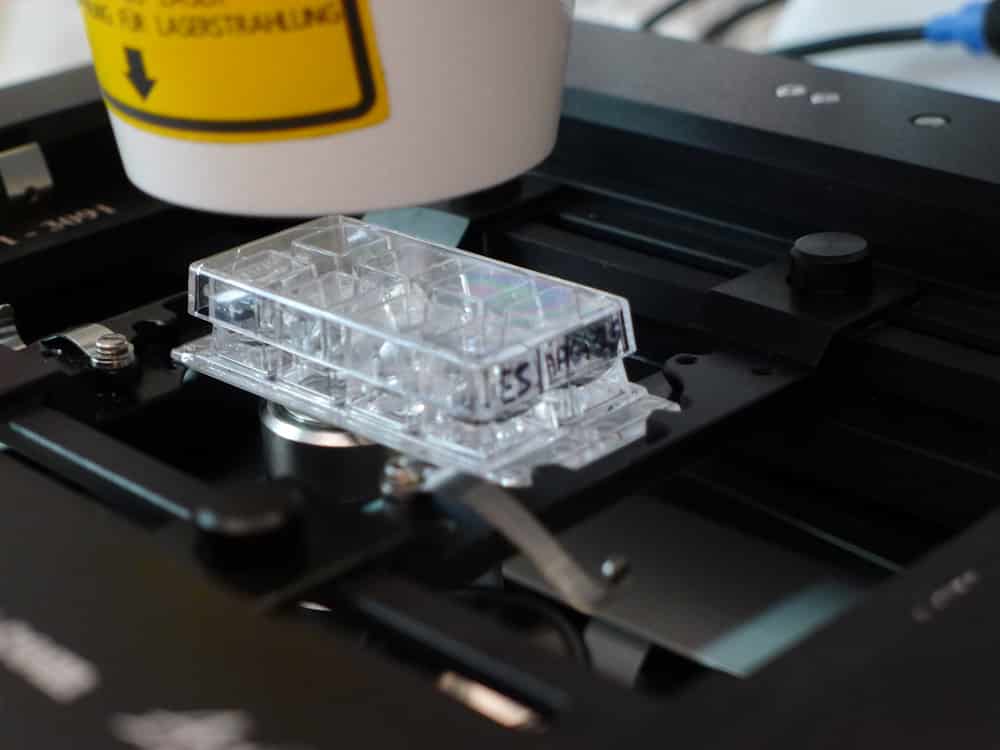
Enzyme-Linked Immunosorbent Assay
Encyclopedias as Topic
Immunoenzyme Techniques
Evaluation Studies as Topic
Color
Steroid regulation of retinol-binding protein in the ovine oviduct. (1/1855)
Two studies were conducted to identify retinol-binding protein (RBP) expression in the ovine oviduct and to determine the role of ovarian steroids in its regulation. Ewes were salpingectomized on Days 1, 5, or 10 of their respective estrous cycles, and oviducts were homogenized for RNA analysis, fixed for immunocytochemistry (ICC), or cultured for 24 h for protein analysis. ICC localized RBP to the epithelium of all oviducts. RBP synthesis was demonstrated by immunoprecipitation of radiolabeled RBP from the medium of oviductal explant cultures. Explant culture medium from oviducts harvested on Day 1 contained significantly more RBP than medium from oviducts collected on Days 5 or 10. Slot-blot analysis demonstrated that steady-state RBP mRNA levels were significantly higher on Day 1 than Day 5 or 10. In the second experiment, ovariectomized ewes were treated with estradiol-17beta (E2), progesterone (P4), E2+P4 (E2+P4), or vehicle control, and oviducts were analyzed as above. P4 alone or in combination with E2 significantly reduced steady-state RBP mRNA levels compared to those in E2-treated animals. Oviductal explants from E2- and E2+P4-treated animals released 3- to 5-fold more RBP into the medium than control and P4 treatments as determined by ELISA. RBP synthesis of metabolically labeled RBP was increased by E2 and E2+P4 treatments. This study demonstrates that P4 applied on an estradiol background negatively regulates RBP gene expression in the oviduct whereas estradiol appears to stimulate RBP synthesis and secretion. (+info)Serum sErbB1 and epidermal growth factor levels as tumor biomarkers in women with stage III or IV epithelial ovarian cancer. (2/1855)
Epithelial ovarian cancer (EOC) has a high mortality rate, which is due primarily to the fact that early clinical symptoms are vague and nonspecific; hence, this disease often goes undetected and untreated until in its advanced stages. Sensitive and reliable methods for detecting earlier stages of EOC are, therefore, urgently needed. Epidermal growth factor (EGF) is a ligand for EGF receptor (ErbB1); this receptor is the product of the c-erbB1 proto-oncogene. ErbB1 overexpression is common in human ovarian carcinoma-derived cell lines and tumors, in which overexpression is thought to play a critical role in tumor etiology and progression. Furthermore, ErbB1 overexpression is associated with disease recurrence and decreased patient survival. Recently, we have developed an acridinium-linked immunosorbent assay that detects a approximately 110-kDa soluble analogue of ErbB1, ie., sErbB1, in serum samples from healthy men and women (A. T. Baron, et al., J. Immunol. Methods, 219: 23-43, 1998). Here, we demonstrate that serum p110 sErbB1 levels are significantly lower in EOC patients with stage III or IV disease prior to (P < 0.0001) and shortly after (P < 0.0001) cytoreductive staging laparotomy than in healthy women of similar ages, whereas EGF levels are significantly higher than those of age-matched healthy women only in serum samples collected shortly after tumor debulking surgery (P < 0.0001). We observe that the preoperative serum sErbB1 concentration range of advanced stage EOC patients barely overlaps with the serum sErbB1 concentration range of healthy women. In addition, we show that serum sErbB1 and EGF levels changed temporally for some EOC patients who were surgically debulked of tumor and who provided a second serum sample during the course of combination chemotherapy. Finally, we observe a significant positive association between sErbB1 and EGF levels only in serum samples of EOC patients collected prior to cytoreductive surgery (correlation coefficient = 0.61968; P = 0.0027). These data suggest that epithelial ovarian tumors concomitantly affect serum sErbB1 and EGF levels. In conclusion, these data indicate that serum sErbB1 and EGF (postoperative only) levels are significantly different between EOC patients and healthy women and that altered and/or changing serum sErbB1 and EGF levels may provide important diagnostic and/or prognostic information useful for the management of patients with EOC. (+info)Differential serodiagnosis for cystic and alveolar echinococcosis using fractions of Echinococcus granulosus cyst fluid (antigen B) and E. multilocularis protoscolex (EM18). (3/1855)
Echinococcus granulosus cyst fluid and E. multilocularis protoscolex extract were fractionated by a single step of preparative isoelectric focusing, resulting in an antigen B-rich fraction (8-kD) and an Em18-rich fraction, respectively. The usefulness of both fractions for differential serodiagnosis of cystic (CE) and alveolar (AE) echinococcosis was evaluated by a large-scale immunoblot analysis on a battery of 354 serum samples. These included 66 from AE patients originating from four different endemic areas, 173 from CE patients originating from seven different endemic areas, 71 from patients with other parasitic diseases, 15 from patients with hepatomas, and 29 from healthy individuals. In an immunoblot with the antigen B-rich fraction, 92% (158 of 173) of the CE sera as well as 79% (52 of 66) of the AE sera reacted with the 8-kD subunit. No cross-reactivity occurred with any sera from patients with cysticercosis, other parasitic diseases, or with hepatomas, or from healthy controls. In an immunoblot with the Em18-rich fraction, all but two sera from AE patients (64 of 66, 97%) recognized Em18, and only nine of 34 CE sera from China reacted with it. All other (139) CE sera from six other countries were negative as were all (115) other non-echinococcosis sera. These findings indicate that antigen B (8-kD) is not species-specific for E. granulosus but is genus-specific for Echinococcus, and that the Em18 antigen is a reliable serologic marker for species-specific differentiation of AE from CE. (+info)Detection of haptoglobin in the high-density lipoprotein and the very high-density lipoprotein fractions from sera of calves with experimental pneumonia and cows with naturally occurring fatty liver. (4/1855)
In addition to the lipoprotein-deficient d > 1.25 fraction, haptoglobin was detected in the high-density lipoprotein (HDL) and the very high-density lipoprotein (VHDL) fractions from sera of calves with experimental pneumonia and cows with naturally occurring fatty liver. It was not found in the chylomicrons, very low-density lipoprotein and low-density lipoprotein fractions. Washing of the HDL fraction did not decrease the haptoglobin concentration. Transferrin and immunoglobulin G were immunoblotted to examine the possibility of contamination of the lipoprotein fractions by the d > 1.25 fraction. The two serum proteins were detected only in the d > 1.25 fraction, not in any lipoprotein fractions. The distribution pattern of haptoglobin in the lipoprotein fractions was distinct from that of serum albumin. Concentrations of haptoglobin in the HDL fractions from pneumonic sera were largely proportional to those in whole sera. Cholesteryl ester concentrations were decreased in sera from calves with pneumonia, as in cows with fatty liver. A protein immunologically related to hemoglobin was also detected in particular in the VHDL fractions from sera of both groups. These results suggest that haptoglobin or a complex with the hemoglobin-like protein may have a role or roles related to the lipid metabolism. (+info)Mutational analysis of apolipoprotein B mRNA editing enzyme (APOBEC1). structure-function relationships of RNA editing and dimerization. (5/1855)
APOBEC1 is the catalytic subunit of an enzyme complex that mediates apolipoprotein (apo) B mRNA editing. It dimerizes in vitro and requires complementation factor(s) for its editing activity. We have performed a systematic analysis of the structure-functional relationship of APOBEC1 by targeted mutagenesis of various sequence motifs within the protein. Using in vitro RNA editing assay, we found that basic amino acid clusters at the amino-terminal region R15R16R17 and R33K34, are essential for apoB mRNA editing. Mutation of R15R16R17 to K15K16K17 and mutation of R33K34 simultaneously to A33A34 almost completely abolished in vitro editing activity. The carboxy-terminal region of APOBEC1 contains a leucine-rich motif. Deletion analysis of this region indicates that residues 181 to 210 are important for in vitro apoB mRNA editing. Single amino acid substitutions demonstrate that L182, I185, and L189 are important residues required for normal editing function. Furthermore, the double mutant P190A/P191A also lost >90% of editing activity which suggests that a beta turn in this region of the molecule may be essential for proper functioning of APOBEC1. It was suggested that dimerization of APOBEC1 creates an active structure for deamination of apoB mRNA. When we examined the dimerization potential of truncated APOBEC1s using both amino and carboxy termini deletion mutants, we found that amino-terminal deletions up to residue A117 did not impair dimerization activity whereas carboxy-terminal deletions showed diminished dimerization. The systematic and extensive mutagenesis experiments in this study provide information on the role of various sequence motifs identified in APOBEC1 in enzyme catalysis and dimerization. (+info)Thyroid hormone induces activation of mitogen-activated protein kinase in cultured cells. (6/1855)
Thyroid hormone [L-thyroxine (T4)] rapidly induced phosphorylation and nuclear translocation (activation) of mitogen-activated protein kinase (MAPK) in HeLa and CV-1 cells in the absence of cytokine or growth factor. A pertussis toxin-sensitive and guanosine 5'-O-(3-thiotriphosphate)-sensitive cell surface mechanism responsive to T4 and agarose-T4, suggesting a G protein-coupled receptor, was implicated. Cells depleted of MAPK or treated with MAPK pathway inhibitors showed reduced activation of MAPK and of the signal transducer and activator of transcription STAT1alpha by T4; they also showed reduced T4 potentiation of the antiviral action of interferon-gamma (IFN-gamma). T4 treatment caused tyrosine-phosphorylated MAPK-STAT1alpha nuclear complex formation and enhanced Ser-727 phosphorylation of STAT1alpha, in the presence or absence of IFN-gamma. STAT1alpha-deficient cells transfected with STAT1alpha containing an alanine-for-serine substitution at residue 727 (STAT1alphaA727) showed minimal T4-stimulated STAT1alpha activation. IFN-gamma induced the antiviral state in cells containing wild-type STAT1alpha (STAT1alphawt) or STAT1alphaA727; T4 potentiated IFN-gamma action in STAT1alphawt cells but not in STAT1alphaA727 cells. T4-directed STAT1alpha Ser-727 phosphorylation is MAPK mediated and results in potentiated STAT1alpha activation and enhanced IFN-gamma activity. (+info)Transferrin stimulates iron absorption, exocytosis, and secretion in cultured intestinal cells. (7/1855)
The cellular mechanism by which basolateral transferrin (Tf) produces an increase in apical-to-basolateral Fe flux in Caco-2 cells was analyzed. After a pulse of 59Fe from the apical medium, three types of basolateral 59Fe efflux were found: a 59Fe efflux that was independent of the presence of Tf in the basolateral medium, a 59Fe efflux in which 59Fe left the cell bound to Tf, and a Tf-dependent 59Fe efflux in which 59Fe came off the cell not bound to Tf. Furthermore, addition of Tf to the basolateral medium doubled the exocytosis rate of Tf and increased the secretion of apolipoprotein A, a basolateral secretion marker. Both apotransferrin and Fe-containing Tf produced similar increases in 59Fe efflux, Tf exocytosis, and apolipoprotein A secretion. The Ca2+ channel inhibitor SKF-96365 inhibited both the Tf-mediated increase in transepithelial Fe transport and the secretion of apolipoprotein A. Thus the activation of transepithelial Fe transport by Tf seems to be mediated by Ca2+ entry into the cells. (+info)Involvement of protein serine and threonine phosphorylation in human sperm capacitation. (8/1855)
The involvement of serine and threonine phosphorylation in human sperm capacitation was investigated. Anti-phosphoserine monoclonal antibody (mAb) recognized six protein bands in the 43-55-kDa, 94 +/- 2-kDa, 110-kDa, and 190-kDa molecular regions, in addition to a faint band each in the 18-kDa and 35-kDa regions. Anti-phosphothreonine mAb recognized protein bands in six similar regions, except that the 18-kDa, 35-kDa, and 94 +/- 2-kDa protein bands were sharper and thicker, and an additional band was observed in the 110-kDa molecular region. In the 43-55-kDa molecular region, there was a well-characterized glycoprotein, designated fertilization antigen, that showed a further increase in serine/threonine phosphorylation after exposure to solubilized human zona pellucida. In a cell-free in vitro kinase assay carried out on beads or in solution, four to eight proteins belonging to similar molecular regions, namely 20 +/- 2 kDa, 43-55 kDa, 94 +/- 2 kDa, and 110 +/- 10 kDa, as well as in 80 +/- 4 and 210 +/- 10 kDa regions, were phosphorylated at dual residues (serine/tyrosine and threonine/tyrosine). Capacitation increased the intensity of serine/threonine phosphorylation per sperm cell, increased the number of sperm cells that were phosphorylated, and induced a subcellular shift in the serine/threonine-specific fluorescence. These findings indicate that protein serine/threonine phosphorylation is involved and may have a physiological role in sperm capacitation. (+info)Hemadsorption is a medical procedure that involves the use of a device to remove certain substances, such as toxic byproducts or excess amounts of cytokines (proteins involved in immune responses), from the bloodstream. This is accomplished by passing the patient's blood through an external filter or adsorbent column, which contains materials that selectively bind to the target molecules. The clean blood is then returned to the patient's circulation.
Hemadsorption can be used as a supportive treatment in various clinical scenarios, such as poisoning, sepsis, and other critical illnesses, where rapid removal of harmful substances from the bloodstream may help improve the patient's condition and outcomes. However, its effectiveness and safety are still subjects of ongoing research and debate.
Immunosorbent techniques are a group of laboratory methods used in immunology and clinical chemistry to isolate or detect specific proteins, antibodies, or antigens from a complex mixture. These techniques utilize the specific binding properties of antibodies or antigens to capture and concentrate target molecules.
The most common immunosorbent technique is the Enzyme-Linked Immunosorbent Assay (ELISA), which involves coating a solid surface with a capture antibody, allowing the sample to bind, washing away unbound material, and then detecting bound antigens or antibodies using an enzyme-conjugated detection reagent. The enzyme catalyzes a colorimetric reaction that can be measured and quantified, providing a sensitive and specific assay for the target molecule.
Other immunosorbent techniques include Radioimmunoassay (RIA), Immunofluorescence Assay (IFA), and Lateral Flow Immunoassay (LFIA). These methods have wide-ranging applications in research, diagnostics, and drug development.
An Enzyme-Linked Immunosorbent Assay (ELISA) is a type of analytical biochemistry assay used to detect and quantify the presence of a substance, typically a protein or peptide, in a liquid sample. It takes its name from the enzyme-linked antibodies used in the assay.
In an ELISA, the sample is added to a well containing a surface that has been treated to capture the target substance. If the target substance is present in the sample, it will bind to the surface. Next, an enzyme-linked antibody specific to the target substance is added. This antibody will bind to the captured target substance if it is present. After washing away any unbound material, a substrate for the enzyme is added. If the enzyme is present due to its linkage to the antibody, it will catalyze a reaction that produces a detectable signal, such as a color change or fluorescence. The intensity of this signal is proportional to the amount of target substance present in the sample, allowing for quantification.
ELISAs are widely used in research and clinical settings to detect and measure various substances, including hormones, viruses, and bacteria. They offer high sensitivity, specificity, and reproducibility, making them a reliable choice for many applications.
Plant pathology is a branch of science that deals with the study of diseases in plants caused by biotic (living) and abiotic (non-living) agents. Biotic agents include bacteria, fungi, viruses, viroids, nematodes, parasitic plants, and insects, while abiotic factors include environmental conditions such as drought, temperature extremes, nutrient deficiencies, and air pollution that can negatively impact plant health.
Plant pathologists aim to understand the causes of plant diseases, their symptoms, how they spread, and their impact on plants and crops. They also develop strategies for managing and controlling plant diseases through cultural practices, breeding resistant varieties, biological control, and chemical or genetic engineering approaches. The knowledge gained from plant pathology is essential for maintaining healthy plants, ensuring food security, and protecting the environment.
An encyclopedia is a comprehensive reference work containing articles on various topics, usually arranged in alphabetical order. In the context of medicine, a medical encyclopedia is a collection of articles that provide information about a wide range of medical topics, including diseases and conditions, treatments, tests, procedures, and anatomy and physiology. Medical encyclopedias may be published in print or electronic formats and are often used as a starting point for researching medical topics. They can provide reliable and accurate information on medical subjects, making them useful resources for healthcare professionals, students, and patients alike. Some well-known examples of medical encyclopedias include the Merck Manual and the Stedman's Medical Dictionary.
Immunoenzyme techniques are a group of laboratory methods used in immunology and clinical chemistry that combine the specificity of antibody-antigen reactions with the sensitivity and amplification capabilities of enzyme reactions. These techniques are primarily used for the detection, quantitation, or identification of various analytes (such as proteins, hormones, drugs, viruses, or bacteria) in biological samples.
In immunoenzyme techniques, an enzyme is linked to an antibody or antigen, creating a conjugate. This conjugate then interacts with the target analyte in the sample, forming an immune complex. The presence and amount of this immune complex can be visualized or measured by detecting the enzymatic activity associated with it.
There are several types of immunoenzyme techniques, including:
1. Enzyme-linked Immunosorbent Assay (ELISA): A widely used method for detecting and quantifying various analytes in a sample. In ELISA, an enzyme is attached to either the capture antibody or the detection antibody. After the immune complex formation, a substrate is added that reacts with the enzyme, producing a colored product that can be measured spectrophotometrically.
2. Immunoblotting (Western blot): A method used for detecting specific proteins in a complex mixture, such as a protein extract from cells or tissues. In this technique, proteins are separated by gel electrophoresis and transferred to a membrane, where they are probed with an enzyme-conjugated antibody directed against the target protein.
3. Immunohistochemistry (IHC): A method used for detecting specific antigens in tissue sections or cells. In IHC, an enzyme-conjugated primary or secondary antibody is applied to the sample, and the presence of the antigen is visualized using a chromogenic substrate that produces a colored product at the site of the antigen-antibody interaction.
4. Immunofluorescence (IF): A method used for detecting specific antigens in cells or tissues by employing fluorophore-conjugated antibodies. The presence of the antigen is visualized using a fluorescence microscope.
5. Enzyme-linked immunosorbent assay (ELISA): A method used for detecting and quantifying specific antigens or antibodies in liquid samples, such as serum or culture supernatants. In ELISA, an enzyme-conjugated detection antibody is added after the immune complex formation, and a substrate is added that reacts with the enzyme to produce a colored product that can be measured spectrophotometrically.
These techniques are widely used in research and diagnostic laboratories for various applications, including protein characterization, disease diagnosis, and monitoring treatment responses.
"Evaluation studies" is a broad term that refers to the systematic assessment or examination of a program, project, policy, intervention, or product. The goal of an evaluation study is to determine its merits, worth, and value by measuring its effects, efficiency, and impact. There are different types of evaluation studies, including formative evaluations (conducted during the development or implementation of a program to provide feedback for improvement), summative evaluations (conducted at the end of a program to determine its overall effectiveness), process evaluations (focusing on how a program is implemented and delivered), outcome evaluations (assessing the short-term and intermediate effects of a program), and impact evaluations (measuring the long-term and broad consequences of a program).
In medical contexts, evaluation studies are often used to assess the safety, efficacy, and cost-effectiveness of new treatments, interventions, or technologies. These studies can help healthcare providers make informed decisions about patient care, guide policymakers in developing evidence-based policies, and promote accountability and transparency in healthcare systems. Examples of evaluation studies in medicine include randomized controlled trials (RCTs) that compare the outcomes of a new treatment to those of a standard or placebo treatment, observational studies that examine the real-world effectiveness and safety of interventions, and economic evaluations that assess the costs and benefits of different healthcare options.
In the context of medical terminology, 'color' is not defined specifically with a unique meaning. Instead, it generally refers to the characteristic or appearance of something, particularly in relation to the color that a person may observe visually. For instance, doctors may describe the color of a patient's skin, eyes, hair, or bodily fluids to help diagnose medical conditions or monitor their progression.
For example, jaundice is a yellowing of the skin and whites of the eyes that can indicate liver problems, while cyanosis refers to a bluish discoloration of the skin and mucous membranes due to insufficient oxygen in the blood. Similarly, doctors may describe the color of stool or urine to help diagnose digestive or kidney issues.
Therefore, 'color' is not a medical term with a specific definition but rather a general term used to describe various visual characteristics of the body and bodily fluids that can provide important diagnostic clues for healthcare professionals.
Antibodies are proteins produced by the immune system in response to the presence of a foreign substance, such as a bacterium or virus. They are capable of identifying and binding to specific antigens (foreign substances) on the surface of these invaders, marking them for destruction by other immune cells. Antibodies are also known as immunoglobulins and come in several different types, including IgA, IgD, IgE, IgG, and IgM, each with a unique function in the immune response. They are composed of four polypeptide chains, two heavy chains and two light chains, that are held together by disulfide bonds. The variable regions of the heavy and light chains form the antigen-binding site, which is specific to a particular antigen.
 Immunosorbent Techniques (medical concept explorer)
 The Different Types of Immunostaining
 Detection of Plasmodium Aldolase Using a Smartphone and Microfluidic Enzyme Linked Immunosorbent Assay
 ELISA) Enzyme-Linked Immunosorbent Assay
 Immunoadsorption therapy for dilated cardiomyopathy using tryptophan column-A prospective, multicenter, randomized, within...
 ELISA - Wikipedia
 Human bocavirus (HBoV) in children with respiratory tract infection by enzyme linked immunosorbent assay (ELISA) and...
 Confirmation of an Enzyme-Linked Immunosorbent Assay to Detect Fluometuron in Soil1
 IJMS | Free Full-Text | Advances in Proteomic Techniques for Cytokine Analysis: Focus on Melanoma Research
 Protein Analysis Techniques - Bio-Rad
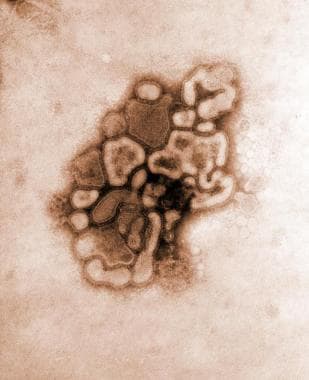
 Pediatric Influenza: Practice Essentials, Background, Pathophysiology
 Renal Biomarker Market Outlook Highlights Major Opportunities by 2020 - PharmiWeb.com
 Medical Dictionary, Dictionary of medicine and human biology, medical, biological and chemical terminology
 Advanced Search Results - Public Health Image Library(PHIL)
 Enzyme-linked Immunosorbent Assays (ELISA): Recent Innovations Take Analyte Detection To New Levels - Drug Discovery World (DDW)
 A placebo-controlled, dose-ranging study of a growth hormone releasing factor in HIV-infected patients with abdominal fat...
 Toxicidade pulmonar da radioterapia conformacional torácica em mulheres com câncer...
 Prediction of malaria transmission drivers in Anopheles mosquitoes using artificial intelligence coupled to MALDI-TOF mass...
 Thieme E-Journals - Hormone and Metabolic Research / Abstract
 High Incidence of Pulmonary Tuberculosis in ART Naive Remunerated Blood Donors with Human Immunodeficiency Virus Type-1...
 Plus it
 Risk factors for the recurrence of relapsing polychondritis | Arthritis Research & Therapy