Hypothalamic Neoplasms
Hypercalcemia in an euthyroid patient with secondary hypoadrenalism and diabetes insipidus due to hypothalamic tumor. (1/47)
A 20-year-old Japanese man with a hypothalamic tumor (most likely germ-cell tumor) which caused secondary hypoadrenalism, hypogonadism and diabetes insipidus developed hypercalcemia and acute renal failure. The serum levels of intact PTH (iPTH), PTH-related protein (PTH-rP), 1,25-dihydroxy vitamin D (1,25- (OH)2 D), ACTH, cortisol, gonadotropins and testosterone were decreased, but his serum levels of triiodothyronine (T3) and thyroxine (T4) were within the normal range at admission, with depressed TSH and slightly increased thyroglobulin. The hypercalcemia was refractory to extensive hydration and calcitonin, but was ameliorated by pamidronate. After irradiation of the hypothalamic tumor, panhypopituitarism gradually developed. The patient has been normocalcemic for the last 2 years and is doing well under replacement therapy with glucocorticoid, L-thyroxine, methyltestosterone and 1-desamino D arginine vasopressin (dDAVP). As to the mechanism of euthyroidism at admission, transient destructive thyroiditis associated with hypopituitarism or delayed development of hypothyroidism following the hypoadrenalism was suggested. This is the first reported case of hypercalcemia in secondary hypoadrenalism due to hypothalamic tumor. Hypercalcemia was most likely induced by increased bone resorption, which was probably elicited by the combined effects of deficient glucocorticoid and sufficient thyroid hormones in addition to hypovolemia and reduced renal calcium excretion. Furthermore, severe dehydration due to diabetes insipidus and disturbance of thirst sensation caused by the hypothalamic tumor aggravated the hypercalcemia, leading to acute renal failure. (+info)Hypercalcemia accompanied by hypothalamic hypopituitarism, central diabetes inspidus and hyperthyroidism. (2/47)
We present here a case of prominent hypercalcemia accompanied by hypothalamic tumor and Graves' disease. A 24-year-old man with hypothalamic tumor showed hypopituitarism, central diabetes inspidus (DI) and hyperthyroidism. Nausea, loss of thirst and appetite, and general fatigue were found with the unveiling of hypercalcemia and hypernatremia. Parathyroid hormone (PTH) and 1alpha-dihydroxyvitamin D levels were suppressed with a normal range of PTH-related protein values. One-desamino-(8-D-arginine)-vasopressin (DDAVP) and half-saline administration normalized hypernatremia, while hypercalcemia was still sustained. Administration of cortisone acetate and thiamazole reduced the elevated serum Ca level. In the present case, concurrent hyperthyroidism was assumed to accelerate skeletal mobilization of calcium into the circulation. Hypocortisolism and central DI was also considered to contribute, to some extent, to the hypercalcemia through renal handling of Ca. (+info)Chordoid glioma: a neoplasm unique to the hypothalamus and anterior third ventricle. (3/47)
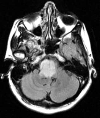
BACKGROUND AND PURPOSE: Chordoid glioma is a new clinicopathologic entity that occurs in the region of the hypothalamus/anterior third ventricle. The aims of this study were to describe the characteristic radiographic features of chordoid glioma, identify specific imaging features that may enable differentiation of chordoid glioma from other suprasellar tumors, and increase neuroradiologists' awareness of this newly described tumor, facilitating prospective diagnosis. METHODS: CT scans and/or MR images of six patients with chordoid glioma were reviewed retrospectively to determine whether any characteristic radiographic features would emerge. Reports of the clinical presentation, pathologic findings, and radiographic findings of another six patients were reviewed and included, for a total patient population of 12 (mean age +/- SD, 46 +/- 13 years). RESULTS: Imaging features were strikingly similar for all tumors. In each case, the mass was ovoid, was well circumscribed, was located in the region of the hypothalamus/anterior third ventricle, and enhanced uniformly and intensely. Tumors were hyperdense to gray matter on CT scans and were isointense on T1-weighted MR images and slightly hyperintense on long-TR MR images. In two patients, vasogenic edema extended into the optic tracts, and in three, there was hydrocephalus. CONCLUSION: Chordoid glioma is a recently described unique histopathologic entity that has been added to the World Health Organization glioma classification scheme and must be included in the differential diagnosis of a suprasellar mass. Distinctive imaging features are its location, ovoid shape, hyperdensity on CT scans, and uniform intense contrast enhancement. (+info)Combined intraarterial carboplatin, intraarterial etoposide phosphate, and IV Cytoxan chemotherapy for progressive optic-hypothalamic gliomas in young children. (4/47)
BACKGROUND AND PURPOSE: Optic pathway and/or hypothalamic astrocytomas in children are often quiescent, but in some cases, more aggressive tumors may cause progressive visual, endocrine, and neurologic deterioration. The initial treatment of these gliomas includes surgery and IV chemotherapy. Radiotherapy is not recommended in young children because of its severe adverse effects on cognitive and neuroendocrine function. This report suggests a new approach using combined intraarterial and IV carboplatin-based chemotherapy for patients for whom first line treatment has already failed. METHODS: Six children (mean age, 57 months) with the diagnosis of optic pathway hypothalamic gliomas, who had tumor progression after surgery and underwent IV chemotherapy, were treated monthly with intraarterially administered carboplatin, intraarterially administered etoposide phosphate, and IV administered Cytoxan. Four of the children had histologically verified pilocytic astrocytomas, and in two cases, diagnosis was made on the basis of clinical findings. Administration of the intraarterial chemotherapy required catheter placement in both internal carotid arteries at the level of C2-C3 and into one of the vertebral arteries at the level of C6-C7, with the patient under general anesthesia. RESULTS: Four of six patients had partial radiographic response, one had stable disease, and one had progressive disease after one cycle. Three patients showed clinical improvement. There were no serious complications associated with the angiographic procedures. Toxicities included bronchospasm that resolved after 3 to 4 minutes in one patient. One patient showed mild ototoxicity, and four patients needed platelet transfusion because of hematologic toxicity of drugs. CONCLUSION: These results suggest that this modality of chemotherapy (administered after failure of systemic [ie, IV] chemotherapy), of progressive optic-hypothalamic astrocytomas in young children may be an effective treatment prior to radiotherapy. (+info)Management of hyponatraemia in patients with acute cerebral insults. (5/47)
Hyponatraemia is a common finding in patients with acute cerebral insults. The main differential diagnosis is between syndrome of inappropriate ADH secretion and cerebral salt wasting. Our aim is to review the topic of hyponatraemia in patients with acute cerebral insults and suggest a clinical approach to diagnosis and management. (+info)Heavily T2-weighted MR imaging of white matter tracts in the hypothalamus: normal and pathologic demonstrations. (6/47)
BACKGROUND AND PURPOSE: The MR appearance of white matter tracts in the hypothalamus and the role of the hypothalamus as a memory mechanism have not been sufficiently described in clinical settings. Heavily T2-weighted black-and-white reversed (T2R) images were assessed to reveal their visualization and clinical significance. METHODS: One hundred healthy subjects and three patients with hypothalamic lesions underwent fast spin-echo MR imaging to reveal the postcommissural fornix (PF) and mammillothalamic tract (MT). RESULTS: The PF was identifiable in axial and/or coronal sections in all healthy subjects. No remarkable asymmetry of its size or course was evident. Both anteroposterior and vertical dimensions ranged from 10.5 to 14 mm. The MT was visible in one or two axial sections above the mammillary body in 64% of healthy subjects and in a coronal section in 36%. Two patients with glioblastoma multiforme and lacunar infarct at the hypothalamus presented with anterograde amnesia; T2R imaging revealed involvement of both the PF and MT. The third patient had a suprasellar craniopharyngioma with PF injury sparing the MT resulting from surgical manipulation and was free of memory deficit. Anterograde amnesia was evident only when both the PF and MT were injured. CONCLUSION: T2R images have made a high rate of detection of the PF and MT possible and could provide a more detailed correlation of hypothalamic neuroanatomy and memory mechanism in clinical settings. (+info)Hypodipsic hypernatremia with intact AVP response to non-osmotic stimuli induced by hypothalamic tumor: a case report. (7/47)
Anatomical lesions of hypothalamic area associated with hypodipsic hypernatremia have been reported only rarely. We report here a case of hypodipsic hypernatremia induced by a hypothalamic lesion. A 25-yr-old man, who had been treated with radiation for hypothalamic tumor 5-yr before, was admitted for evaluation of hypernatremia and hypokalemia. He never felt thirst despite the elevated plasma osmolality and usually refused to drink intentionally. Plasma arginine vasopressin (AVP) level was normal despite the severe hypernatremic hyperosmolar state and urine was not properly concentrated, while AVP secretion was rapidly induced by water deprivation and urine osmolality also progressively increased to the near maximum concentration range. All of these findings were consistent with an isolated defect in osmoregulation of thirst, which was considered as the cause of chronic hypernatremia in the patient without an absolute deficiency in AVP secretion. Hypokalemia could be induced by activation of the renin-angiotensin-aldosterone system as a result of volume depletion. However, inappropriately low values of plasma aldosterone levels despite high plasma renin activity could not induce symptomatic hypokalemia and metabolic alkalosis. The relatively low serum aldosterone levels compared with high plasma renin activity might result from hypernatremia. Hypernatremia and hypokalemia were gradually corrected by intentional water intake only. (+info)Optic pathway glioma: correlation of imaging findings with the presence of neurofibromatosis. (8/47)
BACKGROUND AND PURPOSE: Despite the benign histology of optic pathway glioma (OPG) (low-grade astrocytoma), its biological behavior is unpredictable, and it is unclear whether specific morphologic or anatomic patterns may be predictive of prognosis. It is also unclear whether OPG associated with neurofibromatosis (NF) is a distinct entity from non-NF-OPG. Our purpose was to describe the MR imaging features of OPG, compare the findings between patients with and those without NF, and identify prognostic imaging signs. METHODS: MR examinations of 91 patients with OPG (47 with NF and 44 without) were reviewed at presentation and during follow-up. The images were evaluated for size and extension of tumor, and imaging parameters. Statistical bivariate analysis was used to compare the patients with and those without NF, and Pearson correlation was used to evaluate the correlation between the different imaging parameters and prognosis. Kappa values were calculated to determine intraobserver and interobserver variability. RESULTS: The most common site of involvement in the NF group was the orbital nerve (66%), followed by the chiasm (62%). In the non-NF group, the chiasm was the most common site of involvement (91%); the orbital nerves were involved in only 32%. Extension beyond the optic pathway at diagnosis was uncommon in the NF group (2%) but frequent in the non-NF group (68%). In the NF group, the tumor was smaller and the original shape of the optic pathways was preserved (91% vs. 27% in the non-NF group). The presence of cystic components was significantly more common in the non-NF patients (66% vs. 9% in the NF group). During follow-up, half the NF patients remained stable, in contrast to 5% of the non-NF group. No statistical correlation was found between imaging features and biological behavior of the tumor. CONCLUSION: NF-OPG is a separate entity from non-NF-OPG, with different imaging features and prognosis, thereby warranting a specific diagnostic, clinical, and therapeutic approach. (+info)Hypothalamic neoplasms refer to tumors that originate in the hypothalamus, a small region of the brain that is located at the base of the brain and forms part of the limbic system. The hypothalamus plays a critical role in regulating many bodily functions, including hormone release, temperature regulation, hunger, thirst, sleep, and emotional behavior.
Hypothalamic neoplasms can be benign or malignant and can arise from various cell types within the hypothalamus, such as neurons, glial cells, or supportive tissue. These tumors can cause a variety of symptoms depending on their size, location, and rate of growth. Common symptoms include endocrine disorders (such as diabetes insipidus or precocious puberty), visual disturbances, headaches, behavioral changes, and cognitive impairment.
The diagnosis of hypothalamic neoplasms typically involves a combination of clinical evaluation, imaging studies (such as MRI or CT scans), and sometimes biopsy or surgical removal of the tumor. Treatment options depend on the type, size, and location of the tumor but may include surgery, radiation therapy, chemotherapy, or a combination of these approaches. Regular follow-up care is essential to monitor for recurrence or progression of the tumor.
 Brainstem Gliomas: Practice Essentials, Background, Pathophysiology
 ATSDR - Oak Ridge Reservation - ORRHES Meeting Minutes
 International Classification of Headache Disorders - Wikipedia
 Combined strategy of maximal endoscopic endonasal resection and early radiation therapy for complex cystic and solid...
 MeSH Browser
 Neurofibromatosis 1 and intracranial neoplasms of childhood | MedLink Neurology
 Human Physiology/The female reproductive system - Wikibooks, open books for an open world
 Hypothalamic-Pituitary-Thyroid Axis | Leaders in Pharmaceutical Business Intelligence (LPBI) Group
 Ambien Cr Where To Buy - eostone.com
 49 Back Pain Articles Articles
 Coristoma/metabolismo
 Testosterone LH and FSH Lab Tests
 Postpartum Luteinizing Hormone Release and Maternal Behavior in the Rat after Late-Gestational Depletion of Hypothalamic...
 Severe hypernatremia during postoperative care in patients with craniopharyngioma in: Endocrine Connections Volume 12 Issue 12 ...
 Sexual dysfunctions
 Malgorzata Karbownik-Lewinska - NeL.edu
 BVS Brasil