Major Histocompatibility Complex
Histocompatibility Antigens Class II
Histocompatibility Antigens Class I
Histocompatibility Antigens
Minor Histocompatibility Antigens
Genes, MHC Class II
Histocompatibility
Genes, MHC Class I
Minor Histocompatibility Loci
HLA Antigens
T-Lymphocytes
Antigen Presentation
Histocompatibility Testing
T-Lymphocytes, Cytotoxic
HLA-DR Antigens
HLA-D Antigens
beta 2-Microglobulin
Histocompatibility Antigen H-2D
Molecular Sequence Data
Mice, Inbred Strains
Mice, Inbred C57BL
Amino Acid Sequence
Cytotoxicity, Immunologic
Lymphocyte Activation
Peptides
Antigen-Presenting Cells
Receptors, Antigen, T-Cell
Mice, Inbred BALB C
Interferon-gamma
HLA-B Antigens
HLA-A Antigens
Isoantigens
CD8-Positive T-Lymphocytes
CD4-Positive T-Lymphocytes
Antigens, Differentiation, B-Lymphocyte
Alleles
Immunogenetics
Antigens, CD8
Flow Cytometry
Dendritic Cells
Skin Transplantation
Base Sequence
H-Y Antigen
B-Lymphocytes
HLA-DQ Antigens
HLA-A2 Antigen
Transplantation, Homologous
Clone Cells
HLA-B7 Antigen
HLA-C Antigens
Cells, Cultured
Killer Cells, Natural
Lymphocyte Culture Test, Mixed
Mice, Inbred C3H
Antigens, Surface
HLA-DR1 Antigen
Isoantibodies
Receptors, Antigen, T-Cell, alpha-beta
Thymus Gland
Peptide Fragments
Haplotypes
Immune Tolerance
Hybridomas
Mice, Transgenic
Epitopes, T-Lymphocyte
Graft Rejection
Histocompatibility, Maternal-Fetal
Cytotoxicity Tests, Immunologic
Superantigens
Transplantation Immunology
Antigens, CD
Antigens, CD4
Protein Binding
HLA-DR alpha-Chains
Mice, Inbred CBA
Transfection
HLA-DP Antigens
HLA-DRB1 Chains
Immunity, Cellular
Mice, Inbred DBA
Graft vs Host Reaction
HLA-G Antigens
Graft vs Host Disease
Lymphocytes
Tumor Cells, Cultured
T-Lymphocyte Subsets
Radiation Chimera
Immunodominant Epitopes
Antigens, Neoplasm
Receptors, Immunologic
HLA-DR3 Antigen
L Cells (Cell Line)
HLA-B27 Antigen
Polymorphism, Genetic
Gene Expression Regulation
Species Specificity
Rats, Inbred Lew
Antigens, Differentiation, T-Lymphocyte
Bone Marrow Transplantation
Interleukin-2
T-Lymphocytes, Helper-Inducer
HLA-A1 Antigen
Mice, Inbred A
Antigens, CD80
Genetic Linkage
Genes
Chromosomes, Human, Pair 6
Antibody Formation
HLA-DR4 Antigen
Immunization
Mice, Inbred AKR
HLA-B8 Antigen
Chromosome Mapping
Antigens, Ly
Cytokines
Phenotype
Macrophages
HLA-B40 Antigen
HLA-DR2 Antigen
Cloning, Molecular
Autoimmune Diseases
RNA, Messenger
Crosses, Genetic
Enterotoxins
HLA-DR beta-Chains
Macromolecular Substances
Antigens, CD3
Ligands
ATP-Binding Cassette Transporters
Antigens, CD1
Graft Survival
Antigens, CD86
Cysteine Endopeptidases
Binding Sites
Down-Regulation
Proteasome Endopeptidase Complex
DNA
Autoantigens
Gene Products, nef
Calnexin
Polymerase Chain Reaction
Endoplasmic Reticulum
Receptors, Natural Killer Cell
Protein Conformation
Recombinant Fusion Proteins
Mice, Knockout
Complement C4a
Cell Membrane
nef Gene Products, Human Immunodeficiency Virus
Lectins, C-Type
Lymph Nodes
Immunophenotyping
Cross Reactions
Gene Expression
Cell Differentiation
Epitope Mapping
Muramidase
Autoimmunity
Immunologic Memory
Cell Line, Transformed
Graft vs Leukemia Effect
Mutation
Endocytosis
Fluorescent Antibody Technique
Disease Susceptibility
Receptors, KIR
Genetic Predisposition to Disease
Cytomegalovirus
Models, Molecular
Mice, Inbred NOD
Promoter Regions, Genetic
Receptors, NK Cell Lectin-Like
Myelin Basic Protein
Diabetes Mellitus, Type 1
Models, Immunological
DNA Primers
Membrane Proteins
Genotype
Adenovirus E3 Proteins
Antigen-Antibody Reactions
HLA-B35 Antigen
Macaca mulatta
Standardized nomenclature for inbred strains of mice: sixth listing. (1/714)
Rules for designating inbred strains of mice are presented, along with a list of strains with their origins and characteristics, a table of biochemical polymorphisms, and standard subline designations. (+info)Inhibition of allorecognition by a human class II MHC-derived peptide through the induction of apoptosis. (2/714)
The interaction of the T-cell receptor with the major histocomatibility complex (MHC)-peptide complex is central to T-cell activation. Variation in the nature of the peptide bound within the groove of the MHC molecule may result in an altered T-cell response. Because some naturally processed peptides bound within the groove of the class II MHC molecule are derived from the MHC molecules themselves, we studied the inhibitory effects of synthetic class II MHC peptides on alloimmune responses in vitro. Three peptides derived from a highly conserved region of the class II MHC alpha chains inhibited the rat mixed lymphocyte response (MLR) in a dose-dependent manner, with the human HLA-DQA1 peptide also inhibiting the human and mouse MLR. No effect was seen on mitogen-induced T-cell proliferation. HLA-DQA1 inhibited cytolytic T lymphocyte (CTL) generation in a dose-response fashion, with no reduction in preformed CTL killing, suggesting that the inhibitory effect is targeted at CD4(+) T-cell function. Cell-cycle analysis by flow cytometry showed that restimulation of primed T cells in the presence of HLA-DQA1 resulted in increased apoptosis, whereas unstimulated cells were not affected. These data demonstrate that synthetic peptides derived from highly conserved regions of the class II MHC alpha chain can alter CD4(+) T-lymphocyte alloimmune responses in vitro, and this effect is mediated by the induction of apoptosis in activated T cells. (+info)B cell lymphoproliferative disorders following hematopoietic stem cell transplantation: risk factors, treatment and outcome. (3/714)
Twenty-six cases of B cell lymphoproliferative disorder (BLPD) were identified among 2395 patients following hematopoietic stem cell transplants (HSCT) for which an overall incidence of BLPD was 1.2%. The true incidence was probably higher, since 9/26 of the diagnoses were made at autopsy. No BLPD was observed following autologous HSCT, so risk factor analyses were confined to the 1542 allogeneic HSCT. Factors assessed were HLA-mismatching (> or = 1 antigen), T cell depletion (TCD), presence of acute GvHD (grades II-IV), donor type (related vs unrelated), age of recipient and donor, and underlying disease. Factors found to be statistically significant included patients transplanted for immune deficiency and CML, donor age > or = 18 years, TCD, and HLA-mismatching, with recipients of combined TCD and HLA-mismatched grafts having the highest incidence. Factors found to be statistically significant in a multiple regression analysis were TCD, donor age and immune deficiency, although 7/8 of the patients with immunodeficiencies and BLPD received a TCD graft from a haploidentical parent. The overall mortality was 92% (24/26). One patient had a spontaneous remission, but subsequently died >1 year later of chronic GVHD. Thirteen patients received therapy for BLPD. Three patients received lymphocyte infusions without response. The only patients with responses and longterm survival received alpha interferon (alphaIFN). Of seven patients treated with alphaIFN there were four responses (one partial and three complete). These data demonstrate that alphaIFN can be an effective agent against BLPD following HSCT, if a timely diagnosis is made. (+info)Feasibility of finding an unrelated bone marrow donor on international registries for New Zealand patients. (4/714)
Allogeneic bone marrow transplantation is the treatment of choice for several hematological conditions. Unfortunately, for the majority (70%) of patients an HLA-matched sibling donor is not available and a matched unrelated donor must be found if they are to proceed to allogeneic transplantation. Most of the donors on international registries are of Caucasian ethnic origin. It has been recognized that patients from certain racial groups have a reduced chance of finding an unrelated donor. This study reports the feasibility of finding an unrelated donor for our local New Zealand patients of Caucasian, New Zealand Maori and Pacific Islander ethnic origin presenting with transplantable hematological conditions at a single center. The search was performed on international registries using HLA-A,B and DR typings for our patients. Six of six and five of six matches were evaluated. We have shown that Maori and Pacific Islanders have significantly lower hit rates than Caucasians when searched for 6/6 antigen matches, but there was no significant difference between the three ethnic groups in finding a 5/6 antigen matched donor. This study supports the policy of the New Zealand Bone Marrow Donor Registry in recruiting New Zealand Maori and Pacific Islanders. (+info)Increased risk of chronic graft-versus-host disease, obstructive bronchiolitis, and alopecia with busulfan versus total body irradiation: long-term results of a randomized trial in allogeneic marrow recipients with leukemia. Nordic Bone Marrow Transplantation Group. (5/714)
Leukemic patients receiving marrow from HLA-identical sibling donors were randomized to treatment with either busulfan 16 mg/kg (n = 88) or total body irradiation ([TBI] n = 79) in addition to cyclophosphamide 120 mg/kg. The patients were observed for a period of 5 to 9 years. Busulfan-treated patients had an increased risk of veno-occlusive disease (VOD) of the liver (12% v 1%, P =.01) and hemorrhagic cystitis (32% v 10%, P =.003). Acute graft-versus-host disease (GVHD) was similar in the two groups, but the 7-year cumulative incidence of chronic GVHD was 59% in the busulfan-treated group versus 47% in the TBI group (P =.05). Death from GVHD was more common in the busulfan group (22% v 3%, P <.001). Obstructive bronchiolitis occurred in 26% of the busulfan patients but in only 5% of the TBI patients (P <.01). Complete alopecia developed in 8 busulfan patients and partial alopecia in 17, versus five with partial alopecia in the TBI group (P <.001). Cataracts occurred in 5 busulfan-treated patients and 16 TBI patients (P =.02). The incidence of relapse after 7 years was 29% in both groups. Seven-year transplant-related mortality (TRM) in patients with early disease was 21% in the busulfan group and 12% in the TBI group. In patients with more advanced disease, the corresponding figures were 64% and 22%, respectively (P =.004). Leukemia-free survival (LFS) in patients with early disease was 68% in busulfan-treated patients and 66% in TBI patients. However, 7-year LFS in patients with more advanced disease was 17% in the busulfan group versus 49% in the TBI group (P <.01). In patients with chronic myeloid leukemia (CML) in first chronic phase, 7-year LFS was 72% and 83% in the two groups, respectively. (+info)Transplantation of anergic histoincompatible bone marrow allografts. (6/714)
BACKGROUND: Successful allogeneic bone marrow transplantation relies on global immunosuppression or elimination of T cells. In contrast, the induction of anergy can inactivate specific sets of alloreactive T cells in the donor marrow. Previous work has shown that anergy can be induced by blocking the interaction of the B7 molecule on the surface of antigen-presenting cells with the CD28 molecule on the surface of T cells, thus preventing key signaling events essential for the activation of T cells. To investigate the feasibility of this approach with respect to transplantation of histoincompatible bone marrow, we undertook a clinical trial of ex vivo induction of anergy in T cells present in donor marrow to recipient alloantigens. METHODS: Outcomes in 12 transplant recipients were evaluated. The recipients' peripheral-blood lymphocytes were collected before myeloablation and served as alloantigen-presenting cells. To induce alloantigen-specific anergy, bone marrow from a donor mismatched with the recipient for one HLA haplotype was cocultured with irradiated cells from the recipient for 36 hours in the presence of CTLA-4-Ig, an agent that inhibits B7:CD28-mediated costimulation. After conventional myeloablation and immunoprophylaxis, the treated donor cells were transfused into the recipient. RESULTS: After the induction of anergy, the frequency of T cells capable of recognizing alloantigens of the recipient in donor marrow was sharply reduced (P<0.001), whereas the responsiveness to alloantigens from persons unrelated to the recipient or the donor was unaffected (P=0.51). In the 11 patients who could be evaluated, the haploidentical bone marrow cells engrafted. Of these 11 patients, 3 had acute graft-versus-host disease (GVHD) confined to the gastrointestinal tract. No deaths were attributable to GVHD. Five of the 12 patients were alive and in remission 4.5 to 29 months after transplantation. CONCLUSIONS: Donor bone marrow treated ex vivo to induce anergy to alloantigens from the recipient can reconstitute hematopoiesis in vivo with a relatively low risk of GVHD. (+info)HLA-C and HLA-DQB1 compatibility in unrelated cord blood transplants. (7/714)
BACKGROUND AND OBJECTIVE: Umbilical cord blood (UCB) cells have been definitively proved to be a source of hematopoietic stem cells with repopulating capacity when transplanted into pediatric hosts with neoplastic or non-neoplastic disease. Moreover, due to the immaturity of the UCB lymphoid compartment, these transplants are usually associated with a low incidence and severity of GvHD. This clinical observation and the immaturity of the UCB lymphoid compartment justify the acceptance of UCB units which differ from their recipient by 1 or 2 HLA antigens of the six HLA A, B and DRB1 antigens conventionally typed. Whether the number and type of HLA disparities affect clinical outcome of UCB transplants has not, however, been clearly demonstrated yet. DESIGN AND METHODS: In the present study on 14 pediatric patients with high risk leukemia transplanted with UCB from unrelated donors, evaluation of HLA compatibility was extended to HLA-C and DQB1 genes and correlated to the engraftment rate and occurrence of GvHD. Conditioning regimen and GvHD prophylaxis were identical in all cases. HLA-A and B antigens were typed by serology, whereas DNA based methods were used to define HLA-C gene groups, and HLA-DRB1 and DQB1 alleles. RESULTS: Conventional HLA-A, B and DRB1 typing demonstrated that 12 recipient/donor pairs differed at one HLA locus, while 2 pairs had 2 HLA disparities. The extended HLA-typing showed that only one out of the six pairs with a different HLA-A locus had additional mismatches at HLA-C and DQB1 loci, whereas all the remaining 8 pairs, which already differed at HLA-B and/or DRB1 loci after conventional typing, had additional HLA-C and/or DQB1 mismatches (p = 0.002). By contrast, engraftment rate and occurrence of GvHD did not significantly correlate with level of HLA-mismatches even after extended HLA-typing. INTERPRETATION AND CONCLUSIONS: The present data show that additional mismatched HLA-C and/or DQB1 antigens are significantly more frequent in pairs which after conventional HLA-typing differed at HLA-B and/or DRB1 loci, than in those showing one HLA-A mismatch. This observation provides an additional criterion for selection of UCB donors with the closest HLA-match when more than one unit are available. We did not, however, observe any correlation between engraftment rate, occurrence of GvHD and degree of HLA disparities detected either by standard or extended typing. These data support the notion that certain HLA differences do not affect the clinical outcome of UCB transplants and indicate that the expensive and time consuming molecular typing of HLA-C and DQB1 loci might be avoided for UCB donor selection. (+info)Rapamycin reverses chronic graft vascular disease in a novel cardiac allograft model. (8/714)
BACKGROUND: Chronic graft vascular disease (CGVD) in cardiac allografts has been defined as a slowly evolving vasculopathy unresponsive to conventional immunosuppression. We compared 4 rodent models of CGVD to evaluate the reproducibility of CGVD in heart allografts. Rapamycin (Rapa) and cyclosporine (CSA) were then used to treat CGVD. METHODS AND RESULTS: Hearts were harvested and placed heterotopically into allogenic recipients. CGVD scores of PVG allografts from ACI recipients treated with CSA on days 1 through 10 were significantly elevated on day 90 (n=16) compared with other models (immunosuppression used): (1) Lewis to F344 recipients (CSA), (2) Brown Norway to Lewis (FK506), and (3) DA to Wistar-Firth (methylprednisolone, azathioprine, CSA). Although delayed (day 60 to 90) CSA treatment had no effect (n=6), delayed Rapa (3 mg. kg-1. d-1 IP) reversed CGVD in PVG grafts (0.22+/-0.19 on day 90, n=6). ACI isografts showed no evidence of CGVD (n=6) at day 90. Immunohistochemistry of PVG grafts revealed perivascular infiltrates consisting of CD4(+) T cells and limited numbers of macrophages persisting up to day 90. Flow cytometry demonstrated increased levels of anti-donor antibody at day 90, which was significantly inhibited by Rapa treatment. CONCLUSIONS: PVG grafts developed a significant increase in CGVD without evidence of ongoing myocardial rejection. This CGVD appeared to be mediated by both cellular and humoral mechanisms, given CD4(+) perivascular infiltrates and increased levels of anti-donor antibody. The anti-CGVD effectiveness of Rapa during a period in which there was little myocardial cellular infiltrate supports a novel mechanism of effect such as smooth muscle or B-cell inhibition. (+info)The Major Histocompatibility Complex (MHC) is a group of cell surface proteins in vertebrates that play a central role in the adaptive immune system. They are responsible for presenting peptide antigens to T-cells, which helps the immune system distinguish between self and non-self. The MHC is divided into two classes:
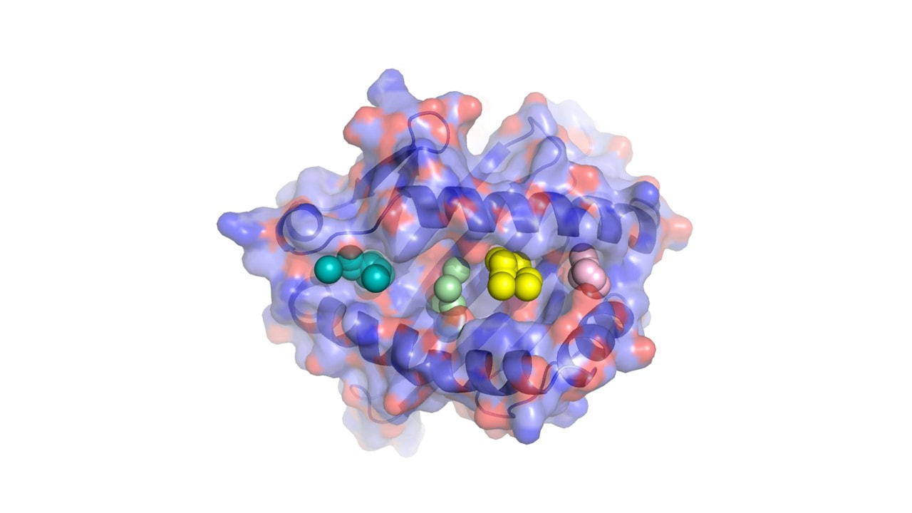
1. MHC Class I: These proteins present endogenous (intracellular) peptides to CD8+ T-cells (cytotoxic T-cells). The MHC class I molecule consists of a heavy chain and a light chain, together with an antigenic peptide.
2. MHC Class II: These proteins present exogenous (extracellular) peptides to CD4+ T-cells (helper T-cells). The MHC class II molecule is composed of two heavy chains and two light chains, together with an antigenic peptide.
MHC genes are highly polymorphic, meaning there are many different alleles within a population. This diversity allows for better recognition and presentation of various pathogens, leading to a more robust immune response. The term "histocompatibility" refers to the compatibility between donor and recipient MHC molecules in tissue transplantation. Incompatible MHC molecules can lead to rejection of the transplanted tissue due to an activated immune response against the foreign MHC antigens.
Histocompatibility antigens Class II are a group of cell surface proteins that play a crucial role in the immune system's response to foreign substances. They are expressed on the surface of various cells, including immune cells such as B lymphocytes, macrophages, dendritic cells, and activated T lymphocytes.
Class II histocompatibility antigens are encoded by the major histocompatibility complex (MHC) class II genes, which are located on chromosome 6 in humans. These antigens are composed of two non-covalently associated polypeptide chains, an alpha (α) and a beta (β) chain, which form a heterodimer. There are three main types of Class II histocompatibility antigens, known as HLA-DP, HLA-DQ, and HLA-DR.
Class II histocompatibility antigens present peptide antigens to CD4+ T helper cells, which then activate other immune cells, such as B cells and macrophages, to mount an immune response against the presented antigen. Because of their role in initiating an immune response, Class II histocompatibility antigens are important in transplantation medicine, where mismatches between donor and recipient can lead to rejection of the transplanted organ or tissue.
Histocompatibility antigens, class I are proteins found on the surface of most cells in the body. They play a critical role in the immune system's ability to differentiate between "self" and "non-self." These antigens are composed of three polypeptides - two heavy chains and one light chain - and are encoded by genes in the major histocompatibility complex (MHC) on chromosome 6 in humans.
Class I MHC molecules present peptide fragments from inside the cell to CD8+ T cells, also known as cytotoxic T cells. This presentation allows the immune system to detect and destroy cells that have been infected by viruses or other intracellular pathogens, or that have become cancerous.
There are three main types of class I MHC molecules in humans: HLA-A, HLA-B, and HLA-C. The term "HLA" stands for human leukocyte antigen, which reflects the original identification of these proteins on white blood cells (leukocytes). The genes encoding these molecules are highly polymorphic, meaning there are many different variants in the population, and matching HLA types is essential for successful organ transplantation to minimize the risk of rejection.
Histocompatibility antigens, also known as human leukocyte antigens (HLAs), are proteins found on the surface of most cells in the body. They play a critical role in the immune system's ability to differentiate between "self" and "non-self" cells. Histocompatibility antigens are encoded by a group of genes called the major histocompatibility complex (MHC).
There are two main types of histocompatibility antigens: class I and class II. Class I antigens are found on almost all nucleated cells, while class II antigens are primarily expressed on immune cells such as B cells, macrophages, and dendritic cells. These antigens present pieces of proteins (peptides) from both inside and outside the cell to T-cells, a type of white blood cell that plays a central role in the immune response.
When foreign peptides are presented to T-cells by histocompatibility antigens, it triggers an immune response aimed at eliminating the threat. This is why histocompatibility antigens are so important in organ transplantation - if the donor's and recipient's antigens do not match closely enough, the recipient's immune system may recognize the transplanted organ as foreign and attack it.
Understanding the role of histocompatibility antigens has been crucial in developing techniques for matching donors and recipients in organ transplantation, as well as in diagnosing and treating various autoimmune diseases and cancers.
Minor histocompatibility antigens (miHA) are proteins that exist in cells which can stimulate an immune response, particularly in the context of transplantation. Unlike major histocompatibility complex (MHC) antigens, which are highly polymorphic and well-known to trigger strong immune responses, miHA are generally less variable and may not be as immediately apparent to the immune system.
Minor histocompatibility antigens can arise from differences in genetic sequences that code for proteins outside of the MHC region. These differences can result in the production of altered or unique peptides that can be presented on the surface of cells via MHC molecules, where they may be recognized as foreign by the immune system.
In the context of transplantation, the recipient's immune system may recognize and attack donor tissues expressing these miHA, leading to graft rejection or graft-versus-host disease (GVHD). This is particularly relevant in hematopoietic stem cell transplantation (HSCT), where the transferred stem cells can differentiate into various cell types, including immune cells that may recognize and attack the recipient's tissues.
Understanding miHA and their role in transplant rejection has led to the development of strategies to minimize graft rejection and GVHD, such as T-cell depletion or targeted therapies against specific miHA.
Major Histocompatibility Complex (MHC) Class II genes are a group of genes that encode cell surface proteins responsible for presenting peptide antigens to CD4+ T cells, which are crucial in the adaptive immune response. These proteins are expressed mainly on professional antigen-presenting cells such as dendritic cells, macrophages, and B cells. MHC Class II molecules present extracellular antigens derived from bacteria, viruses, and other pathogens, facilitating the activation of appropriate immune responses to eliminate the threat. The genes responsible for these proteins are found within the MHC locus on chromosome 6 in humans (chromosome 17 in mice).
Histocompatibility is the compatibility between tissues or organs from different individuals in terms of their histological (tissue) structure and antigenic properties. The term is most often used in the context of transplantation, where it refers to the degree of match between the human leukocyte antigens (HLAs) and other proteins on the surface of donor and recipient cells.
A high level of histocompatibility reduces the risk of rejection of a transplanted organ or tissue by the recipient's immune system, as their immune cells are less likely to recognize the donated tissue as foreign and mount an attack against it. Conversely, a low level of histocompatibility increases the likelihood of rejection, as the recipient's immune system recognizes the donated tissue as foreign and attacks it.
Histocompatibility testing is therefore an essential part of organ and tissue transplantation, as it helps to identify the best possible match between donor and recipient and reduces the risk of rejection.
Major Histocompatibility Complex (MHC) class I genes are a group of genes that encode proteins found on the surface of most nucleated cells in the body. These proteins play a crucial role in the immune system by presenting pieces of protein from inside the cell to T-cells, which are a type of white blood cell. This process allows the immune system to detect and respond to cells that have been infected by viruses or become cancerous.
MHC class I genes are highly polymorphic, meaning there are many different variations of these genes in the population. This diversity is important for the immune system's ability to recognize and respond to a wide variety of pathogens. The MHC class I proteins are composed of three main regions: the heavy chain, which is encoded by the MHC class I gene; a short peptide, which is derived from inside the cell; and a light chain called beta-2 microglobulin, which is not encoded by an MHC gene.
There are three major types of MHC class I genes in humans, known as HLA-A, HLA-B, and HLA-C. These genes are located on chromosome 6 and are among the most polymorphic genes in the human genome. The products of these genes are critical for the immune system's ability to distinguish between self and non-self, and play a key role in organ transplant rejection.
H-2 antigens are a group of cell surface proteins found in mice that play a critical role in the immune system. They are similar to the human leukocyte antigen (HLA) complex in humans and are involved in the presentation of peptide antigens to T cells, which is a crucial step in the adaptive immune response.
The H-2 antigens are encoded by a cluster of genes located on chromosome 17 in mice. They are highly polymorphic, meaning that there are many different variations of these proteins circulating in the population. This genetic diversity allows for a wide range of potential peptide antigens to be presented to T cells, thereby enhancing the ability of the immune system to recognize and respond to a variety of pathogens.
The H-2 antigens are divided into two classes based on their function and structure. Class I H-2 antigens are found on almost all nucleated cells and consist of a heavy chain, a light chain, and a peptide fragment. They present endogenous peptides, such as those derived from viruses that infect the cell, to CD8+ T cells.
Class II H-2 antigens, on the other hand, are found primarily on professional antigen-presenting cells, such as dendritic cells and macrophages. They consist of an alpha chain and a beta chain and present exogenous peptides, such as those derived from bacteria that have been engulfed by the cell, to CD4+ T cells.
Overall, H-2 antigens are essential components of the mouse immune system, allowing for the recognition and elimination of pathogens and infected cells.
Minor histocompatibility loci (MHL) refer to the genetic regions, excluding the major histocompatibility complex (MHC), that contain genes encoding antigens capable of inducing an immune response. These antigens are present in various tissues and cells of the body and can be recognized as foreign by the immune system. In the context of transplantation, MHL mismatches between a donor and recipient can lead to graft rejection or graft-versus-host disease (GVHD) even when MHC matching has been achieved.
MHL antigens are typically peptides derived from proteins that result from polymorphisms in the genes encoding them. These peptides are presented on the cell surface by MHC molecules, allowing T cells to recognize and respond to them. Since there are many more minor histocompatibility loci than major histocompatibility loci, finding a donor who is fully matched at both MHL and MHC levels is extremely challenging.
In summary, minor histocompatibility loci are genetic regions outside the major histocompatibility complex that contain genes encoding antigens capable of inducing an immune response. These antigens can contribute to transplant rejection or GVHD in cases where there is a mismatch between donor and recipient.
HLA (Human Leukocyte Antigen) antigens are a group of proteins found on the surface of cells in our body. They play a crucial role in the immune system's ability to differentiate between "self" and "non-self." HLA antigens are encoded by a group of genes located on chromosome 6, known as the major histocompatibility complex (MHC).
There are three types of HLA antigens: HLA class I, HLA class II, and HLA class III. HLA class I antigens are found on the surface of almost all cells in the body and help the immune system recognize and destroy virus-infected or cancerous cells. They consist of three components: HLA-A, HLA-B, and HLA-C.
HLA class II antigens are primarily found on the surface of immune cells, such as macrophages, B cells, and dendritic cells. They assist in the presentation of foreign particles (like bacteria and viruses) to CD4+ T cells, which then activate other parts of the immune system. HLA class II antigens include HLA-DP, HLA-DQ, and HLA-DR.
HLA class III antigens consist of various molecules involved in immune responses, such as cytokines and complement components. They are not directly related to antigen presentation.
The genetic diversity of HLA antigens is extensive, with thousands of variations or alleles. This diversity allows for a better ability to recognize and respond to a wide range of pathogens. However, this variation can also lead to compatibility issues in organ transplantation, as the recipient's immune system may recognize the donor's HLA antigens as foreign and attack the transplanted organ.
T-lymphocytes, also known as T-cells, are a type of white blood cell that plays a key role in the adaptive immune system's response to infection. They are produced in the bone marrow and mature in the thymus gland. There are several different types of T-cells, including CD4+ helper T-cells, CD8+ cytotoxic T-cells, and regulatory T-cells (Tregs).
CD4+ helper T-cells assist in activating other immune cells, such as B-lymphocytes and macrophages. They also produce cytokines, which are signaling molecules that help coordinate the immune response. CD8+ cytotoxic T-cells directly kill infected cells by releasing toxic substances. Regulatory T-cells help maintain immune tolerance and prevent autoimmune diseases by suppressing the activity of other immune cells.
T-lymphocytes are important in the immune response to viral infections, cancer, and other diseases. Dysfunction or depletion of T-cells can lead to immunodeficiency and increased susceptibility to infections. On the other hand, an overactive T-cell response can contribute to autoimmune diseases and chronic inflammation.
Antigen presentation is the process by which certain cells in the immune system, known as antigen presenting cells (APCs), display foreign or abnormal proteins (antigens) on their surface to other immune cells, such as T-cells. This process allows the immune system to recognize and mount a response against harmful pathogens, infected or damaged cells.
There are two main types of antigen presentation: major histocompatibility complex (MHC) class I and MHC class II presentation.
1. MHC class I presentation: APCs, such as dendritic cells, macrophages, and B-cells, process and load antigens onto MHC class I molecules, which are expressed on the surface of almost all nucleated cells in the body. The MHC class I-antigen complex is then recognized by CD8+ T-cells (cytotoxic T-cells), leading to the destruction of infected or damaged cells.
2. MHC class II presentation: APCs, particularly dendritic cells and B-cells, process and load antigens onto MHC class II molecules, which are mainly expressed on the surface of professional APCs. The MHC class II-antigen complex is then recognized by CD4+ T-cells (helper T-cells), leading to the activation of other immune cells, such as B-cells and macrophages, to eliminate the pathogen or damaged cells.
In summary, antigen presentation is a crucial step in the adaptive immune response, allowing for the recognition and elimination of foreign or abnormal substances that could potentially harm the body.
Histocompatibility testing, also known as tissue typing, is a medical procedure that determines the compatibility of tissues between two individuals, usually a potential donor and a recipient for organ or bone marrow transplantation. The test identifies specific antigens, called human leukocyte antigens (HLAs), found on the surface of most cells in the body. These antigens help the immune system distinguish between "self" and "non-self" cells.
The goal of histocompatibility testing is to find a donor whose HLA markers closely match those of the recipient, reducing the risk of rejection of the transplanted organ or tissue. The test involves taking blood samples from both the donor and the recipient and analyzing them for the presence of specific HLA antigens using various laboratory techniques such as molecular typing or serological testing.
A high degree of histocompatibility between the donor and recipient is crucial to ensure the success of the transplantation procedure, minimize complications, and improve long-term outcomes.
Cytotoxic T-lymphocytes, also known as CD8+ T cells, are a type of white blood cell that plays a central role in the cell-mediated immune system. They are responsible for identifying and destroying virus-infected cells and cancer cells. When a cytotoxic T-lymphocyte recognizes a specific antigen presented on the surface of an infected or malignant cell, it becomes activated and releases toxic substances such as perforins and granzymes, which can create pores in the target cell's membrane and induce apoptosis (programmed cell death). This process helps to eliminate the infected or malignant cells and prevent the spread of infection or cancer.
HLA-DR antigens are a type of human leukocyte antigen (HLA) class II molecule that plays a crucial role in the immune system. They are found on the surface of antigen-presenting cells, such as dendritic cells, macrophages, and B lymphocytes. HLA-DR molecules present peptide antigens to CD4+ T cells, also known as helper T cells, thereby initiating an immune response.
HLA-DR antigens are highly polymorphic, meaning that there are many different variants of these molecules in the human population. This diversity allows for a wide range of potential peptide antigens to be presented and recognized by the immune system. HLA-DR antigens are encoded by genes located on chromosome 6 in the major histocompatibility complex (MHC) region.
In transplantation, HLA-DR compatibility between donor and recipient is an important factor in determining the success of the transplant. Incompatibility can lead to a heightened immune response against the transplanted organ or tissue, resulting in rejection. Additionally, certain HLA-DR types have been associated with increased susceptibility to autoimmune diseases, such as rheumatoid arthritis and multiple sclerosis.
HLA-D antigens, also known as HLA class II antigens, are a group of proteins found on the surface of cells that play an important role in the immune system. "HLA" stands for Human Leukocyte Antigen, which is a part of the major histocompatibility complex (MHC) in humans.
HLA-D antigens are primarily expressed by immune cells such as B lymphocytes, macrophages, and dendritic cells, but they can also be found on other cell types under certain conditions. These antigens help the immune system distinguish between "self" and "non-self" by presenting pieces of proteins (peptides) from both inside and outside the cell to T lymphocytes, a type of white blood cell that is crucial for mounting an immune response.
HLA-D antigens are divided into three subtypes: HLA-DP, HLA-DQ, and HLA-DR. Each subtype has a specific function in presenting peptides to T lymphocytes. The genes that encode HLA-D antigens are highly polymorphic, meaning there are many different variations of these genes in the population. This genetic diversity allows for a better match between an individual's immune system and the wide variety of pathogens they may encounter.
Abnormalities in HLA-D antigens have been associated with several autoimmune diseases, such as rheumatoid arthritis, type 1 diabetes, and multiple sclerosis. Additionally, certain variations in HLA-D genes can influence the severity of infectious diseases, such as HIV/AIDS and hepatitis C.
Beta-2 microglobulin (β2M) is a small protein that is a component of the major histocompatibility complex class I molecule, which plays a crucial role in the immune system. It is found on the surface of almost all nucleated cells in the body and is involved in presenting intracellular peptides to T-cells for immune surveillance.
β2M is produced at a relatively constant rate by cells throughout the body and is freely filtered by the glomeruli in the kidneys. Under normal circumstances, most of the filtrated β2M is reabsorbed and catabolized in the proximal tubules of the nephrons. However, when the glomerular filtration rate (GFR) is decreased, as in chronic kidney disease (CKD), the reabsorption capacity of the proximal tubules becomes overwhelmed, leading to increased levels of β2M in the blood and its subsequent appearance in the urine.
Elevated serum and urinary β2M levels have been associated with various clinical conditions, such as CKD, multiple myeloma, autoimmune disorders, and certain infectious diseases. Measuring β2M concentrations can provide valuable information for diagnostic, prognostic, and monitoring purposes in these contexts.
Histocompatibility antigen H-2D is a type of major histocompatibility complex (MHC) class I molecule found in mice. It is a transmembrane protein located on the surface of nucleated cells, which plays a crucial role in the adaptive immune system. The primary function of H-2D is to present endogenous peptide antigens to CD8+ T cells, also known as cytotoxic T lymphocytes (CTLs).
H-2D molecules are encoded by genes within the H-2D region of the MHC on chromosome 17. These genes have multiple alleles, resulting in a high degree of polymorphism, which contributes to the diversity of the immune response among different mouse strains. The peptide-binding groove of H-2D molecules is formed by two alpha helices and eight beta pleats, creating a specific binding site for antigenic peptides.
The peptides presented by H-2D molecules are derived from intracellular proteins that undergo degradation in the proteasome. These peptides are then transported into the endoplasmic reticulum, where they bind to H-2D molecules with the assistance of chaperone proteins like tapasin and calreticulin. The H-2D-peptide complex is then transported to the cell surface for presentation to CD8+ T cells.
Recognition of H-2D-peptide complexes by CD8+ T cells leads to their activation, proliferation, and differentiation into effector CTLs. Activated CTLs can recognize and eliminate virus-infected or malignant cells displaying specific H-2D-peptide complexes, thereby playing a critical role in the cell-mediated immune response.
In summary, histocompatibility antigen H-2D is a polymorphic MHC class I molecule in mice that presents endogenous peptide antigens to CD8+ T cells, contributing significantly to the adaptive immune response and the elimination of infected or malignant cells.
Molecular sequence data refers to the specific arrangement of molecules, most commonly nucleotides in DNA or RNA, or amino acids in proteins, that make up a biological macromolecule. This data is generated through laboratory techniques such as sequencing, and provides information about the exact order of the constituent molecules. This data is crucial in various fields of biology, including genetics, evolution, and molecular biology, allowing for comparisons between different organisms, identification of genetic variations, and studies of gene function and regulation.
Inbred strains of mice are defined as lines of mice that have been brother-sister mated for at least 20 consecutive generations. This results in a high degree of homozygosity, where the mice of an inbred strain are genetically identical to one another, with the exception of spontaneous mutations.
Inbred strains of mice are widely used in biomedical research due to their genetic uniformity and stability, which makes them useful for studying the genetic basis of various traits, diseases, and biological processes. They also provide a consistent and reproducible experimental system, as compared to outbred or genetically heterogeneous populations.
Some commonly used inbred strains of mice include C57BL/6J, BALB/cByJ, DBA/2J, and 129SvEv. Each strain has its own unique genetic background and phenotypic characteristics, which can influence the results of experiments. Therefore, it is important to choose the appropriate inbred strain for a given research question.
C57BL/6 (C57 Black 6) is an inbred strain of laboratory mouse that is widely used in biomedical research. The term "inbred" refers to a strain of animals where matings have been carried out between siblings or other closely related individuals for many generations, resulting in a population that is highly homozygous at most genetic loci.
The C57BL/6 strain was established in 1920 by crossing a female mouse from the dilute brown (DBA) strain with a male mouse from the black strain. The resulting offspring were then interbred for many generations to create the inbred C57BL/6 strain.
C57BL/6 mice are known for their robust health, longevity, and ease of handling, making them a popular choice for researchers. They have been used in a wide range of biomedical research areas, including studies of cancer, immunology, neuroscience, cardiovascular disease, and metabolism.
One of the most notable features of the C57BL/6 strain is its sensitivity to certain genetic modifications, such as the introduction of mutations that lead to obesity or impaired glucose tolerance. This has made it a valuable tool for studying the genetic basis of complex diseases and traits.
Overall, the C57BL/6 inbred mouse strain is an important model organism in biomedical research, providing a valuable resource for understanding the genetic and molecular mechanisms underlying human health and disease.
An amino acid sequence is the specific order of amino acids in a protein or peptide molecule, formed by the linking of the amino group (-NH2) of one amino acid to the carboxyl group (-COOH) of another amino acid through a peptide bond. The sequence is determined by the genetic code and is unique to each type of protein or peptide. It plays a crucial role in determining the three-dimensional structure and function of proteins.
Immunologic cytotoxicity refers to the damage or destruction of cells that occurs as a result of an immune response. This process involves the activation of immune cells, such as cytotoxic T cells and natural killer (NK) cells, which release toxic substances, such as perforins and granzymes, that can kill target cells.
In addition, antibodies produced by B cells can also contribute to immunologic cytotoxicity by binding to antigens on the surface of target cells and triggering complement-mediated lysis or antibody-dependent cellular cytotoxicity (ADCC) by activating immune effector cells.
Immunologic cytotoxicity plays an important role in the body's defense against viral infections, cancer cells, and other foreign substances. However, it can also contribute to tissue damage and autoimmune diseases if the immune system mistakenly targets healthy cells or tissues.
An epitope is a specific region on the surface of an antigen (a molecule that can trigger an immune response) that is recognized by an antibody, B-cell receptor, or T-cell receptor. It is also commonly referred to as an antigenic determinant. Epitopes are typically composed of linear amino acid sequences or conformational structures made up of discontinuous amino acids in the antigen. They play a crucial role in the immune system's ability to differentiate between self and non-self molecules, leading to the targeted destruction of foreign substances like viruses and bacteria. Understanding epitopes is essential for developing vaccines, diagnostic tests, and immunotherapies.
Lymphocyte activation is the process by which B-cells and T-cells (types of lymphocytes) become activated to perform effector functions in an immune response. This process involves the recognition of specific antigens presented on the surface of antigen-presenting cells, such as dendritic cells or macrophages.
The activation of B-cells leads to their differentiation into plasma cells that produce antibodies, while the activation of T-cells results in the production of cytotoxic T-cells (CD8+ T-cells) that can directly kill infected cells or helper T-cells (CD4+ T-cells) that assist other immune cells.
Lymphocyte activation involves a series of intracellular signaling events, including the binding of co-stimulatory molecules and the release of cytokines, which ultimately result in the expression of genes involved in cell proliferation, differentiation, and effector functions. The activation process is tightly regulated to prevent excessive or inappropriate immune responses that can lead to autoimmunity or chronic inflammation.
Peptides are short chains of amino acid residues linked by covalent bonds, known as peptide bonds. They are formed when two or more amino acids are joined together through a condensation reaction, which results in the elimination of a water molecule and the formation of an amide bond between the carboxyl group of one amino acid and the amino group of another.
Peptides can vary in length from two to about fifty amino acids, and they are often classified based on their size. For example, dipeptides contain two amino acids, tripeptides contain three, and so on. Oligopeptides typically contain up to ten amino acids, while polypeptides can contain dozens or even hundreds of amino acids.
Peptides play many important roles in the body, including serving as hormones, neurotransmitters, enzymes, and antibiotics. They are also used in medical research and therapeutic applications, such as drug delivery and tissue engineering.
Antigen-presenting cells (APCs) are a group of specialized cells in the immune system that play a critical role in initiating and regulating immune responses. They have the ability to engulf, process, and present antigens (molecules derived from pathogens or other foreign substances) on their surface in conjunction with major histocompatibility complex (MHC) molecules. This presentation of antigens allows APCs to activate T cells, which are crucial for adaptive immunity.
There are several types of APCs, including:
1. Dendritic cells (DCs): These are the most potent and professional APCs, found in various tissues throughout the body. DCs can capture antigens from their environment, process them, and migrate to lymphoid organs where they present antigens to T cells.
2. Macrophages: These large phagocytic cells are found in many tissues and play a role in both innate and adaptive immunity. They can engulf and digest pathogens, then present processed antigens on their MHC class II molecules to activate CD4+ T helper cells.
3. B cells: These are primarily responsible for humoral immune responses by producing antibodies against antigens. When activated, B cells can also function as APCs and present antigens on their MHC class II molecules to CD4+ T cells.
The interaction between APCs and T cells is critical for the development of an effective immune response against pathogens or other foreign substances. This process helps ensure that the immune system can recognize and eliminate threats while minimizing damage to healthy tissues.
1. Receptors: In the context of physiology and medicine, receptors are specialized proteins found on the surface of cells or inside cells that detect and respond to specific molecules, known as ligands. These interactions can trigger a range of responses within the cell, such as starting a signaling pathway or changing the cell's behavior. There are various types of receptors, including ion channels, G protein-coupled receptors, and enzyme-linked receptors.
2. Antigen: An antigen is any substance (usually a protein) that can be recognized by the immune system, specifically by antibodies or T-cells, as foreign and potentially harmful. Antigens can be derived from various sources, such as bacteria, viruses, fungi, parasites, or even non-living substances like pollen, chemicals, or toxins. An antigen typically contains epitopes, which are the specific regions that antibodies or T-cell receptors recognize and bind to.
3. T-Cell: Also known as T lymphocytes, T-cells are a type of white blood cell that plays a crucial role in cell-mediated immunity, a part of the adaptive immune system. They are produced in the bone marrow and mature in the thymus gland. There are several types of T-cells, including CD4+ helper T-cells, CD8+ cytotoxic T-cells, and regulatory T-cells (Tregs). T-cells recognize antigens presented to them by antigen-presenting cells (APCs) via their surface receptors called the T-cell receptor (TCR). Once activated, T-cells can proliferate and differentiate into various effector cells that help eliminate infected or damaged cells.
BALB/c is an inbred strain of laboratory mouse that is widely used in biomedical research. The strain was developed at the Institute of Cancer Research in London by Henry Baldwin and his colleagues in the 1920s, and it has since become one of the most commonly used inbred strains in the world.
BALB/c mice are characterized by their black coat color, which is determined by a recessive allele at the tyrosinase locus. They are also known for their docile and friendly temperament, making them easy to handle and work with in the laboratory.
One of the key features of BALB/c mice that makes them useful for research is their susceptibility to certain types of tumors and immune responses. For example, they are highly susceptible to developing mammary tumors, which can be induced by chemical carcinogens or viral infection. They also have a strong Th2-biased immune response, which makes them useful models for studying allergic diseases and asthma.
BALB/c mice are also commonly used in studies of genetics, neuroscience, behavior, and infectious diseases. Because they are an inbred strain, they have a uniform genetic background, which makes it easier to control for genetic factors in experiments. Additionally, because they have been bred in the laboratory for many generations, they are highly standardized and reproducible, making them ideal subjects for scientific research.
Interferon-gamma (IFN-γ) is a soluble cytokine that is primarily produced by the activation of natural killer (NK) cells and T lymphocytes, especially CD4+ Th1 cells and CD8+ cytotoxic T cells. It plays a crucial role in the regulation of the immune response against viral and intracellular bacterial infections, as well as tumor cells. IFN-γ has several functions, including activating macrophages to enhance their microbicidal activity, increasing the presentation of major histocompatibility complex (MHC) class I and II molecules on antigen-presenting cells, stimulating the proliferation and differentiation of T cells and NK cells, and inducing the production of other cytokines and chemokines. Additionally, IFN-γ has direct antiproliferative effects on certain types of tumor cells and can enhance the cytotoxic activity of immune cells against infected or malignant cells.
HLA-B antigens are human leukocyte antigen (HLA) proteins found on the surface of cells that play an important role in the body's immune system. They are part of the major histocompatibility complex (MHC) class I molecules, which present pieces of proteins from inside the cell to T-cells, a type of white blood cell involved in immune responses.
HLA-B antigens are highly polymorphic, meaning that there are many different variations or alleles of this gene in the human population. This genetic diversity allows for a wide range of potential HLA-B proteins to be expressed, which can help recognize and respond to a variety of foreign substances, such as viruses and cancer cells.
The HLA-B antigens are inherited from both parents, and an individual may express one or two different HLA-B antigens depending on their genetic makeup. The specific combination of HLA-B antigens that a person expresses can have implications for their susceptibility to certain diseases, as well as their compatibility with organ transplants.
HLA-A antigens are a type of human leukocyte antigen (HLA) found on the surface of cells in our body. They are proteins that play an important role in the immune system by helping the body recognize and distinguish its own cells from foreign substances such as viruses, bacteria, and transplanted organs.
The HLA-A antigens are part of the major histocompatibility complex (MHC) class I molecules, which present peptide fragments from inside the cell to CD8+ T cells, also known as cytotoxic T lymphocytes (CTLs). The CTLs then recognize and destroy any cells that display foreign or abnormal peptides on their HLA-A antigens.
Each person has a unique set of HLA-A antigens, which are inherited from their parents. These antigens can vary widely between individuals, making it important to match HLA types in organ transplantation to reduce the risk of rejection. Additionally, certain HLA-A antigens have been associated with increased susceptibility or resistance to various diseases, including autoimmune disorders and infectious diseases.
A cell line is a culture of cells that are grown in a laboratory for use in research. These cells are usually taken from a single cell or group of cells, and they are able to divide and grow continuously in the lab. Cell lines can come from many different sources, including animals, plants, and humans. They are often used in scientific research to study cellular processes, disease mechanisms, and to test new drugs or treatments. Some common types of human cell lines include HeLa cells (which come from a cancer patient named Henrietta Lacks), HEK293 cells (which come from embryonic kidney cells), and HUVEC cells (which come from umbilical vein endothelial cells). It is important to note that cell lines are not the same as primary cells, which are cells that are taken directly from a living organism and have not been grown in the lab.
Isoantigens are antigens that are present on the cells or tissues of one individual of a species, but are absent or different in another individual of the same species. They are also known as "alloantigens." Isoantigens are most commonly found on the surface of red blood cells and other tissues, and they can stimulate an immune response when transplanted into a different individual. This is because the recipient's immune system recognizes the isoantigens as foreign and mounts a defense against them. Isoantigens are important in the field of transplantation medicine, as they must be carefully matched between donor and recipient to reduce the risk of rejection.
CD8-positive T-lymphocytes, also known as CD8+ T cells or cytotoxic T cells, are a type of white blood cell that plays a crucial role in the adaptive immune system. They are named after the CD8 molecule found on their surface, which is a protein involved in cell signaling and recognition.
CD8+ T cells are primarily responsible for identifying and destroying virus-infected cells or cancerous cells. When activated, they release cytotoxic granules that contain enzymes capable of inducing apoptosis (programmed cell death) in the target cells. They also produce cytokines such as interferon-gamma, which can help coordinate the immune response and activate other immune cells.
CD8+ T cells are generated in the thymus gland and are a type of T cell, which is a lymphocyte that matures in the thymus and plays a central role in cell-mediated immunity. They recognize and respond to specific antigens presented on the surface of infected or cancerous cells in conjunction with major histocompatibility complex (MHC) class I molecules.
Overall, CD8+ T cells are an essential component of the immune system's defense against viral infections and cancer.
CD4-positive T-lymphocytes, also known as CD4+ T cells or helper T cells, are a type of white blood cell that plays a crucial role in the immune response. They express the CD4 receptor on their surface and help coordinate the immune system's response to infectious agents such as viruses and bacteria.
CD4+ T cells recognize and bind to specific antigens presented by antigen-presenting cells, such as dendritic cells or macrophages. Once activated, they can differentiate into various subsets of effector cells, including Th1, Th2, Th17, and Treg cells, each with distinct functions in the immune response.
CD4+ T cells are particularly important in the immune response to HIV (human immunodeficiency virus), which targets and destroys these cells, leading to a weakened immune system and increased susceptibility to opportunistic infections. The number of CD4+ T cells is often used as a marker of disease progression in HIV infection, with lower counts indicating more advanced disease.
Antigens are substances that can stimulate an immune response, particularly the production of antibodies by B-lymphocytes. Differentiation refers to the process by which cells mature and become more specialized in their functions. In the context of B-lymphocytes, differentiation involves the maturation of naive B-cells into plasma cells that are capable of producing large amounts of antibodies in response to an antigenic stimulus.
B-lymphocytes, also known as B-cells, are a type of white blood cell that plays a critical role in the adaptive immune system. They are responsible for producing antibodies, which are proteins that recognize and bind to specific antigens, marking them for destruction by other immune cells.
When a B-lymphocyte encounters an antigen, it becomes activated and begins to differentiate into a plasma cell. During this process, the B-cell undergoes several changes, including an increase in size, the expression of new surface receptors, and the production of large amounts of antibodies specific to the antigen. These antibodies are then released into the bloodstream, where they can bind to the antigen and help to neutralize or eliminate it.
Overall, the differentiation of B-lymphocytes in response to antigens is a critical component of the adaptive immune system, allowing the body to mount targeted responses to specific pathogens and other foreign substances.
An allele is a variant form of a gene that is located at a specific position on a specific chromosome. Alleles are alternative forms of the same gene that arise by mutation and are found at the same locus or position on homologous chromosomes.
Each person typically inherits two copies of each gene, one from each parent. If the two alleles are identical, a person is said to be homozygous for that trait. If the alleles are different, the person is heterozygous.
For example, the ABO blood group system has three alleles, A, B, and O, which determine a person's blood type. If a person inherits two A alleles, they will have type A blood; if they inherit one A and one B allele, they will have type AB blood; if they inherit two B alleles, they will have type B blood; and if they inherit two O alleles, they will have type O blood.
Alleles can also influence traits such as eye color, hair color, height, and other physical characteristics. Some alleles are dominant, meaning that only one copy of the allele is needed to express the trait, while others are recessive, meaning that two copies of the allele are needed to express the trait.
Immunogenetics is the study of the genetic basis of immune responses. It involves the investigation of the genetic factors that control the development, function, and regulation of the immune system, as well as the genetic mechanisms underlying immune-mediated diseases such as autoimmune disorders, allergies, and transplant rejection. This field combines immunology, genetics, and molecular biology to understand how genes contribute to immune response variability among individuals and populations.
CD8 antigens are a type of protein found on the surface of certain immune cells called cytotoxic T lymphocytes or cytotoxic T cells. These cells play a critical role in the adaptive immune response, which is the specific and targeted response of the immune system to foreign substances (antigens) that invade the body.
CD8 antigens help cytotoxic T cells recognize and respond to infected or abnormal cells, such as those that have been infected by a virus or have become cancerous. When a cytotoxic T cell encounters a cell displaying a specific antigen bound to a CD8 molecule, it becomes activated and releases toxic substances that can kill the target cell.
CD8 antigens are also known as cluster of differentiation 8 antigens or CD8 receptors. They belong to a larger family of proteins called major histocompatibility complex class I (MHC class I) molecules, which present antigens to T cells and play a crucial role in the immune system's ability to distinguish between self and non-self.
The spleen is an organ in the upper left side of the abdomen, next to the stomach and behind the ribs. It plays multiple supporting roles in the body:
1. It fights infection by acting as a filter for the blood. Old red blood cells are recycled in the spleen, and platelets and white blood cells are stored there.
2. The spleen also helps to control the amount of blood in the body by removing excess red blood cells and storing platelets.
3. It has an important role in immune function, producing antibodies and removing microorganisms and damaged red blood cells from the bloodstream.
The spleen can be removed without causing any significant problems, as other organs take over its functions. This is known as a splenectomy and may be necessary if the spleen is damaged or diseased.
Flow cytometry is a medical and research technique used to measure physical and chemical characteristics of cells or particles, one cell at a time, as they flow in a fluid stream through a beam of light. The properties measured include:
* Cell size (light scatter)
* Cell internal complexity (granularity, also light scatter)
* Presence or absence of specific proteins or other molecules on the cell surface or inside the cell (using fluorescent antibodies or other fluorescent probes)
The technique is widely used in cell counting, cell sorting, protein engineering, biomarker discovery and monitoring disease progression, particularly in hematology, immunology, and cancer research.
Dendritic cells (DCs) are a type of immune cell that play a critical role in the body's defense against infection and cancer. They are named for their dendrite-like projections, which they use to interact with and sample their environment. DCs are responsible for processing antigens (foreign substances that trigger an immune response) and presenting them to T cells, a type of white blood cell that plays a central role in the immune system's response to infection and cancer.
DCs can be found throughout the body, including in the skin, mucous membranes, and lymphoid organs. They are able to recognize and respond to a wide variety of antigens, including those from bacteria, viruses, fungi, and parasites. Once they have processed an antigen, DCs migrate to the lymph nodes, where they present the antigen to T cells. This interaction activates the T cells, which then go on to mount a targeted immune response against the invading pathogen or cancerous cells.
DCs are a diverse group of cells that can be divided into several subsets based on their surface markers and function. Some DCs, such as Langerhans cells and dermal DCs, are found in the skin and mucous membranes, where they serve as sentinels for invading pathogens. Other DCs, such as plasmacytoid DCs and conventional DCs, are found in the lymphoid organs, where they play a role in activating T cells and initiating an immune response.
Overall, dendritic cells are essential for the proper functioning of the immune system, and dysregulation of these cells has been implicated in a variety of diseases, including autoimmune disorders and cancer.
Skin transplantation, also known as skin grafting, is a surgical procedure that involves the removal of healthy skin from one part of the body (donor site) and its transfer to another site (recipient site) that has been damaged or lost due to various reasons such as burns, injuries, infections, or diseases. The transplanted skin can help in healing wounds, restoring functionality, and improving the cosmetic appearance of the affected area. There are different types of skin grafts, including split-thickness grafts, full-thickness grafts, and composite grafts, which vary in the depth and size of the skin removed and transplanted. The success of skin transplantation depends on various factors, including the size and location of the wound, the patient's overall health, and the availability of suitable donor sites.
A base sequence in the context of molecular biology refers to the specific order of nucleotides in a DNA or RNA molecule. In DNA, these nucleotides are adenine (A), guanine (G), cytosine (C), and thymine (T). In RNA, uracil (U) takes the place of thymine. The base sequence contains genetic information that is transcribed into RNA and ultimately translated into proteins. It is the exact order of these bases that determines the genetic code and thus the function of the DNA or RNA molecule.
The H-Y antigen is a complex of historically significant, male-specific proteins that are encoded by genes on the Y chromosome. These antigens were first discovered through studies of tissue rejection in animal models and were later found to be important in the field of transplantation immunology.
In a medical definition, the H-Y antigen refers to a group of antigens that are expressed on the cell surface of nucleated cells in males, including those found in tissues such as skin, muscle, and blood cells. They are recognized by the immune system as foreign when transplanted into females, leading to a rejection response.
The H-Y antigen has been the subject of extensive research due to its role in sex determination and differentiation, as well as its potential implications for autoimmune diseases and cancer biology. However, it's worth noting that the clinical relevance of the H-Y antigen is limited, and its study is primarily of academic interest.
B-lymphocytes, also known as B-cells, are a type of white blood cell that plays a key role in the immune system's response to infection. They are responsible for producing antibodies, which are proteins that help to neutralize or destroy pathogens such as bacteria and viruses.
When a B-lymphocyte encounters a pathogen, it becomes activated and begins to divide and differentiate into plasma cells, which produce and secrete large amounts of antibodies specific to the antigens on the surface of the pathogen. These antibodies bind to the pathogen, marking it for destruction by other immune cells such as neutrophils and macrophages.
B-lymphocytes also have a role in presenting antigens to T-lymphocytes, another type of white blood cell involved in the immune response. This helps to stimulate the activation and proliferation of T-lymphocytes, which can then go on to destroy infected cells or help to coordinate the overall immune response.
Overall, B-lymphocytes are an essential part of the adaptive immune system, providing long-lasting immunity to previously encountered pathogens and helping to protect against future infections.
HLA-DQ antigens are a type of human leukocyte antigen (HLA) that are found on the surface of cells in our body. They are a part of the major histocompatibility complex (MHC) class II molecules, which play a crucial role in the immune system by presenting pieces of proteins from outside the cell to CD4+ T cells, also known as helper T cells. This presentation process is essential for initiating an appropriate immune response against potentially harmful pathogens such as bacteria and viruses.
HLA-DQ antigens are encoded by genes located on chromosome 6p21.3 in the HLA region. Each individual inherits a pair of HLA-DQ genes, one from each parent, which can result in various combinations of HLA-DQ alleles. These genetic variations contribute to the diversity of immune responses among different individuals.
HLA-DQ antigens consist of two noncovalently associated polypeptide chains: an alpha (DQA) chain and a beta (DQB) chain. There are several isotypes of HLA-DQ antigens, including DQ1, DQ2, DQ3, DQ4, DQ5, DQ6, DQ7, DQ8, and DQ9, which are determined by the specific combination of DQA and DQB alleles.
Certain HLA-DQ genotypes have been associated with an increased risk of developing certain autoimmune diseases, such as celiac disease (DQ2 and DQ8), type 1 diabetes (DQ2, DQ8), and rheumatoid arthritis (DQ4). Understanding the role of HLA-DQ antigens in these conditions can provide valuable insights into disease pathogenesis and potential therapeutic targets.
HLA-A2 antigen is a type of human leukocyte antigen (HLA) class I molecule, which is found on the surface of cells in our body. HLA molecules are responsible for presenting pieces of proteins (peptides) from inside the cell to the immune system's T-cells, helping them distinguish between "self" and "non-self" proteins.
HLA-A2 is one of the most common HLA class I antigens in the Caucasian population, with an estimated frequency of around 50%. It presents a variety of peptides to T-cells, including those derived from viruses and tumor cells. The presentation of these peptides can trigger an immune response, leading to the destruction of infected or malignant cells.
It is important to note that HLA typing is crucial in organ transplantation, as a mismatch between donor and recipient HLA antigens can lead to rejection of the transplanted organ. Additionally, HLA-A2 has been associated with certain autoimmune diseases and cancer types, making it an area of interest for researchers studying these conditions.
Homologous transplantation is a type of transplant surgery where organs or tissues are transferred between two genetically non-identical individuals of the same species. The term "homologous" refers to the similarity in structure and function of the donated organ or tissue to the recipient's own organ or tissue.
For example, a heart transplant from one human to another is an example of homologous transplantation because both organs are hearts and perform the same function. Similarly, a liver transplant, kidney transplant, lung transplant, and other types of organ transplants between individuals of the same species are also considered homologous transplantations.
Homologous transplantation is in contrast to heterologous or xenogeneic transplantation, where organs or tissues are transferred from one species to another, such as a pig heart transplanted into a human. Homologous transplantation is more commonly performed than heterologous transplantation due to the increased risk of rejection and other complications associated with xenogeneic transplants.
A clone is a group of cells that are genetically identical to each other because they are derived from a common ancestor cell through processes such as mitosis or asexual reproduction. Therefore, the term "clone cells" refers to a population of cells that are genetic copies of a single parent cell.
In the context of laboratory research, cells can be cloned by isolating a single cell and allowing it to divide in culture, creating a population of genetically identical cells. This is useful for studying the behavior and characteristics of individual cell types, as well as for generating large quantities of cells for use in experiments.
It's important to note that while clone cells are genetically identical, they may still exhibit differences in their phenotype (physical traits) due to epigenetic factors or environmental influences.
HLA-B7 antigen is a type of human leukocyte antigen (HLA) found on the surface of cells in our body. The HLAs are proteins that help our immune system recognize and fight off foreign substances, such as viruses and bacteria. Specifically, HLA-B7 is a class I HLA antigen, which presents peptides from inside the cell to CD8+ T cells, a type of white blood cell that plays a crucial role in the immune response.
HLA-B7 has been identified as one of the many different HLA types that can be inherited from our parents. It is located on chromosome 6 and has several subtypes. The HLA-B7 antigen is associated with certain diseases, such as ankylosing spondylitis, a type of arthritis that affects the spine. However, having this HLA type does not necessarily mean that a person will develop the disease, as other genetic and environmental factors are also involved.
It's important to note that HLA typing is used in organ transplantation to match donors and recipients and reduce the risk of rejection. Knowing a patient's HLA type can help identify compatible donors and improve the chances of a successful transplant.
HLA-C antigens are a type of human leukocyte antigen (HLA) found on the surface of cells in the human body. They are part of the major histocompatibility complex (MHC) class I molecules, which play a critical role in the immune system's ability to differentiate between "self" and "non-self" cells.
HLA-C antigens are responsible for presenting peptide fragments from inside the cell to CD8+ T cells, also known as cytotoxic T lymphocytes (CTLs). This presentation allows the CTLs to recognize and destroy infected or damaged cells, helping to prevent the spread of viruses and other pathogens.
Like other HLA antigens, HLA-C antigens are highly polymorphic, meaning that there are many different variations of these molecules in the human population. This diversity allows for a better match between an individual's immune system and the pathogens they encounter, increasing the chances of mounting an effective immune response. However, this same diversity can also make it more challenging to find compatible organ donors for transplantation.
Monoclonal antibodies are a type of antibody that are identical because they are produced by a single clone of cells. They are laboratory-produced molecules that act like human antibodies in the immune system. They can be designed to attach to specific proteins found on the surface of cancer cells, making them useful for targeting and treating cancer. Monoclonal antibodies can also be used as a therapy for other diseases, such as autoimmune disorders and inflammatory conditions.
Monoclonal antibodies are produced by fusing a single type of immune cell, called a B cell, with a tumor cell to create a hybrid cell, or hybridoma. This hybrid cell is then able to replicate indefinitely, producing a large number of identical copies of the original antibody. These antibodies can be further modified and engineered to enhance their ability to bind to specific targets, increase their stability, and improve their effectiveness as therapeutic agents.
Monoclonal antibodies have several mechanisms of action in cancer therapy. They can directly kill cancer cells by binding to them and triggering an immune response. They can also block the signals that promote cancer growth and survival. Additionally, monoclonal antibodies can be used to deliver drugs or radiation directly to cancer cells, increasing the effectiveness of these treatments while minimizing their side effects on healthy tissues.
Monoclonal antibodies have become an important tool in modern medicine, with several approved for use in cancer therapy and other diseases. They are continuing to be studied and developed as a promising approach to treating a wide range of medical conditions.
"Cells, cultured" is a medical term that refers to cells that have been removed from an organism and grown in controlled laboratory conditions outside of the body. This process is called cell culture and it allows scientists to study cells in a more controlled and accessible environment than they would have inside the body. Cultured cells can be derived from a variety of sources, including tissues, organs, or fluids from humans, animals, or cell lines that have been previously established in the laboratory.
Cell culture involves several steps, including isolation of the cells from the tissue, purification and characterization of the cells, and maintenance of the cells in appropriate growth conditions. The cells are typically grown in specialized media that contain nutrients, growth factors, and other components necessary for their survival and proliferation. Cultured cells can be used for a variety of purposes, including basic research, drug development and testing, and production of biological products such as vaccines and gene therapies.
It is important to note that cultured cells may behave differently than they do in the body, and results obtained from cell culture studies may not always translate directly to human physiology or disease. Therefore, it is essential to validate findings from cell culture experiments using additional models and ultimately in clinical trials involving human subjects.
Natural Killer (NK) cells are a type of lymphocyte, which are large granular innate immune cells that play a crucial role in the host's defense against viral infections and malignant transformations. They do not require prior sensitization to target and destroy abnormal cells, such as virus-infected cells or tumor cells. NK cells recognize their targets through an array of germline-encoded activating and inhibitory receptors that detect the alterations in the cell surface molecules of potential targets. Upon activation, NK cells release cytotoxic granules containing perforins and granzymes to induce target cell apoptosis, and they also produce a variety of cytokines and chemokines to modulate immune responses. Overall, natural killer cells serve as a critical component of the innate immune system, providing rapid and effective responses against infected or malignant cells.
A Lymphocyte Culture Test, Mixed (LCTM) is not a standardized medical test with a universally accepted definition. However, in some contexts, it may refer to a laboratory procedure where both T-lymphocytes and B-lymphocytes are cultured together from a sample of peripheral blood or other tissues. This test is sometimes used in research or specialized diagnostic settings to evaluate the immune function or to study the interactions between T-cells and B-cells in response to various stimuli, such as antigens or mitogens.
The test typically involves isolating lymphocytes from a sample, adding them to a culture medium along with appropriate stimulants, and then incubating the mixture for a period of time. The resulting responses, such as proliferation, differentiation, or production of cytokines, can be measured and analyzed to gain insights into the immune function or dysfunction.
It's important to note that LCTM is not a routine diagnostic test and its use and interpretation may vary depending on the specific laboratory or research setting.
'C3H' is the name of an inbred strain of laboratory mice that was developed at the Jackson Laboratory in Bar Harbor, Maine. The mice are characterized by their uniform genetic background and have been widely used in biomedical research for many decades.
The C3H strain is particularly notable for its susceptibility to certain types of cancer, including mammary tumors and lymphomas. It also has a high incidence of age-related macular degeneration and other eye diseases. The strain is often used in studies of immunology, genetics, and carcinogenesis.
Like all inbred strains, the C3H mice are the result of many generations of brother-sister matings, which leads to a high degree of genetic uniformity within the strain. This makes them useful for studying the effects of specific genes or environmental factors on disease susceptibility and other traits. However, it also means that they may not always be representative of the genetic diversity found in outbred populations, including humans.
Surface antigens are molecules found on the surface of cells that can be recognized by the immune system as being foreign or different from the host's own cells. Antigens are typically proteins or polysaccharides that are capable of stimulating an immune response, leading to the production of antibodies and activation of immune cells such as T-cells.
Surface antigens are important in the context of infectious diseases because they allow the immune system to identify and target infected cells for destruction. For example, viruses and bacteria often display surface antigens that are distinct from those found on host cells, allowing the immune system to recognize and attack them. In some cases, these surface antigens can also be used as targets for vaccines or other immunotherapies.
In addition to their role in infectious diseases, surface antigens are also important in the context of cancer. Tumor cells often display abnormal surface antigens that differ from those found on normal cells, allowing the immune system to potentially recognize and attack them. However, tumors can also develop mechanisms to evade the immune system, making it difficult to mount an effective response.
Overall, understanding the properties and behavior of surface antigens is crucial for developing effective immunotherapies and vaccines against infectious diseases and cancer.
An antigen is a substance (usually a protein) that is recognized as foreign by the immune system and stimulates an immune response, leading to the production of antibodies or activation of T-cells. Antigens can be derived from various sources, including bacteria, viruses, fungi, parasites, and tumor cells. They can also come from non-living substances such as pollen, dust mites, or chemicals.
Antigens contain epitopes, which are specific regions on the antigen molecule that are recognized by the immune system. The immune system's response to an antigen depends on several factors, including the type of antigen, its size, and its location in the body.
In general, antigens can be classified into two main categories:
1. T-dependent antigens: These require the help of T-cells to stimulate an immune response. They are typically larger, more complex molecules that contain multiple epitopes capable of binding to both MHC class II molecules on antigen-presenting cells and T-cell receptors on CD4+ T-cells.
2. T-independent antigens: These do not require the help of T-cells to stimulate an immune response. They are usually smaller, simpler molecules that contain repetitive epitopes capable of cross-linking B-cell receptors and activating them directly.
Understanding antigens and their properties is crucial for developing vaccines, diagnostic tests, and immunotherapies.
HLA-DR1 antigen is a type of human leukocyte antigen (HLA) class II histocompatibility antigen. HLAs are proteins found on the surface of cells that help the immune system distinguish between the body's own cells and foreign substances. The HLA-DR1 antigen is encoded by the HLA-DRB1*01 gene and is expressed on the surface of various cells, including B lymphocytes, monocytes, and dendritic cells.
HLA-DR1 is one of several HLA antigens that can be associated with specific diseases or conditions. For example, it has been found to be more common in individuals with certain autoimmune disorders such as rheumatoid arthritis and systemic lupus erythematosus (SLE). Additionally, the presence of HLA-DR1 may influence the outcome of organ transplantation, as it can affect the likelihood of rejection.
It's important to note that while HLA typing can provide useful information for medical purposes, such as matching donors and recipients for organ transplants or identifying genetic susceptibility to certain diseases, it does not definitively predict the development of a particular disease or the outcome of treatment.
Isoantibodies are antibodies produced by the immune system that recognize and react to antigens (markers) found on the cells or tissues of another individual of the same species. These antigens are typically proteins or carbohydrates present on the surface of red blood cells, but they can also be found on other cell types.
Isoantibodies are formed when an individual is exposed to foreign antigens, usually through blood transfusions, pregnancy, or tissue transplantation. The exposure triggers the immune system to produce specific antibodies against these antigens, which can cause a harmful immune response if the individual receives another transfusion or transplant from the same donor in the future.
There are two main types of isoantibodies:
1. Agglutinins: These are IgM antibodies that cause red blood cells to clump together (agglutinate) when mixed with the corresponding antigen. They develop rapidly after exposure and can cause immediate transfusion reactions or hemolytic disease of the newborn in pregnant women.
2. Hemolysins: These are IgG antibodies that destroy red blood cells by causing their membranes to become more permeable, leading to lysis (bursting) of the cells and release of hemoglobin into the plasma. They take longer to develop but can cause delayed transfusion reactions or hemolytic disease of the newborn in pregnant women.
Isoantibodies are detected through blood tests, such as the crossmatch test, which determines compatibility between a donor's and recipient's blood before transfusions or transplants.
1. Receptors: In the context of physiology and medicine, receptors are specialized proteins found on the surface of cells or inside cells that detect and respond to specific molecules, known as ligands. Receptors play a crucial role in signal transduction, enabling cells to communicate with each other and respond to changes in their environment.
2. Antigen: An antigen is any substance (usually a protein) that can be recognized by the immune system and stimulate an immune response. Antigens can be foreign substances such as bacteria, viruses, or pollen, or they can be components of our own cells, such as tumor antigens in cancer cells. Antigens are typically bound and presented to the immune system by specialized cells called antigen-presenting cells (APCs).
3. T-Cell: T-cells, also known as T lymphocytes, are a type of white blood cell that plays a central role in cell-mediated immunity. T-cells are produced in the bone marrow and mature in the thymus gland. There are two main types of T-cells: CD4+ helper T-cells and CD8+ cytotoxic T-cells. Helper T-cells assist other immune cells, such as B-cells and macrophages, in mounting an immune response, while cytotoxic T-cells directly kill infected or cancerous cells.
4. Alpha-Beta: Alpha-beta is a type of T-cell receptor (TCR) that is found on the surface of most mature T-cells. The alpha-beta TCR is composed of two polypeptide chains, an alpha chain and a beta chain, that are held together by disulfide bonds. The alpha-beta TCR recognizes and binds to specific antigens presented in the context of major histocompatibility complex (MHC) molecules on the surface of APCs. This interaction is critical for initiating an immune response against infected or cancerous cells.
The thymus gland is an essential organ of the immune system, located in the upper chest, behind the sternum and surrounding the heart. It's primarily active until puberty and begins to shrink in size and activity thereafter. The main function of the thymus gland is the production and maturation of T-lymphocytes (T-cells), which are crucial for cell-mediated immunity, helping to protect the body from infection and cancer.
The thymus gland provides a protected environment where immune cells called pre-T cells develop into mature T cells. During this process, they learn to recognize and respond appropriately to foreign substances while remaining tolerant to self-tissues, which is crucial for preventing autoimmune diseases.
Additionally, the thymus gland produces hormones like thymosin that regulate immune cell activities and contribute to the overall immune response.
A peptide fragment is a short chain of amino acids that is derived from a larger peptide or protein through various biological or chemical processes. These fragments can result from the natural breakdown of proteins in the body during regular physiological processes, such as digestion, or they can be produced experimentally in a laboratory setting for research or therapeutic purposes.
Peptide fragments are often used in research to map the structure and function of larger peptides and proteins, as well as to study their interactions with other molecules. In some cases, peptide fragments may also have biological activity of their own and can be developed into drugs or diagnostic tools. For example, certain peptide fragments derived from hormones or neurotransmitters may bind to receptors in the body and mimic or block the effects of the full-length molecule.
A haplotype is a group of genes or DNA sequences that are inherited together from a single parent. It refers to a combination of alleles (variant forms of a gene) that are located on the same chromosome and are usually transmitted as a unit. Haplotypes can be useful in tracing genetic ancestry, understanding the genetic basis of diseases, and developing personalized medical treatments.
In population genetics, haplotypes are often used to study patterns of genetic variation within and between populations. By comparing haplotype frequencies across populations, researchers can infer historical events such as migrations, population expansions, and bottlenecks. Additionally, haplotypes can provide information about the evolutionary history of genes and genomic regions.
In clinical genetics, haplotypes can be used to identify genetic risk factors for diseases or to predict an individual's response to certain medications. For example, specific haplotypes in the HLA gene region have been associated with increased susceptibility to certain autoimmune diseases, while other haplotypes in the CYP450 gene family can affect how individuals metabolize drugs.
Overall, haplotypes provide a powerful tool for understanding the genetic basis of complex traits and diseases, as well as for developing personalized medical treatments based on an individual's genetic makeup.
Immune tolerance, also known as immunological tolerance or specific immune tolerance, is a state of unresponsiveness or non-reactivity of the immune system towards a particular substance (antigen) that has the potential to elicit an immune response. This occurs when the immune system learns to distinguish "self" from "non-self" and does not attack the body's own cells, tissues, and organs.
In the context of transplantation, immune tolerance refers to the absence of a destructive immune response towards the transplanted organ or tissue, allowing for long-term graft survival without the need for immunosuppressive therapy. Immune tolerance can be achieved through various strategies, including hematopoietic stem cell transplantation, costimulation blockade, and regulatory T cell induction.
In summary, immune tolerance is a critical mechanism that prevents the immune system from attacking the body's own structures while maintaining the ability to respond appropriately to foreign pathogens and antigens.
A hybridoma is a type of hybrid cell that is created in a laboratory by fusing a cancer cell (usually a B cell) with a normal immune cell. The resulting hybrid cell combines the ability of the cancer cell to grow and divide indefinitely with the ability of the immune cell to produce antibodies, which are proteins that help the body fight infection.
Hybridomas are commonly used to produce monoclonal antibodies, which are identical copies of a single antibody produced by a single clone of cells. These antibodies can be used for a variety of purposes, including diagnostic tests and treatments for diseases such as cancer and autoimmune disorders.
To create hybridomas, B cells are first isolated from the spleen or blood of an animal that has been immunized with a specific antigen (a substance that triggers an immune response). The B cells are then fused with cancer cells using a chemical agent such as polyethylene glycol. The resulting hybrid cells are called hybridomas and are grown in culture medium, where they can be selected for their ability to produce antibodies specific to the antigen of interest. These antibody-producing hybridomas can then be cloned to produce large quantities of monoclonal antibodies.
Transgenic mice are genetically modified rodents that have incorporated foreign DNA (exogenous DNA) into their own genome. This is typically done through the use of recombinant DNA technology, where a specific gene or genetic sequence of interest is isolated and then introduced into the mouse embryo. The resulting transgenic mice can then express the protein encoded by the foreign gene, allowing researchers to study its function in a living organism.
The process of creating transgenic mice usually involves microinjecting the exogenous DNA into the pronucleus of a fertilized egg, which is then implanted into a surrogate mother. The offspring that result from this procedure are screened for the presence of the foreign DNA, and those that carry the desired genetic modification are used to establish a transgenic mouse line.
Transgenic mice have been widely used in biomedical research to model human diseases, study gene function, and test new therapies. They provide a valuable tool for understanding complex biological processes and developing new treatments for a variety of medical conditions.
An epitope is a specific region on an antigen (a substance that triggers an immune response) that is recognized and bound by an antibody or a T-cell receptor. In the case of T-lymphocytes, which are a type of white blood cell that plays a central role in cell-mediated immunity, epitopes are typically presented on the surface of infected cells in association with major histocompatibility complex (MHC) molecules.
T-lymphocytes recognize and respond to epitopes through their T-cell receptors (TCRs), which are membrane-bound proteins that can bind to specific epitopes presented on the surface of infected cells. There are two main types of T-lymphocytes: CD4+ T-cells, also known as helper T-cells, and CD8+ T-cells, also known as cytotoxic T-cells.
CD4+ T-cells recognize epitopes presented in the context of MHC class II molecules, which are typically expressed on the surface of professional antigen-presenting cells such as dendritic cells, macrophages, and B-cells. CD4+ T-cells help to coordinate the immune response by producing cytokines that activate other immune cells.
CD8+ T-cells recognize epitopes presented in the context of MHC class I molecules, which are expressed on the surface of almost all nucleated cells. CD8+ T-cells are able to directly kill infected cells by releasing cytotoxic granules that contain enzymes that can induce apoptosis (programmed cell death) in the target cell.
In summary, epitopes are specific regions on antigens that are recognized and bound by T-lymphocytes through their T-cell receptors. CD4+ T-cells recognize epitopes presented in the context of MHC class II molecules, while CD8+ T-cells recognize epitopes presented in the context of MHC class I molecules.
Graft rejection is an immune response that occurs when transplanted tissue or organ (the graft) is recognized as foreign by the recipient's immune system, leading to the activation of immune cells to attack and destroy the graft. This results in the failure of the transplant and the need for additional medical intervention or another transplant. There are three types of graft rejection: hyperacute, acute, and chronic. Hyperacute rejection occurs immediately or soon after transplantation due to pre-existing antibodies against the graft. Acute rejection typically occurs within weeks to months post-transplant and is characterized by the infiltration of T-cells into the graft. Chronic rejection, which can occur months to years after transplantation, is a slow and progressive process characterized by fibrosis and tissue damage due to ongoing immune responses against the graft.
Histocompatibility, maternal-fetal, refers to the compatibility between the human leukocyte antigens (HLAs) and other antigenic proteins expressed on the fetal tissues and those present in the mother's immune system. The HLAs are a group of proteins encoded by the major histocompatibility complex (MHC) and play a crucial role in the recognition and presentation of foreign peptides to the immune cells.
During pregnancy, the fetal tissues express paternal HLA antigens that can be recognized as non-self by the mother's immune system. However, the maternal-fetal interface, which includes the placenta and decidua, has several mechanisms to prevent the activation of the maternal immune response against the fetus. These mechanisms include the expression of unique HLA molecules (HLA-G, -C, and -E) by the trophoblast cells, which have immunomodulatory functions, as well as the production of anti-inflammatory cytokines and the suppression of pro-inflammatory responses.
Despite these immune tolerance mechanisms, in some cases, the maternal immune system may still recognize the fetal tissues as foreign and mount an immune response, leading to pregnancy complications such as preeclampsia, recurrent miscarriage, or intrauterine growth restriction. The degree of histocompatibility between the mother and fetus can influence the risk of these complications, with a higher degree of mismatch increasing the risk.
In transplantation medicine, the concept of histocompatibility is critical in matching donors and recipients to minimize the risk of rejection. However, in pregnancy, the unique immune environment at the maternal-fetal interface allows for the coexistence of two genetically distinct individuals without the need for full histocompatibility.
Cytotoxicity tests, immunologic are a group of laboratory assays used to measure the immune-mediated damage or destruction (cytotoxicity) of cells. These tests are often used in medical research and clinical settings to evaluate the potential toxicity of drugs, biological agents, or environmental factors on specific types of cells.
Immunologic cytotoxicity tests typically involve the use of immune effector cells, such as cytotoxic T lymphocytes (CTLs) or natural killer (NK) cells, which can recognize and kill target cells that express specific antigens on their surface. The tests may also involve the use of antibodies or other immune molecules that can bind to target cells and trigger complement-mediated cytotoxicity.
There are several types of immunologic cytotoxicity tests, including:
1. Cytotoxic T lymphocyte (CTL) assays: These tests measure the ability of CTLs to recognize and kill target cells that express specific antigens. The test involves incubating target cells with CTLs and then measuring the amount of cell death or damage.
2. Natural killer (NK) cell assays: These tests measure the ability of NK cells to recognize and kill target cells that lack self-antigens or express stress-induced antigens. The test involves incubating target cells with NK cells and then measuring the amount of cell death or damage.
3. Antibody-dependent cellular cytotoxicity (ADCC) assays: These tests measure the ability of antibodies to bind to target cells and recruit immune effector cells, such as NK cells or macrophages, to mediate cell lysis. The test involves incubating target cells with antibodies and then measuring the amount of cell death or damage.
4. Complement-dependent cytotoxicity (CDC) assays: These tests measure the ability of complement proteins to bind to target cells and form a membrane attack complex that leads to cell lysis. The test involves incubating target cells with complement proteins and then measuring the amount of cell death or damage.
Immunologic cytotoxicity tests are important tools in immunology, cancer research, and drug development. They can help researchers understand how immune cells recognize and kill infected or damaged cells, as well as how to develop new therapies that enhance or inhibit these processes.
Superantigens are a unique group of antigens that can cause widespread activation of the immune system. They are capable of stimulating large numbers of T-cells (a type of white blood cell) leading to massive cytokine release, which can result in a variety of symptoms such as fever, rash, and potentially life-threatening conditions like toxic shock syndrome. Superantigens are often produced by certain bacteria and viruses. They differ from traditional antigens because they do not need to be processed and presented by antigen-presenting cells to activate T-cells; instead, they directly bind to the major histocompatibility complex class II molecules and the T-cell receptor's variable region, leading to polyclonal T-cell activation.
Transplantation Immunology is a branch of medicine that deals with the immune responses occurring between a transplanted organ or tissue and the recipient's body. It involves understanding and managing the immune system's reaction to foreign tissue, which can lead to rejection of the transplanted organ. This field also studies the use of immunosuppressive drugs to prevent rejection and the potential risks and side effects associated with their use. The main goal of transplantation immunology is to find ways to promote the acceptance of transplanted tissue while minimizing the risk of infection and other complications.
CD (cluster of differentiation) antigens are cell-surface proteins that are expressed on leukocytes (white blood cells) and can be used to identify and distinguish different subsets of these cells. They are important markers in the field of immunology and hematology, and are commonly used to diagnose and monitor various diseases, including cancer, autoimmune disorders, and infectious diseases.
CD antigens are designated by numbers, such as CD4, CD8, CD19, etc., which refer to specific proteins found on the surface of different types of leukocytes. For example, CD4 is a protein found on the surface of helper T cells, while CD8 is found on cytotoxic T cells.
CD antigens can be used as targets for immunotherapy, such as monoclonal antibody therapy, in which antibodies are designed to bind to specific CD antigens and trigger an immune response against cancer cells or infected cells. They can also be used as markers to monitor the effectiveness of treatments and to detect minimal residual disease (MRD) after treatment.
It's important to note that not all CD antigens are exclusive to leukocytes, some can be found on other cell types as well, and their expression can vary depending on the activation state or differentiation stage of the cells.
CD4 antigens, also known as CD4 proteins or CD4 molecules, are a type of cell surface receptor found on certain immune cells, including T-helper cells and monocytes. They play a critical role in the immune response by binding to class II major histocompatibility complex (MHC) molecules on the surface of antigen-presenting cells and helping to activate T-cells. CD4 antigens are also the primary target of the human immunodeficiency virus (HIV), which causes AIDS, leading to the destruction of CD4-positive T-cells and a weakened immune system.
Protein binding, in the context of medical and biological sciences, refers to the interaction between a protein and another molecule (known as the ligand) that results in a stable complex. This process is often reversible and can be influenced by various factors such as pH, temperature, and concentration of the involved molecules.
In clinical chemistry, protein binding is particularly important when it comes to drugs, as many of them bind to proteins (especially albumin) in the bloodstream. The degree of protein binding can affect a drug's distribution, metabolism, and excretion, which in turn influence its therapeutic effectiveness and potential side effects.
Protein-bound drugs may be less available for interaction with their target tissues, as only the unbound or "free" fraction of the drug is active. Therefore, understanding protein binding can help optimize dosing regimens and minimize adverse reactions.
HLA-DR alpha-chains are a type of major histocompatibility complex (MHC) class II protein that is found on the surface of antigen-presenting cells, such as dendritic cells, macrophages, and B lymphocytes. The HLA-DR alpha-chain combines with an HLA-DR beta-chain to form a heterodimer, which then associates with another heterodimer (HLA-DR alpha/beta + HLA-DR beta/alpha) to create an HLA-DR tetramer.
These tetramers play a critical role in the adaptive immune response by presenting peptide antigens to CD4+ T cells, also known as helper T cells. The peptides are derived from extracellular proteins that have been processed and loaded onto the HLA-DR molecules within the antigen-presenting cell.
The binding of the CD4+ T cell receptor (TCR) to the HLA-DR-peptide complex results in T cell activation, which leads to the production of cytokines and the initiation of an immune response against the presented antigen. Genetic variations in HLA-DR alpha-chains can influence individual susceptibility to various autoimmune diseases and other immune-mediated disorders.
"CBA" is an abbreviation for a specific strain of inbred mice that were developed at the Cancer Research Institute in London. The "Inbred CBA" mice are genetically identical individuals within the same strain, due to many generations of brother-sister matings. This results in a homozygous population, making them valuable tools for research because they reduce variability and increase reproducibility in experimental outcomes.
The CBA strain is known for its susceptibility to certain diseases, such as autoimmune disorders and cancer, which makes it a popular choice for researchers studying those conditions. Additionally, the CBA strain has been widely used in studies related to transplantation immunology, infectious diseases, and genetic research.
It's important to note that while "Inbred CBA" mice are a well-established and useful tool in biomedical research, they represent only one of many inbred strains available for scientific investigation. Each strain has its own unique characteristics and advantages, depending on the specific research question being asked.
Transfection is a term used in molecular biology that refers to the process of deliberately introducing foreign genetic material (DNA, RNA or artificial gene constructs) into cells. This is typically done using chemical or physical methods, such as lipofection or electroporation. Transfection is widely used in research and medical settings for various purposes, including studying gene function, producing proteins, developing gene therapies, and creating genetically modified organisms. It's important to note that transfection is different from transduction, which is the process of introducing genetic material into cells using viruses as vectors.
HLA-DP antigens are a type of human leukocyte antigen (HLA) class II molecule that plays a crucial role in the immune system. The HLAs are proteins found on the surface of cells that help the immune system distinguish between the body's own cells and foreign substances, such as viruses and bacteria.
The HLA-DP antigens are composed of two polypeptide chains, alpha and beta, which are encoded by genes located on chromosome 6 in the human genome. These antigens are expressed on the surface of various cells, including B lymphocytes, dendritic cells, and macrophages.
HLA-DP antigens present peptides to CD4+ T cells, which then become activated and help coordinate the immune response. The HLA-DP antigens have a wide range of peptide specificity, meaning they can bind and present a diverse array of peptides to the immune system.
Variation in HLA genes is common, and differences in these genes can affect an individual's susceptibility or resistance to various diseases, including autoimmune disorders, infectious diseases, and cancer. Therefore, understanding the role of HLA-DP antigens in the immune response is important for developing new therapies and treatments for a variety of medical conditions.
HLA-DRB1 chains are part of the major histocompatibility complex (MHC) class II molecules in the human body. The MHC class II molecules play a crucial role in the immune system by presenting pieces of foreign proteins to CD4+ T cells, which then stimulate an immune response.
HLA-DRB1 chains are one of the two polypeptide chains that make up the HLA-DR heterodimer, the other chain being the HLA-DRA chain. The HLA-DRB1 chain contains specific regions called antigen-binding sites, which bind to and present foreign peptides to CD4+ T cells.
The HLA-DRB1 gene is highly polymorphic, meaning that there are many different variations or alleles of this gene in the human population. These variations can affect an individual's susceptibility or resistance to certain diseases, including autoimmune disorders and infectious diseases. Therefore, the identification and characterization of HLA-DRB1 alleles have important implications for disease diagnosis, treatment, and prevention.
Lymphocyte cooperation is a term used in immunology to describe the interaction and communication between different types of lymphocytes, specifically T cells and B cells, to mount an effective immune response against pathogens.
T cells, also known as T lymphocytes, are a type of white blood cell that plays a central role in cell-mediated immunity. They can directly kill infected cells or produce cytokines that regulate the immune response. B cells, on the other hand, are responsible for humoral immunity, producing antibodies that neutralize pathogens or mark them for destruction by other immune cells.
Lymphocyte cooperation occurs when a T cell recognizes an antigen presented to it by an antigen-presenting cell (APC) in the context of major histocompatibility complex (MHC) molecules. Once activated, the T cell can then interact with B cells that have also been activated by recognizing the same antigen. The T cell provides help to the B cell by producing cytokines that stimulate its proliferation and differentiation into antibody-secreting plasma cells.
This cooperation between T and B cells is crucial for an effective immune response, as it allows for the generation of a targeted and specific response against pathogens. Defects in lymphocyte cooperation can lead to immunodeficiency or autoimmune disorders.
Cellular immunity, also known as cell-mediated immunity, is a type of immune response that involves the activation of immune cells, such as T lymphocytes (T cells), to protect the body against infected or damaged cells. This form of immunity is important for fighting off infections caused by viruses and intracellular bacteria, as well as for recognizing and destroying cancer cells.
Cellular immunity involves a complex series of interactions between various immune cells and molecules. When a pathogen infects a cell, the infected cell displays pieces of the pathogen on its surface in a process called antigen presentation. This attracts T cells, which recognize the antigens and become activated. Activated T cells then release cytokines, chemicals that help coordinate the immune response, and can directly attack and kill infected cells or help activate other immune cells to do so.
Cellular immunity is an important component of the adaptive immune system, which is able to learn and remember specific pathogens in order to mount a faster and more effective response upon subsequent exposure. This form of immunity is also critical for the rejection of transplanted organs, as the immune system recognizes the transplanted tissue as foreign and attacks it.
Ovalbumin is the major protein found in egg white, making up about 54-60% of its total protein content. It is a glycoprotein with a molecular weight of around 45 kDa and has both hydrophilic and hydrophobic regions. Ovalbumin is a single polypeptide chain consisting of 385 amino acids, including four disulfide bridges that contribute to its structure.
Ovalbumin is often used in research as a model antigen for studying immune responses and allergies. In its native form, ovalbumin is not allergenic; however, when it is denatured or degraded into smaller peptides through cooking or digestion, it can become an allergen for some individuals.
In addition to being a food allergen, ovalbumin has been used in various medical and research applications, such as vaccine development, immunological studies, and protein structure-function analysis.
'DBA' is an abbreviation for 'Database of Genotypes and Phenotypes,' but in the context of "Inbred DBA mice," it refers to a specific strain of laboratory mice that have been inbred for many generations. The DBA strain is one of the oldest inbred strains, and it was established in 1909 by C.C. Little at the Bussey Institute of Harvard University.
The "Inbred DBA" mice are genetically identical mice that have been produced by brother-sister matings for more than 20 generations. This extensive inbreeding results in a homozygous population, where all members of the strain have the same genetic makeup. The DBA strain is further divided into several sub-strains, including DBA/1, DBA/2, and DBA/J, among others.
DBA mice are known for their black coat color, which can fade to gray with age, and they exhibit a range of phenotypic traits that make them useful for research purposes. For example, DBA mice have a high incidence of retinal degeneration, making them a valuable model for studying eye diseases. They also show differences in behavior, immune response, and susceptibility to various diseases compared to other inbred strains.
In summary, "Inbred DBA" mice are a specific strain of laboratory mice that have been inbred for many generations, resulting in a genetically identical population with distinct phenotypic traits. They are widely used in biomedical research to study various diseases and biological processes.
Recombinant proteins are artificially created proteins produced through the use of recombinant DNA technology. This process involves combining DNA molecules from different sources to create a new set of genes that encode for a specific protein. The resulting recombinant protein can then be expressed, purified, and used for various applications in research, medicine, and industry.
Recombinant proteins are widely used in biomedical research to study protein function, structure, and interactions. They are also used in the development of diagnostic tests, vaccines, and therapeutic drugs. For example, recombinant insulin is a common treatment for diabetes, while recombinant human growth hormone is used to treat growth disorders.
The production of recombinant proteins typically involves the use of host cells, such as bacteria, yeast, or mammalian cells, which are engineered to express the desired protein. The host cells are transformed with a plasmid vector containing the gene of interest, along with regulatory elements that control its expression. Once the host cells are cultured and the protein is expressed, it can be purified using various chromatography techniques.
Overall, recombinant proteins have revolutionized many areas of biology and medicine, enabling researchers to study and manipulate proteins in ways that were previously impossible.
A "Graft versus Host Reaction" (GVHR) is a condition that can occur after an organ or bone marrow transplant, where the immune cells in the graft (transplanted tissue) recognize and attack the recipient's (host's) tissues as foreign. This reaction occurs because the donor's immune cells (graft) are able to recognize the host's cells as different from their own due to differences in proteins called human leukocyte antigens (HLAs).
The GVHR can affect various organs, including the skin, liver, gastrointestinal tract, and lungs. Symptoms may include rash, diarrhea, jaundice, and respiratory distress. The severity of the reaction can vary widely, from mild to life-threatening.
To prevent or reduce the risk of GVHR, immunosuppressive drugs are often given to the recipient before and after transplantation to suppress their immune system and prevent it from attacking the graft. Despite these measures, GVHR can still occur in some cases, particularly when there is a significant mismatch between the donor and recipient HLAs.
HLA-G antigens are a type of human leukocyte antigen (HLA) class Ib molecule that plays a crucial role in the immune system. HLA molecules are responsible for presenting pieces of proteins from inside the cell to the surface, where they can be recognized by the immune system's T-cells.
HLA-G antigens are primarily expressed in fetal tissues, including trophoblast cells that make up the placenta, and are involved in protecting the fetus from rejection by the mother's immune system during pregnancy. They have also been found to have immunosuppressive effects in other contexts, such as in cancer and transplantation.
HLA-G antigens are highly polymorphic, meaning that there are many different variations or "alleles" of the HLA-G gene that can be inherited from each parent. These genetic differences can affect the structure and function of the HLA-G molecule and may have implications for disease susceptibility and immune responses.
Graft-versus-host disease (GVHD) is a condition that can occur after an allogeneic hematopoietic stem cell transplantation (HSCT), where the donated immune cells (graft) recognize the recipient's tissues (host) as foreign and attack them. This results in inflammation and damage to various organs, particularly the skin, gastrointestinal tract, and liver.
Acute GVHD typically occurs within 100 days of transplantation and is characterized by symptoms such as rash, diarrhea, and liver dysfunction. Chronic GVHD, on the other hand, can occur after 100 days or even years post-transplant and may present with a wider range of symptoms, including dry eyes and mouth, skin changes, lung involvement, and issues with mobility and flexibility in joints.
GVHD is a significant complication following allogeneic HSCT and can have a substantial impact on the patient's quality of life and overall prognosis. Preventative measures, such as immunosuppressive therapy, are often taken to reduce the risk of GVHD, but its management remains a challenge in transplant medicine.
Lymphocytes are a type of white blood cell that is an essential part of the immune system. They are responsible for recognizing and responding to potentially harmful substances such as viruses, bacteria, and other foreign invaders. There are two main types of lymphocytes: B-lymphocytes (B-cells) and T-lymphocytes (T-cells).
B-lymphocytes produce antibodies, which are proteins that help to neutralize or destroy foreign substances. When a B-cell encounters a foreign substance, it becomes activated and begins to divide and differentiate into plasma cells, which produce and secrete large amounts of antibodies. These antibodies bind to the foreign substance, marking it for destruction by other immune cells.
T-lymphocytes, on the other hand, are involved in cell-mediated immunity. They directly attack and destroy infected cells or cancerous cells. T-cells can also help to regulate the immune response by producing chemical signals that activate or inhibit other immune cells.
Lymphocytes are produced in the bone marrow and mature in either the bone marrow (B-cells) or the thymus gland (T-cells). They circulate throughout the body in the blood and lymphatic system, where they can be found in high concentrations in lymph nodes, the spleen, and other lymphoid organs.
Abnormalities in the number or function of lymphocytes can lead to a variety of immune-related disorders, including immunodeficiency diseases, autoimmune disorders, and cancer.
'Tumor cells, cultured' refers to the process of removing cancerous cells from a tumor and growing them in controlled laboratory conditions. This is typically done by isolating the tumor cells from a patient's tissue sample, then placing them in a nutrient-rich environment that promotes their growth and multiplication.
The resulting cultured tumor cells can be used for various research purposes, including the study of cancer biology, drug development, and toxicity testing. They provide a valuable tool for researchers to better understand the behavior and characteristics of cancer cells outside of the human body, which can lead to the development of more effective cancer treatments.
It is important to note that cultured tumor cells may not always behave exactly the same way as they do in the human body, so findings from cell culture studies must be validated through further research, such as animal models or clinical trials.
T-lymphocyte subsets refer to distinct populations of T-cells, which are a type of white blood cell that plays a central role in cell-mediated immunity. The two main types of T-lymphocytes are CD4+ and CD8+ cells, which are defined by the presence or absence of specific proteins called cluster differentiation (CD) molecules on their surface.
CD4+ T-cells, also known as helper T-cells, play a crucial role in activating other immune cells, such as B-lymphocytes and macrophages, to mount an immune response against pathogens. They also produce cytokines that help regulate the immune response.
CD8+ T-cells, also known as cytotoxic T-cells, directly kill infected cells or tumor cells by releasing toxic substances such as perforins and granzymes.
The balance between these two subsets of T-cells is critical for maintaining immune homeostasis and mounting effective immune responses against pathogens while avoiding excessive inflammation and autoimmunity. Therefore, the measurement of T-lymphocyte subsets is essential in diagnosing and monitoring various immunological disorders, including HIV infection, cancer, and autoimmune diseases.
A radiation chimera is not a widely used or recognized medical term. However, in the field of genetics and radiation biology, a "chimera" refers to an individual that contains cells with different genetic backgrounds. A radiation chimera, therefore, could refer to an organism that has become a chimera as a result of exposure to radiation, which can cause mutations and changes in the genetic makeup of cells.
Ionizing radiation, such as that used in cancer treatments or nuclear accidents, can cause DNA damage and mutations in cells. If an organism is exposed to radiation and some of its cells undergo mutations while others do not, this could result in a chimera with genetically distinct populations of cells.
However, it's important to note that the term "radiation chimera" is not commonly used in medical literature or clinical settings. If you encounter this term in a different context, I would recommend seeking clarification from the source to ensure a proper understanding.
Immunodominant epitopes refer to specific regions or segments on an antigen (a molecule that can trigger an immune response) that are particularly effective at stimulating an immune response. These epitopes are often the parts of the antigen that are most recognized by the immune system, and as a result, they elicit a strong response from immune cells such as T-cells or B-cells.
In the context of T-cell responses, immunodominant epitopes are typically short peptide sequences (usually 8-15 amino acids long) that are presented to T-cells by major histocompatibility complex (MHC) molecules on the surface of antigen-presenting cells. The T-cell receptor recognizes and binds to these epitopes, triggering a cascade of immune responses aimed at eliminating the pathogen or foreign substance that contains the antigen.
In some cases, immunodominant epitopes may be the primary targets of vaccines or other immunotherapies, as they can elicit strong and protective immune responses. However, in other cases, immunodominant epitopes may also be associated with immune evasion or tolerance, where the immune system fails to mount an effective response against a pathogen or cancer cell. Understanding the properties and behavior of immunodominant epitopes is therefore crucial for developing effective vaccines and immunotherapies.
Neoplasm antigens, also known as tumor antigens, are substances that are produced by cancer cells (neoplasms) and can stimulate an immune response. These antigens can be proteins, carbohydrates, or other molecules that are either unique to the cancer cells or are overexpressed or mutated versions of normal cellular proteins.
Neoplasm antigens can be classified into two main categories: tumor-specific antigens (TSAs) and tumor-associated antigens (TAAs). TSAs are unique to cancer cells and are not expressed by normal cells, while TAAs are present at low levels in normal cells but are overexpressed or altered in cancer cells.
TSAs can be further divided into viral antigens and mutated antigens. Viral antigens are produced when cancer is caused by a virus, such as human papillomavirus (HPV) in cervical cancer. Mutated antigens are the result of genetic mutations that occur during cancer development and are unique to each patient's tumor.
Neoplasm antigens play an important role in the immune response against cancer. They can be recognized by the immune system, leading to the activation of immune cells such as T cells and natural killer (NK) cells, which can then attack and destroy cancer cells. However, cancer cells often develop mechanisms to evade the immune response, allowing them to continue growing and spreading.
Understanding neoplasm antigens is important for the development of cancer immunotherapies, which aim to enhance the body's natural immune response against cancer. These therapies include checkpoint inhibitors, which block proteins that inhibit T cell activation, and therapeutic vaccines, which stimulate an immune response against specific tumor antigens.
Immunologic receptors are specialized proteins found on the surface of immune cells that recognize and bind to specific molecules, known as antigens, on the surface of pathogens or infected cells. This binding triggers a series of intracellular signaling events that activate the immune cell and initiate an immune response.
There are several types of immunologic receptors, including:
1. T-cell receptors (TCRs): These receptors are found on the surface of T cells and recognize antigens presented in the context of major histocompatibility complex (MHC) molecules.
2. B-cell receptors (BCRs): These receptors are found on the surface of B cells and recognize free antigens in solution.
3. Pattern recognition receptors (PRRs): These receptors are found inside immune cells and recognize conserved molecular patterns associated with pathogens, such as lipopolysaccharides and flagellin.
4. Fc receptors: These receptors are found on the surface of various immune cells and bind to the constant region of antibodies, mediating effector functions such as phagocytosis and antibody-dependent cellular cytotoxicity (ADCC).
Immunologic receptors play a critical role in the recognition and elimination of pathogens and infected cells, and dysregulation of these receptors can lead to immune disorders and diseases.
HLA-DR3 antigen is a type of human leukocyte antigen (HLA) class II histocompatibility antigen. HLAs are proteins found on the surface of cells that help the immune system distinguish between the body's own cells and foreign substances. The HLA-DR3 antigen is encoded by the DRB1*03:01 gene and is commonly found in individuals with certain autoimmune diseases, such as rheumatoid arthritis, type 1 diabetes, and celiac disease.
The HLA-DR3 antigen plays a role in presenting pieces of proteins (peptides) to CD4+ T cells, which are a type of white blood cell that helps coordinate the immune response. The presentation of specific peptides by the HLA-DR3 antigen can lead to an abnormal immune response in some individuals, resulting in the development of autoimmune diseases.
It's important to note that having the HLA-DR3 antigen does not guarantee that a person will develop an autoimmune disease, as other genetic and environmental factors also play a role.
HLA-B27 antigen is a type of human leukocyte antigen (HLA) found on the surface of white blood cells. HLAs are proteins that help the body's immune system distinguish its own cells from foreign substances such as viruses and bacteria.
HLA-B27 is a specific type of HLA-B antigen, which is part of the major histocompatibility complex (MHC) class I molecules. The presence of HLA-B27 antigen can be inherited from parents to their offspring.
While most people with the HLA-B27 antigen do not develop any health problems, this antigen is associated with an increased risk of developing certain inflammatory diseases, particularly spondyloarthritis, a group of disorders that affect the joints and spine. Examples of these conditions include ankylosing spondylitis, reactive arthritis, psoriatic arthritis, and enteropathic arthritis associated with inflammatory bowel disease. However, not everyone with HLA-B27 will develop these diseases, and many people without the antigen can still develop spondyloarthritis.
Genetic polymorphism refers to the occurrence of multiple forms (called alleles) of a particular gene within a population. These variations in the DNA sequence do not generally affect the function or survival of the organism, but they can contribute to differences in traits among individuals. Genetic polymorphisms can be caused by single nucleotide changes (SNPs), insertions or deletions of DNA segments, or other types of genetic rearrangements. They are important for understanding genetic diversity and evolution, as well as for identifying genetic factors that may contribute to disease susceptibility in humans.
'Gene expression regulation' refers to the processes that control whether, when, and where a particular gene is expressed, meaning the production of a specific protein or functional RNA encoded by that gene. This complex mechanism can be influenced by various factors such as transcription factors, chromatin remodeling, DNA methylation, non-coding RNAs, and post-transcriptional modifications, among others. Proper regulation of gene expression is crucial for normal cellular function, development, and maintaining homeostasis in living organisms. Dysregulation of gene expression can lead to various diseases, including cancer and genetic disorders.
Species specificity is a term used in the field of biology, including medicine, to refer to the characteristic of a biological entity (such as a virus, bacterium, or other microorganism) that allows it to interact exclusively or preferentially with a particular species. This means that the biological entity has a strong affinity for, or is only able to infect, a specific host species.
For example, HIV is specifically adapted to infect human cells and does not typically infect other animal species. Similarly, some bacterial toxins are species-specific and can only affect certain types of animals or humans. This concept is important in understanding the transmission dynamics and host range of various pathogens, as well as in developing targeted therapies and vaccines.
I'm happy to help! "Rats, Inbred Lew" is a specific strain of laboratory rats that have been inbred for research purposes. The "Lew" part of the name refers to the location where they were first developed, the Lewis Institute in Lake Bluff, Illinois, USA.
Inbreeding is a process of mating closely related individuals over many generations to create a genetically homogeneous population. This results in a high degree of genetic similarity among members of the strain, making them ideal for use as experimental models because any differences observed between individuals are more likely to be due to the experimental manipulation rather than genetic variation.
Inbred Lew rats have been widely used in biomedical research, particularly in studies related to hypertension and cardiovascular disease. They exhibit a number of unique characteristics that make them useful for these types of studies, including their susceptibility to developing high blood pressure when fed a high-salt diet or given certain drugs.
It's important to note that while inbred strains like Lew rats can be very useful tools for researchers, they are not perfect models for human disease. Because they have been bred in a controlled environment and selected for specific traits, they may not respond to experimental manipulations in the same way that humans or other animals would. Therefore, it's important to interpret findings from these studies with caution and consider multiple lines of evidence before drawing any firm conclusions.
Antigens are substances (usually proteins) on the surface of cells, viruses, fungi, or bacteria that the immune system recognizes as foreign and mounts a response against.
Differentiation in the context of T-lymphocytes refers to the process by which immature T-cells mature and develop into different types of T-cells with specific functions, such as CD4+ helper T-cells or CD8+ cytotoxic T-cells.
T-lymphocytes, also known as T-cells, are a type of white blood cell that plays a central role in cell-mediated immunity. They are produced in the bone marrow and mature in the thymus gland. Once mature, they circulate throughout the body in search of foreign antigens to attack and destroy.
Therefore, 'Antigens, Differentiation, T-Lymphocyte' refers to the process by which T-lymphocytes mature and develop the ability to recognize and respond to specific foreign antigens.
Bone marrow transplantation (BMT) is a medical procedure in which damaged or destroyed bone marrow is replaced with healthy bone marrow from a donor. Bone marrow is the spongy tissue inside bones that produces blood cells. The main types of BMT are autologous, allogeneic, and umbilical cord blood transplantation.
In autologous BMT, the patient's own bone marrow is used for the transplant. This type of BMT is often used in patients with lymphoma or multiple myeloma who have undergone high-dose chemotherapy or radiation therapy to destroy their cancerous bone marrow.
In allogeneic BMT, bone marrow from a genetically matched donor is used for the transplant. This type of BMT is often used in patients with leukemia, lymphoma, or other blood disorders who have failed other treatments.
Umbilical cord blood transplantation involves using stem cells from umbilical cord blood as a source of healthy bone marrow. This type of BMT is often used in children and adults who do not have a matched donor for allogeneic BMT.
The process of BMT typically involves several steps, including harvesting the bone marrow or stem cells from the donor, conditioning the patient's body to receive the new bone marrow or stem cells, transplanting the new bone marrow or stem cells into the patient's body, and monitoring the patient for signs of engraftment and complications.
BMT is a complex and potentially risky procedure that requires careful planning, preparation, and follow-up care. However, it can be a life-saving treatment for many patients with blood disorders or cancer.
Interleukin-2 (IL-2) is a type of cytokine, which are signaling molecules that mediate and regulate immunity, inflammation, and hematopoiesis. Specifically, IL-2 is a growth factor for T cells, a type of white blood cell that plays a central role in the immune response. It is primarily produced by CD4+ T cells (also known as T helper cells) and stimulates the proliferation and differentiation of activated T cells, including effector T cells and regulatory T cells. IL-2 also has roles in the activation and function of other immune cells, such as B cells, natural killer cells, and dendritic cells. Dysregulation of IL-2 production or signaling can contribute to various pathological conditions, including autoimmune diseases, chronic infections, and cancer.
Membrane glycoproteins are proteins that contain oligosaccharide chains (glycans) covalently attached to their polypeptide backbone. They are integral components of biological membranes, spanning the lipid bilayer and playing crucial roles in various cellular processes.
The glycosylation of these proteins occurs in the endoplasmic reticulum (ER) and Golgi apparatus during protein folding and trafficking. The attached glycans can vary in structure, length, and composition, which contributes to the diversity of membrane glycoproteins.
Membrane glycoproteins can be classified into two main types based on their orientation within the lipid bilayer:
1. Type I (N-linked): These glycoproteins have a single transmembrane domain and an extracellular N-terminus, where the oligosaccharides are predominantly attached via asparagine residues (Asn-X-Ser/Thr sequon).
2. Type II (C-linked): These glycoproteins possess two transmembrane domains and an intracellular C-terminus, with the oligosaccharides linked to tryptophan residues via a mannose moiety.
Membrane glycoproteins are involved in various cellular functions, such as:
* Cell adhesion and recognition
* Receptor-mediated signal transduction
* Enzymatic catalysis
* Transport of molecules across membranes
* Cell-cell communication
* Immunological responses
Some examples of membrane glycoproteins include cell surface receptors (e.g., growth factor receptors, cytokine receptors), adhesion molecules (e.g., integrins, cadherins), and transporters (e.g., ion channels, ABC transporters).
T-lymphocytes, also known as T-cells, are a type of white blood cell that plays a key role in the immune response. They help to protect the body from infection and disease by identifying and attacking foreign substances such as viruses and bacteria.
Helper-inducer T-lymphocytes, also known as CD4+ T-cells or Th0 cells, are a specific subset of T-lymphocytes that help to coordinate the immune response. They do this by activating other immune cells, such as B-lymphocytes (which produce antibodies) and cytotoxic T-lymphocytes (which directly attack infected cells). Helper-inducer T-lymphocytes also release cytokines, which are signaling molecules that help to regulate the immune response.
Helper-inducer T-lymphocytes can differentiate into different subsets of T-cells, depending on the type of cytokines they are exposed to. For example, they can differentiate into Th1 cells, which produce cytokines that help to activate cytotoxic T-lymphocytes and macrophages; or Th2 cells, which produce cytokines that help to activate B-lymphocytes and eosinophils.
It is important to note that helper-inducer T-lymphocytes play a crucial role in the immune response, and dysfunction of these cells can lead to immunodeficiency or autoimmune disorders.
HLA-A1 antigen is a type of human leukocyte antigen (HLA) class I molecule that plays an important role in the immune system. The HLAs are proteins found on the surface of cells that help the immune system distinguish between the body's own cells and foreign substances, such as viruses and bacteria.
The HLA-A1 antigen is one of several different types of HLA-A molecules, and it is determined by a specific set of genes located on chromosome 6. The HLA-A1 antigen is expressed on the surface of some cells in the human body and can be detected through laboratory testing.
The HLA-A1 antigen is associated with certain diseases or conditions, such as an increased risk of developing certain types of cancer or autoimmune disorders. It is also used as a marker for tissue typing in organ transplantation to help match donors and recipients and reduce the risk of rejection.
It's important to note that the presence or absence of HLA-A1 antigen alone does not determine whether someone will develop a particular disease or experience a successful organ transplant. Other genetic and environmental factors also play a role in these outcomes.
An antigen is any substance that can stimulate an immune response, particularly the production of antibodies. Viral antigens are antigens that are found on or produced by viruses. They can be proteins, glycoproteins, or carbohydrates present on the surface or inside the viral particle.
Viral antigens play a crucial role in the immune system's recognition and response to viral infections. When a virus infects a host cell, it may display its antigens on the surface of the infected cell. This allows the immune system to recognize and target the infected cells for destruction, thereby limiting the spread of the virus.
Viral antigens are also important targets for vaccines. Vaccines typically work by introducing a harmless form of a viral antigen to the body, which then stimulates the production of antibodies and memory T-cells that can recognize and respond quickly and effectively to future infections with the actual virus.
It's worth noting that different types of viruses have different antigens, and these antigens can vary between strains of the same virus. This is why there are often different vaccines available for different viral diseases, and why flu vaccines need to be updated every year to account for changes in the circulating influenza virus strains.
Inbred A mice are a strain of laboratory mice that have been produced by many generations of brother-sister matings. This results in a high degree of genetic similarity among individuals within the strain, making them useful for research purposes where a consistent genetic background is desired. The Inbred A strain is maintained through continued brother-sister mating. It's important to note that while these mice are called "Inbred A," the designation does not refer to any specific medical condition or characteristic. Instead, it refers to the breeding practices used to create and maintain this particular strain of laboratory mice.
CD80 (also known as B7-1) is a cell surface protein that functions as a costimulatory molecule in the immune system. It is primarily expressed on antigen presenting cells such as dendritic cells, macrophages, and B cells. CD80 binds to the CD28 receptor on T cells, providing a critical second signal necessary for T cell activation and proliferation. This interaction plays a crucial role in the initiation of an effective immune response against pathogens and tumors.
CD80 can also interact with another receptor called CTLA-4 (cytotoxic T lymphocyte antigen 4), which is expressed on activated T cells. The binding of CD80 to CTLA-4 delivers a negative signal that helps regulate the immune response and prevent overactivation, contributing to the maintenance of self-tolerance and preventing autoimmunity.
In summary, CD80 is an important antigen involved in the regulation of the adaptive immune response by modulating T cell activation and proliferation through its interactions with CD28 and CTLA-4 receptors.
Genetic linkage is the phenomenon where two or more genetic loci (locations on a chromosome) tend to be inherited together because they are close to each other on the same chromosome. This occurs during the process of sexual reproduction, where homologous chromosomes pair up and exchange genetic material through a process called crossing over.
The closer two loci are to each other on a chromosome, the lower the probability that they will be separated by a crossover event. As a result, they are more likely to be inherited together and are said to be linked. The degree of linkage between two loci can be measured by their recombination frequency, which is the percentage of meiotic events in which a crossover occurs between them.
Linkage analysis is an important tool in genetic research, as it allows researchers to identify and map genes that are associated with specific traits or diseases. By analyzing patterns of linkage between markers (identifiable DNA sequences) and phenotypes (observable traits), researchers can infer the location of genes that contribute to those traits or diseases on chromosomes.
A gene is a specific sequence of nucleotides in DNA that carries genetic information. Genes are the fundamental units of heredity and are responsible for the development and function of all living organisms. They code for proteins or RNA molecules, which carry out various functions within cells and are essential for the structure, function, and regulation of the body's tissues and organs.
Each gene has a specific location on a chromosome, and each person inherits two copies of every gene, one from each parent. Variations in the sequence of nucleotides in a gene can lead to differences in traits between individuals, including physical characteristics, susceptibility to disease, and responses to environmental factors.
Medical genetics is the study of genes and their role in health and disease. It involves understanding how genes contribute to the development and progression of various medical conditions, as well as identifying genetic risk factors and developing strategies for prevention, diagnosis, and treatment.
Human chromosome pair 6 consists of two rod-shaped structures present in the nucleus of each human cell. They are identical in size and shape and contain genetic material, made up of DNA and proteins, that is essential for the development and function of the human body.
Chromosome pair 6 is one of the 23 pairs of chromosomes found in humans, with one chromosome inherited from each parent. Each chromosome contains thousands of genes that provide instructions for the production of proteins and regulate various cellular processes.
Chromosome pair 6 contains several important genes, including those involved in the development and function of the immune system, such as the major histocompatibility complex (MHC) genes. It also contains genes associated with certain genetic disorders, such as hereditary neuropathy with liability to pressure palsies (HNPP), a condition that affects the nerves, and Waardenburg syndrome, a disorder that affects pigmentation and hearing.
Abnormalities in chromosome pair 6 can lead to various genetic disorders, including numerical abnormalities such as trisomy 6 (three copies of chromosome 6) or monosomy 6 (only one copy of chromosome 6), as well as structural abnormalities such as deletions, duplications, or translocations of parts of the chromosome.
Antibody formation, also known as humoral immune response, is the process by which the immune system produces proteins called antibodies in response to the presence of a foreign substance (antigen) in the body. This process involves several steps:
1. Recognition: The antigen is recognized and bound by a type of white blood cell called a B lymphocyte or B cell, which then becomes activated.
2. Differentiation: The activated B cell undergoes differentiation to become a plasma cell, which is a type of cell that produces and secretes large amounts of antibodies.
3. Antibody production: The plasma cells produce and release antibodies, which are proteins made up of four polypeptide chains (two heavy chains and two light chains) arranged in a Y-shape. Each antibody has two binding sites that can recognize and bind to specific regions on the antigen called epitopes.
4. Neutralization or elimination: The antibodies bind to the antigens, neutralizing them or marking them for destruction by other immune cells. This helps to prevent the spread of infection and protect the body from harmful substances.
Antibody formation is an important part of the adaptive immune response, which allows the body to specifically recognize and respond to a wide variety of pathogens and foreign substances.
HLA-DR4 is a type of human leukocyte antigen (HLA) class II histocompatibility antigen, which is found on the surface of white blood cells. It is encoded by the HLA-DRA and HLA-DRB1 genes, located on chromosome 6. The HLA-DR4 antigen includes several subtypes, such as DRB1*04:01, DRB1*04:02, DRB1*04:03, DRB1*04:04, DRB1*04:05, DRB1*04:06, DRB1*04:07, DRB1*04:08, DRB1*04:09, DRB1*04:10, DRB1*04:11, and DRB1*04:12.
The HLA-DR4 antigen plays a crucial role in the immune system by presenting peptides to CD4+ T cells, which then stimulate an immune response. This antigen is associated with several autoimmune diseases, including rheumatoid arthritis, type 1 diabetes, and multiple sclerosis. However, it's important to note that having the HLA-DR4 antigen does not necessarily mean that a person will develop one of these conditions, as other genetic and environmental factors also contribute to their development.
Immunization is defined medically as the process where an individual is made immune or resistant to an infectious disease, typically through the administration of a vaccine. The vaccine stimulates the body's own immune system to recognize and fight off the specific disease-causing organism, thereby preventing or reducing the severity of future infections with that organism.
Immunization can be achieved actively, where the person is given a vaccine to trigger an immune response, or passively, where antibodies are transferred to the person through immunoglobulin therapy. Immunizations are an important part of preventive healthcare and have been successful in controlling and eliminating many infectious diseases worldwide.
'Inbred AKR mice' is a strain of laboratory mice used in biomedical research. The 'AKR' designation stands for "Akita Radioactive," referring to the location where this strain was first developed in Akita, Japan. These mice are inbred, meaning that they have been produced by many generations of brother-sister matings, resulting in a genetically homogeneous population with minimal genetic variation.
Inbred AKR mice are known for their susceptibility to certain types of leukemia and lymphoma, making them valuable models for studying these diseases and testing potential therapies. They also develop age-related cataracts and have a higher incidence of diabetes than some other strains.
It is important to note that while inbred AKR mice are widely used in research, their genetic uniformity may limit the applicability of findings to more genetically diverse human populations.
HLA-B8 antigen is a type of human leukocyte antigen (HLA) class I histocompatibility antigen. HLAs are proteins that play an important role in the body's immune system by helping to distinguish between the body's own cells and foreign substances such as viruses and bacteria.
The HLA-B8 antigen is a specific variant of the HLA-B gene, which is located on chromosome 6. It is commonly found in approximately 10% of the Caucasian population and is associated with an increased risk of certain autoimmune diseases such as coeliac disease, type 1 diabetes, and autoimmune thyroid disease.
It's important to note that while having the HLA-B8 antigen may increase the risk of developing these conditions, it does not necessarily mean that the person will definitely develop the disease. Other genetic and environmental factors also play a role in the development of these conditions.
Chromosome mapping, also known as physical mapping, is the process of determining the location and order of specific genes or genetic markers on a chromosome. This is typically done by using various laboratory techniques to identify landmarks along the chromosome, such as restriction enzyme cutting sites or patterns of DNA sequence repeats. The resulting map provides important information about the organization and structure of the genome, and can be used for a variety of purposes, including identifying the location of genes associated with genetic diseases, studying evolutionary relationships between organisms, and developing genetic markers for use in breeding or forensic applications.
I'm assuming you are asking for information about "Ly" antigens in the context of human immune system and immunology.
Ly (Lymphocyte) antigens are a group of cell surface markers found on human leukocytes, including T cells, NK cells, and some B cells. These antigens were originally identified through serological analysis and were historically used to distinguish different subsets of lymphocytes based on their surface phenotype.
The "Ly" nomenclature has been largely replaced by the CD (Cluster of Differentiation) system, which is a more standardized and internationally recognized classification system for cell surface markers. However, some Ly antigens are still commonly referred to by their historical names, such as:
* Ly-1 or CD5: A marker found on mature T cells, including both CD4+ and CD8+ subsets.
* Ly-2 or CD8: A marker found on cytotoxic T cells, which are a subset of CD8+ T cells that can directly kill infected or damaged cells.
* Ly-3 or CD56: A marker found on natural killer (NK) cells, which are a type of immune cell that can recognize and destroy virus-infected or cancerous cells without the need for prior activation.
It's worth noting that while these antigens were originally identified through serological analysis, they are now more commonly detected using flow cytometry, which allows for the simultaneous measurement of multiple surface markers on individual cells. This has greatly expanded our ability to identify and characterize different subsets of immune cells and has led to a better understanding of their roles in health and disease.
Cytokines are a broad and diverse category of small signaling proteins that are secreted by various cells, including immune cells, in response to different stimuli. They play crucial roles in regulating the immune response, inflammation, hematopoiesis, and cellular communication.
Cytokines mediate their effects by binding to specific receptors on the surface of target cells, which triggers intracellular signaling pathways that ultimately result in changes in gene expression, cell behavior, and function. Some key functions of cytokines include:
1. Regulating the activation, differentiation, and proliferation of immune cells such as T cells, B cells, natural killer (NK) cells, and macrophages.
2. Coordinating the inflammatory response by recruiting immune cells to sites of infection or tissue damage and modulating their effector functions.
3. Regulating hematopoiesis, the process of blood cell formation in the bone marrow, by controlling the proliferation, differentiation, and survival of hematopoietic stem and progenitor cells.
4. Modulating the development and function of the nervous system, including neuroinflammation, neuroprotection, and neuroregeneration.
Cytokines can be classified into several categories based on their structure, function, or cellular origin. Some common types of cytokines include interleukins (ILs), interferons (IFNs), tumor necrosis factors (TNFs), chemokines, colony-stimulating factors (CSFs), and transforming growth factors (TGFs). Dysregulation of cytokine production and signaling has been implicated in various pathological conditions, such as autoimmune diseases, chronic inflammation, cancer, and neurodegenerative disorders.
A phenotype is the physical or biochemical expression of an organism's genes, or the observable traits and characteristics resulting from the interaction of its genetic constitution (genotype) with environmental factors. These characteristics can include appearance, development, behavior, and resistance to disease, among others. Phenotypes can vary widely, even among individuals with identical genotypes, due to differences in environmental influences, gene expression, and genetic interactions.
Macrophages are a type of white blood cell that are an essential part of the immune system. They are large, specialized cells that engulf and destroy foreign substances, such as bacteria, viruses, parasites, and fungi, as well as damaged or dead cells. Macrophages are found throughout the body, including in the bloodstream, lymph nodes, spleen, liver, lungs, and connective tissues. They play a critical role in inflammation, immune response, and tissue repair and remodeling.
Macrophages originate from monocytes, which are a type of white blood cell produced in the bone marrow. When monocytes enter the tissues, they differentiate into macrophages, which have a larger size and more specialized functions than monocytes. Macrophages can change their shape and move through tissues to reach sites of infection or injury. They also produce cytokines, chemokines, and other signaling molecules that help coordinate the immune response and recruit other immune cells to the site of infection or injury.
Macrophages have a variety of surface receptors that allow them to recognize and respond to different types of foreign substances and signals from other cells. They can engulf and digest foreign particles, bacteria, and viruses through a process called phagocytosis. Macrophages also play a role in presenting antigens to T cells, which are another type of immune cell that helps coordinate the immune response.
Overall, macrophages are crucial for maintaining tissue homeostasis, defending against infection, and promoting wound healing and tissue repair. Dysregulation of macrophage function has been implicated in a variety of diseases, including cancer, autoimmune disorders, and chronic inflammatory conditions.
The HLA-B40 antigen is a human leukocyte antigen (HLA) serotype that is part of the major histocompatibility complex (MHC) class I molecules. These molecules are found on the surface of most nucleated cells and play a crucial role in the immune system by presenting peptides to T-cells.
The HLA-B40 antigen specifically refers to a group of related HLA-B alleles that share a common epitope, which is a specific region of the molecule that can be recognized by antibodies. The HLA-B40 antigen includes several different HLA-B alleles, such as HLA-B*40:01, HLA-B*40:02, and HLA-B*40:06, among others.
The HLA-B40 antigen has been associated with susceptibility to certain diseases, including some forms of cancer and autoimmune disorders. However, the specific role of this antigen in disease susceptibility is not fully understood and is an area of ongoing research.
HLA-DR2 antigen is a type of human leukocyte antigen (HLA) class II histocompatibility antigen. HLAs are proteins that play an important role in the body's immune system. They help the immune system distinguish between the body's own cells and foreign substances, such as viruses and bacteria.
The HLA-DR2 antigen is found on the surface of certain white blood cells called B lymphocytes and activated T lymphocytes. It is encoded by genes located on chromosome 6 in a region known as the major histocompatibility complex (MHC). The HLA-DR2 antigen is further divided into two subtypes, DRB1*1501 and DRB1*1502.
The HLA-DR2 antigen is associated with an increased risk of developing certain autoimmune diseases, such as multiple sclerosis, rheumatoid arthritis, and type 1 diabetes. It is also associated with an increased susceptibility to certain infectious diseases, such as leprosy and tuberculosis.
It's important to note that having the HLA-DR2 antigen does not guarantee that a person will develop an autoimmune or infectious disease, but it may increase their risk. Other genetic and environmental factors also play a role in the development of these conditions.
Molecular cloning is a laboratory technique used to create multiple copies of a specific DNA sequence. This process involves several steps:
1. Isolation: The first step in molecular cloning is to isolate the DNA sequence of interest from the rest of the genomic DNA. This can be done using various methods such as PCR (polymerase chain reaction), restriction enzymes, or hybridization.
2. Vector construction: Once the DNA sequence of interest has been isolated, it must be inserted into a vector, which is a small circular DNA molecule that can replicate independently in a host cell. Common vectors used in molecular cloning include plasmids and phages.
3. Transformation: The constructed vector is then introduced into a host cell, usually a bacterial or yeast cell, through a process called transformation. This can be done using various methods such as electroporation or chemical transformation.
4. Selection: After transformation, the host cells are grown in selective media that allow only those cells containing the vector to grow. This ensures that the DNA sequence of interest has been successfully cloned into the vector.
5. Amplification: Once the host cells have been selected, they can be grown in large quantities to amplify the number of copies of the cloned DNA sequence.
Molecular cloning is a powerful tool in molecular biology and has numerous applications, including the production of recombinant proteins, gene therapy, functional analysis of genes, and genetic engineering.
HLA-DQ beta-chains are a type of human leukocyte antigen (HLA) molecule found on the surface of cells in the human body. The HLAs are a group of proteins that play an important role in the immune system by helping the body recognize and respond to foreign substances, such as viruses and bacteria.
The HLA-DQ beta-chains are part of the HLA-DQ complex, which is a heterodimer made up of two polypeptide chains: an alpha chain (HLA-DQ alpha) and a beta chain (HLA-DQ beta). These chains are encoded by genes located on chromosome 6 in the major histocompatibility complex (MHC) region.
The HLA-DQ complex is involved in presenting peptides to CD4+ T cells, which are a type of white blood cell that plays a central role in the immune response. The peptides presented by the HLA-DQ complex are derived from proteins that have been processed within the cell, and they are used to help the CD4+ T cells recognize and respond to infected or abnormal cells.
Variations in the genes that encode the HLA-DQ beta-chains can affect an individual's susceptibility to certain diseases, including autoimmune disorders and infectious diseases.
Autoimmune diseases are a group of disorders in which the immune system, which normally protects the body from foreign invaders like bacteria and viruses, mistakenly attacks the body's own cells and tissues. This results in inflammation and damage to various organs and tissues in the body.
In autoimmune diseases, the body produces autoantibodies that target its own proteins or cell receptors, leading to their destruction or malfunction. The exact cause of autoimmune diseases is not fully understood, but it is believed that a combination of genetic and environmental factors contribute to their development.
There are over 80 different types of autoimmune diseases, including rheumatoid arthritis, lupus, multiple sclerosis, type 1 diabetes, Hashimoto's thyroiditis, Graves' disease, psoriasis, and inflammatory bowel disease. Symptoms can vary widely depending on the specific autoimmune disease and the organs or tissues affected. Treatment typically involves managing symptoms and suppressing the immune system to prevent further damage.
Messenger RNA (mRNA) is a type of RNA (ribonucleic acid) that carries genetic information copied from DNA in the form of a series of three-base code "words," each of which specifies a particular amino acid. This information is used by the cell's machinery to construct proteins, a process known as translation. After being transcribed from DNA, mRNA travels out of the nucleus to the ribosomes in the cytoplasm where protein synthesis occurs. Once the protein has been synthesized, the mRNA may be degraded and recycled. Post-transcriptional modifications can also occur to mRNA, such as alternative splicing and addition of a 5' cap and a poly(A) tail, which can affect its stability, localization, and translation efficiency.
"Genetic crosses" refer to the breeding of individuals with different genetic characteristics to produce offspring with specific combinations of traits. This process is commonly used in genetics research to study the inheritance patterns and function of specific genes.
There are several types of genetic crosses, including:
1. Monohybrid cross: A cross between two individuals that differ in the expression of a single gene or trait.
2. Dihybrid cross: A cross between two individuals that differ in the expression of two genes or traits.
3. Backcross: A cross between an individual from a hybrid population and one of its parental lines.
4. Testcross: A cross between an individual with unknown genotype and a homozygous recessive individual.
5. Reciprocal cross: A cross in which the male and female parents are reversed to determine if there is any effect of sex on the expression of the trait.
These genetic crosses help researchers to understand the mode of inheritance, linkage, recombination, and other genetic phenomena.
Enterotoxins are types of toxic substances that are produced by certain microorganisms, such as bacteria. These toxins are specifically designed to target and affect the cells in the intestines, leading to symptoms such as diarrhea, vomiting, and abdominal cramps. One well-known example of an enterotoxin is the toxin produced by Staphylococcus aureus bacteria, which can cause food poisoning. Another example is the cholera toxin produced by Vibrio cholerae, which can cause severe diarrhea and dehydration. Enterotoxins work by interfering with the normal functioning of intestinal cells, leading to fluid accumulation in the intestines and subsequent symptoms.
HLA-DR beta-chains are a type of major histocompatibility complex (MHC) class II protein that is found on the surface of antigen-presenting cells, such as dendritic cells, macrophages, and B lymphocytes. The HLA-DR beta-chains combine with HLA-DR alpha-chains to form heterodimeric complexes, which present peptide antigens to CD4+ T cells, also known as helper T cells.
The HLA-DR beta-chains are encoded by the HLA-DRB1 gene, which is located on chromosome 6p21.3 in humans. There are many different variants of the HLA-DRB1 gene, and these genetic differences can affect an individual's susceptibility to various diseases, including autoimmune disorders and infectious diseases.
The HLA-DR beta-chains play a critical role in the immune system by helping to initiate and regulate the adaptive immune response. They bind to peptide antigens that have been processed inside the cell and display them on the cell surface for recognition by CD4+ T cells. This interaction is essential for the activation of CD4+ T cells, which then help to coordinate the immune response by producing cytokines and activating other immune cells.
Macromolecular substances, also known as macromolecules, are large, complex molecules made up of repeating subunits called monomers. These substances are formed through polymerization, a process in which many small molecules combine to form a larger one. Macromolecular substances can be naturally occurring, such as proteins, DNA, and carbohydrates, or synthetic, such as plastics and synthetic fibers.
In the context of medicine, macromolecular substances are often used in the development of drugs and medical devices. For example, some drugs are designed to bind to specific macromolecules in the body, such as proteins or DNA, in order to alter their function and produce a therapeutic effect. Additionally, macromolecular substances may be used in the creation of medical implants, such as artificial joints and heart valves, due to their strength and durability.
It is important for healthcare professionals to have an understanding of macromolecular substances and how they function in the body, as this knowledge can inform the development and use of medical treatments.
Trinitrobenzenes are not typically referred to in the context of medical definitions, as they are chemical compounds that do not have direct relevance to medical fields. However, I can provide you with some general information about trinitrobenzenes.
Trinitrobenzenes are a group of chemical compounds consisting of a benzene ring (a cyclic aromatic hydrocarbon) with three nitro groups (-NO2) attached to it. The most common and well-known compound in this group is 1,3,5-trinitrobenzene (TNB). These compounds are highly explosive and have been used in military and industrial applications for their energetic properties.
In a medical or clinical context, trinitrobenzenes may be encountered only if someone is exposed to these chemicals in an occupational or accidental setting. Exposure can lead to local skin irritation, eye damage, or respiratory issues. Chronic exposure or high-dose acute exposure might cause more severe health problems, including damage to the liver and kidneys. However, trinitrobenzenes are not used as therapeutic agents or diagnostic tools in medicine.
CD3 antigens are a group of proteins found on the surface of T-cells, which are a type of white blood cell that plays a central role in the immune response. The CD3 antigens are composed of several different subunits (ε, δ, γ, and α) that associate to form the CD3 complex, which is involved in T-cell activation and signal transduction.
The CD3 complex is associated with the T-cell receptor (TCR), which recognizes and binds to specific antigens presented by antigen-presenting cells. When the TCR binds to an antigen, it triggers a series of intracellular signaling events that lead to T-cell activation and the initiation of an immune response.
CD3 antigens are important targets for immunotherapy in some diseases, such as certain types of cancer. For example, monoclonal antibodies that target CD3 have been developed to activate T-cells and enhance their ability to recognize and destroy tumor cells. However, CD3-targeted therapies can also cause side effects, such as cytokine release syndrome, which can be serious or life-threatening in some cases.
A ligand, in the context of biochemistry and medicine, is a molecule that binds to a specific site on a protein or a larger biomolecule, such as an enzyme or a receptor. This binding interaction can modify the function or activity of the target protein, either activating it or inhibiting it. Ligands can be small molecules, like hormones or neurotransmitters, or larger structures, like antibodies. The study of ligand-protein interactions is crucial for understanding cellular processes and developing drugs, as many therapeutic compounds function by binding to specific targets within the body.
ATP-binding cassette (ABC) transporters are a family of membrane proteins that utilize the energy from ATP hydrolysis to transport various substrates across extra- and intracellular membranes. These transporters play crucial roles in several biological processes, including detoxification, drug resistance, nutrient uptake, and regulation of cellular cholesterol homeostasis.
The structure of ABC transporters consists of two nucleotide-binding domains (NBDs) that bind and hydrolyze ATP, and two transmembrane domains (TMDs) that form the substrate-translocation pathway. The NBDs are typically located adjacent to each other in the cytoplasm, while the TMDs can be either integral membrane domains or separate structures associated with the membrane.
The human genome encodes 48 distinct ABC transporters, which are classified into seven subfamilies (ABCA-ABCG) based on their sequence similarity and domain organization. Some well-known examples of ABC transporters include P-glycoprotein (ABCB1), multidrug resistance protein 1 (ABCC1), and breast cancer resistance protein (ABCG2).
Dysregulation or mutations in ABC transporters have been implicated in various diseases, such as cystic fibrosis, neurological disorders, and cancer. In cancer, overexpression of certain ABC transporters can contribute to drug resistance by actively effluxing chemotherapeutic agents from cancer cells, making them less susceptible to treatment.
CD1 antigens are a group of molecules found on the surface of certain immune cells, including dendritic cells and B cells. They play a role in the immune system by presenting lipid antigens to T cells, which helps initiate an immune response against foreign substances such as bacteria and viruses. CD1 molecules are distinct from other antigen-presenting molecules like HLA because they present lipids rather than peptides. There are five different types of CD1 molecules (CD1a, CD1b, CD1c, CD1d, and CD1e) that differ in their tissue distribution and the types of lipid antigens they present.
Graft survival, in medical terms, refers to the success of a transplanted tissue or organ in continuing to function and integrate with the recipient's body over time. It is the opposite of graft rejection, which occurs when the recipient's immune system recognizes the transplanted tissue as foreign and attacks it, leading to its failure.
Graft survival depends on various factors, including the compatibility between the donor and recipient, the type and location of the graft, the use of immunosuppressive drugs to prevent rejection, and the overall health of the recipient. A successful graft survival implies that the transplanted tissue or organ has been accepted by the recipient's body and is functioning properly, providing the necessary physiological support for the recipient's survival and improved quality of life.
CD86 is a type of protein found on the surface of certain immune cells called antigen-presenting cells (APCs), such as dendritic cells, macrophages, and B cells. These proteins are known as co-stimulatory molecules and play an important role in activating T cells, a type of white blood cell that is crucial for adaptive immunity.
When APCs encounter a pathogen or foreign substance, they engulf it, break it down into smaller peptides, and display these peptides on their surface in conjunction with another protein called the major histocompatibility complex (MHC) class II molecule. This presentation of antigenic peptides to T cells is not sufficient to activate them fully. Instead, APCs must also provide a co-stimulatory signal through interactions between co-stimulatory molecules like CD86 and receptors on the surface of T cells, such as CD28.
CD86 binds to its receptor CD28 on T cells, providing a critical second signal that promotes T cell activation, proliferation, and differentiation into effector cells. This interaction is essential for the development of an effective immune response against pathogens or foreign substances. In addition to its role in activating T cells, CD86 also helps regulate immune tolerance by contributing to the suppression of self-reactive T cells that could otherwise attack the body's own tissues and cause autoimmune diseases.
Overall, CD86 is an important player in the regulation of the immune response, helping to ensure that T cells are activated appropriately in response to pathogens or foreign substances while also contributing to the maintenance of self-tolerance.
Cysteine endopeptidases are a type of enzymes that cleave peptide bonds within proteins. They are also known as cysteine proteases or cysteine proteinases. These enzymes contain a catalytic triad consisting of three amino acids: cysteine, histidine, and aspartate. The thiol group (-SH) of the cysteine residue acts as a nucleophile and attacks the carbonyl carbon of the peptide bond, leading to its cleavage.
Cysteine endopeptidases play important roles in various biological processes, including protein degradation, cell signaling, and inflammation. They are involved in many physiological and pathological conditions, such as apoptosis, immune response, and cancer. Some examples of cysteine endopeptidases include cathepsins, caspases, and calpains.
It is important to note that these enzymes require a reducing environment to maintain the reduced state of their active site cysteine residue. Therefore, they are sensitive to oxidizing agents and inhibitors that target the thiol group. Understanding the structure and function of cysteine endopeptidases is crucial for developing therapeutic strategies that target these enzymes in various diseases.
In the context of medical and biological sciences, a "binding site" refers to a specific location on a protein, molecule, or cell where another molecule can attach or bind. This binding interaction can lead to various functional changes in the original protein or molecule. The other molecule that binds to the binding site is often referred to as a ligand, which can be a small molecule, ion, or even another protein.
The binding between a ligand and its target binding site can be specific and selective, meaning that only certain ligands can bind to particular binding sites with high affinity. This specificity plays a crucial role in various biological processes, such as signal transduction, enzyme catalysis, or drug action.
In the case of drug development, understanding the location and properties of binding sites on target proteins is essential for designing drugs that can selectively bind to these sites and modulate protein function. This knowledge can help create more effective and safer therapeutic options for various diseases.
Down-regulation is a process that occurs in response to various stimuli, where the number or sensitivity of cell surface receptors or the expression of specific genes is decreased. This process helps maintain homeostasis within cells and tissues by reducing the ability of cells to respond to certain signals or molecules.
In the context of cell surface receptors, down-regulation can occur through several mechanisms:
1. Receptor internalization: After binding to their ligands, receptors can be internalized into the cell through endocytosis. Once inside the cell, these receptors may be degraded or recycled back to the cell surface in smaller numbers.
2. Reduced receptor synthesis: Down-regulation can also occur at the transcriptional level, where the expression of genes encoding for specific receptors is decreased, leading to fewer receptors being produced.
3. Receptor desensitization: Prolonged exposure to a ligand can lead to a decrease in receptor sensitivity or affinity, making it more difficult for the cell to respond to the signal.
In the context of gene expression, down-regulation refers to the decreased transcription and/or stability of specific mRNAs, leading to reduced protein levels. This process can be induced by various factors, including microRNA (miRNA)-mediated regulation, histone modification, or DNA methylation.
Down-regulation is an essential mechanism in many physiological processes and can also contribute to the development of several diseases, such as cancer and neurodegenerative disorders.
The proteasome endopeptidase complex is a large protein complex found in the cells of eukaryotic organisms, as well as in archaea and some bacteria. It plays a crucial role in the degradation of damaged or unneeded proteins through a process called proteolysis. The proteasome complex contains multiple subunits, including both regulatory and catalytic particles.
The catalytic core of the proteasome is composed of four stacked rings, each containing seven subunits, forming a structure known as the 20S core particle. Three of these rings are made up of beta-subunits that contain the proteolytic active sites, while the fourth ring consists of alpha-subunits that control access to the interior of the complex.
The regulatory particles, called 19S or 11S regulators, cap the ends of the 20S core particle and are responsible for recognizing, unfolding, and translocating targeted proteins into the catalytic chamber. The proteasome endopeptidase complex can cleave peptide bonds in various ways, including hydrolysis of ubiquitinated proteins, which is an essential mechanism for maintaining protein quality control and regulating numerous cellular processes, such as cell cycle progression, signal transduction, and stress response.
In summary, the proteasome endopeptidase complex is a crucial intracellular machinery responsible for targeted protein degradation through proteolysis, contributing to various essential regulatory functions in cells.
Oligopeptides are defined in medicine and biochemistry as short chains of amino acids, typically containing fewer than 20 amino acid residues. These small peptides are important components in various biological processes, such as serving as signaling molecules, enzyme inhibitors, or structural elements in some proteins. They can be found naturally in foods and may also be synthesized for use in medical research and therapeutic applications.
Genetic variation refers to the differences in DNA sequences among individuals and populations. These variations can result from mutations, genetic recombination, or gene flow between populations. Genetic variation is essential for evolution by providing the raw material upon which natural selection acts. It can occur within a single gene, between different genes, or at larger scales, such as differences in the number of chromosomes or entire sets of chromosomes. The study of genetic variation is crucial in understanding the genetic basis of diseases and traits, as well as the evolutionary history and relationships among species.
Deoxyribonucleic acid (DNA) is the genetic material present in the cells of organisms where it is responsible for the storage and transmission of hereditary information. DNA is a long molecule that consists of two strands coiled together to form a double helix. Each strand is made up of a series of four nucleotide bases - adenine (A), guanine (G), cytosine (C), and thymine (T) - that are linked together by phosphate and sugar groups. The sequence of these bases along the length of the molecule encodes genetic information, with A always pairing with T and C always pairing with G. This base-pairing allows for the replication and transcription of DNA, which are essential processes in the functioning and reproduction of all living organisms.
Autoantigens are substances that are typically found in an individual's own body, but can stimulate an immune response because they are recognized as foreign by the body's own immune system. In autoimmune diseases, the immune system mistakenly attacks and damages healthy tissues and organs because it recognizes some of their components as autoantigens. These autoantigens can be proteins, DNA, or other molecules that are normally present in the body but have become altered or exposed due to various factors such as infection, genetics, or environmental triggers. The immune system then produces antibodies and activates immune cells to attack these autoantigens, leading to tissue damage and inflammation.
A "gene product" is a general term that refers to the biochemical material or molecule produced by a gene after it has been transcribed and translated. This can include proteins, RNA molecules, or other types of functional genetic material.
In the context of "nef," this refers to a specific protein encoded by the nef gene found in the human immunodeficiency virus (HIV), which causes AIDS. The nef gene is one of the nine genes present in the HIV genome, and it encodes for a protein that plays a crucial role in the viral replication cycle and the pathogenesis of HIV infection.
The nef protein has multiple functions, including downregulation of CD4 receptors on the surface of infected cells, which helps the virus evade the immune response. It also enhances viral infectivity and modulates various cell signaling pathways to promote viral replication and survival. The nef gene product is an important target for HIV research and potential therapeutic interventions.
Calnexin is a type I transmembrane protein found in the endoplasmic reticulum (ER) of eukaryotic cells. It is a chaperone protein involved in the folding and quality control of newly synthesized glycoproteins. Calnexin binds to monoglucosylated oligosaccharides on unfolded or misfolded proteins, facilitating their correct folding and preventing their aggregation. Once the protein is correctly folded, calnexin dissociates from it and it can proceed through the ER for further processing and transport to its final destination in the cell. Calnexin also plays a role in the degradation of misfolded proteins by targeting them for ER-associated degradation (ERAD).
Polymerase Chain Reaction (PCR) is a laboratory technique used to amplify specific regions of DNA. It enables the production of thousands to millions of copies of a particular DNA sequence in a rapid and efficient manner, making it an essential tool in various fields such as molecular biology, medical diagnostics, forensic science, and research.
The PCR process involves repeated cycles of heating and cooling to separate the DNA strands, allow primers (short sequences of single-stranded DNA) to attach to the target regions, and extend these primers using an enzyme called Taq polymerase, resulting in the exponential amplification of the desired DNA segment.
In a medical context, PCR is often used for detecting and quantifying specific pathogens (viruses, bacteria, fungi, or parasites) in clinical samples, identifying genetic mutations or polymorphisms associated with diseases, monitoring disease progression, and evaluating treatment effectiveness.
The endoplasmic reticulum (ER) is a network of interconnected tubules and sacs that are present in the cytoplasm of eukaryotic cells. It is a continuous membranous organelle that plays a crucial role in the synthesis, folding, modification, and transport of proteins and lipids.
The ER has two main types: rough endoplasmic reticulum (RER) and smooth endoplasmic reticulum (SER). RER is covered with ribosomes, which give it a rough appearance, and is responsible for protein synthesis. On the other hand, SER lacks ribosomes and is involved in lipid synthesis, drug detoxification, calcium homeostasis, and steroid hormone production.
In summary, the endoplasmic reticulum is a vital organelle that functions in various cellular processes, including protein and lipid metabolism, calcium regulation, and detoxification.
Natural Killer (NK) cell receptors are a type of cell surface receptors expressed by natural killer cells, which are a crucial component of the innate immune system. These receptors play an essential role in the recognition and elimination of abnormal cells, such as virus-infected or malignantly transformed cells.
There are two major types of NK cell receptors: activating receptors and inhibitory receptors. Activating receptors bind to ligands on the surface of target cells, triggering a signaling cascade that leads to the cytotoxic killing of the abnormal cell. In contrast, inhibitory receptors recognize major histocompatibility complex (MHC) class I molecules on healthy cells and transmit an inhibitory signal, preventing NK cells from attacking normal cells.
The balance between activating and inhibitory signals received by NK cells determines their response to target cells. When the activating signals outweigh the inhibitory ones, NK cells become activated and initiate cytotoxic responses or release cytokines to help coordinate the immune response. Dysregulation of NK cell receptors has been implicated in various diseases, including cancer and autoimmune disorders.
Protein conformation refers to the specific three-dimensional shape that a protein molecule assumes due to the spatial arrangement of its constituent amino acid residues and their associated chemical groups. This complex structure is determined by several factors, including covalent bonds (disulfide bridges), hydrogen bonds, van der Waals forces, and ionic bonds, which help stabilize the protein's unique conformation.
Protein conformations can be broadly classified into two categories: primary, secondary, tertiary, and quaternary structures. The primary structure represents the linear sequence of amino acids in a polypeptide chain. The secondary structure arises from local interactions between adjacent amino acid residues, leading to the formation of recurring motifs such as α-helices and β-sheets. Tertiary structure refers to the overall three-dimensional folding pattern of a single polypeptide chain, while quaternary structure describes the spatial arrangement of multiple folded polypeptide chains (subunits) that interact to form a functional protein complex.
Understanding protein conformation is crucial for elucidating protein function, as the specific three-dimensional shape of a protein directly influences its ability to interact with other molecules, such as ligands, nucleic acids, or other proteins. Any alterations in protein conformation due to genetic mutations, environmental factors, or chemical modifications can lead to loss of function, misfolding, aggregation, and disease states like neurodegenerative disorders and cancer.
Recombinant fusion proteins are artificially created biomolecules that combine the functional domains or properties of two or more different proteins into a single protein entity. They are generated through recombinant DNA technology, where the genes encoding the desired protein domains are linked together and expressed as a single, chimeric gene in a host organism, such as bacteria, yeast, or mammalian cells.
The resulting fusion protein retains the functional properties of its individual constituent proteins, allowing for novel applications in research, diagnostics, and therapeutics. For instance, recombinant fusion proteins can be designed to enhance protein stability, solubility, or immunogenicity, making them valuable tools for studying protein-protein interactions, developing targeted therapies, or generating vaccines against infectious diseases or cancer.
Examples of recombinant fusion proteins include:
1. Etaglunatide (ABT-523): A soluble Fc fusion protein that combines the heavy chain fragment crystallizable region (Fc) of an immunoglobulin with the extracellular domain of the human interleukin-6 receptor (IL-6R). This fusion protein functions as a decoy receptor, neutralizing IL-6 and its downstream signaling pathways in rheumatoid arthritis.
2. Etanercept (Enbrel): A soluble TNF receptor p75 Fc fusion protein that binds to tumor necrosis factor-alpha (TNF-α) and inhibits its proinflammatory activity, making it a valuable therapeutic option for treating autoimmune diseases like rheumatoid arthritis, ankylosing spondylitis, and psoriasis.
3. Abatacept (Orencia): A fusion protein consisting of the extracellular domain of cytotoxic T-lymphocyte antigen 4 (CTLA-4) linked to the Fc region of an immunoglobulin, which downregulates T-cell activation and proliferation in autoimmune diseases like rheumatoid arthritis.
4. Belimumab (Benlysta): A monoclonal antibody that targets B-lymphocyte stimulator (BLyS) protein, preventing its interaction with the B-cell surface receptor and inhibiting B-cell activation in systemic lupus erythematosus (SLE).
5. Romiplostim (Nplate): A fusion protein consisting of a thrombopoietin receptor agonist peptide linked to an immunoglobulin Fc region, which stimulates platelet production in patients with chronic immune thrombocytopenia (ITP).
6. Darbepoetin alfa (Aranesp): A hyperglycosylated erythropoiesis-stimulating protein that functions as a longer-acting form of recombinant human erythropoietin, used to treat anemia in patients with chronic kidney disease or cancer.
7. Palivizumab (Synagis): A monoclonal antibody directed against the F protein of respiratory syncytial virus (RSV), which prevents RSV infection and is administered prophylactically to high-risk infants during the RSV season.
8. Ranibizumab (Lucentis): A recombinant humanized monoclonal antibody fragment that binds and inhibits vascular endothelial growth factor A (VEGF-A), used in the treatment of age-related macular degeneration, diabetic retinopathy, and other ocular disorders.
9. Cetuximab (Erbitux): A chimeric monoclonal antibody that binds to epidermal growth factor receptor (EGFR), used in the treatment of colorectal cancer and head and neck squamous cell carcinoma.
10. Adalimumab (Humira): A fully humanized monoclonal antibody that targets tumor necrosis factor-alpha (TNF-α), used in the treatment of various inflammatory diseases, including rheumatoid arthritis, psoriasis, and Crohn's disease.
11. Bevacizumab (Avastin): A recombinant humanized monoclonal antibody that binds to VEGF-A, used in the treatment of various cancers, including colorectal, lung, breast, and kidney cancer.
12. Trastuzumab (Herceptin): A humanized monoclonal antibody that targets HER2/neu receptor, used in the treatment of breast cancer.
13. Rituximab (Rituxan): A chimeric monoclonal antibody that binds to CD20 antigen on B cells, used in the treatment of non-Hodgkin's lymphoma and rheumatoid arthritis.
14. Palivizumab (Synagis): A humanized monoclonal antibody that binds to the F protein of respiratory syncytial virus, used in the prevention of respiratory syncytial virus infection in high-risk infants.
15. Infliximab (Remicade): A chimeric monoclonal antibody that targets TNF-α, used in the treatment of various inflammatory diseases, including Crohn's disease, ulcerative colitis, rheumatoid arthritis, and ankylosing spondylitis.
16. Natalizumab (Tysabri): A humanized monoclonal antibody that binds to α4β1 integrin, used in the treatment of multiple sclerosis and Crohn's disease.
17. Adalimumab (Humira): A fully human monoclonal antibody that targets TNF-α, used in the treatment of various inflammatory diseases, including rheumatoid arthritis, psoriatic arthritis, ankylosing spondylitis, Crohn's disease, and ulcerative colitis.
18. Golimumab (Simponi): A fully human monoclonal antibody that targets TNF-α, used in the treatment of rheumatoid arthritis, psoriatic arthritis, ankylosing spondylitis, and ulcerative colitis.
19. Certolizumab pegol (Cimzia): A PEGylated Fab' fragment of a humanized monoclonal antibody that targets TNF-α, used in the treatment of rheumatoid arthritis, psoriatic arthritis, ankylosing spondylitis, and Crohn's disease.
20. Ustekinumab (Stelara): A fully human monoclonal antibody that targets IL-12 and IL-23, used in the treatment of psoriasis, psoriatic arthritis, and Crohn's disease.
21. Secukinumab (Cosentyx): A fully human monoclonal antibody that targets IL-17A, used in the treatment of psoriasis, psoriatic arthritis, and ankylosing spondylitis.
22. Ixekizumab (Taltz): A fully human monoclonal antibody that targets IL-17A, used in the treatment of psoriasis and psoriatic arthritis.
23. Brodalumab (Siliq): A fully human monoclonal antibody that targets IL-17 receptor A, used in the treatment of psoriasis.
24. Sarilumab (Kevzara): A fully human monoclonal antibody that targets the IL-6 receptor, used in the treatment of rheumatoid arthritis.
25. Tocilizumab (Actemra): A humanized monoclonal antibody that targets the IL-6 receptor, used in the treatment of rheumatoid arthritis, systemic juvenile idiopathic arthritis, polyarticular juvenile idiopathic arthritis, giant cell arteritis, and chimeric antigen receptor T-cell-induced cytokine release syndrome.
26. Siltuximab (Sylvant): A chimeric monoclonal antibody that targets IL-6, used in the treatment of multicentric Castleman disease.
27. Satralizumab (Enspryng): A humanized monoclonal antibody that targets IL-6 receptor alpha, used in the treatment of neuromyelitis optica spectrum disorder.
28. Sirukumab (Plivensia): A human monoclonal antibody that targets IL-6, used in the treatment
A "knockout" mouse is a genetically engineered mouse in which one or more genes have been deleted or "knocked out" using molecular biology techniques. This allows researchers to study the function of specific genes and their role in various biological processes, as well as potential associations with human diseases. The mice are generated by introducing targeted DNA modifications into embryonic stem cells, which are then used to create a live animal. Knockout mice have been widely used in biomedical research to investigate gene function, disease mechanisms, and potential therapeutic targets.
Complement C4a is a protein fragment or cleavage product generated during the activation of the complement system, which is a part of the immune system. The complement system helps to eliminate pathogens and damaged cells by marking them for destruction and direct lysis. Complement component 4 (C4) is one of the key proteins in this cascade, and it gets cleaved into C4a and C4b during the activation process.
C4a is a small anaphylatoxin with a molecular weight of approximately 9 kDa. It has chemotactic properties, meaning it can attract immune cells like neutrophils to the site of complement activation. Additionally, C4a can induce histamine release from mast cells and basophils, contributing to local inflammation. However, its precise physiological role in the immune response is not entirely clear, and dysregulation of C4a production has been implicated in several pathological conditions, such as autoimmune diseases and allergies.
A cell membrane, also known as the plasma membrane, is a thin semi-permeable phospholipid bilayer that surrounds all cells in animals, plants, and microorganisms. It functions as a barrier to control the movement of substances in and out of the cell, allowing necessary molecules such as nutrients, oxygen, and signaling molecules to enter while keeping out harmful substances and waste products. The cell membrane is composed mainly of phospholipids, which have hydrophilic (water-loving) heads and hydrophobic (water-fearing) tails. This unique structure allows the membrane to be flexible and fluid, yet selectively permeable. Additionally, various proteins are embedded in the membrane that serve as channels, pumps, receptors, and enzymes, contributing to the cell's overall functionality and communication with its environment.
The nef gene in the Human Immunodeficiency Virus (HIV) encodes for the nef protein, which is a key regulatory protein for the virus. The nef gene products, which include the nef protein and its cleavage fragments, play several crucial roles in the viral life cycle and the pathogenesis of HIV infection.
The nef protein is a myristoylated, multifunctional type I transmembrane protein that localizes to the plasma membrane and endosomal compartments. It has been shown to have several effects on both viral replication and host cell functions:
1. Downregulation of CD4 receptor and major histocompatibility complex class I (MHC-I) molecules from the cell surface: By reducing the expression of these molecules, nef helps HIV to evade the immune response and enhances viral infectivity.
2. Enhancement of virion infectivity: Nef can increase the incorporation of viral envelope proteins into virions and promote their fusogenic activity, leading to more efficient infection of target cells.
3. Augmentation of viral replication: Nef contributes to the activation of signaling pathways that stimulate viral gene expression and support the establishment of viral reservoirs in infected cells.
4. Modulation of host cell signal transduction: Nef can interact with various host cell proteins, affecting their functions and contributing to HIV-induced immune dysfunction and disease progression.
The nef gene products are essential for efficient HIV replication and pathogenesis, making them potential targets for antiretroviral therapy and vaccine development.
C-type lectins are a family of proteins that contain one or more carbohydrate recognition domains (CRDs) with a characteristic pattern of conserved sequence motifs. These proteins are capable of binding to specific carbohydrate structures in a calcium-dependent manner, making them important in various biological processes such as cell adhesion, immune recognition, and initiation of inflammatory responses.
C-type lectins can be further classified into several subfamilies based on their structure and function, including selectins, collectins, and immunoglobulin-like receptors. They play a crucial role in the immune system by recognizing and binding to carbohydrate structures on the surface of pathogens, facilitating their clearance by phagocytic cells. Additionally, C-type lectins are involved in various physiological processes such as cell development, tissue repair, and cancer progression.
It is important to note that some C-type lectins can also bind to self-antigens and contribute to autoimmune diseases. Therefore, understanding the structure and function of these proteins has important implications for developing new therapeutic strategies for various diseases.
Lymph nodes are small, bean-shaped organs that are part of the immune system. They are found throughout the body, especially in the neck, armpits, groin, and abdomen. Lymph nodes filter lymph fluid, which carries waste and unwanted substances such as bacteria, viruses, and cancer cells. They contain white blood cells called lymphocytes that help fight infections and diseases by attacking and destroying the harmful substances found in the lymph fluid. When an infection or disease is present, lymph nodes may swell due to the increased number of immune cells and fluid accumulation as they work to fight off the invaders.
Immunophenotyping is a medical laboratory technique used to identify and classify cells, usually in the context of hematologic (blood) disorders and malignancies (cancers), based on their surface or intracellular expression of various proteins and antigens. This technique utilizes specific antibodies tagged with fluorochromes, which bind to the target antigens on the cell surface or within the cells. The labeled cells are then analyzed using flow cytometry, allowing for the detection and quantification of multiple antigenic markers simultaneously.
Immunophenotyping helps in understanding the distribution of different cell types, their subsets, and activation status, which can be crucial in diagnosing various hematological disorders, immunodeficiencies, and distinguishing between different types of leukemias, lymphomas, and other malignancies. Additionally, it can also be used to monitor the progression of diseases, evaluate the effectiveness of treatments, and detect minimal residual disease (MRD) during follow-up care.
Cross reactions, in the context of medical diagnostics and immunology, refer to a situation where an antibody or a immune response directed against one antigen also reacts with a different antigen due to similarities in their molecular structure. This can occur in allergy testing, where a person who is allergic to a particular substance may have a positive test result for a different but related substance because of cross-reactivity between them. For example, some individuals who are allergic to birch pollen may also have symptoms when eating certain fruits, such as apples, due to cross-reactive proteins present in both.
Gene expression is the process by which the information encoded in a gene is used to synthesize a functional gene product, such as a protein or RNA molecule. This process involves several steps: transcription, RNA processing, and translation. During transcription, the genetic information in DNA is copied into a complementary RNA molecule, known as messenger RNA (mRNA). The mRNA then undergoes RNA processing, which includes adding a cap and tail to the mRNA and splicing out non-coding regions called introns. The resulting mature mRNA is then translated into a protein on ribosomes in the cytoplasm through the process of translation.
The regulation of gene expression is a complex and highly controlled process that allows cells to respond to changes in their environment, such as growth factors, hormones, and stress signals. This regulation can occur at various stages of gene expression, including transcriptional activation or repression, RNA processing, mRNA stability, and translation. Dysregulation of gene expression has been implicated in many diseases, including cancer, genetic disorders, and neurological conditions.
Cell differentiation is the process by which a less specialized cell, or stem cell, becomes a more specialized cell type with specific functions and structures. This process involves changes in gene expression, which are regulated by various intracellular signaling pathways and transcription factors. Differentiation results in the development of distinct cell types that make up tissues and organs in multicellular organisms. It is a crucial aspect of embryonic development, tissue repair, and maintenance of homeostasis in the body.
Epitope mapping is a technique used in immunology to identify the specific portion or regions (called epitopes) on an antigen that are recognized and bind to antibodies or T-cell receptors. This process helps to understand the molecular basis of immune responses against various pathogens, allergens, or transplanted tissues.
Epitope mapping can be performed using different methods such as:
1. Peptide scanning: In this method, a series of overlapping peptides spanning the entire length of the antigen are synthesized and tested for their ability to bind to antibodies or T-cell receptors. The peptide that shows binding is considered to contain the epitope.
2. Site-directed mutagenesis: In this approach, specific amino acids within the antigen are altered, and the modified antigens are tested for their ability to bind to antibodies or T-cell receptors. This helps in identifying the critical residues within the epitope.
3. X-ray crystallography and NMR spectroscopy: These techniques provide detailed information about the three-dimensional structure of antigen-antibody complexes, allowing for accurate identification of epitopes at an atomic level.
The results from epitope mapping can be useful in various applications, including vaccine design, diagnostic test development, and understanding the basis of autoimmune diseases.
Viral proteins are the proteins that are encoded by the viral genome and are essential for the viral life cycle. These proteins can be structural or non-structural and play various roles in the virus's replication, infection, and assembly process. Structural proteins make up the physical structure of the virus, including the capsid (the protein shell that surrounds the viral genome) and any envelope proteins (that may be present on enveloped viruses). Non-structural proteins are involved in the replication of the viral genome and modulation of the host cell environment to favor viral replication. Overall, a thorough understanding of viral proteins is crucial for developing antiviral therapies and vaccines.
Muramidase, also known as lysozyme, is an enzyme that hydrolyzes the glycosidic bond between N-acetylmuramic acid and N-acetylglucosamine in peptidoglycan, a polymer found in bacterial cell walls. This enzymatic activity plays a crucial role in the innate immune system by contributing to the destruction of invading bacteria. Muramidase is widely distributed in various tissues and bodily fluids, such as tears, saliva, and milk, and is also found in several types of white blood cells, including neutrophils and monocytes.
Autoimmunity is a medical condition in which the body's immune system mistakenly attacks and destroys healthy tissues within the body. In normal function, the immune system recognizes and fights off foreign substances such as bacteria, viruses, and toxins. However, when autoimmunity occurs, the immune system identifies self-molecules or tissues as foreign and produces an immune response against them.
This misguided response can lead to chronic inflammation, tissue damage, and impaired organ function. Autoimmune diseases can affect various parts of the body, including the joints, skin, glands, muscles, and blood vessels. Some common examples of autoimmune diseases are rheumatoid arthritis, lupus, multiple sclerosis, type 1 diabetes, Hashimoto's thyroiditis, and Graves' disease.
The exact cause of autoimmunity is not fully understood, but it is believed to involve a combination of genetic, environmental, and lifestyle factors that trigger an abnormal immune response in susceptible individuals. Treatment for autoimmune diseases typically involves managing symptoms, reducing inflammation, and suppressing the immune system's overactive response using medications such as corticosteroids, immunosuppressants, and biologics.
Immunologic memory, also known as adaptive immunity, refers to the ability of the immune system to recognize and mount a more rapid and effective response upon subsequent exposure to a pathogen or antigen that it has encountered before. This is a key feature of the vertebrate immune system and allows for long-term protection against infectious diseases.
Immunologic memory is mediated by specialized cells called memory T cells and B cells, which are produced during the initial response to an infection or immunization. These cells persist in the body after the pathogen has been cleared and can quickly respond to future encounters with the same or similar antigens. This rapid response leads to a more effective and efficient elimination of the pathogen, resulting in fewer symptoms and reduced severity of disease.
Immunologic memory is the basis for vaccines, which work by exposing the immune system to a harmless form of a pathogen or its components, inducing an initial response and generating memory cells that provide long-term protection against future infections.
A "cell line, transformed" is a type of cell culture that has undergone a stable genetic alteration, which confers the ability to grow indefinitely in vitro, outside of the organism from which it was derived. These cells have typically been immortalized through exposure to chemical or viral carcinogens, or by introducing specific oncogenes that disrupt normal cell growth regulation pathways.
Transformed cell lines are widely used in scientific research because they offer a consistent and renewable source of biological material for experimentation. They can be used to study various aspects of cell biology, including signal transduction, gene expression, drug discovery, and toxicity testing. However, it is important to note that transformed cells may not always behave identically to their normal counterparts, and results obtained using these cells should be validated in more physiologically relevant systems when possible.
The "Graft versus Leukemia (GvL) Effect" is a term used in the field of hematopoietic stem cell transplantation to describe a desirable outcome where the donor's immune cells (graft) recognize and attack the recipient's leukemia cells (host). This effect occurs when the donor's T-lymphocytes, natural killer cells, and other immune cells become activated against the recipient's malignant cells.
The GvL effect is often observed in patients who have undergone allogeneic hematopoietic stem cell transplantation (allo-HSCT), where the donor and recipient are not genetically identical. The genetic disparity between the donor and recipient creates an environment that allows for the recognition of host leukemia cells as foreign, triggering an immune response against them.
While the GvL effect can be beneficial in eliminating residual leukemia cells, it can also lead to complications such as graft-versus-host disease (GvHD), where the donor's immune cells attack the recipient's healthy tissues. Balancing the GvL effect and minimizing GvHD remains a significant challenge in allo-HSCT.
A mutation is a permanent change in the DNA sequence of an organism's genome. Mutations can occur spontaneously or be caused by environmental factors such as exposure to radiation, chemicals, or viruses. They may have various effects on the organism, ranging from benign to harmful, depending on where they occur and whether they alter the function of essential proteins. In some cases, mutations can increase an individual's susceptibility to certain diseases or disorders, while in others, they may confer a survival advantage. Mutations are the driving force behind evolution, as they introduce new genetic variability into populations, which can then be acted upon by natural selection.
Endocytosis is the process by which cells absorb substances from their external environment by engulfing them in membrane-bound structures, resulting in the formation of intracellular vesicles. This mechanism allows cells to take up large molecules, such as proteins and lipids, as well as small particles, like bacteria and viruses. There are two main types of endocytosis: phagocytosis (cell eating) and pinocytosis (cell drinking). Phagocytosis involves the engulfment of solid particles, while pinocytosis deals with the uptake of fluids and dissolved substances. Other specialized forms of endocytosis include receptor-mediated endocytosis and caveolae-mediated endocytosis, which allow for the specific internalization of molecules through the interaction with cell surface receptors.
A chimera, in the context of medicine and biology, is a single organism that is composed of cells with different genetics. This can occur naturally in some situations, such as when fraternal twins do not fully separate in utero and end up sharing some organs or tissues. The term "chimera" can also refer to an organism that contains cells from two different species, which can happen in certain types of genetic research or medical treatments. For example, a patient's cells might be genetically modified in a lab and then introduced into their body to treat a disease; if some of these modified cells mix with the patient's original cells, the result could be a chimera.
It's worth noting that the term "chimera" comes from Greek mythology, where it referred to a fire-breathing monster that was part lion, part goat, and part snake. In modern scientific usage, the term has a specific technical meaning related to genetics and organisms, but it may still evoke images of fantastical creatures for some people.
The Fluorescent Antibody Technique (FAT) is a type of immunofluorescence assay used in laboratory medicine and pathology for the detection and localization of specific antigens or antibodies in tissues, cells, or microorganisms. In this technique, a fluorescein-labeled antibody is used to selectively bind to the target antigen or antibody, forming an immune complex. When excited by light of a specific wavelength, the fluorescein label emits light at a longer wavelength, typically visualized as green fluorescence under a fluorescence microscope.
The FAT is widely used in diagnostic microbiology for the identification and characterization of various bacteria, viruses, fungi, and parasites. It has also been applied in the diagnosis of autoimmune diseases and certain cancers by detecting specific antibodies or antigens in patient samples. The main advantage of FAT is its high sensitivity and specificity, allowing for accurate detection and differentiation of various pathogens and disease markers. However, it requires specialized equipment and trained personnel to perform and interpret the results.
Disease susceptibility, also known as genetic predisposition or genetic susceptibility, refers to the increased likelihood or risk of developing a particular disease due to inheriting specific genetic variations or mutations. These genetic factors can make an individual more vulnerable to certain diseases compared to those who do not have these genetic changes.
It is important to note that having a genetic predisposition does not guarantee that a person will definitely develop the disease. Other factors, such as environmental exposures, lifestyle choices, and additional genetic variations, can influence whether or not the disease will manifest. In some cases, early detection and intervention may help reduce the risk or delay the onset of the disease in individuals with a known genetic susceptibility.
KIR (Killer-cell Immunoglobulin-like Receptors) are a group of receptors found on the surface of natural killer (NK) cells and some T-cells. These receptors play a crucial role in the regulation of the immune system's response to virally infected or cancerous cells.
KIR receptors can be further classified into two main groups: inhibitory receptors and activating receptors. Inhibitory KIR receptors recognize major histocompatibility complex (MHC) class I molecules on the surface of healthy cells, transmitting an inhibitory signal that prevents NK cells from attacking these cells. Activating KIR receptors, on the other hand, recognize viral or stress-induced ligands and transmit an activating signal, leading to the destruction of infected or abnormal cells.
The interaction between KIR receptors and their ligands is critical for maintaining immune tolerance and preventing autoimmune diseases. Variations in KIR genes and their MHC class I ligands can influence susceptibility to various diseases, including viral infections, cancer, and pregnancy-related complications.
Genetic predisposition to disease refers to an increased susceptibility or vulnerability to develop a particular illness or condition due to inheriting specific genetic variations or mutations from one's parents. These genetic factors can make it more likely for an individual to develop a certain disease, but it does not guarantee that the person will definitely get the disease. Environmental factors, lifestyle choices, and interactions between genes also play crucial roles in determining if a genetically predisposed person will actually develop the disease. It is essential to understand that having a genetic predisposition only implies a higher risk, not an inevitable outcome.
Cytomegalovirus (CMV) is a type of herpesvirus that can cause infection in humans. It is characterized by the enlargement of infected cells (cytomegaly) and is typically transmitted through close contact with an infected person, such as through saliva, urine, breast milk, or sexual contact.
CMV infection can also be acquired through organ transplantation, blood transfusions, or during pregnancy from mother to fetus. While many people infected with CMV experience no symptoms, it can cause serious complications in individuals with weakened immune systems, such as those undergoing cancer treatment or those who have HIV/AIDS.
In newborns, congenital CMV infection can lead to hearing loss, vision problems, and developmental delays. Pregnant women who become infected with CMV for the first time during pregnancy are at higher risk of transmitting the virus to their unborn child. There is no cure for CMV, but antiviral medications can help manage symptoms and reduce the risk of complications in severe cases.
Molecular models are three-dimensional representations of molecular structures that are used in the field of molecular biology and chemistry to visualize and understand the spatial arrangement of atoms and bonds within a molecule. These models can be physical or computer-generated and allow researchers to study the shape, size, and behavior of molecules, which is crucial for understanding their function and interactions with other molecules.
Physical molecular models are often made up of balls (representing atoms) connected by rods or sticks (representing bonds). These models can be constructed manually using materials such as plastic or wooden balls and rods, or they can be created using 3D printing technology.
Computer-generated molecular models, on the other hand, are created using specialized software that allows researchers to visualize and manipulate molecular structures in three dimensions. These models can be used to simulate molecular interactions, predict molecular behavior, and design new drugs or chemicals with specific properties. Overall, molecular models play a critical role in advancing our understanding of molecular structures and their functions.
Bacterial antigens are substances found on the surface or produced by bacteria that can stimulate an immune response in a host organism. These antigens can be proteins, polysaccharides, teichoic acids, lipopolysaccharides, or other molecules that are recognized as foreign by the host's immune system.
When a bacterial antigen is encountered by the host's immune system, it triggers a series of responses aimed at eliminating the bacteria and preventing infection. The host's immune system recognizes the antigen as foreign through the use of specialized receptors called pattern recognition receptors (PRRs), which are found on various immune cells such as macrophages, dendritic cells, and neutrophils.
Once a bacterial antigen is recognized by the host's immune system, it can stimulate both the innate and adaptive immune responses. The innate immune response involves the activation of inflammatory pathways, the recruitment of immune cells to the site of infection, and the production of antimicrobial peptides.
The adaptive immune response, on the other hand, involves the activation of T cells and B cells, which are specific to the bacterial antigen. These cells can recognize and remember the antigen, allowing for a more rapid and effective response upon subsequent exposures.
Bacterial antigens are important in the development of vaccines, as they can be used to stimulate an immune response without causing disease. By identifying specific bacterial antigens that are associated with virulence or pathogenicity, researchers can develop vaccines that target these antigens and provide protection against infection.
In the context of medicine and pharmacology, "kinetics" refers to the study of how a drug moves throughout the body, including its absorption, distribution, metabolism, and excretion (often abbreviated as ADME). This field is called "pharmacokinetics."
1. Absorption: This is the process of a drug moving from its site of administration into the bloodstream. Factors such as the route of administration (e.g., oral, intravenous, etc.), formulation, and individual physiological differences can affect absorption.
2. Distribution: Once a drug is in the bloodstream, it gets distributed throughout the body to various tissues and organs. This process is influenced by factors like blood flow, protein binding, and lipid solubility of the drug.
3. Metabolism: Drugs are often chemically modified in the body, typically in the liver, through processes known as metabolism. These changes can lead to the formation of active or inactive metabolites, which may then be further distributed, excreted, or undergo additional metabolic transformations.
4. Excretion: This is the process by which drugs and their metabolites are eliminated from the body, primarily through the kidneys (urine) and the liver (bile).
Understanding the kinetics of a drug is crucial for determining its optimal dosing regimen, potential interactions with other medications or foods, and any necessary adjustments for special populations like pediatric or geriatric patients, or those with impaired renal or hepatic function.
Inbred NOD (Nonobese Diabetic) mice are a strain of laboratory mice that are genetically predisposed to develop autoimmune diabetes. This strain was originally developed in Japan and has been widely used as an animal model for studying type 1 diabetes and its complications.
NOD mice typically develop diabetes spontaneously at around 12-14 weeks of age, although the onset and severity of the disease can vary between individual mice. The disease is caused by a breakdown in immune tolerance, leading to an autoimmune attack on the insulin-producing beta cells of the pancreas.
Inbred NOD mice are highly valuable for research purposes because they exhibit many of the same genetic and immunological features as human patients with type 1 diabetes. By studying these mice, researchers can gain insights into the underlying mechanisms of the disease and develop new treatments and therapies.
Promoter regions in genetics refer to specific DNA sequences located near the transcription start site of a gene. They serve as binding sites for RNA polymerase and various transcription factors that regulate the initiation of gene transcription. These regulatory elements help control the rate of transcription and, therefore, the level of gene expression. Promoter regions can be composed of different types of sequences, such as the TATA box and CAAT box, and their organization and composition can vary between different genes and species.
NK cell lectin-like receptors are a type of receptor found on natural killer (NK) cells, which are a type of immune cell that plays a role in the body's defense against viruses and cancer. These receptors are characterized by their ability to bind to specific carbohydrate structures on the surface of infected or abnormal cells.
The lectin-like receptors include several different types, such as the natural cytotoxicity receptors (NCRs), the C-type lectin-like receptors (CLRs), and the immunoglobulin-like transcript (ILT) receptors. These receptors recognize and bind to specific ligands on the surface of target cells, which can trigger NK cell activation and the release of cytotoxic granules that kill the target cell.
The lectin-like receptors play an important role in NK cell function and regulation, and dysregulation of these receptors has been implicated in various diseases, including cancer and autoimmune disorders.
Myelin Basic Protein (MBP) is a key structural protein found in the myelin sheath, which is a multilayered membrane that surrounds and insulates nerve fibers (axons) in the nervous system. The myelin sheath enables efficient and rapid transmission of electrical signals (nerve impulses) along the axons, allowing for proper communication between different neurons.
MBP is one of several proteins responsible for maintaining the structural integrity and organization of the myelin sheath. It is a basic protein, meaning it has a high isoelectric point due to its abundance of positively charged amino acids. MBP is primarily located in the intraperiod line of the compact myelin, which is a region where the extracellular leaflets of the apposing membranes come into close contact without fusing.
MBP plays crucial roles in the formation, maintenance, and repair of the myelin sheath:
1. During development, MBP helps mediate the compaction of the myelin sheath by interacting with other proteins and lipids in the membrane.
2. MBP contributes to the stability and resilience of the myelin sheath by forming strong ionic bonds with negatively charged phospholipids in the membrane.
3. In response to injury or disease, MBP can be cleaved into smaller peptides that act as chemoattractants for immune cells, initiating the process of remyelination and repair.
Dysregulation or damage to MBP has been implicated in several demyelinating diseases, such as multiple sclerosis (MS), where the immune system mistakenly attacks the myelin sheath, leading to its degradation and loss. The presence of autoantibodies against MBP is a common feature in MS patients, suggesting that an abnormal immune response to this protein may contribute to the pathogenesis of the disease.
Diabetes Mellitus, Type 1 is a chronic autoimmune disease characterized by the destruction of insulin-producing beta cells in the pancreas, leading to an absolute deficiency of insulin. This results in an inability to regulate blood glucose levels, causing hyperglycemia (high blood sugar). Type 1 diabetes typically presents in childhood or early adulthood, although it can develop at any age. It is usually managed with regular insulin injections or the use of an insulin pump, along with monitoring of blood glucose levels and adjustments to diet and physical activity. Uncontrolled type 1 diabetes can lead to serious complications such as kidney damage, nerve damage, blindness, and cardiovascular disease.
Immunological models are simplified representations or simulations of the immune system's structure, function, and interactions with pathogens or other entities. These models can be theoretical (conceptual), mathematical, or computational and are used to understand, explain, and predict immunological phenomena. They help researchers study complex immune processes and responses that cannot be easily observed or manipulated in vivo.
Theoretical immunological models provide conceptual frameworks for understanding immune system behavior, often using diagrams or flowcharts to illustrate interactions between immune components. Mathematical models use mathematical equations to describe immune system dynamics, allowing researchers to simulate and analyze the outcomes of various scenarios. Computational models, also known as in silico models, are created using computer software and can incorporate both theoretical and mathematical concepts to create detailed simulations of immunological processes.
Immunological models are essential tools for advancing our understanding of the immune system and developing new therapies and vaccines. They enable researchers to test hypotheses, explore the implications of different assumptions, and identify areas requiring further investigation.
DNA primers are short single-stranded DNA molecules that serve as a starting point for DNA synthesis. They are typically used in laboratory techniques such as the polymerase chain reaction (PCR) and DNA sequencing. The primer binds to a complementary sequence on the DNA template through base pairing, providing a free 3'-hydroxyl group for the DNA polymerase enzyme to add nucleotides and synthesize a new strand of DNA. This allows for specific and targeted amplification or analysis of a particular region of interest within a larger DNA molecule.
Membrane proteins are a type of protein that are embedded in the lipid bilayer of biological membranes, such as the plasma membrane of cells or the inner membrane of mitochondria. These proteins play crucial roles in various cellular processes, including:
1. Cell-cell recognition and signaling
2. Transport of molecules across the membrane (selective permeability)
3. Enzymatic reactions at the membrane surface
4. Energy transduction and conversion
5. Mechanosensation and signal transduction
Membrane proteins can be classified into two main categories: integral membrane proteins, which are permanently associated with the lipid bilayer, and peripheral membrane proteins, which are temporarily or loosely attached to the membrane surface. Integral membrane proteins can further be divided into three subcategories based on their topology:
1. Transmembrane proteins, which span the entire width of the lipid bilayer with one or more alpha-helices or beta-barrels.
2. Lipid-anchored proteins, which are covalently attached to lipids in the membrane via a glycosylphosphatidylinositol (GPI) anchor or other lipid modifications.
3. Monotopic proteins, which are partially embedded in the membrane and have one or more domains exposed to either side of the bilayer.
Membrane proteins are essential for maintaining cellular homeostasis and are targets for various therapeutic interventions, including drug development and gene therapy. However, their structural complexity and hydrophobicity make them challenging to study using traditional biochemical methods, requiring specialized techniques such as X-ray crystallography, nuclear magnetic resonance (NMR) spectroscopy, and single-particle cryo-electron microscopy (cryo-EM).
Genotype, in genetics, refers to the complete heritable genetic makeup of an individual organism, including all of its genes. It is the set of instructions contained in an organism's DNA for the development and function of that organism. The genotype is the basis for an individual's inherited traits, and it can be contrasted with an individual's phenotype, which refers to the observable physical or biochemical characteristics of an organism that result from the expression of its genes in combination with environmental influences.
It is important to note that an individual's genotype is not necessarily identical to their genetic sequence. Some genes have multiple forms called alleles, and an individual may inherit different alleles for a given gene from each parent. The combination of alleles that an individual inherits for a particular gene is known as their genotype for that gene.
Understanding an individual's genotype can provide important information about their susceptibility to certain diseases, their response to drugs and other treatments, and their risk of passing on inherited genetic disorders to their offspring.
Adenoviruses are a group of viruses that commonly cause respiratory infections, conjunctivitis, and gastroenteritis. The E3 region of the adenovirus genome encodes several proteins that play important roles in the virus's life cycle and its interactions with the host cell.
The E3 proteins include:
1. E3-10.4K: This protein helps to prevent the infected cell from undergoing programmed cell death (apoptosis), allowing the virus to continue replicating.
2. E3-14.7K: This protein inhibits the host cell's antiviral response by blocking the activation of certain immune signaling pathways.
3. E3-14.5K: This protein helps to prevent the infected cell from presenting viral antigens on its surface, which would otherwise alert the immune system to the infection.
4. E3-19K: This protein helps to stabilize the virion and protect it from being broken down by host cell enzymes.
5. E3-gp19K: This protein is involved in the transport of newly synthesized viral proteins to the endoplasmic reticulum, where they can be assembled into new virions.
6. E3-RID: This protein helps to protect the virus from being neutralized by antibodies produced by the host's immune system.
Overall, the E3 proteins play important roles in helping the adenovirus evade the host's immune response and establish a successful infection.
An antigen-antibody reaction is a specific immune response that occurs when an antigen (a foreign substance, such as a protein or polysaccharide on the surface of a bacterium or virus) comes into contact with a corresponding antibody (a protective protein produced by the immune system in response to the antigen). The antigen and antibody bind together, forming an antigen-antibody complex. This interaction can neutralize the harmful effects of the antigen, mark it for destruction by other immune cells, or activate complement proteins to help eliminate the antigen from the body. Antigen-antibody reactions are a crucial part of the adaptive immune response and play a key role in the body's defense against infection and disease.
HLA-B35 antigen is a type of human leukocyte antigen (HLA) class I histocompatibility antigen. HLAs are proteins that play an important role in the body's immune system. They are found on the surface of cells and help the immune system distinguish between the body's own cells and foreign substances such as viruses and bacteria.
The HLA-B35 antigen is one of many different types of HLA-B antigens, which are located on chromosome 6 in the major histocompatibility complex (MHC) region. The HLA-B35 antigen is encoded by the HLA-B gene and is expressed as a transmembrane glycoprotein.
The HLA-B35 antigen is found in approximately 15-20% of the Caucasian population, but it is less common in other populations. It has been associated with an increased risk of developing certain diseases, including HIV infection and some types of cancer. However, the presence of the HLA-B35 antigen does not necessarily mean that a person will develop these diseases, as many other factors are also involved.
"Macaca mulatta" is the scientific name for the Rhesus macaque, a species of monkey that is native to South, Central, and Southeast Asia. They are often used in biomedical research due to their genetic similarity to humans.
Corneal transplantation, also known as keratoplasty, is a surgical procedure in which all or part of a damaged or diseased cornea is replaced with healthy corneal tissue from a deceased donor. The cornea is the clear, dome-shaped surface at the front of the eye that plays an important role in focusing vision. When it becomes cloudy or misshapen due to injury, infection, or inherited conditions, vision can become significantly impaired.
During the procedure, the surgeon carefully removes a circular section of the damaged cornea and replaces it with a similarly sized piece of donor tissue. The new cornea is then stitched into place using very fine sutures that are typically removed several months after surgery.
Corneal transplantation has a high success rate, with more than 90% of procedures resulting in improved vision. However, as with any surgical procedure, there are risks involved, including infection, rejection of the donor tissue, and bleeding. Regular follow-up care is essential to monitor for any signs of complications and ensure proper healing.
Genetic transcription is the process by which the information in a strand of DNA is used to create a complementary RNA molecule. This process is the first step in gene expression, where the genetic code in DNA is converted into a form that can be used to produce proteins or functional RNAs.
During transcription, an enzyme called RNA polymerase binds to the DNA template strand and reads the sequence of nucleotide bases. As it moves along the template, it adds complementary RNA nucleotides to the growing RNA chain, creating a single-stranded RNA molecule that is complementary to the DNA template strand. Once transcription is complete, the RNA molecule may undergo further processing before it can be translated into protein or perform its functional role in the cell.
Transcription can be either "constitutive" or "regulated." Constitutive transcription occurs at a relatively constant rate and produces essential proteins that are required for basic cellular functions. Regulated transcription, on the other hand, is subject to control by various intracellular and extracellular signals, allowing cells to respond to changing environmental conditions or developmental cues.
Antigenic modulation is a process that occurs when the expression or display of antigens on the surface of cells is altered as a result of interaction with antibodies or other immune effector molecules. This can occur through several mechanisms, including:
1. Internalization and degradation of the antigens by endocytosis after binding to antibodies.
2. Shedding of antigens from the cell surface upon engagement with antibodies or complement components.
3. Changes in the glycosylation or conformation of antigens, which can affect their recognition by the immune system.
4. Upregulation or downregulation of gene expression leading to altered antigen presentation.
Antigenic modulation can have significant implications for the immune response, as it may allow cells to evade detection and elimination by the immune system. This is particularly relevant in the context of cancer and infectious diseases, where antigenic modulation can contribute to disease progression and treatment resistance.
Immunoglobulins (Igs), also known as antibodies, are glycoprotein molecules produced by the immune system's B cells in response to the presence of foreign substances, such as bacteria, viruses, and toxins. These Y-shaped proteins play a crucial role in identifying and neutralizing pathogens and other antigens, thereby protecting the body against infection and disease.
Immunoglobulins are composed of four polypeptide chains: two identical heavy chains and two identical light chains, held together by disulfide bonds. The variable regions of these chains form the antigen-binding sites, which recognize and bind to specific epitopes on antigens. Based on their heavy chain type, immunoglobulins are classified into five main isotypes or classes: IgA, IgD, IgE, IgG, and IgM. Each class has distinct functions in the immune response, such as providing protection in different body fluids and tissues, mediating hypersensitivity reactions, and aiding in the development of immunological memory.
In medical settings, immunoglobulins can be administered therapeutically to provide passive immunity against certain diseases or to treat immune deficiencies, autoimmune disorders, and other conditions that may benefit from immunomodulation.
HLA-A3 antigen is a type of human leukocyte antigen (HLA) found on the surface of cells. The HLAs are proteins that help the body's immune system distinguish between its own cells and foreign substances, such as viruses and bacteria. Specifically, HLA-A3 is a type of class I HLA molecule, which presents peptides from inside the cell to cytotoxic T cells, a type of white blood cell that can destroy infected or damaged cells.
The HLA genes are highly polymorphic, meaning there are many different variations or alleles of these genes in the population. The HLA-A3 antigen is one of several common variants of the HLA-A gene. It is estimated to be present in approximately 15-20% of the Caucasian population and is less common in other ethnic groups.
The HLA-A3 antigen has been associated with several diseases, including certain types of cancer, autoimmune disorders, and infectious diseases. However, the specific role that HLA-A3 plays in these conditions is not fully understood and is an area of ongoing research.
Immunoglobulin allotypes refer to the genetic variations in the constant region of immunoglobulins (antibodies) that are caused by differences in the amino acid sequences. These variations are determined by specific alleles at polymorphic loci on chromosome 14 and 22, which are inherited in a Mendelian fashion.
Immunoglobulin allotypes can be used as markers for ancestry, immune response, and the identification of tissue types in transplantation. They also play a role in the regulation of the immune response and can affect the affinity and specificity of antibodies.
It's important to note that while immunoglobulin allotypes are inherited and do not change over an individual's lifetime, they should not be confused with immunoglobulin isotypes (IgA, IgD, IgE, IgG, and IgM) which refer to the different classes of antibodies that have distinct structures and functions.
Cell division is the process by which a single eukaryotic cell (a cell with a true nucleus) divides into two identical daughter cells. This complex process involves several stages, including replication of DNA, separation of chromosomes, and division of the cytoplasm. There are two main types of cell division: mitosis and meiosis.
Mitosis is the type of cell division that results in two genetically identical daughter cells. It is a fundamental process for growth, development, and tissue repair in multicellular organisms. The stages of mitosis include prophase, prometaphase, metaphase, anaphase, and telophase, followed by cytokinesis, which divides the cytoplasm.
Meiosis, on the other hand, is a type of cell division that occurs in the gonads (ovaries and testes) during the production of gametes (sex cells). Meiosis results in four genetically unique daughter cells, each with half the number of chromosomes as the parent cell. This process is essential for sexual reproduction and genetic diversity. The stages of meiosis include meiosis I and meiosis II, which are further divided into prophase, prometaphase, metaphase, anaphase, and telophase.
In summary, cell division is the process by which a single cell divides into two daughter cells, either through mitosis or meiosis. This process is critical for growth, development, tissue repair, and sexual reproduction in multicellular organisms.
Gene frequency, also known as allele frequency, is a measure in population genetics that reflects the proportion of a particular gene or allele (variant of a gene) in a given population. It is calculated as the number of copies of a specific allele divided by the total number of all alleles at that genetic locus in the population.
For example, if we consider a gene with two possible alleles, A and a, the gene frequency of allele A (denoted as p) can be calculated as follows:
p = (number of copies of allele A) / (total number of all alleles at that locus)
Similarly, the gene frequency of allele a (denoted as q) would be:
q = (number of copies of allele a) / (total number of all alleles at that locus)
Since there are only two possible alleles for this gene in this example, p + q = 1. These frequencies can help researchers understand genetic diversity and evolutionary processes within populations.
Antibody specificity refers to the ability of an antibody to bind to a specific epitope or antigenic determinant on an antigen. Each antibody has a unique structure that allows it to recognize and bind to a specific region of an antigen, typically a small portion of the antigen's surface made up of amino acids or sugar residues. This highly specific binding is mediated by the variable regions of the antibody's heavy and light chains, which form a pocket that recognizes and binds to the epitope.
The specificity of an antibody is determined by its unique complementarity-determining regions (CDRs), which are loops of amino acids located in the variable domains of both the heavy and light chains. The CDRs form a binding site that recognizes and interacts with the epitope on the antigen. The precise fit between the antibody's binding site and the epitope is critical for specificity, as even small changes in the structure of either can prevent binding.
Antibody specificity is important in immune responses because it allows the immune system to distinguish between self and non-self antigens. This helps to prevent autoimmune reactions where the immune system attacks the body's own cells and tissues. Antibody specificity also plays a crucial role in diagnostic tests, such as ELISA assays, where antibodies are used to detect the presence of specific antigens in biological samples.
Tumor Necrosis Factor-alpha (TNF-α) is a cytokine, a type of small signaling protein involved in immune response and inflammation. It is primarily produced by activated macrophages, although other cell types such as T-cells, natural killer cells, and mast cells can also produce it.
TNF-α plays a crucial role in the body's defense against infection and tissue injury by mediating inflammatory responses, activating immune cells, and inducing apoptosis (programmed cell death) in certain types of cells. It does this by binding to its receptors, TNFR1 and TNFR2, which are found on the surface of many cell types.
In addition to its role in the immune response, TNF-α has been implicated in the pathogenesis of several diseases, including autoimmune disorders such as rheumatoid arthritis, inflammatory bowel disease, and psoriasis, as well as cancer, where it can promote tumor growth and metastasis.
Therapeutic agents that target TNF-α, such as infliximab, adalimumab, and etanercept, have been developed to treat these conditions. However, these drugs can also increase the risk of infections and other side effects, so their use must be carefully monitored.
Simian Immunodeficiency Virus (SIV) is a retrovirus that primarily infects African non-human primates and is the direct ancestor of Human Immunodeficiency Virus type 2 (HIV-2). It is similar to HIV in its structure, replication strategy, and ability to cause an immunodeficiency disease in its host. SIV infection in its natural hosts is typically asymptomatic and non-lethal, but it can cause AIDS-like symptoms in other primate species. Research on SIV in its natural hosts has provided valuable insights into the mechanisms of HIV pathogenesis and potential strategies for prevention and treatment of AIDS.
Immunotherapy is a type of medical treatment that uses the body's own immune system to fight against diseases, such as cancer. It involves the use of substances (like vaccines, medications, or immune cells) that stimulate or suppress the immune system to help it recognize and destroy harmful disease-causing cells or agents, like tumor cells.
Immunotherapy can work in several ways:
1. Activating the immune system: Certain immunotherapies boost the body's natural immune responses, helping them recognize and attack cancer cells more effectively.
2. Suppressing immune system inhibitors: Some immunotherapies target and block proteins or molecules that can suppress the immune response, allowing the immune system to work more efficiently against diseases.
3. Replacing or enhancing specific immune cells: Immunotherapy can also involve administering immune cells (like T-cells) that have been genetically engineered or modified to recognize and destroy cancer cells.
Immunotherapies have shown promising results in treating various types of cancer, autoimmune diseases, and allergies. However, they can also cause side effects, as an overactive immune system may attack healthy tissues and organs. Therefore, careful monitoring is necessary during immunotherapy treatment.
Monocytes are a type of white blood cell that are part of the immune system. They are large cells with a round or oval shape and a nucleus that is typically indented or horseshoe-shaped. Monocytes are produced in the bone marrow and then circulate in the bloodstream, where they can differentiate into other types of immune cells such as macrophages and dendritic cells.
Monocytes play an important role in the body's defense against infection and tissue damage. They are able to engulf and digest foreign particles, microorganisms, and dead or damaged cells, which helps to clear them from the body. Monocytes also produce cytokines, which are signaling molecules that help to coordinate the immune response.
Elevated levels of monocytes in the bloodstream can be a sign of an ongoing infection, inflammation, or other medical conditions such as cancer or autoimmune disorders.
Multienzyme complexes are specialized protein structures that consist of multiple enzymes closely associated or bound together, often with other cofactors and regulatory subunits. These complexes facilitate the sequential transfer of substrates along a series of enzymatic reactions, also known as a metabolic pathway. By keeping the enzymes in close proximity, multienzyme complexes enhance reaction efficiency, improve substrate specificity, and maintain proper stoichiometry between different enzymes involved in the pathway. Examples of multienzyme complexes include the pyruvate dehydrogenase complex, the citrate synthase complex, and the fatty acid synthetase complex.
In the field of medicine, "time factors" refer to the duration of symptoms or time elapsed since the onset of a medical condition, which can have significant implications for diagnosis and treatment. Understanding time factors is crucial in determining the progression of a disease, evaluating the effectiveness of treatments, and making critical decisions regarding patient care.
For example, in stroke management, "time is brain," meaning that rapid intervention within a specific time frame (usually within 4.5 hours) is essential to administering tissue plasminogen activator (tPA), a clot-busting drug that can minimize brain damage and improve patient outcomes. Similarly, in trauma care, the "golden hour" concept emphasizes the importance of providing definitive care within the first 60 minutes after injury to increase survival rates and reduce morbidity.
Time factors also play a role in monitoring the progression of chronic conditions like diabetes or heart disease, where regular follow-ups and assessments help determine appropriate treatment adjustments and prevent complications. In infectious diseases, time factors are crucial for initiating antibiotic therapy and identifying potential outbreaks to control their spread.
Overall, "time factors" encompass the significance of recognizing and acting promptly in various medical scenarios to optimize patient outcomes and provide effective care.
Chromium isotopes are different forms of the chemical element Chromium (Cr), which have different numbers of neutrons in their atomic nuclei. This results in each isotope having a different atomic mass, although they all have the same number of protons (24) and therefore share the same chemical properties.
The most common and stable chromium isotopes are Chromium-52 (Cr-52), Chromium-53 (Cr-53), Chromium-54 (Cr-54), and Chromium-56 (Cr-56). The other less abundant isotopes of Chromium, such as Chromium-50 (Cr-50) and Chromium-51 (Cr-51), are radioactive and undergo decay to become stable isotopes.
Chromium is an essential trace element for human health, playing a role in the metabolism of carbohydrates, lipids, and proteins. It is also used in various industrial applications, such as in the production of stainless steel and other alloys.
Adoptive immunotherapy is a type of cancer treatment that involves the removal of immune cells from a patient, followed by their modification and expansion in the laboratory, and then reinfusion back into the patient to help boost their immune system's ability to fight cancer. This approach can be used to enhance the natural ability of T-cells (a type of white blood cell) to recognize and destroy cancer cells.
There are different types of adoptive immunotherapy, including:
1. T-cell transfer therapy: In this approach, T-cells are removed from the patient's tumor or blood, activated and expanded in the laboratory, and then reinfused back into the patient. Some forms of T-cell transfer therapy involve genetically modifying the T-cells to express chimeric antigen receptors (CARs) that recognize specific proteins on the surface of cancer cells.
2. Tumor-infiltrating lymphocyte (TIL) therapy: This type of adoptive immunotherapy involves removing T-cells directly from a patient's tumor, expanding them in the laboratory, and then reinfusing them back into the patient. The expanded T-cells are specifically targeted to recognize and destroy cancer cells.
3. Dendritic cell (DC) vaccine: DCs are specialized immune cells that help activate T-cells. In this approach, DCs are removed from the patient, exposed to tumor antigens in the laboratory, and then reinfused back into the patient to stimulate a stronger immune response against cancer cells.
Adoptive immunotherapy has shown promise in treating certain types of cancer, such as melanoma and leukemia, but more research is needed to determine its safety and efficacy in other types of cancer.
Endosomes are membrane-bound compartments within eukaryotic cells that play a critical role in intracellular trafficking and sorting of various cargoes, including proteins and lipids. They are formed by the invagination of the plasma membrane during endocytosis, resulting in the internalization of extracellular material and cell surface receptors.
Endosomes can be classified into early endosomes, late endosomes, and recycling endosomes based on their morphology, molecular markers, and functional properties. Early endosomes are the initial sorting stations for internalized cargoes, where they undergo sorting and processing before being directed to their final destinations. Late endosomes are more acidic compartments that mature from early endosomes and are responsible for the transport of cargoes to lysosomes for degradation.
Recycling endosomes, on the other hand, are involved in the recycling of internalized cargoes back to the plasma membrane or to other cellular compartments. Endosomal sorting and trafficking are regulated by a complex network of molecular interactions involving various proteins, lipids, and intracellular signaling pathways.
Defects in endosomal function have been implicated in various human diseases, including neurodegenerative disorders, developmental abnormalities, and cancer. Therefore, understanding the mechanisms underlying endosomal trafficking and sorting is of great importance for developing therapeutic strategies to treat these conditions.
A dose-response relationship in immunology refers to the quantitative relationship between the dose or amount of an antigen (a substance that triggers an immune response) and the magnitude or strength of the resulting immune response. Generally, as the dose of an antigen increases, the intensity and/or duration of the immune response also increase, up to a certain point. This relationship helps in determining the optimal dosage for vaccines and immunotherapies, ensuring sufficient immune activation while minimizing potential adverse effects.
HeLa cells are a type of immortalized cell line used in scientific research. They are derived from a cancer that developed in the cervical tissue of Henrietta Lacks, an African-American woman, in 1951. After her death, cells taken from her tumor were found to be capable of continuous division and growth in a laboratory setting, making them an invaluable resource for medical research.
HeLa cells have been used in a wide range of scientific studies, including research on cancer, viruses, genetics, and drug development. They were the first human cell line to be successfully cloned and are able to grow rapidly in culture, doubling their population every 20-24 hours. This has made them an essential tool for many areas of biomedical research.
It is important to note that while HeLa cells have been instrumental in numerous scientific breakthroughs, the story of their origin raises ethical questions about informed consent and the use of human tissue in research.
Sequence homology in nucleic acids refers to the similarity or identity between the nucleotide sequences of two or more DNA or RNA molecules. It is often used as a measure of biological relationship between genes, organisms, or populations. High sequence homology suggests a recent common ancestry or functional constraint, while low sequence homology may indicate a more distant relationship or different functions.
Nucleic acid sequence homology can be determined by various methods such as pairwise alignment, multiple sequence alignment, and statistical analysis. The degree of homology is typically expressed as a percentage of identical or similar nucleotides in a given window of comparison.
It's important to note that the interpretation of sequence homology depends on the biological context and the evolutionary distance between the sequences compared. Therefore, functional and experimental validation is often necessary to confirm the significance of sequence homology.
HLA-DP beta-chains are proteins that are encoded by the HLA-DPB1 gene in humans. HLA, or Human Leukocyte Antigens, are a group of proteins found on the surface of cells that play an important role in the body's immune system. They help the body recognize and distinguish between its own cells and foreign substances such as viruses and bacteria.
HLA-DP beta-chains are one part of the HLA-DP complex, which is a type of MHC class II molecule. MHC class II molecules present pieces of proteins from outside the cell to T-cells, a type of white blood cell that plays a central role in the immune response. The HLA-DP complex is composed of an alpha and beta chain, and the beta-chain is encoded by the HLA-DPB1 gene.
Variations in the HLA-DPB1 gene can affect an individual's susceptibility to certain diseases, including autoimmune disorders and infectious diseases. Additionally, HLA-DP beta-chains can be used as markers for tissue typing in organ transplantation to help match donors and recipients and reduce the risk of rejection.
Adoptive transfer is a medical procedure in which immune cells are transferred from a donor to a recipient with the aim of providing immunity or treating a disease, such as cancer. This technique is often used in the field of immunotherapy and involves isolating specific immune cells (like T-cells) from the donor, expanding their numbers in the laboratory, and then infusing them into the patient. The transferred cells are expected to recognize and attack the target cells, such as malignant or infected cells, leading to a therapeutic effect. This process requires careful matching of donor and recipient to minimize the risk of rejection and graft-versus-host disease.
HLA-B44 is a subtype of the HLA-B antigens, which are part of the human leukocyte antigen (HLA) complex. The HLA complex is located on chromosome 6 and encodes cell surface proteins that play a crucial role in the immune system by presenting peptides to T-cells.
HLA-B44 is a specific serological antigen defined by antibodies. It is further divided into several subtypes, including HLA-B*44:01, HLA-B*44:02, and others. These subtypes differ in their amino acid sequences and may have different peptide-binding specificities.
The HLA-B44 antigen is associated with several diseases, including psoriasis, Behçet's disease, and certain types of cancer. However, the association between HLA-B44 and these diseases is not fully understood, and it is likely that multiple genetic and environmental factors contribute to their development.
Cell separation is a process used to separate and isolate specific cell types from a heterogeneous mixture of cells. This can be accomplished through various physical or biological methods, depending on the characteristics of the cells of interest. Some common techniques for cell separation include:
1. Density gradient centrifugation: In this method, a sample containing a mixture of cells is layered onto a density gradient medium and then centrifuged. The cells are separated based on their size, density, and sedimentation rate, with denser cells settling closer to the bottom of the tube and less dense cells remaining near the top.
2. Magnetic-activated cell sorting (MACS): This technique uses magnetic beads coated with antibodies that bind to specific cell surface markers. The labeled cells are then passed through a column placed in a magnetic field, which retains the magnetically labeled cells while allowing unlabeled cells to flow through.
3. Fluorescence-activated cell sorting (FACS): In this method, cells are stained with fluorochrome-conjugated antibodies that recognize specific cell surface or intracellular markers. The stained cells are then passed through a laser beam, which excites the fluorophores and allows for the detection and sorting of individual cells based on their fluorescence profile.
4. Filtration: This simple method relies on the physical size differences between cells to separate them. Cells can be passed through filters with pore sizes that allow smaller cells to pass through while retaining larger cells.
5. Enzymatic digestion: In some cases, cells can be separated by enzymatically dissociating tissues into single-cell suspensions and then using various separation techniques to isolate specific cell types.
These methods are widely used in research and clinical settings for applications such as isolating immune cells, stem cells, or tumor cells from biological samples.
Mononuclear leukocytes are a type of white blood cells (leukocytes) that have a single, large nucleus. They include lymphocytes (B-cells, T-cells, and natural killer cells), monocytes, and dendritic cells. These cells play important roles in the body's immune system, including defending against infection and disease, and participating in immune responses and surveillance. Mononuclear leukocytes can be found in the bloodstream as well as in tissues throughout the body. They are involved in both innate and adaptive immunity, providing specific and nonspecific defense mechanisms to protect the body from harmful pathogens and other threats.
A Structure-Activity Relationship (SAR) in the context of medicinal chemistry and pharmacology refers to the relationship between the chemical structure of a drug or molecule and its biological activity or effect on a target protein, cell, or organism. SAR studies aim to identify patterns and correlations between structural features of a compound and its ability to interact with a specific biological target, leading to a desired therapeutic response or undesired side effects.
By analyzing the SAR, researchers can optimize the chemical structure of lead compounds to enhance their potency, selectivity, safety, and pharmacokinetic properties, ultimately guiding the design and development of novel drugs with improved efficacy and reduced toxicity.
"Rats, Inbred BN" are a strain of laboratory rats (Rattus norvegicus) that have been inbred for many generations to maintain a high level of genetic consistency and uniformity within the strain. The "BN" designation refers to the place where they were first developed, Bratislava, Czechoslovakia (now Slovakia).
These rats are often used in biomedical research because their genetic homogeneity makes them useful for studying the effects of specific genes or environmental factors on health and disease. They have been widely used as a model organism to study various physiological and pathophysiological processes, including hypertension, kidney function, immunology, and neuroscience.
Inbred BN rats are known for their low renin-angiotensin system activity, which makes them a useful model for studying hypertension and related disorders. They also have a unique sensitivity to dietary protein, making them a valuable tool for studying the relationship between diet and kidney function.
Overall, Inbred BN rats are an important tool in biomedical research, providing researchers with a consistent and well-characterized model organism for studying various aspects of human health and disease.
Immunosuppression is a state in which the immune system's ability to mount an immune response is reduced, compromised or inhibited. This can be caused by certain medications (such as those used to prevent rejection of transplanted organs), diseases (like HIV/AIDS), or genetic disorders. As a result, the body becomes more susceptible to infections and cancer development. It's important to note that immunosuppression should not be confused with immunity, which refers to the body's ability to resist and fight off infections and diseases.
I'm happy to help! However, I believe there may be a slight error in your question. "WF" is not a recognized strain identifier for inbred rats used in scientific research. Instead, "WI" or "Wistar-Imamichi" is sometimes used to refer to an inbred strain of rat developed in Japan.
Assuming you meant to ask about "Rats, Inbred WI," here's a definition:
Inbred WI rats are a strain of laboratory rats that have been selectively bred for research purposes. "Inbred" means that the rats have been brother-sister mated for at least 20 generations, resulting in a high degree of genetic uniformity within the strain. This uniformity makes it easier to control variables and repeat experiments.
WI rats were developed in Japan by crossing outbred Wistar rats with an inbred strain called F344. They have since been maintained as an independent inbred strain.
These rats are often used in biomedical research due to their well-characterized genetic background and consistent phenotypic traits, such as their size, behavior, and susceptibility to certain diseases. However, like all animal models, they have limitations and may not always accurately reflect human physiology or disease processes.
1. Receptors: In the context of physiology and medicine, receptors are specialized proteins found on the surface of cells or inside cells that detect and respond to specific molecules, known as ligands. They play a crucial role in various biological processes, including signal transduction, cell communication, and regulation of physiological functions.
2. Antigen: An antigen is a foreign substance (usually a protein) that triggers an immune response when introduced into the body. Antigens can be derived from various sources, such as bacteria, viruses, fungi, or parasites. They are recognized by the immune system as non-self and stimulate the production of antibodies and activation of immune cells, like T-cells, to eliminate the threat.
3. T-Cell: T-cells, also known as T-lymphocytes, are a type of white blood cell that plays a central role in cell-mediated immunity. They are produced in the bone marrow and mature in the thymus gland. T-cells have receptors on their surface called T-cell receptors (TCRs) that enable them to recognize and respond to specific antigens presented by antigen-presenting cells (APCs). There are several types of T-cells, including CD4+ helper T-cells, CD8+ cytotoxic T-cells, and regulatory T-cells.
4. gamma-delta (γδ) T-Cell: Gamma-delta (γδ) T-cells are a subset of T-cells that possess a distinct T-cell receptor (TCR) composed of gamma and delta chains. Unlike conventional T-cells, which typically recognize peptide antigens presented by major histocompatibility complex (MHC) molecules, γδ T-cells can directly recognize various non-peptide antigens, such as lipids, glycolipids, and small metabolites. They are involved in the early stages of immune responses, tissue homeostasis, and cancer surveillance.
HLA-DR7 antigen is a human leukocyte antigen (HLA) serotype that is part of the major histocompatibility complex (MHC) class II, which plays a crucial role in the immune system. The HLA-DR7 antigen is encoded by the DRB1*07 gene and is expressed on the surface of antigen-presenting cells such as B lymphocytes, monocytes, and dendritic cells.
The HLA-DR7 antigen presents peptide fragments to CD4+ T helper cells, which then activate other immune cells like B cells and cytotoxic T cells to mount an immune response against pathogens or infected cells. The HLA-DR7 serotype is relatively common in many populations, with varying frequencies depending on the ethnic background.
It's important to note that certain HLA types, including HLA-DR7, have been associated with increased susceptibility or resistance to various diseases, such as autoimmune disorders and infectious diseases. However, the relationship between HLA types and disease is complex and not fully understood, as it involves multiple genetic and environmental factors.
Bone marrow cells are the types of cells found within the bone marrow, which is the spongy tissue inside certain bones in the body. The main function of bone marrow is to produce blood cells. There are two types of bone marrow: red and yellow. Red bone marrow is where most blood cell production takes place, while yellow bone marrow serves as a fat storage site.
The three main types of bone marrow cells are:
1. Hematopoietic stem cells (HSCs): These are immature cells that can differentiate into any type of blood cell, including red blood cells, white blood cells, and platelets. They have the ability to self-renew, meaning they can divide and create more hematopoietic stem cells.
2. Red blood cell progenitors: These are immature cells that will develop into mature red blood cells, also known as erythrocytes. Red blood cells carry oxygen from the lungs to the body's tissues and carbon dioxide back to the lungs.
3. Myeloid and lymphoid white blood cell progenitors: These are immature cells that will develop into various types of white blood cells, which play a crucial role in the body's immune system by fighting infections and diseases. Myeloid progenitors give rise to granulocytes (neutrophils, eosinophils, and basophils), monocytes, and megakaryocytes (which eventually become platelets). Lymphoid progenitors differentiate into B cells, T cells, and natural killer (NK) cells.
Bone marrow cells are essential for maintaining a healthy blood cell count and immune system function. Abnormalities in bone marrow cells can lead to various medical conditions, such as anemia, leukopenia, leukocytosis, thrombocytopenia, or thrombocytosis, depending on the specific type of blood cell affected. Additionally, bone marrow cells are often used in transplantation procedures to treat patients with certain types of cancer, such as leukemia and lymphoma, or other hematologic disorders.
Antibodies are proteins produced by the immune system in response to the presence of a foreign substance, such as a bacterium or virus. They are capable of identifying and binding to specific antigens (foreign substances) on the surface of these invaders, marking them for destruction by other immune cells. Antibodies are also known as immunoglobulins and come in several different types, including IgA, IgD, IgE, IgG, and IgM, each with a unique function in the immune response. They are composed of four polypeptide chains, two heavy chains and two light chains, that are held together by disulfide bonds. The variable regions of the heavy and light chains form the antigen-binding site, which is specific to a particular antigen.
Genetic selection, also known as natural selection, is a fundamental mechanism of evolution. It refers to the process by which certain heritable traits become more or less common in a population over successive generations due to differential reproduction of organisms with those traits.
In genetic selection, traits that increase an individual's fitness (its ability to survive and reproduce) are more likely to be passed on to the next generation, while traits that decrease fitness are less likely to be passed on. This results in a gradual change in the distribution of traits within a population over time, leading to adaptation to the environment and potentially speciation.
Genetic selection can occur through various mechanisms, including viability selection (differential survival), fecundity selection (differences in reproductive success), and sexual selection (choices made by individuals during mating). The process of genetic selection is driven by environmental pressures, such as predation, competition for resources, and changes in the availability of food or habitat.
Animal disease models are specialized animals, typically rodents such as mice or rats, that have been genetically engineered or exposed to certain conditions to develop symptoms and physiological changes similar to those seen in human diseases. These models are used in medical research to study the pathophysiology of diseases, identify potential therapeutic targets, test drug efficacy and safety, and understand disease mechanisms.
The genetic modifications can include knockout or knock-in mutations, transgenic expression of specific genes, or RNA interference techniques. The animals may also be exposed to environmental factors such as chemicals, radiation, or infectious agents to induce the disease state.
Examples of animal disease models include:
1. Mouse models of cancer: Genetically engineered mice that develop various types of tumors, allowing researchers to study cancer initiation, progression, and metastasis.
2. Alzheimer's disease models: Transgenic mice expressing mutant human genes associated with Alzheimer's disease, which exhibit amyloid plaque formation and cognitive decline.
3. Diabetes models: Obese and diabetic mouse strains like the NOD (non-obese diabetic) or db/db mice, used to study the development of type 1 and type 2 diabetes, respectively.
4. Cardiovascular disease models: Atherosclerosis-prone mice, such as ApoE-deficient or LDLR-deficient mice, that develop plaque buildup in their arteries when fed a high-fat diet.
5. Inflammatory bowel disease models: Mice with genetic mutations affecting intestinal barrier function and immune response, such as IL-10 knockout or SAMP1/YitFc mice, which develop colitis.
Animal disease models are essential tools in preclinical research, but it is important to recognize their limitations. Differences between species can affect the translatability of results from animal studies to human patients. Therefore, researchers must carefully consider the choice of model and interpret findings cautiously when applying them to human diseases.
A transplantation chimera is a rare medical condition that occurs after an organ or tissue transplant, where the recipient's body accepts and integrates the donor's cells or tissues to such an extent that the two sets of DNA coexist and function together. This phenomenon can lead to the presence of two different genetic profiles in one individual.
In some cases, this may result in the development of donor-derived cells or organs within the recipient's body, which can express the donor's unique genetic traits. Transplantation chimerism is more commonly observed in bone marrow transplants, where the donor's immune cells can repopulate and establish themselves within the recipient's bone marrow and bloodstream.
It is important to note that while transplantation chimerism can be beneficial for the success of the transplant, it may also pose some risks, such as an increased likelihood of developing graft-versus-host disease (GVHD), where the donor's immune cells attack the recipient's tissues.
HLA-DR serological subtyping refers to the identification and classification of specific variants or subtypes of the human leukocyte antigen (HLA) class II DR antigens based on their reactivity with a panel of antibodies in serological assays. HLAs are cell surface proteins that play a crucial role in the immune system by presenting peptide antigens to T-cells. The HLA-DR molecules are involved in the presentation of foreign antigens, such as viruses and bacteria, to the immune system.
The serological subtyping of HLA-DR antigens is performed using a panel of antibodies with known specificity for different HLA-DR epitopes. The reactivity of an individual's HLA-DR antigens with these antibodies allows for the determination of their HLA-DR serological subtype. This information can be useful in various clinical settings, such as histocompatibility testing for organ transplantation and the diagnosis of certain autoimmune diseases.
It is important to note that HLA-DR serological subtyping has largely been replaced by molecular typing methods, which provide more precise and detailed information about HLA alleles. Molecular typing involves the direct sequencing or genotyping of HLA genes, allowing for the identification of specific HLA alleles and their associated amino acid sequences.
Immunoglobulin G (IgG) is a type of antibody, which is a protective protein produced by the immune system in response to foreign substances like bacteria or viruses. IgG is the most abundant type of antibody in human blood, making up about 75-80% of all antibodies. It is found in all body fluids and plays a crucial role in fighting infections caused by bacteria, viruses, and toxins.
IgG has several important functions:
1. Neutralization: IgG can bind to the surface of bacteria or viruses, preventing them from attaching to and infecting human cells.
2. Opsonization: IgG coats the surface of pathogens, making them more recognizable and easier for immune cells like neutrophils and macrophages to phagocytose (engulf and destroy) them.
3. Complement activation: IgG can activate the complement system, a group of proteins that work together to help eliminate pathogens from the body. Activation of the complement system leads to the formation of the membrane attack complex, which creates holes in the cell membranes of bacteria, leading to their lysis (destruction).
4. Antibody-dependent cellular cytotoxicity (ADCC): IgG can bind to immune cells like natural killer (NK) cells and trigger them to release substances that cause target cells (such as virus-infected or cancerous cells) to undergo apoptosis (programmed cell death).
5. Immune complex formation: IgG can form immune complexes with antigens, which can then be removed from the body through various mechanisms, such as phagocytosis by immune cells or excretion in urine.
IgG is a critical component of adaptive immunity and provides long-lasting protection against reinfection with many pathogens. It has four subclasses (IgG1, IgG2, IgG3, and IgG4) that differ in their structure, function, and distribution in the body.
A hapten is a small molecule that can elicit an immune response only when it is attached to a larger carrier protein. On its own, a hapten is too small to be recognized by the immune system as a foreign substance. However, when it binds to a carrier protein, it creates a new antigenic site that can be detected by the immune system. This process is known as haptenization.
Haptens are important in the study of immunology and allergies because they can cause an allergic response when they bind to proteins in the body. For example, certain chemicals found in cosmetics, drugs, or industrial products can act as haptens and trigger an allergic reaction when they come into contact with the skin or mucous membranes. The resulting immune response can cause symptoms such as rash, itching, or inflammation.
Haptens can also be used in the development of vaccines and diagnostic tests, where they are attached to carrier proteins to stimulate an immune response and produce specific antibodies that can be measured or used for therapy.
Intercellular Adhesion Molecule-1 (ICAM-1), also known as CD54, is a transmembrane glycoprotein expressed on the surface of various cell types including endothelial cells, fibroblasts, and immune cells. ICAM-1 plays a crucial role in the inflammatory response and the immune system by mediating the adhesion of leukocytes (white blood cells) to the endothelium, allowing them to migrate into surrounding tissues during an immune response or inflammation.
ICAM-1 contains five immunoglobulin-like domains in its extracellular region and binds to several integrins present on leukocytes, such as LFA-1 (lymphocyte function-associated antigen 1) and Mac-1 (macrophage-1 antigen). This interaction facilitates the firm adhesion of leukocytes to the endothelium, which is a critical step in the extravasation process.
In addition to its role in inflammation and immunity, ICAM-1 has been implicated in several pathological conditions, including atherosclerosis, cancer, and autoimmune diseases. Increased expression of ICAM-1 on endothelial cells is associated with the recruitment of immune cells to sites of injury or infection, making it an important target for therapeutic interventions in various inflammatory disorders.
Lymphocytic choriomeningitis virus (LCMV) is an Old World arenavirus that primarily infects rodents, particularly the house mouse (Mus musculus). The virus is harbored in these mice without causing any apparent disease, but they can shed the virus in their urine, droppings, and saliva.
Humans can contract LCMV through close contact with infected rodents or their excreta, inhalation of aerosolized virus, or ingestion of contaminated food or water. In humans, LCMV infection can cause a mild to severe illness called lymphocytic choriomeningitis (LCM), which primarily affects the meninges (the membranes surrounding the brain and spinal cord) and, less frequently, the brain and spinal cord itself.
The incubation period for LCMV infection is typically 1-2 weeks, after which symptoms may appear. Initial symptoms include fever, malaise, headache, muscle aches, and nausea. In some cases, the illness may progress to involve the meninges (meningitis), resulting in neck stiffness, light sensitivity, and altered mental status. In rare instances, LCMV infection can lead to encephalitis (inflammation of the brain) or myelitis (inflammation of the spinal cord), causing more severe neurological symptoms such as seizures, paralysis, or long-term neurological damage.
Most individuals who contract LCMV recover completely within a few weeks to months; however, immunocompromised individuals are at risk for developing severe and potentially fatal complications from the infection. Pregnant women infected with LCMV may also face an increased risk of miscarriage or fetal abnormalities.
Prevention measures include avoiding contact with rodents, especially house mice, and their excreta, maintaining good hygiene, and using appropriate personal protective equipment when handling potentially contaminated materials. There is no specific treatment for LCMV infection; management typically involves supportive care to alleviate symptoms and address complications as they arise.
HLA-DRB4 chains are a type of major histocompatibility complex (MHC) class II protein. The MHC class II proteins are found on the surface of certain immune cells and play a critical role in the immune system by presenting pieces of foreign particles, such as viruses and bacteria, to T-cells, which are a type of white blood cell that is involved in the immune response.
HLA-DRB4 is a specific gene that codes for one part, or chain, of the HLA-DR MHC class II protein. The HLA-DR protein is made up of two chains: an alpha (DRA) chain and a beta (DRB) chain. There are several different genes that can code for the DRB chain, including HLA-DRB1, HLA-DRB3, HLA-DRB4, HLA-DRB5, and HLA-DRB6. Each of these genes codes for a slightly different version of the DRB chain, and individuals may have any combination of these chains.
HLA-DRB4 chains are found in a minority of the population and have been associated with an increased risk of certain autoimmune diseases, such as rheumatoid arthritis and multiple sclerosis. However, it is important to note that having an HLA-DRB4 chain does not guarantee that an individual will develop one of these conditions, as the development of autoimmune disease is influenced by a complex interplay of genetic and environmental factors.
A "mutant strain of mice" in a medical context refers to genetically engineered mice that have specific genetic mutations introduced into their DNA. These mutations can be designed to mimic certain human diseases or conditions, allowing researchers to study the underlying biological mechanisms and test potential therapies in a controlled laboratory setting.
Mutant strains of mice are created through various techniques, including embryonic stem cell manipulation, gene editing technologies such as CRISPR-Cas9, and radiation-induced mutagenesis. These methods allow scientists to introduce specific genetic changes into the mouse genome, resulting in mice that exhibit altered physiological or behavioral traits.
These strains of mice are widely used in biomedical research because their short lifespan, small size, and high reproductive rate make them an ideal model organism for studying human diseases. Additionally, the mouse genome has been well-characterized, and many genetic tools and resources are available to researchers working with these animals.
Examples of mutant strains of mice include those that carry mutations in genes associated with cancer, neurodegenerative disorders, metabolic diseases, and immunological conditions. These mice provide valuable insights into the pathophysiology of human diseases and help advance our understanding of potential therapeutic interventions.
Cosmids are a type of cloning vector, which are self-replicating DNA molecules that can be used to introduce foreign DNA fragments into a host organism. Cosmids are plasmids that contain the cos site from bacteriophage λ, allowing them to be packaged into bacteriophage heads during an in vitro packaging reaction. This enables the transfer of large DNA fragments (up to 45 kb) into a host cell through transduction. Cosmids are widely used in molecular biology for the construction and analysis of genomic libraries, physical mapping, and DNA sequencing.
Calreticulin is a multifunctional protein found in the endoplasmic reticulum (ER) of eukaryotic cells. Its primary function is as a calcium-binding chaperone, helping to ensure proper folding and quality control of newly synthesized glycoproteins in the ER. Calreticulin also plays roles in ER-to-Golgi transport, regulation of ER calcium homeostasis, and acts as a sensor for ER stress. Additionally, it has been implicated in various cellular processes such as adhesion, migration, phagocytosis, and immune response. Defects in calreticulin have been linked to several diseases, including neurodegenerative disorders and cancer.
'NK cell lectin-like receptor subfamily K' refers to a group of genes that encode for proteins found on natural killer (NK) cells, which are a type of immune cell. These proteins are known as lectin-like receptors because they bind to carbohydrates in a manner similar to lectins.
The NK cell lectin-like receptor subfamily K includes several different genes, including KLRK1 (which encodes for the protein NKG2D), KLRC1 (which encodes for the protein NKG2A), and KLRD1 (which encodes for the protein CD94). These proteins play important roles in regulating NK cell function, including activating or inhibiting NK cells in response to signals from other cells.
NKG2D, for example, binds to ligands expressed on stressed or infected cells, triggering NK cell activation and killing of those cells. NKG2A, on the other hand, binds to a different set of ligands that can inhibit NK cell activation and help prevent the destruction of healthy cells.
Overall, the NK cell lectin-like receptor subfamily K is an important component of the immune system, helping to regulate NK cell function and protect against infection and cancer.
Leukocytes, also known as white blood cells (WBCs), are a crucial component of the human immune system. They are responsible for protecting the body against infections and foreign substances. Leukocytes are produced in the bone marrow and circulate throughout the body in the bloodstream and lymphatic system.
There are several types of leukocytes, including:
1. Neutrophils - These are the most abundant type of leukocyte and are primarily responsible for fighting bacterial infections. They contain enzymes that can destroy bacteria.
2. Lymphocytes - These are responsible for producing antibodies and destroying virus-infected cells, as well as cancer cells. There are two main types of lymphocytes: B-lymphocytes and T-lymphocytes.
3. Monocytes - These are the largest type of leukocyte and help to break down and remove dead or damaged tissues, as well as microorganisms.
4. Eosinophils - These play a role in fighting parasitic infections and are also involved in allergic reactions and inflammation.
5. Basophils - These release histamine and other chemicals that cause inflammation in response to allergens or irritants.
An abnormal increase or decrease in the number of leukocytes can indicate an underlying medical condition, such as an infection, inflammation, or a blood disorder.
Up-regulation is a term used in molecular biology and medicine to describe an increase in the expression or activity of a gene, protein, or receptor in response to a stimulus. This can occur through various mechanisms such as increased transcription, translation, or reduced degradation of the molecule. Up-regulation can have important functional consequences, for example, enhancing the sensitivity or response of a cell to a hormone, neurotransmitter, or drug. It is a normal physiological process that can also be induced by disease or pharmacological interventions.
In genetics, sequence alignment is the process of arranging two or more DNA, RNA, or protein sequences to identify regions of similarity or homology between them. This is often done using computational methods to compare the nucleotide or amino acid sequences and identify matching patterns, which can provide insight into evolutionary relationships, functional domains, or potential genetic disorders. The alignment process typically involves adjusting gaps and mismatches in the sequences to maximize the similarity between them, resulting in an aligned sequence that can be visually represented and analyzed.
Complement C4 is a protein that plays a crucial role in the complement system, which is a part of the immune system that helps to clear pathogens and damaged cells from the body. Complement C4 is involved in the early stages of the complement activation cascade, where it helps to identify and tag foreign or abnormal cells for destruction by other components of the immune system.
Specifically, Complement C4 can be cleaved into two smaller proteins, C4a and C4b, during the complement activation process. C4b then binds to the surface of the target cell and helps to initiate the formation of the membrane attack complex (MAC), which creates a pore in the cell membrane and leads to lysis or destruction of the target cell.
Deficiencies or mutations in the Complement C4 gene can lead to various immune disorders, including certain forms of autoimmune diseases and susceptibility to certain infections.
Interleukin-4 (IL-4) is a type of cytokine, which is a cell signaling molecule that mediates communication between cells in the immune system. Specifically, IL-4 is produced by activated T cells and mast cells, among other cells, and plays an important role in the differentiation and activation of immune cells called Th2 cells.
Th2 cells are involved in the immune response to parasites, as well as in allergic reactions. IL-4 also promotes the growth and survival of B cells, which produce antibodies, and helps to regulate the production of certain types of antibodies. In addition, IL-4 has anti-inflammatory effects and can help to downregulate the immune response in some contexts.
Defects in IL-4 signaling have been implicated in a number of diseases, including asthma, allergies, and certain types of cancer.
Delayed hypersensitivity, also known as type IV hypersensitivity, is a type of immune response that takes place several hours to days after exposure to an antigen. It is characterized by the activation of T cells (a type of white blood cell) and the release of various chemical mediators, leading to inflammation and tissue damage. This reaction is typically associated with chronic inflammatory diseases, such as contact dermatitis, granulomatous disorders (e.g. tuberculosis), and certain autoimmune diseases.
The reaction process involves the following steps:
1. Sensitization: The first time an individual is exposed to an antigen, T cells are activated and become sensitized to it. This process can take several days.
2. Memory: Some of the activated T cells differentiate into memory T cells, which remain in the body and are ready to respond quickly if the same antigen is encountered again.
3. Effector phase: Upon subsequent exposure to the antigen, the memory T cells become activated and release cytokines, which recruit other immune cells (e.g. macrophages) to the site of inflammation. These cells cause tissue damage through various mechanisms, such as phagocytosis, degranulation, and the release of reactive oxygen species.
4. Chronic inflammation: The ongoing immune response can lead to chronic inflammation, which may result in tissue destruction and fibrosis (scarring).
Examples of conditions associated with delayed hypersensitivity include:
* Contact dermatitis (e.g. poison ivy, nickel allergy)
* Tuberculosis
* Leprosy
* Sarcoidosis
* Rheumatoid arthritis
* Type 1 diabetes mellitus
* Multiple sclerosis
* Inflammatory bowel disease (e.g. Crohn's disease, ulcerative colitis)
HLA-DP alpha-chains are part of the human leukocyte antigen (HLA) system, which is located on chromosome 6 and plays a critical role in the immune system. The HLA system encodes cell surface proteins responsible for presenting peptide antigens to T-cells, thereby initiating an immune response.
HLA-DP alpha-chains are one of the three types of HLA class II alpha chains (the others being HLA-DR and HLA-DQ). They combine with HLA-DP beta-chains to form heterodimeric complexes, which further assemble into HLA-DP dimers. These dimers present peptide antigens to CD4+ T-cells, stimulating a helper T-cell response.
The HLA-DP alpha-chain is encoded by the HLA-DPA gene and has a molecular weight of approximately 33 kDa. Like other HLA genes, HLA-DPA exhibits extensive polymorphism, with multiple alleles contributing to the diversity of peptide presentation and immune response among different individuals.
DNA-binding proteins are a type of protein that have the ability to bind to DNA (deoxyribonucleic acid), the genetic material of organisms. These proteins play crucial roles in various biological processes, such as regulation of gene expression, DNA replication, repair and recombination.
The binding of DNA-binding proteins to specific DNA sequences is mediated by non-covalent interactions, including electrostatic, hydrogen bonding, and van der Waals forces. The specificity of binding is determined by the recognition of particular nucleotide sequences or structural features of the DNA molecule.
DNA-binding proteins can be classified into several categories based on their structure and function, such as transcription factors, histones, and restriction enzymes. Transcription factors are a major class of DNA-binding proteins that regulate gene expression by binding to specific DNA sequences in the promoter region of genes and recruiting other proteins to modulate transcription. Histones are DNA-binding proteins that package DNA into nucleosomes, the basic unit of chromatin structure. Restriction enzymes are DNA-binding proteins that recognize and cleave specific DNA sequences, and are widely used in molecular biology research and biotechnology applications.
Immunoenzyme techniques are a group of laboratory methods used in immunology and clinical chemistry that combine the specificity of antibody-antigen reactions with the sensitivity and amplification capabilities of enzyme reactions. These techniques are primarily used for the detection, quantitation, or identification of various analytes (such as proteins, hormones, drugs, viruses, or bacteria) in biological samples.
In immunoenzyme techniques, an enzyme is linked to an antibody or antigen, creating a conjugate. This conjugate then interacts with the target analyte in the sample, forming an immune complex. The presence and amount of this immune complex can be visualized or measured by detecting the enzymatic activity associated with it.
There are several types of immunoenzyme techniques, including:
1. Enzyme-linked Immunosorbent Assay (ELISA): A widely used method for detecting and quantifying various analytes in a sample. In ELISA, an enzyme is attached to either the capture antibody or the detection antibody. After the immune complex formation, a substrate is added that reacts with the enzyme, producing a colored product that can be measured spectrophotometrically.
2. Immunoblotting (Western blot): A method used for detecting specific proteins in a complex mixture, such as a protein extract from cells or tissues. In this technique, proteins are separated by gel electrophoresis and transferred to a membrane, where they are probed with an enzyme-conjugated antibody directed against the target protein.
3. Immunohistochemistry (IHC): A method used for detecting specific antigens in tissue sections or cells. In IHC, an enzyme-conjugated primary or secondary antibody is applied to the sample, and the presence of the antigen is visualized using a chromogenic substrate that produces a colored product at the site of the antigen-antibody interaction.
4. Immunofluorescence (IF): A method used for detecting specific antigens in cells or tissues by employing fluorophore-conjugated antibodies. The presence of the antigen is visualized using a fluorescence microscope.
5. Enzyme-linked immunosorbent assay (ELISA): A method used for detecting and quantifying specific antigens or antibodies in liquid samples, such as serum or culture supernatants. In ELISA, an enzyme-conjugated detection antibody is added after the immune complex formation, and a substrate is added that reacts with the enzyme to produce a colored product that can be measured spectrophotometrically.
These techniques are widely used in research and diagnostic laboratories for various applications, including protein characterization, disease diagnosis, and monitoring treatment responses.
Immunologic surveillance is the concept that the immune system plays a critical role in monitoring and defending the body against the development of malignancies or cancers. The immune cells, particularly T-cells and natural killer (NK) cells, are constantly scanning the body for any abnormal changes in cells, such as mutations or viral infections, that could lead to cancer.
Once these abnormal cells are detected, the immune system mounts an immune response to eliminate them, preventing their proliferation and progression into full-blown cancers. This process of immunologic surveillance is a critical component of the body's defense mechanisms against cancer and helps to maintain tissue homeostasis and prevent tumorigenesis.
However, in some cases, cancer cells may evade or suppress the immune system's surveillance mechanisms, leading to the development and progression of malignancies. Therefore, understanding the mechanisms of immunologic surveillance is crucial for developing novel cancer therapies that harness the power of the immune system to fight against cancer.
Complement C2 is a protein that plays a crucial role in the complement system, which is a part of the immune system that helps to eliminate pathogens and damaged cells from the body. Specifically, C2 is a component of the classical complement pathway, which is activated by the binding of antibodies to antigens on the surface of foreign particles or cells.
When the classical pathway is activated, C2 is cleaved into two fragments: C2a and C2b. C2a then binds to C4b to form the C3 convertase (C4b2a), which cleaves C3 into C3a and C3b. C3b can then go on to form the membrane attack complex, which creates a pore in the membrane of the target cell, leading to its lysis.
In summary, Complement C2 is a protein that helps to activate the complement system and destroy foreign particles or cells through the formation of the C3 convertase and the membrane attack complex.
Congenic mice are strains that have been developed through a specific breeding process to be genetically identical, except for a small region of interest (ROI) that has been introgressed from a donor strain. This is achieved by repeatedly backcrossing the donor ROI onto the genetic background of a recipient strain for many generations, followed by intercrossing within the resulting congenic line to ensure homozygosity of the ROI.
The goal of creating congenic mice is to study the effects of a specific gene or genomic region while minimizing the influence of other genetic differences between strains. This allows researchers to investigate the relationship between genotype and phenotype more accurately, which can be particularly useful in biomedical research for understanding complex traits, diseases, and potential therapeutic targets.
Innate immunity, also known as non-specific immunity or natural immunity, is the inherent defense mechanism that provides immediate protection against potentially harmful pathogens (like bacteria, viruses, fungi, and parasites) without the need for prior exposure. This type of immunity is present from birth and does not adapt to specific threats over time.
Innate immune responses involve various mechanisms such as:
1. Physical barriers: Skin and mucous membranes prevent pathogens from entering the body.
2. Chemical barriers: Enzymes, stomach acid, and lysozyme in tears, saliva, and sweat help to destroy or inhibit the growth of microorganisms.
3. Cellular responses: Phagocytic cells (neutrophils, monocytes, macrophages) recognize and engulf foreign particles and pathogens, while natural killer (NK) cells target and eliminate virus-infected or cancerous cells.
4. Inflammatory response: When an infection occurs, the innate immune system triggers inflammation to increase blood flow, recruit immune cells, and remove damaged tissue.
5. Complement system: A group of proteins that work together to recognize and destroy pathogens directly or enhance phagocytosis by coating them with complement components (opsonization).
Innate immunity plays a crucial role in initiating the adaptive immune response, which is specific to particular pathogens and provides long-term protection through memory cells. Both innate and adaptive immunity work together to maintain overall immune homeostasis and protect the body from infections and diseases.
Lymphocyte depletion is a medical term that refers to the reduction in the number of lymphocytes (a type of white blood cell) in the body. Lymphocytes play a crucial role in the immune system, as they help to fight off infections and diseases.
Lymphocyte depletion can occur due to various reasons, including certain medical treatments such as chemotherapy or radiation therapy, immune disorders, viral infections, or bone marrow transplantation. This reduction in lymphocytes can make a person more susceptible to infections and diseases, as their immune system is weakened.
There are different types of lymphocytes, including T cells, B cells, and natural killer (NK) cells, and lymphocyte depletion can affect one or all of these types. In some cases, lymphocyte depletion may be temporary and resolve on its own or with treatment. However, in other cases, it may be more prolonged and require medical intervention to manage the associated risks and complications.
Immunohistochemistry (IHC) is a technique used in pathology and laboratory medicine to identify specific proteins or antigens in tissue sections. It combines the principles of immunology and histology to detect the presence and location of these target molecules within cells and tissues. This technique utilizes antibodies that are specific to the protein or antigen of interest, which are then tagged with a detection system such as a chromogen or fluorophore. The stained tissue sections can be examined under a microscope, allowing for the visualization and analysis of the distribution and expression patterns of the target molecule in the context of the tissue architecture. Immunohistochemistry is widely used in diagnostic pathology to help identify various diseases, including cancer, infectious diseases, and immune-mediated disorders.
HLA-DQ alpha-chains are a type of human leukocyte antigen (HLA) class II molecule found on the surface of various cells in the body, including immune cells such as B lymphocytes and dendritic cells. HLAs play a critical role in the immune system by presenting pieces of proteins from inside the cell to T-cells, which are responsible for mounting an immune response against potentially harmful pathogens or abnormal cells.
The HLA-DQ alpha-chain is one component of the HLA-DQ heterodimer, which also includes a beta-chain. Together, these two chains form a functional HLA-DQ molecule that can bind and present peptides to CD4+ T-cells (also known as helper T-cells). The HLA-DQ complex is involved in the immune response to various pathogens, including bacteria, viruses, and parasites.
Polymorphisms (variations) in the genes encoding HLA-DQ alpha-chains can contribute to differences in individual susceptibility to certain autoimmune diseases, such as type 1 diabetes, celiac disease, and rheumatoid arthritis. Additionally, specific HLA-DQ genotypes have been associated with increased or decreased risk for these conditions.
Sequence homology, amino acid, refers to the similarity in the order of amino acids in a protein or a portion of a protein between two or more species. This similarity can be used to infer evolutionary relationships and functional similarities between proteins. The higher the degree of sequence homology, the more likely it is that the proteins are related and have similar functions. Sequence homology can be determined through various methods such as pairwise alignment or multiple sequence alignment, which compare the sequences and calculate a score based on the number and type of matching amino acids.
Hematopoietic Stem Cell Transplantation (HSCT) is a medical procedure where hematopoietic stem cells (immature cells that give rise to all blood cell types) are transplanted into a patient. This procedure is often used to treat various malignant and non-malignant disorders affecting the hematopoietic system, such as leukemias, lymphomas, multiple myeloma, aplastic anemia, inherited immune deficiency diseases, and certain genetic metabolic disorders.
The transplantation can be autologous (using the patient's own stem cells), allogeneic (using stem cells from a genetically matched donor, usually a sibling or unrelated volunteer), or syngeneic (using stem cells from an identical twin).
The process involves collecting hematopoietic stem cells, most commonly from the peripheral blood or bone marrow. The collected cells are then infused into the patient after the recipient's own hematopoietic system has been ablated (or destroyed) using high-dose chemotherapy and/or radiation therapy. This allows the donor's stem cells to engraft, reconstitute, and restore the patient's hematopoietic system.
HSCT is a complex and potentially risky procedure with various complications, including graft-versus-host disease, infections, and organ damage. However, it offers the potential for cure or long-term remission in many patients with otherwise fatal diseases.
'NK Cell Lectin-Like Receptor Subfamily A' refers to a group of activating receptors expressed on natural killer (NK) cells, which are a type of immune cell. These receptors are named for their similarity in structure to lectins, which are proteins that bind to carbohydrates.
The NK Cell Lectin-Like Receptor Subfamily A includes several different receptors, such as NKp46, NKp44, and NKp30, that play important roles in the immune response. These receptors recognize specific molecules on the surface of infected or damaged cells, triggering the activation of NK cells and the release of cytotoxic substances that can kill the target cells.
The activation of NK cells through these receptors helps to control infections and prevent the development of cancer. However, abnormal activation of NK cells can also contribute to autoimmune diseases and other conditions. Therefore, understanding the function of NK Cell Lectin-Like Receptor Subfamily A members is important for developing new therapies and treatments for a variety of diseases.
Heart transplantation is a surgical procedure where a diseased, damaged, or failing heart is removed and replaced with a healthy donor heart. This procedure is usually considered as a last resort for patients with end-stage heart failure or severe coronary artery disease who have not responded to other treatments. The donor heart typically comes from a brain-dead individual whose family has agreed to donate their loved one's organs for transplantation. Heart transplantation is a complex and highly specialized procedure that requires a multidisciplinary team of healthcare professionals, including cardiologists, cardiac surgeons, anesthesiologists, perfusionists, nurses, and other support staff. The success rates for heart transplantation have improved significantly over the past few decades, with many patients experiencing improved quality of life and increased survival rates. However, recipients of heart transplants require lifelong immunosuppressive therapy to prevent rejection of the donor heart, which can increase the risk of infections and other complications.
Nuclear proteins are a category of proteins that are primarily found in the nucleus of a eukaryotic cell. They play crucial roles in various nuclear functions, such as DNA replication, transcription, repair, and RNA processing. This group includes structural proteins like lamins, which form the nuclear lamina, and regulatory proteins, such as histones and transcription factors, that are involved in gene expression. Nuclear localization signals (NLS) often help target these proteins to the nucleus by interacting with importin proteins during active transport across the nuclear membrane.
Immunogenetics is the study of all aspects of the genetic basis of immune responses. It involves the investigation of the genetic factors that control the immune response and the role of genetics in immune-mediated diseases. Immunogenetic phenomena refer to the observable characteristics or traits related to the immune system that are influenced by an individual's genetic makeup.
These phenomena can include variations in immune function, susceptibility to infectious diseases, autoimmune disorders, and immune-related adverse reactions to medications. They can also encompass histocompatibility antigens, which are proteins found on the surface of cells that play a critical role in the body's ability to recognize and respond to foreign substances, such as viruses and transplanted organs.
Immunogenetic phenomena are complex and can be influenced by multiple genes, as well as environmental factors. Understanding these phenomena is important for developing personalized approaches to disease prevention, diagnosis, and treatment.
Trans-activators are proteins that increase the transcriptional activity of a gene or a set of genes. They do this by binding to specific DNA sequences and interacting with the transcription machinery, thereby enhancing the recruitment and assembly of the complexes needed for transcription. In some cases, trans-activators can also modulate the chromatin structure to make the template more accessible to the transcription machinery.
In the context of HIV (Human Immunodeficiency Virus) infection, the term "trans-activator" is often used specifically to refer to the Tat protein. The Tat protein is a viral regulatory protein that plays a critical role in the replication of HIV by activating the transcription of the viral genome. It does this by binding to a specific RNA structure called the Trans-Activation Response Element (TAR) located at the 5' end of all nascent HIV transcripts, and recruiting cellular cofactors that enhance the processivity and efficiency of RNA polymerase II, leading to increased viral gene expression.
Signal transduction is the process by which a cell converts an extracellular signal, such as a hormone or neurotransmitter, into an intracellular response. This involves a series of molecular events that transmit the signal from the cell surface to the interior of the cell, ultimately resulting in changes in gene expression, protein activity, or metabolism.
The process typically begins with the binding of the extracellular signal to a receptor located on the cell membrane. This binding event activates the receptor, which then triggers a cascade of intracellular signaling molecules, such as second messengers, protein kinases, and ion channels. These molecules amplify and propagate the signal, ultimately leading to the activation or inhibition of specific cellular responses.
Signal transduction pathways are highly regulated and can be modulated by various factors, including other signaling molecules, post-translational modifications, and feedback mechanisms. Dysregulation of these pathways has been implicated in a variety of diseases, including cancer, diabetes, and neurological disorders.
Chromium radioisotopes are unstable isotopes or variants of the chemical element chromium that emit radiation as they decay into more stable forms. These isotopes have an excess of energy and particles, making them unstable and capable of emitting ionizing radiation in the form of gamma rays or subatomic particles such as alpha or beta particles.
Chromium has several radioisotopes, including chromium-50, chromium-51, and chromium-53, among others. Chromium-51 is one of the most commonly used radioisotopes in medical applications, particularly in diagnostic procedures such as red blood cell labeling and imaging studies.
It's important to note that handling and using radioisotopes require proper training and safety measures due to their potential radiation hazards.
Isogeneic transplantation is a type of transplant where the donor and recipient are genetically identical, meaning they are identical twins or have the same genetic makeup. In this case, the immune system recognizes the transplanted organ or tissue as its own and does not mount an immune response to reject it. This reduces the need for immunosuppressive drugs, which are typically required in other types of transplantation to prevent rejection.
In medical terms, isogeneic transplantation is defined as the transfer of genetic identical tissues or organs between genetically identical individuals, resulting in minimal risk of rejection and no need for immunosuppressive therapy.
Complementarity Determining Regions (CDRs) are the portions of an antibody that recognize and bind to a specific antigen. These regions are located in the variable domains of both the heavy and light chains of the antibody molecule. The CDRs are formed by the hypervariable loops within these domains, which have unique sequences that allow them to bind specifically to a particular epitope on an antigen. There are three CDRs in each variable domain, for a total of six CDRs per antibody. The CDRs are primarily responsible for the antigen-binding specificity and affinity of an antibody.
I'm sorry for any confusion, but "Muromegalovirus" is not a recognized term in medical literature or in virology. It's possible there may be a misunderstanding or typo in your query. Murine gammaherpesvirus 68 (MHV-68) is a virus that infects rodents and is studied in laboratory settings to understand gammaherpesvirus biology and pathogenesis, including Kaposi's sarcoma-associated herpesvirus (KSHV) and Epstein-Barr virus (EBV), which are human pathogens. If you meant to ask about Murine cytomegalovirus (MCMV), that is a different virus and is a member of the betaherpesvirinae subfamily, which can cause serious disease in mice.
If you could provide more context or clarify your question, I would be happy to help further.
Biological evolution is the change in the genetic composition of populations of organisms over time, from one generation to the next. It is a process that results in descendants differing genetically from their ancestors. Biological evolution can be driven by several mechanisms, including natural selection, genetic drift, gene flow, and mutation. These processes can lead to changes in the frequency of alleles (variants of a gene) within populations, resulting in the development of new species and the extinction of others over long periods of time. Biological evolution provides a unifying explanation for the diversity of life on Earth and is supported by extensive evidence from many different fields of science, including genetics, paleontology, comparative anatomy, and biogeography.
HIV-1 (Human Immunodeficiency Virus type 1) is a species of the retrovirus genus that causes acquired immunodeficiency syndrome (AIDS). It is primarily transmitted through sexual contact, exposure to infected blood or blood products, and from mother to child during pregnancy, childbirth, or breastfeeding. HIV-1 infects vital cells in the human immune system, such as CD4+ T cells, macrophages, and dendritic cells, leading to a decline in their numbers and weakening of the immune response over time. This results in the individual becoming susceptible to various opportunistic infections and cancers that ultimately cause death if left untreated. HIV-1 is the most prevalent form of HIV worldwide and has been identified as the causative agent of the global AIDS pandemic.
A tissue donor is an individual who has agreed to allow organs and tissues to be removed from their body after death for the purpose of transplantation to restore the health or save the life of another person. The tissues that can be donated include corneas, heart valves, skin, bone, tendons, ligaments, veins, and cartilage. These tissues can enhance the quality of life for many recipients and are often used in reconstructive surgeries. It is important to note that tissue donation does not interfere with an open casket funeral or other cultural or religious practices related to death and grieving.
CD40 is a type of protein known as a tumor necrosis factor receptor that is found on the surface of various cells in the body, including B cells, dendritic cells, and activated T cells. It plays an important role in the immune system by interacting with another protein called CD154 (also known as CD40 ligand) to activate immune responses.
CD40 antigens are molecules that can stimulate an immune response when introduced into the body because they are recognized as foreign substances by the immune system. They may be used in vaccines or other immunotherapies to induce an immune response against specific targets, such as cancer cells or infectious agents.
CD40 antigens can also be found on some types of tumor cells, and activating CD40 with CD154 has been shown to enhance the anti-tumor immune response in preclinical models. Therefore, CD40 agonists are being investigated as potential cancer therapies.
In summary, CD40 antigens are proteins that can stimulate an immune response and are involved in activating immune cells. They have potential applications in vaccines, immunotherapies, and cancer treatments.
HLA-A11 antigen is a human leukocyte antigen (HLA) serotype that is part of the major histocompatibility complex (MHC) class I molecule. The HLAs are proteins found on the surface of cells that help the immune system distinguish between the body's own cells and foreign substances, such as viruses and bacteria.
The HLA-A11 antigen is encoded by the HLA-A gene located on chromosome 6. It is a type of MHC class I molecule that presents peptides to CD8+ T cells, which are a type of immune cell that can destroy infected or damaged cells.
The HLA-A11 antigen is expressed in a small percentage of the population and has been associated with certain diseases, such as rheumatoid arthritis and narcolepsy. However, its role in these diseases is not fully understood and further research is needed to determine the exact mechanisms involved.
Microsatellite repeats, also known as short tandem repeats (STRs), are repetitive DNA sequences made up of units of 1-6 base pairs that are repeated in a head-to-tail manner. These repeats are spread throughout the human genome and are highly polymorphic, meaning they can have different numbers of repeat units in different individuals.
Microsatellites are useful as genetic markers because of their high degree of variability. They are commonly used in forensic science to identify individuals, in genealogy to trace ancestry, and in medical research to study genetic diseases and disorders. Mutations in microsatellite repeats have been associated with various neurological conditions, including Huntington's disease and fragile X syndrome.
A lymphocyte transfusion is not a standard medical practice. However, the term "lymphocyte transfusion" generally refers to the infusion of lymphocytes, a type of white blood cell, from a donor to a recipient. This procedure is rarely performed and primarily used in research or experimental settings, such as in the context of adoptive immunotherapy for cancer treatment.
In adoptive immunotherapy, T lymphocytes (a subtype of lymphocytes) are collected from the patient or a donor, activated, expanded in the laboratory, and then reinfused into the patient to enhance their immune response against cancer cells. This is not a common procedure and should only be performed under the guidance of experienced medical professionals in specialized centers.
It's important to note that lymphocyte transfusions are different from stem cell or bone marrow transplants, which involve the infusion of hematopoietic stem cells to reconstitute the recipient's entire blood and immune system.
Langerhans cells are specialized dendritic cells that are found in the epithelium, including the skin (where they are named after Paul Langerhans who first described them in 1868) and mucous membranes. They play a crucial role in the immune system as antigen-presenting cells, contributing to the initiation of immune responses.
These cells contain Birbeck granules, unique organelles that are involved in the transportation of antigens from the cell surface to the lysosomes for processing and presentation to T-cells. Langerhans cells also produce cytokines, which help regulate immune responses and attract other immune cells to the site of infection or injury.
It is important to note that although Langerhans cells are a part of the immune system, they can sometimes contribute to the development of certain skin disorders, such as allergic contact dermatitis and some forms of cancer, like Langerhans cell histiocytosis.
Lymphoma is a type of cancer that originates from the white blood cells called lymphocytes, which are part of the immune system. These cells are found in various parts of the body such as the lymph nodes, spleen, bone marrow, and other organs. Lymphoma can be classified into two main types: Hodgkin lymphoma (HL) and non-Hodgkin lymphoma (NHL).
HL is characterized by the presence of a specific type of abnormal lymphocyte called Reed-Sternberg cells, while NHL includes a diverse group of lymphomas that lack these cells. The symptoms of lymphoma may include swollen lymph nodes, fever, night sweats, weight loss, and fatigue.
The exact cause of lymphoma is not known, but it is believed to result from genetic mutations in the lymphocytes that lead to uncontrolled cell growth and division. Exposure to certain viruses, chemicals, and radiation may increase the risk of developing lymphoma. Treatment options for lymphoma depend on various factors such as the type and stage of the disease, age, and overall health of the patient. Common treatments include chemotherapy, radiation therapy, immunotherapy, and stem cell transplantation.
"Miniature Swine" is not a medical term per se, but it is commonly used in the field of biomedical research to refer to certain breeds or types of pigs that are smaller in size compared to traditional farm pigs. These miniature swine are often used as animal models for human diseases due to their similarities with humans in terms of anatomy, genetics, and physiology. Examples of commonly used miniature swine include the Yucatan, Sinclair, and Göttingen breeds. It is important to note that while these animals are often called "miniature," they can still weigh between 50-200 pounds depending on the specific breed or age.
"Competitive binding" is a term used in pharmacology and biochemistry to describe the behavior of two or more molecules (ligands) competing for the same binding site on a target protein or receptor. In this context, "binding" refers to the physical interaction between a ligand and its target.
When a ligand binds to a receptor, it can alter the receptor's function, either activating or inhibiting it. If multiple ligands compete for the same binding site, they will compete to bind to the receptor. The ability of each ligand to bind to the receptor is influenced by its affinity for the receptor, which is a measure of how strongly and specifically the ligand binds to the receptor.
In competitive binding, if one ligand is present in high concentrations, it can prevent other ligands with lower affinity from binding to the receptor. This is because the higher-affinity ligand will have a greater probability of occupying the binding site and blocking access to the other ligands. The competition between ligands can be described mathematically using equations such as the Langmuir isotherm, which describes the relationship between the concentration of ligand and the fraction of receptors that are occupied by the ligand.
Competitive binding is an important concept in drug development, as it can be used to predict how different drugs will interact with their targets and how they may affect each other's activity. By understanding the competitive binding properties of a drug, researchers can optimize its dosage and delivery to maximize its therapeutic effect while minimizing unwanted side effects.
A homozygote is an individual who has inherited the same allele (version of a gene) from both parents and therefore possesses two identical copies of that allele at a specific genetic locus. This can result in either having two dominant alleles (homozygous dominant) or two recessive alleles (homozygous recessive). In contrast, a heterozygote has inherited different alleles from each parent for a particular gene.
The term "homozygote" is used in genetics to describe the genetic makeup of an individual at a specific locus on their chromosomes. Homozygosity can play a significant role in determining an individual's phenotype (observable traits), as having two identical alleles can strengthen the expression of certain characteristics compared to having just one dominant and one recessive allele.
Beta-globulins are a group of proteins found in the beta region of a serum protein electrophoresis, which is a laboratory test used to separate and identify different types of proteins in the blood. This group includes several important proteins such as:
1. Beta-lipoproteins: These are responsible for transporting fat molecules, including cholesterol, throughout the body.
2. Transferrin: A protein that binds and transports iron in the blood.
3. Complement components: These proteins play a crucial role in the immune system's response to infection and inflammation.
4. Beta-2 microglobulin: A protein involved in the functioning of the immune system, elevated levels of which can be found in various conditions such as kidney disease and autoimmune disorders.
5. Hemopexin: A protein that binds and transports heme (a component of hemoglobin) in the blood.
It is important to note that any significant increase or decrease in beta-globulins can indicate an underlying medical condition, such as liver disease, kidney disease, or an autoimmune disorder. Therefore, abnormal results should be further evaluated by a healthcare professional for proper diagnosis and treatment.
Bone marrow is the spongy tissue found inside certain bones in the body, such as the hips, thighs, and vertebrae. It is responsible for producing blood-forming cells, including red blood cells, white blood cells, and platelets. There are two types of bone marrow: red marrow, which is involved in blood cell production, and yellow marrow, which contains fatty tissue.
Red bone marrow contains hematopoietic stem cells, which can differentiate into various types of blood cells. These stem cells continuously divide and mature to produce new blood cells that are released into the circulation. Red blood cells carry oxygen throughout the body, white blood cells help fight infections, and platelets play a crucial role in blood clotting.
Bone marrow also serves as a site for immune cell development and maturation. It contains various types of immune cells, such as lymphocytes, macrophages, and dendritic cells, which help protect the body against infections and diseases.
Abnormalities in bone marrow function can lead to several medical conditions, including anemia, leukopenia, thrombocytopenia, and various types of cancer, such as leukemia and multiple myeloma. Bone marrow aspiration and biopsy are common diagnostic procedures used to evaluate bone marrow health and function.
HLA-DRB5 chains are a type of human leukocyte antigen (HLA) chain that is a component of the major histocompatibility complex (MHC) class II molecule. The HLA system is responsible for regulating the immune system in humans, and MHC class II molecules play a crucial role in presenting peptide antigens to CD4+ T cells, which are critical for adaptive immunity.
The HLA-DRB5 gene encodes for the beta-5 chain of the HLA-DRB5 heterodimer, which pairs with the alpha chain (HLA-DRA) to form the HLA-DRB5 MHC class II molecule. This molecule is expressed on the surface of antigen-presenting cells such as B cells, dendritic cells, and macrophages.
The HLA-DRB5 gene is highly polymorphic, meaning that there are many different alleles or variations of this gene in the human population. These genetic variations can influence an individual's susceptibility to various diseases, including autoimmune disorders and infectious diseases. Therefore, understanding the role of HLA-DRB5 chains in immune function and disease susceptibility is an important area of research in immunogenetics.
Transplantation tolerance, also known as immunological tolerance or transplant tolerance, is a state in which the immune system of a transplant recipient does not mount an immune response against the transplanted organ or tissue. This is an important goal in transplantation medicine to prevent graft rejection and reduce the need for long-term immunosuppressive therapy, which can have significant side effects.
Transplantation tolerance can be achieved through various mechanisms, including the deletion or regulation of donor-reactive T cells, the induction of regulatory T cells (Tregs) that suppress immune responses against the graft, and the modulation of innate immune responses. The development of strategies to induce transplantation tolerance is an active area of research in transplantation medicine.
Regulatory T-lymphocytes (Tregs), also known as suppressor T cells, are a subpopulation of T-cells that play a critical role in maintaining immune tolerance and preventing autoimmune diseases. They function to suppress the activation and proliferation of other immune cells, thereby regulating the immune response and preventing it from attacking the body's own tissues.
Tregs constitutively express the surface markers CD4 and CD25, as well as the transcription factor Foxp3, which is essential for their development and function. They can be further divided into subsets based on their expression of other markers, such as CD127 and CD45RA.
Tregs are critical for maintaining self-tolerance by suppressing the activation of self-reactive T cells that have escaped negative selection in the thymus. They also play a role in regulating immune responses to foreign antigens, such as those encountered during infection or cancer, and can contribute to the immunosuppressive microenvironment found in tumors.
Dysregulation of Tregs has been implicated in various autoimmune diseases, including type 1 diabetes, rheumatoid arthritis, and multiple sclerosis, as well as in cancer and infectious diseases. Therefore, understanding the mechanisms that regulate Treg function is an important area of research with potential therapeutic implications.
Immunity, in medical terms, refers to the body's ability to resist or fight against harmful foreign substances or organisms such as bacteria, viruses, parasites, and fungi. This resistance is achieved through various mechanisms, including the production of antibodies, the activation of immune cells like T-cells and B-cells, and the release of cytokines and other chemical messengers that help coordinate the immune response.
There are two main types of immunity: innate immunity and adaptive immunity. Innate immunity is the body's first line of defense against infection and involves nonspecific mechanisms such as physical barriers (e.g., skin and mucous membranes), chemical barriers (e.g., stomach acid and enzymes), and inflammatory responses. Adaptive immunity, on the other hand, is specific to particular pathogens and involves the activation of T-cells and B-cells, which recognize and remember specific antigens (foreign substances that trigger an immune response). This allows the body to mount a more rapid and effective response to subsequent exposures to the same pathogen.
Immunity can be acquired through natural means, such as when a person recovers from an infection and develops immunity to that particular pathogen, or artificially, through vaccination. Vaccines contain weakened or inactivated forms of a pathogen or its components, which stimulate the immune system to produce a response without causing the disease. This response provides protection against future infections with that same pathogen.
Immunologic graft enhancement refers to the manipulation of the immune system to increase the acceptance and survival of a transplanted tissue or organ (graft) in the recipient's body. This is achieved by suppressing the immune response that recognizes and attacks the graft as foreign, thereby reducing the risk of rejection.
Various strategies can be used for immunologic graft enhancement, including:
1. Immunosuppressive therapy: The use of medications to inhibit the activity of the immune system and prevent it from attacking the graft. Commonly used drugs include corticosteroids, calcineurin inhibitors, antiproliferative agents, and monoclonal antibodies.
2. Induction therapy: The administration of high doses of immunosuppressive drugs before or immediately after transplantation to suppress the initial immune response and reduce the risk of early rejection.
3. Tolerance induction: The manipulation of the recipient's immune system to promote tolerance to the graft, allowing for long-term acceptance without the need for ongoing immunosuppression. This can be achieved through various methods, such as costimulatory blockade, regulatory T cell therapy, or mixed chimerism.
4. Desensitization: The reduction of antibodies against the graft in sensitized recipients, who have previously been exposed to foreign antigens and developed an immune response. This can be achieved through various methods, such as plasmapheresis, intravenous immunoglobulin therapy, or protein A immunoabsorption.
It is important to note that while these strategies can enhance graft survival and reduce the risk of rejection, they also increase the risk of infection and malignancy due to the suppression of the immune system. Therefore, careful monitoring and management of the recipient's immune status is essential for successful transplantation outcomes.
Interleukin-12 (IL-12) is a naturally occurring protein that is primarily produced by activated macrophages and dendritic cells, which are types of immune cells. It plays a crucial role in the regulation of the immune response, particularly in the development of cell-mediated immunity.
IL-12 is composed of two subunits, p35 and p40, which combine to form a heterodimer. This cytokine stimulates the differentiation and activation of naive T cells into Th1 cells, which are important for fighting intracellular pathogens such as viruses and bacteria. IL-12 also enhances the cytotoxic activity of natural killer (NK) cells and CD8+ T cells, which can directly kill infected or malignant cells.
In addition to its role in the immune response, IL-12 has been implicated in the pathogenesis of several autoimmune diseases, including multiple sclerosis, rheumatoid arthritis, and psoriasis. As a result, therapeutic strategies targeting IL-12 or its signaling pathways have been explored as potential treatments for these conditions.
Lysosomes are membrane-bound organelles found in the cytoplasm of eukaryotic cells. They are responsible for breaking down and recycling various materials, such as waste products, foreign substances, and damaged cellular components, through a process called autophagy or phagocytosis. Lysosomes contain hydrolytic enzymes that can break down biomolecules like proteins, nucleic acids, lipids, and carbohydrates into their basic building blocks, which can then be reused by the cell. They play a crucial role in maintaining cellular homeostasis and are often referred to as the "garbage disposal system" of the cell.
Cross-priming is a process in the immune system where antigens from one cell are presented to and recognized by T cells of another cell, leading to an immune response. This mechanism allows for the activation of cytotoxic CD8+ T cells against viruses or cancer cells that may not be directly accessible to the immune system.
In a typical scenario, a professional antigen-presenting cell (APC) such as a dendritic cell captures and processes antigens from an infected or damaged cell. The APC then migrates to the draining lymph node where it presents the antigens on its major histocompatibility complex class I (MHC-I) molecules to CD8+ T cells. This presentation of antigens from one cell to the T cells of another is referred to as cross-priming.
Cross-priming plays a crucial role in the initiation of immune responses against viruses, bacteria, and cancer cells, and has implications for vaccine design and immunotherapy strategies.
T-cell antigen receptor (TCR) specificity refers to the ability of a T-cell's antigen receptor to recognize and bind to a specific antigenic peptide presented in the context of a major histocompatibility complex (MHC) molecule on the surface of an antigen-presenting cell. The TCR is a protein complex found on the surface of T-cells, which plays a critical role in adaptive immunity by identifying and responding to infected or cancerous cells.
The specificity of the TCR is determined by the complementarity-determining regions (CDRs) within its variable domains. These CDRs form a binding site that recognizes and interacts with a specific epitope, typically an 8-12 amino acid long peptide, presented in the groove of an MHC molecule. The TCR-antigen interaction is highly specific, allowing T-cells to distinguish between self and non-self antigens and initiate an appropriate immune response.
In summary, T-cell antigen receptor specificity refers to the unique ability of a T-cell's antigen receptor to recognize and bind to a specific antigenic peptide presented in the context of an MHC molecule, which is critical for the initiation and regulation of adaptive immune responses.
Lymphoid tissue is a specialized type of connective tissue that is involved in the immune function of the body. It is composed of lymphocytes (a type of white blood cell), which are responsible for producing antibodies and destroying infected or cancerous cells. Lymphoid tissue can be found throughout the body, but it is particularly concentrated in certain areas such as the lymph nodes, spleen, tonsils, and Peyer's patches in the small intestine.
Lymphoid tissue provides a site for the activation, proliferation, and differentiation of lymphocytes, which are critical components of the adaptive immune response. It also serves as a filter for foreign particles, such as bacteria and viruses, that may enter the body through various routes. The lymphatic system, which includes lymphoid tissue, helps to maintain the health and integrity of the body by protecting it from infection and disease.
Glycoproteins are complex proteins that contain oligosaccharide chains (glycans) covalently attached to their polypeptide backbone. These glycans are linked to the protein through asparagine residues (N-linked) or serine/threonine residues (O-linked). Glycoproteins play crucial roles in various biological processes, including cell recognition, cell-cell interactions, cell adhesion, and signal transduction. They are widely distributed in nature and can be found on the outer surface of cell membranes, in extracellular fluids, and as components of the extracellular matrix. The structure and composition of glycoproteins can vary significantly depending on their function and location within an organism.
Autoantibodies are defined as antibodies that are produced by the immune system and target the body's own cells, tissues, or organs. These antibodies mistakenly identify certain proteins or molecules in the body as foreign invaders and attack them, leading to an autoimmune response. Autoantibodies can be found in various autoimmune diseases such as rheumatoid arthritis, lupus, and thyroiditis. The presence of autoantibodies can also be used as a diagnostic marker for certain conditions.
"Chickens" is a common term used to refer to the domesticated bird, Gallus gallus domesticus, which is widely raised for its eggs and meat. However, in medical terms, "chickens" is not a standard term with a specific definition. If you have any specific medical concern or question related to chickens, such as food safety or allergies, please provide more details so I can give a more accurate answer.
Melanoma is defined as a type of cancer that develops from the pigment-containing cells known as melanocytes. It typically occurs in the skin but can rarely occur in other parts of the body, including the eyes and internal organs. Melanoma is characterized by the uncontrolled growth and multiplication of melanocytes, which can form malignant tumors that invade and destroy surrounding tissue.
Melanoma is often caused by exposure to ultraviolet (UV) radiation from the sun or tanning beds, but it can also occur in areas of the body not exposed to the sun. It is more likely to develop in people with fair skin, light hair, and blue or green eyes, but it can affect anyone, regardless of their skin type.
Melanoma can be treated effectively if detected early, but if left untreated, it can spread to other parts of the body and become life-threatening. Treatment options for melanoma include surgery, radiation therapy, chemotherapy, immunotherapy, and targeted therapy, depending on the stage and location of the cancer. Regular skin examinations and self-checks are recommended to detect any changes or abnormalities in moles or other pigmented lesions that may indicate melanoma.
CD2 is a type of cell surface protein known as a glycoprotein that is found on the surface of T cells, natural killer (NK) cells, and thymocytes in humans. It plays a role in the activation and regulation of the immune response. CD2 can also function as an adhesion molecule, helping to bind T cells to other cells during an immune response.
An antigen is any substance that can stimulate an immune response, leading to the production of antibodies or the activation of immune cells such as T cells. In the context of CD2, an "antigen" may refer to a specific molecule or structure that interacts with CD2 and triggers a response from T cells or other immune cells.
It's worth noting that while CD2 can interact with certain antigens, it is not itself an antigen in the traditional sense. However, the term "antigen" is sometimes used more broadly to refer to any molecule that interacts with the immune system and triggers a response, so it is possible for CD2 to be referred to as an "antigen" in this context.
I'm sorry for any confusion, but "hybrid cells" is not a standard medical term with a widely accepted or specific definition in the field of medicine. The term "hybrid" is used in various scientific and medical contexts to describe combinations or mixtures of different elements, such as hybridoma cells (a type of fusion cell used in research, created by combining a B cell and a tumor cell) or hybridization (in genetics, the process of combining DNA from two different sources).
Without more specific context, it's difficult to provide an accurate medical definition for "hybrid cells." If you could provide more information about the context in which this term was used, I would be happy to help you further!
Haploidy is a term used in genetics to describe the condition of having half the normal number of chromosomes in a cell or an organism. In humans, for example, a haploid cell contains 23 chromosomes, whereas a diploid cell has 46 chromosomes.
Haploid cells are typically produced through a process called meiosis, which is a type of cell division that occurs in the reproductive organs of sexually reproducing organisms. During meiosis, a diploid cell undergoes two rounds of division to produce four haploid cells, each containing only one set of chromosomes.
In humans, haploid cells are found in the sperm and egg cells, which fuse together during fertilization to create a diploid zygote with 46 chromosomes. Haploidy is important for maintaining the correct number of chromosomes in future generations and preventing genetic abnormalities that can result from having too many or too few chromosomes.
Minor lymphocyte stimulatory antigens (MLSA) are a group of low-profile, nonpolymorphic antigens that can induce a weak proliferative response in T-lymphocytes. They are present on the surface of various cells, including leukocytes and lymphocytes. MLSA are not as well-studied or characterized as major histocompatibility complex (MHC) antigens, but they can still play a role in immune responses, particularly in allograft rejection and autoimmune diseases.
MLSA are also known as minor histocompatibility antigens, and they can stimulate a T-cell response when presented in the context of MHC molecules. The response to MLSA is generally weaker than the response to MHC antigens, but it can still contribute to graft rejection and other immune-mediated disorders.
It's worth noting that the term "minor" in this context refers to the relative strength of the immune response, rather than the importance or significance of these antigens. MLSA can still have important implications for transplantation, immunotherapy, and other areas of medicine.
Simian Acquired Immunodeficiency Syndrome (SAIDS) is not recognized as a medical condition in humans. However, it is a disease that affects non-human primates like African green monkeys and sooty mangabeys. SAIDS is caused by the Simian Immunodeficiency Virus (SIV), which is similar to the Human Immunodeficiency Virus (HIV) that leads to Acquired Immunodeficiency Syndrome (AIDS) in humans.
In non-human primates, SIV infection can lead to a severe immunodeficiency state, characterized by the destruction of CD4+ T cells and impaired immune function, making the host susceptible to various opportunistic infections and cancers. However, it is important to note that most non-human primates infected with SIV do not develop SAIDS spontaneously, unlike humans who acquire HIV infection.
In summary, Simian Acquired Immunodeficiency Syndrome (SAIDS) is a disease affecting non-human primates due to Simian Immunodeficiency Virus (SIV) infection, characterized by immunodeficiency and susceptibility to opportunistic infections and cancers. It should not be confused with Human Immunodeficiency Virus Infection and Acquired Immunodeficiency Syndrome (HIV/AIDS) in humans.
Vaccinia virus is a large, complex DNA virus that belongs to the Poxviridae family. It is the virus used in the production of the smallpox vaccine. The vaccinia virus is not identical to the variola virus, which causes smallpox, but it is closely related and provides cross-protection against smallpox infection.
The vaccinia virus has a unique replication cycle that occurs entirely in the cytoplasm of infected cells, rather than in the nucleus like many other DNA viruses. This allows the virus to evade host cell defenses and efficiently produce new virions. The virus causes the formation of pocks or lesions on the skin, which contain large numbers of virus particles that can be transmitted to others through close contact.
Vaccinia virus has also been used as a vector for the delivery of genes encoding therapeutic proteins, vaccines against other infectious diseases, and cancer therapies. However, the use of vaccinia virus as a vector is limited by its potential to cause adverse reactions in some individuals, particularly those with weakened immune systems or certain skin conditions.
Phylogeny is the evolutionary history and relationship among biological entities, such as species or genes, based on their shared characteristics. In other words, it refers to the branching pattern of evolution that shows how various organisms have descended from a common ancestor over time. Phylogenetic analysis involves constructing a tree-like diagram called a phylogenetic tree, which depicts the inferred evolutionary relationships among organisms or genes based on molecular sequence data or other types of characters. This information is crucial for understanding the diversity and distribution of life on Earth, as well as for studying the emergence and spread of diseases.
An Enzyme-Linked Immunosorbent Assay (ELISA) is a type of analytical biochemistry assay used to detect and quantify the presence of a substance, typically a protein or peptide, in a liquid sample. It takes its name from the enzyme-linked antibodies used in the assay.
In an ELISA, the sample is added to a well containing a surface that has been treated to capture the target substance. If the target substance is present in the sample, it will bind to the surface. Next, an enzyme-linked antibody specific to the target substance is added. This antibody will bind to the captured target substance if it is present. After washing away any unbound material, a substrate for the enzyme is added. If the enzyme is present due to its linkage to the antibody, it will catalyze a reaction that produces a detectable signal, such as a color change or fluorescence. The intensity of this signal is proportional to the amount of target substance present in the sample, allowing for quantification.
ELISAs are widely used in research and clinical settings to detect and measure various substances, including hormones, viruses, and bacteria. They offer high sensitivity, specificity, and reproducibility, making them a reliable choice for many applications.
Proteins are complex, large molecules that play critical roles in the body's functions. They are made up of amino acids, which are organic compounds that are the building blocks of proteins. Proteins are required for the structure, function, and regulation of the body's tissues and organs. They are essential for the growth, repair, and maintenance of body tissues, and they play a crucial role in many biological processes, including metabolism, immune response, and cellular signaling. Proteins can be classified into different types based on their structure and function, such as enzymes, hormones, antibodies, and structural proteins. They are found in various foods, especially animal-derived products like meat, dairy, and eggs, as well as plant-based sources like beans, nuts, and grains.
CD45 is a protein that is found on the surface of many types of white blood cells, including T-cells, B-cells, and natural killer (NK) cells. It is also known as leukocyte common antigen because it is present on almost all leukocytes. CD45 is a tyrosine phosphatase that plays a role in regulating the activity of various proteins involved in cell signaling pathways.
As an antigen, CD45 is used as a marker to identify and distinguish different types of white blood cells. It has several isoforms that are generated by alternative splicing of its mRNA, resulting in different molecular weights. The size of the CD45 isoform can be used to distinguish between different subsets of T-cells and B-cells.
CD45 is an important molecule in the immune system, and abnormalities in its expression or function have been implicated in various diseases, including autoimmune disorders and cancer.
Exons are the coding regions of DNA that remain in the mature, processed mRNA after the removal of non-coding intronic sequences during RNA splicing. These exons contain the information necessary to encode proteins, as they specify the sequence of amino acids within a polypeptide chain. The arrangement and order of exons can vary between different genes and even between different versions of the same gene (alternative splicing), allowing for the generation of multiple protein isoforms from a single gene. This complexity in exon structure and usage significantly contributes to the diversity and functionality of the proteome.
Transcription factors are proteins that play a crucial role in regulating gene expression by controlling the transcription of DNA to messenger RNA (mRNA). They function by binding to specific DNA sequences, known as response elements, located in the promoter region or enhancer regions of target genes. This binding can either activate or repress the initiation of transcription, depending on the properties and interactions of the particular transcription factor. Transcription factors often act as part of a complex network of regulatory proteins that determine the precise spatiotemporal patterns of gene expression during development, differentiation, and homeostasis in an organism.
DNA Sequence Analysis is the systematic determination of the order of nucleotides in a DNA molecule. It is a critical component of modern molecular biology, genetics, and genetic engineering. The process involves determining the exact order of the four nucleotide bases - adenine (A), guanine (G), cytosine (C), and thymine (T) - in a DNA molecule or fragment. This information is used in various applications such as identifying gene mutations, studying evolutionary relationships, developing molecular markers for breeding, and diagnosing genetic diseases.
The process of DNA Sequence Analysis typically involves several steps, including DNA extraction, PCR amplification (if necessary), purification, sequencing reaction, and electrophoresis. The resulting data is then analyzed using specialized software to determine the exact sequence of nucleotides.
In recent years, high-throughput DNA sequencing technologies have revolutionized the field of genomics, enabling the rapid and cost-effective sequencing of entire genomes. This has led to an explosion of genomic data and new insights into the genetic basis of many diseases and traits.
Trophoblasts are specialized cells that make up the outer layer of a blastocyst, which is a hollow ball of cells that forms in the earliest stages of embryonic development. In humans, this process occurs about 5-6 days after fertilization. The blastocyst consists of an inner cell mass (which will eventually become the embryo) and an outer layer of trophoblasts.
Trophoblasts play a crucial role in implantation, which is the process by which the blastocyst attaches to and invades the lining of the uterus. Once implanted, the trophoblasts differentiate into two main layers: the cytotrophoblasts (which are closer to the inner cell mass) and the syncytiotrophoblasts (which form a multinucleated layer that is in direct contact with the maternal tissues).
The cytotrophoblasts proliferate and fuse to form the syncytiotrophoblasts, which have several important functions. They secrete enzymes that help to degrade and remodel the extracellular matrix of the uterine lining, allowing the blastocyst to implant more deeply. They also form a barrier between the maternal and fetal tissues, helping to protect the developing embryo from the mother's immune system.
Additionally, trophoblasts are responsible for the formation of the placenta, which provides nutrients and oxygen to the developing fetus and removes waste products. The syncytiotrophoblasts in particular play a key role in this process by secreting hormones such as human chorionic gonadotropin (hCG), which helps to maintain pregnancy, and by forming blood vessels that allow for the exchange of nutrients and waste between the mother and fetus.
Abnormalities in trophoblast development or function can lead to a variety of pregnancy-related complications, including preeclampsia, intrauterine growth restriction, and gestational trophoblastic diseases such as hydatidiform moles and choriocarcinomas.
A genetic vector is a vehicle, often a plasmid or a virus, that is used to introduce foreign DNA into a host cell as part of genetic engineering or gene therapy techniques. The vector contains the desired gene or genes, along with regulatory elements such as promoters and enhancers, which are needed for the expression of the gene in the target cells.
The choice of vector depends on several factors, including the size of the DNA to be inserted, the type of cell to be targeted, and the efficiency of uptake and expression required. Commonly used vectors include plasmids, adenoviruses, retroviruses, and lentiviruses.
Plasmids are small circular DNA molecules that can replicate independently in bacteria. They are often used as cloning vectors to amplify and manipulate DNA fragments. Adenoviruses are double-stranded DNA viruses that infect a wide range of host cells, including human cells. They are commonly used as gene therapy vectors because they can efficiently transfer genes into both dividing and non-dividing cells.
Retroviruses and lentiviruses are RNA viruses that integrate their genetic material into the host cell's genome. This allows for stable expression of the transgene over time. Lentiviruses, a subclass of retroviruses, have the advantage of being able to infect non-dividing cells, making them useful for gene therapy applications in post-mitotic tissues such as neurons and muscle cells.
Overall, genetic vectors play a crucial role in modern molecular biology and medicine, enabling researchers to study gene function, develop new therapies, and modify organisms for various purposes.
I'm sorry for any confusion, but "Rats, Inbred BB" is not a widely recognized medical term or abbreviation in human or veterinary medicine. The term "inbred" is used in genetics to describe an organism that has resulted from many generations of mating between closely related individuals, which can lead to a higher incidence of homozygosity (the same allele inherited from both parents) and expression of recessive traits.
The "BB" strain could refer to a specific inbred rat strain, but without more context, it's difficult to provide a precise definition. The BB Wistar rat strain is sometimes used in research, and it has been used as a model for studying various medical conditions such as diabetes and hypertension.
If you are looking for information about a specific scientific study or medical condition related to an "Inbred BB" rat strain, I would be happy to help you if you could provide more context or details.
Lymphocyte subsets refer to distinct populations of white blood cells called lymphocytes, which are crucial components of the adaptive immune system. There are two main types of lymphocytes: T cells and B cells, and each type has several subsets based on their surface receptors, functions, and activation status.
1. T cell subsets: These include CD4+ T helper cells (Th cells), CD8+ cytotoxic T cells (Tc cells), regulatory T cells (Tregs), and memory T cells. Th cells are further divided into Th1, Th2, Th17, and Tfh cells based on their cytokine production profiles and functions.
* CD4+ T helper cells (Th cells) play a central role in orchestrating the immune response by producing various cytokines that activate other immune cells.
* CD8+ cytotoxic T cells (Tc cells) directly kill virus-infected or malignant cells upon recognition of specific antigens presented on their surface.
* Regulatory T cells (Tregs) suppress the activation and proliferation of other immune cells to maintain self-tolerance and prevent autoimmunity.
* Memory T cells are long-lived cells that remain in the body after an initial infection or immunization, providing rapid protection upon subsequent encounters with the same pathogen.
2. B cell subsets: These include naïve B cells, memory B cells, and plasma cells. Upon activation by antigens, B cells differentiate into antibody-secreting plasma cells that produce specific antibodies to neutralize or eliminate pathogens.
* Naïve B cells are resting cells that have not yet encountered their specific antigen.
* Memory B cells are long-lived cells generated after initial antigen exposure, which can quickly differentiate into antibody-secreting plasma cells upon re-exposure to the same antigen.
* Plasma cells are terminally differentiated B cells that secrete large amounts of specific antibodies.
Analyzing lymphocyte subsets is essential for understanding immune system function and dysfunction, as well as monitoring the effectiveness of immunotherapies and vaccinations.
Synthetic vaccines are artificially produced, designed to stimulate an immune response and provide protection against specific diseases. Unlike traditional vaccines that are derived from weakened or killed pathogens, synthetic vaccines are created using synthetic components, such as synthesized viral proteins, DNA, or RNA. These components mimic the disease-causing agent and trigger an immune response without causing the actual disease. The use of synthetic vaccines offers advantages in terms of safety, consistency, and scalability in production, making them valuable tools for preventing infectious diseases.
Clonal deletion is a process in the immune system where T cells or B cells that have receptors which are highly reactive to self-antigens are eliminated during development in the thymus or bone marrow, respectively. This helps prevent the development of autoimmune diseases, where the immune system attacks the body's own tissues and organs.
During the development of T cells in the thymus, immature T cells undergo a selection process to ensure that they do not react strongly to self-antigens. Those that do are eliminated through a process called negative selection or clonal deletion. Similarly, developing B cells in the bone marrow that produce antibodies with high affinity for self-antigens are also deleted.
Clonal deletion is an essential mechanism for maintaining self-tolerance and preventing the development of autoimmune diseases. However, if this process fails or is impaired, it can lead to the development of autoimmunity.
Experimental leukemia refers to the stage of research or clinical trials where new therapies, treatments, or diagnostic methods are being studied for leukemia. Leukemia is a type of cancer that affects the blood and bone marrow, leading to an overproduction of abnormal white blood cells.
In the experimental stage, researchers investigate various aspects of leukemia, such as its causes, progression, and potential treatments. They may conduct laboratory studies using cell cultures or animal models to understand the disease better and test new therapeutic approaches. Additionally, clinical trials may be conducted to evaluate the safety and efficacy of novel treatments in human patients with leukemia.
Experimental research in leukemia is crucial for advancing our understanding of the disease and developing more effective treatment strategies. It involves a rigorous and systematic process that adheres to ethical guidelines and scientific standards to ensure the validity and reliability of the findings.
Oligodeoxyribonucleotides (ODNs) are relatively short, synthetic single-stranded DNA molecules. They typically contain 15 to 30 nucleotides, but can range from 2 to several hundred nucleotides in length. ODNs are often used as tools in molecular biology research for various applications such as:
1. Nucleic acid detection and quantification (e.g., real-time PCR)
2. Gene regulation (antisense, RNA interference)
3. Gene editing (CRISPR-Cas systems)
4. Vaccine development
5. Diagnostic purposes
Due to their specificity and affinity towards complementary DNA or RNA sequences, ODNs can be designed to target a particular gene or sequence of interest. This makes them valuable tools in understanding gene function, regulation, and interaction with other molecules within the cell.
X-ray crystallography is a technique used in structural biology to determine the three-dimensional arrangement of atoms in a crystal lattice. In this method, a beam of X-rays is directed at a crystal and diffracts, or spreads out, into a pattern of spots called reflections. The intensity and angle of each reflection are measured and used to create an electron density map, which reveals the position and type of atoms in the crystal. This information can be used to determine the molecular structure of a compound, including its shape, size, and chemical bonds. X-ray crystallography is a powerful tool for understanding the structure and function of biological macromolecules such as proteins and nucleic acids.
Severe Combined Immunodeficiency (SCID) is a group of rare genetic disorders characterized by deficient or absent immune responses. It results from mutations in different genes involved in the development and function of T lymphocytes, B lymphocytes, or both, leading to a severe impairment in cell-mediated and humoral immunity.
Infants with SCID are extremely vulnerable to infections, which can be life-threatening. Common symptoms include chronic diarrhea, failure to thrive, recurrent pneumonia, and persistent candidiasis (thrush). If left untreated, it can lead to severe disability or death within the first two years of life. Treatment typically involves bone marrow transplantation or gene therapy to restore immune function.
Experimental neoplasms refer to abnormal growths or tumors that are induced and studied in a controlled laboratory setting, typically in animals or cell cultures. These studies are conducted to understand the fundamental mechanisms of cancer development, progression, and potential treatment strategies. By manipulating various factors such as genetic mutations, environmental exposures, and pharmacological interventions, researchers can gain valuable insights into the complex processes underlying neoplasm formation and identify novel targets for cancer therapy. It is important to note that experimental neoplasms may not always accurately represent human cancers, and further research is needed to translate these findings into clinically relevant applications.
Passive immunization is a type of temporary immunity that is transferred to an individual through the injection of antibodies produced outside of the body, rather than through the active production of antibodies in the body in response to vaccination or infection. This can be done through the administration of preformed antibodies, such as immune globulins, which contain a mixture of antibodies that provide immediate protection against specific diseases.
Passive immunization is often used in situations where individuals have been exposed to a disease and do not have time to develop their own active immune response, or in cases where individuals are unable to produce an adequate immune response due to certain medical conditions. It can also be used as a short-term measure to provide protection until an individual can receive a vaccination that will confer long-term immunity.
Passive immunization provides immediate protection against disease, but the protection is typically short-lived, lasting only a few weeks or months. This is because the transferred antibodies are gradually broken down and eliminated by the body over time. In contrast, active immunization confers long-term immunity through the production of memory cells that can mount a rapid and effective immune response upon re-exposure to the same pathogen in the future.
Complementary DNA (cDNA) is a type of DNA that is synthesized from a single-stranded RNA molecule through the process of reverse transcription. In this process, the enzyme reverse transcriptase uses an RNA molecule as a template to synthesize a complementary DNA strand. The resulting cDNA is therefore complementary to the original RNA molecule and is a copy of its coding sequence, but it does not contain non-coding regions such as introns that are present in genomic DNA.
Complementary DNA is often used in molecular biology research to study gene expression, protein function, and other genetic phenomena. For example, cDNA can be used to create cDNA libraries, which are collections of cloned cDNA fragments that represent the expressed genes in a particular cell type or tissue. These libraries can then be screened for specific genes or gene products of interest. Additionally, cDNA can be used to produce recombinant proteins in heterologous expression systems, allowing researchers to study the structure and function of proteins that may be difficult to express or purify from their native sources.
Solubility is a fundamental concept in pharmaceutical sciences and medicine, which refers to the maximum amount of a substance (solute) that can be dissolved in a given quantity of solvent (usually water) at a specific temperature and pressure. Solubility is typically expressed as mass of solute per volume or mass of solvent (e.g., grams per liter, milligrams per milliliter). The process of dissolving a solute in a solvent results in a homogeneous solution where the solute particles are dispersed uniformly throughout the solvent.
Understanding the solubility of drugs is crucial for their formulation, administration, and therapeutic effectiveness. Drugs with low solubility may not dissolve sufficiently to produce the desired pharmacological effect, while those with high solubility might lead to rapid absorption and short duration of action. Therefore, optimizing drug solubility through various techniques like particle size reduction, salt formation, or solubilization is an essential aspect of drug development and delivery.
Biological transport refers to the movement of molecules, ions, or solutes across biological membranes or through cells in living organisms. This process is essential for maintaining homeostasis, regulating cellular functions, and enabling communication between cells. There are two main types of biological transport: passive transport and active transport.
Passive transport does not require the input of energy and includes:
1. Diffusion: The random movement of molecules from an area of high concentration to an area of low concentration until equilibrium is reached.
2. Osmosis: The diffusion of solvent molecules (usually water) across a semi-permeable membrane from an area of lower solute concentration to an area of higher solute concentration.
3. Facilitated diffusion: The assisted passage of polar or charged substances through protein channels or carriers in the cell membrane, which increases the rate of diffusion without consuming energy.
Active transport requires the input of energy (in the form of ATP) and includes:
1. Primary active transport: The direct use of ATP to move molecules against their concentration gradient, often driven by specific transport proteins called pumps.
2. Secondary active transport: The coupling of the movement of one substance down its electrochemical gradient with the uphill transport of another substance, mediated by a shared transport protein. This process is also known as co-transport or counter-transport.
Genetic recombination is the process by which genetic material is exchanged between two similar or identical molecules of DNA during meiosis, resulting in new combinations of genes on each chromosome. This exchange occurs during crossover, where segments of DNA are swapped between non-sister homologous chromatids, creating genetic diversity among the offspring. It is a crucial mechanism for generating genetic variability and facilitating evolutionary change within populations. Additionally, recombination also plays an essential role in DNA repair processes through mechanisms such as homologous recombinational repair (HRR) and non-homologous end joining (NHEJ).
Antigens are substances (usually proteins) on the surface of cells, viruses, fungi, or bacteria that can be recognized by the immune system and provoke an immune response. In the context of differentiation, antigens refer to specific markers that identify the developmental stage or lineage of a cell.
Differentiation antigens are proteins or carbohydrates expressed on the surface of cells during various stages of differentiation, which can be used to distinguish between cells at different maturation stages or of different cell types. These antigens play an essential role in the immune system's ability to recognize and respond to abnormal or infected cells while sparing healthy cells.
Examples of differentiation antigens include:
1. CD (cluster of differentiation) molecules: A group of membrane proteins used to identify and define various cell types, such as T cells, B cells, natural killer cells, monocytes, and granulocytes.
2. Lineage-specific antigens: Antigens that are specific to certain cell lineages, such as CD3 for T cells or CD19 for B cells.
3. Maturation markers: Antigens that indicate the maturation stage of a cell, like CD34 and CD38 on hematopoietic stem cells.
Understanding differentiation antigens is crucial in immunology, cancer research, transplantation medicine, and vaccine development.
Coculture techniques refer to a type of experimental setup in which two or more different types of cells or organisms are grown and studied together in a shared culture medium. This method allows researchers to examine the interactions between different cell types or species under controlled conditions, and to study how these interactions may influence various biological processes such as growth, gene expression, metabolism, and signal transduction.
Coculture techniques can be used to investigate a wide range of biological phenomena, including the effects of host-microbe interactions on human health and disease, the impact of different cell types on tissue development and homeostasis, and the role of microbial communities in shaping ecosystems. These techniques can also be used to test the efficacy and safety of new drugs or therapies by examining their effects on cells grown in coculture with other relevant cell types.
There are several different ways to establish cocultures, depending on the specific research question and experimental goals. Some common methods include:
1. Mixed cultures: In this approach, two or more cell types are simply mixed together in a culture dish or flask and allowed to grow and interact freely.
2. Cell-layer cultures: Here, one cell type is grown on a porous membrane or other support structure, while the second cell type is grown on top of it, forming a layered coculture.
3. Conditioned media cultures: In this case, one cell type is grown to confluence and its culture medium is collected and then used to grow a second cell type. This allows the second cell type to be exposed to any factors secreted by the first cell type into the medium.
4. Microfluidic cocultures: These involve growing cells in microfabricated channels or chambers, which allow for precise control over the spatial arrangement and flow of nutrients, waste products, and signaling molecules between different cell types.
Overall, coculture techniques provide a powerful tool for studying complex biological systems and gaining insights into the mechanisms that underlie various physiological and pathological processes.
Restriction mapping is a technique used in molecular biology to identify the location and arrangement of specific restriction endonuclease recognition sites within a DNA molecule. Restriction endonucleases are enzymes that cut double-stranded DNA at specific sequences, producing fragments of various lengths. By digesting the DNA with different combinations of these enzymes and analyzing the resulting fragment sizes through techniques such as agarose gel electrophoresis, researchers can generate a restriction map - a visual representation of the locations and distances between recognition sites on the DNA molecule. This information is crucial for various applications, including cloning, genome analysis, and genetic engineering.
Perforin is a protein that plays a crucial role in the immune system's response to virally infected or cancerous cells. It is primarily produced and released by cytotoxic T-cells and natural killer (NK) cells, two types of white blood cells involved in defending the body against infection and disease.
Perforin functions by creating pores or holes in the membrane of target cells, leading to their lysis or destruction. This process allows for the release of cellular contents and the exposure of intracellular antigens, which can then be processed and presented to other immune cells, thereby enhancing the immune response against the pathogen or abnormal cells.
In summary, perforin is a vital component of the immune system's cytotoxic activity, contributing to the elimination of infected or malignant cells and maintaining overall health and homeostasis in the body.
HLA-B39 is a subtype of the human leukocyte antigen (HLA) B locus, which is part of the major histocompatibility complex (MHC) class I molecules found on the surface of most nucleated cells in the body. The HLA system plays a critical role in the immune system by presenting pieces of proteins from inside the cell to T-cells, helping the immune system distinguish between "self" and "non-self."
HLA-B39 antigen is a specific protein found on the surface of some individuals' cells. It is one of many HLA-B types, which are determined by variations in the HLA-B gene. The HLA-B39 antigen is associated with certain diseases and responses to drugs, but its role in disease susceptibility or resistance is not fully understood.
It is important to note that the presence or absence of a particular HLA type does not guarantee a specific outcome, as other genetic and environmental factors also play a significant role in determining an individual's susceptibility to diseases or responses to treatments.
Immune evasion is a term used in immunology to describe the various strategies employed by pathogens (such as viruses, bacteria, parasites) to avoid or subvert the host's immune system. This can include mechanisms that allow the pathogen to directly inhibit or escape the actions of immune cells, like T cells and neutrophils, or to prevent the detection of their presence by masking themselves from the immune system.
For example, some viruses may change their surface proteins to avoid recognition by antibodies, while others may block the presentation of their antigens to T cells. Similarly, some bacteria can produce enzymes that degrade or modify components of the immune system, allowing them to evade detection and destruction.
Immune evasion is a major challenge in the development of effective vaccines and therapies for infectious diseases, as it allows pathogens to persist and cause chronic infections. Understanding the mechanisms of immune evasion can help researchers develop strategies to overcome these challenges and improve outcomes for patients.
"Inbred strains of rats" are genetically identical rodents that have been produced through many generations of brother-sister mating. This results in a high degree of homozygosity, where the genes at any particular locus in the genome are identical in all members of the strain.
Inbred strains of rats are widely used in biomedical research because they provide a consistent and reproducible genetic background for studying various biological phenomena, including the effects of drugs, environmental factors, and genetic mutations on health and disease. Additionally, inbred strains can be used to create genetically modified models of human diseases by introducing specific mutations into their genomes.
Some commonly used inbred strains of rats include the Wistar Kyoto (WKY), Sprague-Dawley (SD), and Fischer 344 (F344) rat strains. Each strain has its own unique genetic characteristics, making them suitable for different types of research.
Alpha-chain T-cell antigen receptor gene rearrangement refers to the genetic process that occurs during the development of T-cells in the thymus. This process involves the rearrangement of gene segments that encode for the variable region of the alpha chain of the T-cell receptor (TCR).
The TCR is a protein complex found on the surface of T-cells, which plays a critical role in adaptive immunity by recognizing and binding to specific antigens presented by major histocompatibility complex (MHC) molecules. The variable region of the TCR alpha chain is responsible for recognizing and binding to a specific portion of the antigen called the epitope.
During gene rearrangement, the DNA segments that encode for the variable region of the TCR alpha chain are cut and joined together in a random manner, resulting in a unique combination of gene segments that code for a diverse range of TCR alpha chains. This allows for the recognition of a vast array of different antigens, thereby enhancing the ability of the immune system to respond to various pathogens.
Abnormalities in TCR alpha chain gene rearrangement can lead to the production of T-cells with incorrect or non-functional TCRs, which may contribute to the development of certain immunodeficiencies or autoimmune disorders.
Cancer vaccines are a type of immunotherapy that stimulate the body's own immune system to recognize and destroy cancer cells. They can be prophylactic (preventive) or therapeutic (treatment) in nature. Prophylactic cancer vaccines, such as the human papillomavirus (HPV) vaccine, are designed to prevent the initial infection that can lead to certain types of cancer. Therapeutic cancer vaccines, on the other hand, are used to treat existing cancer by boosting the immune system's ability to identify and eliminate cancer cells. These vaccines typically contain specific antigens (proteins or sugars) found on the surface of cancer cells, which help the immune system to recognize and target them.
It is important to note that cancer vaccines are different from vaccines used to prevent infectious diseases, such as measles or influenza. While traditional vaccines introduce a weakened or inactivated form of a virus or bacteria to stimulate an immune response, cancer vaccines focus on training the immune system to recognize and attack cancer cells specifically.
There are several types of cancer vaccines under investigation, including:
1. Autologous cancer vaccines: These vaccines use the patient's own tumor cells, which are processed and then reintroduced into the body to stimulate an immune response.
2. Peptide-based cancer vaccines: These vaccines contain specific pieces (peptides) of proteins found on the surface of cancer cells. They are designed to trigger an immune response against cells that express these proteins.
3. Dendritic cell-based cancer vaccines: Dendritic cells are a type of immune cell responsible for presenting antigens to other immune cells, activating them to recognize and destroy infected or cancerous cells. In this approach, dendritic cells are isolated from the patient's blood, exposed to cancer antigens in the lab, and then reintroduced into the body to stimulate an immune response.
4. DNA-based cancer vaccines: These vaccines use pieces of DNA that code for specific cancer antigens. Once inside the body, these DNA fragments are taken up by cells, leading to the production of the corresponding antigen and triggering an immune response.
5. Viral vector-based cancer vaccines: In this approach, a harmless virus is modified to carry genetic material encoding cancer antigens. When introduced into the body, the virus infects cells, causing them to produce the cancer antigen and stimulating an immune response.
While some cancer vaccines have shown promising results in clinical trials, none have yet been approved for widespread use by regulatory authorities such as the US Food and Drug Administration (FDA). Researchers continue to explore and refine various vaccine strategies to improve their efficacy and safety.
CD11c is a type of integrin molecule found on the surface of certain immune cells, including dendritic cells and some types of macrophages. Integrins are proteins that help cells adhere to each other and to the extracellular matrix, which provides structural support for tissues.
CD11c is a heterodimer, meaning it is composed of two different subunits: CD11c (also known as ITGAX) and CD18 (also known as ITGB2). Dendritic cells express high levels of CD11c on their surface, and this molecule plays an important role in the activation of T cells, which are key players in the adaptive immune response.
CD11c has been used as a marker to identify dendritic cells and other immune cells in research and clinical settings. Antigens are substances that can stimulate an immune response, and CD11c is not typically considered an antigen itself. However, certain viruses or bacteria may be able to bind to CD11c on the surface of infected cells, which could potentially trigger an immune response against the pathogen.
Immunologic capping is a biological process that occurs in immune cells, particularly B lymphocytes and neutrophils. It refers to the redistribution and clustering of immunoglobulin receptors or antibodies on the cell surface upon engagement with their specific antigens. This phenomenon leads to the formation of a cap-like structure at one pole of the cell, which is then internalized by endocytosis, followed by the degradation of the antigen-antibody complex in lysosomes. Immunologic capping helps regulate immune responses and contributes to the elimination of antigens from the cell surface.
Fibroblasts are specialized cells that play a critical role in the body's immune response and wound healing process. They are responsible for producing and maintaining the extracellular matrix (ECM), which is the non-cellular component present within all tissues and organs, providing structural support and biochemical signals for surrounding cells.
Fibroblasts produce various ECM proteins such as collagens, elastin, fibronectin, and laminins, forming a complex network of fibers that give tissues their strength and flexibility. They also help in the regulation of tissue homeostasis by controlling the turnover of ECM components through the process of remodeling.
In response to injury or infection, fibroblasts become activated and start to proliferate rapidly, migrating towards the site of damage. Here, they participate in the inflammatory response, releasing cytokines and chemokines that attract immune cells to the area. Additionally, they deposit new ECM components to help repair the damaged tissue and restore its functionality.
Dysregulation of fibroblast activity has been implicated in several pathological conditions, including fibrosis (excessive scarring), cancer (where they can contribute to tumor growth and progression), and autoimmune diseases (such as rheumatoid arthritis).
Immunoglobulin idiotypes refer to the unique antigenic determinants found on the variable regions of an immunoglobulin (antibody) molecule. These determinants are specific to each individual antibody and can be used to distinguish between different antibodies produced by a single individual or between antibodies produced by different individuals.
The variable region of an antibody is responsible for recognizing and binding to a specific antigen. The amino acid sequence in this region varies between different antibodies, and it is these variations that give rise to the unique idiotypes. Idiotypes can be used as markers to study the immune response, including the clonal selection and affinity maturation of B cells during an immune response.
Immunoglobulin idiotypes are also important in the development of monoclonal antibodies for therapeutic use. By identifying and isolating a specific antibody with the desired idiotype, it is possible to produce large quantities of identical antibodies that can be used to treat various diseases, including cancer and autoimmune disorders.
"Cattle" is a term used in the agricultural and veterinary fields to refer to domesticated animals of the genus *Bos*, primarily *Bos taurus* (European cattle) and *Bos indicus* (Zebu). These animals are often raised for meat, milk, leather, and labor. They are also known as bovines or cows (for females), bulls (intact males), and steers/bullocks (castrated males). However, in a strict medical definition, "cattle" does not apply to humans or other animals.
Multiple Sclerosis (MS) is a chronic autoimmune disease that affects the central nervous system (CNS), which includes the brain, spinal cord, and optic nerves. In MS, the immune system mistakenly attacks the protective covering of nerve fibers, called myelin, leading to damage and scarring (sclerosis). This results in disrupted communication between the brain and the rest of the body, causing a variety of neurological symptoms that can vary widely from person to person.
The term "multiple" refers to the numerous areas of scarring that occur throughout the CNS in this condition. The progression, severity, and specific symptoms of MS are unpredictable and may include vision problems, muscle weakness, numbness or tingling, difficulty with balance and coordination, cognitive impairment, and mood changes. There is currently no cure for MS, but various treatments can help manage symptoms, modify the course of the disease, and improve quality of life for those affected.
Single Nucleotide Polymorphism (SNP) is a type of genetic variation that occurs when a single nucleotide (A, T, C, or G) in the DNA sequence is altered. This alteration must occur in at least 1% of the population to be considered a SNP. These variations can help explain why some people are more susceptible to certain diseases than others and can also influence how an individual responds to certain medications. SNPs can serve as biological markers, helping scientists locate genes that are associated with disease. They can also provide information about an individual's ancestry and ethnic background.
Interleukin-10 (IL-10) is an anti-inflammatory cytokine that plays a crucial role in the modulation of immune responses. It is produced by various cell types, including T cells, macrophages, and dendritic cells. IL-10 inhibits the production of pro-inflammatory cytokines, such as TNF-α, IL-1, IL-6, IL-8, and IL-12, and downregulates the expression of costimulatory molecules on antigen-presenting cells. This results in the suppression of T cell activation and effector functions, which ultimately helps to limit tissue damage during inflammation and promote tissue repair. Dysregulation of IL-10 has been implicated in various pathological conditions, including chronic infections, autoimmune diseases, and cancer.
An antigen-antibody complex is a type of immune complex that forms when an antibody binds to a specific antigen. An antigen is any substance that triggers an immune response, while an antibody is a protein produced by the immune system to neutralize or destroy foreign substances like antigens.
When an antibody binds to an antigen, it forms a complex that can be either soluble or insoluble. Soluble complexes are formed when the antigen is small and can move freely through the bloodstream. Insoluble complexes, on the other hand, are formed when the antigen is too large to move freely, such as when it is part of a bacterium or virus.
The formation of antigen-antibody complexes plays an important role in the immune response. Once formed, these complexes can be recognized and cleared by other components of the immune system, such as phagocytes, which help to prevent further damage to the body. However, in some cases, the formation of large numbers of antigen-antibody complexes can lead to inflammation and tissue damage, contributing to the development of certain autoimmune diseases.
Mycoplasma arthritidis is not a recognized medical term or a specific disease entity in humans. Mycoplasmas are a type of bacteria that lack a cell wall and can cause various infections in humans and animals. However, Mycoplasma arthritidis is a strain that has been primarily studied in animal models, particularly in mice, where it can cause joint inflammation (arthritis).
In mice, Mycoplasma arthritidis infection can lead to a severe and chronic form of arthritis. The bacteria colonize the synovial membrane, which lines the joint cavity, and induce an immune response that results in inflammation and tissue damage. This model has been used to study the pathogenesis of arthritis and potential therapeutic interventions.
However, it is important to note that Mycoplasma arthritidis is not known to cause disease in humans, and its relevance to human health is limited to the insights it provides into the basic mechanisms of bacterial infection and joint inflammation.
Hematopoietic stem cells (HSCs) are immature, self-renewing cells that give rise to all the mature blood and immune cells in the body. They are capable of both producing more hematopoietic stem cells (self-renewal) and differentiating into early progenitor cells that eventually develop into red blood cells, white blood cells, and platelets. HSCs are found in the bone marrow, umbilical cord blood, and peripheral blood. They have the ability to repair damaged tissues and offer significant therapeutic potential for treating various diseases, including hematological disorders, genetic diseases, and cancer.
Molecular evolution is the process of change in the DNA sequence or protein structure over time, driven by mechanisms such as mutation, genetic drift, gene flow, and natural selection. It refers to the evolutionary study of changes in DNA, RNA, and proteins, and how these changes accumulate and lead to new species and diversity of life. Molecular evolution can be used to understand the history and relationships among different organisms, as well as the functional consequences of genetic changes.
Psoriasis is a chronic skin disorder that is characterized by recurrent episodes of red, scaly patches on the skin. The scales are typically silvery-white and often occur on the elbows, knees, scalp, and lower back, but they can appear anywhere on the body. The exact cause of psoriasis is unknown, but it is believed to be related to an immune system issue that causes skin cells to grow too quickly.
There are several types of psoriasis, including plaque psoriasis (the most common form), guttate psoriasis, inverse psoriasis, pustular psoriasis, and erythrodermic psoriasis. The symptoms and severity of the condition can vary widely from person to person, ranging from mild to severe.
While there is no cure for psoriasis, various treatments are available that can help manage the symptoms and improve quality of life. These may include topical medications, light therapy, and systemic medications such as biologics. Lifestyle measures such as stress reduction, quitting smoking, and avoiding triggers (such as certain foods or alcohol) may also be helpful in managing psoriasis.
Lymphokine-activated killer (LAK) cells are a type of immune cell that has been activated to kill certain types of cells, including cancer cells and virus-infected cells. They are called "lymphokine-activated" because they are activated through the action of lymphokines, which are proteins secreted by other immune cells. LAK cells are a type of natural killer (NK) cell, which are a type of white blood cell that plays a role in the body's defense against viruses and cancer.
LAK cells are generated in the laboratory by incubating peripheral blood mononuclear cells (PBMCs), which include lymphocytes and monocytes, with high concentrations of interleukin-2 (IL-2) for several days. This process activates and expands the population of NK cells, resulting in the formation of LAK cells. These activated cells are then able to recognize and kill a wide range of tumor cells and virus-infected cells, regardless of whether they express specific antigens or not.
LAK cell therapy is an experimental form of cancer treatment that involves infusing patients with large numbers of LAK cells in order to enhance their immune response against cancer. While some studies have shown promising results, more research is needed to determine the safety and effectiveness of this approach.
Lysosome-Associated Membrane Glycoproteins (LAMPs) are a group of proteins found in the membrane of lysosomes, which are cellular organelles responsible for breaking down and recycling various biomolecules. LAMPs play a crucial role in maintaining the integrity and function of the lysosomal membrane.
There are two major types of LAMPs: LAMP-1 and LAMP-2. Both proteins share structural similarities, including a large heavily glycosylated domain that faces the lumen of the lysosome and a short hydrophobic region that anchors them to the membrane.
The primary function of LAMPs is to protect the lysosomal membrane from degradation by hydrolytic enzymes present inside the lysosome. They also participate in the process of autophagy, a cellular recycling mechanism, by fusing with autophagosomes (double-membraned vesicles formed during autophagy) to form autolysosomes, where the contents are degraded.
Moreover, LAMPs have been implicated in several cellular processes, such as antigen presentation, cholesterol homeostasis, and intracellular signaling. Mutations in LAMP-2 have been associated with certain genetic disorders, including Danon disease, a rare X-linked dominant disorder characterized by heart problems, muscle weakness, and intellectual disability.
Adenoviruses, Human: A group of viruses that commonly cause respiratory illnesses, such as bronchitis, pneumonia, and croup, in humans. They can also cause conjunctivitis (pink eye), cystitis (bladder infection), and gastroenteritis (stomach and intestinal infection).
Human adenoviruses are non-enveloped, double-stranded DNA viruses that belong to the family Adenoviridae. There are more than 50 different types of human adenoviruses, which can be classified into seven species (A-G). Different types of adenoviruses tend to cause specific illnesses, such as respiratory or gastrointestinal infections.
Human adenoviruses are highly contagious and can spread through close personal contact, respiratory droplets, or contaminated surfaces. They can also be transmitted through contaminated water sources. Some people may become carriers of the virus and experience no symptoms but still spread the virus to others.
Most human adenovirus infections are mild and resolve on their own within a few days to a week. However, some types of adenoviruses can cause severe illness, particularly in people with weakened immune systems, such as infants, young children, older adults, and individuals with HIV/AIDS or organ transplants.
There are no specific antiviral treatments for human adenovirus infections, but supportive care, such as hydration, rest, and fever reduction, can help manage symptoms. Preventive measures include practicing good hygiene, such as washing hands frequently, avoiding close contact with sick individuals, and not sharing personal items like towels or utensils.
Medical Definition of "Herpesvirus 4, Human" (Epstein-Barr Virus)
"Herpesvirus 4, Human," also known as Epstein-Barr virus (EBV), is a member of the Herpesviridae family and is one of the most common human viruses. It is primarily transmitted through saliva and is often referred to as the "kissing disease."
EBV is the causative agent of infectious mononucleosis (IM), also known as glandular fever, which is characterized by symptoms such as fatigue, sore throat, fever, and swollen lymph nodes. The virus can also cause other diseases, including certain types of cancer, such as Burkitt's lymphoma, Hodgkin's lymphoma, and nasopharyngeal carcinoma.
Once a person becomes infected with EBV, the virus remains in the body for the rest of their life, residing in certain white blood cells called B lymphocytes. In most people, the virus remains dormant and does not cause any further symptoms. However, in some individuals, the virus may reactivate, leading to recurrent or persistent symptoms.
EBV infection is diagnosed through various tests, including blood tests that detect antibodies against the virus or direct detection of the virus itself through polymerase chain reaction (PCR) assays. There is no cure for EBV infection, and treatment is generally supportive, focusing on relieving symptoms and managing complications. Prevention measures include practicing good hygiene, avoiding close contact with infected individuals, and not sharing personal items such as toothbrushes or drinking glasses.
NK cell lectin-like receptor subfamily D (also known as NKG2D) is a type II transmembrane protein found on the surface of natural killer (NK) cells, CD8+ T cells, and some γδ T cells. It functions as an activating receptor that recognizes stress-induced ligands expressed on the surface of infected or damaged cells. These ligands include MHC class I chain-related proteins A and B (MICA/B) and UL16-binding proteins (ULBPs). The interaction between NKG2D and its ligands triggers cytotoxic responses and cytokine production, leading to the elimination of target cells.
Epithelium is the tissue that covers the outer surface of the body, lines the internal cavities and organs, and forms various glands. It is composed of one or more layers of tightly packed cells that have a uniform shape and size, and rest on a basement membrane. Epithelial tissues are avascular, meaning they do not contain blood vessels, and are supplied with nutrients by diffusion from the underlying connective tissue.
Epithelial cells perform a variety of functions, including protection, secretion, absorption, excretion, and sensation. They can be classified based on their shape and the number of cell layers they contain. The main types of epithelium are:
1. Squamous epithelium: composed of flat, scalelike cells that fit together like tiles on a roof. It forms the lining of blood vessels, air sacs in the lungs, and the outermost layer of the skin.
2. Cuboidal epithelium: composed of cube-shaped cells with equal height and width. It is found in glands, tubules, and ducts.
3. Columnar epithelium: composed of tall, rectangular cells that are taller than they are wide. It lines the respiratory, digestive, and reproductive tracts.
4. Pseudostratified epithelium: appears stratified or layered but is actually made up of a single layer of cells that vary in height. The nuclei of these cells appear at different levels, giving the tissue a stratified appearance. It lines the respiratory and reproductive tracts.
5. Transitional epithelium: composed of several layers of cells that can stretch and change shape to accommodate changes in volume. It is found in the urinary bladder and ureters.
Epithelial tissue provides a barrier between the internal and external environments, protecting the body from physical, chemical, and biological damage. It also plays a crucial role in maintaining homeostasis by regulating the exchange of substances between the body and its environment.
The "Graft vs Tumor Effect" is a term used in the field of transplantation medicine, particularly in allogeneic hematopoietic stem cell transplantation (HSCT). It refers to the anti-tumor activity exhibited by donor immune cells (graft) against residual malignant cells (tumor) in the recipient's body.
After HSCT, the donor's immune system is reconstituted in the recipient's body. If the donor and recipient are not identical, there may be differences in their major and minor histocompatibility antigens, which can lead to a graft-versus-host disease (GVHD) where the donor's immune cells attack the recipient's tissues. However, these same donor immune cells can also recognize and target any residual tumor cells in the recipient's body, leading to a graft vs tumor effect.
This effect can contribute to the elimination of residual malignant cells and reduce the risk of relapse, particularly in hematological malignancies such as leukemia and lymphoma. However, it is important to balance this effect with the risk of GVHD, which can cause significant morbidity and mortality. Therefore, strategies such as donor selection, graft manipulation, and immunosuppressive therapy are used to optimize the graft vs tumor effect while minimizing GVHD.
Lymphocytic choriomeningitis (LCM) is a viral infectious disease caused by the lymphocytic choriomeningitis virus (LCMV). The infection primarily affects the membranes surrounding the brain and spinal cord (meninges), as well as the cerebrospinal fluid, brain, and spinal cord tissue. It is transmitted to humans through close contact with infected rodents, particularly the house mouse (Mus musculus) or its urine, feces, saliva, or nesting materials.
The symptoms of LCM can vary widely but often include fever, severe headache, stiff neck, sensitivity to light, and sometimes vomiting. In some cases, it may also cause muscle aches, joint pain, and rash. A more severe form of the disease can affect the brain and spinal cord, causing confusion, seizures, or even long-term neurological damage.
LCM is typically diagnosed based on symptoms, laboratory tests, and detection of LCMV in cerebrospinal fluid or blood. Treatment usually involves supportive care to manage symptoms, as there is no specific antiviral therapy available for this infection. Most people with LCM recover completely within a few weeks, but severe cases may require hospitalization and intensive care support.
Preventive measures include avoiding contact with rodents, especially their urine, feces, and saliva, and maintaining good hygiene practices such as washing hands thoroughly after handling animals or being in areas where rodents might be present.
Interferon receptors are cell surface proteins that bind to interferons, which are a group of signaling proteins made and released by host cells in response to the presence of viruses, parasites, or tumor cells. These receptors belong to the class II cytokine receptor family and are found on the membranes of many cell types, including leukocytes, fibroblasts, and endothelial cells.
There are two main types of interferon receptors: type I and type II. Type I interferon receptors (IFNAR) bind to type I interferons (IFN-α, IFN-β, and IFN-ω), while type II interferon receptors (IFNGR) bind to type II interferon (IFN-γ).
Once interferons bind to their respective receptors, they activate a signaling cascade that leads to the expression of genes involved in the immune response, such as those encoding antiviral proteins and cytokines. This helps to protect cells from viral infection and modulate the immune system's response to threats.
Interferon receptors play an essential role in the body's defense against infectious diseases and cancer. Dysregulation of interferon signaling has been implicated in various pathological conditions, including autoimmune disorders and viral infections that evade the immune system.
Nitrobenzenes are organic compounds that contain a nitro group (-NO2) attached to a benzene ring. The chemical formula for nitrobenzene is C6H5NO2. It is a pale yellow, oily liquid with a characteristic sweet and unpleasant odor. Nitrobenzene is not produced or used in large quantities in the United States, but it is still used as an intermediate in the production of certain chemicals.
Nitrobenzenes are classified as toxic and harmful if swallowed, inhaled, or if they come into contact with the skin. They can cause irritation to the eyes, skin, and respiratory tract, and prolonged exposure can lead to more serious health effects such as damage to the nervous system and liver. Nitrobenzenes are also considered to be potential carcinogens, meaning that they may increase the risk of cancer with long-term exposure.
In a medical setting, nitrobenzene poisoning is rare but can occur if someone is exposed to large amounts of this chemical. Symptoms of nitrobenzene poisoning may include headache, dizziness, nausea, vomiting, and difficulty breathing. In severe cases, it can cause convulsions, unconsciousness, and even death. If you suspect that you or someone else has been exposed to nitrobenzenes, it is important to seek medical attention immediately.
Cell compartmentation, also known as intracellular compartmentalization, refers to the organization of cells into distinct functional and spatial domains. This is achieved through the separation of cellular components and biochemical reactions into membrane-bound organelles or compartments. Each compartment has its unique chemical composition and environment, allowing for specific biochemical reactions to occur efficiently and effectively without interfering with other processes in the cell.
Some examples of membrane-bound organelles include the nucleus, mitochondria, chloroplasts, endoplasmic reticulum, Golgi apparatus, lysosomes, peroxisomes, and vacuoles. These organelles have specific functions, such as energy production (mitochondria), protein synthesis and folding (endoplasmic reticulum and Golgi apparatus), waste management (lysosomes), and lipid metabolism (peroxisomes).
Cell compartmentation is essential for maintaining cellular homeostasis, regulating metabolic pathways, protecting the cell from potentially harmful substances, and enabling complex biochemical reactions to occur in a controlled manner. Dysfunction of cell compartmentation can lead to various diseases, including neurodegenerative disorders, cancer, and metabolic disorders.
Cell culture is a technique used in scientific research to grow and maintain cells from plants, animals, or humans in a controlled environment outside of their original organism. This environment typically consists of a sterile container called a cell culture flask or plate, and a nutrient-rich liquid medium that provides the necessary components for the cells' growth and survival, such as amino acids, vitamins, minerals, and hormones.
There are several different types of cell culture techniques used in research, including:
1. Adherent cell culture: In this technique, cells are grown on a flat surface, such as the bottom of a tissue culture dish or flask. The cells attach to the surface and spread out, forming a monolayer that can be observed and manipulated under a microscope.
2. Suspension cell culture: In suspension culture, cells are grown in liquid medium without any attachment to a solid surface. These cells remain suspended in the medium and can be agitated or mixed to ensure even distribution of nutrients.
3. Organoid culture: Organoids are three-dimensional structures that resemble miniature organs and are grown from stem cells or other progenitor cells. They can be used to study organ development, disease processes, and drug responses.
4. Co-culture: In co-culture, two or more different types of cells are grown together in the same culture dish or flask. This technique is used to study cell-cell interactions and communication.
5. Conditioned medium culture: In this technique, cells are grown in a medium that has been conditioned by previous cultures of other cells. The conditioned medium contains factors secreted by the previous cells that can influence the growth and behavior of the new cells.
Cell culture techniques are widely used in biomedical research to study cellular processes, develop drugs, test toxicity, and investigate disease mechanisms. However, it is important to note that cell cultures may not always accurately represent the behavior of cells in a living organism, and results from cell culture experiments should be validated using other methods.
Concanavalin A (Con A) is a type of protein known as a lectin, which is found in the seeds of the plant Canavalia ensiformis, also known as jack bean. It is often used in laboratory settings as a tool to study various biological processes, such as cell division and the immune response, due to its ability to bind specifically to certain sugars on the surface of cells. Con A has been extensively studied for its potential applications in medicine, including as a possible treatment for cancer and viral infections. However, more research is needed before these potential uses can be realized.
Theileriasis is a disease caused by the intracellular parasitic protozoa of the genus Theileria, which primarily infects and affects the erythrocytes (red blood cells) and lymphocytes (white blood cells) of various animals, including domestic and wild ruminants. This disease is mainly transmitted through the bite of infected ticks.
Infection with Theileria parasites can lead to a wide range of clinical signs in affected animals, depending on the specific Theileria species involved and the immune status of the host. Some common symptoms include fever, anemia, weakness, weight loss, lymphadenopathy (swelling of the lymph nodes), jaundice, and abortion in pregnant animals.
Two major Theileria species that cause significant economic losses in livestock are:
1. Theileria parva: This species is responsible for East Coast fever in cattle, which is a severe and often fatal disease endemic to Eastern and Southern Africa.
2. Theileria annulata: This species causes Tropical theileriosis or Mediterranean coast fever in cattle and buffaloes, primarily found in regions around the Mediterranean basin, Middle East, and Asia.
Preventive measures for theileriasis include tick control, use of live vaccines, and management practices that reduce exposure to infected ticks. Treatment options are limited but may involve chemotherapeutic agents such as buparvaquone or parvaquone, which can help control parasitemia (parasite multiplication in the blood) and alleviate clinical signs. However, these treatments do not provide complete immunity against reinfection.
HLA-DR5 is a type of human leukocyte antigen (HLA) Class II histocompatibility antigen. HLAs are proteins found on the surface of cells that help the immune system recognize and distinguish foreign substances from the body's own cells. The HLA-DR5 antigen is further divided into two subtypes, DR51 and DR52, which are encoded by different genes.
The HLA-DR5 antigen is commonly found in approximately 10-15% of the human population and has been associated with an increased risk of developing certain autoimmune diseases such as rheumatoid arthritis, type 1 diabetes, and multiple sclerosis. However, it's important to note that having the HLA-DR5 antigen does not guarantee that a person will develop one of these conditions, and many people with the antigen never develop any autoimmune diseases.
It's also worth mentioning that HLA typing is used in organ transplantation to match donors and recipients and reduce the risk of rejection. The HLA-DR5 antigen is one of several HLAs that may be considered during this process.
Tertiary protein structure refers to the three-dimensional arrangement of all the elements (polypeptide chains) of a single protein molecule. It is the highest level of structural organization and results from interactions between various side chains (R groups) of the amino acids that make up the protein. These interactions, which include hydrogen bonds, ionic bonds, van der Waals forces, and disulfide bridges, give the protein its unique shape and stability, which in turn determines its function. The tertiary structure of a protein can be stabilized by various factors such as temperature, pH, and the presence of certain ions. Any changes in these factors can lead to denaturation, where the protein loses its tertiary structure and thus its function.
Hemocyanin is a copper-containing protein found in the blood of some mollusks and arthropods, responsible for oxygen transport. Unlike hemoglobin in vertebrates, which uses iron to bind oxygen, hemocyanins have copper ions that reversibly bind to oxygen, turning the blood blue when oxygenated. When deoxygenated, the color of the blood is pale blue-gray. Hemocyanins are typically found in a multi-subunit form and are released into the hemolymph (the equivalent of blood in vertebrates) upon exposure to air or oxygen. They play a crucial role in supplying oxygen to various tissues and organs within these invertebrate organisms.
The Golgi apparatus, also known as the Golgi complex or simply the Golgi, is a membrane-bound organelle found in the cytoplasm of most eukaryotic cells. It plays a crucial role in the processing, sorting, and packaging of proteins and lipids for transport to their final destinations within the cell or for secretion outside the cell.
The Golgi apparatus consists of a series of flattened, disc-shaped sacs called cisternae, which are stacked together in a parallel arrangement. These stacks are often interconnected by tubular structures called tubules or vesicles. The Golgi apparatus has two main faces: the cis face, which is closest to the endoplasmic reticulum (ER) and receives proteins and lipids directly from the ER; and the trans face, which is responsible for sorting and dispatching these molecules to their final destinations.
The Golgi apparatus performs several essential functions in the cell:
1. Protein processing: After proteins are synthesized in the ER, they are transported to the cis face of the Golgi apparatus, where they undergo various post-translational modifications, such as glycosylation (the addition of sugar molecules) and sulfation. These modifications help determine the protein's final structure, function, and targeting.
2. Lipid modification: The Golgi apparatus also modifies lipids by adding or removing different functional groups, which can influence their properties and localization within the cell.
3. Protein sorting and packaging: Once proteins and lipids have been processed, they are sorted and packaged into vesicles at the trans face of the Golgi apparatus. These vesicles then transport their cargo to various destinations, such as lysosomes, plasma membrane, or extracellular space.
4. Intracellular transport: The Golgi apparatus serves as a central hub for intracellular trafficking, coordinating the movement of vesicles and other transport carriers between different organelles and cellular compartments.
5. Cell-cell communication: Some proteins that are processed and packaged in the Golgi apparatus are destined for secretion, playing crucial roles in cell-cell communication and maintaining tissue homeostasis.
In summary, the Golgi apparatus is a vital organelle involved in various cellular processes, including post-translational modification, sorting, packaging, and intracellular transport of proteins and lipids. Its proper functioning is essential for maintaining cellular homeostasis and overall organismal health.
Genetic hybridization is a biological process that involves the crossing of two individuals from different populations or species, which can lead to the creation of offspring with new combinations of genetic material. This occurs when the gametes (sex cells) from each parent combine during fertilization, resulting in a zygote with a unique genetic makeup.
In genetics, hybridization can also refer to the process of introducing new genetic material into an organism through various means, such as genetic engineering or selective breeding. This type of hybridization is often used in agriculture and biotechnology to create crops or animals with desirable traits, such as increased disease resistance or higher yields.
It's important to note that the term "hybrid" can refer to both crosses between different populations within a single species (intraspecific hybrids) and crosses between different species (interspecific hybrids). The latter is often more challenging, as significant genetic differences between the two parental species can lead to various reproductive barriers, making it difficult for the hybrid offspring to produce viable offspring of their own.
Th1 cells, or Type 1 T helper cells, are a subset of CD4+ T cells that play a crucial role in the cell-mediated immune response. They are characterized by the production of specific cytokines, such as interferon-gamma (IFN-γ), tumor necrosis factor-alpha (TNF-α), and interleukin-2 (IL-2). Th1 cells are essential for protecting against intracellular pathogens, including viruses, bacteria, and parasites. They activate macrophages to destroy ingested microorganisms, stimulate the differentiation of B cells into plasma cells that produce antibodies, and recruit other immune cells to the site of infection. Dysregulation of Th1 cell responses has been implicated in various autoimmune diseases, such as multiple sclerosis, rheumatoid arthritis, and type 1 diabetes.
Pregnancy is a physiological state or condition where a fertilized egg (zygote) successfully implants and grows in the uterus of a woman, leading to the development of an embryo and finally a fetus. This process typically spans approximately 40 weeks, divided into three trimesters, and culminates in childbirth. Throughout this period, numerous hormonal and physical changes occur to support the growing offspring, including uterine enlargement, breast development, and various maternal adaptations to ensure the fetus's optimal growth and well-being.
Immunologic deficiency syndromes refer to a group of disorders characterized by defective functioning of the immune system, leading to increased susceptibility to infections and malignancies. These deficiencies can be primary (genetic or congenital) or secondary (acquired due to environmental factors, medications, or diseases).
Primary immunodeficiency syndromes (PIDS) are caused by inherited genetic mutations that affect the development and function of immune cells, such as T cells, B cells, and phagocytes. Examples include severe combined immunodeficiency (SCID), common variable immunodeficiency (CVID), Wiskott-Aldrich syndrome, and X-linked agammaglobulinemia.
Secondary immunodeficiency syndromes can result from various factors, including:
1. HIV/AIDS: Human Immunodeficiency Virus infection leads to the depletion of CD4+ T cells, causing profound immune dysfunction and increased vulnerability to opportunistic infections and malignancies.
2. Medications: Certain medications, such as chemotherapy, immunosuppressive drugs, and long-term corticosteroid use, can impair immune function and increase infection risk.
3. Malnutrition: Deficiencies in essential nutrients like protein, vitamins, and minerals can weaken the immune system and make individuals more susceptible to infections.
4. Aging: The immune system naturally declines with age, leading to an increased incidence of infections and poorer vaccine responses in older adults.
5. Other medical conditions: Chronic diseases such as diabetes, cancer, and chronic kidney or liver disease can also compromise the immune system and contribute to immunodeficiency syndromes.
Immunologic deficiency syndromes require appropriate diagnosis and management strategies, which may include antimicrobial therapy, immunoglobulin replacement, hematopoietic stem cell transplantation, or targeted treatments for the underlying cause.
Gene conversion is a process in genetics that involves the non-reciprocal transfer of genetic information from one region of a chromosome to a corresponding region on its homologous chromosome. This process results in a segment of DNA on one chromosome being replaced with a corresponding segment from the other chromosome, leading to a change in the genetic sequence and potentially the phenotype.
Gene conversion can occur during meiosis, as a result of homologous recombination between two similar or identical sequences. It is a natural process that helps maintain genetic diversity within populations and can also play a role in the evolution of genes and genomes. However, gene conversion can also lead to genetic disorders if it occurs in an important gene and results in a deleterious mutation.
Interferon type I is a class of signaling proteins, also known as cytokines, that are produced and released by cells in response to the presence of pathogens such as viruses, bacteria, and parasites. These interferons play a crucial role in the body's innate immune system and help to establish an antiviral state in surrounding cells to prevent the spread of infection.
Interferon type I includes several subtypes, such as interferon-alpha (IFN-α), interferon-beta (IFN-β), and interferon-omega (IFN-ω). When produced, these interferons bind to specific receptors on the surface of nearby cells, triggering a cascade of intracellular signaling events that lead to the activation of genes involved in the antiviral response.
The activation of these genes results in the production of enzymes that inhibit viral replication and promote the destruction of infected cells. Interferon type I also enhances the adaptive immune response by promoting the activation and proliferation of immune cells such as T-cells and natural killer (NK) cells, which can directly target and eliminate infected cells.
Overall, interferon type I plays a critical role in the body's defense against viral infections and is an important component of the immune response to many different types of pathogens.
Reverse Transcriptase Polymerase Chain Reaction (RT-PCR) is a laboratory technique used in molecular biology to amplify and detect specific DNA sequences. This technique is particularly useful for the detection and quantification of RNA viruses, as well as for the analysis of gene expression.
The process involves two main steps: reverse transcription and polymerase chain reaction (PCR). In the first step, reverse transcriptase enzyme is used to convert RNA into complementary DNA (cDNA) by reading the template provided by the RNA molecule. This cDNA then serves as a template for the PCR amplification step.
In the second step, the PCR reaction uses two primers that flank the target DNA sequence and a thermostable polymerase enzyme to repeatedly copy the targeted cDNA sequence. The reaction mixture is heated and cooled in cycles, allowing the primers to anneal to the template, and the polymerase to extend the new strand. This results in exponential amplification of the target DNA sequence, making it possible to detect even small amounts of RNA or cDNA.
RT-PCR is a sensitive and specific technique that has many applications in medical research and diagnostics, including the detection of viruses such as HIV, hepatitis C virus, and SARS-CoV-2 (the virus that causes COVID-19). It can also be used to study gene expression, identify genetic mutations, and diagnose genetic disorders.
Nucleoproteins are complexes formed by the association of proteins with nucleic acids (DNA or RNA). These complexes play crucial roles in various biological processes, such as packaging and protecting genetic material, regulating gene expression, and replication and repair of DNA. In these complexes, proteins interact with nucleic acids through electrostatic, hydrogen bonding, and other non-covalent interactions, leading to the formation of stable structures that help maintain the integrity and function of the genetic material. Some well-known examples of nucleoproteins include histones, which are involved in DNA packaging in eukaryotic cells, and reverse transcriptase, an enzyme found in retroviruses that transcribes RNA into DNA.
Immediate-early proteins (IEPs) are a class of regulatory proteins that play a crucial role in the early stages of gene expression in viral infection and cellular stress responses. These proteins are synthesized rapidly, without the need for new protein synthesis, after the induction of immediate-early genes (IEGs).
In the context of viral infection, IEPs are often the first proteins produced by the virus upon entry into the host cell. They function as transcription factors that bind to specific DNA sequences and regulate the expression of early and late viral genes required for replication and packaging of the viral genome.
IEPs can also be involved in modulating host cell signaling pathways, altering cell cycle progression, and inducing apoptosis (programmed cell death). Dysregulation of IEPs has been implicated in various diseases, including cancer and neurological disorders.
It is important to note that the term "immediate-early proteins" is primarily used in the context of viral infection, while in other contexts such as cellular stress responses or oncogene activation, these proteins may be referred to by different names, such as "early response genes" or "transcription factors."
'Immune sera' refers to the serum fraction of blood that contains antibodies produced in response to an antigenic stimulus, such as a vaccine or an infection. These antibodies are proteins known as immunoglobulins, which are secreted by B cells (a type of white blood cell) and can recognize and bind to specific antigens. Immune sera can be collected from an immunized individual and used as a source of passive immunity to protect against infection or disease. It is often used in research and diagnostic settings to identify or measure the presence of specific antigens or antibodies.
Electrophoresis, polyacrylamide gel (EPG) is a laboratory technique used to separate and analyze complex mixtures of proteins or nucleic acids (DNA or RNA) based on their size and electrical charge. This technique utilizes a matrix made of cross-linked polyacrylamide, a type of gel, which provides a stable and uniform environment for the separation of molecules.
In this process:
1. The polyacrylamide gel is prepared by mixing acrylamide monomers with a cross-linking agent (bis-acrylamide) and a catalyst (ammonium persulfate) in the presence of a buffer solution.
2. The gel is then poured into a mold and allowed to polymerize, forming a solid matrix with uniform pore sizes that depend on the concentration of acrylamide used. Higher concentrations result in smaller pores, providing better resolution for separating smaller molecules.
3. Once the gel has set, it is placed in an electrophoresis apparatus containing a buffer solution. Samples containing the mixture of proteins or nucleic acids are loaded into wells on the top of the gel.
4. An electric field is applied across the gel, causing the negatively charged molecules to migrate towards the positive electrode (anode) while positively charged molecules move toward the negative electrode (cathode). The rate of migration depends on the size, charge, and shape of the molecules.
5. Smaller molecules move faster through the gel matrix and will migrate farther from the origin compared to larger molecules, resulting in separation based on size. Proteins and nucleic acids can be selectively stained after electrophoresis to visualize the separated bands.
EPG is widely used in various research fields, including molecular biology, genetics, proteomics, and forensic science, for applications such as protein characterization, DNA fragment analysis, cloning, mutation detection, and quality control of nucleic acid or protein samples.
Vaccination is a simple, safe, and effective way to protect people against harmful diseases, before they come into contact with them. It uses your body's natural defenses to build protection to specific infections and makes your immune system stronger.
A vaccination usually contains a small, harmless piece of a virus or bacteria (or toxins produced by these germs) that has been made inactive or weakened so it won't cause the disease itself. This piece of the germ is known as an antigen. When the vaccine is introduced into the body, the immune system recognizes the antigen as foreign and produces antibodies to fight it.
If a person then comes into contact with the actual disease-causing germ, their immune system will recognize it and immediately produce antibodies to destroy it. The person is therefore protected against that disease. This is known as active immunity.
Vaccinations are important for both individual and public health. They prevent the spread of contagious diseases and protect vulnerable members of the population, such as young children, the elderly, and people with weakened immune systems who cannot be vaccinated or for whom vaccination is not effective.
Myositis is a medical term that refers to inflammation of the muscle tissue. This condition can cause various symptoms, including muscle weakness, pain, swelling, and stiffness. There are several types of myositis, such as polymyositis, dermatomyositis, and inclusion body myositis, which have different causes and characteristics.
Polymyositis is a type of myositis that affects multiple muscle groups, particularly those close to the trunk of the body. Dermatomyositis is characterized by muscle inflammation as well as a skin rash. Inclusion body myositis is a less common form of myositis that typically affects older adults and can cause both muscle weakness and wasting.
The causes of myositis vary depending on the type, but they can include autoimmune disorders, infections, medications, and other medical conditions. Treatment for myositis may involve medication to reduce inflammation, physical therapy to maintain muscle strength and flexibility, and lifestyle changes to manage symptoms and prevent complications.
A multigene family is a group of genetically related genes that share a common ancestry and have similar sequences or structures. These genes are arranged in clusters on a chromosome and often encode proteins with similar functions. They can arise through various mechanisms, including gene duplication, recombination, and transposition. Multigene families play crucial roles in many biological processes, such as development, immunity, and metabolism. Examples of multigene families include the globin genes involved in oxygen transport, the immune system's major histocompatibility complex (MHC) genes, and the cytochrome P450 genes associated with drug metabolism.
Thymectomy is a surgical procedure that involves the removal of the thymus gland. The thymus gland is a part of the immune system located in the upper chest, behind the sternum (breastbone), and above the heart. It is responsible for producing white blood cells called T-lymphocytes, which help fight infections.
Thymectomy is often performed as a treatment option for patients with certain medical conditions, such as:
* Myasthenia gravis: an autoimmune disorder that causes muscle weakness and fatigue. In some cases, the thymus gland may contain abnormal cells that contribute to the development of myasthenia gravis. Removing the thymus gland can help improve symptoms in some patients with this condition.
* Thymomas: tumors that develop in the thymus gland. While most thymomas are benign (non-cancerous), some can be malignant (cancerous) and may require surgical removal.
* Myasthenic syndrome: a group of disorders characterized by muscle weakness and fatigue, similar to myasthenia gravis. In some cases, the thymus gland may be abnormal and contribute to the development of these conditions. Removing the thymus gland can help improve symptoms in some patients.
Thymectomy can be performed using various surgical approaches, including open surgery (through a large incision in the chest), video-assisted thoracoscopic surgery (VATS, using small incisions and a camera to guide the procedure), or robotic-assisted surgery (using a robot to perform the procedure through small incisions). The choice of surgical approach depends on several factors, including the size and location of the thymus gland, the patient's overall health, and the surgeon's expertise.
"Gene products, GAG" refer to the proteins that are produced by the GAG (Group-specific Antigen) gene found in retroviruses, such as HIV (Human Immunodeficiency Virus). These proteins play a crucial role in the structure and function of the viral particle or virion.
The GAG gene encodes for a polyprotein that is cleaved by a protease into several individual proteins, including matrix (MA), capsid (CA), and nucleocapsid (NC) proteins. These proteins are involved in the formation of the viral core, which encloses the viral RNA genome and associated enzymes required for replication.
The MA protein is responsible for binding to the host cell membrane during viral entry, while the CA protein forms the capsid shell that surrounds the viral RNA and NC protein. The NC protein binds to the viral RNA and helps to package it into the virion during assembly. Overall, GAG gene products are essential for the life cycle of retroviruses and are important targets for antiretroviral therapy in HIV-infected individuals.
Viral envelope proteins are structural proteins found in the envelope that surrounds many types of viruses. These proteins play a crucial role in the virus's life cycle, including attachment to host cells, fusion with the cell membrane, and entry into the host cell. They are typically made up of glycoproteins and are often responsible for eliciting an immune response in the host organism. The exact structure and function of viral envelope proteins vary between different types of viruses.
Cell communication, also known as cell signaling, is the process by which cells exchange and transmit signals between each other and their environment. This complex system allows cells to coordinate their functions and maintain tissue homeostasis. Cell communication can occur through various mechanisms including:
1. Autocrine signaling: When a cell releases a signal that binds to receptors on the same cell, leading to changes in its behavior or function.
2. Paracrine signaling: When a cell releases a signal that binds to receptors on nearby cells, influencing their behavior or function.
3. Endocrine signaling: When a cell releases a hormone into the bloodstream, which then travels to distant target cells and binds to specific receptors, triggering a response.
4. Synaptic signaling: In neurons, communication occurs through the release of neurotransmitters that cross the synapse and bind to receptors on the postsynaptic cell, transmitting electrical or chemical signals.
5. Contact-dependent signaling: When cells physically interact with each other, allowing for the direct exchange of signals and information.
Cell communication is essential for various physiological processes such as growth, development, differentiation, metabolism, immune response, and tissue repair. Dysregulation in cell communication can contribute to diseases, including cancer, diabetes, and neurological disorders.
Nucleic acid hybridization is a process in molecular biology where two single-stranded nucleic acids (DNA, RNA) with complementary sequences pair together to form a double-stranded molecule through hydrogen bonding. The strands can be from the same type of nucleic acid or different types (i.e., DNA-RNA or DNA-cDNA). This process is commonly used in various laboratory techniques, such as Southern blotting, Northern blotting, polymerase chain reaction (PCR), and microarray analysis, to detect, isolate, and analyze specific nucleic acid sequences. The hybridization temperature and conditions are critical to ensure the specificity of the interaction between the two strands.
Mast cell sarcoma is a very rare and aggressive type of cancer that arises from mast cells, which are immune cells found in various tissues throughout the body, particularly connective tissue. Mast cells play a crucial role in the body's immune response and allergic reactions by releasing histamine and other mediators.
Mast cell sarcoma is characterized by the malignant proliferation of mast cells, leading to the formation of tumors. These tumors can grow rapidly and may metastasize (spread) to other parts of the body. Unlike more common mast cell disorders such as mastocytosis, which typically affect the skin, mast cell sarcoma can occur in any part of the body.
The symptoms of mast cell sarcoma can vary widely depending on the location and extent of the tumor. Common signs and symptoms may include pain, swelling, or a palpable mass at the site of the tumor; fatigue; weight loss; and fever. Diagnosis typically involves a combination of clinical evaluation, imaging studies, and biopsy to confirm the presence of malignant mast cells.
Treatment for mast cell sarcoma is generally aggressive and may involve surgery, radiation therapy, chemotherapy, or a combination of these approaches. The prognosis for patients with this condition is often poor, with a high rate of recurrence and metastasis. As such, ongoing research is focused on developing new and more effective therapies for this rare and challenging cancer.
Northern blotting is a laboratory technique used in molecular biology to detect and analyze specific RNA molecules (such as mRNA) in a mixture of total RNA extracted from cells or tissues. This technique is called "Northern" blotting because it is analogous to the Southern blotting method, which is used for DNA detection.
The Northern blotting procedure involves several steps:
1. Electrophoresis: The total RNA mixture is first separated based on size by running it through an agarose gel using electrical current. This separates the RNA molecules according to their length, with smaller RNA fragments migrating faster than larger ones.
2. Transfer: After electrophoresis, the RNA bands are denatured (made single-stranded) and transferred from the gel onto a nitrocellulose or nylon membrane using a technique called capillary transfer or vacuum blotting. This step ensures that the order and relative positions of the RNA fragments are preserved on the membrane, similar to how they appear in the gel.
3. Cross-linking: The RNA is then chemically cross-linked to the membrane using UV light or heat treatment, which helps to immobilize the RNA onto the membrane and prevent it from washing off during subsequent steps.
4. Prehybridization: Before adding the labeled probe, the membrane is prehybridized in a solution containing blocking agents (such as salmon sperm DNA or yeast tRNA) to minimize non-specific binding of the probe to the membrane.
5. Hybridization: A labeled nucleic acid probe, specific to the RNA of interest, is added to the prehybridization solution and allowed to hybridize (form base pairs) with its complementary RNA sequence on the membrane. The probe can be either a DNA or an RNA molecule, and it is typically labeled with a radioactive isotope (such as ³²P) or a non-radioactive label (such as digoxigenin).
6. Washing: After hybridization, the membrane is washed to remove unbound probe and reduce background noise. The washing conditions (temperature, salt concentration, and detergent concentration) are optimized based on the stringency required for specific hybridization.
7. Detection: The presence of the labeled probe is then detected using an appropriate method, depending on the type of label used. For radioactive probes, this typically involves exposing the membrane to X-ray film or a phosphorimager screen and analyzing the resulting image. For non-radioactive probes, detection can be performed using colorimetric, chemiluminescent, or fluorescent methods.
8. Data analysis: The intensity of the signal is quantified and compared to controls (such as housekeeping genes) to determine the relative expression level of the RNA of interest. This information can be used for various purposes, such as identifying differentially expressed genes in response to a specific treatment or comparing gene expression levels across different samples or conditions.
Macrophage activation is a process in which these immune cells become increasingly active and responsive to various stimuli, such as pathogens or inflammatory signals. This activation triggers a series of changes within the macrophages, allowing them to perform important functions like phagocytosis (ingesting and destroying foreign particles or microorganisms), antigen presentation (presenting microbial fragments to T-cells to stimulate an immune response), and production of cytokines and chemokines (signaling molecules that help coordinate the immune response).
There are two main types of macrophage activation: classical (or M1) activation and alternative (or M2) activation. Classical activation is typically induced by interferon-gamma (IFN-γ) and lipopolysaccharide (LPS), leading to a proinflammatory response, enhanced microbicidal activity, and the production of reactive oxygen and nitrogen species. Alternative activation, on the other hand, is triggered by cytokines like interleukin-4 (IL-4) and IL-13, resulting in an anti-inflammatory response, tissue repair, and the promotion of wound healing.
It's important to note that macrophage activation plays a crucial role in various physiological and pathological processes, including immune defense, inflammation, tissue remodeling, and even cancer progression. Dysregulation of macrophage activation has been implicated in several diseases, such as autoimmune disorders, chronic infections, and cancer.
Anti-idiotypic antibodies are a type of immune protein that recognizes and binds to the unique identifying region (idiotype) of another antibody. These antibodies are produced by the immune system as part of a regulatory feedback mechanism, where they can modulate or inhibit the activity of the original antibody. They have been studied for their potential use in immunotherapy and vaccine development.
Tetanus toxin, also known as tetanospasmin, is a potent neurotoxin produced by the bacterium Clostridium tetani. This toxin binds to nerve endings and is transported to the nervous system's inhibitory neurons, where it blocks the release of inhibitory neurotransmitters, particularly glycine and GABA (gamma-aminobutyric acid). As a result, it causes uncontrolled muscle contractions or spasms, which are the hallmark symptoms of tetanus disease.
The toxin has two main components: an N-terminal portion called the light chain, which is the enzymatically active part that inhibits neurotransmitter release, and a C-terminal portion called the heavy chain, which facilitates the toxin's entry into neurons. The heavy chain also contains a binding domain that allows the toxin to recognize specific receptors on nerve cells.
Tetanus toxin is one of the most potent toxins known, with an estimated human lethal dose of just 2.5-3 nanograms per kilogram of body weight when introduced into the bloodstream. Fortunately, tetanus can be prevented through vaccination with the tetanus toxoid, which is part of the standard diphtheria-tetanus-pertussis (DTaP or Tdap) immunization series for children and adolescents and the tetanus-diphtheria (Td) booster for adults.
Carrier proteins, also known as transport proteins, are a type of protein that facilitates the movement of molecules across cell membranes. They are responsible for the selective and active transport of ions, sugars, amino acids, and other molecules from one side of the membrane to the other, against their concentration gradient. This process requires energy, usually in the form of ATP (adenosine triphosphate).
Carrier proteins have a specific binding site for the molecule they transport, and undergo conformational changes upon binding, which allows them to move the molecule across the membrane. Once the molecule has been transported, the carrier protein returns to its original conformation, ready to bind and transport another molecule.
Carrier proteins play a crucial role in maintaining the balance of ions and other molecules inside and outside of cells, and are essential for many physiological processes, including nerve impulse transmission, muscle contraction, and nutrient uptake.
"Pan troglodytes" is the scientific name for a species of great apes known as the Common Chimpanzee. They are native to tropical rainforests in Western and Central Africa. Common Chimpanzees are our closest living relatives, sharing about 98.6% of our DNA. They are highly intelligent and social animals, capable of using tools, exhibiting complex behaviors, and displaying a range of emotions.
Here is a medical definition for 'Pan troglodytes':
The scientific name for the Common Chimpanzee species (genus Pan), a highly intelligent and social great ape native to tropical rainforests in Western and Central Africa. They are our closest living relatives, sharing approximately 98.6% of our DNA. Known for their complex behaviors, tool use, and emotional expression, Common Chimpanzees have been extensively studied in the fields of anthropology, psychology, and primatology to better understand human evolution and behavior.
Brefeldin A is a fungal metabolite that inhibits protein transport from the endoplasmic reticulum to the Golgi apparatus. It disrupts the organization of the Golgi complex and causes the redistribution of its proteins to the endoplasmic reticulum. Brefeldin A is used in research to study various cellular processes, including vesicular transport, protein trafficking, and signal transduction pathways. In medicine, it has been studied as a potential anticancer agent due to its ability to induce apoptosis (programmed cell death) in certain types of cancer cells. However, its clinical use is not yet approved.
Immunoconjugates are biomolecules created by the conjugation (coupling) of an antibody or antibody fragment with a cytotoxic agent, such as a drug, radionuclide, or toxin. This coupling is designed to direct the cytotoxic agent specifically to target cells, usually cancer cells, against which the antibody is directed, thereby increasing the effectiveness and reducing the side effects of the therapy.
The antibody part of the immunoconjugate recognizes and binds to specific antigens (proteins or other molecules) on the surface of the target cells, while the cytotoxic agent part enters the cell and disrupts its function, leading to cell death. The linker between the two parts is designed to be stable in circulation but can release the cytotoxic agent once inside the target cell.
Immunoconjugates are a promising area of research in targeted cancer therapy, as they offer the potential for more precise and less toxic treatments compared to traditional chemotherapy. However, their development and use also pose challenges, such as ensuring that the immunoconjugate binds specifically to the target cells and not to normal cells, optimizing the dose and schedule of treatment, and minimizing the risk of resistance to the therapy.
Arthritis is a medical condition characterized by inflammation in one or more joints, leading to symptoms such as pain, stiffness, swelling, and reduced range of motion. There are many different types of arthritis, including osteoarthritis, rheumatoid arthritis, psoriatic arthritis, gout, and lupus, among others.
Osteoarthritis is the most common form of arthritis and is caused by wear and tear on the joints over time. Rheumatoid arthritis, on the other hand, is an autoimmune disorder in which the body's immune system mistakenly attacks the joint lining, causing inflammation and damage.
Arthritis can affect people of all ages, including children, although it is more common in older adults. Treatment for arthritis may include medications to manage pain and reduce inflammation, physical therapy, exercise, and in some cases, surgery.
CD28 is a co-stimulatory molecule that plays an important role in the activation and regulation of T cells, which are key players in the immune response. It is a type of protein found on the surface of T cells and interacts with other proteins called B7-1 (also known as CD80) and B7-2 (also known as CD86) that are expressed on the surface of antigen-presenting cells (APCs).
When a T cell encounters an APC that is presenting an antigen, the T cell receptor (TCR) on the surface of the T cell recognizes and binds to the antigen. However, this interaction alone is not enough to fully activate the T cell. The engagement of CD28 with B7-1 or B7-2 provides a critical co-stimulatory signal that promotes T cell activation, proliferation, and survival.
CD28 is also an important target for immune checkpoint inhibitors, which are drugs used to treat cancer by blocking the inhibitory signals that prevent T cells from attacking tumor cells. By blocking CD28, these drugs can enhance the anti-tumor response of T cells and improve cancer outcomes.
Molecular weight, also known as molecular mass, is the mass of a molecule. It is expressed in units of atomic mass units (amu) or daltons (Da). Molecular weight is calculated by adding up the atomic weights of each atom in a molecule. It is a useful property in chemistry and biology, as it can be used to determine the concentration of a substance in a solution, or to calculate the amount of a substance that will react with another in a chemical reaction.
Genetic models are theoretical frameworks used in genetics to describe and explain the inheritance patterns and genetic architecture of traits, diseases, or phenomena. These models are based on mathematical equations and statistical methods that incorporate information about gene frequencies, modes of inheritance, and the effects of environmental factors. They can be used to predict the probability of certain genetic outcomes, to understand the genetic basis of complex traits, and to inform medical management and treatment decisions.
There are several types of genetic models, including:
1. Mendelian models: These models describe the inheritance patterns of simple genetic traits that follow Mendel's laws of segregation and independent assortment. Examples include autosomal dominant, autosomal recessive, and X-linked inheritance.
2. Complex trait models: These models describe the inheritance patterns of complex traits that are influenced by multiple genes and environmental factors. Examples include heart disease, diabetes, and cancer.
3. Population genetics models: These models describe the distribution and frequency of genetic variants within populations over time. They can be used to study evolutionary processes, such as natural selection and genetic drift.
4. Quantitative genetics models: These models describe the relationship between genetic variation and phenotypic variation in continuous traits, such as height or IQ. They can be used to estimate heritability and to identify quantitative trait loci (QTLs) that contribute to trait variation.
5. Statistical genetics models: These models use statistical methods to analyze genetic data and infer the presence of genetic associations or linkage. They can be used to identify genetic risk factors for diseases or traits.
Overall, genetic models are essential tools in genetics research and medical genetics, as they allow researchers to make predictions about genetic outcomes, test hypotheses about the genetic basis of traits and diseases, and develop strategies for prevention, diagnosis, and treatment.
In medical terms, the skin is the largest organ of the human body. It consists of two main layers: the epidermis (outer layer) and dermis (inner layer), as well as accessory structures like hair follicles, sweat glands, and oil glands. The skin plays a crucial role in protecting us from external factors such as bacteria, viruses, and environmental hazards, while also regulating body temperature and enabling the sense of touch.
Influenza A virus is defined as a negative-sense, single-stranded, segmented RNA virus belonging to the family Orthomyxoviridae. It is responsible for causing epidemic and pandemic influenza in humans and is also known to infect various animal species, such as birds, pigs, horses, and seals. The viral surface proteins, hemagglutinin (HA) and neuraminidase (NA), are the primary targets for antiviral drugs and vaccines. There are 18 different HA subtypes and 11 known NA subtypes, which contribute to the diversity and antigenic drift of Influenza A viruses. The zoonotic nature of this virus allows for genetic reassortment between human and animal strains, leading to the emergence of novel variants with pandemic potential.
Chimerism is a medical term that refers to the presence of genetically distinct cell populations within an individual. This phenomenon can occur naturally or as a result of a medical procedure such as a stem cell transplant. In natural chimerism, an individual may have cells with different genetic compositions due to events that occurred during embryonic development, such as the fusion of two fertilized eggs (also known as "twinning") or the exchange of cells between twins in utero.
In the context of a stem cell transplant, chimerism can occur when a donor's stem cells engraft and begin to produce new blood cells in the recipient's body. This can result in the presence of both the recipient's own cells and the donor's cells in the recipient's body. The degree of chimerism can vary, with some individuals showing complete chimerism (where all blood cells are derived from the donor) or mixed chimerism (where both the recipient's and donor's cells coexist).
Monitoring chimerism levels is important in stem cell transplantation to assess the success of the procedure and to detect any potential signs of graft rejection or relapse of the original disease.
Virus replication is the process by which a virus produces copies or reproduces itself inside a host cell. This involves several steps:
1. Attachment: The virus attaches to a specific receptor on the surface of the host cell.
2. Penetration: The viral genetic material enters the host cell, either by invagination of the cell membrane or endocytosis.
3. Uncoating: The viral genetic material is released from its protective coat (capsid) inside the host cell.
4. Replication: The viral genetic material uses the host cell's machinery to produce new viral components, such as proteins and nucleic acids.
5. Assembly: The newly synthesized viral components are assembled into new virus particles.
6. Release: The newly formed viruses are released from the host cell, often through lysis (breaking) of the cell membrane or by budding off the cell membrane.
The specific mechanisms and details of virus replication can vary depending on the type of virus. Some viruses, such as DNA viruses, use the host cell's DNA polymerase to replicate their genetic material, while others, such as RNA viruses, use their own RNA-dependent RNA polymerase or reverse transcriptase enzymes. Understanding the process of virus replication is important for developing antiviral therapies and vaccines.
Thy-1, also known as Thy-1 antigen or CD90, is a glycosylphosphatidylinositol (GPI)-anchored protein found on the surface of various cells in the body. It was first discovered as a cell surface antigen on thymocytes, hence the name Thy-1.
Thy-1 is a member of the immunoglobulin superfamily and is widely expressed in different tissues, including the brain, where it is found on the surface of neurons and glial cells. In the immune system, Thy-1 is expressed on the surface of T lymphocytes, natural killer (NK) cells, and some subsets of dendritic cells.
The function of Thy-1 is not fully understood, but it has been implicated in various biological processes, including cell adhesion, signal transduction, and regulation of immune responses. Thy-1 has also been shown to play a role in the development and maintenance of the nervous system, as well as in the pathogenesis of certain neurological disorders.
As an antigen, Thy-1 can be recognized by specific antibodies, which can be used in various research and clinical applications, such as immunohistochemistry, flow cytometry, and cell sorting.
"Specific Pathogen-Free (SPF)" is a term used to describe animals or organisms that are raised and maintained in a controlled environment, free from specific pathogens (disease-causing agents) that could interfere with research outcomes or pose a risk to human or animal health. The "specific" part of the term refers to the fact that the exclusion of pathogens is targeted to those that are relevant to the particular organism or research being conducted.
To maintain an SPF status, animals are typically housed in specialized facilities with strict biosecurity measures, such as air filtration systems, quarantine procedures, and rigorous sanitation protocols. They are usually bred and raised in isolation from other animals, and their health status is closely monitored to ensure that they remain free from specific pathogens.
It's important to note that SPF does not necessarily mean "germ-free" or "sterile," as some microorganisms may still be present in the environment or on the animals themselves, even in an SPF facility. Instead, it means that the animals are free from specific pathogens that have been identified and targeted for exclusion.
In summary, Specific Pathogen-Free Organisms refer to animals or organisms that are raised and maintained in a controlled environment, free from specific disease-causing agents that are relevant to the research being conducted or human/animal health.
Steroid 21-hydroxylase, also known as CYP21A2, is a crucial enzyme involved in the synthesis of steroid hormones in the adrenal gland. Specifically, it catalyzes the conversion of 17-hydroxyprogesterone to 11-deoxycortisol and progesterone to deoxycorticosterone in the glucocorticoid and mineralocorticoid pathways, respectively.
Deficiency or mutations in this enzyme can lead to a group of genetic disorders called congenital adrenal hyperplasia (CAH), which is characterized by impaired cortisol production and disrupted hormonal balance. Depending on the severity of the deficiency, CAH can result in various symptoms such as ambiguous genitalia, precocious puberty, sexual infantilism, infertility, and increased risk of adrenal crisis.
I could not find a specific medical definition for "Vaccines, DNA." However, I can provide you with some information about DNA vaccines.
DNA vaccines are a type of vaccine that uses genetically engineered DNA to stimulate an immune response in the body. They work by introducing a small piece of DNA into the body that contains the genetic code for a specific antigen (a substance that triggers an immune response). The cells of the body then use this DNA to produce the antigen, which prompts the immune system to recognize and attack it.
DNA vaccines have several advantages over traditional vaccines. They are relatively easy to produce, can be stored at room temperature, and can be designed to protect against a wide range of diseases. Additionally, because they use DNA to stimulate an immune response, DNA vaccines do not require the growth and culture of viruses or bacteria, which can make them safer than traditional vaccines.
DNA vaccines are still in the experimental stages, and more research is needed to determine their safety and effectiveness. However, they have shown promise in animal studies and are being investigated as a potential tool for preventing a variety of infectious diseases, including influenza, HIV, and cancer.
The immune system is a complex network of cells, tissues, and organs that work together to defend the body against harmful invaders. It recognizes and responds to threats such as bacteria, viruses, parasites, fungi, and damaged or abnormal cells, including cancer cells. The immune system has two main components: the innate immune system, which provides a general defense against all types of threats, and the adaptive immune system, which mounts specific responses to particular threats.
The innate immune system includes physical barriers like the skin and mucous membranes, chemical barriers such as stomach acid and enzymes in tears and saliva, and cellular defenses like phagocytes (white blood cells that engulf and destroy invaders) and natural killer cells (which recognize and destroy virus-infected or cancerous cells).
The adaptive immune system is more specific and takes longer to develop a response but has the advantage of "remembering" previous encounters with specific threats. This allows it to mount a faster and stronger response upon subsequent exposures, providing immunity to certain diseases. The adaptive immune system includes T cells (which help coordinate the immune response) and B cells (which produce antibodies that neutralize or destroy invaders).
Overall, the immune system is essential for maintaining health and preventing disease. Dysfunction of the immune system can lead to a variety of disorders, including autoimmune diseases, immunodeficiencies, and allergies.
Cell adhesion molecules (CAMs) are a type of protein found on the surface of cells that mediate the attachment or adhesion of cells to either other cells or to the extracellular matrix (ECM), which is the network of proteins and carbohydrates that provides structural and biochemical support to surrounding cells.
CAMs play crucial roles in various biological processes, including tissue development, differentiation, repair, and maintenance of tissue architecture and function. They are also involved in cell signaling, migration, and regulation of the immune response.
There are several types of CAMs, classified based on their structure and function, such as immunoglobulin-like CAMs (IgCAMs), cadherins, integrins, and selectins. Dysregulation of CAMs has been implicated in various diseases, including cancer, inflammation, and neurological disorders.
Contact dermatitis is a type of inflammation of the skin that occurs when it comes into contact with a substance that the individual has developed an allergic reaction to or that causes irritation. It can be divided into two main types: allergic contact dermatitis and irritant contact dermatitis.
Allergic contact dermatitis is caused by an immune system response to a substance, known as an allergen, which the individual has become sensitized to. When the skin comes into contact with this allergen, it triggers an immune reaction that results in inflammation and characteristic symptoms such as redness, swelling, itching, and blistering. Common allergens include metals (such as nickel), rubber, medications, fragrances, and cosmetics.
Irritant contact dermatitis, on the other hand, is caused by direct damage to the skin from a substance that is inherently irritating or corrosive. This can occur after exposure to strong acids, alkalis, solvents, or even prolonged exposure to milder irritants like water or soap. Symptoms of irritant contact dermatitis include redness, pain, burning, and dryness at the site of contact.
The treatment for contact dermatitis typically involves avoiding further exposure to the allergen or irritant, as well as managing symptoms with topical corticosteroids, antihistamines, or other medications as needed. In some cases, patch testing may be performed to identify specific allergens that are causing the reaction.
Granulocyte-Macrophage Colony-Stimulating Factor (GM-CSF) is a type of cytokine, which is a small signaling protein involved in immune response and hematopoiesis (the formation of blood cells). GM-CSF's specific role is to stimulate the production, proliferation, and activation of granulocytes (a type of white blood cell that fights against infection) and macrophages (large white blood cells that eat foreign substances, bacteria, and dead or dying cells).
In medical terms, GM-CSF is often used in therapeutic settings to boost the production of white blood cells in patients undergoing chemotherapy or radiation treatment for cancer. This can help to reduce the risk of infection during these treatments. It can also be used to promote the growth and differentiation of stem cells in bone marrow transplant procedures.
Site-directed mutagenesis is a molecular biology technique used to introduce specific and targeted changes to a specific DNA sequence. This process involves creating a new variant of a gene or a specific region of interest within a DNA molecule by introducing a planned, deliberate change, or mutation, at a predetermined site within the DNA sequence.
The methodology typically involves the use of molecular tools such as PCR (polymerase chain reaction), restriction enzymes, and/or ligases to introduce the desired mutation(s) into a plasmid or other vector containing the target DNA sequence. The resulting modified DNA molecule can then be used to transform host cells, allowing for the production of large quantities of the mutated gene or protein for further study.
Site-directed mutagenesis is a valuable tool in basic research, drug discovery, and biotechnology applications where specific changes to a DNA sequence are required to understand gene function, investigate protein structure/function relationships, or engineer novel biological properties into existing genes or proteins.
KIR2DL3 is a type of killer-cell immunoglobulin-like receptor (KIR) that is expressed on the surface of natural killer (NK) cells and some T cells. These receptors are involved in the regulation of the immune response, particularly in recognizing and responding to virally infected or cancerous cells.
KIR2DL3 is a inhibitory receptor, which means that it transmits a negative signal upon engagement with its ligand, helping to prevent NK cell activation and subsequent destruction of healthy cells. The ligand for KIR2DL3 is HLA-C2, a type of human leukocyte antigen (HLA) class I molecule.
It's important to note that the function of KIR2DL3 and other KIR receptors can be highly variable due to genetic differences in their expression and specificity for different HLA ligands. This variability can have implications for an individual's susceptibility to certain diseases, including viral infections and cancer.
Linkage disequilibrium (LD) is a term used in genetics that refers to the non-random association of alleles at different loci (genetic locations) on a chromosome. This means that certain combinations of genetic variants, or alleles, at different loci occur more frequently together in a population than would be expected by chance.
Linkage disequilibrium can arise due to various factors such as genetic drift, selection, mutation, and population structure. It is often used in the context of genetic mapping studies to identify regions of the genome that are associated with particular traits or diseases. High levels of LD in a region of the genome suggest that the loci within that region are in linkage, meaning they tend to be inherited together.
The degree of LD between two loci can be measured using various statistical methods, such as D' and r-squared. These measures provide information about the strength and direction of the association between alleles at different loci, which can help researchers identify causal genetic variants underlying complex traits or diseases.
Urochordata is a phylum in the animal kingdom that includes sessile, marine organisms commonly known as tunicates or sea squirts. The name "Urochordata" means "tail-cord animals," which refers to the notochord, a flexible, rod-like structure found in the tails of these animals during their larval stage.
Tunicates are filter feeders that draw water into their bodies through a siphon and extract plankton and other organic particles for nutrition. They have a simple body plan, consisting of a protective outer covering called a tunic, an inner body mass with a muscular pharynx, and a tail-like structure called the post-anal tail.
Urochordates are of particular interest to biologists because they are considered to be the closest living relatives to vertebrates (animals with backbones), sharing a common ancestor with them around 550 million years ago. Despite their simple appearance, tunicates have complex developmental processes that involve the formation of notochords, dorsal nerve cords, and other structures that are similar to those found in vertebrate embryos.
Overall, Urochordata is a fascinating phylum that provides important insights into the evolutionary history of animals and their diverse body plans.
A viral vaccine is a biological preparation that introduces your body to a specific virus in a way that helps your immune system build up protection against the virus without causing the illness. Viral vaccines can be made from weakened or inactivated forms of the virus, or parts of the virus such as proteins or sugars. Once introduced to the body, the immune system recognizes the virus as foreign and produces an immune response, including the production of antibodies. These antibodies remain in the body and provide immunity against future infection with that specific virus.
Viral vaccines are important tools for preventing infectious diseases caused by viruses, such as influenza, measles, mumps, rubella, polio, hepatitis A and B, rabies, rotavirus, chickenpox, shingles, and some types of cancer. Vaccination programs have led to the control or elimination of many infectious diseases that were once common.
It's important to note that viral vaccines are not effective against bacterial infections, and separate vaccines must be developed for each type of virus. Additionally, because viruses can mutate over time, it is necessary to update some viral vaccines periodically to ensure continued protection.
Cell proliferation is the process by which cells increase in number, typically through the process of cell division. In the context of biology and medicine, it refers to the reproduction of cells that makes up living tissue, allowing growth, maintenance, and repair. It involves several stages including the transition from a phase of quiescence (G0 phase) to an active phase (G1 phase), DNA replication in the S phase, and mitosis or M phase, where the cell divides into two daughter cells.
Abnormal or uncontrolled cell proliferation is a characteristic feature of many diseases, including cancer, where deregulated cell cycle control leads to excessive and unregulated growth of cells, forming tumors that can invade surrounding tissues and metastasize to distant sites in the body.
HLA-DRB3 chains are part of the human leukocyte antigen (HLA) system, which is located on chromosome 6 and plays a critical role in the immune system. The HLA system encodes proteins that present pieces of protein or peptides to T-cells, which are a type of white blood cell that is involved in the immune response.
HLA-DRB3 chains are specifically part of the HLA class II antigens, which are expressed on the surface of antigen-presenting cells such as B lymphocytes, dendritic cells, and macrophages. The HLA-DRB3 gene encodes for the beta-chain of the HLA-DRB3 heterodimer, which is a complex formed by the association of an alpha and a beta chain.
HLA-DRB3 chains are highly polymorphic, meaning that there are many different variants of this protein in the population. This genetic diversity allows for a wide range of peptides to be presented to T-cells, enhancing the immune system's ability to recognize and respond to a variety of pathogens.
It is worth noting that HLA-DRB3 chains are not present in all individuals, as some people may have null or deletion alleles that prevent the expression of this protein. Additionally, HLA-DRB3 chains have been associated with certain autoimmune diseases, such as rheumatoid arthritis and multiple sclerosis, although the exact role of these proteins in the development of these conditions is not fully understood.
Lipopolysaccharides (LPS) are large molecules found in the outer membrane of Gram-negative bacteria. They consist of a hydrophilic polysaccharide called the O-antigen, a core oligosaccharide, and a lipid portion known as Lipid A. The Lipid A component is responsible for the endotoxic activity of LPS, which can trigger a powerful immune response in animals, including humans. This response can lead to symptoms such as fever, inflammation, and septic shock, especially when large amounts of LPS are introduced into the bloodstream.
Protein transport, in the context of cellular biology, refers to the process by which proteins are actively moved from one location to another within or between cells. This is a crucial mechanism for maintaining proper cell function and regulation.
Intracellular protein transport involves the movement of proteins within a single cell. Proteins can be transported across membranes (such as the nuclear envelope, endoplasmic reticulum, Golgi apparatus, or plasma membrane) via specialized transport systems like vesicles and transport channels.
Intercellular protein transport refers to the movement of proteins from one cell to another, often facilitated by exocytosis (release of proteins in vesicles) and endocytosis (uptake of extracellular substances via membrane-bound vesicles). This is essential for communication between cells, immune response, and other physiological processes.
It's important to note that any disruption in protein transport can lead to various diseases, including neurological disorders, cancer, and metabolic conditions.
Pore-forming cytotoxic proteins are a group of toxins that can create pores or holes in the membranes of cells, leading to cell damage or death. These toxins are produced by various organisms, including bacteria, fungi, and plants, as a defense mechanism or to help establish an infection.
The pore-forming cytotoxic proteins can be divided into two main categories:
1. Membrane attack complex/perforin (MACPF) domain-containing proteins: These are found in many organisms, including humans. They form pores by oligomerizing, or clustering together, in the target cell membrane. An example of this type of toxin is the perforin protein, which is released by cytotoxic T cells and natural killer cells to destroy virus-infected or cancerous cells.
2. Cholesterol-dependent cytolysins (CDCs): These are mainly produced by gram-positive bacteria. They bind to cholesterol in the target cell membrane, forming a prepore structure that then undergoes conformational changes to create a pore. An example of a CDC is alpha-hemolysin from Staphylococcus aureus, which can lyse red blood cells and damage various other cell types.
These pore-forming cytotoxic proteins play a significant role in host-pathogen interactions and have implications for the development of novel therapeutic strategies.
CD40 ligand (CD40L or CD154) is a type II transmembrane protein and a member of the tumor necrosis factor (TNF) superfamily. It is primarily expressed on activated CD4+ T cells, but can also be found on other immune cells such as activated B cells, macrophages, and dendritic cells.
CD40 ligand binds to its receptor, CD40, which is mainly expressed on the surface of antigen-presenting cells (APCs) such as B cells, dendritic cells, and macrophages. The interaction between CD40L and CD40 plays a crucial role in the activation and regulation of the immune response.
CD40L-CD40 signaling is essential for T cell-dependent B cell activation, antibody production, and class switching. It also contributes to the activation and maturation of dendritic cells, promoting their ability to stimulate T cell responses. Dysregulation of CD40L-CD40 signaling has been implicated in various autoimmune diseases, transplant rejection, and cancer.
SCID mice is an acronym for Severe Combined Immunodeficiency mice. These are genetically modified mice that lack a functional immune system due to the mutation or knockout of several key genes required for immunity. This makes them ideal for studying the human immune system, infectious diseases, and cancer, as well as testing new therapies and treatments in a controlled environment without the risk of interference from the mouse's own immune system. SCID mice are often used in xenotransplantation studies, where human cells or tissues are transplanted into the mouse to study their behavior and interactions with the human immune system.
Integrin αXβ2, also known as CD11c/CD18 or complement receptor 4 (CR4), is a heterodimeric integrin that is widely expressed on the surface of various leukocytes, including dendritic cells, monocytes, macrophages, and some subsets of T cells and NK cells. This integrin plays crucial roles in cell-cell adhesion, cell migration, and signaling transduction during immune responses.
Integrin αXβ2 recognizes several ligands, including the complement component iC3b, fibrinogen, and factor X. The binding of these ligands to αXβ2 triggers various intracellular signaling pathways that regulate cell activation, differentiation, and effector functions.
In summary, Integrin αXβ2 is a vital integrin involved in the regulation of immune responses by mediating leukocyte adhesion, migration, and activation.
HLA-DR6 antigen is a human leukocyte antigen (HLA) serotype that is part of the major histocompatibility complex (MHC) class II molecule. HLA proteins are found on the surface of cells and play a critical role in the immune system by presenting pieces of protein from inside the cell to T-cells, which are white blood cells that help protect the body from infection and disease.
The HLA-DR6 antigen is encoded by the DRA and DRB1 genes, and it is expressed on the surface of B-lymphocytes, activated T-lymphocytes, monocytes, and other antigen-presenting cells. The HLA-DR6 serotype has been associated with an increased risk of certain autoimmune diseases, such as rheumatoid arthritis and multiple sclerosis. However, it is important to note that the presence of this antigen alone does not necessarily mean that a person will develop these conditions. Other genetic and environmental factors also play a role in the development of these complex diseases.
A plasmid is a small, circular, double-stranded DNA molecule that is separate from the chromosomal DNA of a bacterium or other organism. Plasmids are typically not essential for the survival of the organism, but they can confer beneficial traits such as antibiotic resistance or the ability to degrade certain types of pollutants.
Plasmids are capable of replicating independently of the chromosomal DNA and can be transferred between bacteria through a process called conjugation. They often contain genes that provide resistance to antibiotics, heavy metals, and other environmental stressors. Plasmids have also been engineered for use in molecular biology as cloning vectors, allowing scientists to replicate and manipulate specific DNA sequences.
Plasmids are important tools in genetic engineering and biotechnology because they can be easily manipulated and transferred between organisms. They have been used to produce vaccines, diagnostic tests, and genetically modified organisms (GMOs) for various applications, including agriculture, medicine, and industry.
Protein folding is the process by which a protein molecule naturally folds into its three-dimensional structure, following the synthesis of its amino acid chain. This complex process is determined by the sequence and properties of the amino acids, as well as various environmental factors such as temperature, pH, and the presence of molecular chaperones. The final folded conformation of a protein is crucial for its proper function, as it enables the formation of specific interactions between different parts of the molecule, which in turn define its biological activity. Protein misfolding can lead to various diseases, including neurodegenerative disorders such as Alzheimer's and Parkinson's disease.
Congenic animals are genetically identical organisms, except for a specific genetic locus or region that has been intentionally altered. In the context of animal research, congenic animals are created through selective breeding to transfer a particular gene or genes from one strain to another while keeping the rest of the genetic background as similar as possible.
The process involves repeatedly backcrossing the offspring of the initial cross between two strains to one of the parental strains for several generations, followed by brother-sister mating to establish a congenic strain. The resulting congenic animals share more than 99% of their genetic material with the recipient strain but carry the donor strain's gene(s) at the specific locus of interest.
Congenic animal models are essential tools in biomedical research, as they allow researchers to study the effects of a particular gene or genetic variant while minimizing the influence of other genetic factors. These models help isolate the contribution of a single gene to a phenotype, disease susceptibility, or drug response, facilitating a better understanding of complex biological processes and potential therapeutic interventions.
Burkitt lymphoma is a type of aggressive non-Hodgkin lymphoma (NHL), which is a cancer that originates in the lymphatic system. It is named after Denis Parsons Burkitt, an Irish surgeon who first described this form of cancer in African children in the 1950s.
Burkitt lymphoma is characterized by the rapid growth and spread of abnormal B-lymphocytes (a type of white blood cell), which can affect various organs and tissues, including the lymph nodes, spleen, liver, gastrointestinal tract, and central nervous system.
There are three main types of Burkitt lymphoma: endemic, sporadic, and immunodeficiency-associated. The endemic form is most common in equatorial Africa and is strongly associated with Epstein-Barr virus (EBV) infection. The sporadic form occurs worldwide but is rare, accounting for less than 1% of all NHL cases in the United States. Immunodeficiency-associated Burkitt lymphoma is seen in individuals with weakened immune systems due to HIV/AIDS or immunosuppressive therapy after organ transplantation.
Burkitt lymphoma typically presents as a rapidly growing mass, often involving the jaw, facial bones, or abdominal organs. Symptoms may include swollen lymph nodes, fever, night sweats, weight loss, and fatigue. Diagnosis is made through a biopsy of the affected tissue, followed by immunohistochemical staining and genetic analysis to confirm the presence of characteristic chromosomal translocations involving the MYC oncogene.
Treatment for Burkitt lymphoma typically involves intensive chemotherapy regimens, often combined with targeted therapy or immunotherapy. The prognosis is generally good when treated aggressively and promptly, with a high cure rate in children and young adults. However, the prognosis may be poorer in older patients or those with advanced-stage disease at diagnosis.
I'm having a bit of difficulty understanding the specific term you're asking about, as "Rats, Inbred ACI" doesn't seem to be a standardized nomenclature in the field of medical or biological sciences. However, I can provide some information that might help you understand this term better.
"Inbred" refers to a strain of animals that have been produced by mating individuals that are closely related over many generations. This results in a population that is highly homozygous at most genetic loci, meaning that the same allele (version of a gene) is present on both copies of the chromosome.
"ACI" is an abbreviation for August Copenhagen Irish, which is a strain of laboratory rats that were developed in the 1920s by crossing several different rat stocks. The ACI rat strain is known for its low incidence of spontaneous tumors and other diseases, making it a popular choice for biomedical research.
Therefore, "Inbred ACI" likely refers to a specific strain of laboratory rats that are genetically identical to each other due to inbreeding, and which belong to the ACI rat strain. However, I would recommend consulting the original source or contacting an expert in the field to confirm this interpretation.
Complement C4b is a protein fragment that is formed during the activation of the complement system, which is a part of the immune system. The complement system helps to eliminate pathogens and damaged cells from the body by tagging them for destruction and attracting immune cells to the site of infection or injury.
C4b is generated when the C4 protein is cleaved into two smaller fragments, C4a and C4b, during the activation of the classical or lectin pathways of the complement system. C4b then binds covalently to the surface of the target cell or pathogen, forming a complex with other complement proteins that can create a membrane attack complex (MAC) and cause cell lysis.
C4b can also act as an opsonin, coating the surface of the target cell or pathogen and making it easier for immune cells to recognize and phagocytose them. Additionally, C4b can activate the alternative pathway of the complement system, leading to further amplification of the complement response.
HLA-B38 Antigen is not a medical condition or disease, but rather a designation for a specific type of human leukocyte antigen (HLA) protein. HLAs are proteins found on the surface of cells that help the immune system distinguish between the body's own cells and foreign substances such as viruses and bacteria.
The HLA-B38 antigen is one of many different types of HLA-B antigens, which are a part of the major histocompatibility complex (MHC) class I molecules. These molecules present pieces of proteins from inside the cell to immune cells called T-cells, triggering an immune response if the protein is identified as foreign.
The HLA-B38 antigen is encoded by a specific gene variant known as HLA-B*38. This gene variant is relatively common in some populations and can be inherited from one or both parents. It has been associated with certain diseases, such as rheumatoid arthritis and certain types of cancer, but its role in these conditions is not fully understood.
It's important to note that HLA typing is a complex process that involves identifying specific genetic variations in the HLA genes. The presence or absence of a particular HLA antigen like HLA-B38 does not necessarily indicate the presence or absence of a particular disease, but may be one factor among many that contribute to an individual's overall risk.
Vitiligo is a medical condition characterized by the loss of pigmentation in patches of skin, resulting in irregular white depigmented areas. It's caused by the destruction of melanocytes, the cells responsible for producing melanin, which gives our skin its color. The exact cause of vitiligo is not fully understood, but it's thought to be an autoimmune disorder where the immune system mistakenly attacks and destroys melanocytes. It can affect people of any age, gender, or ethnicity, although it may be more noticeable in people with darker skin tones. The progression of vitiligo is unpredictable and can vary from person to person. Treatment options include topical creams, light therapy, oral medications, and surgical procedures, but the effectiveness of these treatments varies depending on the individual case.
Fc receptors (FcRs) are specialized proteins found on the surface of various immune cells, including neutrophils, monocytes, macrophages, eosinophils, basophils, mast cells, and B lymphocytes. They play a crucial role in the immune response by recognizing and binding to the Fc region of antibodies (IgG, IgA, and IgE) after they have interacted with their specific antigens.
FcRs can be classified into several types based on the class of antibody they bind:
1. FcγRs - bind to the Fc region of IgG antibodies
2. FcαRs - bind to the Fc region of IgA antibodies
3. FcεRs - bind to the Fc region of IgE antibodies
The binding of antibodies to Fc receptors triggers various cellular responses, such as phagocytosis, degranulation, and antibody-dependent cellular cytotoxicity (ADCC), which contribute to the elimination of pathogens, immune complexes, and other foreign substances. Dysregulation of Fc receptor function has been implicated in several diseases, including autoimmune disorders and allergies.
CD58 (also known as LFA-3) is a cell surface glycoprotein that functions as a co-stimulatory molecule in the immune system. It is found on various cells, including antigen presenting cells such as dendritic cells and B cells. CD58 interacts with its receptor, CD2, which is found on T cells, natural killer (NK) cells, and some other leukocytes. This interaction provides a costimulatory signal that helps to activate T cells and NK cells, enhancing their immune responses against pathogens or infected cells.
In the context of antigens, CD58 may be involved in presenting antigenic peptides to T cells during an adaptive immune response. The interaction between CD58 on antigen-presenting cells and CD2 on T cells contributes to the activation and proliferation of T cells specific to that particular antigen. This process is crucial for the development of effective immunity against infections and cancer.
It's important to note that while CD58 plays a role in immune responses, it is not an antigen itself. An antigen is typically defined as a molecule (usually a protein or polysaccharide) that is recognized by the adaptive immune system and can stimulate an immune response.
The liver is a large, solid organ located in the upper right portion of the abdomen, beneath the diaphragm and above the stomach. It plays a vital role in several bodily functions, including:
1. Metabolism: The liver helps to metabolize carbohydrates, fats, and proteins from the food we eat into energy and nutrients that our bodies can use.
2. Detoxification: The liver detoxifies harmful substances in the body by breaking them down into less toxic forms or excreting them through bile.
3. Synthesis: The liver synthesizes important proteins, such as albumin and clotting factors, that are necessary for proper bodily function.
4. Storage: The liver stores glucose, vitamins, and minerals that can be released when the body needs them.
5. Bile production: The liver produces bile, a digestive juice that helps to break down fats in the small intestine.
6. Immune function: The liver plays a role in the immune system by filtering out bacteria and other harmful substances from the blood.
Overall, the liver is an essential organ that plays a critical role in maintaining overall health and well-being.
A precipitin test is a type of immunodiagnostic test used to detect and measure the presence of specific antibodies or antigens in a patient's serum. The test is based on the principle of antigen-antibody interaction, where the addition of an antigen to a solution containing its corresponding antibody results in the formation of an insoluble immune complex known as a precipitin.
In this test, a small amount of the patient's serum is added to a solution containing a known antigen or antibody. If the patient has antibodies or antigens that correspond to the added reagent, they will bind and form a visible precipitate. The size and density of the precipitate can be used to quantify the amount of antibody or antigen present in the sample.
Precipitin tests are commonly used in the diagnosis of various infectious diseases, autoimmune disorders, and allergies. They can also be used in forensic science to identify biological samples. However, they have largely been replaced by more modern immunological techniques such as enzyme-linked immunosorbent assays (ELISAs) and radioimmunoassays (RIAs).
Ankylosing spondylitis is a type of inflammatory arthritis that primarily affects the spine, although other joints can also be involved. It causes swelling in the spinal joints (vertebrae) that can lead to stiffness and pain. Over time, some of these joints may grow together, causing new bone formation and resulting in a rigid spine. This fusion of the spine is called ankylosis.
The condition typically begins in the sacroiliac joints, where the spine connects to the pelvis. From there, it can spread up the spine and potentially involve other areas of the body such as the eyes, heart, lungs, and gastrointestinal system.
Ankylosing spondylitis has a strong genetic link, with most people carrying the HLA-B27 gene. However, not everyone with this gene will develop the condition. It primarily affects males more often than females and tends to start in early adulthood.
Treatment usually involves a combination of medication, physical therapy, and exercise to help manage pain, maintain mobility, and prevent deformity. In severe cases, surgery may be considered.
A lymphocyte count is a laboratory test that measures the number of white blood cells called lymphocytes in a sample of blood. Lymphocytes are a vital part of the immune system and help fight off infections and diseases. A normal lymphocyte count ranges from 1,000 to 4,800 cells per microliter (µL) of blood for adults.
An abnormal lymphocyte count can indicate an infection, immune disorder, or blood cancer. A low lymphocyte count is called lymphopenia, while a high lymphocyte count is called lymphocytosis. The cause of an abnormal lymphocyte count should be investigated through further testing and clinical evaluation.
Rheumatoid arthritis (RA) is a systemic autoimmune disease that primarily affects the joints. It is characterized by persistent inflammation, synovial hyperplasia, and subsequent damage to the articular cartilage and bone. The immune system mistakenly attacks the body's own tissues, specifically targeting the synovial membrane lining the joint capsule. This results in swelling, pain, warmth, and stiffness in affected joints, often most severely in the hands and feet.
RA can also have extra-articular manifestations, affecting other organs such as the lungs, heart, skin, eyes, and blood vessels. The exact cause of RA remains unknown, but it is believed to involve a complex interplay between genetic susceptibility and environmental triggers. Early diagnosis and treatment are crucial in managing rheumatoid arthritis to prevent joint damage, disability, and systemic complications.
"Theileria parva" is a species of intracellular parasitic protozoa that causes East Coast fever in cattle. It is a member of the genus Theileria and family Theileriidae within the phylum Apicomplexa. This parasite infects and reproduces within bovine lymphocytes, leading to the destruction of host cells and the development of clinical signs such as high fever, lymphadenopathy, anemia, and respiratory distress. Transmission occurs through the bite of infected ticks, primarily of the genus Rhipicephalus appendiculatus. The disease is prevalent in sub-Saharan Africa and poses a significant threat to the livestock industry in endemic areas.
HLA-B51 is a specific type of human leukocyte antigen (HLA) Class I histocompatibility antigen. Histocompatibility antigens are proteins found on the surface of cells that help the immune system recognize and distinguish between "self" and "non-self."
The HLA-B51 antigen is encoded by the HLA-B gene, which is located on chromosome 6. This particular antigen has been associated with a higher risk of developing certain autoimmune diseases, such as Behçet's disease, a rare inflammatory disorder that causes symptoms such as mouth sores, genital sores, eye inflammation, and skin lesions.
It is important to note that while the presence of HLA-B51 antigen may increase the risk of developing Behçet's disease, it does not necessarily mean that an individual will definitely develop the condition. Other genetic and environmental factors are also believed to play a role in its development.
Immunoelectron microscopy (IEM) is a specialized type of electron microscopy that combines the principles of immunochemistry and electron microscopy to detect and localize specific antigens within cells or tissues at the ultrastructural level. This technique allows for the visualization and identification of specific proteins, viruses, or other antigenic structures with a high degree of resolution and specificity.
In IEM, samples are first fixed, embedded, and sectioned to prepare them for electron microscopy. The sections are then treated with specific antibodies that have been labeled with electron-dense markers, such as gold particles or ferritin. These labeled antibodies bind to the target antigens in the sample, allowing for their visualization under an electron microscope.
There are several different methods of IEM, including pre-embedding and post-embedding techniques. Pre-embedding involves labeling the antigens before embedding the sample in resin, while post-embedding involves labeling the antigens after embedding. Post-embedding techniques are generally more commonly used because they allow for better preservation of ultrastructure and higher resolution.
IEM is a valuable tool in many areas of research, including virology, bacteriology, immunology, and cell biology. It can be used to study the structure and function of viruses, bacteria, and other microorganisms, as well as the distribution and localization of specific proteins and antigens within cells and tissues.
"Swine" is a common term used to refer to even-toed ungulates of the family Suidae, including domestic pigs and wild boars. However, in a medical context, "swine" often appears in the phrase "swine flu," which is a strain of influenza virus that typically infects pigs but can also cause illness in humans. The 2009 H1N1 pandemic was caused by a new strain of swine-origin influenza A virus, which was commonly referred to as "swine flu." It's important to note that this virus is not transmitted through eating cooked pork products; it spreads from person to person, mainly through respiratory droplets produced when an infected person coughs or sneezes.
Immunosuppressive agents are medications that decrease the activity of the immune system. They are often used to prevent the rejection of transplanted organs and to treat autoimmune diseases, where the immune system mistakenly attacks the body's own tissues. These drugs work by interfering with the immune system's normal responses, which helps to reduce inflammation and damage to tissues. However, because they suppress the immune system, people who take immunosuppressive agents are at increased risk for infections and other complications. Examples of immunosuppressive agents include corticosteroids, azathioprine, cyclophosphamide, mycophenolate mofetil, tacrolimus, and sirolimus.
Hemagglutinins are glycoprotein spikes found on the surface of influenza viruses. They play a crucial role in the viral infection process by binding to sialic acid receptors on host cells, primarily in the respiratory tract. After attachment, hemagglutinins mediate the fusion of the viral and host cell membranes, allowing the viral genome to enter the host cell and initiate replication.
There are 18 different subtypes of hemagglutinin (H1-H18) identified in influenza A viruses, which naturally infect various animal species, including birds, pigs, and humans. The specificity of hemagglutinins for particular sialic acid receptors can influence host range and tissue tropism, contributing to the zoonotic potential of certain influenza A virus subtypes.
Hemagglutination inhibition (HI) assays are commonly used in virology and epidemiology to measure the antibody response to influenza viruses and determine vaccine effectiveness. In these assays, hemagglutinins bind to red blood cells coated with sialic acid receptors, forming a diffuse mat of cells that can be observed visually. The addition of specific antisera containing antibodies against the hemagglutinin prevents this binding and results in the formation of discrete buttons of red blood cells, indicating a positive HI titer and the presence of neutralizing antibodies.
Recombinant DNA is a term used in molecular biology to describe DNA that has been created by combining genetic material from more than one source. This is typically done through the use of laboratory techniques such as molecular cloning, in which fragments of DNA are inserted into vectors (such as plasmids or viruses) and then introduced into a host organism where they can replicate and produce many copies of the recombinant DNA molecule.
Recombinant DNA technology has numerous applications in research, medicine, and industry, including the production of recombinant proteins for use as therapeutics, the creation of genetically modified organisms (GMOs) for agricultural or industrial purposes, and the development of new tools for genetic analysis and manipulation.
It's important to note that while recombinant DNA technology has many potential benefits, it also raises ethical and safety concerns, and its use is subject to regulation and oversight in many countries.
Self tolerance, also known as immunological tolerance or biological tolerance, is a critical concept in the field of immunology. It refers to the ability of the immune system to distinguish between "self" and "non-self" antigens and to refrain from mounting an immune response against its own cells, tissues, and organs.
In other words, self tolerance is the state of immune non-responsiveness to self antigens, which are molecules or structures that are normally present in an individual's own body. This ensures that the immune system does not attack the body's own cells and cause autoimmune diseases.
Self tolerance is established during the development and maturation of the immune system, particularly in the thymus gland for T cells and the bone marrow for B cells. During this process, immature immune cells that recognize self antigens are either eliminated or rendered tolerant to them, so that they do not mount an immune response against the body's own tissues.
Maintaining self tolerance is essential for the proper functioning of the immune system and for preventing the development of autoimmune diseases, in which the immune system mistakenly attacks the body's own cells and tissues.
A fetus is the developing offspring in a mammal, from the end of the embryonic period (approximately 8 weeks after fertilization in humans) until birth. In humans, the fetal stage of development starts from the eleventh week of pregnancy and continues until childbirth, which is termed as full-term pregnancy at around 37 to 40 weeks of gestation. During this time, the organ systems become fully developed and the body grows in size. The fetus is surrounded by the amniotic fluid within the amniotic sac and is connected to the placenta via the umbilical cord, through which it receives nutrients and oxygen from the mother. Regular prenatal care is essential during this period to monitor the growth and development of the fetus and ensure a healthy pregnancy and delivery.
Genetic enhancer elements are DNA sequences that increase the transcription of specific genes. They work by binding to regulatory proteins called transcription factors, which in turn recruit RNA polymerase II, the enzyme responsible for transcribing DNA into messenger RNA (mRNA). This results in the activation of gene transcription and increased production of the protein encoded by that gene.
Enhancer elements can be located upstream, downstream, or even within introns of the genes they regulate, and they can act over long distances along the DNA molecule. They are an important mechanism for controlling gene expression in a tissue-specific and developmental stage-specific manner, allowing for the precise regulation of gene activity during embryonic development and throughout adult life.
It's worth noting that genetic enhancer elements are often referred to simply as "enhancers," and they are distinct from other types of regulatory DNA sequences such as promoters, silencers, and insulators.
Phagocytosis is the process by which certain cells in the body, known as phagocytes, engulf and destroy foreign particles, bacteria, or dead cells. This mechanism plays a crucial role in the immune system's response to infection and inflammation. Phagocytes, such as neutrophils, monocytes, and macrophages, have receptors on their surface that recognize and bind to specific molecules (known as antigens) on the target particles or microorganisms.
Once attached, the phagocyte extends pseudopodia (cell extensions) around the particle, forming a vesicle called a phagosome that completely encloses it. The phagosome then fuses with a lysosome, an intracellular organelle containing digestive enzymes and other chemicals. This fusion results in the formation of a phagolysosome, where the engulfed particle is broken down by the action of these enzymes, neutralizing its harmful effects and allowing for the removal of cellular debris or pathogens.
Phagocytosis not only serves as a crucial defense mechanism against infections but also contributes to tissue homeostasis by removing dead cells and debris.
Cytoplasm is the material within a eukaryotic cell (a cell with a true nucleus) that lies between the nuclear membrane and the cell membrane. It is composed of an aqueous solution called cytosol, in which various organelles such as mitochondria, ribosomes, endoplasmic reticulum, Golgi apparatus, lysosomes, and vacuoles are suspended. Cytoplasm also contains a variety of dissolved nutrients, metabolites, ions, and enzymes that are involved in various cellular processes such as metabolism, signaling, and transport. It is where most of the cell's metabolic activities take place, and it plays a crucial role in maintaining the structure and function of the cell.
Antilymphocyte serum (ALS) is a type of immune serum that contains antibodies against human lymphocytes. It is produced by immunizing animals, such as horses or rabbits, with human lymphocytes to stimulate an immune response and the production of anti-lymphocyte antibodies. The resulting serum is then collected and can be used as a therapeutic agent to suppress the activity of the immune system in certain medical conditions.
ALS is primarily used in the treatment of transplant rejection, particularly in organ transplantation, where it helps to prevent the recipient's immune system from attacking and rejecting the transplanted organ. It can also be used in the management of autoimmune diseases, such as rheumatoid arthritis and lupus, to suppress the overactive immune response that contributes to these conditions.
It is important to note that the use of ALS carries a risk of side effects, including allergic reactions, fever, and decreased white blood cell counts. Close monitoring and appropriate management of these potential adverse events are essential during treatment with ALS.
Dominant genes refer to the alleles (versions of a gene) that are fully expressed in an individual's phenotype, even if only one copy of the gene is present. In dominant inheritance patterns, an individual needs only to receive one dominant allele from either parent to express the associated trait. This is in contrast to recessive genes, where both copies of the gene must be the recessive allele for the trait to be expressed. Dominant genes are represented by uppercase letters (e.g., 'A') and recessive genes by lowercase letters (e.g., 'a'). If an individual inherits one dominant allele (A) from either parent, they will express the dominant trait (A).
Thymoma is a type of tumor that originates from the thymus gland, which is a part of the immune system located in the chest behind the breastbone. Thymomas are typically slow-growing and often do not cause any symptoms until they have grown quite large or spread to other parts of the body.
Thymomas can be classified into different types based on their appearance under a microscope, such as type A, AB, B1, B2, and B3. These classifications are important because they can help predict how aggressive the tumor is likely to be and how it should be treated.
Symptoms of thymoma may include cough, chest pain, difficulty breathing, or swelling in the face or neck. Thymomas can also be associated with autoimmune disorders such as myasthenia gravis, which affects muscle strength and mobility. Treatment for thymoma typically involves surgical removal of the tumor, often followed by radiation therapy or chemotherapy to help prevent recurrence.
The complement system is a group of proteins found in the blood and on the surface of cells that when activated, work together to help eliminate pathogens such as bacteria, viruses, and fungi from the body. The proteins are normally inactive in the bloodstream. When they encounter an invading microorganism or foreign substance, a series of reactions take place leading to the activation of the complement system. Activation results in the production of effector molecules that can punch holes in the cell membranes of pathogens, recruit and activate immune cells, and help remove debris and dead cells from the body.
There are three main pathways that can lead to complement activation: the classical pathway, the lectin pathway, and the alternative pathway. Each pathway involves a series of proteins that work together in a cascade-like manner to amplify the response and generate effector molecules. The three main effector molecules produced by the complement system are C3b, C4b, and C5b. These molecules can bind to the surface of pathogens, marking them for destruction by other immune cells.
Complement proteins also play a role in the regulation of the immune response. They help to prevent excessive activation of the complement system, which could damage host tissues. Dysregulation of the complement system has been implicated in a number of diseases, including autoimmune disorders and inflammatory conditions.
In summary, Complement System Proteins are a group of proteins that play a crucial role in the immune response by helping to eliminate pathogens and regulate the immune response. They can be activated through three different pathways, leading to the production of effector molecules that mark pathogens for destruction. Dysregulation of the complement system has been linked to various diseases.
An oligonucleotide probe is a short, single-stranded DNA or RNA molecule that contains a specific sequence of nucleotides designed to hybridize with a complementary sequence in a target nucleic acid (DNA or RNA). These probes are typically 15-50 nucleotides long and are used in various molecular biology techniques, such as polymerase chain reaction (PCR), DNA sequencing, microarray analysis, and blotting methods.
Oligonucleotide probes can be labeled with various reporter molecules, like fluorescent dyes or radioactive isotopes, to enable the detection of hybridized targets. The high specificity of oligonucleotide probes allows for the precise identification and quantification of target nucleic acids in complex biological samples, making them valuable tools in diagnostic, research, and forensic applications.
Neoplasm transplantation is not a recognized or established medical procedure in the field of oncology. The term "neoplasm" refers to an abnormal growth of cells, which can be benign or malignant (cancerous). "Transplantation" typically refers to the surgical transfer of living cells, tissues, or organs from one part of the body to another or between individuals.
The concept of neoplasm transplantation may imply the transfer of cancerous cells or tissues from a donor to a recipient, which is not a standard practice due to ethical considerations and the potential harm it could cause to the recipient. In some rare instances, researchers might use laboratory animals to study the transmission and growth of human cancer cells, but this is done for scientific research purposes only and under strict regulatory guidelines.
In summary, there is no medical definition for 'Neoplasm Transplantation' as it does not represent a standard or ethical medical practice.
Confocal microscopy is a powerful imaging technique used in medical and biological research to obtain high-resolution, contrast-rich images of thick samples. This super-resolution technology provides detailed visualization of cellular structures and processes at various depths within a specimen.
In confocal microscopy, a laser beam focused through a pinhole illuminates a small spot within the sample. The emitted fluorescence or reflected light from this spot is then collected by a detector, passing through a second pinhole that ensures only light from the focal plane reaches the detector. This process eliminates out-of-focus light, resulting in sharp images with improved contrast compared to conventional widefield microscopy.
By scanning the laser beam across the sample in a raster pattern and collecting fluorescence at each point, confocal microscopy generates optical sections of the specimen. These sections can be combined to create three-dimensional reconstructions, allowing researchers to study cellular architecture and interactions within complex tissues.
Confocal microscopy has numerous applications in medical research, including studying protein localization, tracking intracellular dynamics, analyzing cell morphology, and investigating disease mechanisms at the cellular level. Additionally, it is widely used in clinical settings for diagnostic purposes, such as analyzing skin lesions or detecting pathogens in patient samples.
I'm sorry for any confusion, but "Smegmamorpha" is not a recognized term in medical or scientific fields. It seems like it might be a made-up word, possibly a combination of "smegma," which refers to the secretions found in the genital area, and "-morpha," which is often used in taxonomy to denote a subgroup or form. However, I cannot find any legitimate scientific or medical use for this term.
Leukemia is a type of cancer that originates from the bone marrow - the soft, inner part of certain bones where new blood cells are made. It is characterized by an abnormal production of white blood cells, known as leukocytes or blasts. These abnormal cells accumulate in the bone marrow and interfere with the production of normal blood cells, leading to a decrease in red blood cells (anemia), platelets (thrombocytopenia), and healthy white blood cells (leukopenia).
There are several types of leukemia, classified based on the specific type of white blood cell affected and the speed at which the disease progresses:
1. Acute Leukemias - These types of leukemia progress rapidly, with symptoms developing over a few weeks or months. They involve the rapid growth and accumulation of immature, nonfunctional white blood cells (blasts) in the bone marrow and peripheral blood. The two main categories are:
- Acute Lymphoblastic Leukemia (ALL) - Originates from lymphoid progenitor cells, primarily affecting children but can also occur in adults.
- Acute Myeloid Leukemia (AML) - Develops from myeloid progenitor cells and is more common in older adults.
2. Chronic Leukemias - These types of leukemia progress slowly, with symptoms developing over a period of months to years. They involve the production of relatively mature, but still abnormal, white blood cells that can accumulate in large numbers in the bone marrow and peripheral blood. The two main categories are:
- Chronic Lymphocytic Leukemia (CLL) - Affects B-lymphocytes and is more common in older adults.
- Chronic Myeloid Leukemia (CML) - Originates from myeloid progenitor cells, characterized by the presence of a specific genetic abnormality called the Philadelphia chromosome. It can occur at any age but is more common in middle-aged and older adults.
Treatment options for leukemia depend on the type, stage, and individual patient factors. Treatments may include chemotherapy, targeted therapy, immunotherapy, stem cell transplantation, or a combination of these approaches.
Cricetinae is a subfamily of rodents that includes hamsters, gerbils, and relatives. These small mammals are characterized by having short limbs, compact bodies, and cheek pouches for storing food. They are native to various parts of the world, particularly in Europe, Asia, and Africa. Some species are popular pets due to their small size, easy care, and friendly nature. In a medical context, understanding the biology and behavior of Cricetinae species can be important for individuals who keep them as pets or for researchers studying their physiology.
Heterotopic transplantation is a type of organ or tissue transplant where the graft is placed in a different location from where it normally resides while still maintaining its original site. This is often done to supplement the function of the existing organ rather than replacing it. A common example of heterotopic transplantation is a heart transplant, where the donor's heart is placed in a new location in the recipient's body, while the recipient's own heart remains in place but is typically nonfunctional. This allows for the possibility of returning the function of the recipient's heart if the transplanted organ fails.
In heterotopic kidney transplantation, the donor kidney is placed in a different location, usually in the lower abdomen, while the recipient's own kidneys are left in place. This approach can be beneficial for recipients with poor renal function or other medical conditions that make traditional kidney transplantation too risky.
Heterotopic transplantation is also used in liver transplantation, where a portion of the donor liver is placed in a different location, typically in the recipient's abdomen, while the recipient's own liver remains in place. This approach can be useful for recipients with acute liver failure or other conditions that make traditional liver transplantation too risky.
One advantage of heterotopic transplantation is that it allows for the possibility of returning the function of the recipient's organ if the transplanted organ fails, as well as reducing the risk of rejection and improving overall outcomes for the recipient. However, this approach also has some disadvantages, such as increased complexity of the surgical procedure, potential for complications related to the placement of the graft, and the need for ongoing immunosuppression therapy to prevent rejection.
Interleukin-2 (IL-2) receptors are a type of cell surface receptor that bind to and interact with the cytokine interleukin-2. IL-2 is a protein that plays an important role in the immune system, particularly in the activation and proliferation of T cells, a type of white blood cell that helps protect the body from infection and disease.
IL-2 receptors are composed of three subunits: alpha (CD25), beta (CD122), and gamma (CD132). These subunits can combine to form different types of IL-2 receptors, each with different functions. The high-affinity IL-2 receptor is made up of all three subunits and is found on the surface of activated T cells. This type of receptor has a strong binding affinity for IL-2 and plays a crucial role in T cell activation and proliferation.
The intermediate-affinity IL-2 receptor, which consists of the beta and gamma subunits, is found on the surface of resting T cells and natural killer (NK) cells. This type of receptor has a lower binding affinity for IL-2 and plays a role in activating and proliferating these cells.
IL-2 receptors are important targets for immunotherapy, as they play a key role in the regulation of the immune response. Drugs that target IL-2 receptors, such as aldesleukin (Proleukin), have been used to treat certain types of cancer and autoimmune diseases.
DNA restriction enzymes, also known as restriction endonucleases, are a type of enzyme that cut double-stranded DNA at specific recognition sites. These enzymes are produced by bacteria and archaea as a defense mechanism against foreign DNA, such as that found in bacteriophages (viruses that infect bacteria).
Restriction enzymes recognize specific sequences of nucleotides (the building blocks of DNA) and cleave the phosphodiester bonds between them. The recognition sites for these enzymes are usually palindromic, meaning that the sequence reads the same in both directions when facing the opposite strands of DNA.
Restriction enzymes are widely used in molecular biology research for various applications such as genetic engineering, genome mapping, and DNA fingerprinting. They allow scientists to cut DNA at specific sites, creating precise fragments that can be manipulated and analyzed. The use of restriction enzymes has been instrumental in the development of recombinant DNA technology and the Human Genome Project.
Viral matrix proteins are structural proteins that play a crucial role in the morphogenesis and life cycle of many viruses. They are often located between the viral envelope and the viral genome, serving as a scaffold for virus assembly and budding. These proteins also interact with other viral components, such as the viral genome, capsid proteins, and envelope proteins, to form an infectious virion. Additionally, matrix proteins can have regulatory functions, influencing viral transcription, replication, and host cell responses. The specific functions of viral matrix proteins vary among different virus families.
Orthomyxoviridae is a family of viruses that includes influenza A, B, and C viruses, which are the causative agents of flu in humans and animals. These viruses are enveloped, meaning they have a lipid membrane derived from the host cell, and have a single-stranded, negative-sense RNA genome. The genome is segmented, meaning it consists of several separate pieces of RNA, which allows for genetic reassortment or "shuffling" when two different strains infect the same cell, leading to the emergence of new strains.
The viral envelope contains two major glycoproteins: hemagglutinin (HA) and neuraminidase (NA). The HA protein is responsible for binding to host cells and facilitating entry into the cell, while NA helps release newly formed virus particles from infected cells by cleaving sialic acid residues on the host cell surface.
Orthomyxoviruses are known to cause respiratory infections in humans and animals, with influenza A viruses being the most virulent and capable of causing pandemics. Influenza B viruses typically cause less severe illness and are primarily found in humans, while influenza C viruses generally cause mild upper respiratory symptoms and are also mainly restricted to humans.
A heterozygote is an individual who has inherited two different alleles (versions) of a particular gene, one from each parent. This means that the individual's genotype for that gene contains both a dominant and a recessive allele. The dominant allele will be expressed phenotypically (outwardly visible), while the recessive allele may or may not have any effect on the individual's observable traits, depending on the specific gene and its function. Heterozygotes are often represented as 'Aa', where 'A' is the dominant allele and 'a' is the recessive allele.
Leupeptins are a type of protease inhibitors, which are substances that can inhibit the activity of enzymes called proteases. Proteases play a crucial role in breaking down proteins into smaller peptides or individual amino acids. Leupeptins are naturally occurring compounds found in some types of bacteria and are often used in laboratory research to study various cellular processes that involve protease activity.
Leupeptins can inhibit several different types of proteases, including serine proteases, cysteine proteases, and some metalloproteinases. They work by binding to the active site of these enzymes and preventing them from cleaving their protein substrates. Leupeptins have been used in various research applications, such as studying protein degradation, signal transduction pathways, and cell death mechanisms.
It is important to note that leupeptins are not typically used as therapeutic agents in clinical medicine due to their potential toxicity and lack of specificity for individual proteases. Instead, they are primarily used as research tools in basic science investigations.
An acute disease is a medical condition that has a rapid onset, develops quickly, and tends to be short in duration. Acute diseases can range from minor illnesses such as a common cold or flu, to more severe conditions such as pneumonia, meningitis, or a heart attack. These types of diseases often have clear symptoms that are easy to identify, and they may require immediate medical attention or treatment.
Acute diseases are typically caused by an external agent or factor, such as a bacterial or viral infection, a toxin, or an injury. They can also be the result of a sudden worsening of an existing chronic condition. In general, acute diseases are distinct from chronic diseases, which are long-term medical conditions that develop slowly over time and may require ongoing management and treatment.
Examples of acute diseases include:
* Acute bronchitis: a sudden inflammation of the airways in the lungs, often caused by a viral infection.
* Appendicitis: an inflammation of the appendix that can cause severe pain and requires surgical removal.
* Gastroenteritis: an inflammation of the stomach and intestines, often caused by a viral or bacterial infection.
* Migraine headaches: intense headaches that can last for hours or days, and are often accompanied by nausea, vomiting, and sensitivity to light and sound.
* Myocardial infarction (heart attack): a sudden blockage of blood flow to the heart muscle, often caused by a buildup of plaque in the coronary arteries.
* Pneumonia: an infection of the lungs that can cause coughing, chest pain, and difficulty breathing.
* Sinusitis: an inflammation of the sinuses, often caused by a viral or bacterial infection.
It's important to note that while some acute diseases may resolve on their own with rest and supportive care, others may require medical intervention or treatment to prevent complications and promote recovery. If you are experiencing symptoms of an acute disease, it is always best to seek medical attention to ensure proper diagnosis and treatment.
Viral core proteins are the structural proteins that make up the viral capsid or protein shell, enclosing and protecting the viral genome. These proteins play a crucial role in the assembly of the virion, assist in the infection process by helping to deliver the viral genome into the host cell, and may also have functions in regulating viral replication. The specific composition and structure of viral core proteins vary among different types of viruses.
An amino acid substitution is a type of mutation in which one amino acid in a protein is replaced by another. This occurs when there is a change in the DNA sequence that codes for a particular amino acid in a protein. The genetic code is redundant, meaning that most amino acids are encoded by more than one codon (a sequence of three nucleotides). As a result, a single base pair change in the DNA sequence may not necessarily lead to an amino acid substitution. However, if a change does occur, it can have a variety of effects on the protein's structure and function, depending on the nature of the substituted amino acids. Some substitutions may be harmless, while others may alter the protein's activity or stability, leading to disease.
Western blotting is a laboratory technique used in molecular biology to detect and quantify specific proteins in a mixture of many different proteins. This technique is commonly used to confirm the expression of a protein of interest, determine its size, and investigate its post-translational modifications. The name "Western" blotting distinguishes this technique from Southern blotting (for DNA) and Northern blotting (for RNA).
The Western blotting procedure involves several steps:
1. Protein extraction: The sample containing the proteins of interest is first extracted, often by breaking open cells or tissues and using a buffer to extract the proteins.
2. Separation of proteins by electrophoresis: The extracted proteins are then separated based on their size by loading them onto a polyacrylamide gel and running an electric current through the gel (a process called sodium dodecyl sulfate-polyacrylamide gel electrophoresis or SDS-PAGE). This separates the proteins according to their molecular weight, with smaller proteins migrating faster than larger ones.
3. Transfer of proteins to a membrane: After separation, the proteins are transferred from the gel onto a nitrocellulose or polyvinylidene fluoride (PVDF) membrane using an electric current in a process called blotting. This creates a replica of the protein pattern on the gel but now immobilized on the membrane for further analysis.
4. Blocking: The membrane is then blocked with a blocking agent, such as non-fat dry milk or bovine serum albumin (BSA), to prevent non-specific binding of antibodies in subsequent steps.
5. Primary antibody incubation: A primary antibody that specifically recognizes the protein of interest is added and allowed to bind to its target protein on the membrane. This step may be performed at room temperature or 4°C overnight, depending on the antibody's properties.
6. Washing: The membrane is washed with a buffer to remove unbound primary antibodies.
7. Secondary antibody incubation: A secondary antibody that recognizes the primary antibody (often coupled to an enzyme or fluorophore) is added and allowed to bind to the primary antibody. This step may involve using a horseradish peroxidase (HRP)-conjugated or alkaline phosphatase (AP)-conjugated secondary antibody, depending on the detection method used later.
8. Washing: The membrane is washed again to remove unbound secondary antibodies.
9. Detection: A detection reagent is added to visualize the protein of interest by detecting the signal generated from the enzyme-conjugated or fluorophore-conjugated secondary antibody. This can be done using chemiluminescent, colorimetric, or fluorescent methods.
10. Analysis: The resulting image is analyzed to determine the presence and quantity of the protein of interest in the sample.
Western blotting is a powerful technique for identifying and quantifying specific proteins within complex mixtures. It can be used to study protein expression, post-translational modifications, protein-protein interactions, and more. However, it requires careful optimization and validation to ensure accurate and reproducible results.
'NZB mice' is a term used to refer to an inbred strain of laboratory mice that are genetically identical to each other and have been used extensively in biomedical research. The 'NZB' designation stands for "New Zealand Black," which refers to the coat color of these mice.
NZB mice are known to spontaneously develop an autoimmune disease that is similar to human systemic lupus erythematosus (SLE), a chronic inflammatory disorder caused by an overactive immune system. This makes them a valuable model for studying the genetic and environmental factors that contribute to the development of SLE, as well as for testing new therapies and treatments.
It's important to note that while NZB mice are an inbred strain, they may still exhibit some variability in their disease phenotype due to genetic modifiers or environmental influences. Therefore, researchers often use large cohorts of mice and standardized experimental conditions to ensure the reproducibility and reliability of their findings.
Kidney transplantation is a surgical procedure where a healthy kidney from a deceased or living donor is implanted into a patient with end-stage renal disease (ESRD) or permanent kidney failure. The new kidney takes over the functions of filtering waste and excess fluids from the blood, producing urine, and maintaining the body's electrolyte balance.
The transplanted kidney is typically placed in the lower abdomen, with its blood vessels connected to the recipient's iliac artery and vein. The ureter of the new kidney is then attached to the recipient's bladder to ensure proper urine flow. Following the surgery, the patient will require lifelong immunosuppressive therapy to prevent rejection of the transplanted organ by their immune system.
Glycosylation is the enzymatic process of adding a sugar group, or glycan, to a protein, lipid, or other organic molecule. This post-translational modification plays a crucial role in modulating various biological functions, such as protein stability, trafficking, and ligand binding. The structure and composition of the attached glycans can significantly influence the functional properties of the modified molecule, contributing to cell-cell recognition, signal transduction, and immune response regulation. Abnormal glycosylation patterns have been implicated in several disease states, including cancer, diabetes, and neurodegenerative disorders.
Dimerization is a process in which two molecules, usually proteins or similar structures, bind together to form a larger complex. This can occur through various mechanisms, such as the formation of disulfide bonds, hydrogen bonding, or other non-covalent interactions. Dimerization can play important roles in cell signaling, enzyme function, and the regulation of gene expression.
In the context of medical research and therapy, dimerization is often studied in relation to specific proteins that are involved in diseases such as cancer. For example, some drugs have been developed to target and inhibit the dimerization of certain proteins, with the goal of disrupting their function and slowing or stopping the progression of the disease.
Beta-chain gene rearrangement in the T-cell antigen receptor (TCR) refers to the genetic process that occurs during the development of T cells, a type of white blood cell crucial for adaptive immunity. The TCR is a heterodimeric protein complex expressed on the surface of T cells, responsible for recognizing and binding to specific peptide antigens presented in the context of major histocompatibility complex (MHC) molecules.
The beta-chain of the TCR is encoded by a set of gene segments called V (variable), D (diversity), J (joining), and C (constant) segments, located on chromosome 7 in humans. During T-cell development in the thymus, the following rearrangement events occur:
1. A random selection and recombination of a V, D, and J segment take place, forming a variable region exon that encodes the antigen-binding site of the beta-chain. This process introduces nucleotide insertions or deletions at the junctions between these segments, further increasing diversity.
2. The rearranged VDJ segment then combines with a C segment through RNA splicing to form a continuous mRNA sequence that encodes the complete beta-chain protein.
3. The resulting beta-chain pairs with an alpha-chain (encoded by similar gene segments on chromosome 14) to create a functional TCR heterodimer, which is then expressed on the T-cell surface.
This gene rearrangement process allows for the generation of a vast array of unique TCRs capable of recognizing various peptide antigens, ensuring broad coverage against potential pathogens and tumor cells.
T-cell lymphoma is a type of cancer that affects the T-cells, which are a specific type of white blood cell responsible for immune function. These lymphomas develop from mature T-cells and can be classified into various subtypes based on their clinical and pathological features.
T-cell lymphomas can arise in many different organs, including the lymph nodes, skin, and other soft tissues. They often present with symptoms such as enlarged lymph nodes, fever, night sweats, and weight loss. The diagnosis of T-cell lymphoma typically involves a biopsy of the affected tissue, followed by immunophenotyping and genetic analysis to determine the specific subtype.
Treatment for T-cell lymphomas may include chemotherapy, radiation therapy, immunotherapy, or stem cell transplantation, depending on the stage and aggressiveness of the disease. The prognosis for T-cell lymphoma varies widely depending on the subtype and individual patient factors.
Immunologic adjuvants are substances that are added to a vaccine to enhance the body's immune response to the antigens contained in the vaccine. They work by stimulating the immune system and promoting the production of antibodies and activating immune cells, such as T-cells and macrophages, which help to provide a stronger and more sustained immune response to the vaccine.
Immunologic adjuvants can be derived from various sources, including bacteria, viruses, and chemicals. Some common examples include aluminum salts (alum), oil-in-water emulsions (such as MF59), and bacterial components (such as lipopolysaccharide or LPS).
The use of immunologic adjuvants in vaccines can help to improve the efficacy of the vaccine, particularly for vaccines that contain weak or poorly immunogenic antigens. They can also help to reduce the amount of antigen needed in a vaccine, which can be beneficial for vaccines that are difficult or expensive to produce.
It's important to note that while adjuvants can enhance the immune response to a vaccine, they can also increase the risk of adverse reactions, such as inflammation and pain at the injection site. Therefore, the use of immunologic adjuvants must be carefully balanced against their potential benefits and risks.
Equidae is the biological family that includes horses, donkeys, zebras, and their extinct relatives. These mammals are known for their hooves, long faces, and distinctive teeth adapted for grazing on grasses. They are also characterized by a unique form of locomotion in which they move both legs on one side of the body together, a gait known as "diagonal couple-hoofed" or "pacing."
The family Equidae belongs to the order Perissodactyla, which includes other odd-toed ungulates such as rhinos and tapirs. The fossil record of Equidae dates back to the early Eocene epoch, around 56 million years ago, with a diverse array of species that inhabited various habitats across the world.
Some notable members of the family Equidae include:
* Equus: This is the genus that includes modern horses, donkeys, and zebras. It has a wide geographic distribution and includes several extinct species such as the now-extinct American wild horse (Equus ferus) and the quagga (Equus quagga), a subspecies of the plains zebra that went extinct in the late 19th century.
* Hyracotherium: Also known as Eohippus, this is one of the earliest and smallest members of Equidae. It lived during the early Eocene epoch and had four toes on its front feet and three toes on its hind feet.
* Mesohippus: This was a slightly larger and more advanced member of Equidae that lived during the middle Eocene epoch. It had four toes on its front feet and three toes on its hind feet, but its middle toe was larger and stronger than in Hyracotherium.
* Merychippus: This was a diverse and successful member of Equidae that lived during the late Miocene epoch. It had a more modern-looking skeleton and teeth adapted for grazing on grasses.
* Pliohippus: This was a transitional form between early members of Equidae and modern horses. It lived during the Pliocene epoch and had a single toe on each foot, like modern horses. Its teeth were also more specialized for grinding grasses.
Herpesviridae infections refer to diseases caused by the Herpesviridae family of double-stranded DNA viruses, which include herpes simplex virus type 1 (HSV-1), herpes simplex virus type 2 (HSV-2), varicella-zoster virus (VZV), cytomegalovirus (CMV), human herpesvirus 6 (HHV-6), human herpesvirus 7 (HHV-7), and human herpesvirus 8 (HHV-8). These viruses can cause a variety of clinical manifestations, ranging from mild skin lesions to severe systemic diseases.
After the initial infection, these viruses typically become latent in various tissues and may reactivate later in life, causing recurrent symptoms. The clinical presentation of Herpesviridae infections depends on the specific virus and the immune status of the host. Common manifestations include oral or genital ulcers (HSV-1 and HSV-2), chickenpox and shingles (VZV), mononucleosis (CMV), roseola (HHV-6), and Kaposi's sarcoma (HHV-8).
Preventive measures include avoiding close contact with infected individuals during the active phase of the infection, practicing safe sex, and avoiding sharing personal items that may come into contact with infectious lesions. Antiviral medications are available to treat Herpesviridae infections and reduce the severity and duration of symptoms.
Staphylococcus aureus is a type of gram-positive, round (coccal) bacterium that is commonly found on the skin and mucous membranes of warm-blooded animals and humans. It is a facultative anaerobe, which means it can grow in the presence or absence of oxygen.
Staphylococcus aureus is known to cause a wide range of infections, from mild skin infections such as pimples, impetigo, and furuncles (boils) to more severe and potentially life-threatening infections such as pneumonia, endocarditis, osteomyelitis, and sepsis. It can also cause food poisoning and toxic shock syndrome.
The bacterium is often resistant to multiple antibiotics, including methicillin, which has led to the emergence of methicillin-resistant Staphylococcus aureus (MRSA) strains that are difficult to treat. Proper hand hygiene and infection control practices are critical in preventing the spread of Staphylococcus aureus and MRSA.
Maternal-fetal exchange, also known as maternal-fetal transport or placental transfer, refers to the physiological process by which various substances are exchanged between the mother and fetus through the placenta. This exchange includes the transfer of oxygen and nutrients from the mother's bloodstream to the fetal bloodstream, as well as the removal of waste products and carbon dioxide from the fetal bloodstream to the mother's bloodstream.
The process occurs via passive diffusion, facilitated diffusion, and active transport mechanisms across the placental barrier, which is composed of fetal capillary endothelial cells, the extracellular matrix, and the syncytiotrophoblast layer of the placenta. The maternal-fetal exchange is crucial for the growth, development, and survival of the fetus throughout pregnancy.
Fluorescence microscopy is a type of microscopy that uses fluorescent dyes or proteins to highlight and visualize specific components within a sample. In this technique, the sample is illuminated with high-energy light, typically ultraviolet (UV) or blue light, which excites the fluorescent molecules causing them to emit lower-energy, longer-wavelength light, usually visible light in the form of various colors. This emitted light is then collected by the microscope and detected to produce an image.
Fluorescence microscopy has several advantages over traditional brightfield microscopy, including the ability to visualize specific structures or molecules within a complex sample, increased sensitivity, and the potential for quantitative analysis. It is widely used in various fields of biology and medicine, such as cell biology, neuroscience, and pathology, to study the structure, function, and interactions of cells and proteins.
There are several types of fluorescence microscopy techniques, including widefield fluorescence microscopy, confocal microscopy, two-photon microscopy, and total internal reflection fluorescence (TIRF) microscopy, each with its own strengths and limitations. These techniques can provide valuable insights into the behavior of cells and proteins in health and disease.
Cell transformation, viral refers to the process by which a virus causes normal cells to become cancerous or tumorigenic. This occurs when the genetic material of the virus integrates into the DNA of the host cell and alters its regulation, leading to uncontrolled cell growth and division. Some viruses known to cause cell transformation include human papillomavirus (HPV), hepatitis B virus (HBV), and certain types of herpesviruses.
Systemic Lupus Erythematosus (SLE) is a complex autoimmune disease that can affect almost any organ or system in the body. In SLE, the immune system produces an exaggerated response, leading to the production of autoantibodies that attack the body's own cells and tissues, causing inflammation and damage. The symptoms and severity of SLE can vary widely from person to person, but common features include fatigue, joint pain, skin rashes (particularly a "butterfly" rash across the nose and cheeks), fever, hair loss, and sensitivity to sunlight.
Systemic lupus erythematosus can also affect the kidneys, heart, lungs, brain, blood vessels, and other organs, leading to a wide range of symptoms such as kidney dysfunction, chest pain, shortness of breath, seizures, and anemia. The exact cause of SLE is not fully understood, but it is believed to involve a combination of genetic, environmental, and hormonal factors. Treatment typically involves medications to suppress the immune system and manage symptoms, and may require long-term management by a team of healthcare professionals.
Transferrin receptors are membrane-bound proteins found on the surface of many cell types, including red and white blood cells, as well as various tissues such as the liver, brain, and placenta. These receptors play a crucial role in iron homeostasis by regulating the uptake of transferrin, an iron-binding protein, into the cells.
Transferrin binds to two ferric ions (Fe3+) in the bloodstream, forming a complex known as holo-transferrin. This complex then interacts with the transferrin receptors on the cell surface, leading to endocytosis of the transferrin-receptor complex into the cell. Once inside the cell, the acidic environment within the endosome causes the release of iron ions from the transferrin molecule, which can then be transported into the cytoplasm for use in various metabolic processes.
After releasing the iron, the apo-transferrin (iron-free transferrin) is recycled back to the cell surface and released back into the bloodstream, where it can bind to more ferric ions and repeat the cycle. This process helps maintain appropriate iron levels within the body and ensures that cells have access to the iron they need for essential functions such as DNA synthesis, energy production, and oxygen transport.
In summary, transferrin receptors are membrane-bound proteins responsible for recognizing and facilitating the uptake of transferrin-bound iron into cells, playing a critical role in maintaining iron homeostasis within the body.
I must clarify that the term "pedigree" is not typically used in medical definitions. Instead, it is often employed in genetics and breeding, where it refers to the recorded ancestry of an individual or a family, tracing the inheritance of specific traits or diseases. In human genetics, a pedigree can help illustrate the pattern of genetic inheritance in families over multiple generations. However, it is not a medical term with a specific clinical definition.
Interleukin-1 (IL-1) is a type of cytokine, which are proteins that play a crucial role in cell signaling. Specifically, IL-1 is a pro-inflammatory cytokine that is involved in the regulation of immune and inflammatory responses in the body. It is produced by various cells, including monocytes, macrophages, and dendritic cells, in response to infection or injury.
IL-1 exists in two forms, IL-1α and IL-1β, which have similar biological activities but are encoded by different genes. Both forms of IL-1 bind to the same receptor, IL-1R, and activate intracellular signaling pathways that lead to the production of other cytokines, chemokines, and inflammatory mediators.
IL-1 has a wide range of biological effects, including fever induction, activation of immune cells, regulation of hematopoiesis (the formation of blood cells), and modulation of bone metabolism. Dysregulation of IL-1 production or activity has been implicated in various inflammatory diseases, such as rheumatoid arthritis, gout, and inflammatory bowel disease. Therefore, IL-1 is an important target for the development of therapies aimed at modulating the immune response and reducing inflammation.
A cell line that is derived from tumor cells and has been adapted to grow in culture. These cell lines are often used in research to study the characteristics of cancer cells, including their growth patterns, genetic changes, and responses to various treatments. They can be established from many different types of tumors, such as carcinomas, sarcomas, and leukemias. Once established, these cell lines can be grown and maintained indefinitely in the laboratory, allowing researchers to conduct experiments and studies that would not be feasible using primary tumor cells. It is important to note that tumor cell lines may not always accurately represent the behavior of the original tumor, as they can undergo genetic changes during their time in culture.
Restriction Fragment Length Polymorphism (RFLP) is a term used in molecular biology and genetics. It refers to the presence of variations in DNA sequences among individuals, which can be detected by restriction enzymes. These enzymes cut DNA at specific sites, creating fragments of different lengths.
In RFLP analysis, DNA is isolated from an individual and treated with a specific restriction enzyme that cuts the DNA at particular recognition sites. The resulting fragments are then separated by size using gel electrophoresis, creating a pattern unique to that individual's DNA. If there are variations in the DNA sequence between individuals, the restriction enzyme may cut the DNA at different sites, leading to differences in the length of the fragments and thus, a different pattern on the gel.
These variations can be used for various purposes, such as identifying individuals, diagnosing genetic diseases, or studying evolutionary relationships between species. However, RFLP analysis has largely been replaced by more modern techniques like polymerase chain reaction (PCR)-based methods and DNA sequencing, which offer higher resolution and throughput.
CD1d is a type of antigen presenting molecule that is expressed on the surface of certain immune cells, including dendritic cells and B cells. Unlike classical MHC molecules, which present peptide antigens to T cells, CD1d presents lipid antigens to a specific subset of T cells called natural killer T (NKT) cells.
CD1d is composed of an alpha-helical heavy chain and a beta-2 microglobulin light chain, and it has a hydrophobic binding groove that can accommodate lipid antigens. CD1d-restricted NKT cells recognize and respond to these lipid antigens through their invariant T cell receptor (TCR), leading to the rapid production of cytokines and the activation of various immune responses.
CD1d-restricted NKT cells have been implicated in a variety of immunological functions, including the regulation of autoimmunity, antitumor immunity, and infectious disease.
Genetic transduction is a process in molecular biology that describes the transfer of genetic material from one bacterium to another by a viral vector called a bacteriophage (or phage). In this process, the phage infects one bacterium and incorporates a portion of the bacterial DNA into its own genetic material. When the phage then infects a second bacterium, it can transfer the incorporated bacterial DNA to the new host. This can result in the horizontal gene transfer (HGT) of traits such as antibiotic resistance or virulence factors between bacteria.
There are two main types of transduction: generalized and specialized. In generalized transduction, any portion of the bacterial genome can be packaged into the phage particle, leading to a random assortment of genetic material being transferred. In specialized transduction, only specific genes near the site where the phage integrates into the bacterial chromosome are consistently transferred.
It's important to note that genetic transduction is not to be confused with transformation or conjugation, which are other mechanisms of HGT in bacteria.
"Listeria monocytogenes" is a gram-positive, facultatively anaerobic, rod-shaped bacterium that is a major cause of foodborne illness. It is widely distributed in the environment and can be found in water, soil, vegetation, and various animal species. This pathogen is particularly notable for its ability to grow at low temperatures, allowing it to survive and multiply in refrigerated foods.
In humans, Listeria monocytogenes can cause a serious infection known as listeriosis, which primarily affects pregnant women, newborns, older adults, and individuals with weakened immune systems. The bacterium can cross the intestinal barrier, enter the bloodstream, and spread to the central nervous system, causing meningitis or encephalitis. Pregnant women infected with Listeria monocytogenes may experience mild flu-like symptoms but are at risk of transmitting the infection to their unborn children, which can result in stillbirth, premature delivery, or severe illness in newborns.
Common sources of Listeria monocytogenes include raw or undercooked meat, poultry, and seafood; unpasteurized dairy products; and ready-to-eat foods like deli meats, hot dogs, and soft cheeses. Proper food handling, cooking, and storage practices can help prevent listeriosis.
"Salmo salar" is the scientific name for the Atlantic salmon, which is a species of ray-finned fish belonging to the family Salmonidae. This anadromous fish is born in freshwater, migrates to the sea as a juvenile, then returns to freshwater to reproduce. The Atlantic salmon is highly valued for its nutritional content and is a popular choice for food worldwide. It's also an important species for recreational fishing and aquaculture.
Post-translational protein processing refers to the modifications and changes that proteins undergo after their synthesis on ribosomes, which are complex molecular machines responsible for protein synthesis. These modifications occur through various biochemical processes and play a crucial role in determining the final structure, function, and stability of the protein.
The process begins with the translation of messenger RNA (mRNA) into a linear polypeptide chain, which is then subjected to several post-translational modifications. These modifications can include:
1. Proteolytic cleavage: The removal of specific segments or domains from the polypeptide chain by proteases, resulting in the formation of mature, functional protein subunits.
2. Chemical modifications: Addition or modification of chemical groups to the side chains of amino acids, such as phosphorylation (addition of a phosphate group), glycosylation (addition of sugar moieties), methylation (addition of a methyl group), acetylation (addition of an acetyl group), and ubiquitination (addition of a ubiquitin protein).
3. Disulfide bond formation: The oxidation of specific cysteine residues within the polypeptide chain, leading to the formation of disulfide bonds between them. This process helps stabilize the three-dimensional structure of proteins, particularly in extracellular environments.
4. Folding and assembly: The acquisition of a specific three-dimensional conformation by the polypeptide chain, which is essential for its function. Chaperone proteins assist in this process to ensure proper folding and prevent aggregation.
5. Protein targeting: The directed transport of proteins to their appropriate cellular locations, such as the nucleus, mitochondria, endoplasmic reticulum, or plasma membrane. This is often facilitated by specific signal sequences within the protein that are recognized and bound by transport machinery.
Collectively, these post-translational modifications contribute to the functional diversity of proteins in living organisms, allowing them to perform a wide range of cellular processes, including signaling, catalysis, regulation, and structural support.
Cathepsins are a type of proteolytic enzymes, which are found in lysosomes and are responsible for breaking down proteins inside the cell. They are classified as papain-like cysteine proteases and play important roles in various physiological processes, including tissue remodeling, antigen presentation, and apoptosis (programmed cell death). There are several different types of cathepsins, including cathepsin B, C, D, F, H, K, L, S, V, and X/Z, each with distinct substrate specificities and functions.
Dysregulation of cathepsins has been implicated in various pathological conditions, such as cancer, neurodegenerative diseases, and inflammatory disorders. For example, overexpression or hyperactivation of certain cathepsins has been shown to contribute to tumor invasion and metastasis, while their inhibition has been explored as a potential therapeutic strategy in cancer treatment. Similarly, abnormal levels of cathepsins have been linked to the progression of neurodegenerative diseases like Alzheimer's and Parkinson's, making them attractive targets for drug development.
I'm sorry for any confusion, but "T-complex genome region" is not a widely recognized or established medical or genetic term. The term "T complex" is sometimes used in the context of genetics to refer to a region on chromosome 6 in mice that contains several genes involved in development and growth. This region is also known as the "developmental regulatory gene complex" or the "T region." However, there isn't a specific medical definition associated with the term "T-complex genome region."
If you are looking for information about a specific genetic region or condition, please provide more context or details so I can give you a more accurate and helpful response.
Genetic markers are specific segments of DNA that are used in genetic mapping and genotyping to identify specific genetic locations, diseases, or traits. They can be composed of short tandem repeats (STRs), single nucleotide polymorphisms (SNPs), restriction fragment length polymorphisms (RFLPs), or variable number tandem repeats (VNTRs). These markers are useful in various fields such as genetic research, medical diagnostics, forensic science, and breeding programs. They can help to track inheritance patterns, identify genetic predispositions to diseases, and solve crimes by linking biological evidence to suspects or victims.
Leukocyte reduction procedures are medical processes that aim to decrease the number of white blood cells (leukocytes) in a unit of blood or blood component, such as red blood cells or platelets. These procedures are often used during transfusions for patients who have heightened reactions to leukocytes, or to lower the risk of complications like febrile non-hemolytic transfusion reactions, allergic reactions, and transmission of certain infectious agents.
The most common method for leukocyte reduction is filtration, where the blood component passes through a specialized filter that captures and removes the white blood cells. This process can reduce the leukocyte count to less than 1 x 10^6 per unit, which is significantly lower than the typical 5-10 x 10^6 leukocytes per unit found in unprocessed components.
Leukocyte reduction procedures are beneficial for specific patient populations, such as those undergoing chemotherapy or bone marrow transplantation, and help improve overall transfusion safety and efficacy.
Uveitis is the inflammation of the uvea, the middle layer of the eye between the retina and the white of the eye (sclera). The uvea consists of the iris, ciliary body, and choroid. Uveitis can cause redness, pain, and vision loss. It can be caused by various systemic diseases, infections, or trauma. Depending on the part of the uvea that's affected, uveitis can be classified as anterior (iritis), intermediate (cyclitis), posterior (choroiditis), or pan-uveitis (affecting all layers). Treatment typically includes corticosteroids and other immunosuppressive drugs to control inflammation.
An AIDS vaccine is a type of preventive vaccine that aims to stimulate the immune system to produce an effective response against the human immunodeficiency virus (HIV), which causes acquired immunodeficiency syndrome (AIDS). The goal of an AIDS vaccine is to induce the production of immune cells and proteins that can recognize and eliminate HIV-infected cells, thereby preventing the establishment of a persistent infection.
Despite decades of research, there is still no licensed AIDS vaccine available. This is due in part to the unique challenges posed by HIV, which has a high mutation rate and can rapidly evolve to evade the immune system's defenses. However, several promising vaccine candidates are currently being tested in clinical trials around the world, and researchers continue to explore new approaches and strategies for developing an effective AIDS vaccine.
Cell adhesion refers to the binding of cells to extracellular matrices or to other cells, a process that is fundamental to the development, function, and maintenance of multicellular organisms. Cell adhesion is mediated by various cell surface receptors, such as integrins, cadherins, and immunoglobulin-like cell adhesion molecules (Ig-CAMs), which interact with specific ligands in the extracellular environment. These interactions lead to the formation of specialized junctions, such as tight junctions, adherens junctions, and desmosomes, that help to maintain tissue architecture and regulate various cellular processes, including proliferation, differentiation, migration, and survival. Disruptions in cell adhesion can contribute to a variety of diseases, including cancer, inflammation, and degenerative disorders.
Hydrozoa is a class of predominantly marine, simple aquatic animals in the phylum Cnidaria. They are characterized by having a polyp form, which is typically colonial and sessile, and a medusa form, which is usually free-swimming and solitary. The polyp stage is often modular, with individual polyps being connected by stolons to form colonies. Hydrozoans have specialized cells called cnidocytes that contain stinging organelles called nematocysts, which they use for capturing prey and defense. Some well-known examples of hydrozoans include the Portuguese man o' war (Physalia physalis) and fire corals (Millepora spp.).
The Islets of Langerhans are clusters of specialized cells within the pancreas, an organ located behind the stomach. These islets are named after Paul Langerhans, who first identified them in 1869. They constitute around 1-2% of the total mass of the pancreas and are distributed throughout its substance.
The Islets of Langerhans contain several types of cells, including:
1. Alpha (α) cells: These produce and release glucagon, a hormone that helps to regulate blood sugar levels by promoting the conversion of glycogen to glucose in the liver when blood sugar levels are low.
2. Beta (β) cells: These produce and release insulin, a hormone that promotes the uptake and utilization of glucose by cells throughout the body, thereby lowering blood sugar levels.
3. Delta (δ) cells: These produce and release somatostatin, a hormone that inhibits the release of both insulin and glucagon and helps regulate their secretion in response to changing blood sugar levels.
4. PP cells (gamma or γ cells): These produce and release pancreatic polypeptide, which plays a role in regulating digestive enzyme secretion and gastrointestinal motility.
Dysfunction of the Islets of Langerhans can lead to various endocrine disorders, such as diabetes mellitus, where insulin-producing beta cells are damaged or destroyed, leading to impaired blood sugar regulation.
Leukocyte transfusion, also known as white blood cell (WBC) transfusion, involves the intravenous administration of leukocytes (white blood cells) from a donor to a recipient. This procedure is typically used in patients with severe immunodeficiency or those undergoing bone marrow transplantation, where they are unable to produce sufficient white blood cells to fight off infections.
Leukocyte transfusions can help boost the recipient's immune system and provide them with temporary protection against infections. However, this procedure carries some risks, including febrile non-hemolytic transfusion reactions, allergic reactions, transmission of infectious diseases, and the potential for transfusion-associated graft-versus-host disease (TA-GVHD). Therefore, leukocyte transfusions are usually reserved for specific clinical situations where the benefits outweigh the risks.
Surface Plasmon Resonance (SPR) is a physical phenomenon that occurs at the interface between a metal and a dielectric material, when electromagnetic radiation (usually light) is shone on it. It involves the collective oscillation of free electrons in the metal, known as surface plasmons, which are excited by the incident light. The resonance condition is met when the momentum and energy of the photons match those of the surface plasmons, leading to a strong absorption of light and an evanescent wave that extends into the dielectric material.
In the context of medical diagnostics and research, SPR is often used as a sensitive and label-free detection technique for biomolecular interactions. By immobilizing one binding partner (e.g., a receptor or antibody) onto the metal surface and flowing the other partner (e.g., a ligand or antigen) over it, changes in the refractive index at the interface can be measured in real-time as the plasmons are disturbed by the presence of bound molecules. This allows for the quantification of binding affinities, kinetics, and specificity with high sensitivity and selectivity.
I cannot provide a specific medical definition for "Melanoma, Experimental," as it is not a standardized medical term. However, I can give you information about melanoma and experimental treatments related to this disease.
Melanoma is a type of cancer that develops from pigment-producing cells known as melanocytes. It usually occurs in the skin but can rarely occur in other parts of the body, such as the eyes or internal organs. Melanoma is characterized by the uncontrolled growth and multiplication of melanocytes, forming malignant tumors.
Experimental treatments for melanoma refer to novel therapeutic strategies that are currently being researched and tested in clinical trials. These experimental treatments may include:
1. Targeted therapies: Drugs that target specific genetic mutations or molecular pathways involved in melanoma growth and progression. Examples include BRAF and MEK inhibitors, such as vemurafenib, dabrafenib, and trametinib.
2. Immunotherapies: Treatments designed to enhance the immune system's ability to recognize and destroy cancer cells. These may include checkpoint inhibitors (e.g., ipilimumab, nivolumab, pembrolizumab), adoptive cell therapies (e.g., CAR T-cell therapy), and therapeutic vaccines.
3. Oncolytic viruses: Genetically modified viruses that can selectively infect and kill cancer cells while leaving healthy cells unharmed. Talimogene laherparepvec (T-VEC) is an example of an oncolytic virus approved for the treatment of advanced melanoma.
4. Combination therapies: The use of multiple experimental treatments in combination to improve efficacy and reduce the risk of resistance. For instance, combining targeted therapies with immunotherapies or different types of immunotherapies.
5. Personalized medicine approaches: Using genetic testing and biomarker analysis to identify the most effective treatment for an individual patient based on their specific tumor characteristics.
It is essential to consult with healthcare professionals and refer to clinical trial databases, such as ClinicalTrials.gov, for up-to-date information on experimental treatments for melanoma.
MART-1, also known as Melanoma Antigen Recognized by T-Cells 1 or Melan-A, is a protein that is primarily found in melanocytes, which are the pigment-producing cells located in the skin, eyes, and hair follicles. It is a member of the family of antigens called melanoma differentiation antigens (MDAs) that are specifically expressed in melanocytes and melanomas. MART-1 is considered a tumor-specific antigen because it is overexpressed in melanoma cells compared to normal cells, making it an attractive target for immunotherapy.
MART-1 is presented on the surface of melanoma cells in complex with major histocompatibility complex (MHC) class I molecules, where it can be recognized by cytotoxic T lymphocytes (CTLs). This recognition triggers an immune response that can lead to the destruction of melanoma cells. MART-1 has been widely used as a target in various immunotherapy approaches, including cancer vaccines and adoptive cell transfer therapies, with the goal of enhancing the body's own immune system to recognize and eliminate melanoma cells.
Vesicular stomatitis Indiana virus (VSIV) is a single-stranded, negative-sense RNA virus that belongs to the family Rhabdoviridae and genus Vesiculovirus. It is the causative agent of vesicular stomatitis (VS), a viral disease that primarily affects horses and cattle, but can also infect other species including swine, sheep, goats, and humans.
The virus is transmitted through direct contact with infected animals or their saliva, as well as through insect vectors such as black flies and sandflies. The incubation period for VS ranges from 2 to 8 days, after which infected animals develop fever, lethargy, and vesicular lesions in the mouth, nose, and feet. These lesions can be painful and may cause difficulty eating or walking.
In humans, VSIV infection is typically asymptomatic or causes mild flu-like symptoms such as fever, muscle aches, and headache. Occasionally, individuals may develop vesicular lesions on their skin or mucous membranes, particularly if they have had contact with infected animals.
Diagnosis of VSIV infection is typically made through virus isolation from lesion exudates or blood, as well as through serological testing. Treatment is generally supportive and aimed at relieving symptoms, as there are no specific antiviral therapies available for VS. Prevention measures include vaccination of susceptible animals, vector control, and biosecurity measures to prevent the spread of infection between animals.
Bacterial toxins are poisonous substances produced and released by bacteria. They can cause damage to the host organism's cells and tissues, leading to illness or disease. Bacterial toxins can be classified into two main types: exotoxins and endotoxins.
Exotoxins are proteins secreted by bacterial cells that can cause harm to the host. They often target specific cellular components or pathways, leading to tissue damage and inflammation. Some examples of exotoxins include botulinum toxin produced by Clostridium botulinum, which causes botulism; diphtheria toxin produced by Corynebacterium diphtheriae, which causes diphtheria; and tetanus toxin produced by Clostridium tetani, which causes tetanus.
Endotoxins, on the other hand, are components of the bacterial cell wall that are released when the bacteria die or divide. They consist of lipopolysaccharides (LPS) and can cause a generalized inflammatory response in the host. Endotoxins can be found in gram-negative bacteria such as Escherichia coli and Pseudomonas aeruginosa.
Bacterial toxins can cause a wide range of symptoms depending on the type of toxin, the dose, and the site of infection. They can lead to serious illnesses or even death if left untreated. Vaccines and antibiotics are often used to prevent or treat bacterial infections and reduce the risk of severe complications from bacterial toxins.
Allergy and Immunology is a medical specialty that deals with the diagnosis and treatment of allergic diseases and immune system disorders. An Allergist/Immunologist is a physician who has undergone specialized training in this field.
Allergies occur when the immune system overreacts to normally harmless substances, such as pollen, dust mites, or certain foods, resulting in symptoms like sneezing, itching, runny nose, and rashes. Immunology, on the other hand, deals with disorders of the immune system, which can be caused by either an overactive or underactive immune response. Examples of immune disorders include autoimmune diseases (where the body attacks its own tissues), immunodeficiency disorders (where the immune system is weakened and unable to fight off infections), and hypersensitivity reactions (overreactions of the immune system to harmless substances).
The Allergist/Immunologist uses various diagnostic tests, such as skin prick tests, blood tests, and challenge tests, to identify the specific allergens or immune triggers that are causing a patient's symptoms. Once the diagnosis is made, they can recommend appropriate treatments, which may include medications, immunotherapy (allergy shots), lifestyle changes, or avoidance of certain substances.
In addition to treating patients, Allergist/Immunologists also conduct research into the underlying causes and mechanisms of allergic diseases and immune disorders, with the goal of developing new and more effective treatments.
The laryngeal mucosa is the mucous membrane that lines the interior surface of the larynx, also known as the voice box. This mucous membrane is composed of epithelial cells and underlying connective tissue, and it plays a crucial role in protecting the underlying tissues of the larynx from damage, infection, and other environmental insults.
The laryngeal mucosa is continuous with the respiratory mucosa that lines the trachea and bronchi, and it contains numerous mucus-secreting glands and cilia that help to trap and remove inhaled particles and microorganisms. Additionally, the laryngeal mucosa is richly innervated with sensory nerve endings that detect changes in temperature, pressure, and other stimuli, allowing for the regulation of breathing, swallowing, and voice production.
Damage to the laryngeal mucosa can occur as a result of various factors, including irritants, infection, inflammation, and trauma, and may lead to symptoms such as pain, swelling, difficulty swallowing, and changes in voice quality.
KIR3DL1 (Killer-cell Immunoglobulin-like Receptor 3DL1) is a type of receptor found on the surface of natural killer (NK) cells, which are a type of white blood cell in the human body's immune system. KIR3DL1 belongs to the family of KIR receptors that recognize and interact with Human Leukocyte Antigens (HLAs) expressed on the surface of other cells.
More specifically, KIR3DL1 recognizes HLA-A and HLA-B allotypes that have a specific motif called the Bw4 epitope. The interaction between KIR3DL1 and HLA-Bw4 can either inhibit or activate NK cell function, depending on the presence of other co-stimulatory signals.
The binding of KIR3DL1 to its ligands plays an essential role in regulating NK cell activity during immune responses against viral infections and cancer. The genetic variability in KIR3DL1 and its ligands has been associated with differences in susceptibility to various diseases, including HIV/AIDS, hepatitis C virus infection, and certain types of cancer.
Th2 cells, or T helper 2 cells, are a type of CD4+ T cell that plays a key role in the immune response to parasites and allergens. They produce cytokines such as IL-4, IL-5, IL-13 which promote the activation and proliferation of eosinophils, mast cells, and B cells, leading to the production of antibodies such as IgE. Th2 cells also play a role in the pathogenesis of allergic diseases such as asthma, atopic dermatitis, and allergic rhinitis.
It's important to note that an imbalance in Th1/Th2 response can lead to immune dysregulation and disease states. For example, an overactive Th2 response can lead to allergic reactions while an underactive Th2 response can lead to decreased ability to fight off parasitic infections.
It's also worth noting that there are other subsets of CD4+ T cells such as Th1, Th17, Treg and others, each with their own specific functions and cytokine production profiles.
"Nef" is an abbreviation for "negative regulatory factor," which is a protein encoded by the "nef" gene in the human immunodeficiency virus (HIV). The nef protein plays a role in the virulence and pathogenesis of HIV infection. It contributes to the degradation of CD4 receptors on the surface of immune cells, which are the primary targets of HIV, making it harder for the immune system to fight off the virus. Additionally, nef helps the virus evade the immune response by interfering with the presentation of viral antigens on the surface of infected cells. Overall, the nef gene and its protein product play important roles in the progression of HIV infection to AIDS.
COS cells are a type of cell line that are commonly used in molecular biology and genetic research. The name "COS" is an acronym for "CV-1 in Origin," as these cells were originally derived from the African green monkey kidney cell line CV-1. COS cells have been modified through genetic engineering to express high levels of a protein called SV40 large T antigen, which allows them to efficiently take up and replicate exogenous DNA.
There are several different types of COS cells that are commonly used in research, including COS-1, COS-3, and COS-7 cells. These cells are widely used for the production of recombinant proteins, as well as for studies of gene expression, protein localization, and signal transduction.
It is important to note that while COS cells have been a valuable tool in scientific research, they are not without their limitations. For example, because they are derived from monkey kidney cells, there may be differences in the way that human genes are expressed or regulated in these cells compared to human cells. Additionally, because COS cells express SV40 large T antigen, they may have altered cell cycle regulation and other phenotypic changes that could affect experimental results. Therefore, it is important to carefully consider the choice of cell line when designing experiments and interpreting results.
A gene product is the biochemical material, such as a protein or RNA, that is produced by the expression of a gene. "pol" in gene products usually refers to "polymerase," which is an enzyme that synthesizes DNA or RNA molecules by adding nucleotides one by one to a growing chain. Therefore, "gene products, pol" typically refer to the proteins that make up various types of RNA and DNA polymerases, which are involved in the transcription and replication of genetic material. These enzymes play crucial roles in many cellular processes, including gene expression, DNA repair, and cell division.
I'm sorry for any confusion, but "horses" is not a medical term. It is the common name given to the species *Equus ferus caballus*, which are large domesticated mammals used for transportation, work, and recreation. If you have any questions about horses or a related topic that you would like a medical perspective on, please let me know and I'd be happy to help!
Lymphotoxin-alpha (LT-alpha), also known as Tumor Necrosis Factor-beta (TNF-beta), is a cytokine that belongs to the TNF superfamily. It is primarily produced by activated CD4+ and CD8+ T cells, and to some extent by B cells, natural killer (NK) cells, and neutrophils. LT-alpha can form homotrimers or heterotrimers with Lymphotoxin-beta (LT-beta), which bind to the LT-beta receptor (LTβR) and herceptin-resistant tumor cells (HRT) on the surface of various cell types, including immune cells, fibroblasts, and endothelial cells.
The activation of the LTβR signaling pathway plays a crucial role in the development and organization of secondary lymphoid organs, such as lymph nodes, Peyer's patches, and spleen. Additionally, LT-alpha has proinflammatory effects, inducing apoptosis in susceptible cells, activating immune cells, and contributing to the pathogenesis of several inflammatory diseases, including rheumatoid arthritis, psoriasis, and Crohn's disease.
The Immunoglobulin (Ig) variable region is the antigen-binding part of an antibody, which is highly variable in its amino acid sequence and therefore specific to a particular epitope (the site on an antigen that is recognized by the antigen-binding site of an antibody). This variability is generated during the process of V(D)J recombination in the maturation of B cells, allowing for a diverse repertoire of antibodies to be produced and recognizing a wide range of potential pathogens.
The variable region is composed of several sub-regions including:
1. The heavy chain variable region (VH)
2. The light chain variable region (VL)
3. The heavy chain joining region (JH)
4. The light chain joining region (JL)
These regions are further divided into framework regions and complementarity-determining regions (CDRs). The CDRs, particularly CDR3, contain the most variability and are primarily responsible for antigen recognition.
A binding site on an antibody refers to the specific region on the surface of the antibody molecule that can recognize and bind to a specific antigen. Antibodies are proteins produced by the immune system in response to the presence of foreign substances called antigens. They have two main functions: to neutralize the harmful effects of antigens and to help eliminate them from the body.
The binding site of an antibody is located at the tips of its Y-shaped structure, formed by the variable regions of the heavy and light chains of the antibody molecule. These regions contain unique amino acid sequences that determine the specificity of the antibody for a particular antigen. The binding site can recognize and bind to a specific epitope or region on the antigen, forming an antigen-antibody complex.
The binding between the antibody and antigen is highly specific and depends on non-covalent interactions such as hydrogen bonds, van der Waals forces, and electrostatic attractions. This interaction plays a crucial role in the immune response, as it allows the immune system to recognize and eliminate pathogens and other foreign substances from the body.
Cytomegalovirus (CMV) infections are caused by the human herpesvirus 5 (HHV-5), a type of herpesvirus. The infection can affect people of all ages, but it is more common in individuals with weakened immune systems, such as those with HIV/AIDS or who have undergone organ transplantation.
CMV can be spread through close contact with an infected person's saliva, urine, blood, tears, semen, or breast milk. It can also be spread through sexual contact or by sharing contaminated objects, such as toys, eating utensils, or drinking glasses. Once a person is infected with CMV, the virus remains in their body for life and can reactivate later, causing symptoms to recur.
Most people who are infected with CMV do not experience any symptoms, but some may develop a mononucleosis-like illness, characterized by fever, fatigue, swollen glands, and sore throat. In people with weakened immune systems, CMV infections can cause more severe symptoms, including pneumonia, gastrointestinal disease, retinitis, and encephalitis.
Congenital CMV infection occurs when a pregnant woman passes the virus to her fetus through the placenta. This can lead to serious complications, such as hearing loss, vision loss, developmental delays, and mental disability.
Diagnosis of CMV infections is typically made through blood tests or by detecting the virus in bodily fluids, such as urine or saliva. Treatment depends on the severity of the infection and the patient's overall health. Antiviral medications may be prescribed to help manage symptoms and prevent complications.
Interferons (IFNs) are a group of signaling proteins made and released by host cells in response to the presence of pathogens such as viruses, bacteria, parasites, or tumor cells. They belong to the larger family of cytokines and are crucial for the innate immune system's defense against infections. Interferons exist in multiple forms, classified into three types: type I (alpha and beta), type II (gamma), and type III (lambda). These proteins play a significant role in modulating the immune response, inhibiting viral replication, regulating cell growth, and promoting apoptosis of infected cells. Interferons are used as therapeutic agents for various medical conditions, including certain viral infections, cancers, and autoimmune diseases.
The placenta is an organ that develops in the uterus during pregnancy and provides oxygen and nutrients to the growing baby through the umbilical cord. It also removes waste products from the baby's blood. The placenta attaches to the wall of the uterus, and the baby's side of the placenta contains many tiny blood vessels that connect to the baby's circulatory system. This allows for the exchange of oxygen, nutrients, and waste between the mother's and baby's blood. After the baby is born, the placenta is usually expelled from the uterus in a process called afterbirth.
Secondary protein structure refers to the local spatial arrangement of amino acid chains in a protein, typically described as regular repeating patterns held together by hydrogen bonds. The two most common types of secondary structures are the alpha-helix (α-helix) and the beta-pleated sheet (β-sheet). In an α-helix, the polypeptide chain twists around itself in a helical shape, with each backbone atom forming a hydrogen bond with the fourth amino acid residue along the chain. This forms a rigid rod-like structure that is resistant to bending or twisting forces. In β-sheets, adjacent segments of the polypeptide chain run parallel or antiparallel to each other and are connected by hydrogen bonds, forming a pleated sheet-like arrangement. These secondary structures provide the foundation for the formation of tertiary and quaternary protein structures, which determine the overall three-dimensional shape and function of the protein.
Cell surface receptors, also known as membrane receptors, are proteins located on the cell membrane that bind to specific molecules outside the cell, known as ligands. These receptors play a crucial role in signal transduction, which is the process of converting an extracellular signal into an intracellular response.
Cell surface receptors can be classified into several categories based on their structure and mechanism of action, including:
1. Ion channel receptors: These receptors contain a pore that opens to allow ions to flow across the cell membrane when they bind to their ligands. This ion flux can directly activate or inhibit various cellular processes.
2. G protein-coupled receptors (GPCRs): These receptors consist of seven transmembrane domains and are associated with heterotrimeric G proteins that modulate intracellular signaling pathways upon ligand binding.
3. Enzyme-linked receptors: These receptors possess an intrinsic enzymatic activity or are linked to an enzyme, which becomes activated when the receptor binds to its ligand. This activation can lead to the initiation of various signaling cascades within the cell.
4. Receptor tyrosine kinases (RTKs): These receptors contain intracellular tyrosine kinase domains that become activated upon ligand binding, leading to the phosphorylation and activation of downstream signaling molecules.
5. Integrins: These receptors are transmembrane proteins that mediate cell-cell or cell-matrix interactions by binding to extracellular matrix proteins or counter-receptors on adjacent cells. They play essential roles in cell adhesion, migration, and survival.
Cell surface receptors are involved in various physiological processes, including neurotransmission, hormone signaling, immune response, and cell growth and differentiation. Dysregulation of these receptors can contribute to the development of numerous diseases, such as cancer, diabetes, and neurological disorders.
CD11 is a group of integrin proteins that are present on the surface of various immune cells, including neutrophils, monocytes, and macrophages. They play a crucial role in the adhesion and migration of these cells to sites of inflammation or injury. CD11 includes three distinct subunits: CD11a (also known as LFA-1), CD11b (also known as Mac-1 or Mo1), and CD11c (also known as p150,95).
Antigens are substances that can stimulate an immune response in the body. In the context of CD11, antigens may refer to specific molecules or structures on pathogens such as bacteria or viruses that can be recognized by CD11-expressing immune cells. These antigens bind to CD11 and trigger a series of intracellular signaling events that lead to the activation and migration of the immune cells to the site of infection or injury.
Therefore, the medical definition of 'antigens, CD11' may refer to specific molecules or structures on pathogens that can bind to CD11 proteins on immune cells and trigger an immune response.
A gene product is the biochemical material, such as a protein or RNA, that is produced by the expression of a gene. Env, short for "envelope," refers to a type of gene product that is commonly found in enveloped viruses. The env gene encodes the viral envelope proteins, which are crucial for the virus's ability to attach to and enter host cells during infection. These envelope proteins typically form a coat around the exterior of the virus and interact with receptors on the surface of the host cell, triggering the fusion or endocytosis processes that allow the viral genome to enter the host cell.
Therefore, in medical terms, 'Gene Products, env' specifically refers to the proteins or RNA produced by the env gene in enveloped viruses, which play a critical role in the virus's infectivity and pathogenesis.
Antibody-producing cells, also known as plasma cells, are a type of white blood cell that is responsible for producing and secreting antibodies in response to a foreign substance or antigen. These cells are derived from B lymphocytes, which become activated upon encountering an antigen and differentiate into plasma cells.
Once activated, plasma cells can produce large amounts of specific antibodies that bind to the antigen, marking it for destruction by other immune cells. Antibody-producing cells play a crucial role in the body's humoral immune response, which helps protect against infection and disease.
'NK Cell Lectin-Like Receptor Subfamily B' refers to a group of genes that encode proteins found on natural killer (NK) cells, which are a type of white blood cell in the human body. These proteins belong to a larger family called C-type lectin receptors (CLRs), which are involved in various immune functions such as pathogen recognition and immune cell activation.
The NK Cell Lectin-Like Receptor Subfamily B includes several genes, such as NKp80, NKp46, and NKp30, that encode proteins expressed on the surface of NK cells. These proteins function as activating receptors, meaning they can trigger NK cell activation and subsequent immune responses when they bind to specific ligands on the surface of infected or abnormal cells.
Overall, the NK Cell Lectin-Like Receptor Subfamily B plays an essential role in the innate immune response against viral infections and cancer by mediating NK cell cytotoxicity and cytokine production.
'Mycobacterium tuberculosis' is a species of slow-growing, aerobic, gram-positive bacteria that demonstrates acid-fastness. It is the primary causative agent of tuberculosis (TB) in humans. This bacterium has a complex cell wall rich in lipids, including mycolic acids, which provides a hydrophobic barrier and makes it resistant to many conventional antibiotics. The ability of M. tuberculosis to survive within host macrophages and resist the immune response contributes to its pathogenicity and the difficulty in treating TB infections.
M. tuberculosis is typically transmitted through inhalation of infectious droplets containing the bacteria, which primarily targets the lungs but can spread to other parts of the body (extrapulmonary TB). The infection may result in a spectrum of clinical manifestations, ranging from latent TB infection (LTBI) to active disease. LTBI represents a dormant state where individuals are infected with M. tuberculosis but do not show symptoms and cannot transmit the bacteria. However, they remain at risk of developing active TB throughout their lifetime, especially if their immune system becomes compromised.
Effective prevention and control strategies for TB rely on early detection, treatment, and public health interventions to limit transmission. The current first-line treatments for drug-susceptible TB include a combination of isoniazid, rifampin, ethambutol, and pyrazinamide for at least six months. Multidrug-resistant (MDR) and extensively drug-resistant (XDR) strains of M. tuberculosis present significant challenges in TB control and require more complex treatment regimens.
"T-lymphocyte gene rearrangement" refers to the process that occurs during the development of T-cells (a type of white blood cell) in which the genes that code for their antigen receptors are rearranged to create a unique receptor that can recognize and bind to specific foreign molecules, such as viruses or tumor cells.
The T-cell receptor (TCR) is made up of two chains, alpha and beta, which are composed of variable and constant regions. During gene rearrangement, the variable region genes are rearranged through a process called V(D)J recombination, in which specific segments of DNA are cut and joined together to form a unique combination that encodes for a diverse range of antigen receptors.
This allows T-cells to recognize and respond to a wide variety of foreign molecules, contributing to the adaptive immune response. However, this process can also lead to errors and the generation of T-cells with self-reactive receptors, which can contribute to autoimmune diseases if not properly regulated.
I believe there may be some confusion in your question. "Rabbits" is a common name used to refer to the Lagomorpha species, particularly members of the family Leporidae. They are small mammals known for their long ears, strong legs, and quick reproduction.
However, if you're referring to "rabbits" in a medical context, there is a term called "rabbit syndrome," which is a rare movement disorder characterized by repetitive, involuntary movements of the fingers, resembling those of a rabbit chewing. It is also known as "finger-chewing chorea." This condition is usually associated with certain medications, particularly antipsychotics, and typically resolves when the medication is stopped or adjusted.
Viral diseases are illnesses caused by the infection and replication of viruses in host organisms. These infectious agents are obligate parasites, meaning they rely on the cells of other living organisms to survive and reproduce. Viruses can infect various types of hosts, including animals, plants, and microorganisms, causing a wide range of diseases with varying symptoms and severity.
Once a virus enters a host cell, it takes over the cell's machinery to produce new viral particles, often leading to cell damage or death. The immune system recognizes the viral components as foreign and mounts an immune response to eliminate the infection. This response can result in inflammation, fever, and other symptoms associated with viral diseases.
Examples of well-known viral diseases include:
1. Influenza (flu) - caused by influenza A, B, or C viruses
2. Common cold - usually caused by rhinoviruses or coronaviruses
3. HIV/AIDS - caused by human immunodeficiency virus (HIV)
4. Measles - caused by measles morbillivirus
5. Hepatitis B and C - caused by hepatitis B virus (HBV) and hepatitis C virus (HCV), respectively
6. Herpes simplex - caused by herpes simplex virus type 1 (HSV-1) or type 2 (HSV-2)
7. Chickenpox and shingles - both caused by varicella-zoster virus (VZV)
8. Rabies - caused by rabies lyssavirus
9. Ebola - caused by ebolaviruses
10. COVID-19 - caused by severe acute respiratory syndrome coronavirus 2 (SARS-CoV-2)
Prevention and treatment strategies for viral diseases may include vaccination, antiviral medications, and supportive care to manage symptoms while the immune system fights off the infection.
"Mycobacterium bovis" is a species of slow-growing, aerobic, gram-positive bacteria in the family Mycobacteriaceae. It is the causative agent of tuberculosis in cattle and other animals, and can also cause tuberculosis in humans, particularly in those who come into contact with infected animals or consume unpasteurized dairy products from infected cows. The bacteria are resistant to many common disinfectants and survive for long periods in a dormant state, making them difficult to eradicate from the environment. "Mycobacterium bovis" is closely related to "Mycobacterium tuberculosis," the bacterium that causes tuberculosis in humans, and both species share many genetic and biochemical characteristics.
Marek's disease is a highly contagious viral infection that primarily affects chickens and other members of the Galliformes order (which includes turkeys, quails, and pheasants). The disease is caused by the alphaherpesvirus known as Gallid herpesvirus 2 or Marek's disease virus (MDV).
The infection primarily targets the chicken's immune system, leading to various clinical manifestations such as:
1. T-cell lymphomas (cancerous growths) in the peripheral nerves, visceral organs, and skin. These tumors can cause paralysis, especially in the legs, and affect the bird's mobility and overall health.
2. Enlarged, pale, or discolored spleens and livers due to the proliferation of infected lymphocytes.
3. Lesions on the feather follicles, skin, and eyes (such as iritis, conjunctivitis, and blindness) caused by viral replication in these areas.
4. Immunosuppression, which makes affected birds more susceptible to secondary bacterial or viral infections, leading to a decline in overall health and production.
Marek's disease is primarily transmitted through the inhalation of dust particles containing infected dander or feather follicle epithelium. The virus can also be spread via contaminated equipment, clothing, and transportation vehicles.
Vaccination is an effective method to control Marek's disease in commercial poultry operations. However, the continuous evolution of more virulent strains poses a challenge for long-term protection and eradication efforts.
The Kirsten murine sarcoma virus (KiMSV) is a type of retrovirus that can cause tumors in mice. It was first discovered in 1968 by Charlotte Kirsten and her colleagues. KiMSV is a complex retrovirus, which means that it contains additional genes beyond the standard gag, pol, and env genes found in simple retroviruses.
In particular, KiMSV contains an oncogene called v-Ki-ras, which encodes a protein that can transform cells and lead to cancer. This oncogene is derived from the host cell's c-Ki-ras gene, which is involved in normal cell signaling pathways. When the viral oncogene is expressed in infected cells, it can cause uncontrolled cell growth and division, leading to the formation of tumors.
KiMSV primarily causes fibrosarcomas, a type of cancer that arises from connective tissue cells called fibroblasts. However, it has also been shown to induce other types of tumors in mice, including leukemias and lymphomas.
While KiMSV is not known to infect humans or cause disease in humans, the study of this virus and its oncogene have provided important insights into the mechanisms of cancer development and progression. The v-Ki-ras oncogene, for example, has been found to be mutated and activated in many human cancers, including lung, colon, and pancreatic cancers.
Artificial bacterial chromosomes (ABCs) are synthetic replicons that are designed to function like natural bacterial chromosomes. They are created through the use of molecular biology techniques, such as recombination and cloning, to construct large DNA molecules that can stably replicate and segregate within a host bacterium.
ABCs are typically much larger than traditional plasmids, which are smaller circular DNA molecules that can also replicate in bacteria but have a limited capacity for carrying genetic information. ABCs can accommodate large DNA inserts, making them useful tools for cloning and studying large genes, gene clusters, or even entire genomes of other organisms.
There are several types of ABCs, including bacterial artificial chromosomes (BACs), P1-derived artificial chromosomes (PACs), and yeast artificial chromosomes (YACs). BACs are the most commonly used type of ABC and can accommodate inserts up to 300 kilobases (kb) in size. They have been widely used in genome sequencing projects, functional genomics studies, and protein production.
Overall, artificial bacterial chromosomes provide a powerful tool for manipulating and studying large DNA molecules in a controlled and stable manner within bacterial hosts.
Microglia are a type of specialized immune cell found in the brain and spinal cord. They are part of the glial family, which provide support and protection to the neurons in the central nervous system (CNS). Microglia account for about 10-15% of all cells found in the CNS.
The primary role of microglia is to constantly survey their environment and eliminate any potentially harmful agents, such as pathogens, dead cells, or protein aggregates. They do this through a process called phagocytosis, where they engulf and digest foreign particles or cellular debris. In addition to their phagocytic function, microglia also release various cytokines, chemokines, and growth factors that help regulate the immune response in the CNS, promote neuronal survival, and contribute to synaptic plasticity.
Microglia can exist in different activation states depending on the nature of the stimuli they encounter. In a resting state, microglia have a small cell body with numerous branches that are constantly monitoring their surroundings. When activated by an injury, infection, or neurodegenerative process, microglia change their morphology and phenotype, retracting their processes and adopting an amoeboid shape to migrate towards the site of damage or inflammation. Based on the type of activation, microglia can release both pro-inflammatory and anti-inflammatory factors that contribute to either neuroprotection or neurotoxicity.
Dysregulation of microglial function has been implicated in several neurological disorders, including Alzheimer's disease, Parkinson's disease, multiple sclerosis, and Amyotrophic Lateral Sclerosis (ALS). Therefore, understanding the role of microglia in health and disease is crucial for developing novel therapeutic strategies to treat these conditions.
Thymus neoplasms are abnormal growths in the thymus gland that result from uncontrolled cell division. The term "neoplasm" refers to any new and abnormal growth of tissue, also known as a tumor. Thymus neoplasms can be benign or malignant (cancerous).
Malignant thymus neoplasms are called thymomas or thymic carcinomas. Thymomas are the most common type and tend to grow slowly, invading nearby tissues and organs. They can also spread (metastasize) to other parts of the body. Thymic carcinomas are rarer and more aggressive, growing and spreading more quickly than thymomas.
Symptoms of thymus neoplasms may include coughing, chest pain, difficulty breathing, or swelling in the neck or upper chest. Treatment options for thymus neoplasms depend on the type, size, location, and stage of the tumor, as well as the patient's overall health. Treatment may include surgery, radiation therapy, chemotherapy, or a combination of these approaches.
1. Receptors: In the context of physiology and medicine, receptors are specialized proteins found on the surface of cells or inside cells that detect and respond to specific molecules, known as ligands. These interactions can trigger a variety of responses within the cell, such as starting a signaling cascade or changing the cell's metabolism. Receptors play crucial roles in various biological processes, including communication between cells, regulation of immune responses, and perception of senses.
2. Antigen: An antigen is any substance (usually a protein) that can be recognized by the adaptive immune system, specifically by B-cells and T-cells. Antigens can be derived from various sources, such as microorganisms (like bacteria, viruses, or fungi), pollen, dust mites, or even components of our own cells (for instance, in autoimmune diseases). An antigen's ability to stimulate an immune response is determined by its molecular structure and whether it can be recognized by the receptors on immune cells.
3. B-Cell: B-cells are a type of white blood cell that plays a critical role in the adaptive immune system, particularly in humoral immunity. They originate from hematopoietic stem cells in the bone marrow and are responsible for producing antibodies, which are proteins that recognize and bind to specific antigens. Each B-cell has receptors on its surface called B-cell receptors (BCRs) that can recognize a unique antigen. When a B-cell encounters its specific antigen, it becomes activated, undergoes proliferation, and differentiates into plasma cells that secrete large amounts of antibodies to neutralize or eliminate the antigen.
I'm sorry for any confusion, but "Sarcoma, Experimental" is not a recognized medical term or definition. Sarcomas are a type of cancer that develop in the body's connective tissues, such as bones, muscles, tendons, cartilage, and fat. There are many different types of sarcomas, classified based on the specific type of tissue they originate from.
Experimental, on the other hand, refers to something that is being tested or tried out for the first time, typically as part of a scientific experiment or clinical trial. In the context of cancer treatment, an experimental therapy might refer to a new drug, procedure, or device that is still being studied in clinical trials to determine its safety and effectiveness.
Therefore, "Sarcoma, Experimental" could potentially refer to a clinical trial or research study involving a new treatment for sarcoma, but it would not be a medical definition in and of itself. If you have any specific questions about sarcomas or experimental treatments, I would recommend consulting with a healthcare professional or medical researcher for more accurate information.
HLA-A24 antigen is a type of human leukocyte antigen (HLA) found on the surface of cells. The HLAs are a group of proteins that play an important role in the body's immune system. They help the immune system distinguish between the body's own cells and foreign substances, such as viruses and bacteria.
The HLA-A24 antigen is one of many different types of HLAs that can be present on the surface of a person's cells. It is located on chromosome 6 and is encoded by the HLA-A gene. The HLA-A24 antigen is found in approximately 15-20% of the Asian population, and is less common in other populations.
The HLA-A24 antigen is involved in presenting pieces of proteins (peptides) to T-cells, a type of white blood cell that plays a central role in the body's immune response. The presentation of these peptides helps the T-cells recognize and respond to foreign substances, such as viruses and cancer cells.
Certain diseases have been associated with the presence of the HLA-A24 antigen, including some types of autoimmune disorders and certain cancers. However, having the HLA-A24 antigen does not necessarily mean that a person will develop these conditions. It is important to note that many other factors, such as genetic and environmental factors, also contribute to the development of these diseases.
NK cell lectin-like receptor subfamily C, also known as NKG2C, is a type of activating receptor found on the surface of natural killer (NK) cells. These receptors are part of the larger family of C-type lectin receptors, which are characterized by their ability to bind carbohydrates in a calcium-dependent manner.
NKG2C is particularly interesting because it can recognize and bind to human leukocyte antigen-E (HLA-E) molecules that are present on the surface of infected or stressed cells. When NKG2C binds to HLA-E, it triggers a signaling pathway inside the NK cell that leads to its activation and the killing of the target cell.
NKG2C has been shown to play an important role in the immune response to viral infections, such as HIV and hCMV, by helping to control the spread of the virus and prevent infection. Additionally, variations in the NKG2C gene have been associated with differences in susceptibility to certain infectious diseases and autoimmune conditions.
Antibodies, viral are proteins produced by the immune system in response to an infection with a virus. These antibodies are capable of recognizing and binding to specific antigens on the surface of the virus, which helps to neutralize or destroy the virus and prevent its replication. Once produced, these antibodies can provide immunity against future infections with the same virus.
Viral antibodies are typically composed of four polypeptide chains - two heavy chains and two light chains - that are held together by disulfide bonds. The binding site for the antigen is located at the tip of the Y-shaped structure, formed by the variable regions of the heavy and light chains.
There are five classes of antibodies in humans: IgA, IgD, IgE, IgG, and IgM. Each class has a different function and is distributed differently throughout the body. For example, IgG is the most common type of antibody found in the bloodstream and provides long-term immunity against viruses, while IgA is found primarily in mucous membranes and helps to protect against respiratory and gastrointestinal infections.
In addition to their role in the immune response, viral antibodies can also be used as diagnostic tools to detect the presence of a specific virus in a patient's blood or other bodily fluids.
RNA (Ribonucleic Acid) is a single-stranded, linear polymer of ribonucleotides. It is a nucleic acid present in the cells of all living organisms and some viruses. RNAs play crucial roles in various biological processes such as protein synthesis, gene regulation, and cellular signaling. There are several types of RNA including messenger RNA (mRNA), ribosomal RNA (rRNA), transfer RNA (tRNA), small nuclear RNA (snRNA), microRNA (miRNA), and long non-coding RNA (lncRNA). These RNAs differ in their structure, function, and location within the cell.
An immunological synapse is a specialized type of junction that forms between an antigen-presenting cell (APC) and a T lymphocyte (T cell), such as a cytotoxic T cell or a helper T cell. It is a highly organized and dynamic structure that plays a critical role in the activation and regulation of the immune response.
The immunological synapse forms when the T cell receptor (TCR) on the surface of the T cell recognizes and binds to a specific antigen presented on the major histocompatibility complex (MHC) molecule of the APC. This interaction leads to the recruitment and activation of various signaling molecules, adhesion molecules, and cytoskeletal proteins, which cluster together in a bull's-eye pattern at the center of the synapse.
The immunological synapse is divided into several distinct regions, including the central supramolecular activation cluster (cSMAC), the peripheral supramolecular activation cluster (pSMAC), and the distal supramolecular activation cluster (dSMAC). The cSMAC contains the TCR, CD3, and CD28 molecules, as well as various signaling proteins. The pSMAC contains adhesion molecules such as LFA-1 and ICAM-1, which help to stabilize the synapse. The dSMAC contains actin and other cytoskeletal proteins that help to maintain the structure of the synapse.
The immunological synapse is a highly dynamic structure that can undergo rapid changes in response to various signals. For example, the size and shape of the synapse can change depending on the strength of the TCR signal, and the composition of the cSMAC can shift as different signaling molecules are recruited or released. These dynamic properties allow the immunological synapse to function as a sophisticated communication hub that regulates the activation and differentiation of T cells during an immune response.
'Cercopithecus aethiops' is the scientific name for the monkey species more commonly known as the green monkey. It belongs to the family Cercopithecidae and is native to western Africa. The green monkey is omnivorous, with a diet that includes fruits, nuts, seeds, insects, and small vertebrates. They are known for their distinctive greenish-brown fur and long tail. Green monkeys are also important animal models in biomedical research due to their susceptibility to certain diseases, such as SIV (simian immunodeficiency virus), which is closely related to HIV.
Cathepsin L is a lysosomal cysteine protease that plays a role in various physiological processes, including protein degradation, antigen presentation, and extracellular matrix remodeling. It is produced as an inactive precursor and activated by cleavage of its propeptide domain. Cathepsin L has a broad specificity for peptide bonds and can cleave both intracellular and extracellular proteins, making it an important player in various pathological conditions such as cancer, neurodegenerative diseases, and infectious diseases. Inhibition of cathepsin L has been explored as a potential therapeutic strategy for these conditions.
IgG receptors, also known as Fcγ receptors (Fc gamma receptors), are specialized protein molecules found on the surface of various immune cells, such as neutrophils, monocytes, macrophages, and some lymphocytes. These receptors recognize and bind to the Fc region of IgG antibodies, one of the five classes of immunoglobulins in the human body.
IgG receptors play a crucial role in immune responses by mediating different effector functions, including:
1. Antibody-dependent cellular cytotoxicity (ADCC): IgG receptors on natural killer (NK) cells and other immune cells bind to IgG antibodies coated on the surface of virus-infected or cancer cells, leading to their destruction.
2. Phagocytosis: When IgG antibodies tag pathogens or foreign particles, phagocytes like neutrophils and macrophages recognize and bind to these immune complexes via IgG receptors, facilitating the engulfment and removal of the targeted particles.
3. Antigen presentation: IgG receptors on antigen-presenting cells (APCs) can internalize immune complexes, process the antigens, and present them to T cells, thereby initiating adaptive immune responses.
4. Inflammatory response regulation: IgG receptors can modulate inflammation by activating or inhibiting downstream signaling pathways in immune cells, depending on the specific type of Fcγ receptor and its activation state.
There are several types of IgG receptors (FcγRI, FcγRII, FcγRIII, and FcγRIV) with varying affinities for different subclasses of IgG antibodies (IgG1, IgG2, IgG3, and IgG4). The distinct functions and expression patterns of these receptors contribute to the complexity and fine-tuning of immune responses in the human body.
I'm sorry for any confusion, but "Mauritius" is not a medical term or condition. It is the name of a country, specifically an island nation in the Indian Ocean. If you have any questions related to medicine or health, I would be happy to try and help answer those for you!
Tumor-infiltrating lymphocytes (TILs) are a type of immune cell that have migrated from the bloodstream into a tumor. They are primarily composed of T cells, B cells, and natural killer (NK) cells. TILs can be found in various types of solid tumors, and their presence and composition have been shown to correlate with patient prognosis and response to certain therapies.
TILs play a crucial role in the immune response against cancer, as they are able to recognize and kill cancer cells. They can also release cytokines and chemokines that attract other immune cells to the tumor site, enhancing the anti-tumor immune response. However, tumors can develop mechanisms to evade or suppress the immune response, including the suppression of TILs.
TILs have emerged as a promising target for cancer immunotherapy, with adoptive cell transfer (ACT) being one of the most widely studied approaches. In ACT, TILs are isolated from a patient's tumor, expanded in the laboratory, and then reinfused back into the patient to enhance their anti-tumor immune response. This approach has shown promising results in clinical trials for several types of cancer, including melanoma and cervical cancer.
Southern blotting is a type of membrane-based blotting technique that is used in molecular biology to detect and locate specific DNA sequences within a DNA sample. This technique is named after its inventor, Edward M. Southern.
In Southern blotting, the DNA sample is first digested with one or more restriction enzymes, which cut the DNA at specific recognition sites. The resulting DNA fragments are then separated based on their size by gel electrophoresis. After separation, the DNA fragments are denatured to convert them into single-stranded DNA and transferred onto a nitrocellulose or nylon membrane.
Once the DNA has been transferred to the membrane, it is hybridized with a labeled probe that is complementary to the sequence of interest. The probe can be labeled with radioactive isotopes, fluorescent dyes, or chemiluminescent compounds. After hybridization, the membrane is washed to remove any unbound probe and then exposed to X-ray film (in the case of radioactive probes) or scanned (in the case of non-radioactive probes) to detect the location of the labeled probe on the membrane.
The position of the labeled probe on the membrane corresponds to the location of the specific DNA sequence within the original DNA sample. Southern blotting is a powerful tool for identifying and characterizing specific DNA sequences, such as those associated with genetic diseases or gene regulation.
I believe there might be a misunderstanding in your question. "Dogs" is not a medical term or condition. It is the common name for a domesticated carnivore of the family Canidae, specifically the genus Canis, which includes wolves, foxes, and other extant and extinct species of mammals. Dogs are often kept as pets and companions, and they have been bred in a wide variety of forms and sizes for different purposes, such as hunting, herding, guarding, assisting police and military forces, and providing companionship and emotional support.
If you meant to ask about a specific medical condition or term related to dogs, please provide more context so I can give you an accurate answer.
Autoimmune encephalomyelitis (EAE) is a model of inflammatory demyelinating disease used in medical research to study the mechanisms of multiple sclerosis (MS) and develop new therapies. It is experimentally induced in laboratory animals, typically mice or rats, through immunization with myelin antigens or T-cell transfer. The resulting immune response leads to inflammation, demyelination, and neurological dysfunction in the central nervous system (CNS), mimicking certain aspects of MS.
EAE is a valuable tool for understanding the pathogenesis of MS and testing potential treatments. However, it is essential to recognize that EAE is an experimental model and may not fully recapitulate all features of human autoimmune encephalomyelitis.
Hemagglutinin (HA) glycoproteins are surface proteins found on influenza viruses. They play a crucial role in the virus's ability to infect and spread within host organisms.
The HAs are responsible for binding to sialic acid receptors on the host cell's surface, allowing the virus to attach and enter the cell. After endocytosis, the viral and endosomal membranes fuse, releasing the viral genome into the host cell's cytoplasm.
There are several subtypes of hemagglutinin (H1-H18) identified so far, with H1, H2, and H3 being common in human infections. The significant antigenic differences among these subtypes make them important targets for the development of influenza vaccines. However, due to their high mutation rate, new vaccine formulations are often required to match the circulating virus strains.
In summary, hemagglutinin glycoproteins on influenza viruses are essential for host cell recognition and entry, making them important targets for diagnosis, prevention, and treatment of influenza infections.
"Gorilla gorilla" is the scientific name for the Western Gorilla, a subspecies of the Gorilla genus. Western Gorillas are divided into two subspecies: the Western Lowland Gorilla (Gorilla gorilla gorilla) and the Cross River Gorilla (Gorilla gorilla diehli). Western Gorillas are native to the forests of central Africa, with Western Lowland Gorillas found in countries such as Gabon, Cameroon, Congo, and Equatorial Guinea, and Cross River Gorillas having a more restricted range along the border region of Nigeria and Cameroon.
Western Lowland Gorillas are the most numerous and widespread of all gorilla subspecies, but they still face significant threats from habitat loss, poaching, and disease. Cross River Gorillas are one of the world's 25 most endangered primates, with only a few hundred individuals remaining in the wild. Conservation efforts are underway to protect both subspecies and their habitats, including anti-poaching patrols, habitat restoration, and community education programs.
Regulatory sequences in nucleic acid refer to specific DNA or RNA segments that control the spatial and temporal expression of genes without encoding proteins. They are crucial for the proper functioning of cells as they regulate various cellular processes such as transcription, translation, mRNA stability, and localization. Regulatory sequences can be found in both coding and non-coding regions of DNA or RNA.
Some common types of regulatory sequences in nucleic acid include:
1. Promoters: DNA sequences typically located upstream of the gene that provide a binding site for RNA polymerase and transcription factors to initiate transcription.
2. Enhancers: DNA sequences, often located at a distance from the gene, that enhance transcription by binding to specific transcription factors and increasing the recruitment of RNA polymerase.
3. Silencers: DNA sequences that repress transcription by binding to specific proteins that inhibit the recruitment of RNA polymerase or promote chromatin compaction.
4. Intron splice sites: Specific nucleotide sequences within introns (non-coding regions) that mark the boundaries between exons (coding regions) and are essential for correct splicing of pre-mRNA.
5. 5' untranslated regions (UTRs): Regions located at the 5' end of an mRNA molecule that contain regulatory elements affecting translation efficiency, stability, and localization.
6. 3' untranslated regions (UTRs): Regions located at the 3' end of an mRNA molecule that contain regulatory elements influencing translation termination, stability, and localization.
7. miRNA target sites: Specific sequences in mRNAs that bind to microRNAs (miRNAs) leading to translational repression or degradation of the target mRNA.
Ursidae is not a medical term, but rather a taxonomic category in biology. It refers to the family of mammals that includes bears. The order of these animals is Carnivora, and Ursidae is one of the eight families within this order.
The members of Ursidae are characterized by their large size, stocky bodies, strong limbs, and a plantigrade posture (walking on the entire sole of the foot). They have a keen sense of smell and most species have a diet that varies widely based on what's available in their environment.
While not directly related to medical terminology, understanding various biological classifications can be helpful in medical fields such as epidemiology or zoonotic diseases, where knowing about different animal families can provide insight into potential disease carriers or transmission patterns.
Mating preference in animals refers to the selection of specific individuals as mates based on certain characteristics or traits. These preferences can be influenced by various factors such as genetic compatibility, physical attributes (e.g., size, color, health), behavioral traits (e.g., dominance, aggression), and environmental conditions.
Mating preferences play a crucial role in the process of sexual selection, which is one of the main mechanisms driving evolutionary change. They can lead to assortative mating, where similar individuals are more likely to mate with each other, or disassortative mating, where dissimilar individuals are more likely to mate.
Mating preferences can also contribute to reproductive isolation between different populations or species, ultimately leading to speciation. In some cases, these preferences may be hard-wired into an animal's behavior, while in others, they might be more flexible and influenced by learning and experience.
HIV (Human Immunodeficiency Virus) is a species of lentivirus (a subgroup of retrovirus) that causes HIV infection and over time, HIV infection can lead to AIDS (Acquired Immunodeficiency Syndrome). This virus attacks the immune system, specifically the CD4 cells, also known as T cells, which are a type of white blood cell that helps coordinate the body's immune response. As HIV destroys these cells, the body becomes more vulnerable to other infections and diseases. It is primarily spread through bodily fluids like blood, semen, vaginal fluids, and breast milk.
It's important to note that while there is no cure for HIV, with proper medical care, HIV can be controlled. Treatment for HIV is called antiretroviral therapy (ART). If taken as prescribed, this medicine reduces the amount of HIV in the body to a very low level, which keeps the immune system working and prevents illness. This treatment also greatly reduces the risk of transmission.
"Cell count" is a medical term that refers to the process of determining the number of cells present in a given volume or sample of fluid or tissue. This can be done through various laboratory methods, such as counting individual cells under a microscope using a specialized grid called a hemocytometer, or using automated cell counters that use light scattering and electrical impedance techniques to count and classify different types of cells.
Cell counts are used in a variety of medical contexts, including hematology (the study of blood and blood-forming tissues), microbiology (the study of microscopic organisms), and pathology (the study of diseases and their causes). For example, a complete blood count (CBC) is a routine laboratory test that includes a white blood cell (WBC) count, red blood cell (RBC) count, hemoglobin level, hematocrit value, and platelet count. Abnormal cell counts can indicate the presence of various medical conditions, such as infections, anemia, or leukemia.
A lung is a pair of spongy, elastic organs in the chest that work together to enable breathing. They are responsible for taking in oxygen and expelling carbon dioxide through the process of respiration. The left lung has two lobes, while the right lung has three lobes. The lungs are protected by the ribcage and are covered by a double-layered membrane called the pleura. The trachea divides into two bronchi, which further divide into smaller bronchioles, leading to millions of tiny air sacs called alveoli, where the exchange of gases occurs.
Cell survival refers to the ability of a cell to continue living and functioning normally, despite being exposed to potentially harmful conditions or treatments. This can include exposure to toxins, radiation, chemotherapeutic drugs, or other stressors that can damage cells or interfere with their normal processes.
In scientific research, measures of cell survival are often used to evaluate the effectiveness of various therapies or treatments. For example, researchers may expose cells to a particular drug or treatment and then measure the percentage of cells that survive to assess its potential therapeutic value. Similarly, in toxicology studies, measures of cell survival can help to determine the safety of various chemicals or substances.
It's important to note that cell survival is not the same as cell proliferation, which refers to the ability of cells to divide and multiply. While some treatments may promote cell survival, they may also inhibit cell proliferation, making them useful for treating diseases such as cancer. Conversely, other treatments may be designed to specifically target and kill cancer cells, even if it means sacrificing some healthy cells in the process.
A chromosome deletion is a type of genetic abnormality that occurs when a portion of a chromosome is missing or deleted. Chromosomes are thread-like structures located in the nucleus of cells that contain our genetic material, which is organized into genes.
Chromosome deletions can occur spontaneously during the formation of reproductive cells (eggs or sperm) or can be inherited from a parent. They can affect any chromosome and can vary in size, from a small segment to a large portion of the chromosome.
The severity of the symptoms associated with a chromosome deletion depends on the size and location of the deleted segment. In some cases, the deletion may be so small that it does not cause any noticeable symptoms. However, larger deletions can lead to developmental delays, intellectual disabilities, physical abnormalities, and various medical conditions.
Chromosome deletions are typically detected through a genetic test called karyotyping, which involves analyzing the number and structure of an individual's chromosomes. Other more precise tests, such as fluorescence in situ hybridization (FISH) or chromosomal microarray analysis (CMA), may also be used to confirm the diagnosis and identify the specific location and size of the deletion.
Membrane transport proteins are specialized biological molecules, specifically integral membrane proteins, that facilitate the movement of various substances across the lipid bilayer of cell membranes. They are responsible for the selective and regulated transport of ions, sugars, amino acids, nucleotides, and other molecules into and out of cells, as well as within different cellular compartments. These proteins can be categorized into two main types: channels and carriers (or pumps). Channels provide a passive transport mechanism, allowing ions or small molecules to move down their electrochemical gradient, while carriers actively transport substances against their concentration gradient, requiring energy usually in the form of ATP. Membrane transport proteins play a crucial role in maintaining cell homeostasis, signaling processes, and many other physiological functions.
'Eimeria' is a genus of protozoan parasites that belong to the phylum Apicomplexa. These microscopic organisms are known to cause a disease called coccidiosis in various animals, including birds, ruminants, and pigs. The life cycle of Eimeria involves both sexual and asexual reproduction, and it typically takes place within the intestinal cells of the host animal.
The infection can lead to a range of symptoms, such as diarrhea, weight loss, dehydration, and even death in severe cases, particularly in young animals. Eimeria species are highly host-specific, meaning that each species tends to infect only one type of animal. For example, Eimeria tenella primarily infects chickens, while Eimeria bovis is known to infect cattle.
Prevention and control measures for coccidiosis include good sanitation practices, such as cleaning and disinfecting animal living areas, as well as the use of anticoccidial drugs in feed or water to prevent infection. Additionally, vaccines are available for some Eimeria species to help protect animals from infection and reduce the severity of clinical signs.
A "gene library" is not a recognized term in medical genetics or molecular biology. However, the closest concept that might be referred to by this term is a "genomic library," which is a collection of DNA clones that represent the entire genetic material of an organism. These libraries are used for various research purposes, such as identifying and studying specific genes or gene functions.
A vaccine is a biological preparation that provides active acquired immunity to a particular infectious disease. It typically contains an agent that resembles the disease-causing microorganism and is often made from weakened or killed forms of the microbe, its toxins, or one of its surface proteins. The agent stimulates the body's immune system to recognize the agent as a threat, destroy it, and "remember" it, so that the immune system can more easily recognize and destroy any of these microorganisms that it encounters in the future.
Vaccines can be prophylactic (to prevent or ameliorate the effects of a future infection by a natural or "wild" pathogen), or therapeutic (to fight disease that is already present). The administration of vaccines is called vaccination. Vaccinations are generally administered through needle injections, but can also be administered by mouth or sprayed into the nose.
The term "vaccine" comes from Edward Jenner's 1796 use of cowpox to create immunity to smallpox. The first successful vaccine was developed in 1796 by Edward Jenner, who showed that milkmaids who had contracted cowpox did not get smallpox. He reasoned that exposure to cowpox protected against smallpox and tested his theory by injecting a boy with pus from a cowpox sore and then exposing him to smallpox, which the boy did not contract. The word "vaccine" is derived from Variolae vaccinae (smallpox of the cow), the term devised by Jenner to denote cowpox. He used it in 1798 during a conversation with a fellow physician and later in the title of his 1801 Inquiry.
Interferon Regulatory Factor-1 (IRF-1) is a protein that belongs to the Interferon Regulatory Factor family. It functions as a transcription factor, which means it regulates the expression of specific genes. IRF-1 plays a crucial role in regulating the immune response and inflammation.
More specifically, IRF-1 is involved in the signaling pathways that are activated by interferons (IFNs), which are proteins released by cells in response to viral or bacterial infections. Once activated, IRF-1 binds to specific DNA sequences in the promoter regions of target genes and activates their transcription.
IRF-1 regulates the expression of a variety of genes involved in the immune response, including those that encode cytokines, chemokines, and major histocompatibility complex (MHC) molecules. It also plays a role in the regulation of cell growth, differentiation, and apoptosis (programmed cell death).
Mutations or dysregulation of IRF-1 have been implicated in various diseases, including cancer, autoimmune disorders, and viral infections.
Mitogens are substances that stimulate mitosis, or cell division, in particular, the proliferation of cells derived from the immune system. They are often proteins or glycoproteins found on the surface of certain bacteria, viruses, and other cells, which can bind to receptors on the surface of immune cells and trigger a signal transduction pathway that leads to cell division.
Mitogens are commonly used in laboratory research to study the growth and behavior of immune cells, as well as to assess the function of the immune system. For example, mitogens can be added to cultures of lymphocytes (a type of white blood cell) to stimulate their proliferation and measure their response to various stimuli.
Examples of mitogens include phytohemagglutinin (PHA), concanavalin A (ConA), and pokeweed mitogen (PWM). It's important to note that while mitogens can be useful tools in research, they can also have harmful effects if they are introduced into the body in large quantities or inappropriately, as they can stimulate an overactive immune response.
A kidney, in medical terms, is one of two bean-shaped organs located in the lower back region of the body. They are essential for maintaining homeostasis within the body by performing several crucial functions such as:
1. Regulation of water and electrolyte balance: Kidneys help regulate the amount of water and various electrolytes like sodium, potassium, and calcium in the bloodstream to maintain a stable internal environment.
2. Excretion of waste products: They filter waste products from the blood, including urea (a byproduct of protein metabolism), creatinine (a breakdown product of muscle tissue), and other harmful substances that result from normal cellular functions or external sources like medications and toxins.
3. Endocrine function: Kidneys produce several hormones with important roles in the body, such as erythropoietin (stimulates red blood cell production), renin (regulates blood pressure), and calcitriol (activated form of vitamin D that helps regulate calcium homeostasis).
4. pH balance regulation: Kidneys maintain the proper acid-base balance in the body by excreting either hydrogen ions or bicarbonate ions, depending on whether the blood is too acidic or too alkaline.
5. Blood pressure control: The kidneys play a significant role in regulating blood pressure through the renin-angiotensin-aldosterone system (RAAS), which constricts blood vessels and promotes sodium and water retention to increase blood volume and, consequently, blood pressure.
Anatomically, each kidney is approximately 10-12 cm long, 5-7 cm wide, and 3 cm thick, with a weight of about 120-170 grams. They are surrounded by a protective layer of fat and connected to the urinary system through the renal pelvis, ureters, bladder, and urethra.
Adaptor Protein Complex 1 (AP-1) is a group of proteins that function as a complex to play a crucial role in the intracellular transport of various molecules, particularly in the formation of vesicles that transport cargo from one compartment of the cell to another. The AP-1 complex is composed of four subunits: γ, β1, μ1, and σ1. It is primarily associated with the trans-Golgi network and early endosomes, where it facilitates the sorting and packaging of cargo into vesicles for transport to various destinations within the cell. The AP-1 complex recognizes specific sorting signals on the membrane proteins and adaptor proteins, thereby ensuring the accurate delivery of cargo to the correct location. Defects in the AP-1 complex have been implicated in several human diseases, including neurological disorders and cancer.
Protein Disulfide-Isomerases (PDIs) are a family of enzymes found in the endoplasmic reticulum (ER) of eukaryotic cells. They play a crucial role in the folding and maturation of proteins by catalyzing the formation, breakage, and rearrangement of disulfide bonds between cysteine residues in proteins. This process helps to stabilize the three-dimensional structure of proteins and is essential for their proper function. PDIs also have chaperone activity, helping to prevent protein aggregation and assisting in the correct folding of nascent polypeptides. Dysregulation of PDI function has been implicated in various diseases, including cancer, neurodegenerative disorders, and diabetes.
Adaptive immunity is a specific type of immune response that involves the activation of immune cells, such as T-lymphocytes and B-lymphocytes, to recognize and respond to specific antigens. This type of immunity is called "adaptive" because it can change over time to better recognize and respond to particular threats.
Adaptive immunity has several key features that distinguish it from innate immunity, which is the other main type of immune response. One of the most important features of adaptive immunity is its ability to specifically recognize and target individual antigens. This is made possible by the presence of special receptors on T-lymphocytes and B-lymphocytes that can bind to specific proteins or other molecules on the surface of invading pathogens.
Another key feature of adaptive immunity is its ability to "remember" previous encounters with antigens. This allows the immune system to mount a more rapid and effective response when it encounters the same antigen again in the future. This is known as immunological memory, and it is the basis for vaccination, which exposes the immune system to a harmless form of an antigen in order to stimulate the production of immunological memory and protect against future infection.
Overall, adaptive immunity plays a crucial role in protecting the body against infection and disease, and it is an essential component of the overall immune response.
Dinitrofluorobenzene (DNFB) is a chemical compound that is often used in laboratory settings for research purposes. It is an aromatic organic compound that contains two nitro groups and a fluorine atom attached to a benzene ring. Dinitrofluorobenzene is primarily known for its ability to act as a hapten, which means it can bind to proteins in the body and stimulate an immune response.
In medical research, DNFB has been used as a contact sensitizer to study the mechanisms of allergic contact dermatitis, a type of skin reaction that occurs when the immune system becomes sensitized to a particular substance and then reacts to it upon subsequent exposure. When applied to the skin, DNFB can cause a red, itchy, and painful rash in individuals who have been previously sensitized to the compound. By studying this reaction, researchers can gain insights into the immune responses that underlie allergic reactions more broadly.
It is important to note that dinitrofluorobenzene is not used as a therapeutic agent in clinical medicine and should only be handled by trained professionals in a controlled laboratory setting due to its potential hazards, including skin and eye irritation, respiratory problems, and potential long-term health effects.
CTLA-4 (Cytotoxic T-Lymphocyte Associated Protein 4) antigen is a type of protein found on the surface of activated T cells, which are a type of white blood cell in the immune system. CTLA-4 plays an important role in regulating the immune response by functioning as a negative regulator of T cell activation.
CTLA-4 binds to CD80 and CD86 molecules on the surface of antigen-presenting cells, which are cells that display foreign antigens to T cells and activate them. By binding to these molecules, CTLA-4 inhibits T cell activation and helps prevent an overactive immune response.
CTLA-4 is a target for cancer immunotherapy because blocking its function can enhance the anti-tumor immune response. Certain drugs called checkpoint inhibitors work by blocking CTLA-4, allowing T cells to remain active and attack tumor cells more effectively.
A Host vs Graft Reaction, also known as graft-versus-host disease (GVHD), is a condition that can occur after a transplant of immunocompetent tissue (like bone marrow or peripheral blood stem cells) from a donor (graft) to a recipient (host). It occurs when the transplanted immune cells recognize the recipient's tissues as foreign and mount an immune response against them. This reaction can cause inflammation and damage to various organs, including the skin, liver, and gastrointestinal tract.
GVHD can be acute or chronic, depending on the time of onset and the severity of symptoms. Acute GVHD typically occurs within 100 days of transplantation and is characterized by a rash, diarrhea, and liver dysfunction. Chronic GVHD, which can occur after day 100, is often more severe and can affect multiple organs, leading to fibrosis and organ dysfunction.
Preventing and managing GVHD is an important consideration in transplant medicine, as it can significantly impact the success of the transplant and the recipient's quality of life. Strategies for preventing and treating GVHD include immunosuppressive therapy, T-cell depletion of the graft, and careful matching of donor and recipient to minimize histocompatibility differences.
"Nude mice" is a term used in the field of laboratory research to describe a strain of mice that have been genetically engineered to lack a functional immune system. Specifically, nude mice lack a thymus gland and have a mutation in the FOXN1 gene, which results in a failure to develop a mature T-cell population. This means that they are unable to mount an effective immune response against foreign substances or organisms.
The name "nude" refers to the fact that these mice also have a lack of functional hair follicles, resulting in a hairless or partially hairless phenotype. This feature is actually a secondary consequence of the same genetic mutation that causes their immune deficiency.
Nude mice are commonly used in research because their weakened immune system makes them an ideal host for transplanted tumors, tissues, and cells from other species, including humans. This allows researchers to study the behavior of these foreign substances in a living organism without the complication of an immune response. However, it's important to note that because nude mice lack a functional immune system, they must be kept in sterile conditions and are more susceptible to infection than normal mice.
Retroviridae is a family of viruses that includes human immunodeficiency virus (HIV) and other viruses that primarily use RNA as their genetic material. The name "retrovirus" comes from the fact that these viruses reverse transcribe their RNA genome into DNA, which then becomes integrated into the host cell's genome. This is a unique characteristic of retroviruses, as most other viruses use DNA as their genetic material.
Retroviruses can cause a variety of diseases in animals and humans, including cancer, neurological disorders, and immunodeficiency syndromes like AIDS. They have a lipid membrane envelope that contains glycoprotein spikes, which allow them to attach to and enter host cells. Once inside the host cell, the viral RNA is reverse transcribed into DNA by the enzyme reverse transcriptase, which is then integrated into the host genome by the enzyme integrase.
Retroviruses can remain dormant in the host genome for extended periods of time, and may be reactivated under certain conditions to produce new viral particles. This ability to integrate into the host genome has also made retroviruses useful tools in molecular biology, where they are used as vectors for gene therapy and other genetic manipulations.
Listeriosis is an infection caused by the bacterium Listeria monocytogenes. It primarily affects older adults, individuals with weakened immune systems, pregnant women, and newborns. The bacteria can be found in contaminated food, water, or soil. Symptoms of listeriosis may include fever, muscle aches, headache, stiff neck, confusion, loss of balance, and convulsions. In severe cases, it can lead to meningitis (inflammation of the membranes surrounding the brain and spinal cord) or bacteremia (bacterial infection in the bloodstream). Pregnant women may experience only mild flu-like symptoms, but listeriosis can lead to miscarriage, stillbirth, premature delivery, or serious illness in newborns.
It's important to note that listeriosis is a foodborne illness, and proper food handling, cooking, and storage practices can help prevent infection. High-risk individuals should avoid consuming unpasteurized dairy products, raw or undercooked meat, poultry, and seafood, as well as soft cheeses made from unpasteurized milk.
A teratoma is a type of germ cell tumor, which is a broad category of tumors that originate from the reproductive cells. A teratoma contains developed tissues from all three embryonic germ layers: ectoderm, mesoderm, and endoderm. This means that a teratoma can contain various types of tissue such as hair, teeth, bone, and even more complex organs like eyes, thyroid, or neural tissue.
Teratomas are usually benign (non-cancerous), but they can sometimes be malignant (cancerous) and can spread to other parts of the body. They can occur anywhere in the body, but they're most commonly found in the ovaries and testicles. When found in these areas, they are typically removed surgically.
Teratomas can also occur in other locations such as the sacrum, coccyx (tailbone), mediastinum (the area between the lungs), and pineal gland (a small gland in the brain). These types of teratomas can be more complex to treat due to their location and potential to cause damage to nearby structures.
Adenovirus E1A proteins are the early region 1A proteins encoded by adenoviruses, a group of viruses that commonly cause respiratory infections in humans. The E1A proteins play a crucial role in the regulation of the viral life cycle and host cell response. They function as transcriptional regulators, interacting with various cellular proteins to modulate gene expression and promote viral replication.
There are two major E1A protein isoforms, 289R and 243R, which differ in their amino-terminal regions due to alternative splicing of the E1A mRNA. The 289R isoform contains an additional 46 amino acids at its N-terminus compared to the 243R isoform. Both isoforms share conserved regions, including a strong transcriptional activation domain and a binding domain for cellular proteins involved in transcriptional regulation, such as retinoblastoma protein (pRb) and p300/CBP.
The interaction between E1A proteins and pRb is particularly important because it leads to the release of E2F transcription factors, which are essential for the initiation of viral DNA replication. By binding and inactivating pRb, E1A proteins promote the expression of cell cycle-regulated genes that facilitate viral replication in dividing cells.
In summary, adenovirus E1A proteins are multifunctional regulatory proteins involved in the control of viral gene expression and host cell response during adenovirus infection. They manipulate cellular transcription factors and pathways to create a favorable environment for viral replication.
Encephalomyelitis is a medical term that refers to inflammation of both the brain (encephalitis) and spinal cord (myelitis). This condition can be caused by various infectious agents, such as viruses, bacteria, fungi, or parasites, or it can be due to an autoimmune response where the body's own immune system attacks the nervous tissue.
The symptoms of encephalomyelitis can vary widely depending on the extent and location of the inflammation, but they may include fever, headache, stiff neck, seizures, muscle weakness, sensory changes, and difficulty with coordination or walking. In severe cases, encephalomyelitis can lead to permanent neurological damage or even death.
Treatment for encephalomyelitis typically involves addressing the underlying cause, such as administering antiviral medications for viral infections or immunosuppressive drugs for autoimmune reactions. Supportive care, such as pain management, physical therapy, and rehabilitation, may also be necessary to help manage symptoms and promote recovery.
Heat-shock proteins (HSPs) are a group of conserved proteins that are produced by cells in response to stressful conditions, such as increased temperature, exposure to toxins, or infection. They play an essential role in protecting cells and promoting their survival under stressful conditions by assisting in the proper folding and assembly of other proteins, preventing protein aggregation, and helping to refold or degrade damaged proteins. HSPs are named according to their molecular weight, for example, HSP70 and HSP90. They are found in all living organisms, from bacteria to humans, indicating their fundamental importance in cellular function and survival.
Molecular chaperones are a group of proteins that assist in the proper folding and assembly of other protein molecules, helping them achieve their native conformation. They play a crucial role in preventing protein misfolding and aggregation, which can lead to the formation of toxic species associated with various neurodegenerative diseases. Molecular chaperones are also involved in protein transport across membranes, degradation of misfolded proteins, and protection of cells under stress conditions. Their function is generally non-catalytic and ATP-dependent, and they often interact with their client proteins in a transient manner.
I'm sorry for any confusion, but "Rats, Mutant Strains" is not a recognized medical term or concept. It may be a term used in science fiction, gaming, or other non-medical contexts to refer to genetically modified rats with altered characteristics. However, in the field of medical research, scientists do conduct studies using various strains of lab rats, some of which have been selectively bred or genetically modified to exhibit specific traits, but these are not referred to as "mutant strains." If you have any questions related to medical definitions or concepts, I'd be happy to help with those!
Neoplasms are abnormal growths of cells or tissues in the body that serve no physiological function. They can be benign (non-cancerous) or malignant (cancerous). Benign neoplasms are typically slow growing and do not spread to other parts of the body, while malignant neoplasms are aggressive, invasive, and can metastasize to distant sites.
Neoplasms occur when there is a dysregulation in the normal process of cell division and differentiation, leading to uncontrolled growth and accumulation of cells. This can result from genetic mutations or other factors such as viral infections, environmental exposures, or hormonal imbalances.
Neoplasms can develop in any organ or tissue of the body and can cause various symptoms depending on their size, location, and type. Treatment options for neoplasms include surgery, radiation therapy, chemotherapy, immunotherapy, and targeted therapy, among others.
Inbreeding, in a medical context, refers to the practice of mating closely related individuals within a given family or breeding population. This leads to an increased proportion of homozygous genes, meaning that the same alleles (versions of a gene) are inherited from both parents. As a result, recessive traits and disorders become more likely to be expressed because the necessary dominant allele may be absent.
In human medicine, consanguinity is the term often used instead of inbreeding, and it refers to relationships between individuals who share a common ancestor. Consanguinity increases the risk of certain genetic disorders due to the increased likelihood of sharing harmful recessive genes. The closer the relationship, the higher the risk.
In animal breeding, inbreeding can lead to reduced fertility, lower birth weights, higher infant mortality, and a decreased lifespan. It is crucial to maintain genetic diversity within populations to ensure their overall health and vigor.
Clonal anergy is a term used in immunology to describe a state of immune tolerance or unresponsiveness in certain T cells, a type of white blood cell that plays a central role in the body's immune response. This condition arises when T cells are exposed to persistent antigens, such as those derived from viruses or tumors, and fail to become fully activated.
In normal circumstances, when a T cell encounters an antigen presented by an antigen-presenting cell (APC), it becomes activated and undergoes clonal expansion, producing many copies of itself that are specific for that particular antigen. These activated T cells then migrate to the site of infection or tissue damage and help coordinate the immune response to eliminate the threat.
However, in some cases, persistent exposure to an antigen can lead to a state of exhaustion or anergy in the T cells, where they are no longer able to respond effectively to that antigen. This is thought to occur due to chronic stimulation and activation of the T cells, which can lead to the upregulation of inhibitory receptors and the downregulation of activating receptors on their surface.
Clonal anergy is a mechanism by which the immune system attempts to prevent excessive or inappropriate immune responses that could cause tissue damage or autoimmunity. However, it can also be a barrier to effective immunotherapy for diseases such as cancer, where T cells need to be activated and able to recognize and eliminate tumor cells.
In summary, clonal anergy is a state of immune tolerance in certain T cells that have been persistently exposed to antigens, leading to their failure to become fully activated and respond effectively to those antigens.
Dinitrobenzenes are a group of organic compounds that contain two nitro groups (-NO2) attached to a benzene ring. There are three isomers of dinitrobenzenes, depending on the position of the nitro groups on the benzene ring:
1. 1,2-Dinitrobenzene: This isomer has both nitro groups attached to adjacent carbon atoms on the benzene ring. It is a yellow crystalline solid with a melting point of 89-90°C and is soluble in organic solvents such as ethanol, ether, and benzene.
2. 1,3-Dinitrobenzene: This isomer has the nitro groups attached to carbon atoms separated by one carbon atom on the benzene ring. It is a white crystalline solid with a melting point of 90°C and is soluble in organic solvents such as ethanol, ether, and benzene.
3. 1,4-Dinitrobenzene: This isomer has the nitro groups attached to opposite carbon atoms on the benzene ring. It is a white crystalline solid with a melting point of 169°C and is soluble in organic solvents such as ethanol, ether, and benzene.
Dinitrobenzenes are used in chemical synthesis, particularly in the production of dyes, pharmaceuticals, and explosives. However, they are also known to be toxic and can cause skin irritation, respiratory problems, and damage to the liver and kidneys if ingested or inhaled in large quantities. Therefore, handling and use of these compounds should be done with caution and appropriate safety measures.
Protein biosynthesis is the process by which cells generate new proteins. It involves two major steps: transcription and translation. Transcription is the process of creating a complementary RNA copy of a sequence of DNA. This RNA copy, or messenger RNA (mRNA), carries the genetic information to the site of protein synthesis, the ribosome. During translation, the mRNA is read by transfer RNA (tRNA) molecules, which bring specific amino acids to the ribosome based on the sequence of nucleotides in the mRNA. The ribosome then links these amino acids together in the correct order to form a polypeptide chain, which may then fold into a functional protein. Protein biosynthesis is essential for the growth and maintenance of all living organisms.
A Genome-Wide Association Study (GWAS) is an analytical approach used in genetic research to identify associations between genetic variants, typically Single Nucleotide Polymorphisms (SNPs), and specific traits or diseases across the entire genome. This method involves scanning the genomes of many individuals, usually thousands, to find genetic markers that occur more frequently in people with a particular disease or trait than in those without it.
The goal of a GWAS is to identify genetic loci (positions on chromosomes) associated with a trait or disease, which can help researchers understand the underlying genetic architecture and biological mechanisms contributing to the condition. It's important to note that while GWAS can identify associations between genetic variants and traits/diseases, these studies do not necessarily prove causation. Further functional validation studies are often required to confirm the role of identified genetic variants in the development or progression of a trait or disease.
I apologize, but "sheep" is not a term that has a medical definition. It is a common animal with the scientific name Ovis aries. If you have any medical or health-related questions, I would be happy to try and help answer those for you.
A neoplasm is a tumor or growth that is formed by an abnormal and excessive proliferation of cells, which can be benign or malignant. Neoplasm proteins are therefore any proteins that are expressed or produced in these neoplastic cells. These proteins can play various roles in the development, progression, and maintenance of neoplasms.
Some neoplasm proteins may contribute to the uncontrolled cell growth and division seen in cancer, such as oncogenic proteins that promote cell cycle progression or inhibit apoptosis (programmed cell death). Others may help the neoplastic cells evade the immune system, allowing them to proliferate undetected. Still others may be involved in angiogenesis, the formation of new blood vessels that supply the tumor with nutrients and oxygen.
Neoplasm proteins can also serve as biomarkers for cancer diagnosis, prognosis, or treatment response. For example, the presence or level of certain neoplasm proteins in biological samples such as blood or tissue may indicate the presence of a specific type of cancer, help predict the likelihood of cancer recurrence, or suggest whether a particular therapy will be effective.
Overall, understanding the roles and behaviors of neoplasm proteins can provide valuable insights into the biology of cancer and inform the development of new diagnostic and therapeutic strategies.
Population Genetics is a subfield of genetics that deals with the genetic composition of populations and how this composition changes over time. It involves the study of the frequency and distribution of genes and genetic variations in populations, as well as the evolutionary forces that contribute to these patterns, such as mutation, gene flow, genetic drift, and natural selection.
Population genetics can provide insights into a wide range of topics, including the history and relationships between populations, the genetic basis of diseases and other traits, and the potential impacts of environmental changes on genetic diversity. This field is important for understanding evolutionary processes at the population level and has applications in areas such as conservation biology, medical genetics, and forensic science.
Mass spectrometry (MS) is an analytical technique used to identify and quantify the chemical components of a mixture or compound. It works by ionizing the sample, generating charged molecules or fragments, and then measuring their mass-to-charge ratio in a vacuum. The resulting mass spectrum provides information about the molecular weight and structure of the analytes, allowing for identification and characterization.
In simpler terms, mass spectrometry is a method used to determine what chemicals are present in a sample and in what quantities, by converting the chemicals into ions, measuring their masses, and generating a spectrum that shows the relative abundances of each ion type.
Organ specificity, in the context of immunology and toxicology, refers to the phenomenon where a substance (such as a drug or toxin) or an immune response primarily affects certain organs or tissues in the body. This can occur due to various reasons such as:
1. The presence of specific targets (like antigens in the case of an immune response or receptors in the case of drugs) that are more abundant in these organs.
2. The unique properties of certain cells or tissues that make them more susceptible to damage.
3. The way a substance is metabolized or cleared from the body, which can concentrate it in specific organs.
For example, in autoimmune diseases, organ specificity describes immune responses that are directed against antigens found only in certain organs, such as the thyroid gland in Hashimoto's disease. Similarly, some toxins or drugs may have a particular affinity for liver cells, leading to liver damage or specific drug interactions.
In a medical or scientific context, "Primates" is a biological order that includes various species of mammals, such as humans, apes, monkeys, and prosimians (like lemurs and lorises). This group is characterized by several distinct features, including:
1. A forward-facing eye position, which provides stereoscopic vision and depth perception.
2. Nails instead of claws on most digits, except for the big toe in some species.
3. A rotating shoulder joint that allows for a wide range of motion in the arms.
4. A complex brain with a well-developed cortex, which is associated with higher cognitive functions like problem-solving and learning.
5. Social structures and behaviors, such as living in groups and exhibiting various forms of communication.
Understanding primates is essential for medical and biological research since many human traits, diseases, and behaviors have their origins within this group.
I'm sorry for any confusion, but "Indolizines" is not a medical term. It is a chemical term that refers to a class of heterocyclic organic compounds which contain a seven-membered ring with two nitrogen atoms and a carbon-carbon double bond. They are used in the synthesis of various pharmaceuticals and natural products, but they are not a medical condition or diagnosis.
Cell movement, also known as cell motility, refers to the ability of cells to move independently and change their location within tissue or inside the body. This process is essential for various biological functions, including embryonic development, wound healing, immune responses, and cancer metastasis.
There are several types of cell movement, including:
1. **Crawling or mesenchymal migration:** Cells move by extending and retracting protrusions called pseudopodia or filopodia, which contain actin filaments. This type of movement is common in fibroblasts, immune cells, and cancer cells during tissue invasion and metastasis.
2. **Amoeboid migration:** Cells move by changing their shape and squeezing through tight spaces without forming protrusions. This type of movement is often observed in white blood cells (leukocytes) as they migrate through the body to fight infections.
3. **Pseudopodial extension:** Cells extend pseudopodia, which are temporary cytoplasmic projections containing actin filaments. These protrusions help the cell explore its environment and move forward.
4. **Bacterial flagellar motion:** Bacteria use a whip-like structure called a flagellum to propel themselves through their environment. The rotation of the flagellum is driven by a molecular motor in the bacterial cell membrane.
5. **Ciliary and ependymal movement:** Ciliated cells, such as those lining the respiratory tract and fallopian tubes, have hair-like structures called cilia that beat in coordinated waves to move fluids or mucus across the cell surface.
Cell movement is regulated by a complex interplay of signaling pathways, cytoskeletal rearrangements, and adhesion molecules, which enable cells to respond to environmental cues and navigate through tissues.
Fluorescein-5-isothiocyanate (FITC) is not a medical term per se, but a chemical compound commonly used in biomedical research and clinical diagnostics. Therefore, I will provide a general definition of this term:
Fluorescein-5-isothiocyanate (FITC) is a fluorescent dye with an absorption maximum at approximately 492-495 nm and an emission maximum at around 518-525 nm. It is widely used as a labeling reagent for various biological molecules, such as antibodies, proteins, and nucleic acids, to study their structure, function, and interactions in techniques like flow cytometry, immunofluorescence microscopy, and western blotting. The isothiocyanate group (-N=C=S) in the FITC molecule reacts with primary amines (-NH2) present in biological molecules to form a stable thiourea bond, enabling specific labeling of target molecules for detection and analysis.
Epithelial cells are types of cells that cover the outer surfaces of the body, line the inner surfaces of organs and glands, and form the lining of blood vessels and body cavities. They provide a protective barrier against the external environment, regulate the movement of materials between the internal and external environments, and are involved in the sense of touch, temperature, and pain. Epithelial cells can be squamous (flat and thin), cuboidal (square-shaped and of equal height), or columnar (tall and narrow) in shape and are classified based on their location and function.
Purinergic P2X5 receptors are a type of ionotropic purinergic receptor that are activated by adenosine triphosphate (ATP) and related nucleotides. They belong to the P2X receptor family, which includes seven subtypes (P2X1-7) that form trimeric channels permeable to cations such as calcium, sodium, and potassium.
The P2X5 receptor is composed of three identical subunits that contain two transmembrane domains, an intracellular N-terminus, and a large extracellular loop with conserved amino acid residues involved in ATP binding. The activation of P2X5 receptors leads to the opening of the ion channel, resulting in membrane depolarization and the initiation of downstream signaling pathways.
P2X5 receptors are widely expressed in various tissues, including the nervous system, immune system, and cardiovascular system. In the nervous system, they play important roles in pain sensation, neuroinflammation, and synaptic plasticity. In the immune system, P2X5 receptors regulate the activation and migration of immune cells, such as macrophages and dendritic cells. In the cardiovascular system, they contribute to the regulation of vascular tone and blood pressure.
Dysregulation of P2X5 receptor function has been implicated in various pathological conditions, including chronic pain, neurodegenerative diseases, and inflammatory disorders. Therefore, targeting P2X5 receptors represents a promising therapeutic strategy for the treatment of these conditions.
Lymphocyte Function-Associated Antigen-1 (LFA-1) is a type of integrin, which is a family of cell surface proteins that are important for cell-cell adhesion and signal transduction. LFA-1 is composed of two subunits, called alpha-L (CD11a) and beta-2 (CD18), and it is widely expressed on various leukocytes, including T cells, B cells, and natural killer cells.
LFA-1 plays a crucial role in the immune system by mediating the adhesion of leukocytes to other cells, such as endothelial cells that line blood vessels, and extracellular matrix components. This adhesion is necessary for leukocyte migration from the bloodstream into tissues during inflammation or immune responses. LFA-1 also contributes to the activation of T cells and their interaction with antigen-presenting cells, such as dendritic cells and macrophages.
The binding of LFA-1 to its ligands, including intercellular adhesion molecule 1 (ICAM-1) and ICAM-2, triggers intracellular signaling pathways that regulate various cellular functions, such as cytoskeletal reorganization, gene expression, and cell survival. Dysregulation of LFA-1 function has been implicated in several immune-related diseases, including autoimmune disorders, inflammatory diseases, and cancer.
Reactive arthritis is a form of inflammatory arthritis that occurs in response to an infection in another part of the body, such as the genitals, urinary tract, or gastrointestinal tract. It is also known as Reiter's syndrome. The symptoms of reactive arthritis include joint pain and swelling, typically affecting the knees, ankles, and feet; inflammation of the eyes, skin, and mucous membranes; and urethritis or cervicitis. It is more common in men than women and usually develops within 1-4 weeks after a bacterial infection. The diagnosis is made based on the symptoms, medical history, physical examination, and laboratory tests. Treatment typically includes antibiotics to eliminate the underlying infection and medications to manage the symptoms of arthritis.
Splenomegaly is a medical term that refers to an enlargement or expansion of the spleen beyond its normal size. The spleen is a vital organ located in the upper left quadrant of the abdomen, behind the stomach and below the diaphragm. It plays a crucial role in filtering the blood, fighting infections, and storing red and white blood cells and platelets.
Splenomegaly can occur due to various underlying medical conditions, including infections, liver diseases, blood disorders, cancer, and inflammatory diseases. The enlarged spleen may put pressure on surrounding organs, causing discomfort or pain in the abdomen, and it may also lead to a decrease in red and white blood cells and platelets, increasing the risk of anemia, infections, and bleeding.
The diagnosis of splenomegaly typically involves a physical examination, medical history, and imaging tests such as ultrasound, CT scan, or MRI. Treatment depends on the underlying cause and may include medications, surgery, or other interventions to manage the underlying condition.
A phagosome is a type of membrane-bound organelle that forms around a particle or microorganism following its engulfment by a cell, through the process of phagocytosis. This results in the formation of a vesicle containing the ingested material, which then fuses with another organelle called a lysosome to form a phago-lysosome. The lysosome contains enzymes that digest and break down the contents of the phagosome, allowing the cell to neutralize and dispose of potentially harmful substances or pathogens.
In summary, phagosomes are important organelles involved in the immune response, helping to protect the body against infection and disease.
I'm sorry for any confusion, but "Macaca" is not a medical term. It is the name of a genus that includes several species of monkeys, commonly known as macaques. These primates are often used in biomedical research due to their similarities with humans in terms of genetics and physiology. If you have any questions related to medicine or health, I would be happy to try to help answer them.
Biological models, also known as physiological models or organismal models, are simplified representations of biological systems, processes, or mechanisms that are used to understand and explain the underlying principles and relationships. These models can be theoretical (conceptual or mathematical) or physical (such as anatomical models, cell cultures, or animal models). They are widely used in biomedical research to study various phenomena, including disease pathophysiology, drug action, and therapeutic interventions.
Examples of biological models include:
1. Mathematical models: These use mathematical equations and formulas to describe complex biological systems or processes, such as population dynamics, metabolic pathways, or gene regulation networks. They can help predict the behavior of these systems under different conditions and test hypotheses about their underlying mechanisms.
2. Cell cultures: These are collections of cells grown in a controlled environment, typically in a laboratory dish or flask. They can be used to study cellular processes, such as signal transduction, gene expression, or metabolism, and to test the effects of drugs or other treatments on these processes.
3. Animal models: These are living organisms, usually vertebrates like mice, rats, or non-human primates, that are used to study various aspects of human biology and disease. They can provide valuable insights into the pathophysiology of diseases, the mechanisms of drug action, and the safety and efficacy of new therapies.
4. Anatomical models: These are physical representations of biological structures or systems, such as plastic models of organs or tissues, that can be used for educational purposes or to plan surgical procedures. They can also serve as a basis for developing more sophisticated models, such as computer simulations or 3D-printed replicas.
Overall, biological models play a crucial role in advancing our understanding of biology and medicine, helping to identify new targets for therapeutic intervention, develop novel drugs and treatments, and improve human health.
Immunoglobulins (Igs), also known as antibodies, are proteins produced by the immune system to recognize and neutralize foreign substances such as pathogens or toxins. They are composed of four polypeptide chains: two heavy chains and two light chains, which are held together by disulfide bonds. The variable regions of the heavy and light chains contain loops that form the antigen-binding site, allowing each Ig molecule to recognize a specific epitope (antigenic determinant) on an antigen.
Genes encoding immunoglobulins are located on chromosome 14 (light chain genes) and chromosomes 22 and 2 (heavy chain genes). The diversity of the immune system is generated through a process called V(D)J recombination, where variable (V), diversity (D), and joining (J) gene segments are randomly selected and assembled to form the variable regions of the heavy and light chains. This results in an enormous number of possible combinations, allowing the immune system to recognize and respond to a vast array of potential threats.
There are five classes of immunoglobulins: IgA, IgD, IgE, IgG, and IgM, each with distinct functions and structures. For example, IgG is the most abundant class in serum and provides long-term protection against pathogens, while IgA is found on mucosal surfaces and helps prevent the entry of pathogens into the body.
Aminopeptidases are a group of enzymes that catalyze the removal of amino acids from the N-terminus of polypeptides and proteins. They play important roles in various biological processes, including protein degradation, processing, and activation. Aminopeptidases are classified based on their specificity for different types of amino acids and the mechanism of their action. Some of the well-known aminopeptidases include leucine aminopeptidase, alanyl aminopeptidase, and arginine aminopeptidase. They are widely distributed in nature and found in various tissues and organisms, including bacteria, plants, and animals. In humans, aminopeptidases are involved in several physiological functions, such as digestion, immune response, and blood pressure regulation.
Adenovirus early proteins refer to the viral proteins that are expressed by adenoviruses during the early phase of their replication cycle. Adenoviruses are a group of viruses that can cause various symptoms, such as respiratory illness, conjunctivitis, and gastroenteritis.
The adenovirus replication cycle is divided into two phases: the early phase and the late phase. During the early phase, which occurs shortly after the virus infects a host cell, the viral genome is transcribed and translated into early proteins that help to prepare the host cell for viral replication. These early proteins play various roles in regulating the host cell's transcription, translation, and DNA replication machinery, as well as inhibiting the host cell's antiviral response.
There are several different adenovirus early proteins that have been identified, each with its own specific function. For example, E1A is an early protein that acts as a transcriptional activator and helps to activate the expression of other viral genes. E1B is another early protein that functions as a DNA-binding protein and inhibits the host cell's apoptosis (programmed cell death) response.
Overall, adenovirus early proteins are critical for the efficient replication of the virus within host cells, and understanding their functions can provide valuable insights into the mechanisms of viral infection and pathogenesis.
CHO cells, or Chinese Hamster Ovary cells, are a type of immortalized cell line that are commonly used in scientific research and biotechnology. They were originally derived from the ovaries of a female Chinese hamster (Cricetulus griseus) in the 1950s.
CHO cells have several characteristics that make them useful for laboratory experiments. They can grow and divide indefinitely under appropriate conditions, which allows researchers to culture large quantities of them for study. Additionally, CHO cells are capable of expressing high levels of recombinant proteins, making them a popular choice for the production of therapeutic drugs, vaccines, and other biologics.
In particular, CHO cells have become a workhorse in the field of biotherapeutics, with many approved monoclonal antibody-based therapies being produced using these cells. The ability to genetically modify CHO cells through various methods has further expanded their utility in research and industrial applications.
It is important to note that while CHO cells are widely used in scientific research, they may not always accurately represent human cell behavior or respond to drugs and other compounds in the same way as human cells do. Therefore, results obtained using CHO cells should be validated in more relevant systems when possible.
High-performance liquid chromatography (HPLC) is a type of chromatography that separates and analyzes compounds based on their interactions with a stationary phase and a mobile phase under high pressure. The mobile phase, which can be a gas or liquid, carries the sample mixture through a column containing the stationary phase.
In HPLC, the mobile phase is a liquid, and it is pumped through the column at high pressures (up to several hundred atmospheres) to achieve faster separation times and better resolution than other types of liquid chromatography. The stationary phase can be a solid or a liquid supported on a solid, and it interacts differently with each component in the sample mixture, causing them to separate as they travel through the column.
HPLC is widely used in analytical chemistry, pharmaceuticals, biotechnology, and other fields to separate, identify, and quantify compounds present in complex mixtures. It can be used to analyze a wide range of substances, including drugs, hormones, vitamins, pigments, flavors, and pollutants. HPLC is also used in the preparation of pure samples for further study or use.
Adenoviridae is a family of viruses that includes many species that can cause various types of illnesses in humans and animals. These viruses are non-enveloped, meaning they do not have a lipid membrane, and have an icosahedral symmetry with a diameter of approximately 70-90 nanometers.
The genome of Adenoviridae is composed of double-stranded DNA, which contains linear chromosomes ranging from 26 to 45 kilobases in length. The family is divided into five genera: Mastadenovirus, Aviadenovirus, Atadenovirus, Siadenovirus, and Ichtadenovirus.
Human adenoviruses are classified under the genus Mastadenovirus and can cause a wide range of illnesses, including respiratory infections, conjunctivitis, gastroenteritis, and upper respiratory tract infections. Some serotypes have also been associated with more severe diseases such as hemorrhagic cystitis, hepatitis, and meningoencephalitis.
Adenoviruses are highly contagious and can be transmitted through respiratory droplets, fecal-oral route, or by contact with contaminated surfaces. They can also be spread through contaminated water sources. Infections caused by adenoviruses are usually self-limiting, but severe cases may require hospitalization and supportive care.
Transcriptional activation is the process by which a cell increases the rate of transcription of specific genes from DNA to RNA. This process is tightly regulated and plays a crucial role in various biological processes, including development, differentiation, and response to environmental stimuli.
Transcriptional activation occurs when transcription factors (proteins that bind to specific DNA sequences) interact with the promoter region of a gene and recruit co-activator proteins. These co-activators help to remodel the chromatin structure around the gene, making it more accessible for the transcription machinery to bind and initiate transcription.
Transcriptional activation can be regulated at multiple levels, including the availability and activity of transcription factors, the modification of histone proteins, and the recruitment of co-activators or co-repressors. Dysregulation of transcriptional activation has been implicated in various diseases, including cancer and genetic disorders.
Active immunotherapy, also known as active immunization or vaccination, is a type of medical treatment that stimulates the immune system to develop an adaptive response against specific antigens, thereby providing protection against future exposures to those antigens. This is typically achieved through the administration of vaccines, which contain either weakened or inactivated pathogens, or components of pathogens (such as proteins or sugars), along with adjuvants that enhance the immune response. The goal of active immunotherapy is to induce long-term immunity by generating memory T and B cells, which can quickly recognize and respond to subsequent infections or reinfections with the targeted pathogen.
In contrast to passive immunotherapy, where preformed antibodies or immune cells are directly administered to a patient for immediate but temporary protection, active immunotherapy relies on the recipient's own immune system to mount a specific and durable response against the antigen of interest. This approach has been instrumental in preventing and controlling various infectious diseases, such as measles, mumps, rubella, polio, hepatitis B, and influenza, among others. Additionally, active immunotherapy is being explored as a potential strategy for treating cancer and other chronic diseases by targeting disease-specific antigens or modulating the immune system to enhance its ability to recognize and eliminate abnormal cells.
Complement Factor B is a protein that plays a crucial role in the complement system, which is a part of the immune system that helps to eliminate pathogens and damaged cells from the body. Specifically, Factor B is a component of the alternative pathway of the complement system, which provides a rapid and amplified response to microbial surfaces.
Factor B is cleaved by another protease called Factor D into two fragments, Ba and Bb. The formation of the C3 convertase (C3bBb) is essential for the activation of the alternative pathway. This complex can cleave and activate more C3 molecules, leading to a cascade of reactions that result in the formation of the membrane attack complex (MAC), which forms pores in the membranes of target cells, causing their lysis and elimination.
Deficiencies or mutations in Complement Factor B can lead to various complement-mediated diseases, such as atypical hemolytic uremic syndrome (aHUS) and age-related macular degeneration (AMD).
An animal model in medicine refers to the use of non-human animals in experiments to understand, predict, and test responses and effects of various biological and chemical interactions that may also occur in humans. These models are used when studying complex systems or processes that cannot be easily replicated or studied in human subjects, such as genetic manipulation or exposure to harmful substances. The choice of animal model depends on the specific research question being asked and the similarities between the animal's and human's biological and physiological responses. Examples of commonly used animal models include mice, rats, rabbits, guinea pigs, and non-human primates.
Antigens are substances (usually proteins) found on the surface of cells, or viruses, that can be recognized by the immune system and stimulate an immune response. In the context of protozoa, antigens refer to the specific proteins or other molecules found on the surface of these single-celled organisms that can trigger an immune response in a host organism.
Protozoa are a group of microscopic eukaryotic organisms that include a diverse range of species, some of which can cause diseases in humans and animals. When a protozoan infects a host, the host's immune system recognizes the protozoan antigens as foreign and mounts an immune response to eliminate the infection. This response involves the activation of various types of immune cells, such as T-cells and B-cells, which recognize and target the protozoan antigens.
Understanding the nature of protozoan antigens is important for developing vaccines and other immunotherapies to prevent or treat protozoan infections. For example, researchers have identified specific antigens on the surface of the malaria parasite that are recognized by the human immune system and have used this information to develop vaccine candidates. However, many protozoan infections remain difficult to prevent or treat, and further research is needed to identify new targets for vaccines and therapies.
Parainfluenza Virus 1, Human (HPIV-1) is a type of respiratory virus that belongs to the family Paramyxoviridae and genus Respirovirus. It is one of the four serotypes of human parainfluenza viruses (HPIVs), which are important causes of acute respiratory infections in children, immunocompromised individuals, and the elderly.
HPIV-1 primarily infects the upper respiratory tract, causing symptoms such as cough, runny nose, sore throat, and fever. However, it can also cause lower respiratory tract infections, including bronchitis, bronchiolitis, and pneumonia, particularly in young children and infants.
HPIV-1 is transmitted through respiratory droplets or direct contact with infected individuals. The incubation period for HPIV-1 infection ranges from 2 to 7 days, after which symptoms can last for up to 10 days. There is no specific antiviral treatment available for HPIV-1 infections, and management typically involves supportive care such as hydration, fever reduction, and respiratory support if necessary.
Prevention measures include good hand hygiene, avoiding close contact with infected individuals, and practicing cough etiquette. Vaccines are not currently available for HPIV-1 infections, but research is ongoing to develop effective vaccines against these viruses.
Organ transplantation is a surgical procedure where an organ or tissue from one person (donor) is removed and placed into another person (recipient) whose organ or tissue is not functioning properly or has been damaged beyond repair. The goal of this complex procedure is to replace the non-functioning organ with a healthy one, thereby improving the recipient's quality of life and overall survival.
Organs that can be transplanted include the heart, lungs, liver, kidneys, pancreas, and intestines. Tissues such as corneas, skin, heart valves, and bones can also be transplanted. The donor may be deceased or living, depending on the type of organ and the medical circumstances.
Organ transplantation is a significant and life-changing event for both the recipient and their families. It requires careful evaluation, matching, and coordination between the donor and recipient, as well as rigorous post-transplant care to ensure the success of the procedure and minimize the risk of rejection.
I apologize for any confusion, but "Belize" is not a medical term or concept. It is the name of a country located in Central America, bordered by Mexico to the north, Guatemala to the west and south, and the Caribbean Sea to the east. If you have any questions related to medical terminology or health-related topics, I would be happy to help with those instead.
Pemphigus is a group of rare, autoimmune blistering diseases that affect the skin and mucous membranes. In these conditions, the immune system mistakenly produces antibodies against desmoglein proteins, which are crucial for maintaining cell-to-cell adhesion in the epidermis (outermost layer of the skin). This results in the loss of keratinocyte cohesion and formation of flaccid blisters filled with serous fluid.
There are several types of pemphigus, including:
1. Pemphigus vulgaris - The most common form, primarily affecting middle-aged to older adults, with widespread erosions and flaccid blisters on the skin and mucous membranes (e.g., mouth, nose, genitals).
2. Pemphigus foliaceus - A more superficial form, mainly involving the skin, causing crusted erosions and scaly lesions without mucosal involvement. It is more prevalent in older individuals and in certain geographical regions like the Middle East.
3. Paraneoplastic pemphigus - A rare type associated with underlying neoplasms (cancers), such as lymphomas or carcinomas, characterized by severe widespread blistering of both skin and mucous membranes, along with antibodies against additional antigens besides desmogleins.
4. IgA pemphigus - A less common form characterized by localized or generalized erosions and blisters, with IgA autoantibodies targeting the basement membrane zone.
Treatment for pemphigus typically involves high-dose systemic corticosteroids, often in combination with immunosuppressive agents (e.g., azathioprine, mycophenolate mofetil, rituximab) to control the disease activity and prevent complications. Regular follow-ups with dermatologists and oral specialists are essential for monitoring treatment response and managing potential side effects.
I'm sorry for any confusion, but "Nitrohydroxyiodophenylacetate" doesn't appear to be a recognized or established medical term or substance. It's possible that there might be a mistake in the spelling or terminology.
If you meant "Hypoiodite," that is a medical term, but it refers to a compound that contains iodine in the +1 oxidation state, typically as a solution of iodine in sodium hydroxide (NaOH).
If you could provide more context or clarify the term, I'd be happy to help further!
Amino acid motifs are recurring patterns or sequences of amino acids in a protein molecule. These motifs can be identified through various sequence analysis techniques and often have functional or structural significance. They can be as short as two amino acids in length, but typically contain at least three to five residues.
Some common examples of amino acid motifs include:
1. Active site motifs: These are specific sequences of amino acids that form the active site of an enzyme and participate in catalyzing chemical reactions. For example, the catalytic triad in serine proteases consists of three residues (serine, histidine, and aspartate) that work together to hydrolyze peptide bonds.
2. Signal peptide motifs: These are sequences of amino acids that target proteins for secretion or localization to specific organelles within the cell. For example, a typical signal peptide consists of a positively charged n-region, a hydrophobic h-region, and a polar c-region that directs the protein to the endoplasmic reticulum membrane for translocation.
3. Zinc finger motifs: These are structural domains that contain conserved sequences of amino acids that bind zinc ions and play important roles in DNA recognition and regulation of gene expression.
4. Transmembrane motifs: These are sequences of hydrophobic amino acids that span the lipid bilayer of cell membranes and anchor transmembrane proteins in place.
5. Phosphorylation sites: These are specific serine, threonine, or tyrosine residues that can be phosphorylated by protein kinases to regulate protein function.
Understanding amino acid motifs is important for predicting protein structure and function, as well as for identifying potential drug targets in disease-associated proteins.
Apoptosis is a programmed and controlled cell death process that occurs in multicellular organisms. It is a natural process that helps maintain tissue homeostasis by eliminating damaged, infected, or unwanted cells. During apoptosis, the cell undergoes a series of morphological changes, including cell shrinkage, chromatin condensation, and fragmentation into membrane-bound vesicles called apoptotic bodies. These bodies are then recognized and engulfed by neighboring cells or phagocytic cells, preventing an inflammatory response. Apoptosis is regulated by a complex network of intracellular signaling pathways that involve proteins such as caspases, Bcl-2 family members, and inhibitors of apoptosis (IAPs).
B-cell lymphoma is a type of cancer that originates from the B-lymphocytes, which are a part of the immune system and play a crucial role in fighting infections. These cells can develop mutations in their DNA, leading to uncontrolled growth and division, resulting in the formation of a tumor.
B-cell lymphomas can be classified into two main categories: Hodgkin's lymphoma and non-Hodgkin's lymphoma. B-cell lymphomas are further divided into subtypes based on their specific characteristics, such as the appearance of the cells under a microscope, the genetic changes present in the cancer cells, and the aggressiveness of the disease.
Some common types of B-cell lymphomas include diffuse large B-cell lymphoma, follicular lymphoma, mantle cell lymphoma, and Burkitt lymphoma. Treatment options for B-cell lymphomas depend on the specific subtype, stage of the disease, and other individual factors. Treatment may include chemotherapy, radiation therapy, immunotherapy, targeted therapy, or stem cell transplantation.
Mucosal immunity refers to the immune system's defense mechanisms that are specifically adapted to protect the mucous membranes, which line various body openings such as the respiratory, gastrointestinal, and urogenital tracts. These membranes are constantly exposed to foreign substances, including potential pathogens, and therefore require a specialized immune response to maintain homeostasis and prevent infection.
Mucosal immunity is primarily mediated by secretory IgA (SIgA) antibodies, which are produced by B cells in the mucosa-associated lymphoid tissue (MALT). These antibodies can neutralize pathogens and prevent them from adhering to and invading the epithelial cells that line the mucous membranes.
In addition to SIgA, other components of the mucosal immune system include innate immune cells such as macrophages, dendritic cells, and neutrophils, which can recognize and respond to pathogens through pattern recognition receptors (PRRs). T cells also play a role in mucosal immunity, particularly in the induction of cell-mediated immunity against viruses and other intracellular pathogens.
Overall, mucosal immunity is an essential component of the body's defense system, providing protection against a wide range of potential pathogens while maintaining tolerance to harmless antigens present in the environment.
Islets of Langerhans transplantation is a surgical procedure that involves the transplantation of isolated islets from a deceased donor's pancreas into another person with type 1 diabetes. The islets of Langerhans are clusters of cells within the pancreas that produce hormones, including insulin, which regulates blood sugar levels.
In type 1 diabetes, the body's immune system mistakenly attacks and destroys these insulin-producing cells, leading to high blood sugar levels. Islet transplantation aims to replace the damaged islets with healthy ones from a donor, allowing the recipient's body to produce and regulate its own insulin again.
The procedure involves extracting the islets from the donor pancreas and infusing them into the recipient's liver through a small incision in the abdomen. Once inside the liver, the islets can sense glucose levels in the bloodstream and release insulin as needed to maintain normal blood sugar levels.
Islet transplantation has shown promising results in improving blood sugar control and reducing the risk of severe hypoglycemia (low blood sugar) in people with type 1 diabetes. However, it requires long-term immunosuppressive therapy to prevent rejection of the transplanted islets, which can have side effects and increase the risk of infections.
A tumor virus infection is a condition in which a person's cells become cancerous or transformed due to the integration and disruption of normal cellular functions by a viral pathogen. These viruses are also known as oncoviruses, and they can cause tumors or cancer by altering the host cell's genetic material, promoting uncontrolled cell growth and division, evading immune surveillance, and inhibiting apoptosis (programmed cell death).
Examples of tumor viruses include:
1. DNA tumor viruses: These are double-stranded DNA viruses that can cause cancer in humans. Examples include human papillomavirus (HPV), hepatitis B virus (HBV), and Merkel cell polyomavirus (MCV).
2. RNA tumor viruses: Also known as retroviruses, these single-stranded RNA viruses can cause cancer in humans. Examples include human T-cell leukemia virus type 1 (HTLV-1) and human immunodeficiency virus (HIV).
Tumor virus infections are responsible for approximately 15-20% of all cancer cases worldwide, making them a significant public health concern. Prevention strategies, such as vaccination against HPV and HBV, have been shown to reduce the incidence of associated cancers.
Whole-Body Irradiation (WBI) is a medical procedure that involves the exposure of the entire body to a controlled dose of ionizing radiation, typically used in the context of radiation therapy for cancer treatment. The purpose of WBI is to destroy cancer cells or suppress the immune system prior to a bone marrow transplant. It can be delivered using various sources of radiation, such as X-rays, gamma rays, or electrons, and is carefully planned and monitored to minimize harm to healthy tissues while maximizing the therapeutic effect on cancer cells. Potential side effects include nausea, vomiting, fatigue, and an increased risk of infection due to decreased white blood cell counts.
I'm sorry for any confusion, but "sharks" are not a medical term. Sharks are a group of elasmobranch fish characterized by a cartilaginous skeleton, five to seven gill slits on the sides of the head, and pectoral and dorsal fins without spines.
If you have any medical questions or terms that you would like defined, I'd be happy to help!
Inflammation is a complex biological response of tissues to harmful stimuli, such as pathogens, damaged cells, or irritants. It is characterized by the following signs: rubor (redness), tumor (swelling), calor (heat), dolor (pain), and functio laesa (loss of function). The process involves the activation of the immune system, recruitment of white blood cells, and release of inflammatory mediators, which contribute to the elimination of the injurious stimuli and initiation of the healing process. However, uncontrolled or chronic inflammation can also lead to tissue damage and diseases.
Virus receptors are specific molecules (commonly proteins) on the surface of host cells that viruses bind to in order to enter and infect those cells. This interaction between the virus and its receptor is a critical step in the infection process. Different types of viruses have different receptor requirements, and identifying these receptors can provide important insights into the biology of the virus and potential targets for antiviral therapies.
'Antibodies, Neoplasm' is a medical term that refers to abnormal antibodies produced by neoplastic cells, which are cells that have undergone uncontrolled division and form a tumor or malignancy. These antibodies can be produced in large quantities and may have altered structures or functions compared to normal antibodies.
Neoplastic antibodies can arise from various types of malignancies, including leukemias, lymphomas, and multiple myeloma. In some cases, these abnormal antibodies can interfere with the normal functioning of the immune system and contribute to the progression of the disease.
In addition, neoplastic antibodies can also be used as tumor markers for diagnostic purposes. For example, certain types of monoclonal gammopathy, such as multiple myeloma, are characterized by the overproduction of a single type of immunoglobulin, which can be detected in the blood or urine and used to monitor the disease.
Overall, 'Antibodies, Neoplasm' is a term that encompasses a wide range of abnormal antibodies produced by neoplastic cells, which can have significant implications for both the diagnosis and treatment of malignancies.
Immunoglobulin M (IgM) is a type of antibody that is primarily found in the blood and lymph fluid. It is the first antibody to be produced in response to an initial exposure to an antigen, making it an important part of the body's primary immune response. IgM antibodies are large molecules that are composed of five basic units, giving them a pentameric structure. They are primarily found on the surface of B cells as membrane-bound immunoglobulins (mlgM), where they function as receptors for antigens. Once an mlgM receptor binds to an antigen, it triggers the activation and differentiation of the B cell into a plasma cell that produces and secretes large amounts of soluble IgM antibodies.
IgM antibodies are particularly effective at agglutination (clumping) and complement activation, which makes them important in the early stages of an immune response to help clear pathogens from the bloodstream. However, they are not as stable or long-lived as other types of antibodies, such as IgG, and their levels tend to decline after the initial immune response has occurred.
In summary, Immunoglobulin M (IgM) is a type of antibody that plays a crucial role in the primary immune response to antigens by agglutination and complement activation. It is primarily found in the blood and lymph fluid, and it is produced by B cells after they are activated by an antigen.
Chloramphenicol O-acetyltransferase is an enzyme that is encoded by the cat gene in certain bacteria. This enzyme is responsible for adding acetyl groups to chloramphenicol, which is an antibiotic that inhibits bacterial protein synthesis. When chloramphenicol is acetylated by this enzyme, it becomes inactivated and can no longer bind to the ribosome and prevent bacterial protein synthesis.
Bacteria that are resistant to chloramphenicol often have a plasmid-borne cat gene, which encodes for the production of Chloramphenicol O-acetyltransferase. This enzyme allows the bacteria to survive in the presence of chloramphenicol by rendering it ineffective. The transfer of this plasmid between bacteria can also confer resistance to other susceptible strains.
In summary, Chloramphenicol O-acetyltransferase is an enzyme that inactivates chloramphenicol by adding acetyl groups to it, making it an essential factor in bacterial resistance to this antibiotic.
Retroviridae infections refer to diseases caused by retroviruses, which are a type of virus that integrates its genetic material into the DNA of the host cell. This allows the virus to co-opt the cell's own machinery to produce new viral particles and infect other cells.
Some well-known retroviruses include human immunodeficiency virus (HIV), which causes AIDS, and human T-lymphotropic virus (HTLV), which can cause certain types of cancer and neurological disorders.
Retroviral infections can have a range of clinical manifestations depending on the specific virus and the host's immune response. HIV infection, for example, is characterized by progressive immunodeficiency that makes the infected individual susceptible to a wide range of opportunistic infections and cancers. HTLV infection, on the other hand, can cause adult T-cell leukemia/lymphoma or tropical spastic paraparesis, a neurological disorder.
Prevention and treatment strategies for retroviral infections depend on the specific virus but may include antiretroviral therapy (ART), vaccination, and behavioral modifications to reduce transmission risk.
A genome is the complete set of genetic material (DNA, or in some viruses, RNA) present in a single cell of an organism. It includes all of the genes, both coding and noncoding, as well as other regulatory elements that together determine the unique characteristics of that organism. The human genome, for example, contains approximately 3 billion base pairs and about 20,000-25,000 protein-coding genes.
The term "genome" was first coined by Hans Winkler in 1920, derived from the word "gene" and the suffix "-ome," which refers to a complete set of something. The study of genomes is known as genomics.
Understanding the genome can provide valuable insights into the genetic basis of diseases, evolution, and other biological processes. With advancements in sequencing technologies, it has become possible to determine the entire genomic sequence of many organisms, including humans, and use this information for various applications such as personalized medicine, gene therapy, and biotechnology.
In the context of medicine, "odors" refer to smells or scents that are produced by certain medical conditions, substances, or bodily functions. These odors can sometimes provide clues about underlying health issues. For example, sweet-smelling urine could indicate diabetes, while foul-smelling breath might suggest a dental problem or gastrointestinal issue. However, it's important to note that while odors can sometimes be indicative of certain medical conditions, they are not always reliable diagnostic tools and should be considered in conjunction with other symptoms and medical tests.
Peritoneal macrophages are a type of immune cell that are present in the peritoneal cavity, which is the space within the abdomen that contains the liver, spleen, stomach, and intestines. These macrophages play a crucial role in the body's defense against infection and injury by engulfing and destroying foreign substances such as bacteria, viruses, and other microorganisms.
Macrophages are large phagocytic cells that originate from monocytes, which are a type of white blood cell produced in the bone marrow. When monocytes enter tissue, they can differentiate into macrophages, which have a variety of functions depending on their location and activation state.
Peritoneal macrophages are involved in various physiological processes, including the regulation of inflammation, tissue repair, and the breakdown of foreign substances. They also play a role in the development and progression of certain diseases, such as cancer and autoimmune disorders.
These macrophages can be collected from animals or humans for research purposes by injecting a solution into the peritoneal cavity and then withdrawing the fluid, which contains the macrophages. These cells can then be studied in vitro to better understand their functions and potential therapeutic targets.
Immunosorbent techniques are a group of laboratory methods used in immunology and clinical chemistry to isolate or detect specific proteins, antibodies, or antigens from a complex mixture. These techniques utilize the specific binding properties of antibodies or antigens to capture and concentrate target molecules.
The most common immunosorbent technique is the Enzyme-Linked Immunosorbent Assay (ELISA), which involves coating a solid surface with a capture antibody, allowing the sample to bind, washing away unbound material, and then detecting bound antigens or antibodies using an enzyme-conjugated detection reagent. The enzyme catalyzes a colorimetric reaction that can be measured and quantified, providing a sensitive and specific assay for the target molecule.
Other immunosorbent techniques include Radioimmunoassay (RIA), Immunofluorescence Assay (IFA), and Lateral Flow Immunoassay (LFIA). These methods have wide-ranging applications in research, diagnostics, and drug development.
Organelles are specialized structures within cells that perform specific functions essential for the cell's survival and proper functioning. They can be thought of as the "organs" of the cell, and they are typically membrane-bound to separate them from the rest of the cellular cytoplasm. Examples of organelles include the nucleus (which contains the genetic material), mitochondria (which generate energy for the cell), ribosomes (which synthesize proteins), endoplasmic reticulum (which is involved in protein and lipid synthesis), Golgi apparatus (which modifies, sorts, and packages proteins and lipids for transport), lysosomes (which break down waste materials and cellular debris), peroxisomes (which detoxify harmful substances and produce certain organic compounds), and vacuoles (which store nutrients and waste products). The specific organelles present in a cell can vary depending on the type of cell and its function.
Cysteine proteinase inhibitors are a type of molecule that bind to and inhibit the activity of cysteine proteases, which are enzymes that cleave proteins at specific sites containing the amino acid cysteine. These inhibitors play important roles in regulating various biological processes, including inflammation, immune response, and programmed cell death (apoptosis). They can also have potential therapeutic applications in diseases where excessive protease activity contributes to pathology, such as cancer, arthritis, and neurodegenerative disorders. Examples of cysteine proteinase inhibitors include cystatins, kininogens, and serpins.
Viral regulatory and accessory proteins are a type of viral protein that play a role in the regulation of viral replication, gene expression, and host immune response. These proteins are not directly involved in the structural components of the virus but instead help to modulate the environment inside the host cell to facilitate viral replication and evade the host's immune system.
Regulatory proteins control various stages of the viral life cycle, such as transcription, translation, and genome replication. They may also interact with host cell regulatory proteins to alter their function and promote viral replication. Accessory proteins, on the other hand, are non-essential for viral replication but can enhance viral pathogenesis or modulate the host's immune response.
The specific functions of viral regulatory and accessory proteins vary widely among different viruses. For example, in human immunodeficiency virus (HIV), the Tat protein is a regulatory protein that activates transcription of the viral genome, while the Vpu protein is an accessory protein that downregulates the expression of CD4 receptors on host cells to prevent superinfection.
Understanding the functions of viral regulatory and accessory proteins is important for developing antiviral therapies and vaccines, as these proteins can be potential targets for inhibiting viral replication or modulating the host's immune response.
Flavivirus is a genus of viruses in the family Flaviviridae. They are enveloped, single-stranded, positive-sense RNA viruses that are primarily transmitted by arthropod vectors such as mosquitoes and ticks. Many flaviviruses cause significant disease in humans, including dengue fever, yellow fever, Japanese encephalitis, West Nile fever, and Zika fever. The name "flavivirus" is derived from the Latin word for "yellow," referring to the yellow fever virus, which was one of the first members of this genus to be discovered.
HIV antigens refer to the proteins present on the surface or within the human immunodeficiency virus (HIV), which can stimulate an immune response in the infected individual. These antigens are recognized by the host's immune system, specifically by CD4+ T cells and antibodies, leading to their activation and production. Two significant HIV antigens are the HIV-1 p24 antigen and the gp120/gp41 envelope proteins. The p24 antigen is a capsid protein found within the viral particle, while the gp120/gp41 complex forms the viral envelope and facilitates viral entry into host cells. Detection of HIV antigens in clinical settings, such as in the ELISA or Western blot tests, helps diagnose HIV infection and monitor disease progression.
Alopecia Areata is a medical condition characterized by the sudden loss of hair in round or oval patches on the scalp or other parts of the body. It is an autoimmune disorder, which means that the immune system mistakenly attacks the hair follicles, leading to hair loss. The condition can affect both adults and children, and it can cause significant emotional distress and impact a person's quality of life. In some cases, the hair may grow back on its own, while in others, treatment may be necessary to promote hair regrowth.
Protein engineering is a branch of molecular biology that involves the modification of proteins to achieve desired changes in their structure and function. This can be accomplished through various techniques, including site-directed mutagenesis, gene shuffling, directed evolution, and rational design. The goal of protein engineering may be to improve the stability, activity, specificity, or other properties of a protein for therapeutic, diagnostic, industrial, or research purposes. It is an interdisciplinary field that combines knowledge from genetics, biochemistry, structural biology, and computational modeling.
Jurkat cells are a type of human immortalized T lymphocyte (a type of white blood cell) cell line that is commonly used in scientific research. They were originally isolated from the peripheral blood of a patient with acute T-cell leukemia. Jurkat cells are widely used as a model system to study T-cell activation, signal transduction, and apoptosis (programmed cell death). They are also used in the study of HIV infection and replication, as they can be infected with the virus and used to investigate viral replication and host cell responses.
I'm sorry for any confusion, but "Hemolytic Plaque Technique" is not a widely recognized or established medical term. It seems like it might be a combination of two different concepts in medical and scientific research: the Hemolytic Assay and the Plaque Assay technique.
A Hemolytic Assay is a method used to measure the amount of hemolysis, or the rupturing of red blood cells, caused by a substance such as a toxin or an antibody. This assay can help determine the concentration of the hemolysin in a sample.
On the other hand, the Plaque Assay Technique is a method used to measure the number of infectious virus particles in a sample. It involves adding a layer of cells (like bacteria) that the virus can infect and then covering it with a nutrient agar overlay. After a period of incubation, clear areas or "plaques" appear in the agar where the viruses have infected and lysed the cells. By counting these plaques, researchers can estimate the number of infectious virus particles present in the original sample.
Therefore, if you're looking for a definition of a Hemolytic Plaque Technique, it might refer to a research method that combines both concepts, possibly measuring the amount of a substance (like an antibody) that causes hemolysis in red blood cells and correlating it with the number of infectious virus particles present. However, I would recommend consulting the original source or author for clarification on their intended meaning.
Coccidiosis is a parasitic infection caused by protozoa of the Eimeria genus, which typically affects the intestinal tract of animals, including humans. The infection occurs when a person or animal ingests oocysts (the infective stage of the parasite) through contaminated food, water, or direct contact with infected feces.
In humans, coccidiosis is most commonly found in children living in poor sanitary conditions and in individuals with weakened immune systems, such as those with HIV/AIDS or organ transplant recipients on immunosuppressive therapy. The infection can cause watery diarrhea, abdominal pain, nausea, vomiting, and fever. In severe cases, it may lead to dehydration, weight loss, and even death in individuals with compromised immune systems.
In animals, particularly in poultry, swine, and ruminants, coccidiosis can cause significant economic losses due to decreased growth rates, poor feed conversion, and increased mortality. Preventive measures include improving sanitation, reducing overcrowding, and administering anticoccidial drugs or vaccines.
Cytochrome c is a small protein that is involved in the electron transport chain, a key part of cellular respiration in which cells generate energy in the form of ATP. Cytochrome c contains a heme group, which binds to and transports electrons. The cytochrome c group refers to a class of related cytochromes that have similar structures and functions. These proteins are found in the mitochondria of eukaryotic cells (such as those of plants and animals) and in the inner membranes of bacteria. They play a crucial role in the production of energy within the cell, and are also involved in certain types of programmed cell death (apoptosis).
Myocarditis is an inflammation of the myocardium, which is the middle layer of the heart wall. The myocardium is composed of cardiac muscle cells and is responsible for the heart's pumping function. Myocarditis can be caused by various infectious and non-infectious agents, including viruses, bacteria, fungi, parasites, autoimmune diseases, toxins, and drugs.
In myocarditis, the inflammation can damage the cardiac muscle cells, leading to decreased heart function, arrhythmias (irregular heart rhythms), and in severe cases, heart failure or even sudden death. Symptoms of myocarditis may include chest pain, shortness of breath, fatigue, palpitations, and swelling in the legs, ankles, or abdomen.
The diagnosis of myocarditis is often based on a combination of clinical presentation, laboratory tests, electrocardiogram (ECG), echocardiography, cardiac magnetic resonance imaging (MRI), and endomyocardial biopsy. Treatment depends on the underlying cause and severity of the disease and may include medications to support heart function, reduce inflammation, control arrhythmias, and prevent further damage to the heart muscle. In some cases, hospitalization and intensive care may be necessary.
The CD4-CD8 ratio is a measurement of the relative numbers of two types of immune cells, CD4+ T cells (also known as helper T cells) and CD8+ T cells (also known as cytotoxic T cells), in the blood. The CD4-CD8 ratio is commonly used as a marker of immune function and health.
CD4+ T cells play an important role in the immune response by helping to coordinate the activity of other immune cells, producing chemical signals that activate them, and producing antibodies. CD8+ T cells are responsible for directly killing infected cells and tumor cells.
A normal CD4-CD8 ratio is typically between 1.0 and 3.0. A lower ratio may indicate an impaired immune system, such as in cases of HIV infection or other immunodeficiency disorders. A higher ratio may be seen in some viral infections, autoimmune diseases, or cancer. It's important to note that the CD4-CD8 ratio should be interpreted in conjunction with other laboratory and clinical findings for a more accurate assessment of immune function.
A transgene is a segment of DNA that has been artificially transferred from one organism to another, typically between different species, to introduce a new trait or characteristic. The term "transgene" specifically refers to the genetic material that has been transferred and has become integrated into the host organism's genome. This technology is often used in genetic engineering and biomedical research, including the development of genetically modified organisms (GMOs) for agricultural purposes or the creation of animal models for studying human diseases.
Transgenes can be created using various techniques, such as molecular cloning, where a desired gene is isolated, manipulated, and then inserted into a vector (a small DNA molecule, such as a plasmid) that can efficiently enter the host organism's cells. Once inside the cell, the transgene can integrate into the host genome, allowing for the expression of the new trait in the resulting transgenic organism.
It is important to note that while transgenes can provide valuable insights and benefits in research and agriculture, their use and release into the environment are subjects of ongoing debate due to concerns about potential ecological impacts and human health risks.
Electron microscopy (EM) is a type of microscopy that uses a beam of electrons to create an image of the sample being examined, resulting in much higher magnification and resolution than light microscopy. There are several types of electron microscopy, including transmission electron microscopy (TEM), scanning electron microscopy (SEM), and reflection electron microscopy (REM).
In TEM, a beam of electrons is transmitted through a thin slice of the sample, and the electrons that pass through the sample are focused to form an image. This technique can provide detailed information about the internal structure of cells, viruses, and other biological specimens, as well as the composition and structure of materials at the atomic level.
In SEM, a beam of electrons is scanned across the surface of the sample, and the electrons that are scattered back from the surface are detected to create an image. This technique can provide information about the topography and composition of surfaces, as well as the structure of materials at the microscopic level.
REM is a variation of SEM in which the beam of electrons is reflected off the surface of the sample, rather than scattered back from it. This technique can provide information about the surface chemistry and composition of materials.
Electron microscopy has a wide range of applications in biology, medicine, and materials science, including the study of cellular structure and function, disease diagnosis, and the development of new materials and technologies.
The thyroid gland is a major endocrine gland located in the neck, anterior to the trachea and extends from the lower third of the Adams apple to the suprasternal notch. It has two lateral lobes, connected by an isthmus, and sometimes a pyramidal lobe. This gland plays a crucial role in the metabolism, growth, and development of the human body through the production of thyroid hormones (triiodothyronine/T3 and thyroxine/T4) and calcitonin. The thyroid hormones regulate body temperature, heart rate, and the production of protein, while calcitonin helps in controlling calcium levels in the blood. The function of the thyroid gland is controlled by the hypothalamus and pituitary gland through the thyroid-stimulating hormone (TSH).
'Cell lineage' is a term used in biology and medicine to describe the developmental history or relationship of a cell or group of cells to other cells, tracing back to the original progenitor or stem cell. It refers to the series of cell divisions and differentiation events that give rise to specific types of cells in an organism over time.
In simpler terms, cell lineage is like a family tree for cells, showing how they are related to each other through a chain of cell division and specialization events. This concept is important in understanding the development, growth, and maintenance of tissues and organs in living beings.
A case-control study is an observational research design used to identify risk factors or causes of a disease or health outcome. In this type of study, individuals with the disease or condition (cases) are compared with similar individuals who do not have the disease or condition (controls). The exposure history or other characteristics of interest are then compared between the two groups to determine if there is an association between the exposure and the disease.
Case-control studies are often used when it is not feasible or ethical to conduct a randomized controlled trial, as they can provide valuable insights into potential causes of diseases or health outcomes in a relatively short period of time and at a lower cost than other study designs. However, because case-control studies rely on retrospective data collection, they are subject to biases such as recall bias and selection bias, which can affect the validity of the results. Therefore, it is important to carefully design and conduct case-control studies to minimize these potential sources of bias.
Disulfides are a type of organic compound that contains a sulfur-sulfur bond. In the context of biochemistry and medicine, disulfide bonds are often found in proteins, where they play a crucial role in maintaining their three-dimensional structure and function. These bonds form when two sulfhydryl groups (-SH) on cysteine residues within a protein molecule react with each other, releasing a molecule of water and creating a disulfide bond (-S-S-) between the two cysteines. Disulfide bonds can be reduced back to sulfhydryl groups by various reducing agents, which is an important process in many biological reactions. The formation and reduction of disulfide bonds are critical for the proper folding, stability, and activity of many proteins, including those involved in various physiological processes and diseases.
Gammaherpesvirinae is a subfamily of herpesviruses, which are double-stranded DNA viruses that can establish lifelong infections in their hosts. Gammaherpesvirinae includes two genera: Lymphocryptovirus and Rhadinovirus.
Lymphocryptovirus genus contains the human herpesvirus 4 (HHV-4), also known as Epstein-Barr virus (EBV), which is a major cause of infectious mononucleosis and is associated with several malignancies, including Burkitt's lymphoma, Hodgkin's lymphoma, nasopharyngeal carcinoma, and gastric cancer.
Rhadinovirus genus contains the human herpesvirus 8 (HHV-8), also known as Kaposi's sarcoma-associated herpesvirus (KSHV), which is associated with several malignancies, including Kaposi's sarcoma, primary effusion lymphoma, and multicentric Castleman's disease.
Gammaherpesviruses primarily infect B cells and epithelial cells, and they can establish latency in their host cells, allowing them to evade the immune system and persist for the lifetime of the host. Infection with these viruses has been linked to various diseases, ranging from benign conditions such as infectious mononucleosis to malignancies such as lymphomas and carcinomas.
HIV Envelope Protein gp160 is a precursor protein that is cleaved to form the two envelope glycoproteins, gp120 and gp41, on the surface of the Human Immunodeficiency Virus (HIV). The gp160 protein plays a crucial role in the viral life cycle as it mediates the attachment and fusion of the virus to the host cell membrane during infection.
The gp160 protein is composed of an extracellular domain, a transmembrane domain, and an intracellular domain. The extracellular domain contains several important regions that are involved in receptor binding and fusion activation. After the virus infects a host cell, the gp160 protein is cleaved by a protease enzyme into two separate proteins: gp120 and gp41.
The gp120 protein remains on the surface of the viral envelope and functions as the primary binding site for the CD4 receptor on the host cell surface, while gp41 spans the viral membrane and mediates the fusion of the viral and host cell membranes. Together, these proteins facilitate the entry of the viral genome into the host cell, which is a critical step in the HIV replication cycle.
Herpesvirus 4, Equid (also known as Equine Herpesvirus 4 or EHV-4) is a species of herpesvirus that primarily infects horses and other equids. It belongs to the family Herpesviridae, subfamily Alphaherpesvirinae, and genus Varicellovirus.
EHV-4 is a double-stranded DNA virus that causes respiratory disease in horses, often characterized by mild upper respiratory symptoms such as fever, nasal discharge, and coughing. The virus can also cause abortion in pregnant mares and neurological signs in some cases. EHV-4 is closely related to Herpesvirus 3, Equid (EHV-3) and Human Herpesvirus 3 (HHV-3 or Varicella-zoster virus), which causes chickenpox in humans.
Like other herpesviruses, EHV-4 establishes lifelong latency in infected horses, residing in the trigeminal ganglia and occasionally reactivating to cause recurrent disease. The virus is transmitted through direct contact with infected animals or contaminated objects such as tack and grooming equipment. While most infections are mild, EHV-4 can cause significant respiratory illness and economic losses in horse populations, particularly in young animals and those under stress.
Hemochromatosis is a medical condition characterized by excessive absorption and accumulation of iron in the body, resulting in damage to various organs. It's often referred to as "iron overload" disorder. There are two main types: primary (hereditary) and secondary (acquired). Primary hemochromatosis is caused by genetic mutations that lead to increased intestinal iron absorption, while secondary hemochromatosis can be the result of various conditions such as multiple blood transfusions, chronic liver disease, or certain types of anemia.
In both cases, the excess iron gets stored in body tissues, particularly in the liver, heart, and pancreas, which can cause organ damage and lead to complications like cirrhosis, liver failure, diabetes, heart problems, and skin discoloration. Early diagnosis and treatment through regular phlebotomy (blood removal) or chelation therapy can help manage the condition and prevent severe complications.
I'm sorry for any confusion, but "p-Azobenzenearsonate" doesn't seem to have a specific medical definition. The term "azobenzenearsonate" refers to a type of chemical compound known as an azo dye, which is formed by the reaction between diazonium salts and phenols. These compounds are widely used in the textile industry for dyeing and printing.
However, I did find some research where "p-azobenzenearsonate" was used as a photosensitive molecule to study its effects on cells, but it is not a term commonly used in medical terminology. If you are referring to a specific medical context or application for this compound, could you please provide more details? I'll do my best to give you accurate and helpful information based on the context you provide.
A chronic disease is a long-term medical condition that often progresses slowly over a period of years and requires ongoing management and care. These diseases are typically not fully curable, but symptoms can be managed to improve quality of life. Common chronic diseases include heart disease, stroke, cancer, diabetes, arthritis, and COPD (chronic obstructive pulmonary disease). They are often associated with advanced age, although they can also affect children and younger adults. Chronic diseases can have significant impacts on individuals' physical, emotional, and social well-being, as well as on healthcare systems and society at large.
Herpesvirus 1, Equid (EHV-1) is a DNA virus belonging to the family Herpesviridae and subfamily Alphaherpesvirinae. It is a species-specific virus that primarily infects horses, donkeys, and mules. The virus is also known as equine abortion virus, equine rhinitis virus type A, and equine herpesvirus 1.
EHV-1 can cause a range of clinical signs in infected animals, including respiratory disease, abortion in pregnant mares, and neurological disorders. The virus is primarily spread through direct contact with infected animals or their respiratory secretions, and it can also be spread through contaminated objects such as tack and feed buckets.
Once an animal is infected with EHV-1, the virus becomes latent in the nervous system and may reactivate later, causing recurrent disease. There is no cure for EHV-1 infection, but vaccines are available to help reduce the severity of clinical signs and prevent the spread of the virus.
Antiporters, also known as exchange transporters, are a type of membrane transport protein that facilitate the exchange of two or more ions or molecules across a biological membrane in opposite directions. They allow for the movement of one type of ion or molecule into a cell while simultaneously moving another type out of the cell. This process is driven by the concentration gradient of one or both of the substances being transported. Antiporters play important roles in various physiological processes, including maintaining electrochemical balance and regulating pH levels within cells.
Oncogene proteins, viral, are cancer-causing proteins that are encoded by the genetic material (DNA or RNA) of certain viruses. These viral oncogenes can be acquired through infection with retroviruses, such as human immunodeficiency virus (HIV), human T-cell leukemia virus (HTLV), and certain types of papillomaviruses and polyomaviruses.
When these viruses infect host cells, they can integrate their genetic material into the host cell's genome, leading to the expression of viral oncogenes. These oncogenes may then cause uncontrolled cell growth and division, ultimately resulting in the formation of tumors or cancers. The process by which viruses contribute to cancer development is complex and involves multiple steps, including the alteration of signaling pathways that regulate cell proliferation, differentiation, and survival.
Examples of viral oncogenes include the v-src gene found in the Rous sarcoma virus (RSV), which causes chicken sarcoma, and the E6 and E7 genes found in human papillomaviruses (HPVs), which are associated with cervical cancer and other anogenital cancers. Understanding viral oncogenes and their mechanisms of action is crucial for developing effective strategies to prevent and treat virus-associated cancers.
Cyclosporine is a medication that belongs to a class of drugs called immunosuppressants. It is primarily used to prevent the rejection of transplanted organs, such as kidneys, livers, and hearts. Cyclosporine works by suppressing the activity of the immune system, which helps to reduce the risk of the body attacking the transplanted organ.
In addition to its use in organ transplantation, cyclosporine may also be used to treat certain autoimmune diseases, such as rheumatoid arthritis and psoriasis. It does this by suppressing the overactive immune response that contributes to these conditions.
Cyclosporine is available in capsule, oral solution, and injectable forms. Common side effects of the medication include kidney problems, high blood pressure, tremors, headache, and nausea. Long-term use of cyclosporine can also increase the risk of certain types of cancer and infections.
It is important to note that cyclosporine should only be used under the close supervision of a healthcare provider, as it requires regular monitoring of blood levels and kidney function.
The Receptor-CD3 Complex is a multimeric protein complex found on the surface of T-cells, a type of white blood cell crucial to the adaptive immune system. The complex plays a critical role in the activation and regulation of T-cells. It is composed of the T-cell receptor (TCR) and the CD3 proteins (CD3δ, ε, γ, and ζ).
The T-cell receptor is responsible for recognizing specific antigens presented in the context of major histocompatibility complex (MHC) molecules on the surface of antigen-presenting cells. The CD3 proteins are involved in signal transduction upon TCR engagement with an antigen, leading to T-cell activation and downstream effects such as cytokine production and cytotoxicity.
An antigen is any substance (usually a protein) that can be recognized by the immune system and stimulate an immune response. Antigens are typically foreign substances, but they can also include self-proteins in certain circumstances, such as during autoimmune diseases. In the context of T-cells, antigens are presented in the form of peptides bound to MHC molecules on the surface of antigen-presenting cells.
T-cells are a type of lymphocyte that plays a central role in cell-mediated immunity. They recognize and respond to specific antigens, contributing to the elimination of infected or damaged cells and providing long-lasting immune protection against pathogens. T-cells can be further classified into various subsets based on their surface receptors and functions, including CD4+ helper T-cells, CD8+ cytotoxic T-cells, regulatory T-cells, and memory T-cells.
Gene transfer techniques, also known as gene therapy, refer to medical procedures where genetic material is introduced into an individual's cells or tissues to treat or prevent diseases. This can be achieved through various methods:
1. **Viral Vectors**: The most common method uses modified viruses, such as adenoviruses, retroviruses, or lentiviruses, to carry the therapeutic gene into the target cells. The virus infects the cell and inserts the new gene into the cell's DNA.
2. **Non-Viral Vectors**: These include methods like electroporation (using electric fields to create pores in the cell membrane), gene guns (shooting gold particles coated with DNA into cells), or liposomes (tiny fatty bubbles that can enclose DNA).
3. **Direct Injection**: In some cases, the therapeutic gene can be directly injected into a specific tissue or organ.
The goal of gene transfer techniques is to supplement or replace a faulty gene with a healthy one, thereby correcting the genetic disorder. However, these techniques are still largely experimental and have their own set of challenges, including potential immune responses, issues with accurate targeting, and risks of mutations or cancer development.
Bacterial proteins are a type of protein that are produced by bacteria as part of their structural or functional components. These proteins can be involved in various cellular processes, such as metabolism, DNA replication, transcription, and translation. They can also play a role in bacterial pathogenesis, helping the bacteria to evade the host's immune system, acquire nutrients, and multiply within the host.
Bacterial proteins can be classified into different categories based on their function, such as:
1. Enzymes: Proteins that catalyze chemical reactions in the bacterial cell.
2. Structural proteins: Proteins that provide structural support and maintain the shape of the bacterial cell.
3. Signaling proteins: Proteins that help bacteria to communicate with each other and coordinate their behavior.
4. Transport proteins: Proteins that facilitate the movement of molecules across the bacterial cell membrane.
5. Toxins: Proteins that are produced by pathogenic bacteria to damage host cells and promote infection.
6. Surface proteins: Proteins that are located on the surface of the bacterial cell and interact with the environment or host cells.
Understanding the structure and function of bacterial proteins is important for developing new antibiotics, vaccines, and other therapeutic strategies to combat bacterial infections.
An epitope is a specific region on an antigen (a substance that triggers an immune response) that is recognized and bound by an antibody or a B-lymphocyte (a type of white blood cell that produces antibodies). Epitopes are also sometimes referred to as antigenic determinants.
B-lymphocytes, or B cells, are a type of immune cell that plays a key role in the humoral immune response. They produce and secrete antibodies, which are proteins that recognize and bind to specific epitopes on antigens. When a B cell encounters an antigen, it binds to the antigen at its surface receptor, which recognizes a specific epitope on the antigen. This binding activates the B cell, causing it to divide and differentiate into plasma cells, which produce and secrete large amounts of antibody that is specific for the epitope on the antigen.
The ability of an antibody or a B cell to recognize and bind to a specific epitope is determined by the structure of the variable region of the antibody or B cell receptor. The variable region is made up of several loops of amino acids, called complementarity-determining regions (CDRs), that form a binding site for the antigen. The CDRs are highly variable in sequence and length, allowing them to recognize and bind to a wide variety of different epitopes.
In summary, an epitope is a specific region on an antigen that is recognized and bound by an antibody or a B-lymphocyte. The ability of an antibody or a B cell to recognize and bind to a specific epitope is determined by the structure of the variable region of the antibody or B cell receptor.
Regulator genes are a type of gene that regulates the activity of other genes in an organism. They do not code for a specific protein product but instead control the expression of other genes by producing regulatory proteins such as transcription factors, repressors, or enhancers. These regulatory proteins bind to specific DNA sequences near the target genes and either promote or inhibit their transcription into mRNA. This allows regulator genes to play a crucial role in coordinating complex biological processes, including development, differentiation, metabolism, and response to environmental stimuli.
There are several types of regulator genes, including:
1. Constitutive regulators: These genes are always active and produce regulatory proteins that control the expression of other genes in a consistent manner.
2. Inducible regulators: These genes respond to specific signals or environmental stimuli by producing regulatory proteins that modulate the expression of target genes.
3. Negative regulators: These genes produce repressor proteins that bind to DNA and inhibit the transcription of target genes, thereby reducing their expression.
4. Positive regulators: These genes produce activator proteins that bind to DNA and promote the transcription of target genes, thereby increasing their expression.
5. Master regulators: These genes control the expression of multiple downstream target genes involved in specific biological processes or developmental pathways.
Regulator genes are essential for maintaining proper gene expression patterns and ensuring normal cellular function. Mutations in regulator genes can lead to various diseases, including cancer, developmental disorders, and metabolic dysfunctions.
Structural models in medicine and biology are theoretical or physical representations used to explain the arrangement, organization, and relationship of various components or parts of a living organism or its systems. These models can be conceptual, graphical, mathematical, or computational and are used to understand complex biological structures and processes, such as molecular interactions, cell signaling pathways, organ system functions, and whole-body physiology. Structural models help researchers and healthcare professionals form hypotheses, design experiments, interpret data, and develop interventions for various medical conditions and diseases.
Blood cells are the formed elements in the blood, including red blood cells (erythrocytes), white blood cells (leukocytes), and platelets (thrombocytes). These cells are produced in the bone marrow and play crucial roles in the body's functions. Red blood cells are responsible for carrying oxygen to tissues and carbon dioxide away from them, while white blood cells are part of the immune system and help defend against infection and disease. Platelets are cell fragments that are essential for normal blood clotting.
Immunoglobulin (Ig) Fab fragments are the antigen-binding portions of an antibody that result from the digestion of the whole antibody molecule by enzymes such as papain. An antibody, also known as an immunoglobulin, is a Y-shaped protein produced by the immune system to identify and neutralize foreign substances like bacteria, viruses, or toxins. The antibody has two identical antigen-binding sites, located at the tips of the two shorter arms, which can bind specifically to a target antigen.
Fab fragments are formed when an antibody is cleaved by papain, resulting in two Fab fragments and one Fc fragment. Each Fab fragment contains one antigen-binding site, composed of a variable region (Fv) and a constant region (C). The Fv region is responsible for the specificity and affinity of the antigen binding, while the C region contributes to the effector functions of the antibody.
Fab fragments are often used in various medical applications, such as immunodiagnostics and targeted therapies, due to their ability to bind specifically to target antigens without triggering an immune response or other effector functions associated with the Fc region.
Genetic drift is a mechanism of evolution that causes changes in the frequency of alleles (versions of a gene) in a population due to random sampling. It occurs when the sample size is small, and therefore the genetic variation may not reflect the population's genetic diversity as a whole. This can lead to the loss of certain alleles and an increase in others, even if those alleles are not necessarily advantageous or disadvantageous. Genetic drift can be a significant factor in shaping the genetic composition of small, isolated populations and can result in the fixation (complete loss or gain) of particular alleles over time.
A subunit vaccine is a type of vaccine that contains a specific piece or component of the microorganism (such as a protein, sugar, or part of the bacterial outer membrane), instead of containing the entire organism. This piece of the microorganism is known as an antigen, and it stimulates an immune response in the body, allowing the development of immunity against the targeted infection without introducing the risk of disease associated with live vaccines.
Subunit vaccines offer several advantages over other types of vaccines. They are generally safer because they do not contain live or weakened microorganisms, making them suitable for individuals with weakened immune systems or specific medical conditions that prevent them from receiving live vaccines. Additionally, subunit vaccines can be designed to focus on the most immunogenic components of a pathogen, potentially leading to stronger and more targeted immune responses.
Examples of subunit vaccines include the Hepatitis B vaccine, which contains a viral protein, and the Haemophilus influenzae type b (Hib) vaccine, which uses pieces of the bacterial polysaccharide capsule. These vaccines have been crucial in preventing serious infectious diseases and reducing associated complications worldwide.
KIR2DL1 (Killer-cell Immunoglobulin-like Receptor, Two Ig Domains and Long Cytoplasmic Tail 1) is a type of receptor found on the surface of natural killer (NK) cells, which are a type of white blood cell in the human body's immune system.
KIR2DL1 belongs to the KIR family of receptors, which recognize and interact with Human Leukocyte Antigen (HLA) class I molecules expressed on the surface of other cells. Specifically, KIR2DL1 recognizes HLA-C group 2 molecules, which have a specific motif at position 80 in their heavy chain (HLA-C2).
KIR2DL1 is an inhibitory receptor, meaning that its activation leads to the dampening of NK cell responses. When KIR2DL1 binds to its ligand HLA-C2 on target cells, it transmits a negative signal that helps prevent NK cell-mediated killing of healthy cells. However, if a cell lacks or has altered expression of HLA-C2 molecules, KIR2DL1 may not be able to transmit the inhibitory signal effectively, leading to NK cell activation and target cell destruction.
In summary, KIR2DL1 is an inhibitory receptor on NK cells that recognizes specific HLA class I molecules (HLA-C2) and helps regulate NK cell responses to maintain immune homeostasis.
Medical Definition:
Mammary tumor virus, mouse (MMTV) is a type of retrovirus that specifically infects mice and is associated with the development of mammary tumors or breast cancer in these animals. The virus is primarily transmitted through mother's milk, leading to a high incidence of mammary tumors in female offspring.
MMTV contains an oncogene, which can integrate into the host's genome and induce uncontrolled cell growth and division, ultimately resulting in the formation of tumors. While MMTV is not known to infect humans, it has been a valuable model for studying retroviral pathogenesis and cancer biology.
"Newborn animals" refers to the very young offspring of animals that have recently been born. In medical terminology, newborns are often referred to as "neonates," and they are classified as such from birth until about 28 days of age. During this time period, newborn animals are particularly vulnerable and require close monitoring and care to ensure their survival and healthy development.
The specific needs of newborn animals can vary widely depending on the species, but generally, they require warmth, nutrition, hydration, and protection from harm. In many cases, newborns are unable to regulate their own body temperature or feed themselves, so they rely heavily on their mothers for care and support.
In medical settings, newborn animals may be examined and treated by veterinarians to ensure that they are healthy and receiving the care they need. This can include providing medical interventions such as feeding tubes, antibiotics, or other treatments as needed to address any health issues that arise. Overall, the care and support of newborn animals is an important aspect of animal medicine and conservation efforts.
Protein sorting signals, also known as sorting motifs or sorting determinants, are specific sequences or domains within a protein that determine its intracellular trafficking and localization. These signals can be found in the amino acid sequence of a protein and are recognized by various sorting machinery such as receptors, coat proteins, and transport vesicles. They play a crucial role in directing newly synthesized proteins to their correct destinations within the cell, including the endoplasmic reticulum (ER), Golgi apparatus, lysosomes, plasma membrane, or extracellular space.
There are several types of protein sorting signals, such as:
1. Signal peptides: These are short sequences of amino acids found at the N-terminus of a protein that direct it to the ER for translocation across the membrane and subsequent processing in the secretory pathway.
2. Transmembrane domains: Hydrophobic regions within a protein that span the lipid bilayer, often serving as anchors to tether proteins to specific organelle membranes or the plasma membrane.
3. Glycosylphosphatidylinositol (GPI) anchors: These are post-translational modifications added to the C-terminus of a protein, allowing it to be attached to the outer leaflet of the plasma membrane.
4. Endoplasmic reticulum retrieval signals: KDEL or KKXX-like sequences found at the C-terminus of proteins that direct their retrieval from the Golgi apparatus back to the ER.
5. Lysosomal targeting signals: Sequences within a protein, such as mannose 6-phosphate (M6P) residues or tyrosine-based motifs, that facilitate its recognition and transport to lysosomes.
6. Nuclear localization signals (NLS): Short sequences of basic amino acids that direct a protein to the nuclear pore complex for import into the nucleus.
7. Nuclear export signals (NES): Sequences rich in leucine residues that facilitate the export of proteins from the nucleus to the cytoplasm.
These various targeting and localization signals help ensure that proteins are delivered to their proper destinations within the cell, allowing for the coordinated regulation of cellular processes and functions.
A genetic locus (plural: loci) is a specific location on a chromosome where a particular gene or DNA sequence is found. It is the precise position where a specific genetic element, such as a gene or marker, is located on a chromsomere. This location is defined in terms of its relationship to other genetic markers and features on the same chromosome. Genetic loci can be used in linkage and association studies to identify the inheritance patterns and potential relationships between genes and various traits or diseases.
Granzymes are a group of proteases (enzymes that break down other proteins) that are stored in the granules of cytotoxic T cells and natural killer (NK) cells. They play an important role in the immune response by inducing apoptosis (programmed cell death) in target cells, such as virus-infected or cancer cells. Granzymes are released into the immunological synapse between the effector and target cells, where they can enter the target cell and cleave specific substrates, leading to the activation of caspases and ultimately apoptosis. There are several different types of granzymes, each with distinct substrate specificities and functions.
Natural Cytotoxicity Triggering Receptor 1 (NKp46, also known as NCR1) is a type of receptor found on the surface of natural killer (NK) cells and some T-cells. It is a member of the Natural Cytotoxicity Receptors (NCR) family which are involved in the recognition and elimination of target cells, such as virus-infected or tumor cells, by NK cells.
NKp46 recognizes specific structures on the surface of target cells, known as ligands, and when it binds to these ligands, it triggers a series of events that can lead to the killing of the target cell. The activation of NKp46 has been shown to play an important role in the immune response against viral infections and tumors.
It's worth noting that mutations in the NKp46 gene have been associated with certain immunodeficiency disorders, highlighting its importance in the immune system.
T-cell receptor (TCR) alpha genes are part of the human genome that contain the genetic information necessary for the development and function of alpha chains of the T-cell receptor. These receptors are found on the surface of T-cells, a type of white blood cell that plays a central role in the adaptive immune response. The TCR recognizes and binds to specific antigens presented in the context of major histocompatibility complex (MHC) molecules on the surface of infected or damaged cells, triggering an immune response.
The TCR alpha genes are located on chromosome 14 and consist of several variable (V), diversity (D), joining (J), and constant (C) gene segments. During the development of T-cells in the thymus, a process called V(D)J recombination randomly assembles these gene segments to generate a diverse repertoire of TCR alpha chains with unique antigen specificities. This allows the immune system to recognize and respond to a wide variety of potential threats.
Antibody affinity refers to the strength and specificity of the interaction between an antibody and its corresponding antigen at a molecular level. It is a measure of how strongly and selectively an antibody binds to its target antigen. A higher affinity indicates a more stable and specific binding, while a lower affinity suggests weaker and less specific interactions. Affinity is typically measured in terms of the dissociation constant (Kd), which describes the concentration of antigen needed to achieve half-maximal binding to an antibody. Generally, a smaller Kd value corresponds to a higher affinity, indicating a tighter and more selective bond. This parameter is crucial in the development of diagnostic and therapeutic applications, such as immunoassays and targeted therapies, where high-affinity antibodies are preferred for improved sensitivity and specificity.
Chloroquine is an antimalarial and autoimmune disease drug. It works by increasing the pH or making the environment less acidic in the digestive vacuoles of malaria parasites, which inhibits the polymerization of heme and the formation of hemozoin. This results in the accumulation of toxic levels of heme that are harmful to the parasite. Chloroquine is also used as an anti-inflammatory agent in the treatment of rheumatoid arthritis, discoid or systemic lupus erythematosus, and photodermatitis.
The chemical name for chloroquine is 7-chloro-4-(4-diethylamino-1-methylbutylamino)quinoline, and it has a molecular formula of C18H26ClN3. It is available in the form of phosphate or sulfate salts for oral administration as tablets or solution.
Chloroquine was first synthesized in 1934 by Bayer scientists, and it has been widely used since the 1940s as a safe and effective antimalarial drug. However, the emergence of chloroquine-resistant strains of malaria parasites has limited its use in some areas. Chloroquine is also being investigated for its potential therapeutic effects on various viral infections, including COVID-19.
Cathepsin D is a lysosomal aspartic protease that plays a role in intracellular protein degradation and turnover. It is produced as an inactive precursor and is activated by cleavage into two subunits within the acidic environment of the lysosome. Cathepsin D is also known to be secreted by certain cells, where it can contribute to extracellular matrix remodeling and tissue degradation. In addition, abnormal levels or activity of cathepsin D have been implicated in various diseases, including cancer, neurodegenerative disorders, and infectious diseases.
Tetanus toxoid is a purified and inactivated form of the tetanus toxin, which is derived from the bacterium Clostridium tetani. It is used as a vaccine to induce active immunity against tetanus, a potentially fatal disease caused by this toxin. The toxoid is produced through a series of chemical treatments that modify the toxic properties of the tetanus toxin while preserving its antigenic qualities. This allows the immune system to recognize and develop protective antibodies against the toxin without causing illness. Tetanus toxoid is often combined with diphtheria and/or pertussis toxoids in vaccines such as DTaP, Tdap, and Td.
Freund's adjuvant is not a medical condition but a substance used in laboratory research to enhance the body's immune response to an antigen or vaccine. It is named after its developer, Jules T. Freund.
There are two types of Freund's adjuvants: complete and incomplete. Freund's complete adjuvant (FCA) contains killed Mycobacterium tuberculosis bacteria, which causes a strong inflammatory response when injected into the body. This makes it an effective adjuvant for experimental vaccines, as it helps to stimulate the immune system and promote a stronger and longer-lasting immune response.
Freund's incomplete adjuvant (FIA) is similar to FCA but does not contain Mycobacterium tuberculosis. It is less potent than FCA but still useful for boosting the immune response to certain antigens.
It is important to note that Freund's adjuvants are not used in human vaccines due to their potential to cause adverse reactions, including granulomas and other inflammatory responses. They are primarily used in laboratory research with animals.
Galliformes is not a medical term, but a taxonomic order in ornithology, which is the study of birds. It includes landfowl such as grouses, turkeys, chickens, pheasants, quails, and other related species. These birds are characterized by their strong and stout bodies, short tails, and rounded wings. They typically inhabit a variety of terrestrial habitats worldwide, except for Australia and some oceanic islands. Some members of this order have cultural and economic significance as sources of food and feathers.
HIV (Human Immunodeficiency Virus) infection is a viral illness that progressively attacks and weakens the immune system, making individuals more susceptible to other infections and diseases. The virus primarily infects CD4+ T cells, a type of white blood cell essential for fighting off infections. Over time, as the number of these immune cells declines, the body becomes increasingly vulnerable to opportunistic infections and cancers.
HIV infection has three stages:
1. Acute HIV infection: This is the initial stage that occurs within 2-4 weeks after exposure to the virus. During this period, individuals may experience flu-like symptoms such as fever, fatigue, rash, swollen glands, and muscle aches. The virus replicates rapidly, and the viral load in the body is very high.
2. Chronic HIV infection (Clinical latency): This stage follows the acute infection and can last several years if left untreated. Although individuals may not show any symptoms during this phase, the virus continues to replicate at low levels, and the immune system gradually weakens. The viral load remains relatively stable, but the number of CD4+ T cells declines over time.
3. AIDS (Acquired Immunodeficiency Syndrome): This is the most advanced stage of HIV infection, characterized by a severely damaged immune system and numerous opportunistic infections or cancers. At this stage, the CD4+ T cell count drops below 200 cells/mm3 of blood.
It's important to note that with proper antiretroviral therapy (ART), individuals with HIV infection can effectively manage the virus, maintain a healthy immune system, and significantly reduce the risk of transmission to others. Early diagnosis and treatment are crucial for improving long-term health outcomes and reducing the spread of HIV.
Fibrosarcoma is a type of soft tissue cancer that develops in the fibrous (or connective) tissue found throughout the body, including tendons, ligaments, and muscles. It is characterized by the malignant proliferation of fibroblasts, which are the cells responsible for producing collagen, a structural protein found in connective tissue.
The tumor typically presents as a painless, firm mass that grows slowly over time. Fibrosarcomas can occur at any age but are more common in adults between 30 and 60 years old. The exact cause of fibrosarcoma is not well understood, but it has been linked to radiation exposure, certain chemicals, and genetic factors.
There are several subtypes of fibrosarcoma, including adult-type fibrosarcoma, infantile fibrosarcoma, and dedifferentiated fibrosarcoma. Treatment usually involves surgical removal of the tumor, often followed by radiation therapy and/or chemotherapy to reduce the risk of recurrence. The prognosis for patients with fibrosarcoma depends on several factors, including the size and location of the tumor, the patient's age and overall health, and the presence or absence of metastasis (spread of cancer to other parts of the body).
Leucine is an essential amino acid, meaning it cannot be produced by the human body and must be obtained through the diet. It is one of the three branched-chain amino acids (BCAAs), along with isoleucine and valine. Leucine is critical for protein synthesis and muscle growth, and it helps to regulate blood sugar levels, promote wound healing, and produce growth hormones.
Leucine is found in various food sources such as meat, dairy products, eggs, and certain plant-based proteins like soy and beans. It is also available as a dietary supplement for those looking to increase their intake for athletic performance or muscle recovery purposes. However, it's important to consult with a healthcare professional before starting any new supplement regimen.
The Fluorescent Antibody Technique (FAT), Indirect is a type of immunofluorescence assay used to detect the presence of specific antigens in a sample. In this method, the sample is first incubated with a primary antibody that binds to the target antigen. After washing to remove unbound primary antibodies, a secondary fluorescently labeled antibody is added, which recognizes and binds to the primary antibody. This indirect labeling approach allows for amplification of the signal, making it more sensitive than direct methods. The sample is then examined under a fluorescence microscope to visualize the location and amount of antigen based on the emitted light from the fluorescent secondary antibody. It's commonly used in diagnostic laboratories for detection of various bacteria, viruses, and other antigens in clinical specimens.
Immunoglobulin heavy chains are proteins that make up the framework of antibodies, which are Y-shaped immune proteins. These heavy chains, along with light chains, form the antigen-binding sites of an antibody, which recognize and bind to specific foreign substances (antigens) in order to neutralize or remove them from the body.
The heavy chain is composed of a variable region, which contains the antigen-binding site, and constant regions that determine the class and function of the antibody. There are five classes of immunoglobulins (IgA, IgD, IgE, IgG, and IgM) that differ in their heavy chain constant regions and therefore have different functions in the immune response.
Immunoglobulin heavy chains are synthesized by B cells, a type of white blood cell involved in the adaptive immune response. The genetic rearrangement of immunoglobulin heavy chain genes during B cell development results in the production of a vast array of different antibodies with unique antigen-binding sites, allowing for the recognition and elimination of a wide variety of pathogens.
RNA-binding proteins (RBPs) are a class of proteins that selectively interact with RNA molecules to form ribonucleoprotein complexes. These proteins play crucial roles in the post-transcriptional regulation of gene expression, including pre-mRNA processing, mRNA stability, transport, localization, and translation. RBPs recognize specific RNA sequences or structures through their modular RNA-binding domains, which can be highly degenerate and allow for the recognition of a wide range of RNA targets. The interaction between RBPs and RNA is often dynamic and can be regulated by various post-translational modifications of the proteins or by environmental stimuli, allowing for fine-tuning of gene expression in response to changing cellular needs. Dysregulation of RBP function has been implicated in various human diseases, including neurological disorders and cancer.
Papain is defined as a proteolytic enzyme that is derived from the latex of the papaya tree (Carica papaya). It has the ability to break down other proteins into smaller peptides or individual amino acids. Papain is widely used in various industries, including the food industry for tenderizing meat and brewing beer, as well as in the medical field for its digestive and anti-inflammatory properties.
In medicine, papain is sometimes used topically to help heal burns, wounds, and skin ulcers. It can also be taken orally to treat indigestion, parasitic infections, and other gastrointestinal disorders. However, its use as a medical treatment is not widely accepted and more research is needed to establish its safety and efficacy.
Gene deletion is a type of mutation where a segment of DNA, containing one or more genes, is permanently lost or removed from a chromosome. This can occur due to various genetic mechanisms such as homologous recombination, non-homologous end joining, or other types of genomic rearrangements.
The deletion of a gene can have varying effects on the organism, depending on the function of the deleted gene and its importance for normal physiological processes. If the deleted gene is essential for survival, the deletion may result in embryonic lethality or developmental abnormalities. However, if the gene is non-essential or has redundant functions, the deletion may not have any noticeable effects on the organism's phenotype.
Gene deletions can also be used as a tool in genetic research to study the function of specific genes and their role in various biological processes. For example, researchers may use gene deletion techniques to create genetically modified animal models to investigate the impact of gene deletion on disease progression or development.
Gene expression regulation, viral, refers to the processes that control the production of viral gene products, such as proteins and nucleic acids, during the viral life cycle. This can involve both viral and host cell factors that regulate transcription, RNA processing, translation, and post-translational modifications of viral genes.
Viral gene expression regulation is critical for the virus to replicate and produce progeny virions. Different types of viruses have evolved diverse mechanisms to regulate their gene expression, including the use of promoters, enhancers, transcription factors, RNA silencing, and epigenetic modifications. Understanding these regulatory processes can provide insights into viral pathogenesis and help in the development of antiviral therapies.
The endothelium is a thin layer of simple squamous epithelial cells that lines the interior surface of blood vessels, lymphatic vessels, and heart chambers. The vascular endothelium, specifically, refers to the endothelial cells that line the blood vessels. These cells play a crucial role in maintaining vascular homeostasis by regulating vasomotor tone, coagulation, platelet activation, inflammation, and permeability of the vessel wall. They also contribute to the growth and repair of the vascular system and are involved in various pathological processes such as atherosclerosis, hypertension, and diabetes.
The Bursa of Fabricius is a lymphoid organ located in the cloaca of birds. It plays a crucial role in the development of the bird's immune system, specifically in the maturation and differentiation of B cells, which are a type of white blood cell responsible for producing antibodies to fight off infections.
The Bursa of Fabricius is named after the Italian anatomist Hieronymus Fabricius (1537-1619), who first described it in 1621. It is a sac-like structure that is lined with epithelial cells and contains lymphoid follicles, which are clusters of B cells at various stages of development.
In chickens, the Bursa of Fabricius begins to develop around the 5th day of incubation and reaches its maximum size by the time the bird is about 3 weeks old. After this point, it gradually involutes and disappears by the time the bird reaches adulthood.
It's worth noting that the Bursa of Fabricius has no direct equivalent in mammals, including humans. While mammals also have lymphoid organs such as the spleen, lymph nodes, and tonsils, these organs serve different functions and are not directly involved in the maturation of B cells.
Bacterial vaccines are types of vaccines that are created using bacteria or parts of bacteria as the immunogen, which is the substance that triggers an immune response in the body. The purpose of a bacterial vaccine is to stimulate the immune system to develop protection against specific bacterial infections.
There are several types of bacterial vaccines, including:
1. Inactivated or killed whole-cell vaccines: These vaccines contain entire bacteria that have been killed or inactivated through various methods, such as heat or chemicals. The bacteria can no longer cause disease, but they still retain the ability to stimulate an immune response.
2. Subunit, protein, or polysaccharide vaccines: These vaccines use specific components of the bacterium, such as proteins or polysaccharides, that are known to trigger an immune response. By using only these components, the vaccine can avoid using the entire bacterium, which may reduce the risk of adverse reactions.
3. Live attenuated vaccines: These vaccines contain live bacteria that have been weakened or attenuated so that they cannot cause disease but still retain the ability to stimulate an immune response. This type of vaccine can provide long-lasting immunity, but it may not be suitable for people with weakened immune systems.
Bacterial vaccines are essential tools in preventing and controlling bacterial infections, reducing the burden of diseases such as tuberculosis, pneumococcal disease, meningococcal disease, and Haemophilus influenzae type b (Hib) disease. They work by exposing the immune system to a harmless form of the bacteria or its components, which triggers the production of antibodies and memory cells that can recognize and fight off future infections with that same bacterium.
It's important to note that while vaccines are generally safe and effective, they may cause mild side effects such as pain, redness, or swelling at the injection site, fever, or fatigue. Serious side effects are rare but can occur, so it's essential to consult with a healthcare provider before receiving any vaccine.
Leukemia, B-cell is a type of cancer that affects the blood and bone marrow, characterized by an overproduction of abnormal B-lymphocytes, a type of white blood cell. These abnormal cells accumulate in the bone marrow and interfere with the production of normal blood cells, leading to anemia, infection, and bleeding.
B-cells are a type of lymphocyte that plays a crucial role in the immune system by producing antibodies to help fight off infections. In B-cell leukemia, the cancerous B-cells do not mature properly and accumulate in the bone marrow, leading to a decrease in the number of healthy white blood cells, red blood cells, and platelets.
There are several types of B-cell leukemia, including acute lymphoblastic leukemia (ALL) and chronic lymphocytic leukemia (CLL). ALL is more common in children and young adults, while CLL is more common in older adults. Treatment options for B-cell leukemia depend on the type and stage of the disease and may include chemotherapy, radiation therapy, stem cell transplantation, or targeted therapies.
Mutagenesis is the process by which the genetic material (DNA or RNA) of an organism is changed in a way that can alter its phenotype, or observable traits. These changes, known as mutations, can be caused by various factors such as chemicals, radiation, or viruses. Some mutations may have no effect on the organism, while others can cause harm, including diseases and cancer. Mutagenesis is a crucial area of study in genetics and molecular biology, with implications for understanding evolution, genetic disorders, and the development of new medical treatments.
Cytosol refers to the liquid portion of the cytoplasm found within a eukaryotic cell, excluding the organelles and structures suspended in it. It is the site of various metabolic activities and contains a variety of ions, small molecules, and enzymes. The cytosol is where many biochemical reactions take place, including glycolysis, protein synthesis, and the regulation of cellular pH. It is also where some organelles, such as ribosomes and vesicles, are located. In contrast to the cytosol, the term "cytoplasm" refers to the entire contents of a cell, including both the cytosol and the organelles suspended within it.
Human Herpesvirus 7 (HHV-7) is a species of the Herpesviridae family and Betaherpesvirinae subfamily. It is a double-stranded DNA virus that primarily infects human hosts. HHV-7 is closely related to Human Herpesvirus 6 (HHV-6) and both viruses share many biological and biochemical properties.
HHV-7 is typically acquired in early childhood, with most people becoming infected before the age of five. Primary infection with HHV-7 can cause a mild illness known as exanthema subitum or roseola infantum, which is characterized by fever and a rash. However, many HHV-7 infections are asymptomatic.
After initial infection, HHV-7 becomes latent in the host's immune cells, particularly CD4+ T-lymphocytes. The virus can reactivate later in life, causing various clinical manifestations such as chronic fatigue syndrome, seizures, and exacerbation of atopic dermatitis. HHV-7 has also been implicated in the development of certain malignancies, including lymphoproliferative disorders and some types of brain tumors.
Like other herpesviruses, HHV-7 establishes a lifelong infection in its human host, with periodic reactivation throughout the individual's lifetime.
Endopeptidases are a type of enzyme that breaks down proteins by cleaving peptide bonds inside the polypeptide chain. They are also known as proteinases or endoproteinases. These enzymes work within the interior of the protein molecule, cutting it at specific points along its length, as opposed to exopeptidases, which remove individual amino acids from the ends of the protein chain.
Endopeptidases play a crucial role in various biological processes, such as digestion, blood coagulation, and programmed cell death (apoptosis). They are classified based on their catalytic mechanism and the structure of their active site. Some examples of endopeptidase families include serine proteases, cysteine proteases, aspartic proteases, and metalloproteases.
It is important to note that while endopeptidases are essential for normal physiological functions, they can also contribute to disease processes when their activity is unregulated or misdirected. For instance, excessive endopeptidase activity has been implicated in the pathogenesis of neurodegenerative disorders, cancer, and inflammatory conditions.
A gold colloid is not a medical term per se, but it is often used in the context of medical applications. It refers to a suspension of sub-nanometer to nanometer-sized gold particles in a fluid, usually water. These particles are small enough to remain suspended and not settle at the bottom due to Brownian motion. Gold colloids have been used in various medical applications, such as diagnostic tests, drug delivery systems, and photothermal therapies, due to their unique optical properties and biocompatibility.
Signal Transducer and Activator of Transcription 1 (STAT1) is a transcription factor that plays a crucial role in the regulation of gene expression in response to cytokines and interferons. It is activated through phosphorylation by Janus kinases (JAKs) upon binding of cytokines to their respective receptors. Once activated, STAT1 forms homodimers or heterodimers with other STAT family members, translocates to the nucleus, and binds to specific DNA sequences called gamma-activated sites (GAS) in the promoter regions of target genes. This results in the modulation of gene expression involved in various cellular processes such as immune responses, differentiation, apoptosis, and cell cycle control. STAT1 also plays a critical role in the antiviral response by mediating the transcription of interferon-stimulated genes (ISGs).
Lagovirus is a genus of viruses in the family *Caliciviridae* that primarily infect lagomorphs, which include rabbits and hares. The most well-known species within this genus is the Rabbit Hemorrhagic Disease Virus (RHDV), which can cause a severe and often fatal disease in rabbits known as Rabbit Hemorrhagic Disease (RHD).
RHD is characterized by symptoms such as high fever, internal bleeding, liver damage, and sudden death. The virus is highly contagious and can be transmitted through direct contact with infected rabbits or their feces, as well as through contaminated food, water, and bedding.
There are several strains of Lagovirus, some of which can also infect other species such as cattle, sheep, and goats, although these infections are less common and typically less severe. Currently, there are no specific treatments or vaccines available for Lagovirus infections in rabbits, and prevention measures such as strict biosecurity and quarantine protocols are essential to controlling the spread of the virus.
NF-κB (Nuclear Factor kappa-light-chain-enhancer of activated B cells) is a protein complex that plays a crucial role in regulating the immune response to infection and inflammation, as well as in cell survival, differentiation, and proliferation. It is composed of several subunits, including p50, p52, p65 (RelA), c-Rel, and RelB, which can form homodimers or heterodimers that bind to specific DNA sequences called κB sites in the promoter regions of target genes.
Under normal conditions, NF-κB is sequestered in the cytoplasm by inhibitory proteins known as IκBs (inhibitors of κB). However, upon stimulation by various signals such as cytokines, bacterial or viral products, and stress, IκBs are phosphorylated, ubiquitinated, and degraded, leading to the release and activation of NF-κB. Activated NF-κB then translocates to the nucleus, where it binds to κB sites and regulates the expression of target genes involved in inflammation, immunity, cell survival, and proliferation.
Dysregulation of NF-κB signaling has been implicated in various pathological conditions such as cancer, chronic inflammation, autoimmune diseases, and neurodegenerative disorders. Therefore, targeting NF-κB signaling has emerged as a potential therapeutic strategy for the treatment of these diseases.
Sequence homology is a term used in molecular biology to describe the similarity between the nucleotide or amino acid sequences of two or more genes or proteins. It is a measure of the degree to which the sequences are related, indicating a common evolutionary origin.
In other words, sequence homology implies that the compared sequences have a significant number of identical or similar residues in the same order, suggesting that they share a common ancestor and have diverged over time through processes such as mutation, insertion, deletion, or rearrangement. The higher the degree of sequence homology, the more closely related the sequences are likely to be.
Sequence homology is often used to identify similarities between genes or proteins from different species, which can provide valuable insights into their functions, structures, and evolutionary relationships. It is commonly assessed using various bioinformatics tools and algorithms, such as BLAST (Basic Local Alignment Search Tool), Clustal Omega, and multiple sequence alignment (MSA) methods.
Experimental arthritis refers to the induction of joint inflammation in animal models for the purpose of studying the disease process and testing potential treatments. This is typically achieved through the use of various methods such as injecting certain chemicals or proteins into the joints, genetically modifying animals to develop arthritis-like symptoms, or immunizing animals to induce an autoimmune response against their own joint tissues. These models are crucial for advancing our understanding of the underlying mechanisms of arthritis and for developing new therapies to treat this debilitating disease.
Tissue distribution, in the context of pharmacology and toxicology, refers to the way that a drug or xenobiotic (a chemical substance found within an organism that is not naturally produced by or expected to be present within that organism) is distributed throughout the body's tissues after administration. It describes how much of the drug or xenobiotic can be found in various tissues and organs, and is influenced by factors such as blood flow, lipid solubility, protein binding, and the permeability of cell membranes. Understanding tissue distribution is important for predicting the potential effects of a drug or toxin on different parts of the body, and for designing drugs with improved safety and efficacy profiles.
Demyelinating diseases are a group of disorders that are characterized by damage to the myelin sheath, which is the protective covering surrounding nerve fibers in the brain, optic nerves, and spinal cord. Myelin is essential for the rapid transmission of nerve impulses, and its damage results in disrupted communication between the brain and other parts of the body.
The most common demyelinating disease is multiple sclerosis (MS), where the immune system mistakenly attacks the myelin sheath. Other demyelinating diseases include:
1. Acute Disseminated Encephalomyelitis (ADEM): An autoimmune disorder that typically follows a viral infection or vaccination, causing widespread inflammation and demyelination in the brain and spinal cord.
2. Neuromyelitis Optica (NMO) or Devic's Disease: A rare autoimmune disorder that primarily affects the optic nerves and spinal cord, leading to severe vision loss and motor disability.
3. Transverse Myelitis: Inflammation of the spinal cord causing damage to both sides of one level (segment) of the spinal cord, resulting in various neurological symptoms such as muscle weakness, numbness, or pain, depending on which part of the spinal cord is affected.
4. Guillain-Barré Syndrome: An autoimmune disorder that causes rapid-onset muscle weakness, often beginning in the legs and spreading to the upper body, including the face and breathing muscles. It occurs when the immune system attacks the peripheral nerves' myelin sheath.
5. Central Pontine Myelinolysis (CPM): A rare neurological disorder caused by rapid shifts in sodium levels in the blood, leading to damage to the myelin sheath in a specific area of the brainstem called the pons.
These diseases can result in various symptoms, such as muscle weakness, numbness, vision loss, difficulty with balance and coordination, and cognitive impairment, depending on the location and extent of the demyelination. Treatment typically focuses on managing symptoms, modifying the immune system's response, and promoting nerve regeneration and remyelination when possible.
Hematopoiesis is the process of forming and developing blood cells. It occurs in the bone marrow and includes the production of red blood cells (erythropoiesis), white blood cells (leukopoiesis), and platelets (thrombopoiesis). This process is regulated by various growth factors, hormones, and cytokines. Hematopoiesis begins early in fetal development and continues throughout a person's life. Disorders of hematopoiesis can result in conditions such as anemia, leukopenia, leukocytosis, thrombocytopenia, or thrombocytosis.
Polymyositis is defined as a rare inflammatory disorder that causes muscle weakness and inflammation (swelling) of the muscles. It primarily affects the skeletal muscles, which are the muscles responsible for voluntary movements such as walking, talking, and swallowing. The onset of polymyositis can occur at any age but is most commonly seen in adults between 31 to 60 years old, with women being slightly more affected than men.
The exact cause of polymyositis remains unknown; however, it is believed to be an autoimmune disorder, where the body's immune system mistakenly attacks its own muscle tissue. Certain factors such as genetics, viral infections, and exposure to certain drugs may contribute to the development of this condition.
Polymyositis can cause various symptoms, including:
- Progressive muscle weakness and wasting, particularly affecting the proximal muscles (those closest to the trunk of the body) such as the hips, thighs, shoulders, and upper arms.
- Difficulty climbing stairs, lifting objects, or rising from a seated position.
- Fatigue and stiffness, especially after periods of inactivity.
- Joint pain and swelling.
- Difficulty swallowing or speaking.
- Shortness of breath due to weakened respiratory muscles.
Diagnosis of polymyositis typically involves a combination of medical history, physical examination, laboratory tests, electromyography (EMG), and muscle biopsy. Treatment usually includes medications such as corticosteroids and immunosuppressants to reduce inflammation and control the immune response. Physical therapy may also be recommended to help maintain muscle strength and flexibility.
If left untreated, polymyositis can lead to significant disability and complications, including respiratory failure, malnutrition, and cardiovascular disease. Early diagnosis and treatment are crucial for improving outcomes and preventing long-term complications.
Immunoglobulin G (IgG) gamma chains are the heavy, constant region proteins found in IgG immunoglobulins, which are a type of antibody. These gamma chains are composed of four subunits - two heavy chains and two light chains - and play a crucial role in the immune response by recognizing and binding to specific antigens, such as pathogens or foreign substances.
IgG is the most abundant type of antibody in human serum and provides long-term immunity against bacterial and viral infections. The gamma chains contain a region that binds to Fc receptors found on various immune cells, which facilitates the destruction of pathogens or foreign substances. Additionally, IgG can cross the placenta, providing passive immunity to the fetus.
Abnormalities in the production or function of IgG gamma chains can lead to various immunodeficiency disorders, such as X-linked agammaglobulinemia, which is characterized by a lack of functional B cells and low levels of IgG antibodies.
A DNA probe is a single-stranded DNA molecule that contains a specific sequence of nucleotides, and is labeled with a detectable marker such as a radioisotope or a fluorescent dye. It is used in molecular biology to identify and locate a complementary sequence within a sample of DNA. The probe hybridizes (forms a stable double-stranded structure) with its complementary sequence through base pairing, allowing for the detection and analysis of the target DNA. This technique is widely used in various applications such as genetic testing, diagnosis of infectious diseases, and forensic science.
The brain is the central organ of the nervous system, responsible for receiving and processing sensory information, regulating vital functions, and controlling behavior, movement, and cognition. It is divided into several distinct regions, each with specific functions:
1. Cerebrum: The largest part of the brain, responsible for higher cognitive functions such as thinking, learning, memory, language, and perception. It is divided into two hemispheres, each controlling the opposite side of the body.
2. Cerebellum: Located at the back of the brain, it is responsible for coordinating muscle movements, maintaining balance, and fine-tuning motor skills.
3. Brainstem: Connects the cerebrum and cerebellum to the spinal cord, controlling vital functions such as breathing, heart rate, and blood pressure. It also serves as a relay center for sensory information and motor commands between the brain and the rest of the body.
4. Diencephalon: A region that includes the thalamus (a major sensory relay station) and hypothalamus (regulates hormones, temperature, hunger, thirst, and sleep).
5. Limbic system: A group of structures involved in emotional processing, memory formation, and motivation, including the hippocampus, amygdala, and cingulate gyrus.
The brain is composed of billions of interconnected neurons that communicate through electrical and chemical signals. It is protected by the skull and surrounded by three layers of membranes called meninges, as well as cerebrospinal fluid that provides cushioning and nutrients.
Tuberculin is not a medical condition but a diagnostic tool used in the form of a purified protein derivative (PPD) to detect tuberculosis infection. It is prepared from the culture filtrate of Mycobacterium tuberculosis, the bacterium that causes TB. The PPD tuberculin is injected intradermally, and the resulting skin reaction is measured after 48-72 hours to determine if a person has developed an immune response to the bacteria, indicating a past or present infection with TB. It's important to note that a positive tuberculin test does not necessarily mean that active disease is present, but it does indicate that further evaluation is needed.
Gene expression profiling is a laboratory technique used to measure the activity (expression) of thousands of genes at once. This technique allows researchers and clinicians to identify which genes are turned on or off in a particular cell, tissue, or organism under specific conditions, such as during health, disease, development, or in response to various treatments.
The process typically involves isolating RNA from the cells or tissues of interest, converting it into complementary DNA (cDNA), and then using microarray or high-throughput sequencing technologies to determine which genes are expressed and at what levels. The resulting data can be used to identify patterns of gene expression that are associated with specific biological states or processes, providing valuable insights into the underlying molecular mechanisms of diseases and potential targets for therapeutic intervention.
In recent years, gene expression profiling has become an essential tool in various fields, including cancer research, drug discovery, and personalized medicine, where it is used to identify biomarkers of disease, predict patient outcomes, and guide treatment decisions.
K562 cells are a type of human cancer cell that are commonly used in scientific research. They are derived from a patient with chronic myelogenous leukemia (CML), a type of cancer that affects the blood and bone marrow.
K562 cells are often used as a model system to study various biological processes, including cell signaling, gene expression, differentiation, and apoptosis (programmed cell death). They are also commonly used in drug discovery and development, as they can be used to test the effectiveness of potential new therapies against cancer.
K562 cells have several characteristics that make them useful for research purposes. They are easy to grow and maintain in culture, and they can be manipulated genetically to express or knock down specific genes. Additionally, K562 cells are capable of differentiating into various cell types, such as red blood cells and megakaryocytes, which allows researchers to study the mechanisms of cell differentiation.
It's important to note that while K562 cells are a valuable tool for research, they do not fully recapitulate the complexity of human CML or other cancers. Therefore, findings from studies using K562 cells should be validated in more complex model systems or in clinical trials before they can be translated into treatments for patients.
Bronchiolitis obliterans is a medical condition characterized by the inflammation and scarring (fibrosis) of the bronchioles, which are the smallest airways in the lungs. This results in the narrowing or complete obstruction of the airways, leading to difficulty breathing and reduced lung function.
The condition is often caused by a respiratory infection, such as adenovirus or mycoplasma pneumonia, but it can also be associated with exposure to certain chemicals, drugs, or radiation therapy. In some cases, the cause may be unknown.
Symptoms of bronchiolitis obliterans include cough, shortness of breath, wheezing, and crackles heard on lung examination. Diagnosis typically involves a combination of medical history, physical exam, imaging studies (such as chest X-ray or CT scan), and pulmonary function tests. In some cases, a biopsy may be necessary to confirm the diagnosis.
Treatment for bronchiolitis obliterans is focused on managing symptoms and preventing further lung damage. This may include bronchodilators to help open up the airways, corticosteroids to reduce inflammation, and oxygen therapy to help with breathing. In severe cases, a lung transplant may be necessary.
Calcium-binding proteins (CaBPs) are a diverse group of proteins that have the ability to bind calcium ions (Ca^2+^) with high affinity and specificity. They play crucial roles in various cellular processes, including signal transduction, muscle contraction, neurotransmitter release, and protection against oxidative stress.
The binding of calcium ions to these proteins induces conformational changes that can either activate or inhibit their functions. Some well-known CaBPs include calmodulin, troponin C, S100 proteins, and parvalbumins. These proteins are essential for maintaining calcium homeostasis within cells and for mediating the effects of calcium as a second messenger in various cellular signaling pathways.
Pseudogenes are defined in medical and genetics terminology as non-functional segments of DNA that resemble functional genes, such as protein-coding genes or RNA genes, but have lost their ability to be expressed or produce a functional product. They are often characterized by the presence of mutations, such as frameshifts, premature stop codons, or deletions, that prevent them from being transcribed or translated into functional proteins or RNAs.
Pseudogenes can arise through various mechanisms, including gene duplication followed by degenerative mutations, retrotransposition of processed mRNA, and the insertion of transposable elements. While they were once considered "genomic fossils" with no biological relevance, recent research has shown that pseudogenes may play important roles in regulating gene expression, modulating protein function, and contributing to disease processes.
It's worth noting that there is ongoing debate in the scientific community about the precise definition and functional significance of pseudogenes, as some may still retain residual functions or regulatory potential.
Posterior uveitis is a type of uveitis that specifically affects the back portion of the uvea, which includes the choroid (a layer of blood vessels that provides nutrients to the outer layers of the retina), the retina (the light-sensitive tissue at the back of the eye), and the optic nerve (which carries visual information from the eye to the brain).
Posterior uveitis can cause symptoms such as blurred vision, floaters, sensitivity to light, and decreased vision. It may also lead to complications such as retinal scarring, cataracts, glaucoma, and retinal detachment if left untreated. The condition can be caused by a variety of factors, including infections, autoimmune diseases, and trauma. Treatment typically involves the use of corticosteroids or other immunosuppressive medications to reduce inflammation and prevent complications.
Ultraviolet microscopy (UV microscopy) is a type of microscopy that uses ultraviolet light to visualize specimens. In this technique, ultraviolet radiation is used as the illumination source, and a special objective lens and filter are used to detect the resulting fluorescence emitted by the specimen.
The sample is usually stained with a fluorescent dye that absorbs the ultraviolet light and re-emits it at a longer wavelength, which can then be detected by the microscope's detector system. This technique allows for the visualization of structures or components within the specimen that may not be visible using traditional brightfield microscopy.
UV microscopy is commonly used in biological research to study the structure and function of cells, tissues, and proteins. It can also be used in forensic science to analyze evidence such as fingerprints, fibers, and other trace materials. However, it's important to note that UV radiation can be harmful to living tissue, so special precautions must be taken when using this technique.
Heterologous transplantation is a type of transplantation where an organ or tissue is transferred from one species to another. This is in contrast to allogeneic transplantation, where the donor and recipient are of the same species, or autologous transplantation, where the donor and recipient are the same individual.
In heterologous transplantation, the immune systems of the donor and recipient are significantly different, which can lead to a strong immune response against the transplanted organ or tissue. This is known as a graft-versus-host disease (GVHD), where the immune cells in the transplanted tissue attack the recipient's body.
Heterologous transplantation is not commonly performed in clinical medicine due to the high risk of rejection and GVHD. However, it may be used in research settings to study the biology of transplantation and to develop new therapies for transplant rejection.
A human genome is the complete set of genetic information contained within the 23 pairs of chromosomes found in the nucleus of most human cells. It includes all of the genes, which are segments of DNA that contain the instructions for making proteins, as well as non-coding regions of DNA that regulate gene expression and provide structural support to the chromosomes.
The human genome contains approximately 3 billion base pairs of DNA and is estimated to contain around 20,000-25,000 protein-coding genes. The sequencing of the human genome was completed in 2003 as part of the Human Genome Project, which has had a profound impact on our understanding of human biology, disease, and evolution.
The intestinal mucosa is the innermost layer of the intestines, which comes into direct contact with digested food and microbes. It is a specialized epithelial tissue that plays crucial roles in nutrient absorption, barrier function, and immune defense. The intestinal mucosa is composed of several cell types, including absorptive enterocytes, mucus-secreting goblet cells, hormone-producing enteroendocrine cells, and immune cells such as lymphocytes and macrophages.
The surface of the intestinal mucosa is covered by a single layer of epithelial cells, which are joined together by tight junctions to form a protective barrier against harmful substances and microorganisms. This barrier also allows for the selective absorption of nutrients into the bloodstream. The intestinal mucosa also contains numerous lymphoid follicles, known as Peyer's patches, which are involved in immune surveillance and defense against pathogens.
In addition to its role in absorption and immunity, the intestinal mucosa is also capable of producing hormones that regulate digestion and metabolism. Dysfunction of the intestinal mucosa can lead to various gastrointestinal disorders, such as inflammatory bowel disease, celiac disease, and food allergies.
Lymphokines are a type of cytokines that are produced and released by activated lymphocytes, a type of white blood cell, in response to an antigenic stimulation. They play a crucial role in the regulation of immune responses and inflammation. Lymphokines can mediate various biological activities such as chemotaxis, activation, proliferation, and differentiation of different immune cells including lymphocytes, monocytes, macrophages, and eosinophils. Examples of lymphokines include interleukins (ILs), interferons (IFNs), tumor necrosis factor (TNF), and colony-stimulating factors (CSFs).
Immunocompetence is the condition of having a properly functioning immune system that can effectively respond to the presence of foreign substances, such as pathogens (like bacteria, viruses, and parasites) and other potentially harmful agents. It involves the ability of the immune system to recognize, attack, and eliminate these foreign substances while also maintaining tolerance to self-tissues and promoting tissue repair.
Immunocompetence is essential for overall health and wellbeing, as it helps protect the body from infections and diseases. Factors that can affect immunocompetence include age, genetics, stress, nutrition, sleep, and certain medical conditions or treatments (like chemotherapy or immunosuppressive drugs) that can weaken the immune system.
The cell nucleus is a membrane-bound organelle found in the eukaryotic cells (cells with a true nucleus). It contains most of the cell's genetic material, organized as DNA molecules in complex with proteins, RNA molecules, and histones to form chromosomes.
The primary function of the cell nucleus is to regulate and control the activities of the cell, including growth, metabolism, protein synthesis, and reproduction. It also plays a crucial role in the process of mitosis (cell division) by separating and protecting the genetic material during this process. The nuclear membrane, or nuclear envelope, surrounding the nucleus is composed of two lipid bilayers with numerous pores that allow for the selective transport of molecules between the nucleoplasm (nucleus interior) and the cytoplasm (cell exterior).
The cell nucleus is a vital structure in eukaryotic cells, and its dysfunction can lead to various diseases, including cancer and genetic disorders.
Interleukin-6 (IL-6) is a cytokine, a type of protein that plays a crucial role in communication between cells, especially in the immune system. It is produced by various cells including T-cells, B-cells, fibroblasts, and endothelial cells in response to infection, injury, or inflammation.
IL-6 has diverse effects on different cell types. In the immune system, it stimulates the growth and differentiation of B-cells into plasma cells that produce antibodies. It also promotes the activation and survival of T-cells. Moreover, IL-6 plays a role in fever induction by acting on the hypothalamus to raise body temperature during an immune response.
In addition to its functions in the immune system, IL-6 has been implicated in various physiological processes such as hematopoiesis (the formation of blood cells), bone metabolism, and neural development. However, abnormal levels of IL-6 have also been associated with several diseases, including autoimmune disorders, chronic inflammation, and cancer.
Immune system diseases, also known as immunological disorders or autoimmune diseases, refer to a group of conditions in which the immune system mistakenly attacks and damages healthy tissues in the body. The immune system is designed to protect the body from harmful substances such as viruses, bacteria, and toxins. However, in immune system diseases, the immune system fails to distinguish between these harmful substances and the body's own cells, leading to an overactive or misdirected response.
There are several types of immune system diseases, including:
1. Allergies: An abnormal immune response to harmless substances such as pollen, dust mites, or certain foods.
2. Autoimmune disorders: A group of conditions in which the immune system attacks healthy tissues, such as rheumatoid arthritis, lupus, and multiple sclerosis.
3. Immunodeficiency disorders: Conditions that weaken the immune system, making it harder for the body to fight off infections, such as HIV/AIDS or primary immunodeficiency diseases.
4. Autoinflammatory disorders: A group of conditions characterized by recurrent episodes of inflammation due to abnormal activation of the immune system, such as familial Mediterranean fever and cryopyrin-associated periodic syndromes.
5. Transplant rejection: A response in which the immune system attacks and rejects transplanted organs or tissues.
Immune system diseases can cause a wide range of symptoms, depending on the specific condition and the severity of the disease. Treatment may involve medications to suppress the immune system, as well as other therapies to manage symptoms and prevent complications.
Interferon-beta (IFN-β) is a type of cytokine - specifically, it's a protein that is produced and released by cells in response to stimulation by a virus or other foreign substance. It belongs to the interferon family of cytokines, which play important roles in the body's immune response to infection.
IFN-β has antiviral properties and helps to regulate the immune system. It works by binding to specific receptors on the surface of cells, which triggers a signaling cascade that leads to the activation of genes involved in the antiviral response. This results in the production of proteins that inhibit viral replication and promote the death of infected cells.
IFN-β is used as a medication for the treatment of certain autoimmune diseases, such as multiple sclerosis (MS). In MS, the immune system mistakenly attacks the protective coating around nerve fibers in the brain and spinal cord, causing inflammation and damage to the nerves. IFN-β has been shown to reduce the frequency and severity of relapses in people with MS, possibly by modulating the immune response and reducing inflammation.
It's important to note that while IFN-β is an important component of the body's natural defense system, it can also have side effects when used as a medication. Common side effects of IFN-β therapy include flu-like symptoms such as fever, chills, and muscle aches, as well as injection site reactions. More serious side effects are rare but can occur, so it's important to discuss the risks and benefits of this treatment with a healthcare provider.
Benign mucous membrane pemphigoid (BMMP) is a type of autoimmune blistering disorder that primarily affects the mucous membranes. It is also known as cicatricial pemphigoid or oral pemphigoid. In this condition, the immune system produces antibodies against proteins called BP230 and BP180, which are found in the basement membrane zone of the mucous membranes and skin. This leads to the separation of the epidermis from the dermis, resulting in blisters and erosions.
The term "benign" is used to describe this condition because it typically has a better prognosis compared to other types of pemphigoid, such as bullous pemphigoid or pemphigus vulgaris. However, the term can be misleading as BMMP can still cause significant morbidity and have serious complications, particularly when it affects vital organs like the eyes or respiratory tract.
BMMP commonly involves the mucous membranes of the mouth, nose, throat, genitals, and anus. The skin is less frequently affected, but when it is, the lesions are usually limited to the areas around the eyes, nose, and mouth. The blisters and erosions can cause pain, discomfort, and difficulty with eating, speaking, swallowing, or breathing, depending on the location of the lesions.
The diagnosis of BMMP is typically made based on clinical presentation, histopathology, direct immunofluorescence (DIF), and indirect immunofluorescence (IIF) tests. Treatment usually involves systemic corticosteroids and other immunosuppressive medications to control the blistering and prevent complications. In severe cases, intravenous immunoglobulin or rituximab may be used.
'Leptospira interrogans serovar pomona' is a subtype of the bacterial species Leptospira interrogans, which causes the disease leptospirosis in humans and animals. The term 'serovar pomona' refers to a specific strain or type within this species that has distinctive antigenic properties. This serovar is associated with transmission from infected animals such as cattle, pigs, and dogs, and can cause symptoms including fever, headache, muscle aches, and in severe cases, kidney and liver damage.
Interferon inducers are substances or agents that stimulate the production of interferons, which are a type of signaling protein released by host cells in response to the presence of viruses, bacteria, parasites, or other pathogens. Interferons play a crucial role in the immune system's defense against infections by inhibiting viral replication and promoting the activation of immune cells.
Interferon inducers can be synthetic or natural compounds that activate specific signaling pathways in the cell leading to the production of interferons. Examples of interferon inducers include:
1. Double-stranded RNA (dsRNA) analogs, such as polyinosinic-polycytidylic acid (Poly I:C), which mimic viral RNA and activate Toll-like receptor 3 (TLR3) and retinoic acid-inducible gene I (RIG-I) pathways.
2. Small molecule activators of cyclic GMP-AMP synthase (cGAS)-stimulator of interferon genes (STING) pathway, such as DMXAA and c-di-GMP, which activate the production of type I interferons in response to cytosolic DNA.
3. Protein kinase R (PKR) activators, such as dsRNA and certain viral proteins, which induce interferon production through the activation of PKR and eukaryotic initiation factor 2α (eIF2α).
4. Interferon regulatory factors (IRFs) activators, such as amycin and resveratrol, which directly activate IRFs leading to the induction of interferons.
Interferon inducers have potential therapeutic applications in the treatment of various diseases, including viral infections, cancer, and autoimmune disorders. However, their use is limited by potential side effects, such as inflammation and immune activation, which may lead to tissue damage and other adverse events.
Genetic engineering, also known as genetic modification, is a scientific process where the DNA or genetic material of an organism is manipulated to bring about a change in its characteristics. This is typically done by inserting specific genes into the organism's genome using various molecular biology techniques. These new genes may come from the same species (cisgenesis) or a different species (transgenesis). The goal is to produce a desired trait, such as resistance to pests, improved nutritional content, or increased productivity. It's widely used in research, medicine, and agriculture. However, it's important to note that the use of genetically engineered organisms can raise ethical, environmental, and health concerns.
Vaccinia is actually not a medical term with a specific definition, but it refers to the virus used in the smallpox vaccine. The vaccinia virus is related to, but less harmful than, the variola virus that causes smallpox. When vaccinia virus is introduced into the skin, it leads to an immune response that protects against smallpox.
The term "vaccinia" also refers to the characteristic pockmark-like lesion that forms on the skin as part of the body's reaction to the vaccine. This lesion is a result of the infection and replication of the vaccinia virus in the skin cells, which triggers an immune response that helps protect against smallpox.
It's worth noting that while the smallpox vaccine is no longer routinely administered due to the eradication of smallpox, it may still be used in certain circumstances, such as in laboratory workers who handle the virus or in the event of a bioterrorism threat involving smallpox.
I'm sorry for any confusion, but "Marsupialia" is not a medical term. It is a taxonomic category in biology, specifically an order that includes mammals known as marsupials. These are mammals that carry their young in a pouch after birth. Examples of marsupials include kangaroos, koalas, and opossums. If you have any medical questions or terms you would like defined, I'd be happy to help!
I apologize for any confusion, but "Muridae" is not a medical term. It is a taxonomic category in biology, specifically it is a family of rodents that includes mice, rats, hamsters, gerbils, and many other species. If you have any questions about medical terminology or concepts, I would be happy to help with those.
Suppressor factors, immunologic, refer to substances that can suppress or decrease the immune response. They were first described in the 1970s and are produced by certain cells of the immune system, such as T cells. Suppressor factors help to maintain immune homeostasis and prevent overactive immune responses that can lead to autoimmune diseases or chronic inflammation.
Immunologic suppressor factors can inhibit the activation and proliferation of various immune cells, including T cells, B cells, and natural killer (NK) cells. They can also suppress the production of cytokines, which are signaling molecules that help regulate the immune response. Suppressor factors have been studied in the context of various diseases, including cancer, autoimmune disorders, and transplant rejection.
However, the concept of immunologic suppressor factors has been controversial, and their precise mechanisms of action are not fully understood. Some researchers have questioned whether they truly exist as distinct entities or whether they represent a heterogeneous group of regulatory molecules with diverse functions. Nonetheless, the study of immunologic suppressor factors remains an active area of research, as understanding how they work could lead to new therapies for a variety of diseases.
HIV Long-Term Survivors are individuals who have been living with HIV for an extended period, typically defined as 10 years or more after the initial diagnosis. Despite the challenges and health complications associated with HIV infection and treatment, these survivors have successfully navigated the complexities of managing their condition and maintaining their health over the long term.
It is important to note that HIV Long-Term Survivors face unique medical and psychosocial challenges, including an increased risk of age-related comorbidities, cognitive decline, and mental health issues such as depression and anxiety. As a result, ongoing medical care and support are essential for maintaining their overall health and wellbeing.
While the definition of HIV Long-Term Survivors may vary depending on the source, it is generally accepted that these individuals have demonstrated remarkable resilience and adaptability in living with HIV over an extended period. Their experiences provide valuable insights into the long-term effects of HIV infection and treatment, as well as the importance of providing comprehensive care and support to those living with this chronic condition.
Parasitic diseases, animal, refer to conditions in animals that are caused by parasites, which are organisms that live on or inside a host and derive benefits from the host at its expense. Parasites can be classified into different groups such as protozoa, helminths (worms), and arthropods (e.g., ticks, fleas).
Parasitic diseases in animals can cause a wide range of clinical signs depending on the type of parasite, the animal species affected, and the location and extent of infection. Some common examples of parasitic diseases in animals include:
* Heartworm disease in dogs and cats caused by Dirofilaria immitis
* Coccidiosis in various animals caused by different species of Eimeria
* Toxoplasmosis in cats and other animals caused by Toxoplasma gondii
* Giardiasis in many animal species caused by Giardia spp.
* Lungworm disease in dogs and cats caused by Angiostrongylus vasorum or Aelurostrongylus abstrusus
* Tapeworm infection in dogs, cats, and other animals caused by different species of Taenia or Dipylidium caninum
Prevention and control of parasitic diseases in animals typically involve a combination of strategies such as regular veterinary care, appropriate use of medications, environmental management, and good hygiene practices.
Transplantation is a medical procedure where an organ or tissue is removed from one person (the donor) and placed into another person (the recipient) for the purpose of replacing the recipient's damaged or failing organ or tissue with a functioning one. The goal of transplantation is to restore normal function, improve quality of life, and extend lifespan in individuals with organ failure or severe tissue damage. Common types of transplants include kidney, liver, heart, lung, pancreas, small intestine, and bone marrow transplantations. The success of a transplant depends on various factors, including the compatibility between the donor and recipient, the health of both individuals, and the effectiveness of immunosuppressive therapy to prevent rejection of the transplanted organ or tissue.
Herpes Simplex is a viral infection caused by the Herpes Simplex Virus (HSV). There are two types of HSV: HSV-1 and HSV-2. Both types can cause sores or blisters on the skin or mucous membranes, but HSV-1 is typically associated with oral herpes (cold sores) and HSV-2 is usually linked to genital herpes. However, either type can infect any area of the body. The virus remains in the body for life and can reactivate periodically, causing recurrent outbreaks of lesions or blisters. It is transmitted through direct contact with infected skin or mucous membranes, such as during kissing or sexual activity.
Lysine is an essential amino acid, which means that it cannot be synthesized by the human body and must be obtained through the diet. Its chemical formula is (2S)-2,6-diaminohexanoic acid. Lysine is necessary for the growth and maintenance of tissues in the body, and it plays a crucial role in the production of enzymes, hormones, and antibodies. It is also essential for the absorption of calcium and the formation of collagen, which is an important component of bones and connective tissue. Foods that are good sources of lysine include meat, poultry, fish, eggs, and dairy products.
Medical Definition:
Murine leukemia virus (MLV) is a type of retrovirus that primarily infects and causes various types of malignancies such as leukemias and lymphomas in mice. It is a complex genus of viruses, with many strains showing different pathogenic properties.
MLV contains two identical single-stranded RNA genomes and has the ability to reverse transcribe its RNA into DNA upon infection, integrating this proviral DNA into the host cell's genome. This is facilitated by an enzyme called reverse transcriptase, which MLV carries within its viral particle.
The virus can be horizontally transmitted between mice through close contact with infected saliva, urine, or milk. Vertical transmission from mother to offspring can also occur either in-utero or through the ingestion of infected breast milk.
MLV has been extensively studied as a model system for retroviral pathogenesis and tumorigenesis, contributing significantly to our understanding of oncogenes and their role in cancer development. It's important to note that Murine Leukemia Virus does not infect humans.
Immunoglobulin constant regions are the invariant portions of antibody molecules (immunoglobulins) that are identical in all antibodies of the same isotype. These regions are responsible for effector functions such as complement activation, binding to Fc receptors, and initiating immune responses. They are composed of amino acid sequences that remain unchanged during antigen-driven somatic hypermutation, allowing them to interact with various components of the immune system. The constant regions are found in the heavy chains (CH) and light chains (CL) of an immunoglobulin molecule. In contrast, the variable regions are responsible for recognizing and binding to specific antigens.
Transplantation conditioning, also known as preparative regimen or immunoablative therapy, refers to the use of various treatments prior to transplantation of cells, tissues or organs. The main goal of transplantation conditioning is to suppress the recipient's immune system, allowing for successful engraftment and minimizing the risk of rejection of the donor tissue.
There are two primary types of transplantation conditioning: myeloablative and non-myeloablative.
1. Myeloablative conditioning is a more intensive regimen that involves the use of high-dose chemotherapy, radiation therapy or both. This approach eliminates not only immune cells but also stem cells in the bone marrow, requiring the recipient to receive a hematopoietic cell transplant (HCT) from the donor to reconstitute their blood and immune system.
2. Non-myeloablative conditioning is a less intensive regimen that primarily targets immune cells while sparing the stem cells in the bone marrow. This approach allows for mixed chimerism, where both recipient and donor immune cells coexist, reducing the risk of severe complications associated with myeloablative conditioning.
The choice between these two types of transplantation conditioning depends on various factors, including the type of transplant, patient's age, overall health, and comorbidities. Both approaches carry risks and benefits, and the decision should be made carefully by a multidisciplinary team of healthcare professionals in consultation with the patient.
The gp100 melanoma antigen, also known as Pmel17 or gp100, is a protein found on the surface of melanocytes, which are the pigment-producing cells in the skin. It is overexpressed in melanoma cells and can be recognized by the immune system as a foreign target, making it an attractive candidate for cancer immunotherapy. The gp100 protein plays a role in the formation and transport of melanosomes, which are organelles involved in the production and distribution of melanin. In melanoma, mutations or abnormal regulation of gp100 can contribute to uncontrolled cell growth and survival, leading to the development of cancer. The gp100 protein is used as a target for various immunotherapeutic approaches, such as vaccines and monoclonal antibodies, to stimulate an immune response against melanoma cells.
Erythrocytes, also known as red blood cells (RBCs), are the most common type of blood cell in circulating blood in mammals. They are responsible for transporting oxygen from the lungs to the body's tissues and carbon dioxide from the tissues to the lungs.
Erythrocytes are formed in the bone marrow and have a biconcave shape, which allows them to fold and bend easily as they pass through narrow blood vessels. They do not have a nucleus or mitochondria, which makes them more flexible but also limits their ability to reproduce or repair themselves.
In humans, erythrocytes are typically disc-shaped and measure about 7 micrometers in diameter. They contain the protein hemoglobin, which binds to oxygen and gives blood its red color. The lifespan of an erythrocyte is approximately 120 days, after which it is broken down in the liver and spleen.
Abnormalities in erythrocyte count or function can lead to various medical conditions, such as anemia, polycythemia, and sickle cell disease.
Sexual behavior in animals refers to a variety of behaviors related to reproduction and mating that occur between members of the same species. These behaviors can include courtship displays, mating rituals, and various physical acts. The specific forms of sexual behavior displayed by a given species are influenced by a combination of genetic, hormonal, and environmental factors.
In some animals, sexual behavior is closely tied to reproductive cycles and may only occur during certain times of the year or under specific conditions. In other species, sexual behavior may be more frequent and less closely tied to reproduction, serving instead as a means of social bonding or communication.
It's important to note that while humans are animals, the term "sexual behavior" is often used in a more specific sense to refer to sexual activities between human beings. The study of sexual behavior in animals is an important area of research within the field of animal behavior and can provide insights into the evolutionary origins of human sexual behavior as well as the underlying mechanisms that drive it.
Genetic therapy, also known as gene therapy, is a medical intervention that involves the use of genetic material, such as DNA or RNA, to treat or prevent diseases. It works by introducing functional genes into cells to replace missing or faulty ones caused by genetic disorders or mutations. The introduced gene is incorporated into the recipient's genome, allowing for the production of a therapeutic protein that can help manage the disease symptoms or even cure the condition.
There are several approaches to genetic therapy, including:
1. Replacing a faulty gene with a healthy one
2. Inactivating or "silencing" a dysfunctional gene causing a disease
3. Introducing a new gene into the body to help fight off a disease, such as cancer
Genetic therapy holds great promise for treating various genetic disorders, including cystic fibrosis, muscular dystrophy, hemophilia, and certain types of cancer. However, it is still an evolving field with many challenges, such as efficient gene delivery, potential immune responses, and ensuring the safety and long-term effectiveness of the therapy.
Enterovirus B, Human (HEVB) is a type of enterovirus that infects humans. Enteroviruses are small viruses that belong to the Picornaviridae family and are named after the Greek word "pico" meaning small. They are further classified into several species, including Human Enterovirus B (HEV-B).
HEVB includes several serotypes, such as Coxsackievirus A9, A16, and B types, and Echoviruses. These viruses are typically transmitted through the fecal-oral route or respiratory droplets and can cause a range of illnesses, from mild symptoms like fever, rash, and sore throat to more severe diseases such as meningitis, myocarditis, and paralysis.
HEVB infections are common worldwide, and people of all ages can be affected. However, young children and individuals with weakened immune systems are at higher risk for severe illness. Prevention measures include good hygiene practices, such as washing hands frequently and avoiding close contact with sick individuals. There is no specific treatment for HEVB infections, and most cases resolve on their own within a few days to a week. However, hospitalization may be necessary for severe cases.
KIR2DL5 is a type of killer-cell immunoglobulin-like receptor (KIR) that is expressed on the surface of natural killer (NK) cells and some T cells. These receptors are involved in the regulation of the immune response, particularly in recognizing and responding to virally infected or cancerous cells.
KIR2DL5 is a subtype of KIR2D receptors, which have two immunoglobulin-like domains. Specifically, KIR2DL5 is a inhibitory receptor, meaning that it transmits a negative signal upon engagement with its ligand, which helps to prevent the destruction of healthy cells.
The ligands for KIR2DL5 are thought to be HLA-C molecules, which are found on the surface of many types of cells and play a critical role in the immune response by presenting pieces of proteins from viruses or cancer cells to T cells. However, the specificity and function of KIR2DL5 are still being studied, and more research is needed to fully understand its role in the immune system.
An endangered species is a species of animal, plant, or other organism that is at risk of becoming extinct because its population is declining or threatened by changing environmental or demographic factors. This term is defined and used in the context of conservation biology and wildlife management to identify species that need protection and preservation efforts.
The International Union for Conservation of Nature (IUCN) maintains a "Red List" of species, categorizing them based on their extinction risk. The categories include "Critically Endangered," "Endangered," "Vulnerable," and "Near Threatened." A species is considered endangered if it meets certain criteria indicating that it faces a very high risk of extinction in the wild.
The primary causes for species to become endangered include habitat loss, fragmentation, degradation, pollution, climate change, overexploitation, and introduction of invasive species. Conservation efforts often focus on protecting habitats, managing threats, and implementing recovery programs to help endangered species recover their populations and reduce the risk of extinction.
2',5'-Oligoadenylate synthetase (2'-5' OAS) is an enzyme that plays a crucial role in the innate immune response to viral infections. It is activated by double-stranded RNA, a molecular pattern often associated with viral replication. Once activated, 2'-5' OAS catalyzes the synthesis of 2'-5'-linked oligoadenylates, which then activate another enzyme called RNase L. RNase L degrades single-stranded RNA, thereby inhibiting viral replication and translation. This defense mechanism helps to limit the spread of viruses within the body. Additionally, 2'-5' OAS has been implicated in regulating cell death pathways and inflammatory responses.
Immunoglobulin fragments refer to the smaller protein units that are formed by the digestion or break-down of an intact immunoglobulin, also known as an antibody. Immunoglobulins are large Y-shaped proteins produced by the immune system to identify and neutralize foreign substances such as pathogens or toxins. They consist of two heavy chains and two light chains, held together by disulfide bonds.
The digestion or break-down of an immunoglobulin can occur through enzymatic cleavage, which results in the formation of distinct fragments. The most common immunoglobulin fragments are:
1. Fab (Fragment, antigen binding) fragments: These are formed by the digestion of an intact immunoglobulin using the enzyme papain. Each Fab fragment contains a single antigen-binding site, consisting of a portion of one heavy chain and one light chain. The Fab fragments retain their ability to bind to specific antigens.
2. Fc (Fragment, crystallizable) fragments: These are formed by the digestion of an intact immunoglobulin using the enzyme pepsin or through the natural breakdown process in the body. The Fc fragment contains the constant region of both heavy chains and is responsible for effector functions such as complement activation, binding to Fc receptors on immune cells, and antibody-dependent cellular cytotoxicity (ADCC).
These immunoglobulin fragments play crucial roles in various immune responses and diagnostic applications. For example, Fab fragments can be used in immunoassays for the detection of specific antigens, while Fc fragments can mediate effector functions that help eliminate pathogens or damaged cells from the body.
Alanine is an alpha-amino acid that is used in the biosynthesis of proteins. The molecular formula for alanine is C3H7NO2. It is a non-essential amino acid, which means that it can be produced by the human body through the conversion of other nutrients, such as pyruvate, and does not need to be obtained directly from the diet.
Alanine is classified as an aliphatic amino acid because it contains a simple carbon side chain. It is also a non-polar amino acid, which means that it is hydrophobic and tends to repel water. Alanine plays a role in the metabolism of glucose and helps to regulate blood sugar levels. It is also involved in the transfer of nitrogen between tissues and helps to maintain the balance of nitrogen in the body.
In addition to its role as a building block of proteins, alanine is also used as a neurotransmitter in the brain and has been shown to have a calming effect on the nervous system. It is found in many foods, including meats, poultry, fish, eggs, dairy products, and legumes.
A platelet transfusion is the process of medically administering platelets, which are small blood cells that help your body form clots to stop bleeding. Platelet transfusions are often given to patients with low platelet counts or dysfunctional platelets due to various reasons such as chemotherapy, bone marrow transplantation, disseminated intravascular coagulation (DIC), and other medical conditions leading to increased consumption or destruction of platelets. This procedure helps to prevent or treat bleeding complications in these patients. It's important to note that platelet transfusions should be given under the supervision of a healthcare professional, taking into account the patient's clinical condition, platelet count, and potential risks associated with transfusion reactions.
Ubiquitin is a small protein that is present in most tissues in the body. It plays a critical role in regulating many important cellular processes, such as protein degradation and DNA repair. Ubiquitin can attach to other proteins in a process called ubiquitination, which can target the protein for degradation or modify its function.
Ubiquitination involves a series of enzymatic reactions that ultimately result in the attachment of ubiquitin molecules to specific lysine residues on the target protein. The addition of a single ubiquitin molecule is called monoubiquitination, while the addition of multiple ubiquitin molecules is called polyubiquitination.
Polyubiquitination can serve as a signal for proteasomal degradation, where the target protein is broken down into its component amino acids by the 26S proteasome complex. Monoubiquitination and other forms of ubiquitination can also regulate various cellular processes, such as endocytosis, DNA repair, and gene expression.
Dysregulation of ubiquitin-mediated protein degradation has been implicated in a variety of diseases, including cancer, neurodegenerative disorders, and inflammatory conditions.
I believe there might be some confusion in your question. "Hares" is a common name used to refer to certain types of fast-running mammals that belong to the family Leporidae and the genus Lepus. They are known for their long ears and powerful hind legs, which allow them to move quickly through open fields.
However, if you are referring to a medical term, it is possible that you may have misspelled the word. If you meant "hairs" instead of "hares," then I can provide you with a definition related to medicine.
In medical terms, hairs refer to the keratinous filaments that grow from follicles in the skin of mammals, including humans. They serve various functions, such as sensory perception, thermoregulation, and protection. Hair growth, structure, and distribution can also provide valuable diagnostic information for certain medical conditions.
Columbidae is the family that includes all pigeons and doves. According to the medical literature, there are no specific medical definitions associated with Columbidae. However, it's worth noting that some species of pigeons and doves are commonly kept as pets or used in research, and may be mentioned in medical contexts related to avian medicine, zoonoses (diseases transmissible from animals to humans), or public health concerns such as bird-related allergies.
Coxsackievirus infections are a type of viral illness caused by Coxsackie A and B viruses, which belong to the family Picornaviridae. These viruses can cause a wide range of symptoms, depending on the specific strain and the age and overall health of the infected individual.
The most common types of Coxsackievirus infections are hand, foot, and mouth disease (HFMD) and herpangina. HFMD is characterized by fever, sore throat, and a rash that typically appears on the hands, feet, and mouth. Herpangina is similar but is usually marked by painful sores in the back of the mouth or throat.
Other possible symptoms of Coxsackievirus infections include:
* Fever
* Headache
* Muscle aches
* Fatigue
* Nausea and vomiting
* Abdominal pain
In some cases, Coxsackievirus infections can lead to more serious complications, such as meningitis (inflammation of the membranes surrounding the brain and spinal cord), myocarditis (inflammation of the heart muscle), or pleurodynia (also known as "devil's grip," a painful inflammation of the chest and abdominal muscles).
Coxsackievirus infections are typically spread through close contact with an infected person, such as through respiratory droplets or by touching contaminated surfaces. The viruses can also be spread through fecal-oral transmission.
There is no specific treatment for Coxsackievirus infections, and most people recover on their own within a week or two. However, severe cases may require hospitalization and supportive care, such as fluids and pain relief. Prevention measures include good hygiene practices, such as washing hands frequently and avoiding close contact with sick individuals.
A leukocyte count, also known as a white blood cell (WBC) count, is a laboratory test that measures the number of leukocytes in a sample of blood. Leukocytes are a vital part of the body's immune system and help fight infection and inflammation. A high or low leukocyte count may indicate an underlying medical condition, such as an infection, inflammation, or a bone marrow disorder. The normal range for a leukocyte count in adults is typically between 4,500 and 11,000 cells per microliter (mcL) of blood. However, the normal range can vary slightly depending on the laboratory and the individual's age and sex.
I'm sorry for any confusion, but there seems to be a mistake in your question. Avian malaria is a disease that affects birds, not humans. It is caused by parasites from the genus Plasmodium, which are different than the ones causing human malaria (Plasmodium falciparum, Plasmodium vivax, Plasmodium ovale, and Plasmodium malariae).
Avian malaria is not a significant public health concern, and it's not transmitted to humans through mosquitoes or any other means. However, it can have serious impacts on bird populations.
ISCOMs, or Immune Stimulating Complexes, are non-inflammatory, virus-like particles that are used as a delivery system for vaccines. They were developed to improve the immune response to antigens, which are substances that trigger an immune response. ISCOMs are made up of saponins, cholesterol, phospholipids, and antigen. The saponins in ISCOMs are derived from the bark of the Quillaia saponaria tree and have adjuvant properties, which means they help to boost the immune response to the antigen.
The unique structure of ISCOMs allows them to be taken up by both immune cells that reside in the skin and mucous membranes (known as antigen-presenting cells) and by cells that line the inside of blood vessels (known as endothelial cells). This broad cellular uptake helps to stimulate both the humoral and cell-mediated arms of the immune system, leading to a strong and balanced immune response.
ISCOMs have been studied as a delivery system for a variety of vaccines, including those against infectious diseases such as HIV, influenza, and tuberculosis. They have also been explored as a potential platform for cancer vaccines.
Thyroiditis is a general term that refers to inflammation of the thyroid gland. It can be caused by various factors such as infections, autoimmune disorders, or medications. Depending on the cause and severity, thyroiditis may lead to overproduction (hyperthyroidism) or underproduction (hypothyroidism) of thyroid hormones, or it can result in a temporary or permanent loss of thyroid function.
There are several types of thyroiditis, including:
1. Hashimoto's thyroiditis - an autoimmune disorder where the body attacks and damages the thyroid gland, leading to hypothyroidism.
2. Subacute granulomatous thyroiditis (De Quervain's thyroiditis) - often follows a viral infection and results in painful inflammation of the thyroid gland, causing hyperthyroidism followed by hypothyroidism.
3. Silent thyroiditis - an autoimmune disorder similar to Hashimoto's thyroiditis but without symptoms like pain or tenderness; it can cause temporary hyperthyroidism and later hypothyroidism.
4. Postpartum thyroiditis - occurs in women after childbirth, causing inflammation of the thyroid gland leading to hyperthyroidism followed by hypothyroidism.
5. Acute suppurative thyroiditis - a rare bacterial infection that causes painful swelling and redness of the thyroid gland, usually requiring antibiotics for treatment.
Symptoms of thyroiditis depend on whether it leads to hyperthyroidism or hypothyroidism. Hyperthyroidism symptoms include rapid heartbeat, weight loss, heat intolerance, anxiety, and tremors. Hypothyroidism symptoms include fatigue, weight gain, cold intolerance, constipation, dry skin, and depression. Treatment varies depending on the type of thyroiditis and its severity.
Adenoviridae infections refer to diseases caused by members of the Adenoviridae family of viruses, which are non-enveloped, double-stranded DNA viruses. These viruses can infect a wide range of hosts, including humans, animals, and birds. In humans, adenovirus infections can cause a variety of symptoms, depending on the specific type of virus and the age and immune status of the infected individual.
Common manifestations of adenovirus infections in humans include:
1. Respiratory illness: Adenoviruses are a common cause of respiratory tract infections, such as bronchitis, pneumonia, and croup. They can also cause conjunctivitis (pink eye) and pharyngoconjunctival fever.
2. Gastrointestinal illness: Some types of adenoviruses can cause diarrhea, vomiting, and abdominal pain, particularly in children and immunocompromised individuals.
3. Genitourinary illness: Adenoviruses have been associated with urinary tract infections, hemorrhagic cystitis, and nephritis.
4. Eye infections: Epidemic keratoconjunctivitis is a severe form of conjunctivitis caused by certain adenovirus types.
5. Central nervous system infections: Adenoviruses have been linked to meningitis, encephalitis, and other neurological disorders, although these are rare.
Transmission of adenoviruses typically occurs through respiratory droplets, contaminated surfaces, or contaminated water. Preventive measures include good hygiene practices, such as handwashing and avoiding close contact with infected individuals. There is no specific treatment for adenovirus infections, but supportive care can help alleviate symptoms. In severe cases or in immunocompromised patients, antiviral therapy may be considered.
Theilovirus is not typically considered a separate virus in modern virology. Instead, it is now classified as a genotype (genotype 3) of the human parechovirus (HPeV), which belongs to the family Picornaviridae. HPeVs are small, non-enveloped, single-stranded RNA viruses that can cause various clinical manifestations, ranging from mild respiratory or gastrointestinal symptoms to severe neurological diseases in infants and young children.
Historically, Theilovirus was first identified as a separate virus in 1958 by H. Theil and K. Maassab, isolated from the feces of healthy children. It was initially classified as a member of the Enterovirus genus but was later reclassified as a distinct genus, Theilovirus, in 1999. However, subsequent genetic analysis revealed that Theilovirus is closely related to HPeVs, and it is now considered a genotype within the HPeV species.
In summary, Theilovirus is not a separate medical term or virus but rather a historical name for what is now classified as human parechovirus genotype 3 (HPeV3).
Green Fluorescent Protein (GFP) is not a medical term per se, but a scientific term used in the field of molecular biology. GFP is a protein that exhibits bright green fluorescence when exposed to light, particularly blue or ultraviolet light. It was originally discovered in the jellyfish Aequorea victoria.
In medical and biological research, scientists often use recombinant DNA technology to introduce the gene for GFP into other organisms, including bacteria, plants, and animals, including humans. This allows them to track the expression and localization of specific genes or proteins of interest in living cells, tissues, or even whole organisms.
The ability to visualize specific cellular structures or processes in real-time has proven invaluable for a wide range of research areas, from studying the development and function of organs and organ systems to understanding the mechanisms of diseases and the effects of therapeutic interventions.
Biotinyllation is a process of introducing biotin (a vitamin) into a molecule, such as a protein or nucleic acid (DNA or RNA), through chemical reaction. This modification allows the labeled molecule to be easily detected and isolated using streptavidin-biotin interaction, which has one of the strongest non-covalent bonds in nature. Biotinylated molecules are widely used in various research applications such as protein-protein interaction studies, immunohistochemistry, and blotting techniques.
Cell transplantation is the process of transferring living cells from one part of the body to another or from one individual to another. In medicine, cell transplantation is often used as a treatment for various diseases and conditions, including neurodegenerative disorders, diabetes, and certain types of cancer. The goal of cell transplantation is to replace damaged or dysfunctional cells with healthy ones, thereby restoring normal function to the affected area.
In the context of medical research, cell transplantation may involve the use of stem cells, which are immature cells that have the ability to develop into many different types of specialized cells. Stem cell transplantation has shown promise in the treatment of a variety of conditions, including spinal cord injuries, stroke, and heart disease.
It is important to note that cell transplantation carries certain risks, such as immune rejection and infection. As such, it is typically reserved for cases where other treatments have failed or are unlikely to be effective.
A point mutation is a type of genetic mutation where a single nucleotide base (A, T, C, or G) in DNA is altered, deleted, or substituted with another nucleotide. Point mutations can have various effects on the organism, depending on the location of the mutation and whether it affects the function of any genes. Some point mutations may not have any noticeable effect, while others might lead to changes in the amino acids that make up proteins, potentially causing diseases or altering traits. Point mutations can occur spontaneously due to errors during DNA replication or be inherited from parents.
Bovine Papillomavirus 4 (BPV-4) is a species of papillomavirus that primarily infects cattle, causing benign warts and papillomas in the skin and mucous membranes. It is not known to infect humans or play a role in human health. BPV-4, like other papillomaviruses, contains a circular double-stranded DNA genome and replicates in the nucleus of infected host cells.
It's worth noting that while BPV-4 is not a human pathogen, related papillomaviruses are known to cause various types of cancer in humans, including cervical, anal, penile, and oropharyngeal cancers. Research on BPV-4 and other animal papillomaviruses has contributed significantly to our understanding of the biology and pathogenesis of human papillomaviruses (HPVs).
I believe there may be a slight confusion in your question. AIDS is a condition caused by the human immunodeficiency virus (HIV) infection, and it weakens the immune system, making people more susceptible to other infections and diseases. There is no vaccine for AIDS itself. However, there are vaccines being developed and tested to prevent HIV infection, which would help prevent AIDS from developing.
SAIDS is not a medical term. If you meant to ask about "HIV vaccines," I can provide a definition:
An HIV vaccine aims to stimulate the immune system to produce an effective response against the human immunodeficiency virus (HIV). An effective HIV vaccine would ideally prevent the initial infection or significantly reduce viral replication and disease progression in infected individuals. Currently, no licensed HIV vaccines are available, but research is ongoing to develop a protective vaccine against HIV infection.
Epstein-Barr virus nuclear antigens (EBV NA) are proteins found inside the nucleus of cells that have been infected with the Epstein-Barr virus (EBV). EBV is a type of herpesvirus that is best known as the cause of infectious mononucleosis (also known as "mono" or "the kissing disease").
There are two main types of EBV NA: EBNA-1 and EBNA-2. These proteins play a role in the replication and survival of the virus within infected cells. They can be detected using laboratory tests, such as immunofluorescence assays or Western blotting, to help diagnose EBV infection or detect the presence of EBV-associated diseases, such as certain types of lymphoma and nasopharyngeal carcinoma.
EBNA-1 is essential for the maintenance and replication of the EBV genome within infected cells, while EBNA-2 activates viral gene expression and modulates the host cell's immune response to promote virus survival. Both proteins are considered potential targets for the development of antiviral therapies and vaccines against EBV infection.
F344 is a strain code used to designate an outbred stock of rats that has been inbreeded for over 100 generations. The F344 rats, also known as Fischer 344 rats, were originally developed at the National Institutes of Health (NIH) and are now widely used in biomedical research due to their consistent and reliable genetic background.
Inbred strains, like the F344, are created by mating genetically identical individuals (siblings or parents and offspring) for many generations until a state of complete homozygosity is reached, meaning that all members of the strain have identical genomes. This genetic uniformity makes inbred strains ideal for use in studies where consistent and reproducible results are important.
F344 rats are known for their longevity, with a median lifespan of around 27-31 months, making them useful for aging research. They also have a relatively low incidence of spontaneous tumors compared to other rat strains. However, they may be more susceptible to certain types of cancer and other diseases due to their inbred status.
It's important to note that while F344 rats are often used as a standard laboratory rat strain, there can still be some genetic variation between individual animals within the same strain, particularly if they come from different suppliers or breeding colonies. Therefore, it's always important to consider the source and history of any animal model when designing experiments and interpreting results.
'Staining and labeling' are techniques commonly used in pathology, histology, cytology, and molecular biology to highlight or identify specific components or structures within tissues, cells, or molecules. These methods enable researchers and medical professionals to visualize and analyze the distribution, localization, and interaction of biological entities, contributing to a better understanding of diseases, cellular processes, and potential therapeutic targets.
Medical definitions for 'staining' and 'labeling' are as follows:
1. Staining: A process that involves applying dyes or stains to tissues, cells, or molecules to enhance their contrast and reveal specific structures or components. Stains can be categorized into basic stains (which highlight acidic structures) and acidic stains (which highlight basic structures). Common staining techniques include Hematoxylin and Eosin (H&E), which differentiates cell nuclei from the surrounding cytoplasm and extracellular matrix; special stains, such as PAS (Periodic Acid-Schiff) for carbohydrates or Masson's trichrome for collagen fibers; and immunostains, which use antibodies to target specific proteins.
2. Labeling: A process that involves attaching a detectable marker or tag to a molecule of interest, allowing its identification, quantification, or tracking within a biological system. Labels can be direct, where the marker is directly conjugated to the targeting molecule, or indirect, where an intermediate linker molecule is used to attach the label to the target. Common labeling techniques include fluorescent labels (such as FITC, TRITC, or Alexa Fluor), enzymatic labels (such as horseradish peroxidase or alkaline phosphatase), and radioactive labels (such as ³²P or ¹⁴C). Labeling is often used in conjunction with staining techniques to enhance the specificity and sensitivity of detection.
Together, staining and labeling provide valuable tools for medical research, diagnostics, and therapeutic development, offering insights into cellular and molecular processes that underlie health and disease.
A conserved sequence in the context of molecular biology refers to a pattern of nucleotides (in DNA or RNA) or amino acids (in proteins) that has remained relatively unchanged over evolutionary time. These sequences are often functionally important and are highly conserved across different species, indicating strong selection pressure against changes in these regions.
In the case of protein-coding genes, the corresponding amino acid sequence is deduced from the DNA sequence through the genetic code. Conserved sequences in proteins may indicate structurally or functionally important regions, such as active sites or binding sites, that are critical for the protein's activity. Similarly, conserved non-coding sequences in DNA may represent regulatory elements that control gene expression.
Identifying conserved sequences can be useful for inferring evolutionary relationships between species and for predicting the function of unknown genes or proteins.
Interleukin-15 (IL-15) is a small protein with a molecular weight of approximately 14 to 15 kilodaltons. It belongs to the class of cytokines known as the four-alpha-helix bundle family, which also includes IL-2, IL-4, and IL-7.
IL-15 is primarily produced by monocytes, macrophages, and dendritic cells, but it can also be produced by other cell types such as fibroblasts, epithelial cells, and endothelial cells. It plays a crucial role in the immune system by regulating the activation, proliferation, and survival of various immune cells, including T cells, natural killer (NK) cells, and dendritic cells.
IL-15 binds to its receptor complex, which consists of three components: IL-15Rα, IL-2/IL-15Rβ, and the common γ-chain (γc). The binding of IL-15 to this receptor complex leads to the activation of several signaling pathways, including the JAK-STAT, MAPK, and PI3K pathways.
IL-15 has a wide range of biological activities, including promoting the survival and proliferation of T cells and NK cells, enhancing their cytotoxic activity, and regulating their differentiation and maturation. It also plays a role in the development and maintenance of memory T cells, which are critical for long-term immunity to pathogens.
Dysregulation of IL-15 signaling has been implicated in various diseases, including autoimmune disorders, chronic inflammation, and cancer. Therefore, IL-15 is a potential target for therapeutic intervention in these conditions.
Stem cell transplantation is a medical procedure where stem cells, which are immature and unspecialized cells with the ability to differentiate into various specialized cell types, are introduced into a patient. The main purpose of this procedure is to restore the function of damaged or destroyed tissues or organs, particularly in conditions that affect the blood and immune systems, such as leukemia, lymphoma, aplastic anemia, and inherited metabolic disorders.
There are two primary types of stem cell transplantation: autologous and allogeneic. In autologous transplantation, the patient's own stem cells are collected, stored, and then reinfused back into their body after high-dose chemotherapy or radiation therapy to destroy the diseased cells. In allogeneic transplantation, stem cells are obtained from a donor (related or unrelated) whose human leukocyte antigen (HLA) type closely matches that of the recipient.
The process involves several steps: first, the patient undergoes conditioning therapy to suppress their immune system and make space for the new stem cells. Then, the harvested stem cells are infused into the patient's bloodstream, where they migrate to the bone marrow and begin to differentiate and produce new blood cells. This procedure requires close monitoring and supportive care to manage potential complications such as infections, graft-versus-host disease, and organ damage.
Thymocytes are a type of white blood cell that develops in the thymus gland. They are immature T-cells, which are a type of lymphocyte that plays a central role in cell-mediated immunity. Thymocytes undergo a process of maturation and selection in the thymus, where they learn to recognize and respond to foreign substances while remaining tolerant to self-tissues. This helps to ensure that the immune system can effectively fight off infections and diseases without attacking the body's own cells and tissues.
Thymocytes are characterized by the expression of both CD4 and CD8 co-receptors on their surface, which help them to interact with other cells of the immune system. During the maturation process, thymocytes that fail to properly rearrange their T-cell receptor genes or that react strongly to self-antigens are eliminated, while those that can recognize and respond to foreign antigens while remaining tolerant to self are allowed to mature and enter the circulation as functional T-cells.
Abnormalities in thymocyte development and function have been implicated in a variety of immune disorders, including autoimmune diseases and certain types of cancer.
Phosphorylation is the process of adding a phosphate group (a molecule consisting of one phosphorus atom and four oxygen atoms) to a protein or other organic molecule, which is usually done by enzymes called kinases. This post-translational modification can change the function, localization, or activity of the target molecule, playing a crucial role in various cellular processes such as signal transduction, metabolism, and regulation of gene expression. Phosphorylation is reversible, and the removal of the phosphate group is facilitated by enzymes called phosphatases.
Viral genes refer to the genetic material present in viruses that contains the information necessary for their replication and the production of viral proteins. In DNA viruses, the genetic material is composed of double-stranded or single-stranded DNA, while in RNA viruses, it is composed of single-stranded or double-stranded RNA.
Viral genes can be classified into three categories: early, late, and structural. Early genes encode proteins involved in the replication of the viral genome, modulation of host cell processes, and regulation of viral gene expression. Late genes encode structural proteins that make up the viral capsid or envelope. Some viruses also have structural genes that are expressed throughout their replication cycle.
Understanding the genetic makeup of viruses is crucial for developing antiviral therapies and vaccines. By targeting specific viral genes, researchers can develop drugs that inhibit viral replication and reduce the severity of viral infections. Additionally, knowledge of viral gene sequences can inform the development of vaccines that stimulate an immune response to specific viral proteins.
The Uvea, also known as the uveal tract or vascular tunic, is the middle layer of the eye between the sclera (the white, protective outer coat) and the retina (the light-sensitive inner layer). It consists of three main parts: the iris (the colored part of the eye), the ciliary body (structures that control the lens shape and produce aqueous humor), and the choroid (a layer of blood vessels that provides oxygen and nutrients to the retina). Inflammation of the uvea is called uveitis.
Retinitis is a medical term that refers to the inflammation of the retina, which is the light-sensitive tissue located at the back of the eye. The retina is responsible for converting light into electrical signals that are then sent to the brain and interpreted as visual images. Retinitis can be caused by various factors, including infections, autoimmune diseases, or genetic conditions.
The inflammation associated with retinitis can affect any part of the retina, but it typically involves the retinal pigment epithelium (RPE) and the photoreceptor cells (rods and cones). Depending on the severity and location of the inflammation, retinitis can cause a range of visual symptoms, such as blurry vision, floaters, loss of peripheral vision, or night blindness.
Retinitis is often distinguished from another condition called retinopathy, which refers to damage to the retina caused by diabetes or other systemic diseases. While both conditions can affect the retina and cause visual symptoms, retinitis is characterized by inflammation, while retinopathy is characterized by damage due to circulatory problems.
It's important to note that retinitis is a serious condition that requires prompt medical attention. If left untreated, it can lead to permanent vision loss or blindness. Treatment options for retinitis depend on the underlying cause and may include antibiotics, corticosteroids, or other immunosuppressive medications.
 Histocompatibility
Minor histocompatibility antigen
Major histocompatibility complex
Major histocompatibility complex and sexual selection
Major histocompatibility complex, class I-related
Major histocompatibility complex, class II, DQ alpha 1
Major histocompatibility complex, class II, DP alpha 1
Paul Terasaki
Steven Marsh (geneticist)
Tetramer assay
Signal peptide peptidase
Marcela Contreras
HLA-DQ8
HLA DR3-DQ2
Donor-specific antibody
Human Immunology
Banff Foundation for Allograft Pathology
HM13
RESF1
Bare lymphocyte syndrome type II
HLA A1-B8 haplotype
Cell-cell recognition
Hybrid resistance
Mixed lymphocyte reaction
Kamala Balakrishnan
MHC restriction
MHC class II
PSMB9
RCCX
Anglerfish
Histocompatibility - Wikipedia
 bbrors | Histocompatibility | BibSonomy
![HLA-F major histocompatibility complex, class I, F [Homo sapiens (human)] - Gene - NCBI](data:image/png;base64,iVBORw0KGgoAAAANSUhEUgAAABAAAAAQCAYAAAAf8/9hAAAB1ElEQVQ4jaWSPWgTcRjGf/ehuWhobO2JxGJRY3taTTRV2yoqSpW6iIWO4iAoUsRBioNDKUWKLU7i4KA4OfhVREQnETRia03k7IdiS0LaQYKJQg3mLtfc30GySNUDn/V5nx/vy/vAf0pqad3db2xquiBJku93s2Tb2eEHdw1rTcsxol23sObTjN7oIp9KVmaU9kMdTxcLAyiqGtA0bfms+XKQULSdQG2EmnUx0q9ughAA8p/CFW0IN3Sv0vUI5p2zIMpUrd5JeP/Jii//80ZJUlrb9lyV8qn3zI5dB8A4MoBWtcITAKBmZe3eRmPzccYf9uIUsyzx6zQd7fMMAIjFdgxpkuPy4clFANbu6qa6fouybXtznxeAoqoBn0/zz5kvBqVQ5DBasJ5gXaPnDQAWFpwCkiwLZekyAMp2wTPAsqy5d8nEZcIHThPQo7jlIua9854BibdvekqKX8PouARAOn6F+c8pT4Bc7svz6U8f77O1cwDVV439PcPU4yHw8AUhhDPyOn4OfWOMuuZfBZp41INTLACorhC2/Jc2zsxMX8vl8lMcPBUHFL5mnpEZGa748sS42esKYS8WLtl2NjE22s/6fScIhtr48W2S5O0zIFwvp3vST6Z+myCvkaonAAAAAElFTkSuQmCC) HLA-F major histocompatibility complex, class I, F [Homo sapiens (human)] - Gene - NCBI
HLA-F major histocompatibility complex, class I, F [Homo sapiens (human)] - Gene - NCBI
 Class II major histocompatibility complex plays an essential role in obesity-induced adipose inflammation
H2 MGI Mouse Complex/Cluster/Region Detail - MGI:95894 - histocompatibility-2, MHC
Molecular constraints of the Major histocompatibility complex I (MHC I) protein - CBG
 Histocompatibility 13 (Norway rat) | Protein Target - PubChem
 Dienekes' Anthropology Blog: Major Histocompatibility Complex (MHC) diversity and human facial attractiveness
Expression of major histocompatibility antigens in human chronic pancreatitis. | Gut
HIV Controllers Exhibit Enhanced Frequencies of Major Histocompatibility Complex Class II Tetramer+ Gag-Specific CD4+ T Cells...
Studies on the Interaction of T-Cells with Major Histocompatibility Complex Class II Antigens | Clinical Science | Portland...
 Promoting RDA, EOSC - 36th European Immunogenetics and Histocompatibility Conference | RDA
 "Direct Staining with Major Histocompatibility Complex Class II Dextra" by Chandirasegaran Massilamany, Arunakumar Gangaplara...
 Remarkably low affinity of CD4/peptide-major histocompatibility complex class II protein interactions | Lund University...
 Histocompatibility Testing For The Xenotransplant Study
 CareDx Showcases Latest Advances Across AlloSeq Lab Products Portfolio at the 36th European Immunogenetics and...
 Citations: Zerumbone`s Effect on Major Histocompatibility Complex Type II Cells in Synovial Membrane of OsteoArthritic Joint
 Dryad | Data -- Genetic wealth, population health: major histocompatibility complex variation in captive and wild ring-tailed...
 Predicting binding affinities of protein ligands from three-dimensional models: application to peptide binding to class I major...
 Treatment of Established Tumors with a Novel Vaccine That Enhances Major Histocompatibility Class II Presentation of Tumor...
Dryad | Data -- Spatio-temporal variation in parasite communities maintains diversity at the major histocompatibility complex...
 Analysis of Expression, Structure and Evolution of Non-Classical Class I Major Histocompatibility Complex Genes - CaltechTHESIS
 Genetic diversity, evolution and selection in the major histocompatibility complex DRB and DQB loci in the family Equidae | BMC...
Analysis of Single Nucleotide Polymorphisms Identifies Major Type 1A Diabetes Locus Telomeric of the Major Histocompatibility...
 Identification of naturally presented peptides associated with chicken major histocompatibility complex class II molecules
Site:Histocompatibility
 Location of T cell and major histocompatibility complex antigens in the human thymus<...
 Histocompatibility, (HLA Testing) - Garcia Laboratory
Histocompatibility 1984. - Department of Oncology
Antigens6
- Histocompatibility, or tissue compatibility, is the property of having the same, or sufficiently similar, alleles of a set of genes called human leukocyte antigens (HLA), or major histocompatibility complex (MHC). (wikipedia.org)
- Expression of major histocompatibility antigens in human chronic pancreatitis. (bmj.com)
- As expression of major histocompatibility (MHC) antigens is a prerequisite for organ specific autoimmunity, the expression of HLA class I (beta 2-microglobulin) and class II (HLA-DR) determinants have been analysed, together with the presence of T-lymphocytes, in 93 patients (64 men and 29 women, mean age 40.6 years) having an operation for chronic pancreatitis. (bmj.com)
- 1. Major histocompatibility complex class II antigens have the central role in the immune response of 'presenting' antigenic peptide to CD4 + T-cells. (portlandpress.com)
- Histocompatibility testing involves the identification of HLA antigens/alleles (tissue typing), HLA antibody screening and identification, and flow cytometry based crossmatching. (uky.edu)
- A role for the involvement of major histocompatibility complex class II antigens seems likely because of the association with human leukocyte antigens DR3 and DR4. (medscape.com)
Immunogenetics Laboratory2
- Histocompatibility & Immunogenetics Laboratory. (uslegalforms.com)
- The Immunogenetics Laboratory offers a Histocompatibility and Immunogenetics Fellowship. (chop.edu)
Immunology2
- Our identification of the first non-human primate-specific minor histocompatibility antigen now published at Frontiers in Immunology. (wisc.edu)
- Edidin, M 1986, ' Major histocompatibility complex haplotypes and the cell physiology of peptide hormones ', Human Immunology , vol. 15, no. 4, pp. 357-365. (johnshopkins.edu)
Genes3
- The major histocompatibility complex ( MHC ) is a gene-dense genomic region within which many genes play a role in vertebrate immune response. (biomedcentral.com)
- Here, we experimentally investigate whether this bias is driven by relatedness per se, or by similarity at the major histocompatibility complex (MHC), genes central to vertebrate acquired immunity, where polymorphism is critical to an individual's ability to combat pathogens. (uea.ac.uk)
- Genetic variants in the major histocompatibility complex class I and class II genes are associated with diisocyanate -induced asthma. (cdc.gov)
Haplotypes2
- OBJECTIVE- HLA-DRB1* 03 -DQB1* 0201 /DRB1* 04 -DQB1* 0302 ( DR3/4-DQ8 ) siblings who share both major histocompatibility complex (MHC) haplotypes identical-by-descent with their proband siblings have a higher risk for type 1A diabetes than DR3/4-DQ8 siblings who do not share both MHC haplotypes identical-by-descent. (diabetesjournals.org)
- This thesis is an investigation of the naturally processed and presented peptides (NPPPs) associated with chicken major histocompatibility complex ('MHC') class II molecules of 'B21' and ' B19' haplotypes. (uoguelph.ca)
Laboratory2
- Histocompatibility testing is a specialized area of clinical laboratory medicine. (uslegalforms.com)
- Loyola University Health Systems' Department of Pathology and Laboratory Medicine is seeking a candidate with Ph.D., M.D. (or M.D. equivalent), or M.D./Ph.D. for a Clinical Pathology faculty position as Director of the ASHI-accredited Histocompatibility Laboratory located in Maywood, Illinois.The Loyola Medicine Transplant Center offers heart, lung, kidney, liver, pancreas, bone marrow and stem cell transplants, as well as multi-organ transplants. (uscap.org)
Proteins3
- Snell coined the term "histocompatibility" to describe the relationship between the H-2 cell-surface proteins and transplant acceptance. (wikipedia.org)
- BioMathProject 2016 : Understanding the molecular determinants of the Major histocompatibility complex I (MHC I) protein interactions with viral or cancer proteins. (unil.ch)
- The major histocompatibility complex (MHC) is an essential set of cell surface proteins that allows the immune system to recognize foreign molecules on the surface of virus-infected or cancer cells. (unil.ch)
ELISA Kit1
- Description: A sandwich ELISA kit for detection of Major Histocompatibility Complex Class I G from Human in samples from blood, serum, plasma, cell culture fluid and other biological fluids. (agctsequencing.com)
Major histocompatib9
- Direct Staining with Major Histocompatibility Complex Class II Dextra" by Chandirasegaran Massilamany, Arunakumar Gangaplara et al. (unl.edu)
- Host defense against Mycobacterium avium does not have an absolute requirement for major histocompatibility complex class I-restricted T cells. (oregonstate.edu)
- Although previous work had found that host response against Mycobacterium tuberculosis involves major histocompatibility complex class I-restricted T cells, our results indicate that chronic deficiency of CD8(+) T cells does not lead to a different expression of the disease and that if CD8(+) T cells are involved in the host response, their function can be assumed by other immune cells. (oregonstate.edu)
- Description: This is Double-antibody Sandwich Enzyme-linked immunosorbent assay for detection of Human Major Histocompatibility Complex Class I G (MHCG) in serum, plasma, tissue homogenates and other biological fluids. (agctsequencing.com)
- Description: Enzyme-linked immunosorbent assay based on the Double-antibody Sandwich method for detection of Human Major Histocompatibility Complex Class I G (MHCG) in samples from serum, plasma, tissue homogenates and other biological fluids with no significant corss-reactivity with analogues from other species. (agctsequencing.com)
- Description: This is Double-antibody Sandwich Enzyme-linked immunosorbent assay for detection of Human Major Histocompatibility Complex Class I A (MHCA) in Tissue homogenates and other biological fluids. (wannabe-anthropologist.com)
- Description: A sandwich quantitative ELISA assay kit for detection of Human Major Histocompatibility Complex Class I B (MHCB) in samples from serum, plasma, tissue homogenates, cell lysates, cell culture supernates or other biological fluids. (wannabe-anthropologist.com)
- Abnormal expression of major histocompatibility complex class II molecules in the placenta may trigger the initiating immunologic event in this organ, which is followed by cross-reactivity with an antigen present in the skin and the characteristic cutaneous signs of the disease. (medscape.com)
- Major histocompatibility complex class III associations have also been observed, as has a high frequency of anti-human leukocyte antigen antibodies. (medscape.com)
Molecules1
- Class I major histocompatibility molecules (MHC) are 45 kilodalton (kD) glycoproteins that associate with a smaller 12 kD polypeptide, β₂-microglobulin. (caltech.edu)
Adaptive Immune R1
- The major histocompatibility complex (MHC) plays a key role in the adaptive immune response to pathogens due to its extraordinary polymorphism. (bvsalud.org)
Organ4
- Histocompatibility testing is most relevant for topics related to whole organ, tissue, or stem cell transplants, where the similarity or difference between the donor's HLA alleles and the recipient's triggers the immune system to reject the transplant. (wikipedia.org)
- Histocompatibility has a measurable effect on whole organ transplantation, increasing life expectancy of both the patient and organ. (wikipedia.org)
- The goal of histocompatibility testing is to ensure that a transplanted organ remains viable in the recipient for the longest period of time. (uslegalforms.com)
- The vast majority of histocompatibility testing is performed in the setting of solid organ and hematopoietic stem cell transplantation. (uky.edu)
Describe1
- Here, we describe class II major histocompatibility complex (MHCII) as an important component of high-fat-diet (HFD)-induced obesity. (nih.gov)
Diversity1
- Genetic diversity in general has been linked to fitness and reproductive success, and genetic diversity within the major histocompatibility complex (MHC) has been linked to immunocompetence and mate preferences. (blogspot.com)
Human3
- The human version of the histocompatibility complex was found by Jean Dausset in the 1950s, when he noticed that recipients of blood transfusions were producing antibodies directed against only the donor cells. (wikipedia.org)
- HLA, the human form of the major histocompatibility complex (MHC), is located on chromosome 6 at 6p21.3. (wikipedia.org)
- An association between Human Leukocyte Histocompatibility Antigen B-27 (HLA-B27) and major arthritis syndromes such as Ankylosing Spondylitis and Reactive Arthritis is well documented (Khan et al. (cdc.gov)
Peptide1
- The αβ T-cell coreceptor CD4 enhances immune responses more than 1 million-fold in some assays, and yet the affinity of CD4 for its ligand, peptide-major histocompatibility class II (pMHC II) on antigen-presenting cells, is so weak that it was previously unquantifiable. (lu.se)
Complex7
- Recently, major histocompatibility complex (MHC) class II tetramers have emerged as a powerful tool for interrogating antigen-specific CD4 + T cells without relying on effector functions. (nih.gov)
- We report here the utility of major histocompatibility complex (MHC) class II dextramers for in situ detection of self-reactive CD4 T cells in two target organs, the brain and heart. (unl.edu)
- Due to its high polymorphism and importance for disease resistance, the major histocompatibility complex (MHC) has been an important focus of many vertebrate genome projects. (biomedcentral.com)
- Genetics of the major histocompatibility complex: the final act. (wikidata.org)
- Neutral Forces and Balancing Selection Interplay to Shape the Major Histocompatibility Complex Spatial Patterns in the Striped Hamster in Inner Mongolia: Suggestive of Broad-Scale Local Adaptation. (bvsalud.org)
- To investigate the association between single nucleotide polymorphisms (SNPs) located across the major histocompatibility complex and susceptibility to diisocyanate -induced asthma (DA). (cdc.gov)
- Genotyping was performed using the Illumina GoldenGate major histocompatibility complex panels. (cdc.gov)
Vast1
- Although this community pioneered the international sharing of data, protocols, methodologies and tools (biological tools or software ones) as early as in the 60th with organisation of International Histocompatibility Workshops and Conferences regularly, it seems that the vast movement of Open Science has not fully penetrated the field. (rd-alliance.org)
Research1
- The National Marrow Donor Program Center for International Blood and Marrow Transplant Research partnered with Versiti, a histocompatibility testing lab. (cdc.gov)
Role2
- The discovery of the MHC and role of histocompatibility in transplantation was a combined effort of many scientists in the 20th century. (wikipedia.org)
- This testing is carried out by the Institute's state-of-the-art Histocompatibility and Immunogenetics Lab, which plays an important role in the transplant process. (uabmedicine.org)
Methods1
- What are the methods of histocompatibility testing? (uslegalforms.com)
Testing1
- What is the importance of histocompatibility testing? (uslegalforms.com)
Conference1
- BRISBANE, Calif.-(BUSINESS WIRE)-CareDx, Inc. (Nasdaq: CDNA), a leading precision medicine company focused on the discovery, development, and commercialization of clinically differentiated, high-value healthcare solutions for transplant patients and caregivers - today announced that it will showcase the latest developments across its AlloSeq®* portfolio during the 36th European Immunogenetics and Histocompatibility Conference taking place April 26-29 in Nantes, France. (portada-online.com)
Important1
- We are committed to advancing the field of immunogenetics and histocompatibility and take great pride in supporting this important event as a platinum sponsor. (portada-online.com)
Director1
- Fellows will be registered with the American College of Histocompatibility & Immunogenetics (ACHI) for a two-year "director-in-training" fellowship to become an American Society for Immunogenetics (ASHI) approved director. (chop.edu)
College1
- The histocompatibility lab is accredited by the American Society for Histocompatibility and Immunogenetics (ASHI) and College of American Pathologists (CAP). (uky.edu)