Histiocytes
Sea-Blue Histiocyte Syndrome
Histiocytosis
Histiocytosis, Non-Langerhans-Cell
Histiocytosis, Sinus
Histiocytic Sarcoma
Histiocytosis, Langerhans-Cell
Histiocytic Disorders, Malignant
Erdheim-Chester Disease
Granuloma, Plasma Cell
Histiocytoma, Benign Fibrous
Granuloma
Lymph Nodes
Panniculitis
Epithelioid Cells
Lymphohistiocytosis, Hemophagocytic
Malacoplakia
Xanthomatosis
Mycobacterium scrofulaceum
Histiocytic Necrotizing Lymphadenitis
Biopsy
Immunoenzyme Techniques
Hodgkin Disease
Mononuclear Phagocyte System
Immunohistochemistry
Histocytochemistry
Rheumatoid Nodule
Apoptosis in the course of granulomatous inflammation in pulmonary sarcoidosis. (1/299)
Sarcoidosis is a chronic inflammatory disease of unknown aetiology characterized by the formation of non-necrotizing granulomas. The course of disease is usually self-limiting with the spontaneous resolution of granuloma. In the immune system, Fas antigen (Fas) and Fas ligand (FasL) are involved in the down regulation of immune reactions by inducing apoptosis. Therefore, it was hypothesized that the Fas/FasL pathway and apoptosis may be associated with the course of granulomatous inflammation in sarcoidosis. Terminal deoxynucleotidyl transferase-mediated biotin nick end-labelling (TUNEL) was performed to assess deoxyribonucleic acid strand breakages as a characteristic of apoptosis. Immunohistochemistry was also performed to detect Fas and FasL protein, and reverse transcriptase polymerase chain reaction (RT-PCR) and RT in situ PCR to detect FasL messenger ribonucleic acid (mRNA). Positive signals for TUNEL were detected in epithelioid histiocytes and lymphocytes within granulomas and in bronchoalveolar lavage (BAL) lymphocytes from patients with sarcoidosis. Positive signals for Fas were also detected in these cells. FasL mRNA was expressed in BAL lymphocytes from 15 of 20 patients with sarcoidosis, but from only one of 10 patients with normal lung parenchyma. FasL protein was expressed in lymphocytes surrounding and within the granuloma. There was a significant correlation between the result of TUNEL and clinical course in patients with sarcoidosis. Apoptosis in epithelioid histiocytes and inflammatory cells seems to participate in the course of granulomatous inflammation. Further studies are needed to determine the role of Fas, FasL and other regulatory factors in apoptosis in the granulomatous inflammation in pulmonary sarcoidosis. (+info)Lack of heat shock response triggers programmed cell death in a rat histiocytic cell line. (2/299)
Stress response is a universal phenomenon. However, a rat histiocytic cell line, BC-8, showed no heat shock response and failed to synthesize heat shock protein 70 (hsp70) upon heat shock at 42 degrees C for 30 min. BC-8 is a clone of AK-5, a rat macrophage tumor line that is adapted to grow in culture and has the same chromosome number and tumorigenic potential as AK-5. An increase in either the incubation temperature or time or both to BC-8 cells leads to loss of cell viability. In addition, heat shock conditions activated apoptotic cell death in these cells as observed by cell fragmentation, formation of nuclear comets, apoptotic bodies, DNA fragmentation and activation of ICE-like cysteine proteases. Results presented here demonstrate that BC-8 cells cannot mount a typical heat shock response unlike all other eukaryotic cells and that in the absence of induction of hsps upon stress, these cells undergo apoptosis at 42 degrees C. (+info)Pathology, immunohistology, and cytokine responses in early phases of human granulocytic ehrlichiosis in a murine model. (3/299)
Human granulocytic ehrlichiosis (HGE) results in fever, pancytopenia, and mild liver injury. We used a mouse model to examine immunity in the pathogenesis of HGE. HGE agent-infected C3H/HeJ mice were necropsied over 21 days. Histologic, immunohistologic, and serologic analyses, blood culture, tissue and blood polymerase chain reaction (PCR), cell counts, serum chemistries, and plasma cytokine ELISAs were performed. No clinical signs were detected. Ehrlichiae were identified in neutrophils in hematopoietic tissues maximally on day 7. Interleukin (IL)-10 levels were high throughout, whereas interferon (IFN)-gamma levels peaked on days 7 and 10 and dropped thereafter. Hepatic lymphohistiocytic aggregates with apoptoses were maximal at day 14. HGE-agent infection of mice induces pathologic changes similar to those in infected humans, despite differences in cytokine profile. The IFN-gamma peak prior to maximal pathologic change, when ehrlichiae are absent in tissues, suggests a role for host immunity in the pathogenesis of HGE. (+info)Lymphohistiocytoid mesothelioma. An often misdiagnosed variant of sarcomatoid malignant mesothelioma. (4/299)
Three cases of lympho-histiocytoid mesothelioma, a rare variant of pleural sarcomatoid malignant mesothelioma, are described. Histologically, the neoplasms were characterized by a diffuse discohesive proliferation of atypical histiocytoid cells intermixed with a marked lymphocytic and lesser plasmacytic infiltrate. One case initially was misdiagnosed as a ganglioneuroma, a second case was misinterpreted as malignant lymphoma, and a third case was sent in consultation with the differential diagnosis of inflammatory pseudotumor vs mesothelioma. Immunohistochemical studies showed strong and generalized expression of cytokeratins and vimentin by the neoplastic histiocytoid cells in all 3 cases. Two cases were positive for calretinin, one of which also was positive for HBME-1, thrombomodulin, and LeuM1. None of the cases stained with the epithelial glycoprotein markers carcinoembryonic antigen, B72.3, and Ber-EP4, or the blood group antigen, BG-8. The immunophenotype of the lymphoplasmacytic infiltrate revealed predominantly reactive, mature T cells, with fewer polytypic plasma cells, histiocytes, and B cells. In lymphohistiocytoid mesothelioma, as in the usual examples of sarcomatoid mesothelioma, the demonstration of cytokeratin expression by the neoplastic cells is the most useful diagnostic finding that allows exclusion of other neoplasms with which this entity may be confused. (+info)Pathologic findings for bacille Calmette-Guerin infections in immunocompetent and immunocompromised patients. (5/299)
The pathologic findings from biopsy specimens from 9 patients with postvaccination bacille Calmette-Guerin (BCG) infection are presented. The patients were vaccinated with BCG during the first 2 days of life. Four patients had normal immunity and 5 patients were immunocompromised. The pathologic findings in both groups were different. Biopsy specimens from patients with normal immunity showed multiple epithelioid granulomas and Langhans giant cells with or without suppuration. Caseous necrosis was minimal. Ziehl-Neelsen stain for acid-fast bacilli showed a few bacilli in 2 cases and was negative in the remaining 2 cases. Biopsy specimens from the second group of patients, who were immunosuppressed, consisted mainly of skin and subcutaneous tissue. These revealed diffuse infiltrates of histiocytes with plump nuclei and abundant "dirty" grayish cytoplasm, which was full of numerous acid-fast bacilli. The clinical course for the 2 groups also was different. Patients with normal immunity generally recover completely, spontaneously or after excision of the suppurative lymph node and usually do not require antibiotic chemotherapy. In immunosuppressed patients, disseminated BCG infection, which may prove fatal, may develop. These patients should receive a full course of antituberculous chemotherapy and, in addition, treatment of the underlying immunologic disorder. (+info)Membrane receptor sites for the identification of lymphoreticular cells in benign and malignant conditions. (6/299)
The cells of the lymphoreticular system are heterogeneous both morphologically and functionally. The bone marrow derived (B) lymphocyte can be identified by the presence of easily detectable surface immunoglobulin and a receptor for antigen-antibody-complement (EAC) complexes. Monocytes and histiocytes also bear a receptor for EAC and in addition possess a receptor for cytophilic antibody detected with red cell--IgG complexes (IgGEA). In man, thymus derived (T) lymphocytes form non-immune rosettes with sheep red blood cells (E). We have examined a number of malignant lymphoreticular populations for the presence of the EAC, EA, and E receptors on suspensions of cells and have adapted the technique to demonstrate the EAC and EA receptors on frozen tissue sections. Rosetted malignant cells can also be cytologically examined on Millipore filters. The malignant cells both in section and suspension from the spleens and lymph nodes of 6 patients with nodular lymphoma bound EAC but not IgGEA or E; by these criteria these malignant cells are of B lymphocytic origin. The malignant cells from the spleens of 2 patients with leukaemic reticuloendotheliosis and 1 patient with malignant histiocytosis could be classified as being of histiocytic origin by the selective binding of IgGEA. In 3 cases of diffuse lymphocytic lymphoma the malignant cells bound only E and are therefore of T lymphocytic origin. The application of these techniques to the classification of malignant lymphoma may lead to important theoretical and therapeutic advances. (+info)T and B lymphocytes and Reed-Sternberg cells in Hodgkin's disease lymph nodes and spleens. (7/299)
Lymphoid cells from twenty-four untreated Hodgkin's disease biopsies were examined for spontaneous sheep erythrocyte and sensitized ox erythrocyte rosette formation for the identification of T cell and cells with Fc and C3 receptors and surface immunoglobulin. Compared with normal tissues mean T-lymphocytes values were elevated in both involved lymph nodes and uninvolved spleens from Hodgkin's patients. Lymphocytes bearing C3 receptors were correspondingly reduced in these tissues. Involved spleen T-cell values fell within the normal range. In normal tissues the sum of lymphocytes with surface immunoglobulin and sheep erythrocyte receptors fell in the range 89-108%. In six biopsies of Hodgkin's tissue the sum was outside the normal range (121-142%). This observation is compatible with surface immunoglobulin-coated T cells. Surface marker characteristics and intracellular immunoglobulin studies of small lymphocytes, lymphoblasts and Hodgkin's cells suggested that the neoplastic cells were of B lymphocyte origin. (+info)Ultrastructural study of Reed-Sternberg cells. Comparison with transformed lymphocytes and histiocytes. (8/299)
The ultrastructural features of Reed-Sternberg cells from 17 patients with Hodgkin's disease were compared with those of histiocytes and transformed lymphocytes in both benign and malignant conditions. Transformed lymphocytes and Reed-Sternberg cells appeared to have similar features, including large nuclei with dispersed chromatin, large nucleoli, and great numbers of cytoplasmic polyribosomes. Histiocytes contained abundant cytoplasmic lysosomal granules and microfilaments. These results are indicative of the origin of Reed-Sternberg cells from lymphocytes. (+info)Histiocytes are a type of immune cell that are part of the mononuclear phagocyte system. They originate from monocytes, which are derived from hematopoietic stem cells in the bone marrow. Histiocytes play an important role in the immune system by engulfing and destroying foreign substances, such as bacteria and viruses, as well as removing dead cells and other debris from the body. They can be found in various tissues throughout the body, including the skin, lymph nodes, spleen, and liver.
Histiocytes include several different types of cells, such as macrophages, dendritic cells, and Langerhans cells. These cells have different functions but all play a role in the immune response. For example, macrophages are involved in inflammation and tissue repair, while dendritic cells are important for presenting antigens to T cells and initiating an immune response.
Abnormal accumulations or dysfunction of histiocytes can lead to various diseases, such as histiocytosis, which is a group of disorders characterized by the abnormal proliferation and accumulation of histiocytes in various tissues.
Sea-Blue Histiocyte Syndrome is a rare, inherited disorder characterized by the accumulation of abnormal histiocytes (a type of white blood cell) in various organs and tissues of the body. The histiocytes have a distinctive appearance, with small vacuoles or "blebs" that give them a foamy or bubbly appearance under the microscope, leading to the name "Sea-Blue."
The syndrome is typically diagnosed in childhood or adolescence and is often associated with neurological symptoms such as ataxia (loss of coordination), seizures, and developmental delay. Other features may include anemia, splenomegaly (enlarged spleen), and bone changes leading to fractures.
Sea-Blue Histiocyte Syndrome is caused by mutations in the SPTPS gene, which provides instructions for making a protein involved in the production of lysosomes, structures inside cells that help break down waste products. The genetic defect leads to an accumulation of lipids and other substances within the histiocytes, causing their characteristic appearance.
Treatment for Sea-Blue Histiocyte Syndrome is generally supportive and aimed at managing symptoms as they arise. This may include physical therapy, medications to control seizures or neurological symptoms, and orthopedic interventions for bone fractures. In some cases, stem cell transplantation may be considered as a treatment option.
Histiocytosis is a term used to describe a group of rare disorders characterized by an abnormal increase in the number of histiocytes, which are a type of white blood cell that helps fight infection and helps in healing processes. These disorders can affect various organs and tissues in the body, leading to different symptoms and severity.
There are several types of histiocytosis, including Langerhans cell histiocytosis (LCH), Erdheim-Chester disease (ECD), and hemophagocytic lymphohistiocytosis (HLH). Each type has its own specific features and diagnostic criteria.
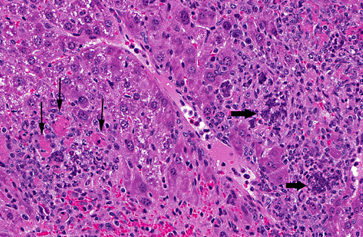
For example, LCH is characterized by the abnormal accumulation of Langerhans cells, a type of histiocyte found in the skin and mucous membranes. These cells can form tumors or lesions in various organs, such as the bones, lungs, liver, and skin.
HLH, on the other hand, is a life-threatening condition that occurs when there is an overactive immune response leading to excessive activation of histiocytes and other immune cells. This can result in fever, enlargement of the liver and spleen, and decreased blood cell counts.
The exact cause of histiocytosis is not fully understood, but it is believed to involve genetic mutations that lead to uncontrolled proliferation and accumulation of histiocytes. Treatment for histiocytosis depends on the type and severity of the disorder and may include chemotherapy, radiation therapy, immunosuppressive drugs, or stem cell transplantation.
Non-Langerhans cell histiocytosis (NLCH) is a group of rare disorders characterized by the abnormal proliferation and accumulation of histiocytes, which are immune cells that normally function to help fight infection. Unlike Langerhans cell histiocytosis (LCH), where the histiocytes involved are positive for the marker CD1a and the protein S-100, in NLCH, the histiocytes involved do not express these markers.
NLCH includes several distinct clinicopathological entities, such as juvenile xanthogranuloma, Erdheim-Chester disease, and Rosai-Dorfman disease. These conditions can affect various organs of the body, including the skin, bones, lungs, central nervous system, and others. The clinical manifestations, prognosis, and treatment options vary depending on the specific type of NLCH and the extent of organ involvement.
It is important to note that while some cases of NLCH may be self-limited or respond well to treatment, others can be aggressive and potentially life-threatening. Therefore, prompt and accurate diagnosis and management are crucial for optimizing patient outcomes.
Sinus histiocytosis is a rare condition characterized by an abnormal accumulation of histiocytes (a type of immune cell) in the sinuses. It is also known as Rosai-Dorfman disease when it occurs as a systemic disorder. In sinus histiocytosis, the histiocytes accumulate in the mucous membranes lining the sinuses, leading to their enlargement and possible obstruction. Symptoms may include nasal congestion, drainage, and pain. The exact cause of sinus histiocytosis is unknown, but it is not contagious or cancerous. Treatment typically involves monitoring and, in some cases, surgery to relieve symptoms caused by blockages.
Histiocytic sarcoma is a rare type of cancer that originates from histiocytes, which are cells that are part of the immune system and found in various tissues throughout the body. These cells normally function to help fight infection and remove foreign substances. In histiocytic sarcoma, there is an abnormal accumulation and proliferation of these cells, leading to the formation of tumors.
Histiocytic sarcoma can affect people of any age but is more commonly found in adults, with a slight male predominance. It can occur in various parts of the body, such as the lymph nodes, skin, soft tissues, and internal organs like the spleen, liver, and lungs. The exact cause of histiocytic sarcoma remains unknown, but it is not considered to be hereditary.
The symptoms of histiocytic sarcoma depend on the location and extent of the tumor(s). Common signs include swollen lymph nodes, fatigue, fever, weight loss, night sweats, and pain or discomfort in the affected area. Diagnosis typically involves a combination of imaging studies (like CT scans, PET scans, or MRI), biopsies, and laboratory tests to confirm the presence of histiocytic sarcoma and assess its extent.
Treatment for histiocytic sarcoma usually involves a multidisciplinary approach, including surgery, radiation therapy, and chemotherapy. The choice of treatment depends on several factors, such as the location and stage of the disease, the patient's overall health, and their personal preferences. Clinical trials may also be an option for some patients, allowing them to access new and experimental therapies.
Prognosis for histiocytic sarcoma is generally poor, with a five-year survival rate of approximately 15-30%. However, outcomes can vary significantly depending on individual factors, such as the patient's age, the extent of the disease at diagnosis, and the effectiveness of treatment. Continued research is necessary to improve our understanding of this rare cancer and develop more effective therapies for those affected.
Langerhans cell histiocytosis (LCH) is a rare disorder characterized by the abnormal proliferation and accumulation of dendritic cells called Langerhans cells in various tissues and organs of the body. These cells are part of the immune system and normally help to fight infection. However, in LCH, an overactive immune response leads to the excessive buildup of these cells, forming granulomas that can damage organs and impair their function.
The exact cause of LCH is not fully understood, but it is thought to involve genetic mutations that lead to uncontrolled cell growth and division. The disorder can affect people of any age, although it is most commonly diagnosed in children under the age of 15.
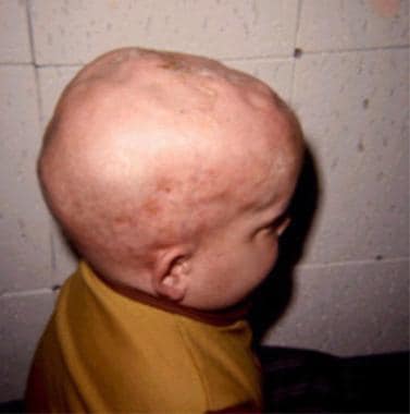
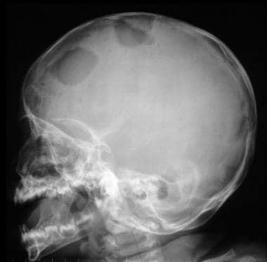
LCH can affect a single organ or multiple organs, depending on the severity and extent of the disease. Commonly affected sites include the bones, skin, lymph nodes, lungs, liver, spleen, and pituitary gland. Symptoms vary widely depending on the location and severity of the disease, but may include bone pain, rashes, fatigue, fever, weight loss, cough, and difficulty breathing.
Treatment for LCH depends on the extent and severity of the disease. In mild cases, observation and monitoring may be sufficient. More severe cases may require chemotherapy, radiation therapy, or surgery to remove affected tissues. In some cases, immunosuppressive drugs or targeted therapies that target specific genetic mutations may be used.
Overall, LCH is a complex and poorly understood disorder that requires careful evaluation and management by a team of medical specialists. While the prognosis for patients with LCH has improved in recent years, some cases can be life-threatening or lead to long-term complications.
Malignant histiocytic disorders are a group of rare and aggressive cancers that affect the mononuclear phagocyte system, which includes histiocytes or cells that originate from bone marrow precursors called monoblasts. These disorders are characterized by the uncontrolled proliferation of malignant histiocytes, leading to tissue invasion and damage.
There are several types of malignant histiocytic disorders, including:
1. Acute Monocytic Leukemia (AML-M5): This is a subtype of acute myeloid leukemia that affects the monocyte cell lineage and can involve the skin, lymph nodes, and other organs.
2. Langerhans Cell Histiocytosis (LCH): Although primarily considered a benign histiocytic disorder, some cases of LCH can progress to a malignant form with aggressive behavior and poor prognosis.
3. Malignant Histiocytosis (MH): This is a rare and aggressive disorder characterized by the infiltration of malignant histiocytes into various organs, including the liver, spleen, and lymph nodes.
4. Histiocytic Sarcoma (HS): This is a highly aggressive cancer that arises from malignant histiocytes and can affect various organs, such as the skin, lymph nodes, and soft tissues.
Symptoms of malignant histiocytic disorders depend on the type and extent of organ involvement but may include fever, fatigue, weight loss, anemia, and enlarged lymph nodes or organs. Treatment typically involves a combination of chemotherapy, radiation therapy, and/or stem cell transplantation. The prognosis for malignant histiocytic disorders is generally poor, with a high risk of relapse and a low overall survival rate.
Erdheim-Chester Disease (ECD) is a rare, progressive histiocytic disorder, characterized by the accumulation of immune cells called histiocytes in various parts of the body. These histiocytes are derived from myeloid precursors and infiltrate different organs and tissues, leading to inflammation, fibrosis, and subsequent damage.
The clinical presentation of ECD is heterogeneous, with symptoms depending on the affected organs. Commonly involved sites include bones (particularly long bones), central nervous system, heart, lungs, skin, and kidneys. Symptoms may range from bone pain, fatigue, and weight loss to neurological manifestations, cardiac dysfunction, respiratory distress, and renal impairment.
Diagnosis of ECD typically involves a combination of imaging studies (such as X-rays, CT scans, MRI, or PET scans), biopsy with histopathological examination, and immunohistochemical analysis to confirm the presence of characteristic histiocytic infiltrates. Genetic testing may also be performed to identify potential genetic mutations associated with ECD.
Treatment options for ECD depend on the extent and severity of organ involvement. Current therapeutic approaches include:
1. Targeted therapy with kinase inhibitors, such as imatinib or vemurafenib, which have shown efficacy in reducing histiocytic infiltration and improving symptoms.
2. Chemotherapy using agents like cladribine or cyclophosphamide, which can help control the disease's progression.
3. Immunosuppressive therapy with corticosteroids or interferon-alpha to manage inflammation and immune response.
4. Radiation therapy for localized bone lesions or symptomatic relief.
5. Supportive care to address specific organ dysfunction, such as heart failure management or respiratory support.
Due to the rarity of ECD, treatment decisions are often made in consultation with multidisciplinary teams experienced in managing histiocytic disorders. Clinical trials evaluating novel therapeutic strategies are also essential for advancing our understanding and improving outcomes for patients with ECD.
Lymphatic diseases refer to a group of conditions that affect the lymphatic system, which is an important part of the immune and circulatory systems. The lymphatic system consists of a network of vessels, organs, and tissues that help to transport lymph fluid throughout the body, fight infection, and remove waste products.
Lymphatic diseases can be caused by various factors, including genetics, infections, cancer, and autoimmune disorders. Some common types of lymphatic diseases include:
1. Lymphedema: A condition that causes swelling in the arms or legs due to a blockage or damage in the lymphatic vessels.
2. Lymphoma: A type of cancer that affects the lymphatic system, including Hodgkin's and non-Hodgkin's lymphoma.
3. Infections: Certain bacterial and viral infections can affect the lymphatic system, such as tuberculosis, cat-scratch disease, and HIV/AIDS.
4. Autoimmune disorders: Conditions such as rheumatoid arthritis, lupus, and scleroderma can cause inflammation and damage to the lymphatic system.
5. Congenital abnormalities: Some people are born with abnormalities in their lymphatic system, such as malformations or missing lymph nodes.
Symptoms of lymphatic diseases may vary depending on the specific condition and its severity. Treatment options may include medication, physical therapy, surgery, or radiation therapy. It is important to seek medical attention if you experience symptoms of a lymphatic disease, as early diagnosis and treatment can improve outcomes.
A "Plasma Cell Granuloma" is a specific type of granulomatous inflammation that is characterized by the presence of numerous plasma cells. Plasma cells are white blood cells that produce antibodies, which are proteins that help the body fight off infections and diseases. In a Plasma Cell Granuloma, there is an excessive accumulation of these cells, leading to the formation of a nodular lesion or mass.
Plasma Cell Granulomas can occur in various organs, including the skin, lungs, gastrointestinal tract, and oral cavity. They are often associated with chronic inflammation, autoimmune disorders, or malignancies. The exact cause of Plasma Cell Granulomas is not always known, but they may be triggered by infections, foreign bodies, or other stimuli that induce an immune response.
Histologically, a Plasma Cell Granuloma is composed of a central area of plasma cells surrounded by a rim of lymphocytes and macrophages. The lesion may also contain multinucleated giant cells, eosinophils, and other inflammatory cells. Treatment options for Plasma Cell Granulomas depend on the location and extent of the lesion, as well as the underlying cause. Surgical excision is often curative, but medical therapy may be necessary in some cases.
Benign fibrous histiocytoma (BFH) is a common benign tumor of the skin and superficial soft tissues. It primarily affects middle-aged adults and is more prevalent in men than women. The exact cause of BFH is unknown, but it's thought to arise from dermal fibroblasts or histiocytes.
Medical Definition: Benign Fibrous Histiocytoma (BFH) is a benign, slowly growing, solitary cutaneous or subcutaneous nodular tumor predominantly composed of a mixture of fibroblastic and histiocytic-like cells. The tumor typically presents as a well-circumscribed, firm, dome-shaped papule or nodule, ranging in size from a few millimeters to several centimeters. Histologically, BFH is characterized by the proliferation of spindle-shaped fibroblasts and histiocytes arranged in a storiform pattern, along with variable amounts of collagen deposition, multinucleated giant cells, and hemosiderin deposits. The lesion usually has a pushing border with no invasion into the surrounding tissues. BFH generally follows a benign clinical course, with local recurrence being uncommon following complete surgical excision.
A granuloma is a small, nodular inflammatory lesion that occurs in various tissues in response to chronic infection, foreign body reaction, or autoimmune conditions. Histologically, it is characterized by the presence of epithelioid macrophages, which are specialized immune cells with enlarged nuclei and abundant cytoplasm, often arranged in a palisading pattern around a central area containing necrotic debris, microorganisms, or foreign material.
Granulomas can be found in various medical conditions such as tuberculosis, sarcoidosis, fungal infections, and certain autoimmune disorders like Crohn's disease. The formation of granulomas is a complex process involving both innate and adaptive immune responses, which aim to contain and eliminate the offending agent while minimizing tissue damage.
Lymph nodes are small, bean-shaped organs that are part of the immune system. They are found throughout the body, especially in the neck, armpits, groin, and abdomen. Lymph nodes filter lymph fluid, which carries waste and unwanted substances such as bacteria, viruses, and cancer cells. They contain white blood cells called lymphocytes that help fight infections and diseases by attacking and destroying the harmful substances found in the lymph fluid. When an infection or disease is present, lymph nodes may swell due to the increased number of immune cells and fluid accumulation as they work to fight off the invaders.
Panniculitis is a medical term that refers to inflammation of the subcutaneous fat, or the layer of fat located just beneath the skin. This condition can affect people of all ages and genders, although it is more commonly seen in middle-aged women. The inflammation can be caused by a variety of factors, including infections, autoimmune disorders, trauma, and medications.
The symptoms of panniculitis may include:
* Red, painful lumps or nodules under the skin
* Skin lesions that may be tender, warm, or bruised
* Swelling and redness in the affected area
* Fever, fatigue, and malaise (a general feeling of illness)
The diagnosis of panniculitis typically involves a physical examination, medical history, and sometimes a biopsy of the affected tissue. Treatment depends on the underlying cause of the inflammation and may include antibiotics, anti-inflammatory medications, or other therapies. In severe cases, hospitalization may be necessary to manage symptoms and prevent complications.
Epithelioid cells are a type of cell that can be found in certain types of tissue in the body, including connective tissue and some organs. These cells have a characteristic appearance under a microscope, with an enlarged, oval or round shape and a pale, abundant cytoplasm. They may also have a nucleus that is centrally located and has a uniform, rounded shape.
Epithelioid cells are often seen in the context of inflammation or disease, particularly in relation to granulomatous disorders such as sarcoidosis and tuberculosis. In these conditions, epithelioid cells can form clusters known as granulomas, which are a hallmark of the diseases. The exact function of epithelioid cells is not fully understood, but they are thought to play a role in the immune response and may help to contain and eliminate foreign substances or pathogens from the body.
Hemophagocytic Lymphohistiocytosis (HLH) is a rare and serious condition characterized by an uncontrolled immune response leading to inflammation and damage in various organs of the body. It occurs when certain immune cells, including lymphocytes and histiocytes (a type of white blood cell), become overactive and start to destroy other blood cells, particularly red blood cells and platelets. This results in symptoms such as fever, enlarged liver and spleen, cytopenia (decreased number of blood cells), and increased levels of inflammatory markers in the body.
HLH can be primary or secondary. Primary HLH is an inherited disorder caused by genetic mutations that affect the immune system's regulation. Secondary HLH, on the other hand, is acquired due to factors such as infections, malignancies, or autoimmune diseases. Treatment for HLH typically involves a combination of chemotherapy, immunosuppressive drugs, and sometimes bone marrow transplantation. Early diagnosis and treatment are crucial for improving outcomes in patients with this condition.
Malacoplakia is a rare inflammatory condition that typically affects the urinary tract, but can also involve other organs such as the genital tract, gastrointestinal tract, lungs, and skin. It is characterized by the presence of large, foamy macrophages called "von Hansemann cells" or "Michaelis-Gutmann bodies," which contain undigested bacterial debris.
The exact cause of malacoplakia is not well understood, but it is thought to be associated with chronic bacterial infections, particularly with Escherichia coli (E. coli). The condition is more common in individuals with compromised immune systems, such as those with HIV/AIDS, organ transplants, or long-term steroid use.
Symptoms of malacoplakia depend on the location and extent of the lesions. In the urinary tract, patients may experience symptoms such as frequent urination, burning during urination, blood in the urine, and abdominal pain. Treatment typically involves a combination of antibiotics to target the underlying bacterial infection and surgical removal of the affected tissue.
Xanthomatosis is a medical term that refers to the condition characterized by the presence of xanthomas, which are yellowish, fat-laden deposits that form under the skin or in other tissues. These deposits consist of lipids, such as cholesterol and triglycerides, and immune cells called macrophages, which have engulfed the lipids.
Xanthomas can occur in various parts of the body, including the eyelids, tendons, joints, and other areas with connective tissue. They may appear as small papules or larger nodules, and their size and number can vary depending on the severity of the underlying disorder.
Xanthomatosis is often associated with genetic disorders that affect lipid metabolism, such as familial hypercholesterolemia, or with acquired conditions that cause high levels of lipids in the blood, such as diabetes, hypothyroidism, and certain liver diseases. Treatment typically involves addressing the underlying disorder and controlling lipid levels through dietary changes, medications, or a combination of both.
Mycobacterium scrofulaceum is a species of mycobacteria that was previously known to cause a type of infection called scrofula, which is a form of tuberculosis affecting the lymph nodes in the neck. However, it's important to note that this organism has rarely been implicated in human disease in recent years, and its clinical significance is currently unclear.
Mycobacterium scrofulaceum is an environmental mycobacteria, which means it can be found in soil and water, and it is not typically transmitted from person to person. Infections caused by this organism are usually acquired through the ingestion of contaminated food or water or through inhalation of aerosolized particles.
The symptoms of infection with Mycobacterium scrofulaceum depend on the site of infection and can include swollen lymph nodes, cough, fever, and weight loss. Treatment typically involves a combination of antibiotics, but the optimal treatment regimen has not been well-studied due to the rarity of infections caused by this organism.
Lymphadenitis is a medical term that refers to the inflammation of one or more lymph nodes, which are small, bean-shaped glands that are part of the body's immune system. Lymph nodes contain white blood cells called lymphocytes, which help fight infection and disease.
Lymphadenitis can occur as a result of an infection in the area near the affected lymph node or as a result of a systemic infection that has spread through the bloodstream. The inflammation causes the lymph node to become swollen, tender, and sometimes painful to the touch.
The symptoms of lymphadenitis may include fever, fatigue, and redness or warmth in the area around the affected lymph node. In some cases, the overlying skin may also appear red and inflamed. Lymphadenitis can occur in any part of the body where there are lymph nodes, including the neck, armpits, groin, and abdomen.
The underlying cause of lymphadenitis must be diagnosed and treated promptly to prevent complications such as the spread of infection or the formation of an abscess. Treatment may include antibiotics, pain relievers, and warm compresses to help reduce swelling and discomfort.
Histiocytic Necrotizing Lymphadenitis is a condition characterized by the inflammation and necrosis (death of tissue) of lymph nodes, caused by an abnormal proliferation and activation of histiocytes (a type of white blood cell). It is also known as Kikuchi's disease. The exact cause of this condition is unknown, but it is thought to be related to an immune response to viral infections or other antigens.
Histopathologically, it is characterized by the presence of necrotizing granulomatous inflammation with histiocytic predominance and absence of neutrophils. The condition is typically self-limiting, with symptoms resolving within a few months without specific treatment. However, in some cases, it can be associated with systemic symptoms or other autoimmune disorders.
A biopsy is a medical procedure in which a small sample of tissue is taken from the body to be examined under a microscope for the presence of disease. This can help doctors diagnose and monitor various medical conditions, such as cancer, infections, or autoimmune disorders. The type of biopsy performed will depend on the location and nature of the suspected condition. Some common types of biopsies include:
1. Incisional biopsy: In this procedure, a surgeon removes a piece of tissue from an abnormal area using a scalpel or other surgical instrument. This type of biopsy is often used when the lesion is too large to be removed entirely during the initial biopsy.
2. Excisional biopsy: An excisional biopsy involves removing the entire abnormal area, along with a margin of healthy tissue surrounding it. This technique is typically employed for smaller lesions or when cancer is suspected.
3. Needle biopsy: A needle biopsy uses a thin, hollow needle to extract cells or fluid from the body. There are two main types of needle biopsies: fine-needle aspiration (FNA) and core needle biopsy. FNA extracts loose cells, while a core needle biopsy removes a small piece of tissue.
4. Punch biopsy: In a punch biopsy, a round, sharp tool is used to remove a small cylindrical sample of skin tissue. This type of biopsy is often used for evaluating rashes or other skin abnormalities.
5. Shave biopsy: During a shave biopsy, a thin slice of tissue is removed from the surface of the skin using a sharp razor-like instrument. This technique is typically used for superficial lesions or growths on the skin.
After the biopsy sample has been collected, it is sent to a laboratory where a pathologist will examine the tissue under a microscope and provide a diagnosis based on their findings. The results of the biopsy can help guide further treatment decisions and determine the best course of action for managing the patient's condition.
Immunoenzyme techniques are a group of laboratory methods used in immunology and clinical chemistry that combine the specificity of antibody-antigen reactions with the sensitivity and amplification capabilities of enzyme reactions. These techniques are primarily used for the detection, quantitation, or identification of various analytes (such as proteins, hormones, drugs, viruses, or bacteria) in biological samples.
In immunoenzyme techniques, an enzyme is linked to an antibody or antigen, creating a conjugate. This conjugate then interacts with the target analyte in the sample, forming an immune complex. The presence and amount of this immune complex can be visualized or measured by detecting the enzymatic activity associated with it.
There are several types of immunoenzyme techniques, including:
1. Enzyme-linked Immunosorbent Assay (ELISA): A widely used method for detecting and quantifying various analytes in a sample. In ELISA, an enzyme is attached to either the capture antibody or the detection antibody. After the immune complex formation, a substrate is added that reacts with the enzyme, producing a colored product that can be measured spectrophotometrically.
2. Immunoblotting (Western blot): A method used for detecting specific proteins in a complex mixture, such as a protein extract from cells or tissues. In this technique, proteins are separated by gel electrophoresis and transferred to a membrane, where they are probed with an enzyme-conjugated antibody directed against the target protein.
3. Immunohistochemistry (IHC): A method used for detecting specific antigens in tissue sections or cells. In IHC, an enzyme-conjugated primary or secondary antibody is applied to the sample, and the presence of the antigen is visualized using a chromogenic substrate that produces a colored product at the site of the antigen-antibody interaction.
4. Immunofluorescence (IF): A method used for detecting specific antigens in cells or tissues by employing fluorophore-conjugated antibodies. The presence of the antigen is visualized using a fluorescence microscope.
5. Enzyme-linked immunosorbent assay (ELISA): A method used for detecting and quantifying specific antigens or antibodies in liquid samples, such as serum or culture supernatants. In ELISA, an enzyme-conjugated detection antibody is added after the immune complex formation, and a substrate is added that reacts with the enzyme to produce a colored product that can be measured spectrophotometrically.
These techniques are widely used in research and diagnostic laboratories for various applications, including protein characterization, disease diagnosis, and monitoring treatment responses.
Hodgkin disease, also known as Hodgkin lymphoma, is a type of cancer that originates in the white blood cells called lymphocytes. It typically affects the lymphatic system, which is a network of vessels and glands spread throughout the body. The disease is characterized by the presence of a specific type of abnormal cell, known as a Reed-Sternberg cell, within the affected lymph nodes.
The symptoms of Hodgkin disease may include painless swelling of the lymph nodes in the neck, armpits, or groin; fever; night sweats; weight loss; and fatigue. The exact cause of Hodgkin disease is unknown, but it is thought to involve a combination of genetic, environmental, and infectious factors.
Hodgkin disease is typically treated with a combination of chemotherapy, radiation therapy, and/or immunotherapy, depending on the stage and extent of the disease. With appropriate treatment, the prognosis for Hodgkin disease is generally very good, with a high cure rate. However, long-term side effects of treatment may include an increased risk of secondary cancers and other health problems.
Lymphoma is a type of cancer that originates from the white blood cells called lymphocytes, which are part of the immune system. These cells are found in various parts of the body such as the lymph nodes, spleen, bone marrow, and other organs. Lymphoma can be classified into two main types: Hodgkin lymphoma (HL) and non-Hodgkin lymphoma (NHL).
HL is characterized by the presence of a specific type of abnormal lymphocyte called Reed-Sternberg cells, while NHL includes a diverse group of lymphomas that lack these cells. The symptoms of lymphoma may include swollen lymph nodes, fever, night sweats, weight loss, and fatigue.
The exact cause of lymphoma is not known, but it is believed to result from genetic mutations in the lymphocytes that lead to uncontrolled cell growth and division. Exposure to certain viruses, chemicals, and radiation may increase the risk of developing lymphoma. Treatment options for lymphoma depend on various factors such as the type and stage of the disease, age, and overall health of the patient. Common treatments include chemotherapy, radiation therapy, immunotherapy, and stem cell transplantation.
The Mononuclear Phagocyte System (MPS) is a network of specialized immune cells distributed throughout the body, primarily consisting of monocytes, macrophages, and dendritic cells. These cells share a common bone marrow-derived precursor and play crucial roles in innate and adaptive immunity. They are involved in various functions such as:
1. Phagocytosis: engulfing and destroying foreign particles, microbes, and cellular debris.
2. Antigen presentation: processing and presenting antigens to T-cells to initiate an adaptive immune response.
3. Cytokine production: releasing pro- and anti-inflammatory cytokines to regulate immune responses and maintain tissue homeostasis.
4. Immune regulation: modulating the activity of other immune cells, including T-cells, B-cells, and natural killer (NK) cells.
The MPS is essential for maintaining tissue integrity, fighting infections, and orchestrating immune responses. Its components are found in various tissues, including the liver (Kupffer cells), spleen, lymph nodes, bone marrow, and connective tissues.
Immunohistochemistry (IHC) is a technique used in pathology and laboratory medicine to identify specific proteins or antigens in tissue sections. It combines the principles of immunology and histology to detect the presence and location of these target molecules within cells and tissues. This technique utilizes antibodies that are specific to the protein or antigen of interest, which are then tagged with a detection system such as a chromogen or fluorophore. The stained tissue sections can be examined under a microscope, allowing for the visualization and analysis of the distribution and expression patterns of the target molecule in the context of the tissue architecture. Immunohistochemistry is widely used in diagnostic pathology to help identify various diseases, including cancer, infectious diseases, and immune-mediated disorders.
Histochemistry is the branch of pathology that deals with the microscopic localization of cellular or tissue components using specific chemical reactions. It involves the application of chemical techniques to identify and locate specific biomolecules within tissues, cells, and subcellular structures. This is achieved through the use of various staining methods that react with specific antigens or enzymes in the sample, allowing for their visualization under a microscope. Histochemistry is widely used in diagnostic pathology to identify different types of tissues, cells, and structures, as well as in research to study cellular and molecular processes in health and disease.
A Rheumatoid nodule is defined as a type of non-suppurative inflammatory lesion that occurs in the subcutaneous tissue, commonly associated with rheumatoid arthritis (RA). These nodules are firm, round to oval shaped, and usually range from 0.5 to 5 cm in size. They are typically found over bony prominences such as the elbow, heel, or fingers, but can occur in various locations throughout the body.
Histologically, rheumatoid nodules are characterized by a central area of fibrinoid necrosis surrounded by palisading histiocytes and fibroblasts, with an outer layer of chronic inflammatory cells, including lymphocytes and plasma cells. Rheumatoid nodules can be asymptomatic or cause pain and discomfort, depending on their size and location. They are more common in patients with severe RA and are associated with a poorer prognosis.
 Histiocyte
Histiocyte Society
List of immune cells
GM1 gangliosidoses
Artificial white blood cells
Histiocytoma
Poorly cohesive gastric carcinoma
List of OMIM disorder codes
Lymph node
Rosai-Dorfman disease
Xanthogranulomatous inflammation
Malignant histiocytosis
Exophiala dermatitidis
Wandering cell
Langerhans cell histiocytosis
Granuloma
Lacazia
Vaginal epithelium
Aggressive NK-cell leukemia
Dennis H. Wright
Bracht-Wachter bodies
Canine histiocytic diseases
Histiocytosis
Vernal keratoconjunctivitis
Mycobacterium leprae
Nonpuerperal mastitis
Ranula
Niemann-Pick disease
Non-ossifying fibroma
Libman-Sacks endocarditis
Pel-Ebstein fever
Histiocytic sarcoma
Histiocytoma (dog)
Chronic multifocal Langerhans cell histiocytosis
Oral mucocele
Histiocyte - Wikipedia
 Sea-blue-histiocyte syndrome
 Histiocyte Disorders Program | Children's National Hospital
 In vivo detection of mucosal healing-involved histiocytes by confocal laser endomicroscopy
 Hepatocellular Carcinoma with Foamy Histiocyte-Like Appearance: A Deceptively Clear Cell Carcinoma Appearing Variant | Case...
 Sea-blue histiocytes and Gaucher cells in bone marrow of patients with chronic myeloid leukaemia. | Journal of Clinical...
 T-cell/histiocyte-rich large B-cell lymphoma shows transcriptional features suggestive of a tolerogenic host immune response
...
T-cell/histiocyte-rich large B-cell lymphoma shows transcriptional features suggestive of a tolerogenic host immune response
...
Histiocytes | Profiles RNS
 Histiocyte Society - IRHDR
Histiocyte Society - LCH Articles
Histiocyte Society - Athens Abstracts
Kikuchi-Fujimoto Disease: An Exuberant Localized T Cell Activation Arrested By Histiocytes?
 Other - Thyroglossal duct cyst: histiocytes and squames (smear, Diff Quik stain). - IMAGE ATLAS
 Histiocytosis: Practice Essentials, Pathophysiology, Epidemiology
Pathology of Prostate Xanthoma: Overview, Clinical Features and Gross Findings, Microscopic Findings
 Hypopituitarism: MedlinePlus Medical Encyclopedia
Histiocytic Medullary Reticulosis | Citedby Results | Acta Haematologica | Karger Publishers
 Diffuse large B cell lymphoma: Outlook, stages, treatment
 Thieme E-Journals - American Journal of Perinatology Reports / Full Text
Toward a unifying entity that encompasses most, but perhaps not all, inflammatory leiomyosarcomas and histiocyte-rich...
 Lakshmi Nayak, MD - Dana-Farber Cancer Institute | Boston, MA
 Advanced Search Results - Public Health Image Library(PHIL)
 Acute liver failure secondary to severe systemic disease from fatal hemophagocytic lymphohistiocytosis: Case report and...
 Pathology Outlines - Silicone leak / lymphadenopathy
Model Details
 Michael D. Hogarty, MD | Children's Hospital of Philadelphia
 Mantle cell lymphoma pathophysiology - wikidoc
Epithelioid3
- Positive signals for TUNEL were detected in epithelioid histiocytes and lymphocytes within granulomas and in bronchoalveolar lavage (BAL) lymphocytes from patients with sarcoidosis. (ersjournals.com)
- Apoptosis in epithelioid histiocytes and inflammatory cells seems to participate in the course of granulomatous inflammation. (ersjournals.com)
- Moreover, cutaneous biopsy, which was performed on the right forearm, revealed granulomas with epithelioid histiocytes and Langerhans-type giant cells. (who.int)
Macrophages3
- Macrophages and dendritic cells are derived from common bone marrow precursor cells that have undergone different differentiation (as histiocytes) under the influence of various environmental (tissue location) and growth factors such as GM-CSF, TNF and IL-4. (wikipedia.org)
- These features may be responsible for the recruitment and activation of T cells, macrophages and dendritic cells, characterizing the stromal component of this lymphoma, and may point towards innate immunity and a tumor tolerogenic immune response in T-cell/histiocyte-rich large B-cell lymphoma. (haematologica.org)
- SIV antigen has been demonstrated by immunohistochemical methods in lymph node sinus histiocytes, macrophages, and giant cells (14) as well as in macrophage-derived cells in brain tissue from diseased monkeys (8). (cdc.gov)
Mononuclear phagoc1
- They circulate through the body and enter various organs, where they undergo differentiation into histiocytes, which are part of the mononuclear phagocytic system (MPS). (wikipedia.org)
Langerhans4
- Some sources consider Langerhans cell derivatives to be histiocytes. (wikipedia.org)
- The most common histiocyte disorders are Langerhans' cell histiocytosis and haemophagocytic lymphohistiocytosis. (wikipedia.org)
- The rare histiocytic disorders (RHD), or non-Langerhans cell disorders, are a diverse group of disorders defined by the accumulation of histiocytes that do not meet the criteria for Langerhans cell histiocytosis (LCH) or hemophagocytic lymphohistiocytosis (HLH). (histiocytesociety.org)
- It is found on Langerhans cells, follicular dendritic cells and histiocytes. (beckman.com)
Granulomas1
- When lung tissue becomes inflamed from an infection or other cause, cells called histiocytes cluster to form nodules called granulomas. (healthline.com)
Lymphocytes3
- In 1994, after three years of progressive neurological dysfunction, diagnosis of Erdheim-Chester disease was made by analysis of biopsies of the femur bones, showing infiltration with foamy histiocytes lacking Birbeck granules and S-100 protein, and with few lymphocytes. (bmj.com)
- Infiltrate composed of lymphocytes and histiocytes. (cdlib.org)
- 2002). The most common manifestation is chronic interstitial pneumonitis with infiltration of lymphocytes, histiocytes, and plasma cells (Saltini and Amicosante 2001). (cdc.gov)
[email protected]1
- Please direct questions regarding abstracts to the Histiocyte Society Secretariat's Office at [email protected] . (histiocytesociety.org)
Macrophage2
- The histiocyte is a tissue macrophage or a dendritic cell (histio, diminutive of histo, meaning tissue, and cyte, meaning cell). (wikipedia.org)
- However, the term histiocyte has been used for multiple purposes in the past, and some cells called "histocytes" do not appear to derive from monocytic-macrophage lines. (wikipedia.org)
Cells10
- Histiocytosis is a group of conditions that involve too many white blood cells (histiocytes) in your child's blood. (childrensnational.org)
- There was also a lesion with foamy histiocyte-like cells corresponding to the white lesion in the face of the cut tumor. (karger.com)
- Sea-blue histiocytes and Gaucher cells in bone marrow of patients with chronic myeloid leukaemia. (bmj.com)
- The aggressive T-cell/histiocyte-rich large B-cell lymphoma and the indolent nodular lymphocyte-predominant Hodgkin's lymphoma are both characterized by a paucity of tumor cells embedded in an overwhelming background. (haematologica.org)
- 4 In the World Health Organization (WHO) classification of 2001, T-cell/histiocyte-rich large B-cell lymphoma (THRLBCL) is defined by the presence of a limited number of scattered large B cells in a background rich in T cells, with or without histiocytes. (haematologica.org)
- Histiocytoses are rare diseases caused by an excess of cells called histiocytes, which can infiltrate the skin, bones, lungs, liver, spleen and the central nervous system. (histiocytesociety.org)
- Other patterns include arrangement of the histiocytes in cords or single cells that percolate between benign prostatic glands. (medscape.com)
- Plasma cells, eosinophils, and histiocytes appear later. (forextrading-madeeasy.com)
- Histiocytosis is an umbrella term that refers to a relatively uncommon group of syndromes associated with the overproduction of certain infection-fighting white blood cells known as histiocytes. (tgh.org)
- Normal histiocytes originate from pluripotent stem cells, which can be found in bone marrow. (medscape.com)
Disorders6
- Children's National offers the highest level of experience with histiocyte disorders in the entire Washington, D.C. area. (childrensnational.org)
- We also collaborate with care providers from all over the world who are also involved in the field of histiocyte disorders, allowing us access to national and international approaches to treatment. (childrensnational.org)
- We participate in research treatment protocols with select institutions around the country who are investigating the newest approaches using the latest scientific advances for histiocyte disorders. (childrensnational.org)
- Physicians, patients and parents of children with rare histiocytoses frequently consult members of the Histiocyte Society on the management of these disorders. (histiocytesociety.org)
- The Histiocyte Society is a nonprofit organization of more than 200 physicians and scientists from around the world committed to improving the lives of patients with histiocytic disorders by conducting clinical and laboratory research into the causes and treatment of this disease. (histiocytesociety.org)
- He has a special interest in neuroblastoma and histiocyte disorders. (chop.edu)
Histiocytoses1
- Histiocytoses describe neoplasias wherein the proliferative cell is the histiocyte. (wikipedia.org)
Foamy2
- This report describes the case of a patient with a morphologically distinctive pattern of HCC with prominent cell cytoplasm that had a foamy histiocyte-like appearance. (karger.com)
- 1 It is associated with tissue infiltration by foamy histiocytes. (bmj.com)
Accumulation3
- Rosai-Dorfman disease is a rare disorder characterized by overproduction (proliferation) and accumulation of a specific type of white blood cell (histiocyte) in the lymph nodes of the body (lymphadenopathy), most often those of the neck (cervical lymphadenopathy). (rarediseases.org)
- In some cases, abnormal accumulation of histiocytes may occur in other areas of the body besides the lymph nodes (extranodal). (rarediseases.org)
- In some cases of Rosai-Dorfman disease, the accumulation of histiocytes into masses may cause compression of vital organs potentially resulting in serious complications. (rarediseases.org)
Inflammatory1
- The histiocytes do not incite an inflammatory reaction. (medscape.com)
Bone marrow1
- Histiocytes are derived from the bone marrow by multiplication from a stem cell. (wikipedia.org)
Immune3
- These histiocytes are part of the immune system by way of two distinct functions: phagocytosis and antigen presentation. (wikipedia.org)
- Conclusions The gene expression profile of T-cell/histiocyte-rich large B-cell lymphoma, in comparison with that of nodular lymphocyte-predominant Hodgkin's lymphoma, shows features suggestive of a distinct tolerogenic host immune response that may play a key role in the aggressive behavior of this lymphoma, and that may serve as a potential target for future therapy. (haematologica.org)
- The goal is to inhibit the overproduction of histiocytes by suppressing the immune system. (tgh.org)
Lymph1
- Results We observed that the microenvironment in nodular lymphocyte-predominant Hodgkin's lymphoma is molecularly very similar to a lymph node characterized by follicular hyperplasia, while the microenvironment in T-cell/histiocyte-rich large B-cell lymphoma is clearly different. (haematologica.org)
Proliferation2
- Malignant histiocytosis is a rare invasive proliferation of neoplastic histiocytes. (bmj.com)
- A zone of fibroblastic proliferation with palisading histiocytes may surround the area. (merckmanuals.com)
Monocyte1
- The term Histiocyte can also simply refer to a cell from monocyte origin outside the blood system, such as in a tissue (as in rheumatoid arthritis as palisading histiocytes surrounding fibrinoid necrosis of rheumatoid nodules). (wikipedia.org)
Biopsies1
- Biopsies demonstrated aggregates of histiocytes with granular eosinophilic material in the lamina propria. (asn-online.org)
Cytoplasm1
- Prostatic xanthoma comprises collections of histiocytes with finely vacuolated, lipid-laden cytoplasm. (medscape.com)
Society3
- Abstracts that include data from ongoing Histiocyte Society studies will not be considered for presentation. (histiocytesociety.org)
- This involves leadership roles within the Children's Oncology Group (COG), the International Neuroblastoma Risk Group (INRG), and the International Histiocyte Society. (chop.edu)
- The information on this page has been written and reviewed by the Histiocytosis Association Board of Trustees Scientific Committee and a member of the Histiocyte Society , and subsequently audited by patients and families to ensure enough information was captured. (histio.org)
Stain1
- Thyroglossal duct cyst: histiocytes and squames (smear, Diff Quik stain). (papsociety.org)
Disease3
- Kikuchi-Fujimoto Disease: An Exuberant Localized T Cell Activation Arrested By Histiocytes? (medscape.com)
- Cite this: Kikuchi-Fujimoto Disease: An Exuberant Localized T Cell Activation Arrested By Histiocytes? (medscape.com)
- Correa H. Kikuchi-Fujimoto Disease: An Exuberant Localized T Cell Activation Arrested By Histiocytes? (medscape.com)
Form1
- We are one of only 10 programs in the world using an aggressive new form of chemotherapy to treat histiocytes. (childrensnational.org)
Reaction1
- By quantitative reverse transcriptase polymerase chain reaction we verified that these 20 selected cases were representative of the entire population of T-cell/histiocyte-rich large B-cell and nodular lymphocyte-predominant Hodgkin's lymphomas. (haematologica.org)
Cases1
- Design and Methods We collected 33 cases of T-cell/histiocyte-rich large B-cell lymphoma and 56 cases of nodular lymphocyte-predominant Hodgkin's lymphoma and performed microarray gene expression profiling on ten cases of each lymphoma, to obtain a better understanding of the lymphoma host response. (haematologica.org)
Presence1
- One immunoglobulin was presented in the histiocytes, suggesting the antecedent presence of antigen-antibody complexes. (cdc.gov)
Categories1
- The various categories of histiocytes are distinguishable by their morphology, phenotype, and size. (wikipedia.org)
Field1
- We are one of only 10 programs in North America advancing the field of histiocyte disorder care. (childrensnational.org)
Shows1
- This graph shows the total number of publications written about "Histiocytes" by people in this website by year, and whether "Histiocytes" was a major or minor topic of these publications. (ouhsc.edu)