Heart Valves
Heart Valve Prosthesis
Heart Valve Diseases
Aortic Valve
Bioprosthesis
Aortic Valve Stenosis
Heart Valve Prosthesis Implantation
Tricuspid Valve
Aortic Valve Insufficiency
Mitral Valve Insufficiency
Endocarditis, Bacterial
Mitral Valve Stenosis
Mitral Valve Prolapse
Endocarditis
Venous Valves
Heart Failure
Heart Defects, Congenital
Heart Diseases
Tricuspid Valve Insufficiency
Thromboembolism
Tissue Engineering
Echocardiography
Cardiac Catheterization
Prosthesis Failure
Models, Cardiovascular
Fetal Heart
Mitral Valve Annuloplasty
Treatment Outcome
Echocardiography, Transesophageal
International Normalized Ratio
Endocardial Cushions
Heart Valve Prolapse
Absorbable Implants
Acenocoumarol
Warfarin
Cardiac Valve Annuloplasty
Heart Sounds
Hemodynamics
Pregnancy Complications, Cardiovascular
Pulmonary Valve Stenosis
Rheumatic Heart Disease
Follow-Up Studies
Glutaral
Echocardiography, Doppler, Color
Bioreactors
Myocardium
Postoperative Complications
Reoperation
Papio ursinus
Echocardiography, Doppler
Embolism, Air
Carcinoid Heart Disease
Balloon Valvuloplasty
Heart Block
Phonocardiography
Multidetector Computed Tomography
Receptor, Serotonin, 5-HT2B
Prospective Studies
Risk Factors
Safety-Based Drug Withdrawals
Chordae Tendineae
Heart Arrest, Induced
Fenfluramine
Tensile Strength
Bioartificial Organs
Heart Neoplasms
Swine
Extracellular Matrix
Sheep
Embolism
Retrospective Studies
Tissue Preservation
Cardiomyopathies
Stress, Mechanical
Prothrombin Time
Tissue Scaffolds
Biocompatible Materials
Amaurosis Fugax
Pericardium
Biomedical Engineering
Ileocecal Valve
Ventricular Function, Left
Biomechanical Phenomena
Risk Assessment
Equipment Failure Analysis
Myocardial Ischemia
Endocarditis, Subacute Bacterial
Intracranial Embolism and Thrombosis
Stroke Volume
Catheterization
Feasibility Studies
Severity of Illness Index
Sternum
Prosthesis-Related Infections
Materials Testing
Organogenesis
Phenindione
Myocytes, Cardiac
Coronary Disease
Predictive Value of Tests
Tissue Culture Techniques
Aortic Valve Prolapse
Echocardiography, Three-Dimensional
Microscopy, Electron, Scanning
Collagen
Heart Septal Defects, Ventricular
Heart Septal Defects
Heart, Artificial
Pulmonary Valve Insufficiency
Hemorheology
Disease Models, Animal
Gene Expression Regulation, Developmental
Ventricular Dysfunction, Left
Dogs
Cardiac-Gated Imaging Techniques
Sus scrofa
Ultrasonography, Doppler, Transcranial
Cardiac Output, Low
Atrial Fibrillation
Phentermine
Blood Flow Velocity
Cardiomyopathy, Dilated
Cardiomegaly
Subgingival Curettage
Vitamin K
SOX9 Transcription Factor
Age Factors
Myocardial Infarction
Pergolide
Batch Cell Culture Techniques
Heart Failure, Systolic
Rabbits
Mediastinum
Cardiotonic Agents
Streptococcus sanguis
Ventricular Remodeling
Streptococcus anginosus
Heart Rate, Fetal
Rheology
Medical Laboratory Science
Pressure
Prognosis
Reproducibility of Results
Heart Arrest
Chondroitinsulfatases
Twist Transcription Factor
Anatomical study of truncus arteriousus communis with embryological and surgical considerations. (1/685)
Twelve specimens of truncus arteriosus communis have been studied anatomically, with special reference to the conal anatomy and to the associated cardiac anomalies which can create additional problems if surgical repair is planned. A wide spectrum of conal morphology has been observed, suggesting that differential conal absorption is a developmental characteristic of truncus arteriousus as well as of transposition complexes. The invariable absence of septation of the ventricular infundibula and semilunar valves, in spite of the variable anatomy of the free wall of the conus, indicates that all types of truncus arteriosus, ontogenetically, should be considered as a single undivided conotruncus. Various types of ventircular septal defect were found: (a) ventricular septal defect with absent crista, in which no remnants of conal septum are present; (b) supracristal ventricular septal defect, in which vestigial conal septum is seen in front of the membranous septum; (c) bulloventricular foramen, associated with univentricular origin of the truncus from the right ventricle. Frequent associated anomalies are underdevelopment of the aortic arch, truncal valve malformations, and obstructive ventricular septal defect. The AV conduction system studied in one case showed an arrangement similar to Fallot's tetralogy with the His bundle and the left bundle-branch in a safe position behind the posteroinferior rim of the defect. The postoperative fate of the frequently abnormal truncal valve and the theoretical indications for total repair for Type IV truncus are also discussed. (+info)Connexin 43 expression reflects neural crest patterns during cardiovascular development. (2/685)
We used transgenic mice in which the promoter sequence for connexin 43 linked to a lacZ reporter was expressed in neural crest but not myocardial cells to document the pattern of cardiac neural crest cells in the caudal pharyngeal arches and cardiac outflow tract. Expression of lacZ was strikingly similar to that of cardiac neural crest cells in quail-chick chimeras. By using this transgenic mouse line to compare cardiac neural crest involvement in cardiac outflow septation and aortic arch artery development in mouse and chick, we were able to note differences and similarities in their cardiovascular development. Similar to neural crest cells in the chick, lacZ-positive cells formed a sheath around the persisting aortic arch arteries, comprised the aorticopulmonary septation complex, were located at the site of final fusion of the conal cushions, and populated the cardiac ganglia. In quail-chick chimeras generated for this study, neural crest cells entered the outflow tract by two pathways, submyocardially and subendocardially. In the mouse only the subendocardial population of lacZ-positive cells could be seen as the cells entered the outflow tract. In addition lacZ-positive cells completely surrounded the aortic sac prior to septation, while in the chick, neural crest cells were scattered around the aortic sac with the bulk of cells distributed in the bridging portion of the aorticopulmonary septation complex. In the chick, submyocardial populations of neural crest cells assembled on opposite sides of the aortic sac and entered the conotruncal ridges. Even though the aortic sac in the mouse was initially surrounded by lacZ-positive cells, the two outflow vessels that resulted from its septation showed differential lacZ expression. The ascending aorta was invested by lacZ-positive cells while the pulmonary trunk was devoid of lacZ staining. In the chick, both of these vessels were invested by neural crest cells, but the cells arrived secondarily by displacement from the aortic arch arteries during vessel elongation. This may indicate a difference in derivation of the pulmonary trunk in the mouse or a difference in distribution of cardiac neural crest cells. An independent mouse neural crest marker is needed to confirm whether the differences are indeed due to species differences in cardiovascular and/or neural crest development. Nevertheless, with the differences noted, we believe that this mouse model faithfully represents the location of cardiac neural crest cells. The similarities in location of lacZ-expressing cells in the mouse to that of cardiac neural crest cells in the chick suggest that this mouse is a good model for studying mammalian cardiac neural crest and that the mammalian cardiac neural crest performs functions similar to those shown for chick. (+info)The presence of infection-related antiphospholipid antibodies in infective endocarditis determines a major risk factor for embolic events. (3/685)
OBJECTIVES: The impact of infection-associated antiphospholipid antibodies (APA) on endothelial cell activation, blood coagulation and fibrinolysis was evaluated in patients with infective endocarditis with and without major embolic events. BACKGROUND: An embolic event is a common and severe complication of infective endocarditis. Despite the fact that APAs are known to be associated with infectious diseases, their pathogenic role in infective endocarditis has not been clearly defined. METHODS: The relationship among the occurrence of major embolic events, echocardiographic vegetation size, endothelial cell activation, thrombin generation, fibrinolysis and APA was examined in 91 patients with definite infective endocarditis, including 26 patients with embolic events and 65 control subjects without embolic events. RESULTS: Overall, 14.3% of patients exhibited elevated APA levels. Embolic events occurred more frequently in patients with elevated levels of APA than in patients without (61.5% vs. 23.1%; p = 0.008). Patients with elevated levels of APA showed higher levels of prothrombin-fragment F1 +2 (p = 0.005), plasminogen-activator inhibitor 1 (p = 0.0002), von Willebrand factor (p = 0.002) and lower levels of activated protein C (p = 0.001) than patients with normal levels of APA. Thrombin generation and endothelial cell activation were both positively correlated with levels of APA. The occurrence of elevated APA levels was frequently associated with structural valve abnormalities (p = 0.01) and vegetations >1.3 cm (p = 0.002). CONCLUSIONS: Infection-associated elevated APA levels in patients with infective endocarditis are related to endothelial cell activation, thrombin generation and impairment of fibrinolysis. This may contribute to the increased risk for major embolic events in these patients. (+info)Ineffectiveness of burst suppression therapy in mitigating perioperative cerebrovascular dysfunction. Multicenter Study of Perioperative Ischemia (McSPI) Research Group. (4/685)
BACKGROUND: Cerebral injury is among the most common and disabling complications of open heart surgery. Attempts to provide neuroprotection have yielded conflicting results. We assessed the potential of propofol-induced burst suppression during open heart surgery to provide cerebral protection as determined by postoperative neuropsychologic function. METHODS: Two hundred twenty-five patients undergoing valve surgery were randomized to receive either sufentanil or sufentanil plus propofol titrated to electroencephalographic burst suppression. Blinded investigators performed neurologic and neuropsychologic testing at baseline, postoperative day (POD) 1 (neurologic testing only), PODs 5-7, and PODs 50-70. Neuropsychologic tests were compared with the results of 40 nonsurgical patients matched for age and education. RESULTS: Electroencephalographic burst suppression was successfully achieved in all 109 propofol patients. However, these patients sustained at least as many adverse neurologic outcomes as the 116 controls: POD 1, 40% versus 25%, P = 0.06; PODs 5-7, -18% versus 8%, P = 0.07; PODs 50-70, -6% versus 6%, P = 0.80. No differences in the incidence of neuropsychologic deficits were detected, with 91% of the propofol patients versus 92% of the control patients being impaired at PODs 5-7, decreasing to 52 and 47%, respectively, by PODs 50-70. No significant differences in the severity of neuropsychologic dysfunction, depression, or anxiety were noted. CONCLUSIONS: Electroencephalographic burst suppression surgery with propofol during cardiac valve replacement did not significantly reduce the incidence or severity of neurologic or neuropsychologic dysfunction. The authors' results suggest that neither cerebral metabolic suppression nor reduction in cerebral blood flow reliably provide neuroprotection during open heart surgery. Other therapeutic approaches must be evaluated to address this important medical problem. (+info)Viability and enzymatic activity of cryopreserved porcine heart valve. (5/685)
Fibroblast viability of a natural tissue valve for replacing a defective heart valve through allograft or xenograft has been suggested to affect its clinical durability. In this study, the cell viability and enzymatic activity of porcine heart valve leaflets were examined in regard to concerning to the preservation process [variable warm ischemic time (WIT), cold ischemic time (CIT), and cryopreservation]. Porcine heart enblocs were obtained and valve dissection was performed after 2, 12, 24, or 36 hours, in respective groups A, B, C, and D, as WIT. Each group was stored for 24 hours as CIT and cryopreserved. Leaflets were dissected from a valved conduit after each process, and cell viability and enzymatic activity in the leaflet were investigated using trypan blue staining and API ZYM kits. WIT extension significantly decreased fibroblast viability (p < 0.05, 92.25 +/- 2.7% at 2 hours, 84.9 +/- 6.7% at 12 hours, 57.0 +/- 10.2% at 24 hours, 55.9 +/- 7.9% at 36 hours), while CIT for 24 hours was also influenced significantly (p < 0.05), whereas cryopreservation demonstrated no effect on cellular viability. In enzyme activity observation, several enzymes related to lipid or nucleotide degradation (esterase, esterase lipase, particularly phosphatase, phosphohydrolase) were remarkably changed following the valve-fabrication process. After 24 hours CIT, these enzymatic activities in groups B, C and D significantly increased, but the activities decreased after cryopreservation. Particularly, both the viability and enzymatic activity showed remarkable changes after CIT in group B (WIT = 12 hours). These results suggest that WIT is more important than CIT in maintaining viability of the valve, and that completing all the cryopreservation process within 12 hours after acquisition is recommended. (+info)Cardiac myosin heavy chains lacking the light chain binding domain cause hypertrophic cardiomyopathy in mice. (6/685)
Myosin is a chemomechanical motor that converts chemical energy into the mechanical work of muscle contraction. More than 40 missense mutations in the cardiac myosin heavy chain (MHC) gene and several mutations in the two myosin light chains cause a dominantly inherited heart disease called familial hypertrophic cardiomyopathy. Very little is known about the biochemical defects in these alleles and how the mutations lead to disease. Because removal of the light chain binding domain in the lever arm of MHC should alter myosin's force transmission but not its catalytic function, we tested the hypothesis that such a mutant MHC would act as a dominant mutation in cardiac muscle. Hearts from transgenic mice expressing this mutant myosin are asymmetrically hypertrophied, with increases in mass primarily restricted to the cardiac anterior wall. Histological examination demonstrates marked cellular hypertrophy, myocyte disorganization, small vessel coronary disease, and severe valvular pathology that included thickening and plaque formation. Skinned myocytes and multicellular preparations from transgenic hearts exhibited decreased Ca2+ sensitivity of tension and decreased relaxation rates after flash photolysis of diazo 2. These experiments demonstrate that alterations in myosin force transmission are sufficient to trigger the development of hypertrophic cardiomyopathy. (+info)Oral d,l sotalol reduces the incidence of postoperative atrial fibrillation in coronary artery bypass surgery patients: a randomized, double-blind, placebo-controlled study. (7/685)
OBJECTIVES: The purpose of this prospective, randomized, double-blind, placebo-controlled study was to assess the efficacy of preoperatively and postoperatively administered oral d,l sotalol in preventing the occurrence of postoperative atrial fibrillation (AF). BACKGROUND: Atrial fibrillation is the most common arrhythmia following coronary artery bypass surgery (CABG). Its etiology, prevention and treatment remain highly controversial. Furthermore, its associated morbidity results in a prolongation of the length of hospital stay post-CABG. METHODS: A total of 85 patients, of which 73 were to undergo CABG and 12 CABG plus valvular surgery (ejection fraction > or = 28% and absence of clinical heart failure), were randomized to receive either sotalol (40 patients; mean dose = 190 +/- 43 mg/day) started 24 to 48 h before open heart surgery and continued for four days postoperatively, or placebo (45 patients, mean dose = 176 +/- 32 mg/day). RESULTS: Atrial fibrillation occurred in a total of 22/85 (26%) patients. The incidence of postoperative AF was significantly (p = 0.008) lower in patients on sotalol (12.5%) as compared with placebo (38%). Significant bradycardia/hypotension, necessitating drug withdrawal, occurred in 2 of 40 (5%) patients on sotalol and none in the placebo group (p = 0.2). None of the patients on sotalol developed Torsade de pointes or sustained ventricular arrhythmias. Postoperative mortality was not significantly different in sotalol versus placebo (0% vs. 2%, p = 1.0). Patients in the sotalol group had a nonsignificantly shorter length of hospital stay as compared with placebo (7 +/- 2 days vs. 8 +/- 4 days; p = 0.24). CONCLUSIONS: The administration of sotalol, in dosages ranging from 80 to 120 mg, was associated with a significant decrease (67%) in postoperative AF in patients undergoing CABG without appreciable side effects. Sotalol should be considered for the prevention of postoperative AF in patients undergoing CABG in the absence of heart failure and significant left ventricular dysfunction. (+info)Expression of the Mf1 gene in developing mouse hearts: implication in the development of human congenital heart defects. (8/685)
The transcription factor FKHL7 gene has recently been associated with the anterior segment dysgenesis disorder of the eye known as Axenfeld-Rieger anomaly (ARA). A growing body of evidence indicates that mutations in FKHL7 cause not only defects in the anterior segment of the eye but defects in the heart valves and septa as well. In order to evaluate its contribution to normal heart septation and valve formation, expression of the mouse homologue Mf1 in embryonic hearts was analyzed by in situ hybridization. A weak but significant level of Mf1 expression could be detected in the endocardium of mouse embryos as early as day 8.5 post-conception (p.c.). Mf1 expression was undetectable in the hearts of day 9.5 p.c. embryos, but by day 10.5-11 p.c., Mf1 transcripts could be found again in the endocardium of both the atrium and ventricle and a relatively strong signal was observed in the dorsal portion of the septum primum, in what appeared to be the spinal vestibule. At day 13 p.c. when aortic and pulmonary trunks are separated, relatively more Mf1 transcripts were detected in the leaflets of aortic, pulmonary, and venous valves, the ventral portion of the septum primum, as well as in the single layer of cells on the edges of the atrioventricular cushion tissues. Surprisingly, there was no signal detected in the developing interventricular septum. At day 15 p.c., overall Mf1 signals were greatly decreased. However, significant levels of expression could still be observed in the atrial septum, the tricuspid valve, the mitral valve, and in the venous valve but not in the interventricular septum. The temporal and spatial expression patterns of the Mf1 gene in developing mouse hearts suggest that Mf1 may play a critical role in the formation of valves and septa with the exception of the interventricular septum. This is further supported by our studies showing that mutations in the FKHL7 gene were associated with defects in the anterior segment of the eye as well as atrial septal defects or mitral valve defects. Dev Dyn 1999;216:16-27. (+info)Heart valves are specialized structures in the heart that ensure unidirectional flow of blood through its chambers during the cardiac cycle. There are four heart valves: the tricuspid valve and the mitral (bicuspid) valve, located between the atria and ventricles, and the pulmonic (pulmonary) valve and aortic valve, located between the ventricles and the major blood vessels leaving the heart.
The heart valves are composed of thin flaps of tissue called leaflets or cusps, which are supported by a fibrous ring. The aortic and pulmonic valves have three cusps each, while the tricuspid and mitral valves have three and two cusps, respectively.
The heart valves open and close in response to pressure differences across them, allowing blood to flow forward into the ventricles during diastole (filling phase) and preventing backflow of blood into the atria during systole (contraction phase). A properly functioning heart valve ensures efficient pumping of blood by the heart and maintains normal blood circulation throughout the body.
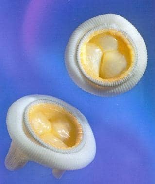
A heart valve prosthesis is a medical device that is implanted in the heart to replace a damaged or malfunctioning heart valve. The prosthetic valve can be made of biological tissue (such as from a pig or cow) or artificial materials (such as carbon or polyester). Its function is to allow for the proper directional flow of blood through the heart, opening and closing with each heartbeat to prevent backflow of blood.
There are several types of heart valve prostheses, including:
1. Mechanical valves: These are made entirely of artificial materials and have a longer lifespan than biological valves. However, they require the patient to take blood-thinning medication for the rest of their life to prevent blood clots from forming on the valve.
2. Bioprosthetic valves: These are made of biological tissue and typically last 10-15 years before needing replacement. They do not require the patient to take blood-thinning medication, but there is a higher risk of reoperation due to degeneration of the tissue over time.
3. Homografts or allografts: These are human heart valves that have been donated and preserved for transplantation. They have similar longevity to bioprosthetic valves and do not require blood-thinning medication.
4. Autografts: In this case, the patient's own pulmonary valve is removed and used to replace the damaged aortic valve. This procedure is called the Ross procedure and has excellent long-term results, but it requires advanced surgical skills and is not widely available.
The choice of heart valve prosthesis depends on various factors, including the patient's age, overall health, lifestyle, and personal preferences.
Heart valve diseases are a group of conditions that affect the function of one or more of the heart's four valves (tricuspid, pulmonic, mitral, and aortic). These valves are responsible for controlling the direction and flow of blood through the heart. Heart valve diseases can cause the valves to become narrowed (stenosis), leaky (regurgitation or insufficiency), or improperly closed (prolapse), leading to disrupted blood flow within the heart and potentially causing symptoms such as shortness of breath, fatigue, chest pain, and irregular heart rhythms. The causes of heart valve diseases can include congenital defects, age-related degenerative changes, infections, rheumatic heart disease, and high blood pressure. Treatment options may include medications, surgical repair or replacement of the affected valve(s), or transcatheter procedures.
The aortic valve is the valve located between the left ventricle (the lower left chamber of the heart) and the aorta (the largest artery in the body, which carries oxygenated blood from the heart to the rest of the body). It is made up of three thin flaps or leaflets that open and close to regulate blood flow. During a heartbeat, the aortic valve opens to allow blood to be pumped out of the left ventricle into the aorta, and then closes to prevent blood from flowing back into the ventricle when it relaxes. Any abnormality or damage to this valve can lead to various cardiovascular conditions such as aortic stenosis, aortic regurgitation, or infective endocarditis.
The mitral valve, also known as the bicuspid valve, is a two-leaflet valve located between the left atrium and left ventricle in the heart. Its function is to ensure unidirectional flow of blood from the left atrium into the left ventricle during the cardiac cycle. The mitral valve consists of two leaflets (anterior and posterior), the chordae tendineae, papillary muscles, and the left atrial and ventricular myocardium. Dysfunction of the mitral valve can lead to various heart conditions such as mitral regurgitation or mitral stenosis.
In medical terms, the heart is a muscular organ located in the thoracic cavity that functions as a pump to circulate blood throughout the body. It's responsible for delivering oxygen and nutrients to the tissues and removing carbon dioxide and other wastes. The human heart is divided into four chambers: two atria on the top and two ventricles on the bottom. The right side of the heart receives deoxygenated blood from the body and pumps it to the lungs, while the left side receives oxygenated blood from the lungs and pumps it out to the rest of the body. The heart's rhythmic contractions and relaxations are regulated by a complex electrical conduction system.
A bioprosthesis is a type of medical implant that is made from biological materials, such as heart valves or tendons taken from animals (xenografts) or humans (allografts). These materials are processed and sterilized to be used in surgical procedures to replace damaged or diseased tissues in the body.
Bioprosthetic implants are often used in cardiac surgery, such as heart valve replacement, because they are less likely to cause an immune response than synthetic materials. However, they may have a limited lifespan due to calcification and degeneration of the biological tissue over time. Therefore, bioprosthetic implants may need to be replaced after several years.
Bioprostheses can also be used in other types of surgical procedures, such as ligament or tendon repair, where natural tissue is needed to restore function and mobility. These prostheses are designed to mimic the properties of native tissues and provide a more physiological solution than synthetic materials.
Aortic valve stenosis is a cardiac condition characterized by the narrowing or stiffening of the aortic valve, which separates the left ventricle (the heart's main pumping chamber) from the aorta (the large artery that carries oxygen-rich blood to the rest of the body). This narrowing or stiffening prevents the aortic valve from opening fully, resulting in reduced blood flow from the left ventricle to the aorta and the rest of the body.
The narrowing can be caused by several factors, including congenital heart defects, calcification (hardening) of the aortic valve due to aging, or scarring of the valve due to rheumatic fever or other inflammatory conditions. As a result, the left ventricle must work harder to pump blood through the narrowed valve, which can lead to thickening and enlargement of the left ventricular muscle (left ventricular hypertrophy).
Symptoms of aortic valve stenosis may include chest pain or tightness, shortness of breath, fatigue, dizziness or fainting, and heart palpitations. Severe aortic valve stenosis can lead to serious complications such as heart failure, arrhythmias, or even sudden cardiac death. Treatment options may include medications to manage symptoms, lifestyle changes, or surgical intervention such as aortic valve replacement.
The pulmonary valve, also known as the pulmonic valve, is a semilunar valve located at the exit of the right ventricle of the heart and the beginning of the pulmonary artery. It has three cusps or leaflets that prevent the backflow of blood from the pulmonary artery into the right ventricle during ventricular diastole, ensuring unidirectional flow of blood towards the lungs for oxygenation.
Heart valve prosthesis implantation is a surgical procedure where an artificial heart valve is inserted to replace a damaged or malfunctioning native heart valve. This can be necessary for patients with valvular heart disease, including stenosis (narrowing) or regurgitation (leaking), who do not respond to medical management and are at risk of heart failure or other complications.
There are two main types of artificial heart valves used in prosthesis implantation: mechanical valves and biological valves. Mechanical valves are made of synthetic materials, such as carbon and metal, and can last a long time but require lifelong anticoagulation therapy to prevent blood clots from forming. Biological valves, on the other hand, are made from animal or human tissue and typically do not require anticoagulation therapy but may have a limited lifespan and may need to be replaced in the future.
The decision to undergo heart valve prosthesis implantation is based on several factors, including the patient's age, overall health, type and severity of valvular disease, and personal preferences. The procedure can be performed through traditional open-heart surgery or minimally invasive techniques, such as robotic-assisted surgery or transcatheter aortic valve replacement (TAVR). Recovery time varies depending on the approach used and individual patient factors.
The tricuspid valve is the heart valve that separates the right atrium and the right ventricle in the human heart. It is called "tricuspid" because it has three leaflets or cusps, which are also referred to as flaps or segments. These cusps are named anterior, posterior, and septal. The tricuspid valve's function is to prevent the backflow of blood from the ventricle into the atrium during systole, ensuring unidirectional flow of blood through the heart.
Aortic valve insufficiency, also known as aortic regurgitation or aortic incompetence, is a cardiac condition in which the aortic valve does not close properly during the contraction phase of the heart cycle. This allows blood to flow back into the left ventricle from the aorta, instead of being pumped out to the rest of the body. As a result, the left ventricle must work harder to maintain adequate cardiac output, which can lead to left ventricular enlargement and heart failure over time if left untreated.
The aortic valve is a trileaflet valve that lies between the left ventricle and the aorta. During systole (the contraction phase of the heart cycle), the aortic valve opens to allow blood to be pumped out of the left ventricle into the aorta and then distributed to the rest of the body. During diastole (the relaxation phase of the heart cycle), the aortic valve closes to prevent blood from flowing back into the left ventricle.
Aortic valve insufficiency can be caused by various conditions, including congenital heart defects, infective endocarditis, rheumatic heart disease, Marfan syndrome, and trauma. Symptoms of aortic valve insufficiency may include shortness of breath, fatigue, chest pain, palpitations, and edema (swelling). Diagnosis is typically made through physical examination, echocardiography, and other imaging studies. Treatment options depend on the severity of the condition and may include medication, surgery to repair or replace the aortic valve, or a combination of both.
Heart rate is the number of heartbeats per unit of time, often expressed as beats per minute (bpm). It can vary significantly depending on factors such as age, physical fitness, emotions, and overall health status. A resting heart rate between 60-100 bpm is generally considered normal for adults, but athletes and individuals with high levels of physical fitness may have a resting heart rate below 60 bpm due to their enhanced cardiovascular efficiency. Monitoring heart rate can provide valuable insights into an individual's health status, exercise intensity, and response to various treatments or interventions.
Mitral valve insufficiency, also known as mitral regurgitation, is a cardiac condition in which the mitral valve located between the left atrium and left ventricle of the heart does not close properly, causing blood to flow backward into the atrium during contraction of the ventricle. This leads to an increased volume load on the left heart chamber and can result in symptoms such as shortness of breath, fatigue, and fluid retention. The condition can be caused by various factors including valve damage due to degenerative changes, infective endocarditis, rheumatic heart disease, or trauma. Treatment options include medication, mitral valve repair, or replacement surgery depending on the severity and underlying cause of the insufficiency.
Bacterial endocarditis is a medical condition characterized by the inflammation and infection of the inner layer of the heart, known as the endocardium. This infection typically occurs when bacteria enter the bloodstream and attach themselves to damaged or abnormal heart valves or other parts of the endocardium. The bacteria can then multiply and cause the formation of vegetations, which are clusters of infected tissue that can further damage the heart valves and lead to serious complications such as heart failure, stroke, or even death if left untreated.
Bacterial endocarditis is a relatively uncommon but potentially life-threatening condition that requires prompt medical attention. Risk factors for developing bacterial endocarditis include pre-existing heart conditions such as congenital heart defects, artificial heart valves, previous history of endocarditis, or other conditions that damage the heart valves. Intravenous drug use is also a significant risk factor for this condition.
Symptoms of bacterial endocarditis may include fever, chills, fatigue, muscle and joint pain, shortness of breath, chest pain, and a new or changing heart murmur. Diagnosis typically involves a combination of medical history, physical examination, blood cultures, and imaging tests such as echocardiography. Treatment usually involves several weeks of intravenous antibiotics to eradicate the infection, and in some cases, surgical intervention may be necessary to repair or replace damaged heart valves.
Mitral valve stenosis is a cardiac condition characterized by the narrowing or stiffening of the mitral valve, one of the four heart valves that regulate blood flow through the heart. This narrowing prevents the mitral valve from fully opening during diastole (relaxation phase of the heart cycle), leading to restricted flow of oxygenated blood from the left atrium into the left ventricle.
The narrowing or stiffening of the mitral valve can be caused by various factors, such as rheumatic heart disease, congenital heart defects, aging, or calcium deposits on the valve leaflets. As a result, the left atrium has to work harder to pump blood into the left ventricle, causing increased pressure in the left atrium and pulmonary veins. This can lead to symptoms such as shortness of breath, fatigue, coughing, and heart palpitations.
Mitral valve stenosis is typically diagnosed through a combination of medical history, physical examination, and imaging techniques like echocardiography or cardiac catheterization. Treatment options may include medications to manage symptoms and prevent complications, as well as surgical interventions such as mitral valve repair or replacement to alleviate the stenosis and improve heart function.
Mitral valve prolapse (MVP) is a heart condition where the mitral valve, which separates the left atrium and left ventricle in the heart, doesn't function properly. In MVP, one or both of the mitral valve flaps (known as leaflets) bulge or billow into the left atrium during the contraction of the left ventricle. This prolapse can cause a leakage of blood back into the atrium, known as mitral regurgitation. In many cases, MVP is asymptomatic and doesn't require treatment, but in some instances, it may lead to complications such as infective endocarditis or arrhythmias. The exact causes of MVP are not fully understood, but it can be associated with certain genetic factors, connective tissue disorders, and mitral valve abnormalities present at birth.
Endocarditis is an inflammation of the inner layer of the heart chambers and heart valves, called the endocardium. This inflammation typically results from a bacterial or, less commonly, fungal infection that travels through the bloodstream and attaches to damaged areas of the heart.
There are two main types of endocarditis:
1. Acute Endocarditis: Develops quickly and can be severe, causing fever, chills, shortness of breath, fatigue, and heart murmurs. It may lead to serious complications like heart failure, embolism (blood clots that travel to other parts of the body), and damage to heart valves.
2. Subacute Endocarditis: Develops more slowly, often causing milder symptoms that can be mistaken for a cold or flu. Symptoms may include fatigue, weakness, fever, night sweats, weight loss, joint pain, and heart murmurs. Subacute endocarditis is more likely to affect people with previously damaged heart valves or congenital heart conditions.
Treatment usually involves several weeks of intravenous antibiotics or antifungal medications, depending on the cause of the infection. In some cases, surgery may be required to repair or replace damaged heart valves. Preventive measures include good oral hygiene and prompt treatment of infections, especially in individuals at a higher risk for endocarditis, such as those with congenital heart defects, artificial heart valves, or previous history of endocarditis.
Venous valves are one-way flaps made of thin, flexible tissue that lie inside your veins. They allow blood to flow towards the heart but prevent it from flowing backward. These valves are especially important in the veins of the legs, where they help to counteract the force of gravity and ensure that blood flows back up to the heart. When venous valves become damaged or weakened, blood can pool in the veins, leading to conditions such as varicose veins or chronic venous insufficiency.
Prosthesis design is a specialized field in medical device technology that involves creating and developing artificial substitutes to replace a missing body part, such as a limb, tooth, eye, or internal organ. The design process typically includes several stages: assessment of the patient's needs, selection of appropriate materials, creation of a prototype, testing and refinement, and final fabrication and fitting of the prosthesis.
The goal of prosthesis design is to create a device that functions as closely as possible to the natural body part it replaces, while also being comfortable, durable, and aesthetically pleasing for the patient. The design process may involve collaboration between medical professionals, engineers, and designers, and may take into account factors such as the patient's age, lifestyle, occupation, and overall health.
Prosthesis design can be highly complex, particularly for advanced devices such as robotic limbs or implantable organs. These devices often require sophisticated sensors, actuators, and control systems to mimic the natural functions of the body part they replace. As a result, prosthesis design is an active area of research and development in the medical field, with ongoing efforts to improve the functionality, comfort, and affordability of these devices for patients.
Heart failure is a pathophysiological state in which the heart is unable to pump sufficient blood to meet the metabolic demands of the body or do so only at the expense of elevated filling pressures. It can be caused by various cardiac disorders, including coronary artery disease, hypertension, valvular heart disease, cardiomyopathy, and arrhythmias. Symptoms may include shortness of breath, fatigue, and fluid retention. Heart failure is often classified based on the ejection fraction (EF), which is the percentage of blood that is pumped out of the left ventricle during each contraction. A reduced EF (less than 40%) is indicative of heart failure with reduced ejection fraction (HFrEF), while a preserved EF (greater than or equal to 50%) is indicative of heart failure with preserved ejection fraction (HFpEF). There is also a category of heart failure with mid-range ejection fraction (HFmrEF) for those with an EF between 40-49%.
Congenital heart defects (CHDs) are structural abnormalities in the heart that are present at birth. They can affect any part of the heart's structure, including the walls of the heart, the valves inside the heart, and the major blood vessels that lead to and from the heart.
Congenital heart defects can range from mild to severe and can cause various symptoms depending on the type and severity of the defect. Some common symptoms of CHDs include cyanosis (a bluish tint to the skin, lips, and fingernails), shortness of breath, fatigue, poor feeding, and slow growth in infants and children.
There are many different types of congenital heart defects, including:
1. Septal defects: These are holes in the walls that separate the four chambers of the heart. The two most common septal defects are atrial septal defect (ASD) and ventricular septal defect (VSD).
2. Valve abnormalities: These include narrowed or leaky valves, which can affect blood flow through the heart.
3. Obstruction defects: These occur when blood flow is blocked or restricted due to narrowing or absence of a part of the heart's structure. Examples include pulmonary stenosis and coarctation of the aorta.
4. Cyanotic heart defects: These cause a lack of oxygen in the blood, leading to cyanosis. Examples include tetralogy of Fallot and transposition of the great arteries.
The causes of congenital heart defects are not fully understood, but genetic factors and environmental influences during pregnancy may play a role. Some CHDs can be detected before birth through prenatal testing, while others may not be diagnosed until after birth or later in childhood. Treatment for CHDs may include medication, surgery, or other interventions to improve blood flow and oxygenation of the body's tissues.
Heart disease is a broad term for a class of diseases that involve the heart or blood vessels. It's often used to refer to conditions that include:
1. Coronary artery disease (CAD): This is the most common type of heart disease. It occurs when the arteries that supply blood to the heart become hardened and narrowed due to the buildup of cholesterol and other substances, which can lead to chest pain (angina), shortness of breath, or a heart attack.
2. Heart failure: This condition occurs when the heart is unable to pump blood efficiently to meet the body's needs. It can be caused by various conditions, including coronary artery disease, high blood pressure, and cardiomyopathy.
3. Arrhythmias: These are abnormal heart rhythms, which can be too fast, too slow, or irregular. They can lead to symptoms such as palpitations, dizziness, and fainting.
4. Valvular heart disease: This involves damage to one or more of the heart's four valves, which control blood flow through the heart. Damage can be caused by various conditions, including infection, rheumatic fever, and aging.
5. Cardiomyopathy: This is a disease of the heart muscle that makes it harder for the heart to pump blood efficiently. It can be caused by various factors, including genetics, viral infections, and drug abuse.
6. Pericardial disease: This involves inflammation or other problems with the sac surrounding the heart (pericardium). It can cause chest pain and other symptoms.
7. Congenital heart defects: These are heart conditions that are present at birth, such as a hole in the heart or abnormal blood vessels. They can range from mild to severe and may require medical intervention.
8. Heart infections: The heart can become infected by bacteria, viruses, or parasites, leading to various symptoms and complications.
It's important to note that many factors can contribute to the development of heart disease, including genetics, lifestyle choices, and certain medical conditions. Regular check-ups and a healthy lifestyle can help reduce the risk of developing heart disease.
Tricuspid valve insufficiency, also known as tricuspid regurgitation, is a cardiac condition in which the tricuspid valve located between the right atrium and right ventricle of the heart does not close properly, allowing blood to flow back into the right atrium during contraction of the right ventricle. This results in a portion of the blood being pumped inefficiently, which can lead to volume overload of the right side of the heart and potentially result in symptoms such as fatigue, weakness, shortness of breath, and fluid retention. The condition can be congenital or acquired, with common causes including dilated cardiomyopathy, infective endocarditis, rheumatic heart disease, and trauma.
Calcinosis is a medical condition characterized by the abnormal deposit of calcium salts in various tissues of the body, commonly under the skin or in the muscles and tendons. These calcium deposits can form hard lumps or nodules that can cause pain, inflammation, and restricted mobility. Calcinosis can occur as a complication of other medical conditions, such as autoimmune disorders, kidney disease, and hypercalcemia (high levels of calcium in the blood). In some cases, the cause of calcinosis may be unknown. Treatment for calcinosis depends on the underlying cause and may include medications to manage calcium levels, physical therapy, and surgical removal of large deposits.
Thromboembolism is a medical condition that refers to the obstruction of a blood vessel by a thrombus (blood clot) that has formed elsewhere in the body and then been transported by the bloodstream to a narrower vessel, where it becomes lodged. This process can occur in various parts of the body, leading to different types of thromboembolisms:
1. Deep Vein Thrombosis (DVT): A thrombus forms in the deep veins, usually in the legs or pelvis, and then breaks off and travels to the lungs, causing a pulmonary embolism.
2. Pulmonary Embolism (PE): A thrombus formed elsewhere, often in the deep veins of the legs, dislodges and travels to the lungs, blocking one or more pulmonary arteries. This can lead to shortness of breath, chest pain, and potentially life-threatening complications if not treated promptly.
3. Cerebral Embolism: A thrombus formed in another part of the body, such as the heart or carotid artery, dislodges and travels to the brain, causing a stroke or transient ischemic attack (TIA).
4. Arterial Thromboembolism: A thrombus forms in an artery and breaks off, traveling to another part of the body and blocking blood flow to an organ or tissue, leading to potential damage or loss of function. Examples include mesenteric ischemia (intestinal damage due to blocked blood flow) and retinal artery occlusion (vision loss due to blocked blood flow in the eye).
Prevention, early detection, and appropriate treatment are crucial for managing thromboembolism and reducing the risk of severe complications.
Tissue engineering is a branch of biomedical engineering that combines the principles of engineering, materials science, and biological sciences to develop functional substitutes for damaged or diseased tissues and organs. It involves the creation of living, three-dimensional structures that can restore, maintain, or improve tissue function. This is typically accomplished through the use of cells, scaffolds (biodegradable matrices), and biologically active molecules. The goal of tissue engineering is to develop biological substitutes that can ultimately restore normal function and structure in damaged tissues or organs.
Echocardiography is a medical procedure that uses sound waves to produce detailed images of the heart's structure, function, and motion. It is a non-invasive test that can help diagnose various heart conditions, such as valve problems, heart muscle damage, blood clots, and congenital heart defects.
During an echocardiogram, a transducer (a device that sends and receives sound waves) is placed on the chest or passed through the esophagus to obtain images of the heart. The sound waves produced by the transducer bounce off the heart structures and return to the transducer, which then converts them into electrical signals that are processed to create images of the heart.
There are several types of echocardiograms, including:
* Transthoracic echocardiography (TTE): This is the most common type of echocardiogram and involves placing the transducer on the chest.
* Transesophageal echocardiography (TEE): This type of echocardiogram involves passing a specialized transducer through the esophagus to obtain images of the heart from a closer proximity.
* Stress echocardiography: This type of echocardiogram is performed during exercise or medication-induced stress to assess how the heart functions under stress.
* Doppler echocardiography: This type of echocardiogram uses sound waves to measure blood flow and velocity in the heart and blood vessels.
Echocardiography is a valuable tool for diagnosing and managing various heart conditions, as it provides detailed information about the structure and function of the heart. It is generally safe, non-invasive, and painless, making it a popular choice for doctors and patients alike.
Heart transplantation is a surgical procedure where a diseased, damaged, or failing heart is removed and replaced with a healthy donor heart. This procedure is usually considered as a last resort for patients with end-stage heart failure or severe coronary artery disease who have not responded to other treatments. The donor heart typically comes from a brain-dead individual whose family has agreed to donate their loved one's organs for transplantation. Heart transplantation is a complex and highly specialized procedure that requires a multidisciplinary team of healthcare professionals, including cardiologists, cardiac surgeons, anesthesiologists, perfusionists, nurses, and other support staff. The success rates for heart transplantation have improved significantly over the past few decades, with many patients experiencing improved quality of life and increased survival rates. However, recipients of heart transplants require lifelong immunosuppressive therapy to prevent rejection of the donor heart, which can increase the risk of infections and other complications.
Cardiac catheterization is a medical procedure used to diagnose and treat cardiovascular conditions. In this procedure, a thin, flexible tube called a catheter is inserted into a blood vessel in the arm or leg and threaded up to the heart. The catheter can be used to perform various diagnostic tests, such as measuring the pressure inside the heart chambers and assessing the function of the heart valves.
Cardiac catheterization can also be used to treat certain cardiovascular conditions, such as narrowed or blocked arteries. In these cases, a balloon or stent may be inserted through the catheter to open up the blood vessel and improve blood flow. This procedure is known as angioplasty or percutaneous coronary intervention (PCI).
Cardiac catheterization is typically performed in a hospital cardiac catheterization laboratory by a team of healthcare professionals, including cardiologists, radiologists, and nurses. The procedure may be done under local anesthesia with sedation or general anesthesia, depending on the individual patient's needs and preferences.
Overall, cardiac catheterization is a valuable tool in the diagnosis and treatment of various heart conditions, and it can help improve symptoms, reduce complications, and prolong life for many patients.
Prosthesis failure is a term used to describe a situation where a prosthetic device, such as an artificial joint or limb, has stopped functioning or failed to meet its intended purpose. This can be due to various reasons, including mechanical failure, infection, loosening of the device, or a reaction to the materials used in the prosthesis.
Mechanical failure can occur due to wear and tear, manufacturing defects, or improper use of the prosthetic device. Infection can also lead to prosthesis failure, particularly in cases where the prosthesis is implanted inside the body. The immune system may react to the presence of the foreign material, leading to inflammation and infection.
Loosening of the prosthesis can also cause it to fail over time, as the device becomes less stable and eventually stops working properly. Additionally, some people may have a reaction to the materials used in the prosthesis, leading to tissue damage or other complications that can result in prosthesis failure.
In general, prosthesis failure can lead to decreased mobility, pain, and the need for additional surgeries or treatments to correct the problem. It is important for individuals with prosthetic devices to follow their healthcare provider's instructions carefully to minimize the risk of prosthesis failure and ensure that the device continues to function properly over time.
Anticoagulants are a class of medications that work to prevent the formation of blood clots in the body. They do this by inhibiting the coagulation cascade, which is a series of chemical reactions that lead to the formation of a clot. Anticoagulants can be given orally, intravenously, or subcutaneously, depending on the specific drug and the individual patient's needs.
There are several different types of anticoagulants, including:
1. Heparin: This is a naturally occurring anticoagulant that is often used in hospitalized patients who require immediate anticoagulation. It works by activating an enzyme called antithrombin III, which inhibits the formation of clots.
2. Low molecular weight heparin (LMWH): LMWH is a form of heparin that has been broken down into smaller molecules. It has a longer half-life than standard heparin and can be given once or twice daily by subcutaneous injection.
3. Direct oral anticoagulants (DOACs): These are newer oral anticoagulants that work by directly inhibiting specific clotting factors in the coagulation cascade. Examples include apixaban, rivaroxaban, and dabigatran.
4. Vitamin K antagonists: These are older oral anticoagulants that work by inhibiting the action of vitamin K, which is necessary for the formation of clotting factors. Warfarin is an example of a vitamin K antagonist.
Anticoagulants are used to prevent and treat a variety of conditions, including deep vein thrombosis (DVT), pulmonary embolism (PE), atrial fibrillation, and prosthetic heart valve thrombosis. It is important to note that anticoagulants can increase the risk of bleeding, so they must be used with caution and regular monitoring of blood clotting times may be required.
Cardiovascular models are simplified representations or simulations of the human cardiovascular system used in medical research, education, and training. These models can be physical, computational, or mathematical and are designed to replicate various aspects of the heart, blood vessels, and blood flow. They can help researchers study the structure and function of the cardiovascular system, test new treatments and interventions, and train healthcare professionals in diagnostic and therapeutic techniques.
Physical cardiovascular models may include artificial hearts, blood vessels, or circulation systems made from materials such as plastic, rubber, or silicone. These models can be used to study the mechanics of heart valves, the effects of different surgical procedures, or the impact of various medical devices on blood flow.
Computational and mathematical cardiovascular models use algorithms and equations to simulate the behavior of the cardiovascular system. These models may range from simple representations of a single heart chamber to complex simulations of the entire circulatory system. They can be used to study the electrical activity of the heart, the biomechanics of blood flow, or the distribution of drugs in the body.
Overall, cardiovascular models play an essential role in advancing our understanding of the human body and improving patient care.
The fetal heart is the cardiovascular organ that develops in the growing fetus during pregnancy. It starts to form around 22 days after conception and continues to develop throughout the first trimester. By the end of the eighth week of gestation, the fetal heart has developed enough to pump blood throughout the body.
The fetal heart is similar in structure to the adult heart but has some differences. It is smaller and more compact, with a four-chambered structure that includes two atria and two ventricles. The fetal heart also has unique features such as the foramen ovale, which is a hole between the right and left atria that allows blood to bypass the lungs, and the ductus arteriosus, a blood vessel that connects the pulmonary artery to the aorta and diverts blood away from the lungs.
The fetal heart is responsible for pumping oxygenated blood from the placenta to the rest of the body and returning deoxygenated blood back to the placenta for re-oxygenation. The rate of the fetal heartbeat is faster than that of an adult, typically ranging from 120 to 160 beats per minute. Fetal heart rate monitoring is a common method used during pregnancy and childbirth to assess the health and well-being of the developing fetus.
Mitral valve annuloplasty is a surgical procedure that involves repairing and reinforcing the mitral valve in the heart, which helps control blood flow between the left atrium and left ventricle. The procedure typically aims to reduce the size of the mitral valve's dilated or stretched opening (annulus) by implanting a prosthetic ring or band around it. This reinforcement helps restore normal valve function, preventing regurgitation or backflow of blood into the atrium during heart contractions.
The procedure is often performed to treat mitral valve regurgitation, which can be caused by various factors such as age-related degenerative changes, infective endocarditis, rheumatic heart disease, or congenital abnormalities. Mitral valve annuloplasty may be done alone or in combination with other cardiac surgeries like mitral valve replacement or repair of the valve leaflets.
Treatment outcome is a term used to describe the result or effect of medical treatment on a patient's health status. It can be measured in various ways, such as through symptoms improvement, disease remission, reduced disability, improved quality of life, or survival rates. The treatment outcome helps healthcare providers evaluate the effectiveness of a particular treatment plan and make informed decisions about future care. It is also used in clinical research to compare the efficacy of different treatments and improve patient care.
Transesophageal echocardiography (TEE) is a type of echocardiogram, which is a medical test that uses sound waves to create detailed images of the heart. In TEE, a special probe containing a transducer is passed down the esophagus (the tube that connects the mouth to the stomach) to obtain views of the heart from behind. This allows for more detailed images of the heart structures and function compared to a standard echocardiogram, which uses a probe placed on the chest. TEE is often used in patients with poor image quality from a standard echocardiogram or when more detailed images are needed to diagnose or monitor certain heart conditions. It is typically performed by a trained cardiologist or sonographer under the direction of a cardiologist.
The International Normalized Ratio (INR) is a standardized measurement of the prothrombin time (PT), which is the time it takes for blood to clot. The INR is used to monitor and regulate the effects of anticoagulant medications, such as warfarin, that affect the blood's ability to clot.
The INR is calculated by dividing the patient's PT by a control value (the PT of normal, healthy blood), raised to the power of a sensitivity factor called the International Sensitivity Index (ISI). The ISI is specific to the thromboplastin reagent used in the PT assay.
The INR provides a consistent and comparable way to monitor anticoagulation therapy across different laboratories, regardless of the thromboplastin reagent used. This helps ensure that patients receive appropriate doses of anticoagulant medications and reduces the risk of bleeding or clotting complications.
In general, an INR range of 2.0 to 3.0 is recommended for most people taking anticoagulants for conditions such as atrial fibrillation, deep vein thrombosis, or pulmonary embolism. However, the target INR range may vary depending on individual patient factors and medical indications.
Cardiac surgical procedures are operations that are performed on the heart or great vessels (the aorta and vena cava) by cardiothoracic surgeons. These surgeries are often complex and require a high level of skill and expertise. Some common reasons for cardiac surgical procedures include:
1. Coronary artery bypass grafting (CABG): This is a surgery to improve blood flow to the heart in patients with coronary artery disease. During the procedure, a healthy blood vessel from another part of the body is used to create a detour around the blocked or narrowed portion of the coronary artery.
2. Valve repair or replacement: The heart has four valves that control blood flow through and out of the heart. If one or more of these valves become damaged or diseased, they may need to be repaired or replaced. This can be done using artificial valves or valves from animal or human donors.
3. Aneurysm repair: An aneurysm is a weakened area in the wall of an artery that can bulge out and potentially rupture. If an aneurysm occurs in the aorta, it may require surgical repair to prevent rupture.
4. Heart transplantation: In some cases, heart failure may be so severe that a heart transplant is necessary. This involves removing the diseased heart and replacing it with a healthy donor heart.
5. Arrhythmia surgery: Certain types of abnormal heart rhythms (arrhythmias) may require surgical treatment. One such procedure is called the Maze procedure, which involves creating a pattern of scar tissue in the heart to disrupt the abnormal electrical signals that cause the arrhythmia.
6. Congenital heart defect repair: Some people are born with structural problems in their hearts that require surgical correction. These may include holes between the chambers of the heart or abnormal blood vessels.
Cardiac surgical procedures carry risks, including bleeding, infection, stroke, and death. However, for many patients, these surgeries can significantly improve their quality of life and longevity.
The endocardial cushions are a part of the embryonic heart that contributes to the formation of the atrioventricular septum and the valves between the chambers of the heart. They are composed of mesenchymal tissue, which is a type of connective tissue that contains cells called mesenchymal stem cells. During fetal development, these cushions grow and fuse together to form the atrioventricular septum, which separates the upper chambers (atria) from the lower chambers (ventricles) of the heart. The endocardial cushions also give rise to the valves that regulate blood flow between the chambers of the heart. Defects in the development of the endocardial cushions can lead to congenital heart defects, such as atrial septal defect and ventricular septal defect.
Heart valve prolapse, also known as mitral valve prolapse or MVP, is a condition in which the leaflets (flaps) of the heart's valves do not close properly. In heart valve prolapse, one or more of the valve leaflets bulge into the upper chamber of the heart (atrium) when the valve closes. This can cause a backflow of blood, known as regurgitation, which can lead to symptoms such as shortness of breath, fatigue, and irregular heart rhythms. Heart valve prolapse is most commonly affects the mitral valve, but it can also affect the other heart valves. The exact cause of heart valve prolapse is not known, but it may be associated with certain factors such as connective tissue disorders, aging, and previous heart conditions. In many cases, heart valve prolapse does not cause any symptoms or complications and may only require regular monitoring by a healthcare professional. However, in some cases, heart valve prolapse can lead to serious complications such as endocarditis (inflammation of the inner lining of the heart) or heart failure, so it is important to seek medical attention if you experience any symptoms or have concerns about your heart health.
Absorbable implants are medical devices that are designed to be placed inside the body during a surgical procedure, where they provide support, stabilization, or other functions, and then gradually break down and are absorbed by the body over time. These implants are typically made from materials such as polymers, proteins, or ceramics that have been engineered to degrade at a controlled rate, allowing them to be resorbed and eliminated from the body without the need for a second surgical procedure to remove them.
Absorbable implants are often used in orthopedic, dental, and plastic surgery applications, where they can help promote healing and support tissue regeneration. For example, absorbable screws or pins may be used to stabilize fractured bones during the healing process, after which they will gradually dissolve and be absorbed by the body. Similarly, absorbable membranes may be used in dental surgery to help guide the growth of new bone and gum tissue around an implant, and then be resorbed over time.
It's important to note that while absorbable implants offer several advantages over non-absorbable materials, such as reduced risk of infection and improved patient comfort, they may also have some limitations. For example, the mechanical properties of absorbable materials may not be as strong as those of non-absorbable materials, which could affect their performance in certain applications. Additionally, the degradation products of absorbable implants may cause local inflammation or other adverse reactions in some patients. As with any medical device, the use of absorbable implants should be carefully considered and discussed with a qualified healthcare professional.
Acenocoumarol is an anticoagulant medication that is used to prevent and treat blood clots. It works by inhibiting the formation of vitamin K-dependent clotting factors, which are necessary for normal blood coagulation. This results in a prolonged bleeding time and reduced risk of blood clots.
Acenocoumarol is a coumarin derivative and is available under various brand names, including Sintrom and Nicoumalone. It is typically administered orally in the form of tablets and its effects are monitored through regular blood tests to ensure that the dosage is appropriate and that the risk of bleeding complications is minimized.
Common side effects of acenocoumarol include easy bruising, nosebleeds, and skin rashes. It may also interact with a variety of other medications, including antibiotics, antifungals, and certain herbal supplements, so it is important to inform your healthcare provider of all medications and supplements you are taking before starting acenocoumarol therapy.
It is important to note that acenocoumarol has a narrow therapeutic index, meaning that the difference between an effective dose and a toxic dose is relatively small. Therefore, it is essential to follow your healthcare provider's instructions carefully when taking this medication and to have regular blood tests to monitor its effects on your coagulation status.
The heart atria are the upper chambers of the heart that receive blood from the veins and deliver it to the lower chambers, or ventricles. There are two atria in the heart: the right atrium receives oxygen-poor blood from the body and pumps it into the right ventricle, which then sends it to the lungs to be oxygenated; and the left atrium receives oxygen-rich blood from the lungs and pumps it into the left ventricle, which then sends it out to the rest of the body. The atria contract before the ventricles during each heartbeat, helping to fill the ventricles with blood and prepare them for contraction.
In the field of medicine, "time factors" refer to the duration of symptoms or time elapsed since the onset of a medical condition, which can have significant implications for diagnosis and treatment. Understanding time factors is crucial in determining the progression of a disease, evaluating the effectiveness of treatments, and making critical decisions regarding patient care.
For example, in stroke management, "time is brain," meaning that rapid intervention within a specific time frame (usually within 4.5 hours) is essential to administering tissue plasminogen activator (tPA), a clot-busting drug that can minimize brain damage and improve patient outcomes. Similarly, in trauma care, the "golden hour" concept emphasizes the importance of providing definitive care within the first 60 minutes after injury to increase survival rates and reduce morbidity.
Time factors also play a role in monitoring the progression of chronic conditions like diabetes or heart disease, where regular follow-ups and assessments help determine appropriate treatment adjustments and prevent complications. In infectious diseases, time factors are crucial for initiating antibiotic therapy and identifying potential outbreaks to control their spread.
Overall, "time factors" encompass the significance of recognizing and acting promptly in various medical scenarios to optimize patient outcomes and provide effective care.
Warfarin is a anticoagulant medication that works by inhibiting the vitamin K-dependent activation of several coagulation factors (factors II, VII, IX, and X). This results in prolonged clotting times and reduced thrombus formation. It is commonly used to prevent and treat blood clots in conditions such as atrial fibrillation, deep vein thrombosis, and pulmonary embolism. Warfarin is also known by its brand names Coumadin and Jantoven.
It's important to note that warfarin has a narrow therapeutic index, meaning that the difference between an effective dose and a toxic one is small. Therefore, it requires careful monitoring of the patient's coagulation status through regular blood tests (INR) to ensure that the dosage is appropriate and to minimize the risk of bleeding complications.
The endocardium is the innermost layer of tissue that lines the chambers of the heart and the valves between them. It is a thin, smooth membrane that is in contact with the blood within the heart. This layer helps to maintain the heart's internal environment, facilitates the smooth movement of blood through the heart, and provides a protective barrier against infection and other harmful substances. The endocardium is composed of simple squamous epithelial cells called endothelial cells, which are supported by a thin layer of connective tissue.
Cardiac valve annuloplasty is a surgical procedure that involves repairing and reinforcing the ring-like structure (annulus) surrounding the heart valves, primarily the mitral or tricuspid valves. This procedure is often performed to correct valve leaks or regurgitation caused by various conditions such as valve disease or dilated cardiomyopathy.
During the annuloplasty procedure, the surgeon typically uses an artificial ring-like device (annuloplasty ring) made of fabric, metal, or a combination of both to reshape and stabilize the damaged annulus. The ring is sewn in place, reducing the size of the valve opening and helping the valve leaflets to coapt properly, thereby preventing valve leaks and improving heart function.
Annuloplasty can be performed as a standalone procedure or in combination with other cardiac surgeries such as valve replacement or repair. The specific technique and approach may vary depending on the individual patient's needs and the surgeon's preference.
Heart sounds are the noises generated by the beating heart and the movement of blood through it. They are caused by the vibration of the cardiac structures, such as the valves, walls, and blood vessels, during the cardiac cycle.
There are two normal heart sounds, often described as "lub-dub," that can be heard through a stethoscope. The first sound (S1) is caused by the closure of the mitral and tricuspid valves at the beginning of systole, when the ventricles contract to pump blood out to the body and lungs. The second sound (S2) is produced by the closure of the aortic and pulmonary valves at the end of systole, as the ventricles relax and the ventricular pressure decreases, allowing the valves to close.
Abnormal heart sounds, such as murmurs, clicks, or extra sounds (S3 or S4), may indicate cardiac disease or abnormalities in the structure or function of the heart. These sounds can be evaluated through a process called auscultation, which involves listening to the heart with a stethoscope and analyzing the intensity, pitch, quality, and timing of the sounds.
Hemodynamics is the study of how blood flows through the cardiovascular system, including the heart and the vascular network. It examines various factors that affect blood flow, such as blood volume, viscosity, vessel length and diameter, and pressure differences between different parts of the circulatory system. Hemodynamics also considers the impact of various physiological and pathological conditions on these variables, and how they in turn influence the function of vital organs and systems in the body. It is a critical area of study in fields such as cardiology, anesthesiology, and critical care medicine.
Cardiovascular complications in pregnancy refer to conditions that affect the heart and blood vessels, which can arise during pregnancy, childbirth, or after delivery. These complications can be pre-existing or new-onset and can range from mild to severe, potentially threatening the life of both the mother and the fetus. Some examples of cardiovascular complications in pregnancy include:
1. Hypertension disorders: This includes chronic hypertension (high blood pressure before pregnancy), gestational hypertension (high blood pressure that develops after 20 weeks of pregnancy), and preeclampsia/eclampsia (a pregnancy-specific disorder characterized by high blood pressure, proteinuria, and potential organ damage).
2. Cardiomyopathy: A condition in which the heart muscle becomes weakened, leading to an enlarged heart and reduced pumping efficiency. Peripartum cardiomyopathy is a specific type that occurs during pregnancy or in the months following delivery.
3. Arrhythmias: Irregularities in the heart's rhythm, such as tachycardia (rapid heartbeat) or bradycardia (slow heartbeat), can occur during pregnancy and may require medical intervention.
4. Valvular heart disease: Pre-existing valve disorders, like mitral stenosis or aortic insufficiency, can worsen during pregnancy due to increased blood volume and cardiac output. Additionally, new valve issues might develop during pregnancy.
5. Venous thromboembolism (VTE): Pregnancy increases the risk of developing blood clots in the veins, particularly deep vein thrombosis (DVT) or pulmonary embolism (PE).
6. Ischemic heart disease: Although rare, coronary artery disease and acute coronary syndrome can occur during pregnancy, especially in women with risk factors such as obesity, diabetes, or smoking history.
7. Heart failure: Severe cardiac dysfunction leading to fluid accumulation, shortness of breath, and reduced exercise tolerance may develop due to any of the above conditions or other underlying heart diseases.
Early recognition, monitoring, and appropriate management of these cardiovascular complications in pregnancy are crucial for maternal and fetal well-being.
Pulmonary Valve Stenosis is a cardiac condition where the pulmonary valve, located between the right ventricle and the pulmonary artery, has a narrowed opening. This stenosis (narrowing) can cause obstruction of blood flow from the right ventricle to the lungs. The narrowing can be caused by a fusion of the valve leaflets, thickened or calcified valve leaflets, or rarely, a dysplastic valve.
The severity of Pulmonary Valve Stenosis is classified based on the gradient pressure across the valve, which is measured during an echocardiogram. A mild stenosis has a gradient of less than 30 mmHg, moderate stenosis has a gradient between 30-59 mmHg, and severe stenosis has a gradient of 60 mmHg or higher.
Mild Pulmonary Valve Stenosis may not require treatment, while more severe cases may need to be treated with balloon valvuloplasty or surgical valve replacement. If left untreated, Pulmonary Valve Stenosis can lead to right ventricular hypertrophy, heart failure, and other complications.
Rheumatic Heart Disease (RHD) is defined as a chronic heart condition caused by damage to the heart valves due to untreated or inadequately treated streptococcal throat infection (strep throat). The immune system's response to this infection can mistakenly attack and damage the heart tissue, leading to inflammation and scarring of the heart valves. This damage can result in narrowing, leakage, or abnormal functioning of the heart valves, which can further lead to complications such as heart failure, stroke, or infective endocarditis.
RHD is a preventable and treatable condition if detected early and managed effectively. It primarily affects children and young adults in developing countries where access to healthcare and antibiotics for strep throat infections may be limited. Long-term management of RHD typically involves medications, regular monitoring, and sometimes surgical intervention to repair or replace damaged heart valves.
Follow-up studies are a type of longitudinal research that involve repeated observations or measurements of the same variables over a period of time, in order to understand their long-term effects or outcomes. In medical context, follow-up studies are often used to evaluate the safety and efficacy of medical treatments, interventions, or procedures.
In a typical follow-up study, a group of individuals (called a cohort) who have received a particular treatment or intervention are identified and then followed over time through periodic assessments or data collection. The data collected may include information on clinical outcomes, adverse events, changes in symptoms or functional status, and other relevant measures.
The results of follow-up studies can provide important insights into the long-term benefits and risks of medical interventions, as well as help to identify factors that may influence treatment effectiveness or patient outcomes. However, it is important to note that follow-up studies can be subject to various biases and limitations, such as loss to follow-up, recall bias, and changes in clinical practice over time, which must be carefully considered when interpreting the results.
I believe there might be a misunderstanding in your question. "Glutaral" does not seem to be a recognized medical term or abbreviation in healthcare and biomedical sciences. It is possible that you may be looking for information on "glutaraldehyde," which is a disinfectant and sterilizing agent used in medical settings.
Glutaraldehyde is a chemical compound with the formula C5H8O2, and it's often used as a 2% solution. It's an effective agent against bacteria, viruses, and fungi, making it useful for sterilizing medical equipment. However, glutaraldehyde can cause respiratory issues and skin irritation in some individuals, so proper handling and use are essential to minimize exposure.
If you meant to ask about a different term or if this answer does not address your question, please provide more context or clarify your request, and I will be happy to help further.
Echocardiography, Doppler, color is a type of ultrasound test that uses sound waves to create detailed moving images of the heart and its blood vessels. In this technique, color Doppler is used to visualize the direction and speed of blood flow through the heart and great vessels. The movement of the red blood cells causes a change in frequency of the reflected sound waves (Doppler shift), which can be used to calculate the velocity and direction of the blood flow. By adding color to the Doppler image, it becomes easier for the interpreting physician to understand the complex three-dimensional motion of blood through the heart. This test is often used to diagnose and monitor various heart conditions, including valve disorders, congenital heart defects, and cardiac muscle diseases.
A bioreactor is a device or system that supports and controls the conditions necessary for biological organisms, cells, or tissues to grow and perform their specific functions. It provides a controlled environment with appropriate temperature, pH, nutrients, and other factors required for the desired biological process to occur. Bioreactors are widely used in various fields such as biotechnology, pharmaceuticals, agriculture, and environmental science for applications like production of therapeutic proteins, vaccines, biofuels, enzymes, and wastewater treatment.
The myocardium is the middle layer of the heart wall, composed of specialized cardiac muscle cells that are responsible for pumping blood throughout the body. It forms the thickest part of the heart wall and is divided into two sections: the left ventricle, which pumps oxygenated blood to the rest of the body, and the right ventricle, which pumps deoxygenated blood to the lungs.
The myocardium contains several types of cells, including cardiac muscle fibers, connective tissue, nerves, and blood vessels. The muscle fibers are arranged in a highly organized pattern that allows them to contract in a coordinated manner, generating the force necessary to pump blood through the heart and circulatory system.
Damage to the myocardium can occur due to various factors such as ischemia (reduced blood flow), infection, inflammation, or genetic disorders. This damage can lead to several cardiac conditions, including heart failure, arrhythmias, and cardiomyopathy.
Postoperative complications refer to any unfavorable condition or event that occurs during the recovery period after a surgical procedure. These complications can vary in severity and may include, but are not limited to:
1. Infection: This can occur at the site of the incision or inside the body, such as pneumonia or urinary tract infection.
2. Bleeding: Excessive bleeding (hemorrhage) can lead to a drop in blood pressure and may require further surgical intervention.
3. Blood clots: These can form in the deep veins of the legs (deep vein thrombosis) and can potentially travel to the lungs (pulmonary embolism).
4. Wound dehiscence: This is when the surgical wound opens up, which can lead to infection and further complications.
5. Pulmonary issues: These include atelectasis (collapsed lung), pneumonia, or respiratory failure.
6. Cardiovascular problems: These include abnormal heart rhythms (arrhythmias), heart attack, or stroke.
7. Renal failure: This can occur due to various reasons such as dehydration, blood loss, or the use of certain medications.
8. Pain management issues: Inadequate pain control can lead to increased stress, anxiety, and decreased mobility.
9. Nausea and vomiting: These can be caused by anesthesia, opioid pain medication, or other factors.
10. Delirium: This is a state of confusion and disorientation that can occur in the elderly or those with certain medical conditions.
Prompt identification and management of these complications are crucial to ensure the best possible outcome for the patient.
A reoperation is a surgical procedure that is performed again on a patient who has already undergone a previous operation for the same or related condition. Reoperations may be required due to various reasons, such as inadequate initial treatment, disease recurrence, infection, or complications from the first surgery. The nature and complexity of a reoperation can vary widely depending on the specific circumstances, but it often carries higher risks and potential complications compared to the original operation.
"Papio ursinus" is the scientific name for the Chacma Baboon, which is a species of baboon that is native to southern Africa. They are Old World monkeys that are found in various habitats such as savannas, woodlands, and mountainous regions. Chacma Baboons are known for their social structure, intelligence, and adaptability. They have a distinctive dog-like face with a heavy build and a dog-like muzzle. Adult males can weigh between 33 to 90 kg (73 to 198 lb), while adult females are smaller and weigh between 15 to 27 kg (33 to 59 lb). Chacma Baboons primarily feed on plants, but they also eat insects, small vertebrates, and fruits. They are an important species in the ecosystem and play a significant role in seed dispersal and nutrient cycling.
Doppler echocardiography is a type of ultrasound test that uses high-frequency sound waves to produce detailed images of the heart and its blood vessels. It measures the direction and speed of blood flow in the heart and major blood vessels leading to and from the heart. This helps to evaluate various conditions such as valve problems, congenital heart defects, and heart muscle diseases.
In Doppler echocardiography, a small handheld device called a transducer is placed on the chest, which emits sound waves that bounce off the heart and blood vessels. The transducer then picks up the returning echoes, which are processed by a computer to create moving images of the heart.
The Doppler effect is used to measure the speed and direction of blood flow. This occurs when the frequency of the sound waves changes as they bounce off moving objects, such as red blood cells. By analyzing these changes, the ultrasound machine can calculate the velocity and direction of blood flow in different parts of the heart.
Doppler echocardiography is a non-invasive test that does not require any needles or dyes. It is generally safe and painless, although patients may experience some discomfort from the pressure applied by the transducer on the chest. The test usually takes about 30 to 60 minutes to complete.
An air embolism is a medical condition that occurs when one or more air bubbles enter the bloodstream and block or obstruct blood vessels. This can lead to various symptoms depending on the severity and location of the obstruction, including shortness of breath, chest pain, confusion, stroke, or even death.
Air embolisms can occur in a variety of ways, such as during certain medical procedures (e.g., when air is accidentally introduced into a vein or artery), trauma to the lungs or blood vessels, scuba diving, or mountain climbing. Treatment typically involves administering oxygen and supportive care, as well as removing the source of the air bubbles if possible. In severe cases, hyperbaric oxygen therapy may be used to help reduce the size of the air bubbles and improve outcomes.
Carcinoid heart disease is a rare complication that occurs in some people with carcinoid tumors, which are slow-growing tumors that typically originate in the digestive tract. These tumors can release hormones and other substances into the bloodstream, which can cause various symptoms. In carcinoid heart disease, these substances cause fibrous plaques to form on the heart valves, leading to thickening and stiffening of the valve leaflets. This can result in leakage or obstruction of the heart valves, causing symptoms such as shortness of breath, fatigue, and fluid retention. Carcinoid heart disease is most commonly affects the tricuspid and pulmonary valves, which are located on the right side of the heart. If left untreated, carcinoid heart disease can lead to serious complications, including heart failure. Treatment typically involves a combination of medications to manage symptoms and control the growth of the tumor, as well as surgery to repair or replace damaged heart valves.
Balloon valvuloplasty is a medical procedure used to treat heart valve stenosis or narrowing. It involves the use of a thin, flexible tube (catheter) with a balloon at its tip, which is guided through a blood vessel to the narrowed heart valve. Once in position, the balloon is inflated to stretch and widen the valve opening, improving blood flow. After the valve is widened, the balloon is deflated and the catheter is removed. This procedure can be performed on various heart valves, including the aortic, mitral, and pulmonary valves.
Heart block is a cardiac condition characterized by the interruption of electrical impulse transmission from the atria (the upper chambers of the heart) to the ventricles (the lower chambers of the heart). This disruption can lead to abnormal heart rhythms, including bradycardia (a slower-than-normal heart rate), and in severe cases, can cause the heart to stop beating altogether. Heart block is typically caused by damage to the heart's electrical conduction system due to various factors such as aging, heart disease, or certain medications.
There are three types of heart block: first-degree, second-degree, and third-degree (also known as complete heart block). Each type has distinct electrocardiogram (ECG) findings and symptoms. Treatment for heart block depends on the severity of the condition and may include monitoring, medication, or implantation of a pacemaker to regulate the heart's electrical activity.
Decanoates are a type of esterified form of certain drugs or medications, particularly in the case of testosterone. The decanoate ester is attached to the testosterone molecule to create a longer-acting formulation. Testosterone decanoate is a slow-release form of testosterone that is used as a replacement therapy for individuals who have low levels of natural testosterone. It is administered through intramuscular injection and has a duration of action of approximately 2-3 weeks.
Other medications may also be available in decanoate ester form, but testosterone decanoate is one of the most commonly used. As with any medication or treatment plan, it's important to consult with a healthcare provider to determine the best course of action based on individual needs and medical history.
Phonocardiography is a non-invasive medical procedure that involves the graphical representation and analysis of sounds produced by the heart. It uses a device called a phonocardiograph to record these sounds, which are then displayed as waveforms on a screen. The procedure is often used in conjunction with other diagnostic techniques, such as electrocardiography (ECG), to help diagnose various heart conditions, including valvular heart disease and heart murmurs.
During the procedure, a specialized microphone called a phonendoscope is placed on the chest wall over the area of the heart. The microphone picks up the sounds generated by the heart's movements, such as the closing and opening of the heart valves, and transmits them to the phonocardiograph. The phonocardiograph then converts these sounds into a visual representation, which can be analyzed for any abnormalities or irregularities in the heart's function.
Phonocardiography is a valuable tool for healthcare professionals, as it can provide important insights into the health and functioning of the heart. By analyzing the waveforms produced during phonocardiography, doctors can identify any potential issues with the heart's valves or other structures, which may require further investigation or treatment. Overall, phonocardiography is an essential component of modern cardiac diagnostics, helping to ensure that patients receive accurate and timely diagnoses for their heart conditions.
Multidetector computed tomography (MDCT) is a type of computed tomography (CT) scan that uses multiple rows of detectors to acquire several slices of images simultaneously, thereby reducing the total time required for the scan and improving the spatial resolution. This technology allows for faster scanning of moving organs, such as the heart, and provides high-resolution images with detailed information about various body structures, including bones, soft tissues, and blood vessels. MDCT has numerous applications in diagnostic imaging, interventional procedures, and cancer staging and treatment follow-up.
A serotonin receptor, specifically the 5-HT2B receptor, is a type of G protein-coupled receptor (GPCR) that binds to the neurotransmitter serotonin (5-hydroxytryptamine or 5-HT). These receptors are located on the cell membrane of certain cells, including neurons and other cell types in various organs.
The 5-HT2B receptor is involved in a variety of physiological functions, such as regulating mood, appetite, sleep, and sensory perception. In the cardiovascular system, activation of 5-HT2B receptors can lead to the proliferation of cardiac fibroblasts and changes in the extracellular matrix, which may contribute to heart valve abnormalities.
In the central nervous system, 5-HT2B receptors have been implicated in several neurological conditions, including migraine, depression, and schizophrenia. However, their precise roles in these disorders are not yet fully understood.
Pharmacologically targeting 5-HT2B receptors has led to the development of drugs for various indications, such as antimigraine medications (e.g., telcagepant) and potential treatments for heart failure (e.g., mavacamten). However, some 5-HT2B receptor agonists have also been associated with serious side effects, such as valvular heart disease, which has limited their clinical use.
Prospective studies, also known as longitudinal studies, are a type of cohort study in which data is collected forward in time, following a group of individuals who share a common characteristic or exposure over a period of time. The researchers clearly define the study population and exposure of interest at the beginning of the study and follow up with the participants to determine the outcomes that develop over time. This type of study design allows for the investigation of causal relationships between exposures and outcomes, as well as the identification of risk factors and the estimation of disease incidence rates. Prospective studies are particularly useful in epidemiology and medical research when studying diseases with long latency periods or rare outcomes.
Medical Definition:
"Risk factors" are any attribute, characteristic or exposure of an individual that increases the likelihood of developing a disease or injury. They can be divided into modifiable and non-modifiable risk factors. Modifiable risk factors are those that can be changed through lifestyle choices or medical treatment, while non-modifiable risk factors are inherent traits such as age, gender, or genetic predisposition. Examples of modifiable risk factors include smoking, alcohol consumption, physical inactivity, and unhealthy diet, while non-modifiable risk factors include age, sex, and family history. It is important to note that having a risk factor does not guarantee that a person will develop the disease, but rather indicates an increased susceptibility.
Safety-based drug withdrawals refer to the removal of a medication from the market due to concerns about its safety profile. This action is typically taken by regulatory authorities, such as the US Food and Drug Administration (FDA), when new information emerges that suggests a drug may pose an unacceptable risk of harm to patients, even if the benefit-risk balance was previously considered favorable.
Safety-based drug withdrawals can occur for various reasons, including the identification of new adverse effects, interactions with other medications or medical conditions, or an increased understanding of the drug's pharmacology or toxicology. In some cases, safety-based drug withdrawals may be temporary, allowing the manufacturer to conduct further studies and address the safety concerns. However, in other instances, the withdrawal may be permanent, leading to the discontinuation of the drug's production and distribution.
It is important to note that safety-based drug withdrawals are rare and typically represent a small fraction of the drugs approved for use. Nevertheless, they serve as an essential mechanism for protecting public health and ensuring that medications on the market are safe and effective for their intended use.
The chordae tendineae are cord-like tendons that attach the heart's papillary muscles to the tricuspid and mitral valves in the heart. They play a crucial role in preventing the backflow of blood into the atria during ventricular contraction. The chordae tendineae ensure that the cusps of the atrioventricular valves close properly and maintain their shape during the cardiac cycle. Damage to these tendons can result in heart conditions such as mitral or tricuspid valve regurgitation.
Thrombosis is the formation of a blood clot (thrombus) inside a blood vessel, obstructing the flow of blood through the circulatory system. When a clot forms in an artery, it can cut off the supply of oxygen and nutrients to the tissues served by that artery, leading to damage or tissue death. If a thrombus forms in the heart, it can cause a heart attack. If a thrombus breaks off and travels through the bloodstream, it can lodge in a smaller vessel, causing blockage and potentially leading to damage in the organ that the vessel supplies. This is known as an embolism.
Thrombosis can occur due to various factors such as injury to the blood vessel wall, abnormalities in blood flow, or changes in the composition of the blood. Certain medical conditions, medications, and lifestyle factors can increase the risk of thrombosis. Treatment typically involves anticoagulant or thrombolytic therapy to dissolve or prevent further growth of the clot, as well as addressing any underlying causes.
Induced heart arrest, also known as controlled cardiac arrest or planned cardiac arrest, is a deliberate medical intervention where cardiac activity is temporarily stopped through the use of medications or electrical disruption. This procedure is typically carried out during a surgical procedure, such as open-heart surgery, where the heart needs to be stilled to allow surgeons to work on it safely.
The most common method used to induce heart arrest is by administering a medication called potassium chloride, which stops the heart's electrical activity. Alternatively, an electrical shock may be delivered to the heart to achieve the same effect. Once the procedure is complete, the heart can be restarted using various resuscitation techniques, such as defibrillation or medication administration.
It's important to note that induced heart arrest is a carefully monitored and controlled medical procedure carried out by trained healthcare professionals in a hospital setting. It should not be confused with sudden cardiac arrest, which is an unexpected and often unpredictable event that occurs outside of a medical setting.
Fenfluramine is a drug that was previously used for the short-term treatment of obesity. It works by suppressing appetite and increasing the feeling of fullness. Fenfluramine is an amphetamine derivative and stimulates the release of serotonin, a neurotransmitter in the brain that helps regulate mood, appetite, and sleep.
Fenfluramine was commonly prescribed in combination with phentermine, another appetite suppressant, under the brand name Fen-Phen. However, in 1997, the U.S. Food and Drug Administration (FDA) issued a public health warning about the potential risk of serious heart valve damage associated with the use of fenfluramine and withdrew its approval for the drug's use. Since then, fenfluramine has not been approved for medical use in many countries, including the United States.
Tensile strength is a material property that measures the maximum amount of tensile (pulling) stress that a material can withstand before failure, such as breaking or fracturing. It is usually measured in units of force per unit area, such as pounds per square inch (psi) or pascals (Pa). In the context of medical devices or biomaterials, tensile strength may be used to describe the mechanical properties of materials used in implants, surgical tools, or other medical equipment. High tensile strength is often desirable in these applications to ensure that the material can withstand the stresses and forces it will encounter during use.
Bioartificial organs are hybrid structures that combine living cells, tissues, or biological materials with non-living synthetic materials to replicate the functions of a natural organ. These constructs are designed to mimic the complex architecture and functionality of native organs, providing a viable alternative to traditional organ transplantation.
The bioartificial organ typically consists of three main components:
1. Scaffold: A porous, biocompatible synthetic material that provides structural support and a framework for cell attachment, growth, and organization. The scaffold can be made from various materials such as polymers, ceramics, or composites, and its design considers factors like mechanical strength, degradation rate, and biocompatibility.
2. Cells: Living cells are seeded onto the scaffold, where they proliferate, differentiate, and synthesize extracellular matrix (ECM) proteins to form functional tissue. The choice of cell type depends on the specific organ being replicated; for example, hepatocytes for a liver or cardiomyocytes for a heart.
3. Vascularization: To ensure adequate nutrient and waste exchange, bioartificial organs require an efficient vascular network. This can be achieved through various methods such as co-culturing endothelial cells with the primary cell type, using bioprinting techniques to create patterned vasculature, or incorporating microfluidic channels within the scaffold.
The development of bioartificial organs holds great promise for addressing the current shortage of donor organs and providing personalized treatment options for patients with organ failure. However, several challenges must be overcome before these constructs can be widely adopted in clinical settings, including optimizing vascularization, maintaining long-term functionality, and ensuring biocompatibility and safety.
Heart neoplasms are abnormal growths or tumors that develop within the heart tissue. They can be benign (noncancerous) or malignant (cancerous). Benign tumors, such as myxomas and rhabdomyomas, are typically slower growing and less likely to spread, but they can still cause serious complications if they obstruct blood flow or damage heart valves. Malignant tumors, such as angiosarcomas and rhabdomyosarcomas, are fast-growing and have a higher risk of spreading to other parts of the body. Symptoms of heart neoplasms can include shortness of breath, chest pain, fatigue, and irregular heart rhythms. Treatment options depend on the type, size, and location of the tumor, and may include surgery, radiation therapy, or chemotherapy.
"Swine" is a common term used to refer to even-toed ungulates of the family Suidae, including domestic pigs and wild boars. However, in a medical context, "swine" often appears in the phrase "swine flu," which is a strain of influenza virus that typically infects pigs but can also cause illness in humans. The 2009 H1N1 pandemic was caused by a new strain of swine-origin influenza A virus, which was commonly referred to as "swine flu." It's important to note that this virus is not transmitted through eating cooked pork products; it spreads from person to person, mainly through respiratory droplets produced when an infected person coughs or sneezes.
The extracellular matrix (ECM) is a complex network of biomolecules that provides structural and biochemical support to cells in tissues and organs. It is composed of various proteins, glycoproteins, and polysaccharides, such as collagens, elastin, fibronectin, laminin, and proteoglycans. The ECM plays crucial roles in maintaining tissue architecture, regulating cell behavior, and facilitating communication between cells. It provides a scaffold for cell attachment, migration, and differentiation, and helps to maintain the structural integrity of tissues by resisting mechanical stresses. Additionally, the ECM contains various growth factors, cytokines, and chemokines that can influence cellular processes such as proliferation, survival, and differentiation. Overall, the extracellular matrix is essential for the normal functioning of tissues and organs, and its dysregulation can contribute to various pathological conditions, including fibrosis, cancer, and degenerative diseases.
Hemorrhage is defined in the medical context as an excessive loss of blood from the circulatory system, which can occur due to various reasons such as injury, surgery, or underlying health conditions that affect blood clotting or the integrity of blood vessels. The bleeding may be internal, external, visible, or concealed, and it can vary in severity from minor to life-threatening, depending on the location and extent of the bleeding. Hemorrhage is a serious medical emergency that requires immediate attention and treatment to prevent further blood loss, organ damage, and potential death.
I apologize, but "sheep" is not a term that has a medical definition. It is a common animal with the scientific name Ovis aries. If you have any medical or health-related questions, I would be happy to try and help answer those for you.
An embolism is a medical condition that occurs when a substance, such as a blood clot or an air bubble, blocks a blood vessel. This can happen in any part of the body, but it is particularly dangerous when it affects the brain (causing a stroke) or the lungs (causing a pulmonary embolism). Embolisms can cause serious harm by preventing oxygen and nutrients from reaching the tissues and organs that need them. They are often the result of underlying medical conditions, such as heart disease or deep vein thrombosis, and may require immediate medical attention to prevent further complications.
Retrospective studies, also known as retrospective research or looking back studies, are a type of observational study that examines data from the past to draw conclusions about possible causal relationships between risk factors and outcomes. In these studies, researchers analyze existing records, medical charts, or previously collected data to test a hypothesis or answer a specific research question.
Retrospective studies can be useful for generating hypotheses and identifying trends, but they have limitations compared to prospective studies, which follow participants forward in time from exposure to outcome. Retrospective studies are subject to biases such as recall bias, selection bias, and information bias, which can affect the validity of the results. Therefore, retrospective studies should be interpreted with caution and used primarily to generate hypotheses for further testing in prospective studies.
Tissue preservation is the process of preventing decomposition or autolysis (self-digestion) of tissues after they have been removed from a living organism. This is typically achieved through the use of fixatives, such as formaldehyde or glutaraldehyde, which stabilize proteins and other cellular structures by creating cross-links between them. Other methods of tissue preservation include freezing, dehydration, and embedding in paraffin or plastic resins. Properly preserved tissues can be stored for long periods of time and used for various research and diagnostic purposes, such as histology, immunohistochemistry, and molecular biology studies.
Myocardial contraction refers to the rhythmic and forceful shortening of heart muscle cells (myocytes) in the myocardium, which is the muscular wall of the heart. This process is initiated by electrical signals generated by the sinoatrial node, causing a wave of depolarization that spreads throughout the heart.
During myocardial contraction, calcium ions flow into the myocytes, triggering the interaction between actin and myosin filaments, which are the contractile proteins in the muscle cells. This interaction causes the myofilaments to slide past each other, resulting in the shortening of the sarcomeres (the functional units of muscle contraction) and ultimately leading to the contraction of the heart muscle.
Myocardial contraction is essential for pumping blood throughout the body and maintaining adequate circulation to vital organs. Any impairment in myocardial contractility can lead to various cardiac disorders, such as heart failure, cardiomyopathy, and arrhythmias.
Cardiomyopathies are a group of diseases that affect the heart muscle, leading to mechanical and/or electrical dysfunction. The American Heart Association (AHA) defines cardiomyopathies as "a heterogeneous group of diseases of the myocardium associated with mechanical and/or electrical dysfunction that usually (but not always) exhibit inappropriate ventricular hypertrophy or dilatation and frequently lead to heart failure."
There are several types of cardiomyopathies, including:
1. Dilated cardiomyopathy (DCM): This is the most common type of cardiomyopathy, characterized by an enlarged left ventricle and impaired systolic function, leading to heart failure.
2. Hypertrophic cardiomyopathy (HCM): In this type, there is abnormal thickening of the heart muscle, particularly in the septum between the two ventricles, which can obstruct blood flow and increase the risk of arrhythmias.
3. Restrictive cardiomyopathy (RCM): This is a rare form of cardiomyopathy characterized by stiffness of the heart muscle, impaired relaxation, and diastolic dysfunction, leading to reduced filling of the ventricles and heart failure.
4. Arrhythmogenic right ventricular cardiomyopathy (ARVC): In this type, there is replacement of the normal heart muscle with fatty or fibrous tissue, primarily affecting the right ventricle, which can lead to arrhythmias and sudden cardiac death.
5. Unclassified cardiomyopathies: These are conditions that do not fit into any of the above categories but still significantly affect the heart muscle and function.
Cardiomyopathies can be caused by genetic factors, acquired conditions (e.g., infections, toxins, or autoimmune disorders), or a combination of both. The diagnosis typically involves a comprehensive evaluation, including medical history, physical examination, electrocardiogram (ECG), echocardiography, cardiac magnetic resonance imaging (MRI), and sometimes genetic testing. Treatment depends on the type and severity of the condition but may include medications, lifestyle modifications, implantable devices, or even heart transplantation in severe cases.
Heart function tests are a group of diagnostic exams that are used to evaluate the structure and functioning of the heart. These tests help doctors assess the pumping efficiency of the heart, the flow of blood through the heart, the presence of any heart damage, and the overall effectiveness of the heart in delivering oxygenated blood to the rest of the body.
Some common heart function tests include:
1. Echocardiogram (Echo): This test uses sound waves to create detailed images of the heart's structure and functioning. It can help detect any damage to the heart muscle, valves, or sac surrounding the heart.
2. Nuclear Stress Test: This test involves injecting a small amount of radioactive substance into the patient's bloodstream and taking images of the heart while it is at rest and during exercise. The test helps evaluate blood flow to the heart and detect any areas of reduced blood flow, which could indicate coronary artery disease.
3. Cardiac Magnetic Resonance Imaging (MRI): This test uses magnetic fields and radio waves to create detailed images of the heart's structure and function. It can help detect any damage to the heart muscle, valves, or other structures of the heart.
4. Electrocardiogram (ECG): This test measures the electrical activity of the heart and helps detect any abnormalities in the heart's rhythm or conduction system.
5. Exercise Stress Test: This test involves walking on a treadmill or riding a stationary bike while being monitored for changes in heart rate, blood pressure, and ECG readings. It helps evaluate exercise capacity and detect any signs of coronary artery disease.
6. Cardiac Catheterization: This is an invasive procedure that involves inserting a catheter into the heart to measure pressures and take samples of blood from different parts of the heart. It can help diagnose various heart conditions, including heart valve problems, congenital heart defects, and coronary artery disease.
Overall, heart function tests play an essential role in diagnosing and managing various heart conditions, helping doctors provide appropriate treatment and improve patient outcomes.
Mechanical stress, in the context of physiology and medicine, refers to any type of force that is applied to body tissues or organs, which can cause deformation or displacement of those structures. Mechanical stress can be either external, such as forces exerted on the body during physical activity or trauma, or internal, such as the pressure changes that occur within blood vessels or other hollow organs.
Mechanical stress can have a variety of effects on the body, depending on the type, duration, and magnitude of the force applied. For example, prolonged exposure to mechanical stress can lead to tissue damage, inflammation, and chronic pain. Additionally, abnormal or excessive mechanical stress can contribute to the development of various musculoskeletal disorders, such as tendinitis, osteoarthritis, and herniated discs.
In order to mitigate the negative effects of mechanical stress, the body has a number of adaptive responses that help to distribute forces more evenly across tissues and maintain structural integrity. These responses include changes in muscle tone, joint positioning, and connective tissue stiffness, as well as the remodeling of bone and other tissues over time. However, when these adaptive mechanisms are overwhelmed or impaired, mechanical stress can become a significant factor in the development of various pathological conditions.
Prothrombin time (PT) is a medical laboratory test that measures the time it takes for blood to clot. It's often used to evaluate the functioning of the extrinsic and common pathways of the coagulation system, which is responsible for blood clotting. Specifically, PT measures how long it takes for prothrombin (a protein produced by the liver) to be converted into thrombin, an enzyme that converts fibrinogen into fibrin and helps form a clot.
Prolonged PT may indicate a bleeding disorder or a deficiency in coagulation factors, such as vitamin K deficiency or the use of anticoagulant medications like warfarin. It's important to note that PT is often reported with an international normalized ratio (INR), which allows for standardization and comparison of results across different laboratories and reagent types.
Tissue scaffolds, also known as bioactive scaffolds or synthetic extracellular matrices, refer to three-dimensional structures that serve as templates for the growth and organization of cells in tissue engineering and regenerative medicine. These scaffolds are designed to mimic the natural extracellular matrix (ECM) found in biological tissues, providing a supportive environment for cell attachment, proliferation, differentiation, and migration.
Tissue scaffolds can be made from various materials, including naturally derived biopolymers (e.g., collagen, alginate, chitosan, hyaluronic acid), synthetic polymers (e.g., polycaprolactone, polylactic acid, poly(lactic-co-glycolic acid)), or a combination of both. The choice of material depends on the specific application and desired properties, such as biocompatibility, biodegradability, mechanical strength, and porosity.
The primary functions of tissue scaffolds include:
1. Cell attachment: Providing surfaces for cells to adhere, spread, and form stable focal adhesions.
2. Mechanical support: Offering a structural framework that maintains the desired shape and mechanical properties of the engineered tissue.
3. Nutrient diffusion: Ensuring adequate transport of nutrients, oxygen, and waste products throughout the scaffold to support cell survival and function.
4. Guided tissue growth: Directing the organization and differentiation of cells through spatial cues and biochemical signals.
5. Biodegradation: Gradually degrading at a rate that matches tissue regeneration, allowing for the replacement of the scaffold with native ECM produced by the cells.
Tissue scaffolds have been used in various applications, such as wound healing, bone and cartilage repair, cardiovascular tissue engineering, and neural tissue regeneration. The design and fabrication of tissue scaffolds are critical aspects of tissue engineering, aiming to create functional substitutes for damaged or diseased tissues and organs.
Biocompatible materials are non-toxic and non-reacting substances that can be used in medical devices, tissue engineering, and drug delivery systems without causing harm or adverse reactions to living tissues or organs. These materials are designed to mimic the properties of natural tissues and are able to integrate with biological systems without being rejected by the body's immune system.
Biocompatible materials can be made from a variety of substances, including metals, ceramics, polymers, and composites. The specific properties of these materials, such as their mechanical strength, flexibility, and biodegradability, are carefully selected to meet the requirements of their intended medical application.
Examples of biocompatible materials include titanium used in dental implants and joint replacements, polyethylene used in artificial hips, and hydrogels used in contact lenses and drug delivery systems. The use of biocompatible materials has revolutionized modern medicine by enabling the development of advanced medical technologies that can improve patient outcomes and quality of life.
Amaurosis fugax is a medical term that describes a temporary loss of vision in one eye, which is often described as a "shade or curtain falling over the field of vision." It's usually caused by a temporary interruption of blood flow to the retina or optic nerve. This condition is often associated with conditions such as giant cell arteritis, carotid artery stenosis, and cardiovascular disease.
It's important to note that Amaurosis fugax can be a warning sign for a more serious medical event, such as a stroke, so it's essential to seek medical attention promptly if you experience any symptoms of this condition.
The pericardium is the double-walled sac that surrounds the heart. It has an outer fibrous layer and an inner serous layer, which further divides into two parts: the parietal layer lining the fibrous pericardium and the visceral layer (epicardium) closely adhering to the heart surface.
The space between these two layers is filled with a small amount of lubricating serous fluid, allowing for smooth movement of the heart within the pericardial cavity. The pericardium provides protection, support, and helps maintain the heart's normal position within the chest while reducing friction during heart contractions.
Biomedical engineering is a field that combines engineering principles and design concepts with medical and biological sciences to develop solutions to healthcare challenges. It involves the application of engineering methods to analyze, understand, and solve problems in biology and medicine, with the goal of improving human health and well-being. Biomedical engineers may work on a wide range of projects, including developing new medical devices, designing artificial organs, creating diagnostic tools, simulating biological systems, and optimizing healthcare delivery systems. They often collaborate with other professionals such as doctors, nurses, and scientists to develop innovative solutions that meet the needs of patients and healthcare providers.
The ileocecal valve, also known as the Bauhin's valve, is a vital physiological structure in the gastrointestinal tract. It is a valve located at the junction between the ileum (the final portion of the small intestine) and the cecum (the first part of the large intestine or colon). This valve functions to control the flow of digesta from the small intestine into the large intestine, preventing backflow from the colon into the small intestine. It is an essential component in maintaining proper digestive function and gut health.
Left ventricular function refers to the ability of the left ventricle (the heart's lower-left chamber) to contract and relax, thereby filling with and ejecting blood. The left ventricle is responsible for pumping oxygenated blood to the rest of the body. Its function is evaluated by measuring several parameters, including:
1. Ejection fraction (EF): This is the percentage of blood that is pumped out of the left ventricle with each heartbeat. A normal ejection fraction ranges from 55% to 70%.
2. Stroke volume (SV): The amount of blood pumped by the left ventricle in one contraction. A typical SV is about 70 mL/beat.
3. Cardiac output (CO): The total volume of blood that the left ventricle pumps per minute, calculated as the product of stroke volume and heart rate. Normal CO ranges from 4 to 8 L/minute.
Assessment of left ventricular function is crucial in diagnosing and monitoring various cardiovascular conditions such as heart failure, coronary artery disease, valvular heart diseases, and cardiomyopathies.
Biomechanics is the application of mechanical laws to living structures and systems, particularly in the field of medicine and healthcare. A biomechanical phenomenon refers to a observable event or occurrence that involves the interaction of biological tissues or systems with mechanical forces. These phenomena can be studied at various levels, from the molecular and cellular level to the tissue, organ, and whole-body level.
Examples of biomechanical phenomena include:
1. The way that bones and muscles work together to produce movement (known as joint kinematics).
2. The mechanical behavior of biological tissues such as bone, cartilage, tendons, and ligaments under various loads and stresses.
3. The response of cells and tissues to mechanical stimuli, such as the way that bone tissue adapts to changes in loading conditions (known as Wolff's law).
4. The biomechanics of injury and disease processes, such as the mechanisms of joint injury or the development of osteoarthritis.
5. The use of mechanical devices and interventions to treat medical conditions, such as orthopedic implants or assistive devices for mobility impairments.
Understanding biomechanical phenomena is essential for developing effective treatments and prevention strategies for a wide range of medical conditions, from musculoskeletal injuries to neurological disorders.
Risk assessment in the medical context refers to the process of identifying, evaluating, and prioritizing risks to patients, healthcare workers, or the community related to healthcare delivery. It involves determining the likelihood and potential impact of adverse events or hazards, such as infectious diseases, medication errors, or medical devices failures, and implementing measures to mitigate or manage those risks. The goal of risk assessment is to promote safe and high-quality care by identifying areas for improvement and taking action to minimize harm.
Equipment Failure Analysis is a process of identifying the cause of failure in medical equipment or devices. This involves a systematic examination and evaluation of the equipment, its components, and operational history to determine why it failed. The analysis may include physical inspection, chemical testing, and review of maintenance records, as well as assessment of design, manufacturing, and usage factors that may have contributed to the failure.
The goal of Equipment Failure Analysis is to identify the root cause of the failure, so that corrective actions can be taken to prevent similar failures in the future. This is important in medical settings to ensure patient safety and maintain the reliability and effectiveness of medical equipment.
Myocardial ischemia is a condition in which the blood supply to the heart muscle (myocardium) is reduced or blocked, leading to insufficient oxygen delivery and potential damage to the heart tissue. This reduction in blood flow typically results from the buildup of fatty deposits, called plaques, in the coronary arteries that supply the heart with oxygen-rich blood. The plaques can rupture or become unstable, causing the formation of blood clots that obstruct the artery and limit blood flow.
Myocardial ischemia may manifest as chest pain (angina pectoris), shortness of breath, fatigue, or irregular heartbeats (arrhythmias). In severe cases, it can lead to myocardial infarction (heart attack) if the oxygen supply is significantly reduced or cut off completely, causing permanent damage or death of the heart muscle. Early diagnosis and treatment of myocardial ischemia are crucial for preventing further complications and improving patient outcomes.
Subacute bacterial endocarditis (SBE) is a type of infective endocarditis that typically has a more indolent course compared to acute bacterial endocarditis. It is caused by organisms that are less virulent and have a higher affinity for damaged heart valves or endocardium.
The most common causative organisms of SBE include Streptococcus viridans, Streptococcus bovis, and enterococci. The infection often develops over a period of weeks to months, with nonspecific symptoms such as fatigue, weakness, fever, weight loss, and night sweats.
SBE can lead to serious complications, including heart failure, valvular damage, embolic events, and even death if left untreated. Treatment typically involves prolonged courses of intravenous antibiotics, with surgical intervention reserved for cases with severe valvular damage or uncontrolled infection.
Preventive measures include appropriate management of underlying heart conditions, prophylactic antibiotic therapy in high-risk individuals undergoing dental or invasive procedures, and good oral hygiene.
1. Intracranial Embolism: This is a medical condition that occurs when a blood clot or other particle (embolus) formed elsewhere in the body, travels through the bloodstream and lodges itself in the intracranial blood vessels, blocking the flow of blood to a part of the brain. This can lead to various neurological symptoms such as weakness, numbness, speech difficulties, or even loss of consciousness, depending on the severity and location of the blockage.
2. Intracranial Thrombosis: This is a medical condition that occurs when a blood clot (thrombus) forms within the intracranial blood vessels. The clot can partially or completely obstruct the flow of blood, leading to various symptoms such as headache, confusion, seizures, or neurological deficits, depending on the severity and location of the thrombosis. Intracranial thrombosis can occur due to various factors including atherosclerosis, hypertension, diabetes, and other medical conditions that increase the risk of blood clot formation.
Stroke volume is a term used in cardiovascular physiology and medicine. It refers to the amount of blood that is pumped out of the left ventricle of the heart during each contraction (systole). Specifically, it is the difference between the volume of blood in the left ventricle at the end of diastole (when the ventricle is filled with blood) and the volume at the end of systole (when the ventricle has contracted and ejected its contents into the aorta).
Stroke volume is an important measure of heart function, as it reflects the ability of the heart to pump blood effectively to the rest of the body. A low stroke volume may indicate that the heart is not pumping efficiently, while a high stroke volume may suggest that the heart is working too hard. Stroke volume can be affected by various factors, including heart disease, high blood pressure, and physical fitness level.
The formula for calculating stroke volume is:
Stroke Volume = End-Diastolic Volume - End-Systolic Volume
Where end-diastolic volume (EDV) is the volume of blood in the left ventricle at the end of diastole, and end-systolic volume (ESV) is the volume of blood in the left ventricle at the end of systole.
Heart injuries, also known as cardiac injuries, refer to any damage or harm caused to the heart muscle, valves, or surrounding structures. This can result from various causes such as blunt trauma (e.g., car accidents, falls), penetrating trauma (e.g., gunshot wounds, stabbing), or medical conditions like heart attacks (myocardial infarction) and infections (e.g., myocarditis, endocarditis).
Some common types of heart injuries include:
1. Contusions: Bruising of the heart muscle due to blunt trauma.
2. Myocardial infarctions: Damage to the heart muscle caused by insufficient blood supply, often due to blocked coronary arteries.
3. Cardiac rupture: A rare but life-threatening condition where the heart muscle tears or breaks open, usually resulting from severe trauma or complications from a myocardial infarction.
4. Valvular damage: Disruption of the heart valves' function due to injury or infection, leading to leakage (regurgitation) or narrowing (stenosis).
5. Pericardial injuries: Damage to the pericardium, the sac surrounding the heart, which can result in fluid accumulation (pericardial effusion), inflammation (pericarditis), or tamponade (compression of the heart by excess fluid).
6. Arrhythmias: Irregular heart rhythms caused by damage to the heart's electrical conduction system.
Timely diagnosis and appropriate treatment are crucial for managing heart injuries, as they can lead to severe complications or even be fatal if left untreated.
Equipment design, in the medical context, refers to the process of creating and developing medical equipment and devices, such as surgical instruments, diagnostic machines, or assistive technologies. This process involves several stages, including:
1. Identifying user needs and requirements
2. Concept development and brainstorming
3. Prototyping and testing
4. Design for manufacturing and assembly
5. Safety and regulatory compliance
6. Verification and validation
7. Training and support
The goal of equipment design is to create safe, effective, and efficient medical devices that meet the needs of healthcare providers and patients while complying with relevant regulations and standards. The design process typically involves a multidisciplinary team of engineers, clinicians, designers, and researchers who work together to develop innovative solutions that improve patient care and outcomes.
Catheterization is a medical procedure in which a catheter (a flexible tube) is inserted into the body to treat various medical conditions or for diagnostic purposes. The specific definition can vary depending on the area of medicine and the particular procedure being discussed. Here are some common types of catheterization:
1. Urinary catheterization: This involves inserting a catheter through the urethra into the bladder to drain urine. It is often performed to manage urinary retention, monitor urine output in critically ill patients, or assist with surgical procedures.
2. Cardiac catheterization: A procedure where a catheter is inserted into a blood vessel, usually in the groin or arm, and guided to the heart. This allows for various diagnostic tests and treatments, such as measuring pressures within the heart chambers, assessing blood flow, or performing angioplasty and stenting of narrowed coronary arteries.
3. Central venous catheterization: A catheter is inserted into a large vein, typically in the neck, chest, or groin, to administer medications, fluids, or nutrition, or to monitor central venous pressure.
4. Peritoneal dialysis catheterization: A catheter is placed into the abdominal cavity for individuals undergoing peritoneal dialysis, a type of kidney replacement therapy.
5. Neurological catheterization: In some cases, a catheter may be inserted into the cerebrospinal fluid space (lumbar puncture) or the brain's ventricular system (ventriculostomy) to diagnose or treat various neurological conditions.
These are just a few examples of catheterization procedures in medicine. The specific definition and purpose will depend on the medical context and the particular organ or body system involved.
A feasibility study is a preliminary investigation or analysis conducted to determine the viability of a proposed project, program, or product. In the medical field, feasibility studies are often conducted before implementing new treatments, procedures, equipment, or facilities. These studies help to assess the practicality and effectiveness of the proposed intervention, as well as its potential benefits and risks.
Feasibility studies in healthcare typically involve several steps:
1. Problem identification: Clearly define the problem that the proposed project, program, or product aims to address.
2. Objectives setting: Establish specific, measurable, achievable, relevant, and time-bound (SMART) objectives for the study.
3. Literature review: Conduct a thorough review of existing research and best practices related to the proposed intervention.
4. Methodology development: Design a methodology for data collection and analysis that will help answer the research questions and achieve the study's objectives.
5. Resource assessment: Evaluate the availability and adequacy of resources, including personnel, time, and finances, required to carry out the proposed intervention.
6. Risk assessment: Identify potential risks and challenges associated with the implementation of the proposed intervention and develop strategies to mitigate them.
7. Cost-benefit analysis: Estimate the costs and benefits of the proposed intervention, including direct and indirect costs, as well as short-term and long-term benefits.
8. Stakeholder engagement: Engage relevant stakeholders, such as patients, healthcare providers, administrators, and policymakers, to gather their input and support for the proposed intervention.
9. Decision-making: Based on the findings of the feasibility study, make an informed decision about whether or not to proceed with the proposed project, program, or product.
Feasibility studies are essential in healthcare as they help ensure that resources are allocated efficiently and effectively, and that interventions are evidence-based, safe, and beneficial for patients.
A Severity of Illness Index is a measurement tool used in healthcare to assess the severity of a patient's condition and the risk of mortality or other adverse outcomes. These indices typically take into account various physiological and clinical variables, such as vital signs, laboratory values, and co-morbidities, to generate a score that reflects the patient's overall illness severity.
Examples of Severity of Illness Indices include the Acute Physiology and Chronic Health Evaluation (APACHE) system, the Simplified Acute Physiology Score (SAPS), and the Mortality Probability Model (MPM). These indices are often used in critical care settings to guide clinical decision-making, inform prognosis, and compare outcomes across different patient populations.
It is important to note that while these indices can provide valuable information about a patient's condition, they should not be used as the sole basis for clinical decision-making. Rather, they should be considered in conjunction with other factors, such as the patient's overall clinical presentation, treatment preferences, and goals of care.
The sternum, also known as the breastbone, is a long, flat bone located in the central part of the chest. It serves as the attachment point for several muscles and tendons, including those involved in breathing. The sternum has three main parts: the manubrium at the top, the body in the middle, and the xiphoid process at the bottom. The upper seven pairs of ribs connect to the sternum via costal cartilages.
Prosthesis-related infections, also known as prosthetic joint infections (PJIs), are infections that occur around or within a prosthetic device, such as an artificial joint. These infections can be caused by bacteria, fungi, or other microorganisms and can lead to serious complications if not treated promptly and effectively.
Prosthesis-related infections can occur soon after the implantation of the prosthetic device (early infection) or months or even years later (late infection). Early infections are often caused by bacteria that enter the surgical site during the procedure, while late infections may be caused by hematogenous seeding (i.e., when bacteria from another source spread through the bloodstream and settle in the prosthetic device) or by contamination during a subsequent medical procedure.
Symptoms of prosthesis-related infections can include pain, swelling, redness, warmth, and drainage around the affected area. In some cases, patients may also experience fever, chills, or fatigue. Diagnosis typically involves a combination of clinical evaluation, laboratory tests (such as blood cultures, joint fluid analysis, and tissue biopsy), and imaging studies (such as X-rays, CT scans, or MRI).
Treatment of prosthesis-related infections usually involves a combination of antibiotics and surgical intervention. The specific treatment approach will depend on the type and severity of the infection, as well as the patient's overall health status. In some cases, it may be necessary to remove or replace the affected prosthetic device.
I'm happy to help! However, I must clarify that "materials testing" is not a medical term per se. It is a term used in various fields, including engineering and materials science, to describe the process of subjecting materials to different tests to evaluate their properties and performance under various conditions.
In the context of medical devices or implants, materials testing may refer to the evaluation of the physical and mechanical properties of materials used in their construction. These tests can include assessments of strength, durability, biocompatibility, and other factors that are critical to ensuring the safety and efficacy of medical devices.
Medical device manufacturers must comply with regulatory standards for materials testing to ensure that their products meet specific requirements for performance, safety, and quality. These standards may vary depending on the type of device, its intended use, and the country or region in which it will be marketed and sold.
Organogenesis is the process of formation and development of organs during embryonic growth. It involves the complex interactions of cells, tissues, and signaling molecules that lead to the creation of specialized structures in the body. This process begins in the early stages of embryonic development, around week 4-8, and continues until birth. During organogenesis, the three primary germ layers (ectoderm, mesoderm, and endoderm) differentiate into various cell types and organize themselves into specific structures that will eventually form the functional organs of the body. Abnormalities in organogenesis can result in congenital disorders or birth defects.
Phenindione is an anticoagulant medication, which is primarily used in the prevention and treatment of thromboembolic disorders such as deep vein thrombosis (DVT), pulmonary embolism (PE), and certain types of strokes. It works by inhibiting the formation of blood clots in the body.
Phenindione is a derivative of indandione, and it functions by blocking the activity of vitamin K-dependent coagulation factors II, VII, IX, and X, thereby prolonging the clotting time of the blood. It is available in oral form as tablets or capsules.
It's important to note that phenindione has largely been replaced by other anticoagulants such as warfarin due to its narrow therapeutic index, higher risk of adverse effects, and interactions with other medications. Therefore, it is not commonly used in clinical practice today.
Cardiac myocytes are the muscle cells that make up the heart muscle, also known as the myocardium. These specialized cells are responsible for contracting and relaxing in a coordinated manner to pump blood throughout the body. They differ from skeletal muscle cells in several ways, including their ability to generate their own electrical impulses, which allows the heart to function as an independent rhythmical pump. Cardiac myocytes contain sarcomeres, the contractile units of the muscle, and are connected to each other by intercalated discs that help coordinate contraction and ensure the synchronous beating of the heart.
Coronary artery disease, often simply referred to as coronary disease, is a condition in which the blood vessels that supply oxygen-rich blood to the heart become narrowed or blocked due to the buildup of fatty deposits called plaques. This can lead to chest pain (angina), shortness of breath, or in severe cases, a heart attack.
The medical definition of coronary artery disease is:
A condition characterized by the accumulation of atheromatous plaques in the walls of the coronary arteries, leading to decreased blood flow and oxygen supply to the myocardium (heart muscle). This can result in symptoms such as angina pectoris, shortness of breath, or arrhythmias, and may ultimately lead to myocardial infarction (heart attack) or heart failure.
Risk factors for coronary artery disease include age, smoking, high blood pressure, high cholesterol, diabetes, obesity, physical inactivity, and a family history of the condition. Lifestyle changes such as quitting smoking, exercising regularly, eating a healthy diet, and managing stress can help reduce the risk of developing coronary artery disease. Medical treatments may include medications to control blood pressure, cholesterol levels, or irregular heart rhythms, as well as procedures such as angioplasty or bypass surgery to improve blood flow to the heart.
The Predictive Value of Tests, specifically the Positive Predictive Value (PPV) and Negative Predictive Value (NPV), are measures used in diagnostic tests to determine the probability that a positive or negative test result is correct.
Positive Predictive Value (PPV) is the proportion of patients with a positive test result who actually have the disease. It is calculated as the number of true positives divided by the total number of positive results (true positives + false positives). A higher PPV indicates that a positive test result is more likely to be a true positive, and therefore the disease is more likely to be present.
Negative Predictive Value (NPV) is the proportion of patients with a negative test result who do not have the disease. It is calculated as the number of true negatives divided by the total number of negative results (true negatives + false negatives). A higher NPV indicates that a negative test result is more likely to be a true negative, and therefore the disease is less likely to be present.
The predictive value of tests depends on the prevalence of the disease in the population being tested, as well as the sensitivity and specificity of the test. A test with high sensitivity and specificity will generally have higher predictive values than a test with low sensitivity and specificity. However, even a highly sensitive and specific test can have low predictive values if the prevalence of the disease is low in the population being tested.
Appetite depressants are medications or substances that reduce or suppress feelings of hunger and appetite. They can be prescribed to treat various medical conditions, such as obesity or binge eating disorder, where weight loss is a recommended treatment goal. Some common appetite depressants include:
1. Phentermine: This medication works by stimulating the release of certain neurotransmitters in the brain that help suppress appetite and increase metabolism. It is often prescribed for short-term use (up to 12 weeks) as part of a comprehensive weight loss plan.
2. Diethylpropion: Similar to phentermine, diethylpropion stimulates the release of neurotransmitters that suppress appetite and increase metabolism. It is also prescribed for short-term use in treating obesity.
3. Naltrexone-bupropion (Contrave): This combination medication helps manage weight by reducing appetite and increasing feelings of fullness. Naltrexone is an opioid antagonist that blocks the rewarding effects of food, while bupropion is an antidepressant that can help reduce cravings for high-calorie foods.
4. Lorcaserin (Belviq): This medication works by selectively activating serotonin receptors in the brain, which helps promote satiety and reduce appetite. It was withdrawn from the US market in 2020 due to concerns about its potential link to an increased risk of cancer.
5. Topiramate (Topamax): Although primarily used as an anticonvulsant, topiramate has also been found to have appetite-suppressing effects. It is often combined with phentermine in a single formulation (Qsymia) for the treatment of obesity.
6. Cannabis: Some studies suggest that cannabinoids, the active compounds in marijuana, may help reduce hunger and promote weight loss by interacting with the endocannabinoid system in the body. However, more research is needed to fully understand its potential as an appetite depressant.
It's important to note that appetite suppressants should only be used under the guidance of a healthcare professional and as part of a comprehensive weight management plan. These medications can have side effects and potential risks, so it's crucial to discuss their use with your doctor before starting any new treatment regimen.
A fibroma is a benign (non-cancerous) tumor that consists primarily of fibrous or connective tissue. It can occur in various parts of the body, including the skin, mouth, and internal organs. The term "fibroma" is often used to describe any benign fibrous growth, but there are specific types of fibromas such as dermatofibroma (found in the skin), oral fibroma (found in the mouth), and benign fibrous histiocytoma (found in soft tissues).
It's important to note that while fibromas are generally harmless, they can cause discomfort or problems depending on their size and location. If a fibroma is causing issues or there's concern about its growth or malignancy, it should be evaluated by a healthcare professional for potential removal or further assessment.
Tissue culture techniques refer to the methods used to maintain and grow cells, tissues or organs from multicellular organisms in an artificial environment outside of the living body, called an in vitro culture. These techniques are widely used in various fields such as biology, medicine, and agriculture for research, diagnostics, and therapeutic purposes.
The basic components of tissue culture include a sterile growth medium that contains nutrients, growth factors, and other essential components to support the growth of cells or tissues. The growth medium is often supplemented with antibiotics to prevent contamination by microorganisms. The cells or tissues are cultured in specialized containers called culture vessels, which can be plates, flasks, or dishes, depending on the type and scale of the culture.
There are several types of tissue culture techniques, including:
1. Monolayer Culture: In this technique, cells are grown as a single layer on a flat surface, allowing for easy observation and manipulation of individual cells.
2. Organoid Culture: This method involves growing three-dimensional structures that resemble the organization and function of an organ in vivo.
3. Co-culture: In co-culture, two or more cell types are grown together to study their interactions and communication.
4. Explant Culture: In this technique, small pieces of tissue are cultured to maintain the original structure and organization of the cells within the tissue.
5. Primary Culture: This refers to the initial culture of cells directly isolated from a living organism. These cells can be further subcultured to generate immortalized cell lines.
Tissue culture techniques have numerous applications, such as studying cell behavior, drug development and testing, gene therapy, tissue engineering, and regenerative medicine.
Aortic valve prolapse is a cardiac condition in which the aortic valve leaflets bulge or billow into the left ventricle during systole, the phase of the heart cycle when the ventricles contract to pump blood out of the heart. The aortic valve typically has three leaflets that open and close to regulate the flow of blood between the left ventricle and the aorta. In aortic valve prolapse, one or more of these leaflets become floppy, allowing blood to leak back into the left ventricle, a condition known as aortic regurgitation.
Aortic valve prolapse can be congenital or acquired. Some people are born with abnormalities in the aortic valve that make it more prone to prolapse, while others may develop the condition due to degenerative changes in the valve tissue over time. Certain factors, such as Marfan syndrome, bicuspid aortic valve, and infective endocarditis, can increase the risk of aortic valve prolapse.
The symptoms of aortic valve prolapse can vary depending on the severity of the condition. Mild cases may not cause any noticeable symptoms, while more severe cases can lead to shortness of breath, fatigue, chest pain, and irregular heart rhythms. Treatment for aortic valve prolapse may include monitoring, medication, or surgical repair or replacement of the aortic valve.
Three-dimensional echocardiography (3DE) is a type of cardiac ultrasound that uses advanced technologies to create a real-time, detailed 3D image of the heart. This imaging technique provides a more comprehensive view of the heart's structure and function compared to traditional 2D echocardiography. By visualizing the heart from multiple angles, 3DE can help physicians better assess complex cardiac conditions, plan treatments, and monitor their effectiveness.
In a 3DE examination, a transducer (a handheld device that emits and receives sound waves) is placed on the chest to capture ultrasound data. This data is then processed by specialized software to create a 3D model of the heart. The procedure is non-invasive and typically takes less than an hour to complete.
Three-dimensional echocardiography has several clinical applications, including:
1. Evaluation of cardiac morphology and function in congenital heart disease
2. Assessment of valvular structure and function, such as mitral or aortic valve regurgitation or stenosis
3. Guidance during interventional procedures like transcatheter aortic valve replacement (TAVR)
4. Quantification of left ventricular volumes, ejection fraction, and mass
5. Assessment of right ventricular size and function
6. Detection and monitoring of cardiac tumors or other masses
7. Pre-surgical planning for complex heart surgeries
Overall, 3DE offers a more accurate and detailed view of the heart, allowing healthcare providers to make informed decisions about patient care and improve outcomes.
Scanning electron microscopy (SEM) is a type of electron microscopy that uses a focused beam of electrons to scan the surface of a sample and produce a high-resolution image. In SEM, a beam of electrons is scanned across the surface of a specimen, and secondary electrons are emitted from the sample due to interactions between the electrons and the atoms in the sample. These secondary electrons are then detected by a detector and used to create an image of the sample's surface topography. SEM can provide detailed images of the surface of a wide range of materials, including metals, polymers, ceramics, and biological samples. It is commonly used in materials science, biology, and electronics for the examination and analysis of surfaces at the micro- and nanoscale.
Collagen is the most abundant protein in the human body, and it is a major component of connective tissues such as tendons, ligaments, skin, and bones. Collagen provides structure and strength to these tissues and helps them to withstand stretching and tension. It is made up of long chains of amino acids, primarily glycine, proline, and hydroxyproline, which are arranged in a triple helix structure. There are at least 16 different types of collagen found in the body, each with slightly different structures and functions. Collagen is important for maintaining the integrity and health of tissues throughout the body, and it has been studied for its potential therapeutic uses in various medical conditions.
A ventricular septal defect (VSD) is a type of congenital heart defect that involves a hole in the wall separating the two lower chambers of the heart, the ventricles. This defect allows oxygenated blood from the left ventricle to mix with deoxygenated blood in the right ventricle, leading to inefficient oxygenation of the body's tissues. The size and location of the hole can vary, and symptoms may range from none to severe, depending on the size of the defect and the amount of blood that is able to shunt between the ventricles. Small VSDs may close on their own over time, while larger defects usually require medical intervention, such as medication or surgery, to prevent complications like pulmonary hypertension and heart failure.
A heart septal defect is a type of congenital heart defect, which means it is present at birth. It involves an abnormal opening in the septum, the wall that separates the two sides of the heart. This opening allows oxygen-rich blood to leak into the oxygen-poor blood chambers in the heart.
There are several types of heart septal defects, including:
1. Atrial Septal Defect (ASD): A hole in the atrial septum, the wall between the two upper chambers of the heart (the right and left atria).
2. Ventricular Septal Defect (VSD): A hole in the ventricular septum, the wall between the two lower chambers of the heart (the right and left ventricles).
3. Atrioventricular Septal Defect (AVSD): A combination of an ASD and a VSD, often accompanied by malformation of the mitral and/or tricuspid valves.
The severity of a heart septal defect depends on the size of the opening and its location in the septum. Small defects may cause no symptoms and may close on their own over time. Larger defects can lead to complications, such as heart failure, pulmonary hypertension, or infective endocarditis, and may require medical or surgical intervention.
The postoperative period is the time following a surgical procedure during which the patient's response to the surgery and anesthesia is monitored, and any complications or adverse effects are managed. This period can vary in length depending on the type of surgery and the individual patient's needs, but it typically includes the immediate recovery phase in the post-anesthesia care unit (PACU) or recovery room, as well as any additional time spent in the hospital for monitoring and management of pain, wound healing, and other aspects of postoperative care.
The goals of postoperative care are to ensure the patient's safety and comfort, promote optimal healing and rehabilitation, and minimize the risk of complications such as infection, bleeding, or other postoperative issues. The specific interventions and treatments provided during this period will depend on a variety of factors, including the type and extent of surgery performed, the patient's overall health and medical history, and any individualized care plans developed in consultation with the patient and their healthcare team.
An artificial heart is a mechanical device designed to replace the function of one or both ventricles of the natural human heart. It can be used as a temporary or permanent solution for patients with end-stage heart failure who are not candidates for heart transplantation. There are different types of artificial hearts, such as total artificial hearts and ventricular assist devices (VADs), which can help to pump blood throughout the body. These devices are typically composed of titanium and polyurethane materials and are powered by external electrical systems. They are designed to mimic the natural heart's action, helping to maintain adequate blood flow and oxygenation to vital organs.
Pulmonary Valve Insufficiency, also known as Pulmonary Regurgitation, is a cardiac condition in which the pulmonary valve located between the right ventricle and the pulmonary artery does not close properly. This leads to the backward leakage or regurgitation of blood from the pulmonary artery into the right ventricle during diastole, causing an increased volume load on the right ventricle.
The severity of Pulmonary Valve Insufficiency can vary from mild to severe and may be caused by congenital heart defects, infective endocarditis, Marfan syndrome, rheumatic heart disease, or as a result of aging, or following certain cardiac procedures such as pulmonary valvotomy or ventriculostomy.
Mild Pulmonary Valve Insufficiency may not cause any symptoms and may only require periodic monitoring. However, severe Pulmonary Valve Insufficiency can lead to right-sided heart failure, arrhythmias, and other complications if left untreated. Treatment options for Pulmonary Valve Insufficiency include medication, surgical repair or replacement of the pulmonary valve, or a combination of these approaches.
Hemorheology is the study of the flow properties of blood and its components, including red blood cells, white blood cells, platelets, and plasma. Specifically, it examines how these components interact with each other and with the walls of blood vessels to affect the flow characteristics of blood under different conditions. Hemorheological factors can influence blood viscosity, which is a major determinant of peripheral vascular resistance and cardiac workload. Abnormalities in hemorheology have been implicated in various diseases such as atherosclerosis, hypertension, diabetes, and sickle cell disease.
Animal disease models are specialized animals, typically rodents such as mice or rats, that have been genetically engineered or exposed to certain conditions to develop symptoms and physiological changes similar to those seen in human diseases. These models are used in medical research to study the pathophysiology of diseases, identify potential therapeutic targets, test drug efficacy and safety, and understand disease mechanisms.
The genetic modifications can include knockout or knock-in mutations, transgenic expression of specific genes, or RNA interference techniques. The animals may also be exposed to environmental factors such as chemicals, radiation, or infectious agents to induce the disease state.
Examples of animal disease models include:
1. Mouse models of cancer: Genetically engineered mice that develop various types of tumors, allowing researchers to study cancer initiation, progression, and metastasis.
2. Alzheimer's disease models: Transgenic mice expressing mutant human genes associated with Alzheimer's disease, which exhibit amyloid plaque formation and cognitive decline.
3. Diabetes models: Obese and diabetic mouse strains like the NOD (non-obese diabetic) or db/db mice, used to study the development of type 1 and type 2 diabetes, respectively.
4. Cardiovascular disease models: Atherosclerosis-prone mice, such as ApoE-deficient or LDLR-deficient mice, that develop plaque buildup in their arteries when fed a high-fat diet.
5. Inflammatory bowel disease models: Mice with genetic mutations affecting intestinal barrier function and immune response, such as IL-10 knockout or SAMP1/YitFc mice, which develop colitis.
Animal disease models are essential tools in preclinical research, but it is important to recognize their limitations. Differences between species can affect the translatability of results from animal studies to human patients. Therefore, researchers must carefully consider the choice of model and interpret findings cautiously when applying them to human diseases.
Developmental gene expression regulation refers to the processes that control the activation or repression of specific genes during embryonic and fetal development. These regulatory mechanisms ensure that genes are expressed at the right time, in the right cells, and at appropriate levels to guide proper growth, differentiation, and morphogenesis of an organism.
Developmental gene expression regulation is a complex and dynamic process involving various molecular players, such as transcription factors, chromatin modifiers, non-coding RNAs, and signaling molecules. These regulators can interact with cis-regulatory elements, like enhancers and promoters, to fine-tune the spatiotemporal patterns of gene expression during development.
Dysregulation of developmental gene expression can lead to various congenital disorders and developmental abnormalities. Therefore, understanding the principles and mechanisms governing developmental gene expression regulation is crucial for uncovering the etiology of developmental diseases and devising potential therapeutic strategies.
Left ventricular dysfunction (LVD) is a condition characterized by the impaired ability of the left ventricle of the heart to pump blood efficiently during contraction. The left ventricle is one of the four chambers of the heart and is responsible for pumping oxygenated blood to the rest of the body.
LVD can be caused by various underlying conditions, such as coronary artery disease, cardiomyopathy, valvular heart disease, or hypertension. These conditions can lead to structural changes in the left ventricle, including remodeling, hypertrophy, and dilation, which ultimately impair its contractile function.
The severity of LVD is often assessed by measuring the ejection fraction (EF), which is the percentage of blood that is pumped out of the left ventricle during each contraction. A normal EF ranges from 55% to 70%, while an EF below 40% is indicative of LVD.
LVD can lead to various symptoms, such as shortness of breath, fatigue, fluid retention, and decreased exercise tolerance. It can also increase the risk of complications, such as heart failure, arrhythmias, and cardiac arrest. Treatment for LVD typically involves managing the underlying cause, along with medications to improve contractility, reduce fluid buildup, and control heart rate. In severe cases, devices such as implantable cardioverter-defibrillators (ICDs) or left ventricular assist devices (LVADs) may be required.
I believe there might be a misunderstanding in your question. "Dogs" is not a medical term or condition. It is the common name for a domesticated carnivore of the family Canidae, specifically the genus Canis, which includes wolves, foxes, and other extant and extinct species of mammals. Dogs are often kept as pets and companions, and they have been bred in a wide variety of forms and sizes for different purposes, such as hunting, herding, guarding, assisting police and military forces, and providing companionship and emotional support.
If you meant to ask about a specific medical condition or term related to dogs, please provide more context so I can give you an accurate answer.
Cardiac-gated imaging techniques are medical diagnostic procedures that involve synchronizing the acquisition of data with the electrical activity of the heart, typically the R-wave of the electrocardiogram (ECG). This allows for the capture of images during specific phases of the cardiac cycle, reducing motion artifacts and improving image quality. These techniques are commonly used in various imaging modalities such as echocardiography, cardiac magnetic resonance imaging (MRI), and nuclear medicine studies like myocardial perfusion imaging. By obtaining images at specific points in the cardiac cycle, these techniques help assess heart function, wall motion abnormalities, valve function, and myocardial perfusion, ultimately aiding in the diagnosis and management of various cardiovascular diseases.
'Sus scrofa' is the scientific name for the wild boar, a species of suid that is native to much of Eurasia and North Africa. It is not a medical term or concept. If you have any questions related to medical terminology or health-related topics, I would be happy to help with those instead!
Transcranial Doppler ultrasonography is a non-invasive diagnostic technique that uses high-frequency sound waves to visualize and measure the velocity of blood flow in the cerebral arteries located in the skull. This imaging modality employs the Doppler effect, which describes the change in frequency of sound waves as they reflect off moving red blood cells. By measuring the frequency shift of the reflected ultrasound waves, the velocity and direction of blood flow can be determined.
Transcranial Doppler ultrasonography is primarily used to assess cerebrovascular circulation and detect abnormalities such as stenosis (narrowing), occlusion (blockage), or embolism (obstruction) in the intracranial arteries. It can also help monitor patients with conditions like sickle cell disease, vasospasm following subarachnoid hemorrhage, and evaluate the effectiveness of treatments such as thrombolysis or angioplasty. The procedure is typically performed by placing a transducer on the patient's skull after applying a coupling gel, and it does not involve radiation exposure or contrast agents.
Streptococcal infections are a type of infection caused by group A Streptococcus bacteria (Streptococcus pyogenes). These bacteria can cause a variety of illnesses, ranging from mild skin infections to serious and potentially life-threatening conditions such as sepsis, pneumonia, and necrotizing fasciitis (flesh-eating disease).
Some common types of streptococcal infections include:
* Streptococcal pharyngitis (strep throat) - an infection of the throat and tonsils that can cause sore throat, fever, and swollen lymph nodes.
* Impetigo - a highly contagious skin infection that causes sores or blisters on the skin.
* Cellulitis - a bacterial infection of the deeper layers of the skin and underlying tissue that can cause redness, swelling, pain, and warmth in the affected area.
* Scarlet fever - a streptococcal infection that causes a bright red rash on the body, high fever, and sore throat.
* Necrotizing fasciitis - a rare but serious bacterial infection that can cause tissue death and destruction of the muscles and fascia (the tissue that covers the muscles).
Treatment for streptococcal infections typically involves antibiotics to kill the bacteria causing the infection. It is important to seek medical attention if you suspect a streptococcal infection, as prompt treatment can help prevent serious complications.
Cardiac output is a measure of the amount of blood that the heart pumps in one minute. It is calculated by multiplying the stroke volume (the amount of blood pumped by the left ventricle during each contraction) by the heart rate (the number of times the heart beats per minute). Low cardiac output refers to a condition in which the heart is not able to pump enough blood to meet the body's needs. This can occur due to various reasons such as heart failure, heart attack, or any other conditions that weaken the heart muscle. Symptoms of low cardiac output may include fatigue, shortness of breath, and decreased mental status. Treatment for low cardiac output depends on the underlying cause and may include medications, surgery, or medical devices to help support heart function.
Oral administration is a route of giving medications or other substances by mouth. This can be in the form of tablets, capsules, liquids, pastes, or other forms that can be swallowed. Once ingested, the substance is absorbed through the gastrointestinal tract and enters the bloodstream to reach its intended target site in the body. Oral administration is a common and convenient route of medication delivery, but it may not be appropriate for all substances or in certain situations, such as when rapid onset of action is required or when the patient has difficulty swallowing.
Atrial fibrillation (A-tre-al fi-bru-la'shun) is a type of abnormal heart rhythm characterized by rapid and irregular beating of the atria, the upper chambers of the heart. In this condition, the electrical signals that coordinate heartbeats don't function properly, causing the atria to quiver instead of contracting effectively. As a result, blood may not be pumped efficiently into the ventricles, which can lead to blood clots, stroke, and other complications. Atrial fibrillation is a common type of arrhythmia and can cause symptoms such as palpitations, shortness of breath, fatigue, and dizziness. It can be caused by various factors, including heart disease, high blood pressure, age, and genetics. Treatment options include medications, electrical cardioversion, and surgical procedures to restore normal heart rhythm.
Phentermine is a defined in the medical field as a psychostimulant medication that is primarily used for short-term weight management. It acts as an appetite suppressant and has sympathomimetic properties, which means it stimulates the sympathetic nervous system, leading to increased heart rate and blood pressure.
Phentermine is available in various forms, including tablets, capsules, and orally disintegrating tablets. It is typically prescribed for individuals with a body mass index (BMI) of 30 or higher, or for those with a BMI of 27 or higher who have weight-related medical conditions such as high blood pressure, diabetes, or high cholesterol.
It's important to note that phentermine is intended for use in conjunction with a reduced-calorie diet and increased physical activity. It should not be used as a sole means of weight loss, and its long-term effectiveness and safety have not been established. Additionally, phentermine can be habit-forming and may cause dependence, so it should only be used under the close supervision of a healthcare provider.
Blood flow velocity is the speed at which blood travels through a specific part of the vascular system. It is typically measured in units of distance per time, such as centimeters per second (cm/s) or meters per second (m/s). Blood flow velocity can be affected by various factors, including cardiac output, vessel diameter, and viscosity of the blood. Measuring blood flow velocity is important in diagnosing and monitoring various medical conditions, such as heart disease, stroke, and peripheral vascular disease.
Dilated cardiomyopathy (DCM) is a type of cardiomyopathy characterized by the enlargement and weakened contraction of the heart's main pumping chamber (the left ventricle). This enlargement and weakness can lead to symptoms such as shortness of breath, fatigue, and fluid retention. DCM can be caused by various factors including genetics, viral infections, alcohol and drug abuse, and other medical conditions like high blood pressure and diabetes. It is important to note that this condition can lead to heart failure if left untreated.
Cardiomegaly is a medical term that refers to an enlarged heart. It can be caused by various conditions such as high blood pressure, heart valve problems, cardiomyopathy, or fluid accumulation around the heart (pericardial effusion). Cardiomegaly can be detected through imaging tests like chest X-rays or echocardiograms. Depending on the underlying cause, treatment options may include medications, lifestyle changes, or in some cases, surgery. It is important to consult with a healthcare professional for proper diagnosis and treatment.
Subgingival curettage is a dental procedure that involves the removal of infected tissue from the area below the gum line (subgingival) down to the bottom of the periodontal pocket. This procedure is typically performed by a dentist or dental hygienist during a deep cleaning or scaling and root planing procedure to treat periodontal disease. The goal of subgingival curettage is to remove damaged, infected, or necrotic tissue from the periodontal pocket, which can help promote healing and reduce the depth of the pocket. This procedure may also be used as a diagnostic tool to assess the extent of periodontal damage and guide treatment planning.
Vitamin K is a fat-soluble vitamin that plays a crucial role in blood clotting and bone metabolism. It is essential for the production of several proteins involved in blood clotting, including factor II (prothrombin), factor VII, factor IX, and factor X. Additionally, Vitamin K is necessary for the synthesis of osteocalcin, a protein that contributes to bone health by regulating the deposition of calcium in bones.
There are two main forms of Vitamin K: Vitamin K1 (phylloquinone), which is found primarily in green leafy vegetables and some vegetable oils, and Vitamin K2 (menaquinones), which is produced by bacteria in the intestines and is also found in some fermented foods.
Vitamin K deficiency can lead to bleeding disorders such as hemorrhage and excessive bruising. While Vitamin K deficiency is rare in adults, it can occur in newborns who have not yet developed sufficient levels of the vitamin. Therefore, newborns are often given a Vitamin K injection shortly after birth to prevent bleeding problems.
Blood circulation, also known as cardiovascular circulation, refers to the process by which blood is pumped by the heart and circulated throughout the body through a network of blood vessels, including arteries, veins, and capillaries. This process ensures that oxygen and nutrients are delivered to cells and tissues, while waste products and carbon dioxide are removed.
The circulation of blood can be divided into two main parts: the pulmonary circulation and the systemic circulation. The pulmonary circulation involves the movement of blood between the heart and the lungs, where it picks up oxygen and releases carbon dioxide. The systemic circulation refers to the movement of blood between the heart and the rest of the body, delivering oxygen and nutrients to cells and tissues while picking up waste products for removal.
The heart plays a central role in blood circulation, acting as a pump that contracts and relaxes to move blood through the body. The contraction of the heart's left ventricle pushes oxygenated blood into the aorta, which then branches off into smaller arteries that carry blood throughout the body. The blood then flows through capillaries, where it exchanges oxygen and nutrients for waste products and carbon dioxide with surrounding cells and tissues. The deoxygenated blood is then collected in veins, which merge together to form larger vessels that eventually return the blood back to the heart's right atrium. From there, the blood is pumped into the lungs to pick up oxygen and release carbon dioxide, completing the cycle of blood circulation.
SOX9 (SRY-related HMG-box gene 9) is a transcription factor that belongs to the SOX family of proteins, which are characterized by a high mobility group (HMG) box DNA-binding domain. SOX9 plays crucial roles in various developmental processes, including sex determination, chondrogenesis, and neurogenesis.
As a transcription factor, SOX9 binds to specific DNA sequences in the promoter or enhancer regions of its target genes and regulates their expression. In the context of sex determination, SOX9 is essential for the development of Sertoli cells in the male gonad, which are responsible for supporting sperm production. SOX9 also plays a role in maintaining the undifferentiated state of stem cells and promoting cell differentiation in various tissues.
Mutations in the SOX9 gene have been associated with several human genetic disorders, including campomelic dysplasia, a severe skeletal disorder characterized by bowed legs, and sex reversal in individuals with XY chromosomes.
"Age factors" refer to the effects, changes, or differences that age can have on various aspects of health, disease, and medical care. These factors can encompass a wide range of issues, including:
1. Physiological changes: As people age, their bodies undergo numerous physical changes that can affect how they respond to medications, illnesses, and medical procedures. For example, older adults may be more sensitive to certain drugs or have weaker immune systems, making them more susceptible to infections.
2. Chronic conditions: Age is a significant risk factor for many chronic diseases, such as heart disease, diabetes, cancer, and arthritis. As a result, age-related medical issues are common and can impact treatment decisions and outcomes.
3. Cognitive decline: Aging can also lead to cognitive changes, including memory loss and decreased decision-making abilities. These changes can affect a person's ability to understand and comply with medical instructions, leading to potential complications in their care.
4. Functional limitations: Older adults may experience physical limitations that impact their mobility, strength, and balance, increasing the risk of falls and other injuries. These limitations can also make it more challenging for them to perform daily activities, such as bathing, dressing, or cooking.
5. Social determinants: Age-related factors, such as social isolation, poverty, and lack of access to transportation, can impact a person's ability to obtain necessary medical care and affect their overall health outcomes.
Understanding age factors is critical for healthcare providers to deliver high-quality, patient-centered care that addresses the unique needs and challenges of older adults. By taking these factors into account, healthcare providers can develop personalized treatment plans that consider a person's age, physical condition, cognitive abilities, and social circumstances.
Myocardial infarction (MI), also known as a heart attack, is a medical condition characterized by the death of a segment of heart muscle (myocardium) due to the interruption of its blood supply. This interruption is most commonly caused by the blockage of a coronary artery by a blood clot formed on the top of an atherosclerotic plaque, which is a buildup of cholesterol and other substances in the inner lining of the artery.
The lack of oxygen and nutrients supply to the heart muscle tissue results in damage or death of the cardiac cells, causing the affected area to become necrotic. The extent and severity of the MI depend on the size of the affected area, the duration of the occlusion, and the presence of collateral circulation.
Symptoms of a myocardial infarction may include chest pain or discomfort, shortness of breath, nausea, lightheadedness, and sweating. Immediate medical attention is necessary to restore blood flow to the affected area and prevent further damage to the heart muscle. Treatment options for MI include medications, such as thrombolytics, antiplatelet agents, and pain relievers, as well as procedures such as percutaneous coronary intervention (PCI) or coronary artery bypass grafting (CABG).
Pergolide is a medication that belongs to a class of drugs called ergoline derivatives. It is primarily used in the management of Parkinson's disease, a neurological disorder characterized by symptoms such as muscle stiffness, tremors, spasms, and poor muscle control. Pergolide works by mimicking the action of dopamine, a neurotransmitter that regulates movement, in the brain.
Specifically, pergolide acts as an agonist at dopamine receptors, particularly D2 and D3 receptors, which helps to reduce the symptoms of Parkinson's disease. It is often used as an adjunct therapy with levodopa, another medication commonly used in the treatment of Parkinson's disease.
However, it is important to note that pergolide has been associated with serious side effects, including heart valve damage and lung scarring, and its use has been significantly restricted or withdrawn in many countries. Therefore, it should only be prescribed and used under the close supervision of a healthcare professional.
Batch cell culture techniques refer to a method of growing cells in which all the necessary nutrients are added to the culture medium at the beginning of the growth period. The cells are allowed to grow and multiply until they exhaust the available nutrients, after which the culture is discarded. This technique is relatively simple and inexpensive but lacks the ability to continuously produce cells over an extended period.
In batch cell culture, cells are grown in a closed system with a fixed volume of medium, and no additional nutrients or fresh medium are added during the growth phase. The cells consume the available nutrients as they grow, leading to a decrease in pH, accumulation of waste products, and depletion of essential factors required for cell growth. As a result, the cells eventually stop growing and enter a stationary phase, after which they begin to die due to lack of nutrients and buildup of toxic metabolites.
Batch cell culture techniques are commonly used in research settings where large quantities of cells are needed for experiments or analysis. However, this method is not suitable for the production of therapeutic proteins or other biologics that require continuous cell growth and protein production over an extended period. For these applications, more complex culture methods such as fed-batch or perfusion culture techniques are used.
Heart failure, systolic is a type of heart failure in which the heart's lower chambers, the ventricles, are not able to contract with enough force to pump an adequate amount of blood throughout the body. This means that the heart cannot effectively pump oxygenated blood to meet the body's needs, leading to symptoms such as shortness of breath, fatigue, and fluid buildup in the lungs and other parts of the body.
Systolic heart failure is often caused by damage to the heart muscle, such as from a heart attack or long-standing high blood pressure. Over time, this damage can weaken the heart muscle and make it harder for the ventricles to contract with enough force to pump blood efficiently.
Treatment for systolic heart failure typically involves medications to help improve heart function, reduce symptoms, and prevent further damage to the heart. Lifestyle changes, such as following a healthy diet, getting regular exercise, and quitting smoking, can also help manage this condition. In some cases, more advanced treatments such as implantable devices or heart transplantation may be necessary.
I believe there may be some confusion in your question. "Rabbits" is a common name used to refer to the Lagomorpha species, particularly members of the family Leporidae. They are small mammals known for their long ears, strong legs, and quick reproduction.
However, if you're referring to "rabbits" in a medical context, there is a term called "rabbit syndrome," which is a rare movement disorder characterized by repetitive, involuntary movements of the fingers, resembling those of a rabbit chewing. It is also known as "finger-chewing chorea." This condition is usually associated with certain medications, particularly antipsychotics, and typically resolves when the medication is stopped or adjusted.
The mediastinum is the medical term for the area in the middle of the chest that separates the two lungs. It contains various vital organs and structures, including:
* The heart and its blood vessels
* The trachea (windpipe) and esophagus (tube connecting the throat to the stomach)
* The thymus gland
* Lymph nodes
* Nerves, including the vagus nerve and phrenic nerves
* Connective tissue and fat
The mediastinum is enclosed by the breastbone in front, the spine in back, and the lungs on either side. Abnormalities in the structures contained within the mediastinum can lead to various medical conditions, such as tumors or infections.
Cardiotonic agents are a type of medication that have a positive inotropic effect on the heart, meaning they help to improve the contractility and strength of heart muscle contractions. These medications are often used to treat heart failure, as they can help to improve the efficiency of the heart's pumping ability and increase cardiac output.
Cardiotonic agents work by increasing the levels of calcium ions inside heart muscle cells during each heartbeat, which in turn enhances the force of contraction. Some common examples of cardiotonic agents include digitalis glycosides (such as digoxin), which are derived from the foxglove plant, and synthetic medications such as dobutamine and milrinone.
While cardiotonic agents can be effective in improving heart function, they can also have potentially serious side effects, including arrhythmias, electrolyte imbalances, and digestive symptoms. As a result, they are typically used under close medical supervision and their dosages may need to be carefully monitored to minimize the risk of adverse effects.
Perfusion, in medical terms, refers to the process of circulating blood through the body's organs and tissues to deliver oxygen and nutrients and remove waste products. It is a measure of the delivery of adequate blood flow to specific areas or tissues in the body. Perfusion can be assessed using various methods, including imaging techniques like computed tomography (CT) scans, magnetic resonance imaging (MRI), and perfusion scintigraphy.
Perfusion is critical for maintaining proper organ function and overall health. When perfusion is impaired or inadequate, it can lead to tissue hypoxia, acidosis, and cell death, which can result in organ dysfunction or failure. Conditions that can affect perfusion include cardiovascular disease, shock, trauma, and certain surgical procedures.
Streptococcus sanguis is a gram-positive, facultatively anaerobic, beta-hemolytic bacterium that belongs to the Streptococcaceae family. It's part of the viridans group streptococci (VGS) and is commonly found in the oral cavity of humans, residing on the surface of teeth and mucous membranes.
S. sanguis is generally considered a commensal organism; however, it can contribute to dental plaque formation and cause endocarditis, particularly in people with pre-existing heart conditions. It's important to note that there are several subspecies of S. sanguis, including S. sanguis I, II, III, and IV, which may have different characteristics and clinical implications.
Medical Definition: Streptococcus sanguis is a gram-positive, facultatively anaerobic, beta-hemolytic bacterium that belongs to the viridans group streptococci (VGS). It is commonly found in the oral cavity and can cause endocarditis in susceptible individuals.
Ventricular remodeling is a structural adaptation process of the heart in response to stress or injury, such as myocardial infarction (heart attack) or pressure overload. This process involves changes in size, shape, and function of the ventricles (the lower chambers of the heart).
In ventricular remodeling, the heart muscle may thicken, enlarge, or become more stiff, leading to alterations in the pumping ability of the heart. These changes can ultimately result in cardiac dysfunction, heart failure, and an increased risk of arrhythmias (irregular heart rhythms).
Ventricular remodeling is often classified into two types:
1. Concentric remodeling: This occurs when the ventricular wall thickens (hypertrophy) without a significant increase in chamber size, leading to a decrease in the cavity volume and an increase in the thickness of the ventricular wall.
2. Eccentric remodeling: This involves an increase in both the ventricular chamber size and wall thickness due to the addition of new muscle cells (hyperplasia) or enlargement of existing muscle cells (hypertrophy). As a result, the overall shape of the ventricle becomes more spherical and less elliptical.
Both types of remodeling can negatively impact heart function and contribute to the development of heart failure. Close monitoring and appropriate treatment are essential for managing ventricular remodeling and preventing further complications.
Streptococcus anginosus, also known as Streptococcus milleri, is a species of Gram-positive cocci bacteria that belongs to the viridans group of streptococci. These bacteria are part of the normal flora in the mouth, upper respiratory tract, gastrointestinal tract, and female genital tract. However, they can cause opportunistic infections when they enter normally sterile areas of the body, such as the bloodstream, brain, or abdomen.
S. anginosus infections are often associated with abscesses, endocarditis, meningitis, and septicemia. They are known for their ability to cause invasive and aggressive infections that can be difficult to treat due to their resistance to antibiotics. S. anginosus infections can occur in people of all ages but are more common in those with weakened immune systems, such as patients with cancer, HIV/AIDS, or diabetes.
The name "anginosus" comes from the Latin word for "painful," which reflects the fact that these bacteria can cause painful infections. The alternative name "milleri" was given to honor the British bacteriologist Alfred Milton Miller, who first described the species in 1902.
Fetal heart rate (FHR) is the number of times a fetus's heart beats in one minute. It is measured through the use of a fetoscope, Doppler ultrasound device, or cardiotocograph (CTG). A normal FHR ranges from 120 to 160 beats per minute (bpm), although it can vary throughout pregnancy and is usually faster than an adult's heart rate. Changes in the FHR pattern may indicate fetal distress, hypoxia, or other conditions that require medical attention. Regular monitoring of FHR during pregnancy, labor, and delivery helps healthcare providers assess fetal well-being and ensure a safe outcome for both the mother and the baby.
Rheology is not a term that is specific to medicine, but rather it is a term used in the field of physics to describe the flow and deformation of matter. It specifically refers to the study of how materials flow or deform under various stresses or strains. This concept can be applied to various medical fields such as studying the flow properties of blood (hematology), understanding the movement of tissues and organs during surgical procedures, or analyzing the mechanical behavior of biological materials like bones and cartilages.
Medical Laboratory Science, also known as Clinical Laboratory Science, is a healthcare profession that involves the performance and interpretation of laboratory tests to detect, diagnose, monitor, and treat diseases. Medical Laboratory Scientists (MLS) work in various settings such as hospitals, clinics, research institutions, and diagnostic laboratories. They analyze body fluids, tissues, and cells using sophisticated instruments and techniques to provide accurate and timely results that aid in the clinical decision-making process.
MLS professionals perform a range of laboratory tests including hematology, clinical chemistry, microbiology, immunology, molecular biology, urinalysis, and blood banking. They follow standardized procedures and quality control measures to ensure the accuracy and reliability of test results. MLS professionals also evaluate complex data, correlate test findings with clinical symptoms, and communicate their findings to healthcare providers.
MLS education typically requires a bachelor's degree in Medical Laboratory Science or a related field, followed by a clinical internship or residency program. Many MLS professionals are certified or licensed by professional organizations such as the American Society for Clinical Pathology (ASCP) and the National Accrediting Agency for Clinical Laboratory Sciences (NAACLS).
The aorta is the largest artery in the human body, which originates from the left ventricle of the heart and carries oxygenated blood to the rest of the body. It can be divided into several parts, including the ascending aorta, aortic arch, and descending aorta. The ascending aorta gives rise to the coronary arteries that supply blood to the heart muscle. The aortic arch gives rise to the brachiocephalic, left common carotid, and left subclavian arteries, which supply blood to the head, neck, and upper extremities. The descending aorta travels through the thorax and abdomen, giving rise to various intercostal, visceral, and renal arteries that supply blood to the chest wall, organs, and kidneys.
In medical terms, pressure is defined as the force applied per unit area on an object or body surface. It is often measured in millimeters of mercury (mmHg) in clinical settings. For example, blood pressure is the force exerted by circulating blood on the walls of the arteries and is recorded as two numbers: systolic pressure (when the heart beats and pushes blood out) and diastolic pressure (when the heart rests between beats).
Pressure can also refer to the pressure exerted on a wound or incision to help control bleeding, or the pressure inside the skull or spinal canal. High or low pressure in different body systems can indicate various medical conditions and require appropriate treatment.
Prognosis is a medical term that refers to the prediction of the likely outcome or course of a disease, including the chances of recovery or recurrence, based on the patient's symptoms, medical history, physical examination, and diagnostic tests. It is an important aspect of clinical decision-making and patient communication, as it helps doctors and patients make informed decisions about treatment options, set realistic expectations, and plan for future care.
Prognosis can be expressed in various ways, such as percentages, categories (e.g., good, fair, poor), or survival rates, depending on the nature of the disease and the available evidence. However, it is important to note that prognosis is not an exact science and may vary depending on individual factors, such as age, overall health status, and response to treatment. Therefore, it should be used as a guide rather than a definitive forecast.
Reproducibility of results in a medical context refers to the ability to obtain consistent and comparable findings when a particular experiment or study is repeated, either by the same researcher or by different researchers, following the same experimental protocol. It is an essential principle in scientific research that helps to ensure the validity and reliability of research findings.
In medical research, reproducibility of results is crucial for establishing the effectiveness and safety of new treatments, interventions, or diagnostic tools. It involves conducting well-designed studies with adequate sample sizes, appropriate statistical analyses, and transparent reporting of methods and findings to allow other researchers to replicate the study and confirm or refute the results.
The lack of reproducibility in medical research has become a significant concern in recent years, as several high-profile studies have failed to produce consistent findings when replicated by other researchers. This has led to increased scrutiny of research practices and a call for greater transparency, rigor, and standardization in the conduct and reporting of medical research.
Cardiac arrest, also known as heart arrest, is a medical condition where the heart suddenly stops beating or functioning properly. This results in the cessation of blood flow to the rest of the body, including the brain, leading to loss of consciousness and pulse. Cardiac arrest is often caused by electrical disturbances in the heart that disrupt its normal rhythm, known as arrhythmias. If not treated immediately with cardiopulmonary resuscitation (CPR) and defibrillation, it can lead to death or permanent brain damage due to lack of oxygen supply. It's important to note that a heart attack is different from cardiac arrest; a heart attack occurs when blood flow to a part of the heart is blocked, often by a clot, causing damage to the heart muscle, but the heart continues to beat. However, a heart attack can sometimes trigger a cardiac arrest.
Chondroitin sulfatases are a group of enzymes that break down chondroitin sulfate, which is a type of glycosaminoglycan (GAG) found in connective tissues such as cartilage, bone, and skin. Glycosaminoglycans are long, complex chains of sugars that help provide structure, hydration, and elasticity to these tissues.
Chondroitin sulfate is composed of alternating units of glucuronic acid and N-acetylgalactosamine, with various sulfate groups attached at different positions along the chain. Chondroitin sulfatases cleave specific bonds within this structure to help regulate the turnover and remodeling of GAGs in tissues.
There are several types of chondroitin sulfatases (designated as chondroitin sulfatase A, B, C, D, etc.), each with distinct substrate specificities and cellular localizations. Defects in these enzymes can lead to various genetic disorders, such as skeletal dysplasias and neurodegenerative diseases, due to the accumulation of unprocessed or partially degraded chondroitin sulfate in tissues.
A Twist Transcription Factor is a family of proteins that regulate gene expression through the process of transcription. The name "Twist" comes from the Drosophila melanogaster (fruit fly) gene, which was first identified due to its role in causing twisted or spiral patterns during embryonic development.
The Twist protein is a basic helix-loop-helix (bHLH) transcription factor that binds to specific DNA sequences and regulates the expression of target genes. It forms homodimers or heterodimers with other bHLH proteins, which then recognize and bind to E-box motifs in the promoter regions of target genes.
Twist proteins have been shown to play critical roles in various biological processes, including cell differentiation, proliferation, migration, and survival. In particular, they have been implicated in cancer progression and metastasis, as they can promote epithelial-mesenchymal transition (EMT), a key step in tumor invasion and dissemination.
Abnormal expression or mutations of Twist transcription factors have been associated with several human diseases, including various types of cancer, developmental disorders, and neurological conditions.
 Pericardial heart valves
Tissue engineering of heart valves
Heart valve
Artificial heart valve
Heart valve repair
Heart valve dysplasia
Decellularization of porcine heart valve
Robot-assisted double heart valve replacement
Aortic valve repair
Percutaneous aortic valve replacement
Björk-Shiley valve
Percutaneous pulmonary valve implantation
Pulmonary valve stenosis
Reed valve
Quadricuspid aortic valve
Mitral valve repair
Pulmonary valve
Aortic valve area calculation
Aortic valve replacement
Aortic valve
Duckbill valve
Mitral valve replacement
Valve replacement
Mitral valve
Bicuspid aortic valve
Tricuspid valve stenosis
Mitral valve prolapse
Valve Kirsipuu
Winged Cloud
Sekai Project
Heart valve - Wikipedia
 artificial heart valves - Healthy.net
 Are mechanical heart valves better than biological ones?
Pericardial heart valves - Wikipedia
 Ventricular assist device in patients with prosthetic heart valves
 Medtronic Valves for Treating Heart Disease
 Mechanical heart valve replacement - Mayo Clinic
 Heart Valve Disease and Your Hereditary Risk
 Heart Valve Infection (Endocarditis): Symptoms, Treatment, Prevention, and FAQs
 Hockey led this Pitt student-researcher to study heart valves | University of Pittsburgh
 mechanical valves Archives - Heart
 Heart Valve Disease | American Heart Association
Mitral valve disease | Heart
Annuloplasty for Heart Valve Repair: Procedure and Recovery
 Bone And Cartilage Growth To Blame For Heart Valve Disease | ScienceDaily
 Heart Valve Disease Clinic | UCSF Health
 Heart Valve News, Articles | The Scientist Magazine®
 Tackling Undertreatment of Serious Heart Valve Disease
ENDOCARDIAL THICKENING ASSOCIATED WITH DISEASED VALVES | Heart
 heart valve model - cropped - Scope
 Leaky Heart Valve: Symptoms, Causes & Treatment
 Prosthetic Heart Valves: Practice Essentials, Background, Design Features
 Heart Valves and Pumping - Schulich Heart Program - Sunnybrook Hospital
 Heart Valve Disease - MU Health Care
 Transesophageal 3D echo for heart valve evaluation
Mitral Valve Repair Reference Center Award | American Heart Association
 Heart Valve Disease Information - Emory Heart and Vascular Center
 Heart Valve Repair or Replacement | Heart Care | Intermountain Healthcare
 FDA Expands Approval of Replacement Heart Valve - eNews Park Forest
 How AI Can Create Personalized Prosthetic Heart Valves | NVIDIA Blog
Tricuspid16
- The four valves in the mammalian heart are two atrioventricular valves separating the upper atria from the lower ventricles - the mitral valve in the left heart, and the tricuspid valve in the right heart. (wikipedia.org)
- On the left are two standard 2D views (taken from the 3D dataset) showing tricuspid and mitral valves (above) and aortal valve (below). (wikipedia.org)
- The atrioventricular valves are the mitral valve , and the tricuspid valve , which are situated between the atria and the ventricles , and prevent backflow from the ventricles into the atria during systole . (wikipedia.org)
- Your tricuspid valve sends blood from your right atrium to your right ventricle. (clevelandclinic.org)
- Heart valve disease interrupts the normal flow of blood through your heart's four valves - aortic valve, mitral valve, pulmonic valve and tricuspid valve - and can be very dangerous to your health. (sutterhealth.org)
- Carcinoid heart disease: successful tricuspid valve replacement. (bmj.com)
- Mitral valve, tricuspid valve, pulmonary valve, and aortic valve are four main valves present in the heart. (medgadget.com)
- But some of these diseases are treated only by replacement and not by repairing, for instance, mitral, tricuspid, and pulmonary valves can be repaired and replaced depending upon the condition of the patient but the aortic valve is not repaired but only replaced with the prosthetic heart valve. (medgadget.com)
- On the basis of position, the market is segmented into mitral valve, tricuspid valve, pulmonary valve, and aortic valve. (medgadget.com)
- Patients with tricuspid valve disease are often referred to Boston Children's. (childrenshospital.org)
- Between 2005 and 2011, we performed more than 400 operations that included a tricuspid valve repair, including more than 70 Cone operations in patients with Ebstein's anomaly since 2006. (childrenshospital.org)
- There have been very few re-operations for recurrent tricuspid valve regurgitation and nearly 100% survival. (childrenshospital.org)
- Our advanced imaging techniques, including two- and three-dimensional cardiac echocardiography and cardiac magnetic resonance imaging (MRI) , are extremely valuable for evaluating patients with tricuspid valve disease. (childrenshospital.org)
- Children born with hypoplastic left heart syndrome (HLHS) often develop significant tricuspid valve regurgitation, in which the blood flows backwards. (childrenshospital.org)
- Many of these patients will benefit if their tricuspid valve can be repaired in a way that minimizes the associated regurgitation. (childrenshospital.org)
- In the past, it was assumed that correcting aortic or mitral valve disease would reduce tricuspid regurgitation. (prnewswire.com)
Valvular17
- About 2.5% of the U.S. population has valvular heart disease . (healthline.com)
- Cardiologists and cardiac surgeons who specialize in valvular heart disease evaluate each patient and make a joint decision on the best treatment approach. (ucsfhealth.org)
- Valvular heart disease is on the rise with the aging of the United States population, and is second only to coronary artery disease as a cause for open heart surgery. (sciencedaily.com)
- The results from the animal studies and now with the human valves demonstrate that valvular heart disease has an active biology which can be treated with medications similar to that of coronary artery disease. (sciencedaily.com)
- Dr. Rajamannan's research will be featured at the American College of Cardiology's 2006 Heart Valve Summit being held June 15-17 in Chicago, which will bring together several of the world's leading cardiologists and cardiac surgeons to provide a comprehensive review of recent advances in the treatment of valvular heart disease. (sciencedaily.com)
- Replacement of diseased valves with prosthetic heart valves reduces the morbidity and mortality associated with native valvular disease, but it comes at the expense of risking complications related to the implanted prosthetic device. (medscape.com)
- Prosthetic valves evaluation: one of the main indications of 3D-TEE is the evaluation of para-valvular leaks. (escardio.org)
- Heart-valve replacements can restore normal blood flow for patients with aortic valvular disease - but only when the prosthesis is a good fit. (nvidia.com)
- As leaders in the specialty of valvular heart disease, Nancy & Bill Thompson Heart Valve Center physicians also partner with physician specialists nationwide to share their expertise through a select network of educational conferences and programs aimed at helping physicians and surgeons from across the nation to increase their knowledge in the treatment of valve disease. (hoag.org)
- Nancy & Bill Thompson Heart Valve Center is one of a handful of clinics nationwide dedicated to state-of-the-art screening, diagnosis and treatment of valvular heart disease. (hoag.org)
- Our structural heart and valvular disease experts offer patients access to the most advanced treatments, including the latest minimally invasive procedures, surgeries, and medical therapies. (medstarhealth.org)
- Early detection of valvular heart disease is a simple process at the Nancy & Bill Thompson Heart Valve Center. (heart-valve-surgery.com)
- In addition, Hoag's valvular heart disease educators help to empower patients with the important information and resources necessary to proactively take control of their heart health. (heart-valve-surgery.com)
- Prosthetic Heart Valves are for the patients that have a heart valvular disease where they need their valve to be replaced. (medicaldevice-network.com)
- Atresia, regurgitation or valvular inadequacy, prolapse and valvular stenosis are some major diseases associated with the heart valve. (medgadget.com)
- Valvular endothelial cells, one of the two types of cells comprising heart valves, can protect valves from degeneration. (drugdiscoverynews.com)
- follow-up at a median 12 months after valve implantation indicated preserved prosthetic valvular function. (cdc.gov)
Cardiac21
- Heart valves are situated around the fibrous rings of the cardiac skeleton . (wikipedia.org)
- The experimental and in vitro testing of this novel device took place in 1970, and in March 1971, Ionescu began, for the first time, the implantation of the pericardial valve in all three cardiac positions in humans. (wikipedia.org)
- The various prosthetic cardiac valves included a mechanical aortic valve (n = 2), a bioprosthetic aortic valve (n = 3), and a mechanical mitral valve (n = 1).The indications for VAD support included bridge to transplantation (n = 2), bridge to recovery (n = 1), and postcardiotomy ventricular failure(n = 3). (nih.gov)
- Inherited heart diseases are also sometimes known as inherited cardiac conditions or genetic cardiac conditions. (healthline.com)
- Without proper treatment, the risk of blood clots , heart failure, and cardiac arrest can increase. (healthline.com)
- We offer you comprehensive care and treat you with the latest advancements in heart care - from advanced cardiac imaging to the latest, minimally invasive treatment options. (muhealth.org)
- This new technique is both easy to use and results to analyse and provides a new window for heart evaluation, not only for the cardiologist, but also for other medical doctors involved in the management of patients suffering from heart diseases, such as cardiac surgeons. (escardio.org)
- As of 2014, we are the only cardiac program in northeastern Wisconsin to offer transcatheter aortic valve replacement (TAVR) as an option for the treatment for severe aortic valve stenosis, the most common serious heart valve condition. (bellin.org)
- To determine if you're a candidate for heart surgery, your doctor may refer you fordiagnostic procedures, such as a cardiac catheterization. (sutterhealth.org)
- Our Cardiac Surgery Program has some of the nation's best outcomes for heart surgery, and we perform more heart surgeries than nearly any other program in the mid-Atlantic region. (medstarhealth.org)
- Each day, cardiologists, interventional cardiologists, cardiac surgeons, and imaging specialists meet as a team to discuss the cases of patients who have been screened for a structural heart problem. (medstarhealth.org)
- Cardiac catheterization is a minimally invasive way to diagnose and treat a variety of heart and vascular conditions by guiding thin, flexible tubes called catheters through blood vessels to problem areas. (medstarhealth.org)
- The cardiac computed tomography scan, or cardiac CT, uses X-rays to create three-dimensional images of your heart and blood vessels. (medstarhealth.org)
- Magnetic resonance imaging, better known as cardiac MRI, is a combination of radio waves, magnets, and computer technology to create images of your heart and blood vessels. (medstarhealth.org)
- Each year, the cardiovascular surgery team repairs more heart valves than 90 percent of all cardiac programs in the nation. (heart-valve-surgery.com)
- Chuck Zhang, Ph.D., a professor at the Georgia Tech School of Industrial and System Engineering got the idea for a 3-D model after a visit by a cardiac imaging specialist and heart surgeon. (dotmed.com)
- The primary function of the heart valve is to ensure unidirectional flow of blood throughout the cardiac cycle with its thin flap made of tissue. (medgadget.com)
- Congenital heart defects and other heart-related disorders are driving the growth of the global prosthetic cardiac devices market. (technavio.com)
- Implantation of prosthetic cardiac valves to treat hemodynamically significant aortic or mitral valve disease has become increasingly common. (medscape.com)
- 3. Stroke and cardiac failure, usually associated with hypertension, are the most common forms of heart disease in the African Region. (who.int)
- 6. Hypertension is closely related to the risk of stroke, coronary heart disease and cardiac and renal failure. (who.int)
Tissue24
- Biological valves are made either from strong, flexible animal tissue or, rarely, human donor tissue. (medicalnewstoday.com)
- It is flexible and shock absorbent, essential qualities for a tissue heart valve support. (wikipedia.org)
- One should however keep in mind that any single investigator should resist the temptation to write a review of such a complex matter as tissue heart valves, and to cover the subject completely and fairly. (wikipedia.org)
- Medtronic's tissue valve expertise benefits from more than 40 years of experience, research, and skill, allowing us to provide the world with not only choice, but with advanced features that make a difference in patients' lives. (medtronic.com)
- Our third generation of tissue valve technology brings you AOA ® tissue treatment, the Physiologic Fixation™ process, and now, the Cinch ® Implant System - demonstrating our commitment to innovation. (medtronic.com)
- The latest addition to Medtronic's tissue valve portfolio, 3f ® Aortic Bioprosthesis, is the only pericardial tissue valve designed to function like a native human aortic valve. (medtronic.com)
- Age, calcium metabolism and glutaraldehyde pretreatment all contribute to the failure of tissue valves. (medtronic.com)
- AOA treatment is Medtronic's biochemical approach to mitigating calcification in the wall and leaflets of tissue valves. (medtronic.com)
- 1,2,3 The Physiologic Fixation process, used in Medtronic's third generation tissue valves, was developed to address the problem of structural valve deterioration caused by mechanical stress and is designed to maintain native collagen structure and porcine aortic root and leaflet geometry. (medtronic.com)
- For example, if heart valve replacement surgery is necessary, biological valves made from pig, cow, or human tissue tend to wear out after 10-15 years . (healthline.com)
- But if you have heart problems, bacteria in your bloodstream can attach to damaged tissue and cause an infection called endocarditis. (webmd.com)
- I wanted to know why diseased valves had were hardened with a glassy whitish appearance -- totally different than healthy tissue, which indicated to me that the actual structure of the valves had changed," she says. (sciencedaily.com)
- Breakdown of valve tissue (myxomatous degeneration), often due to aging. (clevelandclinic.org)
- If repairing your valve isn't possible, your surgeon may choose to replace it with a mechanical valve (a valve made of mechanical parts) or with a tissue valve (a valve from a human donor or an animal). (intermountainhealthcare.org)
- A cardiovascular surgeon removes the diseased valve and replaces it with either a mechanical or tissue valve. (intermountainhealthcare.org)
- Its outlet, the aortic valve, is a thin tissue structure consisting of three leaflets that fit together. (nvidia.com)
- Prosthetic valves come in two varieties: mechanical and tissue. (nvidia.com)
- Tissue or bioprosthetic valves, developed in the last two decades, are made from the outer heart lining of pigs or cows. (nvidia.com)
- An infection of the tissue lining the heart valves, endocarditis is characterized by growths called vegetations made up of clotting components such as platelets and fibrin along with infecting microorganisms. (harvard.edu)
- Prosthetic heart valve includes both Mechanical Heart Valves and Tissue or Biological Heart Valves. (medicaldevice-network.com)
- Tissue heart valve are a combination of tissue and synthetic biomaterials with the tissue itself being flexible. (medicaldevice-network.com)
- In 3-5 years when we have solved the problem of creating tissue with many blood vessels, it will be possible to print more complicated organs like a heart or a liver", says Olaf Diegel. (lu.se)
- Here, we show that tissue hypoxia decreases in mouse aortic valves in the days after birth , concomitant with ECM remodeling and cell cycle arrest of valve interstitial cells . (bvsalud.org)
- Corneas, heart valves, and vascular tissue were not procured. (cdc.gov)
Minimally9
- There are two ways to perform an annuloplasty: open heart or minimally invasive. (healthline.com)
- Recovery time is significantly shorter than the traditional SAVR method because the minimally invasive procedure allows insertion of the replacement valve without removing the damaged natural valve. (beckershospitalreview.com)
- From traditional open-heart surgery to minimally invasive procedures and life-long management, we use the latest techniques to detect and treat conditions affecting the heart valves and muscle that may prevent your heart from pumping blood as well as it should. (sunnybrook.ca)
- At MU Health Care, our doctors provide minimally invasive heart valve replacement. (muhealth.org)
- We have expertise in performing a variety of heart valve surgeries including valve repair and valve replacement both as open heart and minimally invasive procedures. (sutterhealth.org)
- We were one of the country's first five clinical trial sites to evaluate the minimally invasive approach known as a transcatheter aortic valve replacement (TAVR), and we have taken part in every major clinical trial of the procedure ever since. (medstarhealth.org)
- As a Valve Center of Excellence, AdventHealth is leading the field in ground-breaking techniques including minimally invasive and small incision. (heart-valve-surgery.com)
- The 3-D printed model of a patient's heart valve would be especially useful in minimally invasive surgery, according to the research team. (dotmed.com)
- Dr. Sonn walked Bonin through the options, which included an open chest operation or a minimally invasive valve replacement. (mercy.net)
Replacement49
- Increasingly, biological heart valves are being used preferentially to mechanical valves in surgical replacement procedures. (medicalnewstoday.com)
- Aortic regurgitation is another of the most common reasons for valve replacement. (medicalnewstoday.com)
- Surgeons completing modern valve replacement operations must choose between mechanical and biological valves, both of which have their own pros and cons. (medicalnewstoday.com)
- The study, reported in the European Heart Journal , followed 4,500 Swedish aortic valve replacement patients aged 50-69. (medicalnewstoday.com)
- Younger adults requiring aortic valve replacement often receive a mechanical valve given concerns over durability of bioprosthetic valves. (bmj.com)
- However, prior studies have been underpowered to compare long-term survival implications of mechanical versus bioprosthetic valve replacement in younger adults. (bmj.com)
- More than 1.7 million Americans over age 65 have severe aortic stenosis, and without valve replacement, as few as half survive beyond two years 1 . (beckershospitalreview.com)
- Twenty years ago, the standard for care of severe AS cases was surgical aortic valve replacement (SAVR), but even in the best cases, recovery from the procedure was challenging" said Brian Lindman, M.D., MSCI, American Heart Association volunteer expert and medical director of the Structural Heart and Valve Center and associate professor of medicine at Vanderbilt University Medical Center in Nashville, Tennessee. (beckershospitalreview.com)
- They then use specialized tools to repair heart valves or, in the case of transcatheter aortic valve replacement (TAVR) , replace the heart valve altogether. (muhealth.org)
- Intraoperative real time three-dimensional transesophageal echocardiographic measurement of hemodynamic, anatomic and functional changes after aortic valve replacement . (escardio.org)
- While clinical guidelines recommend mitral valve repair over replacement, too many patients who would benefit from repair receive replacement valves, with higher rates of death or complications within five years after surgery. (heart.org)
- The recommended treatment for degenerative mitral valve disease is mitral valve reconstruction, as opposed to valve replacement with a bioprosthetic or mechanical valve, because valve repair is associated with improved survival and fewer long-term complications. (heart.org)
- Heart valve repair/replacement is a treatment for a diseased or damaged heart valve. (intermountainhealthcare.org)
- Patients who are considered "inoperable" or very high risk for surgery may be eligible for a less invasive method of replacing the aortic valve called Transcatheter Aortic Valve Replacement (TAVR). (intermountainhealthcare.org)
- Mitral valve repair or replacement involves heart surgery to repair the mitral valve for many patients, though less invasive options may also be considered. (intermountainhealthcare.org)
- A valve replacement - 90,000 of which take place each year in the U.S. - may be needed if a patient's aortic valve narrows or regurgitates blood, most often due to calcium buildup or congenital abnormalities. (nvidia.com)
- Jeff Shuren, M.D., J.D., director of the FDA's Center for Devices and Radiological Health said, "While larger replacement heart valves have been approved for years, there is an unmet need in young pediatric patients, especially newborns and infants, with congenital valve defects who may be too small to use currently-marketed heart valves. (enewspf.com)
- Each year, more than 35,000 babies in the U.S. are born with congenital heart defects, some of which will require heart valve surgery and, potentially, replacement heart valve surgery. (enewspf.com)
- Previously there have been limited replacement heart valve options available because of the patients' small size. (enewspf.com)
- Scientists have developed a novel technique that prevents coronary artery obstruction during transcatheter aortic valve replacement (TAVR), a rare but often fatal complication. (nih.gov)
- Transcatheter laceration of aortic leaflets to prevent coronary obstruction during transcatheter aortic valve replacement: concept to first-in-human. (nih.gov)
- Learn about TAVR The AdventHealth Cardiovascular Institute maintains an experienced Transcatheter Aortic Valve Replacement (TAVR) Heart Team. (heart-valve-surgery.com)
- AdventHealth Cardiovascular Institute (formerly Florida Hospital) excels at the repair and replacement of heart valves. (heart-valve-surgery.com)
- AdventHealth Cardiovascular Institute has been selected as one of the first sites to offer the recently FDA-approved Edwards SAPIEN Transcatheter Aortic Valve Replacement (TAVR) as a treatment option for certain patients with inoperable, severe, symptomatic aortic valve stenosis. (heart-valve-surgery.com)
- When should heart valve repair or replacement be considered over medications? (goredforwomen.org)
- Your health care team can help you understand and evaluate options for heart valve repair or valve replacement surgery. (goredforwomen.org)
- In this procedure, heart doctors use a catheter to deliver a replacement heart valve. (dotmed.com)
- But it is critical to size the replacement valve as accurately as possible. (dotmed.com)
- These diseases can be treated with the help of the replacement or repair of the heart valve. (medgadget.com)
- The global heart valve replacement market is fragmented based on by material, position, end-user, approach and Region. (medgadget.com)
- Based on region, the global heart valve replacement market is segmented into North America, Europe, Asia Pacific, Latin America and Middle East & Africa (MEA). (medgadget.com)
- In the year 2020, the heart valve replacement market is expected to witness the slight declining of COVID 19 pandemic. (medgadget.com)
- Various medical research institute and manufacturer are trying to find the use heart valve replacement string. (medgadget.com)
- However, the market is anticipated to witness slight fluctuations in demand and supply for heart valve replacement due to closing of international trades. (medgadget.com)
- As per GMR industry analyst Akshata Ahire, "The global heart valve replacement market is anticipated to witness significant growth during forecast period 2020-2027 driven by increasing prevalence of heart valve disease in Elderly Population and the growing prevalence of severe aortic stenosis disorder and mitral regurgitation are boosting the market. (medgadget.com)
- Key players in the market are actively focusing on R&D activities to develop heart valve replacement products. (medgadget.com)
- US based Abbott, holds approximately 20% of market share of global heart valve replacement market in year 2019. (medgadget.com)
- Several governments' bodies globally are willing to incorporate heart valve replacement in public immunization programs given assured quality, safety, and affordability of the string in hospitals. (medgadget.com)
- This, in turn, creates awareness among the people about the heart valve replacement, thus boosting segment growth. (medgadget.com)
- Dick Bonin, 74, was being prepped for the 90 minute TAVR procedure (transcatheter aortic valve replacement) at Mercy Hospital St. Louis . (mercy.net)
- The growing prevalence of heart-related disorders is notably driving the prosthetic heart valves market growth, although factors such as complications associated with prosthetic heart valve replacement may impede the market growth. (technavio.com)
- This technologically innovative device with advanced features improves clinical performance in aortic valve replacement patients. (technavio.com)
- Complications associated with prosthetic heart valve replacement is one of the factors limiting the prosthetic heart valves market growth. (technavio.com)
- The rate of acute complications associated with heart valve replacement is high. (technavio.com)
- Heart valve replacement surgery in India is a process to treat heart valve disease. (dudelol.com)
- Transcatheter aortic valve replacement (TAVR) is a procedure used to replace the aortic valve without opening the chest. (medlineplus.gov)
- Bioprosthetic valves (see the image below) used in heart valve replacement generally offer functional properties (eg, hemodynamics, resistance to thrombosis) that are more similar to those of native valves. (medscape.com)
- In a retrospective cohort analysis of 4253 patients who underwent primary isolated aortic-valve replacement, 15-year survival and stroke rates were equivalent with bioprosthetic and mechanical valves. (medscape.com)
- She had no risk factors for coronary artery disease in her medical history but it was learned that she had undergone a mitral valve replacement 2 years before because of rheumatic mitral stenosis and that no international normalized ratio (INR) analysis had been done in the last 6 months. (who.int)
Endocarditis19
- Endocarditis can cause a new or additional heart murmur, or unusual sound in your heartbeat, or changes to an existing one. (webmd.com)
- If you have a healthy heart, it's unlikely that you'll develop endocarditis. (webmd.com)
- Your odds are higher of getting endocarditis if you have damaged or artificial heart valves, or if you were born with a heart defect. (webmd.com)
- This will show if endocarditis has affected your heart or lungs . (webmd.com)
- In some cases, endocarditis requires surgery to completely clear it, or to replace a damaged heart valve. (webmd.com)
- If you're diagnosed with endocarditis, you may want to get a special card from the American Heart Association to keep in your wallet. (webmd.com)
- Common causes of valve disease include congenital heart conditions (present from birth), rheumatic fever (often associated with untreated strep throat or scarlet fever), the formation of calcium deposits around the valve, atherosclerosis (narrowing and hardening of the arteries), and endocarditis (inflammation of the lining inside the heart). (emoryhealthcare.org)
- In addition to MVP and heart attack, there are many different causes for heart valve disease, including endocarditis, rheumatic fever, aortic aneurysm and hypertension. (sutterhealth.org)
- Endocarditis caused by S. aureus is the most dangerous, with a mortality rate of from 25 to almost 50 percent, but diagnosis can be difficult because symptoms such as fever and heart murmur are vague, and blood tests may not detect the involved bacteria. (harvard.edu)
- Without appropriate antibiotic therapy, S. aureus endocarditis can progress rapidly, damaging or destroying heart valves. (harvard.edu)
- A substantial proportion of patients with infective endocarditis (IE) are subjected to heart valve surgery. (lu.se)
- A diagnostic benefit was defined as providing an agent in blood culture negative endocarditis, providing a new agent in episodes with positive blood cultures, or confirming one of the findings in episodes with a discrepancy between blood and valve cultures. (lu.se)
- A 16S-analysis should be routinely performed on surgically removed valves in blood culture negative endocarditis. (lu.se)
- IMPORTANCE This work demonstrates that it can be of importance to perform both cultures and analysis using 16S-rDNA PCR and sequencing of valves excised from patients undergoing surgery for infective endocarditis. (lu.se)
- 16S-analysis may help both to establish a microbiological etiology in cases of blood culture negative endocarditis and to provide help in situations where there are discrepancies between valve and blood cultures. (lu.se)
- In addition, our results show a high degree of concordance between blood cultures and 16S-analyses, indicating that the latter has a high sensitivity and specificity for the etiological diagnosis of endocarditis in patients who were subjected to heart valve surgery. (lu.se)
- A clinical observation of 2 patients with severe prosthetic Q fever infective endocarditis (IE) diagnosed several months after transcatheter aortic valve implantation (TAVI) indicated that Q fever IE could have been the underlying valve disease but was not detected before TAVI. (cdc.gov)
- A history of fever should raise the possibility of prosthetic valve endocarditis (PVE). (medscape.com)
- Do you have a heart problem (specifically congenital heart murmurs, heart valve problems, congenital heart disease, or bacterial endocarditis)? (cdc.gov)
TAVR6
- TAVR, a procedure used to treat aortic valve stenosis, involves threading a long, thin, flexible tube, called a catheter, through the femoral artery in the leg to the heart. (nih.gov)
- For elderly or frail patients, TAVR offers an effective and less invasive alternative to open heart surgery. (nih.gov)
- BASILICA was developed by Jaffar M. Khan, M.D., at the National, Heart, Lung, and Blood Institute (NHLBI), part of NIH, to increase the safety of TAVR for this subset of patients. (nih.gov)
- Lederman explained that during TAVR, the surgeon places a catheter inside the heart and uses a balloon to open a new valve inside the aortic valve. (nih.gov)
- A biological valve is used for TAVR. (medlineplus.gov)
- TAVR is used for people with severe aortic stenosis who aren't healthy enough to have open chest surgery to replace a valve . (medlineplus.gov)
Disease86
- Is Heart Valve Disease Hereditary? (healthline.com)
- Heart valve disease occurs when one or more heart valves aren't working as well as expected. (healthline.com)
- Heart valve disease may be an inherited or acquired condition. (healthline.com)
- Some causes of heart valve disease are genetic, while others are acquired over someone's life. (healthline.com)
- Approximately 2.5% of U.S. people have heart valve disease. (healthline.com)
- If you have risk factors for heart valve disease, you may be wondering how you can detect and prevent this condition. (healthline.com)
- While not all causes of heart valve disease are preventable, it's important to be aware of signs and promote your overall heart health. (healthline.com)
- Who is most likely to get heart valve disease? (healthline.com)
- Heart valve disease is when one or more of the four valves of the heart has a problem that affects its ability to function properly. (healthline.com)
- At what age do people develop heart valve disease? (healthline.com)
- Some people are born with congenital forms of heart valve disease. (healthline.com)
- Others acquire heart valve disease later in their lives. (healthline.com)
- There is no one set age for someone to develop heart valve disease, but the risk for it increases as you age. (healthline.com)
- A study in 2019 found that 13.3% of people ages 75 and older had heart valve disease, while only 0.7% of people ages 18-44 had it. (healthline.com)
- Can you detect genetic risk for heart valve disease? (healthline.com)
- Researchers are discovering genetic mutations behind some causes of heart valve disease. (healthline.com)
- This opens the door for screening for genetic mutations that may show a risk of heart valve disease. (healthline.com)
- As experts continue to learn more about the genetic mutations behind heart valve disease, detecting genetic risk should become easier and more accurate. (healthline.com)
- You can discuss with your doctor and healthcare team about possible genetic testing and risk factors, that may indicate your likelihood of developing heart valve disease. (healthline.com)
- How can you avoid or prevent heart valve disease? (healthline.com)
- But there are some things you can do to reduce your risks of acquiring heart valve disease. (healthline.com)
- Hear from our ambassadors, who share their experience with heart valve disease. (heart.org)
- They work with the AHA to raise awareness about heart valve disease and share resources for patients and families. (heart.org)
- Heart Insight ® e-news is our trusted, award-winning monthly publication for people living with heart disease, their families and caregivers. (heart.org)
- This can occur from disease or natural changes to the valves. (healthline.com)
- Providing the best care for patients with heart valve disease requires the close collaboration of different types of heart specialists. (ucsfhealth.org)
- The UCSF Heart Valve Disease Clinic brings together interventional cardiologists and cardiothoracic surgeons to offer expert, comprehensive care. (ucsfhealth.org)
- Early treatment is critical with valve disease. (ucsfhealth.org)
- Our surgeons have tremendous success in performing mitral valve repairs in patients with early-stage disease. (ucsfhealth.org)
- We provide patients with advanced valve disease a quantitative assessment of valve function before beginning appropriate treatment. (ucsfhealth.org)
- Research to be published in the April 18 issue of the Journal of the American College of Cardiology provides the first explanation of an active rather than passive process that leads to heart valve degeneration, furthering a Northwestern researcher's effort to lead a paradigm shift in the medical community's beliefs about the cause of valve disease. (sciencedaily.com)
- Heart valve disease leads to 100,000 surgeries in the U.S. each year to repair or replace damaged valves. (sciencedaily.com)
- Mitral valve disease is a leading cause of atrial fibrillation, which is a major culprit in strokes and heart failure. (sciencedaily.com)
- Aortic valve disease can lead to heart failure, arrhythmia, infections in the heart, and sudden death may occur in 15 to 20 percent of people who have symptoms. (sciencedaily.com)
- Dr. Rajamannan has focused her research for the past seven years on advancing the knowledge of mechanisms of aortic and mitral valve disease using animal models and human studies. (sciencedaily.com)
- In an early pioneering study, animals treated with statins had significantly less heart valve disease than the control animals that were not treated. (sciencedaily.com)
- Aortic stenosis (AS) is one of the most common and serious heart valve disease problems. (beckershospitalreview.com)
- The American Heart Association continues to drive efforts for timely diagnosis, appropriate treatment, improved quality of care initiatives and comprehensive patient and caregiver resources to combat the disease. (beckershospitalreview.com)
- The program consists of evidence-based measurement development to better identify and treat patients, as well as multimedia educational resources and self-management care plans for people living with structural heart disease. (beckershospitalreview.com)
- A leaky heart valve is a common form of heart valve disease . (clevelandclinic.org)
- Such damage can lead to a leaky valve or other forms of heart valve disease. (clevelandclinic.org)
- In the U.S., mitral valve regurgitation is the most common form of any type of heart valve disease. (clevelandclinic.org)
- Heart valve disease that interferes with that process is a serious concern that needs diagnosis and treatment. (clevelandclinic.org)
- Visit our online centre for resources and information about heart and vascular disease, including tips on healthy living, treatments and procedures, and videos. (sunnybrook.ca)
- If you have heart valve disease, we are here to help. (muhealth.org)
- You may feel no symptoms of heart valve disease. (muhealth.org)
- Not all heart murmurs are a sign of heart valve disease. (muhealth.org)
- If you have more advanced heart valve disease, you may need surgery to repair or replace your heart valves. (muhealth.org)
- 3D transesophageal echocardiography (3D-TEE) has rapidly become the gold standard in the evaluation of every patient with a valve disease. (escardio.org)
- Congenital heart disease analysis (Figure 2) (8). (escardio.org)
- Topics discussed were the award and a focus on patients who have primary mitral regurgitation, primarily covering patients who have myxomatosis mitral valve disease and mitral valve prolapse. (heart.org)
- In valve disease, the valve may narrow (stenosis), reducing blood flow, or leak (regurgitation), allowing blood to flow backward through the valve. (emoryhealthcare.org)
- Heart valve disease occurs if one or more of the four heart valves, which direct the flow of blood through the heart, fail to function properly. (enewspf.com)
- The researchers hope the technique will eventually help reduce the number of deaths from heart valve disease. (nih.gov)
- Every year, approximately 5 million people in the United States are diagnosed with heart valve disease, and more than 20,000 die, according to the American Heart Association. (nih.gov)
- It was the first heart valve center on the West Coast, featuring advanced technology and nationally recognized physicians, dedicated to the early diagnosis of valve disease. (hoag.org)
- In addition, Nancy & Bill Thompson Heart Valve Center physicians are actively involved in progressive research and clinical trials enabling greater access to advanced valve disease care. (hoag.org)
- It can feel overwhelming to learn you have a heart valve disease-especially if you haven't noticed slowly developing symptoms. (sutterhealth.org)
- Learn more about heart valve disease and why timing impacts your treatment options. (sutterhealth.org)
- Heart valve disease occurs when at least one of your heart's valves doesn't open or close properly, disrupting blood flow. (medstarhealth.org)
- Depending on the severity of your disease, your doctor may recommend surgery to repair or replace the valves of the heart. (medstarhealth.org)
- Our heart valve disease treatment options are available in the Washington D.C., Baltimore, and Maryland areas. (medstarhealth.org)
- Each of the heart's four valves can be affected by heart valve disease. (medstarhealth.org)
- This disease can cause a deformity in the valve structure or can cause the affected valve to leak, narrow, or function improperly. (medstarhealth.org)
- Heart valve disease may be a congenital heart disease a condition that's present at birth. (medstarhealth.org)
- Diagnosing heart valve disease is the first step to developing a treatment plan. (medstarhealth.org)
- Lifestyle changes, medications, and regular checkups may be enough to manage mild heart valve disease. (medstarhealth.org)
- If your disease is severe, you may need surgery to repair or replace the faulty heart valve. (medstarhealth.org)
- Valve disease treatments include monitoring, medication, or surgery to repair or replace a damaged valve. (medstarhealth.org)
- With more than 25 years of experience in heart valve surgery, Hoag is a recognized leader in the diagnosis and treatment of all types of valve disease with superior patient outcomes that rival national figures. (heart-valve-surgery.com)
- According to the American Heart Association, approximately five million people are diagnosed with valve disease each year. (heart-valve-surgery.com)
- Valve disease can develop before birth (congenital) or during one's life. (heart-valve-surgery.com)
- The prosthetic heart valves market in Japan can expand or contract due to a variety of reasons including population demographics, disease incidence and prevalence, macroeconomic issues, and geopolitical considerations. (medicaldevice-network.com)
- People who are diagnosed with heart valve disease may be prescribed medications to help relieve symptoms and decrease the risk of further problems. (goredforwomen.org)
- If your valve disease progresses, surgical treatment may be necessary. (goredforwomen.org)
- Heart surgeons could soon have a new tool in treating patients with aortic stenosis valve disease. (dotmed.com)
- These studies also define a true physiological environment that can be used to study valve function as well as discover new drugs for preventing, slowing or reversing valve disease. (drugdiscoverynews.com)
- The Institute also administers national health education campaigns on women and heart disease, healthy weight for children, and other topics. (nih.gov)
- The CE mark and the FDA approval of Medtronic's new Avalus pericardial aortic surgical valve, which helps in treating aortic valve disease, have driven the global prosthetic heart valves market. (technavio.com)
- Thus, hypoxia maintains a primitive extracellular matrix during heart valve development and promotes extracellular matrix remodeling in adult mice , as occurs in myxomatous disease . (bvsalud.org)
- My mother took me to several doctors before I was diagnosed with rheumatic heart disease (RHD). (who.int)
- The number of cases of coronary heart disease is increasing, though at a slower rate than in Western countries. (who.int)
- Rheumatic heart disease remains a major health concern in the Region. (who.int)
- 3Men tend to develop heart disease earlier than women. (who.int)
- 4The older the people, the greater the chances of their developing heart disease and stroke. (who.int)
- Unhealthy diet , i.e. high intake of food and/or imbalanced diet with high amounts of salt, sugar and saturated fat, and poor consumption of fruits and vegetables, increases cholesterol and body weight, leading to hypertension, diabetes and coronary heart disease. (who.int)
Replacing the aortic valve1
- Valve sparing surgery is a procedure to repair an aortic root aneurysm without replacing the aortic valve. (medstarhealth.org)
Implantation6
- We report our clinical experience of VAD implantation in patients with prosthetic heart valves, including both mechanical and bioprosthetic valves. (nih.gov)
- Six of these patients had a prosthetic valve in place at the time of device implantation. (nih.gov)
- Through a procedure called transcatheter aortic valve implantation (TAVI), researchers were able to successfully insert a closed artificial valve (prosthesis) via catheter and expand it to function in place of the diseased valve. (beckershospitalreview.com)
- The U.S. market for transcatheter heart valve devices comprises transcatheter repair and implantation devices. (prnewswire.com)
- Though the markets for transcatheter repair and implantation devices have both experienced rapid growth in recent years, as of 2015, the majority of the U.S. market for transcatheter heart valve devices is represented by transcatheter aortic valve implantation (TAVI) devices. (prnewswire.com)
- We tested for Coxiella burnettii antibodies in 155 patients in Israel who underwent transcatheter aortic valve implantation. (cdc.gov)
Leaflets7
- The valves incorporate flaps called leaflets or cusps , similar to a duckbill valve or flutter valve , which are pushed open to allow blood flow and which then close together to seal and prevent backflow. (wikipedia.org)
- The entire Delrin structure was covered with seamless Dacron velour and at a later stage, the margins of the scalloped edges were covered with a thin layer of pericardium in an attempt to prevent or reduce the abrasion of the leaflets when in contact with this margin during valve closure. (wikipedia.org)
- If the leaflets are not perfectly designed, the stresses and strains experienced by the leaflet are higher, leading to a shorter lifespan of the valve due to fatigue. (nvidia.com)
- However, in some patients whose hearts have uncommon structures, such as unusually large valve leaflets or small aortic roots, the large leaflets block the flow of blood to the coronary arteries as the new valve's scaffolding opens. (nih.gov)
- Doctor DeBoer was able to repair the aortic valve by simply cutting the three leaflets to a full open position. (heart-valve-surgery.com)
- The study, led by Dr. Carla Lacerda from the Department of Chemical Engineering at Texas Tech University in Lubbock, reports that 10 percent cyclic radial strain on isolated mitral valve anterior leaflets provides a true physiological environment that can be used for mechanistic studies and drug discovery. (drugdiscoverynews.com)
- During postnatal heart valve development, glycosaminoglycan (GAG)-rich valve primordia transform into stratified valve leaflets composed of GAGs, fibrillar collagen , and elastin layers accompanied by decreased cell proliferation as well as thinning and elongation. (bvsalud.org)
Vascular8
- There were no cases of valve thrombosis, intra-vascular haemolysis or sudden, unexpected valve failure. (wikipedia.org)
- Learn more about Heart & Vascular Care at MU Health Care. (muhealth.org)
- In 2006, the Jeffrey M Carlton Heart & Vascular Institute opened the Nancy & Bill Thompson Heart Valve Center. (hoag.org)
- Have general questions for our heart and vascular program? (medstarhealth.org)
- Jeffrey M. Carlton Heart & Vascular Institute at Hoag has assembled a special team of heart valve specialists that are committed to getting patients the best results. (heart-valve-surgery.com)
- Hi, my name is and I am the heart valve clinic coordinator for Jeffrey M. Carlton Heart & Vascular Institute at Hoag. (heart-valve-surgery.com)
- It is an online, continuously updated, searchable database of published scientific literature, CDC and NIH resources, and other materials relevant to the translation of genomic discoveries into improved treatment and prevention of Heart and Vascular Diseases (H), Lung Diseases (L), Blood Diseases (B), and Sleep Disorders (S). HLBS-PopOmics is a specialized database of the overall CDC Public Health Genomics Knowledge Base (PHGKB). (cdc.gov)
- The inaugural posting of HLBS-PopOmics on February 27, 2018, featured seven entries on blood disorders, 1-7 six on heart and vascular disorders, 8-13 one on lung diseases, 14 and one miscellaneous article on expanded prenatal carrier screening for conditions such as cystic fibrosis. (cdc.gov)
Pulmonary valve4
- The other two valves are at the entrance to the arteries leaving the heart these are the semilunar valves - the aortic valve at the aorta , and the pulmonary valve at the pulmonary artery . (wikipedia.org)
- The pulmonary valve has left, right, and anterior cusps. (wikipedia.org)
- the pulmonary valve is not visible. (wikipedia.org)
- Your pulmonary valve sends blood from your right ventricle to your main pulmonary artery . (clevelandclinic.org)
Murmur5
- Have a new heart murmur . (webmd.com)
- They'll also likely listen to your heart with a stethoscope to see if you have a new or changed heart murmur. (webmd.com)
- With sufficient awareness, health care professionals have an opportune moment during annual physicals to raise questions about AS symptoms and ask about the patient's history of heart murmur - efforts that can allow for cardiovascular imaging," Lindman said. (beckershospitalreview.com)
- However, the leading symptom is a heart murmur, a strange whooshing sound your doctor can hear through a stethoscope. (muhealth.org)
- MVP is also called "floppy valve syndrome," click-murmur syndrome and Barlow's syndrome. (sutterhealth.org)
Regurgitation12
- People with moderate or severe regurgitation may need surgery to repair or replace the valve and improve heart function. (clevelandclinic.org)
- Heart valve regurgitation. (clevelandclinic.org)
- There are four types of heart valve regurgitation. (clevelandclinic.org)
- Heart valve regurgitation is very common in globally. (clevelandclinic.org)
- Trace or mild levels of valve regurgitation may cause no symptoms. (clevelandclinic.org)
- Heart valve regurgitation ranges from mild to severe. (clevelandclinic.org)
- Aortic valve evaluation (Figure 3): aortic regurgitation and aortic stenosis assessment (1). (escardio.org)
- Mitral valve regurgitation, the most common type of valve disorder, often comes on suddenly after a heart attack. (sutterhealth.org)
- Sutter's heart specialists offer comprehensive treatment for valve stenosis and regurgitation with excellent outcomes. (sutterhealth.org)
- These procedures use catheters, echocardiogram and X-ray guidance, and technologically advanced implantable devices to treat aortic stenosis and mitral regurgitation, avoiding the need for open heart surgery. (sutterhealth.org)
- The results of the Surgical Interventions for Moderate Ischemic Mitral Regurgitation (IMR) study, supported by NIH's National Heart, Lung, and Blood Institute (NHLBI), were presented today at the American Heart Association Scientific Sessions in Chicago and published simultaneously in the New England Journal of Medicine. (nih.gov)
- Doctors typically treat heart attack patients with this condition, called ischemic mitral regurgitation, by performing coronary artery bypass graft surgery, sometimes adding a procedure to repair the leaky mitral valve. (nih.gov)
Disorders7
- For many with valve disorders, the options are let your malfunctioning heart continue to deteriorate or face high-risk surgery. (sunnybrook.ca)
- Heart valve disorders are sometimes caused - or made worse - by an undiagnosed heart problem. (sutterhealth.org)
- Sutter Health Research in General Cardiology investigates the disorders of the heart as well as some parts of the circulatory system. (sutterhealth.org)
- The Center for Valve Therapies is a multidisciplinary clinic that combines the efforts of referring physicians, cardiologists, interventional cardiologists and cardiothoracic surgeons to provide focused assessments and treatment options for patients with complex valve disorders. (heart-valve-surgery.com)
- The growing prevalence of heart-related disorders is one of the key drivers fueling the prosthetic heart valves market growth. (technavio.com)
- To achieve this goal in human population genomics, the NHLBI has partnered with the CDC Office of Public Health Genomics (OPHG) to launch a heart, lung, blood, and sleep disorders knowledge base in population genomics ( HLBS-PopOmics ). (cdc.gov)
- Most importantly, it allows them to rapidly access information on the status of translation and implementation of the relevance of emerging science in reducing the burden of heart, lung, blood and sleep disorders. (cdc.gov)
Durability5
- these valves are stress tested for durability, and some are capable of remaining viable for an estimated 50,000 years. (medicalnewstoday.com)
- The durability of the valve was good at 5 years of follow-up. (wikipedia.org)
- Pressure fixation of valves can compromise the function and durability of the bioprosthesis. (medtronic.com)
- Durability matters, because with a bioprosthetic valve, a patient may need to undergo a second surgery to replace the prosthetic valve after 10 to 15 years. (nvidia.com)
- A valve well-fitted for a particular patient would be designed to minimize these stresses and increase durability. (nvidia.com)
Procedure5
- Aortic valve replacements have been carried out since the 1960s, and since those early days, the procedure has been repeatedly and significantly improved. (medicalnewstoday.com)
- Those with milder severities can be treated with lifestyle changes or medication, but severe cases might require a valve procedure and it is recommended they are evaluated by a multidisciplinary heart team. (beckershospitalreview.com)
- Valvuloplasty is a catheterization procedure used to treat heart valve stenosis. (intermountainhealthcare.org)
- Can I take medications instead of having a valve procedure? (goredforwomen.org)
- You do not need to be on a heart-lung machine for this procedure. (medlineplus.gov)
Failure12
- This narrowing restricts blood flow from the left ventricle to the aorta and increases the eventual risk of heart failure . (medicalnewstoday.com)
- As with aortic stenosis, the excess work required to pump blood around the body can eventually lead to complications, including heart failure. (medicalnewstoday.com)
- Of the three survivors,two were successfully bridged to heart transplantation and one recovered native ventricular function.Among the three nonsurvivors,acute renal failure developed in each case, and two developed heparin-induced thrombocytopenia. (nih.gov)
- When your heart is unable to properly move blood through its chambers out into the body, serious health conditions can occur, like heart failure , a heart attack , or even death. (healthline.com)
- Leslie works in the Wagner/D'Amore Cardiovascular Engineering Lab in the McGowan Institute for Regenerative Medicine researching CHD valve malformations that can result in heart or valve infections, irregular heartbeats, blood clots, heart failure and strokes. (pitt.edu)
- Early treatment is key to preventing heart failure, but many people with AS are unaware or undiagnosed. (beckershospitalreview.com)
- Even when promptly recognized and treated, acute prosthetic valve failure is associated with a high mortality. (medscape.com)
- This narrowing reduces blood flow to vital organs, resulting in shortness of breath, chest pain, blackouts, and heart failure. (nih.gov)
- For further information please see the research topics of Heart Failure, Electrophysiology, Structural Heart, Ischemic Heart. (sutterhealth.org)
- Vasodilator: Opens blood vessels more fully and can help reduce high blood pressure and slow heart failure. (goredforwomen.org)
- Patients treated with both procedures versus the bypass graft alone showed no differences at one year in recovery from structural damage to the heart's left ventricle, nor in secondary measures such as heart failure, stroke, functional status or quality of life. (nih.gov)
- You are having major heart symptoms, such as chest pain (angina), shortness of breath, fainting spells (syncope), or heart failure. (medlineplus.gov)
Stenosis8
- The most common of which is aortic stenosis, a narrowing of the aortic valve. (medicalnewstoday.com)
- More information on the American Heart Association's Target: Aortic Stenosis program can be found at www.heart.org/TargetAS . (beckershospitalreview.com)
- Mitral valve evaluation: mitral valve prolapse is one of the most common indications for 3D-TEE (Figure 4) (2,3), but it also can be useful for mitral stenosis evaluation, especially in cases of poor transthoracic echocardiographic window (4). (escardio.org)
- 3. Langerveld J, Valocik G, Plokker HW, Ernst SM, Mannaerts HF, Kelder JC, Kamp O, Jaarsma W. Additional value of three-dimensional transesophageal echocardiography for patients with mitral valve stenosis undergoing balloon valvuloplasty . (escardio.org)
- Many patients require open heart surgery to treat aortic stenosis. (intermountainhealthcare.org)
- Aortic valve stenosis is a fatal narrowing of the valve controlling blood leaving the heart to the rest of the body. (nih.gov)
- Aortic stenosis is a condition prevalent in the elderly in which the valves in the left side of the heart narrow. (dotmed.com)
- In adults, aortic stenosis is most often due to calcium deposits that narrow the valve. (medlineplus.gov)
Blood clots1
- Reduces the risk of developing blood clots from poorly circulating blood around faulty heart valves. (goredforwomen.org)
Symptoms7
- Understanding the role of each of the four heart valves can help you learn why you might need to be on the lookout for certain signs and symptoms. (heart.org)
- If it's just a trace amount, you'll likely have no symptoms or problems with heart function. (clevelandclinic.org)
- But moderate to severe leakiness may cause symptoms and/or need treatment to prevent damage to your heart. (clevelandclinic.org)
- What are the symptoms of a leaky heart valve? (clevelandclinic.org)
- Signs and symptoms of prosthetic heart valve malfunction depend on the type of valve, its location, and the nature of the complication. (medscape.com)
- While you may not have had symptoms when you were young, over time the valve may become more and more inefficient, causing symptoms. (muhealth.org)
- If you are at risk and show symptoms, your physician may order diagnostic tests to check for heart valve problems. (muhealth.org)
Heart's valves1
- This is the name for the condition that causes changes to the heart's valves. (healthline.com)
Prosthetic valves1
- We did not perform acute coronary syndrome, coronary elevation myocardial infarction, whereas angioplasty because the obstruction embolism should be kept in mind in the rest present with non-ST elevation was in the distal portion of the vessel those with prosthetic valves even in the myocardial infarction [8]. (who.int)
Surgical3
- While valve replacements and surgical procedures can address malformations, the fixes are usually temporary - parts break down or clog and children eventually outgrow them. (pitt.edu)
- We strive to identify patients with serious heart valve conditions and provide the optimal treatment through a combination of lifestyle modification, medical management and surgical management in order to reduce risk and maximally improve quality of life. (bellin.org)
- The launch of the Avalus valve helped Medtronic to remain the market leader as it is the only MRI-safe stented surgical aortic valve available in the market. (technavio.com)
Transcatheter heart valve2
- The interventional cardiologist weaves an electrified wire the size of a sewing thread through a catheter and uses it to split the original leaflet in two so that it cannot block the coronary artery once it has been pushed aside by the transcatheter heart valve. (nih.gov)
- In June 2015 , the FDA approved Edwards Lifesciences' SAPIEN 3® Transcatheter Heart Valve (THV) and Medtronic's CoreValve Evolut R® System for treating patients who are at high risk for open-heart surgery. (prnewswire.com)
Surgeons2
- Developed in collaboration with the world's most experienced valve repair surgeons, our extensive portfolio of rigid, semi-rigid and flexible repair products gives you an opportunity to select a ring or band for each unique patient need. (medtronic.com)
- The approval of this smaller pediatric mechanical heart valve provides surgeons with a much-needed option for treating these vulnerable, high-risk children. (enewspf.com)
Catheter6
- A doctor guides a thin, flexible tube (catheter) through the femoral artery (in the groin) to the affected valve to perform an annuloplasty. (healthline.com)
- Using X-ray guidance, they thread a thin tube (catheter) through the artery and up into your heart. (muhealth.org)
- It involves inflating a balloon-tipped catheter inside a narrowed valve to increases the valve's mobility. (intermountainhealthcare.org)
- The cardiologist will thread a thin tube called a catheter through the artery to your heart and aortic valve. (medlineplus.gov)
- A small balloon on the end of the catheter will be expanded in your aortic valve. (medlineplus.gov)
- The cardiologist will then guide a new aortic valve over the catheter and balloon and place it in your aortic valve. (medlineplus.gov)
Congenital heart d2
- In between practice and traveling for games, Leslie also manages 18-credit semesters and research on congenital heart defects (CHD). (pitt.edu)
- But congenital heart defects (changes to your valve anatomy that you're born with) or conditions you develop later in life can damage your valves. (clevelandclinic.org)
20193
- coronary stent procedures performed in 2019, the most of all Toronto heart centres. (sunnybrook.ca)
- At GTC 2019, Iowa State University researchers showed how AI can simulate the geometry of heart valves, enabling longer-lasting prosthetics. (nvidia.com)
- WASHINGTON, D.C.-An article published in Experimental Biology and Medicine (Volume 244, Issue 3, February 2019) describes a new model for heart valve physiology. (drugdiscoverynews.com)
Clinical2
- Recognizes those Mitral Valve Repair Reference Centers who have a demonstrated record of superior clinical outcomes, as well as an ongoing commitment to reporting and measuring quality and outcome metrics specific to mitral valve repairs. (heart.org)
- When we were told that Sadie would need surgery right away, and was a candidate for a new clinical trial of a heart valve sized for her small body, we were willing to try it to hopefully save her life," said Lee'or Rutenberg, Sadie's father. (enewspf.com)
Arteries2
- The heart is made up of a series of valves, chambers, arteries, and veins that rhythmically pump and move blood through it and into the body. (healthline.com)
- Pacemaker or automatic defibrillator or artificial material in your heart veins or arteries? (cdc.gov)
Bioprosthetic aortic valve1
- In propensity-matched comparisons, actuarial 15-year mortality rates were 60.6% with the bioprosthetic aortic valve and 62.1% with the mechanical valve. (medscape.com)
Faulty2
- Leslie wants to find a way to fix these faulty valves once and for all. (pitt.edu)
- In a repair, your surgeon will reshape the faulty valve to make it function better. (intermountainhealthcare.org)
Left ventricle6
- Aortic valve , located at the opening between the left ventricle and the aorta. (wikipedia.org)
- Your mitral valve sends blood from your left atrium (top left heart chamber ) to your left ventricle. (clevelandclinic.org)
- Your aortic valve sends blood from your left ventricle to your aorta . (clevelandclinic.org)
- When this valve leaks, some blood flows backward into your left ventricle. (clevelandclinic.org)
- These flaps need to open and close smoothly as the heart pumps blood, with enough overlap to prevent blood from leaking back into the heart's left ventricle. (nvidia.com)
- Researchers assessed each patient's condition at six and 12 months by measuring the amount of blood remaining in the left ventricle after a heart contraction. (nih.gov)
Caused by mitral1
- It can also be caused by mitral valve prolapse (MVP), a condition that happens when the two valve flaps inside the mitral valve do not close evenly. (sutterhealth.org)
Mechanical mitral valve1
- myocardial infarction caused by a was totally occluded by a thrombus thrombus of a mechanical mitral valve. (who.int)
Artificial4
- Patients kitted out with a certain kind of artificial heart valves are being urged to replace them, since the risks of their breaking are greater than supposed. (healthy.net)
- He created this artificial bioprosthetic heart valve as a three-cusp structure made of chemically treated bovine pericardium attached to a Dacron cloth-covered titanium frame. (wikipedia.org)
- The results showed that this original valve exhibited the best haemodynamic performance, at rest and during exercise, when compared with the reported results of all other artificial valves in existence. (wikipedia.org)
- You're more likely to get it if you have heart problems or artificial heart valves, since this is where infection-causing germs can attach and multiply. (webmd.com)
Surgeries4
- A mechanical valve that won't wear out may be more appropriate for someone with a longer life expectancy to minimize the need for follow-up surgeries. (healthline.com)
- Valves made from patients' own tissues, like those Leslie studied in the Wagner/D'Amore lab, can grow as the patient does, thus decreasing the number of surgeries patients need. (pitt.edu)
- That could mean several open-heart surgeries over the lifetime of a patient who gets a bioprosthetic valve relatively early in life. (nvidia.com)
- Since then, I have had 4 open-heart surgeries, and missed a whole year of school. (who.int)
Cardiovascular4
- As with the majority of people in the field who hear about this research, I was surprised when Dr. Rajamannan's unique observations indicated that the process of valve degeneration is instead an active process, linked to inflammation and cellular growth," says Robert Bonow, MD, co-director of the Bluhm Cardiovascular Institute. (sciencedaily.com)
- Led by Anthony Caffarelli, M.D. , Director of Hoag Cardiovascular Surgery and Natesa Pandian, M.D., an internationally recognized cardiologist with valve and echocardiogram expertise, the Nancy & Bill Thompson Heart Valve Center utilizes sophisticated technology and a highly trained physician team to accurately diagnosis and treat heart valve conditions. (heart-valve-surgery.com)
- Cardiovascular devices are used in treating diseases related to the heart and the circulatory system. (medicaldevice-network.com)
- Zhen Qian, chief of Cardiovascular Imaging Research at Piedmont Heart Institute, which has partnered with Georgia Tech researchers on the project, said the 3-D printed models hold great promise for use in preparing for heart procedures. (dotmed.com)
Echocardiography1
- Mitral valve prolapse evaluation is one of the most common indications for transesophageal 3D echocardiography. (escardio.org)
Chest4
- Gordon faced having his chest opened during surgery for mitral valve prolapse - until he came to UCSF. (ucsfhealth.org)
- This narrowing can weaken the heart, causing fatigue, shortness of breath, chest pain and rapid, fluttering heartbeat 2 . (beckershospitalreview.com)
- Chest X-rays use a small dose of radiation to create pictures of the structures inside the chest, including the lungs, heart, and chest wall. (medstarhealth.org)
- Although coronary embolism was performed because her chest pain tral prosthetic valve. (who.int)
20222
- In 2022, GlobalData's Market Model methodology determined that the leading player in the prosthetic heart valves market in Japan was Abbott Laboratories followed by Artivion , Braile Biomedica, Edwards Lifesciences , Gyrus Capital, Lepu Medical Technology (Beijing), Medtronic and TTK Healthcare. (medicaldevice-network.com)
- DUBLIN , Oct. 19, 2015 /PRNewswire/ -- Research and Markets ( http://www.researchandmarkets.com/research/n6479b/us_market_for ) has announced the addition of the "US Market for Heart Valve Devices - 2016 (Forecasted to 2022)" report to their offering. (prnewswire.com)