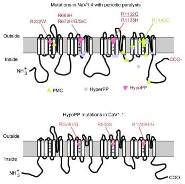
Glycoproteins
Lysosome-Associated Membrane Glycoproteins
Platelet Membrane Glycoproteins
Cell Membrane
Intracellular Membranes
Membranes
Fucose
Membrane Lipids
Oligosaccharides
Glucosamine
Membrane Proteins
Electrophoresis, Polyacrylamide Gel
Lectins
Glycosylation
Erythrocyte Membrane
Membrane Potentials
Glycopeptides
Mannose
Carbohydrate Sequence
Membranes, Artificial
Galactose Oxidase
Viral Envelope Proteins
Molecular Sequence Data
Blood Platelets
Polysaccharides
Sialic Acids
Golgi Apparatus
Mannosyl-Glycoprotein Endo-beta-N-Acetylglucosaminidase
Neuraminidase
Carbohydrates
Amino Acid Sequence
Receptors, Concanavalin A
Lysosomes
Immunoelectrophoresis, Two-Dimensional
Chromatography, Affinity
Membrane Fluidity
Galactose
Amino Sugars
Microscopy, Electron
Wheat Germ Agglutinins
Cricetinae
Cell Membrane Permeability
Endoplasmic Reticulum
N-Acetylneuraminic Acid
Basement Membrane
Glycoside Hydrolases
Sialoglycoproteins
Protein Processing, Post-Translational
Fluorescent Antibody Technique
Bunyaviridae
Cells, Cultured
Tunicamycin
Viral Fusion Proteins
Protein Binding
Sindbis Virus
Cell Fractionation
Peptide-N4-(N-acetyl-beta-glucosaminyl) Asparagine Amidase
Vesicular stomatitis Indiana virus
Base Sequence
Antigens, CD
Platelet Aggregation
Antigens, Surface
Asparagine
Glycolipids
Uukuniemi virus
Mutation
Subcellular Fractions
Monensin
Membrane Transport Proteins
Hexosaminidases
Galactosyltransferases
Hydrogen-Ion Concentration
Concanavalin A
Rabbits
Microsomes
Cattle
Solubility
Chromatography, Gel
Dipeptidyl-Peptidases and Tripeptidyl-Peptidases
Biological Transport
Erythrocytes
Sialyltransferases
Cloning, Molecular
Lipid Bilayers
CHO Cells
Temperature
Protein Transport
Cytoplasm
Platelet Glycoprotein GPIb-IX Complex
Macromolecular Substances
Amino Acids
Transfection
Liver
Polyethylene Glycols
Binding Sites
Kidney
Tritium
Sequence Homology, Amino Acid
Precipitin Tests
Blotting, Western
Mitochondrial Membranes
Dogs
Centrifugation, Density Gradient
Chick Embryo
Liposomes
Ristocetin
Tumor Cells, Cultured
Protein Structure, Tertiary
Synaptic Membranes
Protein Precursors
Blood Proteins
Models, Biological
Rubella virus
Laminin
Carrier Proteins
Immune Sera
Receptor, IGF Type 2
Peptide Fragments
Recombinant Fusion Proteins
Rats, Inbred Strains
Calcium
Mucins
Detergents
Fibroblasts
Chemistry
Immunoelectrophoresis
Palmitic Acid
Edetic Acid
Chemical Phenomena
von Willebrand Factor
Antibodies
Protein Conformation
Microscopy, Fluorescence
Escherichia coli
Platelet Adhesiveness
Immunoglobulin Fab Fragments
Endocytosis
RNA, Messenger
Cercopithecus aethiops
Freeze Fracturing
Receptors, Cell Surface
Peptides
Cell Fusion
Antigen-Antibody Complex
Virion
Models, Molecular
Extraembryonic Membranes
Fibrinogen
Plant Lectins
Phosphatidylcholines
Flow Cytometry
Virus Internalization
Glycoconjugates
Platelet Activation
Microscopy, Confocal
Swainsonine
Microscopy, Immunoelectron
Antigens, CD36
Octoxynol
HeLa Cells
Monosaccharides
Biological Transport, Active
Potassium
Immunoblotting
Protein Structure, Secondary
Cell Membrane Structures
Receptors, Virus
Mitochondria
Fluorescent Dyes
Trypsin
Asialoglycoproteins
Calnexin
alpha-Mannosidase
Macrophages
Signal Transduction
Glycoproteins are complex proteins that contain oligosaccharide chains (glycans) covalently attached to their polypeptide backbone. These glycans are linked to the protein through asparagine residues (N-linked) or serine/threonine residues (O-linked). Glycoproteins play crucial roles in various biological processes, including cell recognition, cell-cell interactions, cell adhesion, and signal transduction. They are widely distributed in nature and can be found on the outer surface of cell membranes, in extracellular fluids, and as components of the extracellular matrix. The structure and composition of glycoproteins can vary significantly depending on their function and location within an organism.
Membrane glycoproteins are proteins that contain oligosaccharide chains (glycans) covalently attached to their polypeptide backbone. They are integral components of biological membranes, spanning the lipid bilayer and playing crucial roles in various cellular processes.
The glycosylation of these proteins occurs in the endoplasmic reticulum (ER) and Golgi apparatus during protein folding and trafficking. The attached glycans can vary in structure, length, and composition, which contributes to the diversity of membrane glycoproteins.
Membrane glycoproteins can be classified into two main types based on their orientation within the lipid bilayer:
1. Type I (N-linked): These glycoproteins have a single transmembrane domain and an extracellular N-terminus, where the oligosaccharides are predominantly attached via asparagine residues (Asn-X-Ser/Thr sequon).
2. Type II (C-linked): These glycoproteins possess two transmembrane domains and an intracellular C-terminus, with the oligosaccharides linked to tryptophan residues via a mannose moiety.
Membrane glycoproteins are involved in various cellular functions, such as:
* Cell adhesion and recognition
* Receptor-mediated signal transduction
* Enzymatic catalysis
* Transport of molecules across membranes
* Cell-cell communication
* Immunological responses
Some examples of membrane glycoproteins include cell surface receptors (e.g., growth factor receptors, cytokine receptors), adhesion molecules (e.g., integrins, cadherins), and transporters (e.g., ion channels, ABC transporters).
Lysosome-Associated Membrane Glycoproteins (LAMPs) are a group of proteins found in the membrane of lysosomes, which are cellular organelles responsible for breaking down and recycling various biomolecules. LAMPs play a crucial role in maintaining the integrity and function of the lysosomal membrane.
There are two major types of LAMPs: LAMP-1 and LAMP-2. Both proteins share structural similarities, including a large heavily glycosylated domain that faces the lumen of the lysosome and a short hydrophobic region that anchors them to the membrane.
The primary function of LAMPs is to protect the lysosomal membrane from degradation by hydrolytic enzymes present inside the lysosome. They also participate in the process of autophagy, a cellular recycling mechanism, by fusing with autophagosomes (double-membraned vesicles formed during autophagy) to form autolysosomes, where the contents are degraded.
Moreover, LAMPs have been implicated in several cellular processes, such as antigen presentation, cholesterol homeostasis, and intracellular signaling. Mutations in LAMP-2 have been associated with certain genetic disorders, including Danon disease, a rare X-linked dominant disorder characterized by heart problems, muscle weakness, and intellectual disability.
Platelet membrane glycoproteins are specialized proteins found on the surface of platelets, which are small blood cells responsible for clotting. These glycoproteins play crucial roles in various processes related to hemostasis and thrombosis, including platelet adhesion, activation, and aggregation.
There are several key platelet membrane glycoproteins, such as:
1. Glycoprotein (GP) Ia/IIa (also known as integrin α2β1): This glycoprotein mediates the binding of platelets to collagen fibers in the extracellular matrix, facilitating platelet adhesion and activation.
2. GP IIb/IIIa (also known as integrin αIIbβ3): This is the most abundant glycoprotein on the platelet surface and functions as a receptor for fibrinogen, von Willebrand factor, and other adhesive proteins. Upon activation, GP IIb/IIIa undergoes conformational changes that enable it to bind these ligands, leading to platelet aggregation and clot formation.
3. GPIb-IX-V: This glycoprotein complex is involved in the initial tethering and adhesion of platelets to von Willebrand factor (vWF) in damaged blood vessels. It consists of four subunits: GPIbα, GPIbβ, GPIX, and GPV.
4. GPVI: This glycoprotein is essential for platelet activation upon contact with collagen. It associates with the Fc receptor γ-chain (FcRγ) to form a signaling complex that triggers intracellular signaling pathways, leading to platelet activation and aggregation.
Abnormalities in these platelet membrane glycoproteins can lead to bleeding disorders or thrombotic conditions. For example, mutations in GPIIb/IIIa can result in Glanzmann's thrombasthenia, a severe bleeding disorder characterized by impaired platelet aggregation. On the other hand, increased expression or activation of these glycoproteins may contribute to the development of arterial thrombosis and cardiovascular diseases.
A cell membrane, also known as the plasma membrane, is a thin semi-permeable phospholipid bilayer that surrounds all cells in animals, plants, and microorganisms. It functions as a barrier to control the movement of substances in and out of the cell, allowing necessary molecules such as nutrients, oxygen, and signaling molecules to enter while keeping out harmful substances and waste products. The cell membrane is composed mainly of phospholipids, which have hydrophilic (water-loving) heads and hydrophobic (water-fearing) tails. This unique structure allows the membrane to be flexible and fluid, yet selectively permeable. Additionally, various proteins are embedded in the membrane that serve as channels, pumps, receptors, and enzymes, contributing to the cell's overall functionality and communication with its environment.
Intracellular membranes refer to the membrane structures that exist within a eukaryotic cell (excluding bacteria and archaea, which are prokaryotic and do not have intracellular membranes). These membranes compartmentalize the cell, creating distinct organelles or functional regions with specific roles in various cellular processes.
Major types of intracellular membranes include:
1. Nuclear membrane (nuclear envelope): A double-membraned structure that surrounds and protects the genetic material within the nucleus. It consists of an outer and inner membrane, perforated by nuclear pores that regulate the transport of molecules between the nucleus and cytoplasm.
2. Endoplasmic reticulum (ER): An extensive network of interconnected tubules and sacs that serve as a major site for protein folding, modification, and lipid synthesis. The ER has two types: rough ER (with ribosomes on its surface) and smooth ER (without ribosomes).
3. Golgi apparatus/Golgi complex: A series of stacked membrane-bound compartments that process, sort, and modify proteins and lipids before they are transported to their final destinations within the cell or secreted out of the cell.
4. Lysosomes: Membrane-bound organelles containing hydrolytic enzymes for breaking down various biomolecules (proteins, carbohydrates, lipids, and nucleic acids) in the process called autophagy or from outside the cell via endocytosis.
5. Peroxisomes: Single-membrane organelles involved in various metabolic processes, such as fatty acid oxidation and detoxification of harmful substances like hydrogen peroxide.
6. Vacuoles: Membrane-bound compartments that store and transport various molecules, including nutrients, waste products, and enzymes. Plant cells have a large central vacuole for maintaining turgor pressure and storing metabolites.
7. Mitochondria: Double-membraned organelles responsible for generating energy (ATP) through oxidative phosphorylation and other metabolic processes, such as the citric acid cycle and fatty acid synthesis.
8. Chloroplasts: Double-membraned organelles found in plant cells that convert light energy into chemical energy during photosynthesis, producing oxygen and organic compounds (glucose) from carbon dioxide and water.
9. Endoplasmic reticulum (ER): A network of interconnected membrane-bound tubules involved in protein folding, modification, and transport; it is divided into two types: rough ER (with ribosomes on the surface) and smooth ER (without ribosomes).
10. Nucleus: Double-membraned organelle containing genetic material (DNA) and associated proteins involved in replication, transcription, RNA processing, and DNA repair. The nuclear membrane separates the nucleoplasm from the cytoplasm and contains nuclear pores for transporting molecules between the two compartments.
In medical terms, membranes refer to thin layers of tissue that cover or line various structures in the body. They are composed of connective tissue and epithelial cells, and they can be found lining the outer surface of the body, internal organs, blood vessels, and nerves. There are several types of membranes in the human body, including:
1. Serous Membranes: These membranes line the inside of body cavities and cover the organs contained within them. They produce a lubricating fluid that reduces friction between the organ and the cavity wall. Examples include the pleura (lungs), pericardium (heart), and peritoneum (abdominal cavity).
2. Mucous Membranes: These membranes line the respiratory, gastrointestinal, and genitourinary tracts, as well as the inner surface of the eyelids and the nasal passages. They produce mucus to trap particles, bacteria, and other substances, which helps protect the body from infection.
3. Synovial Membranes: These membranes line the joint cavities and produce synovial fluid, which lubricates the joints and allows for smooth movement.
4. Meninges: These are three layers of membranes that cover and protect the brain and spinal cord. They include the dura mater (outermost layer), arachnoid mater (middle layer), and pia mater (innermost layer).
5. Amniotic Membrane: This is a thin, transparent membrane that surrounds and protects the fetus during pregnancy. It produces amniotic fluid, which provides a cushion for the developing baby and helps regulate its temperature.
Fucose is a type of sugar molecule that is often found in complex carbohydrates known as glycans, which are attached to many proteins and lipids in the body. It is a hexose sugar, meaning it contains six carbon atoms, and is a type of L-sugar, which means that it rotates plane-polarized light in a counterclockwise direction.
Fucose is often found at the ends of glycan chains and plays important roles in various biological processes, including cell recognition, signaling, and interaction. It is also a component of some blood group antigens and is involved in the development and function of the immune system. Abnormalities in fucosylation (the addition of fucose to glycans) have been implicated in various diseases, including cancer, inflammation, and neurological disorders.
Membrane lipids are the main component of biological membranes, forming a lipid bilayer in which various cellular processes take place. These lipids include phospholipids, glycolipids, and cholesterol. Phospholipids are the most abundant type, consisting of a hydrophilic head (containing a phosphate group) and two hydrophobic tails (composed of fatty acid chains). Glycolipids contain a sugar group attached to the lipid molecule. Cholesterol helps regulate membrane fluidity and permeability. Together, these lipids create a selectively permeable barrier that separates cells from their environment and organelles within cells.
Oligosaccharides are complex carbohydrates composed of relatively small numbers (3-10) of monosaccharide units joined together by glycosidic linkages. They occur naturally in foods such as milk, fruits, vegetables, and legumes. In the body, oligosaccharides play important roles in various biological processes, including cell recognition, signaling, and protection against pathogens.
There are several types of oligosaccharides, classified based on their structures and functions. Some common examples include:
1. Disaccharides: These consist of two monosaccharide units, such as sucrose (glucose + fructose), lactose (glucose + galactose), and maltose (glucose + glucose).
2. Trisaccharides: These contain three monosaccharide units, like maltotriose (glucose + glucose + glucose) and raffinose (galactose + glucose + fructose).
3. Oligosaccharides found in human milk: Human milk contains unique oligosaccharides that serve as prebiotics, promoting the growth of beneficial bacteria in the gut. These oligosaccharides also help protect infants from pathogens by acting as decoy receptors and inhibiting bacterial adhesion to intestinal cells.
4. N-linked and O-linked glycans: These are oligosaccharides attached to proteins in the body, playing crucial roles in protein folding, stability, and function.
5. Plant-derived oligosaccharides: Fructooligosaccharides (FOS) and galactooligosaccharides (GOS) are examples of plant-derived oligosaccharides that serve as prebiotics, promoting the growth of beneficial gut bacteria.
Overall, oligosaccharides have significant impacts on human health and disease, particularly in relation to gastrointestinal function, immunity, and inflammation.
Glucosamine is a natural compound found in the body, primarily in the fluid around joints. It is a building block of cartilage, which is the tissue that cushions bones and allows for smooth joint movement. Glucosamine can also be produced in a laboratory and is commonly sold as a dietary supplement.
Medical definitions of glucosamine describe it as a type of amino sugar that plays a crucial role in the formation and maintenance of cartilage, ligaments, tendons, and other connective tissues. It is often used as a supplement to help manage osteoarthritis symptoms, such as pain, stiffness, and swelling in the joints, by potentially reducing inflammation and promoting cartilage repair.
There are different forms of glucosamine available, including glucosamine sulfate, glucosamine hydrochloride, and N-acetyl glucosamine. Glucosamine sulfate is the most commonly used form in supplements and has been studied more extensively than other forms. While some research suggests that glucosamine may provide modest benefits for osteoarthritis symptoms, its effectiveness remains a topic of ongoing debate among medical professionals.
Membrane proteins are a type of protein that are embedded in the lipid bilayer of biological membranes, such as the plasma membrane of cells or the inner membrane of mitochondria. These proteins play crucial roles in various cellular processes, including:
1. Cell-cell recognition and signaling
2. Transport of molecules across the membrane (selective permeability)
3. Enzymatic reactions at the membrane surface
4. Energy transduction and conversion
5. Mechanosensation and signal transduction
Membrane proteins can be classified into two main categories: integral membrane proteins, which are permanently associated with the lipid bilayer, and peripheral membrane proteins, which are temporarily or loosely attached to the membrane surface. Integral membrane proteins can further be divided into three subcategories based on their topology:
1. Transmembrane proteins, which span the entire width of the lipid bilayer with one or more alpha-helices or beta-barrels.
2. Lipid-anchored proteins, which are covalently attached to lipids in the membrane via a glycosylphosphatidylinositol (GPI) anchor or other lipid modifications.
3. Monotopic proteins, which are partially embedded in the membrane and have one or more domains exposed to either side of the bilayer.
Membrane proteins are essential for maintaining cellular homeostasis and are targets for various therapeutic interventions, including drug development and gene therapy. However, their structural complexity and hydrophobicity make them challenging to study using traditional biochemical methods, requiring specialized techniques such as X-ray crystallography, nuclear magnetic resonance (NMR) spectroscopy, and single-particle cryo-electron microscopy (cryo-EM).
Electrophoresis, polyacrylamide gel (EPG) is a laboratory technique used to separate and analyze complex mixtures of proteins or nucleic acids (DNA or RNA) based on their size and electrical charge. This technique utilizes a matrix made of cross-linked polyacrylamide, a type of gel, which provides a stable and uniform environment for the separation of molecules.
In this process:
1. The polyacrylamide gel is prepared by mixing acrylamide monomers with a cross-linking agent (bis-acrylamide) and a catalyst (ammonium persulfate) in the presence of a buffer solution.
2. The gel is then poured into a mold and allowed to polymerize, forming a solid matrix with uniform pore sizes that depend on the concentration of acrylamide used. Higher concentrations result in smaller pores, providing better resolution for separating smaller molecules.
3. Once the gel has set, it is placed in an electrophoresis apparatus containing a buffer solution. Samples containing the mixture of proteins or nucleic acids are loaded into wells on the top of the gel.
4. An electric field is applied across the gel, causing the negatively charged molecules to migrate towards the positive electrode (anode) while positively charged molecules move toward the negative electrode (cathode). The rate of migration depends on the size, charge, and shape of the molecules.
5. Smaller molecules move faster through the gel matrix and will migrate farther from the origin compared to larger molecules, resulting in separation based on size. Proteins and nucleic acids can be selectively stained after electrophoresis to visualize the separated bands.
EPG is widely used in various research fields, including molecular biology, genetics, proteomics, and forensic science, for applications such as protein characterization, DNA fragment analysis, cloning, mutation detection, and quality control of nucleic acid or protein samples.
Lectins are a type of proteins that bind specifically to carbohydrates and have been found in various plant and animal sources. They play important roles in biological recognition events, such as cell-cell adhesion, and can also be involved in the immune response. Some lectins can agglutinate certain types of cells or precipitate glycoproteins, while others may have a more direct effect on cellular processes. In some cases, lectins from plants can cause adverse effects in humans if ingested, such as digestive discomfort or allergic reactions.
Molecular weight, also known as molecular mass, is the mass of a molecule. It is expressed in units of atomic mass units (amu) or daltons (Da). Molecular weight is calculated by adding up the atomic weights of each atom in a molecule. It is a useful property in chemistry and biology, as it can be used to determine the concentration of a substance in a solution, or to calculate the amount of a substance that will react with another in a chemical reaction.
A cell line is a culture of cells that are grown in a laboratory for use in research. These cells are usually taken from a single cell or group of cells, and they are able to divide and grow continuously in the lab. Cell lines can come from many different sources, including animals, plants, and humans. They are often used in scientific research to study cellular processes, disease mechanisms, and to test new drugs or treatments. Some common types of human cell lines include HeLa cells (which come from a cancer patient named Henrietta Lacks), HEK293 cells (which come from embryonic kidney cells), and HUVEC cells (which come from umbilical vein endothelial cells). It is important to note that cell lines are not the same as primary cells, which are cells that are taken directly from a living organism and have not been grown in the lab.
Glycosylation is the enzymatic process of adding a sugar group, or glycan, to a protein, lipid, or other organic molecule. This post-translational modification plays a crucial role in modulating various biological functions, such as protein stability, trafficking, and ligand binding. The structure and composition of the attached glycans can significantly influence the functional properties of the modified molecule, contributing to cell-cell recognition, signal transduction, and immune response regulation. Abnormal glycosylation patterns have been implicated in several disease states, including cancer, diabetes, and neurodegenerative disorders.
An erythrocyte, also known as a red blood cell, is a type of cell that circulates in the blood and is responsible for transporting oxygen throughout the body. The erythrocyte membrane refers to the thin, flexible barrier that surrounds the erythrocyte and helps to maintain its shape and stability.
The erythrocyte membrane is composed of a lipid bilayer, which contains various proteins and carbohydrates. These components help to regulate the movement of molecules into and out of the erythrocyte, as well as provide structural support and protection for the cell.
The main lipids found in the erythrocyte membrane are phospholipids and cholesterol, which are arranged in a bilayer structure with the hydrophilic (water-loving) heads facing outward and the hydrophobic (water-fearing) tails facing inward. This arrangement helps to maintain the integrity of the membrane and prevent the leakage of cellular components.
The proteins found in the erythrocyte membrane include integral proteins, which span the entire width of the membrane, and peripheral proteins, which are attached to the inner or outer surface of the membrane. These proteins play a variety of roles, such as transporting molecules across the membrane, maintaining the shape of the erythrocyte, and interacting with other cells and proteins in the body.
The carbohydrates found in the erythrocyte membrane are attached to the outer surface of the membrane and help to identify the cell as part of the body's own immune system. They also play a role in cell-cell recognition and adhesion.
Overall, the erythrocyte membrane is a complex and dynamic structure that plays a critical role in maintaining the function and integrity of red blood cells.
Membrane potential is the electrical potential difference across a cell membrane, typically for excitable cells such as nerve and muscle cells. It is the difference in electric charge between the inside and outside of a cell, created by the selective permeability of the cell membrane to different ions. The resting membrane potential of a typical animal cell is around -70 mV, with the interior being negative relative to the exterior. This potential is generated and maintained by the active transport of ions across the membrane, primarily through the action of the sodium-potassium pump. Membrane potentials play a crucial role in many physiological processes, including the transmission of nerve impulses and the contraction of muscle cells.
Glycopeptides are a class of antibiotics that are characterized by their complex chemical structure, which includes both peptide and carbohydrate components. These antibiotics are produced naturally by certain types of bacteria and are effective against a range of Gram-positive bacterial infections, including methicillin-resistant Staphylococcus aureus (MRSA) and vancomycin-resistant Enterococci (VRE).
The glycopeptide antibiotics work by binding to the bacterial cell wall precursor, preventing the cross-linking of peptidoglycan chains that is necessary for the formation of a strong and rigid cell wall. This leads to the death of the bacteria.
Examples of glycopeptides include vancomycin, teicoplanin, and dalbavancin. While these antibiotics have been used successfully for many years, their use is often limited due to concerns about the emergence of resistance and potential toxicity.
Mannose is a simple sugar (monosaccharide) that is similar in structure to glucose. It is a hexose, meaning it contains six carbon atoms. Mannose is a stereoisomer of glucose, meaning it has the same chemical formula but a different structural arrangement of its atoms.
Mannose is not as commonly found in foods as other simple sugars, but it can be found in some fruits, such as cranberries, blueberries, and peaches, as well as in certain vegetables, like sweet potatoes and turnips. It is also found in some dietary fibers, such as those found in beans and whole grains.
In the body, mannose can be metabolized and used for energy, but it is also an important component of various glycoproteins and glycolipids, which are molecules that play critical roles in many biological processes, including cell recognition, signaling, and adhesion.
Mannose has been studied as a potential therapeutic agent for various medical conditions, including urinary tract infections (UTIs), because it can inhibit the attachment of certain bacteria to the cells lining the urinary tract. Additionally, mannose-binding lectins have been investigated for their potential role in the immune response to viral and bacterial infections.
A "carbohydrate sequence" refers to the specific arrangement or order of monosaccharides (simple sugars) that make up a carbohydrate molecule, such as a polysaccharide or an oligosaccharide. Carbohydrates are often composed of repeating units of monosaccharides, and the sequence in which these units are arranged can have important implications for the function and properties of the carbohydrate.
For example, in glycoproteins (proteins that contain carbohydrate chains), the specific carbohydrate sequence can affect how the protein is processed and targeted within the cell, as well as its stability and activity. Similarly, in complex carbohydrates like starch or cellulose, the sequence of glucose units can determine whether the molecule is branched or unbranched, which can have implications for its digestibility and other properties.
Therefore, understanding the carbohydrate sequence is an important aspect of studying carbohydrate structure and function in biology and medicine.
Artificial membranes are synthetic or man-made materials that possess properties similar to natural biological membranes, such as selective permeability and barrier functions. These membranes can be designed to control the movement of molecules, ions, or cells across them, making them useful in various medical and biotechnological applications.
Examples of artificial membranes include:
1. Dialysis membranes: Used in hemodialysis for patients with renal failure, these semi-permeable membranes filter waste products and excess fluids from the blood while retaining essential proteins and cells.
2. Hemofiltration membranes: Utilized in extracorporeal circuits to remove larger molecules, such as cytokines or inflammatory mediators, from the blood during critical illnesses or sepsis.
3. Drug delivery systems: Artificial membranes can be used to encapsulate drugs, allowing for controlled release and targeted drug delivery in specific tissues or cells.
4. Tissue engineering: Synthetic membranes serve as scaffolds for cell growth and tissue regeneration, guiding the formation of new functional tissues.
5. Biosensors: Artificial membranes can be integrated into biosensing devices to selectively detect and quantify biomolecules, such as proteins or nucleic acids, in diagnostic applications.
6. Microfluidics: Artificial membranes are used in microfluidic systems for lab-on-a-chip applications, enabling the manipulation and analysis of small volumes of fluids for various medical and biological purposes.
Galactose oxidase is an enzyme with the systematic name D-galactose:oxygen oxidoreductase. It is found in certain fungi and bacteria, and it catalyzes the following reaction:
D-galactose + O2 -> D-galacto-hexodialdose + H2O2
In this reaction, the enzyme oxidizes the hydroxyl group (-OH) on the sixth carbon atom of D-galactose to an aldehyde group (-CHO), forming D-galacto-hexodialdose. At the same time, it reduces molecular oxygen (O2) to hydrogen peroxide (H2O2).
Galactose oxidase is a copper-containing enzyme and requires the cofactor molybdenum for its activity. It has potential applications in various industrial processes, such as the production of D-galacto-hexodialdose and other sugar derivatives, as well as in biosensors for detecting glucose levels in biological samples.
Viral envelope proteins are structural proteins found in the envelope that surrounds many types of viruses. These proteins play a crucial role in the virus's life cycle, including attachment to host cells, fusion with the cell membrane, and entry into the host cell. They are typically made up of glycoproteins and are often responsible for eliciting an immune response in the host organism. The exact structure and function of viral envelope proteins vary between different types of viruses.
Molecular sequence data refers to the specific arrangement of molecules, most commonly nucleotides in DNA or RNA, or amino acids in proteins, that make up a biological macromolecule. This data is generated through laboratory techniques such as sequencing, and provides information about the exact order of the constituent molecules. This data is crucial in various fields of biology, including genetics, evolution, and molecular biology, allowing for comparisons between different organisms, identification of genetic variations, and studies of gene function and regulation.
Blood platelets, also known as thrombocytes, are small, colorless cell fragments in our blood that play an essential role in normal blood clotting. They are formed in the bone marrow from large cells called megakaryocytes and circulate in the blood in an inactive state until they are needed to help stop bleeding. When a blood vessel is damaged, platelets become activated and change shape, releasing chemicals that attract more platelets to the site of injury. These activated platelets then stick together to form a plug, or clot, that seals the wound and prevents further blood loss. In addition to their role in clotting, platelets also help to promote healing by releasing growth factors that stimulate the growth of new tissue.
Polysaccharides are complex carbohydrates consisting of long chains of monosaccharide units (simple sugars) bonded together by glycosidic linkages. They can be classified based on the type of monosaccharides and the nature of the bonds that connect them.
Polysaccharides have various functions in living organisms. For example, starch and glycogen serve as energy storage molecules in plants and animals, respectively. Cellulose provides structural support in plants, while chitin is a key component of fungal cell walls and arthropod exoskeletons.
Some polysaccharides also have important roles in the human body, such as being part of the extracellular matrix (e.g., hyaluronic acid) or acting as blood group antigens (e.g., ABO blood group substances).
Sialic acids are a family of nine-carbon sugars that are commonly found on the outermost surface of many cell types, particularly on the glycoconjugates of mucins in various secretions and on the glycoproteins and glycolipids of cell membranes. They play important roles in a variety of biological processes, including cell recognition, immune response, and viral and bacterial infectivity. Sialic acids can exist in different forms, with N-acetylneuraminic acid being the most common one in humans.
The Golgi apparatus, also known as the Golgi complex or simply the Golgi, is a membrane-bound organelle found in the cytoplasm of most eukaryotic cells. It plays a crucial role in the processing, sorting, and packaging of proteins and lipids for transport to their final destinations within the cell or for secretion outside the cell.
The Golgi apparatus consists of a series of flattened, disc-shaped sacs called cisternae, which are stacked together in a parallel arrangement. These stacks are often interconnected by tubular structures called tubules or vesicles. The Golgi apparatus has two main faces: the cis face, which is closest to the endoplasmic reticulum (ER) and receives proteins and lipids directly from the ER; and the trans face, which is responsible for sorting and dispatching these molecules to their final destinations.
The Golgi apparatus performs several essential functions in the cell:
1. Protein processing: After proteins are synthesized in the ER, they are transported to the cis face of the Golgi apparatus, where they undergo various post-translational modifications, such as glycosylation (the addition of sugar molecules) and sulfation. These modifications help determine the protein's final structure, function, and targeting.
2. Lipid modification: The Golgi apparatus also modifies lipids by adding or removing different functional groups, which can influence their properties and localization within the cell.
3. Protein sorting and packaging: Once proteins and lipids have been processed, they are sorted and packaged into vesicles at the trans face of the Golgi apparatus. These vesicles then transport their cargo to various destinations, such as lysosomes, plasma membrane, or extracellular space.
4. Intracellular transport: The Golgi apparatus serves as a central hub for intracellular trafficking, coordinating the movement of vesicles and other transport carriers between different organelles and cellular compartments.
5. Cell-cell communication: Some proteins that are processed and packaged in the Golgi apparatus are destined for secretion, playing crucial roles in cell-cell communication and maintaining tissue homeostasis.
In summary, the Golgi apparatus is a vital organelle involved in various cellular processes, including post-translational modification, sorting, packaging, and intracellular transport of proteins and lipids. Its proper functioning is essential for maintaining cellular homeostasis and overall organismal health.
Mannosyl-glycoprotein endo-beta-N-acetylglucosaminidase (MGNAG) is an enzyme that is involved in the breakdown and recycling of glycoproteins, which are proteins that contain oligosaccharide chains attached to them. The enzyme's primary function is to cleave the beta-N-acetylglucosaminyl linkages in the chitobiose core of N-linked glycans, which are complex carbohydrates that are attached to many proteins in eukaryotic cells.
MGNAG is a lysosomal enzyme, meaning it is located within the lysosomes, which are membrane-bound organelles found in the cytoplasm of eukaryotic cells. Lysosomes contain hydrolytic enzymes that break down various biomolecules, including glycoproteins, lipids, and nucleic acids, into their constituent parts for recycling or disposal.
Deficiency in MGNAG activity can lead to a rare genetic disorder known as alpha-mannosidosis, which is characterized by the accumulation of mannose-rich oligosaccharides in various tissues and organs throughout the body. This condition can result in a range of symptoms, including developmental delays, intellectual disability, coarse facial features, skeletal abnormalities, hearing loss, and immune dysfunction.
Neuraminidase is an enzyme that occurs on the surface of influenza viruses. It plays a crucial role in the life cycle of the virus by helping it to infect host cells and to spread from cell to cell within the body. Neuraminidase works by cleaving sialic acid residues from glycoproteins, allowing the virus to detach from infected cells and to move through mucus and other bodily fluids. This enzyme is a major target of antiviral drugs used to treat influenza, such as oseltamivir (Tamiflu) and zanamivir (Relenza). Inhibiting the activity of neuraminidase can help to prevent the spread of the virus within the body and reduce the severity of symptoms.
Carbohydrates are a major nutrient class consisting of organic compounds that primarily contain carbon, hydrogen, and oxygen atoms. They are classified as saccharides, which include monosaccharides (simple sugars), disaccharides (double sugars), oligosaccharides (short-chain sugars), and polysaccharides (complex carbohydrates).
Monosaccharides, such as glucose, fructose, and galactose, are the simplest form of carbohydrates. They consist of a single sugar molecule that cannot be broken down further by hydrolysis. Disaccharides, like sucrose (table sugar), lactose (milk sugar), and maltose (malt sugar), are formed from two monosaccharide units joined together.
Oligosaccharides contain a small number of monosaccharide units, typically less than 20, while polysaccharides consist of long chains of hundreds to thousands of monosaccharide units. Polysaccharides can be further classified into starch (found in plants), glycogen (found in animals), and non-starchy polysaccharides like cellulose, chitin, and pectin.
Carbohydrates play a crucial role in providing energy to the body, with glucose being the primary source of energy for most cells. They also serve as structural components in plants (cellulose) and animals (chitin), participate in various metabolic processes, and contribute to the taste, texture, and preservation of foods.
An amino acid sequence is the specific order of amino acids in a protein or peptide molecule, formed by the linking of the amino group (-NH2) of one amino acid to the carboxyl group (-COOH) of another amino acid through a peptide bond. The sequence is determined by the genetic code and is unique to each type of protein or peptide. It plays a crucial role in determining the three-dimensional structure and function of proteins.
Concanavalin A (Con A) receptors are not a medical term per se, but rather a term used in the field of immunology and cell biology. Concanavalin A is a type of lectin, a protein that can bind to specific sugars found on the surface of cells. Con A receptors refer to the specific binding sites or proteins on the surface of certain types of cells, such as immune cells, that can recognize and bind to Concanavalin A.
When Con A binds to its receptors, it can activate various cellular responses, including changes in cell shape, movement, and metabolism. In research settings, Con A is often used as a tool to study the behavior of immune cells and other cell types that express Con A receptors. However, it's worth noting that Concanavalin A is not typically used in medical treatments or diagnoses.
Lysosomes are membrane-bound organelles found in the cytoplasm of eukaryotic cells. They are responsible for breaking down and recycling various materials, such as waste products, foreign substances, and damaged cellular components, through a process called autophagy or phagocytosis. Lysosomes contain hydrolytic enzymes that can break down biomolecules like proteins, nucleic acids, lipids, and carbohydrates into their basic building blocks, which can then be reused by the cell. They play a crucial role in maintaining cellular homeostasis and are often referred to as the "garbage disposal system" of the cell.
Two-dimensional immunoelectrophoresis (2DE) is a specialized laboratory technique used in the field of clinical pathology and immunology. This technique is a refined version of traditional immunoelectrophoresis that adds an additional electrophoretic separation step, enhancing its resolution and allowing for more detailed analysis of complex protein mixtures.
In two-dimensional immunoelectrophoresis, proteins are first separated based on their isoelectric points (pI) in the initial dimension using isoelectric focusing (IEF). This process involves applying an electric field to a protein mixture contained within a gel matrix, where proteins will migrate and stop migrating once they reach the pH that matches their own isoelectric point.
Following IEF, the separated proteins are then subjected to a second electrophoretic separation in the perpendicular direction (second dimension) based on their molecular weights using sodium dodecyl sulfate-polyacrylamide gel electrophoresis (SDS-PAGE). SDS is a negatively charged molecule that binds to proteins, giving them a uniform negative charge and allowing for separation based solely on size.
Once the two-dimensional separation is complete, the gel is then overlaid with specific antisera to detect and identify proteins of interest. The resulting precipitin arcs formed at the intersection of the antibody and antigen are compared to known standards or patterns to determine the identity and quantity of the separated proteins.
Two-dimensional immunoelectrophoresis is particularly useful in identifying and quantifying proteins in complex mixtures, such as those found in body fluids like serum, urine, or cerebrospinal fluid (CSF). It can be applied to various clinical scenarios, including diagnosis and monitoring of monoclonal gammopathies, autoimmune disorders, and certain infectious diseases.
Affinity chromatography is a type of chromatography technique used in biochemistry and molecular biology to separate and purify proteins based on their biological characteristics, such as their ability to bind specifically to certain ligands or molecules. This method utilizes a stationary phase that is coated with a specific ligand (e.g., an antibody, antigen, receptor, or enzyme) that selectively interacts with the target protein in a sample.
The process typically involves the following steps:
1. Preparation of the affinity chromatography column: The stationary phase, usually a solid matrix such as agarose beads or magnetic beads, is modified by covalently attaching the ligand to its surface.
2. Application of the sample: The protein mixture is applied to the top of the affinity chromatography column, allowing it to flow through the stationary phase under gravity or pressure.
3. Binding and washing: As the sample flows through the column, the target protein selectively binds to the ligand on the stationary phase, while other proteins and impurities pass through. The column is then washed with a suitable buffer to remove any unbound proteins and contaminants.
4. Elution of the bound protein: The target protein can be eluted from the column using various methods, such as changing the pH, ionic strength, or polarity of the buffer, or by introducing a competitive ligand that displaces the bound protein.
5. Collection and analysis: The eluted protein fraction is collected and analyzed for purity and identity, often through techniques like SDS-PAGE or mass spectrometry.
Affinity chromatography is a powerful tool in biochemistry and molecular biology due to its high selectivity and specificity, enabling the efficient isolation of target proteins from complex mixtures. However, it requires careful consideration of the binding affinity between the ligand and the protein, as well as optimization of the elution conditions to minimize potential damage or denaturation of the purified protein.
Membrane fluidity, in the context of cell biology, refers to the ability of the phospholipid bilayer that makes up the cell membrane to change its structure and organization in response to various factors. The membrane is not a static structure but rather a dynamic one, with its lipids constantly moving and changing position.
Membrane fluidity is determined by the fatty acid composition of the phospholipids that make up the bilayer. Lipids with unsaturated fatty acids have kinks in their hydrocarbon chains, which prevent them from packing closely together and increase membrane fluidity. In contrast, lipids with saturated fatty acids can pack closely together, reducing membrane fluidity.
Membrane fluidity is important for various cellular processes, including the movement of proteins within the membrane, the fusion of vesicles with the membrane during exocytosis and endocytosis, and the ability of the membrane to respond to changes in temperature and other environmental factors. Abnormalities in membrane fluidity have been linked to various diseases, including cancer, neurological disorders, and infectious diseases.
Galactose is a simple sugar or monosaccharide that is a constituent of lactose, the disaccharide found in milk and dairy products. It's structurally similar to glucose but with a different chemical structure, and it plays a crucial role in various biological processes.
Galactose can be metabolized in the body through the action of enzymes such as galactokinase, galactose-1-phosphate uridylyltransferase, and UDP-galactose 4'-epimerase. Inherited deficiencies in these enzymes can lead to metabolic disorders like galactosemia, which can cause serious health issues if not diagnosed and treated promptly.
In summary, Galactose is a simple sugar that plays an essential role in lactose metabolism and other biological processes.
Blood platelet disorders are conditions that affect the number and/or function of platelets, which are small blood cells that help your body form clots to stop bleeding. Normal platelet count ranges from 150,000 to 450,000 platelets per microliter of blood. A lower-than-normal platelet count is called thrombocytopenia, while a higher-than-normal platelet count is called thrombocytosis.
There are several types of platelet disorders, including:
1. Immune thrombocytopenia (ITP): A condition in which the immune system mistakenly attacks and destroys platelets, leading to a low platelet count. ITP can be acute (lasting less than six months) or chronic (lasting longer than six months).
2. Thrombotic thrombocytopenic purpura (TTP): A rare but serious condition that causes blood clots to form in small blood vessels throughout the body, leading to a low platelet count, anemia, and other symptoms.
3. Hemolytic uremic syndrome (HUS): A condition that is often caused by a bacterial infection, which can lead to the formation of blood clots in the small blood vessels of the kidneys, resulting in kidney damage and a low platelet count.
4. Hereditary platelet disorders: Some people inherit genetic mutations that can affect the number or function of their platelets, leading to bleeding disorders such as von Willebrand disease or Bernard-Soulier syndrome.
5. Medication-induced thrombocytopenia: Certain medications can cause a decrease in platelet count as a side effect.
6. Platelet dysfunction disorders: Some conditions can affect the ability of platelets to function properly, leading to bleeding disorders such as von Willebrand disease or storage pool deficiency.
Symptoms of platelet disorders may include easy bruising, prolonged bleeding from cuts or injuries, nosebleeds, blood in urine or stools, and in severe cases, internal bleeding. Treatment for platelet disorders depends on the underlying cause and may include medications, surgery, or other therapies.
Amino sugars, also known as glycosamine or hexosamines, are sugar molecules that contain a nitrogen atom as part of their structure. The most common amino sugars found in nature are glucosamine and galactosamine, which are derived from the hexose sugars glucose and galactose, respectively.
Glucosamine is an essential component of the structural polysaccharide chitin, which is found in the exoskeletons of arthropods such as crustaceans and insects, as well as in the cell walls of fungi. It is also a precursor to the glycosaminoglycans (GAGs), which are long, unbranched polysaccharides that are important components of the extracellular matrix in animals.
Galactosamine, on the other hand, is a component of some GAGs and is also found in bacterial cell walls. It is used in the synthesis of heparin and heparan sulfate, which are important anticoagulant molecules.
Amino sugars play a critical role in many biological processes, including cell signaling, inflammation, and immune response. They have also been studied for their potential therapeutic uses in the treatment of various diseases, such as osteoarthritis and cancer.
Electron microscopy (EM) is a type of microscopy that uses a beam of electrons to create an image of the sample being examined, resulting in much higher magnification and resolution than light microscopy. There are several types of electron microscopy, including transmission electron microscopy (TEM), scanning electron microscopy (SEM), and reflection electron microscopy (REM).
In TEM, a beam of electrons is transmitted through a thin slice of the sample, and the electrons that pass through the sample are focused to form an image. This technique can provide detailed information about the internal structure of cells, viruses, and other biological specimens, as well as the composition and structure of materials at the atomic level.
In SEM, a beam of electrons is scanned across the surface of the sample, and the electrons that are scattered back from the surface are detected to create an image. This technique can provide information about the topography and composition of surfaces, as well as the structure of materials at the microscopic level.
REM is a variation of SEM in which the beam of electrons is reflected off the surface of the sample, rather than scattered back from it. This technique can provide information about the surface chemistry and composition of materials.
Electron microscopy has a wide range of applications in biology, medicine, and materials science, including the study of cellular structure and function, disease diagnosis, and the development of new materials and technologies.
Wheat germ agglutinins (WGA) are proteins found in wheat germ that have the ability to bind to specific carbohydrate structures, such as N-acetylglucosamine and sialic acid, which are present on the surface of many cells in the human body. WGA is a type of lectin, a group of proteins that can agglutinate, or clump together, red blood cells and bind to specific sugars on cell membranes.
WGA has been studied for its potential effects on various biological processes, including inflammation, immune response, and gut barrier function. Some research suggests that WGA may interact with the gut epithelium and affect intestinal permeability, potentially contributing to the development of gastrointestinal symptoms in some individuals. However, more research is needed to fully understand the clinical significance of these findings.
It's worth noting that while WGA has been studied for its potential biological effects, it is not currently recognized as a major allergen or toxic component of wheat. However, some people may still choose to avoid foods containing WGA due to personal dietary preferences or sensitivities.
Viral proteins are the proteins that are encoded by the viral genome and are essential for the viral life cycle. These proteins can be structural or non-structural and play various roles in the virus's replication, infection, and assembly process. Structural proteins make up the physical structure of the virus, including the capsid (the protein shell that surrounds the viral genome) and any envelope proteins (that may be present on enveloped viruses). Non-structural proteins are involved in the replication of the viral genome and modulation of the host cell environment to favor viral replication. Overall, a thorough understanding of viral proteins is crucial for developing antiviral therapies and vaccines.
Cricetinae is a subfamily of rodents that includes hamsters, gerbils, and relatives. These small mammals are characterized by having short limbs, compact bodies, and cheek pouches for storing food. They are native to various parts of the world, particularly in Europe, Asia, and Africa. Some species are popular pets due to their small size, easy care, and friendly nature. In a medical context, understanding the biology and behavior of Cricetinae species can be important for individuals who keep them as pets or for researchers studying their physiology.
Monoclonal antibodies are a type of antibody that are identical because they are produced by a single clone of cells. They are laboratory-produced molecules that act like human antibodies in the immune system. They can be designed to attach to specific proteins found on the surface of cancer cells, making them useful for targeting and treating cancer. Monoclonal antibodies can also be used as a therapy for other diseases, such as autoimmune disorders and inflammatory conditions.
Monoclonal antibodies are produced by fusing a single type of immune cell, called a B cell, with a tumor cell to create a hybrid cell, or hybridoma. This hybrid cell is then able to replicate indefinitely, producing a large number of identical copies of the original antibody. These antibodies can be further modified and engineered to enhance their ability to bind to specific targets, increase their stability, and improve their effectiveness as therapeutic agents.
Monoclonal antibodies have several mechanisms of action in cancer therapy. They can directly kill cancer cells by binding to them and triggering an immune response. They can also block the signals that promote cancer growth and survival. Additionally, monoclonal antibodies can be used to deliver drugs or radiation directly to cancer cells, increasing the effectiveness of these treatments while minimizing their side effects on healthy tissues.
Monoclonal antibodies have become an important tool in modern medicine, with several approved for use in cancer therapy and other diseases. They are continuing to be studied and developed as a promising approach to treating a wide range of medical conditions.
Carbohydrate conformation refers to the three-dimensional shape and structure of a carbohydrate molecule. Carbohydrates, also known as sugars, can exist in various conformational states, which are determined by the rotation of their component bonds and the spatial arrangement of their functional groups.
The conformation of a carbohydrate molecule can have significant implications for its biological activity and recognition by other molecules, such as enzymes or antibodies. Factors that can influence carbohydrate conformation include the presence of intramolecular hydrogen bonds, steric effects, and intermolecular interactions with solvent molecules or other solutes.
In some cases, the conformation of a carbohydrate may be stabilized by the formation of cyclic structures, in which the hydroxyl group at one end of the molecule forms a covalent bond with the carbonyl carbon at the other end, creating a ring structure. The most common cyclic carbohydrates are monosaccharides, such as glucose and fructose, which can exist in various conformational isomers known as anomers.
Understanding the conformation of carbohydrate molecules is important for elucidating their biological functions and developing strategies for targeting them with drugs or other therapeutic agents.
Acetylgalactosamine (also known as N-acetyl-D-galactosamine or GalNAc) is a type of sugar molecule called a hexosamine that is commonly found in glycoproteins and proteoglycans, which are complex carbohydrates that are attached to proteins and lipids. It plays an important role in various biological processes, including cell-cell recognition, signal transduction, and protein folding.
In the context of medical research and biochemistry, Acetylgalactosamine is often used as a building block for synthesizing glycoconjugates, which are molecules that consist of a carbohydrate attached to a protein or lipid. These molecules play important roles in many biological processes, including cell-cell recognition, signaling, and immune response.
Acetylgalactosamine is also used as a target for enzymes called glycosyltransferases, which add sugar molecules to proteins and lipids. In particular, Acetylgalactosamine is the acceptor substrate for a class of glycosyltransferases known as galactosyltransferases, which add galactose molecules to Acetylgalactosamine-containing structures.
Defects in the metabolism of Acetylgalactosamine have been linked to various genetic disorders, including Schindler disease and Kanzaki disease, which are characterized by neurological symptoms and abnormal accumulation of glycoproteins in various tissues.
Cell membrane permeability refers to the ability of various substances, such as molecules and ions, to pass through the cell membrane. The cell membrane, also known as the plasma membrane, is a thin, flexible barrier that surrounds all cells, controlling what enters and leaves the cell. Its primary function is to protect the cell's internal environment and maintain homeostasis.
The permeability of the cell membrane depends on its structure, which consists of a phospholipid bilayer interspersed with proteins. The hydrophilic (water-loving) heads of the phospholipids face outward, while the hydrophobic (water-fearing) tails face inward, creating a barrier that is generally impermeable to large, polar, or charged molecules.
However, specific proteins within the membrane, called channels and transporters, allow certain substances to cross the membrane. Channels are protein structures that span the membrane and provide a pore for ions or small uncharged molecules to pass through. Transporters, on the other hand, are proteins that bind to specific molecules and facilitate their movement across the membrane, often using energy in the form of ATP.
The permeability of the cell membrane can be influenced by various factors, such as temperature, pH, and the presence of certain chemicals or drugs. Changes in permeability can have significant consequences for the cell's function and survival, as they can disrupt ion balances, nutrient uptake, waste removal, and signal transduction.
The endoplasmic reticulum (ER) is a network of interconnected tubules and sacs that are present in the cytoplasm of eukaryotic cells. It is a continuous membranous organelle that plays a crucial role in the synthesis, folding, modification, and transport of proteins and lipids.
The ER has two main types: rough endoplasmic reticulum (RER) and smooth endoplasmic reticulum (SER). RER is covered with ribosomes, which give it a rough appearance, and is responsible for protein synthesis. On the other hand, SER lacks ribosomes and is involved in lipid synthesis, drug detoxification, calcium homeostasis, and steroid hormone production.
In summary, the endoplasmic reticulum is a vital organelle that functions in various cellular processes, including protein and lipid metabolism, calcium regulation, and detoxification.
N-Acetylneuraminic Acid (Neu5Ac) is an organic compound that belongs to the family of sialic acids. It is a common terminal sugar found on many glycoproteins and glycolipids on the surface of animal cells. Neu5Ac plays crucial roles in various biological processes, including cell recognition, signaling, and intercellular interactions. It is also involved in the protection against pathogens by serving as a barrier to prevent their attachment to host cells. Additionally, Neu5Ac has been implicated in several disease conditions, such as cancer and inflammation, due to its altered expression and metabolism.
The basement membrane is a thin, specialized layer of extracellular matrix that provides structural support and separates epithelial cells (which line the outer surfaces of organs and blood vessels) from connective tissue. It is composed of two main layers: the basal lamina, which is produced by the epithelial cells, and the reticular lamina, which is produced by the connective tissue. The basement membrane plays important roles in cell adhesion, migration, differentiation, and survival.
The basal lamina is composed mainly of type IV collagen, laminins, nidogens, and proteoglycans, while the reticular lamina contains type III collagen, fibronectin, and other matrix proteins. The basement membrane also contains a variety of growth factors and cytokines that can influence cell behavior.
Defects in the composition or organization of the basement membrane can lead to various diseases, including kidney disease, eye disease, and skin blistering disorders.
Glycoside hydrolases are a class of enzymes that catalyze the hydrolysis of glycosidic bonds found in various substrates such as polysaccharides, oligosaccharides, and glycoproteins. These enzymes break down complex carbohydrates into simpler sugars by cleaving the glycosidic linkages that connect monosaccharide units.
Glycoside hydrolases are classified based on their mechanism of action and the type of glycosidic bond they hydrolyze. The classification system is maintained by the International Union of Biochemistry and Molecular Biology (IUBMB). Each enzyme in this class is assigned a unique Enzyme Commission (EC) number, which reflects its specificity towards the substrate and the type of reaction it catalyzes.
These enzymes have various applications in different industries, including food processing, biofuel production, pulp and paper manufacturing, and biomedical research. In medicine, glycoside hydrolases are used to diagnose and monitor certain medical conditions, such as carbohydrate-deficient glycoprotein syndrome, a rare inherited disorder affecting the structure of glycoproteins.
Sialglycoproteins are a type of glycoprotein that have sialic acid as the terminal sugar in their oligosaccharide chains. These complex molecules are abundant on the surface of many cell types and play important roles in various biological processes, including cell recognition, cell-cell interactions, and protection against proteolytic degradation.
The presence of sialic acid on the outermost part of these glycoproteins makes them negatively charged, which can affect their interaction with other molecules such as lectins, antibodies, and enzymes. Sialglycoproteins are also involved in the regulation of various physiological functions, including blood coagulation, inflammation, and immune response.
Abnormalities in sialglycoprotein expression or structure have been implicated in several diseases, such as cancer, autoimmune disorders, and neurodegenerative conditions. Therefore, understanding the biology of sialoglycoproteins is important for developing new diagnostic and therapeutic strategies for these diseases.
Post-translational protein processing refers to the modifications and changes that proteins undergo after their synthesis on ribosomes, which are complex molecular machines responsible for protein synthesis. These modifications occur through various biochemical processes and play a crucial role in determining the final structure, function, and stability of the protein.
The process begins with the translation of messenger RNA (mRNA) into a linear polypeptide chain, which is then subjected to several post-translational modifications. These modifications can include:
1. Proteolytic cleavage: The removal of specific segments or domains from the polypeptide chain by proteases, resulting in the formation of mature, functional protein subunits.
2. Chemical modifications: Addition or modification of chemical groups to the side chains of amino acids, such as phosphorylation (addition of a phosphate group), glycosylation (addition of sugar moieties), methylation (addition of a methyl group), acetylation (addition of an acetyl group), and ubiquitination (addition of a ubiquitin protein).
3. Disulfide bond formation: The oxidation of specific cysteine residues within the polypeptide chain, leading to the formation of disulfide bonds between them. This process helps stabilize the three-dimensional structure of proteins, particularly in extracellular environments.
4. Folding and assembly: The acquisition of a specific three-dimensional conformation by the polypeptide chain, which is essential for its function. Chaperone proteins assist in this process to ensure proper folding and prevent aggregation.
5. Protein targeting: The directed transport of proteins to their appropriate cellular locations, such as the nucleus, mitochondria, endoplasmic reticulum, or plasma membrane. This is often facilitated by specific signal sequences within the protein that are recognized and bound by transport machinery.
Collectively, these post-translational modifications contribute to the functional diversity of proteins in living organisms, allowing them to perform a wide range of cellular processes, including signaling, catalysis, regulation, and structural support.
The Fluorescent Antibody Technique (FAT) is a type of immunofluorescence assay used in laboratory medicine and pathology for the detection and localization of specific antigens or antibodies in tissues, cells, or microorganisms. In this technique, a fluorescein-labeled antibody is used to selectively bind to the target antigen or antibody, forming an immune complex. When excited by light of a specific wavelength, the fluorescein label emits light at a longer wavelength, typically visualized as green fluorescence under a fluorescence microscope.
The FAT is widely used in diagnostic microbiology for the identification and characterization of various bacteria, viruses, fungi, and parasites. It has also been applied in the diagnosis of autoimmune diseases and certain cancers by detecting specific antibodies or antigens in patient samples. The main advantage of FAT is its high sensitivity and specificity, allowing for accurate detection and differentiation of various pathogens and disease markers. However, it requires specialized equipment and trained personnel to perform and interpret the results.
Bunyaviridae is a family of enveloped, single-stranded RNA viruses that includes more than 350 different species. These viruses are named after the type species, Bunyamwera virus, which was first isolated in 1943 from mosquitoes in Uganda.
The genome of Bunyaviridae viruses is divided into three segments: large (L), medium (M), and small (S). The L segment encodes the RNA-dependent RNA polymerase, which is responsible for replication and transcription of the viral genome. The M segment encodes two glycoproteins that form the viral envelope and are involved in attachment and fusion to host cells. The S segment encodes the nucleocapsid protein, which packages the viral RNA, and a non-structural protein that is involved in modulation of the host immune response.
Bunyaviridae viruses are transmitted to humans and animals through arthropod vectors such as mosquitoes, ticks, and sandflies. Some members of this family can cause severe disease in humans, including Hantavirus pulmonary syndrome, Crimean-Congo hemorrhagic fever, and Rift Valley fever.
Prevention and control measures for Bunyaviridae viruses include avoiding contact with vectors, using insect repellent and wearing protective clothing, and implementing vector control programs. There are no specific antiviral treatments available for most Bunyaviridae infections, although ribavirin has been shown to be effective against some members of the family. Vaccines are available for a few Bunyaviridae viruses, such as Hantavirus and Crimean-Congo hemorrhagic fever virus, but they are not widely used due to limitations in production and distribution.
"Cells, cultured" is a medical term that refers to cells that have been removed from an organism and grown in controlled laboratory conditions outside of the body. This process is called cell culture and it allows scientists to study cells in a more controlled and accessible environment than they would have inside the body. Cultured cells can be derived from a variety of sources, including tissues, organs, or fluids from humans, animals, or cell lines that have been previously established in the laboratory.
Cell culture involves several steps, including isolation of the cells from the tissue, purification and characterization of the cells, and maintenance of the cells in appropriate growth conditions. The cells are typically grown in specialized media that contain nutrients, growth factors, and other components necessary for their survival and proliferation. Cultured cells can be used for a variety of purposes, including basic research, drug development and testing, and production of biological products such as vaccines and gene therapies.
It is important to note that cultured cells may behave differently than they do in the body, and results obtained from cell culture studies may not always translate directly to human physiology or disease. Therefore, it is essential to validate findings from cell culture experiments using additional models and ultimately in clinical trials involving human subjects.
Tunicamycin is not a medical condition or disease, but rather a bacterial antibiotic and a research tool used in biochemistry and cell biology. It is produced by certain species of bacteria, including Streptomyces lysosuperificus and Streptomyces chartreusis.
Tunicamycin works by inhibiting the enzyme that catalyzes the first step in the biosynthesis of N-linked glycoproteins, which are complex carbohydrates that are attached to proteins during their synthesis. This leads to the accumulation of misfolded proteins and endoplasmic reticulum (ER) stress, which can ultimately result in cell death.
In medical research, tunicamycin is often used to study the role of N-linked glycoproteins in various biological processes, including protein folding, quality control, and trafficking. It has also been explored as a potential therapeutic agent for cancer and other diseases, although its use as a drug is limited by its toxicity to normal cells.
Viral fusion proteins are specialized surface proteins found on the envelope of enveloped viruses. These proteins play a crucial role in the viral infection process by mediating the fusion of the viral membrane with the target cell membrane, allowing the viral genetic material to enter the host cell and initiate replication.
The fusion protein is often synthesized as an inactive precursor, which undergoes a series of conformational changes upon interaction with specific receptors on the host cell surface. This results in the exposure of hydrophobic fusion peptides or domains that insert into the target cell membrane, bringing the two membranes into close proximity and facilitating their merger.
A well-known example of a viral fusion protein is the gp120/gp41 complex found on the Human Immunodeficiency Virus (HIV). The gp120 subunit binds to CD4 receptors and chemokine coreceptors on the host cell surface, triggering conformational changes in the gp41 subunit that expose the fusion peptide and enable membrane fusion. Understanding the structure and function of viral fusion proteins is important for developing antiviral strategies and vaccines.
Periodic acid is not a medical term per se, but it is a chemical reagent that is used in some laboratory tests and staining procedures in the field of pathology, which is a medical specialty.
Periodic acid is an oxidizing agent with the chemical formula HIO4 or H5IO6. It is often used in histology (the study of the microscopic structure of tissues) to perform a special staining technique called the periodic acid-Schiff (PAS) reaction. This reaction is used to identify certain types of carbohydrates, such as glycogen and some types of mucins, in tissues.
The periodic acid first oxidizes the carbohydrate molecules, creating aldehydes. These aldehydes then react with a Schiff reagent, which results in a pink or magenta color. This reaction can help pathologists identify and diagnose various medical conditions, such as cancer, infection, and inflammation.
Protein binding, in the context of medical and biological sciences, refers to the interaction between a protein and another molecule (known as the ligand) that results in a stable complex. This process is often reversible and can be influenced by various factors such as pH, temperature, and concentration of the involved molecules.
In clinical chemistry, protein binding is particularly important when it comes to drugs, as many of them bind to proteins (especially albumin) in the bloodstream. The degree of protein binding can affect a drug's distribution, metabolism, and excretion, which in turn influence its therapeutic effectiveness and potential side effects.
Protein-bound drugs may be less available for interaction with their target tissues, as only the unbound or "free" fraction of the drug is active. Therefore, understanding protein binding can help optimize dosing regimens and minimize adverse reactions.
Sindbis virus is an alphavirus that belongs to the Togaviridae family. It's named after the location where it was first isolated, in Sindbis, Egypt, in 1952. This virus is primarily transmitted by mosquitoes and can infect a wide range of animals, including birds and humans. In humans, Sindbis virus infection often causes a mild flu-like illness characterized by fever, rash, and joint pain. However, some people may develop more severe symptoms, such as neurological disorders, although this is relatively rare. There is no specific treatment for Sindbis virus infection, and management typically involves supportive care to alleviate symptoms.
Cell fractionation is a laboratory technique used to separate different cellular components or organelles based on their size, density, and other physical properties. This process involves breaking open the cell (usually through homogenization), and then separating the various components using various methods such as centrifugation, filtration, and ultracentrifugation.
The resulting fractions can include the cytoplasm, mitochondria, nuclei, endoplasmic reticulum, Golgi apparatus, lysosomes, peroxisomes, and other organelles. Each fraction can then be analyzed separately to study the biochemical and functional properties of the individual components.
Cell fractionation is a valuable tool in cell biology research, allowing scientists to study the structure, function, and interactions of various cellular components in a more detailed and precise manner.
In the context of medicine and pharmacology, "kinetics" refers to the study of how a drug moves throughout the body, including its absorption, distribution, metabolism, and excretion (often abbreviated as ADME). This field is called "pharmacokinetics."
1. Absorption: This is the process of a drug moving from its site of administration into the bloodstream. Factors such as the route of administration (e.g., oral, intravenous, etc.), formulation, and individual physiological differences can affect absorption.
2. Distribution: Once a drug is in the bloodstream, it gets distributed throughout the body to various tissues and organs. This process is influenced by factors like blood flow, protein binding, and lipid solubility of the drug.
3. Metabolism: Drugs are often chemically modified in the body, typically in the liver, through processes known as metabolism. These changes can lead to the formation of active or inactive metabolites, which may then be further distributed, excreted, or undergo additional metabolic transformations.
4. Excretion: This is the process by which drugs and their metabolites are eliminated from the body, primarily through the kidneys (urine) and the liver (bile).
Understanding the kinetics of a drug is crucial for determining its optimal dosing regimen, potential interactions with other medications or foods, and any necessary adjustments for special populations like pediatric or geriatric patients, or those with impaired renal or hepatic function.
Vesicular stomatitis Indiana virus (VSIV) is a single-stranded, negative-sense RNA virus that belongs to the family Rhabdoviridae and genus Vesiculovirus. It is the causative agent of vesicular stomatitis (VS), a viral disease that primarily affects horses and cattle, but can also infect other species including swine, sheep, goats, and humans.
The virus is transmitted through direct contact with infected animals or their saliva, as well as through insect vectors such as black flies and sandflies. The incubation period for VS ranges from 2 to 8 days, after which infected animals develop fever, lethargy, and vesicular lesions in the mouth, nose, and feet. These lesions can be painful and may cause difficulty eating or walking.
In humans, VSIV infection is typically asymptomatic or causes mild flu-like symptoms such as fever, muscle aches, and headache. Occasionally, individuals may develop vesicular lesions on their skin or mucous membranes, particularly if they have had contact with infected animals.
Diagnosis of VSIV infection is typically made through virus isolation from lesion exudates or blood, as well as through serological testing. Treatment is generally supportive and aimed at relieving symptoms, as there are no specific antiviral therapies available for VS. Prevention measures include vaccination of susceptible animals, vector control, and biosecurity measures to prevent the spread of infection between animals.
A base sequence in the context of molecular biology refers to the specific order of nucleotides in a DNA or RNA molecule. In DNA, these nucleotides are adenine (A), guanine (G), cytosine (C), and thymine (T). In RNA, uracil (U) takes the place of thymine. The base sequence contains genetic information that is transcribed into RNA and ultimately translated into proteins. It is the exact order of these bases that determines the genetic code and thus the function of the DNA or RNA molecule.
CD (cluster of differentiation) antigens are cell-surface proteins that are expressed on leukocytes (white blood cells) and can be used to identify and distinguish different subsets of these cells. They are important markers in the field of immunology and hematology, and are commonly used to diagnose and monitor various diseases, including cancer, autoimmune disorders, and infectious diseases.
CD antigens are designated by numbers, such as CD4, CD8, CD19, etc., which refer to specific proteins found on the surface of different types of leukocytes. For example, CD4 is a protein found on the surface of helper T cells, while CD8 is found on cytotoxic T cells.
CD antigens can be used as targets for immunotherapy, such as monoclonal antibody therapy, in which antibodies are designed to bind to specific CD antigens and trigger an immune response against cancer cells or infected cells. They can also be used as markers to monitor the effectiveness of treatments and to detect minimal residual disease (MRD) after treatment.
It's important to note that not all CD antigens are exclusive to leukocytes, some can be found on other cell types as well, and their expression can vary depending on the activation state or differentiation stage of the cells.
Borates are a group of minerals that contain boron, oxygen, and hydrogen in various combinations. They can also contain other elements such as sodium, calcium, or potassium. Borates have a wide range of uses, including as flame retardants, insecticides, and preservatives. In medicine, boric acid powder is sometimes used as a mild antiseptic to treat minor cuts, burns, and scrapes. However, it can be toxic if ingested or absorbed through the skin in large amounts, so it should be used with caution.
Platelet aggregation is the clumping together of platelets (thrombocytes) in the blood, which is an essential step in the process of hemostasis (the stopping of bleeding) after injury to a blood vessel. When the inner lining of a blood vessel is damaged, exposure of subendothelial collagen and tissue factor triggers platelet activation. Activated platelets change shape, become sticky, and release the contents of their granules, which include ADP (adenosine diphosphate).
ADP then acts as a chemical mediator to attract and bind additional platelets to the site of injury, leading to platelet aggregation. This forms a plug that seals the damaged vessel and prevents further blood loss. Platelet aggregation is also a crucial component in the formation of blood clots (thrombosis) within blood vessels, which can have pathological consequences such as heart attacks and strokes if they obstruct blood flow to vital organs.
Surface antigens are molecules found on the surface of cells that can be recognized by the immune system as being foreign or different from the host's own cells. Antigens are typically proteins or polysaccharides that are capable of stimulating an immune response, leading to the production of antibodies and activation of immune cells such as T-cells.
Surface antigens are important in the context of infectious diseases because they allow the immune system to identify and target infected cells for destruction. For example, viruses and bacteria often display surface antigens that are distinct from those found on host cells, allowing the immune system to recognize and attack them. In some cases, these surface antigens can also be used as targets for vaccines or other immunotherapies.
In addition to their role in infectious diseases, surface antigens are also important in the context of cancer. Tumor cells often display abnormal surface antigens that differ from those found on normal cells, allowing the immune system to potentially recognize and attack them. However, tumors can also develop mechanisms to evade the immune system, making it difficult to mount an effective response.
Overall, understanding the properties and behavior of surface antigens is crucial for developing effective immunotherapies and vaccines against infectious diseases and cancer.
Asparagine is an organic compound that is classified as a naturally occurring amino acid. It contains an amino group, a carboxylic acid group, and a side chain consisting of a single carbon atom bonded to a nitrogen atom, making it a neutral amino acid. Asparagine is encoded by the genetic codon AAU or AAC in the DNA sequence.
In the human body, asparagine plays important roles in various biological processes, including serving as a building block for proteins and participating in the synthesis of other amino acids. It can also act as a neurotransmitter and is involved in the regulation of cellular metabolism. Asparagine can be found in many foods, particularly in high-protein sources such as meat, fish, eggs, and dairy products.
Glycolipids are a type of lipid (fat) molecule that contain one or more sugar molecules attached to them. They are important components of cell membranes, where they play a role in cell recognition and signaling. Glycolipids are also found on the surface of some viruses and bacteria, where they can be recognized by the immune system as foreign invaders.
There are several different types of glycolipids, including cerebrosides, gangliosides, and globosides. These molecules differ in the number and type of sugar molecules they contain, as well as the structure of their lipid tails. Glycolipids are synthesized in the endoplasmic reticulum and Golgi apparatus of cells, and they are transported to the cell membrane through vesicles.
Abnormalities in glycolipid metabolism or structure have been implicated in a number of diseases, including certain types of cancer, neurological disorders, and autoimmune diseases. For example, mutations in genes involved in the synthesis of glycolipids can lead to conditions such as Tay-Sachs disease and Gaucher's disease, which are characterized by the accumulation of abnormal glycolipids in cells.
I'm sorry for any confusion, but "Uukuniemi virus" is not a commonly used medical term in the English language. It is a term from virology, referring to a specific type of virus discovered in Finland. Uukuniemi virus is a type of hantavirus, which can cause hemorrhagic fever with renal syndrome in humans. However, it's primarily a term used in research and not something that would typically be used in a medical diagnosis or treatment context. If you have any more specific questions about virology or infectious diseases, I'd be happy to try and help answer them!
A mutation is a permanent change in the DNA sequence of an organism's genome. Mutations can occur spontaneously or be caused by environmental factors such as exposure to radiation, chemicals, or viruses. They may have various effects on the organism, ranging from benign to harmful, depending on where they occur and whether they alter the function of essential proteins. In some cases, mutations can increase an individual's susceptibility to certain diseases or disorders, while in others, they may confer a survival advantage. Mutations are the driving force behind evolution, as they introduce new genetic variability into populations, which can then be acted upon by natural selection.
Subcellular fractions refer to the separation and collection of specific parts or components of a cell, including organelles, membranes, and other structures, through various laboratory techniques such as centrifugation and ultracentrifugation. These fractions can be used in further biochemical and molecular analyses to study the structure, function, and interactions of individual cellular components. Examples of subcellular fractions include nuclear extracts, mitochondrial fractions, microsomal fractions (membrane vesicles), and cytosolic fractions (cytoplasmic extracts).
Hemagglutinins are glycoprotein spikes found on the surface of influenza viruses. They play a crucial role in the viral infection process by binding to sialic acid receptors on host cells, primarily in the respiratory tract. After attachment, hemagglutinins mediate the fusion of the viral and host cell membranes, allowing the viral genome to enter the host cell and initiate replication.
There are 18 different subtypes of hemagglutinin (H1-H18) identified in influenza A viruses, which naturally infect various animal species, including birds, pigs, and humans. The specificity of hemagglutinins for particular sialic acid receptors can influence host range and tissue tropism, contributing to the zoonotic potential of certain influenza A virus subtypes.
Hemagglutination inhibition (HI) assays are commonly used in virology and epidemiology to measure the antibody response to influenza viruses and determine vaccine effectiveness. In these assays, hemagglutinins bind to red blood cells coated with sialic acid receptors, forming a diffuse mat of cells that can be observed visually. The addition of specific antisera containing antibodies against the hemagglutinin prevents this binding and results in the formation of discrete buttons of red blood cells, indicating a positive HI titer and the presence of neutralizing antibodies.
Monensin is a type of antibiotic known as a polyether ionophore, which is used primarily in the veterinary field for the prevention and treatment of coccidiosis, a parasitic disease caused by protozoa in animals. It works by selectively increasing the permeability of cell membranes to sodium ions, leading to disruption of the ion balance within the cells of the parasite and ultimately causing its death.
In addition to its use as an animal antibiotic, monensin has also been studied for its potential effects on human health, including its ability to lower cholesterol levels and improve insulin sensitivity in type 2 diabetes. However, it is not currently approved for use in humans due to concerns about toxicity and potential side effects.
Membrane transport proteins are specialized biological molecules, specifically integral membrane proteins, that facilitate the movement of various substances across the lipid bilayer of cell membranes. They are responsible for the selective and regulated transport of ions, sugars, amino acids, nucleotides, and other molecules into and out of cells, as well as within different cellular compartments. These proteins can be categorized into two main types: channels and carriers (or pumps). Channels provide a passive transport mechanism, allowing ions or small molecules to move down their electrochemical gradient, while carriers actively transport substances against their concentration gradient, requiring energy usually in the form of ATP. Membrane transport proteins play a crucial role in maintaining cell homeostasis, signaling processes, and many other physiological functions.
Hexosaminidases are a group of enzymes that play a crucial role in the breakdown of complex carbohydrates, specifically glycoproteins and glycolipids, in the human body. These enzymes are responsible for cleaving the terminal N-acetyl-D-glucosamine (GlcNAc) residues from these molecules during the process of glycosidase digestion.
There are several types of hexosaminidases, including Hexosaminidase A and Hexosaminidase B, which are encoded by different genes and have distinct functions. Deficiencies in these enzymes can lead to serious genetic disorders, such as Tay-Sachs disease and Sandhoff disease, respectively. These conditions are characterized by the accumulation of undigested glycolipids and glycoproteins in various tissues, leading to progressive neurological deterioration and other symptoms.
Galactosyltransferases are a group of enzymes that play a crucial role in the biosynthesis of glycoconjugates, which are complex carbohydrate structures found on the surface of many cell types. These enzymes catalyze the transfer of galactose, a type of sugar, to another molecule, such as another sugar or a lipid, to form a glycosidic bond.
Galactosyltransferases are classified based on the type of donor substrate they use and the type of acceptor substrate they act upon. For example, some galactosyltransferases use UDP-galactose as a donor substrate and transfer galactose to an N-acetylglucosamine (GlcNAc) residue on a protein or lipid, forming a lactosamine unit. Others may use different donor and acceptor substrates to form different types of glycosidic linkages.
These enzymes are involved in various biological processes, including cell recognition, signaling, and adhesion. Abnormalities in the activity of galactosyltransferases have been implicated in several diseases, such as congenital disorders of glycosylation, cancer, and inflammatory conditions. Therefore, understanding the function and regulation of these enzymes is important for developing potential therapeutic strategies for these diseases.
Hydrogen-ion concentration, also known as pH, is a measure of the acidity or basicity of a solution. It is defined as the negative logarithm (to the base 10) of the hydrogen ion activity in a solution. The standard unit of measurement is the pH unit. A pH of 7 is neutral, less than 7 is acidic, and greater than 7 is basic.
In medical terms, hydrogen-ion concentration is important for maintaining homeostasis within the body. For example, in the stomach, a high hydrogen-ion concentration (low pH) is necessary for the digestion of food. However, in other parts of the body such as blood, a high hydrogen-ion concentration can be harmful and lead to acidosis. Conversely, a low hydrogen-ion concentration (high pH) in the blood can lead to alkalosis. Both acidosis and alkalosis can have serious consequences on various organ systems if not corrected.
Concanavalin A (Con A) is a type of protein known as a lectin, which is found in the seeds of the plant Canavalia ensiformis, also known as jack bean. It is often used in laboratory settings as a tool to study various biological processes, such as cell division and the immune response, due to its ability to bind specifically to certain sugars on the surface of cells. Con A has been extensively studied for its potential applications in medicine, including as a possible treatment for cancer and viral infections. However, more research is needed before these potential uses can be realized.
I believe there may be some confusion in your question. "Rabbits" is a common name used to refer to the Lagomorpha species, particularly members of the family Leporidae. They are small mammals known for their long ears, strong legs, and quick reproduction.
However, if you're referring to "rabbits" in a medical context, there is a term called "rabbit syndrome," which is a rare movement disorder characterized by repetitive, involuntary movements of the fingers, resembling those of a rabbit chewing. It is also known as "finger-chewing chorea." This condition is usually associated with certain medications, particularly antipsychotics, and typically resolves when the medication is stopped or adjusted.
Microsomes are subcellular membranous vesicles that are obtained as a byproduct during the preparation of cellular homogenates. They are not naturally occurring structures within the cell, but rather formed due to fragmentation of the endoplasmic reticulum (ER) during laboratory procedures. Microsomes are widely used in various research and scientific studies, particularly in the fields of biochemistry and pharmacology.
Microsomes are rich in enzymes, including the cytochrome P450 system, which is involved in the metabolism of drugs, toxins, and other xenobiotics. These enzymes play a crucial role in detoxifying foreign substances and eliminating them from the body. As such, microsomes serve as an essential tool for studying drug metabolism, toxicity, and interactions, allowing researchers to better understand and predict the effects of various compounds on living organisms.
"Cattle" is a term used in the agricultural and veterinary fields to refer to domesticated animals of the genus *Bos*, primarily *Bos taurus* (European cattle) and *Bos indicus* (Zebu). These animals are often raised for meat, milk, leather, and labor. They are also known as bovines or cows (for females), bulls (intact males), and steers/bullocks (castrated males). However, in a strict medical definition, "cattle" does not apply to humans or other animals.
Solubility is a fundamental concept in pharmaceutical sciences and medicine, which refers to the maximum amount of a substance (solute) that can be dissolved in a given quantity of solvent (usually water) at a specific temperature and pressure. Solubility is typically expressed as mass of solute per volume or mass of solvent (e.g., grams per liter, milligrams per milliliter). The process of dissolving a solute in a solvent results in a homogeneous solution where the solute particles are dispersed uniformly throughout the solvent.
Understanding the solubility of drugs is crucial for their formulation, administration, and therapeutic effectiveness. Drugs with low solubility may not dissolve sufficiently to produce the desired pharmacological effect, while those with high solubility might lead to rapid absorption and short duration of action. Therefore, optimizing drug solubility through various techniques like particle size reduction, salt formation, or solubilization is an essential aspect of drug development and delivery.
Gel chromatography is a type of liquid chromatography that separates molecules based on their size or molecular weight. It uses a stationary phase that consists of a gel matrix made up of cross-linked polymers, such as dextran, agarose, or polyacrylamide. The gel matrix contains pores of various sizes, which allow smaller molecules to penetrate deeper into the matrix while larger molecules are excluded.
In gel chromatography, a mixture of molecules is loaded onto the top of the gel column and eluted with a solvent that moves down the column by gravity or pressure. As the sample components move down the column, they interact with the gel matrix and get separated based on their size. Smaller molecules can enter the pores of the gel and take longer to elute, while larger molecules are excluded from the pores and elute more quickly.
Gel chromatography is commonly used to separate and purify proteins, nucleic acids, and other biomolecules based on their size and molecular weight. It is also used in the analysis of polymers, colloids, and other materials with a wide range of applications in chemistry, biology, and medicine.
Dipeptidyl-peptidases (DPPs) and tripeptidyl-peptidases (TPPs) are two types of enzymes that belong to the class of peptidases, which are proteins that help break down other proteins into smaller peptides or individual amino acids.
Dipeptidyl-peptidases cleave dipeptides (two-amino acid units) from the N-terminus (the end with a free amino group) of polypeptides and proteins, while tripeptidyl-peptidases cleave tripeptides (three-amino acid units) from the same location.
There are several different isoforms of DPPs and TPPs that have been identified in various organisms, including humans. These enzymes play important roles in regulating various physiological processes, such as digestion, immune function, and blood glucose homeostasis.
Inhibitors of DPP-4, one specific isoform of DPPs, have been developed for the treatment of type 2 diabetes, as they help increase the levels of incretin hormones that stimulate insulin secretion and suppress glucagon production.
Biological transport refers to the movement of molecules, ions, or solutes across biological membranes or through cells in living organisms. This process is essential for maintaining homeostasis, regulating cellular functions, and enabling communication between cells. There are two main types of biological transport: passive transport and active transport.
Passive transport does not require the input of energy and includes:
1. Diffusion: The random movement of molecules from an area of high concentration to an area of low concentration until equilibrium is reached.
2. Osmosis: The diffusion of solvent molecules (usually water) across a semi-permeable membrane from an area of lower solute concentration to an area of higher solute concentration.
3. Facilitated diffusion: The assisted passage of polar or charged substances through protein channels or carriers in the cell membrane, which increases the rate of diffusion without consuming energy.
Active transport requires the input of energy (in the form of ATP) and includes:
1. Primary active transport: The direct use of ATP to move molecules against their concentration gradient, often driven by specific transport proteins called pumps.
2. Secondary active transport: The coupling of the movement of one substance down its electrochemical gradient with the uphill transport of another substance, mediated by a shared transport protein. This process is also known as co-transport or counter-transport.
Erythrocytes, also known as red blood cells (RBCs), are the most common type of blood cell in circulating blood in mammals. They are responsible for transporting oxygen from the lungs to the body's tissues and carbon dioxide from the tissues to the lungs.
Erythrocytes are formed in the bone marrow and have a biconcave shape, which allows them to fold and bend easily as they pass through narrow blood vessels. They do not have a nucleus or mitochondria, which makes them more flexible but also limits their ability to reproduce or repair themselves.
In humans, erythrocytes are typically disc-shaped and measure about 7 micrometers in diameter. They contain the protein hemoglobin, which binds to oxygen and gives blood its red color. The lifespan of an erythrocyte is approximately 120 days, after which it is broken down in the liver and spleen.
Abnormalities in erythrocyte count or function can lead to various medical conditions, such as anemia, polycythemia, and sickle cell disease.
Sialyltransferases are a group of enzymes that play a crucial role in the biosynthesis of sialic acids, which are a type of sugar molecule found on the surface of many cell types. These enzymes catalyze the transfer of sialic acid from a donor molecule (usually CMP-sialic acid) to an acceptor molecule, such as a glycoprotein or glycolipid.
The addition of sialic acids to these molecules can affect their function and properties, including their recognition by other cells and their susceptibility to degradation. Sialyltransferases are involved in various biological processes, including cell-cell recognition, inflammation, and cancer metastasis.
There are several different types of sialyltransferases, each with specific substrate preferences and functions. For example, some sialyltransferases add sialic acids to the ends of N-linked glycans, while others add them to O-linked glycans or glycolipids.
Abnormalities in sialyltransferase activity have been implicated in various diseases, including cancer, inflammatory disorders, and neurological conditions. Therefore, understanding the function and regulation of these enzymes is an important area of research with potential implications for disease diagnosis and treatment.
Molecular cloning is a laboratory technique used to create multiple copies of a specific DNA sequence. This process involves several steps:
1. Isolation: The first step in molecular cloning is to isolate the DNA sequence of interest from the rest of the genomic DNA. This can be done using various methods such as PCR (polymerase chain reaction), restriction enzymes, or hybridization.
2. Vector construction: Once the DNA sequence of interest has been isolated, it must be inserted into a vector, which is a small circular DNA molecule that can replicate independently in a host cell. Common vectors used in molecular cloning include plasmids and phages.
3. Transformation: The constructed vector is then introduced into a host cell, usually a bacterial or yeast cell, through a process called transformation. This can be done using various methods such as electroporation or chemical transformation.
4. Selection: After transformation, the host cells are grown in selective media that allow only those cells containing the vector to grow. This ensures that the DNA sequence of interest has been successfully cloned into the vector.
5. Amplification: Once the host cells have been selected, they can be grown in large quantities to amplify the number of copies of the cloned DNA sequence.
Molecular cloning is a powerful tool in molecular biology and has numerous applications, including the production of recombinant proteins, gene therapy, functional analysis of genes, and genetic engineering.
An epitope is a specific region on the surface of an antigen (a molecule that can trigger an immune response) that is recognized by an antibody, B-cell receptor, or T-cell receptor. It is also commonly referred to as an antigenic determinant. Epitopes are typically composed of linear amino acid sequences or conformational structures made up of discontinuous amino acids in the antigen. They play a crucial role in the immune system's ability to differentiate between self and non-self molecules, leading to the targeted destruction of foreign substances like viruses and bacteria. Understanding epitopes is essential for developing vaccines, diagnostic tests, and immunotherapies.
A lipid bilayer is a thin membrane made up of two layers of lipid molecules, primarily phospholipids. The hydrophilic (water-loving) heads of the lipids face outwards, coming into contact with watery environments on both sides, while the hydrophobic (water-fearing) tails point inward, away from the aqueous surroundings. This unique structure allows lipid bilayers to form a stable barrier that controls the movement of molecules and ions in and out of cells and organelles, thus playing a crucial role in maintaining cellular compartmentalization and homeostasis.
CHO cells, or Chinese Hamster Ovary cells, are a type of immortalized cell line that are commonly used in scientific research and biotechnology. They were originally derived from the ovaries of a female Chinese hamster (Cricetulus griseus) in the 1950s.
CHO cells have several characteristics that make them useful for laboratory experiments. They can grow and divide indefinitely under appropriate conditions, which allows researchers to culture large quantities of them for study. Additionally, CHO cells are capable of expressing high levels of recombinant proteins, making them a popular choice for the production of therapeutic drugs, vaccines, and other biologics.
In particular, CHO cells have become a workhorse in the field of biotherapeutics, with many approved monoclonal antibody-based therapies being produced using these cells. The ability to genetically modify CHO cells through various methods has further expanded their utility in research and industrial applications.
It is important to note that while CHO cells are widely used in scientific research, they may not always accurately represent human cell behavior or respond to drugs and other compounds in the same way as human cells do. Therefore, results obtained using CHO cells should be validated in more relevant systems when possible.
Temperature, in a medical context, is a measure of the degree of hotness or coldness of a body or environment. It is usually measured using a thermometer and reported in degrees Celsius (°C), degrees Fahrenheit (°F), or kelvin (K). In the human body, normal core temperature ranges from about 36.5-37.5°C (97.7-99.5°F) when measured rectally, and can vary slightly depending on factors such as time of day, physical activity, and menstrual cycle. Elevated body temperature is a common sign of infection or inflammation, while abnormally low body temperature can indicate hypothermia or other medical conditions.
Protein transport, in the context of cellular biology, refers to the process by which proteins are actively moved from one location to another within or between cells. This is a crucial mechanism for maintaining proper cell function and regulation.
Intracellular protein transport involves the movement of proteins within a single cell. Proteins can be transported across membranes (such as the nuclear envelope, endoplasmic reticulum, Golgi apparatus, or plasma membrane) via specialized transport systems like vesicles and transport channels.
Intercellular protein transport refers to the movement of proteins from one cell to another, often facilitated by exocytosis (release of proteins in vesicles) and endocytosis (uptake of extracellular substances via membrane-bound vesicles). This is essential for communication between cells, immune response, and other physiological processes.
It's important to note that any disruption in protein transport can lead to various diseases, including neurological disorders, cancer, and metabolic conditions.
Cytoplasm is the material within a eukaryotic cell (a cell with a true nucleus) that lies between the nuclear membrane and the cell membrane. It is composed of an aqueous solution called cytosol, in which various organelles such as mitochondria, ribosomes, endoplasmic reticulum, Golgi apparatus, lysosomes, and vacuoles are suspended. Cytoplasm also contains a variety of dissolved nutrients, metabolites, ions, and enzymes that are involved in various cellular processes such as metabolism, signaling, and transport. It is where most of the cell's metabolic activities take place, and it plays a crucial role in maintaining the structure and function of the cell.
Semliki Forest Virus (SFV) is an alphavirus in the Togaviridae family, which is primarily transmitted to vertebrates through mosquito vectors. The virus was initially isolated from mosquitoes in the Semliki Forest of Uganda and has since been found in various parts of Africa and Asia. SFV infection in humans can cause a mild febrile illness characterized by fever, headache, muscle pain, and rash. However, it is more commonly known for causing severe disease in animals, particularly non-human primates and cattle, where it can lead to encephalitis or hemorrhagic fever. SFV has also been used as a model organism in laboratory studies of virus replication and pathogenesis.
The platelet glycoprotein GPIb-IX complex is a crucial receptor on the surface of platelets that plays a vital role in hemostasis and thrombosis. It is a heterotetrameric transmembrane protein complex composed of two disulfide-linked glycoprotein subunits, GPIbα, GPIbβ, GPV (Glycoprotein V), and GPIX (Glycoprotein IX).
The GPIb-IX complex is responsible for the initial interaction between platelets and von Willebrand factor (vWF) in the circulation. When blood vessels are damaged, exposed collagen recruits vWF to the site of injury, where it binds to the GPIbα subunit of the GPIb-IX complex, leading to platelet adhesion and activation. This interaction is critical for primary hemostasis, which helps prevent excessive blood loss from injured vessels.
Genetic mutations or deficiencies in the genes encoding these glycoproteins can lead to bleeding disorders such as Bernard-Soulier syndrome, a rare autosomal recessive disorder characterized by thrombocytopenia and large platelets with impaired vWF binding and platelet adhesion.
Macromolecular substances, also known as macromolecules, are large, complex molecules made up of repeating subunits called monomers. These substances are formed through polymerization, a process in which many small molecules combine to form a larger one. Macromolecular substances can be naturally occurring, such as proteins, DNA, and carbohydrates, or synthetic, such as plastics and synthetic fibers.
In the context of medicine, macromolecular substances are often used in the development of drugs and medical devices. For example, some drugs are designed to bind to specific macromolecules in the body, such as proteins or DNA, in order to alter their function and produce a therapeutic effect. Additionally, macromolecular substances may be used in the creation of medical implants, such as artificial joints and heart valves, due to their strength and durability.
It is important for healthcare professionals to have an understanding of macromolecular substances and how they function in the body, as this knowledge can inform the development and use of medical treatments.
Amino acids are organic compounds that serve as the building blocks of proteins. They consist of a central carbon atom, also known as the alpha carbon, which is bonded to an amino group (-NH2), a carboxyl group (-COOH), a hydrogen atom (H), and a variable side chain (R group). The R group can be composed of various combinations of atoms such as hydrogen, oxygen, sulfur, nitrogen, and carbon, which determine the unique properties of each amino acid.
There are 20 standard amino acids that are encoded by the genetic code and incorporated into proteins during translation. These include:
1. Alanine (Ala)
2. Arginine (Arg)
3. Asparagine (Asn)
4. Aspartic acid (Asp)
5. Cysteine (Cys)
6. Glutamine (Gln)
7. Glutamic acid (Glu)
8. Glycine (Gly)
9. Histidine (His)
10. Isoleucine (Ile)
11. Leucine (Leu)
12. Lysine (Lys)
13. Methionine (Met)
14. Phenylalanine (Phe)
15. Proline (Pro)
16. Serine (Ser)
17. Threonine (Thr)
18. Tryptophan (Trp)
19. Tyrosine (Tyr)
20. Valine (Val)
Additionally, there are several non-standard or modified amino acids that can be incorporated into proteins through post-translational modifications, such as hydroxylation, methylation, and phosphorylation. These modifications expand the functional diversity of proteins and play crucial roles in various cellular processes.
Amino acids are essential for numerous biological functions, including protein synthesis, enzyme catalysis, neurotransmitter production, energy metabolism, and immune response regulation. Some amino acids can be synthesized by the human body (non-essential), while others must be obtained through dietary sources (essential).
Transfection is a term used in molecular biology that refers to the process of deliberately introducing foreign genetic material (DNA, RNA or artificial gene constructs) into cells. This is typically done using chemical or physical methods, such as lipofection or electroporation. Transfection is widely used in research and medical settings for various purposes, including studying gene function, producing proteins, developing gene therapies, and creating genetically modified organisms. It's important to note that transfection is different from transduction, which is the process of introducing genetic material into cells using viruses as vectors.
The liver is a large, solid organ located in the upper right portion of the abdomen, beneath the diaphragm and above the stomach. It plays a vital role in several bodily functions, including:
1. Metabolism: The liver helps to metabolize carbohydrates, fats, and proteins from the food we eat into energy and nutrients that our bodies can use.
2. Detoxification: The liver detoxifies harmful substances in the body by breaking them down into less toxic forms or excreting them through bile.
3. Synthesis: The liver synthesizes important proteins, such as albumin and clotting factors, that are necessary for proper bodily function.
4. Storage: The liver stores glucose, vitamins, and minerals that can be released when the body needs them.
5. Bile production: The liver produces bile, a digestive juice that helps to break down fats in the small intestine.
6. Immune function: The liver plays a role in the immune system by filtering out bacteria and other harmful substances from the blood.
Overall, the liver is an essential organ that plays a critical role in maintaining overall health and well-being.
Polyethylene glycols (PEGs) are a family of synthetic, water-soluble polymers with a wide range of molecular weights. They are commonly used in the medical field as excipients in pharmaceutical formulations due to their ability to improve drug solubility, stability, and bioavailability. PEGs can also be used as laxatives to treat constipation or as bowel cleansing agents prior to colonoscopy examinations. Additionally, some PEG-conjugated drugs have been developed for use in targeted cancer therapies.
In a medical context, PEGs are often referred to by their average molecular weight, such as PEG 300, PEG 400, PEG 1500, and so on. Higher molecular weight PEGs tend to be more viscous and have longer-lasting effects in the body.
It's worth noting that while PEGs are generally considered safe for use in medical applications, some people may experience allergic reactions or hypersensitivity to these compounds. Prolonged exposure to high molecular weight PEGs has also been linked to potential adverse effects, such as decreased fertility and developmental toxicity in animal studies. However, more research is needed to fully understand the long-term safety of PEGs in humans.
Experimental liver neoplasms refer to abnormal growths or tumors in the liver that are intentionally created or manipulated in a laboratory setting for the purpose of studying their development, progression, and potential treatment options. These experimental models can be established using various methods such as chemical induction, genetic modification, or transplantation of cancerous cells or tissues. The goal of this research is to advance our understanding of liver cancer biology and develop novel therapies for liver neoplasms in humans. It's important to note that these experiments are conducted under strict ethical guidelines and regulations to minimize harm and ensure the humane treatment of animals involved in such studies.
In the context of medical and biological sciences, a "binding site" refers to a specific location on a protein, molecule, or cell where another molecule can attach or bind. This binding interaction can lead to various functional changes in the original protein or molecule. The other molecule that binds to the binding site is often referred to as a ligand, which can be a small molecule, ion, or even another protein.
The binding between a ligand and its target binding site can be specific and selective, meaning that only certain ligands can bind to particular binding sites with high affinity. This specificity plays a crucial role in various biological processes, such as signal transduction, enzyme catalysis, or drug action.
In the case of drug development, understanding the location and properties of binding sites on target proteins is essential for designing drugs that can selectively bind to these sites and modulate protein function. This knowledge can help create more effective and safer therapeutic options for various diseases.
A kidney, in medical terms, is one of two bean-shaped organs located in the lower back region of the body. They are essential for maintaining homeostasis within the body by performing several crucial functions such as:
1. Regulation of water and electrolyte balance: Kidneys help regulate the amount of water and various electrolytes like sodium, potassium, and calcium in the bloodstream to maintain a stable internal environment.
2. Excretion of waste products: They filter waste products from the blood, including urea (a byproduct of protein metabolism), creatinine (a breakdown product of muscle tissue), and other harmful substances that result from normal cellular functions or external sources like medications and toxins.
3. Endocrine function: Kidneys produce several hormones with important roles in the body, such as erythropoietin (stimulates red blood cell production), renin (regulates blood pressure), and calcitriol (activated form of vitamin D that helps regulate calcium homeostasis).
4. pH balance regulation: Kidneys maintain the proper acid-base balance in the body by excreting either hydrogen ions or bicarbonate ions, depending on whether the blood is too acidic or too alkaline.
5. Blood pressure control: The kidneys play a significant role in regulating blood pressure through the renin-angiotensin-aldosterone system (RAAS), which constricts blood vessels and promotes sodium and water retention to increase blood volume and, consequently, blood pressure.
Anatomically, each kidney is approximately 10-12 cm long, 5-7 cm wide, and 3 cm thick, with a weight of about 120-170 grams. They are surrounded by a protective layer of fat and connected to the urinary system through the renal pelvis, ureters, bladder, and urethra.
Tritium is not a medical term, but it is a term used in the field of nuclear physics and chemistry. Tritium (symbol: T or 3H) is a radioactive isotope of hydrogen with two neutrons and one proton in its nucleus. It is also known as heavy hydrogen or superheavy hydrogen.
Tritium has a half-life of about 12.3 years, which means that it decays by emitting a low-energy beta particle (an electron) to become helium-3. Due to its radioactive nature and relatively short half-life, tritium is used in various applications, including nuclear weapons, fusion reactors, luminous paints, and medical research.
In the context of medicine, tritium may be used as a radioactive tracer in some scientific studies or medical research, but it is not a term commonly used to describe a medical condition or treatment.
Sequence homology, amino acid, refers to the similarity in the order of amino acids in a protein or a portion of a protein between two or more species. This similarity can be used to infer evolutionary relationships and functional similarities between proteins. The higher the degree of sequence homology, the more likely it is that the proteins are related and have similar functions. Sequence homology can be determined through various methods such as pairwise alignment or multiple sequence alignment, which compare the sequences and calculate a score based on the number and type of matching amino acids.
A precipitin test is a type of immunodiagnostic test used to detect and measure the presence of specific antibodies or antigens in a patient's serum. The test is based on the principle of antigen-antibody interaction, where the addition of an antigen to a solution containing its corresponding antibody results in the formation of an insoluble immune complex known as a precipitin.
In this test, a small amount of the patient's serum is added to a solution containing a known antigen or antibody. If the patient has antibodies or antigens that correspond to the added reagent, they will bind and form a visible precipitate. The size and density of the precipitate can be used to quantify the amount of antibody or antigen present in the sample.
Precipitin tests are commonly used in the diagnosis of various infectious diseases, autoimmune disorders, and allergies. They can also be used in forensic science to identify biological samples. However, they have largely been replaced by more modern immunological techniques such as enzyme-linked immunosorbent assays (ELISAs) and radioimmunoassays (RIAs).
Western blotting is a laboratory technique used in molecular biology to detect and quantify specific proteins in a mixture of many different proteins. This technique is commonly used to confirm the expression of a protein of interest, determine its size, and investigate its post-translational modifications. The name "Western" blotting distinguishes this technique from Southern blotting (for DNA) and Northern blotting (for RNA).
The Western blotting procedure involves several steps:
1. Protein extraction: The sample containing the proteins of interest is first extracted, often by breaking open cells or tissues and using a buffer to extract the proteins.
2. Separation of proteins by electrophoresis: The extracted proteins are then separated based on their size by loading them onto a polyacrylamide gel and running an electric current through the gel (a process called sodium dodecyl sulfate-polyacrylamide gel electrophoresis or SDS-PAGE). This separates the proteins according to their molecular weight, with smaller proteins migrating faster than larger ones.
3. Transfer of proteins to a membrane: After separation, the proteins are transferred from the gel onto a nitrocellulose or polyvinylidene fluoride (PVDF) membrane using an electric current in a process called blotting. This creates a replica of the protein pattern on the gel but now immobilized on the membrane for further analysis.
4. Blocking: The membrane is then blocked with a blocking agent, such as non-fat dry milk or bovine serum albumin (BSA), to prevent non-specific binding of antibodies in subsequent steps.
5. Primary antibody incubation: A primary antibody that specifically recognizes the protein of interest is added and allowed to bind to its target protein on the membrane. This step may be performed at room temperature or 4°C overnight, depending on the antibody's properties.
6. Washing: The membrane is washed with a buffer to remove unbound primary antibodies.
7. Secondary antibody incubation: A secondary antibody that recognizes the primary antibody (often coupled to an enzyme or fluorophore) is added and allowed to bind to the primary antibody. This step may involve using a horseradish peroxidase (HRP)-conjugated or alkaline phosphatase (AP)-conjugated secondary antibody, depending on the detection method used later.
8. Washing: The membrane is washed again to remove unbound secondary antibodies.
9. Detection: A detection reagent is added to visualize the protein of interest by detecting the signal generated from the enzyme-conjugated or fluorophore-conjugated secondary antibody. This can be done using chemiluminescent, colorimetric, or fluorescent methods.
10. Analysis: The resulting image is analyzed to determine the presence and quantity of the protein of interest in the sample.
Western blotting is a powerful technique for identifying and quantifying specific proteins within complex mixtures. It can be used to study protein expression, post-translational modifications, protein-protein interactions, and more. However, it requires careful optimization and validation to ensure accurate and reproducible results.
Mitochondrial membranes refer to the double-layered structure that surrounds the mitochondrion, an organelle found in the cells of most eukaryotes. The outer mitochondrial membrane is a smooth, porous membrane that allows small molecules and ions to pass through freely, while the inner mitochondrial membrane is highly folded and selectively permeable, controlling the movement of larger molecules and maintaining the electrochemical gradient necessary for ATP synthesis. The space between the two membranes is called the intermembrane space, and the space within the inner membrane is called the matrix. Together, these membranes play a crucial role in energy production, metabolism, and cellular homeostasis.
Cell adhesion refers to the binding of cells to extracellular matrices or to other cells, a process that is fundamental to the development, function, and maintenance of multicellular organisms. Cell adhesion is mediated by various cell surface receptors, such as integrins, cadherins, and immunoglobulin-like cell adhesion molecules (Ig-CAMs), which interact with specific ligands in the extracellular environment. These interactions lead to the formation of specialized junctions, such as tight junctions, adherens junctions, and desmosomes, that help to maintain tissue architecture and regulate various cellular processes, including proliferation, differentiation, migration, and survival. Disruptions in cell adhesion can contribute to a variety of diseases, including cancer, inflammation, and degenerative disorders.
I believe there might be a misunderstanding in your question. "Dogs" is not a medical term or condition. It is the common name for a domesticated carnivore of the family Canidae, specifically the genus Canis, which includes wolves, foxes, and other extant and extinct species of mammals. Dogs are often kept as pets and companions, and they have been bred in a wide variety of forms and sizes for different purposes, such as hunting, herding, guarding, assisting police and military forces, and providing companionship and emotional support.
If you meant to ask about a specific medical condition or term related to dogs, please provide more context so I can give you an accurate answer.
Centrifugation, Density Gradient is a medical laboratory technique used to separate and purify different components of a mixture based on their size, density, and shape. This method involves the use of a centrifuge and a density gradient medium, such as sucrose or cesium chloride, to create a stable density gradient within a column or tube.
The sample is carefully layered onto the top of the gradient and then subjected to high-speed centrifugation. During centrifugation, the particles in the sample move through the gradient based on their size, density, and shape, with heavier particles migrating faster and further than lighter ones. This results in the separation of different components of the mixture into distinct bands or zones within the gradient.
This technique is commonly used to purify and concentrate various types of biological materials, such as viruses, organelles, ribosomes, and subcellular fractions, from complex mixtures. It allows for the isolation of pure and intact particles, which can then be collected and analyzed for further study or use in downstream applications.
In summary, Centrifugation, Density Gradient is a medical laboratory technique used to separate and purify different components of a mixture based on their size, density, and shape using a centrifuge and a density gradient medium.
A chick embryo refers to the developing organism that arises from a fertilized chicken egg. It is often used as a model system in biological research, particularly during the stages of development when many of its organs and systems are forming and can be easily observed and manipulated. The study of chick embryos has contributed significantly to our understanding of various aspects of developmental biology, including gastrulation, neurulation, organogenesis, and pattern formation. Researchers may use various techniques to observe and manipulate the chick embryo, such as surgical alterations, cell labeling, and exposure to drugs or other agents.
Liposomes are artificially prepared, small, spherical vesicles composed of one or more lipid bilayers that enclose an aqueous compartment. They can encapsulate both hydrophilic and hydrophobic drugs, making them useful for drug delivery applications in the medical field. The lipid bilayer structure of liposomes is similar to that of biological membranes, which allows them to merge with and deliver their contents into cells. This property makes liposomes a valuable tool in delivering drugs directly to targeted sites within the body, improving drug efficacy while minimizing side effects.
Ristocetin is not a medical condition but a type of antibiotic used to treat infections caused by certain Gram-positive bacteria that are resistant to other antibiotics. Ristocetin is an glycopeptide antibiotic, which works by binding to the bacterial cell wall and inhibiting its synthesis, leading to bacterial death. It is not commonly used due to its potential to cause blood disorders, such as thrombocytopenia (low platelet count) and platelet aggregation.
In medical literature, ristocetin is also known for its use in the laboratory setting as a reagent for the platelet function test, called the ristocetin-induced platelet aggregation (RIPA) assay. This test is used to evaluate the ability of platelets to aggregate and form clots in response to ristocetin, which can help diagnose certain bleeding disorders such as Bernard-Soulier syndrome and von Willebrand disease.
'Tumor cells, cultured' refers to the process of removing cancerous cells from a tumor and growing them in controlled laboratory conditions. This is typically done by isolating the tumor cells from a patient's tissue sample, then placing them in a nutrient-rich environment that promotes their growth and multiplication.
The resulting cultured tumor cells can be used for various research purposes, including the study of cancer biology, drug development, and toxicity testing. They provide a valuable tool for researchers to better understand the behavior and characteristics of cancer cells outside of the human body, which can lead to the development of more effective cancer treatments.
It is important to note that cultured tumor cells may not always behave exactly the same way as they do in the human body, so findings from cell culture studies must be validated through further research, such as animal models or clinical trials.
Tertiary protein structure refers to the three-dimensional arrangement of all the elements (polypeptide chains) of a single protein molecule. It is the highest level of structural organization and results from interactions between various side chains (R groups) of the amino acids that make up the protein. These interactions, which include hydrogen bonds, ionic bonds, van der Waals forces, and disulfide bridges, give the protein its unique shape and stability, which in turn determines its function. The tertiary structure of a protein can be stabilized by various factors such as temperature, pH, and the presence of certain ions. Any changes in these factors can lead to denaturation, where the protein loses its tertiary structure and thus its function.
Cytoplasmic granules are small, membrane-bound organelles or inclusions found within the cytoplasm of cells. They contain various substances such as proteins, lipids, carbohydrates, and genetic material. Cytoplasmic granules have diverse functions depending on their specific composition and cellular location. Some examples include:
1. Secretory granules: These are found in secretory cells and store hormones, neurotransmitters, or enzymes before they are released by exocytosis.
2. Lysosomes: These are membrane-bound organelles that contain hydrolytic enzymes for intracellular digestion of waste materials, foreign substances, and damaged organelles.
3. Melanosomes: Found in melanocytes, these granules produce and store the pigment melanin, which is responsible for skin, hair, and eye color.
4. Weibel-Palade bodies: These are found in endothelial cells and store von Willebrand factor and P-selectin, which play roles in hemostasis and inflammation.
5. Peroxisomes: These are single-membrane organelles that contain enzymes for various metabolic processes, such as β-oxidation of fatty acids and detoxification of harmful substances.
6. Lipid bodies (also called lipid droplets): These are cytoplasmic granules that store neutral lipids, such as triglycerides and cholesteryl esters. They play a role in energy metabolism and intracellular signaling.
7. Glycogen granules: These are cytoplasmic inclusions that store glycogen, a polysaccharide used for energy storage in animals.
8. Protein bodies: Found in plants, these granules store excess proteins and help regulate protein homeostasis within the cell.
9. Electron-dense granules: These are found in certain immune cells, such as mast cells and basophils, and release mediators like histamine during an allergic response.
10. Granules of unknown composition or function may also be present in various cell types.
Synaptic membranes, also known as presynaptic and postsynaptic membranes, are specialized structures in neurons where synaptic transmission occurs. The presynaptic membrane is the portion of the neuron's membrane where neurotransmitters are released into the synaptic cleft, a small gap between two neurons. The postsynaptic membrane, on the other hand, is the portion of the neighboring neuron's membrane that contains receptors for the neurotransmitters released by the presynaptic neuron. Together, these structures facilitate the transmission of electrical signals from one neuron to another through the release and binding of chemical messengers.
Protein precursors, also known as proproteins or prohormones, are inactive forms of proteins that undergo post-translational modification to become active. These modifications typically include cleavage of the precursor protein by specific enzymes, resulting in the release of the active protein. This process allows for the regulation and control of protein activity within the body. Protein precursors can be found in various biological processes, including the endocrine system where they serve as inactive hormones that can be converted into their active forms when needed.
Blood proteins, also known as serum proteins, are a group of complex molecules present in the blood that are essential for various physiological functions. These proteins include albumin, globulins (alpha, beta, and gamma), and fibrinogen. They play crucial roles in maintaining oncotic pressure, transporting hormones, enzymes, vitamins, and minerals, providing immune defense, and contributing to blood clotting.
Albumin is the most abundant protein in the blood, accounting for about 60% of the total protein mass. It functions as a transporter of various substances, such as hormones, fatty acids, and drugs, and helps maintain oncotic pressure, which is essential for fluid balance between the blood vessels and surrounding tissues.
Globulins are divided into three main categories: alpha, beta, and gamma globulins. Alpha and beta globulins consist of transport proteins like lipoproteins, hormone-binding proteins, and enzymes. Gamma globulins, also known as immunoglobulins or antibodies, are essential for the immune system's defense against pathogens.
Fibrinogen is a protein involved in blood clotting. When an injury occurs, fibrinogen is converted into fibrin, which forms a mesh to trap platelets and form a clot, preventing excessive bleeding.
Abnormal levels of these proteins can indicate various medical conditions, such as liver or kidney disease, malnutrition, infections, inflammation, or autoimmune disorders. Blood protein levels are typically measured through laboratory tests like serum protein electrophoresis (SPE) and immunoelectrophoresis (IEP).
Palmitic acid is a type of saturated fatty acid, which is a common component in many foods and also produced by the body. Its chemical formula is C16:0, indicating that it contains 16 carbon atoms and no double bonds. Palmitic acid is found in high concentrations in animal fats, such as butter, lard, and beef tallow, as well as in some vegetable oils, like palm kernel oil and coconut oil.
In the human body, palmitic acid can be synthesized from other substances or absorbed through the diet. It plays a crucial role in various biological processes, including energy storage, membrane structure formation, and signaling pathways regulation. However, high intake of palmitic acid has been linked to an increased risk of developing cardiovascular diseases due to its potential to raise low-density lipoprotein (LDL) cholesterol levels in the blood.
It is essential to maintain a balanced diet and consume palmitic acid-rich foods in moderation, along with regular exercise and a healthy lifestyle, to reduce the risk of chronic diseases.
Biological models, also known as physiological models or organismal models, are simplified representations of biological systems, processes, or mechanisms that are used to understand and explain the underlying principles and relationships. These models can be theoretical (conceptual or mathematical) or physical (such as anatomical models, cell cultures, or animal models). They are widely used in biomedical research to study various phenomena, including disease pathophysiology, drug action, and therapeutic interventions.
Examples of biological models include:
1. Mathematical models: These use mathematical equations and formulas to describe complex biological systems or processes, such as population dynamics, metabolic pathways, or gene regulation networks. They can help predict the behavior of these systems under different conditions and test hypotheses about their underlying mechanisms.
2. Cell cultures: These are collections of cells grown in a controlled environment, typically in a laboratory dish or flask. They can be used to study cellular processes, such as signal transduction, gene expression, or metabolism, and to test the effects of drugs or other treatments on these processes.
3. Animal models: These are living organisms, usually vertebrates like mice, rats, or non-human primates, that are used to study various aspects of human biology and disease. They can provide valuable insights into the pathophysiology of diseases, the mechanisms of drug action, and the safety and efficacy of new therapies.
4. Anatomical models: These are physical representations of biological structures or systems, such as plastic models of organs or tissues, that can be used for educational purposes or to plan surgical procedures. They can also serve as a basis for developing more sophisticated models, such as computer simulations or 3D-printed replicas.
Overall, biological models play a crucial role in advancing our understanding of biology and medicine, helping to identify new targets for therapeutic intervention, develop novel drugs and treatments, and improve human health.
Rubella virus is the sole member of the genus Rubivirus, within the family Togaviridae. It is a positive-sense single-stranded RNA virus that causes the disease rubella (German measles) in humans. The virus is typically transmitted through respiratory droplets and has an incubation period of 12-23 days.
Rubella virus infection during pregnancy, particularly during the first trimester, can lead to serious birth defects known as congenital rubella syndrome (CRS) in the developing fetus. The symptoms of CRS may include hearing impairment, eye abnormalities, heart defects, and developmental delays.
The virus was eradicated from the Americas in 2015 due to widespread vaccination programs. However, it still circulates in other parts of the world, and travelers can bring the virus back to regions where it has been eliminated. Therefore, maintaining high vaccination rates is crucial for preventing the spread of rubella and protecting vulnerable populations from CRS.
Laminin is a family of proteins that are an essential component of the basement membrane, which is a specialized type of extracellular matrix. Laminins are large trimeric molecules composed of three different chains: α, β, and γ. There are five different α chains, three different β chains, and three different γ chains that can combine to form at least 15 different laminin isoforms.
Laminins play a crucial role in maintaining the structure and integrity of basement membranes by interacting with other components of the extracellular matrix, such as collagen IV, and cell surface receptors, such as integrins. They are involved in various biological processes, including cell adhesion, differentiation, migration, and survival.
Laminin dysfunction has been implicated in several human diseases, including cancer, diabetic nephropathy, and muscular dystrophy.
Carrier proteins, also known as transport proteins, are a type of protein that facilitates the movement of molecules across cell membranes. They are responsible for the selective and active transport of ions, sugars, amino acids, and other molecules from one side of the membrane to the other, against their concentration gradient. This process requires energy, usually in the form of ATP (adenosine triphosphate).
Carrier proteins have a specific binding site for the molecule they transport, and undergo conformational changes upon binding, which allows them to move the molecule across the membrane. Once the molecule has been transported, the carrier protein returns to its original conformation, ready to bind and transport another molecule.
Carrier proteins play a crucial role in maintaining the balance of ions and other molecules inside and outside of cells, and are essential for many physiological processes, including nerve impulse transmission, muscle contraction, and nutrient uptake.
'Immune sera' refers to the serum fraction of blood that contains antibodies produced in response to an antigenic stimulus, such as a vaccine or an infection. These antibodies are proteins known as immunoglobulins, which are secreted by B cells (a type of white blood cell) and can recognize and bind to specific antigens. Immune sera can be collected from an immunized individual and used as a source of passive immunity to protect against infection or disease. It is often used in research and diagnostic settings to identify or measure the presence of specific antigens or antibodies.
IGF-2 (Insulin-like Growth Factor 2) receptor is a type of transmembrane protein that plays a role in cell growth, differentiation, and survival. Unlike other receptors in the insulin and IGF family, IGF-2 receptor does not mediate the activation of intracellular signaling pathways upon binding to its ligand (IGF-2). Instead, it acts as a clearance receptor that facilitates the removal of IGF-2 from circulation by transporting it to lysosomes for degradation.
The IGF-2 receptor is also known as cation-independent mannose-6-phosphate receptor (CI-M6PR) because it can also bind and transport mannose-6-phosphate-containing enzymes to lysosomes for degradation.
Mutations in the IGF-2 receptor gene have been associated with certain types of cancer, as well as developmental disorders such as Beckwith-Wiedemann syndrome.
A peptide fragment is a short chain of amino acids that is derived from a larger peptide or protein through various biological or chemical processes. These fragments can result from the natural breakdown of proteins in the body during regular physiological processes, such as digestion, or they can be produced experimentally in a laboratory setting for research or therapeutic purposes.
Peptide fragments are often used in research to map the structure and function of larger peptides and proteins, as well as to study their interactions with other molecules. In some cases, peptide fragments may also have biological activity of their own and can be developed into drugs or diagnostic tools. For example, certain peptide fragments derived from hormones or neurotransmitters may bind to receptors in the body and mimic or block the effects of the full-length molecule.
Recombinant fusion proteins are artificially created biomolecules that combine the functional domains or properties of two or more different proteins into a single protein entity. They are generated through recombinant DNA technology, where the genes encoding the desired protein domains are linked together and expressed as a single, chimeric gene in a host organism, such as bacteria, yeast, or mammalian cells.
The resulting fusion protein retains the functional properties of its individual constituent proteins, allowing for novel applications in research, diagnostics, and therapeutics. For instance, recombinant fusion proteins can be designed to enhance protein stability, solubility, or immunogenicity, making them valuable tools for studying protein-protein interactions, developing targeted therapies, or generating vaccines against infectious diseases or cancer.
Examples of recombinant fusion proteins include:
1. Etaglunatide (ABT-523): A soluble Fc fusion protein that combines the heavy chain fragment crystallizable region (Fc) of an immunoglobulin with the extracellular domain of the human interleukin-6 receptor (IL-6R). This fusion protein functions as a decoy receptor, neutralizing IL-6 and its downstream signaling pathways in rheumatoid arthritis.
2. Etanercept (Enbrel): A soluble TNF receptor p75 Fc fusion protein that binds to tumor necrosis factor-alpha (TNF-α) and inhibits its proinflammatory activity, making it a valuable therapeutic option for treating autoimmune diseases like rheumatoid arthritis, ankylosing spondylitis, and psoriasis.
3. Abatacept (Orencia): A fusion protein consisting of the extracellular domain of cytotoxic T-lymphocyte antigen 4 (CTLA-4) linked to the Fc region of an immunoglobulin, which downregulates T-cell activation and proliferation in autoimmune diseases like rheumatoid arthritis.
4. Belimumab (Benlysta): A monoclonal antibody that targets B-lymphocyte stimulator (BLyS) protein, preventing its interaction with the B-cell surface receptor and inhibiting B-cell activation in systemic lupus erythematosus (SLE).
5. Romiplostim (Nplate): A fusion protein consisting of a thrombopoietin receptor agonist peptide linked to an immunoglobulin Fc region, which stimulates platelet production in patients with chronic immune thrombocytopenia (ITP).
6. Darbepoetin alfa (Aranesp): A hyperglycosylated erythropoiesis-stimulating protein that functions as a longer-acting form of recombinant human erythropoietin, used to treat anemia in patients with chronic kidney disease or cancer.
7. Palivizumab (Synagis): A monoclonal antibody directed against the F protein of respiratory syncytial virus (RSV), which prevents RSV infection and is administered prophylactically to high-risk infants during the RSV season.
8. Ranibizumab (Lucentis): A recombinant humanized monoclonal antibody fragment that binds and inhibits vascular endothelial growth factor A (VEGF-A), used in the treatment of age-related macular degeneration, diabetic retinopathy, and other ocular disorders.
9. Cetuximab (Erbitux): A chimeric monoclonal antibody that binds to epidermal growth factor receptor (EGFR), used in the treatment of colorectal cancer and head and neck squamous cell carcinoma.
10. Adalimumab (Humira): A fully humanized monoclonal antibody that targets tumor necrosis factor-alpha (TNF-α), used in the treatment of various inflammatory diseases, including rheumatoid arthritis, psoriasis, and Crohn's disease.
11. Bevacizumab (Avastin): A recombinant humanized monoclonal antibody that binds to VEGF-A, used in the treatment of various cancers, including colorectal, lung, breast, and kidney cancer.
12. Trastuzumab (Herceptin): A humanized monoclonal antibody that targets HER2/neu receptor, used in the treatment of breast cancer.
13. Rituximab (Rituxan): A chimeric monoclonal antibody that binds to CD20 antigen on B cells, used in the treatment of non-Hodgkin's lymphoma and rheumatoid arthritis.
14. Palivizumab (Synagis): A humanized monoclonal antibody that binds to the F protein of respiratory syncytial virus, used in the prevention of respiratory syncytial virus infection in high-risk infants.
15. Infliximab (Remicade): A chimeric monoclonal antibody that targets TNF-α, used in the treatment of various inflammatory diseases, including Crohn's disease, ulcerative colitis, rheumatoid arthritis, and ankylosing spondylitis.
16. Natalizumab (Tysabri): A humanized monoclonal antibody that binds to α4β1 integrin, used in the treatment of multiple sclerosis and Crohn's disease.
17. Adalimumab (Humira): A fully human monoclonal antibody that targets TNF-α, used in the treatment of various inflammatory diseases, including rheumatoid arthritis, psoriatic arthritis, ankylosing spondylitis, Crohn's disease, and ulcerative colitis.
18. Golimumab (Simponi): A fully human monoclonal antibody that targets TNF-α, used in the treatment of rheumatoid arthritis, psoriatic arthritis, ankylosing spondylitis, and ulcerative colitis.
19. Certolizumab pegol (Cimzia): A PEGylated Fab' fragment of a humanized monoclonal antibody that targets TNF-α, used in the treatment of rheumatoid arthritis, psoriatic arthritis, ankylosing spondylitis, and Crohn's disease.
20. Ustekinumab (Stelara): A fully human monoclonal antibody that targets IL-12 and IL-23, used in the treatment of psoriasis, psoriatic arthritis, and Crohn's disease.
21. Secukinumab (Cosentyx): A fully human monoclonal antibody that targets IL-17A, used in the treatment of psoriasis, psoriatic arthritis, and ankylosing spondylitis.
22. Ixekizumab (Taltz): A fully human monoclonal antibody that targets IL-17A, used in the treatment of psoriasis and psoriatic arthritis.
23. Brodalumab (Siliq): A fully human monoclonal antibody that targets IL-17 receptor A, used in the treatment of psoriasis.
24. Sarilumab (Kevzara): A fully human monoclonal antibody that targets the IL-6 receptor, used in the treatment of rheumatoid arthritis.
25. Tocilizumab (Actemra): A humanized monoclonal antibody that targets the IL-6 receptor, used in the treatment of rheumatoid arthritis, systemic juvenile idiopathic arthritis, polyarticular juvenile idiopathic arthritis, giant cell arteritis, and chimeric antigen receptor T-cell-induced cytokine release syndrome.
26. Siltuximab (Sylvant): A chimeric monoclonal antibody that targets IL-6, used in the treatment of multicentric Castleman disease.
27. Satralizumab (Enspryng): A humanized monoclonal antibody that targets IL-6 receptor alpha, used in the treatment of neuromyelitis optica spectrum disorder.
28. Sirukumab (Plivensia): A human monoclonal antibody that targets IL-6, used in the treatment
"Inbred strains of rats" are genetically identical rodents that have been produced through many generations of brother-sister mating. This results in a high degree of homozygosity, where the genes at any particular locus in the genome are identical in all members of the strain.
Inbred strains of rats are widely used in biomedical research because they provide a consistent and reproducible genetic background for studying various biological phenomena, including the effects of drugs, environmental factors, and genetic mutations on health and disease. Additionally, inbred strains can be used to create genetically modified models of human diseases by introducing specific mutations into their genomes.
Some commonly used inbred strains of rats include the Wistar Kyoto (WKY), Sprague-Dawley (SD), and Fischer 344 (F344) rat strains. Each strain has its own unique genetic characteristics, making them suitable for different types of research.
Calcium is an essential mineral that is vital for various physiological processes in the human body. The medical definition of calcium is as follows:
Calcium (Ca2+) is a crucial cation and the most abundant mineral in the human body, with approximately 99% of it found in bones and teeth. It plays a vital role in maintaining structural integrity, nerve impulse transmission, muscle contraction, hormonal secretion, blood coagulation, and enzyme activation.
Calcium homeostasis is tightly regulated through the interplay of several hormones, including parathyroid hormone (PTH), calcitonin, and vitamin D. Dietary calcium intake, absorption, and excretion are also critical factors in maintaining optimal calcium levels in the body.
Hypocalcemia refers to low serum calcium levels, while hypercalcemia indicates high serum calcium levels. Both conditions can have detrimental effects on various organ systems and require medical intervention to correct.
Mucins are high molecular weight, heavily glycosylated proteins that are the major components of mucus. They are produced and secreted by specialized epithelial cells in various organs, including the respiratory, gastrointestinal, and urogenital tracts, as well as the eyes and ears.
Mucins have a characteristic structure consisting of a protein backbone with numerous attached oligosaccharide side chains, which give them their gel-forming properties and provide a protective barrier against pathogens, environmental insults, and digestive enzymes. They also play important roles in lubrication, hydration, and cell signaling.
Mucins can be classified into two main groups based on their structure and function: secreted mucins and membrane-bound mucins. Secreted mucins are released from cells and form a physical barrier on the surface of mucosal tissues, while membrane-bound mucins are integrated into the cell membrane and participate in cell adhesion and signaling processes.
Abnormalities in mucin production or function have been implicated in various diseases, including chronic inflammation, cancer, and cystic fibrosis.
Detergents are cleaning agents that are often used to remove dirt, grease, and stains from various surfaces. They contain one or more surfactants, which are compounds that lower the surface tension between two substances, such as water and oil, allowing them to mix more easily. This makes it possible for detergents to lift and suspend dirt particles in water so they can be rinsed away.
Detergents may also contain other ingredients, such as builders, which help to enhance the cleaning power of the surfactants by softening hard water or removing mineral deposits. Some detergents may also include fragrances, colorants, and other additives to improve their appearance or performance.
In a medical context, detergents are sometimes used as disinfectants or antiseptics, as they can help to kill bacteria, viruses, and other microorganisms on surfaces. However, it is important to note that not all detergents are effective against all types of microorganisms, and some may even be toxic or harmful if used improperly.
It is always important to follow the manufacturer's instructions when using any cleaning product, including detergents, to ensure that they are used safely and effectively.
Fibroblasts are specialized cells that play a critical role in the body's immune response and wound healing process. They are responsible for producing and maintaining the extracellular matrix (ECM), which is the non-cellular component present within all tissues and organs, providing structural support and biochemical signals for surrounding cells.
Fibroblasts produce various ECM proteins such as collagens, elastin, fibronectin, and laminins, forming a complex network of fibers that give tissues their strength and flexibility. They also help in the regulation of tissue homeostasis by controlling the turnover of ECM components through the process of remodeling.
In response to injury or infection, fibroblasts become activated and start to proliferate rapidly, migrating towards the site of damage. Here, they participate in the inflammatory response, releasing cytokines and chemokines that attract immune cells to the area. Additionally, they deposit new ECM components to help repair the damaged tissue and restore its functionality.
Dysregulation of fibroblast activity has been implicated in several pathological conditions, including fibrosis (excessive scarring), cancer (where they can contribute to tumor growth and progression), and autoimmune diseases (such as rheumatoid arthritis).
In the context of medicine, "chemistry" often refers to the field of study concerned with the properties, composition, and structure of elements and compounds, as well as their reactions with one another. It is a fundamental science that underlies much of modern medicine, including pharmacology (the study of drugs), toxicology (the study of poisons), and biochemistry (the study of the chemical processes that occur within living organisms).
In addition to its role as a basic science, chemistry is also used in medical testing and diagnosis. For example, clinical chemistry involves the analysis of bodily fluids such as blood and urine to detect and measure various substances, such as glucose, cholesterol, and electrolytes, that can provide important information about a person's health status.
Overall, chemistry plays a critical role in understanding the mechanisms of diseases, developing new treatments, and improving diagnostic tests and techniques.
Immunoelectrophoresis (IEP) is a laboratory technique used in the field of clinical pathology and immunology. It is a method for separating and identifying proteins, particularly immunoglobulins or antibodies, in a sample. This technique combines the principles of electrophoresis, which separates proteins based on their electric charge and size, with immunological reactions, which detect specific proteins using antigen-antibody interactions.
In IEP, a protein sample is first separated by electrophoresis in an agarose or agar gel matrix on a glass slide or in a test tube. After separation, an antibody specific to the protein of interest is layered on top of the gel and allowed to diffuse towards the separated proteins. This creates a reaction between the antigen (protein) and the antibody, forming a visible precipitate at the point where they meet. The precipitate line's position and intensity can then be analyzed to identify and quantify the protein of interest.
Immunoelectrophoresis is particularly useful in diagnosing various medical conditions, such as immunodeficiency disorders, monoclonal gammopathies (like multiple myeloma), and other plasma cell dyscrasias. It can help detect abnormal protein patterns, quantify specific immunoglobulins, and identify the presence of M-proteins or Bence Jones proteins, which are indicative of monoclonal gammopathies.
Palmitic acid is a type of saturated fatty acid, which is a common component in many foods and also produced naturally by the human body. Its chemical formula is C16H32O2. It's named after palm trees because it was first isolated from palm oil, although it can also be found in other vegetable oils, animal fats, and dairy products.
In the human body, palmitic acid plays a role in energy production and storage. However, consuming large amounts of this fatty acid has been linked to an increased risk of heart disease due to its association with elevated levels of bad cholesterol (LDL). The World Health Organization recommends limiting the consumption of saturated fats, including palmitic acid, to less than 10% of total energy intake.
Edetic acid, also known as ethylenediaminetetraacetic acid (EDTA), is not a medical term per se, but a chemical compound with various applications in medicine. EDTA is a synthetic amino acid that acts as a chelating agent, which means it can bind to metallic ions and form stable complexes.
In medicine, EDTA is primarily used in the treatment of heavy metal poisoning, such as lead or mercury toxicity. It works by binding to the toxic metal ions in the body, forming a stable compound that can be excreted through urine. This helps reduce the levels of harmful metals in the body and alleviate their toxic effects.
EDTA is also used in some diagnostic tests, such as the determination of calcium levels in blood. Additionally, it has been explored as a potential therapy for conditions like atherosclerosis and Alzheimer's disease, although its efficacy in these areas remains controversial and unproven.
It is important to note that EDTA should only be administered under medical supervision due to its potential side effects and the need for careful monitoring of its use.
Chemical phenomena refer to the changes and interactions that occur at the molecular or atomic level when chemicals are involved. These phenomena can include chemical reactions, in which one or more substances (reactants) are converted into different substances (products), as well as physical properties that change as a result of chemical interactions, such as color, state of matter, and solubility. Chemical phenomena can be studied through various scientific disciplines, including chemistry, biochemistry, and physics.
Von Willebrand factor (vWF) is a large multimeric glycoprotein that plays a crucial role in hemostasis, the process which leads to the cessation of bleeding and the formation of a blood clot. It was named after Erik Adolf von Willebrand, a Finnish physician who first described the disorder associated with its deficiency, known as von Willebrand disease (vWD).
The primary functions of vWF include:
1. Platelet adhesion and aggregation: vWF mediates the initial attachment of platelets to damaged blood vessel walls by binding to exposed collagen fibers and then interacting with glycoprotein Ib (GPIb) receptors on the surface of platelets, facilitating platelet adhesion. Subsequently, vWF also promotes platelet-platelet interactions (aggregation) through its interaction with platelet glycoprotein IIb/IIIa (GPIIb/IIIa) receptors under high shear stress conditions found in areas of turbulent blood flow, such as arterioles and the capillary bed.
2. Transport and stabilization of coagulation factor VIII: vWF serves as a carrier protein for coagulation factor VIII (FVIII), protecting it from proteolytic degradation and maintaining its stability in circulation. This interaction between vWF and FVIII is essential for the proper functioning of the coagulation cascade, particularly in the context of vWD, where impaired FVIII function can lead to bleeding disorders.
3. Wound healing: vWF contributes to wound healing by promoting platelet adhesion and aggregation at the site of injury, which facilitates the formation of a provisional fibrin-based clot that serves as a scaffold for tissue repair and regeneration.
In summary, von Willebrand factor is a vital hemostatic protein involved in platelet adhesion, aggregation, coagulation factor VIII stabilization, and wound healing. Deficiencies or dysfunctions in vWF can lead to bleeding disorders such as von Willebrand disease.
Antibodies are proteins produced by the immune system in response to the presence of a foreign substance, such as a bacterium or virus. They are capable of identifying and binding to specific antigens (foreign substances) on the surface of these invaders, marking them for destruction by other immune cells. Antibodies are also known as immunoglobulins and come in several different types, including IgA, IgD, IgE, IgG, and IgM, each with a unique function in the immune response. They are composed of four polypeptide chains, two heavy chains and two light chains, that are held together by disulfide bonds. The variable regions of the heavy and light chains form the antigen-binding site, which is specific to a particular antigen.
Recombinant proteins are artificially created proteins produced through the use of recombinant DNA technology. This process involves combining DNA molecules from different sources to create a new set of genes that encode for a specific protein. The resulting recombinant protein can then be expressed, purified, and used for various applications in research, medicine, and industry.
Recombinant proteins are widely used in biomedical research to study protein function, structure, and interactions. They are also used in the development of diagnostic tests, vaccines, and therapeutic drugs. For example, recombinant insulin is a common treatment for diabetes, while recombinant human growth hormone is used to treat growth disorders.
The production of recombinant proteins typically involves the use of host cells, such as bacteria, yeast, or mammalian cells, which are engineered to express the desired protein. The host cells are transformed with a plasmid vector containing the gene of interest, along with regulatory elements that control its expression. Once the host cells are cultured and the protein is expressed, it can be purified using various chromatography techniques.
Overall, recombinant proteins have revolutionized many areas of biology and medicine, enabling researchers to study and manipulate proteins in ways that were previously impossible.
Protein conformation refers to the specific three-dimensional shape that a protein molecule assumes due to the spatial arrangement of its constituent amino acid residues and their associated chemical groups. This complex structure is determined by several factors, including covalent bonds (disulfide bridges), hydrogen bonds, van der Waals forces, and ionic bonds, which help stabilize the protein's unique conformation.
Protein conformations can be broadly classified into two categories: primary, secondary, tertiary, and quaternary structures. The primary structure represents the linear sequence of amino acids in a polypeptide chain. The secondary structure arises from local interactions between adjacent amino acid residues, leading to the formation of recurring motifs such as α-helices and β-sheets. Tertiary structure refers to the overall three-dimensional folding pattern of a single polypeptide chain, while quaternary structure describes the spatial arrangement of multiple folded polypeptide chains (subunits) that interact to form a functional protein complex.
Understanding protein conformation is crucial for elucidating protein function, as the specific three-dimensional shape of a protein directly influences its ability to interact with other molecules, such as ligands, nucleic acids, or other proteins. Any alterations in protein conformation due to genetic mutations, environmental factors, or chemical modifications can lead to loss of function, misfolding, aggregation, and disease states like neurodegenerative disorders and cancer.
Fluorescence microscopy is a type of microscopy that uses fluorescent dyes or proteins to highlight and visualize specific components within a sample. In this technique, the sample is illuminated with high-energy light, typically ultraviolet (UV) or blue light, which excites the fluorescent molecules causing them to emit lower-energy, longer-wavelength light, usually visible light in the form of various colors. This emitted light is then collected by the microscope and detected to produce an image.
Fluorescence microscopy has several advantages over traditional brightfield microscopy, including the ability to visualize specific structures or molecules within a complex sample, increased sensitivity, and the potential for quantitative analysis. It is widely used in various fields of biology and medicine, such as cell biology, neuroscience, and pathology, to study the structure, function, and interactions of cells and proteins.
There are several types of fluorescence microscopy techniques, including widefield fluorescence microscopy, confocal microscopy, two-photon microscopy, and total internal reflection fluorescence (TIRF) microscopy, each with its own strengths and limitations. These techniques can provide valuable insights into the behavior of cells and proteins in health and disease.
'Escherichia coli' (E. coli) is a type of gram-negative, facultatively anaerobic, rod-shaped bacterium that commonly inhabits the intestinal tract of humans and warm-blooded animals. It is a member of the family Enterobacteriaceae and one of the most well-studied prokaryotic model organisms in molecular biology.
While most E. coli strains are harmless and even beneficial to their hosts, some serotypes can cause various forms of gastrointestinal and extraintestinal illnesses in humans and animals. These pathogenic strains possess virulence factors that enable them to colonize and damage host tissues, leading to diseases such as diarrhea, urinary tract infections, pneumonia, and sepsis.
E. coli is a versatile organism with remarkable genetic diversity, which allows it to adapt to various environmental niches. It can be found in water, soil, food, and various man-made environments, making it an essential indicator of fecal contamination and a common cause of foodborne illnesses. The study of E. coli has contributed significantly to our understanding of fundamental biological processes, including DNA replication, gene regulation, and protein synthesis.
Platelet adhesiveness refers to the ability of platelets, which are small blood cells that help your body form clots to prevent excessive bleeding, to stick to other cells or surfaces. This process is crucial in hemostasis, the process of stopping bleeding after injury to a blood vessel.
When the endothelium (the lining of blood vessels) is damaged, subendothelial structures are exposed, which can trigger platelet adhesion. Platelets then change shape and release chemical signals that cause other platelets to clump together, forming a platelet plug. This plug helps to seal the damaged vessel and prevent further bleeding.
Platelet adhesiveness is influenced by several factors, including the presence of von Willebrand factor (vWF), a protein in the blood that helps platelets bind to damaged vessels, and the expression of glycoprotein receptors on the surface of platelets. Abnormalities in platelet adhesiveness can lead to bleeding disorders or thrombotic conditions.
Immunoglobulin (Ig) Fab fragments are the antigen-binding portions of an antibody that result from the digestion of the whole antibody molecule by enzymes such as papain. An antibody, also known as an immunoglobulin, is a Y-shaped protein produced by the immune system to identify and neutralize foreign substances like bacteria, viruses, or toxins. The antibody has two identical antigen-binding sites, located at the tips of the two shorter arms, which can bind specifically to a target antigen.
Fab fragments are formed when an antibody is cleaved by papain, resulting in two Fab fragments and one Fc fragment. Each Fab fragment contains one antigen-binding site, composed of a variable region (Fv) and a constant region (C). The Fv region is responsible for the specificity and affinity of the antigen binding, while the C region contributes to the effector functions of the antibody.
Fab fragments are often used in various medical applications, such as immunodiagnostics and targeted therapies, due to their ability to bind specifically to target antigens without triggering an immune response or other effector functions associated with the Fc region.
Mannosidases are a group of enzymes that catalyze the hydrolysis of mannose residues from glycoproteins, oligosaccharides, and glycolipids. These enzymes play a crucial role in the processing and degradation of N-linked glycans, which are carbohydrate structures attached to proteins in eukaryotic cells.
There are several types of mannosidases, including alpha-mannosidase and beta-mannosidase, which differ in their specificity for the type of linkage they cleave. Alpha-mannosidases hydrolyze alpha-1,2-, alpha-1,3-, alpha-1,6-mannosidic bonds, while beta-mannosidases hydrolyze beta-1,4-mannosidic bonds.
Deficiencies in mannosidase activity can lead to various genetic disorders, such as alpha-mannosidosis and beta-mannosidosis, which are characterized by the accumulation of unprocessed glycoproteins and subsequent cellular dysfunction.
Endocytosis is the process by which cells absorb substances from their external environment by engulfing them in membrane-bound structures, resulting in the formation of intracellular vesicles. This mechanism allows cells to take up large molecules, such as proteins and lipids, as well as small particles, like bacteria and viruses. There are two main types of endocytosis: phagocytosis (cell eating) and pinocytosis (cell drinking). Phagocytosis involves the engulfment of solid particles, while pinocytosis deals with the uptake of fluids and dissolved substances. Other specialized forms of endocytosis include receptor-mediated endocytosis and caveolae-mediated endocytosis, which allow for the specific internalization of molecules through the interaction with cell surface receptors.
Messenger RNA (mRNA) is a type of RNA (ribonucleic acid) that carries genetic information copied from DNA in the form of a series of three-base code "words," each of which specifies a particular amino acid. This information is used by the cell's machinery to construct proteins, a process known as translation. After being transcribed from DNA, mRNA travels out of the nucleus to the ribosomes in the cytoplasm where protein synthesis occurs. Once the protein has been synthesized, the mRNA may be degraded and recycled. Post-transcriptional modifications can also occur to mRNA, such as alternative splicing and addition of a 5' cap and a poly(A) tail, which can affect its stability, localization, and translation efficiency.
'Cercopithecus aethiops' is the scientific name for the monkey species more commonly known as the green monkey. It belongs to the family Cercopithecidae and is native to western Africa. The green monkey is omnivorous, with a diet that includes fruits, nuts, seeds, insects, and small vertebrates. They are known for their distinctive greenish-brown fur and long tail. Green monkeys are also important animal models in biomedical research due to their susceptibility to certain diseases, such as SIV (simian immunodeficiency virus), which is closely related to HIV.
Freeze fracturing is not a medical term itself, but it is a technique used in the field of electron microscopy, which is a type of imaging commonly used in scientific research and medical fields to visualize structures at a very small scale, such as cells and cellular components.
In freeze fracturing, a sample is rapidly frozen to preserve its structure and then fractured or split along a plane of weakness, often along the membrane of a cell. The freshly exposed surface is then shadowed with a thin layer of metal, such as platinum or gold, to create a replica of the surface. This replica can then be examined using an electron microscope to reveal details about the structure and organization of the sample at the molecular level.
Freeze fracturing is particularly useful for studying membrane structures, such as lipid bilayers and protein complexes, because it allows researchers to visualize these structures in their native state, without the need for staining or other chemical treatments that can alter or damage the samples.
Cell surface receptors, also known as membrane receptors, are proteins located on the cell membrane that bind to specific molecules outside the cell, known as ligands. These receptors play a crucial role in signal transduction, which is the process of converting an extracellular signal into an intracellular response.
Cell surface receptors can be classified into several categories based on their structure and mechanism of action, including:
1. Ion channel receptors: These receptors contain a pore that opens to allow ions to flow across the cell membrane when they bind to their ligands. This ion flux can directly activate or inhibit various cellular processes.
2. G protein-coupled receptors (GPCRs): These receptors consist of seven transmembrane domains and are associated with heterotrimeric G proteins that modulate intracellular signaling pathways upon ligand binding.
3. Enzyme-linked receptors: These receptors possess an intrinsic enzymatic activity or are linked to an enzyme, which becomes activated when the receptor binds to its ligand. This activation can lead to the initiation of various signaling cascades within the cell.
4. Receptor tyrosine kinases (RTKs): These receptors contain intracellular tyrosine kinase domains that become activated upon ligand binding, leading to the phosphorylation and activation of downstream signaling molecules.
5. Integrins: These receptors are transmembrane proteins that mediate cell-cell or cell-matrix interactions by binding to extracellular matrix proteins or counter-receptors on adjacent cells. They play essential roles in cell adhesion, migration, and survival.
Cell surface receptors are involved in various physiological processes, including neurotransmission, hormone signaling, immune response, and cell growth and differentiation. Dysregulation of these receptors can contribute to the development of numerous diseases, such as cancer, diabetes, and neurological disorders.
Bacterial proteins are a type of protein that are produced by bacteria as part of their structural or functional components. These proteins can be involved in various cellular processes, such as metabolism, DNA replication, transcription, and translation. They can also play a role in bacterial pathogenesis, helping the bacteria to evade the host's immune system, acquire nutrients, and multiply within the host.
Bacterial proteins can be classified into different categories based on their function, such as:
1. Enzymes: Proteins that catalyze chemical reactions in the bacterial cell.
2. Structural proteins: Proteins that provide structural support and maintain the shape of the bacterial cell.
3. Signaling proteins: Proteins that help bacteria to communicate with each other and coordinate their behavior.
4. Transport proteins: Proteins that facilitate the movement of molecules across the bacterial cell membrane.
5. Toxins: Proteins that are produced by pathogenic bacteria to damage host cells and promote infection.
6. Surface proteins: Proteins that are located on the surface of the bacterial cell and interact with the environment or host cells.
Understanding the structure and function of bacterial proteins is important for developing new antibiotics, vaccines, and other therapeutic strategies to combat bacterial infections.
Peptides are short chains of amino acid residues linked by covalent bonds, known as peptide bonds. They are formed when two or more amino acids are joined together through a condensation reaction, which results in the elimination of a water molecule and the formation of an amide bond between the carboxyl group of one amino acid and the amino group of another.
Peptides can vary in length from two to about fifty amino acids, and they are often classified based on their size. For example, dipeptides contain two amino acids, tripeptides contain three, and so on. Oligopeptides typically contain up to ten amino acids, while polypeptides can contain dozens or even hundreds of amino acids.
Peptides play many important roles in the body, including serving as hormones, neurotransmitters, enzymes, and antibiotics. They are also used in medical research and therapeutic applications, such as drug delivery and tissue engineering.
Microvilli are small, finger-like projections that line the apical surface (the side facing the lumen) of many types of cells, including epithelial and absorptive cells. They serve to increase the surface area of the cell membrane, which in turn enhances the cell's ability to absorb nutrients, transport ions, and secrete molecules.
Microvilli are typically found in high density and are arranged in a brush-like border called the "brush border." They contain a core of actin filaments that provide structural support and allow for their movement and flexibility. The membrane surrounding microvilli contains various transporters, channels, and enzymes that facilitate specific functions related to absorption and secretion.
In summary, microvilli are specialized structures on the surface of cells that enhance their ability to interact with their environment by increasing the surface area for transport and secretory processes.
Cell fusion is the process by which two or more cells combine to form a single cell with a single nucleus, containing the genetic material from all of the original cells. This can occur naturally in certain biological processes, such as fertilization (when a sperm and egg cell fuse to form a zygote), muscle development (where multiple muscle precursor cells fuse together to create multinucleated muscle fibers), and during the formation of bone (where osteoclasts, the cells responsible for breaking down bone tissue, are multinucleated).
Cell fusion can also be induced artificially in laboratory settings through various methods, including chemical treatments, electrical stimulation, or viral vectors. Induced cell fusion is often used in research to create hybrid cells with unique properties, such as cybrid cells (cytoplasmic hybrids) and heterokaryons (nuclear hybrids). These hybrid cells can help scientists study various aspects of cell biology, genetics, and disease mechanisms.
In summary, cell fusion is the merging of two or more cells into one, resulting in a single cell with combined genetic material. This process occurs naturally during certain biological processes and can be induced artificially for research purposes.
An antigen-antibody complex is a type of immune complex that forms when an antibody binds to a specific antigen. An antigen is any substance that triggers an immune response, while an antibody is a protein produced by the immune system to neutralize or destroy foreign substances like antigens.
When an antibody binds to an antigen, it forms a complex that can be either soluble or insoluble. Soluble complexes are formed when the antigen is small and can move freely through the bloodstream. Insoluble complexes, on the other hand, are formed when the antigen is too large to move freely, such as when it is part of a bacterium or virus.
The formation of antigen-antibody complexes plays an important role in the immune response. Once formed, these complexes can be recognized and cleared by other components of the immune system, such as phagocytes, which help to prevent further damage to the body. However, in some cases, the formation of large numbers of antigen-antibody complexes can lead to inflammation and tissue damage, contributing to the development of certain autoimmune diseases.
A virion is the complete, infectious form of a virus outside its host cell. It consists of the viral genome (DNA or RNA) enclosed within a protein coat called the capsid, which is often surrounded by a lipid membrane called the envelope. The envelope may contain viral proteins and glycoproteins that aid in attachment to and entry into host cells during infection. The term "virion" emphasizes the infectious nature of the virus particle, as opposed to non-infectious components like individual capsid proteins or naked viral genome.
Molecular models are three-dimensional representations of molecular structures that are used in the field of molecular biology and chemistry to visualize and understand the spatial arrangement of atoms and bonds within a molecule. These models can be physical or computer-generated and allow researchers to study the shape, size, and behavior of molecules, which is crucial for understanding their function and interactions with other molecules.
Physical molecular models are often made up of balls (representing atoms) connected by rods or sticks (representing bonds). These models can be constructed manually using materials such as plastic or wooden balls and rods, or they can be created using 3D printing technology.
Computer-generated molecular models, on the other hand, are created using specialized software that allows researchers to visualize and manipulate molecular structures in three dimensions. These models can be used to simulate molecular interactions, predict molecular behavior, and design new drugs or chemicals with specific properties. Overall, molecular models play a critical role in advancing our understanding of molecular structures and their functions.
Extraembryonic membranes are specialized structures that form around the developing embryo in utero and provide vital support and protection during fetal development. There are three main extraembryonic membranes: the amnion, the chorion, and the allantois.
The amnion is the innermost membrane that surrounds the embryo itself, forming a fluid-filled sac known as the amniotic cavity. This sac provides a protective cushion for the developing embryo and helps to regulate its temperature and moisture levels.
The chorion is the outermost of the extraembryonic membranes, and it forms the boundary between the developing fetus and the mother's uterine wall. The chorion contains blood vessels that exchange nutrients and waste products with the mother's circulation, allowing for the growth and development of the fetus.
The allantois is a small membranous sac that arises from the developing fetal gut and eventually becomes part of the umbilical cord. It serves as a reservoir for fetal urine and helps to exchange waste products between the fetal and maternal circulations.
Together, these extraembryonic membranes play a critical role in supporting fetal development and ensuring a healthy pregnancy.
Fibrinogen is a soluble protein present in plasma, synthesized by the liver. It plays an essential role in blood coagulation. When an injury occurs, fibrinogen gets converted into insoluble fibrin by the action of thrombin, forming a fibrin clot that helps to stop bleeding from the injured site. Therefore, fibrinogen is crucial for hemostasis, which is the process of stopping bleeding and starting the healing process after an injury.
Plant lectins are proteins or glycoproteins that are abundantly found in various plant parts such as seeds, leaves, stems, and roots. They have the ability to bind specifically to carbohydrate structures present on cell membranes, known as glycoconjugates. This binding property of lectins is reversible and non-catalytic, meaning it does not involve any enzymatic activity.
Lectins play several roles in plants, including defense against predators, pathogens, and herbivores. They can agglutinate red blood cells, stimulate the immune system, and have been implicated in various biological processes such as cell growth, differentiation, and apoptosis (programmed cell death). Some lectins also exhibit mitogenic activity, which means they can stimulate the proliferation of certain types of cells.
In the medical field, plant lectins have gained attention due to their potential therapeutic applications. For instance, some lectins have been shown to possess anti-cancer properties and are being investigated as potential cancer treatments. However, it is important to note that some lectins can be toxic or allergenic to humans and animals, so they must be used with caution.
Phosphatidylcholines (PtdCho) are a type of phospholipids that are essential components of cell membranes in living organisms. They are composed of a hydrophilic head group, which contains a choline moiety, and two hydrophobic fatty acid chains. Phosphatidylcholines are crucial for maintaining the structural integrity and function of cell membranes, and they also serve as important precursors for the synthesis of signaling molecules such as acetylcholine. They can be found in various tissues and biological fluids, including blood, and are abundant in foods such as soybeans, eggs, and meat. Phosphatidylcholines have been studied for their potential health benefits, including their role in maintaining healthy lipid metabolism and reducing the risk of cardiovascular disease.
Flow cytometry is a medical and research technique used to measure physical and chemical characteristics of cells or particles, one cell at a time, as they flow in a fluid stream through a beam of light. The properties measured include:
* Cell size (light scatter)
* Cell internal complexity (granularity, also light scatter)
* Presence or absence of specific proteins or other molecules on the cell surface or inside the cell (using fluorescent antibodies or other fluorescent probes)
The technique is widely used in cell counting, cell sorting, protein engineering, biomarker discovery and monitoring disease progression, particularly in hematology, immunology, and cancer research.
Thrombin is a serine protease enzyme that plays a crucial role in the coagulation cascade, which is a complex series of biochemical reactions that leads to the formation of a blood clot (thrombus) to prevent excessive bleeding during an injury. Thrombin is formed from its precursor protein, prothrombin, through a process called activation, which involves cleavage by another enzyme called factor Xa.
Once activated, thrombin converts fibrinogen, a soluble plasma protein, into fibrin, an insoluble protein that forms the structural framework of a blood clot. Thrombin also activates other components of the coagulation cascade, such as factor XIII, which crosslinks and stabilizes the fibrin network, and platelets, which contribute to the formation and growth of the clot.
Thrombin has several regulatory mechanisms that control its activity, including feedback inhibition by antithrombin III, a plasma protein that inactivates thrombin and other serine proteases, and tissue factor pathway inhibitor (TFPI), which inhibits the activation of factor Xa, thereby preventing further thrombin formation.
Overall, thrombin is an essential enzyme in hemostasis, the process that maintains the balance between bleeding and clotting in the body. However, excessive or uncontrolled thrombin activity can lead to pathological conditions such as thrombosis, atherosclerosis, and disseminated intravascular coagulation (DIC).
Acetylglucosamine is a type of sugar that is commonly found in the body and plays a crucial role in various biological processes. It is a key component of glycoproteins and proteoglycans, which are complex molecules made up of protein and carbohydrate components.
More specifically, acetylglucosamine is an amino sugar that is formed by the addition of an acetyl group to glucosamine. It can be further modified in the body through a process called acetylation, which involves the addition of additional acetyl groups.
Acetylglucosamine is important for maintaining the structure and function of various tissues in the body, including cartilage, tendons, and ligaments. It also plays a role in the immune system and has been studied as a potential therapeutic target for various diseases, including cancer and inflammatory conditions.
In summary, acetylglucosamine is a type of sugar that is involved in many important biological processes in the body, and has potential therapeutic applications in various diseases.
Egg proteins, also known as egg white proteins or ovalbumin, refer to the proteins found in egg whites. There are several different types of proteins found in egg whites, including:
1. Ovalbumin (54%): This is the major protein found in egg whites and is responsible for their white color. It has various functions such as providing nutrition, maintaining the structural integrity of the egg, and protecting the egg from bacteria.
2. Conalbumin (13%): Also known as ovotransferrin, this protein plays a role in the defense against microorganisms by binding to iron and making it unavailable for bacterial growth.
3. Ovomucoid (11%): This protein is resistant to digestion and helps protect the egg from being broken down by enzymes in the digestive tract of predators.
4. Lysozyme (3.5%): This protein has antibacterial properties and helps protect the egg from bacterial infection.
5. Globulins (4%): These are a group of simple proteins found in egg whites that have various functions such as providing nutrition, maintaining the structural integrity of the egg, and protecting the egg from bacteria.
6. Avidin (0.05%): This protein binds to biotin, a vitamin, making it unavailable for use by the body. However, cooking denatures avidin and makes the biotin available again.
Egg proteins are highly nutritious and contain all nine essential amino acids, making them a complete source of protein. They are also low in fat and cholesterol, making them a popular choice for those following a healthy diet.
Virus internalization, also known as viral entry, is the process by which a virus enters a host cell to infect it and replicate its genetic material. This process typically involves several steps:
1. Attachment: The viral envelope proteins bind to specific receptors on the surface of the host cell.
2. Entry: The virus then enters the host cell through endocytosis or membrane fusion, depending on the type of virus.
3. Uncoating: Once inside the host cell, the viral capsid is removed, releasing the viral genome into the cytoplasm.
4. Replication: The viral genome then uses the host cell's machinery to replicate itself and produce new viral particles.
It's important to note that the specific mechanisms of virus internalization can vary widely between different types of viruses, and are an active area of research in virology and infectious disease.
In the field of medicine, "time factors" refer to the duration of symptoms or time elapsed since the onset of a medical condition, which can have significant implications for diagnosis and treatment. Understanding time factors is crucial in determining the progression of a disease, evaluating the effectiveness of treatments, and making critical decisions regarding patient care.
For example, in stroke management, "time is brain," meaning that rapid intervention within a specific time frame (usually within 4.5 hours) is essential to administering tissue plasminogen activator (tPA), a clot-busting drug that can minimize brain damage and improve patient outcomes. Similarly, in trauma care, the "golden hour" concept emphasizes the importance of providing definitive care within the first 60 minutes after injury to increase survival rates and reduce morbidity.
Time factors also play a role in monitoring the progression of chronic conditions like diabetes or heart disease, where regular follow-ups and assessments help determine appropriate treatment adjustments and prevent complications. In infectious diseases, time factors are crucial for initiating antibiotic therapy and identifying potential outbreaks to control their spread.
Overall, "time factors" encompass the significance of recognizing and acting promptly in various medical scenarios to optimize patient outcomes and provide effective care.
Glycoconjugates are a type of complex molecule that form when a carbohydrate (sugar) becomes chemically linked to a protein or lipid (fat) molecule. This linkage, known as a glycosidic bond, results in the formation of a new molecule that combines the properties and functions of both the carbohydrate and the protein or lipid component.
Glycoconjugates can be classified into several categories based on the type of linkage and the nature of the components involved. For example, glycoproteins are glycoconjugates that consist of a protein backbone with one or more carbohydrate chains attached to it. Similarly, glycolipids are molecules that contain a lipid anchor linked to one or more carbohydrate residues.
Glycoconjugates play important roles in various biological processes, including cell recognition, signaling, and communication. They are also involved in the immune response, inflammation, and the development of certain diseases such as cancer and infectious disorders. As a result, understanding the structure and function of glycoconjugates is an active area of research in biochemistry, cell biology, and medical science.
Platelet activation is the process by which platelets (also known as thrombocytes) become biologically active and change from their inactive discoid shape to a spherical shape with pseudopodia, resulting in the release of chemical mediators that are involved in hemostasis and thrombosis. This process is initiated by various stimuli such as exposure to subendothelial collagen, von Willebrand factor, or thrombin during vascular injury, leading to platelet aggregation and the formation of a platelet plug to stop bleeding. Platelet activation also plays a role in inflammation, immune response, and wound healing.
Confocal microscopy is a powerful imaging technique used in medical and biological research to obtain high-resolution, contrast-rich images of thick samples. This super-resolution technology provides detailed visualization of cellular structures and processes at various depths within a specimen.
In confocal microscopy, a laser beam focused through a pinhole illuminates a small spot within the sample. The emitted fluorescence or reflected light from this spot is then collected by a detector, passing through a second pinhole that ensures only light from the focal plane reaches the detector. This process eliminates out-of-focus light, resulting in sharp images with improved contrast compared to conventional widefield microscopy.
By scanning the laser beam across the sample in a raster pattern and collecting fluorescence at each point, confocal microscopy generates optical sections of the specimen. These sections can be combined to create three-dimensional reconstructions, allowing researchers to study cellular architecture and interactions within complex tissues.
Confocal microscopy has numerous applications in medical research, including studying protein localization, tracking intracellular dynamics, analyzing cell morphology, and investigating disease mechanisms at the cellular level. Additionally, it is widely used in clinical settings for diagnostic purposes, such as analyzing skin lesions or detecting pathogens in patient samples.
Swainsonine is not a medical condition or disease, but rather a toxin that can cause a medical condition known as "locoism" in animals. Swainsonine is produced by certain plants, including some species of the genera Swainsona and Astragalus, which are commonly known as locoweeds.
Swainsonine inhibits an enzyme called alpha-mannosidase, leading to abnormal accumulation of mannose-rich oligosaccharides in various tissues and organs. This can result in a range of clinical signs, including neurological symptoms such as tremors, ataxia (loss of coordination), and behavioral changes; gastrointestinal symptoms such as diarrhea, weight loss, and decreased appetite; and reproductive problems.
Locoism is most commonly seen in grazing animals such as cattle, sheep, and horses that consume large quantities of locoweeds over an extended period. It can be difficult to diagnose and treat, and prevention through management practices such as rotational grazing and avoiding the introduction of toxic plants into pastures is often the best approach.
Immunoelectron microscopy (IEM) is a specialized type of electron microscopy that combines the principles of immunochemistry and electron microscopy to detect and localize specific antigens within cells or tissues at the ultrastructural level. This technique allows for the visualization and identification of specific proteins, viruses, or other antigenic structures with a high degree of resolution and specificity.
In IEM, samples are first fixed, embedded, and sectioned to prepare them for electron microscopy. The sections are then treated with specific antibodies that have been labeled with electron-dense markers, such as gold particles or ferritin. These labeled antibodies bind to the target antigens in the sample, allowing for their visualization under an electron microscope.
There are several different methods of IEM, including pre-embedding and post-embedding techniques. Pre-embedding involves labeling the antigens before embedding the sample in resin, while post-embedding involves labeling the antigens after embedding. Post-embedding techniques are generally more commonly used because they allow for better preservation of ultrastructure and higher resolution.
IEM is a valuable tool in many areas of research, including virology, bacteriology, immunology, and cell biology. It can be used to study the structure and function of viruses, bacteria, and other microorganisms, as well as the distribution and localization of specific proteins and antigens within cells and tissues.
CD36 is a type of protein found on the surface of certain cells in the human body, including platelets, white blood cells (monocytes and macrophages), and fat (adipose) cells. It is a type of scavenger receptor that plays a role in various biological processes, such as:
1. Fatty acid uptake and metabolism: CD36 helps facilitate the transport of long-chain fatty acids into cells for energy production and storage.
2. Inflammation and immune response: CD36 is involved in the recognition and clearance of foreign substances (pathogens) and damaged or dying cells, which can trigger an immune response.
3. Angiogenesis: CD36 has been implicated in the regulation of blood vessel formation (angiogenesis), particularly during wound healing and tumor growth.
4. Atherosclerosis: CD36 has been associated with the development and progression of atherosclerosis, a condition characterized by the buildup of fats, cholesterol, and other substances in and on the artery walls. This is due to its role in the uptake of oxidized low-density lipoprotein (oxLDL) by macrophages, leading to the formation of foam cells and the development of fatty streaks in the arterial wall.
5. Infectious diseases: CD36 has been identified as a receptor for various pathogens, including malaria parasites, HIV, and some bacteria, which can use this protein to gain entry into host cells.
As an antigen, CD36 is a molecule that can be targeted by the immune system to produce an immune response. Antibodies against CD36 have been found in various diseases, such as autoimmune disorders and certain infections. Modulation of CD36 activity has been suggested as a potential therapeutic strategy for several conditions, including atherosclerosis, diabetes, and infectious diseases.
Octoxynol is a type of surfactant, which is a compound that lowers the surface tension between two substances, such as oil and water. It is a synthetic chemical that is composed of repeating units of octylphenoxy polyethoxy ethanol.
Octoxynol is commonly used in medical applications as a spermicide, as it is able to disrupt the membrane of sperm cells and prevent them from fertilizing an egg. It is found in some contraceptive creams, gels, and films, and is also used as an ingredient in some personal care products such as shampoos and toothpastes.
In addition to its use as a spermicide, octoxynol has been studied for its potential antimicrobial properties, and has been shown to have activity against certain viruses, bacteria, and fungi. However, its use as an antimicrobial agent is not widely established.
It's important to note that octoxynol can cause irritation and allergic reactions in some people, and should be used with caution. Additionally, there is some concern about the potential for octoxynol to have harmful effects on the environment, as it has been shown to be toxic to aquatic organisms at high concentrations.
HeLa cells are a type of immortalized cell line used in scientific research. They are derived from a cancer that developed in the cervical tissue of Henrietta Lacks, an African-American woman, in 1951. After her death, cells taken from her tumor were found to be capable of continuous division and growth in a laboratory setting, making them an invaluable resource for medical research.
HeLa cells have been used in a wide range of scientific studies, including research on cancer, viruses, genetics, and drug development. They were the first human cell line to be successfully cloned and are able to grow rapidly in culture, doubling their population every 20-24 hours. This has made them an essential tool for many areas of biomedical research.
It is important to note that while HeLa cells have been instrumental in numerous scientific breakthroughs, the story of their origin raises ethical questions about informed consent and the use of human tissue in research.
Monosaccharides are simple sugars that cannot be broken down into simpler units by hydrolysis. They are the most basic unit of carbohydrates and are often referred to as "simple sugars." Monosaccharides typically contain three to seven atoms of carbon, but the most common monosaccharides contain five or six carbon atoms.
The general formula for a monosaccharide is (CH2O)n, where n is the number of carbon atoms in the molecule. The majority of monosaccharides have a carbonyl group (aldehyde or ketone) and multiple hydroxyl groups. These functional groups give monosaccharides their characteristic sweet taste and chemical properties.
The most common monosaccharides include glucose, fructose, and galactose, all of which contain six carbon atoms and are known as hexoses. Other important monosaccharides include pentoses (five-carbon sugars) such as ribose and deoxyribose, which play crucial roles in the structure and function of nucleic acids (DNA and RNA).
Monosaccharides can exist in various forms, including linear and cyclic structures. In aqueous solutions, monosaccharides often form cyclic structures through a reaction between the carbonyl group and a hydroxyl group, creating a hemiacetal or hemiketal linkage. These cyclic structures can adopt different conformations, known as anomers, depending on the orientation of the hydroxyl group attached to the anomeric carbon atom.
Monosaccharides serve as essential building blocks for complex carbohydrates, such as disaccharides (e.g., sucrose, lactose, and maltose) and polysaccharides (e.g., starch, cellulose, and glycogen). They also participate in various biological processes, including energy metabolism, cell recognition, and protein glycosylation.
Biological transport, active is the process by which cells use energy to move materials across their membranes from an area of lower concentration to an area of higher concentration. This type of transport is facilitated by specialized proteins called transporters or pumps that are located in the cell membrane. These proteins undergo conformational changes to physically carry the molecules through the lipid bilayer of the membrane, often against their concentration gradient.
Active transport requires energy because it works against the natural tendency of molecules to move from an area of higher concentration to an area of lower concentration, a process known as diffusion. Cells obtain this energy in the form of ATP (adenosine triphosphate), which is produced through cellular respiration.
Examples of active transport include the uptake of glucose and amino acids into cells, as well as the secretion of hormones and neurotransmitters. The sodium-potassium pump, which helps maintain resting membrane potential in nerve and muscle cells, is a classic example of an active transporter.
Potassium is a essential mineral and an important electrolyte that is widely distributed in the human body. The majority of potassium in the body (approximately 98%) is found within cells, with the remaining 2% present in blood serum and other bodily fluids. Potassium plays a crucial role in various physiological processes, including:
1. Regulation of fluid balance and maintenance of normal blood pressure through its effects on vascular tone and sodium excretion.
2. Facilitation of nerve impulse transmission and muscle contraction by participating in the generation and propagation of action potentials.
3. Protein synthesis, enzyme activation, and glycogen metabolism.
4. Regulation of acid-base balance through its role in buffering systems.
The normal serum potassium concentration ranges from 3.5 to 5.0 mEq/L (milliequivalents per liter) or mmol/L (millimoles per liter). Potassium levels outside this range can have significant clinical consequences, with both hypokalemia (low potassium levels) and hyperkalemia (high potassium levels) potentially leading to serious complications such as cardiac arrhythmias, muscle weakness, and respiratory failure.
Potassium is primarily obtained through the diet, with rich sources including fruits (e.g., bananas, oranges, and apricots), vegetables (e.g., leafy greens, potatoes, and tomatoes), legumes, nuts, dairy products, and meat. In cases of deficiency or increased needs, potassium supplements may be recommended under the guidance of a healthcare professional.
Immunoblotting, also known as western blotting, is a laboratory technique used in molecular biology and immunogenetics to detect and quantify specific proteins in a complex mixture. This technique combines the electrophoretic separation of proteins by gel electrophoresis with their detection using antibodies that recognize specific epitopes (protein fragments) on the target protein.
The process involves several steps: first, the protein sample is separated based on size through sodium dodecyl sulfate-polyacrylamide gel electrophoresis (SDS-PAGE). Next, the separated proteins are transferred onto a nitrocellulose or polyvinylidene fluoride (PVDF) membrane using an electric field. The membrane is then blocked with a blocking agent to prevent non-specific binding of antibodies.
After blocking, the membrane is incubated with a primary antibody that specifically recognizes the target protein. Following this, the membrane is washed to remove unbound primary antibodies and then incubated with a secondary antibody conjugated to an enzyme such as horseradish peroxidase (HRP) or alkaline phosphatase (AP). The enzyme catalyzes a colorimetric or chemiluminescent reaction that allows for the detection of the target protein.
Immunoblotting is widely used in research and clinical settings to study protein expression, post-translational modifications, protein-protein interactions, and disease biomarkers. It provides high specificity and sensitivity, making it a valuable tool for identifying and quantifying proteins in various biological samples.
Secondary protein structure refers to the local spatial arrangement of amino acid chains in a protein, typically described as regular repeating patterns held together by hydrogen bonds. The two most common types of secondary structures are the alpha-helix (α-helix) and the beta-pleated sheet (β-sheet). In an α-helix, the polypeptide chain twists around itself in a helical shape, with each backbone atom forming a hydrogen bond with the fourth amino acid residue along the chain. This forms a rigid rod-like structure that is resistant to bending or twisting forces. In β-sheets, adjacent segments of the polypeptide chain run parallel or antiparallel to each other and are connected by hydrogen bonds, forming a pleated sheet-like arrangement. These secondary structures provide the foundation for the formation of tertiary and quaternary protein structures, which determine the overall three-dimensional shape and function of the protein.
A cell membrane, also known as the plasma membrane, is a thin, flexible barrier that surrounds every cell in an organism. It is composed of two layers of phospholipid molecules, which have both hydrophilic (water-attracting) and hydrophobic (water-repelling) properties. This unique structure allows the cell membrane to selectively control the movement of materials into and out of the cell.
The cell membrane is composed of several different types of molecules, including proteins, carbohydrates, and lipids. These molecules are organized into various structures that perform specific functions:
1. Phospholipid bilayer: The main component of the cell membrane is a double layer of phospholipid molecules. Each phospholipid molecule has a hydrophilic head and two hydrophobic tails. The heads face outwards, towards the watery environment inside and outside the cell, while the tails face inwards, creating a hydrophobic barrier that is difficult for most polar molecules to cross.
2. Integral proteins: These proteins are embedded within the phospholipid bilayer and can span all or part of the membrane. They play various roles, such as serving as channels or pumps for the transport of molecules across the membrane, acting as receptors for hormones and other signaling molecules, and providing structural support to the membrane.
3. Peripheral proteins: These proteins are associated with the outer or inner surface of the cell membrane but do not span its entire thickness. They can perform various functions, such as participating in cell-cell recognition, anchoring the cytoskeleton to the membrane, and acting as enzymes that catalyze chemical reactions.
4. Glycolipids: These are lipid molecules with a carbohydrate group attached to them. They are found on the outer surface of the cell membrane and play a role in cell-cell recognition and adhesion.
5. Glycoproteins: These are proteins with carbohydrate groups attached to them. Like glycolipids, they are found on the outer surface of the cell membrane and contribute to cell-cell recognition and adhesion.
6. Membrane microdomains (rafts): These are small, highly organized regions of the cell membrane that contain a high concentration of cholesterol and sphingolipids. They provide a platform for various cellular processes, such as signal transduction, membrane trafficking, and protein sorting.
7. Membrane asymmetry: The inner and outer leaflets of the cell membrane have different lipid compositions. For example, phosphatidylserine is primarily located in the inner leaflet, while sphingomyelin and glycosphingolipids are enriched in the outer leaflet. This asymmetry plays a role in various cellular processes, such as blood clotting and apoptosis (programmed cell death).
The complex structure of the cell membrane allows it to perform its many functions, including maintaining cell shape, providing a barrier between the inside and outside of the cell, regulating the movement of molecules across the membrane, and participating in various signaling pathways.
Virus receptors are specific molecules (commonly proteins) on the surface of host cells that viruses bind to in order to enter and infect those cells. This interaction between the virus and its receptor is a critical step in the infection process. Different types of viruses have different receptor requirements, and identifying these receptors can provide important insights into the biology of the virus and potential targets for antiviral therapies.
Mitochondria are specialized structures located inside cells that convert the energy from food into ATP (adenosine triphosphate), which is the primary form of energy used by cells. They are often referred to as the "powerhouses" of the cell because they generate most of the cell's supply of chemical energy. Mitochondria are also involved in various other cellular processes, such as signaling, differentiation, and apoptosis (programmed cell death).
Mitochondria have their own DNA, known as mitochondrial DNA (mtDNA), which is inherited maternally. This means that mtDNA is passed down from the mother to her offspring through the egg cells. Mitochondrial dysfunction has been linked to a variety of diseases and conditions, including neurodegenerative disorders, diabetes, and aging.
Fluorescent dyes are substances that emit light upon excitation by absorbing light of a shorter wavelength. In a medical context, these dyes are often used in various diagnostic tests and procedures to highlight or mark certain structures or substances within the body. For example, fluorescent dyes may be used in imaging techniques such as fluorescence microscopy or fluorescence angiography to help visualize cells, tissues, or blood vessels. These dyes can also be used in flow cytometry to identify and sort specific types of cells. The choice of fluorescent dye depends on the specific application and the desired properties, such as excitation and emission spectra, quantum yield, and photostability.
Trypsin is a proteolytic enzyme, specifically a serine protease, that is secreted by the pancreas as an inactive precursor, trypsinogen. Trypsinogen is converted into its active form, trypsin, in the small intestine by enterokinase, which is produced by the intestinal mucosa.
Trypsin plays a crucial role in digestion by cleaving proteins into smaller peptides at specific arginine and lysine residues. This enzyme helps to break down dietary proteins into amino acids, allowing for their absorption and utilization by the body. Additionally, trypsin can activate other zymogenic pancreatic enzymes, such as chymotrypsinogen and procarboxypeptidases, thereby contributing to overall protein digestion.
Asialoglycoproteins are glycoproteins that have lost their terminal sialic acid residues. In the body, these molecules are typically recognized and removed from circulation by hepatic lectins, such as the Ashwell-Morrell receptor, found on liver cells. This process is a part of the normal turnover and clearance of glycoproteins in the body.
Calnexin is a type I transmembrane protein found in the endoplasmic reticulum (ER) of eukaryotic cells. It is a chaperone protein involved in the folding and quality control of newly synthesized glycoproteins. Calnexin binds to monoglucosylated oligosaccharides on unfolded or misfolded proteins, facilitating their correct folding and preventing their aggregation. Once the protein is correctly folded, calnexin dissociates from it and it can proceed through the ER for further processing and transport to its final destination in the cell. Calnexin also plays a role in the degradation of misfolded proteins by targeting them for ER-associated degradation (ERAD).
Alpha-Mannosidase is an enzyme that belongs to the glycoside hydrolase family 47. It is responsible for cleaving alpha-1,3-, alpha-1,6-mannosidic linkages in N-linked oligosaccharides during the process of glycoprotein degradation. A deficiency or malfunction of this enzyme can lead to a lysosomal storage disorder known as alpha-Mannosidosis.
Macrophages are a type of white blood cell that are an essential part of the immune system. They are large, specialized cells that engulf and destroy foreign substances, such as bacteria, viruses, parasites, and fungi, as well as damaged or dead cells. Macrophages are found throughout the body, including in the bloodstream, lymph nodes, spleen, liver, lungs, and connective tissues. They play a critical role in inflammation, immune response, and tissue repair and remodeling.
Macrophages originate from monocytes, which are a type of white blood cell produced in the bone marrow. When monocytes enter the tissues, they differentiate into macrophages, which have a larger size and more specialized functions than monocytes. Macrophages can change their shape and move through tissues to reach sites of infection or injury. They also produce cytokines, chemokines, and other signaling molecules that help coordinate the immune response and recruit other immune cells to the site of infection or injury.
Macrophages have a variety of surface receptors that allow them to recognize and respond to different types of foreign substances and signals from other cells. They can engulf and digest foreign particles, bacteria, and viruses through a process called phagocytosis. Macrophages also play a role in presenting antigens to T cells, which are another type of immune cell that helps coordinate the immune response.
Overall, macrophages are crucial for maintaining tissue homeostasis, defending against infection, and promoting wound healing and tissue repair. Dysregulation of macrophage function has been implicated in a variety of diseases, including cancer, autoimmune disorders, and chronic inflammatory conditions.
Signal transduction is the process by which a cell converts an extracellular signal, such as a hormone or neurotransmitter, into an intracellular response. This involves a series of molecular events that transmit the signal from the cell surface to the interior of the cell, ultimately resulting in changes in gene expression, protein activity, or metabolism.
The process typically begins with the binding of the extracellular signal to a receptor located on the cell membrane. This binding event activates the receptor, which then triggers a cascade of intracellular signaling molecules, such as second messengers, protein kinases, and ion channels. These molecules amplify and propagate the signal, ultimately leading to the activation or inhibition of specific cellular responses.
Signal transduction pathways are highly regulated and can be modulated by various factors, including other signaling molecules, post-translational modifications, and feedback mechanisms. Dysregulation of these pathways has been implicated in a variety of diseases, including cancer, diabetes, and neurological disorders.
Cytosol refers to the liquid portion of the cytoplasm found within a eukaryotic cell, excluding the organelles and structures suspended in it. It is the site of various metabolic activities and contains a variety of ions, small molecules, and enzymes. The cytosol is where many biochemical reactions take place, including glycolysis, protein synthesis, and the regulation of cellular pH. It is also where some organelles, such as ribosomes and vesicles, are located. In contrast to the cytosol, the term "cytoplasm" refers to the entire contents of a cell, including both the cytosol and the organelles suspended within it.
A gene product is the biochemical material, such as a protein or RNA, that is produced by the expression of a gene. Env, short for "envelope," refers to a type of gene product that is commonly found in enveloped viruses. The env gene encodes the viral envelope proteins, which are crucial for the virus's ability to attach to and enter host cells during infection. These envelope proteins typically form a coat around the exterior of the virus and interact with receptors on the surface of the host cell, triggering the fusion or endocytosis processes that allow the viral genome to enter the host cell.
Therefore, in medical terms, 'Gene Products, env' specifically refers to the proteins or RNA produced by the env gene in enveloped viruses, which play a critical role in the virus's infectivity and pathogenesis.
"Swine" is a common term used to refer to even-toed ungulates of the family Suidae, including domestic pigs and wild boars. However, in a medical context, "swine" often appears in the phrase "swine flu," which is a strain of influenza virus that typically infects pigs but can also cause illness in humans. The 2009 H1N1 pandemic was caused by a new strain of swine-origin influenza A virus, which was commonly referred to as "swine flu." It's important to note that this virus is not transmitted through eating cooked pork products; it spreads from person to person, mainly through respiratory droplets produced when an infected person coughs or sneezes.
A Structure-Activity Relationship (SAR) in the context of medicinal chemistry and pharmacology refers to the relationship between the chemical structure of a drug or molecule and its biological activity or effect on a target protein, cell, or organism. SAR studies aim to identify patterns and correlations between structural features of a compound and its ability to interact with a specific biological target, leading to a desired therapeutic response or undesired side effects.
By analyzing the SAR, researchers can optimize the chemical structure of lead compounds to enhance their potency, selectivity, safety, and pharmacokinetic properties, ultimately guiding the design and development of novel drugs with improved efficacy and reduced toxicity.
Ion channels are specialized transmembrane proteins that form hydrophilic pores or gaps in the lipid bilayer of cell membranes. They regulate the movement of ions (such as sodium, potassium, calcium, and chloride) across the cell membrane by allowing these charged particles to pass through selectively in response to various stimuli, including voltage changes, ligand binding, mechanical stress, or temperature changes. This ion movement is essential for many physiological processes, including electrical signaling, neurotransmission, muscle contraction, and maintenance of resting membrane potential. Ion channels can be categorized based on their activation mechanisms, ion selectivity, and structural features. Dysfunction of ion channels can lead to various diseases, making them important targets for drug development.
Sodium is an essential mineral and electrolyte that is necessary for human health. In a medical context, sodium is often discussed in terms of its concentration in the blood, as measured by serum sodium levels. The normal range for serum sodium is typically between 135 and 145 milliequivalents per liter (mEq/L).
Sodium plays a number of important roles in the body, including:
* Regulating fluid balance: Sodium helps to regulate the amount of water in and around your cells, which is important for maintaining normal blood pressure and preventing dehydration.
* Facilitating nerve impulse transmission: Sodium is involved in the generation and transmission of electrical signals in the nervous system, which is necessary for proper muscle function and coordination.
* Assisting with muscle contraction: Sodium helps to regulate muscle contractions by interacting with other minerals such as calcium and potassium.
Low sodium levels (hyponatremia) can cause symptoms such as confusion, seizures, and coma, while high sodium levels (hypernatremia) can lead to symptoms such as weakness, muscle cramps, and seizures. Both conditions require medical treatment to correct.
COS cells are a type of cell line that are commonly used in molecular biology and genetic research. The name "COS" is an acronym for "CV-1 in Origin," as these cells were originally derived from the African green monkey kidney cell line CV-1. COS cells have been modified through genetic engineering to express high levels of a protein called SV40 large T antigen, which allows them to efficiently take up and replicate exogenous DNA.
There are several different types of COS cells that are commonly used in research, including COS-1, COS-3, and COS-7 cells. These cells are widely used for the production of recombinant proteins, as well as for studies of gene expression, protein localization, and signal transduction.
It is important to note that while COS cells have been a valuable tool in scientific research, they are not without their limitations. For example, because they are derived from monkey kidney cells, there may be differences in the way that human genes are expressed or regulated in these cells compared to human cells. Additionally, because COS cells express SV40 large T antigen, they may have altered cell cycle regulation and other phenotypic changes that could affect experimental results. Therefore, it is important to carefully consider the choice of cell line when designing experiments and interpreting results.
Zona pellucida is a term used in the field of reproductive biology and it refers to the glycoprotein membrane that surrounds mammalian oocytes (immature egg cells). This membrane plays a crucial role in the fertilization process. It has receptors for sperm, and upon binding with the sperm, it undergoes changes that prevent other sperm from entering, a process known as the zona reaction. This membrane is also involved in the early development of the embryo.
Vero cells are a line of cultured kidney epithelial cells that were isolated from an African green monkey (Cercopithecus aethiops) in the 1960s. They are named after the location where they were initially developed, the Vervet Research Institute in Japan.
Vero cells have the ability to divide indefinitely under certain laboratory conditions and are often used in scientific research, including virology, as a host cell for viruses to replicate. This allows researchers to study the characteristics of various viruses, such as their growth patterns and interactions with host cells. Vero cells are also used in the production of some vaccines, including those for rabies, polio, and Japanese encephalitis.
It is important to note that while Vero cells have been widely used in research and vaccine production, they can still have variations between different cell lines due to factors like passage number or culture conditions. Therefore, it's essential to specify the exact source and condition of Vero cells when reporting experimental results.
I'm sorry for any confusion, but "Indolizines" is not a medical term. It is a chemical term that refers to a class of heterocyclic organic compounds which contain a seven-membered ring with two nitrogen atoms and a carbon-carbon double bond. They are used in the synthesis of various pharmaceuticals and natural products, but they are not a medical condition or diagnosis.
Orosomucoid, also known as α-1-acid glycoprotein or AAG, is a protein found in human plasma. It's a member of the acute phase proteins, which are produced in higher amounts during inflammation and infection. Orosomucoid has a molecular weight of approximately 41-43 kDa and is composed of a single polypeptide chain with five N-linked glycosylation sites. It plays a role in protecting tissues from various harmful substances, such as proteases and oxidants, by binding to them and preventing their interaction with cells. Additionally, orosomucoid has been studied as a potential biomarker for several diseases due to its altered levels during inflammation and cancer.
Mitochondrial membrane potential is the electric potential difference (voltage) across the inner mitochondrial membrane. It is negative inside the mitochondria and positive outside. This electrical gradient is established by the active transport of hydrogen ions (protons) out of the mitochondrial matrix and into the intermembrane space by complexes in the electron transport chain during oxidative phosphorylation. The energy stored in this electrochemical gradient is used to generate ATP, which is the main source of energy for cellular metabolism.
The term "Purple Membrane" is used in the context of medical research and biochemistry to refer to a specific structure within certain types of cells. It's not a commonly used term in general clinical medicine, but it does have significance in specific areas of study.
In a medical definition, Purple Membrane refers to a specialized portion of the cell membrane found in certain halobacteria (salt-loving bacteria). This membrane is called "purple" because it contains a light-absorbing pigment-protein complex called bacteriorhodopsin, which appears purple. Bacteriorhodopsin plays a crucial role in energy production for the cell by converting light energy into chemical energy through a process called chemiosmosis.
It's important to note that this term is highly specialized and not something that would typically come up in routine medical practice or patient care.
Protein sorting signals, also known as sorting motifs or sorting determinants, are specific sequences or domains within a protein that determine its intracellular trafficking and localization. These signals can be found in the amino acid sequence of a protein and are recognized by various sorting machinery such as receptors, coat proteins, and transport vesicles. They play a crucial role in directing newly synthesized proteins to their correct destinations within the cell, including the endoplasmic reticulum (ER), Golgi apparatus, lysosomes, plasma membrane, or extracellular space.
There are several types of protein sorting signals, such as:
1. Signal peptides: These are short sequences of amino acids found at the N-terminus of a protein that direct it to the ER for translocation across the membrane and subsequent processing in the secretory pathway.
2. Transmembrane domains: Hydrophobic regions within a protein that span the lipid bilayer, often serving as anchors to tether proteins to specific organelle membranes or the plasma membrane.
3. Glycosylphosphatidylinositol (GPI) anchors: These are post-translational modifications added to the C-terminus of a protein, allowing it to be attached to the outer leaflet of the plasma membrane.
4. Endoplasmic reticulum retrieval signals: KDEL or KKXX-like sequences found at the C-terminus of proteins that direct their retrieval from the Golgi apparatus back to the ER.
5. Lysosomal targeting signals: Sequences within a protein, such as mannose 6-phosphate (M6P) residues or tyrosine-based motifs, that facilitate its recognition and transport to lysosomes.
6. Nuclear localization signals (NLS): Short sequences of basic amino acids that direct a protein to the nuclear pore complex for import into the nucleus.
7. Nuclear export signals (NES): Sequences rich in leucine residues that facilitate the export of proteins from the nucleus to the cytoplasm.
These various targeting and localization signals help ensure that proteins are delivered to their proper destinations within the cell, allowing for the coordinated regulation of cellular processes and functions.
Endosomes are membrane-bound compartments within eukaryotic cells that play a critical role in intracellular trafficking and sorting of various cargoes, including proteins and lipids. They are formed by the invagination of the plasma membrane during endocytosis, resulting in the internalization of extracellular material and cell surface receptors.
Endosomes can be classified into early endosomes, late endosomes, and recycling endosomes based on their morphology, molecular markers, and functional properties. Early endosomes are the initial sorting stations for internalized cargoes, where they undergo sorting and processing before being directed to their final destinations. Late endosomes are more acidic compartments that mature from early endosomes and are responsible for the transport of cargoes to lysosomes for degradation.
Recycling endosomes, on the other hand, are involved in the recycling of internalized cargoes back to the plasma membrane or to other cellular compartments. Endosomal sorting and trafficking are regulated by a complex network of molecular interactions involving various proteins, lipids, and intracellular signaling pathways.
Defects in endosomal function have been implicated in various human diseases, including neurodegenerative disorders, developmental abnormalities, and cancer. Therefore, understanding the mechanisms underlying endosomal trafficking and sorting is of great importance for developing therapeutic strategies to treat these conditions.
Exocytosis is the process by which cells release molecules, such as hormones or neurotransmitters, to the extracellular space. This process involves the transport of these molecules inside vesicles (membrane-bound sacs) to the cell membrane, where they fuse and release their contents to the outside of the cell. It is a crucial mechanism for intercellular communication and the regulation of various physiological processes in the body.
Adenosine Triphosphate (ATP) is a high-energy molecule that stores and transports energy within cells. It is the main source of energy for most cellular processes, including muscle contraction, nerve impulse transmission, and protein synthesis. ATP is composed of a base (adenine), a sugar (ribose), and three phosphate groups. The bonds between these phosphate groups contain a significant amount of energy, which can be released when the bond between the second and third phosphate group is broken, resulting in the formation of adenosine diphosphate (ADP) and inorganic phosphate. This process is known as hydrolysis and can be catalyzed by various enzymes to drive a wide range of cellular functions. ATP can also be regenerated from ADP through various metabolic pathways, such as oxidative phosphorylation or substrate-level phosphorylation, allowing for the continuous supply of energy to cells.
Giant cells are large, multinucleated cells that result from the fusion of monocytes or macrophages. They can be found in various types of inflammatory and degenerative lesions, including granulomas, which are a hallmark of certain diseases such as tuberculosis and sarcoidosis. There are several types of giant cells, including:
1. Langhans giant cells: These have a horseshoe-shaped or crescentic arrangement of nuclei around the periphery of the cell. They are typically found in granulomas associated with infectious diseases such as tuberculosis and histoplasmosis.
2. Foreign body giant cells: These form in response to the presence of foreign material, such as a splinter or suture, in tissue. The nuclei are usually scattered throughout the cell cytoplasm.
3. Touton giant cells: These are found in certain inflammatory conditions, such as xanthomatosis and granulomatous slack skin. They have a central core of lipid-laden histiocytes surrounded by a ring of nuclei.
4. Osteoclast giant cells: These are multinucleated cells responsible for bone resorption. They can be found in conditions such as giant cell tumors of bone and Paget's disease.
It is important to note that the presence of giant cells alone does not necessarily indicate a specific diagnosis, and their significance must be interpreted within the context of the overall clinical and pathological findings.
Epithelium is the tissue that covers the outer surface of the body, lines the internal cavities and organs, and forms various glands. It is composed of one or more layers of tightly packed cells that have a uniform shape and size, and rest on a basement membrane. Epithelial tissues are avascular, meaning they do not contain blood vessels, and are supplied with nutrients by diffusion from the underlying connective tissue.
Epithelial cells perform a variety of functions, including protection, secretion, absorption, excretion, and sensation. They can be classified based on their shape and the number of cell layers they contain. The main types of epithelium are:
1. Squamous epithelium: composed of flat, scalelike cells that fit together like tiles on a roof. It forms the lining of blood vessels, air sacs in the lungs, and the outermost layer of the skin.
2. Cuboidal epithelium: composed of cube-shaped cells with equal height and width. It is found in glands, tubules, and ducts.
3. Columnar epithelium: composed of tall, rectangular cells that are taller than they are wide. It lines the respiratory, digestive, and reproductive tracts.
4. Pseudostratified epithelium: appears stratified or layered but is actually made up of a single layer of cells that vary in height. The nuclei of these cells appear at different levels, giving the tissue a stratified appearance. It lines the respiratory and reproductive tracts.
5. Transitional epithelium: composed of several layers of cells that can stretch and change shape to accommodate changes in volume. It is found in the urinary bladder and ureters.
Epithelial tissue provides a barrier between the internal and external environments, protecting the body from physical, chemical, and biological damage. It also plays a crucial role in maintaining homeostasis by regulating the exchange of substances between the body and its environment.
Neuraminic acids, also known as sialic acids, are a family of nine-carbon sugars that are commonly found on the outermost layer of many cell surfaces in animals. They play important roles in various biological processes, such as cell recognition, immune response, and viral and bacterial infection. Neuraminic acids can exist in several forms, with N-acetylneuraminic acid (NANA) being the most common one in mammals. They are often found attached to other sugars to form complex carbohydrates called glycoconjugates, which are involved in many cellular functions and interactions.
Glycosylphosphatidylinositols (GPIs) are complex glycolipids that are attached to the outer leaflet of the cell membrane. They play a role in anchoring proteins to the cell surface by serving as a post-translational modification site for certain proteins, known as GPI-anchored proteins.
The structure of GPIs consists of a core glycan backbone made up of three mannose and one glucosamine residue, which is linked to a phosphatidylinositol (PI) anchor via a glycosylphosphatidylinositol anchor addition site. The PI anchor is composed of a diacylglycerol moiety and a phosphatidylinositol headgroup.
GPIs are involved in various cellular processes, including signal transduction, protein targeting, and cell adhesion. They have also been implicated in several diseases, such as cancer and neurodegenerative disorders.
Cholesterol is a type of lipid (fat) molecule that is an essential component of cell membranes and is also used to make certain hormones and vitamins in the body. It is produced by the liver and is also obtained from animal-derived foods such as meat, dairy products, and eggs.
Cholesterol does not mix with blood, so it is transported through the bloodstream by lipoproteins, which are particles made up of both lipids and proteins. There are two main types of lipoproteins that carry cholesterol: low-density lipoproteins (LDL), also known as "bad" cholesterol, and high-density lipoproteins (HDL), also known as "good" cholesterol.
High levels of LDL cholesterol in the blood can lead to a buildup of cholesterol in the walls of the arteries, increasing the risk of heart disease and stroke. On the other hand, high levels of HDL cholesterol are associated with a lower risk of these conditions because HDL helps remove LDL cholesterol from the bloodstream and transport it back to the liver for disposal.
It is important to maintain healthy levels of cholesterol through a balanced diet, regular exercise, and sometimes medication if necessary. Regular screening is also recommended to monitor cholesterol levels and prevent health complications.
Vacuoles are membrane-bound organelles found in the cells of most eukaryotic organisms. They are essentially fluid-filled sacs that store various substances, such as enzymes, waste products, and nutrients. In plants, vacuoles often contain water, ions, and various organic compounds, while in fungi, they may store lipids or pigments. Vacuoles can also play a role in maintaining the turgor pressure of cells, which is critical for cell shape and function.
In animal cells, vacuoles are typically smaller and less numerous than in plant cells. Animal cells have lysosomes, which are membrane-bound organelles that contain digestive enzymes and break down waste materials, cellular debris, and foreign substances. Lysosomes can be considered a type of vacuole, but they are more specialized in their function.
Overall, vacuoles are essential for maintaining the health and functioning of cells by providing a means to store and dispose of various substances.
In genetics, sequence alignment is the process of arranging two or more DNA, RNA, or protein sequences to identify regions of similarity or homology between them. This is often done using computational methods to compare the nucleotide or amino acid sequences and identify matching patterns, which can provide insight into evolutionary relationships, functional domains, or potential genetic disorders. The alignment process typically involves adjusting gaps and mismatches in the sequences to maximize the similarity between them, resulting in an aligned sequence that can be visually represented and analyzed.
The vitelline membrane is a thin, transparent, flexible, and protective membrane that surrounds the yolk in bird, reptile, and some insect eggs. It provides nutrition and physical protection to the developing embryo during incubation. In medical terms, it is not directly relevant as it does not have a counterpart or equivalent structure in mammalian embryology.
Herpesvirus 1, Suid (Suid Herpesvirus 1 or SHV-1), also known as Pseudorabies Virus (PrV), is a species of the genus Varicellovirus in the subfamily Alphaherpesvirinae of the family Herpesviridae. It is a double-stranded DNA virus that primarily infects members of the Suidae family, including domestic pigs and wild boars. The virus can cause a range of symptoms known as Aujeszky's disease in these animals, which may include respiratory distress, neurological issues, and reproductive failures.
SHV-1 is highly contagious and can be transmitted through direct contact with infected animals or their secretions, as well as through aerosol transmission. Although it does not typically infect humans, there have been rare cases of human infection, usually resulting from exposure to infected pigs or their tissues. In these instances, the virus may cause mild flu-like symptoms or more severe neurological issues.
SHV-1 is an important pathogen in the swine industry and has significant economic implications due to its impact on animal health and production. Vaccination programs are widely used to control the spread of the virus and protect susceptible pig populations.
Electric conductivity, also known as electrical conductance, is a measure of a material's ability to allow the flow of electric current through it. It is usually measured in units of Siemens per meter (S/m) or ohm-meters (Ω-m).
In medical terms, electric conductivity can refer to the body's ability to conduct electrical signals, which is important for various physiological processes such as nerve impulse transmission and muscle contraction. Abnormalities in electrical conductivity can be associated with various medical conditions, including neurological disorders and heart diseases.
For example, in electrocardiography (ECG), the electric conductivity of the heart is measured to assess its electrical activity and identify any abnormalities that may indicate heart disease. Similarly, in electromyography (EMG), the electric conductivity of muscles is measured to diagnose neuromuscular disorders.
The nictitating membrane, also known as the third eyelid, is a thin, translucent or transparent partial eyelid located in the inner corner of the eye in many animals. It moves horizontally across the eye and serves to clean, moisten, and protect the eye, especially during sleep or when the animal's eyes are closed. This membrane is present in some birds, reptiles, amphibians, and mammals, including seals and dogs, but is typically absent or poorly developed in primates, including humans.
The Bruch membrane is a thin, layered structure that separates the retina from the choroid in the eye. It is composed of five layers: the basement membrane of the retinal pigment epithelium (RPE), the inner collagenous layer, the elastic layer, the outer collagenous layer, and the basement membrane of the choriocapillaris. The Bruch membrane provides structural support to the RPE and serves as a barrier between the retina and the choroid, allowing for the selective transport of nutrients and waste products. It also plays a role in maintaining the health of the photoreceptors in the retina. Damage to the Bruch membrane is associated with age-related macular degeneration (AMD), a leading cause of vision loss in older adults.
HIV Envelope Protein gp120 is a glycoprotein that is a major component of the outer envelope of the Human Immunodeficiency Virus (HIV). It plays a crucial role in the viral infection process. The "gp" stands for glycoprotein.
The gp120 protein is responsible for binding to CD4 receptors on the surface of human immune cells, particularly T-helper cells or CD4+ cells. This binding initiates the fusion process that allows the virus to enter and infect the cell.
After attachment, a series of conformational changes occur in the gp120 and another envelope protein, gp41, leading to the formation of a bridge between the viral and cell membranes, which ultimately results in the virus entering the host cell.
The gp120 protein is also one of the primary targets for HIV vaccine design due to its critical role in the infection process and its surface location, making it accessible to the immune system. However, its high variability and ability to evade the immune response have posed significant challenges in developing an effective HIV vaccine.
Variants surface glycoproteins (VSGs) in Trypanosoma are a group of molecules found on the surface of the parasitic protozoan that causes African trypanosomiasis, also known as sleeping sickness. These proteins play a crucial role in the survival of the parasite within the host's body by allowing it to evade the host's immune system.
Trypanosoma parasites have a single VSG gene that is actively expressed at any given time, while thousands of other VSG genes remain silent. The expressed VSG protein is located on the surface of the parasite and serves as a target for the host's immune response. However, when the host's immune system produces antibodies against the VSG protein, the parasite undergoes a process called "antigenic variation" where it switches to expressing a different VSG gene, allowing it to evade the immune response.
This continuous switching of VSG genes allows the parasite to avoid clearance by the host's immune system and establish a chronic infection. Understanding the mechanisms of antigenic variation and VSG gene regulation is important for developing new strategies for treating African trypanosomiasis.
"Competitive binding" is a term used in pharmacology and biochemistry to describe the behavior of two or more molecules (ligands) competing for the same binding site on a target protein or receptor. In this context, "binding" refers to the physical interaction between a ligand and its target.
When a ligand binds to a receptor, it can alter the receptor's function, either activating or inhibiting it. If multiple ligands compete for the same binding site, they will compete to bind to the receptor. The ability of each ligand to bind to the receptor is influenced by its affinity for the receptor, which is a measure of how strongly and specifically the ligand binds to the receptor.
In competitive binding, if one ligand is present in high concentrations, it can prevent other ligands with lower affinity from binding to the receptor. This is because the higher-affinity ligand will have a greater probability of occupying the binding site and blocking access to the other ligands. The competition between ligands can be described mathematically using equations such as the Langmuir isotherm, which describes the relationship between the concentration of ligand and the fraction of receptors that are occupied by the ligand.
Competitive binding is an important concept in drug development, as it can be used to predict how different drugs will interact with their targets and how they may affect each other's activity. By understanding the competitive binding properties of a drug, researchers can optimize its dosage and delivery to maximize its therapeutic effect while minimizing unwanted side effects.
In the context of medicine and physiology, permeability refers to the ability of a tissue or membrane to allow the passage of fluids, solutes, or gases. It is often used to describe the property of the capillary walls, which control the exchange of substances between the blood and the surrounding tissues.
The permeability of a membrane can be influenced by various factors, including its molecular structure, charge, and the size of the molecules attempting to pass through it. A more permeable membrane allows for easier passage of substances, while a less permeable membrane restricts the movement of substances.
In some cases, changes in permeability can have significant consequences for health. For example, increased permeability of the blood-brain barrier (a specialized type of capillary that regulates the passage of substances into the brain) has been implicated in a number of neurological conditions, including multiple sclerosis, Alzheimer's disease, and traumatic brain injury.
Substrate specificity in the context of medical biochemistry and enzymology refers to the ability of an enzyme to selectively bind and catalyze a chemical reaction with a particular substrate (or a group of similar substrates) while discriminating against other molecules that are not substrates. This specificity arises from the three-dimensional structure of the enzyme, which has evolved to match the shape, charge distribution, and functional groups of its physiological substrate(s).
Substrate specificity is a fundamental property of enzymes that enables them to carry out highly selective chemical transformations in the complex cellular environment. The active site of an enzyme, where the catalysis takes place, has a unique conformation that complements the shape and charge distribution of its substrate(s). This ensures efficient recognition, binding, and conversion of the substrate into the desired product while minimizing unwanted side reactions with other molecules.
Substrate specificity can be categorized as:
1. Absolute specificity: An enzyme that can only act on a single substrate or a very narrow group of structurally related substrates, showing no activity towards any other molecule.
2. Group specificity: An enzyme that prefers to act on a particular functional group or class of compounds but can still accommodate minor structural variations within the substrate.
3. Broad or promiscuous specificity: An enzyme that can act on a wide range of structurally diverse substrates, albeit with varying catalytic efficiencies.
Understanding substrate specificity is crucial for elucidating enzymatic mechanisms, designing drugs that target specific enzymes or pathways, and developing biotechnological applications that rely on the controlled manipulation of enzyme activities.
Green Fluorescent Protein (GFP) is not a medical term per se, but a scientific term used in the field of molecular biology. GFP is a protein that exhibits bright green fluorescence when exposed to light, particularly blue or ultraviolet light. It was originally discovered in the jellyfish Aequorea victoria.
In medical and biological research, scientists often use recombinant DNA technology to introduce the gene for GFP into other organisms, including bacteria, plants, and animals, including humans. This allows them to track the expression and localization of specific genes or proteins of interest in living cells, tissues, or even whole organisms.
The ability to visualize specific cellular structures or processes in real-time has proven invaluable for a wide range of research areas, from studying the development and function of organs and organ systems to understanding the mechanisms of diseases and the effects of therapeutic interventions.
Parainfluenza Virus 1, Human (HPIV-1) is a type of respiratory virus that belongs to the family Paramyxoviridae and genus Respirovirus. It is one of the four serotypes of human parainfluenza viruses (HPIVs), which are important causes of acute respiratory infections in children, immunocompromised individuals, and the elderly.
HPIV-1 primarily infects the upper respiratory tract, causing symptoms such as cough, runny nose, sore throat, and fever. However, it can also cause lower respiratory tract infections, including bronchitis, bronchiolitis, and pneumonia, particularly in young children and infants.
HPIV-1 is transmitted through respiratory droplets or direct contact with infected individuals. The incubation period for HPIV-1 infection ranges from 2 to 7 days, after which symptoms can last for up to 10 days. There is no specific antiviral treatment available for HPIV-1 infections, and management typically involves supportive care such as hydration, fever reduction, and respiratory support if necessary.
Prevention measures include good hand hygiene, avoiding close contact with infected individuals, and practicing cough etiquette. Vaccines are not currently available for HPIV-1 infections, but research is ongoing to develop effective vaccines against these viruses.
Neutralization tests are a type of laboratory assay used in microbiology and immunology to measure the ability of a substance, such as an antibody or antitoxin, to neutralize the activity of a toxin or infectious agent. In these tests, the substance to be tested is mixed with a known quantity of the toxin or infectious agent, and the mixture is then incubated under controlled conditions. After incubation, the mixture is tested for residual toxicity or infectivity using a variety of methods, such as cell culture assays, animal models, or biochemical assays.
The neutralization titer is then calculated based on the highest dilution of the test substance that completely neutralizes the toxin or infectious agent. Neutralization tests are commonly used in the diagnosis and evaluation of immune responses to vaccines, as well as in the detection and quantification of toxins and other harmful substances.
Examples of neutralization tests include the serum neutralization test for measles antibodies, the plaque reduction neutralization test (PRNT) for dengue virus antibodies, and the cytotoxicity neutralization assay for botulinum neurotoxins.
Hemagglutinin (HA) glycoproteins are surface proteins found on influenza viruses. They play a crucial role in the virus's ability to infect and spread within host organisms.
The HAs are responsible for binding to sialic acid receptors on the host cell's surface, allowing the virus to attach and enter the cell. After endocytosis, the viral and endosomal membranes fuse, releasing the viral genome into the host cell's cytoplasm.
There are several subtypes of hemagglutinin (H1-H18) identified so far, with H1, H2, and H3 being common in human infections. The significant antigenic differences among these subtypes make them important targets for the development of influenza vaccines. However, due to their high mutation rate, new vaccine formulations are often required to match the circulating virus strains.
In summary, hemagglutinin glycoproteins on influenza viruses are essential for host cell recognition and entry, making them important targets for diagnosis, prevention, and treatment of influenza infections.
Diffusion, in the context of medicine and physiology, refers to the process by which molecules move from an area of high concentration to an area of low concentration until they are evenly distributed throughout a space or solution. This passive transport mechanism does not require energy and relies solely on the random motion of particles. Diffusion is a vital process in many biological systems, including the exchange of gases in the lungs, the movement of nutrients and waste products across cell membranes, and the spread of drugs and other substances throughout tissues.
Membrane fusion is a fundamental biological process that involves the merging of two initially separate lipid bilayers, such as those surrounding cells or organelles, to form a single continuous membrane. This process plays a crucial role in various physiological events including neurotransmitter release, hormone secretion, fertilization, viral infection, and intracellular trafficking of proteins and lipids. Membrane fusion is tightly regulated and requires the participation of specific proteins called SNAREs (Soluble NSF Attachment Protein REceptors) and other accessory factors that facilitate the recognition, approximation, and merger of the membranes. The energy required to overcome the repulsive forces between the negatively charged lipid headgroups is provided by these proteins, which undergo conformational changes during the fusion process. Membrane fusion is a highly specific and coordinated event, ensuring that the correct membranes fuse at the right time and place within the cell.
I'm sorry for any confusion, but "HN protein" is not a widely recognized or established medical term. HN is an abbreviation commonly used in virology to refer to the hemagglutinin-neuraminidase protein found on the surface of certain viruses, such as the paramyxoviridae family which includes viruses like parainfluenza and Hendra virus.
The HN protein plays a crucial role in the virus's ability to infect host cells. It helps the virus attach to and enter the host cell, and also assists in the release of new virus particles from infected cells. However, without more specific context, it's difficult to provide a more precise definition of "HN protein." If you have more details about the context in which this term was used, I'd be happy to try to provide a more specific answer.
Immunohistochemistry (IHC) is a technique used in pathology and laboratory medicine to identify specific proteins or antigens in tissue sections. It combines the principles of immunology and histology to detect the presence and location of these target molecules within cells and tissues. This technique utilizes antibodies that are specific to the protein or antigen of interest, which are then tagged with a detection system such as a chromogen or fluorophore. The stained tissue sections can be examined under a microscope, allowing for the visualization and analysis of the distribution and expression patterns of the target molecule in the context of the tissue architecture. Immunohistochemistry is widely used in diagnostic pathology to help identify various diseases, including cancer, infectious diseases, and immune-mediated disorders.
Fluorescence spectrometry is a type of analytical technique used to investigate the fluorescent properties of a sample. It involves the measurement of the intensity of light emitted by a substance when it absorbs light at a specific wavelength and then re-emits it at a longer wavelength. This process, known as fluorescence, occurs because the absorbed energy excites electrons in the molecules of the substance to higher energy states, and when these electrons return to their ground state, they release the excess energy as light.
Fluorescence spectrometry typically measures the emission spectrum of a sample, which is a plot of the intensity of emitted light versus the wavelength of emission. This technique can be used to identify and quantify the presence of specific fluorescent molecules in a sample, as well as to study their photophysical properties.
Fluorescence spectrometry has many applications in fields such as biochemistry, environmental science, and materials science. For example, it can be used to detect and measure the concentration of pollutants in water samples, to analyze the composition of complex biological mixtures, or to study the properties of fluorescent nanomaterials.
Borohydrides are a class of chemical compounds that contain boron and hydrogen ions (H-). The most common borohydride is sodium borohydride (NaBH4), which is a white, solid compound often used in chemistry as a reducing agent. Borohydrides are known for their ability to donate hydride ions (H:-) in chemical reactions, making them useful for reducing various organic and inorganic compounds. Other borohydrides include lithium borohydride (LiBH4), potassium borohydride (KBH4), and calcium borohydride (Ca(BH4)2).
Protein folding is the process by which a protein molecule naturally folds into its three-dimensional structure, following the synthesis of its amino acid chain. This complex process is determined by the sequence and properties of the amino acids, as well as various environmental factors such as temperature, pH, and the presence of molecular chaperones. The final folded conformation of a protein is crucial for its proper function, as it enables the formation of specific interactions between different parts of the molecule, which in turn define its biological activity. Protein misfolding can lead to various diseases, including neurodegenerative disorders such as Alzheimer's and Parkinson's disease.
Epithelial cells are types of cells that cover the outer surfaces of the body, line the inner surfaces of organs and glands, and form the lining of blood vessels and body cavities. They provide a protective barrier against the external environment, regulate the movement of materials between the internal and external environments, and are involved in the sense of touch, temperature, and pain. Epithelial cells can be squamous (flat and thin), cuboidal (square-shaped and of equal height), or columnar (tall and narrow) in shape and are classified based on their location and function.
The nuclear envelope is a complex and double-membrane structure that surrounds the eukaryotic cell's nucleus. It consists of two distinct membranes: the outer nuclear membrane, which is continuous with the endoplasmic reticulum (ER) membrane, and the inner nuclear membrane, which is closely associated with the chromatin and nuclear lamina.
The nuclear envelope serves as a selective barrier between the nucleus and the cytoplasm, controlling the exchange of materials and information between these two cellular compartments. Nuclear pore complexes (NPCs) are embedded in the nuclear envelope at sites where the inner and outer membranes fuse, forming aqueous channels that allow for the passive or active transport of molecules, such as ions, metabolites, and RNA-protein complexes.
The nuclear envelope plays essential roles in various cellular processes, including DNA replication, transcription, RNA processing, and chromosome organization. Additionally, it is dynamically regulated during the cell cycle, undergoing disassembly and reformation during mitosis to facilitate equal distribution of genetic material between daughter cells.
Species specificity is a term used in the field of biology, including medicine, to refer to the characteristic of a biological entity (such as a virus, bacterium, or other microorganism) that allows it to interact exclusively or preferentially with a particular species. This means that the biological entity has a strong affinity for, or is only able to infect, a specific host species.
For example, HIV is specifically adapted to infect human cells and does not typically infect other animal species. Similarly, some bacterial toxins are species-specific and can only affect certain types of animals or humans. This concept is important in understanding the transmission dynamics and host range of various pathogens, as well as in developing targeted therapies and vaccines.
High-performance liquid chromatography (HPLC) is a type of chromatography that separates and analyzes compounds based on their interactions with a stationary phase and a mobile phase under high pressure. The mobile phase, which can be a gas or liquid, carries the sample mixture through a column containing the stationary phase.
In HPLC, the mobile phase is a liquid, and it is pumped through the column at high pressures (up to several hundred atmospheres) to achieve faster separation times and better resolution than other types of liquid chromatography. The stationary phase can be a solid or a liquid supported on a solid, and it interacts differently with each component in the sample mixture, causing them to separate as they travel through the column.
HPLC is widely used in analytical chemistry, pharmaceuticals, biotechnology, and other fields to separate, identify, and quantify compounds present in complex mixtures. It can be used to analyze a wide range of substances, including drugs, hormones, vitamins, pigments, flavors, and pollutants. HPLC is also used in the preparation of pure samples for further study or use.
Viral matrix proteins are structural proteins that play a crucial role in the morphogenesis and life cycle of many viruses. They are often located between the viral envelope and the viral genome, serving as a scaffold for virus assembly and budding. These proteins also interact with other viral components, such as the viral genome, capsid proteins, and envelope proteins, to form an infectious virion. Additionally, matrix proteins can have regulatory functions, influencing viral transcription, replication, and host cell responses. The specific functions of viral matrix proteins vary among different virus families.
Antibodies, viral are proteins produced by the immune system in response to an infection with a virus. These antibodies are capable of recognizing and binding to specific antigens on the surface of the virus, which helps to neutralize or destroy the virus and prevent its replication. Once produced, these antibodies can provide immunity against future infections with the same virus.
Viral antibodies are typically composed of four polypeptide chains - two heavy chains and two light chains - that are held together by disulfide bonds. The binding site for the antigen is located at the tip of the Y-shaped structure, formed by the variable regions of the heavy and light chains.
There are five classes of antibodies in humans: IgA, IgD, IgE, IgG, and IgM. Each class has a different function and is distributed differently throughout the body. For example, IgG is the most common type of antibody found in the bloodstream and provides long-term immunity against viruses, while IgA is found primarily in mucous membranes and helps to protect against respiratory and gastrointestinal infections.
In addition to their role in the immune response, viral antibodies can also be used as diagnostic tools to detect the presence of a specific virus in a patient's blood or other bodily fluids.
Brefeldin A is a fungal metabolite that inhibits protein transport from the endoplasmic reticulum to the Golgi apparatus. It disrupts the organization of the Golgi complex and causes the redistribution of its proteins to the endoplasmic reticulum. Brefeldin A is used in research to study various cellular processes, including vesicular transport, protein trafficking, and signal transduction pathways. In medicine, it has been studied as a potential anticancer agent due to its ability to induce apoptosis (programmed cell death) in certain types of cancer cells. However, its clinical use is not yet approved.
Phosphatidylserines are a type of phospholipids that are essential components of the cell membrane, particularly in the brain. They play a crucial role in maintaining the fluidity and permeability of the cell membrane, and are involved in various cellular processes such as signal transduction, protein anchorage, and apoptosis (programmed cell death). Phosphatidylserines contain a polar head group made up of serine amino acids and two non-polar fatty acid tails. They are abundant in the inner layer of the cell membrane but can be externalized to the outer layer during apoptosis, where they serve as signals for recognition and removal of dying cells by the immune system. Phosphatidylserines have been studied for their potential benefits in various medical conditions, including cognitive decline, Alzheimer's disease, and depression.
An antigen is any substance that can stimulate an immune response, particularly the production of antibodies. Viral antigens are antigens that are found on or produced by viruses. They can be proteins, glycoproteins, or carbohydrates present on the surface or inside the viral particle.
Viral antigens play a crucial role in the immune system's recognition and response to viral infections. When a virus infects a host cell, it may display its antigens on the surface of the infected cell. This allows the immune system to recognize and target the infected cells for destruction, thereby limiting the spread of the virus.
Viral antigens are also important targets for vaccines. Vaccines typically work by introducing a harmless form of a viral antigen to the body, which then stimulates the production of antibodies and memory T-cells that can recognize and respond quickly and effectively to future infections with the actual virus.
It's worth noting that different types of viruses have different antigens, and these antigens can vary between strains of the same virus. This is why there are often different vaccines available for different viral diseases, and why flu vaccines need to be updated every year to account for changes in the circulating influenza virus strains.
A plasmid is a small, circular, double-stranded DNA molecule that is separate from the chromosomal DNA of a bacterium or other organism. Plasmids are typically not essential for the survival of the organism, but they can confer beneficial traits such as antibiotic resistance or the ability to degrade certain types of pollutants.
Plasmids are capable of replicating independently of the chromosomal DNA and can be transferred between bacteria through a process called conjugation. They often contain genes that provide resistance to antibiotics, heavy metals, and other environmental stressors. Plasmids have also been engineered for use in molecular biology as cloning vectors, allowing scientists to replicate and manipulate specific DNA sequences.
Plasmids are important tools in genetic engineering and biotechnology because they can be easily manipulated and transferred between organisms. They have been used to produce vaccines, diagnostic tests, and genetically modified organisms (GMOs) for various applications, including agriculture, medicine, and industry.
'Escherichia coli (E. coli) proteins' refer to the various types of proteins that are produced and expressed by the bacterium Escherichia coli. These proteins play a critical role in the growth, development, and survival of the organism. They are involved in various cellular processes such as metabolism, DNA replication, transcription, translation, repair, and regulation.
E. coli is a gram-negative, facultative anaerobe that is commonly found in the intestines of warm-blooded organisms. It is widely used as a model organism in scientific research due to its well-studied genetics, rapid growth, and ability to be easily manipulated in the laboratory. As a result, many E. coli proteins have been identified, characterized, and studied in great detail.
Some examples of E. coli proteins include enzymes involved in carbohydrate metabolism such as lactase, sucrase, and maltose; proteins involved in DNA replication such as the polymerases, single-stranded binding proteins, and helicases; proteins involved in transcription such as RNA polymerase and sigma factors; proteins involved in translation such as ribosomal proteins, tRNAs, and aminoacyl-tRNA synthetases; and regulatory proteins such as global regulators, two-component systems, and transcription factors.
Understanding the structure, function, and regulation of E. coli proteins is essential for understanding the basic biology of this important organism, as well as for developing new strategies for combating bacterial infections and improving industrial processes involving bacteria.
The cytoskeleton is a complex network of various protein filaments that provides structural support, shape, and stability to the cell. It plays a crucial role in maintaining cellular integrity, intracellular organization, and enabling cell movement. The cytoskeleton is composed of three major types of protein fibers: microfilaments (actin filaments), intermediate filaments, and microtubules. These filaments work together to provide mechanical support, participate in cell division, intracellular transport, and help maintain the cell's architecture. The dynamic nature of the cytoskeleton allows cells to adapt to changing environmental conditions and respond to various stimuli.
Site-directed mutagenesis is a molecular biology technique used to introduce specific and targeted changes to a specific DNA sequence. This process involves creating a new variant of a gene or a specific region of interest within a DNA molecule by introducing a planned, deliberate change, or mutation, at a predetermined site within the DNA sequence.
The methodology typically involves the use of molecular tools such as PCR (polymerase chain reaction), restriction enzymes, and/or ligases to introduce the desired mutation(s) into a plasmid or other vector containing the target DNA sequence. The resulting modified DNA molecule can then be used to transform host cells, allowing for the production of large quantities of the mutated gene or protein for further study.
Site-directed mutagenesis is a valuable tool in basic research, drug discovery, and biotechnology applications where specific changes to a DNA sequence are required to understand gene function, investigate protein structure/function relationships, or engineer novel biological properties into existing genes or proteins.
Hydrolysis is a chemical process, not a medical one. However, it is relevant to medicine and biology.
Hydrolysis is the breakdown of a chemical compound due to its reaction with water, often resulting in the formation of two or more simpler compounds. In the context of physiology and medicine, hydrolysis is a crucial process in various biological reactions, such as the digestion of food molecules like proteins, carbohydrates, and fats. Enzymes called hydrolases catalyze these hydrolysis reactions to speed up the breakdown process in the body.
Blood group antigens are molecular markers found on the surface of red blood cells (RBCs) and sometimes other types of cells in the body. These antigens are proteins, carbohydrates, or glycoproteins that can stimulate an immune response when foreign antigens are introduced into the body.
There are several different blood group systems, but the most well-known is the ABO system, which includes A, B, AB, and O blood groups. The antigens in this system are called ABO antigens. Individuals with type A blood have A antigens on their RBCs, those with type B blood have B antigens, those with type AB blood have both A and B antigens, and those with type O blood have neither A nor B antigens.
Another important blood group system is the Rh system, which includes the D antigen. Individuals who have this antigen are considered Rh-positive, while those who do not have it are considered Rh-negative.
Blood group antigens can cause complications during blood transfusions and pregnancy if there is a mismatch between the donor's or fetus's antigens and the recipient's antibodies. For example, if a person with type A blood receives type B blood, their anti-B antibodies will attack the foreign B antigens on the donated RBCs, causing a potentially life-threatening transfusion reaction. Similarly, if an Rh-negative woman becomes pregnant with an Rh-positive fetus, her immune system may produce anti-D antibodies that can cross the placenta and attack the fetal RBCs, leading to hemolytic disease of the newborn.
It is important for medical professionals to determine a patient's blood group before performing a transfusion or pregnancy-related procedures to avoid these complications.
N-Acetylglucosaminyltransferases (GlcNAc transferases) are a group of enzymes that play a crucial role in the post-translational modification of proteins by adding N-acetylglucosamine (GlcNAc) to specific amino acids in a protein sequence. These enzymes catalyze the transfer of GlcNAc from a donor molecule, typically UDP-GlcNAc, to acceptor proteins, which can be other glycoproteins or proteins without any prior glycosylation.
The addition of N-acetylglucosamine by these enzymes is an essential step in the formation of complex carbohydrate structures called N-linked glycans, which are attached to asparagine residues within the protein sequence. The process of adding GlcNAc can occur in different ways, leading to various types of N-glycan structures, such as oligomannose, hybrid, and complex types.
There are several classes of N-Acetylglucosaminyltransferases (GnTs) based on their substrate specificity and the type of glycosidic linkage they form:
1. GnT I (MGAT1): Transfers GlcNAc to the α1,6 position of the mannose residue in the chitobiose core of N-linked glycans, initiating the formation of complex-type structures.
2. GnT II (MGAT2): Adds a second GlcNAc residue to the β1,4 position of the mannose residue at the non-reducing end of the chitobiose core, forming bi-antennary N-glycans.
3. GnT III (MGAT3): Transfers GlcNAc to the β1,4 position of the mannose residue in the chitobiose core, creating a branching point for further glycosylation and leading to tri- or tetra-antennary N-glycans.
4. GnT IV (MGAT4): Adds GlcNAc to the β1,4 position of the mannose residue at the non-reducing end of antennae, forming multi-branched complex-type structures.
5. GnT V (MGAT5): Transfers GlcNAc to the β1,6 position of the mannose residue in the chitobiose core, leading to hybrid and complex-type N-glycans with bisecting GlcNAc.
6. GnT VI (MGAT6): Adds GlcNAc to the α1,3 position of the mannose residue at the non-reducing end of antennae, forming a-linked poly-N-acetyllactosamine structures.
7. GnT VII (MGAT7): Transfers GlcNAc to the β1,6 position of the N-acetylglucosamine residue in complex-type N-glycans, forming i-antigen structures.
8. GnT VIII (MGAT8): Adds GlcNAc to the α1,3 position of the mannose residue at the non-reducing end of antennae, forming a-linked poly-N-acetyllactosamine structures.
9. GnT IX (MGAT9): Transfers GlcNAc to the β1,6 position of the N-acetylglucosamine residue in complex-type N-glycans, forming i-antigen structures.
10. GnT X (MGAT10): Adds GlcNAc to the α1,3 position of the mannose residue at the non-reducing end of antennae, forming a-linked poly-N-acetyllactosamine structures.
11. GnT XI (MGAT11): Transfers GlcNAc to the β1,6 position of the N-acetylglucosamine residue in complex-type N-glycans, forming i-antigen structures.
12. GnT XII (MGAT12): Adds GlcNAc to the α1,3 position of the mannose residue at the non-reducing end of antennae, forming a-linked poly-N-acetyllactosamine structures.
13. GnT XIII (MGAT13): Transfers GlcNAc to the β1,6 position of the N-acetylglucosamine residue in complex-type N-glycans, forming i-antigen structures.
14. GnT XIV (MGAT14): Adds GlcNAc to the α1,3 position of the mannose residue at the non-reducing end of antennae, forming a-linked poly-N-acetyllactosamine structures.
15. GnT XV (MGAT15): Transfers GlcNAc to the β1,6 position of the N-acetylglucosamine residue in complex-type N-glycans, forming i-antigen structures.
16. GnT XVI (MGAT16): Adds GlcNAc to the α1,3 position of the mannose residue at the non-reducing end of antennae, forming a-linked poly-N-acetyllactosamine structures.
17. GnT XVII (MGAT17): Transfers GlcNAc to the β1,6 position of the N-acetylglucosamine residue in complex-type N-glycans, forming i-antigen structures.
18. GnT XVIII (MGAT18): Adds GlcNAc to the α1,3 position of the mannose residue at the non-reducing end of antennae, forming a-linked poly-N-acetyllactosamine structures.
19. GnT XIX (MGAT19): Transfers GlcNAc to the β1,6 position of the N-acetylglucosamine residue in complex-type N-glycans, forming i-antigen structures.
20. GnT XX (MGAT20): Adds GlcNAc to the α1,3 position of the mannose residue at the non-reducing end of antennae, forming a-linked poly-N-acetyllactosamine structures.
21. GnT XXI (MGAT21): Transfers GlcNAc to the β1,6 position of the N-acetylglucosamine residue in complex-type N-glycans, forming i-antigen structures.
22. GnT XXII (MGAT22): Adds GlcNAc to the α1,3 position of the mannose residue at the non-reducing end of antennae, forming a-linked poly-N-acetyllactosamine structures.
23. GnT XXIII (MGAT23): Transfers GlcNAc to the β1,6 position of the N-acetylglucosamine residue in complex-type N-glycans, forming i-antigen structures.
24. GnT XXIV (MGAT24): Adds GlcNAc to the α1,3 position of the mannose residue at the non-reducing end of antennae, forming a-linked poly-N-acetyllactosamine structures.
25. GnT XXV (MGAT25): Transfers GlcNAc to the β1,6 position of the N-acetylglucosamine residue in complex-type N-glycans, forming i-antigen structures.
26. GnT XXVI (MGAT26): Adds GlcNAc to the α1,3 position of the mannose residue at the non-reducing end of antennae, forming a-linked poly-N-acetyllactosamine structures.
27. GnT XXVII (MGAT27): Transfers GlcNAc to the β1,6 position of the N-acetylglucosamine residue in complex-type N-glycans, forming i-antigen structures.
28. GnT XXVIII (MGAT28): Adds GlcNAc to the α1,3 position of the mannose residue at the non-reducing end of antennae, forming a-linked poly-N-acetyllactosamine structures.
29. GnT XXIX (MGAT29): Transfers GlcNAc to the β1,6 position of the N-acetylglucosamine residue in complex-type N-glycans, forming i-antigen structures.
30. GnT XXX (MG
Magnetic Resonance Spectroscopy (MRS) is a non-invasive diagnostic technique that provides information about the biochemical composition of tissues, including their metabolic state. It is often used in conjunction with Magnetic Resonance Imaging (MRI) to analyze various metabolites within body tissues, such as the brain, heart, liver, and muscles.
During MRS, a strong magnetic field, radio waves, and a computer are used to produce detailed images and data about the concentration of specific metabolites in the targeted tissue or organ. This technique can help detect abnormalities related to energy metabolism, neurotransmitter levels, pH balance, and other biochemical processes, which can be useful for diagnosing and monitoring various medical conditions, including cancer, neurological disorders, and metabolic diseases.
There are different types of MRS, such as Proton (^1^H) MRS, Phosphorus-31 (^31^P) MRS, and Carbon-13 (^13^C) MRS, each focusing on specific elements or metabolites within the body. The choice of MRS technique depends on the clinical question being addressed and the type of information needed for diagnosis or monitoring purposes.
HIV Envelope Protein gp41 is a transmembrane protein that forms a part of the HIV envelope complex. It plays a crucial role in the viral fusion process, where it helps the virus to enter and infect the host cell. The "gp" stands for glycoprotein, indicating that the protein contains carbohydrate chains. The number 41 refers to its molecular weight, which is approximately 41 kilodaltons.
The gp41 protein exists as a trimer on the surface of the viral envelope and interacts with the host cell membrane during viral entry. It contains several functional domains, including an N-terminal fusion peptide, two heptad repeat regions (HR1 and HR2), a transmembrane domain, and a cytoplasmic tail. During viral fusion, the gp41 protein undergoes significant conformational changes, allowing the fusion peptide to insert into the host cell membrane. The HR1 and HR2 regions then interact to form a six-helix bundle structure, which brings the viral and host cell membranes together, facilitating membrane fusion and viral entry.
The gp41 protein is an important target for HIV vaccine development and antiretroviral therapy. Neutralizing antibodies that recognize and bind to specific epitopes on the gp41 protein can prevent viral entry and infection, while small molecule inhibitors that interfere with the formation of the six-helix bundle structure can also block viral fusion and replication.
Phosphatidylethanolamines (PE) are a type of phospholipid that are abundantly found in the cell membranes of living organisms. They play a crucial role in maintaining the structural integrity and functionality of the cell membrane. PE contains a hydrophilic head, which consists of an ethanolamine group linked to a phosphate group, and two hydrophobic fatty acid chains. This unique structure allows PE to form a lipid bilayer, where the hydrophilic heads face outwards and interact with the aqueous environment, while the hydrophobic tails face inwards and interact with each other.
PE is also involved in various cellular processes, such as membrane trafficking, autophagy, and signal transduction. Additionally, PE can be modified by the addition of various functional groups or molecules, which can further regulate its functions and interactions within the cell. Overall, phosphatidylethanolamines are essential components of cellular membranes and play a critical role in maintaining cellular homeostasis.
Mass spectrometry (MS) is an analytical technique used to identify and quantify the chemical components of a mixture or compound. It works by ionizing the sample, generating charged molecules or fragments, and then measuring their mass-to-charge ratio in a vacuum. The resulting mass spectrum provides information about the molecular weight and structure of the analytes, allowing for identification and characterization.
In simpler terms, mass spectrometry is a method used to determine what chemicals are present in a sample and in what quantities, by converting the chemicals into ions, measuring their masses, and generating a spectrum that shows the relative abundances of each ion type.
Gene expression is the process by which the information encoded in a gene is used to synthesize a functional gene product, such as a protein or RNA molecule. This process involves several steps: transcription, RNA processing, and translation. During transcription, the genetic information in DNA is copied into a complementary RNA molecule, known as messenger RNA (mRNA). The mRNA then undergoes RNA processing, which includes adding a cap and tail to the mRNA and splicing out non-coding regions called introns. The resulting mature mRNA is then translated into a protein on ribosomes in the cytoplasm through the process of translation.
The regulation of gene expression is a complex and highly controlled process that allows cells to respond to changes in their environment, such as growth factors, hormones, and stress signals. This regulation can occur at various stages of gene expression, including transcriptional activation or repression, RNA processing, mRNA stability, and translation. Dysregulation of gene expression has been implicated in many diseases, including cancer, genetic disorders, and neurological conditions.
HIV-1 (Human Immunodeficiency Virus type 1) is a species of the retrovirus genus that causes acquired immunodeficiency syndrome (AIDS). It is primarily transmitted through sexual contact, exposure to infected blood or blood products, and from mother to child during pregnancy, childbirth, or breastfeeding. HIV-1 infects vital cells in the human immune system, such as CD4+ T cells, macrophages, and dendritic cells, leading to a decline in their numbers and weakening of the immune response over time. This results in the individual becoming susceptible to various opportunistic infections and cancers that ultimately cause death if left untreated. HIV-1 is the most prevalent form of HIV worldwide and has been identified as the causative agent of the global AIDS pandemic.
Complementary DNA (cDNA) is a type of DNA that is synthesized from a single-stranded RNA molecule through the process of reverse transcription. In this process, the enzyme reverse transcriptase uses an RNA molecule as a template to synthesize a complementary DNA strand. The resulting cDNA is therefore complementary to the original RNA molecule and is a copy of its coding sequence, but it does not contain non-coding regions such as introns that are present in genomic DNA.
Complementary DNA is often used in molecular biology research to study gene expression, protein function, and other genetic phenomena. For example, cDNA can be used to create cDNA libraries, which are collections of cloned cDNA fragments that represent the expressed genes in a particular cell type or tissue. These libraries can then be screened for specific genes or gene products of interest. Additionally, cDNA can be used to produce recombinant proteins in heterologous expression systems, allowing researchers to study the structure and function of proteins that may be difficult to express or purify from their native sources.
Antibody specificity refers to the ability of an antibody to bind to a specific epitope or antigenic determinant on an antigen. Each antibody has a unique structure that allows it to recognize and bind to a specific region of an antigen, typically a small portion of the antigen's surface made up of amino acids or sugar residues. This highly specific binding is mediated by the variable regions of the antibody's heavy and light chains, which form a pocket that recognizes and binds to the epitope.
The specificity of an antibody is determined by its unique complementarity-determining regions (CDRs), which are loops of amino acids located in the variable domains of both the heavy and light chains. The CDRs form a binding site that recognizes and interacts with the epitope on the antigen. The precise fit between the antibody's binding site and the epitope is critical for specificity, as even small changes in the structure of either can prevent binding.
Antibody specificity is important in immune responses because it allows the immune system to distinguish between self and non-self antigens. This helps to prevent autoimmune reactions where the immune system attacks the body's own cells and tissues. Antibody specificity also plays a crucial role in diagnostic tests, such as ELISA assays, where antibodies are used to detect the presence of specific antigens in biological samples.
"Cricetulus" is a genus of rodents that includes several species of hamsters. These small, burrowing animals are native to Asia and have a body length of about 8-15 centimeters, with a tail that is usually shorter than the body. They are characterized by their large cheek pouches, which they use to store food. Some common species in this genus include the Chinese hamster (Cricetulus griseus) and the Daurian hamster (Cricetulus dauuricus). These animals are often kept as pets or used in laboratory research.
Hydrophobic interactions: These are the interactions that occur between non-polar molecules or groups of atoms in an aqueous environment, leading to their association or aggregation. The term "hydrophobic" means "water-fearing" and describes the tendency of non-polar substances to repel water. When non-polar molecules or groups are placed in water, they tend to clump together to minimize contact with the polar water molecules. These interactions are primarily driven by the entropy increase of the system as a whole, rather than energy minimization. Hydrophobic interactions play crucial roles in various biological processes, such as protein folding, membrane formation, and molecular self-assembly.
Hydrophilic interactions: These are the interactions that occur between polar molecules or groups of atoms and water molecules. The term "hydrophilic" means "water-loving" and describes the attraction of polar substances to water. When polar molecules or groups are placed in water, they can form hydrogen bonds with the surrounding water molecules, which helps solvate them. Hydrophilic interactions contribute to the stability and functionality of various biological systems, such as protein structure, ion transport across membranes, and enzyme catalysis.
Proteins are complex, large molecules that play critical roles in the body's functions. They are made up of amino acids, which are organic compounds that are the building blocks of proteins. Proteins are required for the structure, function, and regulation of the body's tissues and organs. They are essential for the growth, repair, and maintenance of body tissues, and they play a crucial role in many biological processes, including metabolism, immune response, and cellular signaling. Proteins can be classified into different types based on their structure and function, such as enzymes, hormones, antibodies, and structural proteins. They are found in various foods, especially animal-derived products like meat, dairy, and eggs, as well as plant-based sources like beans, nuts, and grains.
Glycophorin is a type of protein found on the surface of red blood cells, also known as erythrocytes. These proteins are heavily glycosylated, meaning they have many carbohydrate chains attached to them. Glycophorins play a crucial role in maintaining the structure and flexibility of the red blood cell membrane, and they also help to mediate interactions between the red blood cells and other cells or molecules in the body.
There are several different types of glycophorin proteins, including glycophorin A, B, C, and D. Glycophorin A is the most abundant type and is often used as a marker for identifying the ABO blood group. Mutations in the genes that encode glycophorin proteins can lead to various blood disorders, such as hereditary spherocytosis and hemolytic anemia.
Spermatozoa are the male reproductive cells, or gametes, that are produced in the testes. They are microscopic, flagellated (tail-equipped) cells that are highly specialized for fertilization. A spermatozoon consists of a head, neck, and tail. The head contains the genetic material within the nucleus, covered by a cap-like structure called the acrosome which contains enzymes to help the sperm penetrate the female's egg (ovum). The long, thin tail propels the sperm forward through fluid, such as semen, enabling its journey towards the egg for fertilization.
Porins are a type of protein found in the outer membrane of gram-negative bacteria. They form water-filled channels, or pores, that allow small molecules such as ions, nutrients, and waste products to pass through the otherwise impermeable outer membrane. Porins are important for the survival of gram-negative bacteria, as they enable the selective transport of essential molecules while providing a barrier against harmful substances.
There are different types of porins, classified based on their structure and function. Some examples include:
1. General porins (also known as nonspecific porins): These are the most common type of porins and form large, water-filled channels that allow passive diffusion of small molecules up to 600-700 Da in size. They typically have a trimeric structure, with three identical or similar subunits forming a pore in the membrane.
2. Specific porins: These porins are more selective in the molecules they allow to pass through and often have smaller pores than general porins. They can be involved in the active transport of specific molecules or ions, requiring energy from the cell.
3. Autotransporters: While not strictly considered porins, autotransporter proteins share some structural similarities with porins and are involved in the transport of protein domains across the outer membrane. They consist of an N-terminal passenger domain and a C-terminal translocator domain, which forms a β-barrel pore in the outer membrane through which the passenger domain is transported.
Porins have attracted interest as potential targets for antibiotic development, as they play crucial roles in bacterial survival and virulence. Inhibiting porin function or blocking the pores could disrupt essential processes in gram-negative bacteria, providing a new approach to treating infections caused by these organisms.
Mucus is a viscous, slippery secretion produced by the mucous membranes that line various body cavities such as the respiratory and gastrointestinal tracts. It serves to lubricate and protect these surfaces from damage, infection, and foreign particles. Mucus contains water, proteins, salts, and other substances, including antibodies, enzymes, and glycoproteins called mucins that give it its characteristic gel-like consistency.
In the respiratory system, mucus traps inhaled particles such as dust, allergens, and pathogens, preventing them from reaching the lungs. The cilia, tiny hair-like structures lining the airways, move the mucus upward toward the throat, where it can be swallowed or expelled through coughing or sneezing. In the gastrointestinal tract, mucus helps protect the lining of the stomach and intestines from digestive enzymes and other harmful substances.
Excessive production of mucus can occur in various medical conditions such as allergies, respiratory infections, chronic lung diseases, and gastrointestinal disorders, leading to symptoms such as coughing, wheezing, nasal congestion, and diarrhea.
Glycomics is the study of the glycome, which refers to the complete set of carbohydrates or sugars (glycans) found on the surface of cells and in various biological fluids. Glycomics encompasses the identification, characterization, and functional analysis of these complex carbohydrate structures and their interactions with other molecules, such as proteins and lipids.
Glycans play crucial roles in many biological processes, including cell-cell recognition, signaling, immune response, development, and disease progression. The study of glycomics has implications for understanding the molecular basis of diseases like cancer, diabetes, and infectious disorders, as well as for developing novel diagnostic tools and therapeutic strategies.
I must clarify that the term "Guinea Pigs" is not typically used in medical definitions. However, in colloquial or informal language, it may refer to people who are used as the first to try out a new medical treatment or drug. This is known as being a "test subject" or "in a clinical trial."
In the field of scientific research, particularly in studies involving animals, guinea pigs are small rodents that are often used as experimental subjects due to their size, cost-effectiveness, and ease of handling. They are not actually pigs from Guinea, despite their name's origins being unclear. However, they do not exactly fit the description of being used in human medical experiments.
Acetylglucosaminidase (ACG) is an enzyme that catalyzes the hydrolysis of N-acetyl-beta-D-glucosaminides, which are found in glycoproteins and glycolipids. This enzyme plays a crucial role in the degradation and recycling of these complex carbohydrates within the body.
Deficiency or malfunction of Acetylglucosaminidase can lead to various genetic disorders, such as mucolipidosis II (I-cell disease) and mucolipidosis III (pseudo-Hurler polydystrophy), which are characterized by the accumulation of glycoproteins and glycolipids in lysosomes, resulting in cellular dysfunction and progressive damage to multiple organs.
Virus replication is the process by which a virus produces copies or reproduces itself inside a host cell. This involves several steps:
1. Attachment: The virus attaches to a specific receptor on the surface of the host cell.
2. Penetration: The viral genetic material enters the host cell, either by invagination of the cell membrane or endocytosis.
3. Uncoating: The viral genetic material is released from its protective coat (capsid) inside the host cell.
4. Replication: The viral genetic material uses the host cell's machinery to produce new viral components, such as proteins and nucleic acids.
5. Assembly: The newly synthesized viral components are assembled into new virus particles.
6. Release: The newly formed viruses are released from the host cell, often through lysis (breaking) of the cell membrane or by budding off the cell membrane.
The specific mechanisms and details of virus replication can vary depending on the type of virus. Some viruses, such as DNA viruses, use the host cell's DNA polymerase to replicate their genetic material, while others, such as RNA viruses, use their own RNA-dependent RNA polymerase or reverse transcriptase enzymes. Understanding the process of virus replication is important for developing antiviral therapies and vaccines.
Lipids are a broad group of organic compounds that are insoluble in water but soluble in nonpolar organic solvents. They include fats, waxes, sterols, fat-soluble vitamins (such as vitamins A, D, E, and K), monoglycerides, diglycerides, triglycerides, and phospholipids. Lipids serve many important functions in the body, including energy storage, acting as structural components of cell membranes, and serving as signaling molecules. High levels of certain lipids, particularly cholesterol and triglycerides, in the blood are associated with an increased risk of cardiovascular disease.
Neurons, also known as nerve cells or neurocytes, are specialized cells that constitute the basic unit of the nervous system. They are responsible for receiving, processing, and transmitting information and signals within the body. Neurons have three main parts: the dendrites, the cell body (soma), and the axon. The dendrites receive signals from other neurons or sensory receptors, while the axon transmits these signals to other neurons, muscles, or glands. The junction between two neurons is called a synapse, where neurotransmitters are released to transmit the signal across the gap (synaptic cleft) to the next neuron. Neurons vary in size, shape, and structure depending on their function and location within the nervous system.
Cell polarity refers to the asymmetric distribution of membrane components, cytoskeleton, and organelles in a cell. This asymmetry is crucial for various cellular functions such as directed transport, cell division, and signal transduction. The plasma membrane of polarized cells exhibits distinct domains with unique protein and lipid compositions that define apical, basal, and lateral surfaces of the cell.
In epithelial cells, for example, the apical surface faces the lumen or external environment, while the basolateral surface interacts with other cells or the extracellular matrix. The establishment and maintenance of cell polarity are regulated by various factors including protein complexes, lipids, and small GTPases. Loss of cell polarity has been implicated in several diseases, including cancer and neurological disorders.
Actin is a type of protein that forms part of the contractile apparatus in muscle cells, and is also found in various other cell types. It is a globular protein that polymerizes to form long filaments, which are important for many cellular processes such as cell division, cell motility, and the maintenance of cell shape. In muscle cells, actin filaments interact with another type of protein called myosin to enable muscle contraction. Actins can be further divided into different subtypes, including alpha-actin, beta-actin, and gamma-actin, which have distinct functions and expression patterns in the body.
Chlorides are simple inorganic ions consisting of a single chlorine atom bonded to a single charged hydrogen ion (H+). Chloride is the most abundant anion (negatively charged ion) in the extracellular fluid in the human body. The normal range for chloride concentration in the blood is typically between 96-106 milliequivalents per liter (mEq/L).
Chlorides play a crucial role in maintaining electrical neutrality, acid-base balance, and osmotic pressure in the body. They are also essential for various physiological processes such as nerve impulse transmission, maintenance of membrane potentials, and digestion (as hydrochloric acid in the stomach).
Chloride levels can be affected by several factors, including diet, hydration status, kidney function, and certain medical conditions. Increased or decreased chloride levels can indicate various disorders, such as dehydration, kidney disease, Addison's disease, or diabetes insipidus. Therefore, monitoring chloride levels is essential for assessing a person's overall health and diagnosing potential medical issues.
Freeze etching is not a medical term per se, but it is a technique used in scientific research and analysis, including some medical fields such as microbiology and cell biology. Here's a brief explanation:
Freeze etching (also known as freeze-fracture replication) is a preparation technique for electron microscopy that allows the observation of biological specimens at high resolution. This method involves rapid freezing of a sample to preserve its natural structure, followed by fracturing it at low temperatures to expose internal surfaces. The exposed surface is then etched, or lightly bombarded with ions to remove thin layers of ice and reveal more detail. A layer of metal (usually platinum or gold) is then evaporated onto the surface at an oblique angle, creating a replica of the surface structure. This replica can be examined in a transmission electron microscope (TEM).
This technique is particularly useful for studying cell membranes and their associated structures, as it allows researchers to observe the distribution and organization of proteins and lipids within these membranes at high resolution.
Cross reactions, in the context of medical diagnostics and immunology, refer to a situation where an antibody or a immune response directed against one antigen also reacts with a different antigen due to similarities in their molecular structure. This can occur in allergy testing, where a person who is allergic to a particular substance may have a positive test result for a different but related substance because of cross-reactivity between them. For example, some individuals who are allergic to birch pollen may also have symptoms when eating certain fruits, such as apples, due to cross-reactive proteins present in both.
Unilamellar liposomes are a type of liposome that consists of a single phospholipid bilayer membrane enclosing an aqueous compartment. They are spherical vesicles, ranging in size from 20 nanometers to several micrometers, and can be used as drug delivery systems for various therapeutic agents, including hydrophilic drugs (in the aqueous compartment) and hydrophobic drugs (incorporated into the lipid bilayer). The single membrane structure of unilamellar liposomes distinguishes them from multilamellar liposomes, which have multiple concentric phospholipid bilayers.
Saccharomyces cerevisiae proteins are the proteins that are produced by the budding yeast, Saccharomyces cerevisiae. This organism is a single-celled eukaryote that has been widely used as a model organism in scientific research for many years due to its relatively simple genetic makeup and its similarity to higher eukaryotic cells.
The genome of Saccharomyces cerevisiae has been fully sequenced, and it is estimated to contain approximately 6,000 genes that encode proteins. These proteins play a wide variety of roles in the cell, including catalyzing metabolic reactions, regulating gene expression, maintaining the structure of the cell, and responding to environmental stimuli.
Many Saccharomyces cerevisiae proteins have human homologs and are involved in similar biological processes, making this organism a valuable tool for studying human disease. For example, many of the proteins involved in DNA replication, repair, and recombination in yeast have human counterparts that are associated with cancer and other diseases. By studying these proteins in yeast, researchers can gain insights into their function and regulation in humans, which may lead to new treatments for disease.
Electrophysiology is a branch of medicine that deals with the electrical activities of the body, particularly the heart. In a medical context, electrophysiology studies (EPS) are performed to assess abnormal heart rhythms (arrhythmias) and to evaluate the effectiveness of certain treatments, such as medication or pacemakers.
During an EPS, electrode catheters are inserted into the heart through blood vessels in the groin or neck. These catheters can record the electrical activity of the heart and stimulate it to help identify the source of the arrhythmia. The information gathered during the study can help doctors determine the best course of treatment for each patient.
In addition to cardiac electrophysiology, there are also other subspecialties within electrophysiology, such as neuromuscular electrophysiology, which deals with the electrical activity of the nervous system and muscles.
Scanning electron microscopy (SEM) is a type of electron microscopy that uses a focused beam of electrons to scan the surface of a sample and produce a high-resolution image. In SEM, a beam of electrons is scanned across the surface of a specimen, and secondary electrons are emitted from the sample due to interactions between the electrons and the atoms in the sample. These secondary electrons are then detected by a detector and used to create an image of the sample's surface topography. SEM can provide detailed images of the surface of a wide range of materials, including metals, polymers, ceramics, and biological samples. It is commonly used in materials science, biology, and electronics for the examination and analysis of surfaces at the micro- and nanoscale.
Cell adhesion molecules (CAMs) are a type of protein found on the surface of cells that mediate the attachment or adhesion of cells to either other cells or to the extracellular matrix (ECM), which is the network of proteins and carbohydrates that provides structural and biochemical support to surrounding cells.
CAMs play crucial roles in various biological processes, including tissue development, differentiation, repair, and maintenance of tissue architecture and function. They are also involved in cell signaling, migration, and regulation of the immune response.
There are several types of CAMs, classified based on their structure and function, such as immunoglobulin-like CAMs (IgCAMs), cadherins, integrins, and selectins. Dysregulation of CAMs has been implicated in various diseases, including cancer, inflammation, and neurological disorders.
In the context of medicine and biology, sulfates are ions or compounds that contain the sulfate group (SO4−2). Sulfate is a polyatomic anion with the structure of a sphere. It consists of a central sulfur atom surrounded by four oxygen atoms in a tetrahedral arrangement.
Sulfates can be found in various biological molecules, such as glycosaminoglycans and proteoglycans, which are important components of connective tissue and the extracellular matrix. Sulfate groups play a crucial role in these molecules by providing negative charges that help maintain the structural integrity and hydration of tissues.
In addition to their biological roles, sulfates can also be found in various medications and pharmaceutical compounds. For example, some laxatives contain sulfate salts, such as magnesium sulfate (Epsom salt) or sodium sulfate, which work by increasing the water content in the intestines and promoting bowel movements.
It is important to note that exposure to high levels of sulfates can be harmful to human health, particularly in the form of sulfur dioxide (SO2), a common air pollutant produced by burning fossil fuels. Prolonged exposure to SO2 can cause respiratory problems and exacerbate existing lung conditions.
Premature rupture of fetal membranes (PROM) is a medical condition that occurs when the amniotic sac, which surrounds and protects the developing fetus, breaks or ruptures prematurely before labor begins. The amniotic sac is made up of two layers of fetal membranes - the inner amnion and the outer chorion.
In a normal pregnancy, the fetal membranes rupture spontaneously during labor as a sign that the delivery process has begun. However, if the membranes rupture before 37 weeks of gestation, it is considered premature rupture of membranes. PROM can lead to complications such as preterm labor, infection, and fetal distress.
PROM can be classified into two types based on the timing of membrane rupture:
1. Preterm Premature Rupture of Membranes (PPROM): When the membranes rupture before 37 weeks of gestation, it is called preterm premature rupture of membranes. PPROM increases the risk of preterm labor and delivery, which can lead to various complications for the newborn, such as respiratory distress syndrome, brain bleeding, and developmental delays.
2. Term Premature Rupture of Membranes (TPROM): When the membranes rupture at or after 37 weeks of gestation, it is called term premature rupture of membranes. TPROM may not necessarily lead to complications if labor begins soon after the membrane rupture and there are no signs of infection. However, if labor does not start within 24 hours of membrane rupture, the risk of infection increases, and the healthcare provider may consider inducing labor or performing a cesarean delivery.
The exact cause of premature rupture of fetal membranes is not always known, but several factors can increase the risk, including previous PROM, bacterial infections, smoking, substance abuse, and trauma to the uterus. Healthcare providers monitor women with PROM closely for signs of infection and preterm labor and may recommend treatments such as antibiotics, corticosteroids, or hospitalization to reduce the risk of complications.
A cell wall is a rigid layer found surrounding the plasma membrane of plant cells, fungi, and many types of bacteria. It provides structural support and protection to the cell, maintains cell shape, and acts as a barrier against external factors such as chemicals and mechanical stress. The composition of the cell wall varies among different species; for example, in plants, it is primarily made up of cellulose, hemicellulose, and pectin, while in bacteria, it is composed of peptidoglycan.
Sulfur radioisotopes are unstable forms of the element sulfur that emit radiation as they decay into more stable forms. These isotopes can be used in medical imaging and treatment, such as in the detection and treatment of certain cancers. Common sulfur radioisotopes used in medicine include sulfur-35 and sulfur-32. Sulfur-35 is used in research and diagnostic applications, while sulfur-32 is used in brachytherapy, a type of internal radiation therapy. It's important to note that handling and usage of radioisotopes should be done by trained professionals due to the potential radiation hazards they pose.
The tympanic membrane, also known as the eardrum, is a thin, cone-shaped membrane that separates the external auditory canal from the middle ear. It serves to transmit sound vibrations from the air to the inner ear, where they are converted into electrical signals that can be interpreted by the brain as sound. The tympanic membrane is composed of three layers: an outer layer of skin, a middle layer of connective tissue, and an inner layer of mucous membrane. It is held in place by several small bones and muscles and is highly sensitive to changes in pressure.
DNA primers are short single-stranded DNA molecules that serve as a starting point for DNA synthesis. They are typically used in laboratory techniques such as the polymerase chain reaction (PCR) and DNA sequencing. The primer binds to a complementary sequence on the DNA template through base pairing, providing a free 3'-hydroxyl group for the DNA polymerase enzyme to add nucleotides and synthesize a new strand of DNA. This allows for specific and targeted amplification or analysis of a particular region of interest within a larger DNA molecule.
Enzyme activation refers to the process by which an enzyme becomes biologically active and capable of carrying out its specific chemical or biological reaction. This is often achieved through various post-translational modifications, such as proteolytic cleavage, phosphorylation, or addition of cofactors or prosthetic groups to the enzyme molecule. These modifications can change the conformation or structure of the enzyme, exposing or creating a binding site for the substrate and allowing the enzymatic reaction to occur.
For example, in the case of proteolytic cleavage, an inactive precursor enzyme, known as a zymogen, is cleaved into its active form by a specific protease. This is seen in enzymes such as trypsin and chymotrypsin, which are initially produced in the pancreas as inactive precursors called trypsinogen and chymotrypsinogen, respectively. Once they reach the small intestine, they are activated by enteropeptidase, a protease that cleaves a specific peptide bond, releasing the active enzyme.
Phosphorylation is another common mechanism of enzyme activation, where a phosphate group is added to a specific serine, threonine, or tyrosine residue on the enzyme by a protein kinase. This modification can alter the conformation of the enzyme and create a binding site for the substrate, allowing the enzymatic reaction to occur.
Enzyme activation is a crucial process in many biological pathways, as it allows for precise control over when and where specific reactions take place. It also provides a mechanism for regulating enzyme activity in response to various signals and stimuli, such as hormones, neurotransmitters, or changes in the intracellular environment.
The Descemet membrane is the thin, transparent basement membrane that is produced by the corneal endothelial cells. It is located between the corneal stroma and the corneal endothelium, which is the innermost layer of the cornea. The Descemet membrane provides structural support for the corneal endothelium and helps to maintain the proper hydration and clarity of the cornea. It is named after the French physician Jean Descemet, who first described it in 1752.
A dose-response relationship in the context of drugs refers to the changes in the effects or symptoms that occur as the dose of a drug is increased or decreased. Generally, as the dose of a drug is increased, the severity or intensity of its effects also increases. Conversely, as the dose is decreased, the effects of the drug become less severe or may disappear altogether.
The dose-response relationship is an important concept in pharmacology and toxicology because it helps to establish the safe and effective dosage range for a drug. By understanding how changes in the dose of a drug affect its therapeutic and adverse effects, healthcare providers can optimize treatment plans for their patients while minimizing the risk of harm.
The dose-response relationship is typically depicted as a curve that shows the relationship between the dose of a drug and its effect. The shape of the curve may vary depending on the drug and the specific effect being measured. Some drugs may have a steep dose-response curve, meaning that small changes in the dose can result in large differences in the effect. Other drugs may have a more gradual dose-response curve, where larger changes in the dose are needed to produce significant effects.
In addition to helping establish safe and effective dosages, the dose-response relationship is also used to evaluate the potential therapeutic benefits and risks of new drugs during clinical trials. By systematically testing different doses of a drug in controlled studies, researchers can identify the optimal dosage range for the drug and assess its safety and efficacy.
N-Acetylglucosamine receptors are not a well-defined concept in medicine or biology. N-Acetylglucosamine is a type of sugar that can be found on the surface of many cells in the body, where it can serve as a recognition site for various proteins and antibodies. However, there is no widely accepted definition of "N-Acetylglucosamine receptors" as a distinct class of cellular components with specific functions.
In general, receptors are molecules that bind to specific ligands (such as hormones, neurotransmitters, or drugs) and trigger a response in the cell. N-Acetylglucosamine can be a component of glycoproteins and glycolipids on the cell surface, which can interact with other molecules and play a role in various biological processes, such as cell recognition, adhesion, and signaling. However, these interactions are typically not referred to as "receptor" functions.
Therefore, it is important to note that the term "N-Acetylglucosamine receptors" may not be medically or scientifically accurate, and further clarification may be needed to understand the specific context in which it is being used.
Organelles are specialized structures within cells that perform specific functions essential for the cell's survival and proper functioning. They can be thought of as the "organs" of the cell, and they are typically membrane-bound to separate them from the rest of the cellular cytoplasm. Examples of organelles include the nucleus (which contains the genetic material), mitochondria (which generate energy for the cell), ribosomes (which synthesize proteins), endoplasmic reticulum (which is involved in protein and lipid synthesis), Golgi apparatus (which modifies, sorts, and packages proteins and lipids for transport), lysosomes (which break down waste materials and cellular debris), peroxisomes (which detoxify harmful substances and produce certain organic compounds), and vacuoles (which store nutrients and waste products). The specific organelles present in a cell can vary depending on the type of cell and its function.
"Plant proteins" refer to the proteins that are derived from plant sources. These can include proteins from legumes such as beans, lentils, and peas, as well as proteins from grains like wheat, rice, and corn. Other sources of plant proteins include nuts, seeds, and vegetables.
Plant proteins are made up of individual amino acids, which are the building blocks of protein. While animal-based proteins typically contain all of the essential amino acids that the body needs to function properly, many plant-based proteins may be lacking in one or more of these essential amino acids. However, by consuming a variety of plant-based foods throughout the day, it is possible to get all of the essential amino acids that the body needs from plant sources alone.
Plant proteins are often lower in calories and saturated fat than animal proteins, making them a popular choice for those following a vegetarian or vegan diet, as well as those looking to maintain a healthy weight or reduce their risk of chronic diseases such as heart disease and cancer. Additionally, plant proteins have been shown to have a number of health benefits, including improving gut health, reducing inflammation, and supporting muscle growth and repair.
'Staining and labeling' are techniques commonly used in pathology, histology, cytology, and molecular biology to highlight or identify specific components or structures within tissues, cells, or molecules. These methods enable researchers and medical professionals to visualize and analyze the distribution, localization, and interaction of biological entities, contributing to a better understanding of diseases, cellular processes, and potential therapeutic targets.
Medical definitions for 'staining' and 'labeling' are as follows:
1. Staining: A process that involves applying dyes or stains to tissues, cells, or molecules to enhance their contrast and reveal specific structures or components. Stains can be categorized into basic stains (which highlight acidic structures) and acidic stains (which highlight basic structures). Common staining techniques include Hematoxylin and Eosin (H&E), which differentiates cell nuclei from the surrounding cytoplasm and extracellular matrix; special stains, such as PAS (Periodic Acid-Schiff) for carbohydrates or Masson's trichrome for collagen fibers; and immunostains, which use antibodies to target specific proteins.
2. Labeling: A process that involves attaching a detectable marker or tag to a molecule of interest, allowing its identification, quantification, or tracking within a biological system. Labels can be direct, where the marker is directly conjugated to the targeting molecule, or indirect, where an intermediate linker molecule is used to attach the label to the target. Common labeling techniques include fluorescent labels (such as FITC, TRITC, or Alexa Fluor), enzymatic labels (such as horseradish peroxidase or alkaline phosphatase), and radioactive labels (such as ³²P or ¹⁴C). Labeling is often used in conjunction with staining techniques to enhance the specificity and sensitivity of detection.
Together, staining and labeling provide valuable tools for medical research, diagnostics, and therapeutic development, offering insights into cellular and molecular processes that underlie health and disease.
Biotinyllation is a process of introducing biotin (a vitamin) into a molecule, such as a protein or nucleic acid (DNA or RNA), through chemical reaction. This modification allows the labeled molecule to be easily detected and isolated using streptavidin-biotin interaction, which has one of the strongest non-covalent bonds in nature. Biotinylated molecules are widely used in various research applications such as protein-protein interaction studies, immunohistochemistry, and blotting techniques.
Beta-cyclodextrins are cyclic, oligosaccharide structures made up of 6-8 glucose units linked by α-1,4 glycosidic bonds. They have a hydrophilic outer surface and a hydrophobic central cavity, making them useful for forming inclusion complexes with various hydrophobic molecules in aqueous solutions. This property is exploited in pharmaceutical applications to improve drug solubility, stability, and bioavailability. Additionally, beta-cyclodextrins can be chemically modified to enhance their properties and expand their uses.
Sprague-Dawley rats are a strain of albino laboratory rats that are widely used in scientific research. They were first developed by researchers H.H. Sprague and R.C. Dawley in the early 20th century, and have since become one of the most commonly used rat strains in biomedical research due to their relatively large size, ease of handling, and consistent genetic background.
Sprague-Dawley rats are outbred, which means that they are genetically diverse and do not suffer from the same limitations as inbred strains, which can have reduced fertility and increased susceptibility to certain diseases. They are also characterized by their docile nature and low levels of aggression, making them easier to handle and study than some other rat strains.
These rats are used in a wide variety of research areas, including toxicology, pharmacology, nutrition, cancer, and behavioral studies. Because they are genetically diverse, Sprague-Dawley rats can be used to model a range of human diseases and conditions, making them an important tool in the development of new drugs and therapies.
Transmission electron microscopy (TEM) is a type of microscopy in which an electron beam is transmitted through a ultra-thin specimen, interacting with it as it passes through. An image is formed from the interaction of the electrons with the specimen; the image is then magnified and visualized on a fluorescent screen or recorded on an electronic detector (or photographic film in older models).
TEM can provide high-resolution, high-magnification images that can reveal the internal structure of specimens including cells, viruses, and even molecules. It is widely used in biological and materials science research to investigate the ultrastructure of cells, tissues and materials. In medicine, TEM is used for diagnostic purposes in fields such as virology and bacteriology.
It's important to note that preparing a sample for TEM is a complex process, requiring specialized techniques to create thin (50-100 nm) specimens. These include cutting ultrathin sections of embedded samples using an ultramicrotome, staining with heavy metal salts, and positive staining or negative staining methods.
Histochemistry is the branch of pathology that deals with the microscopic localization of cellular or tissue components using specific chemical reactions. It involves the application of chemical techniques to identify and locate specific biomolecules within tissues, cells, and subcellular structures. This is achieved through the use of various staining methods that react with specific antigens or enzymes in the sample, allowing for their visualization under a microscope. Histochemistry is widely used in diagnostic pathology to identify different types of tissues, cells, and structures, as well as in research to study cellular and molecular processes in health and disease.
Molecular conformation, also known as spatial arrangement or configuration, refers to the specific three-dimensional shape and orientation of atoms that make up a molecule. It describes the precise manner in which bonds between atoms are arranged around a molecular framework, taking into account factors such as bond lengths, bond angles, and torsional angles.
Conformational isomers, or conformers, are different spatial arrangements of the same molecule that can interconvert without breaking chemical bonds. These isomers may have varying energies, stability, and reactivity, which can significantly impact a molecule's biological activity and function. Understanding molecular conformation is crucial in fields such as drug design, where small changes in conformation can lead to substantial differences in how a drug interacts with its target.
Extracorporeal Membrane Oxygenation (ECMO) is a medical procedure that uses a machine to take over the function of the lungs and sometimes also the heart, by pumping and oxygenating the patient's blood outside of their body. This technique is used when a patient's lungs or heart are unable to provide adequate gas exchange or circulation, despite other forms of treatment.
During ECMO, blood is removed from the body through a large catheter or cannula, passed through a membrane oxygenator that adds oxygen and removes carbon dioxide, and then returned to the body through another catheter. This process helps to rest and heal the lungs and/or heart while maintaining adequate oxygenation and circulation to the rest of the body.
ECMO is typically used as a last resort in patients with severe respiratory or cardiac failure who have not responded to other treatments, such as mechanical ventilation or medication. It can be a life-saving procedure, but it also carries risks, including bleeding, infection, and damage to blood vessels or organs.
Virus assembly, also known as virion assembly, is the final stage in the virus life cycle where individual viral components come together to form a complete viral particle or virion. This process typically involves the self-assembly of viral capsid proteins around the viral genome (DNA or RNA) and, in enveloped viruses, the acquisition of a lipid bilayer membrane containing viral glycoproteins. The specific mechanisms and regulation of virus assembly vary among different viral families, but it is often directed by interactions between viral structural proteins and genomic nucleic acid.
Transport vesicles are membrane-bound sacs or containers within cells that are responsible for the intracellular transport of proteins, lipids, and other cargo. These vesicles form when a portion of a donor membrane buds off, enclosing the cargo inside. There are different types of transport vesicles, including:
1. Endoplasmic reticulum (ER) vesicles: These vesicles form from the ER and transport proteins to the Golgi apparatus for further processing.
2. Golgi-derived vesicles: After proteins have been processed in the Golgi, they are packaged into transport vesicles that can deliver them to their final destinations within the cell or to the plasma membrane for secretion.
3. Endocytic vesicles: These vesicles form when a portion of the plasma membrane invaginates and pinches off, engulfing extracellular material or fluid. Examples include clathrin-coated vesicles and caveolae.
4. Lysosomal vesicles: These vesicles transport materials to lysosomes for degradation.
5. Secretory vesicles: These vesicles store proteins and other molecules that will be secreted from the cell. When stimulated, these vesicles fuse with the plasma membrane, releasing their contents to the extracellular space.
"Chickens" is a common term used to refer to the domesticated bird, Gallus gallus domesticus, which is widely raised for its eggs and meat. However, in medical terms, "chickens" is not a standard term with a specific definition. If you have any specific medical concern or question related to chickens, such as food safety or allergies, please provide more details so I can give a more accurate answer.
Amino acid motifs are recurring patterns or sequences of amino acids in a protein molecule. These motifs can be identified through various sequence analysis techniques and often have functional or structural significance. They can be as short as two amino acids in length, but typically contain at least three to five residues.
Some common examples of amino acid motifs include:
1. Active site motifs: These are specific sequences of amino acids that form the active site of an enzyme and participate in catalyzing chemical reactions. For example, the catalytic triad in serine proteases consists of three residues (serine, histidine, and aspartate) that work together to hydrolyze peptide bonds.
2. Signal peptide motifs: These are sequences of amino acids that target proteins for secretion or localization to specific organelles within the cell. For example, a typical signal peptide consists of a positively charged n-region, a hydrophobic h-region, and a polar c-region that directs the protein to the endoplasmic reticulum membrane for translocation.
3. Zinc finger motifs: These are structural domains that contain conserved sequences of amino acids that bind zinc ions and play important roles in DNA recognition and regulation of gene expression.
4. Transmembrane motifs: These are sequences of hydrophobic amino acids that span the lipid bilayer of cell membranes and anchor transmembrane proteins in place.
5. Phosphorylation sites: These are specific serine, threonine, or tyrosine residues that can be phosphorylated by protein kinases to regulate protein function.
Understanding amino acid motifs is important for predicting protein structure and function, as well as for identifying potential drug targets in disease-associated proteins.
A ligand, in the context of biochemistry and medicine, is a molecule that binds to a specific site on a protein or a larger biomolecule, such as an enzyme or a receptor. This binding interaction can modify the function or activity of the target protein, either activating it or inhibiting it. Ligands can be small molecules, like hormones or neurotransmitters, or larger structures, like antibodies. The study of ligand-protein interactions is crucial for understanding cellular processes and developing drugs, as many therapeutic compounds function by binding to specific targets within the body.
Fungal proteins are a type of protein that is specifically produced and present in fungi, which are a group of eukaryotic organisms that include microorganisms such as yeasts and molds. These proteins play various roles in the growth, development, and survival of fungi. They can be involved in the structure and function of fungal cells, metabolism, pathogenesis, and other cellular processes. Some fungal proteins can also have important implications for human health, both in terms of their potential use as therapeutic targets and as allergens or toxins that can cause disease.
Fungal proteins can be classified into different categories based on their functions, such as enzymes, structural proteins, signaling proteins, and toxins. Enzymes are proteins that catalyze chemical reactions in fungal cells, while structural proteins provide support and protection for the cell. Signaling proteins are involved in communication between cells and regulation of various cellular processes, and toxins are proteins that can cause harm to other organisms, including humans.
Understanding the structure and function of fungal proteins is important for developing new treatments for fungal infections, as well as for understanding the basic biology of fungi. Research on fungal proteins has led to the development of several antifungal drugs that target specific fungal enzymes or other proteins, providing effective treatment options for a range of fungal diseases. Additionally, further study of fungal proteins may reveal new targets for drug development and help improve our ability to diagnose and treat fungal infections.
Matrix-Assisted Laser Desorption/Ionization Mass Spectrometry (MALDI-MS) is a type of mass spectrometry that is used to analyze large biomolecules such as proteins and peptides. In this technique, the sample is mixed with a matrix compound, which absorbs laser energy and helps to vaporize and ionize the analyte molecules.
The matrix-analyte mixture is then placed on a target plate and hit with a laser beam, causing the matrix and analyte molecules to desorb from the plate and become ionized. The ions are then accelerated through an electric field and into a mass analyzer, which separates them based on their mass-to-charge ratio.
The separated ions are then detected and recorded as a mass spectrum, which can be used to identify and quantify the analyte molecules present in the sample. MALDI-MS is particularly useful for the analysis of complex biological samples, such as tissue extracts or biological fluids, because it allows for the detection and identification of individual components within those mixtures.
Molecular chaperones are a group of proteins that assist in the proper folding and assembly of other protein molecules, helping them achieve their native conformation. They play a crucial role in preventing protein misfolding and aggregation, which can lead to the formation of toxic species associated with various neurodegenerative diseases. Molecular chaperones are also involved in protein transport across membranes, degradation of misfolded proteins, and protection of cells under stress conditions. Their function is generally non-catalytic and ATP-dependent, and they often interact with their client proteins in a transient manner.
Calreticulin is a multifunctional protein found in the endoplasmic reticulum (ER) of eukaryotic cells. Its primary function is as a calcium-binding chaperone, helping to ensure proper folding and quality control of newly synthesized glycoproteins in the ER. Calreticulin also plays roles in ER-to-Golgi transport, regulation of ER calcium homeostasis, and acts as a sensor for ER stress. Additionally, it has been implicated in various cellular processes such as adhesion, migration, phagocytosis, and immune response. Defects in calreticulin have been linked to several diseases, including neurodegenerative disorders and cancer.
Luminescent proteins are a type of protein that emit light through a chemical reaction, rather than by absorbing and re-emitting light like fluorescent proteins. This process is called bioluminescence. The light emitted by luminescent proteins is often used in scientific research as a way to visualize and track biological processes within cells and organisms.
One of the most well-known luminescent proteins is Green Fluorescent Protein (GFP), which was originally isolated from jellyfish. However, GFP is actually a fluorescent protein, not a luminescent one. A true example of a luminescent protein is the enzyme luciferase, which is found in fireflies and other bioluminescent organisms. When luciferase reacts with its substrate, luciferin, it produces light through a process called oxidation.
Luminescent proteins have many applications in research, including as reporters for gene expression, as markers for protein-protein interactions, and as tools for studying the dynamics of cellular processes. They are also used in medical imaging and diagnostics, as well as in the development of new therapies.
Sodium-Potassium-Exchanging ATPase (also known as Na+/K+ ATPase) is a type of active transporter found in the cell membrane of many types of cells. It plays a crucial role in maintaining the electrochemical gradient and membrane potential of animal cells by pumping sodium ions (Na+) out of the cell and potassium ions (K+) into the cell, using energy derived from ATP hydrolysis.
This transporter is composed of two main subunits: a catalytic α-subunit that contains the binding sites for Na+, K+, and ATP, and a regulatory β-subunit that helps in the proper targeting and functioning of the pump. The Na+/K+ ATPase plays a critical role in various physiological processes, including nerve impulse transmission, muscle contraction, and kidney function.
In summary, Sodium-Potassium-Exchanging ATPase is an essential membrane protein that uses energy from ATP to transport sodium and potassium ions across the cell membrane, thereby maintaining ionic gradients and membrane potentials necessary for normal cellular function.
I'm sorry for any confusion, but "Mannosephosphates" is not a widely recognized or established term in medicine or biochemistry. It seems that this term may be a combination of "mannose," which is a type of sugar (monosaccharide), and "phosphates," which are compounds containing phosphorus. However, without more context, it's difficult to provide an accurate medical definition for this term.
In biochemistry, mannose can be linked to phosphate groups in various ways, such as in the context of mannose-1-phosphate or mannose-6-phosphate, which are involved in different metabolic pathways. If you could provide more information about where you encountered this term, I might be able to give a more precise definition or explanation.
Mitogen receptors are a type of cell surface receptor that become activated in response to the binding of mitogens, which are substances that stimulate mitosis (cell division) and therefore promote growth and proliferation of cells. The activation of mitogen receptors triggers a series of intracellular signaling events that ultimately lead to the transcription of genes involved in cell cycle progression and cell division.
Mitogen receptors include receptor tyrosine kinases (RTKs), G protein-coupled receptors (GPCRs), and cytokine receptors, among others. RTKs are transmembrane proteins that have an intracellular tyrosine kinase domain, which becomes activated upon ligand binding and phosphorylates downstream signaling molecules. GPCRs are seven-transmembrane domain proteins that activate heterotrimeric G proteins upon ligand binding, leading to the activation of various intracellular signaling pathways. Cytokine receptors are typically composed of multiple subunits and activate Janus kinases (JAKs) and signal transducer and activator of transcription (STAT) proteins upon ligand binding.
Abnormal activation of mitogen receptors has been implicated in the development and progression of various diseases, including cancer, autoimmune disorders, and inflammatory conditions. Therefore, understanding the mechanisms underlying mitogen receptor signaling is crucial for the development of targeted therapies for these diseases.
Cross-linking reagents are chemical agents that are used to create covalent bonds between two or more molecules, creating a network of interconnected molecules known as a cross-linked structure. In the context of medical and biological research, cross-linking reagents are often used to stabilize protein structures, study protein-protein interactions, and develop therapeutic agents.
Cross-linking reagents work by reacting with functional groups on adjacent molecules, such as amino groups (-NH2) or sulfhydryl groups (-SH), to form a covalent bond between them. This can help to stabilize protein structures and prevent them from unfolding or aggregating.
There are many different types of cross-linking reagents, each with its own specificity and reactivity. Some common examples include glutaraldehyde, formaldehyde, disuccinimidyl suberate (DSS), and bis(sulfosuccinimidyl) suberate (BS3). The choice of cross-linking reagent depends on the specific application and the properties of the molecules being cross-linked.
It is important to note that cross-linking reagents can also have unintended effects, such as modifying or disrupting the function of the proteins they are intended to stabilize. Therefore, it is essential to use them carefully and with appropriate controls to ensure accurate and reliable results.
BALB/c is an inbred strain of laboratory mouse that is widely used in biomedical research. The strain was developed at the Institute of Cancer Research in London by Henry Baldwin and his colleagues in the 1920s, and it has since become one of the most commonly used inbred strains in the world.
BALB/c mice are characterized by their black coat color, which is determined by a recessive allele at the tyrosinase locus. They are also known for their docile and friendly temperament, making them easy to handle and work with in the laboratory.
One of the key features of BALB/c mice that makes them useful for research is their susceptibility to certain types of tumors and immune responses. For example, they are highly susceptible to developing mammary tumors, which can be induced by chemical carcinogens or viral infection. They also have a strong Th2-biased immune response, which makes them useful models for studying allergic diseases and asthma.
BALB/c mice are also commonly used in studies of genetics, neuroscience, behavior, and infectious diseases. Because they are an inbred strain, they have a uniform genetic background, which makes it easier to control for genetic factors in experiments. Additionally, because they have been bred in the laboratory for many generations, they are highly standardized and reproducible, making them ideal subjects for scientific research.
Medical definitions of water generally describe it as a colorless, odorless, tasteless liquid that is essential for all forms of life. It is a universal solvent, making it an excellent medium for transporting nutrients and waste products within the body. Water constitutes about 50-70% of an individual's body weight, depending on factors such as age, sex, and muscle mass.
In medical terms, water has several important functions in the human body:
1. Regulation of body temperature through perspiration and respiration.
2. Acting as a lubricant for joints and tissues.
3. Facilitating digestion by helping to break down food particles.
4. Transporting nutrients, oxygen, and waste products throughout the body.
5. Helping to maintain healthy skin and mucous membranes.
6. Assisting in the regulation of various bodily functions, such as blood pressure and heart rate.
Dehydration can occur when an individual does not consume enough water or loses too much fluid due to illness, exercise, or other factors. This can lead to a variety of symptoms, including dry mouth, fatigue, dizziness, and confusion. Severe dehydration can be life-threatening if left untreated.
Alpha-glucosidases are a group of enzymes that break down complex carbohydrates into simpler sugars, such as glucose, by hydrolyzing the alpha-1,4 and alpha-1,6 glycosidic bonds in oligosaccharides, disaccharides, and polysaccharides. These enzymes are located on the brush border of the small intestine and play a crucial role in carbohydrate digestion and absorption.
Inhibitors of alpha-glucosidases, such as acarbose and miglitol, are used in the treatment of type 2 diabetes to slow down the digestion and absorption of carbohydrates, which helps to reduce postprandial glucose levels and improve glycemic control.
CD4 antigens, also known as CD4 proteins or CD4 molecules, are a type of cell surface receptor found on certain immune cells, including T-helper cells and monocytes. They play a critical role in the immune response by binding to class II major histocompatibility complex (MHC) molecules on the surface of antigen-presenting cells and helping to activate T-cells. CD4 antigens are also the primary target of the human immunodeficiency virus (HIV), which causes AIDS, leading to the destruction of CD4-positive T-cells and a weakened immune system.
Carbohydrate metabolism is the process by which the body breaks down carbohydrates into glucose, which is then used for energy or stored in the liver and muscles as glycogen. This process involves several enzymes and chemical reactions that convert carbohydrates from food into glucose, fructose, or galactose, which are then absorbed into the bloodstream and transported to cells throughout the body.
The hormones insulin and glucagon regulate carbohydrate metabolism by controlling the uptake and storage of glucose in cells. Insulin is released from the pancreas when blood sugar levels are high, such as after a meal, and promotes the uptake and storage of glucose in cells. Glucagon, on the other hand, is released when blood sugar levels are low and signals the liver to convert stored glycogen back into glucose and release it into the bloodstream.
Disorders of carbohydrate metabolism can result from genetic defects or acquired conditions that affect the enzymes or hormones involved in this process. Examples include diabetes, hypoglycemia, and galactosemia. Proper management of these disorders typically involves dietary modifications, medication, and regular monitoring of blood sugar levels.
The Complement Membrane Attack Complex (MAC), also known as the Terminal Complement Complex (TCC), is a protein structure that forms in the final stages of the complement system's immune response. The complement system is a part of the innate immune system that helps to eliminate pathogens and damaged cells from the body.
The MAC is composed of several proteins, including C5b, C6, C7, C8, and multiple subunits of C9, which assemble on the surface of target cells. The formation of the MAC creates a pore-like structure in the cell membrane, leading to disruption of the membrane's integrity and ultimately causing cell lysis or damage.
The MAC plays an important role in the immune response by helping to eliminate pathogens that have evaded other immune defenses. However, uncontrolled activation of the complement system and formation of the MAC can also contribute to tissue damage and inflammation in various diseases, such as autoimmune disorders, age-related macular degeneration, and ischemia-reperfusion injury.
Phosphatidylinositols (PIs) are a type of phospholipid that are abundant in the cell membrane. They contain a glycerol backbone, two fatty acid chains, and a head group consisting of myo-inositol, a cyclic sugar molecule, linked to a phosphate group.
Phosphatidylinositols can be phosphorylated at one or more of the hydroxyl groups on the inositol ring, forming various phosphoinositides (PtdInsPs) with different functions. These signaling molecules play crucial roles in regulating cellular processes such as membrane trafficking, cytoskeletal organization, and signal transduction pathways that control cell growth, differentiation, and survival.
Phosphatidylinositol 4,5-bisphosphate (PIP2) is a prominent phosphoinositide involved in the regulation of ion channels, enzymes, and cytoskeletal proteins. Upon activation of certain receptors, PIP2 can be cleaved by the enzyme phospholipase C into diacylglycerol (DAG) and inositol 1,4,5-trisphosphate (InsP3), which act as second messengers to trigger downstream signaling events.
GTP-binding proteins, also known as G proteins, are a family of molecular switches present in many organisms, including humans. They play a crucial role in signal transduction pathways, particularly those involved in cellular responses to external stimuli such as hormones, neurotransmitters, and sensory signals like light and odorants.
G proteins are composed of three subunits: α, β, and γ. The α-subunit binds GTP (guanosine triphosphate) and acts as the active component of the complex. When a G protein-coupled receptor (GPCR) is activated by an external signal, it triggers a conformational change in the associated G protein, allowing the α-subunit to exchange GDP (guanosine diphosphate) for GTP. This activation leads to dissociation of the G protein complex into the GTP-bound α-subunit and the βγ-subunit pair. Both the α-GTP and βγ subunits can then interact with downstream effectors, such as enzymes or ion channels, to propagate and amplify the signal within the cell.
The intrinsic GTPase activity of the α-subunit eventually hydrolyzes the bound GTP to GDP, which leads to re-association of the α and βγ subunits and termination of the signal. This cycle of activation and inactivation makes G proteins versatile signaling elements that can respond quickly and precisely to changing environmental conditions.
Defects in G protein-mediated signaling pathways have been implicated in various diseases, including cancer, neurological disorders, and cardiovascular diseases. Therefore, understanding the function and regulation of GTP-binding proteins is essential for developing targeted therapeutic strategies.
Peanut agglutinin (PNA) is a lectin, a type of carbohydrate-binding protein, found in peanuts. It is known to bind specifically to Galβ1-3GalNAc, a disaccharide present on glycoproteins and glycolipids of various cells. PNA has been used in research as a tool for identifying and isolating specific cell types, such as immature red blood cells (reticulocytes) and certain types of cancer cells, due to its affinity for these structures. However, it's important to note that peanut agglutinin may also have potential implications in the development of allergies to peanuts.
Disaccharides are a type of carbohydrate that is made up of two monosaccharide units bonded together. Monosaccharides are simple sugars, such as glucose, fructose, or galactose. When two monosaccharides are joined together through a condensation reaction, they form a disaccharide.
The most common disaccharides include:
* Sucrose (table sugar), which is composed of one glucose molecule and one fructose molecule.
* Lactose (milk sugar), which is composed of one glucose molecule and one galactose molecule.
* Maltose (malt sugar), which is composed of two glucose molecules.
Disaccharides are broken down into their component monosaccharides during digestion by enzymes called disaccharidases, which are located in the brush border of the small intestine. These enzymes catalyze the hydrolysis of the glycosidic bond that links the two monosaccharides together, releasing them to be absorbed into the bloodstream and used for energy.
Disorders of disaccharide digestion and absorption can lead to various symptoms, such as bloating, diarrhea, and abdominal pain. For example, lactose intolerance is a common condition in which individuals lack sufficient levels of the enzyme lactase, leading to an inability to properly digest lactose and resulting in gastrointestinal symptoms.
Protein biosynthesis is the process by which cells generate new proteins. It involves two major steps: transcription and translation. Transcription is the process of creating a complementary RNA copy of a sequence of DNA. This RNA copy, or messenger RNA (mRNA), carries the genetic information to the site of protein synthesis, the ribosome. During translation, the mRNA is read by transfer RNA (tRNA) molecules, which bring specific amino acids to the ribosome based on the sequence of nucleotides in the mRNA. The ribosome then links these amino acids together in the correct order to form a polypeptide chain, which may then fold into a functional protein. Protein biosynthesis is essential for the growth and maintenance of all living organisms.
Fetuins are a group of proteins that are produced by the liver and found in circulation in the blood. The most well-known fetuin, fetuin-A, is a 64 kDa glycoprotein that is synthesized in the liver and secreted into the bloodstream. Fetuin-A plays a role in several physiological processes, including inhibition of tissue calcification, regulation of insulin sensitivity, and modulation of immune responses.
Fetuin-B is another member of the fetuin family that shares some structural similarities with fetuin-A but has distinct functions. Fetuin-B is also produced by the liver and secreted into the bloodstream, where it plays a role in regulating lipid metabolism and insulin sensitivity.
It's worth noting that while both fetuins have been studied for their roles in various physiological processes, there is still much to be learned about their functions and regulation.
HIV Envelope Protein gp160 is a precursor protein that is cleaved to form the two envelope glycoproteins, gp120 and gp41, on the surface of the Human Immunodeficiency Virus (HIV). The gp160 protein plays a crucial role in the viral life cycle as it mediates the attachment and fusion of the virus to the host cell membrane during infection.
The gp160 protein is composed of an extracellular domain, a transmembrane domain, and an intracellular domain. The extracellular domain contains several important regions that are involved in receptor binding and fusion activation. After the virus infects a host cell, the gp160 protein is cleaved by a protease enzyme into two separate proteins: gp120 and gp41.
The gp120 protein remains on the surface of the viral envelope and functions as the primary binding site for the CD4 receptor on the host cell surface, while gp41 spans the viral membrane and mediates the fusion of the viral and host cell membranes. Together, these proteins facilitate the entry of the viral genome into the host cell, which is a critical step in the HIV replication cycle.
Sphingomyelins are a type of sphingolipids, which are a class of lipids that contain sphingosine as a backbone. Sphingomyelins are composed of phosphocholine or phosphoethanolamine bound to the ceramide portion of the molecule through a phosphodiester linkage. They are important components of cell membranes, particularly in the myelin sheath that surrounds nerve fibers. Sphingomyelins can be hydrolyzed by the enzyme sphingomyelinase to form ceramide and phosphorylcholine or phosphorylethanolamine. Abnormalities in sphingomyelin metabolism have been implicated in several diseases, including Niemann-Pick disease, a group of inherited lipid storage disorders.
Viral structural proteins are the protein components that make up the viral particle or capsid, providing structure and stability to the virus. These proteins are encoded by the viral genome and are involved in the assembly of new virus particles during the replication cycle. They can be classified into different types based on their location and function, such as capsid proteins, matrix proteins, and envelope proteins. Capsid proteins form the protein shell that encapsulates the viral genome, while matrix proteins are located between the capsid and the envelope, and envelope proteins are embedded in the lipid bilayer membrane that surrounds some viruses.
Simplexvirus is a genus of viruses in the family Herpesviridae, subfamily Alphaherpesvirinae. This genus contains two species: Human alphaherpesvirus 1 (also known as HSV-1 or herpes simplex virus type 1) and Human alphaherpesvirus 2 (also known as HSV-2 or herpes simplex virus type 2). These viruses are responsible for causing various medical conditions, most commonly oral and genital herpes. They are characterized by their ability to establish lifelong latency in the nervous system and reactivate periodically to cause recurrent symptoms.
Proton-translocating ATPases are complex, multi-subunit enzymes found in the membranes of many organisms, from bacteria to humans. They play a crucial role in energy transduction processes within cells.
In simpler terms, these enzymes help convert chemical energy into a form that can be used to perform mechanical work, such as moving molecules across membranes against their concentration gradients. This is achieved through a process called chemiosmosis, where the movement of ions (in this case, protons or hydrogen ions) down their electrochemical gradient drives the synthesis of ATP, an essential energy currency for cellular functions.
Proton-translocating ATPases consist of two main domains: a catalytic domain responsible for ATP binding and hydrolysis, and a membrane domain that contains the ion transport channel. The enzyme operates in either direction depending on the energy status of the cell: it can use ATP to pump protons out of the cell when there's an excess of chemical energy or utilize the proton gradient to generate ATP during times of energy deficit.
These enzymes are essential for various biological processes, including nutrient uptake, pH regulation, and maintaining ion homeostasis across membranes. In humans, they are primarily located in the inner mitochondrial membrane (forming the F0F1-ATP synthase) and plasma membranes of certain cells (as V-type ATPases). Dysfunction of these enzymes has been linked to several diseases, including neurological disorders and cancer.
Patch-clamp techniques are a group of electrophysiological methods used to study ion channels and other electrical properties of cells. These techniques were developed by Erwin Neher and Bert Sakmann, who were awarded the Nobel Prize in Physiology or Medicine in 1991 for their work. The basic principle of patch-clamp techniques involves creating a high resistance seal between a glass micropipette and the cell membrane, allowing for the measurement of current flowing through individual ion channels or groups of channels.
There are several different configurations of patch-clamp techniques, including:
1. Cell-attached configuration: In this configuration, the micropipette is attached to the outer surface of the cell membrane, and the current flowing across a single ion channel can be measured. This configuration allows for the study of the properties of individual channels in their native environment.
2. Whole-cell configuration: Here, the micropipette breaks through the cell membrane, creating a low resistance electrical connection between the pipette and the inside of the cell. This configuration allows for the measurement of the total current flowing across all ion channels in the cell membrane.
3. Inside-out configuration: In this configuration, the micropipette is pulled away from the cell after establishing a seal, resulting in the exposure of the inner surface of the cell membrane to the solution in the pipette. This configuration allows for the study of the properties of ion channels in isolation from other cellular components.
4. Outside-out configuration: Here, the micropipette is pulled away from the cell after establishing a seal, resulting in the exposure of the outer surface of the cell membrane to the solution in the pipette. This configuration allows for the study of the properties of ion channels in their native environment, but with the ability to control the composition of the extracellular solution.
Patch-clamp techniques have been instrumental in advancing our understanding of ion channel function and have contributed to numerous breakthroughs in neuroscience, pharmacology, and physiology.
Mannosyltransferases are a group of enzymes that catalyze the transfer of mannose (a type of sugar) to specific acceptor molecules during the process of glycosylation. Glycosylation is the attachment of carbohydrate groups, or glycans, to proteins and lipids, which plays a crucial role in various biological processes such as protein folding, quality control, trafficking, and cell-cell recognition.
In particular, mannosyltransferases are involved in the addition of mannose residues to the core oligosaccharide structure of N-linked glycans in the endoplasmic reticulum (ER) and Golgi apparatus of eukaryotic cells. These enzymes use a donor substrate, typically dolichol-phosphate-mannose (DPM), to add mannose molecules to the acceptor substrate, which is an asparagine residue within a growing glycan chain.
There are several classes of mannosyltransferases, each responsible for adding mannose to specific positions within the glycan structure. Defects in these enzymes can lead to various genetic disorders known as congenital disorders of glycosylation (CDG), which can affect multiple organ systems and result in a wide range of clinical manifestations.
A chemical model is a simplified representation or description of a chemical system, based on the laws of chemistry and physics. It is used to explain and predict the behavior of chemicals and chemical reactions. Chemical models can take many forms, including mathematical equations, diagrams, and computer simulations. They are often used in research, education, and industry to understand complex chemical processes and develop new products and technologies.
For example, a chemical model might be used to describe the way that atoms and molecules interact in a particular reaction, or to predict the properties of a new material. Chemical models can also be used to study the behavior of chemicals at the molecular level, such as how they bind to each other or how they are affected by changes in temperature or pressure.
It is important to note that chemical models are simplifications of reality and may not always accurately represent every aspect of a chemical system. They should be used with caution and validated against experimental data whenever possible.
Glucosidases are a group of enzymes that catalyze the hydrolysis of glycosidic bonds, specifically at the non-reducing end of an oligo- or poly saccharide, releasing a single sugar molecule, such as glucose. They play important roles in various biological processes, including digestion of carbohydrates and the breakdown of complex glycans in glycoproteins and glycolipids.
In the context of digestion, glucosidases are produced by the pancreas and intestinal brush border cells to help break down dietary polysaccharides (e.g., starch) into monosaccharides (glucose), which can then be absorbed by the body for energy production or storage.
There are several types of glucosidases, including:
1. α-Glucosidase: This enzyme is responsible for cleaving α-(1→4) and α-(1→6) glycosidic bonds in oligosaccharides and disaccharides, such as maltose, maltotriose, and isomaltose.
2. β-Glucosidase: This enzyme hydrolyzes β-(1→4) glycosidic bonds in cellobiose and other oligosaccharides derived from plant cell walls.
3. Lactase (β-Galactosidase): Although not a glucosidase itself, lactase is often included in this group because it hydrolyzes the β-(1→4) glycosidic bond between glucose and galactose in lactose, yielding free glucose and galactose.
Deficiencies or inhibition of these enzymes can lead to various medical conditions, such as congenital sucrase-isomaltase deficiency (an α-glucosidase deficiency), lactose intolerance (a lactase deficiency), and Gaucher's disease (a β-glucocerebrosidase deficiency).
Nerve tissue proteins are specialized proteins found in the nervous system that provide structural and functional support to nerve cells, also known as neurons. These proteins include:
1. Neurofilaments: These are type IV intermediate filaments that provide structural support to neurons and help maintain their shape and size. They are composed of three subunits - NFL (light), NFM (medium), and NFH (heavy).
2. Neuronal Cytoskeletal Proteins: These include tubulins, actins, and spectrins that provide structural support to the neuronal cytoskeleton and help maintain its integrity.
3. Neurotransmitter Receptors: These are specialized proteins located on the postsynaptic membrane of neurons that bind neurotransmitters released by presynaptic neurons, triggering a response in the target cell.
4. Ion Channels: These are transmembrane proteins that regulate the flow of ions across the neuronal membrane and play a crucial role in generating and transmitting electrical signals in neurons.
5. Signaling Proteins: These include enzymes, receptors, and adaptor proteins that mediate intracellular signaling pathways involved in neuronal development, differentiation, survival, and death.
6. Adhesion Proteins: These are cell surface proteins that mediate cell-cell and cell-matrix interactions, playing a crucial role in the formation and maintenance of neural circuits.
7. Extracellular Matrix Proteins: These include proteoglycans, laminins, and collagens that provide structural support to nerve tissue and regulate neuronal migration, differentiation, and survival.
Disulfides are a type of organic compound that contains a sulfur-sulfur bond. In the context of biochemistry and medicine, disulfide bonds are often found in proteins, where they play a crucial role in maintaining their three-dimensional structure and function. These bonds form when two sulfhydryl groups (-SH) on cysteine residues within a protein molecule react with each other, releasing a molecule of water and creating a disulfide bond (-S-S-) between the two cysteines. Disulfide bonds can be reduced back to sulfhydryl groups by various reducing agents, which is an important process in many biological reactions. The formation and reduction of disulfide bonds are critical for the proper folding, stability, and activity of many proteins, including those involved in various physiological processes and diseases.
Thin-layer chromatography (TLC) is a type of chromatography used to separate, identify, and quantify the components of a mixture. In TLC, the sample is applied as a small spot onto a thin layer of adsorbent material, such as silica gel or alumina, which is coated on a flat, rigid support like a glass plate. The plate is then placed in a developing chamber containing a mobile phase, typically a mixture of solvents.
As the mobile phase moves up the plate by capillary action, it interacts with the stationary phase and the components of the sample. Different components of the mixture travel at different rates due to their varying interactions with the stationary and mobile phases, resulting in distinct spots on the plate. The distance each component travels can be measured and compared to known standards to identify and quantify the components of the mixture.
TLC is a simple, rapid, and cost-effective technique that is widely used in various fields, including forensics, pharmaceuticals, and research laboratories. It allows for the separation and analysis of complex mixtures with high resolution and sensitivity, making it an essential tool in many analytical applications.
Hemolysis is the destruction or breakdown of red blood cells, resulting in the release of hemoglobin into the surrounding fluid (plasma). This process can occur due to various reasons such as chemical agents, infections, autoimmune disorders, mechanical trauma, or genetic abnormalities. Hemolysis may lead to anemia and jaundice, among other complications. It is essential to monitor hemolysis levels in patients undergoing medical treatments that might cause this condition.
Calcium-binding proteins (CaBPs) are a diverse group of proteins that have the ability to bind calcium ions (Ca^2+^) with high affinity and specificity. They play crucial roles in various cellular processes, including signal transduction, muscle contraction, neurotransmitter release, and protection against oxidative stress.
The binding of calcium ions to these proteins induces conformational changes that can either activate or inhibit their functions. Some well-known CaBPs include calmodulin, troponin C, S100 proteins, and parvalbumins. These proteins are essential for maintaining calcium homeostasis within cells and for mediating the effects of calcium as a second messenger in various cellular signaling pathways.
Plasma Membrane Calcium-Transporting ATPases (PMCA) are a type of P-type transmembrane transport proteins located in the plasma membrane of cells. They play a crucial role in maintaining calcium homeostasis within the cell by actively pumping calcium ions (Ca2+) out of the cytoplasm and into the extracellular space, using the energy derived from ATP hydrolysis. This process helps to reduce the intracellular Ca2+ concentration, which is essential for various cellular functions, including signal transduction, muscle contraction, neurotransmitter release, and gene expression. There are four different genes (ATP2B1-4) encoding PMCA isoforms (PMCA1-4), each with distinct expression patterns and biochemical properties, allowing for fine-tuning of calcium regulation in various tissues and cell types.
A genetic vector is a vehicle, often a plasmid or a virus, that is used to introduce foreign DNA into a host cell as part of genetic engineering or gene therapy techniques. The vector contains the desired gene or genes, along with regulatory elements such as promoters and enhancers, which are needed for the expression of the gene in the target cells.
The choice of vector depends on several factors, including the size of the DNA to be inserted, the type of cell to be targeted, and the efficiency of uptake and expression required. Commonly used vectors include plasmids, adenoviruses, retroviruses, and lentiviruses.
Plasmids are small circular DNA molecules that can replicate independently in bacteria. They are often used as cloning vectors to amplify and manipulate DNA fragments. Adenoviruses are double-stranded DNA viruses that infect a wide range of host cells, including human cells. They are commonly used as gene therapy vectors because they can efficiently transfer genes into both dividing and non-dividing cells.
Retroviruses and lentiviruses are RNA viruses that integrate their genetic material into the host cell's genome. This allows for stable expression of the transgene over time. Lentiviruses, a subclass of retroviruses, have the advantage of being able to infect non-dividing cells, making them useful for gene therapy applications in post-mitotic tissues such as neurons and muscle cells.
Overall, genetic vectors play a crucial role in modern molecular biology and medicine, enabling researchers to study gene function, develop new therapies, and modify organisms for various purposes.
Mutagenesis is the process by which the genetic material (DNA or RNA) of an organism is changed in a way that can alter its phenotype, or observable traits. These changes, known as mutations, can be caused by various factors such as chemicals, radiation, or viruses. Some mutations may have no effect on the organism, while others can cause harm, including diseases and cancer. Mutagenesis is a crucial area of study in genetics and molecular biology, with implications for understanding evolution, genetic disorders, and the development of new medical treatments.
The amnion is the innermost fetal membrane in mammals, forming a sac that contains and protects the developing embryo and later the fetus within the uterus. It is one of the extraembryonic membranes that are derived from the outer cell mass of the blastocyst during early embryonic development. The amnion is filled with fluid (amniotic fluid) that allows for the freedom of movement and protection of the developing fetus.
The primary function of the amnion is to provide a protective environment for the growing fetus, allowing for expansion and preventing physical damage from outside forces. Additionally, the amniotic fluid serves as a medium for the exchange of waste products and nutrients between the fetal membranes and the placenta. The amnion also contributes to the formation of the umbilical cord and plays a role in the initiation of labor during childbirth.
Autoradiography is a medical imaging technique used to visualize and localize the distribution of radioactively labeled compounds within tissues or organisms. In this process, the subject is first exposed to a radioactive tracer that binds to specific molecules or structures of interest. The tissue is then placed in close contact with a radiation-sensitive film or detector, such as X-ray film or an imaging plate.
As the radioactive atoms decay, they emit particles (such as beta particles) that interact with the film or detector, causing chemical changes and leaving behind a visible image of the distribution of the labeled compound. The resulting autoradiogram provides information about the location, quantity, and sometimes even the identity of the molecules or structures that have taken up the radioactive tracer.
Autoradiography has been widely used in various fields of biology and medical research, including pharmacology, neuroscience, genetics, and cell biology, to study processes such as protein-DNA interactions, gene expression, drug metabolism, and neuronal connectivity. However, due to the use of radioactive materials and potential hazards associated with them, this technique has been gradually replaced by non-radioactive alternatives like fluorescence in situ hybridization (FISH) or immunofluorescence techniques.
Guanosine diphosphate mannose (GDP-mannose) is a nucleotide sugar that plays a crucial role in the biosynthesis of various glycans, including those found on proteins and lipids. It is formed from mannose-1-phosphate through the action of the enzyme mannose-1-phosphate guanylyltransferase, using guanosine triphosphate (GTP) as a source of energy.
GDP-mannose serves as a donor substrate for several glycosyltransferases involved in the biosynthesis of complex carbohydrates, such as those found in glycoproteins and glycolipids. It is also used in the synthesis of certain polysaccharides, like bacterial cell wall components.
Defects in the metabolism or utilization of GDP-mannose can lead to various genetic disorders, such as congenital disorders of glycosylation (CDG), which can affect multiple organ systems and present with a wide range of clinical manifestations.
The brain is the central organ of the nervous system, responsible for receiving and processing sensory information, regulating vital functions, and controlling behavior, movement, and cognition. It is divided into several distinct regions, each with specific functions:
1. Cerebrum: The largest part of the brain, responsible for higher cognitive functions such as thinking, learning, memory, language, and perception. It is divided into two hemispheres, each controlling the opposite side of the body.
2. Cerebellum: Located at the back of the brain, it is responsible for coordinating muscle movements, maintaining balance, and fine-tuning motor skills.
3. Brainstem: Connects the cerebrum and cerebellum to the spinal cord, controlling vital functions such as breathing, heart rate, and blood pressure. It also serves as a relay center for sensory information and motor commands between the brain and the rest of the body.
4. Diencephalon: A region that includes the thalamus (a major sensory relay station) and hypothalamus (regulates hormones, temperature, hunger, thirst, and sleep).
5. Limbic system: A group of structures involved in emotional processing, memory formation, and motivation, including the hippocampus, amygdala, and cingulate gyrus.
The brain is composed of billions of interconnected neurons that communicate through electrical and chemical signals. It is protected by the skull and surrounded by three layers of membranes called meninges, as well as cerebrospinal fluid that provides cushioning and nutrients.
Mitochondrial membrane transport proteins are a type of integral membrane proteins located in the inner and outer mitochondrial membranes. They play a crucial role in the regulation of molecule exchange between the cytosol and the mitochondrial matrix, allowing only specific ions and molecules to pass through while maintaining the structural and functional integrity of the mitochondria.
The inner mitochondrial membrane transport proteins, also known as the mitochondrial carrier proteins or the solute carriers, are a family of about 50 different types of proteins that facilitate the passage of various metabolites, such as nucleotides, amino acids, fatty acids, and inorganic ions (like calcium, sodium, and potassium). These transport proteins usually function as exchangers or uniporters, moving one type of solute in one direction in exchange for another type of solute or a proton.
The outer mitochondrial membrane is more permeable than the inner membrane due to the presence of voltage-dependent anion channels (VDACs) and other porins that allow small molecules, ions, and metabolites to pass through. VDACs are the most abundant proteins in the outer mitochondrial membrane and play a significant role in controlling the flow of metabolites between the cytosol and the intermembrane space.
In summary, mitochondrial membrane transport proteins are essential for maintaining the proper functioning of mitochondria by regulating the movement of molecules across the inner and outer membranes. They facilitate the exchange of nutrients, metabolites, and ions required for oxidative phosphorylation, energy production, and other cellular processes.
Dimerization is a process in which two molecules, usually proteins or similar structures, bind together to form a larger complex. This can occur through various mechanisms, such as the formation of disulfide bonds, hydrogen bonding, or other non-covalent interactions. Dimerization can play important roles in cell signaling, enzyme function, and the regulation of gene expression.
In the context of medical research and therapy, dimerization is often studied in relation to specific proteins that are involved in diseases such as cancer. For example, some drugs have been developed to target and inhibit the dimerization of certain proteins, with the goal of disrupting their function and slowing or stopping the progression of the disease.
Surface properties in the context of medical science refer to the characteristics and features of the outermost layer or surface of a biological material or structure, such as cells, tissues, organs, or medical devices. These properties can include physical attributes like roughness, smoothness, hydrophobicity or hydrophilicity, and electrical conductivity, as well as chemical properties like charge, reactivity, and composition.
In the field of biomaterials science, understanding surface properties is crucial for designing medical implants, devices, and drug delivery systems that can interact safely and effectively with biological tissues and fluids. Surface modifications, such as coatings or chemical treatments, can be used to alter surface properties and enhance biocompatibility, improve lubricity, reduce fouling, or promote specific cellular responses like adhesion, proliferation, or differentiation.
Similarly, in the field of cell biology, understanding surface properties is essential for studying cell-cell interactions, cell signaling, and cell behavior. Cells can sense and respond to changes in their environment, including variations in surface properties, which can influence cell shape, motility, and function. Therefore, characterizing and manipulating surface properties can provide valuable insights into the mechanisms of cellular processes and offer new strategies for developing therapies and treatments for various diseases.
An asialoglycoprotein receptor (ASGPR) is a type of cell surface receptor found primarily on hepatocytes, which are the main cell type in the liver. These receptors are responsible for recognizing and removing glycoproteins (proteins with attached carbohydrate molecules) from circulation, particularly those that have lost their terminal sialic acid residues through a process called desialylation.
ASGPRs play an essential role in the liver's clearance function by identifying and removing various substances, such as bacteria, viruses, and abnormal or damaged cells, from the bloodstream. There are two main types of ASGPRs, known as ASGPR1 and ASGPR2, which have different structures and functions but work together to mediate the endocytosis and degradation of desialylated glycoproteins.
Understanding the role of ASGPRs in liver function has important implications for developing targeted therapies and diagnostic tools for various liver-related diseases, including hepatitis, cirrhosis, and liver cancer.
A cell-free system is a biochemical environment in which biological reactions can occur outside of an intact living cell. These systems are often used to study specific cellular processes or pathways, as they allow researchers to control and manipulate the conditions in which the reactions take place. In a cell-free system, the necessary enzymes, substrates, and cofactors for a particular reaction are provided in a test tube or other container, rather than within a whole cell.
Cell-free systems can be derived from various sources, including bacteria, yeast, and mammalian cells. They can be used to study a wide range of cellular processes, such as transcription, translation, protein folding, and metabolism. For example, a cell-free system might be used to express and purify a specific protein, or to investigate the regulation of a particular metabolic pathway.
One advantage of using cell-free systems is that they can provide valuable insights into the mechanisms of cellular processes without the need for time-consuming and resource-intensive cell culture or genetic manipulation. Additionally, because cell-free systems are not constrained by the limitations of a whole cell, they offer greater flexibility in terms of reaction conditions and the ability to study complex or transient interactions between biological molecules.
Overall, cell-free systems are an important tool in molecular biology and biochemistry, providing researchers with a versatile and powerful means of investigating the fundamental processes that underlie life at the cellular level.
Sodium dodecyl sulfate (SDS) is not primarily used in medical contexts, but it is widely used in scientific research and laboratory settings within the field of biochemistry and molecular biology. Therefore, I will provide a definition related to its chemical and laboratory usage:
Sodium dodecyl sulfate (SDS) is an anionic surfactant, which is a type of detergent or cleansing agent. Its chemical formula is C12H25NaO4S. SDS is often used in the denaturation and solubilization of proteins for various analytical techniques such as sodium dodecyl sulfate-polyacrylamide gel electrophoresis (SDS-PAGE), a method used to separate and analyze protein mixtures based on their molecular weights.
When SDS interacts with proteins, it binds to the hydrophobic regions of the molecule, causing the protein to unfold or denature. This process disrupts the natural structure of the protein, exposing its constituent amino acids and creating a more uniform, negatively charged surface. The negative charge results from the sulfate group in SDS, which allows proteins to migrate through an electric field during electrophoresis based on their size rather than their native charge or conformation.
While not a medical definition per se, understanding the use of SDS and its role in laboratory techniques is essential for researchers working in biochemistry, molecular biology, and related fields.
Endopeptidases are a type of enzyme that breaks down proteins by cleaving peptide bonds inside the polypeptide chain. They are also known as proteinases or endoproteinases. These enzymes work within the interior of the protein molecule, cutting it at specific points along its length, as opposed to exopeptidases, which remove individual amino acids from the ends of the protein chain.
Endopeptidases play a crucial role in various biological processes, such as digestion, blood coagulation, and programmed cell death (apoptosis). They are classified based on their catalytic mechanism and the structure of their active site. Some examples of endopeptidase families include serine proteases, cysteine proteases, aspartic proteases, and metalloproteases.
It is important to note that while endopeptidases are essential for normal physiological functions, they can also contribute to disease processes when their activity is unregulated or misdirected. For instance, excessive endopeptidase activity has been implicated in the pathogenesis of neurodegenerative disorders, cancer, and inflammatory conditions.
A phenotype is the physical or biochemical expression of an organism's genes, or the observable traits and characteristics resulting from the interaction of its genetic constitution (genotype) with environmental factors. These characteristics can include appearance, development, behavior, and resistance to disease, among others. Phenotypes can vary widely, even among individuals with identical genotypes, due to differences in environmental influences, gene expression, and genetic interactions.
Phosphatidylglycerols are a type of glycerophospholipids, which are major components of biological membranes. They are composed of a glycerol backbone to which two fatty acid chains and a phosphate group are attached. In the case of phosphatidylglycerols, the phosphate group is linked to a glycerol molecule through an ester bond, forming a phosphoglyceride.
Phosphatidylglycerols are unique because they have an additional glycerol molecule attached to the phosphate group, making them more complex than other glycerophospholipids such as phosphatidylcholine or phosphatidylethanolamine. This additional glycerol moiety can be further modified by the addition of various headgroups, leading to the formation of different subclasses of phosphatidylglycerols.
In biological membranes, phosphatidylglycerols are often found in the inner leaflet of the mitochondrial membrane and play important roles in maintaining the structure and function of this organelle. They have also been implicated in various cellular processes such as membrane fusion, protein trafficking, and bacterial cell wall biosynthesis.
Fatty acids are carboxylic acids with a long aliphatic chain, which are important components of lipids and are widely distributed in living organisms. They can be classified based on the length of their carbon chain, saturation level (presence or absence of double bonds), and other structural features.
The two main types of fatty acids are:
1. Saturated fatty acids: These have no double bonds in their carbon chain and are typically solid at room temperature. Examples include palmitic acid (C16:0) and stearic acid (C18:0).
2. Unsaturated fatty acids: These contain one or more double bonds in their carbon chain and can be further classified into monounsaturated (one double bond) and polyunsaturated (two or more double bonds) fatty acids. Examples of unsaturated fatty acids include oleic acid (C18:1, monounsaturated), linoleic acid (C18:2, polyunsaturated), and alpha-linolenic acid (C18:3, polyunsaturated).
Fatty acids play crucial roles in various biological processes, such as energy storage, membrane structure, and cell signaling. Some essential fatty acids cannot be synthesized by the human body and must be obtained through dietary sources.
A cation is a type of ion, which is a charged particle, that has a positive charge. In chemistry and biology, cations are formed when a neutral atom loses one or more electrons during chemical reactions. The removal of electrons results in the atom having more protons than electrons, giving it a net positive charge.
Cations are important in many biological processes, including nerve impulse transmission, muscle contraction, and enzyme function. For example, sodium (Na+), potassium (K+), calcium (Ca2+), and magnesium (Mg2+) are all essential cations that play critical roles in various physiological functions.
In medical contexts, cations can also be relevant in the diagnosis and treatment of various conditions. For instance, abnormal levels of certain cations, such as potassium or calcium, can indicate specific diseases or disorders. Additionally, medications used to treat various conditions may work by altering cation concentrations or activity within the body.
The Fluorescent Antibody Technique (FAT), Indirect is a type of immunofluorescence assay used to detect the presence of specific antigens in a sample. In this method, the sample is first incubated with a primary antibody that binds to the target antigen. After washing to remove unbound primary antibodies, a secondary fluorescently labeled antibody is added, which recognizes and binds to the primary antibody. This indirect labeling approach allows for amplification of the signal, making it more sensitive than direct methods. The sample is then examined under a fluorescence microscope to visualize the location and amount of antigen based on the emitted light from the fluorescent secondary antibody. It's commonly used in diagnostic laboratories for detection of various bacteria, viruses, and other antigens in clinical specimens.
Protein multimerization refers to the process where multiple protein subunits assemble together to form a complex, repetitive structure called a multimer or oligomer. This can involve the association of identical or similar protein subunits through non-covalent interactions such as hydrogen bonding, ionic bonding, and van der Waals forces. The resulting multimeric structures can have various shapes, sizes, and functions, including enzymatic activity, transport, or structural support. Protein multimerization plays a crucial role in many biological processes and is often necessary for the proper functioning of proteins within cells.
Fluorescence Polarization (FP) is not a medical term per se, but a technique used in medical research and diagnostics. Here's a general definition:
Fluorescence Polarization is a biophysical technique used to measure the rotational movement of molecules in solution after they have been excited by polarized light. When a fluorophore (a fluorescent molecule) absorbs light, its electrons become excited and then return to their ground state, releasing energy in the form of light. This emitted light often has different properties than the incident light, one of which can be its polarization. If the fluorophore is large or bound to a large structure, it may not rotate significantly during the time between absorption and emission, resulting in emitted light that maintains the same polarization as the excitation light. Conversely, if the fluorophore is small or unbound, it will rotate rapidly during this period, and the emitted light will be depolarized. By measuring the degree of polarization of the emitted light, researchers can gain information about the size, shape, and mobility of the fluorophore and the molecules to which it is attached. This technique is widely used in various fields including life sciences, biochemistry, and diagnostics.
Sperm-ovum interactions, also known as sperm-egg interactions, refer to the specific series of events that occur between a spermatozoon (sperm) and an oocyte (egg or ovum) during fertilization in sexual reproduction.
The process begins with the sperm's attachment to the zona pellucida, a glycoprotein layer surrounding the oocyte. This interaction is mediated by specific proteins on the surface of both the sperm and the zona pellucida. Following attachment, the sperm undergoes the acrosome reaction, during which enzymes are released from the sperm's head to help digest and penetrate the zona pellucida.
Once the sperm has successfully traversed the zona pellucida, it makes contact with the oocyte's plasma membrane, triggering the fusion of the sperm and egg membranes. This results in the release of the sperm's genetic material into the oocyte's cytoplasm and the initiation of a series of intracellular signaling events within the oocyte that ultimately lead to its completion of meiosis II and formation of a zygote, marking the beginning of embryonic development.
Proper sperm-ovum interactions are crucial for successful fertilization and subsequent embryonic development, and any disruptions in these processes can result in infertility or early pregnancy loss.
Cytoskeletal proteins are a type of structural proteins that form the cytoskeleton, which is the internal framework of cells. The cytoskeleton provides shape, support, and structure to the cell, and plays important roles in cell division, intracellular transport, and maintenance of cell shape and integrity.
There are three main types of cytoskeletal proteins: actin filaments, intermediate filaments, and microtubules. Actin filaments are thin, rod-like structures that are involved in muscle contraction, cell motility, and cell division. Intermediate filaments are thicker than actin filaments and provide structural support to the cell. Microtubules are hollow tubes that are involved in intracellular transport, cell division, and maintenance of cell shape.
Cytoskeletal proteins are composed of different subunits that polymerize to form filamentous structures. These proteins can be dynamically assembled and disassembled, allowing cells to change their shape and move. Mutations in cytoskeletal proteins have been linked to various human diseases, including cancer, neurological disorders, and muscular dystrophies.
Photofluorography is not a widely used medical term, but it generally refers to a radiographic technique that uses fluorescent screens to produce images. It was historically used for mass screening of pulmonary diseases such as tuberculosis. The patient would be exposed to a low-dose X-ray, and the resulting image would be captured on a special film or sensor that is sensitive to light emitted by the fluorescent screen.
However, it's worth noting that photofluorography has largely been replaced by digital radiography and other modern imaging techniques in clinical practice.
An Enzyme-Linked Immunosorbent Assay (ELISA) is a type of analytical biochemistry assay used to detect and quantify the presence of a substance, typically a protein or peptide, in a liquid sample. It takes its name from the enzyme-linked antibodies used in the assay.
In an ELISA, the sample is added to a well containing a surface that has been treated to capture the target substance. If the target substance is present in the sample, it will bind to the surface. Next, an enzyme-linked antibody specific to the target substance is added. This antibody will bind to the captured target substance if it is present. After washing away any unbound material, a substrate for the enzyme is added. If the enzyme is present due to its linkage to the antibody, it will catalyze a reaction that produces a detectable signal, such as a color change or fluorescence. The intensity of this signal is proportional to the amount of target substance present in the sample, allowing for quantification.
ELISAs are widely used in research and clinical settings to detect and measure various substances, including hormones, viruses, and bacteria. They offer high sensitivity, specificity, and reproducibility, making them a reliable choice for many applications.
Magnesium is an essential mineral that plays a crucial role in various biological processes in the human body. It is the fourth most abundant cation in the body and is involved in over 300 enzymatic reactions, including protein synthesis, muscle and nerve function, blood glucose control, and blood pressure regulation. Magnesium also contributes to the structural development of bones and teeth.
In medical terms, magnesium deficiency can lead to several health issues, such as muscle cramps, weakness, heart arrhythmias, and seizures. On the other hand, excessive magnesium levels can cause symptoms like diarrhea, nausea, and muscle weakness. Magnesium supplements or magnesium-rich foods are often recommended to maintain optimal magnesium levels in the body.
Some common dietary sources of magnesium include leafy green vegetables, nuts, seeds, legumes, whole grains, and dairy products. Magnesium is also available in various forms as a dietary supplement, including magnesium oxide, magnesium citrate, magnesium chloride, and magnesium glycinate.
Respirovirus is not typically used as a formal medical term in modern taxonomy. However, historically, it was used to refer to a genus of viruses within the family Paramyxoviridae, order Mononegavirales. This genus included several important human and animal pathogens that cause respiratory infections.
Human respiroviruses include:
1. Human parainfluenza virus (HPIV) types 1, 2, and 3: These viruses are a common cause of upper and lower respiratory tract infections, such as croup, bronchitis, and pneumonia, particularly in young children.
2. Sendai virus (also known as murine respirovirus): This virus primarily infects rodents but can occasionally cause mild respiratory illness in humans, especially those who work closely with these animals.
The term "respirovirus" is not officially recognized by the International Committee on Taxonomy of Viruses (ICTV) anymore, and these viruses are now classified under different genera within the subfamily Pneumovirinae: Human parainfluenza viruses 1 and 3 belong to the genus Orthorubulavirus, while Human parainfluenza virus 2 is placed in the genus Metapneumovirus.
Gene deletion is a type of mutation where a segment of DNA, containing one or more genes, is permanently lost or removed from a chromosome. This can occur due to various genetic mechanisms such as homologous recombination, non-homologous end joining, or other types of genomic rearrangements.
The deletion of a gene can have varying effects on the organism, depending on the function of the deleted gene and its importance for normal physiological processes. If the deleted gene is essential for survival, the deletion may result in embryonic lethality or developmental abnormalities. However, if the gene is non-essential or has redundant functions, the deletion may not have any noticeable effects on the organism's phenotype.
Gene deletions can also be used as a tool in genetic research to study the function of specific genes and their role in various biological processes. For example, researchers may use gene deletion techniques to create genetically modified animal models to investigate the impact of gene deletion on disease progression or development.
I'm sorry for any confusion, but "thermodynamics" is not a term that has a specific medical definition. It is a branch of physics that deals with the relationships between heat and other forms of energy. However, the principles of thermodynamics can be applied to biological systems, including those in the human body, such as in the study of metabolism or muscle function. But in a medical context, "thermodynamics" would not be a term used independently as a diagnosis, treatment, or any medical condition.
Agglutinins are antibodies that cause the particles (such as red blood cells, bacteria, or viruses) to clump together. They recognize and bind to specific antigens on the surface of these particles, forming a bridge between them and causing them to agglutinate or clump. Agglutinins are an important part of the immune system's response to infection and help to eliminate pathogens from the body.
There are two main types of agglutinins:
1. Naturally occurring agglutinins: These are present in the blood serum of most individuals, even before exposure to an antigen. They can agglutinate some bacteria and red blood cells without prior sensitization. For example, anti-A and anti-B agglutinins are naturally occurring antibodies found in people with different blood groups (A, B, AB, or O).
2. Immune agglutinins: These are produced by the immune system after exposure to an antigen. They develop as part of the adaptive immune response and target specific antigens that the body has encountered before. Immunization with vaccines often leads to the production of immune agglutinins, which can provide protection against future infections.
Agglutination reactions are widely used in laboratory tests for various diagnostic purposes, such as blood typing, detecting bacterial or viral infections, and monitoring immune responses.
Spectrin is a type of cytoskeletal protein that is responsible for providing structural support and maintaining the shape of red blood cells (erythrocytes). It is a key component of the erythrocyte membrane skeleton, which provides flexibility and resilience to these cells, allowing them to deform and change shape as they pass through narrow capillaries. Spectrin forms a network of fibers just beneath the cell membrane, along with other proteins such as actin, band 4.1, and band 3. Mutations in spectrin genes can lead to various blood disorders, including hereditary spherocytosis and hemolytic anemia.
Phosphoproteins are proteins that have been post-translationally modified by the addition of a phosphate group (-PO3H2) onto specific amino acid residues, most commonly serine, threonine, or tyrosine. This process is known as phosphorylation and is mediated by enzymes called kinases. Phosphoproteins play crucial roles in various cellular processes such as signal transduction, cell cycle regulation, metabolism, and gene expression. The addition or removal of a phosphate group can activate or inhibit the function of a protein, thereby serving as a switch to control its activity. Phosphoproteins can be detected and quantified using techniques such as Western blotting, mass spectrometry, and immunofluorescence.
I'm sorry for any confusion, but there seems to be a misunderstanding. Mathematics is not a medical term; it is a branch of science dedicated to the study of numbers, shapes, and structures. However, mathematics does have many applications in medicine, such as in modeling disease spread, analyzing medical images, or designing clinical trials. If you have any questions related to mathematics in a medical context, I'd be happy to help clarify those for you!
The "env" gene in the Human Immunodeficiency Virus (HIV) encodes for the envelope proteins gp120 and gp41, which are located on the surface of the viral particle. These proteins play a crucial role in the virus's ability to infect human cells.
The gp120 protein is responsible for binding to CD4 receptors and co-receptors (CCR5 or CXCR4) on the surface of host cells, primarily CD4+ T cells, dendritic cells, and macrophages. This interaction allows the virus to attach to and enter the host cell, initiating infection.
The gp41 protein then facilitates the fusion of the viral and host cell membranes, enabling the viral genetic material to be released into the host cell's cytoplasm. Once inside the host cell, HIV can integrate its genome into the host cell's DNA, leading to the production of new virus particles and the continued spread of infection.
Understanding the function of the env gene products is essential for developing effective HIV treatments and vaccines, as targeting these proteins can prevent viral entry and subsequent infection of host cells.
Hexosamines are amino sugars that are formed by the substitution of an amino group (-NH2) for a hydroxyl group (-OH) in a hexose sugar. The most common hexosamine is N-acetylglucosamine (GlcNAc), which is derived from glucose. Other hexosamines include galactosamine, mannosamine, and fucosamine.
Hexosamines play important roles in various biological processes, including the formation of glycosaminoglycans, proteoglycans, and glycoproteins. These molecules are involved in many cellular functions, such as cell signaling, cell adhesion, and protein folding. Abnormalities in hexosamine metabolism have been implicated in several diseases, including diabetes, cancer, and neurodegenerative disorders.
Tissue distribution, in the context of pharmacology and toxicology, refers to the way that a drug or xenobiotic (a chemical substance found within an organism that is not naturally produced by or expected to be present within that organism) is distributed throughout the body's tissues after administration. It describes how much of the drug or xenobiotic can be found in various tissues and organs, and is influenced by factors such as blood flow, lipid solubility, protein binding, and the permeability of cell membranes. Understanding tissue distribution is important for predicting the potential effects of a drug or toxin on different parts of the body, and for designing drugs with improved safety and efficacy profiles.
An anion is an ion that has a negative electrical charge because it has more electrons than protons. The term "anion" is derived from the Greek word "anion," which means "to go up" or "to move upward." This name reflects the fact that anions are attracted to positively charged electrodes, or anodes, and will move toward them during electrolysis.
Anions can be formed when a neutral atom or molecule gains one or more extra electrons. For example, if a chlorine atom gains an electron, it becomes a chloride anion (Cl-). Anions are important in many chemical reactions and processes, including the conduction of electricity through solutions and the formation of salts.
In medicine, anions may be relevant in certain physiological processes, such as acid-base balance. For example, the concentration of anions such as bicarbonate (HCO3-) and chloride (Cl-) in the blood can affect the pH of the body fluids and help maintain normal acid-base balance. Abnormal levels of anions may indicate the presence of certain medical conditions, such as metabolic acidosis or alkalosis.
Glucose is a simple monosaccharide (or single sugar) that serves as the primary source of energy for living organisms. It's a fundamental molecule in biology, often referred to as "dextrose" or "grape sugar." Glucose has the molecular formula C6H12O6 and is vital to the functioning of cells, especially those in the brain and nervous system.
In the body, glucose is derived from the digestion of carbohydrates in food, and it's transported around the body via the bloodstream to cells where it can be used for energy. Cells convert glucose into a usable form through a process called cellular respiration, which involves a series of metabolic reactions that generate adenosine triphosphate (ATP)—the main currency of energy in cells.
Glucose is also stored in the liver and muscles as glycogen, a polysaccharide (multiple sugar) that can be broken down back into glucose when needed for energy between meals or during physical activity. Maintaining appropriate blood glucose levels is crucial for overall health, and imbalances can lead to conditions such as diabetes mellitus.
The basilar membrane is a key structure within the inner ear that plays a crucial role in hearing. It is a narrow, flexible strip of tissue located inside the cochlea, which is the spiral-shaped organ responsible for converting sound waves into neural signals that can be interpreted by the brain.
The basilar membrane runs along the length of the cochlea's duct and is attached to the rigid bony structures at both ends. It varies in width and stiffness along its length, with the widest and most flexible portion located near the entrance of the cochlea and the narrowest and stiffest portion located near the apex.
When sound waves enter the inner ear, they cause vibrations in the fluid-filled cochlear duct. These vibrations are transmitted to the basilar membrane, causing it to flex up and down. The specific pattern of flexion along the length of the basilar membrane depends on the frequency of the sound wave. Higher frequency sounds cause maximum flexion near the base of the cochlea, while lower frequency sounds cause maximum flexion near the apex.
As the basilar membrane flexes, it causes the attached hair cells to bend. This bending stimulates the hair cells to release neurotransmitters, which then activate the auditory nerve fibers. The pattern of neural activity in the auditory nerve encodes the frequency and amplitude of the sound wave, allowing the brain to interpret the sound.
Overall, the basilar membrane is a critical component of the hearing process, enabling us to detect and discriminate different sounds based on their frequency and amplitude.
A sequence deletion in a genetic context refers to the removal or absence of one or more nucleotides (the building blocks of DNA or RNA) from a specific region in a DNA or RNA molecule. This type of mutation can lead to the loss of genetic information, potentially resulting in changes in the function or expression of a gene. If the deletion involves a critical portion of the gene, it can cause diseases, depending on the role of that gene in the body. The size of the deleted sequence can vary, ranging from a single nucleotide to a large segment of DNA.
Valinomycin is not a medical condition or treatment, but rather it is a naturally occurring antibiotic compound that is produced by certain strains of bacteria. Valinomycin is a cyclic depsipeptide, which means it is made up of a ring of amino acids and alcohols.
Valinomycin is known for its ability to selectively bind to potassium ions (K+) with high affinity and transport them across biological membranes. This property makes valinomycin useful in laboratory research as a tool for studying ion transport and membrane permeability. However, it has no direct medical application in humans or animals.
A cell line that is derived from tumor cells and has been adapted to grow in culture. These cell lines are often used in research to study the characteristics of cancer cells, including their growth patterns, genetic changes, and responses to various treatments. They can be established from many different types of tumors, such as carcinomas, sarcomas, and leukemias. Once established, these cell lines can be grown and maintained indefinitely in the laboratory, allowing researchers to conduct experiments and studies that would not be feasible using primary tumor cells. It is important to note that tumor cell lines may not always accurately represent the behavior of the original tumor, as they can undergo genetic changes during their time in culture.
Pronase is not a medical term itself, but it is a proteolytic enzyme mixture derived from the bacterium Streptomyces griseus. The term "pronase" refers to a group of enzymes that can break down proteins into smaller peptides and individual amino acids by hydrolyzing their peptide bonds.
Pronase is used in various laboratory applications, including protein degradation, DNA and RNA isolation, and the removal of contaminating proteins from nucleic acid samples. It has also been used in some medical research contexts to study protein function and structure, as well as in certain therapeutic settings for its ability to break down proteins.
It is important to note that pronase is not a drug or a medical treatment itself but rather a laboratory reagent with potential applications in medical research and diagnostics.
Mannose-binding lectins (MBLs) are a group of proteins that belong to the collectin family and play a crucial role in the innate immune system. They are primarily produced by the liver and secreted into the bloodstream. MBLs have a specific affinity for mannose sugar residues found on the surface of various microorganisms, including bacteria, viruses, fungi, and parasites.
The primary function of MBLs is to recognize and bind to these mannose-rich structures, which triggers the complement system's activation through the lectin pathway. This process leads to the destruction of the microorganism by opsonization (coating the microbe to enhance phagocytosis) or direct lysis. MBLs also have the ability to neutralize certain viruses and inhibit the replication of others, further contributing to their antimicrobial activity.
Deficiencies in MBL levels or function have been associated with an increased susceptibility to infections, particularly in children and older adults. However, the clinical significance of MBL deficiency remains a subject of ongoing research.
Molecular structure, in the context of biochemistry and molecular biology, refers to the arrangement and organization of atoms and chemical bonds within a molecule. It describes the three-dimensional layout of the constituent elements, including their spatial relationships, bond lengths, and angles. Understanding molecular structure is crucial for elucidating the functions and reactivities of biological macromolecules such as proteins, nucleic acids, lipids, and carbohydrates. Various experimental techniques, like X-ray crystallography, nuclear magnetic resonance (NMR) spectroscopy, and cryo-electron microscopy (cryo-EM), are employed to determine molecular structures at atomic resolution, providing valuable insights into their biological roles and potential therapeutic targets.
Osmolar concentration is a measure of the total number of solute particles (such as ions or molecules) dissolved in a solution per liter of solvent (usually water), which affects the osmotic pressure. It is expressed in units of osmoles per liter (osmol/L). Osmolarity and osmolality are related concepts, with osmolarity referring to the number of osmoles per unit volume of solution, typically measured in liters, while osmolality refers to the number of osmoles per kilogram of solvent. In clinical contexts, osmolar concentration is often used to describe the solute concentration of bodily fluids such as blood or urine.
Gangliosides are a type of complex lipid molecule known as sialic acid-containing glycosphingolipids. They are predominantly found in the outer leaflet of the cell membrane, particularly in the nervous system. Gangliosides play crucial roles in various biological processes, including cell recognition, signal transduction, and cell adhesion. They are especially abundant in the ganglia (nerve cell clusters) of the peripheral and central nervous systems, hence their name.
Gangliosides consist of a hydrophobic ceramide portion and a hydrophilic oligosaccharide chain that contains one or more sialic acid residues. The composition and structure of these oligosaccharide chains can vary significantly among different gangliosides, leading to the classification of various subtypes, such as GM1, GD1a, GD1b, GT1b, and GQ1b.
Abnormalities in ganglioside metabolism or expression have been implicated in several neurological disorders, including Parkinson's disease, Alzheimer's disease, and various lysosomal storage diseases like Tay-Sachs and Gaucher's diseases. Additionally, certain bacterial toxins, such as botulinum neurotoxin and tetanus toxin, target gangliosides to gain entry into neuronal cells, causing their toxic effects.
An ovum is the female reproductive cell, or gamete, produced in the ovaries. It is also known as an egg cell and is released from the ovary during ovulation. When fertilized by a sperm, it becomes a zygote, which can develop into a fetus. The ovum contains half the genetic material necessary to create a new individual.
An oocyte, also known as an egg cell or female gamete, is a large specialized cell found in the ovary of female organisms. It contains half the number of chromosomes as a normal diploid cell, as it is the product of meiotic division. Oocytes are surrounded by follicle cells and are responsible for the production of female offspring upon fertilization with sperm. The term "oocyte" specifically refers to the immature egg cell before it reaches full maturity and is ready for fertilization, at which point it is referred to as an ovum or egg.
Cysteine is a semi-essential amino acid, which means that it can be produced by the human body under normal circumstances, but may need to be obtained from external sources in certain conditions such as illness or stress. Its chemical formula is HO2CCH(NH2)CH2SH, and it contains a sulfhydryl group (-SH), which allows it to act as a powerful antioxidant and participate in various cellular processes.
Cysteine plays important roles in protein structure and function, detoxification, and the synthesis of other molecules such as glutathione, taurine, and coenzyme A. It is also involved in wound healing, immune response, and the maintenance of healthy skin, hair, and nails.
Cysteine can be found in a variety of foods, including meat, poultry, fish, dairy products, eggs, legumes, nuts, seeds, and some grains. It is also available as a dietary supplement and can be used in the treatment of various medical conditions such as liver disease, bronchitis, and heavy metal toxicity. However, excessive intake of cysteine may have adverse effects on health, including gastrointestinal disturbances, nausea, vomiting, and headaches.
Oxidation-Reduction (redox) reactions are a type of chemical reaction involving a transfer of electrons between two species. The substance that loses electrons in the reaction is oxidized, and the substance that gains electrons is reduced. Oxidation and reduction always occur together in a redox reaction, hence the term "oxidation-reduction."
In biological systems, redox reactions play a crucial role in many cellular processes, including energy production, metabolism, and signaling. The transfer of electrons in these reactions is often facilitated by specialized molecules called electron carriers, such as nicotinamide adenine dinucleotide (NAD+/NADH) and flavin adenine dinucleotide (FAD/FADH2).
The oxidation state of an element in a compound is a measure of the number of electrons that have been gained or lost relative to its neutral state. In redox reactions, the oxidation state of one or more elements changes as they gain or lose electrons. The substance that is oxidized has a higher oxidation state, while the substance that is reduced has a lower oxidation state.
Overall, oxidation-reduction reactions are fundamental to the functioning of living organisms and are involved in many important biological processes.
Immunosorbent techniques are a group of laboratory methods used in immunology and clinical chemistry to isolate or detect specific proteins, antibodies, or antigens from a complex mixture. These techniques utilize the specific binding properties of antibodies or antigens to capture and concentrate target molecules.
The most common immunosorbent technique is the Enzyme-Linked Immunosorbent Assay (ELISA), which involves coating a solid surface with a capture antibody, allowing the sample to bind, washing away unbound material, and then detecting bound antigens or antibodies using an enzyme-conjugated detection reagent. The enzyme catalyzes a colorimetric reaction that can be measured and quantified, providing a sensitive and specific assay for the target molecule.
Other immunosorbent techniques include Radioimmunoassay (RIA), Immunofluorescence Assay (IFA), and Lateral Flow Immunoassay (LFIA). These methods have wide-ranging applications in research, diagnostics, and drug development.
An azide is a chemical compound that contains the functional group -N=N+=N-, which consists of three nitrogen atoms joined by covalent bonds. In organic chemistry, azides are often used as reagents in various chemical reactions, such as the azide-alkyne cycloaddition (also known as the "click reaction").
In medical terminology, azides may refer to a class of drugs that contain an azido group and are used for their pharmacological effects. For example, sodium nitroprusside is a vasodilator drug that contains an azido group and is used to treat hypertensive emergencies.
However, it's worth noting that azides can also be toxic and potentially explosive under certain conditions, so they must be handled with care in laboratory settings.
"Wistar rats" are a strain of albino rats that are widely used in laboratory research. They were developed at the Wistar Institute in Philadelphia, USA, and were first introduced in 1906. Wistar rats are outbred, which means that they are genetically diverse and do not have a fixed set of genetic characteristics like inbred strains.
Wistar rats are commonly used as animal models in biomedical research because of their size, ease of handling, and relatively low cost. They are used in a wide range of research areas, including toxicology, pharmacology, nutrition, cancer, cardiovascular disease, and behavioral studies. Wistar rats are also used in safety testing of drugs, medical devices, and other products.
Wistar rats are typically larger than many other rat strains, with males weighing between 500-700 grams and females weighing between 250-350 grams. They have a lifespan of approximately 2-3 years. Wistar rats are also known for their docile and friendly nature, making them easy to handle and work with in the laboratory setting.
Fucosyltransferases (FUTs) are a group of enzymes that catalyze the transfer of fucose, a type of sugar, to specific acceptor molecules, such as proteins and lipids. This transfer results in the addition of a fucose residue to these molecules, creating structures known as fucosylated glycans. These structures play important roles in various biological processes, including cell-cell recognition, inflammation, and cancer metastasis.
There are several different types of FUTs, each with its own specificity for acceptor molecules and the linkage type of fucose it adds. For example, FUT1 and FUT2 add fucose to the terminal position of glycans in a alpha-1,2 linkage, while FUT3 adds fucose in an alpha-1,3 or alpha-1,4 linkage. Mutations in genes encoding FUTs have been associated with various diseases, including congenital disorders of glycosylation and cancer.
In summary, Fucosyltransferases are enzymes that add fucose to acceptor molecules, creating fucosylated glycans that play important roles in various biological processes.
Membrane fusion proteins are specialized protein molecules that play a critical role in the process of membrane fusion, which is a fundamental biological event that allows for the merging of two separate lipid bilayers to form a single continuous membrane. This process is essential for various cellular functions such as exocytosis, endocytosis, neurotransmitter release, viral entry into host cells, and fertilization.
In membrane fusion, membrane fusion proteins undergo conformational changes that bring the two membranes into close proximity, allowing for non-covalent interactions between lipid molecules to overcome their natural repulsion and merge the membranes. The most well-studied membrane fusion proteins are found in the SNARE (Soluble NSF Attachment Protein REceptor) family, which includes both vesicle (v-) SNAREs and target (t-) SNAREs. These proteins interact in a highly specific manner to form a tight complex that brings the vesicle and target membranes together, ultimately leading to fusion.
Membrane fusion proteins can also be classified based on their location within the cell. For example, some are located in the plasma membrane, while others are found in intracellular organelles such as endosomes, lysosomes, and the Golgi apparatus. Additionally, there are viral membrane fusion proteins that facilitate the entry of enveloped viruses into host cells by mediating the fusion of the viral envelope with the host cell membrane.
Overall, membrane fusion proteins are crucial for maintaining normal cellular function and are involved in a wide range of physiological processes, as well as various disease states such as neurodegenerative disorders and viral infections.
The ABO blood-group system is a classification system used in blood transfusion medicine to determine the compatibility of donated blood with a recipient's blood. It is based on the presence or absence of two antigens, A and B, on the surface of red blood cells (RBCs), as well as the corresponding antibodies present in the plasma.
There are four main blood types in the ABO system:
1. Type A: These individuals have A antigens on their RBCs and anti-B antibodies in their plasma.
2. Type B: They have B antigens on their RBCs and anti-A antibodies in their plasma.
3. Type AB: They have both A and B antigens on their RBCs but no natural antibodies against either A or B antigens.
4. Type O: They do not have any A or B antigens on their RBCs, but they have both anti-A and anti-B antibodies in their plasma.
Transfusing blood from a donor with incompatible ABO antigens can lead to an immune response, causing the destruction of donated RBCs and potentially life-threatening complications such as acute hemolytic transfusion reaction. Therefore, it is crucial to match the ABO blood type between donors and recipients before performing a blood transfusion.
C57BL/6 (C57 Black 6) is an inbred strain of laboratory mouse that is widely used in biomedical research. The term "inbred" refers to a strain of animals where matings have been carried out between siblings or other closely related individuals for many generations, resulting in a population that is highly homozygous at most genetic loci.
The C57BL/6 strain was established in 1920 by crossing a female mouse from the dilute brown (DBA) strain with a male mouse from the black strain. The resulting offspring were then interbred for many generations to create the inbred C57BL/6 strain.
C57BL/6 mice are known for their robust health, longevity, and ease of handling, making them a popular choice for researchers. They have been used in a wide range of biomedical research areas, including studies of cancer, immunology, neuroscience, cardiovascular disease, and metabolism.
One of the most notable features of the C57BL/6 strain is its sensitivity to certain genetic modifications, such as the introduction of mutations that lead to obesity or impaired glucose tolerance. This has made it a valuable tool for studying the genetic basis of complex diseases and traits.
Overall, the C57BL/6 inbred mouse strain is an important model organism in biomedical research, providing a valuable resource for understanding the genetic and molecular mechanisms underlying human health and disease.
Nipah virus (NiV) is a zoonotic virus (it is transmitted from animals to humans) that causes severe illness in both humans and animals. It was first identified during an outbreak of disease in pigs and people in Malaysia and Singapore in 1998-1999.
The natural host of the virus are fruit bats of the Pteropodidae Family, Pteropus genus. Transmission to humans may occur through direct contact with infected bats or consumption of date palm sap contaminated by excretions or secretions from infected bats. Human-to-human transmission is also possible through close contact with people's secretions and excretions.
Infection with NiV can lead to a range of clinical presentations, from asymptomatic infection to acute respiratory illness and severe encephalitis (inflammation of the brain). The case fatality rate is estimated to be about 40-75% in humans. There is no vaccine available for either humans or animals. Prevention strategies include avoiding consumption of raw date palm sap, wearing protective clothing while handling infected animals or their contaminated materials, and practicing good hygiene.
Alkaloids are a type of naturally occurring organic compounds that contain mostly basic nitrogen atoms. They are often found in plants, and are known for their complex ring structures and diverse pharmacological activities. Many alkaloids have been used in medicine for their analgesic, anti-inflammatory, and therapeutic properties. Examples of alkaloids include morphine, quinine, nicotine, and caffeine.
I'm sorry for any confusion, but "Protozoan Proteins" is not a specific medical or scientific term. Protozoa are single-celled eukaryotic organisms, and proteins are large biological molecules consisting of one or more chains of amino acid residues. Therefore, "Protozoan Proteins" generally refers to the various types of proteins found in protozoa.
However, if you're looking for information about proteins specific to certain protozoan parasites with medical relevance (such as Plasmodium falciparum, which causes malaria), I would be happy to help! Please provide more context or specify the particular protozoan of interest.
Myristic acid is not typically considered a medical term, but it is a scientific term related to the field of medicine. It is a type of fatty acid that is found in some foods and in the human body. Medically, it may be relevant in discussions of nutrition, metabolism, or lipid disorders.
Here's a definition of myristic acid from a biological or chemical perspective:
Myristic acid is a saturated fatty acid with the chemical formula CH3(CH2)12CO2H. It is a 14-carbon atom chain with a carboxyl group at one end and a methyl group at the other. Myristic acid occurs naturally in some foods, such as coconut oil, palm kernel oil, and dairy products. It is also found in the structural lipids of living cells, where it plays a role in cell signaling and membrane dynamics.
Proteolipids are a type of complex lipid-containing proteins that are insoluble in water and have a high content of hydrophobic amino acids. They are primarily found in the plasma membrane of cells, where they play important roles in maintaining the structural integrity and function of the membrane. Proteolipids are also found in various organelles, including mitochondria, lysosomes, and peroxisomes.
Proteolipids are composed of a hydrophobic protein core that is tightly associated with a lipid bilayer through non-covalent interactions. The protein component of proteolipids typically contains several transmembrane domains that span the lipid bilayer, as well as hydrophilic regions that face the cytoplasm or the lumen of organelles.
Proteolipids have been implicated in various cellular processes, including signal transduction, membrane trafficking, and ion transport. They are also associated with several neurological disorders, such as Alzheimer's disease, Parkinson's disease, and multiple sclerosis. The study of proteolipids is an active area of research in biochemistry and cell biology, with potential implications for the development of new therapies for neurological disorders.
Enzyme inhibitors are substances that bind to an enzyme and decrease its activity, preventing it from catalyzing a chemical reaction in the body. They can work by several mechanisms, including blocking the active site where the substrate binds, or binding to another site on the enzyme to change its shape and prevent substrate binding. Enzyme inhibitors are often used as drugs to treat various medical conditions, such as high blood pressure, abnormal heart rhythms, and bacterial infections. They can also be found naturally in some foods and plants, and can be used in research to understand enzyme function and regulation.
HEK293 cells, also known as human embryonic kidney 293 cells, are a line of cells used in scientific research. They were originally derived from human embryonic kidney cells and have been adapted to grow in a lab setting. HEK293 cells are widely used in molecular biology and biochemistry because they can be easily transfected (a process by which DNA is introduced into cells) and highly express foreign genes. As a result, they are often used to produce proteins for structural and functional studies. It's important to note that while HEK293 cells are derived from human tissue, they have been grown in the lab for many generations and do not retain the characteristics of the original embryonic kidney cells.
Potassium channels are membrane proteins that play a crucial role in regulating the electrical excitability of cells, including cardiac, neuronal, and muscle cells. These channels facilitate the selective passage of potassium ions (K+) across the cell membrane, maintaining the resting membrane potential and shaping action potentials. They are composed of four or six subunits that assemble to form a central pore through which potassium ions move down their electrochemical gradient. Potassium channels can be modulated by various factors such as voltage, ligands, mechanical stimuli, or temperature, allowing cells to fine-tune their electrical properties and respond to different physiological demands. Dysfunction of potassium channels has been implicated in several diseases, including cardiac arrhythmias, epilepsy, and neurodegenerative disorders.
Peptide hydrolases, also known as proteases or peptidases, are a group of enzymes that catalyze the hydrolysis of peptide bonds in proteins and peptides. They play a crucial role in various biological processes such as protein degradation, digestion, cell signaling, and regulation of various physiological functions. Based on their catalytic mechanism and the specificity for the peptide bond, they are classified into several types, including serine proteases, cysteine proteases, aspartic proteases, and metalloproteases. These enzymes have important clinical applications in the diagnosis and treatment of various diseases, such as cancer, viral infections, and inflammatory disorders.
Measles virus is a single-stranded, negative-sense RNA virus belonging to the genus Morbillivirus in the family Paramyxoviridae. It is the causative agent of measles, a highly contagious infectious disease characterized by fever, cough, runny nose, and a red, blotchy rash. The virus primarily infects the respiratory tract and then spreads throughout the body via the bloodstream.
The genome of the measles virus is approximately 16 kilobases in length and encodes for eight proteins: nucleocapsid (N), phosphoprotein (P), matrix protein (M), fusion protein (F), hemagglutinin (H), large protein (L), and two non-structural proteins, V and C. The H protein is responsible for binding to the host cell receptor CD150 (SLAM) and mediating viral entry, while the F protein facilitates fusion of the viral and host cell membranes.
Measles virus is transmitted through respiratory droplets and direct contact with infected individuals. The virus can remain airborne for up to two hours in a closed space, making it highly contagious. Measles is preventable through vaccination, which has led to significant reductions in the incidence of the disease worldwide.
Clathrin is a type of protein that plays a crucial role in the formation of coated vesicles within cells. These vesicles are responsible for transporting materials between different cellular compartments, such as from the plasma membrane to the endoplasmic reticulum or Golgi apparatus. Clathrin molecules form a lattice-like structure that curves around the vesicle, providing stability and shape to the coated vesicle. This process is known as clathrin-mediated endocytosis.
The formation of clathrin-coated vesicles begins with the recruitment of clathrin proteins to specific sites on the membrane, where they assemble into a polygonal lattice structure. As more clathrin molecules join the assembly, the lattice curves and eventually pinches off from the membrane, forming a closed vesicle. The clathrin coat then disassembles, releasing the vesicle to continue with its intracellular transport mission.
Disruptions in clathrin-mediated endocytosis can lead to various cellular dysfunctions and diseases, including neurodegenerative disorders and certain types of cancer.
Transferrin is a glycoprotein that plays a crucial role in the transport and homeostasis of iron in the body. It's produced mainly in the liver and has the ability to bind two ferric (Fe3+) ions in its N-lobe and C-lobe, thus creating transferrin saturation.
This protein is essential for delivering iron to cells while preventing the harmful effects of free iron, which can catalyze the formation of reactive oxygen species through Fenton reactions. Transferrin interacts with specific transferrin receptors on the surface of cells, particularly in erythroid precursors and brain endothelial cells, to facilitate iron uptake via receptor-mediated endocytosis.
In addition to its role in iron transport, transferrin also has antimicrobial properties due to its ability to sequester free iron, making it less available for bacterial growth and survival. Transferrin levels can be used as a clinical marker of iron status, with decreased levels indicating iron deficiency anemia and increased levels potentially signaling inflammation or liver disease.
Alphaviruses are a genus of single-stranded, positive-sense RNA viruses that belong to the family Togaviridae. They are enveloped viruses and have a icosahedral symmetry with a diameter of approximately 70 nanometers. Alphaviruses are transmitted to vertebrates by mosquitoes and other arthropods, and can cause a range of diseases in humans and animals, including arthritis, encephalitis, and rash.
Some examples of alphaviruses that can infect humans include Chikungunya virus, Eastern equine encephalitis virus, Western equine encephalitis virus, Sindbis virus, and Venezuelan equine encephalitis virus. These viruses are usually found in tropical and subtropical regions around the world, and can cause outbreaks of disease in humans and animals.
Alphaviruses have a wide host range, including mammals, birds, reptiles, and insects. They replicate in the cytoplasm of infected cells and have a genome that encodes four non-structural proteins (nsP1 to nsP4) involved in viral replication, and five structural proteins (C, E3, E2, 6K, and E1) that form the virion.
Prevention and control of alphavirus infections rely on avoiding mosquito bites, using insect repellents, wearing protective clothing, and reducing mosquito breeding sites. There are no specific antiviral treatments available for alphavirus infections, but supportive care can help manage symptoms. Vaccines are available for some alphaviruses, such as Eastern equine encephalitis virus and Western equine encephalitis virus, but not for others, such as Chikungunya virus.
Electron microscope tomography (EMT) is a 3D imaging technique used in electron microscopy. It involves collecting a series of images of a sample at different tilt angles, and then using computational algorithms to reconstruct the 3D structure of the sample from these images.
In EMT, a sample is prepared and placed in an electron microscope, where it is exposed to a beam of electrons. The electrons interact with the atoms in the sample, producing contrast that allows the features of the sample to be visualized. By tilting the sample and collecting images at multiple angles, a range of perspectives can be obtained, which are then used to create a 3D reconstruction of the sample.
EMT is a powerful tool for studying the ultrastructure of cells and tissues, as it allows researchers to visualize structures that may not be visible using other imaging techniques. It has been used to study a wide range of biological systems, including viruses, bacteria, organelles, and cells.
EMT is a complex technique that requires specialized equipment and expertise to perform. However, it can provide valuable insights into the structure and function of biological systems, making it an important tool in the field of biology and medicine.
An amino acid substitution is a type of mutation in which one amino acid in a protein is replaced by another. This occurs when there is a change in the DNA sequence that codes for a particular amino acid in a protein. The genetic code is redundant, meaning that most amino acids are encoded by more than one codon (a sequence of three nucleotides). As a result, a single base pair change in the DNA sequence may not necessarily lead to an amino acid substitution. However, if a change does occur, it can have a variety of effects on the protein's structure and function, depending on the nature of the substituted amino acids. Some substitutions may be harmless, while others may alter the protein's activity or stability, leading to disease.
Medical Definition of "Herpesvirus 1, Human" (also known as Human Herpesvirus 1 or HHV-1):
Herpesvirus 1, Human is a type of herpesvirus that primarily causes infection in humans. It is also commonly referred to as human herpesvirus 1 (HHV-1) or oral herpes. This virus is highly contagious and can be transmitted through direct contact with infected saliva, skin, or mucous membranes.
After initial infection, the virus typically remains dormant in the body's nerve cells and may reactivate later, causing recurrent symptoms. The most common manifestation of HHV-1 infection is oral herpes, characterized by cold sores or fever blisters around the mouth and lips. In some cases, HHV-1 can also cause other conditions such as encephalitis (inflammation of the brain) and keratitis (inflammation of the eye's cornea).
There is no cure for HHV-1 infection, but antiviral medications can help manage symptoms and reduce the severity and frequency of recurrent outbreaks.
Rab GTP-binding proteins, also known as Rab GTPases or simply Rabs, are a large family of small GTP-binding proteins that play a crucial role in regulating intracellular vesicle trafficking. They function as molecular switches that cycle between an active GTP-bound state and an inactive GDP-bound state.
In the active state, Rab proteins interact with various effector molecules to mediate specific membrane trafficking events such as vesicle budding, transport, tethering, and fusion. Each Rab protein is thought to have a unique function and localize to specific intracellular compartments or membranes, where they regulate the transport of vesicles and organelles within the cell.
Rab proteins are involved in several important cellular processes, including endocytosis, exocytosis, Golgi apparatus function, autophagy, and intracellular signaling. Dysregulation of Rab GTP-binding proteins has been implicated in various human diseases, such as cancer, neurodegenerative disorders, and infectious diseases.
Immunologic techniques are a group of laboratory methods that utilize the immune system's ability to recognize and respond to specific molecules, known as antigens. These techniques are widely used in medicine, biology, and research to detect, measure, or identify various substances, including proteins, hormones, viruses, bacteria, and other antigens.
Some common immunologic techniques include:
1. Enzyme-linked Immunosorbent Assay (ELISA): A sensitive assay used to detect and quantify antigens or antibodies in a sample. This technique uses an enzyme linked to an antibody or antigen, which reacts with a substrate to produce a colored product that can be measured and quantified.
2. Immunofluorescence: A microscopic technique used to visualize the location of antigens or antibodies in tissues or cells. This technique uses fluorescent dyes conjugated to antibodies, which bind to specific antigens and emit light when excited by a specific wavelength of light.
3. Western Blotting: A laboratory technique used to detect and identify specific proteins in a sample. This technique involves separating proteins based on their size using electrophoresis, transferring them to a membrane, and then probing the membrane with antibodies that recognize the protein of interest.
4. Immunoprecipitation: A laboratory technique used to isolate and purify specific antigens or antibodies from a complex mixture. This technique involves incubating the mixture with an antibody that recognizes the antigen or antibody of interest, followed by precipitation of the antigen-antibody complex using a variety of methods.
5. Radioimmunoassay (RIA): A sensitive assay used to detect and quantify antigens or antibodies in a sample. This technique uses radioactively labeled antigens or antibodies, which bind to specific antigens or antibodies in the sample, allowing for detection and quantification using a scintillation counter.
These techniques are important tools in medical diagnosis, research, and forensic science.
In the context of medicine, particularly in relation to cancer treatment, protons refer to positively charged subatomic particles found in the nucleus of an atom. Proton therapy, a type of radiation therapy, uses a beam of protons to target and destroy cancer cells with high precision, minimizing damage to surrounding healthy tissue. The concentrated dose of radiation is delivered directly to the tumor site, reducing side effects and improving quality of life during treatment.
Ethylmaleimide is a chemical compound that is commonly used in research and scientific studies. Its chemical formula is C7H10N2S. It is known to modify proteins by forming covalent bonds with them, which can alter their function or structure. This property makes it a useful tool in the study of protein function and interactions.
In a medical context, Ethylmaleimide is not used as a therapeutic agent due to its reactivity and potential toxicity. However, it has been used in research to investigate various physiological processes, including the regulation of ion channels and the modulation of enzyme activity. It is important to note that the use of Ethylmaleimide in medical research should be carried out with appropriate precautions and safety measures due to its potential hazards.
I apologize for any confusion, but "plants" is not a term that has a medical definition. The term "plants" refers to a large and diverse group of organisms that obtain their energy through photosynthesis, which is the process of converting sunlight into chemical energy. Plants are typically characterized by having cells with cell walls containing cellulose, chloroplasts containing the pigment chlorophyll, and the ability to synthesize their own food through photosynthesis.
In a medical or biological context, you might be thinking of "plant-based" or "phytomedicine," which refer to the use of plants or plant extracts as a form of medicine or treatment. Phytomedicines have been used for thousands of years in many traditional systems of medicine, and some plant-derived compounds have been found to have therapeutic benefits in modern medicine as well. However, "plants" itself does not have a medical definition.
Protein isoforms are different forms or variants of a protein that are produced from a single gene through the process of alternative splicing, where different exons (or parts of exons) are included in the mature mRNA molecule. This results in the production of multiple, slightly different proteins that share a common core structure but have distinct sequences and functions. Protein isoforms can also arise from genetic variations such as single nucleotide polymorphisms or mutations that alter the protein-coding sequence of a gene. These differences in protein sequence can affect the stability, localization, activity, or interaction partners of the protein isoform, leading to functional diversity and specialization within cells and organisms.
The Lewis blood-group system is one of the human blood group systems, which is based on the presence or absence of two antigens: Lea and Leb. These antigens are carbohydrate structures that can be found on the surface of red blood cells (RBCs) as well as other cells and in various body fluids.
The Lewis system is unique because its antigens are not normally present at birth, but instead develop during early childhood or later in life due to the action of certain enzymes in the digestive tract. The production of Lea and Leb antigens depends on the activity of two genes, FUT3 (also known as Lewis gene) and FUT2 (also known as Secretor gene).
There are four main phenotypes or blood types in the Lewis system:
1. Le(a+b-): This is the most common phenotype, where individuals have both Lea and Leb antigens on their RBCs.
2. Le(a-b+): In this phenotype, individuals lack the Lea antigen but have the Leb antigen on their RBCs.
3. Le(a-b-): This is a rare phenotype where neither Lea nor Leb antigens are present on the RBCs.
4. Le(a+b+): In this phenotype, individuals have both Lea and Leb antigens on their RBCs due to the simultaneous expression of FUT3 and FUT2 genes.
The Lewis blood-group system is not typically associated with transfusion reactions or hemolytic diseases, unlike other blood group systems such as ABO and Rh. However, the presence or absence of Lewis antigens can still have implications for certain medical conditions and tests, including:
* Infectious diseases: Some bacteria and viruses can use the Lewis antigens as receptors to attach to and infect host cells. For example, Helicobacter pylori, which causes gastritis and peptic ulcers, binds to Lea antigens in the stomach.
* Autoimmune disorders: In some cases, autoantibodies against Lewis antigens have been found in patients with autoimmune diseases such as rheumatoid arthritis and systemic lupus erythematosus (SLE).
* Pregnancy: The Lewis antigens can be expressed on the surface of placental cells, and changes in their expression have been linked to pregnancy complications such as preeclampsia and fetal growth restriction.
* Blood typing: Although not a primary factor in blood transfusion compatibility, the Lewis blood-group system is still considered when determining the best match for patients who require frequent transfusions or organ transplants.
Anion Exchange Protein 1, Erythrocyte (AE1), also known as Band 3 protein or SLC4A1, is a transmembrane protein found in the membranes of red blood cells (erythrocytes). It plays a crucial role in maintaining the pH and bicarbonate levels of the blood by facilitating the exchange of chloride ions (Cl-) with bicarbonate ions (HCO3-) between the red blood cells and the plasma.
The anion exchange protein 1 is composed of three major domains: a cytoplasmic domain, a transmembrane domain, and an extracellular domain. The cytoplasmic domain interacts with various proteins involved in regulating the cytoskeleton of the red blood cell, while the transmembrane domain contains the ion exchange site. The extracellular domain is responsible for the interaction between red blood cells and contributes to their aggregation.
Mutations in the AE1 gene can lead to various inherited disorders, such as hereditary spherocytosis, Southeast Asian ovalocytosis, and distal renal tubular acidosis type 1. These conditions are characterized by abnormal red blood cell shapes, impaired kidney function, or both.
Secretory vesicles are membrane-bound organelles found within cells that store and transport secretory proteins and other molecules to the plasma membrane for exocytosis. Exocytosis is the process by which these molecules are released from the cell, allowing them to perform various functions, such as communication with other cells or participation in biochemical reactions. Secretory vesicles can be found in a variety of cell types, including endocrine cells, exocrine cells, and neurons. The proteins and molecules contained within secretory vesicles are synthesized in the rough endoplasmic reticulum and then transported to the Golgi apparatus, where they are processed, modified, and packaged into the vesicles for subsequent release.
Fluorescein is not a medical condition, but rather a diagnostic dye that is used in various medical tests and procedures. It is a fluorescent compound that absorbs light at one wavelength and emits light at another wavelength, which makes it useful for imaging and detecting various conditions.
In ophthalmology, fluorescein is commonly used in eye examinations to evaluate the health of the cornea, conjunctiva, and anterior chamber of the eye. A fluorescein dye is applied to the surface of the eye, and then the eye is examined under a blue light. The dye highlights any damage or abnormalities on the surface of the eye, such as scratches, ulcers, or inflammation.
Fluorescein is also used in angiography, a medical imaging technique used to examine blood vessels in the body. A fluorescein dye is injected into a vein, and then a special camera takes pictures of the dye as it flows through the blood vessels. This can help doctors diagnose and monitor conditions such as cancer, diabetes, and macular degeneration.
Overall, fluorescein is a valuable diagnostic tool that helps medical professionals detect and monitor various conditions in the body.
Vaccinia virus is a large, complex DNA virus that belongs to the Poxviridae family. It is the virus used in the production of the smallpox vaccine. The vaccinia virus is not identical to the variola virus, which causes smallpox, but it is closely related and provides cross-protection against smallpox infection.
The vaccinia virus has a unique replication cycle that occurs entirely in the cytoplasm of infected cells, rather than in the nucleus like many other DNA viruses. This allows the virus to evade host cell defenses and efficiently produce new virions. The virus causes the formation of pocks or lesions on the skin, which contain large numbers of virus particles that can be transmitted to others through close contact.
Vaccinia virus has also been used as a vector for the delivery of genes encoding therapeutic proteins, vaccines against other infectious diseases, and cancer therapies. However, the use of vaccinia virus as a vector is limited by its potential to cause adverse reactions in some individuals, particularly those with weakened immune systems or certain skin conditions.
Micelles are structures formed in a solution when certain substances, such as surfactants, reach a critical concentration called the critical micelle concentration (CMC). At this concentration, these molecules, which have both hydrophilic (water-attracting) and hydrophobic (water-repelling) components, arrange themselves in a spherical shape with the hydrophilic parts facing outward and the hydrophobic parts clustered inside. This formation allows the hydrophobic components to avoid contact with water while the hydrophilic components interact with it. Micelles are important in various biological and industrial processes, such as drug delivery, soil remediation, and the formation of emulsions.
Nucleotidases are a class of enzymes that catalyze the hydrolysis of nucleotides into nucleosides and phosphate groups. Nucleotidases play important roles in various biological processes, including the regulation of nucleotide concentrations within cells, the salvage pathways for nucleotide synthesis, and the breakdown of nucleic acids during programmed cell death (apoptosis).
There are several types of nucleotidases that differ in their substrate specificity and subcellular localization. These include:
1. Nucleoside monophosphatases (NMPs): These enzymes hydrolyze nucleoside monophosphates (NMPs) into nucleosides and inorganic phosphate.
2. Nucleoside diphosphatases (NDPs): These enzymes hydrolyze nucleoside diphosphates (NDPs) into nucleoside monophosphates (NMPs) and inorganic phosphate.
3. Nucleoside triphosphatases (NTPs): These enzymes hydrolyze nucleoside triphosphates (NTPs) into nucleoside diphosphates (NDPs) and inorganic phosphate.
4. 5'-Nucleotidase: This enzyme specifically hydrolyzes the phosphate group from the 5' position of nucleoside monophosphates, producing nucleosides.
5. Pyrophosphatases: These enzymes hydrolyze pyrophosphates into two phosphate groups and play a role in regulating nucleotide metabolism.
Nucleotidases are widely distributed in nature and can be found in various tissues, organs, and biological fluids, including blood, urine, and cerebrospinal fluid. Dysregulation of nucleotidase activity has been implicated in several diseases, such as cancer, neurodegenerative disorders, and infectious diseases.
Adsorption is a process in which atoms, ions, or molecules from a gas, liquid, or dissolved solid accumulate on the surface of a material. This occurs because the particles in the adsorbate (the substance being adsorbed) have forces that attract them to the surface of the adsorbent (the material that the adsorbate is adhering to).
In medical terms, adsorption can refer to the use of materials with adsorptive properties to remove harmful substances from the body. For example, activated charcoal is sometimes used in the treatment of poisoning because it can adsorb a variety of toxic substances and prevent them from being absorbed into the bloodstream.
It's important to note that adsorption is different from absorption, which refers to the process by which a substance is taken up and distributed throughout a material or tissue.
Organoids are 3D tissue cultures grown from stem cells that mimic the structure and function of specific organs. They are used in research to study development, disease, and potential treatments. The term "organoid" refers to the fact that these cultures can organize themselves into structures that resemble rudimentary organs, with differentiated cell types arranged in a pattern similar to their counterparts in the body. Organoids can be derived from various sources, including embryonic stem cells, induced pluripotent stem cells (iPSCs), or adult stem cells, and they provide a valuable tool for studying complex biological processes in a controlled laboratory setting.
Herpesviridae is a family of large, double-stranded DNA viruses that includes several important pathogens affecting humans and animals. The herpesviruses are characterized by their ability to establish latency in infected host cells, allowing them to persist for the lifetime of the host and leading to recurrent episodes of disease.
The family Herpesviridae is divided into three subfamilies: Alphaherpesvirinae, Betaherpesvirinae, and Gammaherpesvirinae. Each subfamily includes several genera and species that infect various hosts, including humans, primates, rodents, birds, and reptiles.
Human herpesviruses include:
* Alphaherpesvirinae: Herpes simplex virus type 1 (HSV-1), Herpes simplex virus type 2 (HSV-2), and Varicella-zoster virus (VZV)
* Betaherpesvirinae: Human cytomegalovirus (HCMV), Human herpesvirus 6A (HHV-6A), Human herpesvirus 6B (HHV-6B), and Human herpesvirus 7 (HHV-7)
* Gammaherpesvirinae: Epstein-Barr virus (EBV) and Kaposi's sarcoma-associated herpesvirus (KSHV, also known as HHV-8)
These viruses are responsible for a wide range of clinical manifestations, from mild skin lesions to life-threatening diseases. Primary infections usually occur during childhood or adolescence and can be followed by recurrent episodes due to virus reactivation from latency.
Hemagglutinins are proteins found on the surface of some viruses, including influenza viruses. They have the ability to bind to specific receptors on the surface of red blood cells, causing them to clump together (a process known as hemagglutination). This property is what allows certain viruses to infect host cells and cause disease. Hemagglutinins play a crucial role in the infection process of influenza viruses, as they facilitate the virus's entry into host cells by binding to sialic acid receptors on the surface of respiratory epithelial cells. There are 18 different subtypes of hemagglutinin (H1-H18) found in various influenza A viruses, and they are a major target of the immune response to influenza infection. Vaccines against influenza contain hemagglutinins from the specific strains of virus that are predicted to be most prevalent in a given season, and induce immunity by stimulating the production of antibodies that can neutralize the virus.
CD15 is a type of antigen that is found on the surface of certain types of white blood cells called neutrophils and monocytes. It is also expressed on some types of cancer cells, including myeloid leukemia cells and some lymphomas. CD15 antigens are part of a group of molecules known as carbohydrate antigens because they contain sugar-like substances called carbohydrates.
CD15 antigens play a role in the immune system's response to infection and disease. They can be recognized by certain types of immune cells, such as natural killer (NK) cells and cytotoxic T cells, which can then target and destroy cells that express CD15 antigens. In cancer, the presence of CD15 antigens on the surface of cancer cells can make them more visible to the immune system, potentially triggering an immune response against the cancer.
CD15 antigens are also used as a marker in laboratory tests to help identify and classify different types of white blood cells and cancer cells. For example, CD15 staining is often used in the diagnosis of acute myeloid leukemia (AML) to distinguish it from other types of leukemia.
Cytoplasmic vesicles are membrane-bound sacs or compartments within the cytoplasm of a cell. They are formed by the pinching off of a portion of the cell membrane (a process called budding) or by the breakdown of larger organelles within the cell. These vesicles can contain various substances, such as proteins, lipids, carbohydrates, and enzymes, and they play a crucial role in many cellular processes, including intracellular transport, membrane trafficking, and waste disposal.
There are several types of cytoplasmic vesicles, including:
1. Endosomes: Vesicles that form when endocytic vesicles fuse with early endosomes, which then mature into late endosomes. These vesicles are involved in the transport and degradation of extracellular molecules that have been taken up by the cell through endocytosis.
2. Lysosomes: Membrane-bound organelles that contain hydrolytic enzymes for breaking down and recycling various biomolecules, such as proteins, carbohydrates, and lipids.
3. Transport vesicles: Small, membrane-bound sacs that transport proteins and other molecules between different cellular compartments. These vesicles can be classified based on their function, such as COPI (coat protein complex I) vesicles, which are involved in retrograde transport from the Golgi apparatus to the endoplasmic reticulum, or COPII (coat protein complex II) vesicles, which are involved in anterograde transport from the endoplasmic reticulum to the Golgi apparatus.
4. Secretory vesicles: Membrane-bound sacs that store proteins and other molecules destined for secretion from the cell. These vesicles fuse with the plasma membrane, releasing their contents into the extracellular space through a process called exocytosis.
5. Autophagosomes: Double-membraned vesicles that form around cytoplasmic components during the process of autophagy, a cellular mechanism for degrading and recycling damaged organelles and protein aggregates. The autophagosome fuses with a lysosome, forming an autolysosome, where the contents are broken down and recycled.
6. Peroxisomes: Membrane-bound organelles that contain enzymes for oxidizing and detoxifying various molecules, such as fatty acids and amino acids. They also play a role in the synthesis of bile acids and plasmalogens, a type of lipid found in cell membranes.
7. Lysosomes: Membrane-bound organelles that contain hydrolytic enzymes for breaking down various biomolecules, such as proteins, carbohydrates, and lipids. They are involved in the degradation of materials delivered to them through endocytosis, phagocytosis, or autophagy.
8. Endosomes: Membrane-bound organelles that form during the process of endocytosis, where extracellular material is internalized into the cell. Early endosomes are involved in sorting and trafficking of internalized molecules, while late endosomes are acidic compartments that mature into lysosomes for degradation of their contents.
9. Golgi apparatus: Membrane-bound organelles that function as a central hub for the processing, modification, and sorting of proteins and lipids. They receive newly synthesized proteins from the endoplasmic reticulum and modify them through various enzymatic reactions before packaging them into vesicles for transport to their final destinations.
10. Endoplasmic reticulum (ER): Membrane-bound organelles that function as a site for protein synthesis, folding, and modification. The ER is continuous with the nuclear membrane and consists of two distinct domains: the rough ER, which contains ribosomes on its surface for protein synthesis, and the smooth ER, which lacks ribosomes and functions in lipid metabolism and detoxification of xenobiotics.
11. Mitochondria: Membrane-bound organelles that function as the powerhouse of the cell, generating ATP through oxidative phosphorylation. They contain their own DNA and are believed to have originated from free-living bacteria that were engulfed by a eukaryotic host cell in an ancient endosymbiotic event.
12. Nucleus: Membrane-bound organelle that contains the genetic material of the cell, including DNA and histone proteins. The nucleus is surrounded by a double membrane called the nuclear envelope, which is perforated by nuclear pores that allow for the selective transport of molecules between the nucleus and the cytoplasm.
13. Cytoskeleton: A network of protein filaments that provide structural support and organization to the cell. The cytoskeleton consists of three main types of filaments: microtubules, intermediate filaments, and actin filaments, which differ in their composition, structure, and function.
14. Plasma membrane: Membrane-bound organelle that surrounds the cell and separates it from its external environment. The plasma membrane is composed of a phospholipid bilayer with embedded proteins and carbohydrate chains, and functions as a selective barrier that regulates the exchange of molecules between the cell and its surroundings.
15. Endoplasmic reticulum (ER): Membrane-bound organelle that consists of an interconnected network of tubules and sacs that extend throughout the cytoplasm. The ER is involved in various cellular processes, including protein synthesis, lipid metabolism, and calcium homeostasis.
16. Golgi apparatus: Membrane-bound organelle that consists of a series of flattened sacs called cisternae, which are arranged in a stack-like structure. The Golgi apparatus is involved in the modification and sorting of proteins and lipids, and plays a key role in the formation of lysosomes, secretory vesicles, and the plasma membrane.
17. Lysosomes: Membrane-bound organelles that contain hydrolytic enzymes that can break down various biomolecules, including proteins, carbohydrates, lipids, and nucleic acids. Lysosomes are involved in the degradation of cellular waste, damaged organelles, and foreign particles, and play a crucial role in the maintenance of cellular homeostasis.
18. Peroxisomes: Membrane-bound organelles that contain various enzymes that are involved in oxidative metabolism, including the breakdown of fatty acids and the detoxification of harmful substances. Peroxisomes also play a role in the biosynthesis of certain lipids and hormones.
19. Mitochondria: Membrane-bound organelles that are involved in energy production, metabolism, and signaling. Mitochondria contain their own DNA and are believed to have originated from ancient bacteria that were engulfed by eukaryotic cells. They consist of an outer membrane, an inner membrane, and a matrix, and are involved in various cellular processes, including oxidative phosphorylation, the citric acid cycle, and the regulation of calcium homeostasis.
20. Nucleus: Membrane-bound organelle that contains the genetic material of the cell, including DNA and histone proteins. The nucleus is involved in various cellular processes, including gene expression, DNA replication, and RNA processing. It is surrounded by a double membrane called the nuclear envelope, which is pierced by numerous pores that allow for the exchange of molecules between the nucleus and the cytoplasm.
21. Endoplasmic reticulum (ER): Membranous network that is involved in protein synthesis, folding, and modification. The ER consists of a system of interconnected tubules and sacs that are continuous with the nuclear envelope. It is divided into two main regions: the rough ER, which is studded with ribosomes and is involved in protein synthesis, and the smooth ER, which lacks ribosomes and is involved in lipid metabolism and detoxification.
22. Golgi apparatus: Membranous organelle that is involved in the sorting, modification, and transport of proteins and lipids. The Golgi apparatus consists of a stack of flattened sacs called cisternae, which are surrounded by vesicles and tubules. It receives proteins and lipids from the ER and modifies them by adding sugar molecules or other modifications before sending them to their final destinations.
23. Lysosomes: Membrane-bound organelles that contain hydrolytic enzymes that break down and recycle cellular waste and foreign materials. Lysosomes are formed by the fusion of vesicles derived
Monosaccharide transport proteins are a type of membrane transport protein that facilitate the passive or active transport of monosaccharides, such as glucose, fructose, and galactose, across cell membranes. These proteins play a crucial role in the absorption, distribution, and metabolism of carbohydrates in the body.
There are two main types of monosaccharide transport proteins: facilitated diffusion transporters and active transporters. Facilitated diffusion transporters, also known as glucose transporters (GLUTs), passively transport monosaccharides down their concentration gradient without the need for energy. In contrast, active transporters, such as the sodium-glucose cotransporter (SGLT), use energy in the form of ATP to actively transport monosaccharides against their concentration gradient.
Monosaccharide transport proteins are found in various tissues throughout the body, including the intestines, kidneys, liver, and brain. They play a critical role in maintaining glucose homeostasis by regulating the uptake and release of glucose into and out of cells. Dysfunction of these transporters has been implicated in several diseases, such as diabetes, cancer, and neurological disorders.
An action potential is a brief electrical signal that travels along the membrane of a nerve cell (neuron) or muscle cell. It is initiated by a rapid, localized change in the permeability of the cell membrane to specific ions, such as sodium and potassium, resulting in a rapid influx of sodium ions and a subsequent efflux of potassium ions. This ion movement causes a brief reversal of the electrical potential across the membrane, which is known as depolarization. The action potential then propagates along the cell membrane as a wave, allowing the electrical signal to be transmitted over long distances within the body. Action potentials play a crucial role in the communication and functioning of the nervous system and muscle tissue.
Diphenylhexatriene (DPH) is a fluorescent chemical compound that is often used in research and scientific studies as a probe to investigate the properties and behavior of lipid membranes in cells. It is particularly useful for studying the mobility and orientation of lipids within membranes, as well as the fluidity and microviscosity of the membrane environment.
When DPH is incorporated into a lipid membrane, it can emit fluorescence when excited with light at a specific wavelength. The intensity and polarization of the emitted fluorescence can provide information about the motion and orientation of the DPH molecules, which in turn can reveal details about the physical properties of the membrane.
It's worth noting that while DPH is a valuable tool for studying lipid membranes, it is not typically used as a medical diagnostic or therapeutic agent.
Glycosides are organic compounds that consist of a glycone (a sugar component) linked to a non-sugar component, known as an aglycone, via a glycosidic bond. They can be found in various plants, microorganisms, and some animals. Depending on the nature of the aglycone, glycosides can be classified into different types, such as anthraquinone glycosides, cardiac glycosides, and saponin glycosides.
These compounds have diverse biological activities and pharmacological effects. For instance:
* Cardiac glycosides, like digoxin and digitoxin, are used in the treatment of heart failure and certain cardiac arrhythmias due to their positive inotropic (contractility-enhancing) and negative chronotropic (heart rate-slowing) effects on the heart.
* Saponin glycosides have potent detergent properties and can cause hemolysis (rupture of red blood cells). They are used in various industries, including cosmetics and food processing, and have potential applications in drug delivery systems.
* Some glycosides, like amygdalin found in apricot kernels and bitter almonds, can release cyanide upon hydrolysis, making them potentially toxic.
It is important to note that while some glycosides have therapeutic uses, others can be harmful or even lethal if ingested or otherwise introduced into the body in large quantities.
Ion transport refers to the active or passive movement of ions, such as sodium (Na+), potassium (K+), chloride (Cl-), and calcium (Ca2+) ions, across cell membranes. This process is essential for various physiological functions, including nerve impulse transmission, muscle contraction, and maintenance of resting membrane potential.
Ion transport can occur through several mechanisms, including:
1. Diffusion: the passive movement of ions down their concentration gradient, from an area of high concentration to an area of low concentration.
2. Facilitated diffusion: the passive movement of ions through specialized channels or transporters in the cell membrane.
3. Active transport: the energy-dependent movement of ions against their concentration gradient, requiring the use of ATP. This process is often mediated by ion pumps, such as the sodium-potassium pump (Na+/K+-ATPase).
4. Co-transport or symport: the coupled transport of two or more different ions or molecules in the same direction, often driven by an electrochemical gradient.
5. Counter-transport or antiport: the coupled transport of two or more different ions or molecules in opposite directions, also often driven by an electrochemical gradient.
Abnormalities in ion transport can lead to various medical conditions, such as cystic fibrosis (which involves defective chloride channel function), hypertension (which may be related to altered sodium transport), and certain forms of heart disease (which can result from abnormal calcium handling).
I'm sorry for any confusion, but static electricity is not a term that has a specific medical definition. Static electricity is an electrical charge that builds up on the surface of objects. This occurs when there is an imbalance of electric charges within or on the surface of a material. It can be caused by certain conditions, such as friction, which can build up an electric charge.
While not a medical term, static electricity can have various effects in different settings, including medical ones. For instance, it can cause issues with electronic equipment used in healthcare settings. Additionally, some people may experience a shock or spark when they touch a conductive object that has been charged with static electricity. However, these occurrences are not typically considered medical conditions or issues.
Glucosyltransferases (GTs) are a group of enzymes that catalyze the transfer of a glucose molecule from an activated donor to an acceptor molecule, resulting in the formation of a glycosidic bond. These enzymes play crucial roles in various biological processes, including the biosynthesis of complex carbohydrates, cell wall synthesis, and protein glycosylation. In some cases, GTs can also contribute to bacterial pathogenesis by facilitating the attachment of bacteria to host tissues through the formation of glucans, which are polymers of glucose molecules.
GTs can be classified into several families based on their sequence similarities and catalytic mechanisms. The donor substrates for GTs are typically activated sugars such as UDP-glucose, TDP-glucose, or GDP-glucose, which serve as the source of the glucose moiety that is transferred to the acceptor molecule. The acceptor can be a wide range of molecules, including other sugars, proteins, lipids, or small molecules.
In the context of human health and disease, GTs have been implicated in various pathological conditions, such as cancer, inflammation, and microbial infections. For example, some GTs can modify proteins on the surface of cancer cells, leading to increased cell proliferation, migration, and invasion. Additionally, GTs can contribute to bacterial resistance to antibiotics by modifying the structure of bacterial cell walls or by producing biofilms that protect bacteria from host immune responses and antimicrobial agents.
Overall, Glucosyltransferases are essential enzymes involved in various biological processes, and their dysregulation has been associated with several human diseases. Therefore, understanding the structure, function, and regulation of GTs is crucial for developing novel therapeutic strategies to target these enzymes and treat related pathological conditions.
Alpha-L-Fucosidase is an enzyme that catalyzes the hydrolysis of the terminal alpha-L-fucose residues from glycoproteins, glycolipids, and other substrates. This enzyme plays a crucial role in the degradation and recycling of complex carbohydrates found on the surface of cells and in various biological fluids. Deficiencies in alpha-L-fucosidase activity can lead to genetic disorders such as fucosidosis, which is characterized by the accumulation of fucose-containing glycoproteins and glycolipids in various tissues and organs, resulting in progressive neurological deterioration and other systemic manifestations.
Collagen is the most abundant protein in the human body, and it is a major component of connective tissues such as tendons, ligaments, skin, and bones. Collagen provides structure and strength to these tissues and helps them to withstand stretching and tension. It is made up of long chains of amino acids, primarily glycine, proline, and hydroxyproline, which are arranged in a triple helix structure. There are at least 16 different types of collagen found in the body, each with slightly different structures and functions. Collagen is important for maintaining the integrity and health of tissues throughout the body, and it has been studied for its potential therapeutic uses in various medical conditions.
Lipid metabolism is the process by which the body breaks down and utilizes lipids (fats) for various functions, such as energy production, cell membrane formation, and hormone synthesis. This complex process involves several enzymes and pathways that regulate the digestion, absorption, transport, storage, and consumption of fats in the body.
The main types of lipids involved in metabolism include triglycerides, cholesterol, phospholipids, and fatty acids. The breakdown of these lipids begins in the digestive system, where enzymes called lipases break down dietary fats into smaller molecules called fatty acids and glycerol. These molecules are then absorbed into the bloodstream and transported to the liver, which is the main site of lipid metabolism.
In the liver, fatty acids may be further broken down for energy production or used to synthesize new lipids. Excess fatty acids may be stored as triglycerides in specialized cells called adipocytes (fat cells) for later use. Cholesterol is also metabolized in the liver, where it may be used to synthesize bile acids, steroid hormones, and other important molecules.
Disorders of lipid metabolism can lead to a range of health problems, including obesity, diabetes, cardiovascular disease, and non-alcoholic fatty liver disease (NAFLD). These conditions may be caused by genetic factors, lifestyle habits, or a combination of both. Proper diagnosis and management of lipid metabolism disorders typically involves a combination of dietary changes, exercise, and medication.
Osmosis is a physiological process in which solvent molecules move from an area of lower solute concentration to an area of higher solute concentration, through a semi-permeable membrane, with the goal of equalizing the solute concentrations on the two sides. This process occurs naturally and is essential for the functioning of cells and biological systems.
In medical terms, osmosis plays a crucial role in maintaining water balance and regulating the distribution of fluids within the body. For example, it helps to control the flow of water between the bloodstream and the tissues, and between the different fluid compartments within the body. Disruptions in osmotic balance can lead to various medical conditions, such as dehydration, swelling, and electrolyte imbalances.
Parainfluenza Virus 3, Human (HPIV-3) is an enveloped, single-stranded RNA virus that belongs to the family Paramyxoviridae and genus Respirovirus. It is one of the four serotypes of human parainfluenza viruses (HPIVs), which are important causes of acute respiratory tract infections in infants, young children, and immunocompromised individuals.
HPIV-3 primarily infects the upper and lower respiratory tract, causing a wide range of clinical manifestations, from mild to severe respiratory illnesses. The incubation period for HPIV-3 infection is typically 3-7 days. In infants and young children, HPIV-3 can cause croup (laryngotracheobronchitis), bronchiolitis, and pneumonia, while in adults, it usually results in mild upper respiratory tract infections, such as the common cold.
The virus is transmitted through direct contact with infected respiratory secretions or contaminated surfaces, and infection can occur throughout the year but tends to peak during fall and winter months. Currently, there are no approved vaccines for HPIV-3; treatment is primarily supportive and focuses on managing symptoms and complications.
Iodine radioisotopes are radioactive isotopes of the element iodine, which decays and emits radiation in the form of gamma rays. Some commonly used iodine radioisotopes include I-123, I-125, I-131. These radioisotopes have various medical applications such as in diagnostic imaging, therapy for thyroid disorders, and cancer treatment.
For example, I-131 is commonly used to treat hyperthyroidism and differentiated thyroid cancer due to its ability to destroy thyroid tissue. On the other hand, I-123 is often used in nuclear medicine scans of the thyroid gland because it emits gamma rays that can be detected by a gamma camera, allowing for detailed images of the gland's structure and function.
It is important to note that handling and administering radioisotopes require specialized training and safety precautions due to their radiation-emitting properties.
Phosphatidylinositol 4,5-Diphosphate (PIP2) is a phospholipid molecule that plays a crucial role as a secondary messenger in various cell signaling pathways. It is a constituent of the inner leaflet of the plasma membrane and is formed by the phosphorylation of Phosphatidylinositol 4-Phosphate (PIP) at the 5th position of the inositol ring by enzyme Phosphoinositide kinase.
PIP2 is involved in several cellular processes, including regulation of ion channels, cytoskeleton dynamics, and membrane trafficking. It also acts as a substrate for the generation of two important secondary messengers, Inositol 1,4,5-Trisphosphate (IP3) and Diacylglycerol (DAG), which are produced by the action of Phospholipase C enzyme in response to various extracellular signals. These second messengers then mediate a variety of cellular responses such as calcium mobilization, gene expression, and cell proliferation.
The trans-Golgi network (TGN) is a structure in the cell's endomembrane system that is involved in the sorting and distribution of proteins and lipids to their final destinations within the cell or for secretion. It is a part of the Golgi apparatus, which consists of a series of flattened, membrane-bound sacs called cisternae. The TGN is located at the trans face (or "exit" side) of the Golgi complex and is the final stop for proteins that have been modified as they pass through the Golgi stacks.
At the TGN, proteins are sorted into different transport vesicles based on their specific targeting signals. These vesicles then bud off from the TGN and move to their respective destinations, such as endosomes, lysosomes, the plasma membrane, or secretory vesicles for exocytosis. The TGN also plays a role in the modification of lipids and the formation of primary lysosomes.
In summary, the trans-Golgi network is a crucial sorting and distribution center within the cell that ensures proteins and lipids reach their correct destinations to maintain proper cellular function.
Two-dimensional (2D) gel electrophoresis is a type of electrophoretic technique used in the separation and analysis of complex protein mixtures. This method combines two types of electrophoresis – isoelectric focusing (IEF) and sodium dodecyl sulfate polyacrylamide gel electrophoresis (SDS-PAGE) – to separate proteins based on their unique physical and chemical properties in two dimensions.
In the first dimension, IEF separates proteins according to their isoelectric points (pI), which is the pH at which a protein carries no net electrical charge. The proteins are focused into narrow zones along a pH gradient established within a gel strip. In the second dimension, SDS-PAGE separates the proteins based on their molecular weights by applying an electric field perpendicular to the first dimension.
The separated proteins form distinct spots on the 2D gel, which can be visualized using various staining techniques. The resulting protein pattern provides valuable information about the composition and modifications of the protein mixture, enabling researchers to identify and compare different proteins in various samples. Two-dimensional gel electrophoresis is widely used in proteomics research, biomarker discovery, and quality control in protein production.
Apoptosis is a programmed and controlled cell death process that occurs in multicellular organisms. It is a natural process that helps maintain tissue homeostasis by eliminating damaged, infected, or unwanted cells. During apoptosis, the cell undergoes a series of morphological changes, including cell shrinkage, chromatin condensation, and fragmentation into membrane-bound vesicles called apoptotic bodies. These bodies are then recognized and engulfed by neighboring cells or phagocytic cells, preventing an inflammatory response. Apoptosis is regulated by a complex network of intracellular signaling pathways that involve proteins such as caspases, Bcl-2 family members, and inhibitors of apoptosis (IAPs).
Isoenzymes, also known as isoforms, are multiple forms of an enzyme that catalyze the same chemical reaction but differ in their amino acid sequence, structure, and/or kinetic properties. They are encoded by different genes or alternative splicing of the same gene. Isoenzymes can be found in various tissues and organs, and they play a crucial role in biological processes such as metabolism, detoxification, and cell signaling. Measurement of isoenzyme levels in body fluids (such as blood) can provide valuable diagnostic information for certain medical conditions, including tissue damage, inflammation, and various diseases.
A muscle is a soft tissue in our body that contracts to produce force and motion. It is composed mainly of specialized cells called muscle fibers, which are bound together by connective tissue. There are three types of muscles: skeletal (voluntary), smooth (involuntary), and cardiac. Skeletal muscles attach to bones and help in movement, while smooth muscles are found within the walls of organs and blood vessels, helping with functions like digestion and circulation. Cardiac muscle is the specific type that makes up the heart, allowing it to pump blood throughout the body.
Bovine Herpesvirus 1 (BoHV-1) is a species-specific virus that belongs to the family Herpesviridae, subfamily Alphaherpesvirinae, and genus Varicellovirus. This virus is the causative agent of Infectious Bovine Rhinotracheitis (IBR), which is a significant respiratory disease in cattle. The infection can also lead to reproductive issues, including abortions, stillbirths, and inflammation of the genital tract (infectious pustular vulvovaginitis) in cows and infertility in bulls.
The virus is primarily transmitted through direct contact with infected animals, their respiratory secretions, or contaminated objects. Once an animal is infected, BoHV-1 establishes a lifelong latency in the nervous system, from where it can periodically reactivate and shed the virus, even without showing any clinical signs. This makes eradication of the virus challenging in cattle populations.
Vaccines are available to control IBR, but they may not prevent infection or shedding entirely. Therefore, ongoing management practices, such as biosecurity measures and surveillance programs, are essential to minimize the impact of this disease on cattle health and productivity.
Electrophoresis is a laboratory technique used in the field of molecular biology and chemistry to separate charged particles, such as DNA, RNA, or proteins, based on their size and charge. This technique uses an electric field to drive the movement of these charged particles through a medium, such as gel or liquid.
In electrophoresis, the sample containing the particles to be separated is placed in a matrix, such as a gel or a capillary tube, and an electric current is applied. The particles in the sample have a net charge, either positive or negative, which causes them to move through the matrix towards the oppositely charged electrode.
The rate at which the particles move through the matrix depends on their size and charge. Larger particles move more slowly than smaller ones, and particles with a higher charge-to-mass ratio move faster than those with a lower charge-to-mass ratio. By comparing the distance that each particle travels in the matrix, researchers can identify and quantify the different components of a mixture.
Electrophoresis has many applications in molecular biology and medicine, including DNA sequencing, genetic fingerprinting, protein analysis, and diagnosis of genetic disorders.
Indicators and reagents are terms commonly used in the field of clinical chemistry and laboratory medicine. Here are their definitions:
1. Indicator: An indicator is a substance that changes its color or other physical properties in response to a chemical change, such as a change in pH, oxidation-reduction potential, or the presence of a particular ion or molecule. Indicators are often used in laboratory tests to monitor or signal the progress of a reaction or to indicate the end point of a titration. A familiar example is the use of phenolphthalein as a pH indicator in acid-base titrations, which turns pink in basic solutions and colorless in acidic solutions.
2. Reagent: A reagent is a substance that is added to a system (such as a sample or a reaction mixture) to bring about a chemical reaction, test for the presence or absence of a particular component, or measure the concentration of a specific analyte. Reagents are typically chemicals with well-defined and consistent properties, allowing them to be used reliably in analytical procedures. Examples of reagents include enzymes, antibodies, dyes, metal ions, and organic compounds. In laboratory settings, reagents are often prepared and standardized according to strict protocols to ensure their quality and performance in diagnostic tests and research applications.
Proteomics is the large-scale study and analysis of proteins, including their structures, functions, interactions, modifications, and abundance, in a given cell, tissue, or organism. It involves the identification and quantification of all expressed proteins in a biological sample, as well as the characterization of post-translational modifications, protein-protein interactions, and functional pathways. Proteomics can provide valuable insights into various biological processes, diseases, and drug responses, and has applications in basic research, biomedicine, and clinical diagnostics. The field combines various techniques from molecular biology, chemistry, physics, and bioinformatics to study proteins at a systems level.
T-lymphocytes, also known as T-cells, are a type of white blood cell that plays a key role in the adaptive immune system's response to infection. They are produced in the bone marrow and mature in the thymus gland. There are several different types of T-cells, including CD4+ helper T-cells, CD8+ cytotoxic T-cells, and regulatory T-cells (Tregs).
CD4+ helper T-cells assist in activating other immune cells, such as B-lymphocytes and macrophages. They also produce cytokines, which are signaling molecules that help coordinate the immune response. CD8+ cytotoxic T-cells directly kill infected cells by releasing toxic substances. Regulatory T-cells help maintain immune tolerance and prevent autoimmune diseases by suppressing the activity of other immune cells.
T-lymphocytes are important in the immune response to viral infections, cancer, and other diseases. Dysfunction or depletion of T-cells can lead to immunodeficiency and increased susceptibility to infections. On the other hand, an overactive T-cell response can contribute to autoimmune diseases and chronic inflammation.
Uridine Diphosphate N-Acetylglucosamine (UDP-GlcNAc) is not a medical term per se, but rather a biochemical term. It is a form of nucleotide sugar that plays a crucial role in several biochemical processes in the human body.
To provide a more detailed definition: UDP-GlcNAc is a nucleotide sugar that serves as a donor substrate for various glycosyltransferases involved in the biosynthesis of glycoproteins, proteoglycans, and glycolipids. It is a key component in the process of N-linked and O-linked glycosylation, which are important post-translational modifications of proteins that occur within the endoplasmic reticulum and Golgi apparatus. UDP-GlcNAc also plays a role in the biosynthesis of hyaluronic acid, a major component of the extracellular matrix.
Abnormal levels or functioning of UDP-GlcNAc have been implicated in various disease states, including cancer and diabetes. However, it is not typically used as a diagnostic marker or therapeutic target in clinical medicine.
Acylation is a medical and biological term that refers to the process of introducing an acyl group (-CO-) into a molecule. This process can occur naturally or it can be induced through chemical reactions. In the context of medicine and biology, acylation often occurs during post-translational modifications of proteins, where an acyl group is added to specific amino acid residues, altering the protein's function, stability, or localization.
An example of acylation in medicine is the administration of neuraminidase inhibitors, such as oseltamivir (Tamiflu), for the treatment and prevention of influenza. These drugs work by inhibiting the activity of the viral neuraminidase enzyme, which is essential for the release of newly formed virus particles from infected cells. Oseltamivir is administered orally as an ethyl ester prodrug, which is then hydrolyzed in the body to form the active acylated metabolite that inhibits the viral neuraminidase.
In summary, acylation is a vital process in medicine and biology, with implications for drug design, protein function, and post-translational modifications.
Fibronectin is a high molecular weight glycoprotein that is found in many tissues and body fluids, including plasma, connective tissue, and the extracellular matrix. It is composed of two similar subunits that are held together by disulfide bonds. Fibronectin plays an important role in cell adhesion, migration, and differentiation by binding to various cell surface receptors, such as integrins, and other extracellular matrix components, such as collagen and heparan sulfate proteoglycans.
Fibronectin has several isoforms that are produced by alternative splicing of a single gene transcript. These isoforms differ in their biological activities and can be found in different tissues and developmental stages. Fibronectin is involved in various physiological processes, such as wound healing, tissue repair, and embryonic development, and has been implicated in several pathological conditions, including fibrosis, tumor metastasis, and thrombosis.
Circular dichroism (CD) is a technique used in physics and chemistry to study the structure of molecules, particularly large biological molecules such as proteins and nucleic acids. It measures the difference in absorption of left-handed and right-handed circularly polarized light by a sample. This difference in absorption can provide information about the three-dimensional structure of the molecule, including its chirality or "handedness."
In more technical terms, CD is a form of spectroscopy that measures the differential absorption of left and right circularly polarized light as a function of wavelength. The CD signal is measured in units of millidegrees (mdeg) and can be positive or negative, depending on the type of chromophore and its orientation within the molecule.
CD spectra can provide valuable information about the secondary and tertiary structure of proteins, as well as the conformation of nucleic acids. For example, alpha-helical proteins typically exhibit a strong positive band near 190 nm and two negative bands at around 208 nm and 222 nm, while beta-sheet proteins show a strong positive band near 195 nm and two negative bands at around 217 nm and 175 nm.
CD spectroscopy is a powerful tool for studying the structural changes that occur in biological molecules under different conditions, such as temperature, pH, or the presence of ligands or other molecules. It can also be used to monitor the folding and unfolding of proteins, as well as the binding of drugs or other small molecules to their targets.
Trypanosoma brucei brucei is a species of protozoan flagellate parasite that causes African trypanosomiasis, also known as sleeping sickness in humans and Nagana in animals. This parasite is transmitted through the bite of an infected tsetse fly (Glossina spp.). The life cycle of T. b. brucei involves two main stages: the insect-dwelling procyclic trypomastigote stage and the mammalian-dwelling bloodstream trypomastigote stage.
The distinguishing feature of T. b. brucei is its ability to change its surface coat, which helps it evade the host's immune system. This allows the parasite to establish a long-term infection in the mammalian host. However, T. b. brucei is not infectious to humans; instead, two other subspecies, Trypanosoma brucei gambiense and Trypanosoma brucei rhodesiense, are responsible for human African trypanosomiasis.
In summary, Trypanosoma brucei brucei is a non-human-infective subspecies of the parasite that causes African trypanosomiasis in animals and serves as an essential model organism for understanding the biology and pathogenesis of related human-infective trypanosomes.
Caveolin 1 is a protein that is a key component of caveolae, which are specialized invaginations of the plasma membrane found in many cell types. Caveolae play important roles in various cellular processes, including endocytosis, cholesterol homeostasis, and signal transduction.
Caveolin 1 is a structural protein that helps to form and maintain the shape of caveolae. It also plays a role in regulating the activity of various signaling molecules that are associated with caveolae, including G proteins, receptor tyrosine kinases, and Src family kinases.
Mutations in the gene that encodes caveolin 1 have been linked to several genetic disorders, including muscular dystrophy, cardiac arrhythmias, and cancer. Additionally, changes in the expression or localization of caveolin 1 have been implicated in a variety of diseases, including diabetes, neurodegenerative disorders, and infectious diseases.
'Gene expression regulation' refers to the processes that control whether, when, and where a particular gene is expressed, meaning the production of a specific protein or functional RNA encoded by that gene. This complex mechanism can be influenced by various factors such as transcription factors, chromatin remodeling, DNA methylation, non-coding RNAs, and post-transcriptional modifications, among others. Proper regulation of gene expression is crucial for normal cellular function, development, and maintaining homeostasis in living organisms. Dysregulation of gene expression can lead to various diseases, including cancer and genetic disorders.
Aquaporins are a type of membrane protein that function as water channels, allowing the selective and efficient transport of water molecules across biological membranes. They play crucial roles in maintaining fluid homeostasis, regulating cell volume, and supporting various physiological processes in the body. In humans, there are 13 different aquaporin subtypes (AQP0 to AQP12) that have been identified, each with distinct tissue expression patterns and functions. Some aquaporins also facilitate the transport of small solutes such as glycerol and urea. Dysfunction or misregulation of aquaporins has been implicated in several pathological conditions, including neurological disorders, cancer, and water balance-related diseases.
Thylakoids are membrane-bound structures located in the chloroplasts of plant cells and some protists. They are the site of the light-dependent reactions of photosynthesis, where light energy is converted into chemical energy in the form of ATP (adenosine triphosphate) and NADPH (nicotinamide adenine dinucleotide phosphate). Thylakoids have a characteristic stacked or disc-like structure, called grana, and are interconnected by unstacked regions called stroma lamellae. The arrangement of thylakoids in grana increases the surface area for absorption of light energy, allowing for more efficient photosynthesis.
Immunologic receptors are specialized proteins found on the surface of immune cells that recognize and bind to specific molecules, known as antigens, on the surface of pathogens or infected cells. This binding triggers a series of intracellular signaling events that activate the immune cell and initiate an immune response.
There are several types of immunologic receptors, including:
1. T-cell receptors (TCRs): These receptors are found on the surface of T cells and recognize antigens presented in the context of major histocompatibility complex (MHC) molecules.
2. B-cell receptors (BCRs): These receptors are found on the surface of B cells and recognize free antigens in solution.
3. Pattern recognition receptors (PRRs): These receptors are found inside immune cells and recognize conserved molecular patterns associated with pathogens, such as lipopolysaccharides and flagellin.
4. Fc receptors: These receptors are found on the surface of various immune cells and bind to the constant region of antibodies, mediating effector functions such as phagocytosis and antibody-dependent cellular cytotoxicity (ADCC).
Immunologic receptors play a critical role in the recognition and elimination of pathogens and infected cells, and dysregulation of these receptors can lead to immune disorders and diseases.
Leucine is an essential amino acid, meaning it cannot be produced by the human body and must be obtained through the diet. It is one of the three branched-chain amino acids (BCAAs), along with isoleucine and valine. Leucine is critical for protein synthesis and muscle growth, and it helps to regulate blood sugar levels, promote wound healing, and produce growth hormones.
Leucine is found in various food sources such as meat, dairy products, eggs, and certain plant-based proteins like soy and beans. It is also available as a dietary supplement for those looking to increase their intake for athletic performance or muscle recovery purposes. However, it's important to consult with a healthcare professional before starting any new supplement regimen.
Galactosides are compounds that contain a galactose molecule. Galactose is a monosaccharide, or simple sugar, that is similar in structure to glucose but has a different chemical formula (C~6~H~10~O~5~). It is found in nature and is a component of lactose, the primary sugar in milk.
Galactosides are formed when a galactose molecule is linked to another molecule through a glycosidic bond. This type of bond is formed between a hydroxyl group (-OH) on the galactose molecule and a functional group on the other molecule. Galactosides can be found in various substances, including some plants and microorganisms, as well as in certain medications and medical supplements.
One common example of a galactoside is lactose, which is a disaccharide consisting of a glucose molecule linked to a galactose molecule through a glycosidic bond. Lactose is the primary sugar found in milk and dairy products, and it is broken down into its component monosaccharides (glucose and galactose) by an enzyme called lactase during digestion.
Other examples of galactosides include various glycoproteins, which are proteins that have one or more galactose molecules attached to them. These types of compounds play important roles in the body, including in cell-cell recognition and communication, as well as in the immune response.
Viral genes refer to the genetic material present in viruses that contains the information necessary for their replication and the production of viral proteins. In DNA viruses, the genetic material is composed of double-stranded or single-stranded DNA, while in RNA viruses, it is composed of single-stranded or double-stranded RNA.
Viral genes can be classified into three categories: early, late, and structural. Early genes encode proteins involved in the replication of the viral genome, modulation of host cell processes, and regulation of viral gene expression. Late genes encode structural proteins that make up the viral capsid or envelope. Some viruses also have structural genes that are expressed throughout their replication cycle.
Understanding the genetic makeup of viruses is crucial for developing antiviral therapies and vaccines. By targeting specific viral genes, researchers can develop drugs that inhibit viral replication and reduce the severity of viral infections. Additionally, knowledge of viral gene sequences can inform the development of vaccines that stimulate an immune response to specific viral proteins.
Cryo-electron microscopy (Cryo-EM) is a type of electron microscopy where the sample is studied at cryogenic temperatures, typically liquid nitrogen temperatures. This technique is used to investigate the structure and shape of biological molecules and complexes, viruses, and other nanoscale particles.
In Cryo-EM, the sample is rapidly frozen to preserve its natural structure and then imaged using a beam of electrons. The images are collected at different angles and then computationally combined to generate a 3D reconstruction of the sample. This technique allows researchers to visualize biological structures in their native environment with near-atomic resolution, providing valuable insights into their function and behavior.
Cryo-EM has become an increasingly popular tool in structural biology due to its ability to image large and complex structures that are difficult or impossible to crystallize for X-ray crystallography. It has been used to determine the structures of many important biological molecules, including membrane proteins, ribosomes, viruses, and protein complexes involved in various cellular processes.
Guanosine triphosphate (GTP) is a nucleotide that plays a crucial role in various cellular processes, such as protein synthesis, signal transduction, and regulation of enzymatic activities. It serves as an energy currency, similar to adenosine triphosphate (ATP), and undergoes hydrolysis to guanosine diphosphate (GDP) or guanosine monophosphate (GMP) to release energy required for these processes. GTP is also a precursor for the synthesis of other essential molecules, including RNA and certain signaling proteins. Additionally, it acts as a molecular switch in many intracellular signaling pathways by binding and activating specific GTPase proteins.
Uridine Diphosphate Galactose (UDP-galactose) is a nucleotide sugar that plays a crucial role in the biosynthesis of glycans, proteoglycans, and glycolipids. It is formed from uridine diphosphate glucose (UDP-glucose) through the action of the enzyme UDP-glucose 4'-epimerase.
In the body, UDP-galactose serves as a galactosyl donor in various metabolic pathways, including lactose synthesis in the mammary gland and the addition of galactose residues to proteoglycans and glycoproteins in the Golgi apparatus. Defects in the metabolism of UDP-galactose have been linked to several genetic disorders, such as galactosemia, which can result in serious health complications if left untreated.
Quaternary protein structure refers to the arrangement and interaction of multiple folded protein molecules in a multi-subunit complex. These subunits can be identical or different forms of the same protein or distinctly different proteins that associate to form a functional complex. The quaternary structure is held together by non-covalent interactions, such as hydrogen bonds, ionic bonds, and van der Waals forces. Understanding quaternary structure is crucial for comprehending the function, regulation, and assembly of many protein complexes involved in various cellular processes.
Phase-contrast microscopy is a type of optical microscopy that allows visualization of transparent or translucent specimens, such as living cells and their organelles, by increasing the contrast between areas with different refractive indices within the sample. This technique works by converting phase shifts in light passing through the sample into changes in amplitude, which can then be observed as differences in brightness and contrast.
In a phase-contrast microscope, a special condenser and objective are used to create an optical path difference between the direct and diffracted light rays coming from the specimen. The condenser introduces a phase shift for the diffracted light, while the objective contains a phase ring that compensates for this shift in the direct light. This results in the direct light appearing brighter than the diffracted light, creating contrast between areas with different refractive indices within the sample.
Phase-contrast microscopy is particularly useful for observing unstained living cells and their dynamic processes, such as cell division, motility, and secretion, without the need for stains or dyes that might affect their viability or behavior.
Immunoprecipitation (IP) is a research technique used in molecular biology and immunology to isolate specific antigens or antibodies from a mixture. It involves the use of an antibody that recognizes and binds to a specific antigen, which is then precipitated out of solution using various methods, such as centrifugation or chemical cross-linking.
In this technique, an antibody is first incubated with a sample containing the antigen of interest. The antibody specifically binds to the antigen, forming an immune complex. This complex can then be captured by adding protein A or G agarose beads, which bind to the constant region of the antibody. The beads are then washed to remove any unbound proteins, leaving behind the precipitated antigen-antibody complex.
Immunoprecipitation is a powerful tool for studying protein-protein interactions, post-translational modifications, and signal transduction pathways. It can also be used to detect and quantify specific proteins in biological samples, such as cells or tissues, and to identify potential biomarkers of disease.
The extracellular matrix (ECM) is a complex network of biomolecules that provides structural and biochemical support to cells in tissues and organs. It is composed of various proteins, glycoproteins, and polysaccharides, such as collagens, elastin, fibronectin, laminin, and proteoglycans. The ECM plays crucial roles in maintaining tissue architecture, regulating cell behavior, and facilitating communication between cells. It provides a scaffold for cell attachment, migration, and differentiation, and helps to maintain the structural integrity of tissues by resisting mechanical stresses. Additionally, the ECM contains various growth factors, cytokines, and chemokines that can influence cellular processes such as proliferation, survival, and differentiation. Overall, the extracellular matrix is essential for the normal functioning of tissues and organs, and its dysregulation can contribute to various pathological conditions, including fibrosis, cancer, and degenerative diseases.
Deoxyribonucleic acid (DNA) is the genetic material present in the cells of organisms where it is responsible for the storage and transmission of hereditary information. DNA is a long molecule that consists of two strands coiled together to form a double helix. Each strand is made up of a series of four nucleotide bases - adenine (A), guanine (G), cytosine (C), and thymine (T) - that are linked together by phosphate and sugar groups. The sequence of these bases along the length of the molecule encodes genetic information, with A always pairing with T and C always pairing with G. This base-pairing allows for the replication and transcription of DNA, which are essential processes in the functioning and reproduction of all living organisms.
Biotin is a water-soluble vitamin, also known as Vitamin B7 or Vitamin H. It is a cofactor for several enzymes involved in metabolism, particularly in the synthesis and breakdown of fatty acids, amino acids, and carbohydrates. Biotin plays a crucial role in maintaining healthy skin, hair, nails, nerves, and liver function. It is found in various foods such as nuts, seeds, whole grains, milk, and vegetables. Biotin deficiency is rare but can occur in people with malnutrition, alcoholism, pregnancy, or certain genetic disorders.
Galactosamine is not a medical condition but a chemical compound. Medically, it might be referred to in the context of certain medical tests or treatments. Here's the scientific definition:
Galactosamine is an amino sugar, a type of monosaccharide (simple sugar) that contains a functional amino group (-NH2) as well as a hydroxyl group (-OH). More specifically, galactosamine is a derivative of galactose, with the chemical formula C6H13NO5. It is an important component of many glycosaminoglycans (GAGs), which are complex carbohydrates found in animal tissues, particularly in connective tissue and cartilage.
In some medical applications, galactosamine has been used as a building block for the synthesis of GAG analogs or as a component of substrates for enzyme assays. It is also used in research to study various biological processes, such as cell growth and differentiation.
Ebolavirus is a genus of viruses in the family Filoviridae, order Mononegavirales. It is named after the Ebola River in the Democratic Republic of Congo (formerly Zaire), where the virus was first identified in 1976. There are six species of Ebolavirus, four of which are known to cause disease in humans: Zaire ebolavirus, Sudan ebolavirus, Bundibugyo ebolavirus, and Tai Forest ebolavirus (formerly Cote d'Ivoire ebolavirus). The fifth species, Reston ebolavirus, is known to cause disease in non-human primates and pigs, but not in humans. The sixth and most recently identified species, Bombali ebolavirus, has not been associated with any human or animal diseases.
Ebolaviruses are enveloped, negative-sense, single-stranded RNA viruses that cause a severe and often fatal hemorrhagic fever in humans and non-human primates. The virus is transmitted to people from wild animals and spreads in the human population through human-to-human transmission. Fruit bats of the Pteropodidae family are considered to be the natural host of Ebolavirus.
The symptoms of Ebolavirus disease (EVD) typically include fever, severe headache, muscle pain, weakness, fatigue, and sore throat, followed by vomiting, diarrhea, rash, impaired kidney and liver function, and in some cases, both internal and external bleeding. The case fatality rate of EVD is variable but has been historically high, ranging from 25% to 90% in past outbreaks depending on the species and the quality of medical care. There are no licensed specific treatments or vaccines available for EVD, although several promising candidates are currently under development.
Glycosphingolipids are a type of complex lipid molecule found in animal cell membranes, particularly in the outer leaflet of the plasma membrane. They consist of a hydrophobic ceramide backbone, which is composed of sphingosine and fatty acids, linked to one or more hydrophilic sugar residues, such as glucose or galactose.
Glycosphingolipids can be further classified into two main groups: neutral glycosphingolipids (which include cerebrosides and gangliosides) and acidic glycosphingolipids (which are primarily gangliosides). Glycosphingolipids play important roles in various cellular processes, including cell recognition, signal transduction, and cell adhesion.
Abnormalities in the metabolism or structure of glycosphingolipids have been implicated in several diseases, such as lysosomal storage disorders (e.g., Gaucher's disease, Fabry's disease) and certain types of cancer (e.g., ganglioside-expressing neuroblastoma).
Influenza A virus is defined as a negative-sense, single-stranded, segmented RNA virus belonging to the family Orthomyxoviridae. It is responsible for causing epidemic and pandemic influenza in humans and is also known to infect various animal species, such as birds, pigs, horses, and seals. The viral surface proteins, hemagglutinin (HA) and neuraminidase (NA), are the primary targets for antiviral drugs and vaccines. There are 18 different HA subtypes and 11 known NA subtypes, which contribute to the diversity and antigenic drift of Influenza A viruses. The zoonotic nature of this virus allows for genetic reassortment between human and animal strains, leading to the emergence of novel variants with pandemic potential.
Cell movement, also known as cell motility, refers to the ability of cells to move independently and change their location within tissue or inside the body. This process is essential for various biological functions, including embryonic development, wound healing, immune responses, and cancer metastasis.
There are several types of cell movement, including:
1. **Crawling or mesenchymal migration:** Cells move by extending and retracting protrusions called pseudopodia or filopodia, which contain actin filaments. This type of movement is common in fibroblasts, immune cells, and cancer cells during tissue invasion and metastasis.
2. **Amoeboid migration:** Cells move by changing their shape and squeezing through tight spaces without forming protrusions. This type of movement is often observed in white blood cells (leukocytes) as they migrate through the body to fight infections.
3. **Pseudopodial extension:** Cells extend pseudopodia, which are temporary cytoplasmic projections containing actin filaments. These protrusions help the cell explore its environment and move forward.
4. **Bacterial flagellar motion:** Bacteria use a whip-like structure called a flagellum to propel themselves through their environment. The rotation of the flagellum is driven by a molecular motor in the bacterial cell membrane.
5. **Ciliary and ependymal movement:** Ciliated cells, such as those lining the respiratory tract and fallopian tubes, have hair-like structures called cilia that beat in coordinated waves to move fluids or mucus across the cell surface.
Cell movement is regulated by a complex interplay of signaling pathways, cytoskeletal rearrangements, and adhesion molecules, which enable cells to respond to environmental cues and navigate through tissues.
A spheroplast is a type of cell structure that is used in some scientific research and studies. It is created through the process of removing the cell wall from certain types of cells, such as bacteria or yeast, while leaving the cell membrane intact. This results in a round, spherical shape, hence the name "spheroplast."
Spheroplasts are often used in research because they allow scientists to study the properties and functions of the cell membrane more easily, without the interference of the rigid cell wall. They can also be used to introduce foreign DNA or other molecules into the cell, as the absence of a cell wall makes it easier for these substances to enter.
It is important to note that spheroplasts are not naturally occurring structures and must be created in a laboratory setting through specialized techniques.
The cell nucleus is a membrane-bound organelle found in the eukaryotic cells (cells with a true nucleus). It contains most of the cell's genetic material, organized as DNA molecules in complex with proteins, RNA molecules, and histones to form chromosomes.
The primary function of the cell nucleus is to regulate and control the activities of the cell, including growth, metabolism, protein synthesis, and reproduction. It also plays a crucial role in the process of mitosis (cell division) by separating and protecting the genetic material during this process. The nuclear membrane, or nuclear envelope, surrounding the nucleus is composed of two lipid bilayers with numerous pores that allow for the selective transport of molecules between the nucleoplasm (nucleus interior) and the cytoplasm (cell exterior).
The cell nucleus is a vital structure in eukaryotic cells, and its dysfunction can lead to various diseases, including cancer and genetic disorders.
In the context of medical terminology, "light" doesn't have a specific or standardized definition on its own. However, it can be used in various medical terms and phrases. For example, it could refer to:
1. Visible light: The range of electromagnetic radiation that can be detected by the human eye, typically between wavelengths of 400-700 nanometers. This is relevant in fields such as ophthalmology and optometry.
2. Therapeutic use of light: In some therapies, light is used to treat certain conditions. An example is phototherapy, which uses various wavelengths of ultraviolet (UV) or visible light for conditions like newborn jaundice, skin disorders, or seasonal affective disorder.
3. Light anesthesia: A state of reduced consciousness in which the patient remains responsive to verbal commands and physical stimulation. This is different from general anesthesia where the patient is completely unconscious.
4. Pain relief using light: Certain devices like transcutaneous electrical nerve stimulation (TENS) units have a 'light' setting, indicating lower intensity or frequency of electrical impulses used for pain management.
Without more context, it's hard to provide a precise medical definition of 'light'.
Surface Plasmon Resonance (SPR) is a physical phenomenon that occurs at the interface between a metal and a dielectric material, when electromagnetic radiation (usually light) is shone on it. It involves the collective oscillation of free electrons in the metal, known as surface plasmons, which are excited by the incident light. The resonance condition is met when the momentum and energy of the photons match those of the surface plasmons, leading to a strong absorption of light and an evanescent wave that extends into the dielectric material.
In the context of medical diagnostics and research, SPR is often used as a sensitive and label-free detection technique for biomolecular interactions. By immobilizing one binding partner (e.g., a receptor or antibody) onto the metal surface and flowing the other partner (e.g., a ligand or antigen) over it, changes in the refractive index at the interface can be measured in real-time as the plasmons are disturbed by the presence of bound molecules. This allows for the quantification of binding affinities, kinetics, and specificity with high sensitivity and selectivity.
Alkaline phosphatase (ALP) is an enzyme found in various body tissues, including the liver, bile ducts, digestive system, bones, and kidneys. It plays a role in breaking down proteins and minerals, such as phosphate, in the body.
The medical definition of alkaline phosphatase refers to its function as a hydrolase enzyme that removes phosphate groups from molecules at an alkaline pH level. In clinical settings, ALP is often measured through blood tests as a biomarker for various health conditions.
Elevated levels of ALP in the blood may indicate liver or bone diseases, such as hepatitis, cirrhosis, bone fractures, or cancer. Therefore, physicians may order an alkaline phosphatase test to help diagnose and monitor these conditions. However, it is essential to interpret ALP results in conjunction with other diagnostic tests and clinical findings for accurate diagnosis and treatment.
Cell differentiation is the process by which a less specialized cell, or stem cell, becomes a more specialized cell type with specific functions and structures. This process involves changes in gene expression, which are regulated by various intracellular signaling pathways and transcription factors. Differentiation results in the development of distinct cell types that make up tissues and organs in multicellular organisms. It is a crucial aspect of embryonic development, tissue repair, and maintenance of homeostasis in the body.
Filipin is not a medical term itself, but it is the name given to a group of compounds that are used in medicine and research. Medically, Filipin is often referred to as Filipin III or Filipin stain, which is a fluorescent polyene antibiotic used in the study of lipids, particularly in diagnosing certain types of lipid storage diseases such as Niemann-Pick disease type C. The Filipin stain binds to unesterified cholesterol and forms complexes that exhibit blue fluorescence under ultraviolet light. This property is used to detect the accumulation of free cholesterol in various tissues and cells, which can be indicative of certain diseases or conditions.
Caveolins are a group of proteins that are the main structural components of caveolae, which are small invaginations or "caves" found in the plasma membrane of many cell types. These proteins play important roles in various cellular processes such as endocytosis, cholesterol homeostasis, and signal transduction.
There are three main caveolin isoforms: caveolin-1, caveolin-2, and caveolin-3. Caveolin-1 is the most well-studied and is expressed in many cell types, while caveolin-2 and caveolin-3 have more restricted expression patterns. Caveolin-1 and caveolin-2 are co-expressed in many cells and can form hetero-oligomers, while caveolin-3 primarily forms homo-oligomers.
Caveolins have a number of functional domains that allow them to interact with various proteins and lipids. For example, the C-terminal domain of caveolin-1 contains a binding site for cholesterol, which helps to regulate the formation and stability of caveolae. Additionally, the N-terminal domain of caveolin-1 contains a binding site for various signaling proteins, allowing it to act as a scaffolding protein that organizes signaling complexes within caveolae.
Mutations in caveolin genes have been associated with several human diseases, including muscular dystrophy, cardiovascular disease, and cancer.
The cervix is the lower, narrow part of the uterus that opens into the vagina. Cervical mucus is a clear or cloudy secretion produced by glands in the cervix. The amount and consistency of cervical mucus changes throughout a woman's menstrual cycle, influenced by hormonal fluctuations.
During the fertile window (approximately mid-cycle), estrogen levels rise, causing the cervical mucus to become more abundant, clear, and stretchy (often described as resembling raw egg whites). This "fertile" mucus facilitates the movement of sperm through the cervix and into the uterus, increasing the chances of fertilization.
As the menstrual cycle progresses and progesterone levels rise after ovulation, cervical mucus becomes thicker, cloudier, and less abundant, making it more difficult for sperm to penetrate. This change in cervical mucus helps prevent additional sperm from entering and fertilizing an already-fertilized egg.
Changes in cervical mucus can be used as a method of natural family planning or fertility awareness, with women checking their cervical mucus daily to identify their most fertile days. However, this method should be combined with other tracking methods for increased accuracy and reliability.
A Radioimmunoprecipitation Assay (RIA) is a highly sensitive laboratory technique used to measure the presence and concentration of specific antigens or antibodies in a sample. This technique combines the use of radioisotopes, immunochemistry, and precipitation reactions.
In an RIA, a known quantity of a radioactively labeled antigen (or hapten) is incubated with a sample containing an unknown amount of antibody (or vice versa). If the specific antigen-antibody pair is present in the sample, they will bind together to form an immune complex. This complex can then be selectively precipitated from the solution using a second antibody that recognizes and binds to the first antibody, thus forming an insoluble immune precipitate.
The amount of radioactivity present in the precipitate is directly proportional to the concentration of antigen or antibody in the sample. By comparing this value to a standard curve generated with known concentrations of antigen or antibody, the unknown concentration can be accurately determined. RIAs have been widely used in research and clinical settings for the quantification of various hormones, drugs, vitamins, and other biomolecules. However, due to safety concerns and regulatory restrictions associated with radioisotopes, non-radioactive alternatives like Enzyme-Linked Immunosorbent Assays (ELISAs) have become more popular in recent years.
Neutralizing antibodies are a type of antibody that defends against pathogens such as viruses or bacteria by neutralizing their ability to infect cells. They do this by binding to specific regions on the surface proteins of the pathogen, preventing it from attaching to and entering host cells. This renders the pathogen ineffective and helps to prevent or reduce the severity of infection. Neutralizing antibodies can be produced naturally in response to an infection or vaccination, or they can be generated artificially for therapeutic purposes.
Sarcolemma is the medical term for the cell membrane that surrounds a muscle fiber or a skeletal muscle cell. It is responsible for providing protection and structure to the muscle fiber, as well as regulating the movement of ions and other molecules in and out of the cell. The sarcolemma plays a crucial role in the excitation-contraction coupling process that allows muscles to contract and relax.
The sarcolemma is composed of two main layers: the outer plasma membrane, which is similar to the cell membranes of other cells, and the inner basal lamina, which provides structural support and helps to anchor the muscle fiber to surrounding tissues. The sarcolemma also contains various ion channels, receptors, and transporters that are involved in regulating muscle function and communication with other cells.
Damage to the sarcolemma can lead to a variety of muscle disorders, including muscular dystrophy and myasthenia gravis.
A neoplasm is a tumor or growth that is formed by an abnormal and excessive proliferation of cells, which can be benign or malignant. Neoplasm proteins are therefore any proteins that are expressed or produced in these neoplastic cells. These proteins can play various roles in the development, progression, and maintenance of neoplasms.
Some neoplasm proteins may contribute to the uncontrolled cell growth and division seen in cancer, such as oncogenic proteins that promote cell cycle progression or inhibit apoptosis (programmed cell death). Others may help the neoplastic cells evade the immune system, allowing them to proliferate undetected. Still others may be involved in angiogenesis, the formation of new blood vessels that supply the tumor with nutrients and oxygen.
Neoplasm proteins can also serve as biomarkers for cancer diagnosis, prognosis, or treatment response. For example, the presence or level of certain neoplasm proteins in biological samples such as blood or tissue may indicate the presence of a specific type of cancer, help predict the likelihood of cancer recurrence, or suggest whether a particular therapy will be effective.
Overall, understanding the roles and behaviors of neoplasm proteins can provide valuable insights into the biology of cancer and inform the development of new diagnostic and therapeutic strategies.
Galectins are a family of animal lectins (carbohydrate-binding proteins) that bind specifically to beta-galactosides. They play important roles in various biological processes, including inflammation, immune response, cancer progression, and development. Galectins are widely distributed in various tissues and organ systems, and they can be found both intracellularly and extracellularly.
There are 15 known mammalian galectins, which are classified into three groups based on their structure: prototype (Gal-1, -2, -5, -7, -10, -13, -14, and -16), chimera-type (Gal-3), and tandem-repeat type (Gal-4, -6, -8, -9, and -12). Each galectin has a unique set of functions, but they often work together to regulate cellular processes.
Abnormal expression or function of galectins has been implicated in various diseases, including cancer, fibrosis, and autoimmune disorders. Therefore, galectins are considered potential targets for the development of new therapeutic strategies.
The acrosome is a specialized structure located on the anterior part of the sperm head in many species of animals, including humans. It contains enzymes that help the sperm penetrate the outer covering of the egg (zona pellucida) during fertilization. The acrosome reaction is the process by which the acrosome releases its enzymes, allowing the sperm to digest a path through the zona pellucida and reach the egg plasma membrane for fusion and fertilization.
The acrosome is formed during spermatogenesis, the process of sperm production in the testis, from the Golgi apparatus, a cellular organelle involved in protein trafficking and modification. The acrosome contains hydrolytic enzymes such as hyaluronidase, acrosin, and proteases that are activated during the acrosome reaction to facilitate sperm-egg fusion.
Abnormalities in acrosome formation or function can lead to infertility in males.
Quaternary ammonium compounds (QACs) are a group of disinfectants and antiseptics that contain a nitrogen atom surrounded by four organic groups, resulting in a charged "quat" structure. They are widely used in healthcare settings due to their broad-spectrum activity against bacteria, viruses, fungi, and spores. QACs work by disrupting the cell membrane of microorganisms, leading to their death. Common examples include benzalkonium chloride and cetyltrimethylammonium bromide. It is important to note that some microorganisms have developed resistance to QACs, and they may not be effective against all types of pathogens.
"Spodoptera" is not a medical term, but a genus name in the insect family Noctuidae. It includes several species of moths commonly known as armyworms or cutworms due to their habit of consuming leaves and roots of various plants, causing significant damage to crops.
Some well-known species in this genus are Spodoptera frugiperda (fall armyworm), Spodoptera litura (tobacco cutworm), and Spodoptera exigua (beet armyworm). These pests can be a concern for medical entomology when they transmit pathogens or cause allergic reactions. For instance, their frass (feces) and shed skins may trigger asthma symptoms in susceptible individuals. However, the insects themselves are not typically considered medical issues unless they directly affect human health.
Filoviridae is a family of negative-sense, single-stranded RNA viruses that includes three genera: Ebolavirus, Marburgvirus, and Cuevavirus. These viruses are known to cause severe hemorrhagic fever in humans and nonhuman primates, with high fatality rates. The most well-known members of this family are Ebola virus and Marburg virus.
The virions of Filoviridae are filamentous, often having a "U," "6," or "hook" shape, and can be up to 14,000 nanometers in length. The genome of these viruses is non-segmented and contains seven genes that encode for structural proteins and enzymes necessary for replication.
Transmission of Filoviridae occurs through direct contact with infected bodily fluids or contaminated surfaces, and infection can result in a range of symptoms including fever, severe headache, muscle pain, weakness, fatigue, and hemorrhage. There are currently no approved vaccines or antiviral treatments for Filoviridae infections, although several are in development.
Paper chromatography is a type of chromatography technique that involves the separation and analysis of mixtures based on their components' ability to migrate differently upon capillary action on a paper medium. This simple and cost-effective method utilizes a paper, typically made of cellulose, as the stationary phase. The sample mixture is applied as a small spot near one end of the paper, and then the other end is dipped into a developing solvent or a mixture of solvents (mobile phase) in a shallow container.
As the mobile phase moves up the paper by capillary action, components within the sample mixture separate based on their partition coefficients between the stationary and mobile phases. The partition coefficient describes how much a component prefers to be in either the stationary or mobile phase. Components with higher partition coefficients in the mobile phase will move faster and further than those with lower partition coefficients.
Once separation is complete, the paper is dried and can be visualized under ultraviolet light or by using chemical reagents specific for the components of interest. The distance each component travels from the origin (point of application) and its corresponding solvent front position are measured, allowing for the calculation of Rf values (retardation factors). Rf is a dimensionless quantity calculated as the ratio of the distance traveled by the component to the distance traveled by the solvent front.
Rf = (distance traveled by component) / (distance traveled by solvent front)
Paper chromatography has been widely used in various applications, such as:
1. Identification and purity analysis of chemical compounds in pharmaceuticals, forensics, and research laboratories.
2. Separation and detection of amino acids, sugars, and other biomolecules in biological samples.
3. Educational purposes to demonstrate the principles of chromatography and separation techniques.
Despite its limitations, such as lower resolution compared to high-performance liquid chromatography (HPLC) and less compatibility with volatile or nonpolar compounds, paper chromatography remains a valuable tool for quick, qualitative analysis in various fields.
Structural models in medicine and biology are theoretical or physical representations used to explain the arrangement, organization, and relationship of various components or parts of a living organism or its systems. These models can be conceptual, graphical, mathematical, or computational and are used to understand complex biological structures and processes, such as molecular interactions, cell signaling pathways, organ system functions, and whole-body physiology. Structural models help researchers and healthcare professionals form hypotheses, design experiments, interpret data, and develop interventions for various medical conditions and diseases.
Intercellular junctions are specialized areas of contact between two or more adjacent cells in multicellular organisms. They play crucial roles in maintaining tissue structure and function by regulating the movement of ions, molecules, and even larger cellular structures from one cell to another. There are several types of intercellular junctions, including:
1. Tight Junctions (Zonulae Occludentes): These are the most apical structures in epithelial and endothelial cells, forming a virtually impermeable barrier to prevent the paracellular passage of solutes and water between the cells. They create a tight seal by connecting the transmembrane proteins of adjacent cells, such as occludin and claudins.
2. Adherens Junctions: These are located just below the tight junctions and help maintain cell-to-cell adhesion and tissue integrity. Adherens junctions consist of cadherin proteins that form homophilic interactions with cadherins on adjacent cells, as well as intracellular adaptor proteins like catenins, which connect to the actin cytoskeleton.
3. Desmosomes: These are another type of cell-to-cell adhesion structure, primarily found in tissues that experience mechanical stress, such as the skin and heart. Desmosomes consist of cadherin proteins (desmocadherins) that interact with each other and connect to intermediate filaments (keratin in epithelial cells) via plakoglobin and desmoplakin.
4. Gap Junctions: These are specialized channels that directly connect the cytoplasm of adjacent cells, allowing for the exchange of small molecules, ions, and second messengers. Gap junctions consist of connexin proteins that form hexameric structures called connexons in the plasma membrane of each cell. When two connexons align, they create a continuous pore or channel between the cells.
In summary, intercellular junctions are essential for maintaining tissue structure and function by regulating paracellular transport, cell-to-cell adhesion, and intercellular communication.
Qa-SNARE proteins, also known as R-SNAREs, are a subgroup of SNARE (Soluble NSF Attachment REceptor) proteins that play a crucial role in intracellular membrane fusion events. These proteins contain a conserved Qa-SNARE domain, which is characterized by the presence of a glutamine (Q) residue at a specific position within the SNARE motif.
Qa-SNAREs are typically located on the vesicle membrane and interact with other SNARE proteins on the target membrane to form a stable complex, known as a SNARE complex. This interaction brings the two membranes into close proximity, allowing for the fusion of the membranes and the release of cargo from the vesicle into the target compartment.
Examples of Qa-SNARE proteins include syntaxin 1, syntaxin 2, syntaxin 3, and syntaxin 4, which are involved in various intracellular trafficking pathways, such as neurotransmitter release, endocytosis, and Golgi transport. Mutations or dysregulation of Qa-SNARE proteins have been implicated in several human diseases, including neurological disorders and cancer.
Divalent cations are ions that carry a positive charge of +2. They are called divalent because they have two positive charges. Common examples of divalent cations include calcium (Ca²+), magnesium (Mg²+), and iron (Fe²+). These ions play important roles in various biological processes, such as muscle contraction, nerve impulse transmission, and bone metabolism. They can also interact with certain drugs and affect their absorption, distribution, and elimination in the body.
'Virus release' in a medical context typically refers to the point at which a virus that has infected a host cell causes that cell to rupture or disintegrate, releasing new viruses into the surrounding tissue or bodily fluids. This is a key step in the replication cycle of many viruses and can lead to the spread of infection throughout the body.
The process of virus release often follows a phase of viral replication inside the host cell, where the virus uses the cell's machinery to produce multiple copies of its genetic material and proteins. Once enough new viruses have been produced, they can cause the host cell membrane to break down, allowing the viruses to exit and infect other cells.
It is important to note that not all viruses follow this pattern of replication, and some may use alternative mechanisms such as budding or exocytosis to release new viruses from infected cells.
Alpha-fetoprotein (AFP) is a protein produced by the yolk sac and the liver during fetal development. In adults, AFP is normally present in very low levels in the blood. However, abnormal production of AFP can occur in certain medical conditions, such as:
* Liver cancer or hepatocellular carcinoma (HCC)
* Germ cell tumors, including non-seminomatous testicular cancer and ovarian cancer
* Hepatitis or liver inflammation
* Certain types of benign liver disease, such as cirrhosis or hepatic adenomas
Elevated levels of AFP in the blood can be detected through a simple blood test. This test is often used as a tumor marker to help diagnose and monitor certain types of cancer, particularly HCC. However, it's important to note that an elevated AFP level alone is not enough to diagnose cancer, and further testing is usually needed to confirm the diagnosis. Additionally, some non-cancerous conditions can also cause elevated AFP levels, so it's important to interpret the test results in the context of the individual's medical history and other diagnostic tests.
Cell division is the process by which a single eukaryotic cell (a cell with a true nucleus) divides into two identical daughter cells. This complex process involves several stages, including replication of DNA, separation of chromosomes, and division of the cytoplasm. There are two main types of cell division: mitosis and meiosis.
Mitosis is the type of cell division that results in two genetically identical daughter cells. It is a fundamental process for growth, development, and tissue repair in multicellular organisms. The stages of mitosis include prophase, prometaphase, metaphase, anaphase, and telophase, followed by cytokinesis, which divides the cytoplasm.
Meiosis, on the other hand, is a type of cell division that occurs in the gonads (ovaries and testes) during the production of gametes (sex cells). Meiosis results in four genetically unique daughter cells, each with half the number of chromosomes as the parent cell. This process is essential for sexual reproduction and genetic diversity. The stages of meiosis include meiosis I and meiosis II, which are further divided into prophase, prometaphase, metaphase, anaphase, and telophase.
In summary, cell division is the process by which a single cell divides into two daughter cells, either through mitosis or meiosis. This process is critical for growth, development, tissue repair, and sexual reproduction in multicellular organisms.
A genetic complementation test is a laboratory procedure used in molecular genetics to determine whether two mutated genes can complement each other's function, indicating that they are located at different loci and represent separate alleles. This test involves introducing a normal or wild-type copy of one gene into a cell containing a mutant version of the same gene, and then observing whether the presence of the normal gene restores the normal function of the mutated gene. If the introduction of the normal gene results in the restoration of the normal phenotype, it suggests that the two genes are located at different loci and can complement each other's function. However, if the introduction of the normal gene does not restore the normal phenotype, it suggests that the two genes are located at the same locus and represent different alleles of the same gene. This test is commonly used to map genes and identify genetic interactions in a variety of organisms, including bacteria, yeast, and animals.
Myristic acid is not typically considered in the context of medical definitions, but it is instead more commonly referred to in the fields of chemistry and biochemistry. However, I can provide you with some relevant information about myristic acid that may be helpful.
Myristic acid is a type of saturated fatty acid, which means it contains no double bonds between its carbon atoms. The chemical formula for myristic acid is CH3(CH2)12COOH. It has a 14-carbon chain and is named after the nutmeg tree (Myristica fragrans), from which it was first isolated. Myristic acid occurs naturally in various plant and animal sources, including coconut oil, palm kernel oil, butterfat, and breast milk.
In a medical context, myristic acid is sometimes discussed due to its potential role in health and disease. For instance, some studies have suggested that high intake of myristic acid may contribute to an increased risk of cardiovascular disease, as it can raise levels of low-density lipoprotein (LDL) cholesterol, also known as "bad" cholesterol. However, more research is needed to fully understand the health implications of myristic acid consumption.
It's worth noting that medical definitions typically focus on specific substances or processes related to human health, disease, and treatment. Myristic acid, while an essential component in biochemistry, may not have a direct medical definition due to its broader relevance in chemistry and food science.
An antigen-antibody reaction is a specific immune response that occurs when an antigen (a foreign substance, such as a protein or polysaccharide on the surface of a bacterium or virus) comes into contact with a corresponding antibody (a protective protein produced by the immune system in response to the antigen). The antigen and antibody bind together, forming an antigen-antibody complex. This interaction can neutralize the harmful effects of the antigen, mark it for destruction by other immune cells, or activate complement proteins to help eliminate the antigen from the body. Antigen-antibody reactions are a crucial part of the adaptive immune response and play a key role in the body's defense against infection and disease.
In the context of medical definitions, polymers are large molecules composed of repeating subunits called monomers. These long chains of monomers can have various structures and properties, depending on the type of monomer units and how they are linked together. In medicine, polymers are used in a wide range of applications, including drug delivery systems, medical devices, and tissue engineering scaffolds. Some examples of polymers used in medicine include polyethylene, polypropylene, polystyrene, polyvinyl chloride (PVC), and biodegradable polymers such as polylactic acid (PLA) and polycaprolactone (PCL).
Electrochemistry is a branch of chemistry that deals with the interconversion of electrical energy and chemical energy. It involves the study of chemical processes that cause electrons to move, resulting in the transfer of electrical charge, and the reverse processes by which electrical energy can be used to drive chemical reactions. This field encompasses various phenomena such as the generation of electricity from chemical sources (as in batteries), the electrolysis of substances, and corrosion. Electrochemical reactions are fundamental to many technologies, including energy storage and conversion, environmental protection, and medical diagnostics.
"Xenopus laevis" is not a medical term itself, but it refers to a specific species of African clawed frog that is often used in scientific research, including biomedical and developmental studies. Therefore, its relevance to medicine comes from its role as a model organism in laboratories.
In a broader sense, Xenopus laevis has contributed significantly to various medical discoveries, such as the understanding of embryonic development, cell cycle regulation, and genetic research. For instance, the Nobel Prize in Physiology or Medicine was awarded in 1963 to John R. B. Gurdon and Sir Michael J. Bishop for their discoveries concerning the genetic mechanisms of organism development using Xenopus laevis as a model system.
A viral attachment, in the context of virology, refers to the initial step in the infection process of a host cell by a virus. This involves the binding or adsorption of the viral particle to specific receptors on the surface of the host cell. The viral attachment proteins, often located on the viral envelope or capsid, recognize and interact with these receptors, leading to a close association between the virus and the host cell. This interaction is highly specific, as different viruses may target various cell types based on their unique receptor-binding preferences. Following attachment, the virus can enter the host cell and initiate the replication cycle, ultimately leading to the production of new viral particles and potential disease manifestations.
Fluorescence is not a medical term per se, but it is widely used in the medical field, particularly in diagnostic tests, medical devices, and research. Fluorescence is a physical phenomenon where a substance absorbs light at a specific wavelength and then emits light at a longer wavelength. This process, often referred to as fluorescing, results in the emission of visible light that can be detected and measured.
In medical terms, fluorescence is used in various applications such as:
1. In-vivo imaging: Fluorescent dyes or probes are introduced into the body to highlight specific structures, cells, or molecules during imaging procedures. This technique can help doctors detect and diagnose diseases such as cancer, inflammation, or infection.
2. Microscopy: Fluorescence microscopy is a powerful tool for visualizing biological samples at the cellular and molecular level. By labeling specific proteins, nucleic acids, or other molecules with fluorescent dyes, researchers can observe their distribution, interactions, and dynamics within cells and tissues.
3. Surgical guidance: Fluorescence-guided surgery is a technique where surgeons use fluorescent markers to identify critical structures such as blood vessels, nerves, or tumors during surgical procedures. This helps ensure precise and safe surgical interventions.
4. Diagnostic tests: Fluorescence-based assays are used in various diagnostic tests to detect and quantify specific biomarkers or analytes. These assays can be performed using techniques such as enzyme-linked immunosorbent assay (ELISA), polymerase chain reaction (PCR), or flow cytometry.
In summary, fluorescence is a physical process where a substance absorbs and emits light at different wavelengths. In the medical field, this phenomenon is harnessed for various applications such as in-vivo imaging, microscopy, surgical guidance, and diagnostic tests.
Chloroplasts are specialized organelles found in the cells of green plants, algae, and some protists. They are responsible for carrying out photosynthesis, which is the process by which these organisms convert light energy from the sun into chemical energy in the form of organic compounds, such as glucose.
Chloroplasts contain the pigment chlorophyll, which absorbs light energy from the sun. They also contain a system of membranes and enzymes that convert carbon dioxide and water into glucose and oxygen through a series of chemical reactions known as the Calvin cycle. This process not only provides energy for the organism but also releases oxygen as a byproduct, which is essential for the survival of most life forms on Earth.
Chloroplasts are believed to have originated from ancient cyanobacteria that were engulfed by early eukaryotic cells and eventually became integrated into their host's cellular machinery through a process called endosymbiosis. Over time, chloroplasts evolved to become an essential component of plant and algal cells, contributing to their ability to carry out photosynthesis and thrive in a wide range of environments.
Neutrophils are a type of white blood cell that are part of the immune system's response to infection. They are produced in the bone marrow and released into the bloodstream where they circulate and are able to move quickly to sites of infection or inflammation in the body. Neutrophils are capable of engulfing and destroying bacteria, viruses, and other foreign substances through a process called phagocytosis. They are also involved in the release of inflammatory mediators, which can contribute to tissue damage in some cases. Neutrophils are characterized by the presence of granules in their cytoplasm, which contain enzymes and other proteins that help them carry out their immune functions.
HIV antibodies are proteins produced by the immune system in response to the presence of HIV (Human Immunodeficiency Virus) in the body. These antibodies are designed to recognize and bind to specific parts of the virus, known as antigens, in order to neutralize or eliminate it.
There are several types of HIV antibodies that can be produced, including:
1. Anti-HIV-1 and anti-HIV-2 antibodies: These are antibodies that specifically target the HIV-1 and HIV-2 viruses, respectively.
2. Antibodies to HIV envelope proteins: These antibodies recognize and bind to the outer envelope of the virus, which is covered in glycoprotein spikes that allow the virus to attach to and enter host cells.
3. Antibodies to HIV core proteins: These antibodies recognize and bind to the interior of the viral particle, where the genetic material of the virus is housed.
The presence of HIV antibodies in the blood can be detected through a variety of tests, including enzyme-linked immunosorbent assay (ELISA) and Western blot. A positive test result for HIV antibodies indicates that an individual has been infected with the virus, although it may take several weeks or months after infection for the antibodies to become detectable.
Sphingolipids are a class of lipids that contain a sphingosine base, which is a long-chain amino alcohol with an unsaturated bond and an amino group. They are important components of animal cell membranes, particularly in the nervous system. Sphingolipids include ceramides, sphingomyelins, and glycosphingolipids.
Ceramides consist of a sphingosine base linked to a fatty acid through an amide bond. They play important roles in cell signaling, membrane structure, and apoptosis (programmed cell death).
Sphingomyelins are formed when ceramides combine with phosphorylcholine, resulting in the formation of a polar head group. Sphingomyelins are major components of the myelin sheath that surrounds nerve cells and are involved in signal transduction and membrane structure.
Glycosphingolipids contain one or more sugar residues attached to the ceramide backbone, forming complex structures that play important roles in cell recognition, adhesion, and signaling. Abnormalities in sphingolipid metabolism have been linked to various diseases, including neurological disorders, cancer, and cardiovascular disease.
Carbon radioisotopes are radioactive isotopes of carbon, which is an naturally occurring chemical element with the atomic number 6. The most common and stable isotope of carbon is carbon-12 (^12C), but there are also several radioactive isotopes, including carbon-11 (^11C), carbon-14 (^14C), and carbon-13 (^13C). These radioisotopes have different numbers of neutrons in their nuclei, which makes them unstable and causes them to emit radiation.
Carbon-11 has a half-life of about 20 minutes and is used in medical imaging techniques such as positron emission tomography (PET) scans. It is produced by bombarding nitrogen-14 with protons in a cyclotron.
Carbon-14, also known as radiocarbon, has a half-life of about 5730 years and is used in archaeology and geology to date organic materials. It is produced naturally in the atmosphere by cosmic rays.
Carbon-13 is stable and has a natural abundance of about 1.1% in carbon. It is not radioactive, but it can be used as a tracer in medical research and in the study of metabolic processes.
Surfactants, also known as surface-active agents, are amphiphilic compounds that reduce the surface tension between two liquids or between a liquid and a solid. They contain both hydrophilic (water-soluble) and hydrophobic (water-insoluble) components in their molecular structure. This unique property allows them to interact with and stabilize interfaces, making them useful in various medical and healthcare applications.
In the medical field, surfactants are commonly used in pulmonary medicine, particularly for treating respiratory distress syndrome (RDS) in premature infants. The lungs of premature infants often lack sufficient amounts of natural lung surfactant, which can lead to RDS and other complications. Exogenous surfactants, derived from animal sources or synthetically produced, are administered to replace the missing or dysfunctional lung surfactant, improving lung compliance and gas exchange.
Surfactants also have applications in topical formulations for dermatology, as they can enhance drug penetration into the skin, reduce irritation, and improve the spreadability of creams and ointments. Additionally, they are used in diagnostic imaging to enhance contrast between tissues and improve visualization during procedures such as ultrasound and X-ray examinations.
A protoplast is not a term that is typically used in medical definitions, but rather it is a term commonly used in cell biology and botany. A protoplast refers to a plant or bacterial cell that has had its cell wall removed, leaving only the plasma membrane and the cytoplasmic contents, including organelles such as mitochondria, chloroplasts, ribosomes, and other cellular structures.
Protoplasts can be created through enzymatic or mechanical means to isolate the intracellular components for various research purposes, such as studying membrane transport, gene transfer, or cell fusion. In some cases, protoplasts may be used in medical research, particularly in areas related to plant pathology and genetic engineering of plants for medical applications.
The tectorial membrane is a specialized structure in the inner ear, more specifically in the cochlea. It is a gelatinous, hair-like structure that is located above and parallel to the organ of Corti, which contains the sensory hair cells responsible for hearing. The tectorial membrane is composed of collagen fibers and a glycoprotein matrix.
The main function of the tectorial membrane is to deflect the stereocilia (hair-like projections) of the inner and outer hair cells as sound waves pass through the cochlea, which in turn triggers nerve impulses that are sent to the brain and interpreted as sound. The tectorial membrane moves in response to sound-induced vibrations of the fluid within the cochlea, causing shearing forces on the stereocilia, leading to the initiation of the hearing process.
X-ray crystallography is a technique used in structural biology to determine the three-dimensional arrangement of atoms in a crystal lattice. In this method, a beam of X-rays is directed at a crystal and diffracts, or spreads out, into a pattern of spots called reflections. The intensity and angle of each reflection are measured and used to create an electron density map, which reveals the position and type of atoms in the crystal. This information can be used to determine the molecular structure of a compound, including its shape, size, and chemical bonds. X-ray crystallography is a powerful tool for understanding the structure and function of biological macromolecules such as proteins and nucleic acids.
Immunoenzyme techniques are a group of laboratory methods used in immunology and clinical chemistry that combine the specificity of antibody-antigen reactions with the sensitivity and amplification capabilities of enzyme reactions. These techniques are primarily used for the detection, quantitation, or identification of various analytes (such as proteins, hormones, drugs, viruses, or bacteria) in biological samples.
In immunoenzyme techniques, an enzyme is linked to an antibody or antigen, creating a conjugate. This conjugate then interacts with the target analyte in the sample, forming an immune complex. The presence and amount of this immune complex can be visualized or measured by detecting the enzymatic activity associated with it.
There are several types of immunoenzyme techniques, including:
1. Enzyme-linked Immunosorbent Assay (ELISA): A widely used method for detecting and quantifying various analytes in a sample. In ELISA, an enzyme is attached to either the capture antibody or the detection antibody. After the immune complex formation, a substrate is added that reacts with the enzyme, producing a colored product that can be measured spectrophotometrically.
2. Immunoblotting (Western blot): A method used for detecting specific proteins in a complex mixture, such as a protein extract from cells or tissues. In this technique, proteins are separated by gel electrophoresis and transferred to a membrane, where they are probed with an enzyme-conjugated antibody directed against the target protein.
3. Immunohistochemistry (IHC): A method used for detecting specific antigens in tissue sections or cells. In IHC, an enzyme-conjugated primary or secondary antibody is applied to the sample, and the presence of the antigen is visualized using a chromogenic substrate that produces a colored product at the site of the antigen-antibody interaction.
4. Immunofluorescence (IF): A method used for detecting specific antigens in cells or tissues by employing fluorophore-conjugated antibodies. The presence of the antigen is visualized using a fluorescence microscope.
5. Enzyme-linked immunosorbent assay (ELISA): A method used for detecting and quantifying specific antigens or antibodies in liquid samples, such as serum or culture supernatants. In ELISA, an enzyme-conjugated detection antibody is added after the immune complex formation, and a substrate is added that reacts with the enzyme to produce a colored product that can be measured spectrophotometrically.
These techniques are widely used in research and diagnostic laboratories for various applications, including protein characterization, disease diagnosis, and monitoring treatment responses.
Cell survival refers to the ability of a cell to continue living and functioning normally, despite being exposed to potentially harmful conditions or treatments. This can include exposure to toxins, radiation, chemotherapeutic drugs, or other stressors that can damage cells or interfere with their normal processes.
In scientific research, measures of cell survival are often used to evaluate the effectiveness of various therapies or treatments. For example, researchers may expose cells to a particular drug or treatment and then measure the percentage of cells that survive to assess its potential therapeutic value. Similarly, in toxicology studies, measures of cell survival can help to determine the safety of various chemicals or substances.
It's important to note that cell survival is not the same as cell proliferation, which refers to the ability of cells to divide and multiply. While some treatments may promote cell survival, they may also inhibit cell proliferation, making them useful for treating diseases such as cancer. Conversely, other treatments may be designed to specifically target and kill cancer cells, even if it means sacrificing some healthy cells in the process.
Lymphocytes are a type of white blood cell that is an essential part of the immune system. They are responsible for recognizing and responding to potentially harmful substances such as viruses, bacteria, and other foreign invaders. There are two main types of lymphocytes: B-lymphocytes (B-cells) and T-lymphocytes (T-cells).
B-lymphocytes produce antibodies, which are proteins that help to neutralize or destroy foreign substances. When a B-cell encounters a foreign substance, it becomes activated and begins to divide and differentiate into plasma cells, which produce and secrete large amounts of antibodies. These antibodies bind to the foreign substance, marking it for destruction by other immune cells.
T-lymphocytes, on the other hand, are involved in cell-mediated immunity. They directly attack and destroy infected cells or cancerous cells. T-cells can also help to regulate the immune response by producing chemical signals that activate or inhibit other immune cells.
Lymphocytes are produced in the bone marrow and mature in either the bone marrow (B-cells) or the thymus gland (T-cells). They circulate throughout the body in the blood and lymphatic system, where they can be found in high concentrations in lymph nodes, the spleen, and other lymphoid organs.
Abnormalities in the number or function of lymphocytes can lead to a variety of immune-related disorders, including immunodeficiency diseases, autoimmune disorders, and cancer.
Hexoses are simple sugars (monosaccharides) that contain six carbon atoms. The most common hexoses include glucose, fructose, and galactose. These sugars play important roles in various biological processes, such as serving as energy sources or forming complex carbohydrates like starch and cellulose. Hexoses are essential for the structure and function of living organisms, including humans.
Reverse Transcriptase Polymerase Chain Reaction (RT-PCR) is a laboratory technique used in molecular biology to amplify and detect specific DNA sequences. This technique is particularly useful for the detection and quantification of RNA viruses, as well as for the analysis of gene expression.
The process involves two main steps: reverse transcription and polymerase chain reaction (PCR). In the first step, reverse transcriptase enzyme is used to convert RNA into complementary DNA (cDNA) by reading the template provided by the RNA molecule. This cDNA then serves as a template for the PCR amplification step.
In the second step, the PCR reaction uses two primers that flank the target DNA sequence and a thermostable polymerase enzyme to repeatedly copy the targeted cDNA sequence. The reaction mixture is heated and cooled in cycles, allowing the primers to anneal to the template, and the polymerase to extend the new strand. This results in exponential amplification of the target DNA sequence, making it possible to detect even small amounts of RNA or cDNA.
RT-PCR is a sensitive and specific technique that has many applications in medical research and diagnostics, including the detection of viruses such as HIV, hepatitis C virus, and SARS-CoV-2 (the virus that causes COVID-19). It can also be used to study gene expression, identify genetic mutations, and diagnose genetic disorders.
Methionine is an essential amino acid, which means that it cannot be synthesized by the human body and must be obtained through the diet. It plays a crucial role in various biological processes, including:
1. Protein synthesis: Methionine is one of the building blocks of proteins, helping to create new proteins and maintain the structure and function of cells.
2. Methylation: Methionine serves as a methyl group donor in various biochemical reactions, which are essential for DNA synthesis, gene regulation, and neurotransmitter production.
3. Antioxidant defense: Methionine can be converted to cysteine, which is involved in the formation of glutathione, a potent antioxidant that helps protect cells from oxidative damage.
4. Homocysteine metabolism: Methionine is involved in the conversion of homocysteine back to methionine through a process called remethylation, which is essential for maintaining normal homocysteine levels and preventing cardiovascular disease.
5. Fat metabolism: Methionine helps facilitate the breakdown and metabolism of fats in the body.
Foods rich in methionine include meat, fish, dairy products, eggs, and some nuts and seeds.
Glycosyltransferases are a group of enzymes that play a crucial role in the synthesis of glycoconjugates, which are complex carbohydrate structures found on the surface of cells and in various biological fluids. These enzymes catalyze the transfer of a sugar moiety from an activated donor molecule to an acceptor molecule, resulting in the formation of a glycosidic bond.
The donor molecule is typically a nucleotide sugar, such as UDP-glucose or CMP-sialic acid, which provides the energy required for the transfer reaction. The acceptor molecule can be a wide range of substrates, including proteins, lipids, and other carbohydrates.
Glycosyltransferases are highly specific in their activity, with each enzyme recognizing a particular donor and acceptor pair. This specificity allows for the precise regulation of glycan structures, which have been shown to play important roles in various biological processes, including cell recognition, signaling, and adhesion.
Defects in glycosyltransferase function can lead to a variety of genetic disorders, such as congenital disorders of glycosylation (CDG), which are characterized by abnormal glycan structures and a wide range of clinical manifestations, including developmental delay, neurological impairment, and multi-organ dysfunction.
Culture techniques are methods used in microbiology to grow and multiply microorganisms, such as bacteria, fungi, or viruses, in a controlled laboratory environment. These techniques allow for the isolation, identification, and study of specific microorganisms, which is essential for diagnostic purposes, research, and development of medical treatments.
The most common culture technique involves inoculating a sterile growth medium with a sample suspected to contain microorganisms. The growth medium can be solid or liquid and contains nutrients that support the growth of the microorganisms. Common solid growth media include agar plates, while liquid growth media are used for broth cultures.
Once inoculated, the growth medium is incubated at a temperature that favors the growth of the microorganisms being studied. During incubation, the microorganisms multiply and form visible colonies on the solid growth medium or turbid growth in the liquid growth medium. The size, shape, color, and other characteristics of the colonies can provide important clues about the identity of the microorganism.
Other culture techniques include selective and differential media, which are designed to inhibit the growth of certain types of microorganisms while promoting the growth of others, allowing for the isolation and identification of specific pathogens. Enrichment cultures involve adding specific nutrients or factors to a sample to promote the growth of a particular type of microorganism.
Overall, culture techniques are essential tools in microbiology and play a critical role in medical diagnostics, research, and public health.
Ion channel gating refers to the process by which ion channels in cell membranes open and close in response to various stimuli, allowing ions such as sodium, potassium, and calcium to flow into or out of the cell. This movement of ions is crucial for many physiological processes, including the generation and transmission of electrical signals in nerve cells, muscle contraction, and the regulation of hormone secretion.
Ion channel gating can be regulated by various factors, including voltage changes across the membrane (voltage-gated channels), ligand binding (ligand-gated channels), mechanical stress (mechanosensitive channels), or other intracellular signals (second messenger-gated channels). The opening and closing of ion channels are highly regulated and coordinated processes that play a critical role in maintaining the proper functioning of cells and organ systems.
Chromatography is a technique used in analytical chemistry for the separation, identification, and quantification of the components of a mixture. It is based on the differential distribution of the components of a mixture between a stationary phase and a mobile phase. The stationary phase can be a solid or liquid, while the mobile phase is a gas, liquid, or supercritical fluid that moves through the stationary phase carrying the sample components.
The interaction between the sample components and the stationary and mobile phases determines how quickly each component will move through the system. Components that interact more strongly with the stationary phase will move more slowly than those that interact more strongly with the mobile phase. This difference in migration rates allows for the separation of the components, which can then be detected and quantified.
There are many different types of chromatography, including paper chromatography, thin-layer chromatography (TLC), gas chromatography (GC), liquid chromatography (LC), and high-performance liquid chromatography (HPLC). Each type has its own strengths and weaknesses, and is best suited for specific applications.
In summary, chromatography is a powerful analytical technique used to separate, identify, and quantify the components of a mixture based on their differential distribution between a stationary phase and a mobile phase.
Pregnancy is a physiological state or condition where a fertilized egg (zygote) successfully implants and grows in the uterus of a woman, leading to the development of an embryo and finally a fetus. This process typically spans approximately 40 weeks, divided into three trimesters, and culminates in childbirth. Throughout this period, numerous hormonal and physical changes occur to support the growing offspring, including uterine enlargement, breast development, and various maternal adaptations to ensure the fetus's optimal growth and well-being.
Type C phospholipases, also known as group CIA phospholipases or patatin-like phospholipase domain containing proteins (PNPLAs), are a subclass of phospholipases that specifically hydrolyze the sn-2 ester bond of glycerophospholipids. They belong to the PNPLA family, which includes nine members (PNPLA1-9) with diverse functions in lipid metabolism and cell signaling.
Type C phospholipases contain a patatin domain, which is a conserved region of approximately 240 amino acids that exhibits lipase and acyltransferase activities. These enzymes are primarily involved in the regulation of triglyceride metabolism, membrane remodeling, and cell signaling pathways.
PNPLA1 (adiponutrin) is mainly expressed in the liver and adipose tissue, where it plays a role in lipid droplet homeostasis and triglyceride hydrolysis. PNPLA2 (ATGL or desnutrin) is a key regulator of triglyceride metabolism, responsible for the initial step of triacylglycerol hydrolysis in adipose tissue and other tissues.
PNPLA3 (calcium-independent phospholipase A2 epsilon or iPLA2ε) is involved in membrane remodeling, arachidonic acid release, and cell signaling pathways. Mutations in PNPLA3 have been associated with an increased risk of developing nonalcoholic fatty liver disease (NAFLD), alcoholic liver disease, and hepatic steatosis.
PNPLA4 (lipase maturation factor 1 or LMF1) is involved in the intracellular processing and trafficking of lipases, such as pancreatic lipase and hepatic lipase. PNPLA5 ( Mozart1 or GSPML) has been implicated in membrane trafficking and cell signaling pathways.
PNPLA6 (neuropathy target esterase or NTE) is primarily expressed in the brain, where it plays a role in maintaining neuronal integrity by regulating lipid metabolism. Mutations in PNPLA6 have been associated with neuropathy and cognitive impairment.
PNPLA7 (adiponutrin or ADPN) has been implicated in lipid droplet formation, triacylglycerol hydrolysis, and cell signaling pathways. Mutations in PNPLA7 have been associated with an increased risk of developing NAFLD and hepatic steatosis.
PNPLA8 (diglyceride lipase or DGLα) is involved in the regulation of intracellular triacylglycerol metabolism, particularly in adipocytes and muscle cells. PNPLA9 (calcium-independent phospholipase A2 gamma or iPLA2γ) has been implicated in membrane remodeling, arachidonic acid release, and cell signaling pathways.
PNPLA10 (calcium-independent phospholipase A2 delta or iPLA2δ) is involved in the regulation of intracellular triacylglycerol metabolism, particularly in adipocytes and muscle cells. PNPLA11 (calcium-independent phospholipase A2 epsilon or iPLA2ε) has been implicated in membrane remodeling, arachidonic acid release, and cell signaling pathways.
PNPLA12 (calcium-independent phospholipase A2 zeta or iPLA2ζ) is involved in the regulation of intracellular triacylglycerol metabolism, particularly in adipocytes and muscle cells. PNPLA13 (calcium-independent phospholipase A2 eta or iPLA2η) has been implicated in membrane remodeling, arachidonic acid release, and cell signaling pathways.
PNPLA14 (calcium-independent phospholipase A2 theta or iPLA2θ) is involved in the regulation of intracellular triacylglycerol metabolism, particularly in adipocytes and muscle cells. PNPLA15 (calcium-independent phospholipase A2 iota or iPLA2ι) has been implicated in membrane remodeling, arachidonic acid release, and cell signaling pathways.
PNPLA16 (calcium-independent phospholipase A2 kappa or iPLA2κ) is involved in the regulation of intracellular triacylglycerol metabolism, particularly in adipocytes and muscle cells. PNPLA17 (calcium-independent phospholipase A2 lambda or iPLA2λ) has been implicated in membrane remodeling, arachidonic acid release, and cell signaling pathways.
PNPLA18 (calcium-independent phospholipase A2 mu or iPLA2μ) is involved in the regulation of intracellular triacylglycerol metabolism, particularly in adipocytes and muscle cells. PNPLA19 (calcium-independent phospholipase A2 nu or iPLA2ν) has been implicated in membrane remodeling, arachidonic acid release, and cell signaling pathways.
PNPLA20 (calcium-independent phospholipase A2 xi or iPLA2ξ) is involved in the regulation of intracellular triacylglycerol metabolism, particularly in adipocytes and muscle cells. PNPLA21 (calcium-independent phospholipase A2 omicron or iPLA2ο) has been implicated in membrane remodeling, arachidonic acid release, and cell signaling pathways.
PNPLA22 (calcium-independent phospholipase A2 pi or iPLA2π) is involved in the regulation of intracellular triacylglycerol metabolism, particularly in adipocytes and muscle cells. PNPLA23 (calcium-independent phospholipase A2 rho or iPLA2ρ) has been implicated in membrane remodeling, arachidonic acid release, and cell signaling pathways.
PNPLA24 (calcium-independent phospholipase A2 sigma or iPLA2σ) is involved in the regulation of intracellular triacylglycerol metabolism, particularly in adipocytes and muscle cells. PNPLA25 (calcium-independent phospholipase A2 tau or iPLA2τ) has been implicated in membrane remodeling, arachidonic acid release, and cell signaling pathways.
PNPLA26 (calcium-independent phospholipase A2 upsilon or iPLA2υ) is involved in the regulation of intracellular triacylglycerol metabolism, particularly in adipocytes and muscle cells. PNPLA27 (calcium-independent phospholipase A2 phi or iPLA2φ) has been implicated in membrane remodeling, arachidonic acid release, and cell signaling pathways.
PNPLA28 (calcium-independent phospholipase A2 chi or iPLA2χ) is involved in the regulation of intracellular triacylglycerol metabolism, particularly in adipocytes and muscle cells. PNPLA29 (calcium-independent phospholipase A2 psi or iPLA2ψ) has been implicated in membrane remodeling, arachidonic acid release, and cell signaling pathways.
PNPLA30 (calcium-independent phospholipase A2 omega or iPLA2ω) is involved in the regulation of intracellular triacylglycerol metabolism, particularly in adipocytes and muscle cells. PNPLA31 (calcium-independent phospholipase A2 pi or iPLA2π) has been implicated in membrane remodeling, arachidonic acid release, and cell signaling pathways.
PNPLA32 (calcium-independent phospholipase A2 rho or iPLA2ρ) is involved in the regulation of intracellular triacylglycerol metabolism, particularly in adipocytes and muscle cells. PNPLA33 (calcium-independent phospholipase A2 sigma or iPLA2σ) has been implicated in membrane remodeling, ar
Bacteriorhodopsins are a type of protein found in certain archaea, a group of single-celled microorganisms. They are most commonly found in the archaea of the genus Halobacterium, which live in extremely salty environments such as salt lakes and solar salterns.
Bacteriorhodopsins are embedded in the cell membrane of these archaea and contain a retinal molecule, which is a type of vitamin A derivative. When exposed to light, the retinal changes shape, which causes a conformational change in the bacteriorhodopsin protein. This leads to the pumping of protons (hydrogen ions) across the cell membrane, generating a proton gradient.
The proton gradient created by bacteriorhodopsins can be used to generate ATP, which is the main energy currency of the cell. Bacteriorhodopsins are therefore involved in energy production in these archaea and are often referred to as light-driven proton pumps. They have also been studied extensively for their potential applications in optoelectronics and biotechnology.
I'd be happy to help! However, I believe there might be a slight misunderstanding in your question. Mitochondria are organelles found in the cells of all complex living organisms, including humans, while the liver is a large, solid organ located in the upper right portion of the abdomen. They don't have a medical definition together. I can certainly provide you with separate definitions for each:
1. Mitochondria: These are double-membrane-bound cellular organelles that generate most of the chemical energy needed to power the cell's biochemical reactions. Commonly known as the "powerhouse of the cell," mitochondria convert organic substrates, such as glucose, fatty acids, and amino acids, into adenosine triphosphate (ATP) through a process called oxidative phosphorylation. Mitochondria are dynamic structures that can change their shape, size, and number through fission (division) and fusion (merging) processes. They play essential roles in various cellular functions, including calcium signaling, apoptosis (programmed cell death), and the regulation of cellular metabolism.
2. Liver: The liver is a large, lobulated organ that lies mainly in the upper right portion of the abdominal cavity, just below the diaphragm. It plays a crucial role in various physiological functions, such as detoxification, protein synthesis, metabolism, and nutrient storage. The liver is responsible for removing toxins from the bloodstream, producing bile to aid in digestion, regulating glucose levels, synthesizing plasma proteins, and storing glycogen, vitamins, and minerals. It also contributes to the metabolism of carbohydrates, lipids, and amino acids, helping maintain energy homeostasis in the body.
I hope this clarifies any confusion! If you have any further questions or need more information, please don't hesitate to ask.
An ion is an atom or molecule that has gained or lost one or more electrons, resulting in a net electric charge. Cations are positively charged ions, which have lost electrons, while anions are negatively charged ions, which have gained electrons. Ions can play a significant role in various physiological processes within the human body, including enzyme function, nerve impulse transmission, and maintenance of acid-base balance. They also contribute to the formation of salts and buffer systems that help regulate fluid composition and pH levels in different bodily fluids.
ATP-binding cassette (ABC) transporters are a family of membrane proteins that utilize the energy from ATP hydrolysis to transport various substrates across extra- and intracellular membranes. These transporters play crucial roles in several biological processes, including detoxification, drug resistance, nutrient uptake, and regulation of cellular cholesterol homeostasis.
The structure of ABC transporters consists of two nucleotide-binding domains (NBDs) that bind and hydrolyze ATP, and two transmembrane domains (TMDs) that form the substrate-translocation pathway. The NBDs are typically located adjacent to each other in the cytoplasm, while the TMDs can be either integral membrane domains or separate structures associated with the membrane.
The human genome encodes 48 distinct ABC transporters, which are classified into seven subfamilies (ABCA-ABCG) based on their sequence similarity and domain organization. Some well-known examples of ABC transporters include P-glycoprotein (ABCB1), multidrug resistance protein 1 (ABCC1), and breast cancer resistance protein (ABCG2).
Dysregulation or mutations in ABC transporters have been implicated in various diseases, such as cystic fibrosis, neurological disorders, and cancer. In cancer, overexpression of certain ABC transporters can contribute to drug resistance by actively effluxing chemotherapeutic agents from cancer cells, making them less susceptible to treatment.
Hexosyltransferases are a group of enzymes that catalyze the transfer of a hexose (a type of sugar molecule made up of six carbon atoms) from a donor molecule to an acceptor molecule. This transfer results in the formation of a glycosidic bond between the two molecules.
Hexosyltransferases are involved in various biological processes, including the biosynthesis of complex carbohydrates, such as glycoproteins and glycolipids, which play important roles in cell recognition, signaling, and communication. These enzymes can transfer a variety of hexose sugars, including glucose, galactose, mannose, fucose, and N-acetylglucosamine, to different acceptor molecules, such as proteins, lipids, or other carbohydrates.
Hexosyltransferases are classified based on the type of donor molecule they use, the type of sugar they transfer, and the type of glycosidic bond they form. Some examples of hexosyltransferases include:
* Glycosyltransferases (GTs): These enzymes transfer a sugar from an activated donor molecule, such as a nucleotide sugar, to an acceptor molecule. GTs are involved in the biosynthesis of various glycoconjugates, including proteoglycans, glycoproteins, and glycolipids.
* Fucosyltransferases (FUTs): These enzymes transfer fucose, a type of hexose sugar, to an acceptor molecule. FUTs are involved in the biosynthesis of various glycoconjugates, including blood group antigens and Lewis antigens.
* Galactosyltransferases (GALTs): These enzymes transfer galactose, another type of hexose sugar, to an acceptor molecule. GALTs are involved in the biosynthesis of various glycoconjugates, including lactose in milk and gangliosides in the brain.
* Mannosyltransferases (MTs): These enzymes transfer mannose, a type of hexose sugar, to an acceptor molecule. MTs are involved in the biosynthesis of various glycoconjugates, including N-linked glycoproteins and yeast cell walls.
Hexosyltransferases play important roles in many biological processes, including cell recognition, signaling, and adhesion. Dysregulation of these enzymes has been implicated in various diseases, such as cancer, inflammation, and neurodegenerative disorders. Therefore, understanding the mechanisms of hexosyltransferases is crucial for developing new therapeutic strategies.
Dolichol phosphates are a type of lipid molecule that play a crucial role in the process of protein glycosylation within the endoplasmic reticulum of eukaryotic cells. Glycosylation is the attachment of carbohydrate groups, or oligosaccharides, to proteins and lipids.
Dolichol phosphates consist of a long, isoprenoid hydrocarbon chain that is attached to two phosphate groups. The hydrocarbon chain can vary in length but typically contains between 10 and 20 isoprene units. These molecules serve as the anchor for the oligosaccharides during the glycosylation process.
In the first step of protein glycosylation, an oligosaccharide is synthesized on a dolichol phosphate molecule through the sequential addition of sugar residues by a series of enzymes. Once the oligosaccharide is complete, it is transferred to the target protein in a process called "oligosaccharyltransferase" (OST)-mediated transfer. This transfer results in the formation of a glycoprotein, which can then undergo further modifications as it moves through the secretory pathway.
Defects in dolichol phosphate metabolism have been linked to various genetic disorders, such as congenital disorder of glycosylation (CDG) types Ib and Id, which are characterized by abnormal protein glycosylation and a wide range of clinical manifestations, including developmental delay, neurological impairment, and multi-systemic involvement.
Liquid chromatography (LC) is a type of chromatography technique used to separate, identify, and quantify the components in a mixture. In this method, the sample mixture is dissolved in a liquid solvent (the mobile phase) and then passed through a stationary phase, which can be a solid or a liquid that is held in place by a solid support.
The components of the mixture interact differently with the stationary phase and the mobile phase, causing them to separate as they move through the system. The separated components are then detected and measured using various detection techniques, such as ultraviolet (UV) absorbance or mass spectrometry.
Liquid chromatography is widely used in many areas of science and medicine, including drug development, environmental analysis, food safety testing, and clinical diagnostics. It can be used to separate and analyze a wide range of compounds, from small molecules like drugs and metabolites to large biomolecules like proteins and nucleic acids.
Drug receptors are specific protein molecules found on the surface of cells, to which drugs can bind. These receptors are part of the cell's communication system and are responsible for responding to neurotransmitters, hormones, and other signaling molecules in the body. When a drug binds to its corresponding receptor, it can alter the receptor's function and trigger a cascade of intracellular events that ultimately lead to a biological response.
Drug receptors can be classified into several types based on their function, including:
1. G protein-coupled receptors (GPCRs): These are the largest family of drug receptors and are involved in various physiological processes such as vision, olfaction, neurotransmission, and hormone signaling. They activate intracellular signaling pathways through heterotrimeric G proteins.
2. Ion channel receptors: These receptors form ion channels that allow the flow of ions across the cell membrane when activated. They are involved in rapid signal transduction and can be directly gated by ligands or indirectly through G protein-coupled receptors.
3. Enzyme-linked receptors: These receptors have an intracellular domain that functions as an enzyme, activating intracellular signaling pathways when bound to a ligand. Examples include receptor tyrosine kinases and receptor serine/threonine kinases.
4. Nuclear receptors: These receptors are located in the nucleus and function as transcription factors, regulating gene expression upon binding to their ligands.
Understanding drug receptors is crucial for developing new drugs and predicting their potential therapeutic and adverse effects. By targeting specific receptors, drugs can modulate cellular responses and produce desired pharmacological actions.
Calcium channels are specialized proteins that span the membrane of cells and allow calcium ions (Ca²+) to flow in and out of the cell. They are crucial for many physiological processes, including muscle contraction, neurotransmitter release, hormone secretion, and gene expression.
There are several types of calcium channels, classified based on their biophysical and pharmacological properties. The most well-known are:
1. Voltage-gated calcium channels (VGCCs): These channels are activated by changes in the membrane potential. They are further divided into several subtypes, including L-type, P/Q-type, N-type, R-type, and T-type. VGCCs play a critical role in excitation-contraction coupling in muscle cells and neurotransmitter release in neurons.
2. Receptor-operated calcium channels (ROCCs): These channels are activated by the binding of an extracellular ligand, such as a hormone or neurotransmitter, to a specific receptor on the cell surface. ROCCs are involved in various physiological processes, including smooth muscle contraction and platelet activation.
3. Store-operated calcium channels (SOCCs): These channels are activated by the depletion of intracellular calcium stores, such as those found in the endoplasmic reticulum. SOCCs play a critical role in maintaining calcium homeostasis and signaling within cells.
Dysregulation of calcium channel function has been implicated in various diseases, including hypertension, arrhythmias, migraine, epilepsy, and neurodegenerative disorders. Therefore, calcium channels are an important target for drug development and therapy.
Newcastle disease virus (NDV) is a single-stranded, negative-sense RNA virus that belongs to the genus Avulavirus in the family Paramyxoviridae. It is the causative agent of Newcastle disease, a highly contagious and often fatal viral infection affecting birds and poultry worldwide. The virus can cause various clinical signs, including respiratory distress, neurological disorders, and decreased egg production, depending on the strain's virulence. NDV has zoonotic potential, but human infections are rare and typically result in mild, flu-like symptoms.
Hemagglutination is a medical term that refers to the agglutination or clumping together of red blood cells (RBCs) in the presence of an agglutinin, which is typically a protein or a polysaccharide found on the surface of certain viruses, bacteria, or incompatible blood types.
In simpler terms, hemagglutination occurs when the agglutinin binds to specific antigens on the surface of RBCs, causing them to clump together and form visible clumps or aggregates. This reaction is often used in diagnostic tests to identify the presence of certain viruses or bacteria, such as influenza or HIV, by mixing a sample of blood or other bodily fluid with a known agglutinin and observing whether hemagglutination occurs.
Hemagglutination inhibition (HI) assays are also commonly used to measure the titer or concentration of antibodies in a serum sample, by adding serial dilutions of the serum to a fixed amount of agglutinin and observing the highest dilution that still prevents hemagglutination. This can help determine whether a person has been previously exposed to a particular pathogen and has developed immunity to it.
A protein subunit refers to a distinct and independently folding polypeptide chain that makes up a larger protein complex. Proteins are often composed of multiple subunits, which can be identical or different, that come together to form the functional unit of the protein. These subunits can interact with each other through non-covalent interactions such as hydrogen bonds, ionic bonds, and van der Waals forces, as well as covalent bonds like disulfide bridges. The arrangement and interaction of these subunits contribute to the overall structure and function of the protein.
A viral plaque assay is a laboratory technique used to measure the infectivity and concentration of viruses in a sample. This method involves infecting a monolayer of cells (usually in a petri dish or multi-well plate) with a known volume of a virus-containing sample, followed by overlaying the cells with a nutrient-agar medium to restrict viral spread and enable individual plaques to form.
After an incubation period that allows for viral replication and cell death, the cells are stained, and clear areas or "plaques" become visible in the monolayer. Each plaque represents a localized region of infected and lysed cells, caused by the progeny of a single infectious virus particle. The number of plaques is then counted, and the viral titer (infectious units per milliliter or PFU/mL) is calculated based on the dilution factor and volume of the original inoculum.
Viral plaque assays are essential for determining viral titers, assessing virus-host interactions, evaluating antiviral agents, and studying viral pathogenesis.
Ionophores are compounds that have the ability to form complexes with ions and facilitate their transportation across biological membranes. They can be either organic or inorganic molecules, and they play important roles in various physiological processes, including ion homeostasis, signal transduction, and antibiotic activity. In medicine and research, ionophores are used as tools to study ion transport, modulate cellular functions, and as therapeutic agents, especially in the treatment of bacterial and fungal infections.
A "knockout" mouse is a genetically engineered mouse in which one or more genes have been deleted or "knocked out" using molecular biology techniques. This allows researchers to study the function of specific genes and their role in various biological processes, as well as potential associations with human diseases. The mice are generated by introducing targeted DNA modifications into embryonic stem cells, which are then used to create a live animal. Knockout mice have been widely used in biomedical research to investigate gene function, disease mechanisms, and potential therapeutic targets.
Ion exchange chromatography is a type of chromatography technique used to separate and analyze charged molecules (ions) based on their ability to exchange bound ions in a solid resin or gel with ions of similar charge in the mobile phase. The stationary phase, often called an ion exchanger, contains fixed ated functional groups that can attract counter-ions of opposite charge from the sample mixture.
In this technique, the sample is loaded onto an ion exchange column containing the charged resin or gel. As the sample moves through the column, ions in the sample compete for binding sites on the stationary phase with ions already present in the column. The ions that bind most strongly to the stationary phase will elute (come off) slower than those that bind more weakly.
Ion exchange chromatography can be performed using either cation exchangers, which exchange positive ions (cations), or anion exchangers, which exchange negative ions (anions). The pH and ionic strength of the mobile phase can be adjusted to control the binding and elution of specific ions.
Ion exchange chromatography is widely used in various applications such as water treatment, protein purification, and chemical analysis.
Isoelectric focusing (IEF) is a technique used in electrophoresis, which is a method for separating proteins or other molecules based on their electrical charges. In IEF, a mixture of ampholytes (molecules that can carry both positive and negative charges) is used to create a pH gradient within a gel matrix. When an electric field is applied, the proteins or molecules migrate through the gel until they reach the point in the gradient where their net charge is zero, known as their isoelectric point (pI). At this point, they focus into a sharp band and stop moving, resulting in a highly resolved separation of the different components based on their pI. This technique is widely used in protein research for applications such as protein identification, characterization, and purification.
An axon is a long, slender extension of a neuron (a type of nerve cell) that conducts electrical impulses (nerve impulses) away from the cell body to target cells, such as other neurons or muscle cells. Axons can vary in length from a few micrometers to over a meter long and are typically surrounded by a myelin sheath, which helps to insulate and protect the axon and allows for faster transmission of nerve impulses.
Axons play a critical role in the functioning of the nervous system, as they provide the means by which neurons communicate with one another and with other cells in the body. Damage to axons can result in serious neurological problems, such as those seen in spinal cord injuries or neurodegenerative diseases like multiple sclerosis.
Dolichol is a type of lipid molecule that is involved in the process of protein glycosylation within the endoplasmic reticulum of eukaryotic cells. Glycosylation is the attachment of sugar molecules to proteins, and it plays a crucial role in various biological processes such as protein folding, trafficking, and cell-cell recognition.
Dolichols are long-chain polyisoprenoid alcohols that serve as carriers for the sugars during glycosylation. They consist of a hydrophobic tail made up of many isoprene units and a hydrophilic head group. The dolichol molecule is first activated by the addition of a diphosphate group to its terminal end, forming dolichyl pyrophosphate.
The sugars that will be attached to the protein are then transferred from their nucleotide sugar donors onto the dolichyl pyrophosphate carrier, creating a dolichol-linked oligosaccharide. This oligosaccharide is then transferred en bloc to the target protein in a process called "oligosaccharyltransferase" (OST) reaction.
Defects in dolichol biosynthesis or function can lead to various genetic disorders, such as congenital disorders of glycosylation (CDG), which are characterized by abnormal protein glycosylation and a wide range of clinical manifestations, including developmental delay, neurological impairment, and multi-systemic involvement.
Desmosomes are specialized intercellular junctions that provide strong adhesion between adjacent epithelial cells and help maintain the structural integrity and stability of tissues. They are composed of several proteins, including desmoplakin, plakoglobin, and cadherins, which form complex structures that anchor intermediate filaments (such as keratin) to the cell membrane. This creates a network of interconnected cells that can withstand mechanical stresses. Desmosomes are particularly abundant in tissues subjected to high levels of tension, such as the skin and heart.
The chorioallantoic membrane (CAM) is a highly vascularized extraembryonic membrane in birds, such as chickens and quails, that forms during the development of the embryo. It is a fusion of the chorion and allantois, which have important functions in gas exchange and waste removal, respectively. The CAM provides a rich source of blood vessels and serves as a site for nutrient and waste transport between the developing embryo and the external environment.
The CAM has been widely used as a model system in various biological research areas, including angiogenesis, tumor biology, and drug development. Its accessibility, robust vascularization, and immune tolerance make it an attractive platform for studying vasculature-related processes and screening potential therapeutic compounds.
In the context of scientific research, the CAM is often manipulated by creating a window in the eggshell, allowing direct observation and experimental access to the membrane. Researchers can then perform various assays, such as grafting tumor cells or applying test compounds, to investigate angiogenesis, tumor growth, and drug responses.
Affinity labels are chemical probes or reagents that can selectively and covalently bind to a specific protein or biomolecule based on its biological function or activity. These labels contain a functional group that interacts with the target molecule, often through non-covalent interactions such as hydrogen bonding, van der Waals forces, or ionic bonds. Once bound, the label then forms a covalent bond with the target molecule, allowing for its isolation and further study.
Affinity labels are commonly used in biochemistry and molecular biology research to identify and characterize specific proteins, enzymes, or receptors. They can be designed to bind to specific active sites, binding pockets, or other functional regions of a protein, allowing researchers to study the structure-function relationships of these molecules.
One example of an affinity label is a substrate analogue that contains a chemically reactive group. This type of affinity label can be used to identify and characterize enzymes by binding to their active sites and forming a covalent bond with the enzyme. The labeled enzyme can then be purified and analyzed to determine its structure, function, and mechanism of action.
Overall, affinity labels are valuable tools for studying the properties and functions of biological molecules in vitro and in vivo.
Culture media is a substance that is used to support the growth of microorganisms or cells in an artificial environment, such as a petri dish or test tube. It typically contains nutrients and other factors that are necessary for the growth and survival of the organisms being cultured. There are many different types of culture media, each with its own specific formulation and intended use. Some common examples include blood agar, which is used to culture bacteria; Sabouraud dextrose agar, which is used to culture fungi; and Eagle's minimum essential medium, which is used to culture animal cells.
Pyridinium compounds are organic salts that contain a positively charged pyridinium ion. Pyridinium is a type of cation that forms when pyridine, a basic heterocyclic organic compound, undergoes protonation. The nitrogen atom in the pyridine ring accepts a proton (H+) and becomes positively charged, forming the pyridinium ion.
Pyridinium compounds have the general structure of C5H5NH+X-, where X- is an anion or negatively charged ion. These compounds are often used in research and industry, including as catalysts, intermediates in chemical synthesis, and in pharmaceuticals. Some pyridinium compounds have been studied for their potential therapeutic uses, such as in the treatment of bacterial infections or cancer. However, it is important to note that some pyridinium compounds can also be toxic or reactive, so they must be handled with care.
Coated pits are specialized regions on the cell membrane that are involved in the process of endocytosis. They are called "coated" pits because they are covered or coated with a layer of proteins and clathrin molecules, which form a lattice-like structure that helps to shape and invaginate the membrane inward, forming a vesicle.
Coated pits play an important role in regulating cellular uptake of various substances, such as nutrients, hormones, and receptors. Once the coated pit has pinched off from the cell membrane, it becomes a coated vesicle, which can then fuse with other intracellular compartments to deliver its contents.
The formation of coated pits is a highly regulated process that involves the recruitment of specific proteins and adaptors to the site of endocytosis. Defects in this process have been implicated in various diseases, including neurodevelopmental disorders and cancer.
Restriction mapping is a technique used in molecular biology to identify the location and arrangement of specific restriction endonuclease recognition sites within a DNA molecule. Restriction endonucleases are enzymes that cut double-stranded DNA at specific sequences, producing fragments of various lengths. By digesting the DNA with different combinations of these enzymes and analyzing the resulting fragment sizes through techniques such as agarose gel electrophoresis, researchers can generate a restriction map - a visual representation of the locations and distances between recognition sites on the DNA molecule. This information is crucial for various applications, including cloning, genome analysis, and genetic engineering.
Dolichol monophosphate mannose (Dol-P-Man) is a type of glycosyl donor that plays a crucial role in the process of protein glycosylation within the endoplasmic reticulum (ER) of eukaryotic cells. Protein glycosylation is the enzymatic attachment of oligosaccharide chains to proteins, which can significantly affect their structure, stability, and function.
Dolichol monophosphate mannose consists of a dolichol molecule, a long-chain polyisoprenoid alcohol, linked to a mannose sugar via a phosphate group. The dolichol component serves as a lipid anchor, allowing Dol-P-Man to participate in the synthesis of oligosaccharides on the cytoplasmic side of the ER membrane.
In the first step of the process, mannose is transferred from a donor molecule, guanosine diphosphate mannose (GDP-Man), to dolichol phosphate (Dol-P) by the enzyme alpha-1,2-mannosyltransferase. This reaction forms Dol-P-Man, which then serves as a substrate for further glycosylation reactions in the ER lumen.
In summary, Dolichol monophosphate mannose is an essential intermediate in the biosynthesis of N-linked oligosaccharides, contributing to the proper folding and functioning of proteins within eukaryotic cells.
I apologize, but "sheep" is not a term that has a medical definition. It is a common animal with the scientific name Ovis aries. If you have any medical or health-related questions, I would be happy to try and help answer those for you.
Oligopeptides are defined in medicine and biochemistry as short chains of amino acids, typically containing fewer than 20 amino acid residues. These small peptides are important components in various biological processes, such as serving as signaling molecules, enzyme inhibitors, or structural elements in some proteins. They can be found naturally in foods and may also be synthesized for use in medical research and therapeutic applications.
A viral vaccine is a biological preparation that introduces your body to a specific virus in a way that helps your immune system build up protection against the virus without causing the illness. Viral vaccines can be made from weakened or inactivated forms of the virus, or parts of the virus such as proteins or sugars. Once introduced to the body, the immune system recognizes the virus as foreign and produces an immune response, including the production of antibodies. These antibodies remain in the body and provide immunity against future infection with that specific virus.
Viral vaccines are important tools for preventing infectious diseases caused by viruses, such as influenza, measles, mumps, rubella, polio, hepatitis A and B, rabies, rotavirus, chickenpox, shingles, and some types of cancer. Vaccination programs have led to the control or elimination of many infectious diseases that were once common.
It's important to note that viral vaccines are not effective against bacterial infections, and separate vaccines must be developed for each type of virus. Additionally, because viruses can mutate over time, it is necessary to update some viral vaccines periodically to ensure continued protection.
The MNSs blood group system is one of the human blood group systems, which is a classification of blood types based on the presence or absence of specific antigens on the surface of red blood cells (RBCs). This system is named after the first two letters of the surnames of the discoverers, Landsteiner and Levine, and the "s" stands for "slight."
The MNSs system includes three main antigens: M, N, and S. The M and N antigens are found on nearly all individuals, except for those who are genetically predisposed to lack both M and N antigens (M+N- or M-N-). These individuals have the "null" phenotype, also known as the "Ms" phenotype.
The S antigen is present in about 80% of people, while the s antigen is found in approximately 20% of people. The presence or absence of these antigens determines an individual's MNSs blood type. There are eight main MNSs blood types: M, N, MN, MS, NS, M+m, N+s, and M+N+S+s+.
The clinical significance of the MNSs system is relatively low compared to other blood group systems like ABO and Rh. However, it can still play a role in transfusion medicine, as antibodies against MNSs antigens may cause hemolytic transfusion reactions or hemolytic disease of the newborn (HDN) in rare cases. Therefore, it is essential to consider the MNSs blood group when performing pretransfusion testing and during pregnancy to ensure compatible blood products and prevent complications.
Electron Spin Resonance (ESR) Spectroscopy, also known as Electron Paramagnetic Resonance (EPR) Spectroscopy, is a technique used to investigate materials with unpaired electrons. It is based on the principle of absorption of energy by the unpaired electrons when they are exposed to an external magnetic field and microwave radiation.
In this technique, a sample is placed in a magnetic field and microwave radiation is applied. The unpaired electrons in the sample absorb energy and change their spin state when the energy of the microwaves matches the energy difference between the spin states. This absorption of energy is recorded as a function of the magnetic field strength, producing an ESR spectrum.
ESR spectroscopy can provide information about the number, type, and behavior of unpaired electrons in a sample, as well as the local environment around the electron. It is widely used in physics, chemistry, and biology to study materials such as free radicals, transition metal ions, and defects in solids.
Glycosaminoglycans (GAGs) are long, unbranched polysaccharides composed of repeating disaccharide units. They are a major component of the extracellular matrix and connective tissues in the body. GAGs are negatively charged due to the presence of sulfate and carboxyl groups, which allows them to attract positively charged ions and water molecules, contributing to their ability to retain moisture and maintain tissue hydration and elasticity.
GAGs can be categorized into four main groups: heparin/heparan sulfate, chondroitin sulfate/dermatan sulfate, keratan sulfate, and hyaluronic acid. These different types of GAGs have varying structures and functions in the body, including roles in cell signaling, inflammation, and protection against enzymatic degradation.
Heparin is a highly sulfated form of heparan sulfate that is found in mast cells and has anticoagulant properties. Chondroitin sulfate and dermatan sulfate are commonly found in cartilage and contribute to its resiliency and ability to withstand compressive forces. Keratan sulfate is found in corneas, cartilage, and bone, where it plays a role in maintaining the structure and function of these tissues. Hyaluronic acid is a large, nonsulfated GAG that is widely distributed throughout the body, including in synovial fluid, where it provides lubrication and shock absorption for joints.
Cardiolipins are a type of phospholipid that are primarily found in the inner mitochondrial membrane of cells. They play a crucial role in several important cellular processes, including energy production, apoptosis (programmed cell death), and maintenance of the structural integrity of the mitochondria.
Cardiolipins are unique because they contain four fatty acid chains, whereas most other phospholipids contain only two. This gives cardiolipins a distinctive conical shape that is important for their function in maintaining the curvature and stability of the inner mitochondrial membrane.
Cardiolipins have also been implicated in various diseases, including neurodegenerative disorders, cancer, and bacterial infections. For example, changes in cardiolipin composition or distribution have been linked to mitochondrial dysfunction in Parkinson's disease and other neurological conditions. Additionally, certain bacteria, such as Neisseria gonorrhoeae and Chlamydia trachomatis, can manipulate host cell cardiolipins to facilitate their own survival and replication.
In summary, cardiolipins are essential phospholipids found in the inner mitochondrial membrane that play a critical role in several cellular processes, and have been implicated in various diseases.
Protein Kinase C (PKC) is a family of serine-threonine kinases that play crucial roles in various cellular signaling pathways. These enzymes are activated by second messengers such as diacylglycerol (DAG) and calcium ions (Ca2+), which result from the activation of cell surface receptors like G protein-coupled receptors (GPCRs) and receptor tyrosine kinases (RTKs).
Once activated, PKC proteins phosphorylate downstream target proteins, thereby modulating their activities. This regulation is involved in numerous cellular processes, including cell growth, differentiation, apoptosis, and membrane trafficking. There are at least 10 isoforms of PKC, classified into three subfamilies based on their second messenger requirements and structural features: conventional (cPKC; α, βI, βII, and γ), novel (nPKC; δ, ε, η, and θ), and atypical (aPKC; ζ and ι/λ). Dysregulation of PKC signaling has been implicated in several diseases, such as cancer, diabetes, and neurological disorders.
Phosphatidylinositol Diacylglycerol-Lyase is an enzyme that plays a crucial role in the breakdown and metabolism of certain lipids known as phosphoinositides. These are important components of cell membranes and are involved in various cellular processes such as signal transduction.
The systematic name for this enzyme is 1-phosphatidyl-1D-myo-inositol-3,4-bisphosphate D-3-phosphoinositide phospholipase C. Its function is to cleave 1,2-diacylglycerol and inositol 1,3,4,5-tetrakisphosphate from 1-phosphatidyl-1D-myo-inositol-3,4-bisphosphate. This reaction is a key step in the phosphoinositide signaling pathway, which is involved in regulating various cellular functions such as cell growth, differentiation, and metabolism.
Defects in this enzyme have been associated with certain diseases, including neurological disorders and cancer. Therefore, understanding its function and regulation is an important area of research in biology and medicine.
A symporter is a type of transmembrane protein that functions to transport two or more molecules or ions across a biological membrane in the same direction, simultaneously. This process is called co-transport and it is driven by the concentration gradient of one of the substrates, which is usually an ion such as sodium (Na+) or proton (H+).
Symporters are classified based on the type of energy that drives the transport process. Primary active transporters, such as symporters, use the energy from ATP hydrolysis or from the electrochemical gradient of ions to move substrates against their concentration gradient. In contrast, secondary active transporters use the energy stored in an existing electrochemical gradient of one substrate to drive the transport of another substrate against its own concentration gradient.
Symporters play important roles in various physiological processes, including nutrient uptake, neurotransmitter reuptake, and ion homeostasis. For example, the sodium-glucose transporter (SGLT) is a symporter that co-transports glucose and sodium ions across the intestinal epithelium and the renal proximal tubule, contributing to glucose absorption and regulation of blood glucose levels. Similarly, the dopamine transporter (DAT) is a symporter that co-transports dopamine and sodium ions back into presynaptic neurons, terminating the action of dopamine in the synapse.
A binding site on an antibody refers to the specific region on the surface of the antibody molecule that can recognize and bind to a specific antigen. Antibodies are proteins produced by the immune system in response to the presence of foreign substances called antigens. They have two main functions: to neutralize the harmful effects of antigens and to help eliminate them from the body.
The binding site of an antibody is located at the tips of its Y-shaped structure, formed by the variable regions of the heavy and light chains of the antibody molecule. These regions contain unique amino acid sequences that determine the specificity of the antibody for a particular antigen. The binding site can recognize and bind to a specific epitope or region on the antigen, forming an antigen-antibody complex.
The binding between the antibody and antigen is highly specific and depends on non-covalent interactions such as hydrogen bonds, van der Waals forces, and electrostatic attractions. This interaction plays a crucial role in the immune response, as it allows the immune system to recognize and eliminate pathogens and other foreign substances from the body.
An antigen is a substance (usually a protein) that is recognized as foreign by the immune system and stimulates an immune response, leading to the production of antibodies or activation of T-cells. Antigens can be derived from various sources, including bacteria, viruses, fungi, parasites, and tumor cells. They can also come from non-living substances such as pollen, dust mites, or chemicals.
Antigens contain epitopes, which are specific regions on the antigen molecule that are recognized by the immune system. The immune system's response to an antigen depends on several factors, including the type of antigen, its size, and its location in the body.
In general, antigens can be classified into two main categories:
1. T-dependent antigens: These require the help of T-cells to stimulate an immune response. They are typically larger, more complex molecules that contain multiple epitopes capable of binding to both MHC class II molecules on antigen-presenting cells and T-cell receptors on CD4+ T-cells.
2. T-independent antigens: These do not require the help of T-cells to stimulate an immune response. They are usually smaller, simpler molecules that contain repetitive epitopes capable of cross-linking B-cell receptors and activating them directly.
Understanding antigens and their properties is crucial for developing vaccines, diagnostic tests, and immunotherapies.
Polymerase Chain Reaction (PCR) is a laboratory technique used to amplify specific regions of DNA. It enables the production of thousands to millions of copies of a particular DNA sequence in a rapid and efficient manner, making it an essential tool in various fields such as molecular biology, medical diagnostics, forensic science, and research.
The PCR process involves repeated cycles of heating and cooling to separate the DNA strands, allow primers (short sequences of single-stranded DNA) to attach to the target regions, and extend these primers using an enzyme called Taq polymerase, resulting in the exponential amplification of the desired DNA segment.
In a medical context, PCR is often used for detecting and quantifying specific pathogens (viruses, bacteria, fungi, or parasites) in clinical samples, identifying genetic mutations or polymorphisms associated with diseases, monitoring disease progression, and evaluating treatment effectiveness.
Orthomyxoviridae is a family of viruses that includes influenza A, B, and C viruses, which are the causative agents of flu in humans and animals. These viruses are enveloped, meaning they have a lipid membrane derived from the host cell, and have a single-stranded, negative-sense RNA genome. The genome is segmented, meaning it consists of several separate pieces of RNA, which allows for genetic reassortment or "shuffling" when two different strains infect the same cell, leading to the emergence of new strains.
The viral envelope contains two major glycoproteins: hemagglutinin (HA) and neuraminidase (NA). The HA protein is responsible for binding to host cells and facilitating entry into the cell, while NA helps release newly formed virus particles from infected cells by cleaving sialic acid residues on the host cell surface.
Orthomyxoviruses are known to cause respiratory infections in humans and animals, with influenza A viruses being the most virulent and capable of causing pandemics. Influenza B viruses typically cause less severe illness and are primarily found in humans, while influenza C viruses generally cause mild upper respiratory symptoms and are also mainly restricted to humans.
The intestinal mucosa is the innermost layer of the intestines, which comes into direct contact with digested food and microbes. It is a specialized epithelial tissue that plays crucial roles in nutrient absorption, barrier function, and immune defense. The intestinal mucosa is composed of several cell types, including absorptive enterocytes, mucus-secreting goblet cells, hormone-producing enteroendocrine cells, and immune cells such as lymphocytes and macrophages.
The surface of the intestinal mucosa is covered by a single layer of epithelial cells, which are joined together by tight junctions to form a protective barrier against harmful substances and microorganisms. This barrier also allows for the selective absorption of nutrients into the bloodstream. The intestinal mucosa also contains numerous lymphoid follicles, known as Peyer's patches, which are involved in immune surveillance and defense against pathogens.
In addition to its role in absorption and immunity, the intestinal mucosa is also capable of producing hormones that regulate digestion and metabolism. Dysfunction of the intestinal mucosa can lead to various gastrointestinal disorders, such as inflammatory bowel disease, celiac disease, and food allergies.
Caveolae are small, flask-shaped invaginations of the plasma membrane that are abundant in many cell types, including endothelial cells, adipocytes, and muscle cells. They are characterized by the presence of caveolin proteins, which play a crucial role in their formation and function.
Caveolae have been implicated in various cellular processes, such as endocytosis, signal transduction, cholesterol homeostasis, and mechanoprotection. They can also serve as platforms for the assembly of signaling complexes and the regulation of various enzymatic activities.
The invaginated structure of caveolae allows them to interact with extracellular molecules and intracellular proteins, facilitating the exchange of materials between the plasma membrane and the cytosol. Dysregulation of caveolae function has been linked to several diseases, including cardiovascular disorders, cancer, and neurological conditions.
Serine endopeptidases are a type of enzymes that cleave peptide bonds within proteins (endopeptidases) and utilize serine as the nucleophilic amino acid in their active site for catalysis. These enzymes play crucial roles in various biological processes, including digestion, blood coagulation, and programmed cell death (apoptosis). Examples of serine endopeptidases include trypsin, chymotrypsin, thrombin, and elastase.
Sepharose is not a medical term itself, but it is a trade name for a type of gel that is often used in medical and laboratory settings. Sepharose is a type of cross-linked agarose gel, which is derived from seaweed. It is commonly used in chromatography, a technique used to separate and purify different components of a mixture based on their physical or chemical properties.
Sepharose gels are available in various forms, including beads and sheets, and they come in different sizes and degrees of cross-linking. These variations allow for the separation and purification of molecules with different sizes, charges, and other properties. Sepharose is known for its high porosity, mechanical stability, and low non-specific binding, making it a popular choice for many laboratory applications.
Cell surface extensions, also known as cellular processes or protrusions, are specialized structures that extend from the plasma membrane of a eukaryotic cell. These extensions include various types of projections such as cilia, flagella, and filopodia, as well as larger and more complex structures like lamellipodia and pseudopodia.
Cilia and flagella are hair-like structures that are involved in cell movement and the sensation of external stimuli. They are composed of a core of microtubules surrounded by the plasma membrane.
Filopodia are thin, finger-like protrusions that contain bundles of actin filaments and are involved in cell motility, sensing the environment, and establishing cell-cell contacts.
Lamellipodia are sheet-like extensions composed of a branched network of actin filaments and are involved in cell migration.
Pseudopodia are large, irregularly shaped protrusions that contain a mixture of actin filaments and other cytoskeletal elements, and are involved in phagocytosis and cell motility.
These cell surface extensions play important roles in various biological processes, including cell motility, sensing the environment, establishing cell-cell contacts, and the uptake of extracellular material.
The extracellular space is the region outside of cells within a tissue or organ, where various biological molecules and ions exist in a fluid medium. This space is filled with extracellular matrix (ECM), which includes proteins like collagen and elastin, glycoproteins, and proteoglycans that provide structural support and biochemical cues to surrounding cells. The ECM also contains various ions, nutrients, waste products, signaling molecules, and growth factors that play crucial roles in cell-cell communication, tissue homeostasis, and regulation of cell behavior. Additionally, the extracellular space includes the interstitial fluid, which is the fluid component of the ECM, and the lymphatic and vascular systems, through which cells exchange nutrients, waste products, and signaling molecules with the rest of the body. Overall, the extracellular space is a complex and dynamic microenvironment that plays essential roles in maintaining tissue structure, function, and homeostasis.
Cyclopentanes are a class of hydrocarbons that contain a cycloalkane ring of five carbon atoms. The chemical formula for cyclopentane is C5H10. It is a volatile, flammable liquid that is used as a solvent and in the production of polymers. Cyclopentanes are also found naturally in petroleum and coal tar.
Cyclopentanes have a unique structure in which the carbon atoms are arranged in a pentagonal shape, with each carbon atom bonded to two other carbon atoms and one or two hydrogen atoms. This structure gives cyclopentane its characteristic "bowl-shaped" geometry, which allows it to undergo various chemical reactions, such as ring-opening reactions, that can lead to the formation of other chemicals.
Cyclopentanes have a variety of industrial and commercial applications. For example, they are used in the production of plastics, resins, and synthetic rubbers. They also have potential uses in the development of new drugs and medical technologies, as their unique structure and reactivity make them useful building blocks for the synthesis of complex molecules.
A conserved sequence in the context of molecular biology refers to a pattern of nucleotides (in DNA or RNA) or amino acids (in proteins) that has remained relatively unchanged over evolutionary time. These sequences are often functionally important and are highly conserved across different species, indicating strong selection pressure against changes in these regions.
In the case of protein-coding genes, the corresponding amino acid sequence is deduced from the DNA sequence through the genetic code. Conserved sequences in proteins may indicate structurally or functionally important regions, such as active sites or binding sites, that are critical for the protein's activity. Similarly, conserved non-coding sequences in DNA may represent regulatory elements that control gene expression.
Identifying conserved sequences can be useful for inferring evolutionary relationships between species and for predicting the function of unknown genes or proteins.
A "cell line, transformed" is a type of cell culture that has undergone a stable genetic alteration, which confers the ability to grow indefinitely in vitro, outside of the organism from which it was derived. These cells have typically been immortalized through exposure to chemical or viral carcinogens, or by introducing specific oncogenes that disrupt normal cell growth regulation pathways.
Transformed cell lines are widely used in scientific research because they offer a consistent and renewable source of biological material for experimentation. They can be used to study various aspects of cell biology, including signal transduction, gene expression, drug discovery, and toxicity testing. However, it is important to note that transformed cells may not always behave identically to their normal counterparts, and results obtained using these cells should be validated in more physiologically relevant systems when possible.
I'm sorry for any confusion, but "Rats, Inbred BUF" is not a standard medical term or abbreviation in human or animal medicine that I'm aware of. It's possible that you may be referring to a specific strain of inbred rats used in scientific research. "BUF" could potentially stand for "Buehler University of Florida," which is a strain of inbred rats developed at the University of Florida. These rats are often used in studies related to cardiovascular and renal physiology. However, I would recommend consulting the original source or contacting a professional in the field to confirm the specific context and accurate definition.
The rough endoplasmic reticulum (RER) is a type of organelle found in eukaryotic cells, which are characterized by the presence of ribosomes on their cytoplasmic surface. These ribosomes give the RER a "rough" appearance and are responsible for the synthesis of proteins that are destined to be exported from the cell or targeted to various organelles within the cell.
The RER is involved in several important cellular processes, including:
1. Protein folding and modification: Once proteins are synthesized by ribosomes on the RER, they are transported into the lumen of the RER where they undergo folding and modifications such as glycosylation.
2. Quality control: The RER plays a crucial role in ensuring that only properly folded and modified proteins are transported to their final destinations within the cell or exported from the cell. Misfolded or improperly modified proteins are retained within the RER and targeted for degradation.
3. Transport: Proteins that are synthesized on the RER are packaged into vesicles and transported to the Golgi apparatus, where they undergo further modifications and sorting before being transported to their final destinations.
Overall, the rough endoplasmic reticulum is a critical organelle for protein synthesis, folding, modification, and transport in eukaryotic cells.
Carbonyl cyanide m-chlorophenyl hydrazone (CCCP) is a chemical compound that is often used in research and scientific studies. It is an ionophore, which is a type of molecule that can transport ions across biological membranes. CCCP specifically transports protons (H+ ions) across membranes.
In biochemistry and cell biology, CCCP is commonly used as an uncoupler of oxidative phosphorylation. This is a process by which cells generate energy in the form of ATP (adenosine triphosphate) using the energy from the electron transport chain. By disrupting the proton gradient across the inner mitochondrial membrane, CCCP prevents the synthesis of ATP and causes a rapid depletion of cellular energy stores.
The medical relevance of CCCP is primarily limited to its use as a research tool in laboratory studies. It is not used as a therapeutic agent in clinical medicine.
B-lymphocytes, also known as B-cells, are a type of white blood cell that plays a key role in the immune system's response to infection. They are responsible for producing antibodies, which are proteins that help to neutralize or destroy pathogens such as bacteria and viruses.
When a B-lymphocyte encounters a pathogen, it becomes activated and begins to divide and differentiate into plasma cells, which produce and secrete large amounts of antibodies specific to the antigens on the surface of the pathogen. These antibodies bind to the pathogen, marking it for destruction by other immune cells such as neutrophils and macrophages.
B-lymphocytes also have a role in presenting antigens to T-lymphocytes, another type of white blood cell involved in the immune response. This helps to stimulate the activation and proliferation of T-lymphocytes, which can then go on to destroy infected cells or help to coordinate the overall immune response.
Overall, B-lymphocytes are an essential part of the adaptive immune system, providing long-lasting immunity to previously encountered pathogens and helping to protect against future infections.
Immunochemistry is a branch of biochemistry and immunology that deals with the chemical basis of antigen-antibody interactions. It involves the application of chemical techniques and principles to the study of immune system components, particularly antibodies and antigens. Immunochemical methods are widely used in various fields such as clinical diagnostics, research, and forensic science for the detection, quantification, and characterization of different molecules, cells, and microorganisms. These methods include techniques like ELISA (Enzyme-Linked Immunosorbent Assay), Western blotting, immunoprecipitation, and immunohistochemistry.
Phylogeny is the evolutionary history and relationship among biological entities, such as species or genes, based on their shared characteristics. In other words, it refers to the branching pattern of evolution that shows how various organisms have descended from a common ancestor over time. Phylogenetic analysis involves constructing a tree-like diagram called a phylogenetic tree, which depicts the inferred evolutionary relationships among organisms or genes based on molecular sequence data or other types of characters. This information is crucial for understanding the diversity and distribution of life on Earth, as well as for studying the emergence and spread of diseases.
Metalloendopeptidases are a type of enzymes that cleave peptide bonds in proteins, specifically at interior positions within the polypeptide chain. They require metal ions as cofactors for their catalytic activity, typically zinc (Zn2+) or cobalt (Co2+). These enzymes play important roles in various biological processes such as protein degradation, processing, and signaling. Examples of metalloendopeptidases include thermolysin, matrix metalloproteinases (MMPs), and neutrophil elastase.
Tumor-associated carbohydrate antigens (TACAs) are a type of tumor antigen that are expressed on the surface of cancer cells. These antigens are abnormal forms of carbohydrates, also known as glycans, which are attached to proteins and lipids on the cell surface.
TACAs are often overexpressed or expressed in a different form on cancer cells compared to normal cells. This makes them attractive targets for cancer immunotherapy because they can be recognized by the immune system as foreign and elicit an immune response. Some examples of TACAs include gangliosides, fucosylated glycans, and sialylated glycans.
Tumor-associated carbohydrate antigens have been studied as potential targets for cancer vaccines, antibody therapies, and other immunotherapeutic approaches. However, their use as targets for cancer therapy is still in the early stages of research and development.
Genetic transcription is the process by which the information in a strand of DNA is used to create a complementary RNA molecule. This process is the first step in gene expression, where the genetic code in DNA is converted into a form that can be used to produce proteins or functional RNAs.
During transcription, an enzyme called RNA polymerase binds to the DNA template strand and reads the sequence of nucleotide bases. As it moves along the template, it adds complementary RNA nucleotides to the growing RNA chain, creating a single-stranded RNA molecule that is complementary to the DNA template strand. Once transcription is complete, the RNA molecule may undergo further processing before it can be translated into protein or perform its functional role in the cell.
Transcription can be either "constitutive" or "regulated." Constitutive transcription occurs at a relatively constant rate and produces essential proteins that are required for basic cellular functions. Regulated transcription, on the other hand, is subject to control by various intracellular and extracellular signals, allowing cells to respond to changing environmental conditions or developmental cues.
Bunyamwera virus is an enveloped, single-stranded RNA virus that belongs to the family Peribunyaviridae and genus Orthobunyavirus. It was first isolated in 1943 from mosquitoes in the Bunyamwera district of Uganda. The viral genome consists of three segments: large (L), medium (M), and small (S).
The virus is primarily transmitted to vertebrates, including humans, through the bite of infected mosquitoes. It can cause a mild febrile illness in humans, characterized by fever, headache, muscle pain, and rash. However, Bunyamwera virus infection is usually asymptomatic or causes only mild symptoms in humans.
Bunyamwera virus has a wide host range, including mammals, birds, and mosquitoes, and is found in many parts of the world, particularly in tropical and subtropical regions. It is an important pathogen in veterinary medicine, causing disease in livestock such as cattle, sheep, and goats.
Research on Bunyamwera virus has contributed significantly to our understanding of the biology and ecology of bunyaviruses, which are a major cause of human and animal diseases worldwide.
Jurkat cells are a type of human immortalized T lymphocyte (a type of white blood cell) cell line that is commonly used in scientific research. They were originally isolated from the peripheral blood of a patient with acute T-cell leukemia. Jurkat cells are widely used as a model system to study T-cell activation, signal transduction, and apoptosis (programmed cell death). They are also used in the study of HIV infection and replication, as they can be infected with the virus and used to investigate viral replication and host cell responses.
Ergosterol is a steroid found in the cell membranes of fungi, which is similar to cholesterol in animals. It plays an important role in maintaining the fluidity and permeability of fungal cell membranes. Ergosterol is also the target of many antifungal medications, which work by disrupting the synthesis of ergosterol or binding to it, leading to increased permeability and eventual death of the fungal cells.
Mannans are a type of complex carbohydrate, specifically a heteropolysaccharide, that are found in the cell walls of certain plants, algae, and fungi. They consist of chains of mannose sugars linked together, often with other sugar molecules such as glucose or galactose.
Mannans have various biological functions, including serving as a source of energy for microorganisms that can break them down. In some cases, mannans can also play a role in the immune response and are used as a component of vaccines to stimulate an immune response.
In the context of medicine, mannans may be relevant in certain conditions such as gut dysbiosis or allergic reactions to foods containing mannans. Additionally, some research has explored the potential use of mannans as a delivery vehicle for drugs or other therapeutic agents.
1,2-Dipalmitoylphosphatidylcholine (DPPC) is a type of phospholipid molecule that is a major component of the lipid bilayer in biological membranes, particularly in lung surfactant. It is composed of two palmitic acid chains attached to a glycerol backbone, which is linked to a phosphate group and a choline headgroup. The chemical formula for DPPC is C44H86NO8P.
In the body, DPPC plays an important role in maintaining the structure and function of cell membranes, as well as reducing surface tension in the lungs. It is also used in research and medical settings as a component of liposomes, which are used for drug delivery and other biomedical applications.
CD81 is a type of protein that is found on the surface of certain cells in the human body. It is a member of the tetraspanin family of proteins, which are involved in various cellular processes including cell adhesion, motility, and activation. CD81 has been shown to be important in the function of the immune system, particularly in the regulation of T cells.
CD81 is also known as a potential antigen, which means that it can stimulate an immune response when introduced into the body. Specifically, CD81 can bind to another protein called CD19, and this interaction has been shown to be important for the activation and survival of B cells, which are a type of white blood cell involved in the production of antibodies.
In some cases, CD81 may be targeted by the immune system in certain autoimmune diseases or during rejection of transplanted organs. Additionally, CD81 has been identified as a potential target for cancer immunotherapy, as it is overexpressed on some types of cancer cells and can help to inhibit the anti-tumor immune response.
Extracellular matrix (ECM) proteins are a group of structural and functional molecules that provide support, organization, and regulation to the cells in tissues and organs. The ECM is composed of a complex network of proteins, glycoproteins, and carbohydrates that are secreted by the cells and deposited outside of them.
ECM proteins can be classified into several categories based on their structure and function, including:
1. Collagens: These are the most abundant ECM proteins and provide strength and stability to tissues. They form fibrils that can withstand high tensile forces.
2. Proteoglycans: These are complex molecules made up of a core protein and one or more glycosaminoglycan (GAG) chains. The GAG chains attract water, making proteoglycans important for maintaining tissue hydration and resilience.
3. Elastin: This is an elastic protein that allows tissues to stretch and recoil, such as in the lungs and blood vessels.
4. Fibronectins: These are large glycoproteins that bind to cells and ECM components, providing adhesion, migration, and signaling functions.
5. Laminins: These are large proteins found in basement membranes, which provide structural support for epithelial and endothelial cells.
6. Tenascins: These are large glycoproteins that modulate cell adhesion and migration, and regulate ECM assembly and remodeling.
Together, these ECM proteins create a microenvironment that influences cell behavior, differentiation, and function. Dysregulation of ECM proteins has been implicated in various diseases, including fibrosis, cancer, and degenerative disorders.
Galectin-1 is a protein that belongs to the galectin family, which are carbohydrate-binding proteins with diverse functions in various biological processes. Galectin-1 is found in both intracellular and extracellular environments and has been implicated in several physiological and pathological conditions.
Galectin-1 is a homodimeric protein composed of two identical subunits, each containing a carbohydrate recognition domain (CRD) that binds to beta-galactoside sugars found on glycoproteins and glycolipids. The CRDs are connected by a linker peptide, which allows the protein to adopt different conformations and interact with various ligands.
Galectin-1 has been shown to regulate cell adhesion, migration, proliferation, apoptosis, and immune responses. In the immune system, Galectin-1 can modulate T-cell activation and differentiation, promote regulatory T-cell function, and induce apoptosis of activated T cells. These properties make Galectin-1 a potential target for immunotherapy in cancer and autoimmune diseases.
In summary, Galectin-1 is a multifunctional protein involved in various biological processes, including immune regulation, cell adhesion, and migration. Its role in disease pathogenesis and potential therapeutic applications are currently under investigation.
I apologize, but I am not able to provide a medical definition for "Egtazic Acid" because it is not a term that is recognized in the field of medicine or pharmacology. It is possible that you may have meant "Egтарic Acid," which also does not have a specific medical meaning, or "Ethylene Glycol Tetraacetic Acid (EGTA)," which is a chemical compound used in research and medicine for its ability to bind calcium ions. If you have any other questions, I would be happy to try to help answer them.
Adenylate cyclase is an enzyme that catalyzes the conversion of adenosine triphosphate (ATP) to cyclic adenosine monophosphate (cAMP). It plays a crucial role in various cellular processes, including signal transduction and metabolism. Adenylate cyclase is activated by hormones and neurotransmitters that bind to G-protein-coupled receptors on the cell membrane, leading to the production of cAMP, which then acts as a second messenger to regulate various intracellular responses. There are several isoforms of adenylate cyclase, each with distinct regulatory properties and subcellular localization.
Mitochondrial proteins are any proteins that are encoded by the nuclear genome or mitochondrial genome and are located within the mitochondria, an organelle found in eukaryotic cells. These proteins play crucial roles in various cellular processes including energy production, metabolism of lipids, amino acids, and steroids, regulation of calcium homeostasis, and programmed cell death or apoptosis.
Mitochondrial proteins can be classified into two main categories based on their origin:
1. Nuclear-encoded mitochondrial proteins (NEMPs): These are proteins that are encoded by genes located in the nucleus, synthesized in the cytoplasm, and then imported into the mitochondria through specific import pathways. NEMPs make up about 99% of all mitochondrial proteins and are involved in various functions such as oxidative phosphorylation, tricarboxylic acid (TCA) cycle, fatty acid oxidation, and mitochondrial dynamics.
2. Mitochondrial DNA-encoded proteins (MEPs): These are proteins that are encoded by the mitochondrial genome, synthesized within the mitochondria, and play essential roles in the electron transport chain (ETC), a key component of oxidative phosphorylation. The human mitochondrial genome encodes only 13 proteins, all of which are subunits of complexes I, III, IV, and V of the ETC.
Defects in mitochondrial proteins can lead to various mitochondrial disorders, which often manifest as neurological, muscular, or metabolic symptoms due to impaired energy production. These disorders are usually caused by mutations in either nuclear or mitochondrial genes that encode mitochondrial proteins.
The proteome is the entire set of proteins produced or present in an organism, system, organ, or cell at a certain time under specific conditions. It is a dynamic collection of protein species that changes over time, responding to various internal and external stimuli such as disease, stress, or environmental factors. The study of the proteome, known as proteomics, involves the identification and quantification of these protein components and their post-translational modifications, providing valuable insights into biological processes, functional pathways, and disease mechanisms.
Ouabain is defined as a cardiac glycoside, a type of steroid, that is found in the seeds and roots of certain plants native to Africa. It is used in medicine as a digitalis-like agent to increase the force of heart contractions and slow the heart rate, particularly in the treatment of congestive heart failure and atrial fibrillation. Ouabain functions by inhibiting the sodium-potassium pump (Na+/K+-ATPase) in the cell membrane, leading to an increase in intracellular sodium and calcium ions, which ultimately enhances cardiac muscle contractility. It is also known as g-strophanthin or ouabaine.
A mutant protein is a protein that has undergone a genetic mutation, resulting in an altered amino acid sequence and potentially changed structure and function. These changes can occur due to various reasons such as errors during DNA replication, exposure to mutagenic substances, or inherited genetic disorders. The alterations in the protein's structure and function may have no significant effects, lead to benign phenotypic variations, or cause diseases, depending on the type and location of the mutation. Some well-known examples of diseases caused by mutant proteins include cystic fibrosis, sickle cell anemia, and certain types of cancer.
Carbonyl cyanide p-trifluoromethoxyphenylhydrazone (CCP) is a chemical compound that functions as an ionophore, which is a type of molecule that can transport ions across biological membranes. CCP is specifically known to transport protons (H+) and has been used in research as a tool to study the role of proton transport in various cellular processes.
CCP is also a potent mitochondrial uncoupler, which means that it disrupts the normal functioning of the mitochondria, the energy-producing structures in cells. By doing so, CCP can cause a rapid and irreversible decline in ATP (adenosine triphosphate) production, leading to cell death.
Due to its potent toxicity, CCP is not used as a therapeutic agent but rather as a research tool to study mitochondrial function and cellular metabolism. It is important to handle this compound with care and follow appropriate safety protocols when working with it in the laboratory.
A phase transition in the context of medicine and physiology often refers to the transformation of a substance or matter from one state to another within the body, typically in relation to temperature or pressure changes. However, I couldn't find a widely accepted medical definition for "phase transition."
In physics and chemistry, a phase transition is a process where a thermodynamic system changes from one phase or state of matter to another, such as:
1. Solid to liquid (melting)
2. Liquid to gas (vaporization)
3. Gas to liquid (condensation)
4. Solid to gas (sublimation)
5. Changes between different crystalline structures of the same substance (polymorphic phase transitions)
While not a direct medical definition, these concepts are relevant in various biochemical and physiological processes, such as protein folding, cell membrane fluidity, and temperature regulation in the body.
Cell aggregation is the process by which individual cells come together and adhere to each other to form a group or cluster. This phenomenon can occur naturally during embryonic development, tissue repair, and wound healing, as well as in the formation of multicellular organisms such as slime molds. In some cases, cell aggregation may also be induced in the laboratory setting through the use of various techniques, including the use of cell culture surfaces that promote cell-to-cell adhesion or the addition of factors that stimulate the expression of adhesion molecules on the cell surface.
Cell aggregation can be influenced by a variety of factors, including the type and properties of the cells involved, as well as environmental conditions such as pH, temperature, and nutrient availability. The ability of cells to aggregate is often mediated by the presence of adhesion molecules on the cell surface, such as cadherins, integrins, and immunoglobulin-like cell adhesion molecules (Ig-CAMs). These molecules interact with each other and with extracellular matrix components to promote cell-to-cell adhesion and maintain the stability of the aggregate.
In some contexts, abnormal or excessive cell aggregation can contribute to the development of diseases such as cancer, fibrosis, and inflammatory disorders. For example, the aggregation of cancer cells can facilitate their invasion and metastasis, while the accumulation of fibrotic cells in tissues can lead to organ dysfunction and failure. Understanding the mechanisms that regulate cell aggregation is therefore an important area of research with potential implications for the development of new therapies and treatments for a variety of diseases.
Microfilament proteins are a type of structural protein that form part of the cytoskeleton in eukaryotic cells. They are made up of actin monomers, which polymerize to form long, thin filaments. These filaments are involved in various cellular processes such as muscle contraction, cell division, and cell motility. Microfilament proteins also interact with other cytoskeletal components like intermediate filaments and microtubules to maintain the overall shape and integrity of the cell. Additionally, they play a crucial role in the formation of cell-cell junctions and cell-matrix adhesions, which are essential for tissue structure and function.
Phosphoric diester hydrolases are a class of enzymes that catalyze the hydrolysis of phosphoric diester bonds. These enzymes are also known as phosphatases or nucleotidases. They play important roles in various biological processes, such as signal transduction, metabolism, and regulation of cellular activities.
Phosphoric diester hydrolases can be further classified into several subclasses based on their substrate specificity and catalytic mechanism. For example, alkaline phosphatases (ALPs) are a group of phosphoric diester hydrolases that preferentially hydrolyze phosphomonoester bonds in a variety of organic molecules, releasing phosphate ions and alcohols. On the other hand, nucleotidases are a subclass of phosphoric diester hydrolases that specifically hydrolyze the phosphodiester bonds in nucleotides, releasing nucleosides and phosphate ions.
Overall, phosphoric diester hydrolases are essential for maintaining the balance of various cellular processes by regulating the levels of phosphorylated molecules and nucleotides.
Lipoproteins are complex particles composed of multiple proteins and lipids (fats) that play a crucial role in the transport and metabolism of fat molecules in the body. They consist of an outer shell of phospholipids, free cholesterols, and apolipoproteins, enclosing a core of triglycerides and cholesteryl esters.
There are several types of lipoproteins, including:
1. Chylomicrons: These are the largest lipoproteins and are responsible for transporting dietary lipids from the intestines to other parts of the body.
2. Very-low-density lipoproteins (VLDL): Produced by the liver, VLDL particles carry triglycerides to peripheral tissues for energy storage or use.
3. Low-density lipoproteins (LDL): Often referred to as "bad cholesterol," LDL particles transport cholesterol from the liver to cells throughout the body. High levels of LDL in the blood can lead to plaque buildup in artery walls and increase the risk of heart disease.
4. High-density lipoproteins (HDL): Known as "good cholesterol," HDL particles help remove excess cholesterol from cells and transport it back to the liver for excretion or recycling. Higher levels of HDL are associated with a lower risk of heart disease.
Understanding lipoproteins and their roles in the body is essential for assessing cardiovascular health and managing risks related to heart disease and stroke.
Sodium Chloride is defined as the inorganic compound with the chemical formula NaCl, representing a 1:1 ratio of sodium and chloride ions. It is commonly known as table salt or halite, and it is used extensively in food seasoning and preservation due to its ability to enhance flavor and inhibit bacterial growth. In medicine, sodium chloride is used as a balanced electrolyte solution for rehydration and as a topical wound irrigant and antiseptic. It is also an essential component of the human body's fluid balance and nerve impulse transmission.
In a medical context, "hot temperature" is not a standard medical term with a specific definition. However, it is often used in relation to fever, which is a common symptom of illness. A fever is typically defined as a body temperature that is higher than normal, usually above 38°C (100.4°F) for adults and above 37.5-38°C (99.5-101.3°F) for children, depending on the source.
Therefore, when a medical professional talks about "hot temperature," they may be referring to a body temperature that is higher than normal due to fever or other causes. It's important to note that a high environmental temperature can also contribute to an elevated body temperature, so it's essential to consider both the body temperature and the environmental temperature when assessing a patient's condition.
The periplasm is a term used in the field of microbiology, specifically in reference to gram-negative bacteria. It refers to the compartment or region located between the bacterial cell's inner membrane (cytoplasmic membrane) and its outer membrane. This space contains a unique mixture of proteins, ions, and other molecules that play crucial roles in various cellular processes, such as nutrient uptake, waste excretion, and the maintenance of cell shape.
The periplasm is characterized by its peptidoglycan layer, which provides structural support to the bacterial cell and protects it from external pressures. This layer is thinner in gram-negative bacteria compared to gram-positive bacteria, which do not have an outer membrane and thus lack a periplasmic space.
Understanding the periplasmic region of gram-negative bacteria is essential for developing antibiotics and other therapeutic agents that can target specific cellular processes or disrupt bacterial growth and survival.
According to the World Health Organization (WHO), Marburgviruses are toxiviral hemorrhagic fever-causing agents that belong to the Filoviridae family, which also includes Ebolaviruses. These enveloped, non-segmented, negative-stranded RNA viruses cause a severe and often fatal illness in humans and non-human primates. The Marburg virus was initially discovered in 1967, after simultaneous outbreaks occurred in laboratories in Marburg and Frankfurt, Germany, and in Belgrade, Yugoslavia (now Serbia).
The virions of Marburgviruses are typically filamentous or U-shaped and measure approximately 80 nm in diameter. The genome consists of a single non-segmented, negative-sense RNA molecule that encodes seven structural proteins: nucleoprotein (NP), polymerase cofactor protein (VP35), matrix protein (VP40), glycoprotein (GP), transcription activator protein (VP30), RNA-dependent RNA polymerase (L), and a small hydrophobic protein (sVP24 or VP80).
Marburgviruses are primarily transmitted to humans through contact with the bodily fluids of infected animals, such as bats and non-human primates. Human-to-human transmission can occur via direct contact with infected individuals' blood, secretions, organs, or other bodily fluids, as well as through contaminated surfaces and materials.
The incubation period for Marburg virus disease (MVD) typically ranges from 2 to 21 days. Initial symptoms include fever, chills, headache, muscle aches, and general malaise. As the disease progresses, patients may develop severe watery diarrhea, abdominal pain, nausea, vomiting, and unexplained bleeding or bruising. In fatal cases, MVD can cause multi-organ failure, shock, and death, often within 7 to 14 days after symptom onset.
Currently, there are no approved vaccines or antiviral treatments specifically for Marburg virus infections. However, supportive care, such as fluid replacement, electrolyte management, and treatment of secondary infections, can help improve outcomes for MVD patients. Preventive measures, including the use of personal protective equipment (PPE) and proper infection control practices, are crucial to reducing the risk of transmission during outbreaks.
Cholera toxin is a protein toxin produced by the bacterium Vibrio cholerae, which causes the infectious disease cholera. The toxin is composed of two subunits, A and B, and its primary mechanism of action is to alter the normal function of cells in the small intestine.
The B subunit of the toxin binds to ganglioside receptors on the surface of intestinal epithelial cells, allowing the A subunit to enter the cell. Once inside, the A subunit activates a signaling pathway that results in the excessive secretion of chloride ions and water into the intestinal lumen, leading to profuse, watery diarrhea, dehydration, and other symptoms associated with cholera.
Cholera toxin is also used as a research tool in molecular biology and immunology due to its ability to modulate cell signaling pathways. It has been used to study the mechanisms of signal transduction, protein trafficking, and immune responses.
A mucous membrane is a type of moist, protective lining that covers various body surfaces inside the body, including the respiratory, gastrointestinal, and urogenital tracts, as well as the inner surface of the eyelids and the nasal cavity. These membranes are composed of epithelial cells that produce mucus, a slippery secretion that helps trap particles, microorganisms, and other foreign substances, preventing them from entering the body or causing damage to tissues. The mucous membrane functions as a barrier against infection and irritation while also facilitating the exchange of gases, nutrients, and waste products between the body and its environment.
Cyclic adenosine monophosphate (cAMP) is a key secondary messenger in many biological processes, including the regulation of metabolism, gene expression, and cellular excitability. It is synthesized from adenosine triphosphate (ATP) by the enzyme adenylyl cyclase and is degraded by the enzyme phosphodiesterase.
In the body, cAMP plays a crucial role in mediating the effects of hormones and neurotransmitters on target cells. For example, when a hormone binds to its receptor on the surface of a cell, it can activate a G protein, which in turn activates adenylyl cyclase to produce cAMP. The increased levels of cAMP then activate various effector proteins, such as protein kinases, which go on to regulate various cellular processes.
Overall, the regulation of cAMP levels is critical for maintaining proper cellular function and homeostasis, and abnormalities in cAMP signaling have been implicated in a variety of diseases, including cancer, diabetes, and neurological disorders.
Phosphatidylinositol phosphates (PIPs) are a family of lipid molecules that play crucial roles as secondary messengers in intracellular signaling pathways. They are formed by the phosphorylation of the hydroxyl group on the inositol ring of phosphatidylinositol (PI), a fundamental component of cell membranes.
There are seven main types of PIPs, classified based on the number and position of phosphate groups attached to the inositol ring:
1. Phosphatidylinositol 4-monophosphate (PI4P) - one phosphate group at the 4th position
2. Phosphatidylinositol 5-monophosphate (PI5P) - one phosphate group at the 5th position
3. Phosphatidylinositol 3,4-bisphosphate (PI(3,4)P2) - two phosphate groups at the 3rd and 4th positions
4. Phosphatidylinositol 3,5-bisphosphate (PI(3,5)P2) - two phosphate groups at the 3rd and 5th positions
5. Phosphatidylinositol 4,5-bisphosphate [PI(4,5)P2] - two phosphate groups at the 4th and 5th positions
6. Phosphatidylinositol 3,4,5-trisphosphate [PI(3,4,5)P3] - three phosphate groups at the 3rd, 4th, and 5th positions
7. Phosphatidylinositol 3-phosphate (PI3P) - one phosphate group at the 3rd position
These PIPs are involved in various cellular processes such as membrane trafficking, cytoskeleton organization, cell survival, and metabolism. Dysregulation of PIP metabolism has been implicated in several diseases, including cancer, diabetes, and neurological disorders.
3T3 cells are a type of cell line that is commonly used in scientific research. The name "3T3" is derived from the fact that these cells were developed by treating mouse embryo cells with a chemical called trypsin and then culturing them in a flask at a temperature of 37 degrees Celsius.
Specifically, 3T3 cells are a type of fibroblast, which is a type of cell that is responsible for producing connective tissue in the body. They are often used in studies involving cell growth and proliferation, as well as in toxicity tests and drug screening assays.
One particularly well-known use of 3T3 cells is in the 3T3-L1 cell line, which is a subtype of 3T3 cells that can be differentiated into adipocytes (fat cells) under certain conditions. These cells are often used in studies of adipose tissue biology and obesity.
It's important to note that because 3T3 cells are a type of immortalized cell line, they do not always behave exactly the same way as primary cells (cells that are taken directly from a living organism). As such, researchers must be careful when interpreting results obtained using 3T3 cells and consider any potential limitations or artifacts that may arise due to their use.
Northern blotting is a laboratory technique used in molecular biology to detect and analyze specific RNA molecules (such as mRNA) in a mixture of total RNA extracted from cells or tissues. This technique is called "Northern" blotting because it is analogous to the Southern blotting method, which is used for DNA detection.
The Northern blotting procedure involves several steps:
1. Electrophoresis: The total RNA mixture is first separated based on size by running it through an agarose gel using electrical current. This separates the RNA molecules according to their length, with smaller RNA fragments migrating faster than larger ones.
2. Transfer: After electrophoresis, the RNA bands are denatured (made single-stranded) and transferred from the gel onto a nitrocellulose or nylon membrane using a technique called capillary transfer or vacuum blotting. This step ensures that the order and relative positions of the RNA fragments are preserved on the membrane, similar to how they appear in the gel.
3. Cross-linking: The RNA is then chemically cross-linked to the membrane using UV light or heat treatment, which helps to immobilize the RNA onto the membrane and prevent it from washing off during subsequent steps.
4. Prehybridization: Before adding the labeled probe, the membrane is prehybridized in a solution containing blocking agents (such as salmon sperm DNA or yeast tRNA) to minimize non-specific binding of the probe to the membrane.
5. Hybridization: A labeled nucleic acid probe, specific to the RNA of interest, is added to the prehybridization solution and allowed to hybridize (form base pairs) with its complementary RNA sequence on the membrane. The probe can be either a DNA or an RNA molecule, and it is typically labeled with a radioactive isotope (such as ³²P) or a non-radioactive label (such as digoxigenin).
6. Washing: After hybridization, the membrane is washed to remove unbound probe and reduce background noise. The washing conditions (temperature, salt concentration, and detergent concentration) are optimized based on the stringency required for specific hybridization.
7. Detection: The presence of the labeled probe is then detected using an appropriate method, depending on the type of label used. For radioactive probes, this typically involves exposing the membrane to X-ray film or a phosphorimager screen and analyzing the resulting image. For non-radioactive probes, detection can be performed using colorimetric, chemiluminescent, or fluorescent methods.
8. Data analysis: The intensity of the signal is quantified and compared to controls (such as housekeeping genes) to determine the relative expression level of the RNA of interest. This information can be used for various purposes, such as identifying differentially expressed genes in response to a specific treatment or comparing gene expression levels across different samples or conditions.
Nuclear Magnetic Resonance (NMR) Biomolecular is a research technique that uses magnetic fields and radio waves to study the structure and dynamics of biological molecules, such as proteins and nucleic acids. This technique measures the magnetic properties of atomic nuclei within these molecules, specifically their spin, which can be influenced by the application of an external magnetic field.
When a sample is placed in a strong magnetic field, the nuclei absorb and emit electromagnetic radiation at specific frequencies, known as resonance frequencies, which are determined by the molecular structure and environment of the nuclei. By analyzing these resonance frequencies and their interactions, researchers can obtain detailed information about the three-dimensional structure, dynamics, and interactions of biomolecules.
NMR spectroscopy is a non-destructive technique that allows for the study of biological molecules in solution, which makes it an important tool for understanding the function and behavior of these molecules in their natural environment. Additionally, NMR can be used to study the effects of drugs, ligands, and other small molecules on biomolecular structure and dynamics, making it a valuable tool in drug discovery and development.
Cation transport proteins are a type of membrane protein that facilitate the movement of cations (positively charged ions) across biological membranes. These proteins play a crucial role in maintaining ion balance and electrical excitability within cells, as well as in various physiological processes such as nutrient uptake, waste elimination, and signal transduction.
There are several types of cation transport proteins, including:
1. Ion channels: These are specialized protein structures that form a pore or channel through the membrane, allowing ions to pass through rapidly and selectively. They can be either voltage-gated or ligand-gated, meaning they open in response to changes in electrical potential or binding of specific molecules, respectively.
2. Ion pumps: These are active transport proteins that use energy from ATP hydrolysis to move ions against their electrochemical gradient, effectively pumping them from one side of the membrane to the other. Examples include the sodium-potassium pump (Na+/K+-ATPase) and calcium pumps (Ca2+ ATPase).
3. Ion exchangers: These are antiporter proteins that facilitate the exchange of one ion for another across the membrane, maintaining electroneutrality. For example, the sodium-proton exchanger (NHE) moves a proton into the cell in exchange for a sodium ion being moved out.
4. Symporters: These are cotransporter proteins that move two or more ions together in the same direction, often coupled with the transport of a solute molecule. An example is the sodium-glucose cotransporter (SGLT), which facilitates glucose uptake into cells by coupling its movement with that of sodium ions.
Collectively, cation transport proteins help maintain ion homeostasis and contribute to various cellular functions, including electrical signaling, enzyme regulation, and metabolic processes. Dysfunction in these proteins can lead to a range of diseases, such as neurological disorders, cardiovascular disease, and kidney dysfunction.
"Pregnancy proteins" is not a standard medical term, but it may refer to specific proteins that are produced or have increased levels during pregnancy. Two common pregnancy-related proteins are:
1. Human Chorionic Gonadotropin (hCG): A hormone produced by the placenta shortly after fertilization. It is often detected in urine or blood tests to confirm pregnancy. Its primary function is to maintain the corpus luteum, which produces progesterone and estrogen during early pregnancy until the placenta takes over these functions.
2. Pregnancy-Specific beta-1 Glycoprotein (SP1): A protein produced by the placental trophoblasts during pregnancy. Its function is not well understood, but it may play a role in implantation, placentation, and protection against the mother's immune system. SP1 levels increase throughout pregnancy and are used as a marker for fetal growth and well-being.
These proteins have clinical significance in monitoring pregnancy progression, detecting potential complications, and diagnosing certain pregnancy-related conditions.
The epididymis is a tightly coiled tube located on the upper and posterior portion of the testicle that serves as the site for sperm maturation and storage. It is an essential component of the male reproductive system. The epididymis can be divided into three parts: the head (where newly produced sperm enter from the testicle), the body, and the tail (where mature sperm exit and are stored). Any abnormalities or inflammation in the epididymis may lead to discomfort, pain, or infertility.
Sucrose is a type of simple sugar, also known as a carbohydrate. It is a disaccharide, which means that it is made up of two monosaccharides: glucose and fructose. Sucrose occurs naturally in many fruits and vegetables and is often extracted and refined for use as a sweetener in food and beverages.
The chemical formula for sucrose is C12H22O11, and it has a molecular weight of 342.3 g/mol. In its pure form, sucrose is a white, odorless, crystalline solid that is highly soluble in water. It is commonly used as a reference compound for determining the sweetness of other substances, with a standard sucrose solution having a sweetness value of 1.0.
Sucrose is absorbed by the body through the small intestine and metabolized into glucose and fructose, which are then used for energy or stored as glycogen in the liver and muscles. While moderate consumption of sucrose is generally considered safe, excessive intake can contribute to weight gain, tooth decay, and other health problems.
The pancreas is a glandular organ located in the abdomen, posterior to the stomach. It has both exocrine and endocrine functions. The exocrine portion of the pancreas consists of acinar cells that produce and secrete digestive enzymes into the duodenum via the pancreatic duct. These enzymes help in the breakdown of proteins, carbohydrates, and fats in food.
The endocrine portion of the pancreas consists of clusters of cells called islets of Langerhans, which include alpha, beta, delta, and F cells. These cells produce and secrete hormones directly into the bloodstream, including insulin, glucagon, somatostatin, and pancreatic polypeptide. Insulin and glucagon are critical regulators of blood sugar levels, with insulin promoting glucose uptake and storage in tissues and glucagon stimulating glycogenolysis and gluconeogenesis to raise blood glucose when it is low.
Virulence, in the context of medicine and microbiology, refers to the degree or severity of damage or harm that a pathogen (like a bacterium, virus, fungus, or parasite) can cause to its host. It is often associated with the ability of the pathogen to invade and damage host tissues, evade or suppress the host's immune response, replicate within the host, and spread between hosts.
Virulence factors are the specific components or mechanisms that contribute to a pathogen's virulence, such as toxins, enzymes, adhesins, and capsules. These factors enable the pathogen to establish an infection, cause tissue damage, and facilitate its transmission between hosts. The overall virulence of a pathogen can be influenced by various factors, including host susceptibility, environmental conditions, and the specific strain or species of the pathogen.
Crystallization is a process in which a substance transitions from a liquid or dissolved state to a solid state, forming a crystal lattice. In the medical context, crystallization can refer to the formation of crystals within the body, which can occur under certain conditions such as changes in pH, temperature, or concentration of solutes. These crystals can deposit in various tissues and organs, leading to the formation of crystal-induced diseases or disorders.
For example, in patients with gout, uric acid crystals can accumulate in joints, causing inflammation, pain, and swelling. Similarly, in nephrolithiasis (kidney stones), minerals in the urine can crystallize and form stones that can obstruct the urinary tract. Crystallization can also occur in other medical contexts, such as in the formation of dental calculus or plaque, and in the development of cataracts in the eye.
'Arabidopsis' is a genus of small flowering plants that are part of the mustard family (Brassicaceae). The most commonly studied species within this genus is 'Arabidopsis thaliana', which is often used as a model organism in plant biology and genetics research. This plant is native to Eurasia and Africa, and it has a small genome that has been fully sequenced. It is known for its short life cycle, self-fertilization, and ease of growth, making it an ideal subject for studying various aspects of plant biology, including development, metabolism, and response to environmental stresses.
GTP (Guanosine Triphosphate) Phosphohydrolases are a group of enzymes that catalyze the hydrolysis of GTP to GDP (Guanosine Diphosphate) and inorganic phosphate. This reaction plays a crucial role in regulating various cellular processes, including signal transduction pathways, protein synthesis, and vesicle trafficking.
The human genome encodes several different types of GTP Phosphohydrolases, such as GTPase-activating proteins (GAPs), GTPase effectors, and G protein-coupled receptors (GPCRs). These enzymes share a common mechanism of action, in which they utilize the energy released from GTP hydrolysis to drive conformational changes that enable them to interact with downstream effector molecules and modulate their activity.
Dysregulation of GTP Phosphohydrolases has been implicated in various human diseases, including cancer, neurodegenerative disorders, and infectious diseases. Therefore, understanding the structure, function, and regulation of these enzymes is essential for developing novel therapeutic strategies to target these conditions.
Bacterial adhesion is the initial and crucial step in the process of bacterial colonization, where bacteria attach themselves to a surface or tissue. This process involves specific interactions between bacterial adhesins (proteins, fimbriae, or pili) and host receptors (glycoproteins, glycolipids, or extracellular matrix components). The attachment can be either reversible or irreversible, depending on the strength of interaction. Bacterial adhesion is a significant factor in initiating biofilm formation, which can lead to various infectious diseases and medical device-associated infections.
Atomic Force Microscopy (AFM) is a type of microscopy that allows visualization and measurement of surfaces at the atomic level. It works by using a sharp probe, called a tip, that is mounted on a flexible cantilever. The tip is brought very close to the surface of the sample and as the sample is scanned, the forces between the tip and the sample cause the cantilever to deflect. This deflection is measured and used to generate a topographic map of the surface with extremely high resolution, often on the order of fractions of a nanometer. AFM can be used to study both conductive and non-conductive samples, and can operate in various environments, including air and liquid. It has applications in fields such as materials science, biology, and chemistry.
"Spin labels" are a term used in the field of magnetic resonance, including nuclear magnetic resonance (NMR) and electron paramagnetic resonance (EPR). They refer to molecules or atoms that have been chemically attached to a system of interest and possess a stable, unpaired electron. This unpaired electron behaves like a tiny magnet and can be manipulated using magnetic fields and radiofrequency pulses in EPR experiments. The resulting changes in the electron's spin state can provide information about the local environment, dynamics, and structure of the system to which it is attached. Spin labels are often used in biochemistry and materials science to study complex biological systems or materials at the molecular level.
Differential scanning calorimetry (DSC) is a thermoanalytical technique used to measure the difference in the amount of heat required to increase the temperature of a sample and a reference as a function of temperature. It is commonly used to study phase transitions, such as melting, crystallization, and glass transition, as well as chemical reactions, in a wide range of materials, including polymers, pharmaceuticals, and biological samples.
In DSC, the sample and reference are placed in separate pans and heated at a constant rate. The heat flow required to maintain this heating rate is continuously measured for both the sample and the reference. As the temperature of the sample changes during a phase transition or chemical reaction, the heat flow required to maintain the same heating rate will change relative to the reference. This allows for the measurement of the enthalpy change (ΔH) associated with the transition or reaction.
Differential scanning calorimetry is a powerful tool in materials science and research as it can provide information about the thermal behavior, stability, and composition of materials. It can also be used to study the kinetics of reactions and phase transitions, making it useful for optimizing processing conditions and developing new materials.
Haplorhini is a term used in the field of primatology and physical anthropology to refer to a parvorder of simian primates, which includes humans, apes (both great and small), and Old World monkeys. The name "Haplorhini" comes from the Greek words "haploos," meaning single or simple, and "rhinos," meaning nose.
The defining characteristic of Haplorhini is the presence of a simple, dry nose, as opposed to the wet, fleshy noses found in other primates, such as New World monkeys and strepsirrhines (which include lemurs and lorises). The nostrils of haplorhines are located close together at the tip of the snout, and they lack the rhinarium or "wet nose" that is present in other primates.
Haplorhini is further divided into two infraorders: Simiiformes (which includes apes and Old World monkeys) and Tarsioidea (which includes tarsiers). These groups are distinguished by various anatomical and behavioral differences, such as the presence or absence of a tail, the structure of the hand and foot, and the degree of sociality.
Overall, Haplorhini is a group of primates that share a number of distinctive features related to their sensory systems, locomotion, and social behavior. Understanding the evolutionary history and diversity of this group is an important area of research in anthropology, biology, and psychology.
Cathepsin A is a lysosomal protein that belongs to the peptidase family. It plays a role in various biological processes, including protein degradation and activation, cell signaling, and inflammation. Cathepsin A has both endopeptidase and exopeptidase activities, which allow it to cleave and process a wide range of substrates.
In addition to its enzymatic functions, cathepsin A also plays a structural role in the formation and stability of the protective protein complex called the "serglycin-cathepsin A proteoglycan complex." This complex protects certain proteases from degradation and helps regulate their activity within the lysosome.
Deficiencies or mutations in cathepsin A have been linked to several diseases, including a rare genetic disorder called galactosialidosis, which is characterized by developmental delays, coarse facial features, and progressive neurological deterioration.
Proteoglycans are complex, highly negatively charged macromolecules that are composed of a core protein covalently linked to one or more glycosaminoglycan (GAG) chains. They are a major component of the extracellular matrix (ECM) and play crucial roles in various biological processes, including cell signaling, regulation of growth factor activity, and maintenance of tissue structure and function.
The GAG chains, which can vary in length and composition, are long, unbranched polysaccharides that are composed of repeating disaccharide units containing a hexuronic acid (either glucuronic or iduronic acid) and a hexosamine (either N-acetylglucosamine or N-acetylgalactosamine). These GAG chains can be sulfated to varying degrees, which contributes to the negative charge of proteoglycans.
Proteoglycans are classified into four major groups based on their core protein structure and GAG composition: heparan sulfate/heparin proteoglycans, chondroitin/dermatan sulfate proteoglycans, keratan sulfate proteoglycans, and hyaluronan-binding proteoglycans. Each group has distinct functions and is found in specific tissues and cell types.
In summary, proteoglycans are complex macromolecules composed of a core protein and one or more GAG chains that play important roles in the ECM and various biological processes, including cell signaling, growth factor regulation, and tissue structure maintenance.
Tyrosine is an non-essential amino acid, which means that it can be synthesized by the human body from another amino acid called phenylalanine. Its name is derived from the Greek word "tyros," which means cheese, as it was first isolated from casein, a protein found in cheese.
Tyrosine plays a crucial role in the production of several important substances in the body, including neurotransmitters such as dopamine, norepinephrine, and epinephrine, which are involved in various physiological processes, including mood regulation, stress response, and cognitive functions. It also serves as a precursor to melanin, the pigment responsible for skin, hair, and eye color.
In addition, tyrosine is involved in the structure of proteins and is essential for normal growth and development. Some individuals may require tyrosine supplementation if they have a genetic disorder that affects tyrosine metabolism or if they are phenylketonurics (PKU), who cannot metabolize phenylalanine, which can lead to elevated tyrosine levels in the blood. However, it is important to consult with a healthcare professional before starting any supplementation regimen.
The isoelectric point (pI) is a term used in biochemistry and molecular biology to describe the pH at which a molecule, such as a protein or peptide, carries no net electrical charge. At this pH, the positive and negative charges on the molecule are equal and balanced. The pI of a protein can be calculated based on its amino acid sequence and is an important property that affects its behavior in various chemical and biological environments. Proteins with different pIs may have different solubilities, stabilities, and interactions with other molecules, which can impact their function and role in the body.
Brain chemistry refers to the chemical processes that occur within the brain, particularly those involving neurotransmitters, neuromodulators, and neuropeptides. These chemicals are responsible for transmitting signals between neurons (nerve cells) in the brain, allowing for various cognitive, emotional, and physical functions.
Neurotransmitters are chemical messengers that transmit signals across the synapse (the tiny gap between two neurons). Examples of neurotransmitters include dopamine, serotonin, norepinephrine, GABA (gamma-aminobutyric acid), and glutamate. Each neurotransmitter has a specific role in brain function, such as regulating mood, motivation, attention, memory, and movement.
Neuromodulators are chemicals that modify the effects of neurotransmitters on neurons. They can enhance or inhibit the transmission of signals between neurons, thereby modulating brain activity. Examples of neuromodulators include acetylcholine, histamine, and substance P.
Neuropeptides are small protein-like molecules that act as neurotransmitters or neuromodulators. They play a role in various physiological functions, such as pain perception, stress response, and reward processing. Examples of neuropeptides include endorphins, enkephalins, and oxytocin.
Abnormalities in brain chemistry can lead to various neurological and psychiatric conditions, such as depression, anxiety disorders, schizophrenia, Parkinson's disease, and Alzheimer's disease. Understanding brain chemistry is crucial for developing effective treatments for these conditions.
Also known as Varicella-zoster virus (VZV), Herpesvirus 3, Human is a species-specific alphaherpesvirus that causes two distinct diseases: chickenpox (varicella) during primary infection and herpes zoster (shingles) upon reactivation of latent infection.
Chickenpox is typically a self-limiting disease characterized by a generalized, pruritic vesicular rash, fever, and malaise. After resolution of the primary infection, VZV remains latent in the sensory ganglia and can reactivate later in life to cause herpes zoster, which is characterized by a unilateral, dermatomal vesicular rash and pain.
Herpesvirus 3, Human is highly contagious and spreads through respiratory droplets or direct contact with the chickenpox rash. Vaccination is available to prevent primary infection and reduce the risk of complications associated with chickenpox and herpes zoster.
Inbred strains of mice are defined as lines of mice that have been brother-sister mated for at least 20 consecutive generations. This results in a high degree of homozygosity, where the mice of an inbred strain are genetically identical to one another, with the exception of spontaneous mutations.
Inbred strains of mice are widely used in biomedical research due to their genetic uniformity and stability, which makes them useful for studying the genetic basis of various traits, diseases, and biological processes. They also provide a consistent and reproducible experimental system, as compared to outbred or genetically heterogeneous populations.
Some commonly used inbred strains of mice include C57BL/6J, BALB/cByJ, DBA/2J, and 129SvEv. Each strain has its own unique genetic background and phenotypic characteristics, which can influence the results of experiments. Therefore, it is important to choose the appropriate inbred strain for a given research question.
Hyaline Membrane Disease (HMD) is a medical condition primarily seen in newborns, also known as Infant Respiratory Distress Syndrome (IRDS). It's characterized by the presence of hyaline membranes, which are made up of proteins and cellular debris, on the inside surfaces of the alveoli (air sacs) in the lungs.
These membranes can interfere with the normal gas exchange process, making it difficult for the newborn to breathe effectively. The condition is often associated with premature birth, as the surfactant that coats the inside of the lungs and keeps them inflated isn't fully produced until around the 35th week of gestation.
The lack of sufficient surfactant can lead to collapse of the alveoli (atelectasis), inflammation, and the formation of hyaline membranes. HMD is a significant cause of morbidity and mortality in premature infants, but with early detection and proper medical care, including the use of artificial surfactant, oxygen therapy, and mechanical ventilation, many babies can recover.
The complement system is a group of proteins found in the blood and on the surface of cells that when activated, work together to help eliminate pathogens such as bacteria, viruses, and fungi from the body. The proteins are normally inactive in the bloodstream. When they encounter an invading microorganism or foreign substance, a series of reactions take place leading to the activation of the complement system. Activation results in the production of effector molecules that can punch holes in the cell membranes of pathogens, recruit and activate immune cells, and help remove debris and dead cells from the body.
There are three main pathways that can lead to complement activation: the classical pathway, the lectin pathway, and the alternative pathway. Each pathway involves a series of proteins that work together in a cascade-like manner to amplify the response and generate effector molecules. The three main effector molecules produced by the complement system are C3b, C4b, and C5b. These molecules can bind to the surface of pathogens, marking them for destruction by other immune cells.
Complement proteins also play a role in the regulation of the immune response. They help to prevent excessive activation of the complement system, which could damage host tissues. Dysregulation of the complement system has been implicated in a number of diseases, including autoimmune disorders and inflammatory conditions.
In summary, Complement System Proteins are a group of proteins that play a crucial role in the immune response by helping to eliminate pathogens and regulate the immune response. They can be activated through three different pathways, leading to the production of effector molecules that mark pathogens for destruction. Dysregulation of the complement system has been linked to various diseases.
Inborn errors of carbohydrate metabolism refer to genetic disorders that affect the body's ability to break down and process carbohydrates, which are sugars and starches that provide energy for the body. These disorders are caused by defects in enzymes or transport proteins that play a critical role in the metabolic pathways involved in carbohydrate metabolism.
There are several types of inborn errors of carbohydrate metabolism, including:
1. Galactosemia: This disorder affects the body's ability to metabolize the sugar galactose, which is found in milk and other dairy products. It is caused by a deficiency of the enzyme galactose-1-phosphate uridylyltransferase.
2. Glycogen storage diseases: These disorders affect the body's ability to store and break down glycogen, which is a complex carbohydrate that serves as a source of energy for the body. There are several types of glycogen storage diseases, each caused by a deficiency in a different enzyme involved in glycogen metabolism.
3. Hereditary fructose intolerance: This disorder affects the body's ability to metabolize the sugar fructose, which is found in fruits and sweeteners. It is caused by a deficiency of the enzyme aldolase B.
4. Pentose phosphate pathway disorders: These disorders affect the body's ability to metabolize certain sugars and generate energy through the pentose phosphate pathway. They are caused by defects in enzymes involved in this pathway.
Symptoms of inborn errors of carbohydrate metabolism can vary widely depending on the specific disorder and its severity. Treatment typically involves dietary restrictions, supplementation with necessary enzymes or cofactors, and management of complications. In some cases, enzyme replacement therapy or even organ transplantation may be considered.
Baculoviridae is a family of large, double-stranded DNA viruses that infect arthropods, particularly insects. The virions (virus particles) are enclosed in a rod-shaped or occlusion body called a polyhedron, which provides protection and stability in the environment. Baculoviruses have a wide host range within the order Lepidoptera (moths and butterflies), Hymenoptera (sawflies, bees, wasps, and ants), and Diptera (flies). They are important pathogens in agriculture and forestry, causing significant damage to insect pests.
The Baculoviridae family is divided into four genera: Alphabaculovirus, Betabaculovirus, Gammabaculovirus, and Deltabaculovirus. The two most well-studied and economically important genera are Alphabaculovirus (nuclear polyhedrosis viruses or NPVs) and Betabaculovirus (granulosis viruses or GVs).
Baculoviruses have a biphasic replication cycle, consisting of a budded phase and an occluded phase. During the budded phase, the virus infects host cells and produces enveloped virions that can spread to other cells within the insect. In the occluded phase, large numbers of non-enveloped virions are produced and encapsidated in a protein matrix called a polyhedron. These polyhedra accumulate in the infected insect's tissues, providing protection from environmental degradation and facilitating transmission to new hosts through oral ingestion or other means.
Baculoviruses have been extensively studied as models for understanding viral replication, gene expression, and host-pathogen interactions. They also have potential applications in biotechnology and pest control, including the production of recombinant proteins, gene therapy vectors, and environmentally friendly insecticides.
Viscosity is a physical property of a fluid that describes its resistance to flow. In medical terms, viscosity is often discussed in relation to bodily fluids such as blood or synovial fluid (found in joints). The unit of measurement for viscosity is the poise, although it is more commonly expressed in millipascals-second (mPa.s) in SI units. Highly viscous fluids flow more slowly than less viscous fluids. Changes in the viscosity of bodily fluids can have significant implications for health and disease; for example, increased blood viscosity has been associated with cardiovascular diseases, while decreased synovial fluid viscosity can contribute to joint pain and inflammation in conditions like osteoarthritis.
The small intestine is the portion of the gastrointestinal tract that extends from the pylorus of the stomach to the beginning of the large intestine (cecum). It plays a crucial role in the digestion and absorption of nutrients from food. The small intestine is divided into three parts: the duodenum, jejunum, and ileum.
1. Duodenum: This is the shortest and widest part of the small intestine, approximately 10 inches long. It receives chyme (partially digested food) from the stomach and begins the process of further digestion with the help of various enzymes and bile from the liver and pancreas.
2. Jejunum: The jejunum is the middle section, which measures about 8 feet in length. It has a large surface area due to the presence of circular folds (plicae circulares), finger-like projections called villi, and microvilli on the surface of the absorptive cells (enterocytes). These structures increase the intestinal surface area for efficient absorption of nutrients, electrolytes, and water.
3. Ileum: The ileum is the longest and final section of the small intestine, spanning about 12 feet. It continues the absorption process, mainly of vitamin B12, bile salts, and any remaining nutrients. At the end of the ileum, there is a valve called the ileocecal valve that prevents backflow of contents from the large intestine into the small intestine.
The primary function of the small intestine is to absorb the majority of nutrients, electrolytes, and water from ingested food. The mucosal lining of the small intestine contains numerous goblet cells that secrete mucus, which protects the epithelial surface and facilitates the movement of chyme through peristalsis. Additionally, the small intestine hosts a diverse community of microbiota, which contributes to various physiological functions, including digestion, immunity, and protection against pathogens.
A computer simulation is a process that involves creating a model of a real-world system or phenomenon on a computer and then using that model to run experiments and make predictions about how the system will behave under different conditions. In the medical field, computer simulations are used for a variety of purposes, including:
1. Training and education: Computer simulations can be used to create realistic virtual environments where medical students and professionals can practice their skills and learn new procedures without risk to actual patients. For example, surgeons may use simulation software to practice complex surgical techniques before performing them on real patients.
2. Research and development: Computer simulations can help medical researchers study the behavior of biological systems at a level of detail that would be difficult or impossible to achieve through experimental methods alone. By creating detailed models of cells, tissues, organs, or even entire organisms, researchers can use simulation software to explore how these systems function and how they respond to different stimuli.
3. Drug discovery and development: Computer simulations are an essential tool in modern drug discovery and development. By modeling the behavior of drugs at a molecular level, researchers can predict how they will interact with their targets in the body and identify potential side effects or toxicities. This information can help guide the design of new drugs and reduce the need for expensive and time-consuming clinical trials.
4. Personalized medicine: Computer simulations can be used to create personalized models of individual patients based on their unique genetic, physiological, and environmental characteristics. These models can then be used to predict how a patient will respond to different treatments and identify the most effective therapy for their specific condition.
Overall, computer simulations are a powerful tool in modern medicine, enabling researchers and clinicians to study complex systems and make predictions about how they will behave under a wide range of conditions. By providing insights into the behavior of biological systems at a level of detail that would be difficult or impossible to achieve through experimental methods alone, computer simulations are helping to advance our understanding of human health and disease.
Cytidine monophosphate N-acetylneuraminic acid, often abbreviated as CMP-Neu5Ac or CMP-NANA, is a nucleotide sugar that plays a crucial role in the biosynthesis of complex carbohydrates known as glycoconjugates. These molecules are important components of cell membranes and have various functions, including cell recognition and communication.
CMP-Neu5Ac is formed from N-acetylneuraminic acid (Neu5Ac) and cytidine triphosphate (CTP) in a reaction catalyzed by the enzyme CMP-sialic acid synthetase. Once synthesized, CMP-Neu5Ac serves as the activated donor of Neu5Ac residues in the process of glycosylation, where Neu5Ac is added to the non-reducing end of oligosaccharide chains on glycoproteins and gangliosides. This reaction is catalyzed by sialyltransferases, a family of enzymes that use CMP-Neu5Ac as their substrate.
Abnormal levels or functions of CMP-Neu5Ac and its associated enzymes have been implicated in various diseases, including cancer, neurodevelopmental disorders, and microbial infections. Therefore, understanding the biology of CMP-Neu5Ac and its role in glycosylation is essential for developing new therapeutic strategies to target these conditions.
Fluorescein-5-isothiocyanate (FITC) is not a medical term per se, but a chemical compound commonly used in biomedical research and clinical diagnostics. Therefore, I will provide a general definition of this term:
Fluorescein-5-isothiocyanate (FITC) is a fluorescent dye with an absorption maximum at approximately 492-495 nm and an emission maximum at around 518-525 nm. It is widely used as a labeling reagent for various biological molecules, such as antibodies, proteins, and nucleic acids, to study their structure, function, and interactions in techniques like flow cytometry, immunofluorescence microscopy, and western blotting. The isothiocyanate group (-N=C=S) in the FITC molecule reacts with primary amines (-NH2) present in biological molecules to form a stable thiourea bond, enabling specific labeling of target molecules for detection and analysis.
Osmotic pressure is a fundamental concept in the field of physiology and biochemistry. It refers to the pressure that is required to be applied to a solution to prevent the flow of solvent (like water) into it, through a semi-permeable membrane, when the solution is separated from a pure solvent or a solution of lower solute concentration.
In simpler terms, osmotic pressure is the force that drives the natural movement of solvent molecules from an area of lower solute concentration to an area of higher solute concentration, across a semi-permeable membrane. This process is crucial for maintaining the fluid balance and nutrient transport in living organisms.
The osmotic pressure of a solution can be determined by its solute concentration, temperature, and the ideal gas law. It is often expressed in units of atmospheres (atm), millimeters of mercury (mmHg), or pascals (Pa). In medical contexts, understanding osmotic pressure is essential for managing various clinical conditions such as dehydration, fluid and electrolyte imbalances, and dialysis treatments.
A Sodium-Hydrogen Antiporter (NHA) is a type of membrane transport protein that exchanges sodium ions (Na+) and protons (H+) across a biological membrane. It is also known as a Na+/H+ antiporter or exchanger. This exchange mechanism plays a crucial role in regulating pH, cell volume, and intracellular sodium concentration within various cells and organelles, including the kidney, brain, heart, and mitochondria.
In general, NHA transporters utilize the energy generated by the electrochemical gradient of sodium ions across a membrane to drive the uphill transport of protons from inside to outside the cell or organelle. This process helps maintain an optimal intracellular pH and volume, which is essential for proper cellular function and homeostasis.
There are several isoforms of Sodium-Hydrogen Antiporters found in different tissues and organelles, each with distinct physiological roles and regulatory mechanisms. Dysfunction or alterations in NHA activity have been implicated in various pathophysiological conditions, such as hypertension, heart failure, neurological disorders, and cancer.
Intracellular fluid (ICF) refers to the fluid that is contained within the cells of the body. It makes up about two-thirds of the total body water and is found in the cytosol, which is the liquid inside the cell's membrane. The intracellular fluid contains various ions, nutrients, waste products, and other molecules that are necessary for the proper functioning of the cell.
The main ions present in the ICF include potassium (K+), magnesium (Mg2+), and phosphate (HPO42-). The concentration of these ions inside the cell is different from their concentration outside the cell, which creates an electrochemical gradient that plays a crucial role in various physiological processes such as nerve impulse transmission, muscle contraction, and cell volume regulation.
Maintaining the balance of intracellular fluid is essential for normal cell function, and any disruption in this balance can lead to various health issues. Factors that can affect the ICF balance include changes in hydration status, electrolyte imbalances, and certain medical conditions such as kidney disease or heart failure.
N-Acetyllactosamine Synthase (Galβ1,3GlcNAc-T) is an enzyme that catalyzes the transfer of N-acetylglucosamine (GlcNAc) from UDP-N-acetylglucosamine to a terminal β-D-galactose residue of glycoproteins or glycolipids, forming β1,3 linkages and creating the disaccharide N-acetyllactosamine (Galβ1-3GlcNAc). This enzyme plays a crucial role in the biosynthesis of complex carbohydrates called mucin-type O-glycans and some types of A, B, H, Le^a^, and Le^b^ blood group antigens. There are two major isoforms of this enzyme, β3GnT1 and β3GnT2, which differ in their substrate specificities and tissue distributions.
Medical Definition of "Multiprotein Complexes" :
Multiprotein complexes are large molecular assemblies composed of two or more proteins that interact with each other to carry out specific cellular functions. These complexes can range from relatively simple dimers or trimers to massive structures containing hundreds of individual protein subunits. They are formed through a process known as protein-protein interaction, which is mediated by specialized regions on the protein surface called domains or motifs.
Multiprotein complexes play critical roles in many cellular processes, including signal transduction, gene regulation, DNA replication and repair, protein folding and degradation, and intracellular transport. The formation of these complexes is often dynamic and regulated in response to various stimuli, allowing for precise control of their function.
Disruption of multiprotein complexes can lead to a variety of diseases, including cancer, neurodegenerative disorders, and infectious diseases. Therefore, understanding the structure, composition, and regulation of these complexes is an important area of research in molecular biology and medicine.
A capsid is the protein shell that encloses and protects the genetic material of a virus. It is composed of multiple copies of one or more proteins that are arranged in a specific structure, which can vary in shape and symmetry depending on the type of virus. The capsid plays a crucial role in the viral life cycle, including protecting the viral genome from host cell defenses, mediating attachment to and entry into host cells, and assisting with the assembly of new virus particles during replication.
Neoplasm antigens, also known as tumor antigens, are substances that are produced by cancer cells (neoplasms) and can stimulate an immune response. These antigens can be proteins, carbohydrates, or other molecules that are either unique to the cancer cells or are overexpressed or mutated versions of normal cellular proteins.
Neoplasm antigens can be classified into two main categories: tumor-specific antigens (TSAs) and tumor-associated antigens (TAAs). TSAs are unique to cancer cells and are not expressed by normal cells, while TAAs are present at low levels in normal cells but are overexpressed or altered in cancer cells.
TSAs can be further divided into viral antigens and mutated antigens. Viral antigens are produced when cancer is caused by a virus, such as human papillomavirus (HPV) in cervical cancer. Mutated antigens are the result of genetic mutations that occur during cancer development and are unique to each patient's tumor.
Neoplasm antigens play an important role in the immune response against cancer. They can be recognized by the immune system, leading to the activation of immune cells such as T cells and natural killer (NK) cells, which can then attack and destroy cancer cells. However, cancer cells often develop mechanisms to evade the immune response, allowing them to continue growing and spreading.
Understanding neoplasm antigens is important for the development of cancer immunotherapies, which aim to enhance the body's natural immune response against cancer. These therapies include checkpoint inhibitors, which block proteins that inhibit T cell activation, and therapeutic vaccines, which stimulate an immune response against specific tumor antigens.
Sperm maturation is the process by which spermatids, immature sperm cells produced in meiosis, transform into fully developed spermatozoa capable of fertilization. This complex process occurs in the seminiferous tubules of the testes and includes several stages:
1. **Golfi formation:** The first step involves the spermatids reorganizing their cytoplasm and forming a cap-like structure called the acrosome, which contains enzymes that help the sperm penetrate the egg's outer layers during fertilization.
2. **Flagellum development:** The spermatid also develops a tail (flagellum), enabling it to move independently. This is achieved through the assembly of microtubules and other associated proteins.
3. **Nuclear condensation and elongation:** The sperm's DNA undergoes significant compaction, making the nucleus smaller and more compact. Concurrently, the nucleus elongates and aligns with the flagellum.
4. **Mitochondrial positioning:** Mitochondria, which provide energy for sperm motility, migrate to the midpiece of the sperm, close to the base of the flagellum.
5. **Chromatin packaging:** Histones, proteins that help package DNA in non-sperm cells, are replaced by transition proteins and then protamines, which further compact and protect the sperm's DNA.
6. **Sperm release (spermiation):** The mature sperm is finally released from the supporting Sertoli cells into the lumen of the seminiferous tubule, where it mixes with fluid secreted by the testicular tissue to form seminal plasma.
This entire process takes approximately 64 days in humans.
Sequence homology in nucleic acids refers to the similarity or identity between the nucleotide sequences of two or more DNA or RNA molecules. It is often used as a measure of biological relationship between genes, organisms, or populations. High sequence homology suggests a recent common ancestry or functional constraint, while low sequence homology may indicate a more distant relationship or different functions.
Nucleic acid sequence homology can be determined by various methods such as pairwise alignment, multiple sequence alignment, and statistical analysis. The degree of homology is typically expressed as a percentage of identical or similar nucleotides in a given window of comparison.
It's important to note that the interpretation of sequence homology depends on the biological context and the evolutionary distance between the sequences compared. Therefore, functional and experimental validation is often necessary to confirm the significance of sequence homology.
Microsomes, liver refers to a subcellular fraction of liver cells (hepatocytes) that are obtained during tissue homogenization and subsequent centrifugation. These microsomal fractions are rich in membranous structures known as the endoplasmic reticulum (ER), particularly the rough ER. They are involved in various important cellular processes, most notably the metabolism of xenobiotics (foreign substances) including drugs, toxins, and carcinogens.
The liver microsomes contain a variety of enzymes, such as cytochrome P450 monooxygenases, that are crucial for phase I drug metabolism. These enzymes help in the oxidation, reduction, or hydrolysis of xenobiotics, making them more water-soluble and facilitating their excretion from the body. Additionally, liver microsomes also host other enzymes involved in phase II conjugation reactions, where the metabolites from phase I are further modified by adding polar molecules like glucuronic acid, sulfate, or acetyl groups.
In summary, liver microsomes are a subcellular fraction of liver cells that play a significant role in the metabolism and detoxification of xenobiotics, contributing to the overall protection and maintenance of cellular homeostasis within the body.
Herpesvirus 1, Equid (EHV-1) is a DNA virus belonging to the family Herpesviridae and subfamily Alphaherpesvirinae. It is a species-specific virus that primarily infects horses, donkeys, and mules. The virus is also known as equine abortion virus, equine rhinitis virus type A, and equine herpesvirus 1.
EHV-1 can cause a range of clinical signs in infected animals, including respiratory disease, abortion in pregnant mares, and neurological disorders. The virus is primarily spread through direct contact with infected animals or their respiratory secretions, and it can also be spread through contaminated objects such as tack and feed buckets.
Once an animal is infected with EHV-1, the virus becomes latent in the nervous system and may reactivate later, causing recurrent disease. There is no cure for EHV-1 infection, but vaccines are available to help reduce the severity of clinical signs and prevent the spread of the virus.
Hantaan virus (HTNV) is a species of the genus Orthohantavirus, which causes hemorrhagic fever with renal syndrome (HFRS) in humans. This enveloped, single-stranded, negative-sense RNA virus is primarily transmitted to humans through contact with infected rodents or their excreta, particularly the striped field mouse (Apodemus agrarius) in Asia. The virus was initially isolated in 1976 from the Hantaan River area in Korea.
HTNV infection leads to a spectrum of clinical manifestations in HFRS, ranging from mild to severe forms. The symptoms often include fever, headache, muscle pain, nausea, vomiting, abdominal pain, and blurred vision. In severe cases, it can cause acute renal failure, hypotension, and hemorrhagic complications. The incubation period for HTNV infection typically ranges from 7 to 42 days.
Prevention strategies include avoiding contact with rodents, reducing rodent populations in living areas, using personal protective equipment when handling potentially infected materials, and ensuring proper food storage and waste disposal practices. No specific antiviral treatment is available for HFRS caused by HTNV; however, supportive care, such as fluid replacement and hemodialysis, can help manage severe symptoms and improve outcomes.
N-Acetylgalactosaminyltransferases (GalNAc-Ts) are a family of enzymes that play a crucial role in the process of protein glycosylation. Protein glycosylation is the attachment of carbohydrate groups, also known as glycans, to proteins. This modification significantly influences various biological processes such as protein folding, stability, trafficking, and recognition.
GalNAc-Ts specifically catalyze the transfer of N-acetylgalactosamine (GalNAc) from a donor molecule, UDP-GalNAc, to serine or threonine residues on acceptor proteins. This initial step of adding GalNAc to proteins is called mucin-type O-glycosylation and sets the stage for further glycan additions by other enzymes.
There are at least 20 different isoforms of GalNAc-Ts identified in humans, each with distinct substrate specificities, tissue distributions, and subcellular localizations. Aberrant expression or dysfunction of these enzymes has been implicated in various diseases, including cancer, where altered glycosylation patterns contribute to tumor progression and metastasis.
Digitonin is a type of saponin, which is a natural substance found in some plants. It is often used in laboratory settings as a detergent to disrupt cell membranes and make it easier to study the contents of cells. Digitonin specifically binds to cholesterol in cell membranes, making it a useful tool for studying cholesterol-rich structures such as lipid rafts. It is not used as a medication in humans.
ADP-ribosylation factors (ARFs) are a family of small GTP-binding proteins that play a crucial role in intracellular membrane traffic, actin dynamics, and signal transduction. They function as molecular switches, cycling between an active GTP-bound state and an inactive GDP-bound state.
ARFs are involved in the regulation of vesicle formation, budding, and transport, primarily through their ability to activate phospholipase D and recruit coat proteins to membranes. There are six isoforms of ARFs (ARF1-6) that share a high degree of sequence similarity but have distinct cellular functions and subcellular localizations.
ADP-ribosylation factors get their name from the fact that they were originally identified as proteins that become ADP-ribosylated by cholera toxin, an enzyme produced by Vibrio cholerae bacteria. However, this post-translational modification is not required for their cellular functions.
Defects in ARF function have been implicated in various human diseases, including cancer, neurodegenerative disorders, and infectious diseases. Therefore, understanding the regulation and function of ARFs is an important area of research in biology and medicine.
Cytochalasin B is a fungal metabolite that inhibits actin polymerization in cells, which can disrupt the cytoskeleton and affect various cellular processes such as cell division and motility. It is often used in research to study actin dynamics and cell shape.
Ovomucin is a glycoprotein found in the egg white (albumen) of birds. It is one of the major proteins in egg white, making up about 10-15% of its total protein content. Ovomucin is known for its ability to form a gel-like structure when egg whites are beaten, which helps to protect the developing embryo inside the egg.
Ovomucin has several unique properties that make it medically interesting. For example, it has been shown to have antibacterial and antiviral activities, and may help to prevent microbial growth in the egg. Additionally, ovomucin is a complex mixture of proteins with varying molecular weights and structures, which makes it a subject of interest for researchers studying protein structure and function.
In recent years, there has been some research into the potential medical uses of ovomucin, including its possible role in wound healing and as a potential treatment for respiratory infections. However, more research is needed to fully understand the potential therapeutic applications of this interesting protein.
Vesicle-Associated Membrane Protein 2 (VAMP-2), also known as Synaptobrevin-2, is a type of SNARE (Soluble N-ethylmaleimide sensitive factor Attachment protein REceptor) protein found in neurons. It is primarily located on the membranes of synaptic vesicles, which are small membrane-bound compartments that store neurotransmitters in the presynaptic terminal.
VAMP-2 plays a crucial role in the process of synaptic vesicle fusion with the presynaptic plasma membrane during neurotransmitter release. This protein interacts with other SNARE proteins, such as syntaxin and SNAP-25, to form a stable complex that brings the vesicle and plasma membranes into close proximity, allowing for the fusion of the two membranes and subsequent release of neurotransmitters into the synaptic cleft.
Mutations in the VAMP-2 gene have been associated with certain neurological disorders, such as autism spectrum disorder and epilepsy, highlighting its importance in normal neuronal function.
Transferrin receptors are membrane-bound proteins found on the surface of many cell types, including red and white blood cells, as well as various tissues such as the liver, brain, and placenta. These receptors play a crucial role in iron homeostasis by regulating the uptake of transferrin, an iron-binding protein, into the cells.
Transferrin binds to two ferric ions (Fe3+) in the bloodstream, forming a complex known as holo-transferrin. This complex then interacts with the transferrin receptors on the cell surface, leading to endocytosis of the transferrin-receptor complex into the cell. Once inside the cell, the acidic environment within the endosome causes the release of iron ions from the transferrin molecule, which can then be transported into the cytoplasm for use in various metabolic processes.
After releasing the iron, the apo-transferrin (iron-free transferrin) is recycled back to the cell surface and released back into the bloodstream, where it can bind to more ferric ions and repeat the cycle. This process helps maintain appropriate iron levels within the body and ensures that cells have access to the iron they need for essential functions such as DNA synthesis, energy production, and oxygen transport.
In summary, transferrin receptors are membrane-bound proteins responsible for recognizing and facilitating the uptake of transferrin-bound iron into cells, playing a critical role in maintaining iron homeostasis within the body.
Beta-galactosidase is an enzyme that catalyzes the hydrolysis of beta-galactosides into monosaccharides. It is found in various organisms, including bacteria, yeast, and mammals. In humans, it plays a role in the breakdown and absorption of certain complex carbohydrates, such as lactose, in the small intestine. Deficiency of this enzyme in humans can lead to a disorder called lactose intolerance. In scientific research, beta-galactosidase is often used as a marker for gene expression and protein localization studies.
Electric capacitance is a measure of the amount of electrical charge that a body or system can hold for a given electric potential. In other words, it is a measure of the capacity of a body or system to store an electric charge. The unit of electric capacitance is the farad (F), which is defined as the capacitance of a conductor that, when charged with one coulomb of electricity, has a potential difference of one volt between its surfaces.
In medical terms, electric capacitance may be relevant in the context of electrical stimulation therapies, such as transcutaneous electrical nerve stimulation (TENS) or functional electrical stimulation (FES). In these therapies, electrodes are placed on the skin and a controlled electric current is applied to stimulate nerves or muscles. The electric capacitance of the tissue and electrodes can affect the distribution and intensity of the electric field, which in turn can influence the therapeutic effect.
It is important to note that while electric capacitance is a fundamental concept in physics and engineering, it is not a commonly used term in medical practice or research. Instead, terms such as impedance or resistance are more commonly used to describe the electrical properties of biological tissues.
Agglutination is a medical term that refers to the clumping together of particles, such as cells, bacteria, or precipitates, in a liquid medium. It most commonly occurs due to the presence of antibodies in the fluid that bind to specific antigens on the surface of the particles, causing them to adhere to one another and form visible clumps.
In clinical laboratory testing, agglutination is often used as a diagnostic tool to identify the presence of certain antibodies or antigens in a patient's sample. For example, a common application of agglutination is in blood typing, where the presence of specific antigens on the surface of red blood cells causes them to clump together when mixed with corresponding antibodies.
Agglutination can also occur in response to certain infectious agents, such as bacteria or viruses, that display antigens on their surface. In these cases, the agglutination reaction can help diagnose an infection and guide appropriate treatment.
Cyclodextrins are cyclic, oligosaccharide structures made up of 6-8 glucose units joined together in a ring by alpha-1,4 glycosidic bonds. They have a hydrophilic outer surface and a hydrophobic central cavity, which makes them useful for forming inclusion complexes with various hydrophobic guest molecules. This property allows cyclodextrins to improve the solubility, stability, and bioavailability of drugs, and they are used in pharmaceutical formulations as excipients. Additionally, cyclodextrins have applications in food, cosmetic, and chemical industries.
Medicinal plants are defined as those plants that contain naturally occurring chemical compounds which can be used for therapeutic purposes, either directly or indirectly. These plants have been used for centuries in various traditional systems of medicine, such as Ayurveda, Chinese medicine, and Native American medicine, to prevent or treat various health conditions.
Medicinal plants contain a wide variety of bioactive compounds, including alkaloids, flavonoids, tannins, terpenes, and saponins, among others. These compounds have been found to possess various pharmacological properties, such as anti-inflammatory, analgesic, antimicrobial, antioxidant, and anticancer activities.
Medicinal plants can be used in various forms, including whole plant material, extracts, essential oils, and isolated compounds. They can be administered through different routes, such as oral, topical, or respiratory, depending on the desired therapeutic effect.
It is important to note that while medicinal plants have been used safely and effectively for centuries, they should be used with caution and under the guidance of a healthcare professional. Some medicinal plants can interact with prescription medications or have adverse effects if used inappropriately.
Chemical precipitation is a process in which a chemical compound becomes a solid, insoluble form, known as a precipitate, from a liquid solution. This occurs when the concentration of the compound in the solution exceeds its solubility limit and forms a separate phase. The reaction that causes the formation of the precipitate can be a result of various factors such as changes in temperature, pH, or the addition of another chemical reagent.
In the medical field, chemical precipitation is used in diagnostic tests to detect and measure the presence of certain substances in body fluids, such as blood or urine. For example, a common test for kidney function involves adding a chemical reagent to a urine sample, which causes the excess protein in the urine to precipitate out of solution. The amount of precipitate formed can then be measured and used to diagnose and monitor kidney disease.
Chemical precipitation is also used in the treatment of certain medical conditions, such as heavy metal poisoning. In this case, a chelating agent is administered to bind with the toxic metal ions in the body, forming an insoluble compound that can be excreted through the urine or feces. This process helps to reduce the amount of toxic metals in the body and alleviate symptoms associated with poisoning.
Chymotrypsin is a proteolytic enzyme, specifically a serine protease, that is produced in the pancreas and secreted into the small intestine as an inactive precursor called chymotrypsinogen. Once activated, chymotrypsin helps to digest proteins in food by breaking down specific peptide bonds in protein molecules. Its activity is based on the recognition of large hydrophobic side chains in amino acids like phenylalanine, tryptophan, and tyrosine. Chymotrypsin plays a crucial role in maintaining normal digestion and absorption processes in the human body.
Disc electrophoresis is a type of electrophoresis technique used to separate and analyze DNA, RNA, or proteins based on their size and electrical charge. In this method, the samples are placed in a gel matrix (usually agarose or polyacrylamide) and an electric field is applied. The smaller and/or more negatively charged molecules migrate faster through the gel and separate from larger and/or less charged molecules, creating a pattern of bands that can be visualized and analyzed.
The term "disc" refers to the characteristic disc-shaped pattern that is often seen in the separated protein bands when using this technique. This pattern is created by the interaction between the size, charge, and shape of the proteins, resulting in a distinct banding pattern that can be used for identification and analysis.
Disc electrophoresis is widely used in molecular biology and genetics research, as well as in diagnostic testing and forensic science.
Microtubules are hollow, cylindrical structures composed of tubulin proteins in the cytoskeleton of eukaryotic cells. They play crucial roles in various cellular processes such as maintaining cell shape, intracellular transport, and cell division (mitosis and meiosis). Microtubules are dynamic, undergoing continuous assembly and disassembly, which allows them to rapidly reorganize in response to cellular needs. They also form part of important cellular structures like centrioles, basal bodies, and cilia/flagella.
The kidney cortex is the outer region of the kidney where most of the functional units called nephrons are located. It plays a crucial role in filtering blood and regulating water, electrolyte, and acid-base balance in the body. The kidney cortex contains the glomeruli, proximal tubules, loop of Henle, and distal tubules, which work together to reabsorb necessary substances and excrete waste products into the urine.
Calcium signaling is the process by which cells regulate various functions through changes in intracellular calcium ion concentrations. Calcium ions (Ca^2+^) are crucial second messengers that play a critical role in many cellular processes, including muscle contraction, neurotransmitter release, gene expression, and programmed cell death (apoptosis).
Intracellular calcium levels are tightly regulated by a complex network of channels, pumps, and exchangers located on the plasma membrane and intracellular organelles such as the endoplasmic reticulum (ER) and mitochondria. These proteins control the influx, efflux, and storage of calcium ions within the cell.
Calcium signaling is initiated when an external signal, such as a hormone or neurotransmitter, binds to a specific receptor on the plasma membrane. This interaction triggers the opening of ion channels, allowing extracellular Ca^2+^ to flow into the cytoplasm. In some cases, this influx of calcium ions is sufficient to activate downstream targets directly. However, in most instances, the increase in intracellular Ca^2+^ serves as a trigger for the release of additional calcium from internal stores, such as the ER.
The release of calcium from the ER is mediated by ryanodine receptors (RyRs) and inositol trisphosphate receptors (IP3Rs), which are activated by specific second messengers generated in response to the initial external signal. The activation of these channels leads to a rapid increase in cytoplasmic Ca^2+^, creating a transient intracellular calcium signal known as a "calcium spark" or "calcium puff."
These localized increases in calcium concentration can then propagate throughout the cell as waves of elevated calcium, allowing for the spatial and temporal coordination of various cellular responses. The duration and amplitude of these calcium signals are finely tuned by the interplay between calcium-binding proteins, pumps, and exchangers, ensuring that appropriate responses are elicited in a controlled manner.
Dysregulation of intracellular calcium signaling has been implicated in numerous pathological conditions, including neurodegenerative diseases, cardiovascular disorders, and cancer. Therefore, understanding the molecular mechanisms governing calcium homeostasis and signaling is crucial for the development of novel therapeutic strategies targeting these diseases.
4-Chloro-7-nitrobenzofurazan is not a medical term, but a chemical compound with the formula C6H2ClN3O4. It is an orange crystalline powder that is used in research and industrial applications, particularly as a reagent in chemical reactions. It is not a substance that is typically encountered in medical settings or treatments.
Vacuolar Proton-Translocating ATPases (V-ATPases) are complex enzyme systems that are found in the membranes of various intracellular organelles, such as vacuoles, endosomes, lysosomes, and Golgi apparatus. They play a crucial role in the establishment and maintenance of electrochemical gradients across these membranes by actively pumping protons (H+) from the cytosol to the lumen of the organelles.
The V-ATPases are composed of two major components: a catalytic domain, known as V1, which contains multiple subunits and is responsible for ATP hydrolysis; and a membrane-bound domain, called V0, which consists of several subunits and facilitates proton translocation. The energy generated from ATP hydrolysis in the V1 domain is used to drive conformational changes in the V0 domain, resulting in the vectorial transport of protons across the membrane.
These electrochemical gradients established by V-ATPases are essential for various cellular processes, including secondary active transport, maintenance of organellar pH, protein sorting and trafficking, and regulation of cell volume. Dysfunction in V-ATPases has been implicated in several human diseases, such as neurodegenerative disorders, renal tubular acidosis, and certain types of cancer.
Dynamins are a family of large GTPase proteins that play important roles in membrane trafficking processes, such as endocytosis and vesicle budding. They are involved in the constriction and separation of membranes during these events by forming helical structures around the necks of budding vesicles and hydrolyzing GTP to provide the mechanical force required for membrane fission. Dynamins have also been implicated in other cellular processes, including cytokinesis, actin dynamics, and maintenance of mitochondrial morphology. There are three main isoforms of dynamin in mammals: dynamin 1, dynamin 2, and dynamin 3, which differ in their expression patterns, subcellular localization, and functions.
Osmotic fragility is a term used in medicine, specifically in the field of hematology. It refers to the susceptibility or tendency of red blood cells (RBCs) to undergo lysis (rupture or breaking open) when exposed to hypotonic solutions (solutions with lower osmotic pressure than the RBCs). This test is often used to diagnose and monitor hereditary spherocytosis, a genetic disorder that affects the structure and stability of red blood cells.
In this condition, the RBC membrane proteins are defective, leading to abnormally shaped and fragile cells. When these abnormal RBCs come into contact with hypotonic solutions, they rupture more easily than normal RBCs due to their decreased osmotic resistance. The degree of osmotic fragility can be measured through a laboratory test called the "osmotic fragility test," which evaluates the stability and structural integrity of RBCs in response to varying osmotic pressures.
In summary, osmotic fragility is a medical term that describes the increased susceptibility of red blood cells to lysis when exposed to hypotonic solutions, often associated with hereditary spherocytosis or other conditions affecting RBC membrane stability.
Paramyxoviridae is a family of negative-sense, single-stranded RNA viruses that include several medically important pathogens. These viruses are characterized by their enveloped particles and helical symmetry. The paramyxoviruses can cause respiratory infections, neurological disorders, and other systemic diseases in humans, animals, and birds.
Some notable members of the Paramyxoviridae family include:
* Human respirovirus (also known as human parainfluenza virus): causes upper and lower respiratory tract infections in children and adults.
* Human orthopneumovirus (also known as respiratory syncytial virus, or RSV): a major cause of bronchiolitis and pneumonia in infants and young children.
* Measles morbillivirus: causes measles, a highly contagious viral disease characterized by fever, rash, and cough.
* Mumps virus: causes mumps, an acute infectious disease that primarily affects the salivary glands.
* Hendra virus and Nipah virus: zoonotic paramyxoviruses that can cause severe respiratory and neurological disease in humans and animals.
Effective vaccines are available for some paramyxoviruses, such as measles and mumps, but there are currently no approved vaccines for others, such as RSV and Nipah virus. Antiviral therapies are also limited, with only a few options available for the treatment of severe paramyxovirus infections.
Mucin-1, also known as MUC1, is a type of protein called a transmembrane mucin. It is heavily glycosylated and found on the surface of many types of epithelial cells, including those that line the respiratory, gastrointestinal, and urogenital tracts.
Mucin-1 has several functions, including:
* Protecting the underlying epithelial cells from damage caused by friction, chemicals, and microorganisms
* Helping to maintain the integrity of the mucosal barrier
* Acting as a receptor for various signaling molecules
* Participating in immune responses
In cancer, MUC1 can be overexpressed or aberrantly glycosylated, which can contribute to tumor growth and metastasis. As a result, MUC1 has been studied as a potential target for cancer immunotherapy.
"Mesocricetus" is a genus of rodents, more commonly known as hamsters. It includes several species of hamsters that are native to various parts of Europe and Asia. The best-known member of this genus is the Syrian hamster, also known as the golden hamster or Mesocricetus auratus, which is a popular pet due to its small size and relatively easy care. These hamsters are burrowing animals and are typically solitary in the wild.
Ultracentrifugation is a medical and laboratory technique used for the separation of particles of different sizes, densities, or shapes from a mixture based on their sedimentation rates. This process involves the use of a specialized piece of equipment called an ultracentrifuge, which can generate very high centrifugal forces, much greater than those produced by a regular centrifuge.
In ultracentrifugation, a sample is placed in a special tube and spun at extremely high speeds, causing the particles within the sample to separate based on their size, shape, and density. The larger or denser particles will sediment faster and accumulate at the bottom of the tube, while smaller or less dense particles will remain suspended in the solution or sediment more slowly.
Ultracentrifugation is a valuable tool in various fields, including biochemistry, molecular biology, and virology. It can be used to purify and concentrate viruses, subcellular organelles, membrane fractions, ribosomes, DNA, and other macromolecules from complex mixtures. The technique can also provide information about the size, shape, and density of these particles, making it a crucial method for characterizing and studying their properties.
"Physicochemical phenomena" is not a term that has a specific medical definition. However, in general terms, physicochemical phenomena refer to the physical and chemical interactions and processes that occur within living organisms or biological systems. These phenomena can include various properties and reactions such as pH levels, osmotic pressure, enzyme kinetics, and thermodynamics, among others.
In a broader context, physicochemical phenomena play an essential role in understanding the mechanisms of drug action, pharmacokinetics, and toxicity. For instance, the solubility, permeability, and stability of drugs are all physicochemical properties that can affect their absorption, distribution, metabolism, and excretion (ADME) within the body.
Therefore, while not a medical definition per se, an understanding of physicochemical phenomena is crucial to the study and practice of pharmacology, toxicology, and other related medical fields.
Fluorescence Resonance Energy Transfer (FRET) is not strictly a medical term, but it is a fundamental concept in biophysical and molecular biology research, which can have medical applications. Here's the definition of FRET:
Fluorescence Resonance Energy Transfer (FRET) is a distance-dependent energy transfer process between two fluorophores, often referred to as a donor and an acceptor. The process occurs when the emission spectrum of the donor fluorophore overlaps with the excitation spectrum of the acceptor fluorophore. When the donor fluorophore is excited, it can transfer its energy to the acceptor fluorophore through non-radiative dipole-dipole coupling, resulting in the emission of light from the acceptor at a longer wavelength than that of the donor.
FRET efficiency depends on several factors, including the distance between the two fluorophores, their relative orientation, and the spectral overlap between their excitation and emission spectra. FRET is typically efficient when the distance between the donor and acceptor is less than 10 nm (nanometers), making it a powerful tool for measuring molecular interactions, conformational changes, and distances at the molecular level.
In medical research, FRET has been used to study various biological processes, such as protein-protein interactions, enzyme kinetics, and gene regulation. It can also be used in developing biosensors for detecting specific molecules or analytes in clinical samples, such as blood or tissue.
Fertilization is the process by which a sperm cell (spermatozoon) penetrates and fuses with an egg cell (ovum), resulting in the formation of a zygote. This fusion of genetic material from both the male and female gametes initiates the development of a new organism. In human biology, fertilization typically occurs in the fallopian tube after sexual intercourse, when a single sperm out of millions is able to reach and penetrate the egg released from the ovary during ovulation. The successful fusion of these two gametes marks the beginning of pregnancy.
Uridine diphosphate sugars (UDP-sugars) are nucleotide sugars that play a crucial role in the biosynthesis of glycans, which are complex carbohydrates found on the surface of many cell types. UDP-sugars consist of a uridine diphosphate molecule linked to a sugar moiety, such as glucose, galactose, or xylose. These molecules serve as activated donor substrates for glycosyltransferases, enzymes that catalyze the transfer of sugar residues to acceptor molecules, including proteins and other carbohydrates. UDP-sugars are essential for various biological processes, such as cell recognition, signaling, and protein folding. Dysregulation of UDP-sugar metabolism has been implicated in several diseases, including cancer and congenital disorders of glycosylation.
Proline is an organic compound that is classified as a non-essential amino acid, meaning it can be produced by the human body and does not need to be obtained through the diet. It is encoded in the genetic code as the codon CCU, CCC, CCA, or CCG. Proline is a cyclic amino acid, containing an unusual secondary amine group, which forms a ring structure with its carboxyl group.
In proteins, proline acts as a structural helix breaker, disrupting the alpha-helix structure and leading to the formation of turns and bends in the protein chain. This property is important for the proper folding and function of many proteins. Proline also plays a role in the stability of collagen, a major structural protein found in connective tissues such as tendons, ligaments, and skin.
In addition to its role in protein structure, proline has been implicated in various cellular processes, including signal transduction, apoptosis, and oxidative stress response. It is also a precursor for the synthesis of other biologically important compounds such as hydroxyproline, which is found in collagen and elastin, and glutamate, an excitatory neurotransmitter in the brain.
The Periodic Acid-Schiff (PAS) reaction is a histological staining method used to detect the presence of certain carbohydrates, such as glycogen and glycoproteins, in tissues or cells. This technique involves treating the tissue with periodic acid, which oxidizes the vicinal hydroxyl groups in the carbohydrates, creating aldehydes. The aldehydes then react with Schiff's reagent, forming a magenta-colored complex that is visible under a microscope.
The PAS reaction is commonly used to identify and analyze various tissue components, such as basement membranes, fungal cell walls, and mucins in the respiratory and gastrointestinal tracts. It can also be used to diagnose certain medical conditions, like kidney diseases, where abnormal accumulations of carbohydrates occur in the renal tubules or glomeruli.
In summary, the Periodic Acid-Schiff reaction is a staining method that detects specific carbohydrates in tissues or cells, which can aid in diagnostic and research applications.
Anti-bacterial agents, also known as antibiotics, are a type of medication used to treat infections caused by bacteria. These agents work by either killing the bacteria or inhibiting their growth and reproduction. There are several different classes of anti-bacterial agents, including penicillins, cephalosporins, fluoroquinolones, macrolides, and tetracyclines, among others. Each class of antibiotic has a specific mechanism of action and is used to treat certain types of bacterial infections. It's important to note that anti-bacterial agents are not effective against viral infections, such as the common cold or flu. Misuse and overuse of antibiotics can lead to antibiotic resistance, which is a significant global health concern.
Colchicine is a medication that is primarily used to treat gout, a type of arthritis characterized by sudden and severe attacks of pain, swelling, redness, and tenderness in the joints. It works by reducing inflammation and preventing the formation of uric acid crystals that cause gout symptoms.
Colchicine is also used to treat familial Mediterranean fever (FMF), a genetic disorder that causes recurrent fevers and inflammation in the abdomen, chest, and joints. It can help prevent FMF attacks and reduce their severity.
The medication comes in the form of tablets or capsules that are taken by mouth. Common side effects of colchicine include diarrhea, nausea, vomiting, and abdominal pain. In rare cases, it can cause more serious side effects such as muscle weakness, nerve damage, and bone marrow suppression.
It is important to follow the dosage instructions carefully when taking colchicine, as taking too much of the medication can be toxic. People with certain health conditions, such as liver or kidney disease, may need to take a lower dose or avoid using colchicine altogether.
Cell communication, also known as cell signaling, is the process by which cells exchange and transmit signals between each other and their environment. This complex system allows cells to coordinate their functions and maintain tissue homeostasis. Cell communication can occur through various mechanisms including:
1. Autocrine signaling: When a cell releases a signal that binds to receptors on the same cell, leading to changes in its behavior or function.
2. Paracrine signaling: When a cell releases a signal that binds to receptors on nearby cells, influencing their behavior or function.
3. Endocrine signaling: When a cell releases a hormone into the bloodstream, which then travels to distant target cells and binds to specific receptors, triggering a response.
4. Synaptic signaling: In neurons, communication occurs through the release of neurotransmitters that cross the synapse and bind to receptors on the postsynaptic cell, transmitting electrical or chemical signals.
5. Contact-dependent signaling: When cells physically interact with each other, allowing for the direct exchange of signals and information.
Cell communication is essential for various physiological processes such as growth, development, differentiation, metabolism, immune response, and tissue repair. Dysregulation in cell communication can contribute to diseases, including cancer, diabetes, and neurological disorders.
Synthetic vaccines are artificially produced, designed to stimulate an immune response and provide protection against specific diseases. Unlike traditional vaccines that are derived from weakened or killed pathogens, synthetic vaccines are created using synthetic components, such as synthesized viral proteins, DNA, or RNA. These components mimic the disease-causing agent and trigger an immune response without causing the actual disease. The use of synthetic vaccines offers advantages in terms of safety, consistency, and scalability in production, making them valuable tools for preventing infectious diseases.
The chorion is the outermost fetal membrane that surrounds the developing conceptus (the embryo or fetus and its supporting structures). It forms early in pregnancy as an extraembryonic structure, meaning it arises from cells that will not become part of the actual body of the developing organism. The chorion plays a crucial role in pregnancy by contributing to the formation of the placenta, which provides nutrients and oxygen to the growing embryo/fetus and removes waste products.
One of the most important functions of the chorion is to produce human chorionic gonadotropin (hCG), a hormone that signals the presence of pregnancy and maintains the corpus luteum, a temporary endocrine structure in the ovary that produces progesterone during early pregnancy. Progesterone is essential for preparing the uterus for implantation and maintaining the pregnancy.
The chorion consists of two layers: an inner cytotrophoblast layer and an outer syncytiotrophoblast layer. The cytotrophoblast layer is made up of individual cells, while the syncytiotrophoblast layer is a multinucleated mass of fused cytotrophoblast cells. These layers interact with the maternal endometrium (the lining of the uterus) to form the placenta and facilitate exchange between the mother and the developing fetus.
In summary, the chorion is a vital extraembryonic structure in pregnancy that contributes to the formation of the placenta, produces hCG, and interacts with the maternal endometrium to support fetal development.
Fabaceae is the scientific name for a family of flowering plants commonly known as the legume, pea, or bean family. This family includes a wide variety of plants that are important economically, agriculturally, and ecologically. Many members of Fabaceae have compound leaves and produce fruits that are legumes, which are long, thin pods that contain seeds. Some well-known examples of plants in this family include beans, peas, lentils, peanuts, clover, and alfalfa.
In addition to their importance as food crops, many Fabaceae species have the ability to fix nitrogen from the atmosphere into the soil through a symbiotic relationship with bacteria that live in nodules on their roots. This makes them valuable for improving soil fertility and is one reason why they are often used in crop rotation and as cover crops.
It's worth noting that Fabaceae is sometimes still referred to by its older scientific name, Leguminosae.
An open reading frame (ORF) is a continuous stretch of DNA or RNA sequence that has the potential to be translated into a protein. It begins with a start codon (usually "ATG" in DNA, which corresponds to "AUG" in RNA) and ends with a stop codon ("TAA", "TAG", or "TGA" in DNA; "UAA", "UAG", or "UGA" in RNA). The sequence between these two points is called a coding sequence (CDS), which, when transcribed into mRNA and translated into amino acids, forms a polypeptide chain.
In eukaryotic cells, ORFs can be located in either protein-coding genes or non-coding regions of the genome. In prokaryotic cells, multiple ORFs may be present on a single strand of DNA, often organized into operons that are transcribed together as a single mRNA molecule.
It's important to note that not all ORFs necessarily represent functional proteins; some may be pseudogenes or result from errors in genome annotation. Therefore, additional experimental evidence is typically required to confirm the expression and functionality of a given ORF.
Glucosides are chemical compounds that consist of a glycosidic bond between a sugar molecule (typically glucose) and another non-sugar molecule, which can be an alcohol, phenol, or steroid. They occur naturally in various plants and some microorganisms.
Glucosides are not medical terms per se, but they do have significance in pharmacology and toxicology because some of them may release the sugar portion upon hydrolysis, yielding aglycone, which can have physiological effects when ingested or absorbed into the body. Some glucosides are used as medications or dietary supplements due to their therapeutic properties, while others can be toxic if consumed in large quantities.
Physical chemistry is a branch of chemistry that deals with the fundamental principles and laws governing the behavior of matter and energy at the molecular and atomic levels. It combines elements of physics, chemistry, mathematics, and engineering to study the properties, composition, structure, and transformation of matter. Key areas of focus in physical chemistry include thermodynamics, kinetics, quantum mechanics, statistical mechanics, electrochemistry, and spectroscopy.
In essence, physical chemists aim to understand how and why chemical reactions occur, what drives them, and how they can be controlled or predicted. This knowledge is crucial for developing new materials, medicines, energy technologies, and other applications that benefit society.
I'm sorry for any confusion, but "horses" is not a medical term. It is the common name given to the species *Equus ferus caballus*, which are large domesticated mammals used for transportation, work, and recreation. If you have any questions about horses or a related topic that you would like a medical perspective on, please let me know and I'd be happy to help!
Chemical fractionation is a process used in analytical chemistry to separate and isolate individual components or fractions from a mixture based on their chemical properties. This technique typically involves the use of various chemical reactions, such as precipitation, extraction, or chromatography, to selectively interact with specific components in the mixture and purify them.
In the context of medical research or clinical analysis, chemical fractionation may be used to isolate and identify individual compounds in a complex biological sample, such as blood, urine, or tissue. For example, fractionating a urine sample might involve separating out various metabolites, proteins, or other molecules based on their solubility, charge, or other chemical properties, allowing researchers to study the individual components and their roles in health and disease.
It's worth noting that while chemical fractionation can be a powerful tool for analyzing complex mixtures, it can also be time-consuming and technically challenging, requiring specialized equipment and expertise to perform accurately and reliably.
Tandem mass spectrometry (MS/MS) is a technique used to identify and quantify specific molecules, such as proteins or metabolites, within complex mixtures. This method uses two or more sequential mass analyzers to first separate ions based on their mass-to-charge ratio and then further fragment the selected ions into smaller pieces for additional analysis. The fragmentation patterns generated in MS/MS experiments can be used to determine the structure and identity of the original molecule, making it a powerful tool in various fields such as proteomics, metabolomics, and forensic science.
"Anura" is a term used in the field of zoology, particularly in the study of amphibians. It refers to a order that includes frogs and toads. The name "Anura" comes from the Greek language, with "an-" meaning "without," and "oura" meaning "tail." This is a reference to the fact that members of this order lack tails in their adult form.
The Anura order is characterized by several distinct features:
1. They have short, powerful legs that are well adapted for jumping or leaping.
2. Their forelimbs are smaller and less specialized than their hind limbs.
3. Most anurans have a moist, glandular skin, which helps them to breathe and absorb water.
4. Anura includes both aquatic and terrestrial species, with varying degrees of adaptations for each environment.
5. They lay their eggs in water, and their larvae (tadpoles) are aquatic, undergoing a process called metamorphosis to transform into the adult form.
Anura contains approximately 7,000 known species, making it one of the largest orders of vertebrates. They have a cosmopolitan distribution and can be found on every continent except Antarctica. Anurans play essential roles in many ecosystems as both predators and prey, contributing to the regulation of insect populations and serving as indicators of environmental health.
Methylmannosides are not a recognized medical term or a specific medical condition. However, in biochemistry, methylmannosides refer to a type of glycosylation pattern where a methyl group (-CH3) is attached to a mannose sugar molecule. Mannose is a type of monosaccharide or simple sugar that is commonly found in various glycoproteins and glycolipids in the human body.
Methylmannosides can be formed through the enzymatic transfer of a methyl group from a donor molecule, such as S-adenosylmethionine (SAM), to the mannose sugar by methyltransferase enzymes. These modifications can play important roles in various biological processes, including protein folding, trafficking, and quality control, as well as cell-cell recognition and signaling.
It's worth noting that while methylmannosides have significant biochemical importance, they are not typically referred to in medical contexts unless discussing specific biochemical or molecular research studies.
Cell transformation, viral refers to the process by which a virus causes normal cells to become cancerous or tumorigenic. This occurs when the genetic material of the virus integrates into the DNA of the host cell and alters its regulation, leading to uncontrolled cell growth and division. Some viruses known to cause cell transformation include human papillomavirus (HPV), hepatitis B virus (HBV), and certain types of herpesviruses.
The Electron Transport Chain (ETC) is a series of complexes in the inner mitochondrial membrane that are involved in the process of cellular respiration. It is the final pathway for electrons derived from the oxidation of nutrients such as glucose, fatty acids, and amino acids to be transferred to molecular oxygen. This transfer of electrons drives the generation of a proton gradient across the inner mitochondrial membrane, which is then used by ATP synthase to produce ATP, the main energy currency of the cell.
The electron transport chain consists of four complexes (I-IV) and two mobile electron carriers (ubiquinone and cytochrome c). Electrons from NADH and FADH2 are transferred to Complex I and Complex II respectively, which then pass them along to ubiquinone. Ubiquinone then transfers the electrons to Complex III, which passes them on to cytochrome c. Finally, cytochrome c transfers the electrons to Complex IV, where they combine with oxygen and protons to form water.
The transfer of electrons through the ETC is accompanied by the pumping of protons from the mitochondrial matrix to the intermembrane space, creating a proton gradient. The flow of protons back across the inner membrane through ATP synthase drives the synthesis of ATP from ADP and inorganic phosphate.
Overall, the electron transport chain is a crucial process for generating energy in the form of ATP in the cell, and it plays a key role in many metabolic pathways.
Fourier Transform Infrared (FTIR) spectroscopy is a type of infrared spectroscopy that uses the Fourier transform mathematical technique to convert the raw data obtained from an interferometer into a more interpretable spectrum. This technique allows for the simultaneous collection of a wide range of wavelengths, resulting in increased sensitivity and speed compared to traditional dispersive infrared spectroscopy.
FTIR spectroscopy measures the absorption or transmission of infrared radiation by a sample as a function of frequency, providing information about the vibrational modes of the molecules present in the sample. This can be used for identification and quantification of chemical compounds, analysis of molecular structure, and investigation of chemical interactions and reactions.
In summary, FTIR spectroscopy is a powerful analytical technique that uses infrared radiation to study the vibrational properties of molecules, with increased sensitivity and speed due to the use of Fourier transform mathematical techniques and an interferometer.
Fluorescence Recovery After Photobleaching (FRAP) is a microscopy technique used to study the mobility and diffusion of molecules in biological samples, particularly within living cells. This technique involves the use of an intense laser beam to photobleach (or permanently disable) the fluorescence of a specific region within a sample that has been labeled with a fluorescent probe or dye. The recovery of fluorescence in this bleached area is then monitored over time, as unbleached molecules from adjacent regions move into the bleached area through diffusion or active transport.
The rate and extent of fluorescence recovery can provide valuable information about the mobility, binding interactions, and dynamics of the labeled molecules within their native environment. FRAP is widely used in cell biology research to investigate various processes such as protein-protein interactions, membrane fluidity, organelle dynamics, and gene expression regulation.
The synovial membrane, also known as the synovium, is the soft tissue that lines the inner surface of the capsule of a synovial joint, which is a type of joint that allows for smooth movement between bones. This membrane secretes synovial fluid, a viscous substance that lubricates and nourishes the cartilage and helps to reduce friction within the joint during movement.
The synovial membrane has a highly specialized structure, consisting of two layers: the intima and the subintima. The intima is a thin layer of cells that are in direct contact with the synovial fluid, while the subintima is a more fibrous layer that contains blood vessels and nerves.
The main function of the synovial membrane is to produce and regulate the production of synovial fluid, as well as to provide nutrients to the articular cartilage. It also plays a role in the immune response within the joint, helping to protect against infection and inflammation. However, abnormalities in the synovial membrane can lead to conditions such as rheumatoid arthritis, where the membrane becomes inflamed and produces excess synovial fluid, leading to pain, swelling, and joint damage.
In the context of medical research, "methods" refers to the specific procedures or techniques used in conducting a study or experiment. This includes details on how data was collected, what measurements were taken, and what statistical analyses were performed. The methods section of a medical paper allows other researchers to replicate the study if they choose to do so. It is considered one of the key components of a well-written research article, as it provides transparency and helps establish the validity of the findings.
Lactoperoxidase is a type of peroxidase enzyme that is present in various secretory fluids, including milk, saliva, and tears. In milk, lactoperoxidase plays an important role in the natural defense system by helping to protect against microbial growth. It does this by catalyzing the oxidation of thiocyanate ions (SCN-) in the presence of hydrogen peroxide (H2O2) to produce hypothiocyanite (OSCN-), which is a potent antimicrobial agent.
Lactoperoxidase is a glycoprotein with a molecular weight of approximately 78 kDa, and it is composed of four identical subunits, each containing a heme group that binds to the hydrogen peroxide molecule during the enzymatic reaction. Lactoperoxidase has been studied for its potential therapeutic applications in various fields, including oral health, food preservation, and wound healing.
Lipoylation is the post-translational modification of proteins by attaching lipoic acid (also known as α-lipoic acid or octanoic acid) to specific lysine residues in the protein. This process plays a crucial role in mitochondrial energy metabolism, particularly in the functioning of multi-enzyme complexes involved in the citric acid cycle and oxidative phosphorylation.
The lipoic acid cofactor is covalently attached to the target proteins by enzymes called lipoyltransferases. Once attached, lipoic acid can undergo reversible oxidation-reduction reactions, which facilitate the transfer of electrons and acetyl groups during metabolic processes. These redox reactions are essential for the proper functioning of critical mitochondrial enzymes such as pyruvate dehydrogenase complex (PDH), α-ketoglutarate dehydrogenase complex (KGDHC), and branched-chain ketoacid dehydrogenase complex (BCKDC).
Dysregulation of lipoylation has been implicated in various diseases, including neurodegenerative disorders, metabolic conditions, and cancer. Therefore, understanding the molecular mechanisms underlying lipoylation is important for developing potential therapeutic strategies to target these diseases.
Paramyxovirinae is a subfamily of viruses in the family Paramyxoviridae, order Mononegavirales. These viruses are enveloped, negative-sense, single-stranded RNA viruses that cause various diseases in animals and humans. The subfamily includes several important human pathogens such as:
1. Respiratory syncytial virus (RSV): A major cause of respiratory tract infections in infants, young children, and older adults.
2. Human metapneumovirus (HMPV): Another common cause of respiratory illness, particularly in children.
3. Parainfluenza viruses (PIVs): Responsible for upper and lower respiratory tract infections, including croup, bronchitis, and pneumonia.
4. Mumps virus: Causes the infectious disease mumps, characterized by swelling of the salivary glands.
5. Measles virus: A highly contagious virus that causes measles, a serious respiratory illness with characteristic rash.
6. Hendra virus and Nipah virus: Zoonotic viruses that can cause severe respiratory and neurological diseases in humans and animals.
These viruses share common structural and genetic features, such as an enveloped virion with a helical nucleocapsid, and a genome consisting of non-segmented, negative-sense single-stranded RNA. They also utilize similar replication strategies and have related gene arrangements.
I believe there might be a slight confusion in your question. Sulfuric acid is not a medical term, but instead a chemical compound with the formula H2SO4. It's one of the most important industrial chemicals, being a strong mineral acid with numerous applications.
If you are asking for a definition related to human health or medicine, I can tell you that sulfuric acid has no physiological role in humans. Exposure to sulfuric acid can cause irritation and burns to the skin, eyes, and respiratory tract. Prolonged exposure may lead to more severe health issues. However, it is not a term typically used in medical diagnoses or treatments.
"Energy transfer" is a general term used in the field of physics and physiology, including medical sciences, to describe the process by which energy is passed from one system, entity, or location to another. In the context of medicine, energy transfer often refers to the ways in which cells and organ systems exchange and utilize various forms of energy for proper functioning and maintenance of life.
In a more specific sense, "energy transfer" may refer to:
1. Bioenergetics: This is the study of energy flow through living organisms, including the conversion, storage, and utilization of energy in biological systems. Key processes include cellular respiration, photosynthesis, and metabolic pathways that transform energy into forms useful for growth, maintenance, and reproduction.
2. Electron transfer: In biochemistry, electrons are transferred between molecules during redox reactions, which play a crucial role in energy production and consumption within cells. Examples include the electron transport chain (ETC) in mitochondria, where high-energy electrons from NADH and FADH2 are passed along a series of protein complexes to generate an electrochemical gradient that drives ATP synthesis.
3. Heat transfer: This is the exchange of thermal energy between systems or objects due to temperature differences. In medicine, heat transfer can be relevant in understanding how body temperature is regulated and maintained, as well as in therapeutic interventions such as hyperthermia or cryotherapy.
4. Mechanical energy transfer: This refers to the transmission of mechanical force or motion from one part of the body to another. For instance, muscle contractions generate forces that are transmitted through tendons and bones to produce movement and maintain posture.
5. Radiation therapy: In oncology, ionizing radiation is used to treat cancer by transferring energy to malignant cells, causing damage to their DNA and leading to cell death or impaired function.
6. Magnetic resonance imaging (MRI): This non-invasive diagnostic technique uses magnetic fields and radio waves to excite hydrogen nuclei in the body, which then release energy as they return to their ground state. The resulting signals are used to generate detailed images of internal structures and tissues.
In summary, "energy transfer" is a broad term that encompasses various processes by which different forms of energy (thermal, mechanical, electromagnetic, etc.) are exchanged or transmitted between systems or objects in the context of medicine and healthcare.
Carboxypeptidases are a group of enzymes that catalyze the cleavage of peptide bonds at the carboxyl-terminal end of polypeptides or proteins. They specifically remove the last amino acid residue from the protein chain, provided that it has a free carboxyl group and is not blocked by another chemical group. Carboxypeptidases are classified into two main types based on their catalytic mechanism: serine carboxypeptidases and metallo-carboxypeptidases.
Serine carboxypeptidases, also known as chymotrypsin C or carboxypeptidase C, use a serine residue in their active site to catalyze the hydrolysis of peptide bonds. They are found in various organisms, including animals and bacteria.
Metallo-carboxypeptidases, on the other hand, require a metal ion (usually zinc) for their catalytic activity. They can be further divided into several subtypes based on their structure and substrate specificity. For example, carboxypeptidase A prefers to cleave hydrophobic amino acids from the carboxyl-terminal end of proteins, while carboxypeptidase B specifically removes basic residues (lysine or arginine).
Carboxypeptidases have important roles in various biological processes, such as protein maturation, digestion, and regulation of blood pressure. Dysregulation of these enzymes has been implicated in several diseases, including cancer, neurodegenerative disorders, and cardiovascular disease.
Uridine Diphosphate Glucose (UDP-glucose) is a nucleotide sugar that plays a crucial role in the synthesis and metabolism of carbohydrates in the body. It is formed from uridine triphosphate (UTP) and glucose-1-phosphate through the action of the enzyme UDP-glucose pyrophosphorylase.
UDP-glucose serves as a key intermediate in various biochemical pathways, including glycogen synthesis, where it donates glucose molecules to form glycogen, a large polymeric storage form of glucose found primarily in the liver and muscles. It is also involved in the biosynthesis of other carbohydrate-containing compounds such as proteoglycans and glycolipids.
Moreover, UDP-glucose is an essential substrate for the enzyme glucosyltransferase, which is responsible for adding glucose molecules to various acceptor molecules during the process of glycosylation. This post-translational modification is critical for the proper folding and functioning of many proteins.
Overall, UDP-glucose is a vital metabolic intermediate that plays a central role in carbohydrate metabolism and protein function.
'Alphaherpesvirinae' is a subfamily of viruses within the family Herpesviridae. These viruses are characterized by their ability to establish latency in neurons and undergo rapid replication. The subfamily includes several human pathogens, such as:
1. Human herpesvirus 1 (HHV-1, or HSV-1): also known as herpes simplex virus type 1, it primarily causes oral herpes (cold sores) but can also cause genital herpes.
2. Human herpesvirus 2 (HHV-2, or HSV-2): also known as herpes simplex virus type 2, it mainly causes genital herpes, although it can also cause oral herpes.
3. Varicella-zoster virus (VZV, or HHV-3): responsible for causing both chickenpox (varicella) and shingles (zoster) infections.
After the initial infection, these viruses can remain dormant in the nervous system and reactivate later, leading to recurrent symptoms.
DNA Sequence Analysis is the systematic determination of the order of nucleotides in a DNA molecule. It is a critical component of modern molecular biology, genetics, and genetic engineering. The process involves determining the exact order of the four nucleotide bases - adenine (A), guanine (G), cytosine (C), and thymine (T) - in a DNA molecule or fragment. This information is used in various applications such as identifying gene mutations, studying evolutionary relationships, developing molecular markers for breeding, and diagnosing genetic diseases.
The process of DNA Sequence Analysis typically involves several steps, including DNA extraction, PCR amplification (if necessary), purification, sequencing reaction, and electrophoresis. The resulting data is then analyzed using specialized software to determine the exact sequence of nucleotides.
In recent years, high-throughput DNA sequencing technologies have revolutionized the field of genomics, enabling the rapid and cost-effective sequencing of entire genomes. This has led to an explosion of genomic data and new insights into the genetic basis of many diseases and traits.
A proton pump is a specialized protein structure that functions as an enzyme, known as a proton pump ATPase, which actively transports hydrogen ions (protons) across a membrane. This process creates a gradient of hydrogen ions, resulting in an electrochemical potential difference, also known as a proton motive force. The main function of proton pumps is to generate and maintain this gradient, which can be used for various purposes, such as driving the synthesis of ATP (adenosine triphosphate) or transporting other molecules against their concentration gradients.
In the context of gastric physiology, the term "proton pump" often refers to the H+/K+-ATPase present in the parietal cells of the stomach. This proton pump is responsible for secreting hydrochloric acid into the stomach lumen, contributing to the digestion and sterilization of ingested food. Inhibiting this specific proton pump with medications like proton pump inhibitors (PPIs) is a common treatment strategy for gastric acid-related disorders such as gastroesophageal reflux disease (GERD), peptic ulcers, and Zollinger-Ellison syndrome.
A rod cell outer segment is a specialized structure in the retina of the eye that is responsible for photoreception, or the conversion of light into electrical signals. Rod cells are one of the two types of photoreceptor cells in the retina, with the other type being cone cells. Rod cells are more sensitive to light than cone cells and are responsible for low-light vision and peripheral vision.
The outer segment of a rod cell is a long, thin structure that contains stacks of discs filled with the visual pigment rhodopsin. When light hits the rhodopsin molecules in the discs, it causes a chemical reaction that leads to the activation of a signaling pathway within the rod cell. This ultimately results in the generation of an electrical signal that is transmitted to the brain via the optic nerve.
The outer segment of a rod cell is constantly being regenerated and broken down through a process called shedding and renewal. The tips of the outer segments are shed and phagocytosed by cells called retinal pigment epithelial (RPE) cells, which help to maintain the health and function of the rod cells.
Organ specificity, in the context of immunology and toxicology, refers to the phenomenon where a substance (such as a drug or toxin) or an immune response primarily affects certain organs or tissues in the body. This can occur due to various reasons such as:
1. The presence of specific targets (like antigens in the case of an immune response or receptors in the case of drugs) that are more abundant in these organs.
2. The unique properties of certain cells or tissues that make them more susceptible to damage.
3. The way a substance is metabolized or cleared from the body, which can concentrate it in specific organs.
For example, in autoimmune diseases, organ specificity describes immune responses that are directed against antigens found only in certain organs, such as the thyroid gland in Hashimoto's disease. Similarly, some toxins or drugs may have a particular affinity for liver cells, leading to liver damage or specific drug interactions.
The allantois is a fetal membranous structure in mammals, including humans, that arises from the posterior end of the embryonic hindgut during early development. It plays an essential role in the exchange of waste products and nutrients between the developing fetus and the mother's uterus.
The allantois serves as a reservoir for urinary waste produced by the fetal kidneys, which are the primitive metanephros at this stage. As the allantois grows, it extends toward the chorion, another fetal membrane lining the uterine wall. The point where these two structures meet forms the allantoic bud, which eventually develops into the umbilical cord.
In some non-mammalian vertebrates, like birds and reptiles, the allantois plays a significant role in gas exchange and calcium transport for eggshell formation. However, in humans and other mammals, its primary function is to form part of the umbilical cord, which connects the developing fetus to the placenta, allowing for nutrient and waste exchange between the mother and the fetus.
After birth, the remnants of the allantois become a small fibrous structure called the urachus or median umbilical ligament, which extends from the bladder to the umbilicus. This structure usually obliterates during infancy but may persist as a variant anatomical feature in some individuals.
Phospholipases A are a group of enzymes that hydrolyze phospholipids into fatty acids and lysophospholipids by cleaving the ester bond at the sn-1 or sn-2 position of the glycerol backbone. There are three main types of Phospholipases A:
* Phospholipase A1 (PLA1): This enzyme specifically hydrolyzes the ester bond at the sn-1 position, releasing a free fatty acid and a lysophospholipid.
* Phospholipase A2 (PLA2): This enzyme specifically hydrolyzes the ester bond at the sn-2 position, releasing a free fatty acid (often arachidonic acid, which is a precursor for eicosanoids) and a lysophospholipid.
* Phospholipase A/B (PLA/B): This enzyme has both PLA1 and PLA2 activity and can hydrolyze the ester bond at either the sn-1 or sn-2 position.
Phospholipases A play important roles in various biological processes, including cell signaling, membrane remodeling, and host defense. They are also involved in several diseases, such as atherosclerosis, neurodegenerative disorders, and cancer.
Mannosides are glycosylated compounds that consist of a mannose sugar molecule (a type of monosaccharide) linked to another compound, often a protein or lipid. They are formed when an enzyme called a glycosyltransferase transfers a mannose molecule from a donor substrate, such as a nucleotide sugar (like GDP-mannose), to an acceptor molecule.
Mannosides can be found on the surface of many types of cells and play important roles in various biological processes, including cell recognition, signaling, and protein folding. They are also involved in the immune response and have been studied as potential therapeutic targets for a variety of diseases, including infectious diseases and cancer.
It's worth noting that mannosides can be further classified based on the specific linkage between the mannose molecule and the acceptor compound. For example, an N-linked mannoside is one in which the mannose is linked to a nitrogen atom on the acceptor protein, while an O-linked mannoside is one in which the mannose is linked to an oxygen atom on the acceptor protein.
Alamethicin is a polypeptide antibiotic that is produced by the fungus Trichoderma viride. It is primarily used in research to create artificial ion channels in synthetic lipid bilayers, which allows scientists to study the electrical properties of membranes and the transport of ions across them. Alamethicin is not used as a therapeutic drug in humans or animals.
Deoxycholic acid is a bile acid, which is a natural molecule produced in the liver and released into the intestine to aid in the digestion of fats. It is also a secondary bile acid, meaning that it is formed from the metabolism of primary bile acids by bacteria in the gut.
Deoxycholic acid has a chemical formula of C~24~H~39~NO~4~ and a molecular weight of 391.57 g/mol. It is a white crystalline powder that is soluble in water and alcohol. In the body, deoxycholic acid acts as a detergent to help break down dietary fats into smaller droplets, which can then be absorbed by the intestines.
In addition to its role in digestion, deoxycholic acid has been investigated for its potential therapeutic uses. For example, it is approved by the US Food and Drug Administration (FDA) as an injectable treatment for reducing fat in the submental area (the region below the chin), under the brand name Kybella. When injected into this area, deoxycholic acid causes the destruction of fat cells, which are then naturally eliminated from the body over time.
It's important to note that while deoxycholic acid is a natural component of the human body, its therapeutic use can have potential side effects and risks, so it should only be used under the supervision of a qualified healthcare professional.
Yeasts are single-celled microorganisms that belong to the fungus kingdom. They are characterized by their ability to reproduce asexually through budding or fission, and they obtain nutrients by fermenting sugars and other organic compounds. Some species of yeast can cause infections in humans, known as candidiasis or "yeast infections." These infections can occur in various parts of the body, including the skin, mouth, genitals, and internal organs. Common symptoms of a yeast infection may include itching, redness, irritation, and discharge. Yeast infections are typically treated with antifungal medications.
"Newborn animals" refers to the very young offspring of animals that have recently been born. In medical terminology, newborns are often referred to as "neonates," and they are classified as such from birth until about 28 days of age. During this time period, newborn animals are particularly vulnerable and require close monitoring and care to ensure their survival and healthy development.
The specific needs of newborn animals can vary widely depending on the species, but generally, they require warmth, nutrition, hydration, and protection from harm. In many cases, newborns are unable to regulate their own body temperature or feed themselves, so they rely heavily on their mothers for care and support.
In medical settings, newborn animals may be examined and treated by veterinarians to ensure that they are healthy and receiving the care they need. This can include providing medical interventions such as feeding tubes, antibiotics, or other treatments as needed to address any health issues that arise. Overall, the care and support of newborn animals is an important aspect of animal medicine and conservation efforts.
Immunologic capping is a biological process that occurs in immune cells, particularly B lymphocytes and neutrophils. It refers to the redistribution and clustering of immunoglobulin receptors or antibodies on the cell surface upon engagement with their specific antigens. This phenomenon leads to the formation of a cap-like structure at one pole of the cell, which is then internalized by endocytosis, followed by the degradation of the antigen-antibody complex in lysosomes. Immunologic capping helps regulate immune responses and contributes to the elimination of antigens from the cell surface.
Filtration in the medical context refers to a process used in various medical treatments and procedures, where a substance is passed through a filter with the purpose of removing impurities or unwanted components. The filter can be made up of different materials such as paper, cloth, or synthetic membranes, and it works by trapping particles or molecules based on their size, shape, or charge.
For example, filtration is commonly used in kidney dialysis to remove waste products and excess fluids from the blood. In this case, the patient's blood is pumped through a special filter called a dialyzer, which separates waste products and excess fluids from the blood based on size differences between these substances and the blood cells. The clean blood is then returned to the patient's body.
Filtration is also used in other medical applications such as water purification, air filtration, and tissue engineering. In each case, the goal is to remove unwanted components or impurities from a substance, making it safer or more effective for use in medical treatments and procedures.
Lipopolysaccharides (LPS) are large molecules found in the outer membrane of Gram-negative bacteria. They consist of a hydrophilic polysaccharide called the O-antigen, a core oligosaccharide, and a lipid portion known as Lipid A. The Lipid A component is responsible for the endotoxic activity of LPS, which can trigger a powerful immune response in animals, including humans. This response can lead to symptoms such as fever, inflammation, and septic shock, especially when large amounts of LPS are introduced into the bloodstream.
Bovine Serum Albumin (BSA) is not a medical term per se, but a biochemical term. It is widely used in medical and biological research. Here's the definition:
Bovine Serum Albumin is a serum albumin protein derived from cows. It is often used as a stabilizer, an emulsifier, or a protein source in various laboratory and industrial applications, including biochemical experiments, cell culture media, and diagnostic kits. BSA has a high solubility in water and can bind to many different types of molecules, making it useful for preventing unwanted interactions between components in a solution. It also has a consistent composition and is relatively inexpensive compared to human serum albumin, which are factors that contribute to its widespread use.
The endothelium is a thin layer of simple squamous epithelial cells that lines the interior surface of blood vessels, lymphatic vessels, and heart chambers. The vascular endothelium, specifically, refers to the endothelial cells that line the blood vessels. These cells play a crucial role in maintaining vascular homeostasis by regulating vasomotor tone, coagulation, platelet activation, inflammation, and permeability of the vessel wall. They also contribute to the growth and repair of the vascular system and are involved in various pathological processes such as atherosclerosis, hypertension, and diabetes.
2-Naphthylamine is a crystalline solid organic compound that is classified as a primary aromatic amine. Its chemical formula is C10H9N. It is an intensely orange-red to reddish-brown substance that is slightly soluble in water and more soluble in organic solvents.
2-Naphthylamine is produced by the reduction of 2-naphthol or its derivatives. Historically, it was used as an intermediate in the synthesis of azo dyes and other chemical compounds. However, due to its toxicity and carcinogenicity, its use has been largely discontinued in many industries.
Exposure to 2-Naphthylamine can occur through inhalation, skin contact, or ingestion, and it has been associated with an increased risk of bladder cancer and other health effects. Therefore, appropriate safety measures must be taken when handling this compound, including the use of personal protective equipment (PPE) such as gloves, lab coats, and eye protection.
Ankyrins are a group of proteins that play a crucial role in the organization and function of the plasma membrane in cells. They are characterized by the presence of ankyrin repeats, which are structural motifs that mediate protein-protein interactions. Ankyrins serve as adaptor proteins that link various membrane proteins to the underlying cytoskeleton, providing stability and organization to the plasma membrane.
There are several isoforms of ankyrins, including ankyrin-R, ankyrin-B, and ankyrin-G, which differ in their expression patterns and functions. Ankyrin-R is primarily expressed in neurons and is involved in the localization and clustering of ion channels and transporters at specialized domains of the plasma membrane, such as nodes of Ranvier and axon initial segments. Ankyrin-B is widely expressed and has been implicated in the regulation of various cellular processes, including cell adhesion, signaling, and trafficking. Ankyrin-G is predominantly found in muscle and neuronal tissues and plays a role in the organization of ion channels and transporters at the sarcolemma and nodes of Ranvier.
Mutations in ankyrin genes have been associated with various human diseases, including neurological disorders, cardiac arrhythmias, and hemolytic anemia.
Medical Definition:
Murine leukemia virus (MLV) is a type of retrovirus that primarily infects and causes various types of malignancies such as leukemias and lymphomas in mice. It is a complex genus of viruses, with many strains showing different pathogenic properties.
MLV contains two identical single-stranded RNA genomes and has the ability to reverse transcribe its RNA into DNA upon infection, integrating this proviral DNA into the host cell's genome. This is facilitated by an enzyme called reverse transcriptase, which MLV carries within its viral particle.
The virus can be horizontally transmitted between mice through close contact with infected saliva, urine, or milk. Vertical transmission from mother to offspring can also occur either in-utero or through the ingestion of infected breast milk.
MLV has been extensively studied as a model system for retroviral pathogenesis and tumorigenesis, contributing significantly to our understanding of oncogenes and their role in cancer development. It's important to note that Murine Leukemia Virus does not infect humans.
The retina is the innermost, light-sensitive layer of tissue in the eye of many vertebrates and some cephalopods. It receives light that has been focused by the cornea and lens, converts it into neural signals, and sends these to the brain via the optic nerve. The retina contains several types of photoreceptor cells including rods (which handle vision in low light) and cones (which are active in bright light and are capable of color vision).
In medical terms, any pathological changes or diseases affecting the retinal structure and function can lead to visual impairment or blindness. Examples include age-related macular degeneration, diabetic retinopathy, retinal detachment, and retinitis pigmentosa among others.
Succinimides are a group of anticonvulsant medications used to treat various types of seizures. They include drugs such as ethosuximide, methsuximide, and phensuximide. These medications work by reducing the abnormal electrical activity in the brain that leads to seizures.
The name "succinimides" comes from their chemical structure, which contains a five-membered ring containing two nitrogen atoms and a carbonyl group. This structure is similar to that of other anticonvulsant medications, such as barbiturates, but the succinimides have fewer side effects and are less likely to cause sedation or respiratory depression.
Succinimides are primarily used to treat absence seizures, which are characterized by brief periods of staring and lack of responsiveness. They may also be used as adjunctive therapy in the treatment of generalized tonic-clonic seizures and other types of seizures.
Like all medications, succinimides can cause side effects, including nausea, vomiting, dizziness, headache, and rash. More serious side effects, such as blood dyscrasias, liver toxicity, and Stevens-Johnson syndrome, are rare but have been reported. It is important for patients taking succinimides to be monitored regularly by their healthcare provider to ensure safe and effective use of the medication.
Mass spectrometry with electrospray ionization (ESI-MS) is an analytical technique used to identify and quantify chemical species in a sample based on the mass-to-charge ratio of charged particles. In ESI-MS, analytes are ionized through the use of an electrospray, where a liquid sample is introduced through a metal capillary needle at high voltage, creating an aerosol of charged droplets. As the solvent evaporates, the analyte molecules become charged and can be directed into a mass spectrometer for analysis.
ESI-MS is particularly useful for the analysis of large biomolecules such as proteins, peptides, and nucleic acids, due to its ability to gently ionize these species without fragmentation. The technique provides information about the molecular weight and charge state of the analytes, which can be used to infer their identity and structure. Additionally, ESI-MS can be interfaced with separation techniques such as liquid chromatography (LC) for further purification and characterization of complex samples.
Micropore filters are medical devices used to filter or sterilize fluids and gases. They are made of materials like cellulose, mixed cellulose ester, or polyvinylidene fluoride with precise pore sizes, typically ranging from 0.1 to 10 micrometers in diameter. These filters are used to remove bacteria, fungi, and other particles from solutions in laboratory and medical settings, such as during the preparation of injectable drugs, tissue culture media, or sterile fluids for medical procedures. They come in various forms, including syringe filters, vacuum filters, and bottle-top filters, and are often used with the assistance of a vacuum or positive pressure to force the fluid through the filter material.
Polyisoprenyl Phosphate Oligosaccharides are a type of molecule that play a role in the process of protein glycosylation, which is the attachment of sugar molecules to proteins. They consist of a polyisoprenyl phosphate molecule, which is a long-chain alcohol with isoprene units, linked to an oligosaccharide, which is a short chain of simple sugars. These molecules are involved in the transfer of the oligosaccharide to the protein during glycosylation, and they play a crucial role in the proper folding and functioning of many proteins in the body. They are found in various organisms, including bacteria, plants, and animals.
Mammals are a group of warm-blooded vertebrates constituting the class Mammalia, characterized by the presence of mammary glands (which produce milk to feed their young), hair or fur, three middle ear bones, and a neocortex region in their brain. They are found in a diverse range of habitats and come in various sizes, from tiny shrews to large whales. Examples of mammals include humans, apes, monkeys, dogs, cats, bats, mice, raccoons, seals, dolphins, horses, and elephants.
Sterols are a type of organic compound that is derived from steroids and found in the cell membranes of organisms. In animals, including humans, cholesterol is the most well-known sterol. Sterols help to maintain the structural integrity and fluidity of cell membranes, and they also play important roles as precursors for the synthesis of various hormones and other signaling molecules. Phytosterols are plant sterols that have been shown to have cholesterol-lowering effects in humans when consumed in sufficient amounts.
Photoreceptor cells are specialized neurons in the retina of the eye that convert light into electrical signals. These cells consist of two types: rods and cones. Rods are responsible for vision at low light levels and provide black-and-white, peripheral, and motion sensitivity. Cones are active at higher light levels and are capable of color discrimination and fine detail vision. Both types of photoreceptor cells contain light-sensitive pigments that undergo chemical changes when exposed to light, triggering a series of electrical signals that ultimately reach the brain and contribute to visual perception.
'Dictyostelium' is a genus of social amoebae that are commonly found in soil and decaying organic matter. These microscopic organisms have a unique life cycle, starting as individual cells that feed on bacteria. When food becomes scarce, the cells undergo a developmental process where they aggregate together to form a multicellular slug-like structure called a pseudoplasmodium or grex. This grex then moves and differentiates into a fruiting body that can release spores for further reproduction.
Dictyostelium discoideum is the most well-studied species in this genus, serving as a valuable model organism for research in various fields such as cell biology, developmental biology, and evolutionary biology. The study of Dictyostelium has contributed significantly to our understanding of fundamental biological processes like chemotaxis, signal transduction, and cell differentiation.
"Laureates" is not a medical term. However, if you are referring to "laurates" as a salt or ester of lauric acid, then here's the definition:
Laurates are organic compounds that contain a laurate group, which is the anion (negatively charged ion) derived from lauric acid. Lauric acid is a saturated fatty acid with a 12-carbon chain, and its anion has the chemical formula CH3(CH2)10COO-.
Laurates can be formed by reacting lauric acid with a base to form a salt (e.g., sodium laurate, potassium laurate) or by reacting it with an alcohol to form an ester (e.g., methyl laurate, ethyl laurate). These compounds have various applications in industry, including as surfactants, emulsifiers, and solubilizers in personal care products, cosmetics, and pharmaceuticals.
A two-hybrid system technique is a type of genetic screening method used in molecular biology to identify protein-protein interactions within an organism, most commonly baker's yeast (Saccharomyces cerevisiae) or Escherichia coli. The name "two-hybrid" refers to the fact that two separate proteins are being examined for their ability to interact with each other.
The technique is based on the modular nature of transcription factors, which typically consist of two distinct domains: a DNA-binding domain (DBD) and an activation domain (AD). In a two-hybrid system, one protein of interest is fused to the DBD, while the second protein of interest is fused to the AD. If the two proteins interact, the DBD and AD are brought in close proximity, allowing for transcriptional activation of a reporter gene that is linked to a specific promoter sequence recognized by the DBD.
The main components of a two-hybrid system include:
1. Bait protein (fused to the DNA-binding domain)
2. Prey protein (fused to the activation domain)
3. Reporter gene (transcribed upon interaction between bait and prey proteins)
4. Promoter sequence (recognized by the DBD when brought in proximity due to interaction)
The two-hybrid system technique has several advantages, including:
1. Ability to screen large libraries of potential interacting partners
2. High sensitivity for detecting weak or transient interactions
3. Applicability to various organisms and protein types
4. Potential for high-throughput analysis
However, there are also limitations to the technique, such as false positives (interactions that do not occur in vivo) and false negatives (lack of detection of true interactions). Additionally, the fusion proteins may not always fold or localize correctly, leading to potential artifacts. Despite these limitations, two-hybrid system techniques remain a valuable tool for studying protein-protein interactions and have contributed significantly to our understanding of various cellular processes.
A multigene family is a group of genetically related genes that share a common ancestry and have similar sequences or structures. These genes are arranged in clusters on a chromosome and often encode proteins with similar functions. They can arise through various mechanisms, including gene duplication, recombination, and transposition. Multigene families play crucial roles in many biological processes, such as development, immunity, and metabolism. Examples of multigene families include the globin genes involved in oxygen transport, the immune system's major histocompatibility complex (MHC) genes, and the cytochrome P450 genes associated with drug metabolism.
Integrins are a type of cell-adhesion molecule that play a crucial role in cell-cell and cell-extracellular matrix (ECM) interactions. They are heterodimeric transmembrane receptors composed of non-covalently associated α and β subunits, which form more than 24 distinct integrin heterodimers in humans.
Integrins bind to specific ligands, such as ECM proteins (e.g., collagen, fibronectin, laminin), cell surface molecules, and soluble factors, through their extracellular domains. The intracellular domains of integrins interact with the cytoskeleton and various signaling proteins, allowing them to transduce signals from the ECM into the cell (outside-in signaling) and vice versa (inside-out signaling).
These molecular interactions are essential for numerous biological processes, including cell adhesion, migration, proliferation, differentiation, survival, and angiogenesis. Dysregulation of integrin function has been implicated in various pathological conditions, such as cancer, fibrosis, inflammation, and autoimmune diseases.
Biological toxins are poisonous substances that are produced by living organisms such as bacteria, plants, and animals. They can cause harm to humans, animals, or the environment. Biological toxins can be classified into different categories based on their mode of action, such as neurotoxins (affecting the nervous system), cytotoxins (damaging cells), and enterotoxins (causing intestinal damage).
Examples of biological toxins include botulinum toxin produced by Clostridium botulinum bacteria, tetanus toxin produced by Clostridium tetani bacteria, ricin toxin from the castor bean plant, and saxitoxin produced by certain types of marine algae.
Biological toxins can cause a range of symptoms depending on the type and amount of toxin ingested or exposed to, as well as the route of exposure (e.g., inhalation, ingestion, skin contact). They can cause illnesses ranging from mild to severe, and some can be fatal if not treated promptly and effectively.
Prevention and control measures for biological toxins include good hygiene practices, vaccination against certain toxin-producing bacteria, avoidance of contaminated food or water sources, and personal protective equipment (PPE) when handling or working with potential sources of toxins.
A trisaccharide is a type of carbohydrate molecule composed of three monosaccharide units joined together by glycosidic bonds. Monosaccharides are simple sugars, such as glucose, fructose, and galactose, which serve as the building blocks of more complex carbohydrates.
In a trisaccharide, two monosaccharides are linked through a glycosidic bond to form a disaccharide, and then another monosaccharide is attached to the disaccharide via another glycosidic bond. The formation of these bonds involves the loss of a water molecule (dehydration synthesis) between the hemiacetal or hemiketal group of one monosaccharide and the hydroxyl group of another.
Examples of trisaccharides include raffinose (glucose + fructose + galactose), maltotriose (glucose + glucose + glucose), and melezitose (glucose + fructose + glucose). Trisaccharides can be found naturally in various foods, such as honey, sugar beets, and some fruits and vegetables. They play a role in energy metabolism, serving as an energy source for the body upon digestion into monosaccharides, which are then absorbed into the bloodstream and transported to cells for energy production or storage.
The intestines, also known as the bowel, are a part of the digestive system that extends from the stomach to the anus. They are responsible for the further breakdown and absorption of nutrients from food, as well as the elimination of waste products. The intestines can be divided into two main sections: the small intestine and the large intestine.
The small intestine is a long, coiled tube that measures about 20 feet in length and is lined with tiny finger-like projections called villi, which increase its surface area and enhance nutrient absorption. The small intestine is where most of the digestion and absorption of nutrients takes place.
The large intestine, also known as the colon, is a wider tube that measures about 5 feet in length and is responsible for absorbing water and electrolytes from digested food, forming stool, and eliminating waste products from the body. The large intestine includes several regions, including the cecum, colon, rectum, and anus.
Together, the intestines play a critical role in maintaining overall health and well-being by ensuring that the body receives the nutrients it needs to function properly.
In the context of medical terminology, "solutions" refers to a homogeneous mixture of two or more substances, in which one substance (the solute) is uniformly distributed within another substance (the solvent). The solvent is typically the greater component of the solution and is capable of dissolving the solute.
Solutions can be classified based on the physical state of the solvent and solute. For instance, a solution in which both the solvent and solute are liquids is called a liquid solution or simply a solution. A solid solution is one where the solvent is a solid and the solute is either a gas, liquid, or solid. Similarly, a gas solution refers to a mixture where the solvent is a gas and the solute can be a gas, liquid, or solid.
In medical applications, solutions are often used as vehicles for administering medications, such as intravenous (IV) fluids, oral rehydration solutions, eye drops, and topical creams or ointments. The composition of these solutions is carefully controlled to ensure the appropriate concentration and delivery of the active ingredients.
Potassium chloride is an essential electrolyte that is often used in medical settings as a medication. It's a white, crystalline salt that is highly soluble in water and has a salty taste. In the body, potassium chloride plays a crucial role in maintaining fluid and electrolyte balance, nerve function, and muscle contraction.
Medically, potassium chloride is commonly used to treat or prevent low potassium levels (hypokalemia) in the blood. Hypokalemia can occur due to various reasons such as certain medications, kidney diseases, vomiting, diarrhea, or excessive sweating. Potassium chloride is available in various forms, including tablets, capsules, and liquids, and it's usually taken by mouth.
It's important to note that potassium chloride should be used with caution and under the supervision of a healthcare provider, as high levels of potassium (hyperkalemia) can be harmful and even life-threatening. Hyperkalemia can cause symptoms such as muscle weakness, irregular heartbeat, and cardiac arrest.
A gene is a specific sequence of nucleotides in DNA that carries genetic information. Genes are the fundamental units of heredity and are responsible for the development and function of all living organisms. They code for proteins or RNA molecules, which carry out various functions within cells and are essential for the structure, function, and regulation of the body's tissues and organs.
Each gene has a specific location on a chromosome, and each person inherits two copies of every gene, one from each parent. Variations in the sequence of nucleotides in a gene can lead to differences in traits between individuals, including physical characteristics, susceptibility to disease, and responses to environmental factors.
Medical genetics is the study of genes and their role in health and disease. It involves understanding how genes contribute to the development and progression of various medical conditions, as well as identifying genetic risk factors and developing strategies for prevention, diagnosis, and treatment.
Amidohydrolases are a class of enzymes that catalyze the hydrolysis of amides and related compounds, resulting in the formation of an acid and an alcohol. This reaction is also known as amide hydrolysis or amide bond cleavage. Amidohydrolases play important roles in various biological processes, including the metabolism of xenobiotics (foreign substances) and endogenous compounds (those naturally produced within an organism).
The term "amidohydrolase" is a broad one that encompasses several specific types of enzymes, such as proteases, esterases, lipases, and nitrilases. These enzymes have different substrate specificities and catalytic mechanisms but share the common ability to hydrolyze amide bonds.
Proteases, for example, are a major group of amidohydrolases that specifically cleave peptide bonds in proteins. They are involved in various physiological processes, such as protein degradation, digestion, and regulation of biological pathways. Esterases and lipases hydrolyze ester bonds in various substrates, including lipids and other organic compounds. Nitrilases convert nitriles into carboxylic acids and ammonia by cleaving the nitrile bond (C≡N) through hydrolysis.
Amidohydrolases are found in various organisms, from bacteria to humans, and have diverse applications in industry, agriculture, and medicine. For instance, they can be used for the production of pharmaceuticals, biofuels, detergents, and other chemicals. Additionally, inhibitors of amidohydrolases can serve as therapeutic agents for treating various diseases, such as cancer, viral infections, and neurodegenerative disorders.
Phagocytosis is the process by which certain cells in the body, known as phagocytes, engulf and destroy foreign particles, bacteria, or dead cells. This mechanism plays a crucial role in the immune system's response to infection and inflammation. Phagocytes, such as neutrophils, monocytes, and macrophages, have receptors on their surface that recognize and bind to specific molecules (known as antigens) on the target particles or microorganisms.
Once attached, the phagocyte extends pseudopodia (cell extensions) around the particle, forming a vesicle called a phagosome that completely encloses it. The phagosome then fuses with a lysosome, an intracellular organelle containing digestive enzymes and other chemicals. This fusion results in the formation of a phagolysosome, where the engulfed particle is broken down by the action of these enzymes, neutralizing its harmful effects and allowing for the removal of cellular debris or pathogens.
Phagocytosis not only serves as a crucial defense mechanism against infections but also contributes to tissue homeostasis by removing dead cells and debris.
Muscle proteins are a type of protein that are found in muscle tissue and are responsible for providing structure, strength, and functionality to muscles. The two major types of muscle proteins are:
1. Contractile proteins: These include actin and myosin, which are responsible for the contraction and relaxation of muscles. They work together to cause muscle movement by sliding along each other and shortening the muscle fibers.
2. Structural proteins: These include titin, nebulin, and desmin, which provide structural support and stability to muscle fibers. Titin is the largest protein in the human body and acts as a molecular spring that helps maintain the integrity of the sarcomere (the basic unit of muscle contraction). Nebulin helps regulate the length of the sarcomere, while desmin forms a network of filaments that connects adjacent muscle fibers together.
Overall, muscle proteins play a critical role in maintaining muscle health and function, and their dysregulation can lead to various muscle-related disorders such as muscular dystrophy, myopathies, and sarcopenia.
Immunodiffusion is a laboratory technique used in immunology to detect and measure the presence of specific antibodies or antigens in a sample. It is based on the principle of diffusion, where molecules move from an area of high concentration to an area of low concentration until they reach equilibrium. In this technique, a sample containing an unknown quantity of antigen or antibody is placed in a gel or agar medium that contains a known quantity of antibody or antigen, respectively.
The two substances then diffuse towards each other and form a visible precipitate at the point where they meet and reach equivalence, which indicates the presence and quantity of the specific antigen or antibody in the sample. There are several types of immunodiffusion techniques, including radial immunodiffusion (RID) and double immunodiffusion (Ouchterlony technique). These techniques are widely used in diagnostic laboratories to identify and measure various antigens and antibodies, such as those found in infectious diseases, autoimmune disorders, and allergic reactions.
Galectin-3 is a type of protein belonging to the galectin family, which binds to carbohydrates (sugars) and plays a role in various biological processes such as inflammation, immune response, and cancer. It is also known as Mac-2 binding protein or LGALS3.
Galectin-3 is unique among galectins because it can form oligomers (complexes of multiple subunits) and has a wide range of functions in the body. It is involved in cell adhesion, proliferation, differentiation, apoptosis (programmed cell death), and angiogenesis (formation of new blood vessels).
In the context of disease, Galectin-3 has been implicated in several pathological conditions such as fibrosis, heart failure, and cancer. High levels of Galectin-3 have been associated with poor prognosis in patients with heart failure, and it is considered a potential biomarker for this condition. In addition, Galectin-3 has been shown to promote tumor growth, angiogenesis, and metastasis, making it a target for cancer therapy.
Carbocyanines are a class of organic compounds that contain a polymethine chain, which is a type of carbon-based structure with alternating single and double bonds, and one or more cyanine groups. A cyanine group is a functional group consisting of a nitrogen atom connected to two carbon atoms by double bonds, with the remaining valences on the carbon atoms being satisfied by other groups.
Carbocyanines are known for their strong absorption and fluorescence properties in the visible and near-infrared regions of the electromagnetic spectrum. These properties make them useful as dyes and fluorescent labels in various applications, including biomedical research, clinical diagnostics, and material science.
In medicine, carbocyanines are sometimes used as fluorescent contrast agents for imaging purposes. They can be injected into the body and accumulate in certain tissues or organs, where they emit light when excited by a specific wavelength of light. This allows doctors to visualize the distribution of the agent and potentially detect abnormalities such as tumors or inflammation.
It is important to note that while carbocyanines have potential medical applications, they are not themselves medications or drugs. They are tools used in various medical procedures and research.
Cesium is a chemical element with the symbol "Cs" and atomic number 55. It is a soft, silvery-golden alkali metal that is highly reactive. Cesium is never found in its free state in nature due to its high reactivity. Instead, it is found in minerals such as pollucite.
In the medical field, cesium-137 is a radioactive isotope of cesium that has been used in certain medical treatments and diagnostic procedures. For example, it has been used in the treatment of cancer, particularly in cases where other forms of radiation therapy have not been effective. It can also be used as a source of radiation in brachytherapy, a type of cancer treatment that involves placing radioactive material directly into or near tumors.
However, exposure to high levels of cesium-137 can be harmful and may increase the risk of cancer and other health problems. Therefore, its use in medical treatments is closely regulated and monitored to ensure safety.
 SCARB2 - Wikipedia
 Dystrophinopathies Treatment & Management: Medical Care, Biologic Agents, Gene Therapy
 ENPP1 gene: MedlinePlus Genetics
 Tumor Purity Coexpressed Genes Related to Immune Microenvironment and Clinical Outcomes of Lung Adenocarcinoma
 CD51/CD61 (Integrin alpha v beta 3) Monoclonal Antibody (23C6), Biotin (13-0519-82)
...
CD51/CD61 (Integrin alpha v beta 3) Monoclonal Antibody (23C6), Biotin (13-0519-82)
... Human LAMP1/CD107a Alexa Fluor® 488-conjugated Antibody IC7985G: R&D Systems
 Genetic markers as prognostic indicators in breast cancer
 CD200R1 Antibodies
 Genetics and Molecular Pathophysiology of Thrombotic States | IntechOpen
 Intracellular Adhesion Molecule 3 monoclonal antibody Flow Cytometry, Immunohistochemistry, Western, Immunofluorescence,...
 PE Mouse Anti-Human CD161
 Tumour suppressor genes and angiogenesis: the role of TP53 in fibroblasts.
 SMART: CADG domain annotation
 Reciprocal H3.3 gene editing identifies K27M and G34R mechanisms in pediatric glioma including NOTCH signaling | Communications...
 Viruses | Free Full-Text | RNA-Sequencing Analysis of 5' Capped RNAs Identifies Many New Differentially Expressed Genes in...
 Vicki R. Kelley, PhD - DF/HCC
 Converting Intravenous Dosing to Subcutaneous Dosing With Recombinant Human Hyaluronidase
 WTS database | WHO FCTC
 ITGB3
- Early...
ITGB3
- Early...![ZP2 zona pellucida glycoprotein 2 [Homo sapiens (human)] - Gene - NCBI](data:image/png;base64,iVBORw0KGgoAAAANSUhEUgAAABAAAAAQCAYAAAAf8/9hAAAB1ElEQVQ4jaWSPWgTcRjGf/ehuWhobO2JxGJRY3taTTRV2yoqSpW6iIWO4iAoUsRBioNDKUWKLU7i4KA4OfhVREQnETRia03k7IdiS0LaQYKJQg3mLtfc30GySNUDn/V5nx/vy/vAf0pqad3db2xquiBJku93s2Tb2eEHdw1rTcsxol23sObTjN7oIp9KVmaU9kMdTxcLAyiqGtA0bfms+XKQULSdQG2EmnUx0q9ughAA8p/CFW0IN3Sv0vUI5p2zIMpUrd5JeP/Jii//80ZJUlrb9lyV8qn3zI5dB8A4MoBWtcITAKBmZe3eRmPzccYf9uIUsyzx6zQd7fMMAIjFdgxpkuPy4clFANbu6qa6fouybXtznxeAoqoBn0/zz5kvBqVQ5DBasJ5gXaPnDQAWFpwCkiwLZekyAMp2wTPAsqy5d8nEZcIHThPQo7jlIua9854BibdvekqKX8PouARAOn6F+c8pT4Bc7svz6U8f77O1cwDVV439PcPU4yHw8AUhhDPyOn4OfWOMuuZfBZp41INTLACorhC2/Jc2zsxMX8vl8lMcPBUHFL5mnpEZGa748sS42esKYS8WLtl2NjE22s/6fScIhtr48W2S5O0zIFwvp3vST6Z+myCvkaonAAAAAElFTkSuQmCC) ZP2 zona pellucida glycoprotein 2 [Homo sapiens (human)] - Gene - NCBI
ZP2 zona pellucida glycoprotein 2 [Homo sapiens (human)] - Gene - NCBI RECOMBINANT VIRUSES INCORPORATING A PROTEASE CLEAVABLE PROTEIN - MEDICAL RES COUNCIL