Gastrointestinal Neoplasms
Esophagus
Barrett Esophagus
Carcinoma, Squamous Cell
Pancreatic Neoplasms
Colorectal Neoplasms
Anastomosis, Surgical
Esophagogastric Junction
Upper Gastrointestinal Tract
Neoplasm Staging
Treatment Outcome
Retrospective Studies
Esophageal Fistula
Combined Modality Therapy
Survival Rate
Prognosis
Precancerous Conditions
Fluorouracil
Congresses as Topic
Carcinoma
Postoperative Complications
Anastomotic Leak
Metaplasia
Cardia
Neoplasms
Dihydrouracil Dehydrogenase (NADP)
Esophageal Achalasia
Follow-Up Studies
Antineoplastic Combined Chemotherapy Protocols
Lymph Node Excision
Risk Factors
Appetite Stimulants
Survival Analysis
Gastroesophageal Reflux
Antigens, Tumor-Associated, Carbohydrate
Prospective Studies
Lye
Tumor Markers, Biological
Gene Expression Regulation, Neoplastic
Stomach
Neoplasm Metastasis
Rationalization
Laparoscopy
Hernia, Hiatal
Early Detection of Cancer
Gastrostomy
Enteral Nutrition
Jejunostomy
Mucous Membrane
Iran
Asbestos
Carcinoembryonic Antigen
Chemotherapy, Adjuvant
Neoplasm Proteins
Surgical Stapling
Gastrectomy
Endoscopy, Gastrointestinal
Incidence
Genetic Predisposition to Disease
Human Development
Disease Progression
Neoplasm Seeding
Tracheoesophageal Fistula
Deoxycytidine
Mice, Nude
Mutation
Patient Positioning
Deglutition Disorders
Thymidine Phosphorylase
Frameshift Mutation
DNA Methylation
Gastrins
Thorax
Endoscopy
Immunohistochemistry
Risk
Antigens, Neoplasm
Reconstructive Surgical Procedures
Reverse Transcriptase Polymerase Chain Reaction
Neoplasm Recurrence, Local
Tampons, Surgical
Respiratory Tract Fistula
CA-19-9 Antigen
RNA, Messenger
Cisplatin
Quality of Life
Cefmetazole
Neoadjuvant Therapy
Surgical Procedures, Minimally Invasive
Camptothecin
Thoracic Surgery, Video-Assisted
Polymerase Chain Reaction
Clinical Trials as Topic
Malnutrition
Gastrointestinal Diseases
Esophagitis
Sensitivity and Specificity
Ovarian Neoplasms
Predictive Value of Tests
Cohort Studies
Registries
Case-Control Studies
Biopsy
Radiotherapy, Adjuvant
Gastroplasty
Microsatellite Instability
Gastric Mucosa
Microsatellite Repeats
Peutz-Jeghers Syndrome
Promoter Regions, Genetic
Neoplasms, Squamous Cell
Postoperative Care
Bronchial Fistula
Risk Assessment
Esophagitis, Peptic
Tomography, X-Ray Computed
Preoperative Care
Apoptosis
Suture Techniques
Lymphatic Metastasis
Tumor Cells, Cultured
Esophageal Perforation
Signal Transduction
Tegafur
Peristalsis
Mitomycin
Disease-Free Survival
Cell Survival
Leucovorin
Cyclooxygenase 2
Age Factors
Cancer Vaccines
Esophagectomy is a surgical procedure in which part or all of the esophagus (the muscular tube that connects the throat to the stomach) is removed. This surgery is typically performed as a treatment for esophageal cancer, although it may also be used to treat other conditions such as severe damage to the esophagus from acid reflux or benign tumors.
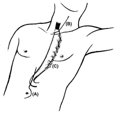
During an esophagectomy, the surgeon will make incisions in the neck, chest, and/or abdomen to access the esophagus. The affected portion of the esophagus is then removed, and the remaining ends are reconnected, often using a section of the stomach or colon to create a new conduit for food to pass from the throat to the stomach.
Esophagectomy is a complex surgical procedure that requires significant expertise and experience on the part of the surgeon. It carries risks such as bleeding, infection, and complications related to anesthesia. Additionally, patients who undergo esophagectomy may experience difficulty swallowing, chronic pain, and other long-term complications. However, for some patients with esophageal cancer or other serious conditions affecting the esophagus, esophagectomy may be the best available treatment option.
Gastrointestinal (GI) neoplasms refer to abnormal growths in the gastrointestinal tract, which can be benign or malignant. The gastrointestinal tract includes the mouth, esophagus, stomach, small intestine, large intestine, rectum, and anus.
Benign neoplasms are non-cancerous growths that do not invade nearby tissues or spread to other parts of the body. They can sometimes be removed completely and may not cause any further health problems.
Malignant neoplasms, on the other hand, are cancerous growths that can invade nearby tissues and organs and spread to other parts of the body through the bloodstream or lymphatic system. These types of neoplasms can be life-threatening if not diagnosed and treated promptly.
GI neoplasms can cause various symptoms, including abdominal pain, bloating, changes in bowel habits, nausea, vomiting, weight loss, and anemia. The specific symptoms may depend on the location and size of the neoplasm.
There are many types of GI neoplasms, including adenocarcinomas, gastrointestinal stromal tumors (GISTs), lymphomas, and neuroendocrine tumors. The diagnosis of GI neoplasms typically involves a combination of medical history, physical examination, imaging studies, and biopsy. Treatment options may include surgery, radiation therapy, chemotherapy, targeted therapy, or immunotherapy.
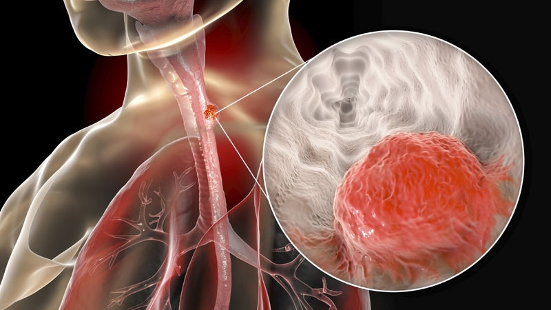
The esophagus is the muscular tube that connects the throat (pharynx) to the stomach. It is located in the midline of the neck and chest, passing through the diaphragm to enter the abdomen and join the stomach. The main function of the esophagus is to transport food and liquids from the mouth to the stomach for digestion.
The esophagus has a few distinct parts: the upper esophageal sphincter (a ring of muscle that separates the esophagus from the throat), the middle esophagus, and the lower esophageal sphincter (another ring of muscle that separates the esophagus from the stomach). The lower esophageal sphincter relaxes to allow food and liquids to enter the stomach and then contracts to prevent stomach contents from flowing back into the esophagus.
The walls of the esophagus are made up of several layers, including mucosa (a moist tissue that lines the inside of the tube), submucosa (a layer of connective tissue), muscle (both voluntary and involuntary types), and adventitia (an outer layer of connective tissue).
Common conditions affecting the esophagus include gastroesophageal reflux disease (GERD), Barrett's esophagus, esophageal cancer, esophageal strictures, and eosinophilic esophagitis.
Esophageal neoplasms refer to abnormal growths in the tissue of the esophagus, which is the muscular tube that connects the throat to the stomach. These growths can be benign (non-cancerous) or malignant (cancerous). Malignant esophageal neoplasms are typically classified as either squamous cell carcinomas or adenocarcinomas, depending on the type of cell from which they originate.
Esophageal cancer is a serious and often life-threatening condition that can cause symptoms such as difficulty swallowing, chest pain, weight loss, and coughing. Risk factors for esophageal neoplasms include smoking, heavy alcohol consumption, gastroesophageal reflux disease (GERD), and Barrett's esophagus. Treatment options may include surgery, radiation therapy, chemotherapy, or a combination of these approaches.
Barrett esophagus is a condition in which the tissue lining of the lower esophagus changes, becoming more like the tissue that lines the intestines (intestinal metaplasia). This change can increase the risk of developing esophageal adenocarcinoma, a type of cancer. The exact cause of Barrett esophagus is not known, but it is often associated with long-term gastroesophageal reflux disease (GERD), also known as chronic acid reflux.
In Barrett esophagus, the normal squamous cells that line the lower esophagus are replaced by columnar epithelial cells. This change is usually detected during an upper endoscopy and biopsy. The diagnosis of Barrett esophagus is confirmed when the biopsy shows intestinal metaplasia in the lower esophagus.
It's important to note that not everyone with GERD will develop Barrett esophagus, and not everyone with Barrett esophagus will develop esophageal cancer. However, if you have been diagnosed with Barrett esophagus, your healthcare provider may recommend regular endoscopies and biopsies to monitor the condition and reduce the risk of cancer. Treatment options for Barrett esophagus include medications to control acid reflux, lifestyle changes, and in some cases, surgery.
Stomach neoplasms refer to abnormal growths in the stomach that can be benign or malignant. They include a wide range of conditions such as:
1. Gastric adenomas: These are benign tumors that develop from glandular cells in the stomach lining.
2. Gastrointestinal stromal tumors (GISTs): These are rare tumors that can be found in the stomach and other parts of the digestive tract. They originate from the stem cells in the wall of the digestive tract.
3. Leiomyomas: These are benign tumors that develop from smooth muscle cells in the stomach wall.
4. Lipomas: These are benign tumors that develop from fat cells in the stomach wall.
5. Neuroendocrine tumors (NETs): These are tumors that develop from the neuroendocrine cells in the stomach lining. They can be benign or malignant.
6. Gastric carcinomas: These are malignant tumors that develop from the glandular cells in the stomach lining. They are the most common type of stomach neoplasm and include adenocarcinomas, signet ring cell carcinomas, and others.
7. Lymphomas: These are malignant tumors that develop from the immune cells in the stomach wall.
Stomach neoplasms can cause various symptoms such as abdominal pain, nausea, vomiting, weight loss, and difficulty swallowing. The diagnosis of stomach neoplasms usually involves a combination of imaging tests, endoscopy, and biopsy. Treatment options depend on the type and stage of the neoplasm and may include surgery, chemotherapy, radiation therapy, or targeted therapy.
Adenocarcinoma is a type of cancer that arises from glandular epithelial cells. These cells line the inside of many internal organs, including the breasts, prostate, colon, and lungs. Adenocarcinomas can occur in any of these organs, as well as in other locations where glands are present.
The term "adenocarcinoma" is used to describe a cancer that has features of glandular tissue, such as mucus-secreting cells or cells that produce hormones. These cancers often form glandular structures within the tumor mass and may produce mucus or other substances.
Adenocarcinomas are typically slow-growing and tend to spread (metastasize) to other parts of the body through the lymphatic system or bloodstream. They can be treated with surgery, radiation therapy, chemotherapy, targeted therapy, or a combination of these treatments. The prognosis for adenocarcinoma depends on several factors, including the location and stage of the cancer, as well as the patient's overall health and age.
Esophagoscopy is a medical procedure that involves the visual examination of the esophagus, which is the tube that connects the throat to the stomach. This procedure is typically carried out using an esophagogastroduodenoscope (EGD), a flexible tube with a camera and light on the end.
During the procedure, the EGD is inserted through the mouth and down the throat into the esophagus, allowing the medical professional to examine its lining for any abnormalities such as inflammation, ulcers, or tumors. The procedure may also involve taking tissue samples (biopsies) for further examination and testing.
Esophagoscopy is commonly used to diagnose and monitor conditions such as gastroesophageal reflux disease (GERD), Barrett's esophagus, esophageal cancer, and other disorders affecting the esophagus. It may also be used to treat certain conditions, such as removing polyps or foreign objects from the esophagus.
Thoracoscopy is a surgical procedure in which a thoracoscope, a type of endoscope, is inserted through a small incision between the ribs to examine the lungs and pleural space (the space surrounding the lungs). It allows the surgeon to directly view the chest cavity, take biopsies, and perform various operations. This procedure is often used in the diagnosis and treatment of pleural effusions, lung cancer, and other chest conditions.
Squamous cell carcinoma is a type of skin cancer that begins in the squamous cells, which are flat, thin cells that form the outer layer of the skin (epidermis). It commonly occurs on sun-exposed areas such as the face, ears, lips, and backs of the hands. Squamous cell carcinoma can also develop in other areas of the body including the mouth, lungs, and cervix.
This type of cancer usually develops slowly and may appear as a rough or scaly patch of skin, a red, firm nodule, or a sore or ulcer that doesn't heal. While squamous cell carcinoma is not as aggressive as some other types of cancer, it can metastasize (spread) to other parts of the body if left untreated, making early detection and treatment important.
Risk factors for developing squamous cell carcinoma include prolonged exposure to ultraviolet (UV) radiation from the sun or tanning beds, fair skin, a history of sunburns, a weakened immune system, and older age. Prevention measures include protecting your skin from the sun by wearing protective clothing, using a broad-spectrum sunscreen with an SPF of at least 30, avoiding tanning beds, and getting regular skin examinations.
Pancreatic neoplasms refer to abnormal growths in the pancreas that can be benign or malignant. The pancreas is a gland located behind the stomach that produces hormones and digestive enzymes. Pancreatic neoplasms can interfere with the normal functioning of the pancreas, leading to various health complications.
Benign pancreatic neoplasms are non-cancerous growths that do not spread to other parts of the body. They are usually removed through surgery to prevent any potential complications, such as blocking the bile duct or causing pain.
Malignant pancreatic neoplasms, also known as pancreatic cancer, are cancerous growths that can invade and destroy surrounding tissues and organs. They can also spread (metastasize) to other parts of the body, such as the liver, lungs, or bones. Pancreatic cancer is often aggressive and difficult to treat, with a poor prognosis.
There are several types of pancreatic neoplasms, including adenocarcinomas, neuroendocrine tumors, solid pseudopapillary neoplasms, and cystic neoplasms. The specific type of neoplasm is determined through various diagnostic tests, such as imaging studies, biopsies, and blood tests. Treatment options depend on the type, stage, and location of the neoplasm, as well as the patient's overall health and preferences.
Esophageal stenosis is a medical condition characterized by the narrowing or constriction of the esophagus, which is the muscular tube that connects the throat to the stomach. This narrowing can make it difficult to swallow food and liquids, leading to symptoms such as dysphagia (difficulty swallowing), pain or discomfort while swallowing, regurgitation, and weight loss.
Esophageal stenosis can be caused by a variety of factors, including:
1. Scarring or fibrosis due to prolonged acid reflux or gastroesophageal reflux disease (GERD)
2. Radiation therapy for cancer treatment
3. Ingestion of corrosive substances
4. Eosinophilic esophagitis, an allergic condition that affects the esophagus
5. Esophageal tumors or cancers
6. Surgical complications
Depending on the underlying cause and severity of the stenosis, treatment options may include medications to manage symptoms, dilation procedures to widen the narrowed area, or surgery to remove the affected portion of the esophagus. It is important to seek medical attention if you experience any difficulty swallowing or other symptoms related to esophageal stenosis.
Esophageal diseases refer to a range of medical conditions that affect the esophagus, which is the muscular tube that connects the throat to the stomach. Here are some common esophageal diseases with their brief definitions:
1. Gastroesophageal reflux disease (GERD): A chronic condition in which stomach acid or bile flows back into the esophagus, causing symptoms such as heartburn, chest pain, and difficulty swallowing.
2. Esophagitis: Inflammation of the esophageal lining, often caused by GERD, infection, or medication.
3. Esophageal stricture: Narrowing of the esophagus due to scarring or inflammation, which can make swallowing difficult.
4. Esophageal cancer: Cancer that forms in the tissues of the esophagus, often as a result of long-term GERD or smoking.
5. Esophageal motility disorders: Disorders that affect the normal movement and function of the esophagus, such as achalasia, diffuse spasm, and nutcracker esophagus.
6. Barrett's esophagus: A condition in which the lining of the lower esophagus changes, increasing the risk of esophageal cancer.
7. Esophageal diverticula: Small pouches that form in the esophageal wall, often causing difficulty swallowing or regurgitation.
8. Eosinophilic esophagitis (EoE): A chronic immune-mediated disorder characterized by inflammation of the esophagus due to an allergic reaction.
These are some of the common esophageal diseases, and their diagnosis and treatment may vary depending on the severity and underlying cause of the condition.
'Digestive System Neoplasms' refer to new and abnormal growths of tissue in the digestive system that can be benign or malignant. These growths are also known as tumors, and they can occur in any part of the digestive system, including the esophagus, stomach, small intestine, large intestine (colon and rectum), liver, bile ducts, pancreas, and gallbladder. Neoplasms in the digestive system can interfere with normal digestion and absorption of nutrients, cause bleeding, obstruct the digestive tract, and spread to other parts of the body (metastasis) if they are malignant.
Benign neoplasms are not cancerous and do not usually spread to other parts of the body. They can often be removed surgically and may not require further treatment. Malignant neoplasms, on the other hand, are cancerous and can invade nearby tissues and organs and spread to other parts of the body. Treatment for malignant neoplasms in the digestive system typically involves a combination of surgery, radiation therapy, and chemotherapy.
The causes of digestive system neoplasms are varied and include genetic factors, environmental exposures, lifestyle factors (such as diet and smoking), and infectious agents. Prevention strategies may include maintaining a healthy diet, avoiding tobacco and excessive alcohol consumption, practicing safe sex, getting vaccinated against certain viral infections, and undergoing regular screenings for certain types of neoplasms (such as colonoscopies for colorectal cancer).
An esophagostomy is a surgical opening created between the esophagus and the skin of the neck or chest. It is typically performed as an emergency procedure in cases where there is an obstruction or injury to the esophagus that cannot be managed through less invasive means. The esophagostomy provides a temporary or permanent access point for feeding, medication administration, or decompression of the esophagus.
The procedure involves creating an incision in the neck or chest and exposing the esophagus. A small opening is then made in the esophageal wall, and a tube is inserted through the opening and brought out through the skin. The tube may be secured in place with sutures or staples, and a dressing is applied to protect the site from infection.
After surgery, patients with an esophagostomy will require close monitoring and care to ensure proper healing and prevent complications such as infection, bleeding, or leakage of digestive fluids. The tube may be removed once the underlying condition has been treated and the esophagus has healed.
Colorectal neoplasms refer to abnormal growths in the colon or rectum, which can be benign or malignant. These growths can arise from the inner lining (mucosa) of the colon or rectum and can take various forms such as polyps, adenomas, or carcinomas.
Benign neoplasms, such as hyperplastic polyps and inflammatory polyps, are not cancerous but may need to be removed to prevent the development of malignant tumors. Adenomas, on the other hand, are precancerous lesions that can develop into colorectal cancer if left untreated.
Colorectal cancer is a malignant neoplasm that arises from the uncontrolled growth and division of cells in the colon or rectum. It is one of the most common types of cancer worldwide and can spread to other parts of the body through the bloodstream or lymphatic system.
Regular screening for colorectal neoplasms is recommended for individuals over the age of 50, as early detection and removal of precancerous lesions can significantly reduce the risk of developing colorectal cancer.
Esophagoplasty is a surgical procedure that involves reconstructing or reshaping the esophagus, which is the muscular tube that connects the throat to the stomach. This procedure may be performed to treat various conditions such as esophageal atresia (a birth defect in which the esophagus does not develop properly), esophageal stricture (narrowing of the esophagus), or esophageal cancer.
During an esophagoplasty, a surgeon may use tissue from another part of the body, such as the stomach or colon, to reconstruct the esophagus. The specific technique used will depend on the individual patient's needs and the nature of their condition.
It is important to note that esophagoplasty is a complex surgical procedure that carries risks such as bleeding, infection, and complications related to anesthesia. Patients who undergo this procedure may require extensive postoperative care and rehabilitation to recover fully.
Surgical anastomosis is a medical procedure that involves the connection of two tubular structures, such as blood vessels or intestines, to create a continuous passage. This technique is commonly used in various types of surgeries, including vascular, gastrointestinal, and orthopedic procedures.
During a surgical anastomosis, the ends of the two tubular structures are carefully prepared by removing any damaged or diseased tissue. The ends are then aligned and joined together using sutures, staples, or other devices. The connection must be secure and leak-free to ensure proper function and healing.
The success of a surgical anastomosis depends on several factors, including the patient's overall health, the location and condition of the structures being joined, and the skill and experience of the surgeon. Complications such as infection, bleeding, or leakage can occur, which may require additional medical intervention or surgery.
Proper postoperative care is also essential to ensure the success of a surgical anastomosis. This may include monitoring for signs of complications, administering medications to prevent infection and promote healing, and providing adequate nutrition and hydration.
The esophagogastric junction (EGJ) is the region of the gastrointestinal tract where the esophagus (the tube that carries food from the mouth to the stomach) meets the stomach. It serves as a physiological sphincter, which helps control the direction of flow and prevent reflux of gastric contents back into the esophagus. The EGJ is also known as the gastroesophageal junction or cardia.
Colonic neoplasms refer to abnormal growths in the large intestine, also known as the colon. These growths can be benign (non-cancerous) or malignant (cancerous). The two most common types of colonic neoplasms are adenomas and carcinomas.
Adenomas are benign tumors that can develop into cancer over time if left untreated. They are often found during routine colonoscopies and can be removed during the procedure.
Carcinomas, on the other hand, are malignant tumors that invade surrounding tissues and can spread to other parts of the body. Colorectal cancer is the third leading cause of cancer-related deaths in the United States, and colonic neoplasms are a significant risk factor for developing this type of cancer.
Regular screenings for colonic neoplasms are recommended for individuals over the age of 50 or those with a family history of colorectal cancer or other risk factors. Early detection and removal of colonic neoplasms can significantly reduce the risk of developing colorectal cancer.
The Upper Gastrointestinal (GI) Tract refers to the segment of the digestive system that includes the mouth, pharynx, esophagus, stomach, and duodenum, which is the first part of the small intestine. This region is responsible for the initial stages of digestion, such as mechanical breakdown of food by chewing and churning, and chemical breakdown through enzymes and acids. It's also where the majority of nutrient absorption occurs. Various medical conditions, including infections, inflammation, and cancers, can affect the upper GI tract.
Neoplasm staging is a systematic process used in medicine to describe the extent of spread of a cancer, including the size and location of the original (primary) tumor and whether it has metastasized (spread) to other parts of the body. The most widely accepted system for this purpose is the TNM classification system developed by the American Joint Committee on Cancer (AJCC) and the Union for International Cancer Control (UICC).
In this system, T stands for tumor, and it describes the size and extent of the primary tumor. N stands for nodes, and it indicates whether the cancer has spread to nearby lymph nodes. M stands for metastasis, and it shows whether the cancer has spread to distant parts of the body.
Each letter is followed by a number that provides more details about the extent of the disease. For example, a T1N0M0 cancer means that the primary tumor is small and has not spread to nearby lymph nodes or distant sites. The higher the numbers, the more advanced the cancer.
Staging helps doctors determine the most appropriate treatment for each patient and estimate the patient's prognosis. It is an essential tool for communication among members of the healthcare team and for comparing outcomes of treatments in clinical trials.
Treatment outcome is a term used to describe the result or effect of medical treatment on a patient's health status. It can be measured in various ways, such as through symptoms improvement, disease remission, reduced disability, improved quality of life, or survival rates. The treatment outcome helps healthcare providers evaluate the effectiveness of a particular treatment plan and make informed decisions about future care. It is also used in clinical research to compare the efficacy of different treatments and improve patient care.
Retrospective studies, also known as retrospective research or looking back studies, are a type of observational study that examines data from the past to draw conclusions about possible causal relationships between risk factors and outcomes. In these studies, researchers analyze existing records, medical charts, or previously collected data to test a hypothesis or answer a specific research question.
Retrospective studies can be useful for generating hypotheses and identifying trends, but they have limitations compared to prospective studies, which follow participants forward in time from exposure to outcome. Retrospective studies are subject to biases such as recall bias, selection bias, and information bias, which can affect the validity of the results. Therefore, retrospective studies should be interpreted with caution and used primarily to generate hypotheses for further testing in prospective studies.
An esophageal fistula is an abnormal connection or passage between the esophagus (the tube that carries food and liquids from the throat to the stomach) and another organ, such as the trachea (windpipe) or the skin. This condition can result from complications of certain medical conditions, including cancer, prolonged infection, or injury to the esophagus.
Esophageal fistulas can cause a variety of symptoms, including difficulty swallowing, coughing, chest pain, and fever. They can also lead to serious complications, such as pneumonia or sepsis, if left untreated. Treatment for an esophageal fistula typically involves surgical repair of the abnormal connection, along with management of any underlying conditions that may have contributed to its development.
A gastric fistula is an abnormal connection or passage between the stomach and another organ or the skin surface. This condition can occur as a result of complications from surgery, injury, infection, or certain diseases such as cancer. Symptoms may include persistent drainage from the site of the fistula, pain, malnutrition, and infection. Treatment typically involves surgical repair of the fistula and management of any underlying conditions.
Combined modality therapy (CMT) is a medical treatment approach that utilizes more than one method or type of therapy simultaneously or in close succession, with the goal of enhancing the overall effectiveness of the treatment. In the context of cancer care, CMT often refers to the combination of two or more primary treatment modalities, such as surgery, radiation therapy, and systemic therapies (chemotherapy, immunotherapy, targeted therapy, etc.).
The rationale behind using combined modality therapy is that each treatment method can target cancer cells in different ways, potentially increasing the likelihood of eliminating all cancer cells and reducing the risk of recurrence. The specific combination and sequence of treatments will depend on various factors, including the type and stage of cancer, patient's overall health, and individual preferences.
For example, a common CMT approach for locally advanced rectal cancer may involve preoperative (neoadjuvant) chemoradiation therapy, followed by surgery to remove the tumor, and then postoperative (adjuvant) chemotherapy. This combined approach allows for the reduction of the tumor size before surgery, increases the likelihood of complete tumor removal, and targets any remaining microscopic cancer cells with systemic chemotherapy.
It is essential to consult with a multidisciplinary team of healthcare professionals to determine the most appropriate CMT plan for each individual patient, considering both the potential benefits and risks associated with each treatment method.
Medical survival rate is a statistical measure used to determine the percentage of patients who are still alive for a specific period of time after their diagnosis or treatment for a certain condition or disease. It is often expressed as a five-year survival rate, which refers to the proportion of people who are alive five years after their diagnosis. Survival rates can be affected by many factors, including the stage of the disease at diagnosis, the patient's age and overall health, the effectiveness of treatment, and other health conditions that the patient may have. It is important to note that survival rates are statistical estimates and do not necessarily predict an individual patient's prognosis.
Prognosis is a medical term that refers to the prediction of the likely outcome or course of a disease, including the chances of recovery or recurrence, based on the patient's symptoms, medical history, physical examination, and diagnostic tests. It is an important aspect of clinical decision-making and patient communication, as it helps doctors and patients make informed decisions about treatment options, set realistic expectations, and plan for future care.
Prognosis can be expressed in various ways, such as percentages, categories (e.g., good, fair, poor), or survival rates, depending on the nature of the disease and the available evidence. However, it is important to note that prognosis is not an exact science and may vary depending on individual factors, such as age, overall health status, and response to treatment. Therefore, it should be used as a guide rather than a definitive forecast.
A precancerous condition, also known as a premalignant condition, is a state of abnormal cellular growth and development that has a higher-than-normal potential to progress into cancer. These conditions are characterized by the presence of certain anomalies in the cells, such as dysplasia (abnormal changes in cell shape or size), which can indicate an increased risk for malignant transformation.
It is important to note that not all precancerous conditions will eventually develop into cancer, and some may even regress on their own. However, individuals with precancerous conditions are often at a higher risk of developing cancer compared to the general population. Regular monitoring and appropriate medical interventions, if necessary, can help manage this risk and potentially prevent or detect cancer at an early stage when it is more treatable.
Examples of precancerous conditions include:
1. Dysplasia in the cervix (cervical intraepithelial neoplasia or CIN)
2. Atypical ductal hyperplasia or lobular hyperplasia in the breast
3. Actinic keratosis on the skin
4. Leukoplakia in the mouth
5. Barrett's esophagus in the digestive tract
Regular medical check-ups, screenings, and lifestyle modifications are crucial for individuals with precancerous conditions to monitor their health and reduce the risk of cancer development.
Fluorouracil is a antineoplastic medication, which means it is used to treat cancer. It is a type of chemotherapy drug known as an antimetabolite. Fluorouracil works by interfering with the growth of cancer cells and ultimately killing them. It is often used to treat colon, esophageal, stomach, and breast cancers, as well as skin conditions such as actinic keratosis and superficial basal cell carcinoma. Fluorouracil may be given by injection or applied directly to the skin in the form of a cream.
It is important to note that fluorouracil can have serious side effects, including suppression of bone marrow function, mouth sores, stomach and intestinal ulcers, and nerve damage. It should only be used under the close supervision of a healthcare professional.
Thoracotomy is a surgical procedure that involves making an incision on the chest wall to gain access to the thoracic cavity, which contains the lungs, heart, esophagus, trachea, and other vital organs. The incision can be made on the side (lateral thoracotomy), back (posterolateral thoracotomy), or front (median sternotomy) of the chest wall, depending on the specific surgical indication.
Thoracotomy is performed for various indications, including lung biopsy, lung resection, esophagectomy, heart surgery, and mediastinal mass removal. The procedure allows the surgeon to directly visualize and access the organs within the thoracic cavity, perform necessary procedures, and control bleeding if needed.
After the procedure, the incision is typically closed with sutures or staples, and a chest tube may be placed to drain any accumulated fluid or air from the pleural space around the lungs. The patient will require postoperative care and monitoring in a hospital setting until their condition stabilizes.
The term "Congresses as Topic" refers to large, formal meetings that are held to discuss and exchange information on a specific topic or field, usually academic or professional in nature. In the context of medical science, a congress is an event where healthcare professionals, researchers, and experts gather to present and discuss the latest research, developments, and innovations in their field. Medical congresses can cover a wide range of topics, including specific diseases, treatments, medical specialties, public health issues, or healthcare policies. These events often include keynote speeches, panel discussions, workshops, poster sessions, and networking opportunities for attendees. Examples of well-known medical congresses are the annual meetings of the American Medical Association, the American Heart Association, and the European Society of Cardiology.
Carcinoma is a type of cancer that develops from epithelial cells, which are the cells that line the inner and outer surfaces of the body. These cells cover organs, glands, and other structures within the body. Carcinomas can occur in various parts of the body, including the skin, lungs, breasts, prostate, colon, and pancreas. They are often characterized by the uncontrolled growth and division of abnormal cells that can invade surrounding tissues and spread to other parts of the body through a process called metastasis. Carcinomas can be further classified based on their appearance under a microscope, such as adenocarcinoma, squamous cell carcinoma, and basal cell carcinoma.
Postoperative complications refer to any unfavorable condition or event that occurs during the recovery period after a surgical procedure. These complications can vary in severity and may include, but are not limited to:
1. Infection: This can occur at the site of the incision or inside the body, such as pneumonia or urinary tract infection.
2. Bleeding: Excessive bleeding (hemorrhage) can lead to a drop in blood pressure and may require further surgical intervention.
3. Blood clots: These can form in the deep veins of the legs (deep vein thrombosis) and can potentially travel to the lungs (pulmonary embolism).
4. Wound dehiscence: This is when the surgical wound opens up, which can lead to infection and further complications.
5. Pulmonary issues: These include atelectasis (collapsed lung), pneumonia, or respiratory failure.
6. Cardiovascular problems: These include abnormal heart rhythms (arrhythmias), heart attack, or stroke.
7. Renal failure: This can occur due to various reasons such as dehydration, blood loss, or the use of certain medications.
8. Pain management issues: Inadequate pain control can lead to increased stress, anxiety, and decreased mobility.
9. Nausea and vomiting: These can be caused by anesthesia, opioid pain medication, or other factors.
10. Delirium: This is a state of confusion and disorientation that can occur in the elderly or those with certain medical conditions.
Prompt identification and management of these complications are crucial to ensure the best possible outcome for the patient.
An anastomotic leak is a medical condition that occurs after a surgical procedure where two hollow organs or vessels are connected (anastomosed). It refers to the failure of the connection, resulting in a communication between the inside of the connected structures and the outside, which can lead to the escape of fluids, such as digestive contents or blood, into the surrounding tissues.
Anastomotic leaks can occur in various parts of the body where anastomoses are performed, including the gastrointestinal tract, vasculature, and respiratory system. The leakage can cause localized or systemic infection, inflammation, sepsis, organ failure, or even death if not promptly diagnosed and treated.
The risk of anastomotic leaks depends on several factors, such as the patient's overall health, the type and location of the surgery, the quality of the surgical technique, and the presence of any underlying medical conditions that may affect wound healing. Treatment options for anastomotic leaks vary depending on the severity and location of the leak, ranging from conservative management with antibiotics and bowel rest to surgical intervention, such as drainage, revision of the anastomosis, or resection of the affected segment.
Rectal neoplasms refer to abnormal growths in the tissues of the rectum, which can be benign or malignant. They are characterized by uncontrolled cell division and can invade nearby tissues or spread to other parts of the body (metastasis). The most common type of rectal neoplasm is rectal cancer, which often begins as a small polyp or growth in the lining of the rectum. Other types of rectal neoplasms include adenomas, carcinoids, and gastrointestinal stromal tumors (GISTs). Regular screenings are recommended for early detection and treatment of rectal neoplasms.
Metaplasia is a term used in pathology to describe the replacement of one differentiated cell type with another differentiated cell type within a tissue or organ. It is an adaptive response of epithelial cells to chronic irritation, inflammation, or injury and can be reversible if the damaging stimulus is removed. Metaplastic changes are often associated with an increased risk of cancer development in the affected area.
For example, in the case of gastroesophageal reflux disease (GERD), chronic exposure to stomach acid can lead to metaplasia of the esophageal squamous epithelium into columnar epithelium, a condition known as Barrett's esophagus. This metaplastic change is associated with an increased risk of developing esophageal adenocarcinoma.
The cardia is a term used in anatomical context to refer to the upper part of the stomach that surrounds and opens into the lower end of the esophagus. It is responsible for controlling the passage of food from the esophagus into the stomach and is also known as the cardiac orifice or cardiac sphincter. Any medical condition that affects this area, such as gastroesophageal reflux disease (GERD), can lead to symptoms like heartburn, difficulty swallowing, and chest pain.
Neoplasms are abnormal growths of cells or tissues in the body that serve no physiological function. They can be benign (non-cancerous) or malignant (cancerous). Benign neoplasms are typically slow growing and do not spread to other parts of the body, while malignant neoplasms are aggressive, invasive, and can metastasize to distant sites.
Neoplasms occur when there is a dysregulation in the normal process of cell division and differentiation, leading to uncontrolled growth and accumulation of cells. This can result from genetic mutations or other factors such as viral infections, environmental exposures, or hormonal imbalances.
Neoplasms can develop in any organ or tissue of the body and can cause various symptoms depending on their size, location, and type. Treatment options for neoplasms include surgery, radiation therapy, chemotherapy, immunotherapy, and targeted therapy, among others.
Esophageal achalasia is a rare disorder of the esophagus, the tube that carries food from the mouth to the stomach. In this condition, the muscles at the lower end of the esophagus fail to relax properly during swallowing, making it difficult for food and liquids to pass into the stomach. This results in symptoms such as difficulty swallowing (dysphagia), regurgitation of food, chest pain, and weight loss. The cause of esophageal achalasia is not fully understood, but it is believed to be related to damage to the nerves that control the muscles of the esophagus. Treatment options include medications to relax the lower esophageal sphincter, botulinum toxin injections, and surgical procedures such as laparoscopic Heller myotomy or peroral endoscopic myotomy (POEM).
Follow-up studies are a type of longitudinal research that involve repeated observations or measurements of the same variables over a period of time, in order to understand their long-term effects or outcomes. In medical context, follow-up studies are often used to evaluate the safety and efficacy of medical treatments, interventions, or procedures.
In a typical follow-up study, a group of individuals (called a cohort) who have received a particular treatment or intervention are identified and then followed over time through periodic assessments or data collection. The data collected may include information on clinical outcomes, adverse events, changes in symptoms or functional status, and other relevant measures.
The results of follow-up studies can provide important insights into the long-term benefits and risks of medical interventions, as well as help to identify factors that may influence treatment effectiveness or patient outcomes. However, it is important to note that follow-up studies can be subject to various biases and limitations, such as loss to follow-up, recall bias, and changes in clinical practice over time, which must be carefully considered when interpreting the results.
Antineoplastic combined chemotherapy protocols refer to a treatment plan for cancer that involves the use of more than one antineoplastic (chemotherapy) drug given in a specific sequence and schedule. The combination of drugs is used because they may work better together to destroy cancer cells compared to using a single agent alone. This approach can also help to reduce the likelihood of cancer cells becoming resistant to the treatment.
The choice of drugs, dose, duration, and frequency are determined by various factors such as the type and stage of cancer, patient's overall health, and potential side effects. Combination chemotherapy protocols can be used in various settings, including as a primary treatment, adjuvant therapy (given after surgery or radiation to kill any remaining cancer cells), neoadjuvant therapy (given before surgery or radiation to shrink the tumor), or palliative care (to alleviate symptoms and prolong survival).
It is important to note that while combined chemotherapy protocols can be effective in treating certain types of cancer, they can also cause significant side effects, including nausea, vomiting, hair loss, fatigue, and an increased risk of infection. Therefore, patients undergoing such treatment should be closely monitored and managed by a healthcare team experienced in administering chemotherapy.
Lymph node excision is a surgical procedure in which one or more lymph nodes are removed from the body for the purpose of examination. This procedure is often conducted to help diagnose or stage various types of cancer, as malignant cells may spread to the lymphatic system and eventually accumulate within nearby lymph nodes.
During a lymph node excision, an incision is made in the skin overlying the affected lymph node(s). The surgeon carefully dissects the tissue surrounding the lymph node(s) to isolate them from adjacent structures before removing them. In some cases, a sentinel lymph node biopsy may be performed instead, where only the sentinel lymph node (the first lymph node to which cancer cells are likely to spread) is removed and examined.
The excised lymph nodes are then sent to a laboratory for histopathological examination, which involves staining and microscopic evaluation of the tissue to determine whether it contains any malignant cells. The results of this examination can help guide further treatment decisions and provide valuable prognostic information.
Medical Definition:
"Risk factors" are any attribute, characteristic or exposure of an individual that increases the likelihood of developing a disease or injury. They can be divided into modifiable and non-modifiable risk factors. Modifiable risk factors are those that can be changed through lifestyle choices or medical treatment, while non-modifiable risk factors are inherent traits such as age, gender, or genetic predisposition. Examples of modifiable risk factors include smoking, alcohol consumption, physical inactivity, and unhealthy diet, while non-modifiable risk factors include age, sex, and family history. It is important to note that having a risk factor does not guarantee that a person will develop the disease, but rather indicates an increased susceptibility.
Appetite stimulants are medications or substances that increase the desire to eat or improve appetite. They work by affecting brain chemicals, hormones, or other systems involved in regulating hunger and fullness. Some commonly used appetite stimulants include:
1. Megestrol acetate: a synthetic progestin hormone that is often prescribed for cancer-related weight loss and anorexia. It works by stimulating appetite and promoting weight gain.
2. Dronabinol: a synthetic form of THC, the active ingredient in marijuana. It is approved for treating AIDS-related anorexia and chemotherapy-induced nausea and vomiting. Dronabinol can increase appetite and promote weight gain.
3. Corticosteroids: medications that mimic the effects of hormones produced by the adrenal gland. They can help improve appetite, but their long-term use is associated with significant side effects.
4. Cyproheptadine: an antihistamine medication that can also stimulate appetite. It is sometimes used off-label to treat appetite loss in various conditions, such as cancer or HIV/AIDS.
5. Ghrelin agonists: these are medications that mimic the effects of ghrelin, a hormone produced by the stomach that increases hunger and appetite. Currently, there are no FDA-approved ghrelin agonists for appetite stimulation, but research is ongoing.
It's important to note that while appetite stimulants can help improve food intake in some individuals, they may not be effective for everyone, and their use should be carefully monitored due to potential side effects and interactions with other medications. Always consult a healthcare professional before starting any new medication or supplement.
Survival analysis is a branch of statistics that deals with the analysis of time to event data. It is used to estimate the time it takes for a certain event of interest to occur, such as death, disease recurrence, or treatment failure. The event of interest is called the "failure" event, and survival analysis estimates the probability of not experiencing the failure event until a certain point in time, also known as the "survival" probability.
Survival analysis can provide important information about the effectiveness of treatments, the prognosis of patients, and the identification of risk factors associated with the event of interest. It can handle censored data, which is common in medical research where some participants may drop out or be lost to follow-up before the event of interest occurs.
Survival analysis typically involves estimating the survival function, which describes the probability of surviving beyond a certain time point, as well as hazard functions, which describe the instantaneous rate of failure at a given time point. Other important concepts in survival analysis include median survival times, restricted mean survival times, and various statistical tests to compare survival curves between groups.
Gastroesophageal reflux (GER) is the retrograde movement of stomach contents into the esophagus, which can cause discomfort and symptoms. It occurs when the lower esophageal sphincter (a ring of muscle between the esophagus and stomach) relaxes inappropriately, allowing the acidic or non-acidic gastric contents to flow back into the esophagus.
Gastroesophageal reflux becomes gastroesophageal reflux disease (GERD) when it is more severe, persistent, and/or results in complications such as esophagitis, strictures, or Barrett's esophagus. Common symptoms of GERD include heartburn, regurgitation, chest pain, difficulty swallowing, and chronic cough or hoarseness.
Tumor-associated carbohydrate antigens (TACAs) are a type of tumor antigen that are expressed on the surface of cancer cells. These antigens are abnormal forms of carbohydrates, also known as glycans, which are attached to proteins and lipids on the cell surface.
TACAs are often overexpressed or expressed in a different form on cancer cells compared to normal cells. This makes them attractive targets for cancer immunotherapy because they can be recognized by the immune system as foreign and elicit an immune response. Some examples of TACAs include gangliosides, fucosylated glycans, and sialylated glycans.
Tumor-associated carbohydrate antigens have been studied as potential targets for cancer vaccines, antibody therapies, and other immunotherapeutic approaches. However, their use as targets for cancer therapy is still in the early stages of research and development.
Prospective studies, also known as longitudinal studies, are a type of cohort study in which data is collected forward in time, following a group of individuals who share a common characteristic or exposure over a period of time. The researchers clearly define the study population and exposure of interest at the beginning of the study and follow up with the participants to determine the outcomes that develop over time. This type of study design allows for the investigation of causal relationships between exposures and outcomes, as well as the identification of risk factors and the estimation of disease incidence rates. Prospective studies are particularly useful in epidemiology and medical research when studying diseases with long latency periods or rare outcomes.
"Lye" is not a medical term, but rather a common name for sodium hydroxide (NaOH) or potassium hydroxide (KOH), which are strong alkalis used in industry. In a medical context, these substances might be referred to as caustic soda or caustic potash. They can cause severe burns and damage to tissue if they come into contact with the skin or eyes, and if ingested they can be harmful or fatal.
Tumor markers are substances that can be found in the body and their presence can indicate the presence of certain types of cancer or other conditions. Biological tumor markers refer to those substances that are produced by cancer cells or by other cells in response to cancer or certain benign (non-cancerous) conditions. These markers can be found in various bodily fluids such as blood, urine, or tissue samples.
Examples of biological tumor markers include:
1. Proteins: Some tumor markers are proteins that are produced by cancer cells or by other cells in response to the presence of cancer. For example, prostate-specific antigen (PSA) is a protein produced by normal prostate cells and in higher amounts by prostate cancer cells.
2. Genetic material: Tumor markers can also include genetic material such as DNA, RNA, or microRNA that are shed by cancer cells into bodily fluids. For example, circulating tumor DNA (ctDNA) is genetic material from cancer cells that can be found in the bloodstream.
3. Metabolites: Tumor markers can also include metabolic products produced by cancer cells or by other cells in response to cancer. For example, lactate dehydrogenase (LDH) is an enzyme that is released into the bloodstream when cancer cells break down glucose for energy.
It's important to note that tumor markers are not specific to cancer and can be elevated in non-cancerous conditions as well. Therefore, they should not be used alone to diagnose cancer but rather as a tool in conjunction with other diagnostic tests and clinical evaluations.
Neoplastic gene expression regulation refers to the processes that control the production of proteins and other molecules from genes in neoplastic cells, or cells that are part of a tumor or cancer. In a normal cell, gene expression is tightly regulated to ensure that the right genes are turned on or off at the right time. However, in cancer cells, this regulation can be disrupted, leading to the overexpression or underexpression of certain genes.
Neoplastic gene expression regulation can be affected by a variety of factors, including genetic mutations, epigenetic changes, and signals from the tumor microenvironment. These changes can lead to the activation of oncogenes (genes that promote cancer growth and development) or the inactivation of tumor suppressor genes (genes that prevent cancer).
Understanding neoplastic gene expression regulation is important for developing new therapies for cancer, as targeting specific genes or pathways involved in this process can help to inhibit cancer growth and progression.
In anatomical terms, the stomach is a muscular, J-shaped organ located in the upper left portion of the abdomen. It is part of the gastrointestinal tract and plays a crucial role in digestion. The stomach's primary functions include storing food, mixing it with digestive enzymes and hydrochloric acid to break down proteins, and slowly emptying the partially digested food into the small intestine for further absorption of nutrients.
The stomach is divided into several regions, including the cardia (the area nearest the esophagus), the fundus (the upper portion on the left side), the body (the main central part), and the pylorus (the narrowed region leading to the small intestine). The inner lining of the stomach, called the mucosa, is protected by a layer of mucus that prevents the digestive juices from damaging the stomach tissue itself.
In medical contexts, various conditions can affect the stomach, such as gastritis (inflammation of the stomach lining), peptic ulcers (sores in the stomach or duodenum), gastroesophageal reflux disease (GERD), and stomach cancer. Symptoms related to the stomach may include abdominal pain, bloating, nausea, vomiting, heartburn, and difficulty swallowing.
A thoracoscope is not a medical condition, but a medical device used in the field of thoracic surgery. It is a type of endoscope that allows surgeons to view the inside of the chest cavity (thorax) through small incisions. The thoracoscope has a light source and a camera at its tip, which transmits images to a video monitor. This enables the surgeon to inspect the lungs, pleura, mediastinum, and diaphragm, take biopsies, and perform various surgical procedures, such as pleurodesis or lung resection, minimizing invasiveness and promoting faster recovery compared to traditional open thoracotomy.
Neoplasm metastasis is the spread of cancer cells from the primary site (where the original or primary tumor formed) to other places in the body. This happens when cancer cells break away from the original (primary) tumor and enter the bloodstream or lymphatic system. The cancer cells can then travel to other parts of the body and form new tumors, called secondary tumors or metastases.
Metastasis is a key feature of malignant neoplasms (cancers), and it is one of the main ways that cancer can cause harm in the body. The metastatic tumors may continue to grow and may cause damage to the organs and tissues where they are located. They can also release additional cancer cells into the bloodstream or lymphatic system, leading to further spread of the cancer.
The metastatic tumors are named based on the location where they are found, as well as the type of primary cancer. For example, if a patient has a primary lung cancer that has metastasized to the liver, the metastatic tumor would be called a liver metastasis from lung cancer.
It is important to note that the presence of metastases can significantly affect a person's prognosis and treatment options. In general, metastatic cancer is more difficult to treat than cancer that has not spread beyond its original site. However, there are many factors that can influence a person's prognosis and response to treatment, so it is important for each individual to discuss their specific situation with their healthcare team.
In the context of psychology and psychiatry, "rationalization" is not a term that has a specific medical definition. However, it is a psychological concept that is often used in medical settings. Rationalization refers to the process of creating logical explanations or justifications for behaviors, emotions, or beliefs that may actually be driven by unconscious desires or motives.
Rationalization can serve as a defense mechanism that allows individuals to avoid acknowledging unpleasant or uncomfortable feelings, thoughts, or impulses. By providing a rational explanation for their behavior, individuals can maintain a positive self-image and avoid feeling anxious, guilty, or threatened.
For example, a person who engages in excessive spending may rationalize their behavior by telling themselves that they deserve to treat themselves or that they need the items they are purchasing. In reality, their overspending may be driven by deeper emotional issues such as low self-esteem or a fear of missing out.
While rationalization is not a medical term per se, it is an important concept in understanding human behavior and motivation, and it can have implications for mental health treatment. Therapists may help individuals identify instances of rationalization and explore the underlying emotions and motivations that are driving their behavior. By gaining insight into these unconscious processes, individuals can develop more adaptive coping mechanisms and make more informed choices about their actions and decisions.
Laparoscopy is a surgical procedure that involves the insertion of a laparoscope, which is a thin tube with a light and camera attached to it, through small incisions in the abdomen. This allows the surgeon to view the internal organs without making large incisions. It's commonly used to diagnose and treat various conditions such as endometriosis, ovarian cysts, infertility, and appendicitis. The advantages of laparoscopy over traditional open surgery include smaller incisions, less pain, shorter hospital stays, and quicker recovery times.
A hiatal hernia is a type of hernia that occurs when a part of the stomach protrudes or squeezes through an opening (hiatus) in the diaphragm, the muscular partition between the chest and abdominal cavities. Normally, the esophagus passes through this opening to connect to the stomach, but in a hiatal hernia, a portion of the stomach also moves up into the chest cavity through the hiatus.
There are two main types of hiatal hernias: sliding and paraesophageal. In a sliding hiatal hernia, the junction between the esophagus and stomach (gastroesophageal junction) slides upward into the chest cavity, which is the most common type. Paraesophageal hiatal hernias are less common but can be more severe, as they involve the stomach herniating alongside the esophagus, potentially leading to complications like obstruction or strangulation of the blood supply to the stomach.
Many people with hiatal hernias do not experience symptoms, but some may have heartburn, acid reflux, regurgitation, difficulty swallowing, chest pain, or shortness of breath. Treatment depends on the severity and associated symptoms, ranging from lifestyle modifications and medications to surgical repair in severe cases.
Early detection of cancer refers to the identification of malignant cells or tumors in their initial stages, before they have had a chance to grow and spread. This is typically achieved through various screening methods and tests that are designed to detect specific types of cancers. The goal of early detection is to increase the chances of successful treatment and improve the overall prognosis for patients.
Some common methods used for early cancer detection include:
1. Regular screenings such as mammograms, colonoscopies, and Pap tests, which can help identify precancerous or cancerous cells in their earliest stages.
2. Imaging tests like CT scans, MRIs, and PET scans, which can help detect tumors that may not be visible through other screening methods.
3. Blood tests that look for specific biomarkers or tumor markers, which can indicate the presence of cancer in the body.
4. Genetic testing to identify individuals who may be at higher risk of developing certain types of cancer due to inherited genetic mutations.
It's important to note that while early detection is an important tool in the fight against cancer, it is not a guarantee of successful treatment or cure. However, it can significantly improve the odds of successful treatment and increase the chances of survival for many patients.
A cell line that is derived from tumor cells and has been adapted to grow in culture. These cell lines are often used in research to study the characteristics of cancer cells, including their growth patterns, genetic changes, and responses to various treatments. They can be established from many different types of tumors, such as carcinomas, sarcomas, and leukemias. Once established, these cell lines can be grown and maintained indefinitely in the laboratory, allowing researchers to conduct experiments and studies that would not be feasible using primary tumor cells. It is important to note that tumor cell lines may not always accurately represent the behavior of the original tumor, as they can undergo genetic changes during their time in culture.
Gastrostomy is a surgical procedure that creates an opening through the abdominal wall into the stomach. This opening, called a stoma or gastrostomy tract, allows for the passage of a tube (gastrostomy tube) that can be used to provide enteral nutrition and hydration directly into the stomach when a person is unable to consume food or fluids by mouth due to various medical conditions such as dysphagia, neurological disorders, or head and neck cancers.
Gastrostomy tubes come in different types and sizes, including percutaneous endoscopic gastrostomy (PEG) tubes, laparoscopic gastrostomy tubes, and open surgical gastrostomy tubes. The choice of the procedure depends on various factors such as the patient's medical condition, anatomy, and overall health status.
The primary purpose of a gastrostomy is to ensure adequate nutrition and hydration for individuals who have difficulty swallowing or are unable to consume enough food or fluids by mouth to meet their nutritional needs. It can also help prevent complications associated with prolonged fasting, such as malnutrition, dehydration, and weight loss.
Enteral nutrition refers to the delivery of nutrients to a person through a tube that is placed into the gastrointestinal tract, specifically into the stomach or small intestine. This type of nutrition is used when a person is unable to consume food or liquids by mouth due to various medical conditions such as swallowing difficulties, malabsorption, or gastrointestinal disorders.
Enteral nutrition can be provided through different types of feeding tubes, including nasogastric tubes, which are inserted through the nose and down into the stomach, and gastrostomy or jejunostomy tubes, which are placed directly into the stomach or small intestine through a surgical incision.
The nutrients provided through enteral nutrition may include commercially prepared formulas that contain a balance of carbohydrates, proteins, fats, vitamins, and minerals, or blenderized whole foods that are pureed and delivered through the feeding tube. The choice of formula or type of feed depends on the individual's nutritional needs, gastrointestinal function, and medical condition.
Enteral nutrition is a safe and effective way to provide nutrition support to people who are unable to meet their nutritional needs through oral intake alone. It can help prevent malnutrition, promote wound healing, improve immune function, and enhance overall health and quality of life.
A jejunostomy is a surgical procedure where an opening (stoma) is created in the lower part of the small intestine, called the jejunum. This stoma allows for the passage of nutrients and digestive enzymes from the small intestine into a tube or external pouch, bypassing the mouth, esophagus, stomach, and upper small intestine (duodenum).
Jejunostomy is typically performed to provide enteral nutrition support in patients who are unable to consume food or liquids by mouth due to various medical conditions such as dysphagia, gastroparesis, bowel obstruction, or after certain surgical procedures. The jejunostomy tube can be used for short-term or long-term nutritional support, depending on the patient's needs and underlying medical condition.
A mucous membrane is a type of moist, protective lining that covers various body surfaces inside the body, including the respiratory, gastrointestinal, and urogenital tracts, as well as the inner surface of the eyelids and the nasal cavity. These membranes are composed of epithelial cells that produce mucus, a slippery secretion that helps trap particles, microorganisms, and other foreign substances, preventing them from entering the body or causing damage to tissues. The mucous membrane functions as a barrier against infection and irritation while also facilitating the exchange of gases, nutrients, and waste products between the body and its environment.
Antineoplastic agents are a class of drugs used to treat malignant neoplasms or cancer. These agents work by inhibiting the growth and proliferation of cancer cells, either by killing them or preventing their division and replication. Antineoplastic agents can be classified based on their mechanism of action, such as alkylating agents, antimetabolites, topoisomerase inhibitors, mitotic inhibitors, and targeted therapy agents.
Alkylating agents work by adding alkyl groups to DNA, which can cause cross-linking of DNA strands and ultimately lead to cell death. Antimetabolites interfere with the metabolic processes necessary for DNA synthesis and replication, while topoisomerase inhibitors prevent the relaxation of supercoiled DNA during replication. Mitotic inhibitors disrupt the normal functioning of the mitotic spindle, which is essential for cell division. Targeted therapy agents are designed to target specific molecular abnormalities in cancer cells, such as mutated oncogenes or dysregulated signaling pathways.
It's important to note that antineoplastic agents can also affect normal cells and tissues, leading to various side effects such as nausea, vomiting, hair loss, and myelosuppression (suppression of bone marrow function). Therefore, the use of these drugs requires careful monitoring and management of their potential adverse effects.
I'm sorry for any confusion, but "Iran" is not a medical term or concept. It is the name of a country located in southwest Asia. If you have any questions about medical terminology or health-related topics, I would be happy to try and help answer them!
Lung neoplasms refer to abnormal growths or tumors in the lung tissue. These tumors can be benign (non-cancerous) or malignant (cancerous). Malignant lung neoplasms are further classified into two main types: small cell lung carcinoma and non-small cell lung carcinoma. Lung neoplasms can cause symptoms such as cough, chest pain, shortness of breath, and weight loss. They are often caused by smoking or exposure to secondhand smoke, but can also occur due to genetic factors, radiation exposure, and other environmental carcinogens. Early detection and treatment of lung neoplasms is crucial for improving outcomes and survival rates.
Medical oncology is a branch of medicine that deals with the prevention, diagnosis, and treatment of cancer using systemic medications, including chemotherapy, hormonal therapy, targeted therapy, and immunotherapy. Medical oncologists are specialized physicians who manage cancer patients throughout their illness, from diagnosis to survivorship or end-of-life care. They work closely with other healthcare professionals, such as surgeons, radiation oncologists, radiologists, pathologists, and nurses, to provide comprehensive cancer care for their patients. The primary goal of medical oncology is to improve the quality of life and overall survival of cancer patients while minimizing side effects and toxicities associated with cancer treatments.
Intestinal neoplasms refer to abnormal growths in the tissues of the intestines, which can be benign or malignant. These growths are called neoplasms and they result from uncontrolled cell division. In the case of intestinal neoplasms, these growths occur in the small intestine, large intestine (colon), rectum, or appendix.
Benign intestinal neoplasms are not cancerous and often do not invade surrounding tissues or spread to other parts of the body. However, they can still cause problems if they grow large enough to obstruct the intestines or cause bleeding. Common types of benign intestinal neoplasms include polyps, leiomyomas, and lipomas.
Malignant intestinal neoplasms, on the other hand, are cancerous and can invade surrounding tissues and spread to other parts of the body. The most common type of malignant intestinal neoplasm is adenocarcinoma, which arises from the glandular cells lining the inside of the intestines. Other types of malignant intestinal neoplasms include lymphomas, sarcomas, and carcinoid tumors.
Symptoms of intestinal neoplasms can vary depending on their size, location, and type. Common symptoms include abdominal pain, bloating, changes in bowel habits, rectal bleeding, weight loss, and fatigue. If you experience any of these symptoms, it is important to seek medical attention promptly.
Asbestos is a group of naturally occurring mineral fibers that are resistant to heat, chemical reactions, and electrical currents. There are six types of asbestos, but the most common ones are chrysotile, amosite, and crocidolite. Asbestos has been widely used in various construction materials, such as roofing shingles, ceiling and floor tiles, paper products, and cement products.
Exposure to asbestos can cause serious health problems, including lung cancer, mesothelioma (a rare form of cancer that affects the lining of the lungs, heart, or abdomen), and asbestosis (a chronic lung disease characterized by scarring of the lung tissue). These health risks are related to the inhalation of asbestos fibers, which can become lodged in the lungs and cause inflammation and scarring over time.
As a result, the use of asbestos has been heavily regulated in many countries, and its use is banned in several others. Despite these regulations, asbestos remains a significant public health concern due to the large number of buildings and products that still contain it.
Carcinoembryonic antigen (CEA) is a protein that is normally produced in small amounts during fetal development. In adults, low levels of CEA can be found in the blood, but elevated levels are typically associated with various types of cancer, particularly colon, rectal, and breast cancer.
Measurement of CEA levels in the blood is sometimes used as a tumor marker to monitor response to treatment, detect recurrence, or screen for secondary cancers in patients with a history of certain types of cancer. However, it's important to note that CEA is not a specific or sensitive indicator of cancer and can be elevated in various benign conditions such as inflammation, smoking, and some gastrointestinal diseases. Therefore, the test should be interpreted in conjunction with other clinical and diagnostic findings.
Cachexia is a complex metabolic disorder characterized by severe weight loss, muscle wasting, and weakness. It is often associated with chronic diseases such as cancer, HIV/AIDS, heart failure, kidney disease, and chronic obstructive pulmonary disease (COPD). Cachexia differs from simple malnutrition or starvation in that it involves a significant loss of muscle mass and an imbalance in energy metabolism, even when adequate calories are consumed.
The hallmark features of cachexia include:
1. Weight loss: Unintentional loss of more than 5% of body weight over 12 months or less, or more than 2% in individuals already underweight.
2. Muscle wasting: Reduction in skeletal muscle mass and strength, leading to weakness and functional impairment.
3. Fatigue and anorexia: Decreased appetite and reduced food intake due to various factors such as inflammation, hormonal imbalances, and psychological distress.
4. Inflammation: Elevated levels of pro-inflammatory cytokines (e.g., TNF-α, IL-1, IL-6) that contribute to metabolic dysregulation and muscle wasting.
5. Insulin resistance: Impaired glucose uptake and utilization by cells, leading to increased blood glucose levels and altered energy metabolism.
6. Altered protein metabolism: Increased protein breakdown and decreased protein synthesis in skeletal muscles, contributing to muscle wasting.
7. Altered lipid metabolism: Increased lipolysis (breakdown of fat) and impaired lipogenesis (formation of fat), leading to loss of adipose tissue and altered energy storage.
Cachexia significantly impacts patients' quality of life, treatment outcomes, and overall survival. Currently, there is no single effective treatment for cachexia, and management typically involves addressing the underlying disease, nutritional support, exercise interventions, and pharmacological therapies to target specific aspects of the metabolic dysregulation associated with this condition.
Adjuvant chemotherapy is a medical treatment that is given in addition to the primary therapy, such as surgery or radiation, to increase the chances of a cure or to reduce the risk of recurrence in patients with cancer. It involves the use of chemicals (chemotherapeutic agents) to destroy any remaining cancer cells that may not have been removed by the primary treatment. This type of chemotherapy is typically given after the main treatment has been completed, and its goal is to kill any residual cancer cells that may be present in the body and reduce the risk of the cancer coming back. The specific drugs used and the duration of treatment will depend on the type and stage of cancer being treated.
A duodenoscope is a type of endoscope that is used for performing minimally invasive diagnostic and therapeutic procedures in the gastrointestinal tract, specifically in the duodenum, which is the first part of the small intestine. The duodenoscope is a flexible tube with a camera and a light at its tip, allowing physicians to visualize the inside of the duodenum and surrounding organs. It also has channels that can deliver therapies or enable the removal of tissue samples for biopsy. Duodenoscopes are commonly used in procedures such as endoscopic retrograde cholangiopancreatography (ERCP), which involves the examination and treatment of the bile and pancreatic ducts.
A neoplasm is a tumor or growth that is formed by an abnormal and excessive proliferation of cells, which can be benign or malignant. Neoplasm proteins are therefore any proteins that are expressed or produced in these neoplastic cells. These proteins can play various roles in the development, progression, and maintenance of neoplasms.
Some neoplasm proteins may contribute to the uncontrolled cell growth and division seen in cancer, such as oncogenic proteins that promote cell cycle progression or inhibit apoptosis (programmed cell death). Others may help the neoplastic cells evade the immune system, allowing them to proliferate undetected. Still others may be involved in angiogenesis, the formation of new blood vessels that supply the tumor with nutrients and oxygen.
Neoplasm proteins can also serve as biomarkers for cancer diagnosis, prognosis, or treatment response. For example, the presence or level of certain neoplasm proteins in biological samples such as blood or tissue may indicate the presence of a specific type of cancer, help predict the likelihood of cancer recurrence, or suggest whether a particular therapy will be effective.
Overall, understanding the roles and behaviors of neoplasm proteins can provide valuable insights into the biology of cancer and inform the development of new diagnostic and therapeutic strategies.
Surgical stapling is a medical technique that uses specialized staplers to place linear staple lines to close surgical incisions, connect or remove organs and tissues during surgical procedures. Surgical staples are made of titanium or stainless steel and can be absorbable or non-absorbable. They provide secure, fast, and accurate wound closure, reducing the risk of infection and promoting faster healing compared to traditional suturing methods.
The surgical stapler consists of a handle, an anvil, and a cartridge containing multiple staples. The device is loaded with staple cartridges and used to approximate tissue edges before deploying the staples. Once the staples are placed, the stapler is removed, leaving the staple line in place.
Surgical stapling has various applications, including gastrointestinal anastomosis, lung resection, vascular anastomosis, and skin closure. It is widely used in different types of surgeries, such as open, laparoscopic, and robotic-assisted procedures. The use of surgical stapling requires proper training and expertise to ensure optimal patient outcomes.
Endoscopy of the digestive system, also known as gastrointestinal (GI) endoscopy, is a medical procedure that allows healthcare professionals to visually examine the inside lining of the digestive tract using a flexible tube with a light and camera attached to it, called an endoscope. This procedure can help diagnose and treat various conditions affecting the digestive system, including gastroesophageal reflux disease (GERD), ulcers, inflammatory bowel disease (IBD), and cancer.
There are several types of endoscopy procedures that focus on different parts of the digestive tract:
1. Esophagogastroduodenoscopy (EGD): This procedure examines the esophagus, stomach, and duodenum (the first part of the small intestine). It is often used to investigate symptoms such as difficulty swallowing, abdominal pain, or bleeding in the upper GI tract.
2. Colonoscopy: This procedure explores the large intestine (colon) and rectum. It is commonly performed to screen for colon cancer, as well as to diagnose and treat conditions like inflammatory bowel disease, diverticulosis, or polyps.
3. Sigmoidoscopy: Similar to a colonoscopy, this procedure examines the lower part of the colon (sigmoid colon) and rectum. It is often used as a screening tool for colon cancer and to investigate symptoms like rectal bleeding or changes in bowel habits.
4. Upper GI endoscopy: This procedure focuses on the esophagus, stomach, and duodenum, using a thin, flexible tube with a light and camera attached to it. It is used to diagnose and treat conditions such as GERD, ulcers, and difficulty swallowing.
5. Capsule endoscopy: This procedure involves swallowing a small capsule containing a camera that captures images of the digestive tract as it passes through. It can help diagnose conditions in the small intestine that may be difficult to reach with traditional endoscopes.
Endoscopy is typically performed under sedation or anesthesia to ensure patient comfort during the procedure. The images captured by the endoscope are displayed on a monitor, allowing the healthcare provider to assess the condition of the digestive tract and make informed treatment decisions.
A Gastrectomy is a surgical procedure involving the removal of all or part of the stomach. This procedure can be total (complete resection of the stomach), partial (removal of a portion of the stomach), or sleeve (removal of a portion of the stomach to create a narrow sleeve-shaped pouch).
Gastrectomies are typically performed to treat conditions such as gastric cancer, benign tumors, severe peptic ulcers, and in some cases, for weight loss in individuals with morbid obesity. The type of gastrectomy performed depends on the patient's medical condition and the extent of the disease.
Following a gastrectomy, patients may require adjustments to their diet and lifestyle, as well as potential supplementation of vitamins and minerals that would normally be absorbed in the stomach. In some cases, further reconstructive surgery might be necessary to reestablish gastrointestinal continuity.
Gastrointestinal endoscopy is a medical procedure that allows direct visualization of the inner lining of the digestive tract, which includes the esophagus, stomach, small intestine, large intestine (colon), and sometimes the upper part of the small intestine (duodenum). This procedure is performed using an endoscope, a long, thin, flexible tube with a light and camera at its tip. The endoscope is inserted through the mouth for upper endoscopy or through the rectum for lower endoscopy (colonoscopy), and the images captured by the camera are transmitted to a monitor for the physician to view.
Gastrointestinal endoscopy can help diagnose various conditions, such as inflammation, ulcers, tumors, polyps, or bleeding in the digestive tract. It can also be used for therapeutic purposes, such as removing polyps, taking tissue samples (biopsies), treating bleeding, and performing other interventions to manage certain digestive diseases.
There are different types of gastrointestinal endoscopy procedures, including:
1. Upper Endoscopy (Esophagogastroduodenoscopy or EGD): This procedure examines the esophagus, stomach, and duodenum.
2. Colonoscopy: This procedure examines the colon and rectum.
3. Sigmoidoscopy: A limited examination of the lower part of the colon (sigmoid colon) using a shorter endoscope.
4. Enteroscopy: An examination of the small intestine, which can be performed using various techniques, such as push enteroscopy, single-balloon enteroscopy, or double-balloon enteroscopy.
5. Capsule Endoscopy: A procedure that involves swallowing a small capsule containing a camera, which captures images of the digestive tract as it passes through.
Gastrointestinal endoscopy is generally considered safe when performed by experienced medical professionals. However, like any medical procedure, there are potential risks and complications, such as bleeding, infection, perforation, or adverse reactions to sedatives used during the procedure. Patients should discuss these risks with their healthcare provider before undergoing gastrointestinal endoscopy.
In epidemiology, the incidence of a disease is defined as the number of new cases of that disease within a specific population over a certain period of time. It is typically expressed as a rate, with the number of new cases in the numerator and the size of the population at risk in the denominator. Incidence provides information about the risk of developing a disease during a given time period and can be used to compare disease rates between different populations or to monitor trends in disease occurrence over time.
Genetic predisposition to disease refers to an increased susceptibility or vulnerability to develop a particular illness or condition due to inheriting specific genetic variations or mutations from one's parents. These genetic factors can make it more likely for an individual to develop a certain disease, but it does not guarantee that the person will definitely get the disease. Environmental factors, lifestyle choices, and interactions between genes also play crucial roles in determining if a genetically predisposed person will actually develop the disease. It is essential to understand that having a genetic predisposition only implies a higher risk, not an inevitable outcome.
Human development in a medical context often refers to the process of physical, cognitive, and emotional growth and development that occurs from conception through adulthood. This includes the development of organs, organ systems, and bodily functions, as well as the acquisition of skills, knowledge, and social-emotional capabilities.
The medical community may also use the term "human development" to refer to the study of how genetic and environmental factors influence this growth and development over time. This field draws on insights from a range of disciplines, including genetics, neuroscience, psychology, sociology, and anthropology, among others.
It's important to note that human development is a complex and dynamic process that can be influenced by a wide range of factors, including genetics, nutrition, physical activity, stress, social support, and cultural context, among others. Understanding the many factors that contribute to healthy human development is an important goal for researchers, clinicians, and policymakers alike.
Disease progression is the worsening or advancement of a medical condition over time. It refers to the natural course of a disease, including its development, the severity of symptoms and complications, and the impact on the patient's overall health and quality of life. Understanding disease progression is important for developing appropriate treatment plans, monitoring response to therapy, and predicting outcomes.
The rate of disease progression can vary widely depending on the type of medical condition, individual patient factors, and the effectiveness of treatment. Some diseases may progress rapidly over a short period of time, while others may progress more slowly over many years. In some cases, disease progression may be slowed or even halted with appropriate medical interventions, while in other cases, the progression may be inevitable and irreversible.
In clinical practice, healthcare providers closely monitor disease progression through regular assessments, imaging studies, and laboratory tests. This information is used to guide treatment decisions and adjust care plans as needed to optimize patient outcomes and improve quality of life.
Neoplasm seeding, also known as tumor seeding or iatrogenic implantation, is a rare complication that can occur during surgical procedures. It refers to the accidental spread of cancer cells from the primary tumor site to other locations in the body, usually along the path of a surgical incision or via bodily fluids. This can result in new tumor growths (metastases) at these sites, which may complicate treatment and worsen the patient's prognosis.
Neoplasm seeding is more commonly associated with certain types of surgeries, such as those involving the liver, pancreas, or other organs with highly vascular tumors. It can also occur during biopsy procedures, where a needle is used to remove tissue samples for diagnostic purposes. While neoplasm seeding is a known risk of these procedures, it is relatively uncommon and often outweighed by the benefits of timely and effective treatment.
A tracheoesophageal fistula (TEF) is an abnormal connection between the trachea (windpipe) and the esophagus (tube that carries food from the mouth to the stomach). This congenital anomaly is usually present at birth and can vary in size and location. It can cause complications such as respiratory distress, feeding difficulties, and recurrent lung infections. TEF is often treated surgically to separate the trachea and esophagus and restore their normal functions.
Deoxycytidine is a chemical compound that is a component of DNA, one of the nucleic acids in living organisms. It is a nucleoside, consisting of the sugar deoxyribose and the base cytosine. Deoxycytidine pairs with guanine via hydrogen bonds to form base pairs in the double helix structure of DNA.
In biochemistry, deoxycytidine can also exist as a free nucleoside, not bound to other molecules. It is involved in various cellular processes related to DNA metabolism and replication. Deoxycytidine can be phosphorylated to form deoxycytidine monophosphate (dCMP), which is an important intermediate in the synthesis of DNA.
It's worth noting that while deoxycytidine is a component of DNA, its counterpart in RNA is cytidine, which contains ribose instead of deoxyribose as the sugar component.
"Nude mice" is a term used in the field of laboratory research to describe a strain of mice that have been genetically engineered to lack a functional immune system. Specifically, nude mice lack a thymus gland and have a mutation in the FOXN1 gene, which results in a failure to develop a mature T-cell population. This means that they are unable to mount an effective immune response against foreign substances or organisms.
The name "nude" refers to the fact that these mice also have a lack of functional hair follicles, resulting in a hairless or partially hairless phenotype. This feature is actually a secondary consequence of the same genetic mutation that causes their immune deficiency.
Nude mice are commonly used in research because their weakened immune system makes them an ideal host for transplanted tumors, tissues, and cells from other species, including humans. This allows researchers to study the behavior of these foreign substances in a living organism without the complication of an immune response. However, it's important to note that because nude mice lack a functional immune system, they must be kept in sterile conditions and are more susceptible to infection than normal mice.
An esophageal diverticulum is a small pouch or sac that forms as a result of a protrusion or herniation of the inner lining (mucosa) of the esophagus through the outer layer of muscle in the wall of the esophagus. Esophageal diverticula can occur in any part of the esophagus, but they are most commonly found in the lower third of the esophagus, near the junction with the stomach.
Esophageal diverticula may be congenital (present at birth) or acquired (develop later in life). Acquired esophageal diverticula are often associated with underlying conditions such as esophageal motility disorders, strictures, or tumors that increase the pressure inside the esophagus and cause the mucosa to bulge out through weakened areas of the esophageal wall.
Symptoms of esophageal diverticula may include difficulty swallowing (dysphagia), regurgitation of undigested food, chest pain, heartburn, and recurrent respiratory infections due to aspiration of food or saliva into the lungs. Treatment options for esophageal diverticula depend on the size and location of the diverticulum, as well as the presence of any underlying conditions. Small asymptomatic diverticula may not require treatment, while larger symptomatic diverticula may be treated with surgical removal or endoscopic repair.
Chylothorax is a medical condition characterized by the accumulation of lymphatic fluid called chyle in the pleural space, which is the space between the lungs and the chest wall. Chyle is a milky-white fluid that contains nutrients, electrolytes, and immune cells, and it is normally transported through the thoracic duct to the bloodstream.
Chylothorax can occur due to various reasons, such as trauma, surgery, tumors, or congenital abnormalities that disrupt the normal flow of chyle. As a result, chyle leaks into the pleural space, causing symptoms such as cough, chest pain, difficulty breathing, and fever.
The diagnosis of chylothorax is usually made through imaging studies such as chest X-ray or CT scan, and confirmed by analyzing the fluid for the presence of chylomicrons, which are lipid particles found in chyle. The treatment options for chylothorax include dietary modifications, such as a low-fat diet with medium-chain triglycerides, chest tube drainage, and surgical interventions such as thoracic duct ligation or pleurodesis.
A mutation is a permanent change in the DNA sequence of an organism's genome. Mutations can occur spontaneously or be caused by environmental factors such as exposure to radiation, chemicals, or viruses. They may have various effects on the organism, ranging from benign to harmful, depending on where they occur and whether they alter the function of essential proteins. In some cases, mutations can increase an individual's susceptibility to certain diseases or disorders, while in others, they may confer a survival advantage. Mutations are the driving force behind evolution, as they introduce new genetic variability into populations, which can then be acted upon by natural selection.
Patient positioning in a medical context refers to the arrangement and placement of a patient's body in a specific posture or alignment on a hospital bed, examination table, or other medical device during medical procedures, surgeries, or diagnostic imaging examinations. The purpose of patient positioning is to optimize the patient's comfort, ensure their safety, facilitate access to the surgical site or area being examined, enhance the effectiveness of medical interventions, and improve the quality of medical images in diagnostic tests.
Proper patient positioning can help prevent complications such as pressure ulcers, nerve injuries, and respiratory difficulties. It may involve adjusting the height and angle of the bed, using pillows, blankets, or straps to support various parts of the body, and communicating with the patient to ensure they are comfortable and aware of what to expect during the procedure.
In surgical settings, patient positioning is carefully planned and executed by a team of healthcare professionals, including surgeons, anesthesiologists, nurses, and surgical technicians, to optimize surgical outcomes and minimize risks. In diagnostic imaging examinations, such as X-rays, CT scans, or MRIs, patient positioning is critical for obtaining high-quality images that can aid in accurate diagnosis and treatment planning.
Deglutition disorders, also known as swallowing disorders, are conditions that affect the ability to move food or liquids from the mouth to the stomach safely and efficiently. These disorders can occur at any stage of the swallowing process, which includes oral preparation (chewing and manipulating food in the mouth), pharyngeal phase (activating muscles and structures in the throat to move food toward the esophagus), and esophageal phase (relaxing and contracting the esophagus to propel food into the stomach).
Symptoms of deglutition disorders may include coughing or choking during or after eating, difficulty initiating a swallow, food sticking in the throat or chest, regurgitation, unexplained weight loss, and aspiration (inhaling food or liquids into the lungs), which can lead to pneumonia.
Deglutition disorders can be caused by various factors, such as neurological conditions (e.g., stroke, Parkinson's disease, multiple sclerosis), structural abnormalities (e.g., narrowing or blockage of the esophagus), muscle weakness or dysfunction, and cognitive or behavioral issues. Treatment for deglutition disorders may involve dietary modifications, swallowing exercises, medications, or surgical interventions, depending on the underlying cause and severity of the condition.
In the field of medicine, "time factors" refer to the duration of symptoms or time elapsed since the onset of a medical condition, which can have significant implications for diagnosis and treatment. Understanding time factors is crucial in determining the progression of a disease, evaluating the effectiveness of treatments, and making critical decisions regarding patient care.
For example, in stroke management, "time is brain," meaning that rapid intervention within a specific time frame (usually within 4.5 hours) is essential to administering tissue plasminogen activator (tPA), a clot-busting drug that can minimize brain damage and improve patient outcomes. Similarly, in trauma care, the "golden hour" concept emphasizes the importance of providing definitive care within the first 60 minutes after injury to increase survival rates and reduce morbidity.
Time factors also play a role in monitoring the progression of chronic conditions like diabetes or heart disease, where regular follow-ups and assessments help determine appropriate treatment adjustments and prevent complications. In infectious diseases, time factors are crucial for initiating antibiotic therapy and identifying potential outbreaks to control their spread.
Overall, "time factors" encompass the significance of recognizing and acting promptly in various medical scenarios to optimize patient outcomes and provide effective care.
Thymidine phosphorylase (TP) is an enzyme that plays a role in the metabolism of nucleosides, specifically thymidine. The medical definition of thymidine phosphorylase is:
An enzyme that catalyzes the conversion of thymidine to thymine and deoxyribose-1-phosphate. Thymidine phosphorylase has been identified as a key enzyme in the angiogenic (formation of new blood vessels) pathway, where it facilitates the release of pro-angiogenic factors such as vascular endothelial growth factor (VEGF).
In addition to its role in nucleoside metabolism and angiogenesis, thymidine phosphorylase has been implicated in cancer biology. Increased levels of thymidine phosphorylase have been found in various human cancers, including colorectal, breast, lung, and pancreatic cancers. These high levels of thymidine phosphorylase are associated with poor prognosis and increased angiogenesis, contributing to tumor growth and metastasis.
Thus, thymidine phosphorylase is a crucial enzyme in nucleoside metabolism, angiogenesis, and cancer biology, making it an important target for the development of novel anti-cancer therapies.
The term "DNA, neoplasm" is not a standard medical term or concept. DNA refers to deoxyribonucleic acid, which is the genetic material present in the cells of living organisms. A neoplasm, on the other hand, is a tumor or growth of abnormal tissue that can be benign (non-cancerous) or malignant (cancerous).
In some contexts, "DNA, neoplasm" may refer to genetic alterations found in cancer cells. These genetic changes can include mutations, amplifications, deletions, or rearrangements of DNA sequences that contribute to the development and progression of cancer. Identifying these genetic abnormalities can help doctors diagnose and treat certain types of cancer more effectively.
However, it's important to note that "DNA, neoplasm" is not a term that would typically be used in medical reports or research papers without further clarification. If you have any specific questions about DNA changes in cancer cells or neoplasms, I would recommend consulting with a healthcare professional or conducting further research on the topic.
A frameshift mutation is a type of genetic mutation that occurs when the addition or deletion of nucleotides in a DNA sequence is not divisible by three. Since DNA is read in groups of three nucleotides (codons), which each specify an amino acid, this can shift the "reading frame," leading to the insertion or deletion of one or more amino acids in the resulting protein. This can cause a protein to be significantly different from the normal protein, often resulting in a nonfunctional protein and potentially causing disease. Frameshift mutations are typically caused by insertions or deletions of nucleotides, but they can also result from more complex genetic rearrangements.
DNA methylation is a process by which methyl groups (-CH3) are added to the cytosine ring of DNA molecules, often at the 5' position of cytospine phosphate-deoxyguanosine (CpG) dinucleotides. This modification is catalyzed by DNA methyltransferase enzymes and results in the formation of 5-methylcytosine.
DNA methylation plays a crucial role in the regulation of gene expression, genomic imprinting, X chromosome inactivation, and suppression of transposable elements. Abnormal DNA methylation patterns have been associated with various diseases, including cancer, where tumor suppressor genes are often silenced by promoter methylation.
In summary, DNA methylation is a fundamental epigenetic modification that influences gene expression and genome stability, and its dysregulation has important implications for human health and disease.
Gastrins are a group of hormones that are produced by G cells in the stomach lining. These hormones play an essential role in regulating gastric acid secretion and motor functions of the gastrointestinal tract. The most well-known gastrin is known as "gastrin-17," which is released into the bloodstream and stimulates the release of hydrochloric acid from parietal cells in the stomach lining.
Gastrins are stored in secretory granules within G cells, and their release is triggered by several factors, including the presence of food in the stomach, gastrin-releasing peptide (GRP), and vagus nerve stimulation. Once released, gastrins bind to specific receptors on parietal cells, leading to an increase in intracellular calcium levels and the activation of enzymes that promote hydrochloric acid secretion.
Abnormalities in gastrin production can lead to several gastrointestinal disorders, including gastrinomas (tumors that produce excessive amounts of gastrin), which can cause severe gastric acid hypersecretion and ulcers. Conversely, a deficiency in gastrin production can result in hypochlorhydria (low stomach acid levels) and impaired digestion.
The thorax is the central part of the human body, located between the neck and the abdomen. In medical terms, it refers to the portion of the body that contains the heart, lungs, and associated structures within a protective cage made up of the sternum (breastbone), ribs, and thoracic vertebrae. The thorax is enclosed by muscles and protected by the ribcage, which helps to maintain its structural integrity and protect the vital organs contained within it.
The thorax plays a crucial role in respiration, as it allows for the expansion and contraction of the lungs during breathing. This movement is facilitated by the flexible nature of the ribcage, which expands and contracts with each breath, allowing air to enter and exit the lungs. Additionally, the thorax serves as a conduit for major blood vessels, such as the aorta and vena cava, which carry blood to and from the heart and the rest of the body.
Understanding the anatomy and function of the thorax is essential for medical professionals, as many conditions and diseases can affect this region of the body. These may include respiratory disorders such as pneumonia or chronic obstructive pulmonary disease (COPD), cardiovascular conditions like heart attacks or aortic aneurysms, and musculoskeletal issues involving the ribs, spine, or surrounding muscles.
Endoscopy is a medical procedure that involves the use of an endoscope, which is a flexible tube with a light and camera at the end, to examine the interior of a body cavity or organ. The endoscope is inserted through a natural opening in the body, such as the mouth or anus, or through a small incision. The images captured by the camera are transmitted to a monitor, allowing the physician to visualize the internal structures and detect any abnormalities, such as inflammation, ulcers, or tumors. Endoscopy can also be used for diagnostic purposes, such as taking tissue samples for biopsy, or for therapeutic purposes, such as removing polyps or performing minimally invasive surgeries.
Immunohistochemistry (IHC) is a technique used in pathology and laboratory medicine to identify specific proteins or antigens in tissue sections. It combines the principles of immunology and histology to detect the presence and location of these target molecules within cells and tissues. This technique utilizes antibodies that are specific to the protein or antigen of interest, which are then tagged with a detection system such as a chromogen or fluorophore. The stained tissue sections can be examined under a microscope, allowing for the visualization and analysis of the distribution and expression patterns of the target molecule in the context of the tissue architecture. Immunohistochemistry is widely used in diagnostic pathology to help identify various diseases, including cancer, infectious diseases, and immune-mediated disorders.
In the context of medicine, risk is the probability or likelihood of an adverse health effect or the occurrence of a negative event related to treatment or exposure to certain hazards. It is usually expressed as a ratio or percentage and can be influenced by various factors such as age, gender, lifestyle, genetics, and environmental conditions. Risk assessment involves identifying, quantifying, and prioritizing risks to make informed decisions about prevention, mitigation, or treatment strategies.
Neoplasm antigens, also known as tumor antigens, are substances that are produced by cancer cells (neoplasms) and can stimulate an immune response. These antigens can be proteins, carbohydrates, or other molecules that are either unique to the cancer cells or are overexpressed or mutated versions of normal cellular proteins.
Neoplasm antigens can be classified into two main categories: tumor-specific antigens (TSAs) and tumor-associated antigens (TAAs). TSAs are unique to cancer cells and are not expressed by normal cells, while TAAs are present at low levels in normal cells but are overexpressed or altered in cancer cells.
TSAs can be further divided into viral antigens and mutated antigens. Viral antigens are produced when cancer is caused by a virus, such as human papillomavirus (HPV) in cervical cancer. Mutated antigens are the result of genetic mutations that occur during cancer development and are unique to each patient's tumor.
Neoplasm antigens play an important role in the immune response against cancer. They can be recognized by the immune system, leading to the activation of immune cells such as T cells and natural killer (NK) cells, which can then attack and destroy cancer cells. However, cancer cells often develop mechanisms to evade the immune response, allowing them to continue growing and spreading.
Understanding neoplasm antigens is important for the development of cancer immunotherapies, which aim to enhance the body's natural immune response against cancer. These therapies include checkpoint inhibitors, which block proteins that inhibit T cell activation, and therapeutic vaccines, which stimulate an immune response against specific tumor antigens.
Reconstructive surgical procedures are a type of surgery aimed at restoring the form and function of body parts that are defective or damaged due to various reasons such as congenital abnormalities, trauma, infection, tumors, or disease. These procedures can involve the transfer of tissue from one part of the body to another, manipulation of bones, muscles, and tendons, or use of prosthetic materials to reconstruct the affected area. The goal is to improve both the physical appearance and functionality of the body part, thereby enhancing the patient's quality of life. Examples include breast reconstruction after mastectomy, cleft lip and palate repair, and treatment of severe burns.
Reverse Transcriptase Polymerase Chain Reaction (RT-PCR) is a laboratory technique used in molecular biology to amplify and detect specific DNA sequences. This technique is particularly useful for the detection and quantification of RNA viruses, as well as for the analysis of gene expression.
The process involves two main steps: reverse transcription and polymerase chain reaction (PCR). In the first step, reverse transcriptase enzyme is used to convert RNA into complementary DNA (cDNA) by reading the template provided by the RNA molecule. This cDNA then serves as a template for the PCR amplification step.
In the second step, the PCR reaction uses two primers that flank the target DNA sequence and a thermostable polymerase enzyme to repeatedly copy the targeted cDNA sequence. The reaction mixture is heated and cooled in cycles, allowing the primers to anneal to the template, and the polymerase to extend the new strand. This results in exponential amplification of the target DNA sequence, making it possible to detect even small amounts of RNA or cDNA.
RT-PCR is a sensitive and specific technique that has many applications in medical research and diagnostics, including the detection of viruses such as HIV, hepatitis C virus, and SARS-CoV-2 (the virus that causes COVID-19). It can also be used to study gene expression, identify genetic mutations, and diagnose genetic disorders.
Local neoplasm recurrence is the return or regrowth of a tumor in the same location where it was originally removed or treated. This means that cancer cells have survived the initial treatment and started to grow again in the same area. It's essential to monitor and detect any local recurrence as early as possible, as it can affect the prognosis and may require additional treatment.
Surgical tampons are medical devices that are used to pack or plug a cavity or wound in the body during surgical procedures. They are typically made of gauze, rayon, or synthetic materials and come in various shapes and sizes to accommodate different surgical needs. Surgical tampons can help control bleeding, prevent the accumulation of fluids, and maintain the position of organs or tissues during surgery. After the procedure, they are usually removed or allowed to dissolve naturally. It is important to note that surgical tampons should not be confused with feminine hygiene tampons used for menstruation.
I am not aware of a specific medical definition for the term "China." Generally, it is used to refer to:
1. The People's Republic of China (PRC), which is a country in East Asia. It is the most populous country in the world and the fourth largest by geographical area. Its capital city is Beijing.
2. In a historical context, "China" was used to refer to various dynasties and empires that existed in East Asia over thousands of years. The term "Middle Kingdom" or "Zhongguo" (中国) has been used by the Chinese people to refer to their country for centuries.
3. In a more general sense, "China" can also be used to describe products or goods that originate from or are associated with the People's Republic of China.
If you have a specific context in which you encountered the term "China" related to medicine, please provide it so I can give a more accurate response.
A respiratory tract fistula is an abnormal connection or passage between the respiratory tract (which includes the nose, throat, windpipe, and lungs) and another organ or structure, such as the skin, digestive tract, or blood vessels. This condition can lead to complications such as air leakage, infection, and difficulty breathing. The causes of respiratory tract fistulas vary and can include trauma, surgery, infection, or cancer. Treatment depends on the location and severity of the fistula and may involve surgical repair, antibiotics, or other therapies.
CA 19-9 antigen, also known as carbohydrate antigen 19-9, is a tumor marker that is commonly found in the blood. It is a type of sialylated Lewis blood group antigen, which is a complex carbohydrate molecule found on the surface of many cells in the body.
CA 19-9 antigen is often elevated in people with certain types of cancer, particularly pancreatic cancer, bile duct cancer, and colon cancer. However, it can also be elevated in noncancerous conditions such as pancreatitis, liver cirrhosis, and cholestasis. Therefore, CA 19-9 antigen is not a specific or sensitive marker for cancer, and its use as a screening test for cancer is not recommended.
Instead, CA 19-9 antigen is often used as a tumor marker to monitor the response to treatment in people with known cancers, particularly pancreatic cancer. A decrease in CA 19-9 antigen levels may indicate that the cancer is responding to treatment, while an increase may suggest that the cancer is growing or has recurred. However, it is important to note that CA 19-9 antigen levels can also be affected by other factors, such as the size and location of the tumor, the presence of obstructive jaundice, and the patient's overall health status. Therefore, CA 19-9 antigen should always be interpreted in conjunction with other clinical and diagnostic findings.
Messenger RNA (mRNA) is a type of RNA (ribonucleic acid) that carries genetic information copied from DNA in the form of a series of three-base code "words," each of which specifies a particular amino acid. This information is used by the cell's machinery to construct proteins, a process known as translation. After being transcribed from DNA, mRNA travels out of the nucleus to the ribosomes in the cytoplasm where protein synthesis occurs. Once the protein has been synthesized, the mRNA may be degraded and recycled. Post-transcriptional modifications can also occur to mRNA, such as alternative splicing and addition of a 5' cap and a poly(A) tail, which can affect its stability, localization, and translation efficiency.
Liver neoplasms refer to abnormal growths in the liver that can be benign or malignant. Benign liver neoplasms are non-cancerous tumors that do not spread to other parts of the body, while malignant liver neoplasms are cancerous tumors that can invade and destroy surrounding tissue and spread to other organs.
Liver neoplasms can be primary, meaning they originate in the liver, or secondary, meaning they have metastasized (spread) to the liver from another part of the body. Primary liver neoplasms can be further classified into different types based on their cell of origin and behavior, including hepatocellular carcinoma, cholangiocarcinoma, and hepatic hemangioma.
The diagnosis of liver neoplasms typically involves a combination of imaging studies, such as ultrasound, CT scan, or MRI, and biopsy to confirm the type and stage of the tumor. Treatment options depend on the type and extent of the neoplasm and may include surgery, radiation therapy, chemotherapy, or liver transplantation.
Cisplatin is a chemotherapeutic agent used to treat various types of cancers, including testicular, ovarian, bladder, head and neck, lung, and cervical cancers. It is an inorganic platinum compound that contains a central platinum atom surrounded by two chloride atoms and two ammonia molecules in a cis configuration.
Cisplatin works by forming crosslinks between DNA strands, which disrupts the structure of DNA and prevents cancer cells from replicating. This ultimately leads to cell death and slows down or stops the growth of tumors. However, cisplatin can also cause damage to normal cells, leading to side effects such as nausea, vomiting, hearing loss, and kidney damage. Therefore, it is essential to monitor patients closely during treatment and manage any adverse effects promptly.
Quality of Life (QOL) is a broad, multidimensional concept that usually includes an individual's physical health, psychological state, level of independence, social relationships, personal beliefs, and their relationship to salient features of their environment. It reflects the impact of disease and treatment on a patient's overall well-being and ability to function in daily life.
The World Health Organization (WHO) defines QOL as "an individual's perception of their position in life in the context of the culture and value systems in which they live and in relation to their goals, expectations, standards and concerns." It is a subjective concept, meaning it can vary greatly from person to person.
In healthcare, QOL is often used as an outcome measure in clinical trials and other research studies to assess the impact of interventions or treatments on overall patient well-being.
Cefmetazole is a second-generation cephalosporin antibiotic, which is used to treat various bacterial infections. It works by interfering with the bacteria's ability to form a cell wall, leading to bacterial cell death. Cefmetazole has a broad spectrum of activity against both Gram-positive and Gram-negative bacteria, including many strains that are resistant to other antibiotics.
Common side effects of cefmetazole include diarrhea, nausea, vomiting, and headache. More serious side effects can include allergic reactions, seizures, and changes in blood cell counts or liver function. As with all antibiotics, it is important to take cefmetazole exactly as directed by a healthcare provider, and to complete the full course of treatment even if symptoms improve.
Neoadjuvant therapy is a treatment regimen that is administered to patients before they undergo definitive or curative surgery for their cancer. The main goal of neoadjuvant therapy is to reduce the size and extent of the tumor, making it easier to remove surgically and increasing the likelihood of complete resection. This type of therapy often involves the use of chemotherapy, radiation therapy, or targeted therapy, and it can help improve treatment outcomes by reducing the risk of recurrence and improving overall survival rates. Neoadjuvant therapy is commonly used in the treatment of various types of cancer, including breast, lung, esophageal, rectal, and bladder cancer.
Antimetabolites are a class of antineoplastic (chemotherapy) drugs that interfere with the metabolism of cancer cells and inhibit their growth and proliferation. These agents are structurally similar to naturally occurring metabolites, such as amino acids, nucleotides, and folic acid, which are essential for cellular replication and growth. Antimetabolites act as false analogs and get incorporated into the growing cells' DNA or RNA, causing disruption of the normal synthesis process, leading to cell cycle arrest and apoptosis (programmed cell death).
Examples of antimetabolite drugs include:
1. Folate antagonists: Methotrexate, Pemetrexed
2. Purine analogs: Mercaptopurine, Thioguanine, Fludarabine, Cladribine
3. Pyrimidine analogs: 5-Fluorouracil (5-FU), Capecitabine, Cytarabine, Gemcitabine
These drugs are used to treat various types of cancers, such as leukemias, lymphomas, breast, ovarian, and gastrointestinal cancers. Due to their mechanism of action, antimetabolites can also affect normal, rapidly dividing cells in the body, leading to side effects like myelosuppression (decreased production of blood cells), mucositis (inflammation and ulceration of the gastrointestinal tract), and alopecia (hair loss).
Minimally invasive surgical procedures are a type of surgery that is performed with the assistance of specialized equipment and techniques to minimize trauma to the patient's body. This approach aims to reduce blood loss, pain, and recovery time as compared to traditional open surgeries. The most common minimally invasive surgical procedure is laparoscopy, which involves making small incisions (usually 0.5-1 cm) in the abdomen or chest and inserting a thin tube with a camera (laparoscope) to visualize the internal organs.
The surgeon then uses long, slender instruments inserted through separate incisions to perform the necessary surgical procedures, such as cutting, coagulation, or suturing. Other types of minimally invasive surgical procedures include arthroscopy (for joint surgery), thoracoscopy (for chest surgery), and hysteroscopy (for uterine surgery). The benefits of minimally invasive surgical procedures include reduced postoperative pain, shorter hospital stays, quicker return to normal activities, and improved cosmetic results. However, not all surgeries can be performed using minimally invasive techniques, and the suitability of a particular procedure depends on various factors, including the patient's overall health, the nature and extent of the surgical problem, and the surgeon's expertise.
Palliative care is a type of medical care that focuses on relieving the pain, symptoms, and stress of serious illnesses. The goal is to improve quality of life for both the patient and their family. It is provided by a team of doctors, nurses, and other specialists who work together to address the physical, emotional, social, and spiritual needs of the patient. Palliative care can be provided at any stage of an illness, alongside curative treatments, and is not dependent on prognosis.
The World Health Organization (WHO) defines palliative care as: "an approach that improves the quality of life of patients and their families facing the problems associated with life-threatening illness, through the prevention and relief of suffering by means of early identification and impeccable assessment and treatment of pain and other problems, physical, psychological and spiritual."
Camptothecin is a topoisomerase I inhibitor, which is a type of chemotherapeutic agent used in cancer treatment. It works by interfering with the function of an enzyme called topoisomerase I, which helps to uncoil DNA during cell division. By inhibiting this enzyme, camptothecin prevents the cancer cells from dividing and growing, ultimately leading to their death.
Camptothecin is found naturally in the bark and stem of the Camptotheca acuminata tree, also known as the "happy tree," which is native to China. It was first isolated in 1966 and has since been developed into several synthetic derivatives, including irinotecan and topotecan, which are used clinically to treat various types of cancer, such as colon, lung, and ovarian cancers.
Like other chemotherapeutic agents, camptothecin can have significant side effects, including nausea, vomiting, diarrhea, and myelosuppression (suppression of bone marrow function). It is important for patients receiving camptothecin-based therapies to be closely monitored by their healthcare team to manage these side effects effectively.
Thoracic surgery, video-assisted (VATS) is a minimally invasive surgical technique used to diagnose and treat various conditions related to the chest cavity, including the lungs, pleura, mediastinum, esophagus, and diaphragm. In VATS, a thoracoscope, a type of endoscope with a camera and light source, is inserted through small incisions in the chest wall to provide visualization of the internal structures. The surgeon then uses specialized instruments to perform the necessary surgical procedures, such as biopsies, lung resections, or esophageal repairs. Compared to traditional open thoracic surgery, VATS typically results in less postoperative pain, shorter hospital stays, and quicker recoveries for patients.
Prostatic neoplasms refer to abnormal growths in the prostate gland, which can be benign or malignant. The term "neoplasm" simply means new or abnormal tissue growth. When it comes to the prostate, neoplasms are often referred to as tumors.
Benign prostatic neoplasms, such as prostate adenomas, are non-cancerous overgrowths of prostate tissue. They usually grow slowly and do not spread to other parts of the body. While they can cause uncomfortable symptoms like difficulty urinating, they are generally not life-threatening.
Malignant prostatic neoplasms, on the other hand, are cancerous growths. The most common type of prostate cancer is adenocarcinoma, which arises from the glandular cells in the prostate. Prostate cancer often grows slowly and may not cause any symptoms for many years. However, some types of prostate cancer can be aggressive and spread quickly to other parts of the body, such as the bones or lymph nodes.
It's important to note that while prostate neoplasms can be concerning, early detection and treatment can significantly improve outcomes for many men. Regular check-ups with a healthcare provider are key to monitoring prostate health and catching any potential issues early on.
Polymerase Chain Reaction (PCR) is a laboratory technique used to amplify specific regions of DNA. It enables the production of thousands to millions of copies of a particular DNA sequence in a rapid and efficient manner, making it an essential tool in various fields such as molecular biology, medical diagnostics, forensic science, and research.
The PCR process involves repeated cycles of heating and cooling to separate the DNA strands, allow primers (short sequences of single-stranded DNA) to attach to the target regions, and extend these primers using an enzyme called Taq polymerase, resulting in the exponential amplification of the desired DNA segment.
In a medical context, PCR is often used for detecting and quantifying specific pathogens (viruses, bacteria, fungi, or parasites) in clinical samples, identifying genetic mutations or polymorphisms associated with diseases, monitoring disease progression, and evaluating treatment effectiveness.
Clinical trials are research studies that involve human participants and are designed to evaluate the safety and efficacy of new medical treatments, drugs, devices, or behavioral interventions. The purpose of clinical trials is to determine whether a new intervention is safe, effective, and beneficial for patients, as well as to compare it with currently available treatments. Clinical trials follow a series of phases, each with specific goals and criteria, before a new intervention can be approved by regulatory authorities for widespread use.
Clinical trials are conducted according to a protocol, which is a detailed plan that outlines the study's objectives, design, methodology, statistical analysis, and ethical considerations. The protocol is developed and reviewed by a team of medical experts, statisticians, and ethicists, and it must be approved by an institutional review board (IRB) before the trial can begin.
Participation in clinical trials is voluntary, and participants must provide informed consent before enrolling in the study. Informed consent involves providing potential participants with detailed information about the study's purpose, procedures, risks, benefits, and alternatives, as well as their rights as research subjects. Participants can withdraw from the study at any time without penalty or loss of benefits to which they are entitled.
Clinical trials are essential for advancing medical knowledge and improving patient care. They help researchers identify new treatments, diagnostic tools, and prevention strategies that can benefit patients and improve public health. However, clinical trials also pose potential risks to participants, including adverse effects from experimental interventions, time commitment, and inconvenience. Therefore, it is important for researchers to carefully design and conduct clinical trials to minimize risks and ensure that the benefits outweigh the risks.
Malnutrition is a condition that results from eating a diet in which one or more nutrients are either not enough or are too much such that the body's function is not maintained. It can also refer to a deficiency or excess of vitamins, minerals, protein, energy, and/or water. This condition can have negative effects on physical and mental health. Malnutrition includes undernutrition (wasting, stunting, underweight), overnutrition (overweight, obesity) and micronutrient deficiencies or excesses.
It's important to note that malnutrition is different from malabsorption, which is the inability to absorb nutrients from food. Malabsorption can also lead to malnutrition if it results in a lack of necessary nutrients for the body's function.
Gastrointestinal diseases refer to a group of conditions that affect the gastrointestinal (GI) tract, which includes the organs from the mouth to the anus, responsible for food digestion, absorption, and elimination of waste. These diseases can affect any part of the GI tract, causing various symptoms such as abdominal pain, bloating, diarrhea, constipation, nausea, vomiting, and weight loss.
Common gastrointestinal diseases include:
1. Gastroesophageal reflux disease (GERD) - a condition where stomach acid flows back into the esophagus, causing heartburn and other symptoms.
2. Peptic ulcers - sores that develop in the lining of the stomach or duodenum, often caused by bacterial infection or long-term use of nonsteroidal anti-inflammatory drugs (NSAIDs).
3. Inflammatory bowel disease (IBD) - a group of chronic inflammatory conditions of the intestine, including Crohn's disease and ulcerative colitis.
4. Irritable bowel syndrome (IBS) - a functional gastrointestinal disorder characterized by abdominal pain, bloating, and altered bowel habits.
5. Celiac disease - an autoimmune disorder where the ingestion of gluten leads to damage in the small intestine.
6. Diverticular disease - a condition that affects the colon, causing diverticula (small pouches) to form and potentially become inflamed or infected.
7. Constipation - a common gastrointestinal symptom characterized by infrequent bowel movements, hard stools, and difficulty passing stools.
8. Diarrhea - a common gastrointestinal symptom characterized by loose, watery stools and frequent bowel movements.
9. Food intolerances and allergies - adverse reactions to specific foods or food components that can cause various gastrointestinal symptoms.
10. Gastrointestinal infections - caused by bacteria, viruses, parasites, or fungi that can lead to a range of symptoms, including diarrhea, vomiting, and abdominal pain.
Esophagitis is a medical condition characterized by inflammation and irritation of the esophageal lining, which is the muscular tube that connects the throat to the stomach. This inflammation can cause symptoms such as difficulty swallowing, chest pain, heartburn, and acid reflux.
Esophagitis can be caused by various factors, including gastroesophageal reflux disease (GERD), infection, allergies, medications, and chronic vomiting. Prolonged exposure to stomach acid can also cause esophagitis, leading to a condition called reflux esophagitis.
If left untreated, esophagitis can lead to complications such as strictures, ulcers, and Barrett's esophagus, which is a precancerous condition that increases the risk of developing esophageal cancer. Treatment for esophagitis typically involves addressing the underlying cause, managing symptoms, and protecting the esophageal lining to promote healing.
Sensitivity and specificity are statistical measures used to describe the performance of a diagnostic test or screening tool in identifying true positive and true negative results.
* Sensitivity refers to the proportion of people who have a particular condition (true positives) who are correctly identified by the test. It is also known as the "true positive rate" or "recall." A highly sensitive test will identify most or all of the people with the condition, but may also produce more false positives.
* Specificity refers to the proportion of people who do not have a particular condition (true negatives) who are correctly identified by the test. It is also known as the "true negative rate." A highly specific test will identify most or all of the people without the condition, but may also produce more false negatives.
In medical testing, both sensitivity and specificity are important considerations when evaluating a diagnostic test. High sensitivity is desirable for screening tests that aim to identify as many cases of a condition as possible, while high specificity is desirable for confirmatory tests that aim to rule out the condition in people who do not have it.
It's worth noting that sensitivity and specificity are often influenced by factors such as the prevalence of the condition in the population being tested, the threshold used to define a positive result, and the reliability and validity of the test itself. Therefore, it's important to consider these factors when interpreting the results of a diagnostic test.
Ovarian neoplasms refer to abnormal growths or tumors in the ovary, which can be benign (non-cancerous) or malignant (cancerous). These growths can originate from various cell types within the ovary, including epithelial cells, germ cells, and stromal cells. Ovarian neoplasms are often classified based on their cell type of origin, histological features, and potential for invasive or metastatic behavior.
Epithelial ovarian neoplasms are the most common type and can be further categorized into several subtypes, such as serous, mucinous, endometrioid, clear cell, and Brenner tumors. Some of these epithelial tumors have a higher risk of becoming malignant and spreading to other parts of the body.
Germ cell ovarian neoplasms arise from the cells that give rise to eggs (oocytes) and can include teratomas, dysgerminomas, yolk sac tumors, and embryonal carcinomas. Stromal ovarian neoplasms develop from the connective tissue cells supporting the ovary and can include granulosa cell tumors, thecomas, and fibromas.
It is essential to diagnose and treat ovarian neoplasms promptly, as some malignant forms can be aggressive and potentially life-threatening if not managed appropriately. Regular gynecological exams, imaging studies, and tumor marker tests are often used for early detection and monitoring of ovarian neoplasms. Treatment options may include surgery, chemotherapy, or radiation therapy, depending on the type, stage, and patient's overall health condition.
The Predictive Value of Tests, specifically the Positive Predictive Value (PPV) and Negative Predictive Value (NPV), are measures used in diagnostic tests to determine the probability that a positive or negative test result is correct.
Positive Predictive Value (PPV) is the proportion of patients with a positive test result who actually have the disease. It is calculated as the number of true positives divided by the total number of positive results (true positives + false positives). A higher PPV indicates that a positive test result is more likely to be a true positive, and therefore the disease is more likely to be present.
Negative Predictive Value (NPV) is the proportion of patients with a negative test result who do not have the disease. It is calculated as the number of true negatives divided by the total number of negative results (true negatives + false negatives). A higher NPV indicates that a negative test result is more likely to be a true negative, and therefore the disease is less likely to be present.
The predictive value of tests depends on the prevalence of the disease in the population being tested, as well as the sensitivity and specificity of the test. A test with high sensitivity and specificity will generally have higher predictive values than a test with low sensitivity and specificity. However, even a highly sensitive and specific test can have low predictive values if the prevalence of the disease is low in the population being tested.
A cohort study is a type of observational study in which a group of individuals who share a common characteristic or exposure are followed up over time to determine the incidence of a specific outcome or outcomes. The cohort, or group, is defined based on the exposure status (e.g., exposed vs. unexposed) and then monitored prospectively to assess for the development of new health events or conditions.
Cohort studies can be either prospective or retrospective in design. In a prospective cohort study, participants are enrolled and followed forward in time from the beginning of the study. In contrast, in a retrospective cohort study, researchers identify a cohort that has already been assembled through medical records, insurance claims, or other sources and then look back in time to assess exposure status and health outcomes.
Cohort studies are useful for establishing causality between an exposure and an outcome because they allow researchers to observe the temporal relationship between the two. They can also provide information on the incidence of a disease or condition in different populations, which can be used to inform public health policy and interventions. However, cohort studies can be expensive and time-consuming to conduct, and they may be subject to bias if participants are not representative of the population or if there is loss to follow-up.
A registry in the context of medicine is a collection or database of standardized information about individuals who share a certain condition or attribute, such as a disease, treatment, exposure, or demographic group. These registries are used for various purposes, including:
* Monitoring and tracking the natural history of diseases and conditions
* Evaluating the safety and effectiveness of medical treatments and interventions
* Conducting research and generating hypotheses for further study
* Providing information to patients, clinicians, and researchers
* Informing public health policy and decision-making
Registries can be established for a wide range of purposes, including disease-specific registries (such as cancer or diabetes registries), procedure-specific registries (such as joint replacement or cardiac surgery registries), and population-based registries (such as birth defects or cancer registries). Data collected in registries may include demographic information, clinical data, laboratory results, treatment details, and outcomes.
Registries can be maintained by a variety of organizations, including hospitals, clinics, academic medical centers, professional societies, government agencies, and industry. Participation in registries is often voluntary, although some registries may require informed consent from participants. Data collected in registries are typically de-identified to protect the privacy of individuals.
Peritoneal neoplasms refer to tumors or cancerous growths that develop in the peritoneum, which is the thin, transparent membrane that lines the inner wall of the abdomen and covers the organs within it. These neoplasms can be benign (non-cancerous) or malignant (cancerous). Malignant peritoneal neoplasms are often associated with advanced stages of gastrointestinal, ovarian, or uterine cancers and can spread (metastasize) to other parts of the abdomen.
Peritoneal neoplasms can cause various symptoms such as abdominal pain, bloating, nausea, vomiting, loss of appetite, and weight loss. Diagnosis typically involves imaging tests like CT scans or MRIs, followed by a biopsy to confirm the presence of cancerous cells. Treatment options may include surgery, chemotherapy, radiation therapy, or a combination of these approaches, depending on the type, stage, and location of the neoplasm.
Breast neoplasms refer to abnormal growths in the breast tissue that can be benign or malignant. Benign breast neoplasms are non-cancerous tumors or growths, while malignant breast neoplasms are cancerous tumors that can invade surrounding tissues and spread to other parts of the body.
Breast neoplasms can arise from different types of cells in the breast, including milk ducts, milk sacs (lobules), or connective tissue. The most common type of breast cancer is ductal carcinoma, which starts in the milk ducts and can spread to other parts of the breast and nearby structures.
Breast neoplasms are usually detected through screening methods such as mammography, ultrasound, or MRI, or through self-examination or clinical examination. Treatment options for breast neoplasms depend on several factors, including the type and stage of the tumor, the patient's age and overall health, and personal preferences. Treatment may include surgery, radiation therapy, chemotherapy, hormone therapy, or targeted therapy.
A case-control study is an observational research design used to identify risk factors or causes of a disease or health outcome. In this type of study, individuals with the disease or condition (cases) are compared with similar individuals who do not have the disease or condition (controls). The exposure history or other characteristics of interest are then compared between the two groups to determine if there is an association between the exposure and the disease.
Case-control studies are often used when it is not feasible or ethical to conduct a randomized controlled trial, as they can provide valuable insights into potential causes of diseases or health outcomes in a relatively short period of time and at a lower cost than other study designs. However, because case-control studies rely on retrospective data collection, they are subject to biases such as recall bias and selection bias, which can affect the validity of the results. Therefore, it is important to carefully design and conduct case-control studies to minimize these potential sources of bias.
A biopsy is a medical procedure in which a small sample of tissue is taken from the body to be examined under a microscope for the presence of disease. This can help doctors diagnose and monitor various medical conditions, such as cancer, infections, or autoimmune disorders. The type of biopsy performed will depend on the location and nature of the suspected condition. Some common types of biopsies include:
1. Incisional biopsy: In this procedure, a surgeon removes a piece of tissue from an abnormal area using a scalpel or other surgical instrument. This type of biopsy is often used when the lesion is too large to be removed entirely during the initial biopsy.
2. Excisional biopsy: An excisional biopsy involves removing the entire abnormal area, along with a margin of healthy tissue surrounding it. This technique is typically employed for smaller lesions or when cancer is suspected.
3. Needle biopsy: A needle biopsy uses a thin, hollow needle to extract cells or fluid from the body. There are two main types of needle biopsies: fine-needle aspiration (FNA) and core needle biopsy. FNA extracts loose cells, while a core needle biopsy removes a small piece of tissue.
4. Punch biopsy: In a punch biopsy, a round, sharp tool is used to remove a small cylindrical sample of skin tissue. This type of biopsy is often used for evaluating rashes or other skin abnormalities.
5. Shave biopsy: During a shave biopsy, a thin slice of tissue is removed from the surface of the skin using a sharp razor-like instrument. This technique is typically used for superficial lesions or growths on the skin.
After the biopsy sample has been collected, it is sent to a laboratory where a pathologist will examine the tissue under a microscope and provide a diagnosis based on their findings. The results of the biopsy can help guide further treatment decisions and determine the best course of action for managing the patient's condition.
Adjuvant radiotherapy is a type of cancer treatment that uses radiation therapy as an adjunct to a primary surgical procedure. The goal of adjuvant radiotherapy is to eliminate any remaining microscopic cancer cells that may be present in the surrounding tissues after surgery, thereby reducing the risk of local recurrence and improving the chances of cure.
Radiotherapy involves the use of high-energy radiation to destroy cancer cells and shrink tumors. In adjuvant radiotherapy, the radiation is usually delivered to the tumor bed and regional lymph nodes in order to target any potential sites of residual disease. The timing and dosing of adjuvant radiotherapy may vary depending on the type and stage of cancer being treated, as well as other factors such as patient age and overall health status.
Adjuvant radiotherapy is commonly used in the treatment of various types of cancer, including breast, colorectal, lung, head and neck, and gynecologic cancers. Its use has been shown to improve survival rates and reduce the risk of recurrence in many cases, making it an important component of comprehensive cancer care.
Gastroplasty is a surgical procedure that involves reducing the size of the stomach to treat morbid obesity. It is also known as vertical banded gastroplasty or stomach stapling. In this procedure, a part of the stomach is permanently stapled vertically to create a small pouch at the top of the stomach. This restricts the amount of food that can be eaten at one time and causes a feeling of fullness with smaller amounts of food.
The goal of gastroplasty is to help patients lose weight by reducing their calorie intake, promoting weight loss, and improving overall health. However, it is important to note that gastroplasty requires significant lifestyle changes, including regular exercise and healthy eating habits, to maintain long-term weight loss success.
As with any surgical procedure, there are risks associated with gastroplasty, such as infection, bleeding, and complications related to anesthesia. It is important for patients to discuss these risks with their healthcare provider before deciding whether or not to undergo the procedure.
Microsatellite instability (MSI) is a genetic phenomenon characterized by alterations in the number of repeat units in microsatellites, which are short repetitive DNA sequences distributed throughout the genome. MSI arises due to defects in the DNA mismatch repair system, leading to accumulation of errors during DNA replication and cell division.
This condition is often associated with certain types of cancer, such as colorectal, endometrial, and gastric cancers. The presence of MSI in tumors may indicate a better prognosis and potential response to immunotherapy, particularly those targeting PD-1 or PD-L1 pathways.
MSI is typically determined through molecular testing, which compares the length of microsatellites in normal and tumor DNA samples. A high level of instability, known as MSI-High (MSI-H), is indicative of a dysfunctional mismatch repair system and increased likelihood of cancer development.
Gastric mucosa refers to the innermost lining of the stomach, which is in contact with the gastric lumen. It is a specialized mucous membrane that consists of epithelial cells, lamina propria, and a thin layer of smooth muscle. The surface epithelium is primarily made up of mucus-secreting cells (goblet cells) and parietal cells, which secrete hydrochloric acid and intrinsic factor, and chief cells, which produce pepsinogen.
The gastric mucosa has several important functions, including protection against self-digestion by the stomach's own digestive enzymes and hydrochloric acid. The mucus layer secreted by the epithelial cells forms a physical barrier that prevents the acidic contents of the stomach from damaging the underlying tissues. Additionally, the bicarbonate ions secreted by the surface epithelial cells help neutralize the acidity in the immediate vicinity of the mucosa.
The gastric mucosa is also responsible for the initial digestion of food through the action of hydrochloric acid and pepsin, an enzyme that breaks down proteins into smaller peptides. The intrinsic factor secreted by parietal cells plays a crucial role in the absorption of vitamin B12 in the small intestine.
The gastric mucosa is constantly exposed to potential damage from various factors, including acid, pepsin, and other digestive enzymes, as well as mechanical stress due to muscle contractions during digestion. To maintain its integrity, the gastric mucosa has a remarkable capacity for self-repair and regeneration. However, chronic exposure to noxious stimuli or certain medical conditions can lead to inflammation, erosions, ulcers, or even cancer of the gastric mucosa.
Monoclonal antibodies are a type of antibody that are identical because they are produced by a single clone of cells. They are laboratory-produced molecules that act like human antibodies in the immune system. They can be designed to attach to specific proteins found on the surface of cancer cells, making them useful for targeting and treating cancer. Monoclonal antibodies can also be used as a therapy for other diseases, such as autoimmune disorders and inflammatory conditions.
Monoclonal antibodies are produced by fusing a single type of immune cell, called a B cell, with a tumor cell to create a hybrid cell, or hybridoma. This hybrid cell is then able to replicate indefinitely, producing a large number of identical copies of the original antibody. These antibodies can be further modified and engineered to enhance their ability to bind to specific targets, increase their stability, and improve their effectiveness as therapeutic agents.
Monoclonal antibodies have several mechanisms of action in cancer therapy. They can directly kill cancer cells by binding to them and triggering an immune response. They can also block the signals that promote cancer growth and survival. Additionally, monoclonal antibodies can be used to deliver drugs or radiation directly to cancer cells, increasing the effectiveness of these treatments while minimizing their side effects on healthy tissues.
Monoclonal antibodies have become an important tool in modern medicine, with several approved for use in cancer therapy and other diseases. They are continuing to be studied and developed as a promising approach to treating a wide range of medical conditions.
Microsatellite repeats, also known as short tandem repeats (STRs), are repetitive DNA sequences made up of units of 1-6 base pairs that are repeated in a head-to-tail manner. These repeats are spread throughout the human genome and are highly polymorphic, meaning they can have different numbers of repeat units in different individuals.
Microsatellites are useful as genetic markers because of their high degree of variability. They are commonly used in forensic science to identify individuals, in genealogy to trace ancestry, and in medical research to study genetic diseases and disorders. Mutations in microsatellite repeats have been associated with various neurological conditions, including Huntington's disease and fragile X syndrome.
Neoplasm invasiveness is a term used in pathology and oncology to describe the aggressive behavior of cancer cells as they invade surrounding tissues and organs. This process involves the loss of cell-to-cell adhesion, increased motility and migration, and the ability of cancer cells to degrade the extracellular matrix (ECM) through the production of enzymes such as matrix metalloproteinases (MMPs).
Invasive neoplasms are cancers that have spread beyond the original site where they first developed and have infiltrated adjacent tissues or structures. This is in contrast to non-invasive or in situ neoplasms, which are confined to the epithelial layer where they originated and have not yet invaded the underlying basement membrane.
The invasiveness of a neoplasm is an important prognostic factor in cancer diagnosis and treatment, as it can indicate the likelihood of metastasis and the potential effectiveness of various therapies. In general, more invasive cancers are associated with worse outcomes and require more aggressive treatment approaches.
Peutz-Jeghers Syndrome (PJS) is a rare genetic disorder characterized by the development of benign tumors called hamartomas in the gastrointestinal tract and pigmented macules on the skin and mucous membranes. The syndrome is caused by mutations in the STK11/LKB1 gene, which is involved in regulating cell growth and division.
Individuals with PJS have an increased risk of developing various types of cancer, including gastrointestinal tract cancers, breast cancer, ovarian cancer, lung cancer, and cervical cancer. The diagnosis of PJS is typically made based on the presence of characteristic clinical features, such as multiple pigmented macules on the skin and mucous membranes, and a history of benign gastrointestinal tumors or family history of PJS.
Management of PJS involves regular surveillance for gastrointestinal tumors and cancer screening, as well as genetic counseling and testing for family members who may be at risk. Treatment options depend on the location and size of the tumors and may include endoscopic removal or surgery.
Promoter regions in genetics refer to specific DNA sequences located near the transcription start site of a gene. They serve as binding sites for RNA polymerase and various transcription factors that regulate the initiation of gene transcription. These regulatory elements help control the rate of transcription and, therefore, the level of gene expression. Promoter regions can be composed of different types of sequences, such as the TATA box and CAAT box, and their organization and composition can vary between different genes and species.
Squamous cell neoplasms are abnormal growths or tumors that originate from squamous cells, which are flat, scale-like cells that make up the outer layer of the skin and the lining of mucous membranes. These neoplasms can be benign (noncancerous) or malignant (cancerous). When malignant, they are called squamous cell carcinomas.
Squamous cell carcinomas often develop in areas exposed to excessive sunlight or ultraviolet radiation, such as the skin, lips, and mouth. They can also occur in other areas of the body, including the cervix, anus, and lungs. Risk factors for developing squamous cell carcinoma include fair skin, a history of sunburns, exposure to certain chemicals or radiation, and a weakened immune system.
Symptoms of squamous cell carcinomas may include rough or scaly patches on the skin, a sore that doesn't heal, a wart-like growth, or a raised bump with a central depression. Treatment for squamous cell carcinomas typically involves surgical removal of the tumor, along with radiation therapy or chemotherapy in some cases. Early detection and treatment can help prevent the spread of the cancer to other parts of the body.
Postoperative care refers to the comprehensive medical treatment and nursing attention provided to a patient following a surgical procedure. The goal of postoperative care is to facilitate the patient's recovery, prevent complications, manage pain, ensure proper healing of the incision site, and maintain overall health and well-being until the patient can resume their normal activities.
This type of care includes monitoring vital signs, managing pain through medication or other techniques, ensuring adequate hydration and nutrition, helping the patient with breathing exercises to prevent lung complications, encouraging mobility to prevent blood clots, monitoring for signs of infection or other complications, administering prescribed medications, providing wound care, and educating the patient about postoperative care instructions.
The duration of postoperative care can vary depending on the type and complexity of the surgical procedure, as well as the individual patient's needs and overall health status. It may be provided in a hospital setting, an outpatient surgery center, or in the patient's home, depending on the level of care required.
A bronchial fistula is an abnormal connection or passage between the bronchial tree (the airways in the lungs) and the surrounding tissues, such as the pleural space (the space between the lungs and the chest wall), blood vessels, or other organs. This condition can result from various causes, including lung injury, infection, surgery, or certain diseases such as cancer or tuberculosis.
Bronchial fistulas can lead to symptoms like coughing, wheezing, shortness of breath, and chest pain. They may also cause air leaks, pneumothorax (collapsed lung), or chronic infections. Treatment for bronchial fistulas depends on the underlying cause and severity of the condition but often involves surgical repair or closure of the abnormal connection.
Risk assessment in the medical context refers to the process of identifying, evaluating, and prioritizing risks to patients, healthcare workers, or the community related to healthcare delivery. It involves determining the likelihood and potential impact of adverse events or hazards, such as infectious diseases, medication errors, or medical devices failures, and implementing measures to mitigate or manage those risks. The goal of risk assessment is to promote safe and high-quality care by identifying areas for improvement and taking action to minimize harm.
Peptic esophagitis is a medical condition that refers to inflammation and damage of the lining of the esophagus caused by stomach acid backing up into the esophagus. This is also known as gastroesophageal reflux disease (GERD). The term "peptic" indicates that digestive enzymes or stomach acids are involved in the cause of the condition.
Peptic esophagitis can cause symptoms such as heartburn, chest pain, difficulty swallowing, and painful swallowing. If left untreated, it can lead to complications like strictures, ulcers, and Barrett's esophagus, which is a precancerous condition. Treatment typically involves lifestyle changes, medications to reduce acid production, and sometimes surgery.
X-ray computed tomography (CT or CAT scan) is a medical imaging method that uses computer-processed combinations of many X-ray images taken from different angles to produce cross-sectional (tomographic) images (virtual "slices") of the body. These cross-sectional images can then be used to display detailed internal views of organs, bones, and soft tissues in the body.
The term "computed tomography" is used instead of "CT scan" or "CAT scan" because the machines take a series of X-ray measurements from different angles around the body and then use a computer to process these data to create detailed images of internal structures within the body.
CT scanning is a noninvasive, painless medical test that helps physicians diagnose and treat medical conditions. CT imaging provides detailed information about many types of tissue including lung, bone, soft tissue and blood vessels. CT examinations can be performed on every part of the body for a variety of reasons including diagnosis, surgical planning, and monitoring of therapeutic responses.
In computed tomography (CT), an X-ray source and detector rotate around the patient, measuring the X-ray attenuation at many different angles. A computer uses this data to construct a cross-sectional image by the process of reconstruction. This technique is called "tomography". The term "computed" refers to the use of a computer to reconstruct the images.
CT has become an important tool in medical imaging and diagnosis, allowing radiologists and other physicians to view detailed internal images of the body. It can help identify many different medical conditions including cancer, heart disease, lung nodules, liver tumors, and internal injuries from trauma. CT is also commonly used for guiding biopsies and other minimally invasive procedures.
In summary, X-ray computed tomography (CT or CAT scan) is a medical imaging technique that uses computer-processed combinations of many X-ray images taken from different angles to produce cross-sectional images of the body. It provides detailed internal views of organs, bones, and soft tissues in the body, allowing physicians to diagnose and treat medical conditions.
Preoperative care refers to the series of procedures, interventions, and preparations that are conducted before a surgical operation. The primary goal of preoperative care is to ensure the patient's well-being, optimize their physical condition, reduce potential risks, and prepare them mentally and emotionally for the upcoming surgery.
Preoperative care typically includes:
1. Preoperative assessment: A thorough evaluation of the patient's overall health status, including medical history, physical examination, laboratory tests, and diagnostic imaging, to identify any potential risk factors or comorbidities that may impact the surgical procedure and postoperative recovery.
2. Informed consent: The process of ensuring the patient understands the nature of the surgery, its purpose, associated risks, benefits, and alternative treatment options. The patient signs a consent form indicating they have been informed and voluntarily agree to undergo the surgery.
3. Preoperative instructions: Guidelines provided to the patient regarding their diet, medication use, and other activities in the days leading up to the surgery. These instructions may include fasting guidelines, discontinuing certain medications, or arranging for transportation after the procedure.
4. Anesthesia consultation: A meeting with the anesthesiologist to discuss the type of anesthesia that will be used during the surgery and address any concerns related to anesthesia risks, side effects, or postoperative pain management.
5. Preparation of the surgical site: Cleaning and shaving the area where the incision will be made, as well as administering appropriate antimicrobial agents to minimize the risk of infection.
6. Medical optimization: Addressing any underlying medical conditions or correcting abnormalities that may negatively impact the surgical outcome. This may involve adjusting medications, treating infections, or managing chronic diseases such as diabetes.
7. Emotional and psychological support: Providing counseling, reassurance, and education to help alleviate anxiety, fear, or emotional distress related to the surgery.
8. Preoperative holding area: The patient is transferred to a designated area near the operating room where they are prepared for surgery by changing into a gown, having intravenous (IV) lines inserted, and receiving monitoring equipment.
By following these preoperative care guidelines, healthcare professionals aim to ensure that patients undergo safe and successful surgical procedures with optimal outcomes.
Apoptosis is a programmed and controlled cell death process that occurs in multicellular organisms. It is a natural process that helps maintain tissue homeostasis by eliminating damaged, infected, or unwanted cells. During apoptosis, the cell undergoes a series of morphological changes, including cell shrinkage, chromatin condensation, and fragmentation into membrane-bound vesicles called apoptotic bodies. These bodies are then recognized and engulfed by neighboring cells or phagocytic cells, preventing an inflammatory response. Apoptosis is regulated by a complex network of intracellular signaling pathways that involve proteins such as caspases, Bcl-2 family members, and inhibitors of apoptosis (IAPs).
Suture techniques refer to the various methods used by surgeons to sew or stitch together tissues in the body after an injury, trauma, or surgical incision. The main goal of suturing is to approximate and hold the edges of the wound together, allowing for proper healing and minimizing scar formation.
There are several types of suture techniques, including:
1. Simple Interrupted Suture: This is one of the most basic suture techniques where the needle is passed through the tissue at a right angle, creating a loop that is then tightened to approximate the wound edges. Multiple stitches are placed along the length of the incision or wound.
2. Continuous Locking Suture: In this technique, the needle is passed continuously through the tissue in a zigzag pattern, with each stitch locking into the previous one. This creates a continuous line of sutures that provides strong tension and support to the wound edges.
3. Running Suture: Similar to the continuous locking suture, this technique involves passing the needle continuously through the tissue in a straight line. However, instead of locking each stitch, the needle is simply passed through the previous loop before being tightened. This creates a smooth and uninterrupted line of sutures that can be easily removed after healing.
4. Horizontal Mattress Suture: In this technique, two parallel stitches are placed horizontally across the wound edges, creating a "mattress" effect that provides additional support and tension to the wound. This is particularly useful in deep or irregularly shaped wounds.
5. Vertical Mattress Suture: Similar to the horizontal mattress suture, this technique involves placing two parallel stitches vertically across the wound edges. This creates a more pronounced "mattress" effect that can help reduce tension and minimize scarring.
6. Subcuticular Suture: In this technique, the needle is passed just below the surface of the skin, creating a smooth and barely visible line of sutures. This is particularly useful in cosmetic surgery or areas where minimizing scarring is important.
The choice of suture technique depends on various factors such as the location and size of the wound, the type of tissue involved, and the patient's individual needs and preferences. Proper suture placement and tension are crucial for optimal healing and aesthetic outcomes.
Lymphatic metastasis is the spread of cancer cells from a primary tumor to distant lymph nodes through the lymphatic system. It occurs when malignant cells break away from the original tumor, enter the lymphatic vessels, and travel to nearby or remote lymph nodes. Once there, these cancer cells can multiply and form new tumors, leading to further progression of the disease. Lymphatic metastasis is a common way for many types of cancer to spread and can have significant implications for prognosis and treatment strategies.
'Tumor cells, cultured' refers to the process of removing cancerous cells from a tumor and growing them in controlled laboratory conditions. This is typically done by isolating the tumor cells from a patient's tissue sample, then placing them in a nutrient-rich environment that promotes their growth and multiplication.
The resulting cultured tumor cells can be used for various research purposes, including the study of cancer biology, drug development, and toxicity testing. They provide a valuable tool for researchers to better understand the behavior and characteristics of cancer cells outside of the human body, which can lead to the development of more effective cancer treatments.
It is important to note that cultured tumor cells may not always behave exactly the same way as they do in the human body, so findings from cell culture studies must be validated through further research, such as animal models or clinical trials.
Esophageal perforation is a medical condition that refers to a hole or tear in the esophagus, which is the muscular tube that connects the throat to the stomach. This condition can occur as a result of various factors such as trauma, forceful vomiting (Boerhaave's syndrome), swallowing sharp objects, or complications from medical procedures like endoscopy.
Esophageal perforation is a serious medical emergency that requires immediate attention and treatment. If left untreated, it can lead to severe complications such as mediastinitis (inflammation of the tissue surrounding the heart), sepsis, and even death. Treatment typically involves surgical repair of the perforation, antibiotics to prevent infection, and supportive care to manage any associated symptoms or complications.
Cell proliferation is the process by which cells increase in number, typically through the process of cell division. In the context of biology and medicine, it refers to the reproduction of cells that makes up living tissue, allowing growth, maintenance, and repair. It involves several stages including the transition from a phase of quiescence (G0 phase) to an active phase (G1 phase), DNA replication in the S phase, and mitosis or M phase, where the cell divides into two daughter cells.
Abnormal or uncontrolled cell proliferation is a characteristic feature of many diseases, including cancer, where deregulated cell cycle control leads to excessive and unregulated growth of cells, forming tumors that can invade surrounding tissues and metastasize to distant sites in the body.
Nutritional status is a concept that refers to the condition of an individual in relation to their nutrient intake, absorption, metabolism, and excretion. It encompasses various aspects such as body weight, muscle mass, fat distribution, presence of any deficiencies or excesses of specific nutrients, and overall health status.
A comprehensive assessment of nutritional status typically includes a review of dietary intake, anthropometric measurements (such as height, weight, waist circumference, blood pressure), laboratory tests (such as serum albumin, total protein, cholesterol levels, vitamin and mineral levels), and clinical evaluation for signs of malnutrition or overnutrition.
Malnutrition can result from inadequate intake or absorption of nutrients, increased nutrient requirements due to illness or injury, or excessive loss of nutrients due to medical conditions. On the other hand, overnutrition can lead to obesity and related health problems such as diabetes, cardiovascular disease, and certain types of cancer.
Therefore, maintaining a good nutritional status is essential for overall health and well-being, and it is an important consideration in the prevention, diagnosis, and treatment of various medical conditions.
Signal transduction is the process by which a cell converts an extracellular signal, such as a hormone or neurotransmitter, into an intracellular response. This involves a series of molecular events that transmit the signal from the cell surface to the interior of the cell, ultimately resulting in changes in gene expression, protein activity, or metabolism.
The process typically begins with the binding of the extracellular signal to a receptor located on the cell membrane. This binding event activates the receptor, which then triggers a cascade of intracellular signaling molecules, such as second messengers, protein kinases, and ion channels. These molecules amplify and propagate the signal, ultimately leading to the activation or inhibition of specific cellular responses.
Signal transduction pathways are highly regulated and can be modulated by various factors, including other signaling molecules, post-translational modifications, and feedback mechanisms. Dysregulation of these pathways has been implicated in a variety of diseases, including cancer, diabetes, and neurological disorders.
Tegafur is an antineoplastic agent, which is a type of drug used to treat cancer. It is a prodrug of 5-fluorouracil (5-FU), meaning that it is converted into 5-FU in the body after administration. 5-FU is a chemotherapeutic agent that interferes with DNA and RNA synthesis, ultimately leading to the death of cancer cells.
Tegafur is used alone or in combination with other antineoplastic agents to treat various types of cancers, including colon, rectal, gastric, breast, and head and neck cancers. It works by disrupting the growth of cancer cells, which are rapidly dividing cells.
Like all chemotherapeutic agents, Tegafur has potential side effects, including nausea, vomiting, diarrhea, mouth sores, and hair loss. Additionally, it can cause myelosuppression, a condition in which the production of blood cells in the bone marrow is decreased, leading to an increased risk of infection, anemia, and bleeding. Therefore, patients receiving Tegafur require regular monitoring of their blood counts and other laboratory tests to ensure that they are tolerating the treatment well.
Peristalsis is an involuntary muscular movement that occurs in the digestive tract, including the esophagus, stomach, and intestines. It is characterized by alternate contraction and relaxation of the smooth muscles in the walls of these organs, which creates a wave-like motion that helps propel food, fluids, and waste through the digestive system.
The process of peristalsis begins with a narrowing or constriction of the muscle in one area of the digestive tract, followed by a relaxation of the muscle in the adjacent area. This creates a localized contraction that moves along the length of the organ, pushing its contents forward. The wave of contractions continues to move along the digestive tract until it reaches the anus, where waste is eliminated from the body.
Peristalsis plays a crucial role in maintaining proper digestion and absorption of nutrients, as well as in the elimination of waste products from the body. Disorders that affect peristalsis, such as gastrointestinal motility disorders, can lead to symptoms such as abdominal pain, bloating, constipation, or diarrhea.
Perioperative care is a multidisciplinary approach to the management of patients before, during, and after surgery with the goal of optimizing outcomes and minimizing complications. It encompasses various aspects such as preoperative evaluation and preparation, intraoperative monitoring and management, and postoperative recovery and rehabilitation. The perioperative period begins when a decision is made to pursue surgical intervention and ends when the patient has fully recovered from the procedure. This care is typically provided by a team of healthcare professionals including anesthesiologists, surgeons, nurses, physical therapists, and other specialists as needed.
DNA Mutational Analysis is a laboratory test used to identify genetic variations or changes (mutations) in the DNA sequence of a gene. This type of analysis can be used to diagnose genetic disorders, predict the risk of developing certain diseases, determine the most effective treatment for cancer, or assess the likelihood of passing on an inherited condition to offspring.
The test involves extracting DNA from a patient's sample (such as blood, saliva, or tissue), amplifying specific regions of interest using polymerase chain reaction (PCR), and then sequencing those regions to determine the precise order of nucleotide bases in the DNA molecule. The resulting sequence is then compared to reference sequences to identify any variations or mutations that may be present.
DNA Mutational Analysis can detect a wide range of genetic changes, including single-nucleotide polymorphisms (SNPs), insertions, deletions, duplications, and rearrangements. The test is often used in conjunction with other diagnostic tests and clinical evaluations to provide a comprehensive assessment of a patient's genetic profile.
It is important to note that not all mutations are pathogenic or associated with disease, and the interpretation of DNA Mutational Analysis results requires careful consideration of the patient's medical history, family history, and other relevant factors.
Mitomycin is an antineoplastic antibiotic derived from Streptomyces caespitosus. It is primarily used in cancer chemotherapy, particularly in the treatment of various carcinomas including gastrointestinal tract malignancies and breast cancer. Mitomycin works by forming cross-links in DNA, thereby inhibiting its replication and transcription, which ultimately leads to cell death.
In addition to its systemic use, mitomycin is also used topically in ophthalmology for the treatment of certain eye conditions such as glaucoma and various ocular surface disorders. The topical application of mitomycin can help reduce scarring and fibrosis by inhibiting the proliferation of fibroblasts.
It's important to note that mitomycin has a narrow therapeutic index, meaning there is only a small range between an effective dose and a toxic one. Therefore, its use should be closely monitored to minimize side effects, which can include myelosuppression, mucositis, alopecia, and potential secondary malignancies.
Disease-free survival (DFS) is a term used in medical research and clinical practice, particularly in the field of oncology. It refers to the length of time after primary treatment for a cancer during which no evidence of the disease can be found. This means that the patient shows no signs or symptoms of the cancer, and any imaging studies or other tests do not reveal any tumors or other indications of the disease.
DFS is often used as an important endpoint in clinical trials to evaluate the effectiveness of different treatments for cancer. By measuring the length of time until the cancer recurs or a new cancer develops, researchers can get a better sense of how well a particular treatment is working and whether it is improving patient outcomes.
It's important to note that DFS is not the same as overall survival (OS), which refers to the length of time from primary treatment until death from any cause. While DFS can provide valuable information about the effectiveness of cancer treatments, it does not necessarily reflect the impact of those treatments on patients' overall survival.
Cell survival refers to the ability of a cell to continue living and functioning normally, despite being exposed to potentially harmful conditions or treatments. This can include exposure to toxins, radiation, chemotherapeutic drugs, or other stressors that can damage cells or interfere with their normal processes.
In scientific research, measures of cell survival are often used to evaluate the effectiveness of various therapies or treatments. For example, researchers may expose cells to a particular drug or treatment and then measure the percentage of cells that survive to assess its potential therapeutic value. Similarly, in toxicology studies, measures of cell survival can help to determine the safety of various chemicals or substances.
It's important to note that cell survival is not the same as cell proliferation, which refers to the ability of cells to divide and multiply. While some treatments may promote cell survival, they may also inhibit cell proliferation, making them useful for treating diseases such as cancer. Conversely, other treatments may be designed to specifically target and kill cancer cells, even if it means sacrificing some healthy cells in the process.
Leucovorin is the pharmaceutical name for a form of folic acid, also known as folinic acid. It is used in medicine as a medication to reduce the toxic effects of certain chemotherapy drugs, such as methotrexate, that work by blocking the action of folic acid in the body. Leucovorin is able to bypass this blockage and restore some of the necessary functions of folic acid, helping to prevent or reduce the severity of side effects like nausea, vomiting, and damage to the mucous membranes.
Leucovorin may also be used in combination with fluorouracil chemotherapy to enhance its effectiveness in treating certain types of cancer. It is important to note that leucovorin should only be used under the supervision of a healthcare professional, as it can interact with other medications and have potentially serious side effects if not used properly.
A diet, in medical terms, refers to the planned and regular consumption of food and drinks. It is a balanced selection of nutrient-rich foods that an individual eats on a daily or periodic basis to meet their energy needs and maintain good health. A well-balanced diet typically includes a variety of fruits, vegetables, whole grains, lean proteins, and low-fat dairy products.
A diet may also be prescribed for therapeutic purposes, such as in the management of certain medical conditions like diabetes, hypertension, or obesity. In these cases, a healthcare professional may recommend specific restrictions or modifications to an individual's regular diet to help manage their condition and improve their overall health.
It is important to note that a healthy and balanced diet should be tailored to an individual's age, gender, body size, activity level, and any underlying medical conditions. Consulting with a healthcare professional, such as a registered dietitian or nutritionist, can help ensure that an individual's dietary needs are being met in a safe and effective way.
Cyclooxygenase-2 (COX-2) is an enzyme involved in the synthesis of prostaglandins, which are hormone-like substances that play a role in inflammation, pain, and fever. COX-2 is primarily expressed in response to stimuli such as cytokines and growth factors, and its expression is associated with the development of inflammation.
COX-2 inhibitors are a class of nonsteroidal anti-inflammatory drugs (NSAIDs) that selectively block the activity of COX-2, reducing the production of prostaglandins and providing analgesic, anti-inflammatory, and antipyretic effects. These medications are often used to treat pain and inflammation associated with conditions such as arthritis, menstrual cramps, and headaches.
It's important to note that while COX-2 inhibitors can be effective in managing pain and inflammation, they may also increase the risk of cardiovascular events such as heart attack and stroke, particularly when used at high doses or for extended periods. Therefore, it's essential to use these medications under the guidance of a healthcare provider and to follow their instructions carefully.
"Age factors" refer to the effects, changes, or differences that age can have on various aspects of health, disease, and medical care. These factors can encompass a wide range of issues, including:
1. Physiological changes: As people age, their bodies undergo numerous physical changes that can affect how they respond to medications, illnesses, and medical procedures. For example, older adults may be more sensitive to certain drugs or have weaker immune systems, making them more susceptible to infections.
2. Chronic conditions: Age is a significant risk factor for many chronic diseases, such as heart disease, diabetes, cancer, and arthritis. As a result, age-related medical issues are common and can impact treatment decisions and outcomes.
3. Cognitive decline: Aging can also lead to cognitive changes, including memory loss and decreased decision-making abilities. These changes can affect a person's ability to understand and comply with medical instructions, leading to potential complications in their care.
4. Functional limitations: Older adults may experience physical limitations that impact their mobility, strength, and balance, increasing the risk of falls and other injuries. These limitations can also make it more challenging for them to perform daily activities, such as bathing, dressing, or cooking.
5. Social determinants: Age-related factors, such as social isolation, poverty, and lack of access to transportation, can impact a person's ability to obtain necessary medical care and affect their overall health outcomes.
Understanding age factors is critical for healthcare providers to deliver high-quality, patient-centered care that addresses the unique needs and challenges of older adults. By taking these factors into account, healthcare providers can develop personalized treatment plans that consider a person's age, physical condition, cognitive abilities, and social circumstances.
Manometry is a medical test that measures pressure inside various parts of the gastrointestinal tract. It is often used to help diagnose digestive disorders such as achalasia, gastroparesis, and irritable bowel syndrome. During the test, a thin, flexible tube called a manometer is inserted through the mouth or rectum and into the area being tested. The tube is connected to a machine that measures and records pressure readings. These readings can help doctors identify any abnormalities in muscle function or nerve reflexes within the digestive tract.
Cancer vaccines are a type of immunotherapy that stimulate the body's own immune system to recognize and destroy cancer cells. They can be prophylactic (preventive) or therapeutic (treatment) in nature. Prophylactic cancer vaccines, such as the human papillomavirus (HPV) vaccine, are designed to prevent the initial infection that can lead to certain types of cancer. Therapeutic cancer vaccines, on the other hand, are used to treat existing cancer by boosting the immune system's ability to identify and eliminate cancer cells. These vaccines typically contain specific antigens (proteins or sugars) found on the surface of cancer cells, which help the immune system to recognize and target them.
It is important to note that cancer vaccines are different from vaccines used to prevent infectious diseases, such as measles or influenza. While traditional vaccines introduce a weakened or inactivated form of a virus or bacteria to stimulate an immune response, cancer vaccines focus on training the immune system to recognize and attack cancer cells specifically.
There are several types of cancer vaccines under investigation, including:
1. Autologous cancer vaccines: These vaccines use the patient's own tumor cells, which are processed and then reintroduced into the body to stimulate an immune response.
2. Peptide-based cancer vaccines: These vaccines contain specific pieces (peptides) of proteins found on the surface of cancer cells. They are designed to trigger an immune response against cells that express these proteins.
3. Dendritic cell-based cancer vaccines: Dendritic cells are a type of immune cell responsible for presenting antigens to other immune cells, activating them to recognize and destroy infected or cancerous cells. In this approach, dendritic cells are isolated from the patient's blood, exposed to cancer antigens in the lab, and then reintroduced into the body to stimulate an immune response.
4. DNA-based cancer vaccines: These vaccines use pieces of DNA that code for specific cancer antigens. Once inside the body, these DNA fragments are taken up by cells, leading to the production of the corresponding antigen and triggering an immune response.
5. Viral vector-based cancer vaccines: In this approach, a harmless virus is modified to carry genetic material encoding cancer antigens. When introduced into the body, the virus infects cells, causing them to produce the cancer antigen and stimulating an immune response.
While some cancer vaccines have shown promising results in clinical trials, none have yet been approved for widespread use by regulatory authorities such as the US Food and Drug Administration (FDA). Researchers continue to explore and refine various vaccine strategies to improve their efficacy and safety.
Occupational diseases are health conditions or illnesses that occur as a result of exposure to hazards in the workplace. These hazards can include physical, chemical, and biological agents, as well as ergonomic factors and work-related psychosocial stressors. Examples of occupational diseases include respiratory illnesses caused by inhaling dust or fumes, hearing loss due to excessive noise exposure, and musculoskeletal disorders caused by repetitive movements or poor ergonomics. The development of an occupational disease is typically related to the nature of the work being performed and the conditions in which it is carried out. It's important to note that these diseases can be prevented or minimized through proper risk assessment, implementation of control measures, and adherence to safety regulations.
 Esophagectomy
Acute esophageal necrosis
Esophageal achalasia
Barrett's esophagus
Digestive system surgery
Index of oncology articles
Esophageal cancer
Sandeep Nayak
ICD-9-CM Volume 3
Esophagectomy - Wikipedia
NEURL | Cancer Genetics Web
 OPUS 4 | Search
 100th robotic Ivor Lewis esophagectomy: Important milestone for complex procedure - Roswell Park Comprehensive Cancer Center
 Stage IV first week of chemo - Cancer Survivors Network
 Sudish Murthy, MD, PhD | Cleveland Clinic
Esophageal Cancer Treatment | Miami Cancer Institute
Thoracoscopic esophagectomy for esophageal cancer with situs inversus totalis: a case report and literature review<...
 食管癌 - 医生与科室 - 妙佑医疗国际
 Gastroenterologist in vadodara | gastrologist in Vadodara | Gastrointestinal & Minimal Access Surgery
Imaging of the oesophagus: beyond cancer | Insights into Imaging | Full Text
 Oncology - Cancer Care | TriStar Greenview Regional Hospital
 Current Treatment of Esophageal Cancer and Promising Clinical Trials Underway
 Esophageal Cancer | MD Anderson Cancer Center Madrid
 Dr. Jeffrey Fronza, MD - Gastroenterological Surgery Specialist in Wheaton, IL | Healthgrades
 Esophageal Cancer Clinical Presentation: History, Physical Examination
 General Surgeons in Camp Lejeune, NC | Schedule Online | Healthline FindCare
 Digestive Health Institute (Gastroenterology) | Cooper University Health Care
 Dr. David Demos, MD, Cardiothoracic Surgery Specialist - Summit, WI | Sharecare
 Búsqueda | Portal Regional de la BVS
Endoscopic Mucosal Resection Valuable for Cancer Diagnosis
 Esophageal Cancer | MedlinePlus
Esophageal Cancer: Practice Essentials, Background, Anatomy
 Рак стравоходу - Захворювання шлунково-кишкового тракту - MSD Manual Professional Edition
The Platelet-to-Lymphocyte Ratio Is an Independent Prognostic Factor for Patients With Esophageal Cancer Who Receive Curative...
Esophageal Cancer Workup: Approach Considerations, Imaging Studies, Staging
 SciELO - Brazil - COMPARATIVE ANALYSIS OF LATE RESULTS OF CERVICAL ESOPHAGOGASTRIC ANASTOMOSIS BY MANUAL AND MECHANICAL SUTURE...
 Intraoperative Echocardiography | Treatment & Management | Point of Care
 Serial pseudoprogression of metastatic malignant melanoma in a patient treated with nivolumab: a case report | BMC Cancer |...
 News Demo - Dr.Mehta's Hospitals
Barrett's Esophagus27
- The rationale for esophagectomy as the optimal therapy for Barrett's esophagus with high-grade dysplasia. (medscape.com)
- ACG Clinical Guideline: Diagnosis and Management of Barrett's Esophagus. (medscape.com)
- The Digestive Health Institute at Cooper is home to several innovative centers for the diagnosis, care and treatment of GI conditions, including Barrett's esophagus, gastrointestinal bleeding and complex colon polyps. (cooperhealth.org)
- The specialists in the Barrett's Esophagus Center are pioneers in radiofrequency ablation. (cooperhealth.org)
- Barrett's esophagus is a known risk factor for esophageal adenocarcinoma. (medscape.com)
- The goal of therapy in Barrett's esophagus is total Barrett's eradication to treat not only the known neoplasia, but also the rest of the at-risk epithelium. (medscape.com)
- Weighing the pros and cons, Konda concluded that EMR is technically easier and adequate in most cases of Barrett's esophagus, while ESD may be preferred in select cases with concern for submucosal carcinoma or nonlifting lesions. (medscape.com)
- Barrett's esophagus describes abnormal dysplasia and is thought to perhaps be a precursor to the development of further cell mutations and adenocarcinoma. (medicinenet.com)
- Using whole-genome sequencing to contrast genomic alterations in patients with stable Barrett's esophagus compared to patients whose Barrett's progressed to esophageal adenocarcinoma, Paulson et al reported that DNA changes presaging esophageal cancer can be spotted years before cancer develops. (ascopost.com)
- Learn about the relationship between Barrett's esophagus and esophageal cancer. (globalmedicaldata.com)
- Risk factors for esophageal adenocarcinoma also include obesity, gastroesophageal reflux disease (GERD), and associated Barrett's esophagus. (globalmedicaldata.com)
- What is Barrett's Esophagus? (gastroconsa.com)
- Barrett's esophagus is a change in the lining of the esophagus caused by chronic reflux. (gastroconsa.com)
- Barrett's esophagus alone has no signs or symptoms. (gastroconsa.com)
- Based on these risk factors and your medical/family history, your doctor may recommend screening for Barrett's esophagus. (gastroconsa.com)
- Those who have chronic symptoms (more than five years) are more likely to develop Barrett's esophagus. (gastroconsa.com)
- Barrett's esophagus is more common in middle-aged and older adults. (gastroconsa.com)
- Black and Asian populations have a lower risk of developing Barrett's esophagus. (gastroconsa.com)
- Current and past smokers are more likely to develop Barrett's esophagus. (gastroconsa.com)
- A doctor may suspect Barrett's esophagus by reviewing a patient's medical history, current symptoms, and risk factors. (gastroconsa.com)
- Because Barrett's esophagus doesn't affect all the tissue in your esophagus, it can also be difficult to detect in some patients. (gastroconsa.com)
- No dysplasia: Barrett's esophagus is present, but precancerous changes are not found. (gastroconsa.com)
- Treating Barrett's esophagus depends on your overall health and extent of dysplasia in your esophagus. (gastroconsa.com)
- Regardless of the dysplasia amount (if any), the primary goal of treating Barrett's esophagus is to stop the damage to the esophagus. (gastroconsa.com)
- One potential complication of Barrett's esophagus is that, over time, the abnormal esophageal lining can develop early precancerous changes. (gastroconsa.com)
- studies that follow patients with Barrett's esophagus reveal that only 0.5 percent of patients develop esophageal cancer per year. (gastroconsa.com)
- Furthermore, patients with Barrett's esophagus appear to live just as long as people who are free of this condition. (gastroconsa.com)
Stomach34
- In most cases, the stomach is transplanted into the neck and the stomach takes the place originally occupied by the esophagus. (wikipedia.org)
- The Ivor Lewis esophagectomy, the most common operation for treating tumors in the lower esophagus and the gastroesophageal junction, removes the lower portion of the esophagus and upper portion of the stomach, constructs a gastric conduit from a part of the remaining stomach, and reconnects it to the esophagus. (roswellpark.org)
- The stomach is then attached to the remaining part of the healthy esophagus. (baptisthealth.net)
- When the stomach is not available or if it needs to be removed because it also has cancer, portions of the large or small intestine may be used instead so you can eat. (baptisthealth.net)
- The National Comprehensive Cancer Network (NCCN) guidelines and Memorial Sloan Kettering's internal guidelines both split the esophagus and the stomach at the gastroesophageal junction. (cancernetwork.com)
- Adenocarcinoma occurs in glandular tissue, most often in the lower part of the esophagus near the stomach. (mdanderson.es)
- The team at the Cooper Digestive Health Institute provides the latest diagnostic procedures and treatment options for patients with disorders of the pancreas, liver, gallbladder, esophagus, stomach, small intestine and colon. (cooperhealth.org)
- The esophagus is a hollow tube that carries food and liquids from your throat to your stomach. (medlineplus.gov)
- All of the described TEE views are obtained when the TEE probe is located in the esophagus or stomach. (statpearls.com)
- The surgery involved removing the pharynx (throat), larynx (voice box) and esophagus (food pipe) and a part of the stomach as enbloc removal of cancer. (mehtahospital.com)
- Esophagectomy, the current standard of care for resectable esophageal cancer, is a complex surgical resection requiring stomach "pull-up" or the use of an intestinal segment that is resected with its arteries and veins and then repositioned in the chest to become the replacement esophagus. (hregen.com)
- The most serious are pulmonary complications like respiratory failure and pneumonia, followed by leakage of gastrointestinal fluid in the chest from the place where the esophagus is surgically connected with the stomach or the intestine, which in turn can also lead to serious infections. (hregen.com)
- Esophageal Atresia (EA) is a devastating congenital defect that causes infants to be born with a gap between their esophagus and stomach. (hregen.com)
- In many cases the only option is to anatomically replace the missing esophagus either by pulling up the stomach or by using a section of the gastrointestinal tract. (hregen.com)
- Gastro-Esophageal Reflux Disease abbreviated as GERD is a health condition that occurs when the muscle separating the esophagus from the stomach becomes weak and allows the contents of the stomach to enter into the esophagus. (sahasrahospitals.com)
- As a result, the digestive acids from the stomach move up from the stomach to the esophagus and cause a burning sensation accompanied by pain and a feeling of unusual pressure in the chest. (sahasrahospitals.com)
- GERD causes heartburn which is an indicator of an underlying health issue such as stomach ulcer, gallbladder disease, stomach blockage or some issue with the esophagus. (sahasrahospitals.com)
- The Anti-Reflux surgery is a laparoscopic procedure where the doctor makes 5 small incisions to reinforce the valve between the esophagus and the stomach. (sahasrahospitals.com)
- An esophagoscopy is a test designed to examine the esophagus, the muscular tube leading from the mouth into the stomach. (gwdocs.com)
- There are muscles that encircle the esophagus that allow it to contract and push food and liquid toward the stomach. (medicinenet.com)
- Adenocarcinoma of the gastroesophageal junction (GEJ or GE junction, a specific part of the esophagus where the esophagus and stomach meet) is a specific subset of esophagus cancers . (medicinenet.com)
- When acid backwashes from the stomach into the lower esophagus, inflammation and cell damage may occur. (medicinenet.com)
- Adenocarcinoma of the GE junction describes cancers that arise either in the lower esophagus or upper stomach, very close to the GE junction. (medicinenet.com)
- The esophagus is a muscular tube-like structure (about 10 inches long in adults) that connects the mouth to the stomach and is the first part of the gastrointestinal tract. (globalmedicaldata.com)
- Surgery for this cancer may involve removal (resection) of all or part of the esophagus (esophagectomy) and/or other structures (part of the stomach, lymph nodes, or other parts of affected organs may be removed). (globalmedicaldata.com)
- However, surgery for esophageal cancer is relatively more complex and includes gastric dissociation, tubular stomach construction, and esophagogastric anastomosis. (biomedcentral.com)
- The esophagus is a 10-inch long, hollow, muscular tube that connects the throat to the stomach. (cancer.net)
- When a person swallows, the walls of the esophagus squeeze together to push food down into the stomach. (cancer.net)
- Esophageal cancer can also spread to the lungs, liver, stomach, and other parts of the body. (cancer.net)
- This type begins in the glandular tissue in the lower part of the esophagus where the esophagus and the stomach come together. (cancer.net)
- Currently, it is providing OP and IP services to patients from all 13 districts of Andhra Pradesh and also neighbouring states which includes diagnosis and treatment of functional, inflammatory and malignant diseases of Esophagus, stomach, small and large bowel, Liver and Biliary tract and the Pancreas. (ap.nic.in)
- Content from the stomach and small intestine flow back (reflux) into the esophagus and cause irritation. (gastroconsa.com)
- This irritation changes the lining of the esophagus and makes it similar to the lining of the stomach and the intestine. (gastroconsa.com)
- An upper endoscopy is a test that allows a physician to see the inside of the esophagus and stomach using a small lighted tube. (gastroconsa.com)
Resection9
- it is a sign that the cancer has progressed beyond the point at which surgical resection remains possible. (medscape.com)
- Torek F. The first successful resection of the thoracic portion of the esophagus for carcinoma. (medscape.com)
- Carcinoma of the esophagus: successful resection of lower end of esophagus with reestablishment of esophageal gastric continuity. (medscape.com)
- Surgeons should perform endoscopic mucosal resection (EMR) for all visible lesions in the presence of neoplasia to make an accurate histopathologic diagnosis of early-stage esophageal cancer, said a physician presenting at the 2022 Gastrointestinal Cancers Symposium. (medscape.com)
- This risk of prevalent cancer, especially in visible lesions, is the reason that we should address all visible lesions with endoscopic resection, especially in the setting of dysplasia," Konda said. (medscape.com)
- They found the procedure longer, but the en bloc resection was higher in ESD. (medscape.com)
- Stage I-III (locoregional disease) - Available modalities are endoscopic therapies (eg, mucosal resection or ablation), esophagectomy, preoperative chemoradiation, and definitive chemoradiation. (medscape.com)
- In tracheal cancer and trauma, when there is extensive damage to the trachea, currently there is no standard technique that allows to preserve tracheal length and function following resection, leading to high rates of complications and mortality. (hregen.com)
- Cite this: Endoscopic Resection of Esophageal Cancer Requires Long-term Postop Surveillance - Medscape - Nov 19, 2021. (medscape.com)
Tract13
- The principal objective is to remove the esophagus, a part of the gastrointestinal tract. (wikipedia.org)
- To discuss current and emerging therapy options for esophageal cancer, ONCOLOGY spoke with Geoffrey Y. Ku, MD, a medical oncologist at Memorial Sloan Kettering Cancer Center in New York, who specializes in the treatment of malignancies of the gastrointestinal tract, including esophageal tumors. (cancernetwork.com)
- X-rays of the gastrointestinal tract. (mdanderson.es)
- Among the anastomoses of the gastrointestinal tract, those of the esophagus are of special interest due to several anatomical or even general peculiarities. (scielo.br)
- The esophagus is the first part of the gastrointestinal tract. (medicinenet.com)
- In a Japanese case-control study reported in JAMA Oncology, Momozawa et al found that germline BRCA1/2 pathogenic variants were associated with an increased risk of biliary tract, esophageal, and gastric cancers, in addition to an increased risk of cancers that have well-established associations. (ascopost.com)
- The gastrocolic reflex is a physiological response to food intake that stimulates the movement of the lower gastrointestinal tract. (semichealth.com)
- After-meal contractions in the gastrointestinal tract that leads to a sudden urge to pass stool. (semichealth.com)
- Instead, it is an accumulation of symptoms that affect the gastrointestinal tract. (semichealth.com)
- It is part of a person's gastrointestinal (GI) tract, also called the digestive system. (cancer.net)
- The Department of Surgical Gastroenterology provides service to patients with complicated diseases of pancreas, biliary tract, gastrointestinal tract, liver and spleen. (ap.nic.in)
- Upper gastrointestinal surgery , often referred to as upper GI surgery , refers to a practise of surgery that focuses on the upper parts of the gastrointestinal tract . (cloudfront.net)
- There are many operations relevant to the upper gastrointestinal tract that are best done only by those who keep constant practise, owing to their complexity. (cloudfront.net)
Endoscopy13
- Upper gastrointestinal endoscopy revealed a type 2 tumor in the middle thoracic esophagus, which was diagnosed as squamous cell carcinoma by endoscopic biopsy. (elsevierpure.com)
- A complaint of dysphagia in an adult should always prompt an endoscopy to help rule out the presence of esophageal cancer. (medscape.com)
- Flexible endoscopy with biopsy is the primary method for diagnosis of esophageal cancer. (medscape.com)
- Endoscopy revealed a growth in hypopharynx (throat) and upper esophagus (food pipe) which was biopsied and proved to be cancer. (mehtahospital.com)
- There is general agreement among the guidelines that patients with chronic GERD and multiple other risk factors associated with esophageal adenocarcinoma should undergo upper gastrointestinal endoscopy to screen for Barrett esophagus or esophageal adenocarcinoma. (medscape.com)
- The 2019 ASGE guideline found insufficient evidence on the effectiveness of screening for Barrett esophagus, but suggested that if screening endoscopy is performed, it should be done in an at-risk population, defined as individuals with a family history of esophageal adenocarcinoma or Barrett esophagus (high risk) or patients with GERD plus at least 1 other risk factor (moderate risk). (medscape.com)
- As a conditional recommendation, the ASGE suggests performing surveillance endoscopy in patients with nondysplastic Barrett esophagus. (medscape.com)
- Screening endoscopy is not recommended for women of any age or men younger than 50, regardless of other risk factors, because of the low incidence of cancer in these populations. (medscape.com)
- No further surveillance is recommended if endoscopy shows negative results for Barrett esophagus. (medscape.com)
- The 2022 ACG guidelines suggest that use of a swallowable, nonendoscopic capsule sponge device combined with a biomarker obtained from the device (trefoil factor 3 [TFF3] or methylated DNA markers [MDMs]) is an acceptable alternative to endoscopy for screening for Barrett esophagus. (medscape.com)
- CRIM was defined by negative biopsies from the tubular esophagus and the gastroesophageal junction at one posttreatment surveillance endoscopy. (medscape.com)
- The 2015 American Society for Gatrointestinal Endoscopy (ASGE) guidelines for the use of endoscopy in the management of GERD recommends endoscopic screening in select patients with multiple risk factors for Barrett esophagus be considered, but also advises that patients be informed that there is insufficient evidence that this practice prevents cancer or prolongs survival. (medscape.com)
- Upper gastrointestinal endoscopy revealed a half-circumferential ulcerative lesion in the thoracic lower esophagus (Fig. 1 a). (springeropen.com)
Oncology8
- All adult female patients coming to Department of Oncology with reports of breast cancer biopsy and receptor status were included. (cancerindex.org)
- Oncology refers to the diagnosis and treatment of cancer. (tristarhealth.com)
- A cancer diagnosis can be overwhelming, but the oncology team at TriStar Greenview Regional Hospital helps you navigate your cancer journey. (tristarhealth.com)
- Guideline] NCCN Clinical Practice Guidelines in Oncology: Esophageal and Esophagogastric Junction Cancers. (medscape.com)
- The specialist team from The Department of Surgical Gastroenterology, Gastrointestinal Oncology and Minimally Invasive Surgery at Dr Mehta's Hospitals, a 200 bedded Multi-Specialty Tertiary Care Unit at Velappanchavadi, Chennai , successfully removed cancer in hypopharynx and upper esophagus through complete minimally invasive surgery. (mehtahospital.com)
- Further, his special area of interest is in laparoscopic gastrointestinal and Thoracic oncology . (carehospitals.com)
- As reported in Nature by Yelena Y. Janjigian, MD, of the Gastrointestinal Oncology Service at Memorial Sloan Kettering Cancer Center, and colleagues, the first interim analysis of the phase III KEYNOTE-811 trial has shown a significantly higher objective response rate with the addition of. (ascopost.com)
- Over the past year, the U.S. Food and Drug Administration granted approval to several novel drugs and new indications for older therapeutic agents used in gastrointestinal oncology. (ascopost.com)
Minimally Invasive Esophagectomy3
- Postoperative intensive care unit stay after minimally invasive esophagectomy shows large hospital variation. (zgt.nl)
- A propensity-matched analysis comparing survival after primary minimally invasive esophagectomy followed by adjuvant therapy to neoadjuvant therapy for esophagogastric adenocarcinoma. (springer.com)
- A new approach for laparoscopic gastric dissociation in minimally invasive esophagectomy (MIE) was attempted. (biomedcentral.com)
Endoscopic4
- Conclusion: Endoscopic treatments with SEMS and EVT for anastomotic leaks following oncological Ivor Lewis esophagectomies are not cost-efficient for German hospitals. (uni-frankfurt.de)
- Our resources include the latest advances in the diagnosis and treatment of gastrointestinal disorders, including minimally invasive endoscopic equipment. (cooperhealth.org)
- Endoscopic therapy is more reasonable for a T1a lesion, especially since the alternative, esophagectomy, may have a mortality rate of 2% or higher, she said, while for T1b tumors, surgical or systemic treatments are warranted. (medscape.com)
- ASO Author Reflections: Does AI Guided Endoscopic Response Evaluation After Neoadjuvant Chemotherapy Encourage Individualized Treatment Strategy in Esophageal Cancer Patients? (elsevierpure.com)
Gastric8
- Purpose: Anastomotic leakage is a major surgical complication following esophagectomy and gastric pull-up. (uni-frankfurt.de)
- Conclusion: Measurement of TC stenosis in preoperative contrast-enhanced computed tomography scans proved to be more reliable than calcification scores in predicting anastomotic leakage and should, therefore, be used in the risk assessment of patients undergoing esophagectomy and gastric pull-up. (uni-frankfurt.de)
- We performed thoracoscopic esophagectomy with radical lymph node dissection in the right decubitus position and hand-assisted laparoscopic gastric mobilization in the supine position. (elsevierpure.com)
- and a type III tumor is a proximal gastric cancer extending upwards to the gastroesophageal junction. (cancernetwork.com)
- All underwent esophageal mucosectomy, performing anastomosis of the esophagus stump with the gastric tube at the cervical level. (scielo.br)
- Gulam A. Manji, MD, PhD, of Columbia University Medical Center, discusses phase II results on perioperative combination chemotherapy and pembrolizumab in patients with resectable gastric cancer. (ascopost.com)
- Our findings demonstrate that laparoscopic gastric dissociation using the two-port approach in MIE is a safe and effective procedure, with short-term outcomes comparable to those of the traditional five-port procedure in patients with esophageal cancer. (biomedcentral.com)
- Therefore, in this study, we retrospectively analyzed the clinical data of 206 consecutive patients with esophageal cancer who were treated with MIE by the same operator to evaluate the short-term outcomes, safety, and feasibility of laparoscopic gastric dissociation using the two-port approach in MIE (i.e., modified two-port McKeown procedure) compared with the traditional five-port procedure. (biomedcentral.com)
Gastroesophageal Reflux1
- Gastroesophageal reflux disease (GERD) and Barrett esophagus significantly increase the risk for development of adenocarcinoma of the esophagus. (medscape.com)
GERD4
- For patient education information, see the Heartburn and GERD Center , Esophageal Cancer (Cancer of the Esophagus) , and the Esophageal Cancer Directory . (medscape.com)
- GERD must never be ignored as chronic cases of acid reflux can damage the esophagus and lead to infection, bleeding, and even blockage. (sahasrahospitals.com)
- Adenocarcinoma involves the lower third of the esophagus and is thought to be related to gastro- esophageal reflux disease ( GERD ). (medicinenet.com)
- Over a period of time, abnormal cells begin to form (metaplasia) and if the GERD is not treated and inflammation continues, the lining of the esophagus begins to change the way it looks and functions (dysplasia). (medicinenet.com)
Dysplasia5
- A diagnosis of high-grade dysplasia by biopsy is associated with a 40% risk of prevalent cancer, mostly intramucosal carcinoma. (medscape.com)
- shorter intervals are indicated in patients with Barrett esophagus and dysplasia. (medscape.com)
- The protective changes in the lower oesophagus brought about by persistent acid exposure can result in dysplasia, with a 0.5% risk of development of adenocarcinoma per year [ 13 ]. (omicsonline.org)
- Pathologists, doctors who are experts at examining tissue samples, will help confirm the presence of abnormal, precancerous cell growth (dysplasia) in your esophagus. (gastroconsa.com)
- High-grade dysplasia is often the precursor to esophageal cancer. (gastroconsa.com)
Cells that line the esophagus4
- Esophageal cancer describes the disease where cells that line the esophagus change or mutate and become malignant . (medicinenet.com)
- Esophageal cancer occurs because changes occur in the DNA of cells that line the esophagus. (medicinenet.com)
- Esophageal cancer, also called esophagus cancer, begins in the cells that line the esophagus. (cancer.net)
- This type of esophageal cancer starts in squamous cells that line the esophagus. (cancer.net)
Liver1
- Physical examination findings in patients with esophageal cancer are typically normal, unless the cancer has metastasized to neck nodes or the liver. (medscape.com)
Transthoracic4
- A transthoracic esophagectomy (TTE) involves opening the thorax (chest). (wikipedia.org)
- TEE is able to provide excellent ultrasonic imaging compared to transthoracic echocardiography (TTE) because of the proximal location of the esophagus next to the heart and great vessels, and avoidance of the lungs and ribs as impediments to imaging. (statpearls.com)
- All of the 36 sufferers underwent transthoracic esophagectomy with two-field lymphadenoectomy. (healthandwellnesssource.org)
- Treatment of malignant diseases - Transhiatal or transthoracic esophagectomy, Esophagogastrectomy for GE junction tumors, palliative procedures such as esophageal stenting. (ap.nic.in)
Middle third of the esophagus2
- Esophageal squamous cell carcinoma affects the squamous cells and usually develops within the middle third of the esophagus. (medicinenet.com)
- The majority of tumors in 23 out of 36 patients (63.9%) were found to be present in the middle third of the esophagus. (healthandwellnesssource.org)
20231
- In 2023, an estimated 21,560 adults (17,030 men and 4,530 women) in the United States will be diagnosed with esophageal cancer. (cancer.net)
Tumors of the esophagus1
- There are other types of very rare tumors of the esophagus. (cancer.net)
Tumor9
- It can also be used to shrink a tumor so you can swallow better, to reduce pain, or to eliminate spots where the cancer has spread in other parts of the body. (baptisthealth.net)
- Esophagectomy - The surgeon removes the tumor, part of the esophagus, tissue around the tumor, and lymph nodes where cancer cells may have spread. (baptisthealth.net)
- The first is the location of the tumor, and the second is the histology of the cancer. (cancernetwork.com)
- Sym015(Anti-MET) en Pacientes con tumor solido avanzado. (mdanderson.es)
- Bronchoscopy is indicated for cancers of the middle and upper third of the thoracic esophagus (tumor at or above carina) to help exclude invasion of the trachea or bronchi. (medscape.com)
- ASO Author Reflections: Improvement of Esophageal Cancer Staging by Implementing Mandard Tumor Regression Score. (zgt.nl)
- 17 18 19 reported the fact that immune system from the tumor bearing web host interacts with the tumor throughout its advancement and therefore this feature provide as a healing focus on for anti-cancer immunotherapy. (healthandwellnesssource.org)
- Our purpose was to determine whether clinical stages based on the 8th edition American Joint Committee on Cancer Tumor-Node-Metastasis staging system could guide treatment decision. (springer.com)
- Cancer begins when healthy cells change and grow out of control, forming a mass called a tumor. (cancer.net)
Adenocarcinoma of the esophagus3
- Incidence of adenocarcinoma of the esophagus among white Americans by sex, stage, and age. (medscape.com)
- Adenocarcinoma of the esophagus affects the lower third of the esophagus. (medicinenet.com)
- Squamous cell carcinoma of the esophagus and adenocarcinoma of the esophagus have different risk factors and thus require different approaches to prevention. (medscape.com)
Colon1
- In some cases, the removed esophagus is replaced by another hollow structure, such as the patient's colon. (wikipedia.org)
Digestive System3
- Gastrointestinal (GI) Surgeons are specialists who are skilled in performing advanced and complex surgeries of the digestive system by Minimal Access Surgery (MAS) techniques like laparoscopy and robotic techniques. (aadicura.com)
- Digestive system surgery , or gastrointestinal surgery , can be divided into upper GI surgery and lower GI surgery. (cloudfront.net)
- Surgery on the digestive system's organs is referred to as digestive system surgery, gastrointestinal surgery, or gastrointestinal (GI) surgery. (cloudfront.net)
Surgical7
- Esophagectomy or oesophagectomy is the surgical removal of all or parts of the esophagus. (wikipedia.org)
- While robotic surgery is increasingly common for other types of operations, few centers have the surgical qualification and high-volume experience to offer robotic surgery for these difficult procedures for cancers involving the esophagus and gastroesophageal junction. (roswellpark.org)
- Roswell Park's surgical team led by Moshim Kukar, MD , FACS, has performed over 500 minimally invasive esophagectomies since 2012, and used robotic surgery for all esophagectomies exclusively since 2018, with more than 150 total robotic esophagectomy procedures. (roswellpark.org)
- The highly committed medical team at Dr. Mehta's Hospitals, comprising of Laparoscopic Cancer Surgeon & Surgical Gastroenterologist Dr Bala Murugan S., Anesthetist Dr Raj Kumar & Dr Thirumurugan and Oral Maxillofacial Surgeons swung into action for evaluation. (mehtahospital.com)
- As any delay in treatment would increase the cancer spread and also patient wanted immediate surgery, we promptly decided to go with surgical intervention. (mehtahospital.com)
- We offer highly-specialized, surgical treatment that addresses a wide range of health conditions that affect the lung, esophagus, chest wall, mediastinum, and tissues located between the sternum and vertebral column. (gwdocs.com)
- This really needs to be done in a multidisciplinary setting where you have both experienced endoscopists and thoracic surgeons and/or surgical oncologists who do esophagectomies to make these decisions about optimal treatment," he said, "as well as pathologists who are more experienced in what to look for in terms of depth or lateral margins. (medscape.com)
20182
- Patients and Methods: This study included 168 patients who underwent curative surgery followed by perioperative adjuvant chemotherapy for esophageal cancer between 2005 and 2018. (iiarjournals.org)
- He worked as a DNB Clinical Coordinator and consultant senior oncologist at Basavatarakam Indo-American Cancer Hospital, Hyderabad from 2012 to 2018. (carehospitals.com)
Dysphagia3
- Dysphagia, the most common presenting symptom of esophageal cancer, is initially experienced for solids but eventually progresses to include liquids. (medscape.com)
- Стравохідна павутина An esophageal web is a thin mucosal membrane that grows across the lumen of the upper esophagus and may cause dysphagia. (msdmanuals.com)
- The predominant symptom of esophageal cancer is dysphagia, which simply means difficulty in swallowing food and liquids. (broomeoncology.com)
Pancreas1
- 7. Pancreatic Surgery: procedures involving the pancreas, such as the Whipple surgery (pancreaticoduodenectomy), which is used to treat some forms of pancreatic cancer and other serious pancreatic diseases. (cloudfront.net)
Squamous cell17
- The definitive diagnosis was squamous cell carcinoma, pT2N1M0, pStage IIB according to the Union for International Cancer Control. (elsevierpure.com)
- For esophageal squamous cell cancer, we consider standard therapy to be definitive chemoradiation with observation. (cancernetwork.com)
- The CROSS trial, conducted in the Netherlands, reported a pathologic response rate of almost 50% for patients with squamous cell cancer of the esophagus who received chemoradiation. (cancernetwork.com)
- In general, most patients who develop squamous cell cancers have comorbidities, since this is a cancer related to smoking and drinking. (cancernetwork.com)
- Squamous cell carcinoma occurs in the cells lining the esophagus. (mdanderson.es)
- Until the 1970s, the most common type of esophageal cancer in the United States was squamous cell carcinoma, which has smoking and alcohol consumption as risk factors. (medscape.com)
- In lung cancer, although none of the four main cell types is exclusively central or peripheral in location, the majority of small-cell lung cancers and squamous cell carcinomas are centrally located in the bronchi. (hregen.com)
- Squamous cell cancer of the esophagus is related to the use of alcohol and tobacco products, both smoke and smokeless. (medicinenet.com)
- In the past, squamous cell cancer was the most common worldwide, but that has changed. (medicinenet.com)
- Adenocarcinoma is more common than squamous cell cancer in western countries, including the U.S. The reason for this change has yet to be determined. (medicinenet.com)
- Oesophageal cancer is group comprising several histological types, chiefly squamous cell (SCC), adenocarcinoma, leiomyosarcoma and other rarer types. (omicsonline.org)
- The benefits of neoadjuvant chemoradiation (NCRT) compared to upfront esophagectomy (UE) in esophageal squamous cell carcinoma (ESCC) is controversial. (springer.com)
- A propensity-matched analysis comparing survival after esophagectomy followed by adjuvant chemoradiation to surgery alone for esophageal squamous cell carcinoma. (springer.com)
- The two main types of this cancer are adenocarcinoma and squamous cell carcinoma. (globalmedicaldata.com)
- Risk factors for esophageal squamous cell carcinoma include age 65 or older, male sex, smoking, excessive alcohol use, diet, previous esophagus damage and stricture, drinking very hot liquids, and perhaps infections like human papillomavirus (HPV). (globalmedicaldata.com)
- Both the National Comprehensive Cancer Network (NCCN) guidelines and the National Cancer Institute (NCI) PDQ cancer information summary for esophageal cancer prevention conclude that smoking cessation decreases the risk of squamous cell carcinoma. (medscape.com)
- The primary treatment for esophageal squamous cell carcinoma (ESCC) without distant metastasis is surgery, which can include esophagectomy, radiation therapy, and chemotherapy. (springeropen.com)
Ivor Lewis1
- Roswell Park's Moshim Kukar, MD, recently completed his 100th robotic Ivor Lewis esophagectomy, an important milestone for this complex procedure. (roswellpark.org)
Complications after esophagectomy1
- Ferguson MK, Celauro AD, Prachand V. Prediction of major pulmonary complications after esophagectomy. (hregen.com)
Diagnosed with esophageal5
- First, what are the mainstay adjuvant and neoadjuvant therapy options for patients diagnosed with esophageal cancers? (cancernetwork.com)
- It helps explain the number of people who are diagnosed with esophageal cancer and general survival rates. (cancer.net)
- You will find information about the estimated number of people who will be diagnosed with esophageal cancer each year. (cancer.net)
- How many people are diagnosed with esophageal cancer? (cancer.net)
- Worldwide, an estimated 604,100 people were diagnosed with esophageal cancer in 2020. (cancer.net)
Preoperative5
- Patients and methods: Preoperative contrast-enhanced computed tomography scans of 164 consecutive patients with primary esophageal cancer were evaluated by two radiologists to apply a calcification score (0-3 scale) assessing the aorta, the celiac axis and the right and left postceliac arteries. (uni-frankfurt.de)
- We assessed the relationship of the esophagus with neighboring organs using preoperative three-dimensional imaging. (elsevierpure.com)
- We evaluated the clinical impact of the preoperative platelet-to-lymphocyte ratio (PLR) in patients with resectable esophageal cancer who received curative treatment. (iiarjournals.org)
- [ 70 ] In a study that compared the role of CT, EUS, and EUS-FNA for preoperative nodal staging in 125 patients with esophageal cancer, EUS-FNA was more sensitive than CT (83% vs. 29%) and more accurate than CT (87% vs. 51%) or EUS (87% vs. 74%) for nodal staging. (medscape.com)
- Preoperative chemoradiotherapy for esophageal or junctional cancer. (springer.com)
Colorectal Cancer2
- Crohn's disease and ulcerative colitis can lead to serious complications like colorectal cancer, intestinal perforation, bowel obstruction, gastrointestinal fistulas (abnormal tunnels that connect to the skin or another organ), and malnutrition. (semichealth.com)
- This procedure is typically done to address problems including colorectal cancer, diverticular disease, or inflammatory bowel disease. (cloudfront.net)
Patients27
- This procedure is usually done for patients with esophageal cancer. (wikipedia.org)
- P53, detected either immunohistochemically or serologically, can serve to predict poor prognosis and survival in breast cancer patients, unless newer anti-p53 treatments are introduced in Pakistan. (cancerindex.org)
- By performing these complex procedures robotically, resulting in a less invasive operation, speedier recovery and reduced complications (most do not need intensive care and are up walking the next day), our patients are able to begin subsequent systemic therapy sooner, specifically recently approved immunotherapy that minimizes cancer recurrences and leads to higher cure rates. (roswellpark.org)
- The other important message is that many esophageal cancer patients experience an improvement in their ability to swallow after the first or second cycle of chemotherapy. (cancer.org)
- Dr. Murthy specializes in minimally invasive surgery for patients with lung cancer and esophageal cancer. (clevelandclinic.org)
- Three-dimensional imaging effectively assesses the anatomical structure and contributes to safer thoracoscopic esophagectomy for esophageal cancer patients with SIT. (elsevierpure.com)
- Ferguson MK, Durkin A. Long-term survival after esophagectomy for Barrett's adenocarcinoma in endoscopically surveyed and nonsurveyed patients. (medscape.com)
- Genetic causes are unclear, but 50% of patients with tylosis (hyperkeratosis palmaris et plantaris), an autosomal dominant disorder, have esophageal cancer by age 45, and 95% have it by age 55. (msdmanuals.com)
- Conclusion: The PLR had a clinical impact on the long-term oncological outcomes of patients with esophageal cancer treated with curative intent. (iiarjournals.org)
- Therefore, the PLR might be a promising prognostic factor for patients with esophageal cancer. (iiarjournals.org)
- Patients with stage IV esophageal cancer have metastatic cancer that has spread to distant sites. (broomeoncology.com)
- The development of more effective cancer treatments requires that new and innovative therapies be evaluated with cancer patients. (broomeoncology.com)
- Optimal treatment of patients with stage IV esophageal cancer often requires more than one therapeutic approach. (broomeoncology.com)
- Patients with stage IV esophageal cancer often have widespread cancer at the time of diagnosis and cannot be cured with surgery. (broomeoncology.com)
- In one clinical study, doctors compared the outcomes of 39 patients with stage IV esophageal cancer who underwent an esophagectomy for palliation with the outcomes of 49 patients with stage IV esophageal cancer who underwent more complete removal of cancer. (broomeoncology.com)
- This study suggests that palliative esophagectomy relieves symptoms in the majority of patients with inoperable esophageal cancer. (broomeoncology.com)
- It could also be argued that both groups had palliative surgery since the majority of patients who undergo surgery with curative intent have rapid recurrence of cancer in the first year or two after surgery. (broomeoncology.com)
- Single chemotherapy drugs such as Platinol ® , fluorouracil, Mutamycin ® , doxorubicin, and Ellence ® can result in clinical remissions in patients with esophageal cancer. (broomeoncology.com)
- Recent studies indicate that taxanes (paclitaxel and Taxotere ® ) may be the most active single chemotherapy drugs for the treatment of esophageal cancer, with complete remissions occurring in up to 15% of patients. (broomeoncology.com)
- For example, in a recently published clinical trial, 61 patients with advanced unresectable or metastatic esophageal cancer were treated with Platinol ® , fluorouracil and paclitaxel. (broomeoncology.com)
- Congential defects, trauma and cancers of the esophagus, bronchi have a devastating impact on patients. (hregen.com)
- His mission is to provide the best treatment for cancer patients . (carehospitals.com)
- Characteristics and correlates of increasing use of surgery in Taiwanese cancer patients' last month of life, 2001-2010. (springer.com)
- Delays in surgery for esophageal cancer did not appear to have much impact on patients' relative survival for early-stage cancer compared with patients who had surgery early, but they did reduce the relative survival rate by almost half for patients with more advanced disease, according to an. (ascopost.com)
- Depression and grief are commonly seen in cancer patients. (globalmedicaldata.com)
- the treating doctors may be able to link patients to other researchers around the country that are doing clinical trials of the newest treatments for this cancer. (globalmedicaldata.com)
- This retrospective study included 206 consecutive patients with esophageal cancer who underwent a modified two-port laparoscopic or the traditional five-port McKeown procedure at our institution from August 2019 to August 2021. (biomedcentral.com)
Transhiatal1
- A transhiatal esophagectomy (THE) is performed on the neck and abdomen simultaneously. (wikipedia.org)
Type of esophageal cancer3
- This type of esophageal cancer is more common in black people. (mdanderson.es)
- It is the most common type of esophageal cancer. (mdanderson.es)
- There has been a shift in the type of esophageal cancer that is now prevalent in the United States and Western Europe. (medicinenet.com)
Disorders5
- Data table showing topics related to specific cancers and associated disorders. (cancerindex.org)
- We organize the many non-malignant diseases of the oesophagus into two major categories: luminal disorders and wall disorders. (springeropen.com)
- We have organised the non-malignant diseases of the oesophagus into two major categories for pedagogical purposes: disorders involving lumen and disorders involving the wall. (springeropen.com)
- This classification scheme for nonmalignant disorders of the oesophagus and the common disorders in each category can be seen in Fig. 1 . (springeropen.com)
- At least one of them should have special expertise in gastrointestinal disorders. (gastroconsa.com)
Chemotherapy2
- Chemotherapy uses medicine that goes through the body to kill cancer cells wherever they are. (baptisthealth.net)
- Chemotherapy may be combined with radiation therapy to treat esophageal cancer, a combination that is called chemoradiation. (baptisthealth.net)
Symptoms7
- Esophageal cancer symptoms of are often not evident during its early stages. (mdanderson.es)
- Early esophageal cancer usually does not cause symptoms. (medlineplus.gov)
- The purpose of receiving cancer treatment may be to improve symptoms through local control of the cancer, increase a patient's chance of cure, or prolong a patient's survival. (broomeoncology.com)
- Current treatment approaches are primarily directed at controlling the symptoms of cancer and prolonging survival. (broomeoncology.com)
- Difficulty swallowing and cough are symptoms of esophageal cancer. (medicinenet.com)
- What are the symptoms and signs of esophageal cancer? (medicinenet.com)
- Esophageal cancer tends not to be associated with symptoms until it grows large enough to narrow the esophagus and make it difficult for food to pass. (medicinenet.com)
Biopsy1
- Your doctor uses imaging tests and a biopsy to diagnose esophageal cancer. (medlineplus.gov)
Types of esophageal1
- Treatment is similar for both of these types of esophageal cancer. (cancer.net)
Outcomes1
- citation needed] In those who have had an esophagectomy for cancer, omentoplasty (a procedure in which part of the greater omentum is used to cover or fill a defect, augment arterial or portal venous circulation, absorb effusions, or increase lymphatic drainage) appears to improve outcomes. (wikipedia.org)
Barrett1
- ACG recommendations for the surveillance and therapy of confirmed Barrett esophagus are listed in the table below. (medscape.com)
Procedure1
- The team performed Tamil Nadu's 1st complete Laparoscopic procedure to remove the throat cancer. (mehtahospital.com)
Underwent1
- The patient underwent thoracoscopic subtotal esophagectomy and regional lymph node dissection. (springeropen.com)
Surgery for esophageal cancer2
- Background: Complications after surgery for esophageal cancer are associated with significant resource utilization. (uni-frankfurt.de)
- Consequences of Refusing Surgery for Esophageal Cancer: A National Cancer Database Analysis. (medscape.com)
Treatment of esophageal cancer2
- Dr. Ku discusses considerations in the treatment of esophageal cancer, current treatments, and the emerging role of immunotherapy. (cancernetwork.com)
- Multimodality approaches for the curative treatment of esophageal cancer. (springer.com)
Mortality4
- Global cancer statistics 2020: GLOBOCAN estimates of incidence and mortality worldwide for 36 cancers in 185 countries. (medscape.com)
- Worldwide, esophageal cancer is the eighth-most common cancer and the sixth leading cause of cancer-related mortality ( 1 , 2 ). (iiarjournals.org)
- Doubling of 30-day mortality by 90 days after esophagectomy. (hregen.com)
- Oesophageal cancer: estimated incidence, mortality and prevalence worldwide in 2012. (hregen.com)
Removal1
- 6. Esophagectomy: Removal of the esophagus in whole or in part, usually to treat esophageal cancer. (cloudfront.net)
Tissue5
- Proton therapy - Proton therapy delivers high doses of radiation to control and manage cancer while significantly reducing damage to healthy tissue and vital organs. (baptisthealth.net)
- Using a thin, lighted tube called an esophagoscope, your doctor will examine your esophagus and remove tissue to be examined later in the lab for potential signs of disease. (gwdocs.com)
- Cancer or malignant cells are abnormally formed or mutated body cells that may grow to form tissue masses or tumors that can spread to other organs. (globalmedicaldata.com)
- This is the definitive test where the tissue is examined by a pathologist using a microscope and the presence of cancer cells is confirmed. (globalmedicaldata.com)
- After examining the appearance of the esophagus, the doctor may remove small tissue samples (biopsies) to make the diagnosis. (gastroconsa.com)
Survival5
- Despite significant improvements in technique and postoperative care, the long-term survival for esophageal cancer is still poor. (wikipedia.org)
- While incidence is increasing overall 5-year survival remains poor making this relatively rare cancer the 6th leading cause of cancer related death world-wide [ 1 - 3 ]. (omicsonline.org)
- Induction therapy does not improve survival for clinical stage T2N0 esophageal cancer. (springer.com)
- Remember, survival rates depend on several factors, and no 2 people with cancer are the same. (cancer.net)
- What is the survival rate for esophageal cancer? (cancer.net)
Upper5
- It is best done exclusively by doctors who specialise in thoracic surgery or upper gastrointestinal surgery. (wikipedia.org)
- Successfully removes cancer of hypopharynx and upper esophagus through complete minimally invasive techniques. (mehtahospital.com)
- PPI drugs have also been associated with premature deaths due to cardiovascular diseases and upper gastrointestinal cancer. (sahasrahospitals.com)
- Squamous cells describe thin, flat cells that line the inner surface of the entire esophagus and this type of cancer may occur anywhere in the esophagus, though most are found in the upper half. (medicinenet.com)
- It usually develops in the upper and middle part of the esophagus. (cancer.net)
Diagnosis6
- Types of cancer, how to prevent them, diagnosis and treatment. (mdanderson.es)
- CT scans are used after a positive diagnosis to determine the extent, or spread of the cancer. (mdanderson.es)
- Oesophageal cancer: ESMO Clinical Practice Guideline for diagnosis, treatment and follow-up. (medscape.com)
- In 2013, the Society of Thoracic Surgeons released clinical practice guidelines to assist in the diagnosis and treatment of localized esophageal cancer. (medscape.com)
- Gastro-oesophageal cancer remains a devastating diagnosis for the patient and a challenge for the clinician. (omicsonline.org)
- Every person is different, with different factors influencing their risk of being diagnosed with this cancer and the chance of recovery after a diagnosis. (cancer.net)
Sarcoma2
- There are more rare forms of cancer that affect the esophagus, including lymphoma , malignant melanoma , sarcoma, choriocarcinoma, and small cell cancer. (medicinenet.com)
- These include small cell neuroendocrine cancers , lymphomas , and sarcoma and make up less than 1% of esophageal cancers. (cancer.net)
Thoracoscopic1
- Thoracoscopic/ single Port VATS/ RATS for Lung cancer. (carehospitals.com)
Stage IV esophageal cancer3
- My husband was recently diagnosed with stage IV esophageal cancer and had his first week of chemo which after two relatively benign days went downhill fast. (cancer.org)
- The following is a general overview of the treatment of stage IV esophageal cancer. (broomeoncology.com)
- There are currently no standard curative therapies for treatment of stage IV esophageal cancer. (broomeoncology.com)
Adjuvant2
- It is usually very difficult to give adjuvant treatment, but particularly so after an esophagectomy. (cancernetwork.com)
- Esophagectomy with perioperative adjuvant treatment is the standard treatment for resectable esophageal cancer ( 3 , 4 ). (iiarjournals.org)