Fracture Fixation
Fracture Fixation, Internal
Fracture Fixation, Intramedullary
Fracture Healing
Hip Fractures
Bone Plates
Bone Nails
Femoral Neck Fractures
Bone Wires
Internal Fixators
Osteoporotic Fractures
Radius Fractures
Fractures, Spontaneous
Fibula
Fractures, Stress
Prostheses and Implants
Nitrogen Fixation
Rib Fractures
Skull Fractures
Fixation, Ocular
Treatment Outcome
Postoperative Complications
Fractures, Compression
Retrospective Studies
External Fixators
Osteoporosis
Tissue Fixation
Complement Fixation Tests
Colles' Fracture
Orbital Fractures
Bony Callus
Periprosthetic Fractures
Casts, Surgical
Bone Density
Orthopedic Fixation Devices
Clavicle
Pelvic Bones
Thoracic Vertebrae
Dislocations
Range of Motion, Articular
Osteoporosis, Postmenopausal
Lumbar Vertebrae
Bone Density Conservation Agents
Fixatives
Odontoid Process
Follow-Up Studies
Carpal Bones
Traction
Recovery of Function
Orthopedic Procedures
Biomechanical Phenomena
Prosthesis Failure
Calcaneus
Bone Cements
Tarsal Bones
Reoperation
Injury Severity Score
Axis
Surgical Procedures, Minimally Invasive
Prospective Studies
Vertebroplasty
Spinal Fusion
Absorptiometry, Photon
Tomography, X-Ray Computed
Diphosphonates
Risk Factors
Weight-Bearing
Tibia
Radius
Metacarpal Bones
Trauma Severity Indices
Polymethyl Methacrylate
Alendronate
Incidence
Orthopedics
Accidents, Traffic
Skull Fracture, Basilar
Immobilization
Materials Testing
Saccades
Tibial Fractures
Bone Malalignment
Bone and Bones
Equipment Failure
Splints
Kyphoplasty
Acetabulum
Stress, Mechanical
Titanium
Talus
Soft Tissue Injuries
Leg Length Inequality
Fractures, Ununited
Kyphosis
Bone Remodeling
Formaldehyde
Cervical Vertebrae
Ilizarov Technique
Hemiarthroplasty
Pseudarthrosis
Scapula
Femur Head
Nitrogenase
Cementation
Osseointegration
Humeral Fractures
Tensile Strength
Supination
Wrist Joint
Glutaral
Age Factors
Foreign-Body Migration
Braces
Histological Techniques
Finite Element Analysis
Olecranon Process
Dental Restoration Failure
Pronation
Osmium
Dental Stress Analysis
Compressive Strength
Tarsal Joints
Risk Assessment
Monteggia's Fracture
Etidronic Acid
Arthrodesis
Early Ambulation
Cohort Studies
Finger Phalanges
Reconstructive Surgical Procedures
Carbon Cycle
Bone Diseases, Metabolic
Equipment Failure Analysis
Acetylene
Manipulation, Orthopedic
Alloys
Surgical Fixation Devices
Symbiosis
Fractures, Open
Teriparatide
Jaw Fixation Techniques
Tooth, Nonvital
Joint Prosthesis
Freeze Fracturing
Debridement
Dental Porcelain
The role of fibular length and the width of the ankle mortise in post-traumatic osteoarthrosis after malleolar fracture. (1/515)
We assessed the role of fibular length and the width of the ankle mortise as risk factors in the occurrence of post-traumatic osteoarthritis of the ankle joint by comparison of radiographs of the affected and unaffected sides. A shortened fibular malleolus (P < 0.01), a wide ankle mortise (P < 0.01) and Weber type B fracture (P < 0.01) were significantly associated with the development of osteoarthrosis but an elongated fibular (P > 0.05) and a narrowing of the ankle mortise (P > 0.07) were not. (+info)Avulsion fracture of the anterior half of the foramen magnum involving the bilateral occipital condyles and the inferior clivus--case report. (2/515)
A 38-year-old male presented with an avulsion fracture of the anterior half of the foramen magnum due to a traffic accident. He had palsy of the bilateral VI, left IX, and left X cranial nerves, weakness of his left upper extremity, and crossed sensory loss. He was treated conservatively and placed in a halo brace for 16 weeks. After immobilization, swallowing, hoarseness, and left upper extremity weakness improved. Hyperextension with a rotatory component probably resulted in strain in the tectorial membrane and alar ligaments, resulting in avulsion fracture at the sites of attachment, the bilateral occipital condyles and the inferior portion of the clivus. Conservative treatment is probably optimum even for this unusual and severe type of occipital condyle fracture. (+info)Sledging related spinal injuries and fracture patterns: a report on five cases. (3/515)
The cases are reported of five patients who presented to The Queens Medical Centre, Nottingham after a sledging accident. All five patients presented consecutively during the first weekend in 1997 having sustained the accident in the same public park. The mechanism and subsequent fracture type is described for each. These injuries are preventable, and increasing public awareness of the risk of sledging in public places may reduce the incidence. (+info)Prevention of skin and soft tissue entrapment in tibial segment transportation. (4/515)
We report of a ten year old patient with soft tissue damage and bone defect of the tibia as a sequel of osteomyelitis. After excision and stabilization with an Ilizarov fixateur segment transportation was started. In order to avoid skin and soft tissue entrapment in the docking region, we used a metal cage as a space provider, which was shortened as segment transportation progressed. To our knowledge this simple method has not been described so far. (+info)Failure of reduction with an external fixator in the management of injuries of the pelvic ring. Long-term evaluation of 110 patients. (5/515)
We reviewed 110 patients with an unstable fracture of the pelvic ring who had been treated with a trapezoidal external fixator after a mean follow-up of 4.1 years. There were eight open-book (type B1, B3-1) injuries, 62 lateral compression (type B2, B3-2) and 40 rotationally and vertically unstable (type C1-C3) injuries. The rate of complications was high with loss of reduction in 57%, malunion in 58%, nonunion in 5%, infection at the pin site in 24%, loosening of the pins in 2%, injury to the lateral femoral cutaneous nerve in 2%, and pressure sores in 3%. The external fixator failed to give and maintain a proper reduction in six of the eight open-book injuries, in 20 of the 62 lateral compression injuries, and in 38 of the 40 type-C injuries. Poor functional results were usually associated with failure of reduction and an unsatisfactory radiological appearance. In type-C injuries more than 10 mm of residual vertical displacement of the injury to the posterior pelvic ring was significantly related to poor outcome. In 14 patients in this unsatisfactory group poor functional results were also affected by associated nerve injuries. In lateral compression injuries the degree of displacement of fractures of the pubic rami caused by internal rotation of the hemipelvis was an important prognostic factor. External fixation may be useful in the acute phase of resuscitation but it is of limited value in the definitive treatment of an unstable type-C injury and in type-B open-book injuries. It is usually unnecessary in minimally displaced lateral compression injuries. (+info)The influence of stiffness of the fixator on maturation of callus after segmental transport. (6/515)
The treatment of large bony defects by callus distraction is well accepted, but the duration of treatment is long and the rate of complications increases accordingly. We have examined the effect of the stiffness of the axial fixator on reducing the time for maturation of callus. We created a mid-diaphyseal defect of 15 mm in the metatarsal bone in sheep and stabilised it with a ring fixator. After four days a bony segment was transported for 16 days at 1 mm per day. After 64 days the animals were divided into four groups, three with axial interfragmentary movement (IFM) of 0.5, 1.2 and 3.0 mm, respectively, and a control group. The 3.0 mm IFM group had the smallest bone density (p = 0.001) and area of callus and the largest IFM after 12 weeks; it also had typical clinical signs of hypertrophic nonunion. The most rapid stiffening of the callus was in the 0.5 mm group which had the smallest IFM (p = 0.04) after 12 weeks and radiological signs of bridging of the defect. These results indicate that suitable dynamic axial stimulation can enhance maturation of distraction callus when the initial amplitude is small, but that a large IFM can lead to delayed union. (+info)Fracture sacrum. (7/515)
An extremely rare case of combined transverse and vertical fracture of sacrum with neurological deficit is reported here with a six month follow-up. The patient also had an L1 compression fracture. The patient has recovered significantly with conservative management. (+info)The biology of fracture healing: optimising outcome. (8/515)
Optimising the results of fracture treatment requires a holistic view of both patients and treatment. The nature of the patient determines the priority targets for outcome, which differ widely between the elderly and the young, and between the victims of high and low energy trauma. The efficacy of treatment depends on the overall process of care and rehabilitation as well as the strategy adopted to achieve bone healing. The rational basis for fracture treatment is the interaction between three elements: (i) the cell biology of bone regeneration; (ii) the revascularisation of devitalized bone and soft tissue adjacent to the fracture; and (iii) the mechanical environment of the fracture. The development of systems for early fracture stabilisation has been an advance. However, narrow thinking centred only on the restoration of mechanical integrity leads to poor strategy--the aim is to optimise the environment for bone healing. Future advances may come from the adjuvant use of molecular stimuli to bone regeneration. (+info)Fracture fixation is a surgical procedure in orthopedic trauma surgery where a fractured bone is stabilized using various devices and techniques to promote proper healing and alignment. The goal of fracture fixation is to maintain the broken bone ends in correct anatomical position and length, allowing for adequate stability during the healing process.
There are two main types of fracture fixation:
1. Internal fixation: In this method, metal implants like plates, screws, or intramedullary rods are inserted directly into the bone to hold the fragments in place. These implants can be either removed or left in the body once healing is complete, depending on the type and location of the fracture.
2. External fixation: This technique involves placing pins or screws through the skin and into the bone above and below the fracture site. These pins are then connected to an external frame that maintains alignment and stability. External fixators are typically used when there is significant soft tissue damage, infection, or when internal fixation is not possible due to the complexity of the fracture.
The choice between internal and external fixation depends on various factors such as the type and location of the fracture, patient's age and overall health, surgeon's preference, and potential complications. Both methods aim to provide a stable environment for bone healing while minimizing the risk of malunion, nonunion, or deformity.
Fracture fixation, internal, is a surgical procedure where a fractured bone is fixed using metal devices such as plates, screws, or rods that are implanted inside the body. This technique helps to maintain the alignment and stability of the broken bone while it heals. The implants may be temporarily or permanently left inside the body, depending on the nature and severity of the fracture. Internal fixation allows for early mobilization and rehabilitation, which can result in a faster recovery and improved functional outcome.
Intramedullary fracture fixation is a surgical technique used to stabilize and align bone fractures. In this procedure, a metal rod or nail is inserted into the marrow cavity (intramedullary canal) of the affected bone, spanning the length of the fracture. The rod is then secured to the bone using screws or other fixation devices on either side of the fracture. This provides stability and helps maintain proper alignment during the healing process.
The benefits of intramedullary fixation include:
1. Load sharing: The intramedullary rod shares some of the load bearing capacity with the bone, which can help reduce stress on the healing bone.
2. Minimal soft tissue dissection: Since the implant is inserted through the medullary canal, there is less disruption to the surrounding muscles, tendons, and ligaments compared to other fixation methods.
3. Biomechanical stability: Intramedullary fixation provides rotational and bending stiffness, which helps maintain proper alignment of the fracture fragments during healing.
4. Early mobilization: Patients with intramedullary fixation can often begin weight bearing and rehabilitation exercises earlier than those with other types of fixation, leading to faster recovery times.
Common indications for intramedullary fracture fixation include long bone fractures in the femur, tibia, humerus, and fibula, as well as certain pelvic and spinal fractures. However, the choice of fixation method depends on various factors such as patient age, fracture pattern, location, and associated injuries.
A bone fracture is a medical condition in which there is a partial or complete break in the continuity of a bone due to external or internal forces. Fractures can occur in any bone in the body and can vary in severity from a small crack to a shattered bone. The symptoms of a bone fracture typically include pain, swelling, bruising, deformity, and difficulty moving the affected limb. Treatment for a bone fracture may involve immobilization with a cast or splint, surgery to realign and stabilize the bone, or medication to manage pain and prevent infection. The specific treatment approach will depend on the location, type, and severity of the fracture.
Fracture healing is the natural process by which a broken bone repairs itself. When a fracture occurs, the body responds by initiating a series of biological and cellular events aimed at restoring the structural integrity of the bone. This process involves the formation of a hematoma (a collection of blood) around the fracture site, followed by the activation of inflammatory cells that help to clean up debris and prepare the area for repair.
Over time, specialized cells called osteoblasts begin to lay down new bone matrix, or osteoid, along the edges of the broken bone ends. This osteoid eventually hardens into new bone tissue, forming a bridge between the fracture fragments. As this process continues, the callus (a mass of newly formed bone and connective tissue) gradually becomes stronger and more compact, eventually remodeling itself into a solid, unbroken bone.
The entire process of fracture healing can take several weeks to several months, depending on factors such as the severity of the injury, the patient's age and overall health, and the location of the fracture. In some cases, medical intervention may be necessary to help promote healing or ensure proper alignment of the bone fragments. This may include the use of casts, braces, or surgical implants such as plates, screws, or rods.
A femoral fracture is a medical term that refers to a break in the thigh bone, which is the longest and strongest bone in the human body. The femur extends from the hip joint to the knee joint and is responsible for supporting the weight of the upper body and allowing movement of the lower extremity. Femoral fractures can occur due to various reasons such as high-energy trauma, low-energy trauma in individuals with weak bones (osteoporosis), or as a result of a direct blow to the thigh.
Femoral fractures can be classified into different types based on their location, pattern, and severity. Some common types of femoral fractures include:
1. Transverse fracture: A break that occurs straight across the bone.
2. Oblique fracture: A break that occurs at an angle across the bone.
3. Spiral fracture: A break that occurs in a helical pattern around the bone.
4. Comminuted fracture: A break that results in multiple fragments of the bone.
5. Open or compound fracture: A break in which the bone pierces through the skin.
6. Closed or simple fracture: A break in which the bone does not pierce through the skin.
Femoral fractures can cause severe pain, swelling, bruising, and difficulty walking or bearing weight on the affected leg. Diagnosis typically involves a physical examination, medical history, and imaging tests such as X-rays or CT scans. Treatment may involve surgical intervention, including the use of metal rods, plates, or screws to stabilize the bone, followed by rehabilitation and physical therapy to restore mobility and strength.
A hip fracture is a medical condition referring to a break in the upper part of the femur (thigh) bone, which forms the hip joint. The majority of hip fractures occur due to falls or direct trauma to the area. They are more common in older adults, particularly those with osteoporosis, a condition that weakens bones and makes them more prone to breaking. Hip fractures can significantly impact mobility and quality of life, often requiring surgical intervention and rehabilitation.
Bone plates are medical devices used in orthopedic surgery to stabilize and hold together fractured or broken bones during the healing process. They are typically made of surgical-grade stainless steel, titanium, or other biocompatible materials. The plate is shaped to fit the contour of the bone and is held in place with screws that are inserted through the plate and into the bone on either side of the fracture. This provides stability and alignment to the broken bones, allowing them to heal properly. Bone plates can be used to treat a variety of fractures, including those that are complex or unstable. After healing is complete, the bone plate may be left in place or removed, depending on the individual's needs and the surgeon's recommendation.
Bone screws are medical devices used in orthopedic and trauma surgery to affix bone fracture fragments or to attach bones to other bones or to metal implants such as plates, rods, or artificial joints. They are typically made of stainless steel or titanium alloys and have a threaded shaft that allows for purchase in the bone when tightened. The head of the screw may have a hexagonal or star-shaped design to allow for precise tightening with a screwdriver. Bone screws come in various shapes, sizes, and designs, including fully threaded, partially threaded, cannulated (hollow), and headless types, depending on their intended use and location in the body.
I believe you are referring to "bone pins" or "bone nails" rather than "bone nails." These terms are used in the medical field to describe surgical implants made of metal or biocompatible materials that are used to stabilize and hold together fractured bones during the healing process. They can also be used in spinal fusion surgery to provide stability and promote bone growth between vertebrae.
Bone pins or nails typically have a threaded or smooth shaft, with a small diameter that allows them to be inserted into the medullary canal of long bones such as the femur or tibia. They may also have a head or eyelet on one end that allows for attachment to external fixation devices or other surgical instruments.
The use of bone pins and nails has revolutionized orthopedic surgery, allowing for faster healing times, improved stability, and better functional outcomes for patients with fractures or spinal deformities.
A femoral neck fracture is a type of hip fracture that occurs in the narrow, vertical section of bone just below the ball of the femur (thigh bone) that connects to the hip socket. This area is called the femoral neck. Femoral neck fractures can be categorized into different types based on their location and the direction of the fractured bone.
These fractures are typically caused by high-energy trauma, such as car accidents or falls from significant heights, in younger individuals. However, in older adults, particularly those with osteoporosis, femoral neck fractures can also result from low-energy trauma, like a simple fall from standing height.
Femoral neck fractures are often serious and require prompt medical attention. Treatment usually involves surgery to realign and stabilize the broken bone fragments, followed by rehabilitation to help regain mobility and strength. Potential complications of femoral neck fractures include avascular necrosis (loss of blood flow to the femoral head), nonunion or malunion (improper healing), and osteoarthritis in the hip joint.
A spinal fracture, also known as a vertebral compression fracture, is a break in one or more bones (vertebrae) of the spine. This type of fracture often occurs due to weakened bones caused by osteoporosis, but it can also result from trauma such as a car accident or a fall.
In a spinal fracture, the front part of the vertebra collapses, causing the height of the vertebra to decrease, while the back part of the vertebra remains intact. This results in a wedge-shaped deformity of the vertebra. Multiple fractures can lead to a hunched forward posture known as kyphosis or dowager's hump.
Spinal fractures can cause pain, numbness, tingling, or weakness in the back, legs, or arms, depending on the location and severity of the fracture. In some cases, spinal cord compression may occur, leading to more severe symptoms such as paralysis or loss of bladder and bowel control.
I'm not aware of a medical term called "bone wires." The term "wiring" is used in orthopedic surgery to describe the use of metal wire to hold bones or fractures in place during healing. However, I couldn't find any specific medical definition or term related to "bone wires." It may be a colloquialism, a term used in a specific context, or a term from science fiction. If you could provide more context about where you encountered this term, I might be able to give a more accurate answer.
A comminuted fracture is a type of bone break where the bone is shattered into three or more pieces. This type of fracture typically occurs after high-energy trauma, such as a car accident or a fall from a great height. Commminuted fractures can also occur in bones that are weakened by conditions like osteoporosis or cancer. Because of the severity and complexity of comminuted fractures, they often require extensive treatment, which may include surgery to realign and stabilize the bone fragments using metal screws, plates, or rods.
Internal fixators are medical devices that are implanted into the body through surgery to stabilize and hold broken or fractured bones in the correct position while they heal. These devices can be made from various materials, such as metal (stainless steel or titanium) or bioabsorbable materials. Internal fixators can take many forms, including plates, screws, rods, nails, wires, or cages, depending on the type and location of the fracture.
The main goal of using internal fixators is to promote bone healing by maintaining accurate reduction and alignment of the fractured bones, allowing for early mobilization and rehabilitation. This can help reduce the risk of complications such as malunion, nonunion, or deformity. Internal fixators are typically removed once the bone has healed, although some bioabsorbable devices may not require a second surgery for removal.
It is important to note that while internal fixators provide stability and support for fractured bones, they do not replace the need for proper immobilization, protection, or rehabilitation during the healing process. Close follow-up with an orthopedic surgeon is essential to ensure appropriate healing and address any potential complications.
Osteoporotic fractures are breaks or cracks in bones that occur as a result of osteoporosis, a condition characterized by weak and brittle bones. Osteoporosis causes bones to lose density and strength, making them more susceptible to fractures, even from minor injuries or falls.
The most common types of osteoporotic fractures are:
1. Hip fractures: These occur when the upper part of the thigh bone (femur) breaks, often due to a fall. Hip fractures can be serious and may require surgery and hospitalization.
2. Vertebral compression fractures: These occur when the bones in the spine (vertebrae) collapse, causing height loss, back pain, and deformity. They are often caused by everyday activities, such as bending or lifting.
3. Wrist fractures: These occur when the bones in the wrist break, often due to a fall. Wrist fractures are common in older adults with osteoporosis.
4. Other fractures: Osteoporotic fractures can also occur in other bones, such as the pelvis, ribs, and humerus (upper arm bone).
Prevention is key in managing osteoporosis and reducing the risk of osteoporotic fractures. This includes getting enough calcium and vitamin D, engaging in regular weight-bearing exercise, avoiding smoking and excessive alcohol consumption, and taking medications as prescribed by a healthcare provider.
A radius fracture is a break in the bone that runs from the wrist to the elbow, located on the thumb side of the forearm. Radius fractures can occur as a result of a fall, direct blow to the forearm, or a high-energy collision such as a car accident. There are various types of radius fractures, including:
1. Distal radius fracture: A break at the end of the radius bone, near the wrist joint, which is the most common type of radius fracture.
2. Radial shaft fracture: A break in the middle portion of the radius bone.
3. Radial head and neck fractures: Breaks in the upper part of the radius bone, near the elbow joint.
4. Comminuted fracture: A complex radius fracture where the bone is broken into multiple pieces.
5. Open (compound) fracture: A radius fracture with a wound or laceration in the skin, allowing for communication between the outside environment and the fractured bone.
6. Intra-articular fracture: A radius fracture that extends into the wrist joint or elbow joint.
7. Torus (buckle) fracture: A stable fracture where one side of the bone is compressed, causing it to buckle or bend, but not break completely through.
Symptoms of a radius fracture may include pain, swelling, tenderness, bruising, deformity, limited mobility, and in some cases, numbness or tingling in the fingers. Treatment options depend on the type and severity of the fracture but can range from casting to surgical intervention with implant fixation.
A foreign-body reaction is an immune response that occurs when a non-native substance, or "foreign body," is introduced into the human body. This can include things like splinters, surgical implants, or even injected medications. The immune system recognizes these substances as foreign and mounts a response to try to eliminate them.
The initial response to a foreign body is often an acute inflammatory reaction, characterized by the release of chemical mediators that cause vasodilation, increased blood flow, and the migration of white blood cells to the site. This can result in symptoms such as redness, swelling, warmth, and pain.
If the foreign body is not eliminated, a chronic inflammatory response may develop, which can lead to the formation of granulation tissue, fibrosis, and encapsulation of the foreign body. In some cases, this reaction can cause significant tissue damage or impede proper healing.
It's worth noting that not all foreign bodies necessarily elicit a strong immune response. The nature and size of the foreign body, as well as its location in the body, can all influence the severity of the reaction.
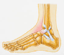
Ankle injuries refer to damages or traumas that occur in the ankle joint and its surrounding structures, including bones, ligaments, tendons, and muscles. The ankle joint is a complex structure composed of three bones: the tibia (shinbone), fibula (lower leg bone), and talus (a bone in the foot). These bones are held together by various strong ligaments that provide stability and enable proper movement.
There are several types of ankle injuries, with the most common being sprains, strains, and fractures:
1. Ankle Sprain: A sprain occurs when the ligaments surrounding the ankle joint get stretched or torn due to sudden twisting, rolling, or forced movements. The severity of a sprain can range from mild (grade 1) to severe (grade 3), with partial or complete tearing of the ligament(s).
2. Ankle Strain: A strain is an injury to the muscles or tendons surrounding the ankle joint, often caused by overuse, excessive force, or awkward positioning. This results in pain, swelling, and difficulty moving the ankle.
3. Ankle Fracture: A fracture occurs when one or more bones in the ankle joint break due to high-impact trauma, such as a fall, sports injury, or vehicle accident. Fractures can vary in severity, from small cracks to complete breaks that may require surgery and immobilization for proper healing.
Symptoms of ankle injuries typically include pain, swelling, bruising, tenderness, and difficulty walking or bearing weight on the affected ankle. Immediate medical attention is necessary for severe injuries, such as fractures, dislocations, or significant ligament tears, to ensure appropriate diagnosis and treatment. Treatment options may include rest, ice, compression, elevation (RICE), immobilization with a brace or cast, physical therapy, medication, or surgery, depending on the type and severity of the injury.
Spontaneous fractures are bone breaks that occur without any identifiable trauma or injury. They are typically caused by underlying medical conditions that weaken the bones, making them more susceptible to breaking under normal stress or weight. The most common cause of spontaneous fractures is osteoporosis, a condition characterized by weak and brittle bones. Other potential causes include various bone diseases, certain cancers, long-term use of corticosteroids, and genetic disorders affecting bone strength.
It's important to note that while the term "spontaneous" implies that the fracture occurred without any apparent cause, it is usually the result of an underlying medical condition. Therefore, if you experience a spontaneous fracture, seeking medical attention is crucial to diagnose and manage the underlying cause to prevent future fractures and related complications.
The fibula is a slender bone located in the lower leg of humans and other vertebrates. It runs parallel to the larger and more robust tibia, and together they are known as the bones of the leg or the anterior tibial segment. The fibula is the lateral bone in the leg, positioned on the outside of the tibia.
In humans, the fibula extends from the knee joint proximally to the ankle joint distally. Its proximal end, called the head of the fibula, articulates with the lateral condyle of the tibia and forms part of the inferior aspect of the knee joint. The narrowed portion below the head is known as the neck of the fibula.
The shaft of the fibula, also called the body of the fibula, is a long, thin structure that descends from the neck and serves primarily for muscle attachment rather than weight-bearing functions. The distal end of the fibula widens to form the lateral malleolus, which is an important bony landmark in the ankle region. The lateral malleolus articulates with the talus bone of the foot and forms part of the ankle joint.
The primary functions of the fibula include providing attachment sites for muscles that act on the lower leg, ankle, and foot, as well as contributing to the stability of the ankle joint through its articulation with the talus bone. Fractures of the fibula can occur due to various injuries, such as twisting or rotational forces applied to the ankle or direct trauma to the lateral aspect of the lower leg.
Stress fractures are defined as small cracks or severe bruising in bones that occur from repetitive stress or overuse. They most commonly occur in weight-bearing bones, such as the legs and feet, but can also occur in the arms, hips, and back. Stress fractures differ from regular fractures because they typically do not result from a single, traumatic event. Instead, they are caused by repeated stress on the bone that results in microscopic damage over time. Athletes, military personnel, and individuals who engage in high-impact activities or have weak bones (osteoporosis) are at increased risk of developing stress fractures. Symptoms may include pain, swelling, tenderness, and difficulty walking or bearing weight on the affected bone.
The elbow joint, also known as the cubitus joint, is a hinge joint that connects the humerus bone of the upper arm to the radius and ulna bones of the forearm. It allows for flexion and extension movements of the forearm, as well as some degree of rotation. The main articulation occurs between the trochlea of the humerus and the trochlear notch of the ulna, while the radial head of the radius also contributes to the joint's stability and motion. Ligaments, muscles, and tendons surround and support the elbow joint, providing strength and protection during movement.
Prostheses: Artificial substitutes or replacements for missing body parts, such as limbs, eyes, or teeth. They are designed to restore the function, appearance, or mobility of the lost part. Prosthetic devices can be categorized into several types, including:
1. External prostheses: Devices that are attached to the outside of the body, like artificial arms, legs, hands, and feet. These may be further classified into:
a. Cosmetic or aesthetic prostheses: Primarily designed to improve the appearance of the affected area.
b. Functional prostheses: Designed to help restore the functionality and mobility of the lost limb.
2. Internal prostheses: Implanted artificial parts that replace missing internal organs, bones, or tissues, such as heart valves, hip joints, or intraocular lenses.
Implants: Medical devices or substances that are intentionally placed inside the body to replace or support a missing or damaged biological structure, deliver medication, monitor physiological functions, or enhance bodily functions. Examples of implants include:
1. Orthopedic implants: Devices used to replace or reinforce damaged bones, joints, or cartilage, such as knee or hip replacements.
2. Cardiovascular implants: Devices that help support or regulate heart function, like pacemakers, defibrillators, and artificial heart valves.
3. Dental implants: Artificial tooth roots that are placed into the jawbone to support dental prostheses, such as crowns, bridges, or dentures.
4. Neurological implants: Devices used to stimulate nerves, brain structures, or spinal cord tissues to treat various neurological conditions, like deep brain stimulators for Parkinson's disease or cochlear implants for hearing loss.
5. Ophthalmic implants: Artificial lenses that are placed inside the eye to replace a damaged or removed natural lens, such as intraocular lenses used in cataract surgery.
An ulna fracture is a break in the ulna bone, which is one of the two long bones in the forearm. The ulna is located on the pinky finger side of the forearm and functions to support the elbow joint and assist in rotation and movement of the forearm. Ulna fractures can occur at various points along the bone, including the shaft, near the wrist, or at the elbow end of the bone. Symptoms may include pain, swelling, bruising, tenderness, deformity, limited mobility, and in some cases, numbness or tingling in the fingers. Treatment typically involves immobilization with a cast or splint, followed by rehabilitation exercises to restore strength and range of motion. In severe cases, surgery may be required to realign and stabilize the fractured bone.
Nitrogen fixation is a process by which nitrogen gas (N2) in the air is converted into ammonia (NH3) or other chemically reactive forms, making it available to plants and other organisms for use as a nutrient. This process is essential for the nitrogen cycle and for the growth of many types of plants, as most plants cannot utilize nitrogen gas directly from the air.
In the medical field, nitrogen fixation is not a commonly used term. However, in the context of microbiology and infectious diseases, some bacteria are capable of fixing nitrogen and this ability can contribute to their pathogenicity. For example, certain species of bacteria that colonize the human body, such as those found in the gut or on the skin, may be able to fix nitrogen and use it for their own growth and survival. In some cases, these bacteria may also release fixed nitrogen into the environment, which can have implications for the ecology and health of the host and surrounding ecosystems.
Rib fractures are breaks or cracks in the bones that make up the rib cage, which is the protective structure around the lungs and heart. Rib fractures can result from direct trauma to the chest, such as from a fall, motor vehicle accident, or physical assault. They can also occur from indirect forces, such as during coughing fits in people with weakened bones (osteoporosis).
Rib fractures are painful and can make breathing difficult, particularly when taking deep breaths or coughing. In some cases, rib fractures may lead to complications like punctured lungs (pneumothorax) or collapsed lungs (atelectasis), especially if multiple ribs are broken in several places.
It is essential to seek medical attention for suspected rib fractures, as proper diagnosis and management can help prevent further complications and promote healing. Treatment typically involves pain management, breathing exercises, and, in some cases, immobilization or surgery.
A mandibular fracture is a break or crack in the lower jaw (mandible) bone. It can occur at any point along the mandible, but common sites include the condyle (the rounded end near the ear), the angle (the curved part of the jaw), and the symphysis (the area where the two halves of the jaw meet in the front). Mandibular fractures are typically caused by trauma, such as a direct blow to the face or a fall. Symptoms may include pain, swelling, bruising, difficulty chewing or speaking, and malocclusion (misalignment) of the teeth. Treatment usually involves immobilization with wires or screws to allow the bone to heal properly.
A skull fracture is a break in one or more of the bones that form the skull. It can occur from a direct blow to the head, penetrating injuries like gunshot wounds, or from strong rotational forces during an accident. There are several types of skull fractures, including:
1. Linear Skull Fracture: This is the most common type, where there's a simple break in the bone without any splintering, depression, or displacement. It often doesn't require treatment unless it's near a sensitive area like an eye or ear.
2. Depressed Skull Fracture: In this type, a piece of the skull is pushed inward toward the brain. Surgery may be needed to relieve pressure on the brain and repair the fracture.
3. Diastatic Skull Fracture: This occurs along the suture lines (the fibrous joints between the skull bones) that haven't fused yet, often seen in infants and young children.
4. Basilar Skull Fracture: This involves fractures at the base of the skull. It can be serious due to potential injury to the cranial nerves and blood vessels located in this area.
5. Comminuted Skull Fracture: In this severe type, the bone is shattered into many pieces. These fractures usually require extensive surgical repair.
Symptoms of a skull fracture can include pain, swelling, bruising, bleeding (if there's an open wound), and in some cases, clear fluid draining from the ears or nose (cerebrospinal fluid leak). Severe fractures may cause brain injury, leading to symptoms like confusion, loss of consciousness, seizures, or neurological deficits. Immediate medical attention is necessary for any suspected skull fracture.
Ocular fixation is a term used in ophthalmology and optometry to refer to the ability of the eyes to maintain steady gaze or visual focus on an object. It involves the coordinated movement of the extraocular muscles that control eye movements, allowing for clear and stable vision.
In medical terminology, fixation specifically refers to the state in which the eyes are aligned and focused on a single point in space. This is important for maintaining visual perception and preventing blurring or double vision. Ocular fixation can be affected by various factors such as muscle weakness, nerve damage, or visual processing disorders.
Assessment of ocular fixation is often used in eye examinations to evaluate visual acuity, eye alignment, and muscle function. Abnormalities in fixation may indicate the presence of underlying eye conditions or developmental delays that require further investigation and treatment.
Treatment outcome is a term used to describe the result or effect of medical treatment on a patient's health status. It can be measured in various ways, such as through symptoms improvement, disease remission, reduced disability, improved quality of life, or survival rates. The treatment outcome helps healthcare providers evaluate the effectiveness of a particular treatment plan and make informed decisions about future care. It is also used in clinical research to compare the efficacy of different treatments and improve patient care.
A tooth fracture is a dental health condition characterized by a break or crack in the tooth structure. It can occur in different parts of the tooth, including the crown (the visible part), root, or filling. Tooth fractures can result from various factors such as trauma, biting or chewing on hard objects, grinding or clenching teeth, and having large, old amalgam fillings that weaken the tooth structure over time. Depending on the severity and location of the fracture, it may cause pain, sensitivity, or affect the tooth's functionality and appearance. Treatment options for tooth fractures vary from simple bonding to root canal treatment or even extraction in severe cases. Regular dental check-ups are essential for early detection and management of tooth fractures.
An intra-articular fracture is a type of fracture that involves the joint surface or articular cartilage of a bone. These types of fractures can occur in any joint, but they are most commonly seen in the weight-bearing joints such as the knee, ankle, and wrist.
Intra-articular fractures can be caused by high-energy trauma, such as motor vehicle accidents or falls from significant heights, or by low-energy trauma, such as a simple fall in older adults with osteoporosis.
These types of fractures are often complex and may involve displacement or depression of the joint surface, which can increase the risk of developing post-traumatic arthritis. Therefore, prompt diagnosis and appropriate treatment are essential to ensure optimal outcomes and minimize long-term complications. Treatment options for intra-articular fractures may include surgical fixation with plates, screws, or pins, as well as joint replacement in some cases.
Postoperative complications refer to any unfavorable condition or event that occurs during the recovery period after a surgical procedure. These complications can vary in severity and may include, but are not limited to:
1. Infection: This can occur at the site of the incision or inside the body, such as pneumonia or urinary tract infection.
2. Bleeding: Excessive bleeding (hemorrhage) can lead to a drop in blood pressure and may require further surgical intervention.
3. Blood clots: These can form in the deep veins of the legs (deep vein thrombosis) and can potentially travel to the lungs (pulmonary embolism).
4. Wound dehiscence: This is when the surgical wound opens up, which can lead to infection and further complications.
5. Pulmonary issues: These include atelectasis (collapsed lung), pneumonia, or respiratory failure.
6. Cardiovascular problems: These include abnormal heart rhythms (arrhythmias), heart attack, or stroke.
7. Renal failure: This can occur due to various reasons such as dehydration, blood loss, or the use of certain medications.
8. Pain management issues: Inadequate pain control can lead to increased stress, anxiety, and decreased mobility.
9. Nausea and vomiting: These can be caused by anesthesia, opioid pain medication, or other factors.
10. Delirium: This is a state of confusion and disorientation that can occur in the elderly or those with certain medical conditions.
Prompt identification and management of these complications are crucial to ensure the best possible outcome for the patient.
A compression fracture is a type of bone fracture that occurs when there is a collapse of a vertebra in the spine. This type of fracture is most commonly seen in the thoracic and lumbar regions of the spine. Compression fractures are often caused by weakened bones due to osteoporosis, but they can also result from trauma or tumors that weaken the bone.
In a compression fracture, the front part (anterior) of the vertebra collapses, while the back part (posterior) remains intact, causing the height of the vertebra to decrease. This can lead to pain, deformity, and decreased mobility. In severe cases, multiple compression fractures can result in a condition called kyphosis, which is an abnormal curvature of the spine that leads to a hunchback appearance.
Compression fractures are typically diagnosed through imaging tests such as X-rays, CT scans, or MRI scans. Treatment may include pain medication, bracing, physical therapy, or in some cases, surgery. Preventive measures such as maintaining a healthy diet, getting regular exercise, and taking medications to prevent or treat osteoporosis can help reduce the risk of compression fractures.
Retrospective studies, also known as retrospective research or looking back studies, are a type of observational study that examines data from the past to draw conclusions about possible causal relationships between risk factors and outcomes. In these studies, researchers analyze existing records, medical charts, or previously collected data to test a hypothesis or answer a specific research question.
Retrospective studies can be useful for generating hypotheses and identifying trends, but they have limitations compared to prospective studies, which follow participants forward in time from exposure to outcome. Retrospective studies are subject to biases such as recall bias, selection bias, and information bias, which can affect the validity of the results. Therefore, retrospective studies should be interpreted with caution and used primarily to generate hypotheses for further testing in prospective studies.
An external fixator is a type of orthopedic device used in the treatment of severe fractures or deformities of bones. It consists of an external frame that is attached to the bone with pins or wires that pass through the skin and into the bone. This provides stability to the injured area while allowing for alignment and adjustment of the bone during the healing process.
External fixators are typically used in cases where traditional casting or internal fixation methods are not feasible, such as when there is extensive soft tissue damage, infection, or when a limb needs to be gradually stretched or shortened. They can also be used in reconstructive surgery for bone defects or deformities.
The external frame of the fixator is made up of bars and clamps that are adjustable, allowing for precise positioning and alignment of the bones. The pins or wires that attach to the bone are carefully inserted through small incisions in the skin, and are held in place by the clamps on the frame.
External fixators can be used for a period of several weeks to several months, depending on the severity of the injury and the individual's healing process. During this time, the patient may require regular adjustments and monitoring by an orthopedic surgeon or other medical professional. Once the bone has healed sufficiently, the external fixator can be removed in a follow-up procedure.
Osteoporosis is a systemic skeletal disease characterized by low bone mass, deterioration of bone tissue, and disruption of bone architecture, leading to increased risk of fractures, particularly in the spine, wrist, and hip. It mainly affects older people, especially postmenopausal women, due to hormonal changes that reduce bone density. Osteoporosis can also be caused by certain medications, medical conditions, or lifestyle factors such as smoking, alcohol abuse, and a lack of calcium and vitamin D in the diet. The diagnosis is often made using bone mineral density testing, and treatment may include medication to slow bone loss, promote bone formation, and prevent fractures.
Tissue fixation is a process in histology (the study of the microscopic structure of tissues) where fixed tissue samples are prepared for further examination, typically through microscopy. The goal of tissue fixation is to preserve the original three-dimensional structure and biochemical composition of tissues and cells as much as possible, making them stable and suitable for various analyses.
The most common method for tissue fixation involves immersing the sample in a chemical fixative, such as formaldehyde or glutaraldehyde. These fixatives cross-link proteins within the tissue, creating a stable matrix that maintains the original structure and prevents decay. Other methods of tissue fixation may include freezing or embedding samples in various media to preserve their integrity.
Properly fixed tissue samples can be sectioned, stained, and examined under a microscope, allowing pathologists and researchers to study cellular structures, diagnose diseases, and understand biological processes at the molecular level.
Complement fixation tests are a type of laboratory test used in immunology and serology to detect the presence of antibodies in a patient's serum. These tests are based on the principle of complement activation, which is a part of the immune response. The complement system consists of a group of proteins that work together to help eliminate pathogens from the body.
In a complement fixation test, the patient's serum is mixed with a known antigen and complement proteins. If the patient has antibodies against the antigen, they will bind to it and activate the complement system. This results in the consumption or "fixation" of the complement proteins, which are no longer available to participate in a secondary reaction.
A second step involves adding a fresh source of complement proteins and a dye-labeled antibody that recognizes a specific component of the complement system. If complement was fixed during the first step, it will not be available for this secondary reaction, and the dye-labeled antibody will remain unbound. Conversely, if no antibodies were present in the patient's serum, the complement proteins would still be available for the second reaction, leading to the binding of the dye-labeled antibody.
The mixture is then examined under a microscope or using a spectrophotometer to determine whether the dye-labeled antibody has bound. If it has not, this indicates that the patient's serum contains antibodies specific to the antigen used in the test, and a positive result is recorded.
Complement fixation tests have been widely used for the diagnosis of various infectious diseases, such as syphilis, measles, and influenza. However, they have largely been replaced by more modern serological techniques, like enzyme-linked immunosorbent assays (ELISAs) and nucleic acid amplification tests (NAATs), due to their increased sensitivity, specificity, and ease of use.
A Colles' fracture is a specific type of fracture in the distal end of the radius bone in the forearm, which is the larger of the two bones in the lower arm. This type of fracture occurs when the wrist is forcefully bent backward (dorsiflexion), often as a result of falling onto an outstretched hand.
In a Colles' fracture, the distal end of the radius bone breaks and is displaced downward and angulated backward, resulting in a characteristic "dinner fork" deformity. This type of fracture is more common in older individuals, particularly women with osteoporosis, but can also occur in younger people as a result of high-energy trauma.
Colles' fractures are typically treated with immobilization using a cast or splint to hold the bones in proper alignment while they heal. In some cases, surgery may be necessary to realign and stabilize the fracture, particularly if there is significant displacement or instability of the bone fragments.
Orbital fractures refer to breaks in the bones that make up the eye socket, also known as the orbit. These bones include the maxilla, zygoma, frontal bone, and palatine bone. Orbital fractures can occur due to trauma, such as a blunt force injury or a penetrating wound.
There are several types of orbital fractures, including:
1. Blowout fracture: This occurs when the thin bone of the orbital floor is broken, often due to a direct blow to the eye. The force of the impact can cause the eyeball to move backward, breaking the bone and sometimes trapping the muscle that moves the eye (the inferior rectus).
2. Blow-in fracture: This type of fracture involves the breakage of the orbital roof, which is the bone that forms the upper boundary of the orbit. It typically occurs due to high-impact trauma, such as a car accident or a fall from a significant height.
3. Direct fracture: A direct fracture happens when there is a break in one or more of the bones that form the walls of the orbit. This type of fracture can result from a variety of traumas, including motor vehicle accidents, sports injuries, and assaults.
4. Indirect fracture: An indirect fracture occurs when the force of an injury is transmitted to the orbit through tissues surrounding it, causing the bone to break. The most common type of indirect orbital fracture is a blowout fracture.
Orbital fractures can cause various symptoms, including pain, swelling, bruising, and double vision. In some cases, the fracture may also lead to enophthalmos (sinking of the eye into the orbit) or telecanthus (increased distance between the inner corners of the eyes). Imaging tests, such as CT scans, are often used to diagnose orbital fractures and determine the best course of treatment. Treatment may include observation, pain management, and in some cases, surgery to repair the fracture and restore normal function.
In the field of medicine, "time factors" refer to the duration of symptoms or time elapsed since the onset of a medical condition, which can have significant implications for diagnosis and treatment. Understanding time factors is crucial in determining the progression of a disease, evaluating the effectiveness of treatments, and making critical decisions regarding patient care.
For example, in stroke management, "time is brain," meaning that rapid intervention within a specific time frame (usually within 4.5 hours) is essential to administering tissue plasminogen activator (tPA), a clot-busting drug that can minimize brain damage and improve patient outcomes. Similarly, in trauma care, the "golden hour" concept emphasizes the importance of providing definitive care within the first 60 minutes after injury to increase survival rates and reduce morbidity.
Time factors also play a role in monitoring the progression of chronic conditions like diabetes or heart disease, where regular follow-ups and assessments help determine appropriate treatment adjustments and prevent complications. In infectious diseases, time factors are crucial for initiating antibiotic therapy and identifying potential outbreaks to control their spread.
Overall, "time factors" encompass the significance of recognizing and acting promptly in various medical scenarios to optimize patient outcomes and provide effective care.
Bony callus is a medical term that refers to the specialized tissue that forms in response to a bone fracture. It is a crucial part of the natural healing process, as it helps to stabilize and protect the broken bone while it mends.
When a bone is fractured, the body responds by initiating an inflammatory response, which triggers the production of various cells and signaling molecules that promote healing. As part of this process, specialized cells called osteoblasts begin to produce new bone tissue at the site of the fracture. This tissue is initially soft and pliable, allowing it to bridge the gap between the broken ends of the bone.
Over time, this soft callus gradually hardens and calcifies, forming a bony callus that helps to stabilize the fracture and provide additional support as the bone heals. The bony callus is typically composed of a mixture of woven bone (which is less organized than normal bone) and more structured lamellar bone (which is similar in structure to normal bone).
As the bone continues to heal, the bony callus may be gradually remodeled and reshaped by osteoclasts, which are specialized cells that break down and remove excess or unwanted bone tissue. This process helps to restore the bone's original shape and strength, allowing it to function normally again.
It is worth noting that excessive bony callus formation can sometimes lead to complications, such as stiffness, pain, or decreased range of motion in the affected limb. In some cases, surgical intervention may be necessary to remove or reduce the size of the bony callus and promote proper healing.
Wrist injuries refer to damages or traumas affecting the structures of the wrist, including bones, ligaments, tendons, muscles, and cartilage. These injuries can occur due to various reasons such as falls, accidents, sports-related impacts, or repetitive stress. Common types of wrist injuries include fractures (such as scaphoid fracture), sprains (like ligament tears), strains (involving muscles or tendons), dislocations, and carpal tunnel syndrome. Symptoms may include pain, swelling, tenderness, bruising, limited mobility, and in severe cases, deformity or numbness. Immediate medical attention is necessary for proper diagnosis and treatment to ensure optimal recovery and prevent long-term complications.
Periprosthetic fractures are defined as fractures that occur in close proximity to a prosthetic joint, such as those found in total hip or knee replacements. These types of fractures typically occur as a result of low-energy trauma, and can be caused by a variety of factors including osteoporosis, bone weakness, or loosening of the prosthetic implant.
Periprosthetic fractures are classified based on the location of the fracture in relation to the prosthesis, as well as the stability of the implant. Treatment options for periprosthetic fractures may include non-surgical management, such as immobilization with a brace or cast, or surgical intervention, such as open reduction and internal fixation (ORIF) or revision arthroplasty.
The management of periprosthetic fractures can be complex and requires careful consideration of various factors, including the patient's age, overall health status, bone quality, and functional needs. As such, these types of fractures are typically managed by orthopedic surgeons with experience in joint replacement surgery and fracture care.
Surgical casts are medical devices used to immobilize and protect injured body parts, typically fractured or broken bones, during the healing process. They are usually made of plaster or fiberglass materials that harden when wet and conform to the shape of the affected area once applied. The purpose of a surgical cast is to restrict movement and provide stability to the injured site, allowing for proper alignment and healing of the bones.
The casting process involves first aligning the broken bone fragments into their correct positions, often through manual manipulation or surgical intervention. Once aligned, the cast material is applied in layers, with each layer being allowed to dry before adding the next. This creates a rigid structure that encases and supports the injured area. The cast must be kept dry during the healing process to prevent it from becoming weakened or damaged.
Surgical casts come in various shapes and sizes depending on the location and severity of the injury. They may also include additional components such as padding, Velcro straps, or window openings to allow for regular monitoring of the skin and underlying tissue. In some cases, removable splints or functional braces may be used instead of traditional casts, providing similar support while allowing for limited movement and easier adjustments.
It is essential to follow proper care instructions when wearing a surgical cast, including elevating the injured limb, avoiding excessive weight-bearing, and monitoring for signs of complications such as swelling, numbness, or infection. Regular check-ups with a healthcare provider are necessary to ensure proper healing and adjust the cast if needed.
Bone density refers to the amount of bone mineral content (usually measured in grams) in a given volume of bone (usually measured in cubic centimeters). It is often used as an indicator of bone strength and fracture risk. Bone density is typically measured using dual-energy X-ray absorptiometry (DXA) scans, which provide a T-score that compares the patient's bone density to that of a young adult reference population. A T-score of -1 or above is considered normal, while a T-score between -1 and -2.5 indicates osteopenia (low bone mass), and a T-score below -2.5 indicates osteoporosis (porous bones). Regular exercise, adequate calcium and vitamin D intake, and medication (if necessary) can help maintain or improve bone density and prevent fractures.
Orthopedic fixation devices are medical implants used in orthopedic surgery to provide stability and promote the healing of fractured or broken bones, as well as joints or spinal segments. These devices can be internal or external and include a variety of products such as:
1. Intramedullary nails: Long rods that are inserted into the center of a bone to stabilize fractures in long bones like the femur or tibia.
2. Plates and screws: Metal plates are attached to the surface of a bone with screws to hold the fragments together while they heal.
3. Screws: Used alone or in combination with other devices, they can be used to stabilize small fractures or to fix implants like total joint replacements.
4. Wires: Used to hold bone fragments together, often in conjunction with other devices.
5. External fixators: A external frame attached to the bones using pins or wires that is placed outside the skin to provide stability and alignment of fractured bones.
6. Spinal fixation devices: These include pedicle screws, rods, hooks, and plates used to stabilize spinal fractures or deformities.
7. Orthopedic staples: Small metal staples used to stabilize small bone fragments or for joint fusion.
The choice of orthopedic fixation device depends on the location and severity of the injury or condition being treated. The primary goal of these devices is to provide stability, promote healing, and restore function.
The clavicle, also known as the collarbone, is a long, slender bone that lies horizontally between the breastbone (sternum) and the shoulder blade (scapula). It is part of the shoulder girdle and plays a crucial role in supporting the upper limb. The clavicle has two ends: the medial end, which articulates with the sternum, and the lateral end, which articulates with the acromion process of the scapula. It is a common site of fracture due to its superficial location and susceptibility to direct trauma.
Zygomatic fractures, also known as "tripod fractures" or "malar fractures," refer to breaks in the zygomatic bone, which is the cheekbone. This type of facial fracture typically occurs due to significant trauma, such as a forceful blow to the face during sports injuries, traffic accidents, or physical assaults.
In zygomatic fractures, the bone can be displaced or depressed, leading to various symptoms, including:
* Facial asymmetry or flattening of the cheek area
* Bruising and swelling around the eyes (periorbital ecchymosis) and cheeks
* Diplopia (double vision) due to muscle entrapment or trauma to the eye muscles
* Subconjunctival hemorrhage (bleeding in the white part of the eye)
* Trismus (difficulty opening the mouth) due to muscle spasms or injury to the temporomandibular joint
* Numbness or altered sensation in the upper lip, cheek, or side of the nose
Diagnosis is usually made through a combination of physical examination and imaging techniques like X-rays, CT scans, or MRI. Treatment typically involves closed reduction (manipulation without surgery) or open reduction with internal fixation (surgical reconstruction using plates and screws). The primary goal of treatment is to restore the facial structure's integrity, symmetry, and function while minimizing complications and promoting optimal healing.
The pelvic bones, also known as the hip bones, are a set of three irregularly shaped bones that connect to form the pelvic girdle in the lower part of the human body. They play a crucial role in supporting the spine and protecting the abdominal and pelvic organs.
The pelvic bones consist of three bones:
1. The ilium: This is the largest and uppermost bone, forming the majority of the hip bone and the broad, flaring part of the pelvis known as the wing of the ilium or the iliac crest, which can be felt on the side of the body.
2. The ischium: This is the lower and back portion of the pelvic bone that forms part of the sitting surface or the "sit bones."
3. The pubis: This is the front part of the pelvic bone, which connects to the other side at the pubic symphysis in the midline of the body.
The pelvic bones are joined together at the acetabulum, a cup-shaped socket that forms the hip joint and articulates with the head of the femur (thigh bone). The pelvic bones also have several openings for the passage of blood vessels, nerves, and reproductive and excretory organs.
The shape and size of the pelvic bones differ between males and females due to their different roles in childbirth and locomotion. Females typically have a wider and shallower pelvis than males to accommodate childbirth, while males usually have a narrower and deeper pelvis that is better suited for weight-bearing and movement.
The thoracic vertebrae are the 12 vertebrae in the thoracic region of the spine, which is the portion between the cervical and lumbar regions. These vertebrae are numbered T1 to T12, with T1 being closest to the skull and T12 connecting to the lumbar region.
The main function of the thoracic vertebrae is to provide stability and support for the chest region, including protection for the vital organs within, such as the heart and lungs. Each thoracic vertebra has costal facets on its sides, which articulate with the heads of the ribs, forming the costovertebral joints. This connection between the spine and the ribcage allows for a range of movements while maintaining stability.
The thoracic vertebrae have a unique structure compared to other regions of the spine. They are characterized by having long, narrow bodies, small bony processes, and prominent spinous processes that point downwards. This particular shape and orientation of the thoracic vertebrae contribute to their role in limiting excessive spinal movement and providing overall trunk stability.
Maxillary fractures, also known as Le Fort fractures, are complex fractures that involve the upper jaw or maxilla. Named after the French surgeon René Le Fort who first described them in 1901, these fractures are categorized into three types (Le Fort I, II, III) based on the pattern and level of bone involvement.
1. Le Fort I fracture: This type of maxillary fracture involves a horizontal separation through the lower part of the maxilla, just above the teeth's roots. It often results from direct blows to the lower face or chin.
2. Le Fort II fracture: A Le Fort II fracture is characterized by a pyramidal-shaped fracture pattern that extends from the nasal bridge through the inferior orbital rim and maxilla, ending at the pterygoid plates. This type of fracture usually results from forceful impacts to the midface or nose.
3. Le Fort III fracture: A Le Fort III fracture is a severe craniofacial injury that involves both the upper and lower parts of the face. It is also known as a "craniofacial dysjunction" because it separates the facial bones from the skull base. The fracture line extends through the nasal bridge, orbital rims, zygomatic arches, and maxilla, ending at the pterygoid plates. Le Fort III fractures typically result from high-impact trauma to the face, such as car accidents or assaults.
These fractures often require surgical intervention for proper alignment and stabilization of the facial bones.
A dislocation is a condition in which a bone slips out of its normal position in a joint. This can happen as a result of trauma or injury, such as a fall or direct blow to the body. Dislocations can cause pain, swelling, and limited mobility in the affected area. In some cases, a dislocation may also damage surrounding tissues, such as ligaments, tendons, and nerves.
Dislocations are typically treated by reducing the dislocation, which means putting the bone back into its normal position. This is usually done with the help of medication to relieve pain and relaxation techniques to help the person stay still during the reduction. In some cases, surgery may be necessary to repair damaged tissues or if the dislocation cannot be reduced through other methods. After the dislocation has been reduced, the joint may be immobilized with a splint or sling to allow it to heal properly.
It is important to seek medical attention promptly if you suspect that you have a dislocation. If left untreated, a dislocation can lead to further complications, such as joint instability and chronic pain.
An accidental fall is an unplanned, unexpected event in which a person suddenly and involuntarily comes to rest on the ground or other lower level, excluding intentional changes in position (e.g., jumping to catch a ball) and landings that are part of a planned activity (e.g., diving into a pool). Accidental falls can occur for various reasons, such as environmental hazards, muscle weakness, balance problems, visual impairment, or certain medical conditions. They are a significant health concern, particularly among older adults, as they can lead to serious injuries, loss of independence, reduced quality of life, and increased mortality.
Articular Range of Motion (AROM) is a term used in physiotherapy and orthopedics to describe the amount of movement available in a joint, measured in degrees of a circle. It refers to the range through which synovial joints can actively move without causing pain or injury. AROM is assessed by measuring the degree of motion achieved by active muscle contraction, as opposed to passive range of motion (PROM), where the movement is generated by an external force.
Assessment of AROM is important in evaluating a patient's functional ability and progress, planning treatment interventions, and determining return to normal activities or sports participation. It is also used to identify any restrictions in joint mobility that may be due to injury, disease, or surgery, and to monitor the effectiveness of rehabilitation programs.
Postmenopausal osteoporosis is a specific type of osteoporosis that occurs in women after they have gone through menopause. It is defined as a skeletal disorder characterized by compromised bone strength, leading to an increased risk of fractures. In this condition, the decline in estrogen levels that occurs during menopause accelerates bone loss, resulting in a decrease in bone density and quality, which can lead to fragility fractures, particularly in the hips, wrists, and spine.
It's important to note that while postmenopausal osteoporosis is more common in women, men can also develop osteoporosis due to other factors such as aging, lifestyle choices, and medical conditions.
The lumbar vertebrae are the five largest and strongest vertebrae in the human spine, located in the lower back region. They are responsible for bearing most of the body's weight and providing stability during movement. The lumbar vertebrae have a characteristic shape, with a large body in the front, which serves as the main weight-bearing structure, and a bony ring in the back, formed by the pedicles, laminae, and processes. This ring encloses and protects the spinal cord and nerves. The lumbar vertebrae are numbered L1 to L5, starting from the uppermost one. They allow for flexion, extension, lateral bending, and rotation movements of the trunk.
Forearm injuries refer to damages or traumas that affect the anatomy and function of the forearm, which is the area between the elbow and wrist. This region consists of two long bones (the radius and ulna) and several muscles, tendons, ligaments, nerves, and blood vessels that enable movements such as flexion, extension, pronation, and supination of the hand and wrist.
Common forearm injuries include:
1. Fractures: Breaks in the radius or ulna bones can occur due to high-energy trauma, falls, or sports accidents. These fractures may be simple (stable) or compound (displaced), and might require immobilization, casting, or surgical intervention depending on their severity and location.
2. Sprains and Strains: Overstretching or tearing of the ligaments connecting the bones in the forearm or the muscles and tendons responsible for movement can lead to sprains and strains. These injuries often cause pain, swelling, bruising, and limited mobility.
3. Dislocations: In some cases, forceful trauma might result in the dislocation of the radioulnar joint, where the ends of the radius and ulna meet. This injury can be extremely painful and may necessitate immediate medical attention to realign the bones and stabilize the joint.
4. Tendonitis: Repetitive motions or overuse can cause inflammation and irritation of the tendons in the forearm, resulting in a condition known as tendonitis. This injury typically presents with localized pain, swelling, and stiffness that worsen with activity.
5. Nerve Injuries: Direct trauma, compression, or stretching can damage nerves in the forearm, leading to numbness, tingling, weakness, or paralysis in the hand and fingers. Common nerve injuries include radial nerve neuropathy and ulnar nerve entrapment.
6. Compartment Syndrome: Forearm compartment syndrome occurs when increased pressure within one of the forearm's fascial compartments restricts blood flow to the muscles, nerves, and tissues inside. This condition can result from trauma, bleeding, or swelling and requires immediate medical intervention to prevent permanent damage.
Accurate diagnosis and appropriate treatment are crucial for managing forearm injuries and ensuring optimal recovery. Patients should consult with a healthcare professional if they experience persistent pain, swelling, stiffness, weakness, or numbness in their forearms or hands.
Bone density conservation agents, also known as anti-resorptive agents or bone-sparing drugs, are a class of medications that help to prevent the loss of bone mass and reduce the risk of fractures. They work by inhibiting the activity of osteoclasts, the cells responsible for breaking down and reabsorbing bone tissue during the natural remodeling process.
Examples of bone density conservation agents include:
1. Bisphosphonates (e.g., alendronate, risedronate, ibandronate, zoledronic acid) - These are the most commonly prescribed class of bone density conservation agents. They bind to hydroxyapatite crystals in bone tissue and inhibit osteoclast activity, thereby reducing bone resorption.
2. Denosumab (Prolia) - This is a monoclonal antibody that targets RANKL (Receptor Activator of Nuclear Factor-κB Ligand), a key signaling molecule involved in osteoclast differentiation and activation. By inhibiting RANKL, denosumab reduces osteoclast activity and bone resorption.
3. Selective estrogen receptor modulators (SERMs) (e.g., raloxifene) - These medications act as estrogen agonists or antagonists in different tissues. In bone tissue, SERMs mimic the bone-preserving effects of estrogen by inhibiting osteoclast activity and reducing bone resorption.
4. Hormone replacement therapy (HRT) - Estrogen hormone replacement therapy has been shown to preserve bone density in postmenopausal women; however, its use is limited due to increased risks of breast cancer, cardiovascular disease, and thromboembolic events.
5. Calcitonin - This hormone, secreted by the thyroid gland, inhibits osteoclast activity and reduces bone resorption. However, it has largely been replaced by other more effective bone density conservation agents.
These medications are often prescribed for individuals at high risk of fractures due to conditions such as osteoporosis or metabolic disorders that affect bone health. It is essential to follow the recommended dosage and administration guidelines to maximize their benefits while minimizing potential side effects. Regular monitoring of bone density, blood calcium levels, and other relevant parameters is also necessary during treatment with these medications.
Fixatives are substances used in histology and pathology to preserve tissue specimens for microscopic examination. They work by stabilizing the structural components of cells and tissues, preventing decomposition and autolysis. This helps to maintain the original structure and composition of the specimen as closely as possible, allowing for accurate diagnosis and research. Commonly used fixatives include formalin, glutaraldehyde, methanol, and ethanol. The choice of fixative depends on the specific type of tissue being preserved and the intended use of the specimen.
The odontoid process, also known as the dens, is a tooth-like projection from the second cervical vertebra (axis). It fits into a ring formed by the first vertebra (atlas), allowing for movement between these two vertebrae. The odontoid process helps to support the head and facilitates movements such as nodding and shaking. It is an essential structure in maintaining stability and mobility of the upper spine.
The diaphysis refers to the shaft or middle portion of a long bone in the body. It is the part that is typically cylindrical in shape and contains the medullary cavity, which is filled with yellow marrow. The diaphysis is primarily composed of compact bone tissue, which provides strength and support for weight-bearing and movement.
In contrast to the diaphysis, the ends of long bones are called epiphyses, and they are covered with articular cartilage and contain spongy bone tissue filled with red marrow, which is responsible for producing blood cells. The area where the diaphysis meets the epiphysis is known as the metaphysis, and it contains growth plates that are responsible for the longitudinal growth of bones during development.
Multiple trauma, also known as polytrauma, is a medical term used to describe severe injuries to the body that are sustained in more than one place or region. It often involves damage to multiple organ systems and can be caused by various incidents such as traffic accidents, falls from significant heights, high-energy collisions, or violent acts.
The injuries sustained in multiple trauma may include fractures, head injuries, internal bleeding, chest and abdominal injuries, and soft tissue injuries. These injuries can lead to a complex medical situation requiring immediate and ongoing care from a multidisciplinary team of healthcare professionals, including emergency physicians, trauma surgeons, critical care specialists, nurses, rehabilitation therapists, and mental health providers.
Multiple trauma is a serious condition that can result in long-term disability or even death if not treated promptly and effectively.
The femur is the medical term for the thigh bone, which is the longest and strongest bone in the human body. It connects the hip bone to the knee joint and plays a crucial role in supporting the weight of the body and allowing movement during activities such as walking, running, and jumping. The femur is composed of a rounded head, a long shaft, and two condyles at the lower end that articulate with the tibia and patella to form the knee joint.
Follow-up studies are a type of longitudinal research that involve repeated observations or measurements of the same variables over a period of time, in order to understand their long-term effects or outcomes. In medical context, follow-up studies are often used to evaluate the safety and efficacy of medical treatments, interventions, or procedures.
In a typical follow-up study, a group of individuals (called a cohort) who have received a particular treatment or intervention are identified and then followed over time through periodic assessments or data collection. The data collected may include information on clinical outcomes, adverse events, changes in symptoms or functional status, and other relevant measures.
The results of follow-up studies can provide important insights into the long-term benefits and risks of medical interventions, as well as help to identify factors that may influence treatment effectiveness or patient outcomes. However, it is important to note that follow-up studies can be subject to various biases and limitations, such as loss to follow-up, recall bias, and changes in clinical practice over time, which must be carefully considered when interpreting the results.
Carpal bones are the eight small bones that make up the wrist joint in humans and other primates. These bones are arranged in two rows, with four bones in each row. The proximal row includes the scaphoid, lunate, triquetral, and pisiform bones, while the distal row includes the trapezium, trapezoid, capitate, and hamate bones.
The carpal bones play an essential role in the function of the wrist joint by providing stability, support, and mobility. They allow for a wide range of movements, including flexion, extension, radial deviation, ulnar deviation, and circumduction. The complex structure of the carpal bones also helps to absorb shock and distribute forces evenly across the wrist during activities such as gripping or lifting objects.
Injuries to the carpal bones, such as fractures or dislocations, can be painful and may require medical treatment to ensure proper healing and prevent long-term complications. Additionally, degenerative conditions such as arthritis can affect the carpal bones, leading to pain, stiffness, and decreased mobility in the wrist joint.
Traction, in medical terms, refers to the application of a pulling force to distract or align parts of the body, particularly bones, joints, or muscles, with the aim of immobilizing, reducing displacement, or realigning them. This is often achieved through the use of various devices such as tongs, pulleys, weights, or specialized traction tables. Traction may be applied manually or mechanically and can be continuous or intermittent, depending on the specific medical condition being treated. Common indications for traction include fractures, dislocations, spinal cord injuries, and certain neurological conditions.
"Recovery of function" is a term used in medical rehabilitation to describe the process in which an individual regains the ability to perform activities or tasks that were previously difficult or impossible due to injury, illness, or disability. This can involve both physical and cognitive functions. The goal of recovery of function is to help the person return to their prior level of independence and participation in daily activities, work, and social roles as much as possible.
Recovery of function may be achieved through various interventions such as physical therapy, occupational therapy, speech-language therapy, and other rehabilitation strategies. The specific approach used will depend on the individual's needs and the nature of their impairment. Recovery of function can occur spontaneously as the body heals, or it may require targeted interventions to help facilitate the process.
It is important to note that recovery of function does not always mean a full return to pre-injury or pre-illness levels of ability. Instead, it often refers to the person's ability to adapt and compensate for any remaining impairments, allowing them to achieve their maximum level of functional independence and quality of life.
Orthopedic procedures are surgical or nonsurgical methods used to treat musculoskeletal conditions, including injuries, deformities, or diseases of the bones, joints, muscles, ligaments, and tendons. These procedures can range from simple splinting or casting to complex surgeries such as joint replacements, spinal fusions, or osteotomies (cutting and repositioning bones). The primary goal of orthopedic procedures is to restore function, reduce pain, and improve the quality of life for patients.
The scaphoid bone is one of the eight carpal bones located in the wrist, which connect the forearm bones (radius and ulna) to the hand bones (metacarpals). It is situated on the thumb side of the wrist and has a unique shape that resembles a boat or a small cashew nut. The scaphoid bone plays a crucial role in the mobility and stability of the wrist joint. Injuries to this bone, such as fractures or dislocations, are common in sports activities, falls, or accidents and may require medical attention for proper diagnosis and treatment.
Biomechanics is the application of mechanical laws to living structures and systems, particularly in the field of medicine and healthcare. A biomechanical phenomenon refers to a observable event or occurrence that involves the interaction of biological tissues or systems with mechanical forces. These phenomena can be studied at various levels, from the molecular and cellular level to the tissue, organ, and whole-body level.
Examples of biomechanical phenomena include:
1. The way that bones and muscles work together to produce movement (known as joint kinematics).
2. The mechanical behavior of biological tissues such as bone, cartilage, tendons, and ligaments under various loads and stresses.
3. The response of cells and tissues to mechanical stimuli, such as the way that bone tissue adapts to changes in loading conditions (known as Wolff's law).
4. The biomechanics of injury and disease processes, such as the mechanisms of joint injury or the development of osteoarthritis.
5. The use of mechanical devices and interventions to treat medical conditions, such as orthopedic implants or assistive devices for mobility impairments.
Understanding biomechanical phenomena is essential for developing effective treatments and prevention strategies for a wide range of medical conditions, from musculoskeletal injuries to neurological disorders.
Prosthesis failure is a term used to describe a situation where a prosthetic device, such as an artificial joint or limb, has stopped functioning or failed to meet its intended purpose. This can be due to various reasons, including mechanical failure, infection, loosening of the device, or a reaction to the materials used in the prosthesis.
Mechanical failure can occur due to wear and tear, manufacturing defects, or improper use of the prosthetic device. Infection can also lead to prosthesis failure, particularly in cases where the prosthesis is implanted inside the body. The immune system may react to the presence of the foreign material, leading to inflammation and infection.
Loosening of the prosthesis can also cause it to fail over time, as the device becomes less stable and eventually stops working properly. Additionally, some people may have a reaction to the materials used in the prosthesis, leading to tissue damage or other complications that can result in prosthesis failure.
In general, prosthesis failure can lead to decreased mobility, pain, and the need for additional surgeries or treatments to correct the problem. It is important for individuals with prosthetic devices to follow their healthcare provider's instructions carefully to minimize the risk of prosthesis failure and ensure that the device continues to function properly over time.
The calcaneus is the largest tarsal bone in the human foot, and it is commonly known as the heel bone. It articulates with the cuboid bone anteriorly, the talus bone superiorly, and several tendons and ligaments that help to form the posterior portion of the foot's skeletal structure. The calcaneus plays a crucial role in weight-bearing and movement, as it forms the lower part of the leg's ankle joint and helps to absorb shock during walking or running.
Bone cements are medical-grade materials used in orthopedic and trauma surgery to fill gaps between bone surfaces and implants, such as artificial joints or screws. They serve to mechanically stabilize the implant and provide a smooth, load-bearing surface. The two most common types of bone cement are:
1. Polymethylmethacrylate (PMMA) cement: This is a two-component system consisting of powdered PMMA and liquid methyl methacrylate monomer. When mixed together, they form a dough-like consistency that hardens upon exposure to air. PMMA cement has been widely used for decades in joint replacement surgeries, such as hip or knee replacements.
2. Calcium phosphate (CP) cement: This is a two-component system consisting of a powdered CP compound and an aqueous solution. When mixed together, they form a paste that hardens through a chemical reaction at body temperature. CP cement has lower mechanical strength compared to PMMA but demonstrates better biocompatibility, bioactivity, and the ability to resorb over time.
Both types of bone cements have advantages and disadvantages, and their use depends on the specific surgical indication and patient factors.
The tarsal bones are a group of seven articulating bones in the foot that make up the posterior portion of the foot, located between the talus bone of the leg and the metatarsal bones of the forefoot. They play a crucial role in supporting the body's weight and facilitating movement.
There are three categories of tarsal bones:
1. Proximal row: This includes the talus, calcaneus (heel bone), and navicular bones. The talus articulates with the tibia and fibula to form the ankle joint, while the calcaneus is the largest tarsal bone and forms the heel. The navicular bone is located between the talus and the cuneiform bones.
2. Intermediate row: This includes the cuboid bone, which is located laterally (on the outside) to the navicular bone and articulates with the calcaneus, fourth and fifth metatarsals, and the cuneiform bones.
3. Distal row: This includes three cuneiform bones - the medial, intermediate, and lateral cuneiforms - which are located between the navicular bone proximally and the first, second, and third metatarsal bones distally. The medial cuneiform is the largest of the three and articulates with the navicular bone, first metatarsal, and the intermediate cuneiform. The intermediate cuneiform articulates with the medial and lateral cuneiforms and the second metatarsal. The lateral cuneiform articulates with the intermediate cuneiform, cuboid, and fourth metatarsal.
Together, these bones form a complex network of joints that allow for movement and stability in the foot. Injuries or disorders affecting the tarsal bones can result in pain, stiffness, and difficulty walking.
A reoperation is a surgical procedure that is performed again on a patient who has already undergone a previous operation for the same or related condition. Reoperations may be required due to various reasons, such as inadequate initial treatment, disease recurrence, infection, or complications from the first surgery. The nature and complexity of a reoperation can vary widely depending on the specific circumstances, but it often carries higher risks and potential complications compared to the original operation.
Bone transplantation, also known as bone grafting, is a surgical procedure in which bone or bone-like material is transferred from one part of the body to another or from one person to another. The graft may be composed of cortical (hard outer portion) bone, cancellous (spongy inner portion) bone, or a combination of both. It can be taken from different sites in the same individual (autograft), from another individual of the same species (allograft), or from an animal source (xenograft). The purpose of bone transplantation is to replace missing bone, provide structural support, and stimulate new bone growth. This procedure is commonly used in orthopedic, dental, and maxillofacial surgeries to repair bone defects caused by trauma, tumors, or congenital conditions.
Osteotomy is a surgical procedure in which a bone is cut to shorten, lengthen, or change its alignment. It is often performed to correct deformities or to realign bones that have been damaged by trauma or disease. The bone may be cut straight across (transverse osteotomy) or at an angle (oblique osteotomy). After the bone is cut, it can be realigned and held in place with pins, plates, or screws until it heals. This procedure is commonly performed on bones in the leg, such as the femur or tibia, but can also be done on other bones in the body.
The Injury Severity Score (ISS) is a medical scoring system used to assess the severity of trauma in patients with multiple injuries. It's based on the Abbreviated Injury Scale (AIS), which classifies each injury by body region on a scale from 1 (minor) to 6 (maximum severity).
The ISS is calculated by summing the squares of the highest AIS score in each of the three most severely injured body regions. The possible ISS ranges from 0 to 75, with higher scores indicating more severe injuries. An ISS over 15 is generally considered a significant injury, and an ISS over 25 is associated with a high risk of mortality. It's important to note that the ISS has limitations, as it doesn't consider the number or type of injuries within each body region, only the most severe one.
Spinal injuries refer to damages or traumas that occur to the vertebral column, which houses and protects the spinal cord. These injuries can be caused by various factors such as trauma from accidents (motor vehicle, sports-related, falls, etc.), violence, or degenerative conditions like arthritis, disc herniation, or spinal stenosis.
Spinal injuries can result in bruising, fractures, dislocations, or compression of the vertebrae, which may then cause damage to the spinal cord and its surrounding tissues, nerves, and blood vessels. The severity of a spinal injury can range from mild, with temporary symptoms, to severe, resulting in permanent impairment or paralysis below the level of injury.
Symptoms of spinal injuries may include:
- Pain or stiffness in the neck or back
- Numbness, tingling, or weakness in the limbs
- Loss of bladder or bowel control
- Difficulty walking or maintaining balance
- Paralysis or loss of sensation below the level of injury
- In severe cases, respiratory problems and difficulty in breathing
Immediate medical attention is crucial for spinal injuries to prevent further damage and ensure proper treatment. Treatment options may include immobilization, surgery, medication, rehabilitation, and physical therapy.
In medical terms, "axis" is used to describe a line or lines along which a structure or body part can move or around which it is oriented. It is often used in anatomical context to refer to specific axes of movement or alignment for various parts of the body. For example:
* The axial skeleton, also known as the upright skeleton, includes the skull, vertebral column, and chest cage.
* In neurology, the term "axis" is used to describe the second cervical vertebra (C2), which is also called the axis because it serves as a pivot point for head movement.
* The term "longitudinal axis" is used to describe an imaginary line that runs from the head to the foot, passing through the center of the body.
* In imaging studies such as X-rays or MRIs, the term "axis" may be used to describe a specific orientation or alignment for the image.
Overall, the term "axis" is used in medicine to describe lines or planes that serve as reference points for movement, alignment, or orientation of various body structures and parts.
Minimally invasive surgical procedures are a type of surgery that is performed with the assistance of specialized equipment and techniques to minimize trauma to the patient's body. This approach aims to reduce blood loss, pain, and recovery time as compared to traditional open surgeries. The most common minimally invasive surgical procedure is laparoscopy, which involves making small incisions (usually 0.5-1 cm) in the abdomen or chest and inserting a thin tube with a camera (laparoscope) to visualize the internal organs.
The surgeon then uses long, slender instruments inserted through separate incisions to perform the necessary surgical procedures, such as cutting, coagulation, or suturing. Other types of minimally invasive surgical procedures include arthroscopy (for joint surgery), thoracoscopy (for chest surgery), and hysteroscopy (for uterine surgery). The benefits of minimally invasive surgical procedures include reduced postoperative pain, shorter hospital stays, quicker return to normal activities, and improved cosmetic results. However, not all surgeries can be performed using minimally invasive techniques, and the suitability of a particular procedure depends on various factors, including the patient's overall health, the nature and extent of the surgical problem, and the surgeon's expertise.
Prospective studies, also known as longitudinal studies, are a type of cohort study in which data is collected forward in time, following a group of individuals who share a common characteristic or exposure over a period of time. The researchers clearly define the study population and exposure of interest at the beginning of the study and follow up with the participants to determine the outcomes that develop over time. This type of study design allows for the investigation of causal relationships between exposures and outcomes, as well as the identification of risk factors and the estimation of disease incidence rates. Prospective studies are particularly useful in epidemiology and medical research when studying diseases with long latency periods or rare outcomes.
Vertebroplasty is a medical procedure used to treat spinal fractures, particularly those resulting from osteoporosis or cancer. The procedure involves injecting a type of bone cement called polymethylmethacrylate (PMMA) into the damaged vertebra. This helps to stabilize the bone, reduce pain, and improve function.
During the procedure, a small incision is made in the skin, and a hollow needle is guided using fluoroscopy (a type of X-ray guidance) into the fractured vertebra. Once in place, the PMMA cement is injected into the bone, where it hardens quickly, providing stability to the fractured vertebra.
It's important to note that while vertebroplasty can be an effective treatment for some patients with spinal fractures, it's not always necessary or appropriate. The decision to undergo this procedure should be made in consultation with a healthcare provider and based on a thorough evaluation of the patient's individual needs and circumstances.
The humerus is the long bone in the upper arm that extends from the shoulder joint (glenohumeral joint) to the elbow joint. It articulates with the glenoid cavity of the scapula to form the shoulder joint and with the radius and ulna bones at the elbow joint. The proximal end of the humerus has a rounded head that provides for movement in multiple planes, making it one of the most mobile joints in the body. The greater and lesser tubercles are bony prominences on the humeral head that serve as attachment sites for muscles that move the shoulder and arm. The narrow shaft of the humerus provides stability and strength for weight-bearing activities, while the distal end forms two articulations: one with the ulna (trochlea) and one with the radius (capitulum). Together, these structures allow for a wide range of motion in the shoulder and elbow joints.
Spinal fusion is a surgical procedure where two or more vertebrae in the spine are fused together to create a solid bone. The purpose of this procedure is to restrict movement between the fused vertebrae, which can help reduce pain and stabilize the spine. This is typically done using bone grafts or bone graft substitutes, along with hardware such as rods, screws, or cages to hold the vertebrae in place while they heal together. The procedure may be recommended for various spinal conditions, including degenerative disc disease, spinal stenosis, spondylolisthesis, scoliosis, or fractures.
Prosthesis design is a specialized field in medical device technology that involves creating and developing artificial substitutes to replace a missing body part, such as a limb, tooth, eye, or internal organ. The design process typically includes several stages: assessment of the patient's needs, selection of appropriate materials, creation of a prototype, testing and refinement, and final fabrication and fitting of the prosthesis.
The goal of prosthesis design is to create a device that functions as closely as possible to the natural body part it replaces, while also being comfortable, durable, and aesthetically pleasing for the patient. The design process may involve collaboration between medical professionals, engineers, and designers, and may take into account factors such as the patient's age, lifestyle, occupation, and overall health.
Prosthesis design can be highly complex, particularly for advanced devices such as robotic limbs or implantable organs. These devices often require sophisticated sensors, actuators, and control systems to mimic the natural functions of the body part they replace. As a result, prosthesis design is an active area of research and development in the medical field, with ongoing efforts to improve the functionality, comfort, and affordability of these devices for patients.
The "femur neck" is the narrow, upper part of the femur (thigh bone) where it connects to the pelvis. It is the region through which the femoral head articulates with the acetabulum to form the hip joint. The femur neck is a common site for fractures, especially in older adults with osteoporosis.
Photon Absorptiometry is a medical technique used to measure the absorption of photons (light particles) by tissues or materials. In clinical practice, it is often used as a non-invasive method for measuring bone mineral density (BMD). This technique uses a low-energy X-ray beam or gamma ray to penetrate the tissue and then measures the amount of radiation absorbed by the bone. The amount of absorption is related to the density and thickness of the bone, allowing for an assessment of BMD. It can be used to diagnose osteoporosis and monitor treatment response in patients with bone diseases. There are two types of photon absorptiometry: single-photon absorptiometry (SPA) and dual-photon absorptiometry (DPA). SPA uses one energy level, while DPA uses two different energy levels to measure BMD, providing more precise measurements.
X-ray computed tomography (CT or CAT scan) is a medical imaging method that uses computer-processed combinations of many X-ray images taken from different angles to produce cross-sectional (tomographic) images (virtual "slices") of the body. These cross-sectional images can then be used to display detailed internal views of organs, bones, and soft tissues in the body.
The term "computed tomography" is used instead of "CT scan" or "CAT scan" because the machines take a series of X-ray measurements from different angles around the body and then use a computer to process these data to create detailed images of internal structures within the body.
CT scanning is a noninvasive, painless medical test that helps physicians diagnose and treat medical conditions. CT imaging provides detailed information about many types of tissue including lung, bone, soft tissue and blood vessels. CT examinations can be performed on every part of the body for a variety of reasons including diagnosis, surgical planning, and monitoring of therapeutic responses.
In computed tomography (CT), an X-ray source and detector rotate around the patient, measuring the X-ray attenuation at many different angles. A computer uses this data to construct a cross-sectional image by the process of reconstruction. This technique is called "tomography". The term "computed" refers to the use of a computer to reconstruct the images.
CT has become an important tool in medical imaging and diagnosis, allowing radiologists and other physicians to view detailed internal images of the body. It can help identify many different medical conditions including cancer, heart disease, lung nodules, liver tumors, and internal injuries from trauma. CT is also commonly used for guiding biopsies and other minimally invasive procedures.
In summary, X-ray computed tomography (CT or CAT scan) is a medical imaging technique that uses computer-processed combinations of many X-ray images taken from different angles to produce cross-sectional images of the body. It provides detailed internal views of organs, bones, and soft tissues in the body, allowing physicians to diagnose and treat medical conditions.
Diphosphonates are a class of medications that are used to treat bone diseases, such as osteoporosis and Paget's disease. They work by binding to the surface of bones and inhibiting the activity of bone-resorbing cells called osteoclasts. This helps to slow down the breakdown and loss of bone tissue, which can help to reduce the risk of fractures.
Diphosphonates are typically taken orally in the form of tablets, but some forms may be given by injection. Commonly prescribed diphosphonates include alendronate (Fosamax), risedronate (Actonel), and ibandronate (Boniva). Side effects of diphosphonates can include gastrointestinal symptoms such as nausea, heartburn, and abdominal pain. In rare cases, they may also cause esophageal ulcers or osteonecrosis of the jaw.
It is important to follow the instructions for taking diphosphonates carefully, as they must be taken on an empty stomach with a full glass of water and the patient must remain upright for at least 30 minutes after taking the medication to reduce the risk of esophageal irritation. Regular monitoring of bone density and kidney function is also recommended while taking these medications.
Medical Definition:
"Risk factors" are any attribute, characteristic or exposure of an individual that increases the likelihood of developing a disease or injury. They can be divided into modifiable and non-modifiable risk factors. Modifiable risk factors are those that can be changed through lifestyle choices or medical treatment, while non-modifiable risk factors are inherent traits such as age, gender, or genetic predisposition. Examples of modifiable risk factors include smoking, alcohol consumption, physical inactivity, and unhealthy diet, while non-modifiable risk factors include age, sex, and family history. It is important to note that having a risk factor does not guarantee that a person will develop the disease, but rather indicates an increased susceptibility.
Hip arthroplasty, also known as hip replacement surgery, is a medical procedure where the damaged or diseased joint surfaces of the hip are removed and replaced with artificial components. These components typically include a metal or ceramic ball that replaces the head of the femur (thigh bone), and a polyethylene or ceramic socket that replaces the acetabulum (hip socket) in the pelvis.
The goal of hip arthroplasty is to relieve pain, improve joint mobility, and restore function to the hip joint. This procedure is commonly performed in patients with advanced osteoarthritis, rheumatoid arthritis, hip fractures, or other conditions that cause significant damage to the hip joint.
There are several types of hip replacement surgeries, including traditional total hip arthroplasty, partial (hemi) hip arthroplasty, and resurfacing hip arthroplasty. The choice of procedure depends on various factors, such as the patient's age, activity level, overall health, and the extent of joint damage.
After surgery, patients typically require rehabilitation to regain strength, mobility, and function in the affected hip. With proper care and follow-up, most patients can expect significant pain relief and improved quality of life following hip arthroplasty.
"Weight-bearing" is a term used in the medical field to describe the ability of a body part or limb to support the weight or pressure exerted upon it, typically while standing, walking, or performing other physical activities. In a clinical setting, healthcare professionals often use the term "weight-bearing exercise" to refer to physical activities that involve supporting one's own body weight, such as walking, jogging, or climbing stairs. These exercises can help improve bone density, muscle strength, and overall physical function, particularly in individuals with conditions affecting the bones, joints, or muscles.
In addition, "weight-bearing" is also used to describe the positioning of a body part during medical imaging studies, such as X-rays or MRIs. For example, a weight-bearing X-ray of the foot or ankle involves taking an image while the patient stands on the affected limb, allowing healthcare providers to assess any alignment or stability issues that may not be apparent in a non-weight-bearing position.
The tibia, also known as the shin bone, is the larger of the two bones in the lower leg and part of the knee joint. It supports most of the body's weight and is a major insertion point for muscles that flex the foot and bend the leg. The tibia articulates with the femur at the knee joint and with the fibula and talus bone at the ankle joint. Injuries to the tibia, such as fractures, are common in sports and other activities that put stress on the lower leg.
The radius is one of the two bones in the forearm in humans and other vertebrates. In humans, it runs from the lateral side of the elbow to the thumb side of the wrist. It is responsible for rotation of the forearm and articulates with the humerus at the elbow and the carpals at the wrist. Any medical condition or injury that affects the radius can impact the movement and function of the forearm and hand.
The metacarpal bones are the long slender bones that make up the middle part of the hand, located between the carpals (wrist bones) and the phalanges (finger bones). There are five metacarpal bones in total, with one for each finger and thumb. Each bone has a base attached to the carpals, a shaft, and a head that connects to the phalanges. The metacarpal bones play a crucial role in hand function, providing stability and support during gripping and manipulation movements.
"Trauma severity indices" refer to various scoring systems used by healthcare professionals to evaluate the severity of injuries in trauma patients. These tools help standardize the assessment and communication of injury severity among different members of the healthcare team, allowing for more effective and consistent treatment planning, resource allocation, and prognosis estimation.
There are several commonly used trauma severity indices, including:
1. Injury Severity Score (ISS): ISS is an anatomical scoring system that evaluates the severity of injuries based on the Abbreviated Injury Scale (AIS). The body is divided into six regions, and the square of the highest AIS score in each region is summed to calculate the ISS. Scores range from 0 to 75, with higher scores indicating more severe injuries.
2. New Injury Severity Score (NISS): NISS is a modification of the ISS that focuses on the three most severely injured body regions, regardless of their anatomical location. The three highest AIS scores are squared and summed to calculate the NISS. This scoring system tends to correlate better with mortality than the ISS in some studies.
3. Revised Trauma Score (RTS): RTS is a physiological scoring system that evaluates the patient's respiratory, cardiovascular, and neurological status upon arrival at the hospital. It uses variables such as Glasgow Coma Scale (GCS), systolic blood pressure, and respiratory rate to calculate a score between 0 and 7.84, with lower scores indicating more severe injuries.
4. Trauma and Injury Severity Score (TRISS): TRISS is a combined anatomical and physiological scoring system that estimates the probability of survival based on ISS or NISS, RTS, age, and mechanism of injury (blunt or penetrating). It uses logistic regression equations to calculate the predicted probability of survival.
5. Pediatric Trauma Score (PTS): PTS is a physiological scoring system specifically designed for children under 14 years old. It evaluates six variables, including respiratory rate, oxygen saturation, systolic blood pressure, capillary refill time, GCS, and temperature to calculate a score between -6 and +12, with lower scores indicating more severe injuries.
These scoring systems help healthcare professionals assess the severity of trauma, predict outcomes, allocate resources, and compare patient populations in research settings. However, they should not replace clinical judgment or individualized care for each patient.
Polymethyl methacrylate (PMMA) is a type of synthetic resin that is widely used in the medical field due to its biocompatibility and versatility. It is a transparent, rigid, and lightweight material that can be easily molded into different shapes and forms. Here are some of the medical definitions of PMMA:
1. A biocompatible acrylic resin used in various medical applications such as bone cement, intraocular lenses, dental restorations, and drug delivery systems.
2. A type of synthetic material that is used as a bone cement to fix prosthetic joint replacements and vertebroplasty for the treatment of spinal fractures.
3. A transparent and shatter-resistant material used in the manufacture of medical devices such as intravenous (IV) fluid bags, dialyzer housings, and oxygenators.
4. A drug delivery system that can be used to administer drugs locally or systemically, such as intraocular sustained-release drug implants for the treatment of chronic eye diseases.
5. A component of dental restorations such as fillings, crowns, and bridges due to its excellent mechanical properties and esthetic qualities.
Overall, PMMA is a versatile and valuable material in the medical field, with numerous applications that take advantage of its unique properties.
A hip prosthesis, also known as a total hip replacement, is a surgical implant designed to replace the damaged or diseased components of the human hip joint. The procedure involves replacing the femoral head (the ball at the top of the thigh bone) and the acetabulum (the socket in the pelvis) with artificial parts, typically made from materials such as metal, ceramic, or plastic.
The goal of a hip prosthesis is to relieve pain, improve joint mobility, and restore function, allowing patients to return to their normal activities and enjoy an improved quality of life. The procedure is most commonly performed in individuals with advanced osteoarthritis, rheumatoid arthritis, or other degenerative conditions that have caused significant damage to the hip joint.
There are several different types of hip prostheses available, each with its own unique design and set of benefits and risks. The choice of prosthesis will depend on a variety of factors, including the patient's age, activity level, overall health, and specific medical needs. In general, however, all hip prostheses are designed to provide a durable, long-lasting solution for patients suffering from debilitating joint pain and stiffness.
Alendronate is a medication that falls under the class of bisphosphonates. It is commonly used in the treatment and prevention of osteoporosis in postmenopausal women and men, as well as in the management of glucocorticoid-induced osteoporosis and Paget's disease of bone.
Alendronate works by inhibiting the activity of osteoclasts, which are cells responsible for breaking down and reabsorbing bone tissue. By reducing the activity of osteoclasts, alendronate helps to slow down bone loss and increase bone density, thereby reducing the risk of fractures.
The medication is available in several forms, including tablets and oral solutions, and is typically taken once a week for osteoporosis prevention and treatment. It is important to follow the dosing instructions carefully, as improper administration can reduce the drug's effectiveness or increase the risk of side effects. Common side effects of alendronate include gastrointestinal symptoms such as heartburn, stomach pain, and nausea.
A jaw fracture, also known as a mandibular fracture, is a break in the lower jawbone. It can occur at any point along the bone, from the condyle (the rounded end that articulates with the skull) to the symphysis (the area where the two halves of the jaw meet in the front).
Jaw fractures are typically caused by trauma, such as a direct blow to the face during sports injuries, traffic accidents, or physical assaults. They can also result from falls, particularly in older adults with osteoporosis.
Symptoms of jaw fractures may include pain, swelling, bruising, difficulty speaking, chewing, or opening the mouth wide, and malocclusion (the teeth do not fit together properly when biting down). In some cases, there may be visible deformity or mobility in the jaw.
Diagnosis of jaw fractures typically involves a thorough physical examination, dental X-rays, CT scans, or other imaging studies to assess the location and severity of the fracture. Treatment may involve immobilization with wires or braces, pain management, antibiotics to prevent infection, and in some cases, surgery to realign and stabilize the bone fragments.
The pubic bone, also known as the pubis or pubic symphysis, is a part of the pelvis - the complex ring-like structure that forms the lower part of the trunk and supports the weight of the upper body. The pubic bone is the anterior (front) portion of the pelvic girdle, located at the bottom of the abdomen, and it connects to the other side at the pubic symphysis, a cartilaginous joint.
The pubic bone plays an essential role in supporting the lower limbs and providing attachment for various muscles involved in movements like walking, running, and jumping. It also protects some abdominal organs and contributes to the structure of the pelvic outlet, which is crucial during childbirth.
A cadaver is a deceased body that is used for medical research or education. In the field of medicine, cadavers are often used in anatomy lessons, surgical training, and other forms of medical research. The use of cadavers allows medical professionals to gain a deeper understanding of the human body and its various systems without causing harm to living subjects. Cadavers may be donated to medical schools or obtained through other means, such as through consent of the deceased or their next of kin. It is important to handle and treat cadavers with respect and dignity, as they were once living individuals who deserve to be treated with care even in death.
In epidemiology, the incidence of a disease is defined as the number of new cases of that disease within a specific population over a certain period of time. It is typically expressed as a rate, with the number of new cases in the numerator and the size of the population at risk in the denominator. Incidence provides information about the risk of developing a disease during a given time period and can be used to compare disease rates between different populations or to monitor trends in disease occurrence over time.
Orthopedics is a branch of medicine that deals with the prevention, diagnosis, and treatment of disorders of the musculoskeletal system, which includes the bones, joints, muscles, ligaments, tendons, and nerves. The goal of orthopedic care is to help patients maintain or restore their mobility, function, and quality of life through a variety of treatments, including medication, physical therapy, bracing, and surgery. Orthopedic surgeons are medical doctors who have completed additional training in the diagnosis and treatment of musculoskeletal conditions, and they may specialize in specific areas such as sports medicine, spine care, joint replacement, or pediatric orthopedics.
Traffic accidents are incidents that occur when a vehicle collides with another vehicle, a pedestrian, an animal, or a stationary object, resulting in damage or injury. These accidents can be caused by various factors such as driver error, distracted driving, drunk driving, speeding, reckless driving, poor road conditions, and adverse weather conditions. Traffic accidents can range from minor fender benders to severe crashes that result in serious injuries or fatalities. They are a significant public health concern and cause a substantial burden on healthcare systems, emergency services, and society as a whole.
A basilar skull fracture is a type of skull fracture that involves the base of the skull. It is a serious and potentially life-threatening injury, as it can cause damage to the brainstem and cranial nerves. A basilar skull fracture may occur as a result of a severe head trauma, such as from a fall, car accident, or violent assault.
In a basilar skull fracture, the bones that form the base of the skull (the occipital bone, sphenoid bone, and temporal bones) are broken. This type of fracture can be difficult to diagnose on a routine skull X-ray, and may require further imaging studies such as a CT scan or MRI to confirm the diagnosis.
Symptoms of a basilar skull fracture may include:
* Battle's sign: a bruise behind the ear
* Raccoon eyes: bruising around the eyes
* Clear fluid leaking from the nose or ears (cerebrospinal fluid)
* Hearing loss
* Facial paralysis
* Difficulty swallowing
* Changes in level of consciousness
If you suspect that someone has a basilar skull fracture, it is important to seek medical attention immediately. This type of injury requires prompt treatment and close monitoring to prevent complications such as infection or brain swelling.
Foot injuries refer to any damage or trauma caused to the various structures of the foot, including the bones, muscles, tendons, ligaments, blood vessels, and nerves. These injuries can result from various causes such as accidents, sports activities, falls, or repetitive stress. Common types of foot injuries include fractures, sprains, strains, contusions, dislocations, and overuse injuries like plantar fasciitis or Achilles tendonitis. Symptoms may vary depending on the type and severity of the injury but often include pain, swelling, bruising, difficulty walking, and reduced range of motion. Proper diagnosis and treatment are crucial to ensure optimal healing and prevent long-term complications.
Immobilization is a medical term that refers to the restriction of normal mobility or motion of a body part, usually to promote healing and prevent further injury. This is often achieved through the use of devices such as casts, splints, braces, slings, or traction. The goal of immobilization is to keep the injured area in a fixed position so that it can heal properly without additional damage. It may be used for various medical conditions, including fractures, dislocations, sprains, strains, and soft tissue injuries. Immobilization helps reduce pain, minimize swelling, and protect the injured site from movement that could worsen the injury or impair healing.
A volar plate, also known as the palmar plate, is a strong band of tissue found in the joints of the hand (metacarpophalangeal and interphalangeal joints) that helps to provide stability and prevent hyperextension. It is located on the palmar or volar side (front side) of the joint, and it is attached to the proximal phalanx and the metacarpal bone. Injuries to the volar plate can occur due to sports accidents or falls, leading to conditions such as a volar plate injury or a gamekeeper's thumb.
I'm happy to help! However, I must clarify that "materials testing" is not a medical term per se. It is a term used in various fields, including engineering and materials science, to describe the process of subjecting materials to different tests to evaluate their properties and performance under various conditions.
In the context of medical devices or implants, materials testing may refer to the evaluation of the physical and mechanical properties of materials used in their construction. These tests can include assessments of strength, durability, biocompatibility, and other factors that are critical to ensuring the safety and efficacy of medical devices.
Medical device manufacturers must comply with regulatory standards for materials testing to ensure that their products meet specific requirements for performance, safety, and quality. These standards may vary depending on the type of device, its intended use, and the country or region in which it will be marketed and sold.
A saccade is a quick, rapid, and ballistic conjugate eye movement that shifts the point of fixation from one target to another. It helps in rapidly repositioning the fovea (the central part of the retina with the highest visual acuity) to focus on different targets of interest in the visual scene. Saccades are essential for efficient scanning and exploration of our environment, allowing us to direct our high-resolution vision towards various points of interest. They typically take only about 20-200 milliseconds to complete and can reach peak velocities of up to 500 degrees per second or more, depending on the amplitude of the movement. Saccades are a critical component of normal visual function and are often studied in fields such as ophthalmology, neurology, and neuroscience.
Eye movements, also known as ocular motility, refer to the voluntary or involuntary motion of the eyes that allows for visual exploration of our environment. There are several types of eye movements, including:
1. Saccades: rapid, ballistic movements that quickly shift the gaze from one point to another.
2. Pursuits: smooth, slow movements that allow the eyes to follow a moving object.
3. Vergences: coordinated movements of both eyes in opposite directions, usually in response to a three-dimensional stimulus.
4. Vestibulo-ocular reflex (VOR): automatic eye movements that help stabilize the gaze during head movement.
5. Optokinetic nystagmus (OKN): rhythmic eye movements that occur in response to large moving visual patterns, such as when looking out of a moving vehicle.
Abnormalities in eye movements can indicate neurological or ophthalmological disorders and are often assessed during clinical examinations.
A tibial fracture is a medical term that refers to a break in the shin bone, which is called the tibia. The tibia is the larger of the two bones in the lower leg and is responsible for supporting much of your body weight. Tibial fractures can occur in various ways, such as from high-energy trauma like car accidents or falls, or from low-energy trauma in individuals with weakened bones due to osteoporosis or other medical conditions.
Tibial fractures can be classified into different types based on the location, pattern, and severity of the break. Some common types of tibial fractures include:
1. Transverse fracture: A straight break that goes across the bone.
2. Oblique fracture: A diagonal break that slopes across the bone.
3. Spiral fracture: A break that spirals around the bone, often caused by twisting or rotational forces.
4. Comminuted fracture: A break where the bone is shattered into multiple pieces.
5. Open fracture: A break in which the bone pierces through the skin, increasing the risk of infection.
6. Closed fracture: A break in which the bone does not pierce through the skin.
Tibial fractures can cause symptoms such as pain, swelling, bruising, deformity, and difficulty walking or bearing weight on the affected leg. Treatment for tibial fractures may include immobilization with a cast or brace, surgery to realign and stabilize the bone with plates, screws, or rods, and rehabilitation to restore strength, mobility, and function to the injured limb.
Bone malalignment is a term used to describe the abnormal alignment or positioning of bones in relation to each other. This condition can occur as a result of injury, deformity, surgery, or disease processes that affect the bones and joints. Bone malalignment can cause pain, stiffness, limited mobility, and an increased risk of further injury. In some cases, bone malalignment may require treatment such as bracing, physical therapy, or surgery to correct the alignment and improve function.
"Bone" is the hard, dense connective tissue that makes up the skeleton of vertebrate animals. It provides support and protection for the body's internal organs, and serves as a attachment site for muscles, tendons, and ligaments. Bone is composed of cells called osteoblasts and osteoclasts, which are responsible for bone formation and resorption, respectively, and an extracellular matrix made up of collagen fibers and mineral crystals.
Bones can be classified into two main types: compact bone and spongy bone. Compact bone is dense and hard, and makes up the outer layer of all bones and the shafts of long bones. Spongy bone is less dense and contains large spaces, and makes up the ends of long bones and the interior of flat and irregular bones.
The human body has 206 bones in total. They can be further classified into five categories based on their shape: long bones, short bones, flat bones, irregular bones, and sesamoid bones.
A cartilage fracture is not a common injury because cartilage itself does not have bones, and it is difficult to fracture something that is not hard. However, there are situations where the term "cartilage fracture" can be used. One such situation is when the articular cartilage, which covers the ends of bones in joints, gets damaged or injured. This type of injury is also known as a chondral fracture or osteochondral fracture (if the bone beneath the cartilage is also involved). These injuries can occur due to trauma, such as a fall or a direct blow to the joint, and can cause pain, swelling, and limited mobility in the affected joint.
Equipment failure is a term used in the medical field to describe the malfunction or breakdown of medical equipment, devices, or systems that are essential for patient care. This can include simple devices like syringes and thermometers, as well as complex machines such as ventilators, infusion pumps, and imaging equipment.
Equipment failure can have serious consequences for patients, including delayed or inappropriate treatment, injury, or even death. It is therefore essential that medical equipment is properly maintained, tested, and repaired to ensure its safe and effective operation.
There are many potential causes of equipment failure, including:
* Wear and tear from frequent use
* Inadequate cleaning or disinfection
* Improper handling or storage
* Power supply issues
* Software glitches or bugs
* Mechanical failures or defects
* Human error or misuse
To prevent equipment failure, healthcare facilities should have established policies and procedures for the acquisition, maintenance, and disposal of medical equipment. Staff should be trained in the proper use and handling of equipment, and regular inspections and testing should be performed to identify and address any potential issues before they lead to failure.
A splint is a device used to support, protect, and immobilize injured body parts, such as bones, joints, or muscles. It can be made from various materials like plastic, metal, or fiberglass. Splints are often used to keep the injured area in a stable position, reducing pain, swelling, and further damage while the injury heals. They come in different shapes and sizes, tailored to fit specific body parts and injuries. A splint can be adjustable or custom-made, depending on the patient's needs. It is essential to follow healthcare professionals' instructions for using and caring for a splint to ensure proper healing and prevent complications.
Kyphoplasty is a surgical procedure used to treat vertebral compression fractures, which are commonly caused by osteoporosis or cancer. The goal of kyphoplasty is to stabilize the fracture, reduce pain, and restore some or all of the lost vertebral body height.
During the procedure, a small incision is made in the back, and a narrow tube is inserted into the damaged vertebra under the guidance of fluoroscopy (a type of continuous X-ray imaging). A special balloon is then inflated inside the vertebral body to create a cavity or space. This process helps to restore the height of the vertebra and correct any abnormal kyphosis (hunchback) deformity that may have developed due to the fracture.
Once the desired cavity has been created, bone cement is injected into the space to stabilize the vertebra. The cement hardens quickly, providing immediate support and pain relief. After the procedure, patients are usually advised to limit their physical activity for a short period of time to allow the cement to fully set.
It's important to note that kyphoplasty is not suitable for all types of spinal fractures or conditions, and its effectiveness may vary depending on the individual case. Therefore, a thorough evaluation by a spine specialist is necessary before deciding whether this procedure is appropriate.
Leg injuries refer to damages or harm caused to any part of the lower extremity, including the bones, muscles, tendons, ligaments, blood vessels, and other soft tissues. These injuries can result from various causes such as trauma, overuse, or degenerative conditions. Common leg injuries include fractures, dislocations, sprains, strains, contusions, and cuts. Symptoms may include pain, swelling, bruising, stiffness, weakness, or difficulty walking. The specific treatment for a leg injury depends on the type and severity of the injury.
Arm injuries refer to any damage or harm sustained by the structures of the upper limb, including the bones, muscles, tendons, ligaments, nerves, and blood vessels. These injuries can occur due to various reasons such as trauma, overuse, or degenerative conditions. Common arm injuries include fractures, dislocations, sprains, strains, tendonitis, and nerve damage. Symptoms may include pain, swelling, bruising, limited mobility, numbness, or weakness in the affected area. Treatment varies depending on the type and severity of the injury, and may include rest, ice, compression, elevation, physical therapy, medication, or surgery.
The spine, also known as the vertebral column, is a complex structure in the human body that is part of the axial skeleton. It is composed of 33 individual vertebrae (except in some people where there are fewer due to fusion of certain vertebrae), intervertebral discs, facet joints, ligaments, muscles, and nerves.
The spine has several important functions:
1. Protection: The spine protects the spinal cord, which is a major component of the nervous system, by enclosing it within a bony canal.
2. Support: The spine supports the head and upper body, allowing us to maintain an upright posture and facilitating movement of the trunk and head.
3. Movement: The spine enables various movements such as flexion (bending forward), extension (bending backward), lateral flexion (bending sideways), and rotation (twisting).
4. Weight-bearing: The spine helps distribute weight and pressure evenly across the body, reducing stress on individual vertebrae and other structures.
5. Blood vessel and nerve protection: The spine protects vital blood vessels and nerves that pass through it, including the aorta, vena cava, and spinal nerves.
The spine is divided into five regions: cervical (7 vertebrae), thoracic (12 vertebrae), lumbar (5 vertebrae), sacrum (5 fused vertebrae), and coccyx (4 fused vertebrae, also known as the tailbone). Each region has unique characteristics that allow for specific functions and adaptations to the body's needs.
The acetabulum is the cup-shaped cavity in the pelvic bone (specifically, the os coxa) where the head of the femur bone articulates to form the hip joint. It provides a stable and flexible connection between the lower limb and the trunk, allowing for a wide range of movements such as flexion, extension, abduction, adduction, rotation, and circumduction. The acetabulum is lined with articular cartilage, which facilitates smooth and frictionless movement of the hip joint. Its stability is further enhanced by various ligaments, muscles, and the labrum, a fibrocartilaginous rim that deepens the socket and increases its contact area with the femoral head.
Mechanical stress, in the context of physiology and medicine, refers to any type of force that is applied to body tissues or organs, which can cause deformation or displacement of those structures. Mechanical stress can be either external, such as forces exerted on the body during physical activity or trauma, or internal, such as the pressure changes that occur within blood vessels or other hollow organs.
Mechanical stress can have a variety of effects on the body, depending on the type, duration, and magnitude of the force applied. For example, prolonged exposure to mechanical stress can lead to tissue damage, inflammation, and chronic pain. Additionally, abnormal or excessive mechanical stress can contribute to the development of various musculoskeletal disorders, such as tendinitis, osteoarthritis, and herniated discs.
In order to mitigate the negative effects of mechanical stress, the body has a number of adaptive responses that help to distribute forces more evenly across tissues and maintain structural integrity. These responses include changes in muscle tone, joint positioning, and connective tissue stiffness, as well as the remodeling of bone and other tissues over time. However, when these adaptive mechanisms are overwhelmed or impaired, mechanical stress can become a significant factor in the development of various pathological conditions.
Athletic injuries are damages or injuries to the body that occur while participating in sports, physical activities, or exercise. These injuries can be caused by a variety of factors, including:
1. Trauma: Direct blows, falls, collisions, or crushing injuries can cause fractures, dislocations, contusions, lacerations, or concussions.
2. Overuse: Repetitive motions or stress on a particular body part can lead to injuries such as tendonitis, stress fractures, or muscle strains.
3. Poor technique: Using incorrect form or technique during exercise or sports can put additional stress on muscles, joints, and ligaments, leading to injury.
4. Inadequate warm-up or cool-down: Failing to properly prepare the body for physical activity or neglecting to cool down afterwards can increase the risk of injury.
5. Lack of fitness or flexibility: Insufficient strength, endurance, or flexibility can make individuals more susceptible to injuries during sports and exercise.
6. Environmental factors: Extreme weather conditions, poor field or court surfaces, or inadequate equipment can contribute to the risk of athletic injuries.
Common athletic injuries include ankle sprains, knee injuries, shoulder dislocations, tennis elbow, shin splints, and concussions. Proper training, warm-up and cool-down routines, use of appropriate protective gear, and attention to technique can help prevent many athletic injuries.
Titanium is not a medical term, but rather a chemical element (symbol Ti, atomic number 22) that is widely used in the medical field due to its unique properties. Medically, it is often referred to as a biocompatible material used in various medical applications such as:
1. Orthopedic implants: Titanium and its alloys are used for making joint replacements (hips, knees, shoulders), bone plates, screws, and rods due to their high strength-to-weight ratio, excellent corrosion resistance, and biocompatibility.
2. Dental implants: Titanium is also commonly used in dental applications like implants, crowns, and bridges because of its ability to osseointegrate, or fuse directly with bone tissue, providing a stable foundation for replacement teeth.
3. Cardiovascular devices: Titanium alloys are used in the construction of heart valves, pacemakers, and other cardiovascular implants due to their non-magnetic properties, which prevent interference with magnetic resonance imaging (MRI) scans.
4. Medical instruments: Due to its resistance to corrosion and high strength, titanium is used in the manufacturing of various medical instruments such as surgical tools, needles, and catheters.
In summary, Titanium is a chemical element with unique properties that make it an ideal material for various medical applications, including orthopedic and dental implants, cardiovascular devices, and medical instruments.
The talus is a bone in the foot that articulates with the tibia and fibula to form the ankle joint, also known as the talocrural joint. It is unique because it doesn't have any muscle attachments and gets its blood supply from surrounding vessels. Its main function is to transfer weight and force during movement from the lower leg to the foot.
The Cervical Atlas, also known as C1 or the atlas vertebra, is the uppermost and most superior of the seven cervical vertebrae in the human spine. It plays a crucial role in supporting and facilitating the movement of the head, as it articulates with both the occipital bone (forming the joint called the atlanto-occipital joint) and the axis (or C2) vertebra (forming the atlantoaxial joint). The unique structure of the cervical atlas lacks a body, instead having an anterior and posterior arch with two lateral masses that form the facet joints for articulation with the axis. This arrangement allows for a wide range of motion in the neck, including flexion, extension, lateral bending, and rotation.
Soft tissue injuries refer to damages that occur in the body's connective tissues, such as ligaments, tendons, and muscles. These injuries can be caused by various events, including accidents, falls, or sports-related impacts. Common soft tissue injuries include sprains, strains, and contusions (bruises).
Sprains occur when the ligaments, which connect bones to each other, are stretched or torn. This usually happens in the joints like ankles, knees, or wrists. Strains, on the other hand, involve injuries to the muscles or tendons, often resulting from overuse or sudden excessive force. Contusions occur when blood vessels within the soft tissues get damaged due to a direct blow or impact, causing bleeding and subsequent bruising in the affected area.
Soft tissue injuries can cause pain, swelling, stiffness, and limited mobility. In some cases, these injuries may require medical treatment, including physical therapy, medication, or even surgery, depending on their severity and location. It is essential to seek proper medical attention for soft tissue injuries to ensure appropriate healing and prevent long-term complications or chronic pain.
The atlanto-axial joint is the joint between the first and second cervical vertebrae, also known as C1 (atlas) and C2 (axis). It consists of two separate joints: the median atlanto-axial joint, which is a pivot joint that allows for rotation of the head, and the paired lateral atlanto-axial joints, which are plane joints that allow for limited gliding movements.
The atlanto-axial joint is surrounded by several ligaments that provide stability and limit excessive movement. The transverse ligament, located on the anterior aspect of the joint, is particularly important as it prevents excessive movement of the atlas on the axis and helps to protect the spinal cord.
Abnormalities or injuries to the atlanto-axial joint can result in instability and potentially serious neurological complications.
'Leg length inequality' (LLIS) is a condition where there is a discrepancy in the lengths of an individual's lower extremities, specifically the bones of the thigh (femur) and/or the leg (tibia/fibula). This discrepancy can be congenital or acquired due to various causes such as fractures, infections, or surgical procedures. The inequality can lead to functional scoliosis, lower back pain, and other musculoskeletal issues. It is typically diagnosed through physical examination and imaging studies like X-rays, and may be treated with various methods including orthotics, shoe lifts, or in some cases, surgical intervention.
Ununited fracture is a medical term used to describe a fractured bone that has failed to heal properly. This condition is also known as a nonunion fracture. In a normal healing process, the broken ends of the bone will grow together, or "unite," over time as new bone tissue forms. However, in some cases, the bones may not reconnect due to various reasons such as infection, poor blood supply, excessive motion at the fracture site, or inadequate stabilization of the fracture.
Ununited fractures can cause significant pain, swelling, and deformity in the affected area. They may also lead to a decreased range of motion, weakness, and instability in the joint near the fracture. Treatment for ununited fractures typically involves surgical intervention to promote bone healing, such as bone grafting or internal fixation with screws or plates. In some cases, electrical stimulation or ultrasound therapy may also be used to help promote bone growth and healing.
Kyphosis is a medical term used to describe an excessive curvature of the spine in the sagittal plane, leading to a rounded or humped back appearance. This condition often affects the thoracic region of the spine and can result from various factors such as age-related degenerative changes, congenital disorders, Scheuermann's disease, osteoporosis, or traumatic injuries. Mild kyphosis may not cause any significant symptoms; however, severe cases can lead to pain, respiratory difficulties, and decreased quality of life. Treatment options typically include physical therapy, bracing, and, in some cases, surgical intervention.
Bone remodeling is the normal and continuous process by which bone tissue is removed from the skeleton (a process called resorption) and new bone tissue is formed (a process called formation). This ongoing cycle allows bones to repair microdamage, adjust their size and shape in response to mechanical stress, and maintain mineral homeostasis. The cells responsible for bone resorption are osteoclasts, while the cells responsible for bone formation are osteoblasts. These two cell types work together to maintain the structural integrity and health of bones throughout an individual's life.
During bone remodeling, the process can be divided into several stages:
1. Activation: The initiation of bone remodeling is triggered by various factors such as microdamage, hormonal changes, or mechanical stress. This leads to the recruitment and activation of osteoclast precursor cells.
2. Resorption: Osteoclasts attach to the bone surface and create a sealed compartment called a resorption lacuna. They then secrete acid and enzymes that dissolve and digest the mineralized matrix, creating pits or cavities on the bone surface. This process helps remove old or damaged bone tissue and releases calcium and phosphate ions into the bloodstream.
3. Reversal: After resorption is complete, the osteoclasts undergo apoptosis (programmed cell death), and mononuclear cells called reversal cells appear on the resorbed surface. These cells prepare the bone surface for the next stage by cleaning up debris and releasing signals that attract osteoblast precursors.
4. Formation: Osteoblasts, derived from mesenchymal stem cells, migrate to the resorbed surface and begin producing a new organic matrix called osteoid. As the osteoid mineralizes, it forms a hard, calcified structure that gradually replaces the resorbed bone tissue. The osteoblasts may become embedded within this newly formed bone as they differentiate into osteocytes, which are mature bone cells responsible for maintaining bone homeostasis and responding to mechanical stress.
5. Mineralization: Over time, the newly formed bone continues to mineralize, becoming stronger and more dense. This process helps maintain the structural integrity of the skeleton and ensures adequate calcium storage.
Throughout this continuous cycle of bone remodeling, hormones, growth factors, and mechanical stress play crucial roles in regulating the balance between resorption and formation. Disruptions to this delicate equilibrium can lead to various bone diseases, such as osteoporosis, where excessive resorption results in weakened bones and increased fracture risk.
The ilium is the largest and broadest of the three parts that make up the hip bone or coxal bone. It is the uppermost portion of the pelvis and forms the side of the waist. The ilium has a curved, fan-like shape and articulates with the sacrum at the back to form the sacroiliac joint. The large, concave surface on the top of the ilium is called the iliac crest, which can be felt as a prominent ridge extending from the front of the hip to the lower back. This region is significant in orthopedics and physical examinations for its use in assessing various medical conditions and performing certain maneuvers during the physical examination.
Formaldehyde is a colorless, pungent, and volatile chemical compound with the formula CH2O. It is a naturally occurring substance that is found in certain fruits like apples and vegetables, as well as in animals. However, the majority of formaldehyde used in industry is synthetically produced.
In the medical field, formaldehyde is commonly used as a preservative for biological specimens such as organs, tissues, and cells. It works by killing bacteria and inhibiting the decaying process. Formaldehyde is also used in the production of various industrial products, including adhesives, resins, textiles, and paper products.
However, formaldehyde can be harmful to human health if inhaled or ingested in large quantities. It can cause irritation to the eyes, nose, throat, and skin, and prolonged exposure has been linked to respiratory problems and cancer. Therefore, it is essential to handle formaldehyde with care and use appropriate safety measures when working with this chemical compound.
The cervical vertebrae are the seven vertebrae that make up the upper part of the spine, also known as the neck region. They are labeled C1 to C7, with C1 being closest to the skull and C7 connecting to the thoracic vertebrae in the chest region. The cervical vertebrae have unique structures to allow for a wide range of motion in the neck while also protecting the spinal cord and providing attachment points for muscles and ligaments.
The Ilizarov technique is a surgical method used for limb lengthening and reconstruction. It involves the use of an external fixation device, which consists of rings connected by adjustable rods and wires that are attached to the bone. This apparatus allows for gradual distraction (slow, steady stretching) of the bone, allowing new bone tissue to grow in the gap created by the distraction. The Ilizarov technique can be used to treat various conditions such as limb length discrepancies, bone deformities, and nonunions (failed healing of a fracture). It is named after its developer, Gavriil Abramovich Ilizarov, a Soviet orthopedic surgeon.
Hemiarthroplasty is a surgical procedure where only one half (hemi-) of a joint is replaced with an artificial component, usually a metal ball attached to a stem that fits into the bone. This procedure is most commonly performed on the shoulder or hip joints. In a hip hemiarthroplasty, it involves replacing the femoral head (the ball part of the thighbone) which has been damaged due to fracture or arthritis. The acetabulum (socket part of the pelvis) is not replaced and remains as it is. This procedure aims to relieve pain, restore mobility, and improve joint function.
Pseudarthrosis is a medical term that refers to a false joint or a nonunion of bones, meaning that the broken bone ends do not heal properly and continue to move at the fracture site. This condition can cause pain, instability, and deformity in the affected limb. It may require additional treatment such as surgery to promote bone healing and stabilization.
The scapula, also known as the shoulder blade, is a flat, triangular bone located in the upper back region of the human body. It serves as the site of attachment for various muscles that are involved in movements of the shoulder joint and arm. The scapula has several important features:
1. Three borders (anterior, lateral, and medial)
2. Three angles (superior, inferior, and lateral)
3. Spine of the scapula - a long, horizontal ridge that divides the scapula into two parts: supraspinous fossa (above the spine) and infraspinous fossa (below the spine)
4. Glenoid cavity - a shallow, concave surface on the lateral border that articulates with the humerus to form the shoulder joint
5. Acromion process - a bony projection at the top of the scapula that forms part of the shoulder joint and serves as an attachment point for muscles and ligaments
6. Coracoid process - a hook-like bony projection extending from the anterior border, which provides attachment for muscles and ligaments
Understanding the anatomy and function of the scapula is essential in diagnosing and treating various shoulder and upper back conditions.
The femoral head is the rounded, ball-like top portion of the femur (thigh bone) that fits into the hip socket (acetabulum) to form the hip joint. It has a smooth, articular cartilage surface that allows for smooth and stable articulation with the pelvis. The femoral head is connected to the femoral neck, which is a narrower section of bone that angles downward and leads into the shaft of the femur. Together, the femoral head and neck provide stability and range of motion to the hip joint.
Nitrogenase is not a medical term, but a biological term used in the field of microbiology and biochemistry. It refers to an enzyme complex found in certain bacteria and archaea that have the ability to fix nitrogen gas (N2) from the atmosphere into ammonia (NH3), a form of nitrogen that can be utilized by plants and other organisms for growth and development. This process is known as biological nitrogen fixation, which is essential for maintaining the global nitrogen cycle and supporting life on Earth.
The medical field may refer to nitrogenase in relation to human health in the context of understanding the role of nitrogen-fixing bacteria in soil fertility and their impact on agriculture and food production. However, there is no direct medical definition or application for nitrogenase.
In the medical field, cementation refers to the process of using a type of dental cement or bonding agent to attach a dental restoration (such as a crown, bridge, or false tooth) to a natural tooth or implant. The cement helps to create a strong and secure attachment, while also helping to seal the restoration and prevent the entry of bacteria and saliva.
Dental cement can be made from various materials, including glass ionomers, resin-modified glass ionomers, zinc phosphate, and polycarboxylate cements. The choice of cement depends on several factors, such as the type of restoration being attached, the location in the mouth, and the patient's individual needs and preferences.
Cementation is an important step in many dental procedures, as it helps to ensure the longevity and success of the restoration. Proper technique and material selection are crucial for achieving a successful cementation that will last for years to come.
A closed fracture, also known as a simple fracture, is a type of bone break where the skin remains intact and there is no open wound. The bone may be broken in such a way that it does not pierce the skin, but still requires medical attention for proper diagnosis, treatment, and healing. Closed fractures can range from hairline cracks to complete breaks and can occur due to various reasons, including trauma, overuse, or weakened bones. It is important to seek immediate medical care if a closed fracture is suspected, as improper healing can lead to long-term complications such as decreased mobility, chronic pain, or deformity.
Osseointegration is a direct structural and functional connection between living bone and the surface of an implant. It's a process where the bone grows in and around the implant, which is typically made of titanium or another biocompatible material. This process provides a solid foundation for dental prosthetics, such as crowns, bridges, or dentures, or for orthopedic devices like artificial limbs. The success of osseointegration depends on various factors, including the patient's overall health, the quality and quantity of available bone, and the surgical technique used for implant placement.
A humeral fracture is a medical term that refers to a break in the humerus bone, which is the long bone located in the upper arm that runs from the shoulder to the elbow. Humeral fractures can occur anywhere along the length of the bone and can vary in severity, from small hairline cracks to complete breaks that separate the bone into several pieces.
These types of fractures can be caused by a variety of factors, including trauma, falls, sports injuries, or repetitive stress injuries. Symptoms of a humeral fracture may include pain, swelling, bruising, deformity, limited mobility, and difficulty moving the arm.
Humeral fractures are typically diagnosed through physical examination, medical history, and imaging tests such as X-rays or CT scans. Treatment options for humeral fractures depend on the severity and location of the break, and may include immobilization with a sling or cast, surgery to realign and stabilize the bone with plates, screws, or rods, or physical therapy to help restore mobility and strength to the arm.
Tensile strength is a material property that measures the maximum amount of tensile (pulling) stress that a material can withstand before failure, such as breaking or fracturing. It is usually measured in units of force per unit area, such as pounds per square inch (psi) or pascals (Pa). In the context of medical devices or biomaterials, tensile strength may be used to describe the mechanical properties of materials used in implants, surgical tools, or other medical equipment. High tensile strength is often desirable in these applications to ensure that the material can withstand the stresses and forces it will encounter during use.
Supination is a term used in the medical field, particularly in the study of anatomy and orthopedics. It refers to the external rotation of the forearm so that the palm faces forward or upward. This motion allows for the hand to be in a position to perform actions such as lifting, holding, or throwing objects. It's also used to describe the movement of the foot when it rolls outward, which is important for normal walking and running gait. Abnormal supination can lead to issues with mobility and pain in the affected limb.
The wrist joint, also known as the radiocarpal joint, is a condyloid joint that connects the distal end of the radius bone in the forearm to the proximal row of carpal bones in the hand (scaphoid, lunate, and triquetral bones). It allows for flexion, extension, radial deviation, and ulnar deviation movements of the hand. The wrist joint is surrounded by a capsule and reinforced by several ligaments that provide stability and strength to the joint.
I believe there might be a misunderstanding in your question. "Glutaral" does not seem to be a recognized medical term or abbreviation in healthcare and biomedical sciences. It is possible that you may be looking for information on "glutaraldehyde," which is a disinfectant and sterilizing agent used in medical settings.
Glutaraldehyde is a chemical compound with the formula C5H8O2, and it's often used as a 2% solution. It's an effective agent against bacteria, viruses, and fungi, making it useful for sterilizing medical equipment. However, glutaraldehyde can cause respiratory issues and skin irritation in some individuals, so proper handling and use are essential to minimize exposure.
If you meant to ask about a different term or if this answer does not address your question, please provide more context or clarify your request, and I will be happy to help further.
"Age factors" refer to the effects, changes, or differences that age can have on various aspects of health, disease, and medical care. These factors can encompass a wide range of issues, including:
1. Physiological changes: As people age, their bodies undergo numerous physical changes that can affect how they respond to medications, illnesses, and medical procedures. For example, older adults may be more sensitive to certain drugs or have weaker immune systems, making them more susceptible to infections.
2. Chronic conditions: Age is a significant risk factor for many chronic diseases, such as heart disease, diabetes, cancer, and arthritis. As a result, age-related medical issues are common and can impact treatment decisions and outcomes.
3. Cognitive decline: Aging can also lead to cognitive changes, including memory loss and decreased decision-making abilities. These changes can affect a person's ability to understand and comply with medical instructions, leading to potential complications in their care.
4. Functional limitations: Older adults may experience physical limitations that impact their mobility, strength, and balance, increasing the risk of falls and other injuries. These limitations can also make it more challenging for them to perform daily activities, such as bathing, dressing, or cooking.
5. Social determinants: Age-related factors, such as social isolation, poverty, and lack of access to transportation, can impact a person's ability to obtain necessary medical care and affect their overall health outcomes.
Understanding age factors is critical for healthcare providers to deliver high-quality, patient-centered care that addresses the unique needs and challenges of older adults. By taking these factors into account, healthcare providers can develop personalized treatment plans that consider a person's age, physical condition, cognitive abilities, and social circumstances.
Foreign-body migration is a medical condition that occurs when a foreign object, such as a surgical implant, tissue graft, or trauma-induced fragment, moves from its original position within the body to a different location. This displacement can cause various complications and symptoms depending on the type of foreign body, the location it migrated to, and the individual's specific physiological response.
Foreign-body migration may result from insufficient fixation or anchoring of the object during implantation, inadequate wound healing, infection, or an inflammatory reaction. Symptoms can include pain, swelling, redness, or infection at the new location, as well as potential damage to surrounding tissues and organs. Diagnosis typically involves imaging techniques like X-rays, CT scans, or MRIs to locate the foreign body, followed by a surgical procedure to remove it and address any resulting complications.
Arthroplasty is a surgical procedure to restore the integrity and function of a joint. The term is derived from two Greek words: "arthro" meaning joint, and "plasty" meaning to mold or form. There are several types of arthroplasty, but most involve resurfacing the damaged joint cartilage with artificial materials such as metal, plastic, or ceramic.
The goal of arthroplasty is to relieve pain, improve mobility, and restore function in a joint that has been damaged by arthritis, injury, or other conditions. The most common types of arthroplasty are total joint replacement (TJR) and partial joint replacement (PJR).
In TJR, the surgeon removes the damaged ends of the bones in the joint and replaces them with artificial components called prostheses. These prostheses can be made of metal, plastic, or ceramic materials, and are designed to mimic the natural movement and function of the joint.
In PJR, only one side of the joint is resurfaced, typically because the damage is less extensive. This procedure is less invasive than TJR and may be recommended for younger patients who are still active or have a higher risk of complications from a full joint replacement.
Other types of arthroplasty include osteotomy, in which the surgeon cuts and reshapes the bone to realign the joint; arthrodesis, in which the surgeon fuses two bones together to create a stable joint; and resurfacing, in which the damaged cartilage is removed and replaced with a smooth, artificial surface.
Arthroplasty is typically recommended for patients who have tried other treatments, such as physical therapy, medication, or injections, but have not found relief from their symptoms. While arthroplasty can be highly effective in relieving pain and improving mobility, it is not without risks, including infection, blood clots, and implant failure. Patients should discuss the benefits and risks of arthroplasty with their healthcare provider to determine if it is the right treatment option for them.
In the field of dentistry, braces are devices used to align and straighten teeth and improve jaw position. They are typically made of metal or ceramic brackets that are bonded to the teeth, along with wires and rubber bands that apply pressure and move the teeth into proper alignment over time. The length of treatment with braces can vary but typically lasts from 1-3 years. Regular adjustments are necessary to ensure effective movement of the teeth.
The purpose of wearing braces is to correct malocclusions, such as overbites, underbites, crossbites, and open bites, as well as crowded or crooked teeth. This can lead to improved dental health, better oral function, and a more aesthetically pleasing smile. It's important to maintain good oral hygiene while wearing braces to prevent issues like tooth decay and gum disease. After the braces are removed, retainers may be used to maintain the new alignment of the teeth.
Histological techniques are a set of laboratory methods and procedures used to study the microscopic structure of tissues, also known as histology. These techniques include:
1. Tissue fixation: The process of preserving tissue specimens to maintain their structural integrity and prevent decomposition. This is typically done using formaldehyde or other chemical fixatives.
2. Tissue processing: The preparation of fixed tissues for embedding by removing water, fat, and other substances that can interfere with sectioning and staining. This is usually accomplished through a series of dehydration, clearing, and infiltration steps.
3. Embedding: The placement of processed tissue specimens into a solid support medium, such as paraffin or plastic, to facilitate sectioning.
4. Sectioning: The cutting of thin slices (usually 4-6 microns thick) from embedded tissue blocks using a microtome.
5. Staining: The application of dyes or stains to tissue sections to highlight specific structures or components. This can be done through a variety of methods, including hematoxylin and eosin (H&E) staining, immunohistochemistry, and special stains for specific cell types or molecules.
6. Mounting: The placement of stained tissue sections onto glass slides and covering them with a mounting medium to protect the tissue from damage and improve microscopic visualization.
7. Microscopy: The examination of stained tissue sections using a light or electron microscope to observe and analyze their structure and composition.
These techniques are essential for the diagnosis and study of various diseases, including cancer, neurological disorders, and infections. They allow pathologists and researchers to visualize and understand the cellular and molecular changes that occur in tissues during disease processes.
Finger injuries refer to any damage or trauma caused to the fingers, which can include cuts, bruises, dislocations, fractures, and sprains. These injuries can occur due to various reasons such as accidents, sports activities, falls, or direct blows to the finger. Symptoms of finger injuries may include pain, swelling, stiffness, deformity, numbness, or inability to move the finger. The treatment for finger injuries varies depending on the type and severity of the injury, but may include rest, immobilization, ice, compression, elevation, physical therapy, medication, or surgery. It is essential to seek medical attention promptly for proper diagnosis and treatment of finger injuries to prevent further complications and ensure optimal recovery.
I'm sorry for any confusion, but "Finite Element Analysis" (FEA) is not a medical term. It is a computational technique used in engineering and physical sciences. FEA is a computerized method for predicting how a product reacts to real-world forces, vibration, heat, fluid flow, and other physical effects. It's a way that engineers can simulate the performance of a product or system before it is built, which can help reduce costs, improve quality, and shorten the development time.
However, in a medical context, FEA might be used in the field of biomechanical engineering to analyze the mechanical behavior of biological systems, such as bones, joints, or soft tissues, under various loads and conditions. This can help researchers and clinicians better understand the mechanisms of injury, disease, or the effects of treatment, and develop more effective prevention, diagnostic, or therapeutic strategies.
The olecranon process is a bony prominence and the tip of the ulna bone, which forms the point of the elbow. It serves as an attachment site for several muscles and tendons, including the triceps brachii muscle, and provides structure to the back of the elbow joint. The olecranon process also articulates with the humerus bone to form the hinge joint that allows for extension and flexion of the forearm.
Dental restoration failure refers to the breakdown or loss of functionality of a dental restoration, which is a procedure performed to restore the function, integrity, and morphology of a tooth that has been damaged due to decay, trauma, or wear. The restoration can include fillings, crowns, veneers, bridges, and implants. Failure of dental restorations can occur due to various reasons such as recurrent decay, fracture, poor fit, or material failure, leading to further damage or loss of the tooth.
Pronation is a term used in the medical field, particularly in the study of human biomechanics and orthopedics. It refers to the normal motion that occurs in the subtalar joint of the foot, which allows the foot to adapt to various surfaces and absorb shock during walking or running.
During pronation, the arch of the foot collapses, and the heel rolls inward, causing the forefoot to rotate outward. This motion helps distribute the forces of impact evenly across the foot and lower limb, reducing stress on individual structures and providing stability during weight-bearing activities.
However, excessive pronation can lead to biomechanical issues and increase the risk of injuries such as plantar fasciitis, shin splints, and knee pain. Proper assessment and management of foot mechanics, including orthotics or physical therapy interventions, may be necessary to address excessive pronation and related conditions.
I'm sorry for the confusion, but "Osmium" is not a medical term. It is actually a chemical element with symbol "Os" and atomic number 76. Osmium is a hard, brittle, blue-gray or blue-black transition metal in the platinum group that is found as a minor constituent in some mineral deposits such as placer gold.
It's primarily used in the manufacture of fountain pen tips, electrical contacts, and other specialized applications. Osmium tetroxide, a volatile, pale yellow, water-soluble compound formed by the reaction of osmium with oxygen or chlorine, is used as a fixative and stain in electron microscopy, histology, and in mortuary science for the stabilization and staining of tissues. However, exposure to osmium tetroxide can cause respiratory irritation and damage, so it's handled with great care.
Dental stress analysis is a method used in dentistry to evaluate the amount and distribution of forces that act upon teeth and surrounding structures during biting, chewing, or other functional movements. This analysis helps dental professionals identify areas of excessive stress or strain that may lead to dental problems such as tooth fracture, mobility, or periodontal (gum) disease. By identifying these areas, dentists can develop treatment plans to reduce the risk of dental issues and improve overall oral health.
Dental stress analysis typically involves the use of specialized equipment, such as strain gauges, T-scan occlusal analysis systems, or finite element analysis software, to measure and analyze the forces that act upon teeth during various functional movements. The results of the analysis can help dentists determine the best course of treatment, which may include adjusting the bite, restoring damaged teeth with crowns or fillings, or fabricating custom-made oral appliances to redistribute the forces evenly across the dental arch.
Overall, dental stress analysis is an important tool in modern dentistry that helps dental professionals diagnose and treat dental problems related to occlusal (bite) forces, ensuring optimal oral health and function for their patients.
Compressive strength is a measure of the maximum compressive load that a material or structure can withstand before failure or deformation. It is typically expressed in units of pressure, such as pounds per square inch (psi) or megapascals (MPa). Compressive strength is an important property in the design and analysis of structures and materials, as it helps to ensure their safety and durability under compressive loads.
In medical terminology, compressive strength may refer to the ability of biological tissues, such as bone or cartilage, to withstand compressive forces without deforming or failing. For example, osteoporosis is a condition characterized by reduced bone density and compressive strength, which can increase the risk of fractures in affected individuals. Similarly, degenerative changes in articular cartilage can lead to decreased compressive strength and joint pain or stiffness.
The tarsal joints are a series of articulations in the foot that involve the bones of the hindfoot and midfoot. There are three main tarsal joints:
1. Talocrural joint (also known as the ankle joint): This is the joint between the talus bone of the lower leg and the tibia and fibula bones of the lower leg, as well as the calcaneus bone of the foot. It allows for dorsiflexion and plantarflexion movements of the foot.
2. Subtalar joint: This is the joint between the talus bone and the calcaneus bone. It allows for inversion and eversion movements of the foot.
3. Tarsometatarsal joints (also known as the Lisfranc joint): These are the joints between the tarsal bones of the midfoot and the metatarsal bones of the forefoot. They allow for flexion, extension, abduction, and adduction movements of the foot.
These joints play an important role in the stability and mobility of the foot, allowing for various movements during activities such as walking, running, and jumping.
Risk assessment in the medical context refers to the process of identifying, evaluating, and prioritizing risks to patients, healthcare workers, or the community related to healthcare delivery. It involves determining the likelihood and potential impact of adverse events or hazards, such as infectious diseases, medication errors, or medical devices failures, and implementing measures to mitigate or manage those risks. The goal of risk assessment is to promote safe and high-quality care by identifying areas for improvement and taking action to minimize harm.
A Monteggia's fracture is a specific type of injury to the forearm bones and the radial head at the elbow. It involves a fracture of the ulna (one of the two bones in the forearm) near the elbow, with dislocation of the radial head (the round knob at the end of the other forearm bone, the radius). This injury is named after Giovanni Battista Monteggia, an Italian surgeon who first described it in 1814.
Monteggia's fracture can be further classified into four types based on the direction and location of the ulna fracture and the position of the radial head dislocation. Proper diagnosis and treatment are crucial to ensure optimal recovery and prevent long-term complications, such as elbow stiffness or malalignment of the forearm bones.
Etidronic acid is a type of medication known as a bisphosphonate. It is used to treat conditions such as Paget's disease, osteoporosis, and certain types of cancer that have spread to the bones.
Etidronic acid works by inhibiting the activity of cells called osteoclasts, which are responsible for breaking down bone tissue. This helps to slow down the process of bone loss and can increase bone density, making bones stronger and less likely to break.
The medication is available in the form of a solution that is given intravenously (through a vein) in a hospital or clinic setting. It may be given as a single dose or as multiple doses over a period of time, depending on the condition being treated and the individual patient's needs.
As with any medication, etidronic acid can have side effects, including nausea, vomiting, diarrhea, and bone pain. It is important for patients to discuss the potential risks and benefits of this medication with their healthcare provider before starting treatment.
Arthrodesis is a surgical procedure to fuse together the bones of a joint, in order to restrict its movement and provide stability. This procedure is typically performed when a joint has been severely damaged by injury, arthritis, or other conditions, and non-surgical treatments have failed to relieve symptoms such as pain and instability.
During the surgery, the cartilage that normally cushions the ends of the bones is removed, and the bones are realigned and held in place with hardware such as plates, screws, or rods. Over time, the bones grow together, forming a solid fusion that restricts joint motion.
Arthrodesis can be performed on various joints throughout the body, including the spine, wrist, ankle, and knee. While this procedure can provide significant pain relief and improve function, it does limit the range of motion in the fused joint, which may impact mobility and daily activities. Therefore, arthrodesis is typically considered a last resort when other treatments have failed.
Early ambulation, also known as early mobilization or early rehabilitation, refers to the practice of encouraging patients to get out of bed and start moving around as soon as possible after a surgical procedure or medical event such as a stroke. The goal of early ambulation is to prevent complications associated with prolonged bed rest, including muscle weakness, joint stiffness, blood clots, pneumonia, and pressure ulcers. It can also help improve patients' overall recovery, strength, and functional ability.
The specific timeline for early ambulation will depend on the individual patient's medical condition and healthcare provider's recommendations. However, in general, it is recommended to start mobilizing patients as soon as they are medically stable and able to do so safely, often within the first 24-48 hours after surgery or an event. This may involve sitting up in bed, standing, taking a few steps with assistance, or walking a short distance with the help of a walker or other assistive device.
Healthcare providers such as physicians, nurses, and physical therapists work together to develop a safe and effective early ambulation plan for each patient, taking into account their individual needs, abilities, and limitations.
A cohort study is a type of observational study in which a group of individuals who share a common characteristic or exposure are followed up over time to determine the incidence of a specific outcome or outcomes. The cohort, or group, is defined based on the exposure status (e.g., exposed vs. unexposed) and then monitored prospectively to assess for the development of new health events or conditions.
Cohort studies can be either prospective or retrospective in design. In a prospective cohort study, participants are enrolled and followed forward in time from the beginning of the study. In contrast, in a retrospective cohort study, researchers identify a cohort that has already been assembled through medical records, insurance claims, or other sources and then look back in time to assess exposure status and health outcomes.
Cohort studies are useful for establishing causality between an exposure and an outcome because they allow researchers to observe the temporal relationship between the two. They can also provide information on the incidence of a disease or condition in different populations, which can be used to inform public health policy and interventions. However, cohort studies can be expensive and time-consuming to conduct, and they may be subject to bias if participants are not representative of the population or if there is loss to follow-up.
Equipment design, in the medical context, refers to the process of creating and developing medical equipment and devices, such as surgical instruments, diagnostic machines, or assistive technologies. This process involves several stages, including:
1. Identifying user needs and requirements
2. Concept development and brainstorming
3. Prototyping and testing
4. Design for manufacturing and assembly
5. Safety and regulatory compliance
6. Verification and validation
7. Training and support
The goal of equipment design is to create safe, effective, and efficient medical devices that meet the needs of healthcare providers and patients while complying with relevant regulations and standards. The design process typically involves a multidisciplinary team of engineers, clinicians, designers, and researchers who work together to develop innovative solutions that improve patient care and outcomes.
A finger phalanx is the medical term for the bones that make up each finger, excluding the thumb. The fingers typically consist of three phalanges, named proximally (nearest to the hand), middle, and distally (farthest from the hand). Each phalanx has a base, body, and head. The base articulates with the preceding bone, while the head articulates with the following bone to form a joint. The thumb, having only two phalanges, consists of a proximal and distal phalanx.
Arthroplasty, replacement, is a surgical procedure where a damaged or diseased joint surface is removed and replaced with an artificial implant or device. The goal of this surgery is to relieve pain, restore function, and improve the quality of life for patients who have severe joint damage due to arthritis or other conditions.
During the procedure, the surgeon removes the damaged cartilage and bone from the joint and replaces them with a metal, plastic, or ceramic component that replicates the shape and function of the natural joint surface. The most common types of joint replacement surgery are hip replacement, knee replacement, and shoulder replacement.
The success rate of joint replacement surgery is generally high, with many patients experiencing significant pain relief and improved mobility. However, as with any surgical procedure, there are risks involved, including infection, blood clots, implant loosening or failure, and nerve damage. Therefore, it's essential to discuss the potential benefits and risks of joint replacement surgery with a healthcare provider before making a decision.
A hip dislocation is a medical emergency that occurs when the head of the femur (thighbone) slips out of its socket in the pelvis. This can happen due to high-energy trauma, such as a car accident or a severe fall. Hip dislocations can also occur in people with certain health conditions that make their hips more prone to displacement, such as developmental dysplasia of the hip.
There are two main types of hip dislocations: posterior and anterior. In a posterior dislocation, the femur head moves out of the back of the socket, which is the most common type. In an anterior dislocation, the femur head moves out of the front of the socket. Both types of hip dislocations can cause severe pain, swelling, and difficulty moving the affected leg.
Immediate medical attention is necessary for a hip dislocation to realign the bones and prevent further damage. Treatment typically involves sedation or anesthesia to relax the muscles around the joint, followed by a closed reduction procedure to gently guide the femur head back into the socket. In some cases, surgery may be required to repair any associated injuries, such as fractures or damaged ligaments. After treatment, physical therapy and rehabilitation are usually necessary to restore strength, mobility, and function to the affected hip joint.
The ulna is one of the two long bones in the forearm, the other being the radius. It runs from the elbow to the wrist and is located on the medial side of the forearm, next to the bone called the humerus in the upper arm. The ulna plays a crucial role in the movement of the forearm and also serves as an attachment site for various muscles.
Reconstructive surgical procedures are a type of surgery aimed at restoring the form and function of body parts that are defective or damaged due to various reasons such as congenital abnormalities, trauma, infection, tumors, or disease. These procedures can involve the transfer of tissue from one part of the body to another, manipulation of bones, muscles, and tendons, or use of prosthetic materials to reconstruct the affected area. The goal is to improve both the physical appearance and functionality of the body part, thereby enhancing the patient's quality of life. Examples include breast reconstruction after mastectomy, cleft lip and palate repair, and treatment of severe burns.
The carbon cycle is a biogeochemical cycle that describes the movement of carbon atoms between the Earth's land, atmosphere, and oceans. It involves the exchange of carbon between various reservoirs, including the biosphere (living organisms), pedosphere (soil), lithosphere (rocks and minerals), hydrosphere (water), and atmosphere.
The carbon cycle is essential for the regulation of Earth's climate and the functioning of ecosystems. Carbon moves between these reservoirs through various processes, including photosynthesis, respiration, decomposition, combustion, and weathering. Plants absorb carbon dioxide from the atmosphere during photosynthesis and convert it into organic matter, releasing oxygen as a byproduct. When plants and animals die, they decompose, releasing the stored carbon back into the atmosphere or soil.
Human activities, such as burning fossil fuels and deforestation, have significantly altered the natural carbon cycle, leading to an increase in atmospheric carbon dioxide concentrations and contributing to global climate change. Therefore, understanding the carbon cycle and its processes is crucial for developing strategies to mitigate the impacts of climate change and promote sustainable development.
Metabolic bone diseases are a group of conditions that affect the bones and are caused by disorders in the body's metabolism. These disorders can result in changes to the bone structure, density, and strength, leading to an increased risk of fractures and other complications. Some common examples of metabolic bone diseases include:
1. Osteoporosis: a condition characterized by weak and brittle bones that are more likely to break, often as a result of age-related bone loss or hormonal changes.
2. Paget's disease of bone: a chronic disorder that causes abnormal bone growth and deformities, leading to fragile and enlarged bones.
3. Osteomalacia: a condition caused by a lack of vitamin D or problems with the body's ability to absorb it, resulting in weak and soft bones.
4. Hyperparathyroidism: a hormonal disorder that causes too much parathyroid hormone to be produced, leading to bone loss and other complications.
5. Hypoparathyroidism: a hormonal disorder that results in low levels of parathyroid hormone, causing weak and brittle bones.
6. Renal osteodystrophy: a group of bone disorders that occur as a result of chronic kidney disease, including osteomalacia, osteoporosis, and high turnover bone disease.
Treatment for metabolic bone diseases may include medications to improve bone density and strength, dietary changes, exercise, and lifestyle modifications. In some cases, surgery may be necessary to correct bone deformities or fractures.
Equipment Failure Analysis is a process of identifying the cause of failure in medical equipment or devices. This involves a systematic examination and evaluation of the equipment, its components, and operational history to determine why it failed. The analysis may include physical inspection, chemical testing, and review of maintenance records, as well as assessment of design, manufacturing, and usage factors that may have contributed to the failure.
The goal of Equipment Failure Analysis is to identify the root cause of the failure, so that corrective actions can be taken to prevent similar failures in the future. This is important in medical settings to ensure patient safety and maintain the reliability and effectiveness of medical equipment.
Acetylene is defined as a colorless, highly flammable gas with a distinctive odor, having the chemical formula C2H2. It is the simplest and lightest hydrocarbon in which two carbon atoms are bonded together by a triple bond. Acetylene is used as a fuel in welding and cutting torches, and it can also be converted into other chemicals, such as vinyl acetate and acetic acid. In medical terms, acetylene is not a substance that is commonly used or discussed.
Orthopedic manipulation is a hands-on technique that is used by healthcare professionals, such as orthopedic doctors, chiropractors, and physical therapists, to diagnose and treat muscle and joint disorders. This manual procedure involves moving the joints or soft tissues in a specific direction and amplitude with the aim of improving joint mobility, reducing pain, relieving muscle tension, and enhancing overall function.
Orthopedic manipulation can be performed on various parts of the body, including the spine, extremities, and cranial structures. It is often used as a complementary treatment alongside other therapeutic interventions, such as exercise, medication, or surgery, to manage a wide range of musculoskeletal conditions, including but not limited to:
* Back pain and stiffness
* Neck pain and stiffness
* Joint pain and inflammation
* Muscle spasms and tension
* Headaches and migraines
* Disc disorders
* Sprains and strains
* Postural dysfunctions
It is important to note that orthopedic manipulation should only be performed by trained and licensed healthcare professionals, as improper techniques can lead to injury or further damage. Patients should consult with their healthcare provider to determine if orthopedic manipulation is an appropriate treatment option for their specific condition.
'Alloys' is not a medical term. It is a term used in materials science and engineering to describe a mixture or solid solution composed of two or more elements, at least one of which is a metal. The components are typically present in significant amounts (>1% by weight). The properties of alloys, such as their strength, durability, and corrosion resistance, often differ from those of the constituent elements.
While not directly related to medicine, some alloys do have medical applications. For example, certain alloys are used in orthopedic implants, dental restorations, and other medical devices due to their desirable properties such as biocompatibility, strength, and resistance to corrosion.
Surgical fixation devices are medical implants used in various surgical procedures to provide stability, alignment, and support to fractured or damaged bones, joints, or soft tissues. These devices help promote healing by holding the affected area in the correct position until the body can repair itself. Common types of surgical fixation devices include:
1. Plates: Thin, flat metal pieces contoured to fit against the surface of a bone. They are often held in place with screws and used to stabilize fractures or support weakened bones.
2. Screws: Threaded rods that can be inserted into bones to hold them together or fixate implants such as plates or prosthetic joints.
3. Pins: Smooth or threaded wires used to temporarily or permanently hold bone fragments in place. They are often removed once healing is complete.
4. Intramedullary nails: Long rods placed inside the marrow cavity of a long bone (e.g., femur, tibia) to provide stability and alignment after a fracture.
5. External fixators: Devices attached to the outside of the body with pins or wires that pass through the skin and into the bones. They are used to stabilize complex fractures or injuries when internal fixation is not possible or advisable.
6. Interbody fusion cages: Cylindrical or box-shaped devices placed between two vertebrae during spinal fusion surgery to restore disc height and provide stability while promoting bone growth.
7. Sutures and staples: Used to approximate soft tissue edges (e.g., skin, muscles, ligaments) after surgical repair.
The choice of surgical fixation device depends on various factors, such as the location and severity of the injury, patient age and health status, and surgeon preference.
In the context of medicine and biology, symbiosis is a type of close and long-term biological interaction between two different biological organisms. Generally, one organism, called the symbiont, lives inside or on another organism, called the host. This interaction can be mutually beneficial (mutualistic), harmful to the host organism (parasitic), or have no effect on either organism (commensal).
Examples of mutualistic symbiotic relationships in humans include the bacteria that live in our gut and help us digest food, as well as the algae that live inside corals and provide them with nutrients. Parasitic symbioses, on the other hand, involve organisms like viruses or parasitic worms that live inside a host and cause harm to it.
It's worth noting that while the term "symbiosis" is often used in popular culture to refer to any close relationship between two organisms, in scientific contexts it has a more specific meaning related to long-term biological interactions.
An open fracture, also known as a compound fracture, is a type of bone injury in which the bone breaks and penetrates through the skin, creating an open wound. This condition exposes the fractured bone to the external environment, increasing the risk of infection and complicating the healing process. Open fractures can result from high-energy trauma such as car accidents, falls from significant heights, or industrial incidents. Immediate medical attention is crucial for proper treatment and prevention of infection.
Teriparatide is a synthetic form of parathyroid hormone (PTH), which is a natural hormone produced by the parathyroid glands in the body. The medication contains the active fragment of PTH, known as 1-34 PTH, and it is used in medical treatment to stimulate new bone formation and increase bone density.
Teriparatide is primarily prescribed for the management of osteoporosis in postmenopausal women and men with a high risk of fractures who have not responded well to other osteoporosis therapies, such as bisphosphonates. It is administered via subcutaneous injection, typically once daily.
By increasing bone formation and reducing bone resorption, teriparatide helps improve bone strength and structure, ultimately decreasing the risk of fractures in treated individuals. The medication's effects on bone metabolism can lead to improvements in bone mineral density (BMD) and microarchitecture, making it an essential tool for managing severe osteoporosis and reducing fracture risk.
Jaw fixation techniques, also known as maxillomandibular fixation (MMF), are procedures used in dental and oral surgery to hold the jaw in a specific position. This is typically done by wiring the upper and lower teeth together or using elastic bands and other devices to keep the jaws aligned. The technique is often used after surgical procedures on the jaw, such as corrective jaw surgery (orthognathic surgery) or fracture repair, to help promote proper healing and alignment of the bones. It may also be used in the management of temporomandibular joint disorders or other conditions affecting the jaw. The duration of jaw fixation can vary depending on the specific procedure and individual patient needs, but it typically lasts several weeks.
A nonvital tooth is one that no longer has a living or viable pulp, which contains the nerves and blood vessels inside the tooth. This condition can occur due to various reasons such as tooth decay that has progressed deeply into the tooth, dental trauma, or previous invasive dental procedures. As a result, the tooth loses its sensitivity to temperature changes and may darken in color. Nonvital teeth typically require root canal treatment to remove the dead pulp tissue, disinfect the canals, and fill them with an inert material to preserve the tooth structure and function.
A joint prosthesis, also known as an artificial joint or a replacement joint, is a surgical implant used to replace all or part of a damaged or diseased joint. The most common types of joint prostheses are total hip replacements and total knee replacements. These prostheses typically consist of a combination of metal, plastic, and ceramic components that are designed to replicate the movement and function of a natural joint.
Joint prostheses are usually recommended for patients who have severe joint pain or mobility issues that cannot be adequately managed with other treatments such as physical therapy, medication, or lifestyle changes. The goal of joint replacement surgery is to relieve pain, improve joint function, and enhance the patient's quality of life.
Joint prostheses are typically made from materials such as titanium, cobalt-chrome alloys, stainless steel, polyethylene plastic, and ceramics. The choice of material depends on a variety of factors, including the patient's age, activity level, weight, and overall health.
While joint replacement surgery is generally safe and effective, there are risks associated with any surgical procedure, including infection, blood clots, implant loosening or failure, and nerve damage. Patients who undergo joint replacement surgery typically require several weeks of rehabilitation and physical therapy to regain strength and mobility in the affected joint.
Hip injuries refer to damages or harm caused to the hip joint or its surrounding structures, including bones, muscles, tendons, ligaments, and cartilage. These injuries can occur due to various reasons such as falls, accidents, sports-related activities, or degenerative conditions. Common hip injuries include fractures, dislocations, strains, sprains, bursitis, and labral tears. Symptoms may include pain, swelling, bruising, stiffness, limited mobility, and inability to bear weight on the affected leg. Proper diagnosis and treatment are crucial to ensure optimal recovery and prevent long-term complications.
Freeze fracturing is not a medical term itself, but it is a technique used in the field of electron microscopy, which is a type of imaging commonly used in scientific research and medical fields to visualize structures at a very small scale, such as cells and cellular components.
In freeze fracturing, a sample is rapidly frozen to preserve its structure and then fractured or split along a plane of weakness, often along the membrane of a cell. The freshly exposed surface is then shadowed with a thin layer of metal, such as platinum or gold, to create a replica of the surface. This replica can then be examined using an electron microscope to reveal details about the structure and organization of the sample at the molecular level.
Freeze fracturing is particularly useful for studying membrane structures, such as lipid bilayers and protein complexes, because it allows researchers to visualize these structures in their native state, without the need for staining or other chemical treatments that can alter or damage the samples.
X-ray microtomography, often referred to as micro-CT, is a non-destructive imaging technique used to visualize and analyze the internal structure of objects with high spatial resolution. It is based on the principles of computed tomography (CT), where multiple X-ray images are acquired at different angles and then reconstructed into cross-sectional slices using specialized software. These slices can be further processed to create 3D visualizations, allowing researchers and clinicians to examine the internal structure and composition of samples in great detail. Micro-CT is widely used in materials science, biology, medicine, and engineering for various applications such as material characterization, bone analysis, and defect inspection.
Fluoroscopy is a type of medical imaging that uses X-rays to obtain real-time moving images of the internal structures of the body. A continuous X-ray beam is passed through the body part being examined, and the resulting fluoroscopic images are transmitted to a monitor, allowing the medical professional to view the structure and movement of the internal organs and bones in real time.
Fluoroscopy is often used to guide minimally invasive procedures such as catheterization, stent placement, or joint injections. It can also be used to diagnose and monitor a variety of medical conditions, including gastrointestinal disorders, musculoskeletal injuries, and cardiovascular diseases.
It is important to note that fluoroscopy involves exposure to ionizing radiation, and the risks associated with this exposure should be carefully weighed against the benefits of the procedure. Medical professionals are trained to use the lowest possible dose of radiation necessary to obtain the desired diagnostic information.
"Device Removal" in a medical context generally refers to the surgical or nonsurgical removal of a medical device that has been previously implanted in a patient's body. The purpose of removing the device may vary, depending on the individual case. Some common reasons for device removal include infection, malfunction, rejection, or when the device is no longer needed.
Examples of medical devices that may require removal include pacemakers, implantable cardioverter-defibrillators (ICDs), artificial joints, orthopedic hardware, breast implants, cochlear implants, and intrauterine devices (IUDs). The procedure for device removal will depend on the type of device, its location in the body, and the reason for its removal.
It is important to note that device removal carries certain risks, such as bleeding, infection, damage to surrounding tissues, or complications related to anesthesia. Therefore, the decision to remove a medical device should be made carefully, considering both the potential benefits and risks of the procedure.
Debridement is a medical procedure that involves the removal of dead, damaged, or infected tissue to improve the healing process or prevent further infection. This can be done through various methods such as surgical debridement (removal of tissue using scalpel or scissors), mechanical debridement (use of wound irrigation or high-pressure water jet), autolytic debridement (using the body's own enzymes to break down and reabsorb dead tissue), and enzymatic debridement (application of topical enzymes to dissolve necrotic tissue). The goal of debridement is to promote healthy tissue growth, reduce the risk of infection, and improve overall wound healing.
A surgical wound infection, also known as a surgical site infection (SSI), is defined by the Centers for Disease Control and Prevention (CDC) as an infection that occurs within 30 days after surgery (or within one year if an implant is left in place) and involves either:
1. Purulent drainage from the incision;
2. Organisms isolated from an aseptically obtained culture of fluid or tissue from the incision;
3. At least one of the following signs or symptoms of infection: pain or tenderness, localized swelling, redness, or heat; and
4. Diagnosis of surgical site infection by the surgeon or attending physician.
SSIs can be classified as superficial incisional, deep incisional, or organ/space infections, depending on the depth and extent of tissue involvement. They are a common healthcare-associated infection and can lead to increased morbidity, mortality, and healthcare costs.
Dental porcelain is a type of biocompatible ceramic material that is commonly used in restorative and cosmetic dentistry to create tooth-colored restorations such as crowns, veneers, inlays, onlays, and bridges. It is made from a mixture of powdered porcelain and water, which is heated to high temperatures to form a hard, glass-like substance. Dental porcelain has several desirable properties for dental restorations, including:
1. High strength and durability: Dental porcelain is strong enough to withstand the forces of biting and chewing, making it suitable for use in load-bearing restorations such as crowns and bridges.
2. Natural appearance: Dental porcelain can be matched closely to the color, translucency, and texture of natural teeth, allowing for highly aesthetic restorations that blend seamlessly with the surrounding dentition.
3. Biocompatibility: Dental porcelain is biologically inert and does not cause adverse reactions or toxicity in the body, making it a safe choice for dental restorations.
4. Chemical resistance: Dental porcelain is resistant to staining and chemical attack from substances such as coffee, tea, red wine, and acidic foods and drinks.
5. Low thermal conductivity: Dental porcelain has low thermal conductivity, which means it does not transmit heat or cold readily, reducing the risk of temperature sensitivity in dental restorations.
Overall, dental porcelain is a versatile and reliable material for creating high-quality, natural-looking, and durable dental restorations.
In the field of medicine, ceramics are commonly referred to as inorganic, non-metallic materials that are made up of compounds such as oxides, carbides, and nitrides. These materials are often used in medical applications due to their biocompatibility, resistance to corrosion, and ability to withstand high temperatures. Some examples of medical ceramics include:
1. Bioceramics: These are ceramic materials that are used in medical devices and implants, such as hip replacements, dental implants, and bone grafts. They are designed to be biocompatible, which means they can be safely implanted into the body without causing an adverse reaction.
2. Ceramic coatings: These are thin layers of ceramic material that are applied to medical devices and implants to improve their performance and durability. For example, ceramic coatings may be used on orthopedic implants to reduce wear and tear, or on cardiovascular implants to prevent blood clots from forming.
3. Ceramic membranes: These are porous ceramic materials that are used in medical filtration systems, such as hemodialysis machines. They are designed to selectively filter out impurities while allowing essential molecules to pass through.
4. Ceramic scaffolds: These are three-dimensional structures made of ceramic material that are used in tissue engineering and regenerative medicine. They provide a framework for cells to grow and multiply, helping to repair or replace damaged tissues.
Overall, medical ceramics play an important role in modern healthcare, providing safe and effective solutions for a wide range of medical applications.
 Dynamic compression plate
Rib fracture
Busch fracture
Internal fixation
Calcaneal fracture
Patella fracture
Scaphoid fracture
Hip fracture
Dynamic hip screw
Fibrous joint
Kirschner wire
Crus fracture
Munjed Al Muderis
Thyroplasty
Fixation (surgical)
Trimalleolar fracture
Smith's fracture
Shoulder surgery
Mirel's score
Sacrum
Bimalleolar fracture
Segond fracture
Rolando fracture
Tibia shaft fracture
Methyl methacrylate
Wrist arthroscopy
Maurice Edmond Müller
Taylor Spatial Frame
Posterolateral corner injuries
Pelvic fracture
Herbert screw
Gustilo open fracture classification
Norman Lester Rowe
Child bone fracture
Hume fracture
 F3437 Standard Test Methods for Metallic Bone Plates Used in Small Bone Fracture Fixation
 Pelvis - sacral fracture. Reduction and fixation with iliosacral screws
 Fracture Fixation Using Interlocking Nails - WSAVA2005 - VIN
 Open reduction and internal fixation of a middle-third clavicle fracture with a superior plate
 Timing of fixation of major fractures in blunt polytrauma: role of conventional indicators in clinical decision making
 Effect of the percutaneous pedicle screw fixation at the fractured vertebra on the treatment of thoracolumbar fractures |...
Timing for Definitive Fixation for Pelvis and Acetabulum Fractures
 Intramedullary Nail Fixation with Autologous Bone Marrow Transplantation in an Incomplete Atypical Femoral Fracture Patient:...
 Image: L5 fracture and L5-L6 luxation fixation, dog, 6 weeks after surgery - Merck Veterinary Manual
tibial fracture fixation with Intramedullary rod | Doctor Stock
 Tibia Fracture in Staged Limb Salvage Using External Ring Fixation and Intramedullary Nailing: A Report of Two Cases in:...
 Loading of fracture fixation plates - AnyBody Technology
 Open Reduction Internal Fixation of Facial Fractures | High Impact, LLC
 Inadequate Proximal Screw Fixation Increases Risk of Failure Following Plate Fixation of Diaphyseal Humerus Fractures |...
Distal Tibiofibular Malreduction in Ankle Fractures Received Fibula Plate and Syndesmotic Screw Fixation in: Journal of the...
 Left Ankle Fractures with Subsequent Surgical Fixation and Placement of an External Fixator : Medical Exhibit
Femoral Neck Shortening After Fracture Fixation With Multiple Cancellous Screws: Incidence and Effect on Function - McMaster...
 Clavicle Fracture Fixation with a Rockwood Pin: Tips, Tricks, and Techniques - Foundation for Orthopaedic Research and...
 Effectiveness of rib fixation compared to pain medication alone on pain control in patients with uncomplicated rib fractures:...
 Open Reduction Internal Fixation of Comminuted Humerus Fracture
 Sports medicine stats: Surgical fixation of clavicle fractures | Dr Geier
 Fracture Fixation and Implants | OrthoVetSuperSite
 Knee Fracture Fixation : Medical Exhibit
 A new condylar fracture fixation technique
 Radial Head Fracture Fixation | Arm Docs
 Plate fixation of multifragmentary olecranon fracture
Fixation Surgery of Ankle Fracture : Medical Animation
Single versus double row suture anchor fixation for greater tuberosity fractures - a biomechanical study | BMC Musculoskeletal...
Percutaneous fixation of proximal humeral fractures<...
 Diaphyseal Femur Fractures: Practice Essentials, Anatomy, Pathophysiology and Etiology
ORIF9
- Common complications for open reduction internal fixation (ORIF) of humeral shaft fractures include radial nerve injury, infection, and hardware failure 3 . (researchsquare.com)
- We reviewed all patients who underwent humeral shaft ORIF over a 9-year period to evaluate modes and location of failure by fracture type and fixation construct, as well as to determine the risk factors contributing to failure. (researchsquare.com)
- Our study assesses the clinical impact of performing an ankle arthroscopy during ankle fracture open reduction and internal fixation (ORIF). (michaelgreasermd.com)
- Open reduction and internal fixation (ORIF) is a surgical technique employed in severe proximal humerus fractures to restore the normal anatomy and improve range-of-motion and function. (northernorthopaedic.com.au)
- Performed as an outpatient Open Reduction Internal Fixation (ORIF) procedure, patients wear a custom splint or brace for support. (oibortho.com)
- Fracture surgery is performed as an open reduction internal fixation (ORIF) procedure under general anesthesia. (oibortho.com)
- Although the concept of internal fixation dates back to the mid-1800s, Lister introduced open reduction, internal fixation (ORIF) of patella fractures in the 1860s. (medscape.com)
- The complete name of this surgery is open reduction and internal fixation (ORIF). (medlineplus.gov)
- Treatment is with immediate splinting with traction followed by open reduction with internal fixation (ORIF). (msdmanuals.com)
External fixation4
- Plate fixation and external fixation are used less frequently, but these have a place in the decision-making process for the ideal treatment in certain cases. (medscape.com)
- Surgical options include external fixation, intramedullary fixation or by using plates and screws. (kevinrothmd.com)
- External fixation is a temporary fixation and used for severe open fractures. (kevinrothmd.com)
- What is Internal and External Fixation of Foot and Ankle Fractures? (houstonscoliosis.com)
Screws20
- Step 5: Apply a contoured plate to the superior or anterior surface of the clavicle, and obtain at least 6 cortices of fixation on each side with strategic nonlocking and locking screws. (healthpartners.com)
- All patients received percutaneous pedicle screw fixation, and they were randomly divided into two groups to undergo either the placement of pedicle screws into the fractured vertebra (fractured group) or not (control group). (springer.com)
- Percutaneous screw fixation combined with intermediate screws at the fractured vertebra could more effectively restore and maintain fractured vertebral height, and is an acceptable, minimally invasive surgical choice for patients with type A thoracolumbar fractures. (springer.com)
- Dick JC, Jones MP, Zdeblick TA, Kunz DN, Horton WC (1994) A biomechanical comparison evaluating the use of intermediate screws and cross-linkage in lumbar pedicle fixation. (springer.com)
- Fracture characteristics (AO type, comminution, location) and fracture fixation (plate type, multiplanar, number of screws proximal and distal to the fracture) were compared between aseptic mechanical failure and those without failure. (researchsquare.com)
- There was significant differences in fixation between the aseptic mechanical failure group and those without failure, specifically in the number of screws for proximal fixation (p=0.008) and distal fixation (p=0.040). (researchsquare.com)
- The current dogma of humeral shaft fracture stabilization is to use a minimum of 3 screws proximal and distal to the fracture, however the current study demonstrates this is associated with higher rates of mechanical failure. (researchsquare.com)
- In contrast, constructs with 4 bicortical screws or more of fixation on either side of the fracture had lower failure rates and may help to reduce the risk of mechanical failure. (researchsquare.com)
- BACKGROUND: Shortening of the femoral neck after fracture fixation with multiple parallel screws decreases the moment arm for the abductor muscles of the hip. (mcmaster.ca)
- This study aimed to assess the incidence of femoral neck shortening quantitatively and qualitatively in patients with femoral neck fractures treated with multiple cannulated screws, and to investigate its influence on functional outcome. (mcmaster.ca)
- METHODS: We conducted an observational study in a consecutive series of 56 patients with united fractures of the femoral neck treated with multiple cancellous screws. (mcmaster.ca)
- CONCLUSIONS: Femoral neck shortening after femoral neck fracture fixation with multiple cancellous screws is common and it has a significant negative impact on physical functioning. (mcmaster.ca)
- A study looking at the surgery to openly reduce the fracture and fix it with a plate and screws showed that 94% returned to the same level of sports. (drdavidgeier.com)
- The aim of this study was to compare the interfragmentary compressive force and area of compression generated by cortical screws inserted as either a lag screw or position screw in simulated lateral humeral condylar fractures. (orthovetsupersite.net)
- The fracture is exposed, the bone fragments realigned and then fixed with screws or a plate and screws. (armdocs.com)
- If the bone is weak due to osteoporosis or fragmented, the plate and screws may not hold well and the fixation may fail. (armdocs.com)
- Small diameter 2.7 locking screws with flexible configuration are useful for comminuted fractures. (aofoundation.org)
- A large screw will be inserted through the femoral neck and head, and other screws will be inserted across the length of the plate to hold the fracture together. (kevinrothmd.com)
- The surgeon carefully realigns the bones and stabilizes the fractures with several screws. (orthoriverside.com)
- Fractures were fixed using a plate and screws. (handmicro.org)
Bone35
- 4.1 Due to the variety of small bone fractures, plates used for the fixation of these fractures come in a variety of shapes and configurations. (astm.org)
- 1.1 This standard is intended to provide guidance for the static testing of small bone metallic plates used for fracture fixation. (astm.org)
- Thus, this standard offers alternative test methods that are more appropriate for metallic bone plates used in small bone fracture fixation. (astm.org)
- The present report describes an incomplete atypical femoral fracture (AFF) patient who underwent simultaneous autogenous bone transplantation to the resected fracture region during intramedullary nail fixation. (hindawi.com)
- After the fixation, the cortical bone at the fracture region was excised as a wedge-shaped block, and bone marrow extracted from the hollow reamer was simultaneously transplanted to the resected fracture region. (hindawi.com)
- Histological examination showed few bone formation features at the fracture line in the excised lateral cortical bone. (hindawi.com)
- 1 ] reported that long-term alendronate therapy could cause severely suppressed bone turnover (SSBT), resulting in delayed healing of nonspinal fractures. (hindawi.com)
- Moreover, recent histological studies clarified few bone formation features at the thin fracture lines of incomplete AFF cases [ 4 ]. (hindawi.com)
- Anteroposterior radiographs showing fracture region just after surgery (c) and complete bone repair at 7 months after surgery (d). (hindawi.com)
- If the fracture fixation plates are too rigid, they will shield the bone from load and cause the fracture to heal slowly, and if the plates are too thin, they may break. (anybodytech.com)
- With the AnyBody Modeling System, a virtual fracture can be simulated on the bone enabling the net forces and moments acting on the fracture to be computed. (anybodytech.com)
- We think this can be accounted for due to the variable bone quality, thinner cortices and multiple torsional forces in the proximal shaft that may warrant special consideration for fixation. (researchsquare.com)
- Following incision, carry dissection down to expose the 2 fracture ends, removing all soft tissue and adhesions at the fracture site to maximize bone contact. (foreonline.org)
- After a submandibular approach to access the fracture site, a bur was used to make a perforation in the base of the condylar segment and a long screw (11 mm) was inserted in place leaving a portion protruding from the bone (6 mm). (bvsalud.org)
- The indications for surgical treatment are: ramus shortened by more than 5 mm, greater than 30° angles on the fractured condyle and no contact between fragments, although when the bone fragments have suffered smaller displacements surgical treatment can still be used 5 . (bvsalud.org)
- These fractures often involve a high energy mechanism in brittle metaphyseal bone. (aofoundation.org)
- A hip fracture is a break that occurs near the hip in the upper part of the femur or thigh bone. (kevinrothmd.com)
- Long term use of certain medicines, such as bisphosphonates to treat osteoporosis (a disease causing weak bones) and other bone diseases, increases the risk of subtrochanteric hip fractures. (kevinrothmd.com)
- Pins are inserted into each of the fractured fragment and supported with tubes close to the bone. (kevinrothmd.com)
- The goal of surgery is to anatomically align the fracture and then fixate it so the bone can heal. (oibortho.com)
- Make an incision over the fractured bone. (oibortho.com)
- Nondisplaced talar neck fractures that are not visible on plain x-rays but diagnosed with other imaging modalities, such as magnetic resonance imaging (MRI), computed tomography (CT), or bone scans, may be treated nonoperatively. (teachmeorthopedics.info)
- This procedure is used to correct a "tongue-type" fracture of the calcaneus (the heel bone). (orthoriverside.com)
- Foot and ankle fractures are breaks or cracks in any bone of your foot and ankle joint. (houstonscoliosis.com)
- A bone graft may be placed between or around the fracture to fill any defects. (houstonscoliosis.com)
- Holes are made in your bone above and below the fracture using a drill. (houstonscoliosis.com)
- Conclusions: Our study demonstrated that a certain degree of C-C separation remained after plate fixation for Neer type II and V distal clavicle fractures, even when bone union was achieved. (elsevierpure.com)
- Immobilization by casting, bracing, or splinting a joint above and below the fracture was used for most long bone fractures - except femur fractures, for which traction was the mainstay of treatment. (medscape.com)
- Although this process has been called secondary bone union or indirect fracture repair, it is the natural and expected way fractures heal. (medscape.com)
- In this model, cutter heads of the osteons reach the fracture and cross it where bone-to-bone contact exists. (medscape.com)
- A dynamic compression plate (DCP) is a metallic plate used in orthopedics for internal fixation of bone, typically after fractures. (wikipedia.org)
- citation needed] When plates are used, atrophy of the bone beneath the plate may predispose the bone to fracture. (wikipedia.org)
- In the most common surgery to repair a femur fracture, the surgeon inserts a rod or large nail into the center of the bone. (medlineplus.gov)
- Overview of Fractures A fracture is a break in a bone. (msdmanuals.com)
- Most fractures result from a single, significant force applied to normal bone. (msdmanuals.com)
Shaft fractures13
- Operative treatment of humeral shaft fractures (AO/OTA 12) is being performed more frequently. (researchsquare.com)
- This study analyzes risk factors associated with mechanical failure following plate fixation of humeral shaft fractures in order to further elucidate the mode and location of failure. (researchsquare.com)
- A retrospective review of 351 humeral shaft fractures was completed at a single level I trauma center. (researchsquare.com)
- Eleven of eighty-five humeral shaft fractures had aseptic mechanical failure requiring revision (12.9%), following initial plate fixation. (researchsquare.com)
- Ideal plate and screw fixation construct for open reduction and internal fixation of humeral shaft fractures remains controversial. (researchsquare.com)
- Multiple metacarpal shaft fractures are higher-energy injuries than single metacarpal shaft injuries and are considered relatively unstable because reduction is easily lost after splinting. (handmicro.org)
- Medical records of patients with adjacent metacarpal shaft fractures who underwent surgery were retrospectively analyzed. (handmicro.org)
- Plate fixation through a single incisional approach performed for adjacent metacarpal shaft fractures showed satisfactory results. (handmicro.org)
- This could be a recommendable approach for two unstable adjoining metacarpal shaft fractures. (handmicro.org)
- Femoral shaft fractures usually result from severe force and are clinically obvious. (msdmanuals.com)
- The usual injury mechanism for femoral shaft fractures is severe direct force or an axial load to the flexed knee (typically in a motor vehicle crash or automobile-pedestrian collision). (msdmanuals.com)
- How To Apply a Femoral Traction Splint Femoral traction splints are used to temporarily stabilize and possibly reduce femoral shaft fractures. (msdmanuals.com)
- Splint femoral shaft fractures as soon as possible. (msdmanuals.com)
Screw22
- 4.2 Most of the testing described herein is focused on a "functional unit," which can be described as a single-line fracture being spanned by a plate with one screw hole on each side of the fracture. (astm.org)
- The interlocking nail nullifies all the forces acting over the fracture line, and thanks to screw fixation, it prevents implant migration that--in accordance with our opinion--is a frequent complication associated with intramedullary nails. (vin.com)
- The purpose of this study was to evaluate the efficacy and safety of percutaneous pedicle screw fixation at the fractured vertebra in the treatment of thoracolumbar fractures. (springer.com)
- Kim DY, Lee SH, Chung SK, Lee HY (2005) Comparison of multifidus muscle atrophy and trunk extension muscle strength: percutaneous versus open pedicle screw fixation. (springer.com)
- Grossbach AJ, Dahdaleh NS, Abel TJ, Woods GD, Dlouhy BJ, Hitchon PW (2013) Flexion-distraction injuries of the thoracolumbar spine: open fusion versus percutaneous pedicle screw fixation. (springer.com)
- Sakai R, Uchino M, Yoneo T, Ohtaki Y, Minehara H, Matsuura T, Gomi T, Ujihira M (2017), " Influence of hooks and a lag screw on internal fixation plates for lateral malleolar fracture: a biomechanical and ergonomic study ", J. Orthop. (anybodytech.com)
- Analysis of sacro-iliac joint screw fixation: does quality of reduction and screw orientation influence joint stability? (anybodytech.com)
- Previous biomechanical work on optimal screw configurations, plate type and the use of locking constructs have been studied as a result, but they investigate a very narrow range of fracture patterns and configurations 4 , 5 . (researchsquare.com)
- Provisional or definitive fixation may be implemented at that time, but sometimes it helps to use a screw through a posterior plate for its fixation, as shown here. (aofoundation.org)
- Intramedullary fixation involves managing the fracture with a long intramedullary nail which is fixed with a large screw. (kevinrothmd.com)
- After removal of plates, fractures may occur through the screw holes. (wikipedia.org)
- The sliding screw plate (dynamic compression screw, dynamic hip screw) may be used to treat intertrochanteric fractures as well as other injuries. (wikipedia.org)
- The sliding screw telescopes and provides fixation while allowing impaction to occur at the fracture during healing and weight bearing. (wikipedia.org)
- Among the complications of sliding screw plate fixation are "cutting out" of the nail, penetration of the screw into the joint, bending or breaking of the nail, and disengagement of the screw from the barrel and even protrusion of the screw into the vertebrae. (wikipedia.org)
- Purpose To compare the efficacy and safety of the percutaneous screw fixation (PSF) and the open pedicle screw fixation (OPSF) on thoracolumbar (TL) fracture. (researchgate.net)
- Methods Sixty-four adult patients with TL vertebral fractures who underwent open or percutaneous posterior short-segment transpedicular screw fixation between January of 2013 and September of 2015 were retrospectively reviewed. (researchgate.net)
- The mean surgery time was 62 minutes (range 42-130 minutes) for open and 58 minutes (range 35 to 128 minutes) for percutaneous screw fixation. (researchgate.net)
- Conclusion Both open and percutaneous screw fixations are safe and effective. (researchgate.net)
- Conclusion: Treatment of PLDs and TSPLDs with k-wire and screw fixation using a volar approach and without any ligament repair or reconstruction results in minimal manipulation and has favorable short-term outcomes. (ac.ir)
- Complications in dorsal percutaneous cannulated screw fixation of nondisplaced scaphoid waist fractures. (ac.ir)
- Percutaneous cannulated screw fixation of acute scaphoid fractures. (ac.ir)
- comparison of temporary screw versus k-wire fixation. (ac.ir)
Complications9
- Verlaan JJ, Diekerhof CH, Buskens E, van der Tweel I, Verbout AJ, Dhert WJ, Oner FC (2004) Surgical treatment of traumatic fractures of the thoracic and lumbar spine: a systematic review of the literature on techniques, complications, and outcome. (springer.com)
- Accordingly, it is important to understand the complications associated with operative fixation. (researchsquare.com)
- The aim of this study is to describe the use of String of Pearls (SOP) plates for the surgical management of feline pelvic fractures, including surgical technique, proposed indications, complications, and outcomes in a cohort of cats. (orthovetsupersite.net)
- To describe the surgical technique and evaluate the outcome and complications after coccygeal vertebral fracture repair in dogs using internal fixation. (orthovetsupersite.net)
- This study aimed to compare the biomechanical properties of three stabilisation techniques in feline patella transverse fractures and select the strongest method with potentially minimal complications. (orthovetsupersite.net)
- [ 1 ] Because these fractures most often result from high-energy trauma, one must have a high index of suspension for complications or other injuries. (medscape.com)
- Since the ribs protect the organs located in the chest and aid in breathing, a rib fracture can sometimes lead to serious complications if left untreated. (icts.com.sg)
- This is known as a displaced rib fracture and is more likely to result in complications such as punctured lungs. (icts.com.sg)
- This study aimed to reveal the extent of postoperative C-C separation after plate fixation for distal clavicle fractures and to evaluate the relationship between residual C-C separation and the risk of postoperative complications. (elsevierpure.com)
Operative fixation5
- However, this treatment paradigm is being challenged, with increasing rates of operative fixation 2 . (researchsquare.com)
- When operative fixation is employed, it is critical to understand appropriate fixation strategies to achieve bony union and decrease rates of failure. (researchsquare.com)
- While a recent meta-analysis concluded that operative fixation of complicated flail chest provided a better outcome [ 8 ], the impact of surgery on pain in case of uncomplicated rib fractures is seldom studied. (biomedcentral.com)
- However, recent evidence has emerged indicating that operative fixation presents lower nonunion rates, better functional outcomes, improved cosmesis, and greater patient satisfaction compared with closed treatment. (drdavidgeier.com)
- Background: Ankle fractures are one of the most common orthopedic injuries, and although most patients have a satisfactory outcome following operative fixation, there are patients that have persistent pain despite anatomic reduction. (michaelgreasermd.com)
Surgical Fixation3
- This 3D medical animation depicts a fractured (broken) ankle with a surgical fixation plate applied to it for stabilization. (nucleusmedicalmedia.com)
- Surgical Fixation of Fractures helps heal forearm, hand, wrist, and finger fractures by aligning and fixating bones. (oibortho.com)
- Early surgical fixation initially was complicated by many obstacles, such as infection, poorly conceived implants and techniques, metal allergy, and a limited understanding of the biology and mechanics of fracture healing. (medscape.com)
Percutaneous2
- Court C, Vincent C (2012) Percutaneous fixation of thoracolumbar fractures: current concepts. (springer.com)
- arthroscopically assisted percutaneous fixation for trans-scaphoid perilunate fracture dislocation. (ac.ir)
Plate8
- When hardware failure occurs, it is important to understand how and where plate fixation fails and factors contributing to this failure. (researchsquare.com)
- Warsaw, Indiana) provides a viable alternative to plate fixation for young, active patients with simple displaced mid-shaft clavicle fractures (Figure 1). (foreonline.org)
- The Rockwood clavicle pin requires a smaller incision over the fracture compared with plate fixation. (foreonline.org)
- Hardware removal after plate fixation leaves multiple bicortical holes. (foreonline.org)
- Disadvantages to the use of the Rockwood clavicle pin include weaker initial fixation from an intramedullary device compared with plate fixation, as well as the inherent need for a second surgery to remove the pin. (foreonline.org)
- Biomechanical studies have shown that intramedullary pin fixation provides less rotational stiffness and allows for more displacement at equal loads when compared with plate fixation. (foreonline.org)
- Open reduction and stabilization of the condyle can present a challenge for oral and maxillofacial surgeons, especially if the condyle has suffered severe medial displacement and offers a narrow neck for plate fixation. (bvsalud.org)
- Methods: We retrospectively reviewed 60 patients with a displaced distal clavicle fracture that was treated with a Scorpion plate without C-C reconstruction and successfully united. (elsevierpure.com)
Open fractures1
- Risk factors such as substantial displacement or comminution, far lateral fractures, fractures in the elderly, open fractures, or those occurring in polytrauma scenarios are appropriate indications for surgery. (healthpartners.com)
Fragments7
- The aim of the procedure is to reconstitute the initial curvature and length of the clavicle, restore a normal connection from the arm to the axial skeleton, and provide stable fixation of the proximal and distal fragments, to allow an immediate full range of motion during rehabilitation. (healthpartners.com)
- Step 3: Expose the fracture site, identify and prepare the fragments unless they are comminuted, and preserve soft-tissue attachments to the extent possible. (healthpartners.com)
- Every effort should be directed at anatomical reduction and stable fixation of the major articular surface fragments, particularly the coronoid process. (aofoundation.org)
- However, if fracture fragments are 5 mm apart or the angle between the fragments is more than 45 degrees, they are considered displaced and will require surgical intervention such as open reduction and internal fixation. (northernorthopaedic.com.au)
- Stay sutures are placed in the tendons of the rotator cuff muscles to gain control of the fracture fragments. (northernorthopaedic.com.au)
- Your surgeon brings the fractured fragments into the correct anatomical alignment by manipulation and pulling on the stay sutures. (northernorthopaedic.com.au)
- K-wires are used to temporarily secure the fracture fragments. (northernorthopaedic.com.au)
Trauma10
- However, in blunt trauma patients, the role of conventional parameters for decision making regarding the timing of fracture treatment is poorly described. (nih.gov)
- A recent analysis of 1074 trauma patients admitted to an emergency department [ 5 ] highlighted a linear relationship between the number of fractured ribs and length of stay in the hospital and opioid use. (biomedcentral.com)
- A subtrochanteric hip fracture is most frequently caused from minor trauma in elderly patients with weak bones, and by high-energy trauma in young people. (kevinrothmd.com)
- While the curved design of the ribs makes them naturally resistant to injury, rib fractures are still fairly common injuries and are usually caused by trauma to the chest. (icts.com.sg)
- It is also likely that the trauma leading to the rib fracture could have caused other injuries in the body. (icts.com.sg)
- The most common cause of rib fractures is trauma to the chest, often from a car accident, a fall, hitting a hard object, or a sports-related accident. (icts.com.sg)
- This is especially useful if there has been a delay in receiving treatment, or if it is likely that the trauma leading to the rib fracture could have caused other injuries as well. (icts.com.sg)
- Kirschner (K) wire fixation of upper limb fractures is a common procedure in all trauma and orthopaedic departments. (boa.ac.uk)
- Disruption of the endosteal and periosteal blood supply occurs with the initial trauma, and maintaining adequate blood supply to the fracture site is essential for healing. (medscape.com)
- The OTA Fracturebook: Current Practice of Trauma and Fracture Management will be the new official textbook of the OTA. (ota.org)
Injuries11
- Mandibular Condyle, Mandibular Fractures, Mandibular Injuries. (bvsalud.org)
- However, with the discovery of skeletal radiology near the end of the 19th century came an understanding of the forces acting on fractured bones and a change in the treatment of such injuries. (medscape.com)
- Displaced talus fractures are uncommon but devastating injuries. (teachmeorthopedics.info)
- Two thirds of the talus is covered in articular cartilage, and all fractures are articular injuries affecting one or more of the adjacent joints. (teachmeorthopedics.info)
- Type I talar neck fractures are nondisplaced injuries and can be managed nonoperatively in cooperative patients who agree to frequent follow-up x-rays. (teachmeorthopedics.info)
- Talar neck and body fractures are usually the result of high-energy injuries. (teachmeorthopedics.info)
- infections from gunshot injuries received during the April 2006 East Timor conflict (for a description of these events and further reading, see http://en.wikipedia.org/wiki/2006_east_timorese_crisis ). (cdc.gov)
- Limiting factors to the appropriate care of dental fractures in the emergency department setting include lack of knowledgeable and willing on-call dental professionals 24 hours a day and a lack of knowledge, experience, and focused training of emergency physicians in the care of dental injuries. (medscape.com)
- Ellis II: Injuries in this category are fractures that involve the enamel as well as the dentin layer. (medscape.com)
- [ 2 , 3 ] A Korean study found that among the most common risk factors for tooth fracture are failure to wear a seatbelt in a motor vehicle, failure to wear a helmet while riding a motorcycle or bicycle, and injuries associated with the use of earphones and smartphones. (medscape.com)
- In addition to fractures, musculoskeletal injuries include Joint dislocations. (msdmanuals.com)
Humerus fracture1
- There was an open reduction, internal fixation of a comminuted humerus fracture. (anatomicaljustice.com)
Reduction of the fracture2
- Satisfactory exposure is confirmed with reduction of the fracture. (foreonline.org)
- The open reduction and internal fixation surgery involves the reduction of the fracture and securing of the correctly aligned bones through the healing process. (northernorthopaedic.com.au)
Lateral2
- Anteroposterior radiograph showing a transverse thin fracture line with localized periosteal and endosteal thickening at the left subtrochanteric lateral cortex (a). (hindawi.com)
- Postoperative lateral radiographic view of a dog with a fracture of the L5 vertebral body and fixation of L5-L6 luxation. (merckvetmanual.com)
Overlying the fracture site1
- If it is not palpable due to swelling, we recommend confirming the fracture location with fluoroscopy to help make a small incision overlying the fracture site. (foreonline.org)
Internal10
- Open reduction and internal fixation has become a reliable technique to treat complex middle-third clavicle fractures (AO/OTA B-15). (healthpartners.com)
- In the case described here, the patient underwent general anesthesia for open reduction and internal fixation. (bvsalud.org)
- The introduction of rigid internal fixation devices has further complicated the question of whether to employ an open or closed technique for condylar fractures 3 . (bvsalud.org)
- Surgical treatment consists of open reduction via an intra-oral or extra-oral approach using internal semi-rigid or rigid fixation. (bvsalud.org)
- Early attempts at internal fixation of such fractures achieved little success until Küntscher developed and utilized the intramedullary nail in 1937. (medscape.com)
- Cochrane Abstracts , Evidence Central , evidence.unboundmedicine.com/evidence/view/Cochrane/435296/all/Internal_fixation_implants_for_intracapsular_hip_fractures_in_adults. (unboundmedicine.com)
- During the 1950s, Danis and Muller began to define the principles and techniques of internal fixation. (medscape.com)
- Anatomic reduction and absolute stabilization of a fracture by internal fixation alter the biology of fracture healing by diminishing strain (elongation force) on the healing tissue at the fracture site. (medscape.com)
- Absolute stability with no fracture gap (eg, via open reduction and internal fixation using interfragmental compression and plating) presents a low strain and results in primary healing (cutting cone) without the production of callus. (medscape.com)
- You may have had surgery called an open reduction internal fixation. (medlineplus.gov)
Femoral neck2
- Hip fractures can occur either due to a break in the femoral neck, in the area between the greater and lesser trochanter or below the lesser trochanter. (kevinrothmd.com)
- If the fracture resulted from great force, hip x-rays should always be done to look for an ipsilateral femoral neck fracture. (msdmanuals.com)
Tibial fracture1
- Because traction splints apply traction to the lower leg, they should not be used if patients also have a tibial fracture. (msdmanuals.com)
Proximal humerus5
- A fracture of the proximal humerus, the region closest to the shoulder joint, can affect your work and activities of daily living. (northernorthopaedic.com.au)
- The parts of the proximal humerus are frequently involved in fractures. (northernorthopaedic.com.au)
- Proximal humerus fractures can cause pain and decreased mobility of the arm. (northernorthopaedic.com.au)
- The elderly are more prone to proximal humerus fractures from accidents such as falling on an outstretched arm. (northernorthopaedic.com.au)
- Most proximal humerus fractures are not displaced and can be treated by a supportive sling and early rehabilitation. (northernorthopaedic.com.au)
Transverse1
- An anteroposterior radiograph showed a transverse thin fracture line with localized periosteal and endosteal thickening, which is compatible with subtrochanteric incomplete AFF. (hindawi.com)
Patients10
- Patients with shortened fractures (8 of 13) had significantly lower Physical Functioning (p = 0.01) and Role Physical (p = 0.04) SF-36 subscores. (mcmaster.ca)
- In patients with uncomplicated and simple rib fractures, indication for rib osteosynthesis is not clear. (biomedcentral.com)
- This trial is a pragmatic multicenter, randomized, superiority, controlled, two-arm, not-blinded, trial that compares pain evolution between rib fixation and standard pain medication versus standard pain medication alone in patients with uncomplicated rib fractures. (biomedcentral.com)
- Surgical treatment with rib fixation could result in better clinical recovery of patients with uncomplicated rib fractures. (biomedcentral.com)
- reported that 23% of patients had chronic pain one year after simple rib fractures. (biomedcentral.com)
- Some retrospective studies showed promising results of rib fixation with surgery in patients with uncomplicated rib fractures: De Moya et al. (biomedcentral.com)
- A minimally displaced talar body fracture in a patient with significant neuropathy may be best treated nonoperatively, whereas a Hawkins III talar neck fracture with posteromedial extrusion of the talar body should be treated operatively, even in patients with significant neuropathy, to relieve soft-tissue and/or neurovascular compromise. (teachmeorthopedics.info)
- Distal clavicle fractures were divided as per the Neer classification into type IIA (12 patients), IIB (36 patients), and V (12 patients) groups. (elsevierpure.com)
- Patients 1-5 had comminuted compound fractures associated with intraoperative deep wound tissue that was culture positive for Acinetobacter spp. (cdc.gov)
- Nov. 29 - WEBINAR - Failed IT Fractures: What's Best for Patients? (ota.org)
Ulnar fractures1
- Reduction of a large coronoid fragment of multifragmentary proximal ulnar fractures may often be performed through the fracture site. (aofoundation.org)
Diaphyseal fractures2
Subtrochanteric fractures2
- Nevertheless, in the subtrochanteric fractures and distal fractures, we apply the nail in a different fashion (see subtrochanteric and supracondylean fractures, and Figures 9 and 10). (vin.com)
- Surgery is usually the main treatment for subtrochanteric fractures. (kevinrothmd.com)
Outcomes2
- The aim of this study was to analyze the characteristics of this rare type of fracture and report the surgical outcomes. (handmicro.org)
- Background: The purpose of this study was to evaluate the treatment outcomes of perilunate dislocations (PLDs) and transscaphoid perilunate fracture dislocations (TSPLDs) treated with operative volar approach without ligament repair or reconstruction. (ac.ir)
Implant2
- The best use of this implant is fixation of simple fractures in which good cortical contact and compression can be achieved. (foreonline.org)
- Our objective was to determine whether abnormal fracture healing was associated with implant failure after fracture fixation in dogs and cats in a consecutive series of cases. (orthovetsupersite.net)
Condylar fractures1
- The best way to treat condylar fractures has been an issue of contention since the early 1940s 2 . (bvsalud.org)
Incision2
- After intramedullary nail fixation, the skin incision was extended 2 cm distally from the blade insertion. (hindawi.com)
- Through a posterior incision, release the joint capsule medially and laterally at the fracture site. (aofoundation.org)
Surgery2
- Surgery may be recommended for the treatment of some types of forearm, hand, wrist and finger fractures. (oibortho.com)
- This is most useful in severe and complex rib fractures that require surgery, as 3D reconstruction can aid in visualisations for operative planning. (icts.com.sg)
Clavicle fracture1
- Marie C (2015), " Strength analysis of clavicle fracture fixation devices and fixation techniques using finite element analysis with musculoskeletal force input ", Med. (anybodytech.com)
Stabilization1
- S. Stabilization of perilunate and transscaphoid perilunate fracture-dislocations via a combined palmar and dorsal approach. (ac.ir)
Reduce the fracture1
- We recommend a reduction at this time to confirm that there is enough exposure to reduce the fracture (Figure 4). (foreonline.org)
Clinical3
- Clinical studies about this fixation method for humerus, femur and tibial fractures have been performed throughout this decade. (vin.com)
- Tian Y, Zhu Y, Yin B, Zhang F, Liu B, Chen W, Zhang Y (2016) Age- and gender-specific clinical characteristics of acute adult spine fractures in China. (springer.com)
- The objective of this article is to report on a clinical case in which a novel condylar fracture fixation technique was employed. (bvsalud.org)
Dislocations2
Acute2
- Current management of acute scaphoid fractures: a review. (ac.ir)
- In Ellis II and III fractures in which the dentin or pulp is exposed, the clinician caring for the tooth fracture in the acute setting must create a seal over these injured teeth to protect the pulp from intraoral flora and potential infection. (medscape.com)
Intramedullary nail1
- She underwent intramedullary nail fixation for fear of a complete fracture. (hindawi.com)
Rigid fixation1
- Fractures with less than an anatomic reduction and less rigid fixation (ie, those with large gaps and low strain via external fixator, casting, and intramedullary [IM] nailing) heal with callous formation or secondary healing with progression through several different tissue types and eventual remodeling. (medscape.com)
Flail chest1
- One serious type of rib fracture is flail chest or flail segments. (icts.com.sg)
Heal2
- Occasionally the fracture may fail to heal. (armdocs.com)
- In most cases, rib fractures can be treated at home and will heal on their own over time. (icts.com.sg)
Wounds1
- The length of your recovery will depend on how severe your fracture is, whether you have skin wounds, and how severe they are. (medlineplus.gov)
Bones7
- The physiological forces that act on bones and devices are crucial to know in the design and development of fracture fixation plates. (anybodytech.com)
- A fractured or broken rib refers to a crack or a break in one of the bones in the rib cage, which comprises 12 pairs of ribs and their attachment by cartilage to the sternum. (icts.com.sg)
- Fixation of fractures is a surgical method of reconnecting the broken or cracked bones and fixing them in the correct place using orthopedic hardware. (houstonscoliosis.com)
- The devices are screwed into fractured bones to exit the skin and are attached to a stabilizing structure outside the body. (houstonscoliosis.com)
- The main indication is a fracture in one or many bones of your foot and/or ankle. (houstonscoliosis.com)
- The broken bones are put back and held together with fixation devices. (houstonscoliosis.com)
- We fixed scaphoid fractures thorough a volar approach and 3 k-wires were inserted to stabilize the scaphoid to the adjacent bones. (ac.ir)
General anesthesia1
- Fixation of foot and ankle fractures is performed under general anesthesia. (houstonscoliosis.com)
Nail1
- In the event of a very proximal or a sub-trochanteric fracture, the tunnel must always be done progressively using a small diameter nail. (vin.com)
Pelvis1
- Pelvis - sacral fracture. (aofoundation.org)
Treatment10
- Nonoperative treatment of these fractures may result in higher rates of symptomatic malunion, nonunion, dissatisfaction with cosmetic appearance, and even dysfunction and muscular weakness. (healthpartners.com)
- Siebenga J, Leferink VJ, Segers MJ, Elzinga MJ, Bakker FC, Haarman HJ, Rommens PM, ten Duis HJ, Patka P (2006) Treatment of traumatic thoracolumbar spine fractures: a multicenter prospective randomized study of operative versus nonsurgical treatment. (springer.com)
- has been favored as the treatment for most clavicular fractures. (drdavidgeier.com)
- There are basically two different approaches to treatment of condyle fractures: the functional method and the surgical method. (bvsalud.org)
- There are two different treatment approaches for these fractures: functional or surgical 3,4 . (bvsalud.org)
- Functional treatment consists of closed reduction with maxillomandibular fixation for a period of approximately 14 days 5 . (bvsalud.org)
- The mainstay of treatment has been reamed interlocking intramedullary nailing, but a variety of treatment options now exist for solitary fractures or fractures with associated injury. (medscape.com)
- Operative treatment is usually necessary to restore hind foot anatomy and mechanics, as well as joint congruity in the majority of these fractures. (teachmeorthopedics.info)
- On occasion, poor soft-tissue conditions complicate the operative treatment of these fractures. (teachmeorthopedics.info)
- However, few studies have been published on operative treatment because these fractures are rare. (handmicro.org)